Chitosan Chemistry and Pharmaceutical Perspectives
68
Chitosan Chemistry and Pharmaceutical Perspectives M. N. V. Ravi Kumar,* ,² R. A. A. Muzzarelli, ‡ C. Muzzarelli, ‡ H. Sashiwa, § and A. J. Domb | Department of Pharmaceutics, National Institute of Pharmaceutical Education and Research (NIPER), Sector 67, S. A. S. Nagar, Mohali, Punjab-160 062, India, Institute of Biochemistry, Faculty of Medicine, Polytechnic University, Via Ranieri 67, IT-60100 Ancona, Italy, Green Biotechnology Research Group, The Special Division for Human Life Technology, National Institute of Advanced Industrial Science and Technology, 1-8-31 Midorigaoka, Ikeda, Osaka-563-8577, Japan, and Department of Medicinal Chemistry & Natural Products, The Hebrew University of Jerusalem, School of Pharmacy-Faculty of Medicine, Jerusalem 91120, Israel Received March 2, 2004 Contents 1. Introduction 6018 2. General Aspects of Chitin and Chitosan 6019 2.1. Lipo-Chitooligomers 6020 2.2. Chitins 6020 2.3. Chitosans 6020 2.4. Chitosan Derivatives of Major Importance 6021 2.4.1. N-Carboxymethyl Chitosan 6022 2.4.2. Hydrophobic Chitosans 6022 2.4.3. Chitosans with Methoxyphenyl Functions 6022 2.4.4. Tyrosine Glucan 6022 2.4.5. Highly Cationic Chitosans 6022 2.4.6. Polyurethane-type Chitosans 6022 2.4.7. Hydroxyalkyl Chitosans 6022 3. Chemical Modification of Chitin and Chitosan 6022 3.1. Trimethylated, N-Succinylated, Thiolated, and Azidated Chitosans 6023 3.2. Oxychitin and Fluorinated Chitins 6024 3.3. Sugar-Modified Chitosan 6024 3.4. Chitosan-Dendrimer Hybrid 6026 3.5. Cyclodextrin-Linked Chitosan 6027 3.6. Biodegradation of Modified Chitosans 6028 3.7. Crown-Ether-Bound Chitosan 6029 3.8. Chemical Grafting of Chitosan 6030 3.9. Enzymatic Modification of Chitosan 6030 3.10. Others 6031 4. Chitin and Chitosan in Different Forms 6032 4.1. Nanoparticles 6032 4.2. Microspheres 6035 4.2.1. Preparation of Microspheres by Spray Drying 6036 4.2.2. Spray-Dried Chitosans 6036 4.2.3. Spray-Dried Polyelectrolyte Complexes 6037 4.2.4. Use of Chitosan Carbamate Ammonium Salt 6037 4.2.5. Preparation of Microspheres by Multiple Emulsion/Solvent Evaporation and Coacervation Methods 6038 4.3. Hydrogels 6039 4.4. Films 6041 4.5. Fibers 6043 4.5.1. Early Approaches to Fiber Production 6043 4.5.2. Properties of O-Acylchitins 6044 4.5.3. Dibutyryl Chitin 6044 4.5.4. Chitosan Fibers 6045 4.6. Tablets 6045 5. Drug Delivery 6047 5.1. Oral Administration 6047 5.1.1. Drug Delivery to the Colon 6047 5.1.2. Cholesterol and Overweight Lowering 6050 5.1.3. Osteoarthritis Prevention and Treatment 6051 5.2. Nasal 6052 5.3. Parenteral 6054 5.4. Transdermal 6056 5.4.1. Roles of Chitosan in Affecting the Skin Barrier 6057 5.5. Implants 6057 5.6. Ophthalmic Preparations 6058 5.7. Gene Delivery 6059 5.7.1. Chitosan/Plasmid Complexes: Toxicity and Transfection Efficiencies 6060 5.7.2. Mechanism of Action of Chitosan/Plasmid Complexes 6061 6. Dressings, Scaffolds, and Radiopharmaceuticals 6064 6.1. Wound Dressing Materials 6064 6.1.1. Hemostatic Action 6064 6.1.2. Biodegradability 6064 6.1.3. Hyaluronan Synthesis 6064 6.1.4. Biocompatibility 6065 6.1.5. Wound Healing 6065 6.1.6. Macrophage Activation 6065 6.1.7. Antiinflammatory Action and Angiogenesis Stimulation 6066 6.1.8. Granulation and Scar Formation 6066 6.1.9. Dressing Materials and Specific Uses 6066 6.1.10. Gel-Forming Freeze-Dried Sponges 6067 6.1.11. Skin Substitutes 6067 6.1.12. Nerve Regeneration 6067 6.1.13. Cartilage and Bone Tissue Regeneration 6067 6.1.14. Bone Substitutes 6069 * To whom correspondence should be addressed. Phone: +91-172- 2214683-87. Fax: +91-172-2214692. E-mail: mnvrkumar@ yahoo.com or [email protected]. National Institute of Pharmaceutical Education and Research (NIPER). ‡ Polytechnic University. § National Institute of Advanced Industrial Science and Tech- nology. | The Hebrew University of Jerusalem. 6017 Chem. Rev. 2004, 104, 6017-6084 10.1021/cr030441b CCC: $48.50 © 2004 American Chemical Society Published on Web 12/08/2004
-
Upload
andhrauniversity -
Category
Documents
-
view
0 -
download
0
Transcript of Chitosan Chemistry and Pharmaceutical Perspectives

Chitosan Chemistry and Pharmaceutical Perspectives
M. N. V. Ravi Kumar,*,† R. A. A. Muzzarelli,‡ C. Muzzarelli,‡ H. Sashiwa,§ and A. J. Domb|
Department of Pharmaceutics, National Institute of Pharmaceutical Education and Research (NIPER), Sector 67, S. A. S. Nagar,Mohali, Punjab-160 062, India, Institute of Biochemistry, Faculty of Medicine, Polytechnic University, Via Ranieri 67, IT-60100 Ancona, Italy,
Green Biotechnology Research Group, The Special Division for Human Life Technology, National Institute of Advanced Industrial Science andTechnology, 1-8-31 Midorigaoka, Ikeda, Osaka-563-8577, Japan, and Department of Medicinal Chemistry & Natural Products,
The Hebrew University of Jerusalem, School of Pharmacy-Faculty of Medicine, Jerusalem 91120, Israel
Received March 2, 2004
Contents
1. Introduction 60182. General Aspects of Chitin and Chitosan 6019
2.1. Lipo-Chitooligomers 60202.2. Chitins 60202.3. Chitosans 60202.4. Chitosan Derivatives of Major Importance 6021
2.4.1. N-Carboxymethyl Chitosan 60222.4.2. Hydrophobic Chitosans 60222.4.3. Chitosans with Methoxyphenyl Functions 60222.4.4. Tyrosine Glucan 60222.4.5. Highly Cationic Chitosans 60222.4.6. Polyurethane-type Chitosans 60222.4.7. Hydroxyalkyl Chitosans 6022
3. Chemical Modification of Chitin and Chitosan 60223.1. Trimethylated, N-Succinylated, Thiolated, and
Azidated Chitosans6023
3.2. Oxychitin and Fluorinated Chitins 60243.3. Sugar-Modified Chitosan 60243.4. Chitosan−Dendrimer Hybrid 60263.5. Cyclodextrin-Linked Chitosan 60273.6. Biodegradation of Modified Chitosans 60283.7. Crown-Ether-Bound Chitosan 60293.8. Chemical Grafting of Chitosan 60303.9. Enzymatic Modification of Chitosan 6030
3.10. Others 60314. Chitin and Chitosan in Different Forms 6032
4.1. Nanoparticles 60324.2. Microspheres 6035
4.2.1. Preparation of Microspheres by SprayDrying
6036
4.2.2. Spray-Dried Chitosans 60364.2.3. Spray-Dried Polyelectrolyte Complexes 60374.2.4. Use of Chitosan Carbamate Ammonium
Salt6037
4.2.5. Preparation of Microspheres by MultipleEmulsion/Solvent Evaporation andCoacervation Methods
6038
4.3. Hydrogels 60394.4. Films 60414.5. Fibers 6043
4.5.1. Early Approaches to Fiber Production 60434.5.2. Properties of O-Acylchitins 60444.5.3. Dibutyryl Chitin 60444.5.4. Chitosan Fibers 6045
4.6. Tablets 60455. Drug Delivery 6047
5.1. Oral Administration 60475.1.1. Drug Delivery to the Colon 60475.1.2. Cholesterol and Overweight Lowering 60505.1.3. Osteoarthritis Prevention and Treatment 6051
5.2. Nasal 60525.3. Parenteral 60545.4. Transdermal 6056
5.4.1. Roles of Chitosan in Affecting the SkinBarrier
6057
5.5. Implants 60575.6. Ophthalmic Preparations 60585.7. Gene Delivery 6059
5.7.1. Chitosan/Plasmid Complexes: Toxicityand Transfection Efficiencies
6060
5.7.2. Mechanism of Action of Chitosan/PlasmidComplexes
6061
6. Dressings, Scaffolds, and Radiopharmaceuticals 60646.1. Wound Dressing Materials 6064
6.1.1. Hemostatic Action 60646.1.2. Biodegradability 60646.1.3. Hyaluronan Synthesis 60646.1.4. Biocompatibility 60656.1.5. Wound Healing 60656.1.6. Macrophage Activation 60656.1.7. Antiinflammatory Action and Angiogenesis
Stimulation6066
6.1.8. Granulation and Scar Formation 60666.1.9. Dressing Materials and Specific Uses 6066
6.1.10. Gel-Forming Freeze-Dried Sponges 60676.1.11. Skin Substitutes 60676.1.12. Nerve Regeneration 60676.1.13. Cartilage and Bone Tissue Regeneration 60676.1.14. Bone Substitutes 6069
* To whom correspondence should be addressed. Phone: +91-172-2214683-87. Fax: +91-172-2214692. E-mail: [email protected] or [email protected].† National Institute of Pharmaceutical Education and Research(NIPER).‡ Polytechnic University.§ National Institute of Advanced Industrial Science and Tech-nology.| The Hebrew University of Jerusalem.
6017Chem. Rev. 2004, 104, 6017−6084
10.1021/cr030441b CCC: $48.50 © 2004 American Chemical SocietyPublished on Web 12/08/2004

6.1.15. Effects of Oral Delivery on WoundHealing
6069
6.2. Tissue Engineering 60706.3. Radiopharmaceuticals 6073
6.3.1. Gadolinium-157 60736.3.2. Holmium-166 and Samarium-153 6074
7. Concluding Remarks 60768. References 6076
1. IntroductionIn the chitin bibliography, a major reference is the
book written by Richards in 1951 “primarily forentomologists and other invertebrate zoologists” butalso, in the author’s hope, for chemists: the percep-tion of the need for more chemical information ismanifest in the preface of the book.1 In fact, the chitinchemistry developed to that date was inadequate: forinstance, color tests were not specific enough toprovide a reliable picture of the distribution andlocation of chitin in living organisms.
Specific and accurate techniques for chitin identi-fication became accessible in 1963 with the improve-ment of the X-ray diffraction methods by Rudall2 andthe enzymatic method of Jeuniaux.3 The book editedby Hepburn,4 starting with a biographical chapterhonoring A. G. Richards followed by chapters onbiochemical modifications of chitin by Rudall andMuzzarelli, remains a remarkable contribution.
The books by Hepburn (1976)4 and Neville (1975)5
were immediately followed by the first book on chitinauthored by Muzzarelli (1977)6 and, same year, bythe 1st International Conference on Chitin andChitosan (proceedings published by Muzzarelli andPariser, 1978).7
Prior to these events, interest in chitin was culti-vated mainly by zoologists, marine entomologists,and physiologists, but in the late 1970s, chemists allover the world devoted attention to chitin. In par-ticular, it was immediately realized that chitin wasan abundant source of chitosan, the unique cationicpolysaccharide (as opposed to a variety of easilyaccessible anionic polysaccharides), and, as such, wassuperior to man-made cationic derivatives of celluloseand starch. The inherent biodegradability of chitinand chitosan was interpreted as an appealing char-acteristic property rather than a drawback (at a timewhen artificial polymers such as nylon were cel-ebrated for their inertness).
The development of chitin science in the lastquarter of the century followed periods dominated byspecific topics that can be roughly related to (i)technological advances (spinning, coloring, uptake ofsoluble species, cosmetic functional ingredients); (ii)biochemical significance (blood coagulation, woundhealing, bone regeneration, immunoadjuvant activ-ity); (iii) inhibition of biosynthesis (insecticides); (iv)chitin enzymology (isolation and characterization ofchitinases, their molecular biology, biosynthesis, hy-drolases with unspecific chitinolytic activity); (v)combinations of chitosan with natural and syntheticpolymers (grafting, polyelectrolyte complexation;blends, coatings; (vi) use of chitosan as a dietary
supplement and food preservative (anticholester-olemic dietary products, antimicrobial coatings forgrains and exotic fruits). Each of these topics had a“high season” that produced a burst of publications;as a whole, a deeper knowledge on chitosan wasobtained, apart from fruitful integration of interdis-ciplinary interests.
Majeti N. V. Ravi Kumar was born in 1972. He received his Ph.D. degreein 2000 from the Indian Institute of Technology Roorkee (formerly theUniversity of Roorkee), India, on chitosan controlled drug-release formula-tions. He was associated with Professor Ramesh Gupta’s Carcinogenesisand Toxicology Research Group as a postdoctoral scholar (2000−2002)at the University of Kentucky Medical Center, Lexington, KY. He was arecipient of the Alexander von Humboldt Research Fellowship (2002−2003) and worked with Professor Claus-Michael Lehr at the Departmentof Biopharmaceutics & Pharmaceutical Technology, Saarland University,Germany. Currently, he is Assistant Professor of Pharmaceutics at NationalInstitute of Pharmaceutical Education and Research (NIPER), India. Hehas about 75 publications to his credit that include research papers,reviews, conference proceedings,and course materials. He is a memberof the Royal Society of Chemistry (London), Controlled Release Society(USA), and many other scientific societies. He is on the editorial board ofthe Journal of Biomedical Nanotechnology. His current research interestsare in biomaterials, drug delivery, nanoscience, and biosensitive hydrogelsfor protein and peptide delivery.
Riccardo Muzzarelli was born in 1937. He is Professor of Enzymology atthe Institute of Biochemistry, Polytechnic University of Ancona, Italy, Doctorof Chemistry, Specialist of Radioisotope Techniques. He organized thefirst International Conference on Chitin and Chitosan, Boston, MA, 1977.He was Chairman and organizer of a series of Symposia at Senigallia,Italy, on Chitin Chemistry and Chitin Enzymology (1985, 1993, 1996, and2001). He is a member of the scientific board of the international journalCarbohydrate Polymers (1985 to present). He is also a member of theItalian Chemical Society, Italian Society for Macromolecules, and ItalianBiochemical Society. He has authored over 310 scientific articles, mainlyon chitin and chitosan, and is author or editor of 14 scientific books. Hisfamous books are Chitin (1977), Chitin in Nature and Technology (1986),Chitin and Chitinases (1986), and Chitin Enzymology (2001).
6018 Chemical Reviews, 2004, Vol. 104, No. 12 Kumar et al.

Today, drug delivery seems to be the topic ofinterest with a better understanding of the basics inchitin and chitosan chemistry, mainly chemical modi-fications, biodegradation, effects on various tissues,distribution to various body organs, mucoadhesion,association of chitosan with inorganic compounds,and advanced technological transformations.
The key considerations that justify this interest arethat chitosan is biocompatible and does not elicitadverse reactions when in contact with human cells.Chitosan can be degraded by ubiquitous enzymes inthe human body, and oligomers can activate mac-rophages and stimulate synthesis of hyaluronan.Moreover, they provide building blocks for the re-construction of extracellular matrix components. On
the other hand, chitosan is recognized by tumor cells,and therefore, it can bring drugs to their targetselectively.
Chitosan is a safe and friendly substance for thehuman organism; therefore, medical and pharma-ceutical applications can easily be worked out withjoint efforts from specialists in various fields.
The present review intends to provide interdisci-plinary insight in the scientific knowledge immedi-ately usable to realize the potential of chitosan in thepharmaceutical field.
2. General Aspects of Chitin and Chitosan
Chitin and chitosan are polysaccharides that sup-port numerous living organisms.8,9 They are clearlydescribed not only in encyclopedias, handbooks,monographs, and articles, but also in the AmericanStandard Testing Materials (ASTM) standard guidesand in the Pharmacopoeias of various countries.10
Most commonly, chitin means the skeletal materialof invertebrates. At least 10 Gtons (1 × 10¹³ Kg) ofchitin are constantly present in the biosphere.11
R-Chitin occurs in the calyces of hydrozoa, the eggshells of nematodes and rotifers, the radulae ofmollusks, and the cuticles of arthropods, while â-chitinis part of the shells of brachiopods and mollusks, thecuttlefish bone, the squid pen, and the pogonophoratubes. Chitin is found in exoskeletons, peritrophicmembranes, and cocoons of insects. In the fungalwalls, chitin varies in crystallinity, degree of covalentbonding to other wall components, and degree ofacetylation.12,13
All these organisms synthesize chitin according toa common pathway that ends with the polymeriza-tion of N-acetylglucosamine from the activated pre-
Corrado Muzzarelli was born in 1970. He is affiliated to the Institute ofBiochemistry, Polytechnic University of Ancona, Italy. He has obtainedhis Doctor of Biological Sciences degree, with majors in marine biology,and is presently preparing to receive his Ph.D. degree in AppliedBiochemistry at the University of Siena, Italy (to be completed 2004). Heis a recipient of the “Braconnot” prize from the European Chitin Society,1999. He is a member of the Italian Society for Macromolecules andItalian Biochemical Society. He is co-author of 27 publications ininternational journals and is co-editor of the book Chitosan in Pharmacyand Chemistry (2002).
Hitoshi Sashiwa was born in Osaka, Japan, in 1963. He received hisPh.D. degree from Hokkaido University (Japan) under the supervision ofProfessor S. Tokura in 1991. He worked at Tottori University (Japan) asAssistant Associated Professor from 1988 to 2000. He worked withProfesor R. Roy at the University of Ottawa (Canada) for 2 years (1998−2000). He worked at AIST Kansai (Japan) as a postdoctoral scholar during2000−2004. He has been affiliated with Kaneka Co., Ltd. (Japan) sinceApril 2004. His research interests include chemical modification of chitinand chitosan and their biomedical applications. He is a member of TheSociety of Polymer Science, Japan, and the Japanese Society for Chitinand Chitosan. He is the sole author of 45 publications and co-author of30 publications.
Abraham J. Domb was born in 1953. He received his Ph.D. degree inPolymer Chemistry and B. Pharm degree in 1984 from the HebrewUniversity of Jerusalem, Israel. He spent 1 year as a postdoctoral fellowat Syntex Research in California and 2 years at MIT/Harvard (with BobLanger and J. Folkman) as a research associate. From 1988 to 1992 hefounded and headed the Drug Delivery and Polymer division at NovaPharmaceuticals in Baltimore. Since 1992 he has been Professor forMedicinal Chemistry and Natural Products at the School of Pharmacy,The Hebrew University. He has published over 150 peer-reviewed articles,42 book chapters, 2 books, and 4 special issues as well as about 70U.S. patents (50 Issued). His research interests are in biopolymers, genetherapy, controlled drug delivery, cancer therapy, biodegradable polymers,hydrogels, coating of medical devices, nanoparticulate systems, andpolymer complexes.
Chitosan Chemistry and Pharmaceutical Perspectives Chemical Reviews, 2004, Vol. 104, No. 12 6019

cursor UDP-GlcNAc. The regulation, biochemistry,and genetics of chitin synthases are well known.
The synthetic pathway includes the action of chitinsynthases that accept substrate UDP-N-acetyl-glucosamine and feed nascent chitin into the extra-cellular matrix. In crustacea, the Golgi apparatus isdirectly concerned with the synthesis and secretionof chitin.14 In this process the nitrogen comes fromglutamine. The equation for the chitin synthesisreaction is
Fungal chitin synthases are found as integral pro-teins of the plasma membrane and in chitosomes; adivalent cation, Mg(II), is necessary for enzymeactivity, but neither primers nor a lipid intermediateare required. The enzyme is allosteric; the substrateand free GlcNAc activate the enzyme. The byproductof the enzymatic activity, UDP, is strongly inhibitoryto chitin synthase; however, it may be metabolizedreadily to UMP by a diphosphatase.15
The chitin is modified to impart the structurerequired by the functions of each particular tissue,via crystallization, deacetylation, cross-linking toother biopolymers, and quinone tanning. Very com-plex structures result, endowed with high mechanicaland chemical resistance and capable of exceptionalperformances.
In a living organism, chitin production is finelytuned with chitin resorption to permit morphogenesisand growth: delicate equilibria therefore exist be-tween various enzymatic systems with opposite ac-tivities.
2.1. Lipo-ChitooligomersRhizobia are nitrogen-fixing bacteria that promote
the deformation of the root hairs of leguminousplants. The bacteria then invade the roots by meansof the infection thread, a newly formed tube, andinduce formation of the nodule, a specialized organwhere they multiply and reduce atmospheric nitrogento ammonia that is utilized by the plant. The sym-biosis between plants and the rhizobia is specific.16
The lipo-chitooligomers consist of an oligosaccha-ride backbone of â-1,4-linked N-acetyl-D-glucosaminecarrying a fatty acyl group on the nitrogen atom ofthe nonreducing end unit. Rhizobia produce complexmixtures of lipo-chitooligomer species: the numberof units varies between 3 and 5. The fatty acylmoieties seem to reflect the composition of the fattyacyl pool present as components of the phospholipids.Special fatty acyl moieties can be present.17,18
The addition of lipo-chitooligomers to carrot cellline in arrested embryonic development shows thatlipo-chitooligomers have a role in normal plantdevelopment, beyond legumes.19,20 Chitin-like com-pounds might even play a role in vertebrate embryo-genesis.21 The frog, zebrafish, and mouse DG42protein in vitro function as chitooligomer synthase,22
which appears to be inhibited by polyoxin D andnikkomycin.23
A method to synthesize lipo-chitooligomers in vitrowas based on the chemical modification of partiallydeacetylated chitin tetramers, i.e., on amide bondformation upon reaction of the amino group with thefatty acid anhydride in organic media at 37 °C.24
2.2. ChitinsIn the areas of fisheries, textiles, food, and ecology,
academia and industry researchers were promptedto upgrade chitin in order to exploit a renewableresource and alleviate waste problems. Today chitinand chitosan from different animal sources are com-mercially available.
The shells of crabs, shrimps, prawns, and lobsterscoming from the peeling machines in canning facto-ries are used for the industrial preparation of chitin.The isolation includes two steps: demineralizationwith HCl and deproteination with aqueous NaOH.Lipids and pigments may also be extracted. Theseoperations are mainly empiric and vary with thedifferently mineralized shells, seasons, and presenceof different crustaceans in the catch.
Isolated chitin is a highly ordered copolymer of2-acetamido-2-deoxy-â-D-glucose and 2-amino-2-deoxy-â-D-glucose. As a point of difference from otherabundant polysaccharides, chitin contains nitrogen.6Chitobiose, O-(2-amino-2-deoxy-â-D-glucopyranosyl)-(1-4)-2-amino-2-deoxy-D-glucose, is the structuralunit of native chitin.25 Bound water is also a part ofthe structure.
The molecular order becomes macroscopically evi-dent when microfibrillar fragments of purified crus-tacean chitins are prepared in 3 M HCl at 104 °C:after removal of the acid, sonication yields colloidalsuspensions that self-assemble spontaneously in achiral nematic liquid crystalline phase and reproducethe helicoidal organization that characterize thecuticles.26 The polymorphic forms of chitin differ inthe packing and polarities of adjacent chains insuccessive sheets; in the â-form, all chains are alignedin a parallel manner, which is not the case inR-chitin. The molecular order of chitin depends onthe physiological role and tissue characteristics. Thegrasping spines of Sagitta are made of pure R-chitin,because they should be suitably hard to hold a prey,while the centric diatom Thalassiosira contains pureâ-chitin. According to Noishiki et al.,27 â-chitin canbe converted to R-chitin by treatment with 20%NaOH followed by washing with water.
The solubility of chitin is remarkably poorer thanthat of cellulose, because of the high crystallinity ofchitin, supported by hydrogen bonds mainly throughthe acetamido group. Dimethylacetamide containing5-9% LiCl (DMAc/LiCl) and N-methyl-2-pyrrolidi-none/LiCl are systems where chitin can be dissolvedup to 5%. The main chain of chitin is rigid at roomtemperature, so that mesomorphic properties may beexpected at a sufficiently high concentration ofpolymer.28,29
2.3. ChitosansChitosan indicates a continuum of progressively
deacetylated chitins. At the elemental analysis, chi-
UDP-GlcNAc + (GlcNAc)n f
(GlcNAc)n+1 + UDP
6020 Chemical Reviews, 2004, Vol. 104, No. 12 Kumar et al.

tosans have nitrogen content higher than 7% anddegree of acetylation lower than 0.40. Removal of theacetyl group is a harsh treatment usually performedwith concentrated NaOH solution (either aqueous oralcoholic). Protection from oxygen, with a nitrogenpurge or by addition of sodium borohydride to thealkali solution, is necessary in order to avoid unde-sirable reactions such as depolymerization and gen-eration of reactive species. The excess amount ofNaOH represents however an economic and ecologi-cal worry; therefore, alternatives are being sought inorder to keep the NaOH to a minimum: for instance,chitin is mixed with NaOH powder (weight ratio 1:5)by extrusion at 180 °C, and highly deacetylated andsoluble chitosan is obtained with just one-half of theNaOH needed for aqueous system.30
The presence of a prevailing number of 2-amino-2-deoxyglucose units in a chitosan permits bringingthe polymer into solution by salt formation. Chitosanis a primary aliphatic amine that can be protonatedby selected acids, the pK of the chitosan amine being6.3. The following salts, among others, are watersoluble: formate, acetate, lactate, malate, citrate,glyoxylate, pyruvate, glycolate, and ascorbate.
Therefore, chitosan is not a cellulose-like polysac-charide considering the presence of four elements inits formula, its cationicity, and the consequent capac-ity to form poyelectrolyte complexes and nitrogenderivatives, according to the chemistry of the primaryamino group. The film-forming ability of chitosan isanother important aspect that cannot be found withcellulose. This shows that chitosan is not intrac-table: for instance, chitosans in water are solublewhen hydrogen-bond formation is prevented by par-tial random re-acetylation of the amino groups, orinsertion of N-acetylglucosamine or lactobionic acidside chains, or glycosylation at C6 via oxazolinederivatives; recent examples of chitosans bearingmono- or disaccharide side chains are available.31,32
Despite the alteration due to deacetylation, chito-san from crab tendon possesses a crystal structureshowing an orthorhombic unit cell with dimensionsa ) 0.828, b ) 0.862, and c ) 1.043 nm (fiber axis).The unit cell comprises four glucosamine units; twochains pass through the unit cell with an antiparallelpacking arrangement. Main hydrogen bonds are O3‚‚‚O5 (intramolecular) and N2‚‚‚O6 (intermolecular).25
The crystal structures of salts and derivatives havealso been determined, for instance, for chitosanascorbate and salicylate among others. The structuralunit is represented in Figure 1.
The quality of chitosan can be assessed accordingto various methods, some of which are recommendedby the ASTM and the U.S. Pharmacopoeia (USP).33-37
Chitosan can be promptly depolymerized. Thereaction of nitrous acid with chitosan is selective,rapid, and easily controlled: stoichiometry and prod-ucts are well established. Nitrosating species attackthe glucosamine but not the N-acetylglucosaminemoieties and cleave the anydroglycosidic linkage. Therate-limiting step is nitrosation of the unprotonatedamine by nitrous acid.38 Hydrogen peroxide can alsobe conveniently used to depolymerize chitosan. Oli-gomers are prepared at ambient temperature with
no side-product formation by fluorolysis of chitin andchitosan.37 By enzymatic means, chitosan can beeasily depolymerized by a variety of hydrolasesincluding lysozyme, pectinase, cellulases, hemicellu-lases, lipases, and amylases, among others, thusshowing a peculiar vulnerability to enzymes otherthan chitosanases.39-44
2.4. Chitosan Derivatives of Major Importance
Before describing the most important modifiedchitosans, it is fair to mention two important modi-fied chitins. In concentrated NaOH, chitin becomesalkali chitin, which reacts with 2-chloroethanol toyield O-(2-hydroxyethyl) chitin, known as glycolchitin: this compound was probably the first deriva-tive to find practical use (recommended substrate forlysozyme). Alkali chitin with sodium monochloro-acetate yields the widely used water-soluble O-carboxymethylchitin sodium salt.45 The latter is alsoparticularly susceptible to lysozyme, and its oligo-mers are degraded by N-acetylglucosaminidase; thus,it is convenient for medical applications, includingbone regeneration. Muramatsu et al.46 showed thatthe stability of O-carboxymethyl chitin sponges ob-tained by freeze drying can be modulated by vacuumheating and γ irradiation, due to thermal cross-linking that compensates for the molecular weightdecrease produced by γ-rays.
The Schiff reaction between chitosan and alde-hydes or ketones yields the corresponding aldiminesand ketimines, which are converted to N-alkyl de-rivatives upon hydrogenation with borohydride. Chi-tosan acetate salt can be converted into chitin uponheating.47
The following are important examples of modifiedchitosans that currently have niche markets orprominent places in advanced research; a detailed listof chitin and chitosan derivatives of pharmaceuticalimportance are presented in section 3.
Figure 1. Chemical structure of a disaccharide segmentof chitosan, showing position numbering. Two angles, psiand ø, defining the chain conformation and the angle chidefining the O6 orientation are shown. Dashed lines denotethe O3-O5 hydrogen bonds. Hydrogen bonds connectingvarious positions of adjacent chains are omitted.
Chitosan Chemistry and Pharmaceutical Perspectives Chemical Reviews, 2004, Vol. 104, No. 12 6021

2.4.1. N-Carboxymethyl Chitosan
By using glyoxylic acid, water-soluble N-carboxy-methyl chitosan is obtained: the product is a glucancarrying pendant glycine groups.48 N-Carboxymethylchitosan from crab and shrimp chitosans is obtainedin water-soluble form by proper selection of thereactant ratio, i.e., with equimolar quantities ofglyoxylic acid and amino groups. The product is inpart N-monocarboxymethylated (0.3), N,N-dicar-boxymethylated (0.3), and N-acetylated depending onthe starting chitosan (0.08-0.15).49
N-Carboxymethyl chitosan as a 1.0% solution atpH 4.80 is a valuable functional ingredient of cos-metic hydrating creams in view of its durable mois-turizing effect on the skin.50 The film-forming abilityof N-carboxymethyl chitosan assists in imparting apleasant feeling of smoothness to the skin and inprotecting it from adverse environmental conditionsand consequences of the use of detergents. N-Car-boxymethyl chitosan was found to be superior tohyaluronic acid as far as hydrating effects areconcerned.
2.4.2. Hydrophobic Chitosans
Hydrophobic associating water-soluble polymersare a new class of industrially important macromol-ecules. Hydrophobic derivatives of chitosan can beeasily obtained from long-chain acyl chlorides andanhydrides. Some of these are intended to mimic theendotoxins.51
2.4.3. Chitosans with Methoxyphenyl Functions
The methoxyphenyl aldehydes vanillin, o-vanillin,syringaldehyde, and veratraldehyde react with chi-tosan under normal as well as reducing conditionsto impart insolubility and other characteristics tochitosan. The films obtained from veratraldehydeare insoluble, biodegradable, and mechanically re-sistant.52
2.4.4. Tyrosine Glucan
These derivatives were inspired by the chemistryof the cuticle tanning in vivo. Stable and self-sustaining gels are obtained from tyrosine glucan (amodified chitosan synthesized with 4-hydroxy-phenylpyruvic acid) in the presence of tyrosinase.Similar gels are obtained from 3-hydroxybenzalde-hyde, 4-hydroxybenzaldehyde, and 3,4-dihydroxy-benzaldehyde: all of them are hydrolyzed by lysozyme,lipase, and papain. No cross-linking is observed forchitosan derivatives of vanillin, syringaldehyde, andsalicylaldehyde. With collagen + chitosan + tanninmixtures under the catalytic action of tyrosinase,partially crystalline, hard, mechanically resistant,and scarcely wettable materials are obtained upondrying. In contrast, the products obtained fromalbumin, pseudocollagen, and gelatin in the presenceof a number of phenols and chitosan under compa-rable conditions are brittle. Phenoxyacetate is usedin the production of penicillin and is often recycled;to remove p-hydroxylated derivatives of this precur-sor, tyrosinase is used followed by adsorption of thequinone species on chitosan.53-55
2.4.5. Highly Cationic Chitosans
Trimethyl chitosan was prepared from iodomethaneby various authors; Curti et al.56 reported an alterna-tive method exploring functionalized compounds suchas choline dichloride carrying the preformed tri-methylammonium group that can react with chitosanto yield highly cationic chitosans (Figure 2); the othernew cationic derivative is N-(2-hydroxy)propyl-3-trimethylammonium chitosan chloride as reported byXu et al..57 The Chitopearl products (Fuji SpinningCo., Japan) belong to this class of chitosans, wherethe cross-linking compound contains two quaternarynitrogens.
2.4.6. Polyurethane-type Chitosans
Some other types of Chitopearl spherical chitosanparticles are produced from diisocyanates and suit-able for chromatographic purposes and as enzymesupports.58 Chitins of various origins in DMA-LiClsolution react with excess 1,6-diisocyanatohexane.Upon exposure to water vapor for 2 days, flexible andopaque materials are produced, whose main charac-teristics are insolubility in aqueous and organicsolvents, remarkable crystallinity, typical infraredspectrum, high N/C ratio (0.287), and relatively highdegree of substitution (0.29) but no thermoplasticity.Chitosan similarly treated under heterogeneous con-ditions in anhydrous pyridine yields reaction prod-ucts with a lower degree of substitution (0.17).Microencapsulation of lactic acid bacteria based onthe cross-linking of chitosan by 1,6-diisocyanato-hexane has been performed.59
2.4.7. Hydroxyalkyl Chitosans
These chitosans are obtained on reacting chitosanwith epoxides: depending on the epoxide conditions(pH, solvent, and temperature), the reaction maytake place predominantly at the amino or alcoholgroup, yielding N-hydroxyalkyl- or O-hydroxyalkylchitosans or a mixture of both, as shown in Figure3.
3. Chemical Modification of Chitin and Chitosan
Numerous works have been published on thechemical modification of chitin and chitosan;60-75
however, these natural polymers are still beingmodified to their potential, leading to various deriva-tives with improved properties. The following sectionhighlights the recent studies on chemical modifica-tions of chitin and chitosan from a pharmaceuticalviewpoint.
Figure 2. Highly cationic chitosan containing the trieth-ylene glycol choline ether glutarate.
6022 Chemical Reviews, 2004, Vol. 104, No. 12 Kumar et al.

3.1. Trimethylated, N-Succinylated, Thiolated, andAzidated Chitosans
Generally, cationic polymers have been used tocollect and deliver DNA in vitro and in vivo. Theyhave lead to increased transfection efficiencies.76,77
Chitosan showed good ability to collect and deliverplasmid DNA in Cos-1 cell (monkey kidney) culture.78
Furthermore, trimethyl chitosan chloride (TMC)(80% degree of quaternization), bearing antennarygalactose residue through a 6-O-linked carboxymeth-yl (CM) group, served as DNA carrier.79 Jungingerand co-workers80-84 reported the synthesis of TMC(1: Figure 4) by methyl iodide using low molecularweight (MW) chitosan (DP < 20) and evaluated theirpotential as gene carriers in epithelial cell line. Byvirtue of the strong basic property of the quaternaryammonium group, TMC is more suitable for collect-ing and delivering DNA than plain chitosan. Inves-tigations were also carried out on mono-N-CM-chit-osan (2), which was demonstrated to be an efficientintestinal absorption enhancer for anionic polymersuch as low MW heparins in Caco-2 cell (human coloncarcinoma) monolayers and in rats.85,86
Mucoadhesive drug delivery systems promise sev-eral advantages that arise from the localization at agiven target site (I), a prolonged resistance time atthe site of drug adsorption (II), and an intensifiedcontact with the mucosa increasing the drug concen-tration gradient (III). Lehr et al.87 were the first todemonstrate the mucoadhesive properties of chitosan.Recent studies suggest that polymers bearing thiolgroups provide much higher adhesive properties thanpolymers generally considered to be mucoadhesive.88
Kast and Bernkop-Schnurch89 prepared chitosan-thioglycolic acid conjugate (3) mediated by carbodi-imide. The conjugate 3 showed 10-fold increase in ad-hesion property compared with unmodified chitosan.
Other interesting biomedical applications usingchemically modified chitosan have been reported.Ishihara and co-workers90-92 prepared a new photo-cross-linkable chitosan bearing p-azidebenzoic acidand lactobionic acid (4). This derivative could becross-linked by UV irradiation, resulting in a rubber-like flexible hydrogel. The hydrogel showed excellentproperties such as strong tissue-adhesive, significant
Figure 3. Reaction scheme for chitosan with epoxide. Under certain conditions, substitution degrees higher than 2 mayoccur.
Figure 4. Chemical modification of chitosan for biomedical use. (Reproduced with permission from refs 80, 86, 89, 92,and 93. Copyright 1998, 2001, 2001, 2002, and 2001 Elsevier.)
Chitosan Chemistry and Pharmaceutical Perspectives Chemical Reviews, 2004, Vol. 104, No. 12 6023
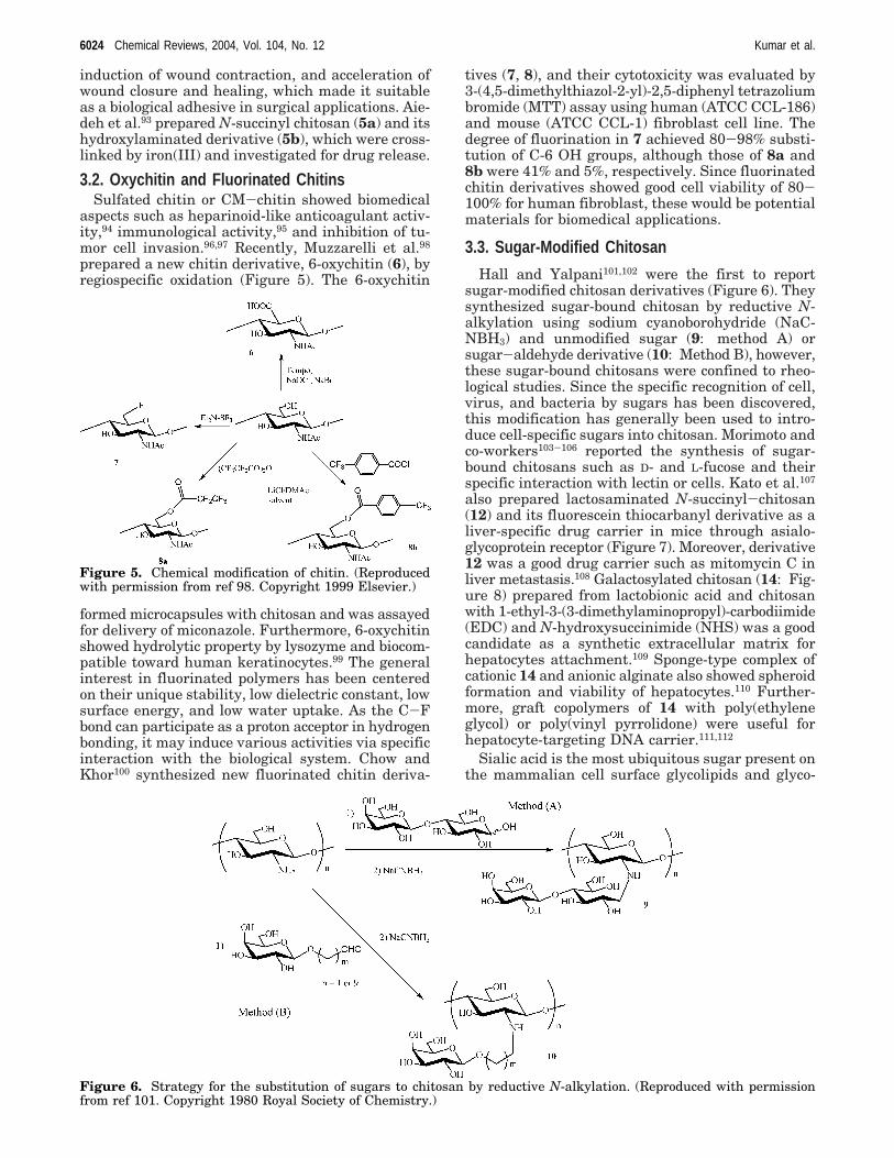
induction of wound contraction, and acceleration ofwound closure and healing, which made it suitableas a biological adhesive in surgical applications. Aie-deh et al.93 prepared N-succinyl chitosan (5a) and itshydroxylaminated derivative (5b), which were cross-linked by iron(III) and investigated for drug release.
3.2. Oxychitin and Fluorinated ChitinsSulfated chitin or CM-chitin showed biomedical
aspects such as heparinoid-like anticoagulant activ-ity,94 immunological activity,95 and inhibition of tu-mor cell invasion.96,97 Recently, Muzzarelli et al.98
prepared a new chitin derivative, 6-oxychitin (6), byregiospecific oxidation (Figure 5). The 6-oxychitin
formed microcapsules with chitosan and was assayedfor delivery of miconazole. Furthermore, 6-oxychitinshowed hydrolytic property by lysozyme and biocom-patible toward human keratinocytes.99 The generalinterest in fluorinated polymers has been centeredon their unique stability, low dielectric constant, lowsurface energy, and low water uptake. As the C-Fbond can participate as a proton acceptor in hydrogenbonding, it may induce various activities via specificinteraction with the biological system. Chow andKhor100 synthesized new fluorinated chitin deriva-
tives (7, 8), and their cytotoxicity was evaluated by3-(4,5-dimethylthiazol-2-yl)-2,5-diphenyl tetrazoliumbromide (MTT) assay using human (ATCC CCL-186)and mouse (ATCC CCL-1) fibroblast cell line. Thedegree of fluorination in 7 achieved 80-98% substi-tution of C-6 OH groups, although those of 8a and8b were 41% and 5%, respectively. Since fluorinatedchitin derivatives showed good cell viability of 80-100% for human fibroblast, these would be potentialmaterials for biomedical applications.
3.3. Sugar-Modified Chitosan
Hall and Yalpani101,102 were the first to reportsugar-modified chitosan derivatives (Figure 6). Theysynthesized sugar-bound chitosan by reductive N-alkylation using sodium cyanoborohydride (NaC-NBH3) and unmodified sugar (9: method A) orsugar-aldehyde derivative (10: Method B), however,these sugar-bound chitosans were confined to rheo-logical studies. Since the specific recognition of cell,virus, and bacteria by sugars has been discovered,this modification has generally been used to intro-duce cell-specific sugars into chitosan. Morimoto andco-workers103-106 reported the synthesis of sugar-bound chitosans such as D- and L-fucose and theirspecific interaction with lectin or cells. Kato et al.107
also prepared lactosaminated N-succinyl-chitosan(12) and its fluorescein thiocarbanyl derivative as aliver-specific drug carrier in mice through asialo-glycoprotein receptor (Figure 7). Moreover, derivative12 was a good drug carrier such as mitomycin C inliver metastasis.108 Galactosylated chitosan (14: Fig-ure 8) prepared from lactobionic acid and chitosanwith 1-ethyl-3-(3-dimethylaminopropyl)-carbodiimide(EDC) and N-hydroxysuccinimide (NHS) was a goodcandidate as a synthetic extracellular matrix forhepatocytes attachment.109 Sponge-type complex ofcationic 14 and anionic alginate also showed spheroidformation and viability of hepatocytes.110 Further-more, graft copolymers of 14 with poly(ethyleneglycol) or poly(vinyl pyrrolidone) were useful forhepatocyte-targeting DNA carrier.111,112
Sialic acid is the most ubiquitous sugar present onthe mammalian cell surface glycolipids and glyco-
Figure 6. Strategy for the substitution of sugars to chitosan by reductive N-alkylation. (Reproduced with permissionfrom ref 101. Copyright 1980 Royal Society of Chemistry.)
Figure 5. Chemical modification of chitin. (Reproducedwith permission from ref 98. Copyright 1999 Elsevier.)
6024 Chemical Reviews, 2004, Vol. 104, No. 12 Kumar et al.

proteins and is the key epitope recognized as beingessential for a number of pathogenic infections. More-over, sialic-acid-containing polymers have been shownto be potent inhibitors of hemagglutination of humanerythrocytes by influenza viruses.113-117 Sashiwa andRoy118,119 prepared sialic-acid-bound chitosan (16: Fi-gure 9) as a new family of sialic-acid-containing poly-mers using p-formylphenyl-R-sialoside (15) by reduc-tive N-alkylation. Since derivative 16 was insolublein water, continuous N-succinylation was carried outand a water-soluble derivative was obtained (17). Thespecific binding with wheat germ agglutinin lectinwas shown in water-soluble derivative 17.
Human antibodies against R-galactosyl epitope areresponsible for acute rejection of xenotransplantatedorgans from lower animals. Artificial glycopolymershaving R-galactosyl epitope are of interest from theviewpoint of medical transplantation of pig liver sincethey can block immune rejection. Water-soluble R-galactosyl chitosan (18: Figure 10), prepared follow-ing the same strategy adopted for chitosan-sialicacid conjugates, showed specific binding againstR-galactosyl specific lectin (Griffonia simplicifolia).120
Chitosan-sialic acid or R-glactosyl conjugates withdifferent degrees of substitution have been prepared,and their lectin binding property has been evalu-
ated.121 These conjugates are believed to inhibitinfluenza viruses or can act as blocking agents foracute rejection.
Figure 7. Synthesis of lactosaminated N-succinyl-chitosan. (Reproduced with permission from ref 107. Copyright 2001Elsevier.)
Figure 8. Synthesis of galactosylated chitosan. (Reproduc-ed with permission from ref 109. Copyright 2003 Elsevier.)
Figure 9. Synthesis of sialic-acid-chitosan and its N-succinylation. (Reproduced with permission from ref 119.Copyright 2000 Royal Society of Chemistry.)
Figure 10. Structure of water-soluble R-galactosyl chito-san. (Reproduced with permission from ref 120. Copyright2000 American Chemical Society.)
Chitosan Chemistry and Pharmaceutical Perspectives Chemical Reviews, 2004, Vol. 104, No. 12 6025

3.4. Chitosan −Dendrimer HybridDendrimers are attractive macromolecules owing
to their multifunctional properties122-124 and usefulapplications as viral and pathogenic cell adhesioninhibitors.125,126 Increasing scientific efforts have goneinto the design and synthesis of dendrimers.127-129
Dendronized polymers, on the other hand, are alsoattractive because of their rod-like conformation andnanostructure.130,131 Although, several investigationshave been published toward the synthesis of den-dronized polymers,132,133 very few reports are avail-able on dendronized polysaccharides, especially re-lated to chitin and chitosan backbone.
Sashiwa et al.134-140 established at first the syn-thesis of a variety of chitosan-dendrimer hybridsmainly by two procedures (Figure 11). In method A,the corresponding dendrimers bearing aldehyde andspacer are synthesized, and then these are reactedwith chitosan by reductive N-alkylation. This proce-dure is advantageous because no cross-linking takesplace during the reaction. However, generation ofreactive dendrimer is limited owing to its sterichindrance. It is possible to generate more reactivedendrimers following method B, which uses com-mercial amino-dendrimers such as poly(amido-amine) (PAMAM) and poly(ethylene imine) (PEI)dendrimers. However, method B suffers from thepossibilities of cross-linking. The typical example ofa “tree-type” hybrid generated by method A is shownin Figure 12.134,139 The terminology “tree-type hybrid”is based on the assumption that chitosan is a trunk,the spacer part is a main branch, dendrimer is asubbranch, and the functional sugar is a flower (orleaf). In this case, tetraethylene glycol was modifiedin 5 or 7 steps to synthesize the scaffold of dendrimer.PAMAM dendrimers of generation (G) from 1 to 3bearing tetraethylene glycol spacer were prepared,attached to sialic acid by reductive N-alkylation, andfinally attached to chitosan. The degree of substitu-tion (DS) of dendrimer decreased with increasinggeneration as 0.08 (G ) 1), 0.04 (G ) 2), and 0.02 (G) 3) owing to the steric hindrance of dendrimer.Figure 13 shows the different types of chitosan-dendrimer hybrids.135 Sialic acid dendron bearing afocal aldehyde end group was synthesized by a
reiterative amide bond strategy. Trivalent (G ) 1)and nonavalent (G ) 2) dendrons having gallic acidas the branching unit and triethylene glycol as thespacer arm were prepared and initially attached toa sialic acid p-phenylisothiocyanate derivative. Thefocal aldehyde sialodendrons were then convergentlyattached onto chitosan. The DS of sialodendrimerwere 0.13 (G ) 1) and 0.06 (G ) 2). Further biologicalevaluation of these promising hybrids is being inves-tigated toward the inhibition of viral pathogensincluding the influenza virus.
The chitosan-dendrimer hybrid prepared by methodB is shown in Figure 14.136 As the construction of hy-brid was difficult from original chitosan, a derivative,N-methoxycarbonylethyl chitosan (21), was used aschitosan backbone. PAMAM dendrimers (G ) 1-5)having a 1,4-diaminobutane core were attached to 21by amidation under conditions that prevent cross-linking. The hybrids 21 could be prepared even inhigh generations (G ) 4 or 5), although the DS ofdendrimer decreased with increasing generation ofdendrimer from 0.53 (G ) 1) to 0.17 (G ) 4) or 0.11(G ) 5). Since this hybrid was soluble in acidic water,undesired cross-linking would not occur. However,two or more intermolecular binding points were ob-served. In any case, sialic acid was successfully at-tached to the primary amine of dendrimer part withDS ranging from 0.7 to 1.4 per sugar unit, whichmeans highly convergent synthesis of sialic acid inchitosan backbone. Given the fact that influenza vir-us hemagglutinins exist as several clusters of trimers(200-300/virions),141 it is likely that the novel den-dronized chitosan-sialic acid hybrids prepared bymethod B would present added beneficial architec-tures not present in previously reported sialodendrim-ers.115,142-144 Preliminary biological evaluation ofanalogous hyperbranched sialodendrimers has al-ready shown increased inhibitory properties.125
Figure 11. Synthetic strategy of chitosan-dendrimerhybrid. (Reproduced with permission from ref 136. Copy-right 2001 American Chemical Society.)
Figure 12. Chemical structure of chitosan-sialoden-drimer hybrid. (Reproduced with permission from ref 139.Copyright 2002 Elsevier.)
6026 Chemical Reviews, 2004, Vol. 104, No. 12 Kumar et al.

3.5. Cyclodextrin-Linked ChitosanCyclodextrins (CD) have gained prominence in re-
cent years because the cavity, of hydrophobic nature,is capable of binding aromatic and other small or-ganic molecules and therefore provides ideal bindingsites. CD-linked chitosan is interesting for drug deli-
very, cosmetics, and analytical chemistry.145 Al-though the functionalization of hydroxyl groups atthe 6-position in CD is relatively easy, the secondaryalcohols (2 and 3 positions) are most important inbinding studies.146 Sakairi and co-workers147,148 pre-pared R-CD-linked chitosan (Figure 15, 23) using 2-O-formylmethyl-R-CD by reductive N-alkylation andconfirmed the host-guest complex of 23 with p-ni-trophenol. Chen and Wang149 obtained CD-linked
Figure 13. Hybridization of chitosan with sialodendrimer, composed of gallic acid as a junction point. (Reproduced withpermission from ref 135. Copyright 2001 American Chemical Society.)
Figure 14. Reaction of N-methoxycarbonylethylchitosanwith PAMAM dendrimer. (Reproduced with permissionfrom ref 136. Copyright 2001 American Chemical Society.)
Figure 15. Cyclodextrin-linked chitosan. (Reproducedwith permission from refs 148-150. Copyright 1998 and2001 Wiley-Liss, Inc. (http://www3.interscience.wiley.com/cgi-bin/jabout/30035/ProductInformation.html.))
Chitosan Chemistry and Pharmaceutical Perspectives Chemical Reviews, 2004, Vol. 104, No. 12 6027

chitosan (24) using tosylated â-CD and further eval-uated the potential of â-CD for the release of 131I2 invivo. The CD-linked chitosan could also be preparedby the intermediate of its monochlorotriazinyl de-rivative (25).150 This compound was used for decon-tamination of waters containing textile dyes. Aninsoluble cross-linked chitosan bearing â-CD wasprepared using N-succinyl chitosan and aminated-â-CD via amide bond formation.151 The â-CD-linkedchitosan using 1,6-hexamethlene diisocyanate asspacer was also prepared by Sreenivasan.152 Thismaterial interacts with cholesterol and might beuseful as an adsorbent.
3.6. Biodegradation of Modified ChitosansIn the field of electronic equipment, such as com-
puter and medical instruments, high-speed and high-density information processing is becoming in-creasingly common, and there is the danger that evenlow-intensity electromagnetic radiation may causemalfunction. For example, use of mobile telephonesand portable computers is restricted in aircrafts andhospitals. Hence, there is a need for highly reliableelectromagnetic radiation shielding materials. Bio-degradability of binder between plastics and shield-ing materials is required for recycling of plastics, andsince chitosan is biodegradable, it would be usefulas a binder. Chitosan itself, however, is hydrophilic,while plastics are hydrophobic; therefore, modifica-tion of chitosan to impart solubility in organicsolvents is necessary for spraying on the surface ofthe plastics. The hydrophobic ester group contributessolubility in organic solvents and is hydrolyzed by
enzymes such as lipase (Figure 16). Sashiwa etal.153-155 reported the synthesis of organosoluble andbiodegradable acylchitosans intended for the electro-magnetic shielding materials. Acylchitosans bearinglong acyl chains (27: n > 4), hydrophobic pivaloyl(28), or benzoyl (29) groups showed organosolubleproperties. Interestingly, O-acetylchitosan (26) showedwater solubility like acetylcellulose.155 These chitosanderivatives (26, 27, and 29) showed good biodegrada-tion. Furthermore, these acylchitosans showed goodbinding properties between plastics and electromag-netic shielding materials; thus, they are useful asbiodegradable binders.
Alternatively, the biodegradation has been inves-tigated on some water-soluble chitosan derivativesprepared by Michael reaction (Figure 17).156,157 Thisreaction has been developed as a new method for thechemical modification of chitosan158 or partiallydeacetylated chitin,159 and the resulting productswere used as precursors for the construction ofchitosan-dendrimer hybrid.136,138 Most recently,Michael reaction of chitosan with acrylic acid wasalso reported using water as solvent.156 In this case,acrylic acid acted as both proton donor to dissolvechitosan in water and reagent for Michael reaction,so that water-soluble N-carboxyethylchitosan (30)was successfully prepared. If water-soluble acrylreagents could be applied for this reaction, noveltypes of functional groups will be introduced by asimple procedure. Various chitosan derivatives wereprepared by this reaction in water and acetic acidwith various acryl reagents.157 Biodegradation data
Figure 16. (A) Concept and (B) synthesis of organo-soluble chitosan derivatives. (Reproduced with permission from ref154. Copyright 2002 American Chemical Society.)
Figure 17. Michael reaction of chitosan with acrylic acid and its esters in water. (Reproduced with permission from ref157. Copyright 2003 American Chemical Society.)
6028 Chemical Reviews, 2004, Vol. 104, No. 12 Kumar et al.

of these chitosan derivatives by standard activatedsludge is presented in Table 1. In any case, thebiodegradability was enhanced by chemical modifica-tion compared with original chitosan. Excellent bio-degradability was shown in 30 with various DS,although it gradually decreased with increasing DS.Derivatives 33, 34, and 35 modified with poly-(ethyleneglycol) (PEG), quaternary ammonium, andamido groups also showed good biodegradability.Moderate biodegradation was shown by 31 and 36bearing hydroxyethyl and nitrile groups. These re-sults suggest that biodegradation was very associatedwith the chemical structure of chitosan derivatives.Thus, carboxyl, quaternary ammonium, amido, andPEG groups were convenient for biodegradation, buthydroxyethyl and nitrile groups were not. On theother hand, biodegradation was independent of thewater solubility of chitosan derivatives. Biodegrada-
tion of chitosan is quite slow by standard activatedsludge. Modified chitosans are more prone to biodeg-radation owing to the destroyed crystalline structureof chitosan.
3.7. Crown-Ether-Bound ChitosanCrown ethers have particular molecular structures
and good complexing selectivity for metal ions. Thesecrown-ether-bound chitosans will have a strongercomplexing capacity and better selectivity for metalions because of the synergistic effect of high molec-ular weight. Tang et al.160 prepared the crown-ether-bound chitosan with Schiff’s-base-type (37a) and itsreduced form (37b) Figure 18. Their chemical struc-tures were characterized by elemental analysis, IR,X-ray, and solid-state 13C NMR analyses. Crown-ether-bound chitosans had not only good adsorptioncapacities for metal ions Pd2+, Au3+, and Ag+, but alsohigh selectivity for the adsorption of Pd2+ in thepresence of Cu2+ and Hg2+. Cross-linked types ofcrown-ether-bound chitosans were also reported (Fig-ure 19).161 These cross-linked derivatives have spacenet structures with embedded crown ethers, and eachmesh has a certain space volume. When originalchitosan reacted with 4,4′-dibromobenzo-18-crown-6-crown ether, the cross-linked product between6-OH and NH2 was obtained (38). However, thisproduct would include heterogeneous cross-linkingstructure between 6-OH and 6-OH or NH2 and NH2.Benzylidene-protected chitosan (CTB) would producea homogeneous cross-linking structure between 6-OHand 6-OH (39). These crown-ether-bound chitosans
Table 1. Biodegradation of Chitosan and ItsDerivatives156,158
sample DS solubility in H2O biodegradation,a %
chitosan 0.00 no 1.630 0.18 yes 67.330 0.27 yes 62.530 0.46 yes 51.331 0.44 yes 8.632 0.26 yes 5.033 0.29 nob 24.834 0.38 yes 33.635 0.24 no 27.836 0.49 no 7.7
a Time, 21 days. b Water-insoluble after lyophilization.
Figure 18. Crown-ether-bound chitosans. (Reproduced with permission from ref 160. Copyright 2002 Wiley-Liss, Inc.(http://www3.interscience.wiley.com/cgi-bin/jabout/30035/ProductInformation.html))
Figure 19. Cross-linked type of crown-ether-bound chitosan. (Reproduced with permission from ref 161. Copyright 2002Wiley-Liss, Inc. (http://www3.interscience.wiley.com/cgi-bin/jabout/30035/ProductInformation.html))
Chitosan Chemistry and Pharmaceutical Perspectives Chemical Reviews, 2004, Vol. 104, No. 12 6029

would be useful for separation and preconcentrationof heavy or precious metal ions in aqueous environ-ments.
On the other hand, calixarenes have demonstratedoutstanding complex ability toward ions, organicmolecules, etc, and are considered the third best hostmolecules, after cyclodextrins and crown ethers. Liet al.162 reported the first synthesis of calixarene-modified chitosan (Figure 20). The adsorption prop-erties of calixarene-modified chitosan (I and II) weregreatly varied compared with that of original chito-san, especially with the adsorption capacity towardAg+ and Hg2+, because of the presence of the calix-arene moiety. These derivatives did not dissolve ingeneral organic solvent; however, they can easily bepowdered and are thus better adsorbents than simplechitosan.
3.8. Chemical Grafting of ChitosanGraft copolymerization onto chitin and chitosan is
an important study for the functionalization andpractical use of them. A lot of initiators have beeninvestigated for grafting such as ceric ion, Fenton’sreagent, γ-irradiation, various radicals, and ring-opening methods.163 An interesting feature of poly-oxazoline chains is the fact that they are regardedas pseudo-peptides having good flexibility.164 It hasalso been disclosed that oxazoline-grafted chitosan(Figure 21, 41, degree of deacetylation 50%) has thecapability of incorporating lipase P and catalase andincreasing hydrolytic activity compared with freeenzymes.165 Furthermore, the molecular shape ofwater-soluble grafted chitosan 41 was evaluated byatomic force microscopy (AFM), cryo-transmissionelectron microscopy (cryo-TEM), and small-angle
neutron scattering (SANS) analysis. Grafted chitosanbearing short graft chain formed a ring structure(40-60 nm of diameter) unimolecularly, while thatbearing middle chain length was monodisperse spheri-cal (30-40 nm), whereas the longer chain aggregatedintermolecularly that led to larger particles (100-400 nm).166 These studies should be useful for thestrategy to regulate molecular design and guest-binding properties of water-soluble-grafted chitosan.
Homo- and copolymers based on lactic acid havebeen widely used in sutures and drug-release systemsowing to their biodegradability in the animal body,while pH-sensitive polymer gels have potential usein the delivery of drugs to specific regions of thegastrointestinal tract. Novel pH-sensitive physicalcross-linked hydrogels were synthesized by graftingD,L-lactic acid onto amino groups in chitosan withoutcatalysis (Figure 22, 42).167 pH sensitivity was dueto aggregation of the hydrophobic side chains. Thespecific solution content of hydrogels decreased whenthe pH value and ionic strength were increased.
Although grafting on chitin and chitosan has beenperformed by high-energy irradiation or the additionof initiators such as cerium(IV) and redox system,these methods affect degradation of the polysaccha-ride backbone, thus giving rise to the grafted prod-ucts with complicated and ambiguous structure.Kurita et al.168 synthesized graft copolymer ontochitin by use of the mercapto group (Figure 23).Methyl methacrylate (MMA) was efficiently graftedonto mercaptochitin in DMSO, and grafting percent-age reached 1300%. Although the side-chain estergroups were resistant to aqueous NaOH only, hy-drolysis of ester could be achieved with a mixture ofaqueous NaOH and DMSO. The grafted chitins (43)showed pronounced activity in terms of moistureabsorption and lysozyme susceptibility when com-pared to unmodified chitins.
3.9. Enzymatic Modification of ChitosanThe enzymatic approach to modification of chitin
and chitosan is interesting owing to its specificity andenvironmental impact compared with chemical modi-fication. With respect to health and safety, enzymesoffer potential by eliminating the need for (andhazards associated with) reactive reagents. Payneand co-workers169 reported the enzymatic grafting ofphenolic compounds onto chitosan to confer watersolubility under basic conditions (Figure 24). Tyro-sinase converts a wide range of phenolic substratesinto electrophilic o-quinones. Using slightly acidicconditions (pH 6), chitosan could be modified underhomogeneous conditions with chlorogenic acid, anatural product. The modified chitosan was dissolvedin both acidic and basic conditions, although thedegree of modification was low. Quinone chemistry,however, remains poorly characterized because of itscomplexity. It can undergo two different reactions toyield either Schiff base (44) or Michael-type adducts(45). Since it is possible for quinones to undergoeither or both types of reactions with amines, as wellas undergoing oligomer-forming reactions with otherquinones, it is common for reactions between quino-nes and amines to yield a complex mixture of
Figure 20. Calixarene-bound chitosan. (Reproduced withpermission from ref 162. Copyright 2003 Wiley-Liss, Inc.(http://www3.interscience.wiley.com/cgi-bin/jabout/30035/ProductInformation.html))
Figure 21. Oxazoline-grafted chitosan (DDA ) 50%).(Reproduced with permission from ref 166. Copyright 2002Wiley. (http://www3.interscience.wiley.com/cgi-bin/jabout/30035/ProductInformation.html))
6030 Chemical Reviews, 2004, Vol. 104, No. 12 Kumar et al.

products.170 Furthermore, to alter the surface andrheological properties of chitosan, hexyloxyphenolwas grafted onto chitosan mediated by tyrosinase.171
On the basis of the contact angle measurements,heterogeneous modification of chitosan film yieldeda hydrophobic surface owing to the substitute, whilehomogeneously modified chitosan offered rheologicalproperties characteristic of associating water-solublepolymers.
Therefore, the biochemically relevant quinonesstudied so far are preferred materials for medicalapplications. For instance, menadione, a syntheticnaphthoquinone derivative having the same physi-ological properties of vitamin K, is particularly proneto rapid reaction with chitosans and greatly modifiesits spectral characteristics and increases the surfacehydrophobicity of the chitosan films.172 Researchunder way will provide information on the biologicalproperties of these enzymatically modified chitosans.
3.10. OthersColloidal systems have found numerous applica-
tions as promising delivery vehicles of drugs, pro-teins, antigens, and genes due to their reduced toxicside effect and improvement of therapeutic effect.173
The micellar behavior of polymeric self-assemblysystem offers an advantage as one of the colloidalsystems that has been widely investigated in thefields of biotechnology and pharmaceutics.174 Precisecontrol of the size and structure are the criticaldesign parameters of a micellar system for drugdelivery applications. To control the size of self-
aggregates (SA), chitosan was depolymerized withsodium nitrite and hydrophobically modified withdeoxycholic acid to form SA in aqueous media (Figure25).175 The size of SA was 130-300 nm in diameter.Due to the chain rigidity of chitosan, the structureof SA was suggested to be cylindrical bamboo-like andmight form a very poor spherical form of a bird’s nest-like structure. The potential applications of SA as agene delivery carrier were tested, and significantinfluence of transfection efficiency by SA was ob-served against Cos-1 cells (up to a factor of 10). Thisapproach to control the size and structure of chitosan-derived SA may find a wide range of applications ingene delivery as well as general drug delivery ap-plications. Lee et al.176 reported the delivery ofadriamycin (ADR) using SA of the deoxycholic-acid-modified chitosan (46). Deoxycholic acid was co-valently conjugated to chitosan via EDC-mediatedreaction to generate SA nanoparticles. ADR was
Figure 22. D,L-Lactic-acid-grafted chitosan. (Reproduced with permission from ref 167. Copyright 1999 Wiley-Liss, Inc.(http://www3.interscience.wiley.com/cgi-bin/jabout/30035/ProductInformation.html))
Figure 23. Grafting of MMA onto mercaptochitin. (Reproduced with permission from ref 168. Copyright 2002 AmericanChemical Society.)
Figure 24. Enzymatic grafting of chitosan with phenol and tyrosinase. (Reproduced with permission from ref 169. Copyright1999 Wiley-Liss, Inc. (http://www3.interscience.wiley.com/cgi-bin/jabout/30035/ProductInformation.html))
Figure 25. Deoxycholic-acid-modified chitosan. (Repro-duced with permission from ref 175. Copyright 2001American Chemical Society.)
Chitosan Chemistry and Pharmaceutical Perspectives Chemical Reviews, 2004, Vol. 104, No. 12 6031

physically entrapped inside the SA, and slow releaseof ADR was achieved.
The formation of hydrogels from polymers usingnoncovalent cross-linking is a useful method ofpreparing hydrogels for drug delivery since these gelsare likely to be more biocompatible as gel formationdoes not require the use of organic solvents orchemical reactions which may be potentially del-eterious to the drug loaded. Such physically cross-linked chitosan-based gels are formed by exploitingeither hydrogen-bonding or hydrophobic attractions.Uchegbu and co-workers177 focused on the use ofpendant hydrophobic groups to achieve noncovalentcross-linking. Palmitoyl glycol chitosan (GCP, Figure26) hydrogel has been evaluated as an erodible
controlled release system for the delivery of hydro-philic macromolecules.178 Fluorescein isothiocyan-ate (FITC)-dextran and/or amphiphilic derivativesGelucire 50/13 or vitamin E D-R-tocopherol poly-(ethylene glycol) succinate were used as model macro-molecules. Hydration and erosion were governed bythe hydrophobicity of the gel and the presence of theamphiphilic additives. The controlled release of FITC-dextran was governed by the hydrophobicity of thegel. In their subsequent study, GCP hydrogel wasevaluated as a deliverer of hydrophobic drugs, den-bufylline, via the buccal route.179 The buccal routehas been advocated as a possible route of administra-tion for drugs which undergo extensive hepatic first-pass metabolism or are susceptible to degradation inthe gastrointestinal tract.
Glass beads have received attention as supportingmaterial owing to their controllable and narrow sizedispersion properties in addition to their mechanicalstrength. Sakairi and co-workers180 reported a newhybrid that adsorbs transition-metal ions by thesurface modification of nonporous beads with chito-san. Glass beads bearing aldehyde groups wereproduced and modified with chitosan by reductiveN-alkylation (Figure 27). Metal ions such as Cu2+,Ag+, Pb2+, Fe3+, and Cd2+ were collected (over 90%)on a column of chitosan-modified glass beads. Theyalso reported another type of chitosan-modified glassbeads through a 1,3-thiazolidine linker.181 In thiscase, terminal aldehyde group (49) produced by
nitrous acid degradation of chitosan was used for thecoupling with L-cysteine linker of glass beads (Figure28). This method to prepare chitosan-modified glassbeads could be applied for a variety of silica materi-als.
4. Chitin and Chitosan in Different Forms4.1. Nanoparticles
Alonso and co-workers182 reported the preparationof nanoparticles based on ionic gelation made solelyof hydrophilic polymers. The preparation process isextremely mild and involves the mixture of twoaqueous phases at room temperature. One phasecontains the polysaccharide chitosan (CS) and poly-(ethylene oxide), and the other contains polyanionsodium tripolyphosphate (TPP). The authors claimedthat the particle size (200-1000 nm) and zetapotential (between +20 and +60 mV) of nanoparticlescan be modulated by varying the ratio CS/PEO-PPO.They also demonstrated that these new nanoparticleshave great protein loading capacity (entrapmentefficiency up to 80% of the protein) and provide acontinuous release of the entrapped protein for upto 1 week.182 Furthermore, they performed in-depthinvestigations to understand the physicochemicalproperties, surface composition, and mechanisms ofprotein association to chitosan and CS/PEO-PPOnanoparticles.183 The electron micrographs showedthat the particles are spherical. The CS/PEO-PPOnanoparticles exhibited a compact core surroundedby a thick fluffy coat, presumably consisting of PEO-PPO, which could not be found with the particles ofchitosan alone (Figure 29).
Freeze-drying procedure for improving the shelf lifeof the chitosan nanoparticles using various cryopro-tective agents was also investigated, and negligibledifferences between the freeze-dried and fresh par-ticles were found.184 Alonso and co-workers185 studiedtwo different types of chitosan in the form of hydro-
Figure 26. Synthesis of palmitoyl glycol chitosan. (Re-produced with permission from ref 178. Copyright 2002Elsevier.)
Figure 27. Modification of glass bead with chitosan. (Reproduced with permission from ref 180. Copyright 2002 Elsevier.)
Figure 28. Chitosan-modified glass bead through a 1,3-thiazolidine linker. (Reproduced with permission from ref181. Copyright 2003 Elsevier.)
6032 Chemical Reviews, 2004, Vol. 104, No. 12 Kumar et al.

chloride salt. The particles were prepared followingthe above-described methods, leading to a particlesize of 300-400 nm with a positive surface chargeand entrapment efficiency of 55% [insulin/nanopar-ticles (w/w): 55/100]. These particles were used toaddress the difficulties in the nasal absorption ofinsulin, discussed in the following subsections. Theyalso explored chitosan nanoparticles for entrapmentand release studies of the hydrophilic anthracyclinedrug, doxorubicin (DOX).186 They approached theproblem posed by hydrophilicity and cationic chargeof the drug by complexing it with polyanion, dextransulfate, leading to enhanced drug loading.187 Thesenanoparticles were also used for improved deliveryof the drugs to the ocular surface, and cyclosporin A(CyA) was used as a model drug.187 These nanopar-ticles had a mean size of 293 nm Figure 30, a zetapotential of +37 mV, and high CyA associationefficiency and loading (73% and 9%, respectively).Furthermore, in a recent review on colloidal particlesas delivery systems for macromolecules, variouspossibilities for forming particles and their bio-pharmaceutical applications have been discussed.188
Recently, Alonso and co-workers carried out ex-tensive investigations on the design of biodegradablenanoparticles for protein delivery.189 As a part of thisstudy, they prepared three types of particles of whichtwo were related to chitosan and the third was aPEG-PLA-derived particle. They prepared sole chi-tosan particles as reported in their previous pub-
lications182-187 and chitosan-coated PLGA-lecithinparticles by a critically modified double emulsionmethod. These particles were intended for either oralor nasal administration. The preparation process ofCS-PLGA particles is depicted in Scheme 1. Table2 shows the properties of various nanoparticles usedfor comparison purposes in this study.189
Tian and Groves190 reported the formulation andbiological activity of antineoplastic proteoglycansderived from Mycobacterium vaccae in chitosan nano-particles. They prepared chitosan nanoparticles be-tween 600 and 700 nm without the use of organicsolvents. They found that the adsorption and releaseof bovine serum albumin seemed to be affected bythe charge of the two reactants and that at high dosesnot all adsorbate was released. They observed aninitial burst release and a further steady release for4 h in water.190
Ohya et al.191 reported PEG-grafted chitosan nano-particles as peptide drug carriers. They observednanoparticle formation through intermolecular hy-drogen bonding in an aqueous solution. The incor-
Figure 29. Electron transmission microphotography of(a) chitosan nanoparticles, (b) chitosan/PEO-PPO nano-particles (concentration of PEO-PPO in the chitosansolution, 10 mg/mL). (Reproduced with permission fromref 182. Copyright 1997 Wiley-Liss, Inc. (http://www3.interscience.wiley.com/cgi-bin/jabout/30035/Product-Information.html))
Figure 30. Transmission electron micrograph of the CyA-loaded chitosan nanoparticles. (Reproduced with permis-sion from ref 187. Copyright 2001 Elsevier.)
Scheme 1. Flow Chart Depicting StepwisePreparation of Chitosan-PLGA Particles
Chitosan Chemistry and Pharmaceutical Perspectives Chemical Reviews, 2004, Vol. 104, No. 12 6033

poration and release of insulin was dependent on thedegree of introduction of PEG chain on chitosan andobserved sustained release phenomena over time.
Lee et al.176 reported a novel and simple methodfor delivery of adriamycin using self-aggregates ofdeoxycholic-acid-modified chitosan. Deoxycholic acidwas covalently conjugated to chitosan via EDC-mediated reaction, generating self-aggregated chito-san nanoparticles. The active component adriamycinwas entrapped physically within the nanoparticles,and the formed self-aggregates were analyzed byphoton correlation spectroscopy (PCS), fluorescencespectroscopy, and atomic force microscopy. Theyfound self-aggregates are spherical in shape and thatthe initial drug concentration has an influence on thesize of the particles formed. They achieved about 49.6wt % loading efficiency with slow release phenomenaover time in PBS (pH 7.2).176 Kim et al.175 exploredthese self-aggregates of deoxycholic-acid-modifiedchitosan (DAMC) as DNA carriers. They explainedthe critical aspects involved in the self-assemblyformation of deoxycholic-acid-modified chitosan. Fig-ure 31 shows the simulated structure of chitosan,DAMC, and self-aggregates formed by two and fourDAMC molecules, respectively.
Yamamoto et al.192 reported mucoadhesive lipo-somes coated with chitosan for drug delivery. Theyachieved the mucoadhesive formulation by mixingthe chitosan solution with a drug-loaded liposomalsuspension prepared using a thin lipid film hydrationmethod. A linear correlation was observed betweenthe amount of chitosan used for coating and thepercentage adhesion of chitosan-coated liposomes tothe intestinal sac. They observed a reduced initialburst release of carboxy fluorescein with the chitosan-coated liposomes, and a further sustained release wasobserved for about 24 h. They demonstrated thatthese nanoparticles would serve for insulin release.192
Yang et al.193 investigated the formation of posi-tively charged poly(butyl cyanoacrylate) nanopar-ticles stabilized by chitosan. They showed that thesize of the particles is influenced by various factorssuch as pH, the concentration and volume of chitosansolution, and the molecular weight of chitosan.Nimodipine was used as a model drug in thesestudies with a resulting mean particle size of 31.6nm.
Maitra and co-workers194 reported a procedure toprepare ultrafine cross-linked chitosan nanoparticlesin AOT/n-hexane reverse micellar system. Theyobserved that the particle size is influenced by thedegree of cross-linking and was found to be 30 nm
when 10% of the amino groups in the polymeric chainhave been cross-linked, whereas it was 110 nm whenall the amino groups were cross-linked; these par-
Table 2. Particle Size, Zeta Potential, Theoretical Loading, and Encapsulation Efficiency Values of CS,PEG-PLA, and CS-PLGA Nanoparticles Containing TT and CS Nanoparticles Containing Insulina
polymer protein loaded size (nm) ú potential (mV)theoreticalloading (%)
encapsulationefficiency (%)
PLA tetanus toxoid 192 ( 12 -47.9 ( 1.5 1 36.7 ( 0.3PEG-PLA tetanus toxoid 196 ( 20 -23.9 ( 1.2 1 31.1 ( 0.5CS-PLGA tetanus toxoid 500 ( 29 +21.8 ( 1.1 1 90.0 ( 3.8CS tetanus toxoid 354 ( 27 +37.1 ( 5.9 10 55.1 ( 3.4CS insulin 337 ( 14 +36.9 ( 0.3 40 94.7 ( 2.1
a Reproduced with permission from ref 189. Copyright 2002 Elsevier.
Figure 31. Simulated structure of (a) chitosan and (b)DAMC. (c and d) Simulated structure of assembled self-aggregates formed by two and four DAMC molecules,respectively. (Reproduced with permission from ref 175.Copyright 2001 American Chemical Society.)
6034 Chemical Reviews, 2004, Vol. 104, No. 12 Kumar et al.

ticles were thoroughly characterized. The electronmicrographs reveal that the particles were sphericalin shape and that lower cross-linking of the particlesleads to smaller aggregates, while highly denseaggregates were formed at 100% cross-linking. Thebiodistribution of the particles after intravenousinjection in mice showed that these particles remainin the blood for a considerable amount of time Figure32 and distribute in the heart, liver, kidneys, bladder,and vertebral column. Other than these organs, theparticles were distributed in the bone marrow (Figure32), leaving the possibility of using these particlesfor bone imaging and targeting purposes.194
Andersson and Lofroth195 investigated a new micro-emulsion based on heparin/chitosan complex suitablefor oral administration. The microemulsion is basedon the ingredients that are acceptable to humans.These microemulsions were studied with or withoutbiologically active ingredients by dynamic light scat-tering, turbidity, diffusion-NMR, and conductivity.Appropriate mixing and modifications of these micro-emulsions lead to nanometer-sized heparin/chitosancomplexes.195
Pan et al.196 reported chitosan nanoparticles forprotein delivery following a reported method182 withslight modifications. The critical investigations in-clude determination of the formation zone of thenanoparticles, where they used different concentra-tions of chitosan and TPP. On adding TPP to thechitosan solution under stirring, they observed three
different systems, viz., solution, suspension, andaggregates. Varied chitosan concentrations (0.9-3.0mg/mL) and TPP (0.3-0.8 mg/mL) lead to differentparticle sizes.
Nanoparticles of methotrexate (MTX) were pre-pared using O-carboxymethyl chitosan (O-CMC) aswall-forming materials and an isoelectric-criticaltechnique under ambient condition.197 The effects ofthe MTX/O-CMC ratio and amount of cross-linkingagents on drug release in different media wereevaluated. The changes of size and effective diameterof O-CMC nanoparticles were detected by SEM anda laser light scattering system before and after drugrelease. The authors claimed that these nanoparticlesconstitute an attractive alternative to other anti-cancer drugs and enzyme carriers.197
Kumar et al.198 reported a emulsion-diffusion-evaporation technique to make cationic nanospherescomposed of biodegradable and biocompatible co-polyester poly(L-lactide acid-co-glycolide) (PLGA).PVA-chitosan blend was used to stabilize the PLGAnanospheres. The nanospheres have cationic surfacecharge and can readily bind DNA electrostatically.One of the noticeable finding from these investiga-tions is that PVA is the most essential componentrequired to stabilize the PLGA nanoparticles; how-ever, the unbound PVA is hard to eliminate afterseveral washings. On the other hand, chitosan alonecould not stabilize the particles formed. Therefore, ablend of PVA-chitosan was used, leading to mono-dispersed cationic PLGA nanoparticles with no un-bound PVA-chitosan blend Figure 33.
Since the first reports on nanoparticles for drug/vaccine delivery about 3 decades ago, a considerableamount of work has been done. It is quite under-standable that nanoparticles have distinct advan-tages as far as pharmaceutical applications areconcerned; however, they suffer from problems inscalability and in some instances reproducibility. Itis very unfortunate that we are still at infancy in thisarea of research.
4.2. MicrospheresAs a drug carrier chitosan helps overcome certain
adverse characteristics of drugs such as insolubilityand hydrophobicity, but the semicrystalline powderdoes not lend itself to direct compression. Neverthe-less, chitosan powders have been evaluated in directcompression tests, and the formulations so far de-
Figure 32. Organ distribution of 99mTc-radiolabeledchitosan nanoparticles, 30, 60, and 240 min postinjection.(Reproduced with permission from ref 194. Copyright 2002Elsevier.)
Figure 33. Atomic force microscope images of (A) nanospheres with PVA alone as stabilizer, (B) nanospheres with chitosanalone, (C) PVA-chitosan blend (bar ) 150 nm). (Adapted with permission from ref 198. Copyright 2004 Elsevier.)
Chitosan Chemistry and Pharmaceutical Perspectives Chemical Reviews, 2004, Vol. 104, No. 12 6035

veloped include excipients to facilitate compres-sion.199,200 For example, commercial chitosan tabletsfor weight control contain magnesium stearate as abinder with negative consequences on the efficacy ofchitosan.
4.2.1. Preparation of Microspheres by Spray DryingSpray drying includes four sequential stages: atom-
ization through a spray nozzle, contact of the sprayedfeed with warm air, drying of the droplets, andcollection of the solid chitosan Figure 34. Chitosansolutions with drug can be fed to a spray drier at aslightly acidic pH. The size of the particles is influ-enced by various process parameters such as size ofthe nozzle, rate of feeding, and inlet air temperature.When acetone is convenient for dissolving certaindrugs, aqueous solutions may contain some organicsolvent, for instance, a 0.5% chitosan solution maybe made with a mixture of water, glacial acetic acid,and acetone in 49.5/0.5/50 (v/v).
One point concerning exposure of the sample to hotair needs to be explained: the inlet air temperatureis measured prior to flowing into the drying chamberand may be set at 160 °C or higher; however, thegradient between the wet surface and unsaturatedgas actually leads to evaporation at much lowertemperatures.
4.2.2. Spray-Dried ChitosansThe spray-drying technique has been applied to
chitosan suspensions, chitosan salts, chitosan gelatin-ethylene oxide, and chitosan-ethylcellulose mixture.A variety of chitosan salt solutions were spray dried,leading to microspheres having diameters between2 and 5 µm with improved binding functionality. Thechitosan microsphere free-flowing powder is compres-sible and hence most suitable as a drug carrier.201-209
The following are some examples.Chitosan microspheres were prepared by spray
drying using type-A gelatin and ethylene oxide-propylene oxide block copolymer as modifiers. Theparticle shape, size, and surface morphology of micro-spheres and the in vitro drug release were sig-nificantly affected by the concentration of gelatin.Betamethasone disodium phosphate (BTM)-loaded
microspheres demonstrated good drug stability (<1%hydrolysis product), high entrapment efficiency (95%),and positive surface charge. Formulation factors werecorrelated to particulate characteristics for optimiz-ing BTM microspheres in pulmonary delivery.206
By properly choosing excipient type and concentra-tion and varying the spray-drying parameters, a highdegree of control was achieved over the physicalproperties of the dry chitosan powders. The beta-methasone release rates were influenced by the drug/polymer ratio and found to increase when the drugloading was decreased. The in vitro release of beta-methasone showed a dose-dependent burst followedby a slower release phase that was proportional tothe drug concentration in the range 14-44% w/w.206
Chitosan microspheres containing chlorhexidinediacetate, an antiseptic, were prepared by a spraydrying. Chlorhexidine from the chitosan micro-spheres dissolved more quickly in vitro than chlor-hexidine powder. The minimum inhibitory concen-tration, minimum bacterial concentration, and killingtime showed that loading of chlorhexidine into chi-tosan was able to maintain or improve the anti-microbial activity of the drug, the improvement beingparticularly high against Candida albicans. It shouldbe noted that the drug did not decompose despite itsthermal lability above 70 °C. Buccal tablets wereprepared by direct compression of the microsphereswith mannitol alone or with sodium alginate. Aftertheir in vivo administration, determination of chlor-hexidine in saliva showed the capacity of theseformulations to give a prolonged release of the drugin the buccal cavity.210
An inclusion complex composed of progesterone andhydroxypropyl-â-cyclodextrin (HPCD) was preparedby the spray-drying and freeze-drying methods.Progesterone alone and its inclusion complex withHPCD were incorporated into chitosan by spraydrying and freeze drying. Release data showed sig-nificant improvement of the dissolution rate of proges-terone, and a controlled release was obtained in thepresence of chitosan.211
Gemfibrozil, an acidic antihyperlipoproteinemicdrug, was covalently linked to poly[R,â-(N-2-hydroxy-ethyl-D,L-aspartamide)] (PHEA) and poly [R,â-(N-3-hydroxypropyl-D,L-aspartamide)] (PHPA) by an esterlinkage. Microspheres composed of different molec-ular weight chitosans alone or as a mixture with2-hydroxypropylmethyl cellulose (HPMC), PHEA, orPHPA and with different polymer/drug ratios (2:1and 3:1, w/w) were prepared by spray drying. In vitrodissolution tests showed that microspheres werealways suitable to modulate gemfibrozil release andthat the best conditions were achieved by micro-spheres composed of the low molecular weight chi-tosan combined with PHPA/HPMC with either a 2:1or 3:1(w/w) polymer/drug ratio.212
The general chemical behavior of chitosan, how-ever, should be considered in order to avoid certaindifficulties stemming from its insolubility at pHhigher than 6.5 and its reactivity under the thermalconditions of the sprayer. For example, spray dryingof 1-2% chitosan in acid solution at 168 °C seems tobe easy; however, the release of a drug from the
Figure 34. Methylpyrrolidinone chitosan microspheresobtained by spray drying.
6036 Chemical Reviews, 2004, Vol. 104, No. 12 Kumar et al.

spray-dried chitosan depends on the acetic acidconcentration due to the acetylation reaction occur-ring at the temperature to which the salt is exposed,certainly lower than 168 °C but significantly high forside reactions to occur. In fact, the degree of acet-ylation of chitosan increased during spray dryingaffected its enzymatic degradability.208
Chitosan can be spray dried at neutral pH if acolloidal suspension is prepared with NaOH. Never-theless, this preparation is time consuming due tothe difficulties involved in washing and removal ofexcess alkali and salts. Chitosan has been recentlyfound to be soluble in alkaline media, viz., NH4HCO3solutions, by forming ammonium carbamate (Chit-NHCO2
-NH4+), i.e., a transient anionic form that
keeps it soluble at pH 9.6, while reversibly maskingthe polycationic nature of chitosan. Because am-monium carbamates and NH4HCO3 decompose ther-mally and liberate CO2, NH3, and water, this alkalinesystem is perfectly suitable for producing chitosanmicrospheres by spray drying.213
4.2.3. Spray-Dried Polyelectrolyte Complexes
Chitosan forms polyelectrolyte complexes withpolyanions, such as 6-oxychitin, heparin, carra-geenan, pectin, xanthan, acacia gum, hyaluronic acid,alginic acid, poly(acrylic acid), carboxymethyl cel-lulose, DNA, and other macromolecules.205,214-227
These reactions are very fast and in general lead tothe immediate disordered precipitation of the in-soluble coacervates upon mixing, particularly whenthe final pH value is neutral or higher. The poly-anions are normally in the form of sodium salt andshow alkaline hydrolysis that contributes to chitosaninsolubilization upon mixing. Some examples arediscussed below.
Continued blood anticoagulation is achieved bysubcutaneous administration of low molecular weightheparin or with an orally active anticoagulant suchas warfarin. An oral heparin would avoid the incon-venience of subcutaneous injections and adverseevents associated with warfarin. Alginate beads withentrapped heparin were enteric coated with chitosanand cellulose acetate phthalate via carbodiimide. Nosignificant release of heparin was observed fromenteric-coated microspheres (12%) during treatmentwith 0.1 M HCl, pH 1.0, for 4 h, but acid-treatedcapsules released almost all the entrapped heparininto Tris-HCl, pH 7.4, media within 6 h. The formu-lation provided controlled release of heparin thanksto the polyelectrolyte complexes formed by chitosanwith alginate and heparin.228
Microparticles containing gentamicin, an antibac-terial, were prepared using chitosan, hyaluronan, anda combination of both polymers by a solvent evapora-tion method. These formulations were administerednasally via an insufflator. The microparticulate sys-tems composed of chitosan and chitosan + hyaluro-nan considerably enhanced the bioavailability ofgentamicin (31.4% and 42.9%, respectively) withhyaluronan microparticles inducing a less significantenhancement (23.3%). Previous in vitro dissolutionand studies on frog palate indicated that thesemicroparticulate formulations were all mucoadhesive.
Furthermore, these particles demonstrated prolongeddrug release and better bioavailability of gentamicinwhen compared with a gentamicin solution admin-istered nasally in vivo. In the chitosan + hyaluronanformulation, the polymers appeared to improve theabsorption of incorporated gentamicin synergically incomparison to the individual polymers.229
The diffusion of a model protein, bovine serumalbumin, from dried chitosan-coated alginate micro-capsules was studied. Factors tested included algi-nate and chitosan concentration, calcium concentra-tion in the gelation medium, loading rate, chitosanmolecular mass, and pH of the gelation medium aswell as washing procedure.230
Alginate/chitosan particles were prepared by ionicgelation (Ca2+ and Al3+). The release of sodiumdiclofenac was prevented at acidic pH, while it wascomplete in a few minutes at pH 6.4 and 7.2. Thealginate/chitosan ratio and the nature of the cationcontrols the release rate of the drug.231
Chitosan gel beads were prepared in 10% aminoacid solution (pH 9) and modified by forming anelectrostatic complex between the amino group ofchitosan and the carboxyl group of chondroitin sul-fate. The in vitro release of prednisolone from the gelbeads was studied by implanting into air pouchesprepared subcutaneously on the dorsal surfaces ofmice. No inflammatory response was observed. Thein vivo release of prednisolone from the gel beads andtheir biodegradation were slower than in controls.232
From the foregoing studies it is evident thatpreparation of microspheres by spray drying is notan easy task. The difficulty is due to immediatecoacervation of polyelectrolytes of opposite chargesupon mixing, and the resulting suspensions clog thesprayer and prevent production of microspheres. Asseen above, some alternative methods have also beentried to make chitosan-polyanion microparticles.One of the major hurdles in preparing chitosan-polyanion microparticles by spray-drying techniqueis that chitosan itself is insoluble at pH values above6.3; for instance, it is known that phase separationoccurs when hyaluronan is mixed with chitosan,particularly for stoichiometric ratios (SR ) -NH2/-COOH) close to 1.0.
The complex formation corresponds to charge neu-tralization at least partially; therefore, the degree ofcomplexation by charge neutralization is usuallyprovided. For the system chitosan + hyaluronan, thedegree of complexation is ca. 1.0 regardless of thestoichiometric ratio value (provided SR > 1); more-over, the degree of complexation is nearly independ-ent of the degree of acetylation of chitosan up to 0.40.For higher degrees of acetylation (randomly re-acetylated chitosans) the complexation mechanisminvolves less cooperation. The chitosan-hyaluronaninsoluble complexes are stable in acidic and alkalinemedia: they do not dissolve in NaOH but dissolve in0.2 M HCl. The complex is destabilized in NaClbrines.233
4.2.4. Use of Chitosan Carbamate Ammonium Salt
Chitosan hydrochloride salt solution (10 g, contain-ing 1.00 g of chitosan and a stoichiometric amount
Chitosan Chemistry and Pharmaceutical Perspectives Chemical Reviews, 2004, Vol. 104, No. 12 6037

of HCl) was poured into a saturated NH4HCO3solution (prepared from 40 mL of water and 9.6 g ofsalt; 20 °C; final pH 9.6) and incubated at 20 °C for5 days with no stirring to react according to thefollowing equation
Additions of NH4HCO3 were made on the second andfourth day. The rigid and transparent hydrogel wasseparated by centrifugation at 12 000 rpm and keptat 4 °C (ca. 10 g); no syneresis and no microbialgrowth occurred over 30-day storage at 4 °C.
The chitosan carbamate ammonium salt preparedfrom chitosan hydrochloride was poured into a 4-foldweight of water and stirred to obtain a clear solutionthat was immediately submitted to spray drying toobtain amorphous chitosan-free base, as demon-strated by instrumental analysis. The expanded formof these microspheres was revealed by their bulkdensity (0.025 g/cm3), which was much smallercompared to microspheres from systems containingacids other than HCl.213
For the preparation of spray-dried polyelectrolytecomplexes, the polyanion (alginate, carboxymethylcellulose, 6-oxychitin, carboxymethyl guar, hayl-uronic acid, and others) was dissolved in diluteNH4HCO3 solution and mixed with the chitosancarbamate solution just before spray drying. Theexcess NH4HCO3 decomposed thermally between 60and 107 °C; on the other hand, the carbamatefunction released carbon dioxide under the effect ofthe temperature at which the spray drier was oper-ated, thus regenerating chitosan at the very momentof the polyelectrolyte microsphere formation.
In most cases the microspheres were insoluble. Thepolysaccharides might be partially cross-linked viaamido groups formed by the carboxyl groups of thepolyanion and the restored free amino group ofchitosan. The susceptibility to enzymatic hydrolysisby lysozyme was poor, mainly because lysozyme, astrongly cationic protein, can be inactivated byanionic polysaccharides.234
Despite the chemical differences (alcohol groups inguaran, carboxyl groups in xanthan, and partiallyesterified carboxyl groups in pectin), these threepolysaccharides on combination with chitosan in themicrospheres appear to be able to bring chitosan intosolution. This is particularly interesting if one con-siders the solubility of these three polysaccharidesin water and their important applications in the foodand pharmaceutical industries.
4.2.5. Preparation of Microspheres by Multiple Emulsion/Solvent Evaporation and Coacervation Methods
This multiple emulsion technique includes threesteps: (1) Preparation of a primary o/w emulsion in
which the oil-dispersed phase is constituted of CH2Cl2and the aqueous continuous phase is a mixture of 2%v/v acetic acid solution:methanol (4/1 v/v) containingchitosan (1.6%) and Tween (1.6 w/v); (2) Multipleemulsion formation with mineral oil (oily outerphase) containing Span 20 (2% w/v); (3) Evaporationof aqueous solvents under reduced pressure. Detailscan be found in various publications.215 Chemicalcross-linking is an option of this method if the cross-linking agent is added just after the emulsion forma-tion; enzymatic cross linking can also be performed.235
Physical cross-linking may take place to a certainextent if chitosan is exposed to high temperature.
The emulsion technique is convenient when thedrug is particularly sensitive to certain parametersconnected to the spray drying. The emulsion tech-nique may be associated to cross-linking or othertreatments of the microspheres. The following ex-amples are self-explanatory.
Microspheres of polyacrylamide-grafted-chitosanwere used to encapsulate indomethacin, a nonsteroi-dal antiinflammatory drug (NSAID). The micro-spheres were produced by the water-in-oil emulsiontechnique and cross-linking with glutaraldehyde. Therelease of indomethacin is dependent upon the degreeof cross-linking and on the amount of drug loading.This was further supported by the calculation ofdrug-diffusion coefficients.236
Microspheres of chitosan, cross-linked with glutar-aldehyde, sulfuric acid, or heat treatment, have beenprepared to encapsulate diclofenac sodium. Chitosanmicrospheres were produced in water-in-oil emulsionfollowed by cross-linking in the water phase. Thecross-linking of chitosan took place at the free aminogroup in all cases and lead to the formation of iminegroups or ionic bonds. Polymer crystallinity increasedafter cross-linking. Microspheres had smooth sur-faces, with sizes in the range 40-230 µm. Loadingof diclofenac sodium has been carried out by soakingthe already swollen cross-linked microspheres in asaturated solution of diclofenac sodium. Up to 28-30% w/w loading was observed for the sulfuric acidcross-linked microspheres, whereas 23-29% and 15-23% of loadings were obtained for the glutaraldehydeand thermally cross-linked microspheres, respec-tively. The in vitro release studies were performedin 7.4 pH buffer solution. The 32% glutaraldehydecross-linked microspheres showed the slowest re-lease, i.e., 41% at 420 min, and the fastest release of81% at 500 min by thermal cross-linking for 3 h.237
Chitosan microspheres containing 5-fluorouracil (5-FU), tegafur (FT), and doxifluridine (DFUR) wereprepared by the dry-in-oil method using silicone oilwith no surfactant as a dispersion medium. The fairlylarge chitosan microspheres had drug contents of4-22% (w/w).238
Eudragit RS microspheres containing chitosanhydrochloride were prepared by solvent evaporationusing acetone/liquid paraffin solvent system, andtheir properties were compared with Eudragit RSmicrospheres without chitosan. The content ofpipemidic acid, an antibacterial, increased in largermicrospheres as a consequence of accumulation ofundissolved pipemidic acid particles in larger drop-
Chit-NH2 + NH4HCO3 )
Chit-NHCO2-NH4
+ + H2O
Chit-NHCO2-NH4
+ 98heat
Chit-NH2 + CO2 + NH3 carbamate decomposition
NH4HCO39860-107 °C
CO2 + NH3 + H2O salt decomposition
6038 Chemical Reviews, 2004, Vol. 104, No. 12 Kumar et al.

lets. Pipemidic acid release was faster from micro-spheres with chitosan.239
Extensive investigations on pH-sensitive chitosanmicrospheres were carried out.240-245 Diclofenac so-dium, thyamine hydrochloride, chlorphenramine male-ate, and Isoniazid were used as model drugs in theseinvestigations. In these studies, widely used productsin medical and pharmaceutical areas, viz., glycineand poly(ethylene glycol), were employed as spacergroups to enhance the flexibility of the polymernetworks and influence the swelling behavior throughmacromolecular interactions. The procedure is basedon adding drugs to chitosan solution, and micro-spheres were prepared by simple coacervation. Theswelling behavior, solubility, hydrolytic degradation,and drug loading capacity of the beads were inves-tigated.242,243 Effect of the cross-linker on microsphereproperties was studied by varying the amounts ofcross-linker.243 The microspheres exhibit high pHsensitivity. The swelling ratio of the microspheres atpH 2.0 is higher than that at pH 7.4. This pH-sensitive swelling is due to the transition of beadnetwork between the collapsed and expanded rates,which is related to ionization degree of amino groupson chitosan in different pH solutions. Chitosan-PEGsystem showed faster release rates, when comparedto the chitosan-glycine system, due to the waterdiffusivity and pore forming properties of PEG;however, the drug release continued beyond 48 h inboth cases. The effects of the amount of drug loadedand cross-linking agent on the delivery profiles werereported as well.240-245 Kumar246 reviewed variousapplications of chitin and chitosan with an emphasison pharmaceutical drug delivery systems.
4.3. HydrogelsGel materials are utilized in a variety of techno-
logical applications and are currently investigated foradvanced exploitations such as the formulation of“intelligent gels” and the synthesis of “molecularlyimprinted polymers”.
A typical simple example of gel formation wasprovided with chitosan tripolyphosphate and chitosanpolyphosphate gel beads. pH-responsive swellingability, drug-release characteristics, and morphologyof the chitosan gel bead depend on polyelectrolytecomplexation mechanism and molecular weight of thehydrolyzed chitosan.247 The complexation mechanismof chitosan beads gelled in pentasodium tripolyphos-phate or polyphosphoric acid solution was ionotropiccross-linking or interpolymer complex, respectively.The chitosan-polyphosphoric acid gel bead is a betterpolymer carrier for the sustained release of anti-cancer drugs in simulated intestinal and gastric juicemedium than the chitosan-tripolyphosphate gelbeads. Pet food based on these gels has been devel-oped. The good performances of chitosan itself andits derivatives in the dietary food and in the phar-maceutical areas, accompanied by the more thoroughunderstanding of the chemistry of chitosan-basedgels, would certainly lead to increasing applicationssoon. This biopolymer, for its unique cationic nature,has gained a position among the hydrocolloids.
One of the simplest ways to prepare a chitin gel isto treat chitosan acetate salt solution with carbodi-
imide to restore acetamido groups. Thermally notreversible gels are obtained by N-acylation of chito-sans: N-acetyl-, N-propionyl-, and N-butyryl chitosangels are prepared using 10% aqueous acetic, propi-onic, and butyric acids as solvents for treatment withappropriate acyl anhydride. Both N- and O-acylationare found, but the gelation also occurs by selectiveN-acylation in the presence of organic solvents, suchas methanol, formamide, and ethylene glycol. Theimportance of many variables has been studied bydetermining their effects on the gelation, such aschitosan and acyl anhydride concentrations, temper-ature, molecular weight of acylating compound, andextent of N-acylation. Probably the gelation is dueto the aggregation of chitosan chains through hydro-phobic associations. Applications for N-acylchitosangels are reported in the literature.248
Chitosan gels can also be prepared by using largeorganic counterions. The process involves mixing ofheated solutions of chitosan acetate and sodium saltof either 1-naphthol-4-sulfonic acid (NSA) or 1-naph-thylamine-4-sulfonic acid, followed by cooling. Thechitosan concentration required for gel formation islow, about 2-5 g/L, and similar to the concentrationsused for gel formation with other polysaccharidessuch as the carrageenans.
Gellike properties were found in N-carboxymethylchitosan: this behavior was explained in terms ofassociation of ordered chains into a cohesive network,analogous to that in normal gels but with weakerinteractions between associating chains, i.e., a weakgel.50
Chitosan gel beads could be prepared in amino acidsolutions of pH about 9, despite the requirement fora pH above 12 for gelation in water.249 This phenom-enon was observed not only in amino acid solutionsbut also in solutions of compounds having aminogroups. A solute concentration of more than 10% wasrequired for preparation of gel beads at pH 9.Gelation of the chitosan beads required about 25-40 min, depending on the species of amino acid.
Novel pH-sensitive hydrogels were synthesized bygrafting D,L-lactic acid onto the amino groups inchitosan without a catalyst; polyester substituentsprovide the basis for hydrophobic interactions thatcontribute to the formation of hydrogels.167,250 Theswelling mechanisms in enzyme-free simulated gas-tric fluid (SGF, pH 2.2) or simulated intestinal fluid(SIF, pH 7.4) at 37 °C were investigated. The crystal-linity of chitosan gradually decreased after grafting,since the side chains substitute the -NH2 groups ofchitosan randomly along the chain and destroy theregularity of packing between chitosan chains. Wateruptake of the hydrogels was investigated as a func-tion of side-chain length and degree of substitution.The influence of pH and salt concentration on theswelling behavior of the hydrogels was determinedand interpreted.
Stable and self-sustaining gels were obtained fromtyrosine glucan (a modified chitosan synthesized byreaction of chitosan with 4-hydroxyphenylpyruvicacid) in the presence of tyrosinase that oxidizes thephenol to quinone, thus starting cross-linking withresidual free amino groups. Gels were also obtained
Chitosan Chemistry and Pharmaceutical Perspectives Chemical Reviews, 2004, Vol. 104, No. 12 6039

with 3-hydroxybenzaldehyde, 4-hydroxybenzalde-hyde, and 3,4-dihydroxybenzaldehyde.52,235
As an extension of these works, a mushroomtyrosinase was observed to catalyze the oxidation ofphenolic moieties of the synthetic polymer poly(4-hydroxystyrene) (PHS) in water-methanol. Althoughoxidation was rapid, only a small number of phenolicmoieties of the PHS polymer (1-2%) underwentoxidation. Enzymatically oxidized PHS was observedto undergo a subsequent nonenzymatic reaction withchitosan. Ultraviolet spectra of this chitosan filmsuggested that oxidized PHS was grafted onto chi-tosan.251,169
Further progress252,253 led to internally skinnedpolysulfone capillary membranes coated with a vis-cous chitosan gel and useful as immobilization matrixfor polyphenol oxidase. Bench-scale, single-capillarymembrane bioreactors were used to determine theinfluence of the chitosan coating on product removalafter substrate conversion by immobilized polyphenoloxidase during treatment of industrial phenolic ef-fluents. The results indicate that greater efficiencywas achieved in the removal of polyphenol oxidase-generated products by the chitosan membrane coat-ing as compared with chitosan flakes.
A very popular cross-linking agent for chitosan isglutaraldehyde, as proposed by Muzzarelli et al.254
Chitosan networks were obtained by reaction withglutaraldehyde in lactic acid solution (pH 4-5) atmolar ratio amino groups/carbonyl functions about10-20: reduction gave stable chemical gels.Arguelles-Monal et al.255 studied the rheology of thechitosan-glutaraldehyde gel system. By reaction ofchitosan with aldehydes, N-alkylidene- or N-arylidenechitosan gels are produced: the extent of modificationof the amino groups is about 80%, and the minimumamount of aldehyde required for gel formation in-creases by increasing the aldehyde molecular weight.The gels are colorless, rigid, and infusible up to 200°C. By using glutaraldehyde, chemical gels are pro-duced following cross-linking interchains: the rateof gelation depends on the chitosan and glutaralde-hyde concentrations, temperature, and addition ofneutral electrolytes.256
Investigations on novel biocompatible hydrogelsbased exclusively on polysaccharide chains werereported, where chitosan was linked with dialdehydeobtained from scleroglucan by controlled periodateoxidation.257 The reaction takes place at pH 1,0 andreduction of the resulting Schiff base is performedwith NaCNBH3. The swelling capacity of the hydrogelis remarkable, which is dependent on the highlyhydrophilic character of both polysaccharides and thepH of the bathing solutions.
Semi-interpenetrating network was synthesizedwith chitosan, cross-linked by glyoxal, and poly-(ethylene oxide).258 Chitosan was characterized by itsdegree of deacetylation, determined by infrared spec-troscopy, and its average viscometric molecular weight.Swelling studies were performed on the chitosan/poly(ethylene oxide) semi-interpenetrating networkand the reference hydrogel (cross-linked chitosan) atpH 1.2 and 7.2. The semi-interpenetrating networkdisplayed a high capacity to swell, adjustable by pH.
Rheological studies performed in simple shearing andoscillation showed that the semi-interpenetratingnetwork had elastic properties. Young modulus wasdetermined by texture analysis, in uniaxial compres-sion and indentation. Comparison between the semi-interpenetrating network and the reference gel,regarding the mechanical and swelling properties,demonstrates the interest of the addition of poly-(ethylene oxide).
Poly(ethyleneglycol) dialdehyde diethyl acetals ofdifferent molecular sizes were synthesized and usedto generate in situ PEG dialdehydes for the cross-linking of partially reacetylated chitosan via Schiff’sreaction and hydrogenation of the aldimines. Thewater-soluble products obtained were thoroughlycharacterized. Upon freeze drying, they aggregatedto yield insoluble soft and spongy biomaterials thatswelled immediately upon contact with water. Whenexposed to papain and lipase, at physiological pHvalues, progressive dissolution of the biomaterialswas observed, but no dissolution took place withlysozyme, collagenase, and amylase. They were foundto be biocompatible toward Caco-2 cells. These cross-linked partially acetylated chitosans seem to besuitable for medical applications when prompt re-sorption is sought.259
At a high degree of protonation of the aminogroups, the cationic chitosan spontaneously formsmacromolecular complexes by reaction with anionicpolyelectrolytes. These complexes are generally waterinsoluble and form hydrogels. Several reviews onpolyelectrolytes have been published.260,261 A varietyof polyelectrolytes can be obtained by changing thechemical structure of component polymers, such asmolecular weight, flexibility, functional group struc-ture, charge density, hydrophilicity and hydrophobic-ity, stereoregularity, and compatibility, as well aschanging reaction conditions, such as pH, ionicstrength, polymer concentration, mixing ratio, andtemperature. This, therefore, may lead to a diversityof physical and chemical properties of the complexes.
Polyelectrolytes of chitosan with other polysaccha-rides, proteins, DNA, and synthetic and inorganicpolymers were investigated. A hydrogel with highsensitivity was prepared with chitosan (DA ) 0.18)and dextran sulfate:262 the maximum volume of thecomplex gel was observed in a dilute NaOH solutionat pH 10.5 and was about 300 times as large as thevolume at pH values below 9. This behavior, probablydue to both equal and high densities of amino andsulfate groups and flexibility of anionic polymerchains, might be useful in various application fields.263
Microcapsules can be used for mammalian cellculture and the controlled release of drugs, vaccines,antibiotics, and hormones. To prevent the loss ofencapsulated materials, the microcapsules should becoated with another polymer that forms a membraneat the surface. A most well-known system is theencapsulation of the alginate beads with poly-L-lysine. A covalently cross-linked superswelling so-dium alginate gel type was also investigated forencapsulation of insulin producing cells.264 Becausepoly-L-lysine has some limitations due to high costand toxicity, systems of alginate beads coated with
6040 Chemical Reviews, 2004, Vol. 104, No. 12 Kumar et al.
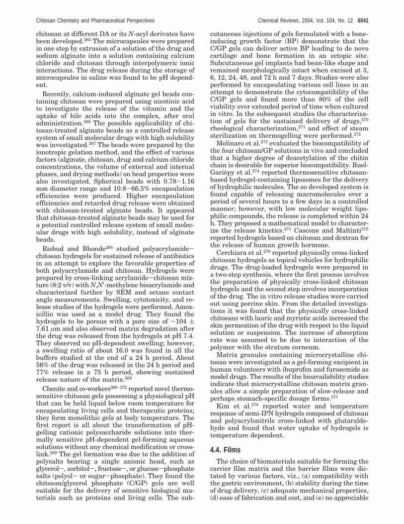
chitosan at different DA or its N-acyl derivates havebeen developed.265 The microcapsules were preparedin one step by extrusion of a solution of the drug andsodium alginate into a solution containing calciumchloride and chitosan through interpolymeric ionicinteractions. The drug release during the storage ofmicrocapsules in saline was found to be pH depend-ent.
Recently, calcium-induced alginate gel beads con-taining chitosan were prepared using nicotinic acidto investigate the release of the vitamin and theuptake of bile acids into the complex, after oraladministration.266 The possible applicability of chi-tosan-treated alginate beads as a controlled releasesystem of small molecular drugs with high solubilitywas investigated.267 The beads were prepared by theionotropic gelation method, and the effect of variousfactors (alginate, chitosan, drug and calcium chlorideconcentrations, the volume of external and internalphases, and drying methods) on bead properties werealso investigated. Spherical beads with 0.78-1.16mm diameter range and 10.8-66.5% encapsulationefficiencies were produced. Higher encapsulationefficiencies and retarded drug release were obtainedwith chitosan-treated alginate beads. It appearedthat chitosan-treated alginate beads may be used fora potential controlled release system of small molec-ular drugs with high solubility, instead of alginatebeads.
Risbud and Bhonde268 studied polyacrylamide-chitosan hydrogels for sustained release of antibioticsin an attempt to explore the favorable properties ofboth polyacrylamide and chitosan. Hydrogels wereprepared by cross-linking acrylamide-chitosan mix-ture (8:2 v/v) with N,N′-methylene bisacrylamide andcharacterized further by SEM and octane contactangle measurements. Swelling, cytotoxicity, and re-lease studies of the hydrogels were performed. Amox-icillin was used as a model drug. They found thehydrogels to be porous with a pore size of ∼104 (7.61 µm and also observed matrix degradation afterthe drug was released from the hydrogels at pH 7.4.They observed no pH-dependent swelling; however,a swelling ratio of about 16.0 was found in all thebuffers studied at the end of a 24 h period. About56% of the drug was released in the 24 h period and77% release in a 75 h period, showing sustainedrelease nature of the matrix.268
Chenite and co-workers269-272 reported novel thermo-sensitive chitosan gels possessing a physiological pHthat can be held liquid below room temperature forencapsulating living cells and therapeutic proteins;they form monolithic gels at body temperature. Thefirst report is all about the transformation of pH-gelling cationic polysaccharide solutions into ther-mally sensitive pH-dependent gel-forming aqueoussolutions without any chemical modification or cross-link.269 The gel formation was due to the addition ofpolysalts bearing a single anionic head, such asglycerol-, sorbitol-, fructose-, or glucose-phosphatesalts (polyol- or sugar-phosphate). They found thechitosan/glycerol phosphate (C/GP) gels are wellsuitable for the delivery of sensitive biological ma-terials such as proteins and living cells. The sub-
cutaneous injections of gels formulated with a bone-inducing growth factor (BP) demonstrate that theC/GP gels can deliver active BP leading to de novocartilage and bone formation in an ectopic site.Subcutaneous gel implants had bean-like shape andremained morphologically intact when excised at 3,6, 12, 24, 48, and 72 h and 7 days. Studies were alsoperformed by encapsulating various cell lines in anattempt to demonstrate the cytocompatibility of theC/GP gels and found more than 80% of the cellviability over extended period of time when culturedin vitro. In the subsequent studies the characteriza-tion of gels for the sustained delivery of drugs,270
rheological characterization,271 and effect of steamsterilization on thermogelling were performed.272
Molinaro et al.273 evaluated the biocompatibility ofthe four chitosan/GP solutions in vivo and concludedthat a higher degree of deacetylation of the chitinchain is desirable for superior biocompatibility. Ruel-Gariepy et al.274 reported thermosensitive chitosan-based hydrogel-containing liposomes for the deliveryof hydrophilic molecules. The so developed system isfound capable of releasing macromolecules over aperiod of several hours to a few days in a controlledmanner; however, with low molecular weight lipo-philic compounds, the release is completed within 24h. They proposed a mathematical model to character-ize the release kinetics.271 Cascone and Maltinti275
reported hydrogels based on chitosan and dextran forthe release of human growth hormone.
Cerchiara et al.276 reported physically cross-linkedchitosan hydrogels as topical vehicles for hydrophilicdrugs. The drug-loaded hydrogels were prepared ina two-step synthesis, where the first process involvesthe preparation of physically cross-linked chitosanhydrogels and the second step involves incorporationof the drug. The in vitro release studies were carriedout using porcine skin. From the detailed investiga-tions it was found that the physically cross-linkedchitosans with lauric and myristic acids increased theskin permeation of the drug with respect to the liquidsolution or suspension. The increase of absorptionrate was assumed to be due to interaction of thepolymer with the stratum corneum.
Matrix granules containing microcrystalline chi-tosan were investigated as a gel-forming excipient inhuman volunteers with ibuprofen and furosemide asmodel drugs. The results of the bioavailability studiesindicate that microcrystalline chitosan matrix gran-ules allow a simple preparation of slow-release andperhaps stomach-specific dosage forms.277
Kim et al.278 reported water and temperatureresponse of semi-IPN hydrogels composed of chitosanand polyacrylonitrile cross-linked with glutaralde-hyde and found that water uptake of hydrogels istemperature dependent.
4.4. FilmsThe choice of biomaterials suitable for forming the
carrier film matrix and the barrier films were dic-tated by various factors, viz., (a) compatibility withthe gastric environment, (b) stability during the timeof drug delivery, (c) adequate mechanical properties,(d) ease of fabrication and cost, and (e) no appreciable
Chitosan Chemistry and Pharmaceutical Perspectives Chemical Reviews, 2004, Vol. 104, No. 12 6041

swelling in water and softening point above 37 °C.Several polymers were found to fill all or most of theabove criteria and used to prepare carrier films. Oftensome plasticizers and excipients were used along withthe base polymer to impart a suitable degree offlexibility and facilitate diffusion of the drug from thefilms, respectively. Chitosan possesses good film-forming properties, and an update on chitosan filmsfor pharmaceutical applications is presented here.
Tomihata and Ikada279 reported in vitro and in vivodegradation profiles of chitin and chitosan films bysolution casting method using specimens of varieddeacetylation degrees, viz., 68.8%, 73.3%, 84.0%,90.1%, and 100%; the thickness of the films was 150µm. The equilibrated water content of the filmsdecreased with increase in deacetylation, while thetensile strength of the water-swollen films increasedwith increasing deacetylation. The maximum watercontent and minimum tensile strength observed fora specimen deacetylated between 0 and 68.8 mol %may be ascribed to the lowered crystallinity bydeacetylation of chitin, since both chitin and chitosanare crystalline polymers. Unlike their physical prop-erties, in vitro and in vivo degradations of these filmsoccurred less rapidly without passing a maximum orminimum, as their degree of deacetylation becamehigher. The degradation of the films was studied invivo as subcutaneous implants. It was found that therate of in vivo biodegradation was very high for chitincompared with that for deacetylated chitin. The filmswhich were more than 73.3% deacetylated showedslower biodegradation. No noticeable tissue reactionswere found with highly deacetylated derivativesincluding chitosan, although they had cationic pri-mary amines in the molecule.
Recently, there has been a growing interest ingrafting of vinyl monomers onto chitosan for bio-medical and industrial applications.163 This chemicalcombination of natural and synthetic polymers yieldsnew materials which could have desirable propertiesincluding biodegradability. Singh and Ray280 pre-pared chitosan films grafted by 2-hydroxyethyl meth-acrylate (HEMA) under 60Co γ-irradiation. Theyobserved that the tensile properties largely affectedby the graft levels.
Alonso and co-workers281 carried out design andevaluation of chitosan/ethylcellulose mucoadhesivebilayered devices for buccal drug delivery. The in-vestigations highlight preparation of devices com-prising a drug-containing mucoadhesive layer and adrug-free backing layer, Figure 35, by two different
methods. Bilaminated films were produced by cast-ing/solvent evaporation technique, and bilayeredtablets were obtained by direct compression. Themucoadhesive layer was composed of a mixture ofdrug and chitosan with or without an anionic cross-linking polymer (polycarbophil, sodium alginate, gel-lan gum), and the backing layer was made of ethyl-cellulose. It was clearly evident that hydrophilicchitosan was easily laminated onto hydrophobicethylcellulose and that a perfect binding between themucoadhesive and the backing layers was achieved,Figure 36. The double-layered structure design wasexpected to provide drug delivery in a unidirectionalfashion to the mucosa and avoid loss of drug dueto wash out with saliva. Using nifedipine and pro-pranolol hydrochloride as slightly and highly water-soluble model drugs, respectively, it was demon-strated that these new devices show promisingpotential for use in controlled delivery of drugs to theoral cavity. The drug release was due to the solubilityand diffusion coefficient of the drug in the polymerfilm, consequently leading to solubility in the releasemedium. SEM observation of the chitosan filmsrevealed the absence of pores before and after thedrug-release assay. The un-cross-linked chitosan-containing devices absorbed a large quantity of water,gelled, and then eroded, allowing drug release. Thebilayered films showed sustained drug release in aphosphate buffer (pH 6.4). Furthermore, tablets thatdisplayed controlled swelling and drug release andadequate adhesivity were produced by in situ cross-linking the chitosan with polycarbophil.
Polyelectrolyte complex (PEC) of pectin, chitosan,and hydroxypropyl methylcellulose was prepared asa model for bimodal drug release.282 The PEC forma-tion between pectin and chitosan was investigatedby examining the viscosities of supernatant solutionsafter removal of the precipitated complex. The PECformation was found to be pH dependent. The amountof pectin, relative to chitosan, required for optimalPEC formation increased as the pH of the solutionwas reduced, and at pH less than 1.3, there was no
Figure 35. Schematic explanation of the structure ofmucoadhesive bilayers. (Reproduced with permission fromref 281. Copyright 1998 Elsevier.)
Figure 36. Scanning electron micrograph of a cross-section of a mucoadhesive bilaminated film: (a) ethyl-cellulose/DBP backing layer and (b) chitosan/glycerinmucoadhesive layer. (Reproduced with permission from ref281. Copyright 1998 Elsevier.)
6042 Chemical Reviews, 2004, Vol. 104, No. 12 Kumar et al.

evidence for the formation of the PEC. Minimalswelling was observed when the pectin:chitosanweight ratio was optimal for PEC formation, sug-gesting formation of the PEC in situ. The perme-ability of the films to paracetamol as a modelcompound was dependent on film composition andmarkedly increased after exposure to pectinolyticenzymes, used to mimic conditions in the colon. Theseresults led authors to conclude that similar formula-tions, applied as a film coat to tablets, could be usedto achieve bimodal drug release with colonic condi-tions acting as a trigger for an increased rate ofrelease.282
Chitosan films made with acetic acid (chitosan-AA) and lactic acid (chitosan-LA) have been testedfor their potential as wound healing materials.283
Mechanical, bioadhesive, and biological evaluation ofthe prepared films were carried out. The results werecompared to the commercial preparation Omiderm.Chitosan-LA exhibited a lower tensile strength;however, it was more flexible and bioadhesive thanchitosan-AA. Chitosan-LA and Omiderm did notcause any skin allergic reactions, in contrast, chito-san-AA showed skin irritation. Nevertheless, no signof toxicity was encountered when the extracts of threepreparations were administered parenterally.283
Chitosan membranes to provide an alternativemeans of evaluating transdermal drug delivery sys-tems were developed and evaluated.284 The mem-branes were prepared by cast-drying method. Theeffects of concentration of chitosan, sodium tripoly-phosphate, as well as cross-linking time on flux andlag time were studied using central composite design.A mathematical model was developed to assess thepermeation of the drugs through different animalskins. The investigations suggest that chitosan mem-brane at a particular composition simulated thepermeation of diclofenac sodium through rat skin.284
Poly(ethylene glycol) (PEG) is water soluble andavailable in different molecular weights and exhibitsuseful properties such as protein resistance, lowtoxicity, and immunogenicity. Furthermore, the PEGscan abrogate the immunogenicity of proteins andpreserve their biological properties. Taking advan-tage of the biocompatibility of chitosan as well as thatof PEG, Zhang et al.285 reported chitosan-PEG blendfilms for improved protein adsorption and cell growth.These extensive studies suggest that PEG enhancedthe protein adsorption, cell adhesion, growth, andproliferation, but the effects were impaired by exces-sive PEG. The experiments also demonstrated thatthe optimum PEG concentration helped to maintainthe natural structure of the protein adsorbed on thematerials and that maintaining the natural structurebenefited cell growth. However, authors found nonoticeable differences on the mechanical propertiesand biocompatibility of the films when differentmolecular weights of PEG were tested, suggestingthat molecular weight differences were not significantenough to affect the material properties.285
Several other modifications were made to improvethe properties of chitosan films. One of the mostrecent studies described the reactions with acidchlorides and acid anhydrides.286 Critical analysis led
them to conclude that the substitution took place atthe amino groups of chitosan, thus forming amidelinkages, and the modification proceeded to a depthof at least 1 µm. The improvement in hydrophobicitywas observed when the films carried stearoyl groups(C17H35CO-). However, modifications with succinicanhydride or phthalic anhydride did produce hydro-philic films. The improved surface hydrophobicityaffected by the stearoyl groups promoted proteinadsorption. In contrast, selective adsorption behaviorwas observed in the case of the chitosan filmsmodified with anhydride derivatives. Lysozyme ad-sorption was enhanced by H-bonding and chargeattraction with the hydrophilic surface, while theamount of albumin adsorbed was decreased possiblydue to negative charges that gave rise to repulsionbetween the modified surface and albumin. Thus,authors have demonstrated that by heterogeneouschemical modification it is possible to fine tunesurface properties that can influence the response tobiomacromolecules.286
Biocompatible chitosan-gelatin composite filmswere prepared by varying the ratio of constituents,and their possible applicability in tissue regenerationhas been evaluated in cultured PC12 cells.287 In-creased water uptake of chitosan film was observedwhen blended with gelatin. Composite film exhibiteda lower Young’s modulus and a higher percentage ofelongation-at-break compared with chitosan film,especially in a wet state. The results also furtherindicate that the amount of adsorption of fibronectinon composite films was much higher than on chitosanfilm. The cells cultured on the composite film with60 wt % gelatin differentiated more rapidly andextended longer neurites than on chitosan film. Theseresults suggest that the soft and elastic complex ofchitosan and gelatin, which has better nerve cellaffinity compared to chitosan alone, is an interestingbiomaterial for nerve regeneration. Chitosans andmodified chitosans have been extensively investi-gated for various other applications as well.246,288-293
4.5. Fibers
4.5.1. Early Approaches to Fiber ProductionThe subject of fiber production from chitin and
chitosan has been reviewed from various viewpointsdue to inherent interest in the exploitation of chitinas a textile material.294-299 Fiber production, however,has appeared since the beginning to be a challengingand difficult task. As it became established forcellulose, the xanthate process was developed forchitin, but the chitin fiber had poor tensile strength.300
The unsatisfactory mechanical properties and envi-ronmental concerns with the xanthate process led tothe development of halogenated and amide-LiClsolvent systems.
The halogenated solvents such as chlorinatedhydrocarbons, chloroethanol with sulfuric acid, andtrichloroacetic acid were developed in the early1970s.301 Chitin filaments were proposed as absorb-able surgical sutures even though the halogenatedsolvents act as plasticizers.302,303
The trichloroacetic-chloral hydrate-dichloro-ethane (4:4:2) solvent system was found to dissolve
Chitosan Chemistry and Pharmaceutical Perspectives Chemical Reviews, 2004, Vol. 104, No. 12 6043

chitin, but the resulting filaments had no substan-tially improved mechanical properties.304,305 More-over, implementation of these spin systems repre-sents a problem: trichloroacetic and dichloroaceticacids are corrosive and degrade chitin even after ashort exposure, chlorohydrocarbons are environmen-tally unacceptable, formic acid can act as a sensitizer,and fluorinated alcohols and ketones are toxic.
A new approach was based on the N,N-dimethyl-acetamide-LiCl and N-methyl-2-pyrrolidone-LiClsolvent systems306,307 that permitted obtaining betterdry tensile strengths, although they did not provideadequate wet tenacities due to poor crystallinity andpoor consolidation of the fiber. A further problem wasthe removal of Li from the fiber once it had formed,because the Li ion solvates the chitin amido groups.It also seems that these fibers could not offer ad-equate knot resistance, and these data jeopardizedtheir further studies for surgical applications, despitethe industrial efforts profused.308
4.5.2. Properties of O-Acylchitins
The studies on the chemical synthesis of O-acylchitins were followed by studies on their biocompat-ibility, and hence, their potential use as materialsfor blood contacting surfaces has been investigatedby measuring, inter alia, their critical surface ten-sions, clotting times, and plasma protein absorption(Table 3).309
The accessibility of chitin, mono-O-acetylchitin, anddi-O-acetylchitin to lysozyme, as determined by theweight loss as a function of time, has been found toincrease in the order chitin < mono-O-acetylchitin< di- O-acetylchitin.310 The molecular motion anddielectric relaxation behavior of chitin and O-acetyl-,O-butyryl-, O-hexanoyl, and O-decanoylchitin havebeen studied.311,312 Chitin and O-acetylchitin showedonly one peak in the plot of the temperature de-pendence of the loss permittivity, whereas thosederivatives having longer O-acyl groups showed twopeaks.
The peak corresponding to the higher temperaturerelaxation process, â, moved to higher temperatureswith increase in the size of the O-acyl group, exclud-ing chitin itself, as did that for the lower temperatureγ process in the three derivatives that exhibit thissecond relaxation process. The presence of two re-laxation processes in these three samples, but onlyone in chitin and O-acetylchitin, was confirmed by
NMR measurements. A comparison with the sidechain relaxation in poly(n-alkyl methacrylate)s313,314
indicates that the â relaxation is due to the motionof the O-acyl groups as a whole and hence is observedin all the samples, while the γ relaxation is due tolocal motion of the O-acyl group and hence only ariseswith the longer members of the series.
4.5.3. Dibutyryl Chitin
Among the O-acyl chitins, dibutyryl chitin (DBC),an ester of chitin at 3 and 6 positions, having theprerogative of being soluble in various solvents, suchas methanol, ethanol, ethylene chloride, and acetone,has attracted particular attention. Therefore, it be-comes easy to manufacture woven, nonwoven, andknitted textiles useful for medical applications.
Dibutyryl chitin with a degree of O-substitution ofca. 1.8 was obtained using methanesulfonic acid asboth catalyst and solvent;315 the DBC filaments weremanufactured as follows: dry-spun fibers were ob-tained from a 23% solution of DBC in acetone intoair (elongation at break 34-47%); the wet spinningwas performed from a 16% solution in dimethyl-formamide into a water coagulating bath (elongationat break 8.3%). The tensile strength was found to besmall, as justified by the low crystallinity and lowinternal overall orientation of the filaments.316
A simpler and cheaper method for the synthesis ofdibutyrylchitin using perchloric acid as a catalyst andbutyric anhydride as acylation agent has been workedout under heterogeneous conditions on krill, shrimp,crab, and insect chitins. The preferred krill chitin hada degree of acetylation of 0.98 and an intrinsicviscosity of 13.33 dL/g (determined in DMAc + 5%LiCl solutions), which corresponded to a viscosityaverage molecular weight of chitin Mv ) 286.7 KDa.The acylation mixture was prepared by pouringperchloric acid into butyric anhydride at ca. -12 °Cand added to the chitin powder placed in the reactor(ca. 0 °C for 30 min, ca. 20 °C later). Weight-averagemolecular weight values were usually in the range120-200 KDa. DBC fibers were spun using Pt-Auspinneret with a hole diameter of 80 µm; 14.5%solution of polymer in DMF was used as a dope.Fibers of DBC had a linear density of 34.7 tex,tenacity in standard condition of 11.9 cN/tex and inthe wet state of 6.80 cN/tex, knot strength of 1.06cN/tex, extensibility of 7.40% in the dry state and8.04% in the wet state. In the IR spectrum of DBCthere was no hydroxyl group band at ca. 3500 cm-1.There were new strong bands characteristic of thefatty acid esters at 1740 and ca. 1450 cm-1.317
As for the biological properties of DBC, the per-centage of the haemolysis counted from three samplesof chitin was as low as 0.086%, for dibutyryl chitin itwas 0.72%, and in both cases did not exceed the valueof 1% admitted for the standards. The evaluation ofcytotoxicity was carried out on the reference cellseries of the mouse fibroblasts 3T3/Balb, after 48 and72 h. No agglutination, vacuolization, or cells mem-brane lysis was observed. The intracutaneous irrita-tion of each extract sample was evaluated on albinoNew Zealand rabbits: intracutaneous reactivity ofthe DBC extracts showed no irritation.
Table 3. Film Properties of Chitin and O-Acylchitins(chitin esters at 3 and 6 positions) with Relevance toBiomedical Applications (modified from ref 309)
clotting time ratioO-acylgroup
acylationdegree glass
siliconizedglass
albuminadsorption(µg cm-2)
chitin 0 1.3 0.7 0.02formyl 1.4 1.6 1.0 0.01acetyl 2.0 3.8 1.9 0.02propionyl 1.9 2.4 0.9butyryl 1.8 2.0 1.2 0.06hexanoyl 2.0 2.5 1.1 0.05decanoyl 1.8 2.5 1.1 0.06dodecanoyl 1.9 2.5 1.1 0.11benzoyl 1.9 2.6 1.0 0.04
6044 Chemical Reviews, 2004, Vol. 104, No. 12 Kumar et al.

The pathomorphological findings including macro-scopic and microscopic assessment testified excellentbiocompatibility. The healing process included theshort exudative phase, which after 14 days wasfollowed by the proliferative phase. The latter endedwith the development of a connective tissue capsule,whose thickness was ca. one-fourth of the fiberdiameter. A significant resorption of DBC fibers wasobserved during the fifth month since implanta-tion.318
Because O-acyl chitins appear to be scarcely sus-ceptible to lysozyme, the susceptibility of DBC tolipases has been studied in order to get insight onits biodegradability in vivo. The changes in infraredand X-ray diffraction spectra of the fibers support theslow degradation of DBC by lipases.319
Considering the experimental evidence available,nonwoven DBC seems suitable for incorporation intomodified chitosans having recognized efficacy inwound healing, such as methylpyrrolidinone chito-san, in view of imparting mechanical strength to thedressing, while avoiding use of nondegradable fi-bers.320
4.5.4. Chitosan FibersNontoxic and noncorrosive solvent systems can be
used when chitosan is considered instead of chitinfor the manufacture of fibers. Many articles andpatents address the spinning of chitosan solutions.Although there was interest in taking advantage ofthe liquid crystalline nature of chitosan,321 no greatimprovement of the mechanical properties was ob-tained. The same can be said for alkyl chitin fibers.322
Cross-linking agents have been proposed for im-provement of chitin fibers in the wet state. Epi-chlorohydrin is a convenient base-catalyzed cross-linker to be used in 0.067 M NaOH (pH 10) at 40 °C.The wet strength of the fibers was considerablyimproved, whereas cross-linking had a negligibleeffect on the dry fiber properties.323 Of course, themore extended the chemical modification, the moreunpredictable the biochemical characteristics andeffects in vivo. Every modified chitin or modifiedchitosan fiber should be studied in terms of biocom-patibility, biodegradability, and overall effects on thewounded tissues.
The present trend is to coat other polysaccharidefibers with a film of chitosan or modified chitosan toimpart novel characteristics to the textile. For ex-ample, a detailed study has been made on alginatefibers coated with various chitosans that actuallyform polyelectrolyte complexes and, under certainconditions, improve the general characteristics of thefiber while keeping the biochemical significance ofchitosan.324 Another study described wet-spun chi-tosan-collagen fibers, their chemical modification,and their blood compatibility.325
This approach to the use of chitosan seems to leadto satisfactory results, as testified by the Japaneseproduction of socks, underwear, and similar items.
4.6. TabletsChitosan has been investigated as an excipient in
the pharmaceutical industry to be used in direct
tablet compression, as a tablet disintegrant, for theproduction of controlled release of solid dosage forms,or for the improvement of drug dissolution. Animmediate release formulation was obtained whenchitosan was used as a pharmaceutical excipient fordirectly compressed tablets.326 Chitosan direct com-pression tablets triggered the sustained release ofprednisolone,327 a water-insoluble drug. The releaserate was controlled by changing the relative amountsand degree of deacetylation of chitosan and car-boxymethylcellulose. A diclofenac tablet formulationproduced using both the direct compression and wetgranulation methods with chitosan also displayedsustained release characteristics.328 These studiessuggest that the release rate is affected by the degreeof N-deacetylation of chitosan. Furthermore, thereport concluded that the ionic strength of the dis-solution medium did not affect the dissolution of thetablets; however, the pH of the medium had asignificant effect.328 A decade later, Sabnis et al.209
demonstrated the possible interaction of diclofenacwith chitosan amino groups leading to the sustainedrelease of the former. Further studies evaluated thebioadhesive strengths of chitosan and hyaluronatecomplexes and found that complexes exhibited su-perior bioadhesiveness in comparison to chitosanalone and were also pH dependent.329 Chitosan andalginate tablets showed sustained release of theo-phylline irrespective of pH.330
A mucoadhesive polymer was developed from thechitosan-EDTA conjugate which inhibited enzymaticactivity:331 it could be used for overcoming enzymaticbarriers for orally administered peptide drugs; how-ever, in terms of adhesive properties, it was foundto be 42% lower than plain chitosan.332 Inhibition ofthe enzymatic activity with chitosan-EDTA was alsoobserved.333 Low insulin degradation was observedusing direct compressed tablets of chitosan-EDTA,suggesting its usefulness as a potential deliverysystem for protein and peptide drugs via oral route.334
Bilayered tablets obtained by direct compression andcomprising a drug containing a mucoadhesive chito-san layer and a backing layer of ethyl cellulosedisplayed controlled delivery of nifedipine and pro-pranolol hydrochloride.335 Diltiazem tablet formula-tions were made by direct mixing of the drug with amixture of chitosan and sodium alginate with a viewto make mucoadhesive formulations for oral deliv-ery.336 The bioavailability of the drug varied with theroute of administration and was found to be 69.6%and 30.4% when a tablet formulation of 1:4 chitosan/alginate was administered sublingually and orally torabbits, respectively.
Phaechamud et al.337 investigated chitosan citrateas a film former and its compatibility with watersoluble anionic dyes and drug dissolution from coatedtablets. The interaction of various dyes with chitosanstudied using FTIR indicated that anionic dyes tendto interact with cationic polymer chitosan. BrilliantBlue and Green FS solutions (concentration 0.02-1.00% (w:w) chitosan) were made. Propranolol.HCltablets coated with these colored film-coating solu-tions exhibited good appearance, and no color migra-tion was observed. Drug dissolution from these coated
Chitosan Chemistry and Pharmaceutical Perspectives Chemical Reviews, 2004, Vol. 104, No. 12 6045

tablets was dictated by the pH of the medium,corresponding to the ability of chitosan to be pro-tonated. These investigations suggested that colorincorporation slightly retarded drug dissolution inacidic medium, where the drug dissolved from Bril-liant Blue coatings was faster than from that ofGreen FS. This was expected due to the positivecharge of Brilliant Blue and more SO3H groups inits molecular structure, exhibiting higher watersolubility. The drug dissolution from freshly preparedcoated tablets, coated tablets after exposure to ac-celerated conditions, and storage at room tempera-ture for 12 months was evaluated. In the laterstudies, the release mechanism of the chitosan-coatedtablets with a view to establish the mechanisticmodel for drug release was investigated.338 Theconclusions for this study were made assuming thatthe core tablets are spherical and the drug dissolutionis diffusion dependent. In order for the drug releaseto occur, the dissolution medium penetrates throughthe coated film and leaches out the drug through thefilm and the lag time, ttag, is set for these processes.It was predicted that the coated film itself dis-solves and becomes thinner with time. As it iscompletely dissipated, the undissolved core tabletbecomes naked, allowing the drug release to proceedjust as the spherical core tablet. Drug-release ratewas given as a differential equation. Model adapta-tion was carried out to the release data of propranololhydrochloride tablets coated with chitosan and keptin different storage conditions. The authors claimedthat the goodness-of-fit of model was better than anyother conventional equations tested, and estimatedmodel parameters were informative.
Microsphere technology was adopted for the devel-opment of chitosan tablets containing chlorhexidinediacetate as a buccal formulation.210 Tablets used inthese studies were prepared by direct compressionof the microparticles with mannitol alone or withsodium alginate. The microparticles were preparedby a spray-drying technique. Chlorhexidine in thechitosan microspheres dissolves more quickly in vitrothan does chlorhexidine powder. The antimicrobialactivity of the microparticles was investigated asminimum inhibitory concentration, minimum bacte-rial concentration, and killing time. Chitosan micro-particles alone have an antimicrobial activity due tothe polymer itself, and it was further found that theloading of chlorhexidine into chitosan is able tomaintain or improve the antimicrobial activity of thedrug. The improvement is particularly high againstCandida albicans. This is important for a formulationwhose potential use is against buccal infections. Aftertheir in vivo administration, determination of chlor-hexidine in saliva showed the capacity of theseformulations to give a prolonged release of the drugin the buccal cavity.
Indomethacin tablet formulations using variouspolysaccharides or synthetic polymers as binderswere developed and enteric coated with Eudragit-L100 to give protection in the stomach.339 These tabletswere evaluated for their potential as colon-specificdrug delivery systems in vitro. The dissolution stud-ies suggest that the rate is dependent on the type
and concentration of the polysaccharide or syntheticpolymer binder used. The results demonstrate thatenteric-coated tablets containing 3% chitosan as abinder showed only 12.5% drug release in the first 5h, which is the usual upper gastrointestinal transittime, whereas tablets prepared using guar gum asbinder failed to protect drug release under similarconditions, thereby leading to the conclusion thatchitosan is superior to guar gum for water-insolubledrugs used in these studies. The authors also sug-gested that the formulations developed using chito-san and Eudragit could serve as highly site-specificdevices since drug release would be at a retarded rateuntil microbial degradation or polymer solubilizationtakes place in the colon.339
Tablets were compressed from a physical mixturecontaining salicylic acid (model drug), chitosan, andmagnesium stearate.200 Five commercial chitosans,varying in degree of deacetylation and molecularweight, were selected. Tablets were compressed at5000, 10 000, and 15 000 psi using a Carver and asingle punch tablet press. The interaction betweenchitosan and the drug has been studied using dif-ferential scanning calorimeter. Analysis of variance(ANOVA) indicated that the compression did notsignificantly affect the crushing strength or therelease profile of drug from the chitosan matrixtablets. Furthermore, the ANOVA also indicated thatthe tablet press used during manufacture did notaffect the above properties; however, the chitosanssignificantly affected the crushing strength as wellas the release profile of drug from chitosan matrixtablets. The same group studied directly compressedtablets (200 mg) containing tetracycline, chitosan,and magnesium stearate prepared for exploringspray-dried chitosans as excipients.204 The chitosansused were spray and thermally dried, previouslyN-deacetylated, and depolymerized. The tablets werecharacterized for dimensions, weight, friability, crush-ing strengths, disintegration, and dissolution andfound that certain parameters such as the tabletweights, thickness, and diameters were not affectedby the chitosan selected. However, friability of tabletscontaining thermally dried chitosans was generallyhigher (and crushing strengths were lower) thantablets containing spray-dried chitosans. It wasnoticed that chitosan molecular weight, degree ofN-deacetylation, and drying method used signifi-cantly affected crushing strengths; on the other hand,disintegration times were affected only by the typeof chitosan but not by the drying method used. Thedissolution studies show the dependency on thechitosan type, while the drying method has noinfluence.
Most recently, N-acylation of chitosan with variousfatty acid (C6-C16) chlorides with a view to increaseits hydrophobicity and their application in drugdelivery has been reported.340 The studies withunmodified chitosan suggested a low degree of orderand a weak tablet crushing strength. N-Acylationwas dealt with different chain lengths in whichchitosan acylated with a short chain length (C6)possessed similar properties but exhibited significantswelling; however, acylation with longer side chains
6046 Chemical Reviews, 2004, Vol. 104, No. 12 Kumar et al.
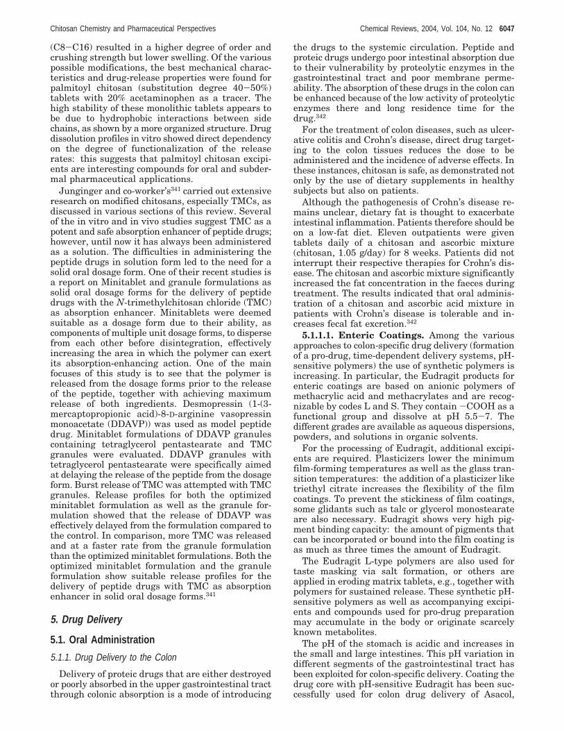
(C8-C16) resulted in a higher degree of order andcrushing strength but lower swelling. Of the variouspossible modifications, the best mechanical charac-teristics and drug-release properties were found forpalmitoyl chitosan (substitution degree 40-50%)tablets with 20% acetaminophen as a tracer. Thehigh stability of these monolithic tablets appears tobe due to hydrophobic interactions between sidechains, as shown by a more organized structure. Drugdissolution profiles in vitro showed direct dependencyon the degree of functionalization of the releaserates: this suggests that palmitoyl chitosan excipi-ents are interesting compounds for oral and subder-mal pharmaceutical applications.
Junginger and co-worker’s341 carried out extensiveresearch on modified chitosans, especially TMCs, asdiscussed in various sections of this review. Severalof the in vitro and in vivo studies suggest TMC as apotent and safe absorption enhancer of peptide drugs;however, until now it has always been administeredas a solution. The difficulties in administering thepeptide drugs in solution form led to the need for asolid oral dosage form. One of their recent studies isa report on Minitablet and granule formulations assolid oral dosage forms for the delivery of peptidedrugs with the N-trimethylchitosan chloride (TMC)as absorption enhancer. Minitablets were deemedsuitable as a dosage form due to their ability, ascomponents of multiple unit dosage forms, to dispersefrom each other before disintegration, effectivelyincreasing the area in which the polymer can exertits absorption-enhancing action. One of the mainfocuses of this study is to see that the polymer isreleased from the dosage forms prior to the releaseof the peptide, together with achieving maximumrelease of both ingredients. Desmopressin (1-(3-mercaptopropionic acid)-8-D-arginine vasopressinmonoacetate (DDAVP)) was used as model peptidedrug. Minitablet formulations of DDAVP granulescontaining tetraglycerol pentastearate and TMCgranules were evaluated. DDAVP granules withtetraglycerol pentastearate were specifically aimedat delaying the release of the peptide from the dosageform. Burst release of TMC was attempted with TMCgranules. Release profiles for both the optimizedminitablet formulation as well as the granule for-mulation showed that the release of DDAVP waseffectively delayed from the formulation compared tothe control. In comparison, more TMC was releasedand at a faster rate from the granule formulationthan the optimized minitablet formulations. Both theoptimized minitablet formulation and the granuleformulation show suitable release profiles for thedelivery of peptide drugs with TMC as absorptionenhancer in solid oral dosage forms.341
5. Drug Delivery
5.1. Oral Administration
5.1.1. Drug Delivery to the Colon
Delivery of proteic drugs that are either destroyedor poorly absorbed in the upper gastrointestinal tractthrough colonic absorption is a mode of introducing
the drugs to the systemic circulation. Peptide andproteic drugs undergo poor intestinal absorption dueto their vulnerability by proteolytic enzymes in thegastrointestinal tract and poor membrane perme-ability. The absorption of these drugs in the colon canbe enhanced because of the low activity of proteolyticenzymes there and long residence time for thedrug.342
For the treatment of colon diseases, such as ulcer-ative colitis and Crohn’s disease, direct drug target-ing to the colon tissues reduces the dose to beadministered and the incidence of adverse effects. Inthese instances, chitosan is safe, as demonstrated notonly by the use of dietary supplements in healthysubjects but also on patients.
Although the pathogenesis of Crohn’s disease re-mains unclear, dietary fat is thought to exacerbateintestinal inflammation. Patients therefore should beon a low-fat diet. Eleven outpatients were giventablets daily of a chitosan and ascorbic mixture(chitosan, 1.05 g/day) for 8 weeks. Patients did notinterrupt their respective therapies for Crohn’s dis-ease. The chitosan and ascorbic mixture significantlyincreased the fat concentration in the faeces duringtreatment. The results indicated that oral adminis-tration of a chitosan and ascorbic acid mixture inpatients with Crohn’s disease is tolerable and in-creases fecal fat excretion.342
5.1.1.1. Enteric Coatings. Among the variousapproaches to colon-specific drug delivery (formationof a pro-drug, time-dependent delivery systems, pH-sensitive polymers) the use of synthetic polymers isincreasing. In particular, the Eudragit products forenteric coatings are based on anionic polymers ofmethacrylic acid and methacrylates and are recog-nizable by codes L and S. They contain -COOH as afunctional group and dissolve at pH 5.5-7. Thedifferent grades are available as aqueous dispersions,powders, and solutions in organic solvents.
For the processing of Eudragit, additional excipi-ents are required. Plasticizers lower the minimumfilm-forming temperatures as well as the glass tran-sition temperatures: the addition of a plasticizer liketriethyl citrate increases the flexibility of the filmcoatings. To prevent the stickiness of film coatings,some glidants such as talc or glycerol monostearateare also necessary. Eudragit shows very high pig-ment binding capacity: the amount of pigments thatcan be incorporated or bound into the film coating isas much as three times the amount of Eudragit.
The Eudragit L-type polymers are also used fortaste masking via salt formation, or others areapplied in eroding matrix tablets, e.g., together withpolymers for sustained release. These synthetic pH-sensitive polymers as well as accompanying excipi-ents and compounds used for pro-drug preparationmay accumulate in the body or originate scarcelyknown metabolites.
The pH of the stomach is acidic and increases inthe small and large intestines. This pH variation indifferent segments of the gastrointestinal tract hasbeen exploited for colon-specific delivery. Coating thedrug core with pH-sensitive Eudragit has been suc-cessfully used for colon drug delivery of Asacol,
Chitosan Chemistry and Pharmaceutical Perspectives Chemical Reviews, 2004, Vol. 104, No. 12 6047
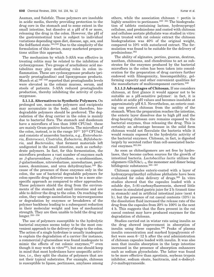
Asamax, and Salofalc. Those polymers are insolublein acidic media, thereby providing protection to thedrug core in the stomach and to some extent in thesmall intestine, but dissolve at a pH of 6 or more,releasing the drug in the colon. However, the pH ofthe gastrointestinal tract is subject to individualvariations depending upon diet, disease, age, sex, andthe fed/fasted state.344,345 Due to the simplicity of theformulation of this device, many marketed prepara-tions utilize this approach.
The mechanism by which 5-ASA was effective intreating colitis may be related to the inhibition ofcyclooxygenase. Two groups of arachidonic acid me-tabolites may play mediating roles in colon in-flammation. These are cyclooxygenase products (pri-marily prostaglandins) and lipoxygenase products.Musch et al.346-348 reported that prostaglandin levelswere elevated in the colonic mucosa, serum, andstools of patients. 5-ASA reduced prostaglandinproduction, thereby inhibiting the activity of cyclo-oxygenase.
5.1.1.2. Alternatives to Synthetic Polymers. Onprolonged use, man-made polymers and excipientsmay accumulate in the body, and therefore, bio-degradable polymers should be preferred. The deg-radation of the drug carrier in the colon is mainlydue to bacterial flora. The stomach and duodenumhave a microflora of less than 1000 CFU/ml: theseare mainly Gram-positive bacteria. The microflora ofthe colon, instead, is in the range 1011-1012 CFU/mLand consists of anaerobic bacteria, e.g., Enterobacte-ria, Enterococci, Clostridia, Eubacteria, Bifidobacte-ria, and Bacteroides, that ferment materials leftundigested in the small intestine, such as carbohy-drate polymers. In fact, for this fermentation, themicroflora produces a vast number of enzymes suchas â-glucuronidase, â-xylosidase, R-arabinosidase,â-galactosidase, nitroreductase, azoreductase, pecti-nases, deaminase, and urea dehydroxylase.349 Be-cause of the presence of these enzymes only in thecolon, the use of bacterial degradable polymers forcolon-specific drug delivery seems to be a more site-specific approach as compared to other approaches.These polymers shield the drug from the environ-ments of the stomach and small intestine and areable to deliver the drug to the colon. On reaching thecolon, they undergo assimilation by microorganismsor degradation by enzymes or breakdown of thepolymer backbone leading to a subsequent reductionin their molecular weight and loss of mechanicalstrength. They are then unable to hold the drug anylonger.350-356
The use of polymers susceptible to the hydrolyticbacterial enzymes seems today to be the most con-venient approach to the delivery of drugs to the colon.The action of a single hydrolase is usually inadequateto explain the degradation of a carrier (for instance,a commercial â-glucosidase was found inadequate tomimic the effects of rat colonic enzymes,357 eventhough it may work in vitro358), but one should keepin mind that most hydrolases have unspecific activi-ties, i.e., they split the chains of polymers that arenot their typical substrates. For example, chitosanis susceptible to lipase, pectinases, amylases among
others, while the association chitosan + pectin ishighly sensitive to pectinases.359-361 The biodegrada-tion of tablets containing lactose, hydroxypropylcellulose, and prednisolone and coated with chitosanand cellulose acetate phthalate was studied in vitro:when treated with rat colonic extract the chitosanbiodegradation was 40% of the original weight,compared to 10% with autoclaved extract. The for-mulation was found to be suitable for the delivery ofprednisolone.362
The ability of alginates, pectins, guaran, dextran,xanthan, chitosans, and chondroitins to act as sub-strates for the enzymes produced by the bacterialmicroflora in the colon has been taken into consid-eration for the preparation of drug carriers furtherendowed with filmogenicity, biocompatibility, gel-forming capacity and other convenient qualities forthe manufacture of multicomponent carriers.339
5.1.1.3 Advantages of Chitosan. If one considerschitosan, at first glance it would appear not to besuitable as a pH-sensitive polymer: in fact, it issoluble at acidic pH values and becomes insoluble atapproximately pH 6.5. Nevertheless, an enteric coat-ing can protect chitosan from the acidity of thestomach. When the preparation reaches the intestine,the enteric layer dissolves due to high pH and thedrug-bearing chitosan core remains exposed to thebacterial enzymes, thus releasing the drug. This iscertainly an advantage because in the solid formchitosan would not flocculate the bacteria while itwould remain exposed to the hydrolytic activity ofthe bacterial enzymes. Chitosan, in fact, is digestedlargely by secreted rather than cell-associated bacte-rial enzymes.363,364
As soon as chitooligomers are set free by hydro-lases, they become carbon sources for the growth ofintestinal bacteria. Lactobacillus lactis utilizes theoligomers (GlcNAc)1-6, the monomer and dimer beingbifidogenic substances.365-367
Chitosan capsules enteric-coated with a layer ofhydroxypropylmethyl cellulose phthalate have beenevaluated for colon delivery of drugs.368 In vitrostudies showed that the capsules loaded with asoluble dye, 5-(6)-carboxyfluorescein, showed littlerelease in simulated gastric juice for 2 h (transit timein stomach) and in artificial intestinal juice (next 4h), but the presence of rat caecal contents (33%) inthe dissolution fluid increased the release rate of thedrug from the capsules from 20% to 100% in the next4 h. This suggests that the flora present in the ratcaecal content may have produced enzymes for thedegradation of chitosan.
Studies carried out in wistar rats using insulin asthe drug showed improvement in absorption ofinsulin using these capsules.196 Peaks of plasmainsulin concentration and marked hypoglycemic ef-fect were seen 6-12 h after the oral administrationof chitosan capsules loaded with insulin. It was alsoseen that insulin absorption in the large intestineincreased in the presence of absorption enhancerssuch as sodium glycocholate. The latter was foundto be more effective than aprotinin, soybean trypsininhibitor, sodium oleate, bacitracin, and n-dodecyl-â-D-maltopyranoside.
6048 Chemical Reviews, 2004, Vol. 104, No. 12 Kumar et al.

Acceleration of healing with R68070 (a thrombox-ane synthase inhibitor) on 2,4,6-trinitrobenzene sul-fonic-acid-induced ulcerative colitis in rat usingchitosan capsule as a carrier was compared to asuspension of R68070 in carboxymethyl cellulose.369
However, when the doses were increased, a markedreduction in colitis was seen with R68070 when givenin chitosan capsules rather than in carboxymethylcellulose suspension, leading to the conclusion thatthese capsules have a good potential for being usedas carriers in colon-specific drug delivery systems.Most important, chitosan capsules loaded with 5-ASAprovided a higher therapeutic effect than 5-ASAsuspensions with carboxymethyl cellulose, based onmyeloperoxidase activity, damage score, and colon/body weight ratio.
The release of 5-ASA and sodium diclofenac fromchitosan or chitosan-HCl was studied in vitro byusing matrixes prepared by compression of micro-spheres with drug powders (20%). The action of ratcaecal microflora accelerated the drug release fromthe chitosan matrix, which was a reliable system for5-ASA (t50 ) 1.97 h) or sodium diclofenac (t50 ) 3.58h).370
Chitosan capsules manufactured by Aicello Chem.Co. Ltd. were filled with 5-ASA and then coated withhydroxypropylcellulose phthalate as an enteric coat-ing dissolved in acetone + ethanol (1:1). Analogouspreparations were made with insulin. Levels of5-ASA in the rat large intestine 4 h after oraladministration were close to 0.45 mg (i.e., 1/10 of theadministered quantity) compared to ca. 0.12 mg forthe same amount of 5-ASA as a CMC suspension.
In practice, the enteric-coated capsules were stablein the stomach and the small intestine. They weredegraded by microorganisms in the caecal content.The delivery to the colon was specific, and the heal-ing effect was accelerated by chitosan. Similarly,insulin absorption in the large intestine was im-proved with absorption enhancers such as sodiumglycocholate.346,371,372 Gelatin capsules were coatedwith Eudragit containing chitosan particles for thesame purposes
The effect of chitosan tripolyphosphate nanopar-ticles (250-400 nm) on the absorption of insulin wasstudied by measuring the decrease of the plasmaglucose concentration and the relative pharmacologi-cal availability.373 Chitosan tripolyphosphate showedexcellent association with insulin and improved theintestinal absorption of insulin to a greater extentcompared to insulin-chitosan solution.
Semisynthetic derivatives of chitosan i.e., chitosansuccinate and chitosan phthalate, were prepared byreacting chitosan separately with succinic andphthalic anhydrides.374 Sodium diclofenac was dis-persed in their matrixes. In vitro studies showed thatthese matrixes resisted dissolution under acidicconditions and showed improved dissolution underbasic conditions, suggesting their suitability for colon-specific drug delivery systems.
5.1.1.4. Natural Enteric Coatings. Pectin, beingsoluble in water, is unable to protect its drug loadduring passage through the stomach and smallintestine, but pectin chitosan complexes were found
to be suitable for colon-specific delivery of indometha-cin and paracetamol.375
In earlier studies the formation of chitosan pectincomplexes was deemed interesting because of theefficacy in controlling the release of indomethacin andsulfamethaxazole while maintaining the biodegrad-ability towards pectinases.376
An ionic complex of highly methoxylated pectin(anionic) with Eudragit-RL (cationic, insoluble pH-independent) was incorporated in swellable Eudragit-NE30D (neutral ester) and used for targeting drugsto the colon.377 In the case of polyelectrolytic com-plexes of pectin and chitosan, the lowest permeabilityto paracetamol was found for materials having aprevalent content of nonamidated low-methoxylatedpectins and highly deacetylated.378 Indomethacin wasalso studied in similar carriers.221
Combination of pectin, chitosan, and hydroxy-propylmethyl cellulose in the ratio 3:1:1 is insolubleand degraded by pectinases. These tablets were usedin human volunteers to carry 99mTc and found to beable to pass through the stomach and small intestineintact, as is evident by scintigraphic images. Theresults led to the conclusion that degradation of thesecoatings takes place in the ascending and transversecolon by colonic bacteria. Figure 37. 379
Guaran has been similarly evaluated as a com-pression coating: the formulation with 150 mg ofguaran as a coating showed 95% release of 5-amino-salic acid in the presence of rat caecal content after26 h.380
Galactomannan from locust bean should be cross-linked for these purposes, considering its solubilityin water. However, combination of galactomannanand chitosan proved capable of protecting the coretablet containing mesalazine during conditions mim-icking mouth to colon transit. When galactomannanand chitosan were in a ratio of 4:1, the bioavailabilityand dissolution profile in rat caecal content qualifiedthe complex as a carrier for drug targeting to thecolon.381
Chitosan-alginate beads loaded with a modelprotein, bovine serum albumin, were investigated toexplore the temporary protection of protein againstacidic and enzymatic degradation during gastricpassage. During incubation in simulated gastric fluid(pH 1.2), the beads showed swelling and started tofloat but did not show any sign of erosion. Releasestudies were done in simulated gastric fluid (pH 1.2)and subsequently in simulated intestinal fluid (pH7.5), where the beads were found to erode, burst, andrelease the protein. Chitosan-reinforced calcium-alginate beads showed delay in the release of albu-min. The multilayer beads disintegrated very slowly.The enzymes pepsin and pancreatin did not changethe characteristics of albumin-loaded chitosan-algi-nate beads. Alginate beads reinforced with chitosanoffer an excellent perspective for controlled gas-trointestinal passage of proteic drugs.382
Chitosan microspheres obtained by emulsificationin paraffin, coconut, or sunflower oil and cross-linkedto various degrees with glutaraldehyde were dis-persed in poly(acrylic acid) solution for coating. Oncedry, they were dispersed in alginate solution (3%) and
Chitosan Chemistry and Pharmaceutical Perspectives Chemical Reviews, 2004, Vol. 104, No. 12 6049

microcapsules were obtained by dropping the lattersuspension into CaCl2 (2%) solution. The muco-adhesivity was then evaluated. The mechanisminvolved liberation of the chitosan polyacrylate mi-crospheres from the microcapsules in the intestineand attachment of the microspheres to the colonmucosae for prolonged drug release.383 It is known,in fact, that chitosan strongly reacts with mucin, evenin the presence of minor amounts of polyacrylate.384
In conclusion, it was demonstrated that chitosancapsules were stable in the stomach and smallintestine. However, in rats, they were specificallydegraded by microorganisms in the caecal contentswhen they reached the colon. Furthermore, insulinabsorption from the large intestine was improved byconcomitant administration with a variety of addi-tives. 5-ASA can be delivered specifically to the colon,and the healing effect of 5-ASA was accelerated usingchitosan capsules. These capsules may thereforeconstitute useful carriers for the colon-specific deliv-ery of peptides, including insulin, and antiinflam-matory drugs, including 5-ASA.346
It seems that the preparation of chitosan 5-amino-salicylate salt could be a further extension of thesestudies; however, the characteristic properties of thissalt are unknown, while the chitosan salts of gentisic,salicylic, and acetylsalicylic acids have been charac-terized.385
5.1.2. Cholesterol and Overweight Lowering
When discussing the role of chitosan as a choles-terol lowering nutriceutical and considering possiblemechanisms for its action, one should keep in mindthat animal studies might not be predictive of results
in humans because of the presence of chitinases inthe digestive systems of many animals, as a point ofdifference from humans.386 Chitosan for the oraladministration to humans is generally recognized assafe.387
5.1.2.1. Cholesterol Lowering in Animals. Sug-ano et al.388 were the first to report the cholesterollowering ability of chitosan, and the report suggeststhat a diet containing 5% chitosan reduced livercholesterol concentration by one-half or more incholesterol-fed rats, and a similar trend was observedby other investigators as well.389 In a 4 week study,a 4% chitosan diet reduced both liver and serumcholesterol dramatically in rats fed diets containing1% cholesterol + 0.1% bile salts.390 Chitosan has alsobeen shown to reduce plasma cholesterol in choles-terol-fed broiler chickens at dietary concentrationsof 1.5-3.0%.391,392 Thus, the ability of chitosan toreduce cholesterol in animal models is well estab-lished.
The apoprotein E-deficient mouse has extremehypercholesterolemia, atherosclerosis develops rap-idly, and the lesions are histologically similar to thoseseen in humans.393 Deficient mice fed diets containing5% chitosan for 6 months had serum cholesterol only64% of the control group.394 The atherosclerotic lesionarea in the aortic arch was reduced by one-half andin the total aorta by 42% in mice fed chitosan. Theseresults strongly suggest that chitosan consumptionhas the potential to reduce the atherosclerosis riskin humans.
5.1.2.2. Cholesterol Lowering in Humans. Chi-tosan was first shown to reduce serum cholesterol inhumans in 1993, when adult males fed chitosan-
Figure 37. Scintiscan showing the tablet (intact) in the small intestine of subject 4. Time after tablet administration )60 min. (b) Scintiscan showing the tablet having disintegrated and starting to spread in the ascending colon of subject 4.Time after tablet administration ) 305 min. (c) Scintiscan showing the tablet having extensively spread in the ascendingcolon of subject 4. Time after tablet administration ) 365 min. (d) Scintiscan showing the tablet having extensively spreadtoward the transverse colon of subject 4. Time after tablet administration ) 405 min. (Reproduced with permission fromref 379. Copyright 1999 Elsevier.)
6050 Chemical Reviews, 2004, Vol. 104, No. 12 Kumar et al.
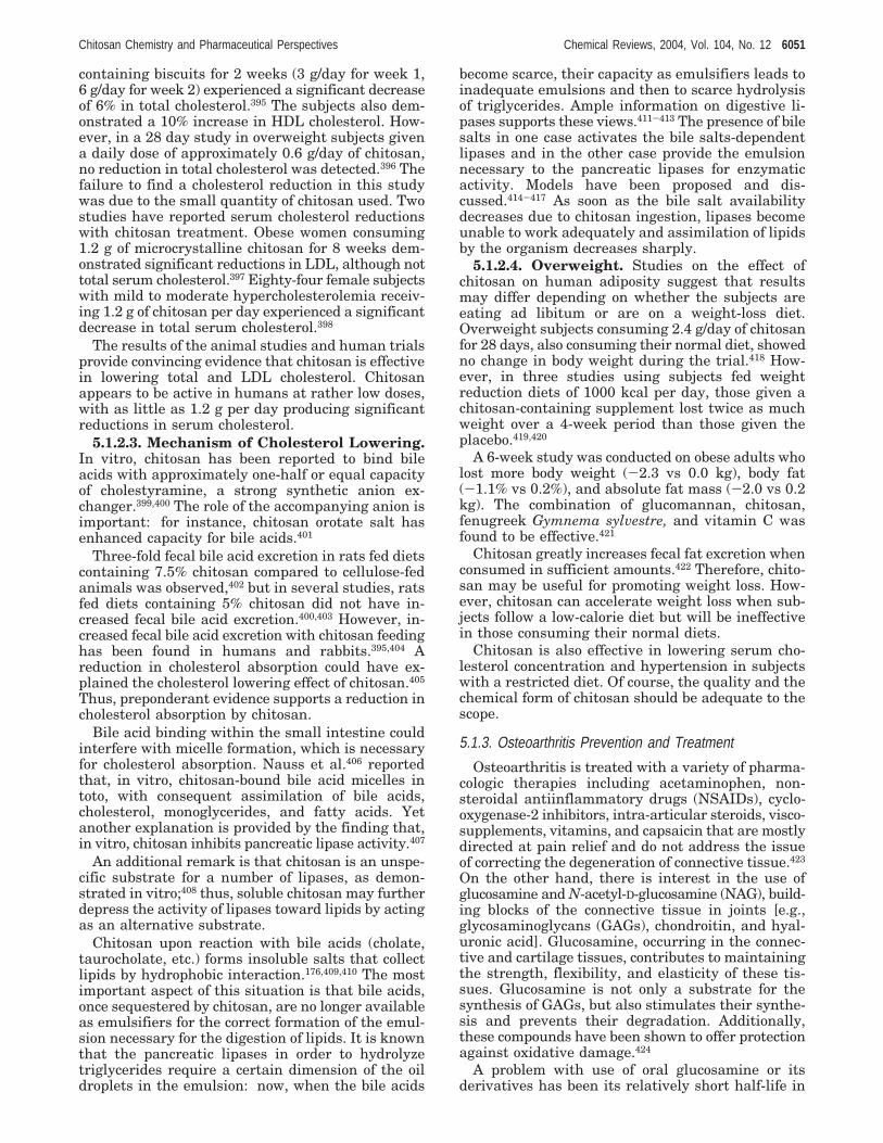
containing biscuits for 2 weeks (3 g/day for week 1,6 g/day for week 2) experienced a significant decreaseof 6% in total cholesterol.395 The subjects also dem-onstrated a 10% increase in HDL cholesterol. How-ever, in a 28 day study in overweight subjects givena daily dose of approximately 0.6 g/day of chitosan,no reduction in total cholesterol was detected.396 Thefailure to find a cholesterol reduction in this studywas due to the small quantity of chitosan used. Twostudies have reported serum cholesterol reductionswith chitosan treatment. Obese women consuming1.2 g of microcrystalline chitosan for 8 weeks dem-onstrated significant reductions in LDL, although nottotal serum cholesterol.397 Eighty-four female subjectswith mild to moderate hypercholesterolemia receiv-ing 1.2 g of chitosan per day experienced a significantdecrease in total serum cholesterol.398
The results of the animal studies and human trialsprovide convincing evidence that chitosan is effectivein lowering total and LDL cholesterol. Chitosanappears to be active in humans at rather low doses,with as little as 1.2 g per day producing significantreductions in serum cholesterol.
5.1.2.3. Mechanism of Cholesterol Lowering.In vitro, chitosan has been reported to bind bileacids with approximately one-half or equal capacityof cholestyramine, a strong synthetic anion ex-changer.399,400 The role of the accompanying anion isimportant: for instance, chitosan orotate salt hasenhanced capacity for bile acids.401
Three-fold fecal bile acid excretion in rats fed dietscontaining 7.5% chitosan compared to cellulose-fedanimals was observed,402 but in several studies, ratsfed diets containing 5% chitosan did not have in-creased fecal bile acid excretion.400,403 However, in-creased fecal bile acid excretion with chitosan feedinghas been found in humans and rabbits.395,404 Areduction in cholesterol absorption could have ex-plained the cholesterol lowering effect of chitosan.405
Thus, preponderant evidence supports a reduction incholesterol absorption by chitosan.
Bile acid binding within the small intestine couldinterfere with micelle formation, which is necessaryfor cholesterol absorption. Nauss et al.406 reportedthat, in vitro, chitosan-bound bile acid micelles intoto, with consequent assimilation of bile acids,cholesterol, monoglycerides, and fatty acids. Yetanother explanation is provided by the finding that,in vitro, chitosan inhibits pancreatic lipase activity.407
An additional remark is that chitosan is an unspe-cific substrate for a number of lipases, as demon-strated in vitro;408 thus, soluble chitosan may furtherdepress the activity of lipases toward lipids by actingas an alternative substrate.
Chitosan upon reaction with bile acids (cholate,taurocholate, etc.) forms insoluble salts that collectlipids by hydrophobic interaction.176,409,410 The mostimportant aspect of this situation is that bile acids,once sequestered by chitosan, are no longer availableas emulsifiers for the correct formation of the emul-sion necessary for the digestion of lipids. It is knownthat the pancreatic lipases in order to hydrolyzetriglycerides require a certain dimension of the oildroplets in the emulsion: now, when the bile acids
become scarce, their capacity as emulsifiers leads toinadequate emulsions and then to scarce hydrolysisof triglycerides. Ample information on digestive li-pases supports these views.411-413 The presence of bilesalts in one case activates the bile salts-dependentlipases and in the other case provide the emulsionnecessary to the pancreatic lipases for enzymaticactivity. Models have been proposed and dis-cussed.414-417 As soon as the bile salt availabilitydecreases due to chitosan ingestion, lipases becomeunable to work adequately and assimilation of lipidsby the organism decreases sharply.
5.1.2.4. Overweight. Studies on the effect ofchitosan on human adiposity suggest that resultsmay differ depending on whether the subjects areeating ad libitum or are on a weight-loss diet.Overweight subjects consuming 2.4 g/day of chitosanfor 28 days, also consuming their normal diet, showedno change in body weight during the trial.418 How-ever, in three studies using subjects fed weightreduction diets of 1000 kcal per day, those given achitosan-containing supplement lost twice as muchweight over a 4-week period than those given theplacebo.419,420
A 6-week study was conducted on obese adults wholost more body weight (-2.3 vs 0.0 kg), body fat(-1.1% vs 0.2%), and absolute fat mass (-2.0 vs 0.2kg). The combination of glucomannan, chitosan,fenugreek Gymnema sylvestre, and vitamin C wasfound to be effective.421
Chitosan greatly increases fecal fat excretion whenconsumed in sufficient amounts.422 Therefore, chito-san may be useful for promoting weight loss. How-ever, chitosan can accelerate weight loss when sub-jects follow a low-calorie diet but will be ineffectivein those consuming their normal diets.
Chitosan is also effective in lowering serum cho-lesterol concentration and hypertension in subjectswith a restricted diet. Of course, the quality and thechemical form of chitosan should be adequate to thescope.
5.1.3. Osteoarthritis Prevention and Treatment
Osteoarthritis is treated with a variety of pharma-cologic therapies including acetaminophen, non-steroidal antiinflammatory drugs (NSAIDs), cyclo-oxygenase-2 inhibitors, intra-articular steroids, visco-supplements, vitamins, and capsaicin that are mostlydirected at pain relief and do not address the issueof correcting the degeneration of connective tissue.423
On the other hand, there is interest in the use ofglucosamine and N-acetyl-D-glucosamine (NAG), build-ing blocks of the connective tissue in joints [e.g.,glycosaminoglycans (GAGs), chondroitin, and hyal-uronic acid]. Glucosamine, occurring in the connec-tive and cartilage tissues, contributes to maintainingthe strength, flexibility, and elasticity of these tis-sues. Glucosamine is not only a substrate for thesynthesis of GAGs, but also stimulates their synthe-sis and prevents their degradation. Additionally,these compounds have been shown to offer protectionagainst oxidative damage.424
A problem with use of oral glucosamine or itsderivatives has been its relatively short half-life in
Chitosan Chemistry and Pharmaceutical Perspectives Chemical Reviews, 2004, Vol. 104, No. 12 6051
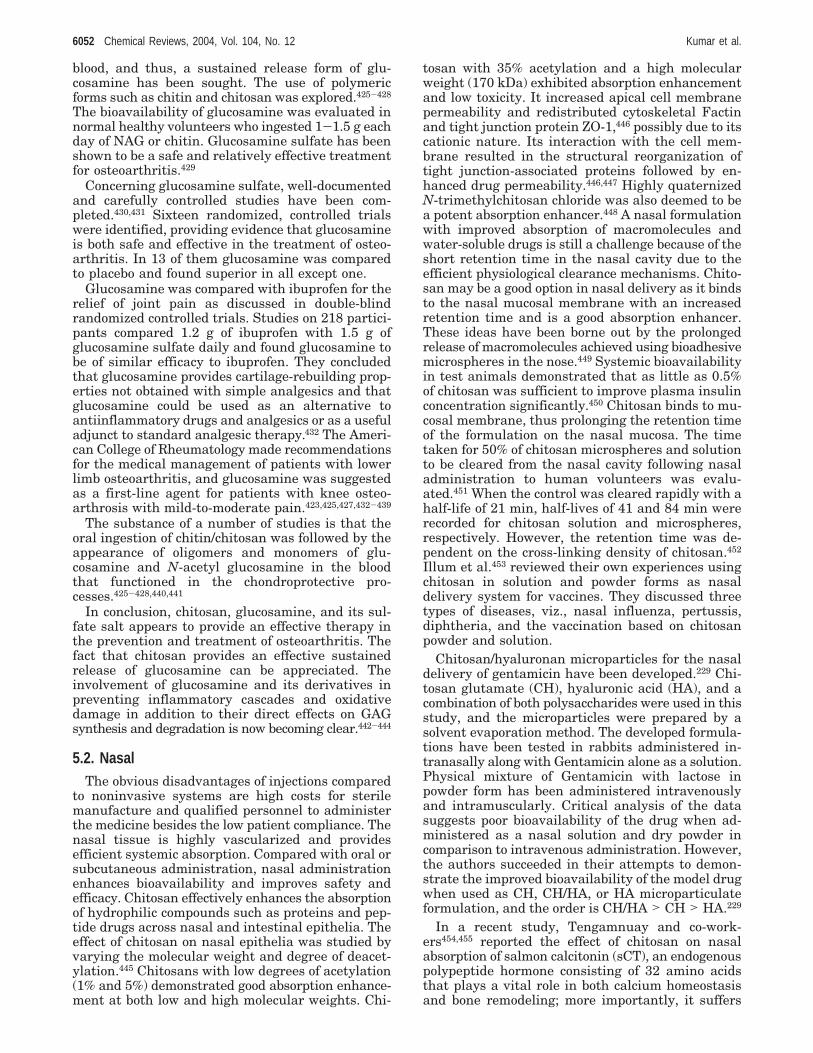
blood, and thus, a sustained release form of glu-cosamine has been sought. The use of polymericforms such as chitin and chitosan was explored.425-428
The bioavailability of glucosamine was evaluated innormal healthy volunteers who ingested 1-1.5 g eachday of NAG or chitin. Glucosamine sulfate has beenshown to be a safe and relatively effective treatmentfor osteoarthritis.429
Concerning glucosamine sulfate, well-documentedand carefully controlled studies have been com-pleted.430,431 Sixteen randomized, controlled trialswere identified, providing evidence that glucosamineis both safe and effective in the treatment of osteo-arthritis. In 13 of them glucosamine was comparedto placebo and found superior in all except one.
Glucosamine was compared with ibuprofen for therelief of joint pain as discussed in double-blindrandomized controlled trials. Studies on 218 partici-pants compared 1.2 g of ibuprofen with 1.5 g ofglucosamine sulfate daily and found glucosamine tobe of similar efficacy to ibuprofen. They concludedthat glucosamine provides cartilage-rebuilding prop-erties not obtained with simple analgesics and thatglucosamine could be used as an alternative toantiinflammatory drugs and analgesics or as a usefuladjunct to standard analgesic therapy.432 The Ameri-can College of Rheumatology made recommendationsfor the medical management of patients with lowerlimb osteoarthritis, and glucosamine was suggestedas a first-line agent for patients with knee osteo-arthrosis with mild-to-moderate pain.423,425,427,432-439
The substance of a number of studies is that theoral ingestion of chitin/chitosan was followed by theappearance of oligomers and monomers of glu-cosamine and N-acetyl glucosamine in the bloodthat functioned in the chondroprotective pro-cesses.425-428,440,441
In conclusion, chitosan, glucosamine, and its sul-fate salt appears to provide an effective therapy inthe prevention and treatment of osteoarthritis. Thefact that chitosan provides an effective sustainedrelease of glucosamine can be appreciated. Theinvolvement of glucosamine and its derivatives inpreventing inflammatory cascades and oxidativedamage in addition to their direct effects on GAGsynthesis and degradation is now becoming clear.442-444
5.2. NasalThe obvious disadvantages of injections compared
to noninvasive systems are high costs for sterilemanufacture and qualified personnel to administerthe medicine besides the low patient compliance. Thenasal tissue is highly vascularized and providesefficient systemic absorption. Compared with oral orsubcutaneous administration, nasal administrationenhances bioavailability and improves safety andefficacy. Chitosan effectively enhances the absorptionof hydrophilic compounds such as proteins and pep-tide drugs across nasal and intestinal epithelia. Theeffect of chitosan on nasal epithelia was studied byvarying the molecular weight and degree of deacet-ylation.445 Chitosans with low degrees of acetylation(1% and 5%) demonstrated good absorption enhance-ment at both low and high molecular weights. Chi-
tosan with 35% acetylation and a high molecularweight (170 kDa) exhibited absorption enhancementand low toxicity. It increased apical cell membranepermeability and redistributed cytoskeletal Factinand tight junction protein ZO-1,446 possibly due to itscationic nature. Its interaction with the cell mem-brane resulted in the structural reorganization oftight junction-associated proteins followed by en-hanced drug permeability.446,447 Highly quaternizedN-trimethylchitosan chloride was also deemed to bea potent absorption enhancer.448 A nasal formulationwith improved absorption of macromolecules andwater-soluble drugs is still a challenge because of theshort retention time in the nasal cavity due to theefficient physiological clearance mechanisms. Chito-san may be a good option in nasal delivery as it bindsto the nasal mucosal membrane with an increasedretention time and is a good absorption enhancer.These ideas have been borne out by the prolongedrelease of macromolecules achieved using bioadhesivemicrospheres in the nose.449 Systemic bioavailabilityin test animals demonstrated that as little as 0.5%of chitosan was sufficient to improve plasma insulinconcentration significantly.450 Chitosan binds to mu-cosal membrane, thus prolonging the retention timeof the formulation on the nasal mucosa. The timetaken for 50% of chitosan microspheres and solutionto be cleared from the nasal cavity following nasaladministration to human volunteers was evalu-ated.451 When the control was cleared rapidly with ahalf-life of 21 min, half-lives of 41 and 84 min wererecorded for chitosan solution and microspheres,respectively. However, the retention time was de-pendent on the cross-linking density of chitosan.452
Illum et al.453 reviewed their own experiences usingchitosan in solution and powder forms as nasaldelivery system for vaccines. They discussed threetypes of diseases, viz., nasal influenza, pertussis,diphtheria, and the vaccination based on chitosanpowder and solution.
Chitosan/hyaluronan microparticles for the nasaldelivery of gentamicin have been developed.229 Chi-tosan glutamate (CH), hyaluronic acid (HA), and acombination of both polysaccharides were used in thisstudy, and the microparticles were prepared by asolvent evaporation method. The developed formula-tions have been tested in rabbits administered in-tranasally along with Gentamicin alone as a solution.Physical mixture of Gentamicin with lactose inpowder form has been administered intravenouslyand intramuscularly. Critical analysis of the datasuggests poor bioavailability of the drug when ad-ministered as a nasal solution and dry powder incomparison to intravenous administration. However,the authors succeeded in their attempts to demon-strate the improved bioavailability of the model drugwhen used as CH, CH/HA, or HA microparticulateformulation, and the order is CH/HA > CH > HA.229
In a recent study, Tengamnuay and co-work-ers454,455 reported the effect of chitosan on nasalabsorption of salmon calcitonin (sCT), an endogenouspolypeptide hormone consisting of 32 amino acidsthat plays a vital role in both calcium homeostasisand bone remodeling; more importantly, it suffers
6052 Chemical Reviews, 2004, Vol. 104, No. 12 Kumar et al.

from poor bioavailability. â-Cyclodextrin, one of themost commonly used enhancers, was used as astandard for comparison purposes. Two types ofchitosan, the free amine and the glutamate salt, wereevaluated. The plasma calcium lowering effect ineach sCT-treated rat was determined by calculatingthe total percent decrease in plasma calcium (%D),where plain chitosan showed an increase in %D withthe decrease in pH following an increase in ionizationand hydration of the free amine chitosan at moreacidic pH. However, chitosan glutamate showed anincrease in %D with increasing pH, with maximumhypocalcemic effect observed at pH 6.0. The optimalpH was found to be 4 and 6, respectively. Theabsorption-enhancing effect of both chitosans wasconcentration dependent and 1.25%.
Hamman et al.456,457 tested five TMCs with differ-ent degrees of quaternization as nasal deliverysystems. These polymers were administered togetherwith [14C]-mannitol in the nasal route of rats at pH6.20 and 7.40, respectively. The absorption was foundto be pH dependent as all the tested polymers showedan increase in absorption at pH 6.20; however, at pH7.40, the absorption is dependent on the degree ofquaternization of the polymer. Polymer beyond 36%quaternization exhibited increased activity at pH7.40, and the increase was observed until 48% andstabilized on further increase. This effect of thedegree of quaternization on the absorption behaviorwas predicted due to steric effects caused by themethyl groups and changes in the flexibility of theTMC molecules and, therefore, led them to concludethat degree of quaternization has a major role in theabsorption enhancement of this polymer across thenasal epithelia in a neutral environment.456
Cross-linked microspheres for nasal administrationof LHRH analogue were prepared by water-in-oilemulsion followed by cross-linking with glutaralde-hyde.458 Chitosan in three different forms along withgoserelin as a LHRH analogue were administerednasally to sheep. The bioavailability of the activesubstance was found to be greatly dependent on theformulation used and followed the order cross-linkedmicrospheres > dry blend > solution.458
Alonso and co-workers184,185 developed freeze-driedinsulin-chitosan nanoparticulate formulations inorder to reduce plasma glucose levels following nasaladministration. Insulin release studies in vitro wereperformed for the freshly prepared formulations aswell as freeze dried. Both formulations behave alikeat pH 4.3, exhibiting rapid release due to solubilityof insulin as well as chitosan at the working pH.However, due to lack of the solubility of insulin atpH 6.4, further studies at this pH were not carriedout. On the basis of these investigations, the authorsconcluded that solubility is the factor that affects therelease of insulin whereas the freeze-drying processhad no influence on the release rates. In vivo studieswere performed in conscious male New Zealandalbino mice and found that these chitosan formula-tions are able to reduce the plasma glucose levels toa greater extent than the normal insulin-chitosansolutions.
As a part of their extensive studies on chitosan asnasal delivery vehicles, Illum and co-workers459
investigated the effect on mucociliary transport ve-locity of 0.25% solutions of five different types ofchitosan with varying molecular weights and degreesof deacetylation. The mucociliary transport velocitywas determined by monitoring the speed of move-ment of graphite particles placed on the palate beforeand after 10 rain exposure times to chitosan solutionsusing a video camera and a novel image analysissystem. The five types of chitosan tested were shownto have no toxic effect on the frog palate clearancemechanism. The transient decrease of mucociliarytransport velocity was considered to be the result ofan ionic interaction between the positively chargedchitosan and the negatively charged mucus. Theauthors claim that this technique is very efficient, alarge number of particles can be tracked simulta-neously and accurately, and changes in transportvelocity of the particle speed and direction during therecording period can be identified as well. They alsostudied different types of chitosan solutions as nasalpeptide absorption enhancers.460 The systemic ab-sorption of insulin was monitored by measuring theglucose levels, and it was found that all chitosansolutions studied produce clinically relevant levels ofinsulin in blood. The effect of 0.25% chitosan solutionon the nasal epithelial membranes was also studiedusing the rat nasal perfusion method, which showedminimal damage by the chitosan used for thesestudies.461 The cilia beat frequency (CBF) in guineapigs after nasal administration of chitosan solutionwas also studied for 28 days, and none of thechitosans used showed any effect on CBF, suggestingno harm in using various types of chitosan for nasaldelivery applications.451 In continuation of the stud-ies, three different bioadhesive formulations, viz.,starch microspheres, chitosan microspheres, andchitosan solution, were administered nasally, andtheir clearance characteristics in humans was inves-tigated. Using γ-scintigraphy technique, the timetaken for 50% of these bioadhesive materials and acontrol to be cleared from the nasal cavity, after nasaladministration to human volunteers, was evaluated.The rate of clearance follows the order control (21min) > chitosan solution (41 min) > starch micro-sphere (68 min) > chitosan microspheres (84 min),suggesting that chitosan as well as starch have goodbioadhesive characteristics. These results furthersupport the hypothesis that chitosan delivery systemscan reduce the rate of clearance from the nasal cavity,thereby increasing the contact time of the carrieddrug with the nasal mucosa, resulting in an increasein the bioavailability of drugs incorporated into thesesystems. Similar studies were performed in conscioussheep.462 They also demonstrated low glucose levelsin anesthetized rat and conscious sheep models usingchitosan nanoparticles.463 Extensive work on develop-ment of nasal vaccines based on chitosan has beencarried out, and the findings were summarized.464-468
The possibility to generate both a systemic andlocal immune response makes the mucosal systeman attractive site for immunization; however, themucosal administration of protein and peptide anti-
Chitosan Chemistry and Pharmaceutical Perspectives Chemical Reviews, 2004, Vol. 104, No. 12 6053

gens still suffers poor immune response. The mucosaladministration of tetanus toxoid (TT) in the presenceof a nonionic block copolymer, Pluronic F127 (F127),with chitosan or lysophosphatidylcholine (LPC) onthe systemic and mucosal immune response has beenstudied.469 Balb/c mice, immunized intraperitoneally(i.p.) with TT and boosted intranasally (i.n.) with TTin F127/chitosan, showed a significant enhancementin the systemic anti-TT antibody response comparedto mice boosted i.n. with TT in PBS or mice boostedi.n. with TT in F127/LPC. The antigen-specific IgAresponse in the nasal and lung washes of theseanimals was found to increase in anti-TT mucosalIgA response in the group boosted with TT in F127/chitosan. A similar effect was observed when miceimmunized and boosted i.n. with TT in F127/chitosanshowed a significant enhancement of their systemicanti-TT IgG and mucosal IgA antibody responsescompared to the animals immunized and boosted i.n.with TT in PBS or TT in F127/LPC. Thus, F127/chitosan represents a novel mucosal vaccine deliverysystem consisting of two components that appear toexert an additive or synergistic effect on the immuneresponse.469 One of the most recent reviews discussesthe intranasal vaccination against plague, tetanus,and diphtheria.470
Microspheres composed of hydrophilic polymers(chitosan, PVA, Carbopol, hydroxpropyl methyl-cellulose) were prepared by solvent evaporationtechnique and tested for their potential as deliverydevices for nasal route.471 Microspheres prepared bychitosan and PVA appear to be smooth and spherical,whereas the other ones are irregular. The particlesize for Carbopol was bigger than that of the othersand followed the order Carbopol > chitosan > HPMC> PVA. The order of larger mucoadhesion wasCarbopol > chitosan-PVA ) HPMC; however, chi-tosan > HPMC. FITC was encapsulated in themicrospheres, and the release rates were found to bemuch slower in the case of chitosan microspheresthan the others. Similar studies were carried outusing various particulate carrier systems472-477 andgels478 for improved bioavailability of the activesubstances.
From the foregoing sections it is well understoodthat chitosans are potent nontoxic absorption en-hancers after nasal administration; however, Schip-per et al.479 claims that their effects on the intestinalepithelium in vivo has not been studied in detail.Therefore, they studied the effects of chitosans withvarying molecular weights and degrees of acetylationon the absorption of a poorly absorbed model drug(atenolol) in intestinal epithelial cell layers with orwithout a mucus layer and in an in situ perfusionmodel of rat ileum. The effects of the chitosans onepithelial morphology and release of lactate dehy-drogenase (LDH) into the perfusate were investigatedin the in situ model. The observations suggest thatchitosans had pronounced effects on the permeabilityof mucus-free Caco-2 layers and enhanced the per-meation of atenolol 10-15-fold, with different ab-sorption kinetics for different chitosans, in accord-ance with previous results, and this was not the casewith atenolol absorption through rat ileum. LDH
release from the tissues perfused with chitosans didnot increase, indicating that the chitosans were usedat nontoxic concentrations. Morphological examina-tion of the perfused ileal tissues revealed more mucusdischarge from the tissues exposed to chitosans thanfrom controls, which suggested that the dischargedmucus may inhibit the binding of chitosan to theepithelial surface and hence decrease the absorption-enhancing effect. This hypothesis was supported bystudies with intestinal epithelial HT29-H goblet cellscovered with mucus. The binding of chitosan to theepithelial cell surface and subsequent absorption-enhancing effects were significantly reduced in mucus-covered HT29-H cultures. When the mucus wasremoved prior to the addition of chitosan, the cellsurface binding and absorption-enhancing effects ofthe chitosans were increased. These extensive studiesled them to conclude that the modest absorption-enhancing effects of unformulated chitosan solutionsin the perfused rat ileum are a result of the mucusbarrier in this tissue. This effect may be overcomeby increasing the local concentrations of both chito-san and drug, i.e, through formulation of the chitosaninto a particulate dosage form.479 Chitosan formula-tions have also been used for the relief of break-through pain in the cancer patients.480
5.3. ParenteralChitin and chitosan were administered parenter-
ally to mice, and their toxicity was evaluated incomparison to orally administered mice.481 It wasfound that 5 mg of chitin every 2 weeks on intra-peritoneal injections appeared to be normal over aperiod of 12 weeks; however, loss of body wasobserved and inactivity started in the fifth week.482
Histological studies in chitin-administered animalssuggests that many macrophages with hyperplasiawere observed in the mesenterium and foreign bodygiant cell-type polykaryocytes were observed in thespleen, which was not the case when administeredchitosan. Also, subcutaneous injections of 5 mg ofchitin lead to polykaryocytes; however, no otherchanges and no abnormalities were noticed withchitosan. These investigations suggest that one shouldbe careful while using chitin and chitosan for long-term applications.
Injection of in situ gel-forming biopolymer is be-coming increasingly attractive for the developmentof therapeutic implants and vehicles.482 Subcutaneousinjection of a chitosan hydrogel with human coagula-tion factor IX triggered sustained release and pro-longed plasma levels compared with intravenous andsubcutaneous injection.483 An injectable chitosan gelwith morphine sulfate was found to be stable duringlong-term storage.484 Beagle dogs that received sub-cutaneous injection of this gel presented constantplasma concentration compared with conventionalmorphine sulfate. The antinociceptive actions ofmorphine were also sustained with these injectablegels compared to the free drug in the experimentalrats.485 Intramuscular injection of progesterone-loaded chitosan microspheres exhibited a constantplasma concentration of approximately 1-2 ng/mLfor up to 5 months in rabbits.486
6054 Chemical Reviews, 2004, Vol. 104, No. 12 Kumar et al.
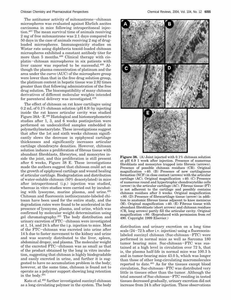
The antitumor activity of mitoxantrone-chitosanmicrospheres was evaluated against Ehrlich ascitescarcinoma in mice following intraperitoneal injec-tion.487 The mean survival time of animals receiving2 mg of free mitoxantrone was 2.1 days compared to50 days in the case of animals receiving 2 mg of drug-loaded microspheres. Immunogenicity studies onWistar rats using diphtheria toxoid-loaded chitosanmicrospheres exhibited a constant antibody titer formore than 5 months.488 Clinical therapy with cis-platin-chitosan microspheres in six patients withliver cancer was reported to be successful.489 Al-though the plasma concentration of platinum and thearea under the curve (AUC) of the microsphere groupwere lower than that in the free drug solution group,the platinum content in hepatic tissue was 2.92 timesgreater than that following administration of the freedrug solution. The biocompatibility of many chitosanderivatives of different molecular weights intendedfor parenteral delivery was investigated.476
The effect of chitosan on rat knee cartilages using0.2 mL of 0.1% chitosan solution (pH 6.9) by injectinginside the rat knees articular cavity was studiedFigure 38A-E.490 Histological and histomorphometricstudies after 1, 3, and 6 weeks postinjection wereperformed on undecalcified samples embedded inpolymethylmetacrylate. These investigations suggestthat after the 1st and sixth weeks chitosan signifi-cantly slows the decrease in epiphyseal cartilagethicknesses and significantly increases articularcartilage chondrocyte densities. However, chitosansolution induces a proliferation of fibrous tissue withabundant fibroblasts, fibrocytes, and monocytes in-side the joint, and this proliferation is still presentafter 6 weeks, Figure 38 E. These investigationsmade the authors suggest that chitosan could act onthe growth of epiphyseal cartilage and wound healingof articular cartilage. Biodegradation and distributionof water-soluble chitosan (50% deacetylated) in miceafter intraperitoneal (i.p.) injection was studied,whereas in vitro studies were carried out by incubat-ing with lysozyme, murine plasma, and urine.491
Chitosan and fluorescein isothiocyanate-labeled chi-tosan have been used for the entire study, and thedegradation rates were found to be accelerated in thepresence of lysozyme, plasma, and urine, which wasconfirmed by molecular weight determination usinggel chromatography.491 The body distribution andurinary excretion of FTC-chitosan were investigatedat 1, 14, and 24 h after the i.p. injection to mice. Mostof the FTC-chitosan was excreted into urine after14 h due to faster movement to the kidney and urineand was scarcely distributed to the liver, spleen,abdominal dropsy, and plasma. The molecular weightof the excreted FTC-chitosan was as small as thatof the product obtained by the long in vitro incuba-tion, suggesting that chitosan is highly biodegradableand easily excreted in urine, and further it is sug-gested to have no accumulation problem in the body;however, at the same time, chitosan is found not tooperate as a polymer support showing long retentionin the body.491
Kato et al.492 further investigated succinyl chitosanas a long circulating polymer in the system. The body
distribution and urinary excretion on a long timescale (24-72 h after i.v. injection) using a fluorescein-labeled succinyl chitosan (Suc-chitosan-FTC) wereperformed in normal mice as well as Sarcoma 180tumor bearing mice. Suc-chitosan-FTC was sus-tained at a high level in circulation over 72 h, thatis, the plasma half-life in normal mice was 100.3 hand in tumor-bearing mice 43.0 h, which was longerthan those of other long-circulating macromoleculesreported to date.491 As for the tissues except bloodcirculation, Suc-chitosan-FTC was distributed verylittle in tissues other than the tumor. Although thetotal amount of Suc-chitosan-FTC residing in testedtissues decreased gradually, urinary excretion did notincrease from 24 h after injection. These observations
Figure 38. (A) Joint injected with 0.1% chitosan solutionat pH 6.9 1 week after injection. Presence of numerousfibroblasts and monocytes trapped into fibrosis (arrows).Presence of possible chitosan residues (CR). Originalmagnification ×40. (B) Presence of new cartilaginousformation (NCF) in close contact (arrows) with the articularcartilage (AC). Original magnification ×40. (C) Presenceof numerous round and hypertrophic chondrocytelike cells(arrow) in the articular cartilage (AC). Fibrous tissue (FT)is not adherent to the cartilage and possibly containschitosan residues after 3 weeks. Original magnification×80. (D) Presence of fibrocartilage tissue (arrow) in addi-tion to anatomic fibrous tissue adjacent to knee meniscus(M). Original magnification ×40. (E) Fibrous tissue withabundant fibroblasts (short arrows) and chitosan residues(CR, long arrows) partly fill the articular cavity. Originalmagnification ×80. (Reproduced with permission from ref490. Copyright 1999 Elsevier.)
Chitosan Chemistry and Pharmaceutical Perspectives Chemical Reviews, 2004, Vol. 104, No. 12 6055
suggested that Suc-chitosan-FTC may be eliminatedby mechanisms other than in the urine or moved totissues other than those tested. The ratio of tumoraccumulation reached a plateau at 48 h after injec-tion, and the accumulation level, approximately 10%,was similar to those observed for other reported long-circulating macromolecules.492 The preparation andbiodistribution into the intestine, bone, lymph nodes,and male genital organs after i.v. administration oflactosaminated N-succinyl chitosan drug carrier wasalso studied.493 Similarly, lactosaminated N-succinylchitosan as a liver-specific drug carrier in mice wasalso studied.107 Qin et al.494 studied antitumor activityof water-soluble low molecular weight chitosan pre-pared by enzymatic hydrolysis.
Domard and co-workers495 studied the optimalconditions for using chitin-based gels in periodontalsurgery. Rheological properties during injection es-pecially the flow through the tip of the syringe, thecapacity of adaptation of the solution to the walls ofthe bone disease, and the injection time are essentialfactors to be considered. The acetylation parametersfound to be key features affecting the injections areas follows: a molar ratio acetic anhydride/glucos-amine residue of R ) 1.5, a temperature of themaster solution of 12 °C, a mixture of hydro alcoholicsolution/acetylating reactive stirred for 45 s at roomtemperature, and a chitin concentration of 3.6%. Thisconcentration allows limiting the synthesis to im-prove the mechanical properties of the gel and obtaina viscosity suitable for injection. Doping of the gelby means of chitosan powder insoluble under theseconditions led to an improvement of the biologicalactivity of the gel.
In vitro and in vivo evaluations of sulfated chito-sans as blood anticoagulants were performed.496
Several derivatives with different polymerization andsulfation degrees were evaluated for their potential.The results suggest that low molecular weights (Mw61-82 KDa, polymerization degree 188-252) andhigh sulfination patterns (sulfur 15.6-16.9%, sul-fination degree 1.58-1.86) were slowly cleared andthat there was more antithrombin activity (30-52IU/mg).496 The authors observed differences in vivovs in vitro, especially in activated partial thrombo-plastin time, therefore, expressing a preference forthe in vivo system for evaluating the antithrombinactivity of these derivatives.496
Thermosensitive gels have been extensively inves-tigated for several applications.269-273 In a recentstudy, the potential of thermosensitive chitosan gelfor local delivery of paclitaxel was evaluated.497 Theauthors claimed that one intratumoral injection ofthe thermosensitive hydrogel containing paclitaxelwas as efficacious as four intravenous injections ofTaxol in inhibiting the growth of EMT-6 cancer cellsin mice but in a less toxic manner.497
5.4. TransdermalThe transdermal delivery of drugs into systemic
circulation has generated much interest during thepast decade. Transdermal drug delivery systems(TDDS) offer many advantages over the conventionaldosage forms or controlled release peroral delivery
systems. TDDS provides constant blood levels (1-7days), avoids first-pass metabolism, increased patientcompliance, and dose dumping never occurs. Thechoice of drugs delivered transdermally, clinicalneeds, and drug pharmacokinetics are some of theimportant considerations in the development of TDDS.Although novel approaches such as iontophoresis andultrasound are gaining importance as a means toincrease drug permeation into systemic circulation,clinical products based on these approaches are stillfar away. The importance of appropriate animalmodel selection in the development and evaluationof TDDS cannot be ignored. The cost per milligramof drug delivered transdermally is more expensivethan peroral route. The added cost could be justifiedif TDDS improved patient compliance and reducedtoxicity.
A chitosan-based transdermal delivery system wasdeveloped using cross-linked chitosan membrane andchitosan gel498 where the membrane was the rate-controlling device and the gel acting as the reser-voir: the cross-link density of the membrane dictatedthe drug permeability. Similarly, results were re-ported when a combination of chitosan with collagenwas used as a rate-controlling membrane.499 How-ever, alginate gel was used as the drug reservoirinstead of chitosan gel. It has been suggested thatwater-soluble drugs permeate mainly through thepores in the chitosan membrane, whereas hydropho-bic drugs permeate by both partition and pore mech-anisms.500 A transdermal film dosage form of capto-pril was prepared from chitosan plasticized withpoly(ethylene glycol).501 A chitosan patch containing20 mg of drug improved drug availability when testedon the back skin of male albino rats compared withan oral dose of 4.3 mg/kg. No significant irritantreaction was observed on the skin.
Many previous studies have shown chitosan-medi-ated decreases in the trans-epithelial electrical re-sistance. This is accompanied by an increase in thepermeability of the monolayers to inert hydrophilicprotein markers such as inulin or mannitol. A chi-tosan-mediated increase in permeability to inertmarker points to passive transport that is possibleonly through the paracellular space.502-505
Chitosan has been used as an aid to transdermaldrug delivery both in vitro and in vivo in numerousstudies. The ability of chitosan to aid the permeationof several drugs across excised skin from numerousspecies has been shown. The diffusion of buprenor-phine, nonivamide, and propranolol hydrochloridefrom chitosan hydrogels through excised dorsal skinwas shown to be greater than passive diffusion fromcontrol media.276,506,507 Delivery of capsaicin acrossexcised nude mouse skin was significantly enhancedby its incorporation into a chitosan gel compared tocommercially available creams.508 This means thatchitosan-based transdermal drug delivery systemshave the potential to be more effective than otherknown delivery systems. The use of chitosan asdelivery vehicle resulted in improved delivery com-pared to other polymer hydrogels. Iontophoresis (theapplication of an electric current to aid the passageof drugs across the epithelium) along with dermal
6056 Chemical Reviews, 2004, Vol. 104, No. 12 Kumar et al.

application of chitosan-drug hydrogels resulted ina greater drug flux across the skin barrier comparedto either methods individually, i.e., over a 10-foldincrease of transdermal drug flux compared to con-trol. This result may be of clinical interest.
The ability of chitosan to facilitate the systemicavailability of transdermally applied drugs has beenshown in vivo. Delivery of the drug bromocriptinefrom chitosan hydrogels across rabbit skin in vivoresulted in a similar concentration of circulating drugas observed following administration of the stand-ard oral dosage.509 The high fraction of this drug lostby hepatic metabolism following oral administra-tion underlines the need for its delivery by a trans-dermal route. Transdermal delivery of the â-blockeroxprenolol hydrochloride from chitosan films wasshown to have a systemic effect in rats: reducedarterial blood pressure was observed.510 These dataimply that the pharmacological efficacy of drugs isnot impaired by delivery in this manner and thatchitosan can facilitate the transdermal delivery ofdrugs for systemic effect.
5.4.1. Roles of Chitosan in Affecting the Skin Barrier
Effective transdermal drug delivery involves per-meation of the drug through a number of barrierssthe delivery system itself, the epidermis, the dermis,and in the case of systemic drugs endothelial cellwalls and into the blood stream. The stratum cor-neum has been thought to be the primary barrier todrug delivery in the human skin for many years, andthe limiting factor of transdermal drug delivery hasgenerally been regarded as the diffusion rate throughthis structure. Skin that had the stratum corneumremoved was highly permeable, whereas an isolatedstratum corneum was almost as impermeable as theentire skin. The barrier function of the stratumcorneum is thought to be dependent upon the pres-ence of extracellular lipids within the paracellularspace. Lamellar granules, lipid containing vesicles,fuse into the plasma membrane, resulting in theextrusion of the lipids into the extracellular space.The lipids are covalently bound to the outer surfaceof the cell membranes, resulting in the creation of alipid envelope that renders the stratum corneumrelatively impermeable. The presence of large amountsof extracellular lipid means that lipophilic drugs candiffuse down a continuous lipophilic path through thestratum corneum more easily than hydrophilic drugs.
There is evidence that the tight junctions in skinmay also contribute toward the skin barrier.511,512
Given the key role of chitosan-mediated tight junctiondisruption in trans-epithelial drug delivery in vitro,tight junctions in skin may be investigated in orderto understand the action of chitosan in transdermaldrug delivery.
Chitosan may increase the permeability of thestratum corneum possibly by occlusion. Second, ap-plication of chitosan may result in disruption of thetight junctions immediately below the stratum cor-neum, allowing permeation of drug through into theunderlying layers of the epidermis. The drug wouldthen diffuse down its concentration gradient into theunderlying tissues. The permeability of the stratum
corneum and the presence of chitosan disrupt theunderlying tight junctions, thus increasing drugpermeation through both barriers simultaneously.This novel model may provide a new direction toinvestigate future chitosan-mediated transdermaldrug delivery systems.
5.5. ImplantsImplantable devices are expected to be intelligent,
nontoxic, nonthrombogenic, noncarcinogenic, andeasily implantable with adequate storage capacity,drug stability, biodegradability, and sterilizability.Khor and Lim68 discussed various applications ofchitosan implants in a recent review. The currenttrend of using natural materials as implantabledevices is justified by two important reasons: (1)natural materials have been shown to better promotehealing at a faster rate and are expected to exhibitgreater compatibility with humans and (2) for tissueengineering purposes where cells are seeded onto thebiomaterial implants. Chitin and chitosan have beenused in orthopedic and periodontal applications.513,514
Chitosan and hydroxypropyl chitosan presented en-zymatic degradation in vitro and following sub-cutaneous implantation in rats. A combination ofthese two formulations was studied as film-typeimplants515 containing uracil that showed sustaineddrug release in rats. Chitosan was also tested as localimplants in the form of microspheres and fibers assustained release depots for endothelial growth fac-tors.516 Endothelial cell growth factors have a rela-tively short half-life. This angiogenic factor shouldtherefore be administered frequently in order toreach an effective concentration. Drug-loaded ma-trixes implanted subcutaneously in rats showed aburst effect; however, the release continued for morethan 3 weeks. N-Succinyl-chitosan-mitomycin C asan implant for controlled drug release, and itsantitumor activity has been evaluated.517
Sustained release implants of herb extracts usingchitosan-gelatin matrix were developed.518 In vitroand in vivo degradation studies using lysozymes andfemale Wistar rats were performed, respectively. Noside effects on in vivo implants were observed,suggesting their use as medical devices. The biode-gradability of these implants is a further advantage,as no surgical removal of the exhausted implants isneeded.518
Microsphere-based chitosan implants with smallparticle size, low crystallinity, and good sphericitywere prepared by a spray-drying method followed bycross-linking with genipin;519 glutaraldehyde cross-linked microspheres were used as controls. Histologi-cal study of the genipin cross-linked chitosan micro-spheres injected into the skeletal muscle of a ratmodel showed a less inflammatory reaction than itsglutaraldehyde cross-linked counterparts. On theother hand, the degradation studies suggest thatglutaraldehyde cross-linked particles started to de-grade by 12 weeks, whereas the genipin cross-linkedparticles were intact until 20 weeks; this suggeststhat these implants are suitable for long-term ap-plications.519
Most recently, chitosan and sodium hyaluronateimplants for controlled release of insulin were stud-
Chitosan Chemistry and Pharmaceutical Perspectives Chemical Reviews, 2004, Vol. 104, No. 12 6057

ied.520 The optimum conditions for polyion complexformation between chitosan and hyaluronate were in-vestigated and found to influence the insulin release;however, the compression exerted to manufacture theimplant had no role to play in the release kinetics.520
5.6. Ophthalmic PreparationsTraditional aqueous solutions, suspensions, and
ointments for ophthalmic use have the disadvantagethat the major part of the drug is lost within secondsdue to the nasolacrymal duct drainage; therefore,repeated administration is needed. The prime re-quirement for the controlled delivery of ophthalmicpreparations is therefore bioadhesiveness that in-creases the contact time with the cornea, leading toimproved drug absorption at the site.
The viscosity of the polymer solutions might induceone to believe that high viscosity would be anadvantage; however, a study involving different fluidshaving the same viscosity and containing pylocarpineshowed that the viscosity relevance is scarce.521 Whatis important, on the other hand, is to modify as littleas possible the rate of shear of the eye fluid, whichhas a very wide range (0.03-28 500 s-1) dependingon the movement of the eyelids. As a matter of fact,the chitosan solutions have viscoplastic and pseudo-plastic characteristics that do not modify the lacry-mal film, thus providing a good patient compliance.In consideration of this favorable aspect and of thechitosan bioadhesiveness, the prime requirement foran effective ocular therapeutic system, attempts weremade to take advantage of the cationic property ofchitosan in ophthalmic delivery.522-526 Chitosan- andpoly-L-lysine-coated ε-polycaprolactone nanocapsuleshave been tested for their efficacy in ocular deliveryexploring the favorable mucoadhesion property ofthese cationic polymers. An improved interactionbetween corneal epithelium and the cationic polymerswas expected; however, the cationic modification didnot alter the drug-release properties. The bioavail-ability studies suggested no increase in case of poly-L-lysine-coated nanocapsules; however, chitosan coat-ings improved twice that of uncoated nanocapsules.Therefore, these findings suggest that positive chargeis not a contributing factor for the improved bioavail-ability of the drug, but the specific nature of chitosanis responsible for the enhanced uptake.527 The dif-ference between PEG and chitosan coatings on ε-poly-caprolactone nanocapsules for ocular delivery hasbeen studied: the PEG coating accelerates the trans-port of the nanocapsules across the whole epithelium,whereas the chitosan coating favors retention of thenanocapsules in the superficial layers of the epithe-lium.528
The ability of chitosan as an ocular delivery devicehas been evaluated by comparing chitosan formula-tions with commercial collyrium Tobrex. Chitosan asa gel has been tested for the increased precornealdrug residence times, expecting it to slow drugelimination by lacrymal flow by increasing solutionviscosity and interacting with the negative chargesof the mucus. The influence of the process parametersas well as the effect of the molecular weight andconcentration of polysaccharide was thoroughly stud-
ied. An ocular irritation test, using confocal laserscanning ophthalmoscopy (CLSO) combined withcorneal fluorescein staining, clearly demonstrated theexcellent tolerance of chitosan after topical admin-istration onto the corneal surface. γ-Scintigraphicdata showed that the clearance of the formulationslabeled with 99mTc-DTPA was significantly delayedin the presence of chitosan with respect to thecollyrium, regardless of the concentration and mo-lecular weight of chitosan in solution. At least a 3-foldincrease of the corneal residence time was achievedin the presence of chitosan when compared to con-trol.529
Alonso and co-workers187 carried out extensiveinvestigations on chitosan nanoparticles as discussedin the foregoing sections of this article. They also tookthe challenge to prove the ability of chitosan nano-particles to be efficient ocular delivery devices forimproved bioavailability of the drugs and at the sametime preventing the systemic exposure of the admin-istered drug. In vivo experiments were performed inrabbits: therapeutic concentrations in external ocu-lar tissues (i.e., cornea and conjunctiva) were main-tained for at least 48 h, and at the same timecyclosporin (CyA) levels in inner ocular structures(i.e., iris/ciliary body and aqueous humor), blood, andplasma were negligible or undetectable.
The ability of chitosan hydrochloride to enhancethe transcorneal permeability of the drug has beendemonstrated.530 Poly(ethylene oxide) (PEO) wasused as a base material to which ofloxacin-containingchitosan microspheres prepared by spray drying wereadded and powder compressed, resulting in circularinserts (6 mm). The effect on addition of chitosan hasbeen studied systematically, where 10%, 20%, and30% responded according to the increase in theirconcentrations exhibiting accelerated erosion, therebyrelease of the entrapped drug. PEO molecular weightalso has an influence on erosion and drug release,where PEO 900 showed better performance in com-parison to PEO 2000. Experiments in the rabbit’s eyeformed a superficial gel that adhered to the applica-tion site, which gradually spread over the cornea anderoded with time. There were signs of irritation inthe eye; however, the levels in chitosan-based sys-tems were found to be lower when compared tochitosan-free inserts. The release and bioavailabilityrates were much higher for the PEO-chitosan-HClwhen compared to the plain inserts of either.530
Also, Felt et al.531 reported that co-administrationof ofloxacin and chitosan in eyedrops increased thebioavailability of the antibiotic. The authors ascribedthis effect to the high viscosity of the chitosansolution. These results are of relevance to the treat-ment of external ocular infections. It was also re-ported that chitosan is able to enhance drug pen-etration through the corneal epithelium, therebypotentially improving topical treatment of endo-phthalmic infections. Another study ascertained theability of chitosan‚HCl and N-trimethyl chitosanchloride (TMC), added to ofloxacin eyedrops, toimprove transcorneal antibiotic transport.532 Thepolycationic chitosan‚HCl and TMC are believed toexert their effects by interacting with the negatively
6058 Chemical Reviews, 2004, Vol. 104, No. 12 Kumar et al.

charged cornea. TMC was more effective because itis soluble, whereas chitosan precipitates at the pHof the tear fluid.
Another chitosan derivative, N-carboxymethyl chi-tosan, did not enhance the corneal permeability;nevertheless, it mediated zero-order ofloxacin absorp-tion, leading to a time-constant effective antibioticlevel in the aqueous.532 Also, N,O-carboxymethylchitosan is suitable as an excipient in ophthalmicformulations to improve the retention and bioavail-ability of drugs such as pilocarpine, timolol maleate,neomycin sulfate, and ephedrine. Most of the drugsare sensitive to pH, and the composition should havean acidic pH to enhance the stability of the drug.Therefore, delivery should be made through an anion-exchange resin that adjusts the pH at around 7 thatis physiologically acceptable.533 Chitosan solutions donot lend themselves to thermal sterilization. A chi-tosan suspension, however, can be sterilized at 121°C for 15 min and then treated with sterile solutionof a suitable acid (HCl). The presence of mannitol,sorbitol, and glycerol seems to stabilize the sterilizedsolutions. This preparation was found to be particu-larly useful for pharmaceutical and ophthalmic com-positions.534
The favorable characteristics of chitosan have beenconsidered in proposing tear substitutes535 that ap-pear to be particularly suitable in light of thediscovery of chitin-like substances in the humanocular mucus.536 Chitosan, therefore, has a uniqueplace among the ophthalmic drug delivery systems.537
5.7. Gene DeliveryTransfection is the genetic modification of cultured
animal cells by the uptake of DNA from the culturemedium. The DNA to be transfected is usually in theform of the recombinant plasmid or other type ofDNA vector containing the gene of interest. Severalmethods have been developed to facilitate the pro-cess, among which the most widely used are basedon calcium phosphate- or diethylaminoethyl dextrin-mediated transfection.
Mumper et al.78,538 were the first to describe thepotential of chitosan as a gene carrier. The lowtoxicity of chitosan and its nature make it attractivefor gene delivery purposes.449,450,539 In early studies,chitosan has been shown to bind nucleic acids,540 andit is known that chitosan may actually be endocy-tosed into the cell.476
As for the majority of polycations, the interactionbetween chitosan and DNA is electrostatic.541 Suchinteraction appears to be strong enough that thechitosan-DNA complex does not dissociate until ithas entered the cell.542 Also, it was shown thatchitosan plays an important role in both membraneadhesion543 and lysosomal escape544 of the encapsu-lated DNA, providing a plausible explanation for theability of the polymer in cell transfection. Hayatsuet al.545 observed that a treatment of DNA/chitosancomplex with phosphate-buffered saline resulted inthe release of only minute quantities of DNA (0.05%),thereby indicating the formation of a tight andcompact complex. Lee et al.546 examined the potentialof a water-soluble and low molecular weight chitosan
(LMWC) having an average degree of polymerizationof 136 (∼20 kDa) and a degree of deacetylation of72.5%. Complexes of plasmid/LMWC were preparedby mixing the two components in PBS medium, andthe stability of the complexes was judged by gelretardation assay. The complexes were completelyretarded at a 1:2 weight ratio (plasmid/chitosan),indicating total condensation. Also, it was demon-strated that plasmid/LMWC formulation efficientlyprotects the plasmid from DNase degrading enzymes.
The hybrid DNA-chitosan systems can be classi-fied into two categories which differ in their mech-anism of formation and morphology: complexes andnanospheres.
(1) Chitosan-DNA complexes: simple mixing oftwo solutions of DNA and chitosan generated broadparticulate complexes with mean sizes between 100and 600 nm, depending on the molecular weight ofthe chitosan.538 Stable complexes are usually formedwhen chitosan is added in molar excess relative tothe negatively charged DNA with zeta potentialvalues between +0 and 20 mV, depending upon thedegree of excess.547
(2) Chitosan-DNA nanospheres: the method fornanoparticle formation was modified by augmentingthe mixing speed and temperature upon blending ofthe two solutions and incorporating a dilute salt(Na2SO4) in the DNA solution and a desolvatingagent in the polymer solution.548 Such modificationsinduced significant changes in particle morphology,creating a monodisperse, spherical suspension be-tween 200 and 500 nm which sharply differed fromthe loose rodlike78 and toroidal78,547 structures re-ported for chitosan-DNA complexes. Mao et al.541
examined the influence of several parameters on thesize of chitosan-DNA nanospheres. The particle sizeswere not affected by the sodium sulfate concentrationin the range of 2.5-25 mM and neither by theplasmid size. On the other hand, chitosan-DNAcomplex sizes were found to clearly depend on themolecular weight of the chitosan (108-540 kDa) usedand N/P ratio.78,83,549 This nanosphere formulationwas found to additionally trap low levels of smallmolecules such as chloroquine, a well-known lyso-somotropic agent, enabling a simultaneous deliveryof DNA and drugs. Given their novel properties, thesecompact structures were considered distinct from thetraditional complexes and named chitosan-DNAnanospheres.541
At physiological pH, not all amino groups of chi-tosan are protonated; therefore, the N/P ratio isadopted, where N is chitosan nitrogen and P is DNAphosphate. At a N/P ratio of 2, large-size complexeswere obtained with a zeta potential of the com-plexes close to 0 mV, indicating full retardation ofDNA and aggregation of the complex at neutralsurface charge.550
The electrostatic nature of the interaction of chi-tosan with DNA was assessed by ethidium bromidecompetitive studies.541 When chitosan is added to asolution of ethidium bromide-stained DNA, fluores-cence decreases as a result of competitive binding ofcationic chitosan to DNA. Ethidium bromide quench-ing assay in combination with confocal laser scanning
Chitosan Chemistry and Pharmaceutical Perspectives Chemical Reviews, 2004, Vol. 104, No. 12 6059

microscopy was also applied to determine the distri-bution of encapsulated DNA in chitosan nanopar-ticles.548 Mumper et al.538 indirectly demonstrated theabsence of free DNA after complex formation by gelelectrophoresis.541 Another method for determiningDNA loading is by pico Green assay after digestionwith chitosanase and lysozyme. Usually 95% loadingof DNA is achieved by simple mixing of chitosan andDNA. Stability studies have shown that the cross-linked chitosan/DNA nanoparticles stored in waterremained stable for more than 3 months, whereasun-cross-linked formulation remained stable for fewhours only.8 Also, complexation of DNA with highlypurified chitosan having different molecular weightsat N/P 1 resulted in almost complete inhibition ofdegradation by DNase I and DNase II.551 The resist-ance to degrading enzymes is probably achieved bychange in the tertiary DNA structure, causing sterichindrance.
5.7.1. Chitosan/Plasmid Complexes: Toxicity andTransfection Efficiencies
In recent years, cationic polymers have been in theforefront in the field of gene delivery and cell trans-fection. This is an attractive choice as simple mixingof plasmid DNA with the polycation in aqueousmedium leads to electrostatically driven self-assembly complexes in the nanoscale range.552 Theuse of chitosan in the field of gene delivery hasseveral advantages, some of which are listed.78,538,541,548
(1) Conjugation of ligands to chitosan-DNA com-plexes for targeting and receptor-mediated endocy-tosis.
(2) Incorporation of lysosomotropic agents to en-hance the escape from the lysosome and hence reducedegradation of complex.
(3) Coencapsulation of other bioactive agents formultiple plasmids.
(4) High protection against serum nuclease degra-dation.
(5) Lyophilization of polyplexes for long time stor-age without comparable loss in the activity.
Mao et al.541 performed several in vitro transfectionexperiments and showed that chitosan-DNA nano-spheres were able to transfect HEK293, IB-3-1, HeLacells but with remarkably lower levels than thecommercial lipid-based transfecting agent, Lipo-fectamine. Although, coencapsulation of chloroquinesignificantly increased the transfection efficiencies ofanother natural, cationic biopolymer (gelatin), chi-tosan did not substantially benefit from the same co-incorporation.541,548 Sato et al.553 investigated thenature of chitosan-DNA complexes and their trans-fection efficiencies. The team showed that cell uptakeof chitosan-DNA complex was dependent on cell typeand the complex mixtures and was far superior topolygalactosamine-DNA complexes in transfectingA549 cells.69 The optimal charge ratio (N/P) for celltransfection varied from cell to cell type.550,554-556
Corsi et al.557 established optimal conditions for thechitosan-DNA complexation. The complexes were ofa mean diameter inferior to 100 nm (Figure 39) withthe interior of the particles showing homogeneousDNA distribution. The efficacy of transfection in vitro
on HEK293, MG63, and MSC (Mesenchymal stemcells) demonstrated cell-type dependence, withHEK293 cells having a higher gene expression,however, not surpassing that seen with Lipofectamine.Cell viability studies applying the MTT test con-firmed the low toxicity of chitosan, in marked con-trast to Lipofectamine which causes approximately50% toxicity. These results suggest that chitosan-DNA nanoparticles have favorable characteristics fornonviral gene delivery and are cell dependent andnot cytotoxic. The transfection efficiencies of chito-san-DNA-based complexes were not affected by thepresence of 10% serum in cell culture.541
Sato et al.69 further investigated the effect of theexperimental conditions, such as medium pH, serumcontent, molecular weight of chitosan, and N/P ratio,on the transfection efficiency of plasmid/chitosancomplexes. The transfection efficiencies were evalu-ated in A549 cells applying pGL3 encoding to lu-ciferase. The medium was 10% FBS with two pHvalues: 6.9 and 7.6. The transfection efficiency at pH6.9 was remarkably higher than that at pH 7.6. Thisdependence is probably attributed to the protonationof amines in chitosan. The pKa of amines in chitosanhas been reported to be around 6.3-6.4.558,559 Thismeans that at a pH lower than 7, amino groups ofchitosan are charged and could bind DNA throughelectrostatic interaction. In contrast, a higher pHvalue (i.e., 7.6) led to a remarkable decrease of theamino group protonation and hence to a drasticdecrease of the transfection efficiencies. On thecontrary, when the transfection efficiencies of theLipofection/DNA, a lipid-based formulation, weretested at various pH values, no remarkable differ-ences in the transfection yields were obtained. Thiswas attributed to the chemical nature of the quater-nary ammonium salt of the headgroup of the lipid,which is not affected by the pH of the medium. Thetransfection efficiency of plasmid/chitosan complexwas assessed as a function of N/P ratio. The optimal
Figure 39. Atomic force microscopy analysis of complexesformed between chitosan and DNA. (Reproduced withpermission from ref 557. Copyright 2003 Elsevier.)
6060 Chemical Reviews, 2004, Vol. 104, No. 12 Kumar et al.

ratio for transfection was found to be 5:1. Higher N/Pratios (i.e., 10:1 and 20:1) resulted in a drasticdecrease in the transfection yields. Also, it was foundthat the highest transfection yields were obtainedwhen plasmid concentrations of 5-10 µg/mL wereapplied. The transfection efficiency was also assessedas a function of the molecular mass of chitosan. Thetransfection experiments were tested in A548, B16melanoma, and HeLa cells in 10% FBS. Chitosansof 15 and 52 kDa largely promoted gene expressionin all tested cell lines. Heptamer of an averagemolecular weight of 1.3 kDa did not induce transgeneexpression in all tested cell lines. Also, it was foundthat the transfection yield obtained with high mo-lecular weight chitosan (>100 kDa) was less than thetransfection efficiency obtained with chitosan of 15and 52 kDa. As a second control, polygalactosamine(pGalN) was assessed for its transfection efficienciesand found to be inactive in all tested cell lines. Theinactivity of pGalN/plasmid complex was attributedto intracellular processes and suggests that thepGalN/plasmid complex is very stable even in theendosome and the plasmid may not release from it.Maximum transfection yield of chitosan/plasmidcomplex was obtained in 20% FBS medium, inmarked contrast to Lipofectin which gave no trans-fection in this medium. Higher serum contents (i.e.,30% and 50% FBS) resulted in a gradual decreasein the transfection efficiencies.
Richardson et al.551 prepared highly purified chi-tosan fractions of <5000 Da (N1), 5000-10 000 Da(N2), and >10 000 Da (N3) by hydrolysis of highmolecular weight chitosan and characterized themwith respect to their cytotoxicity, ability to causehaemolysis, and ability to complex and protect DNAfrom degradation. All chitosan fractions displayedlittle cytotoxicity against CCRF-CEM and L123 cells(IC50 > 1 mg/mL), and they were not hemolytic (<15%lysis after 1 and 5 h). Also, chitosan fractions werefound to exhibit a stronger interaction with DNAwhen compared to poly(L-lysine) and resulted in atotal inhibition of DNA degradation by DNase II.After intravenous injection of chitosan-DNA com-plexes, all the chitosan fractions showed rapid bloodclearance. The plasma levels at 1 h were 32.2 (10.5% of recovered dose for N1 and 2.6 ( 0.5% ofrecovered dose for N3. Liver accumulation was mo-lecular mass dependent, being 26.5 ( 4.9% of therecovered dose for N1 and 82.7 ( 1.9% of therecovered dose for N3. The observations that thehighly purified chitosan fractions used were neithertoxic nor hemolytic, that they have the ability tocomplex and protect DNA from degradation, and thatlow molecular weight chitosan (N1) can be adminis-tered intravenously with minor liver accumulationsuggest there is potential to investigate further lowmolecular weight chitosans as components of asynthetic gene delivery system.551 Chitosan HCl saltwas the most toxic, having an IC50 (inhibitory con-centration 50%) of 0.21 ( 0.04 mg mL-1, which wasonly 4-fold less toxic than poly(L-lysine) (IC50 of 0.05( 0.01 mg mL-1).7 High molecular weight chitosanwas the most toxic, the resulting ranking beingchitosan HCl > chitosan hydroglutamate > glycolchitosan > chitosan hydrolactate.
Sato and co-workers69,560 examined the correlationbetween complex uptake and degree of gene expres-sion and found a strong proportional correlation inserum-free medium. However, when serum-contain-ing medium was applied, there was no relationshipbetween the transfection activity and cell uptake.This result indicates that cell uptake of the complexesdeclines as a result of nonspecific interaction withthe serum components. Nevertheless, there was thehighest transfection activity in the presence of 10%or 20% serum due to cell viability being optimal fortransfection under these conditions.
5.7.2. Mechanism of Action of Chitosan/PlasmidComplexes
The mechanism of cell transfection with plasmid-chitosan complexes was investigated by followingthree processes.560
(1) Cell uptake: observation with a confocal laserscanning microscope showed that FITC-plasmid/chitosan complexes (at N/P 5) were taken up byendocytosis. The fluorescence of FITC-plasmid andTexas Red-dextran was observed at the same cel-lular localization after 1 h postincubation. The uptakeof plasmid/chitosan (40 kDa) complexes was moni-tored also with a flow cytofluorometer. Cell uptakeat 37 °C was about 3-fold more than that measuredat 4 °C, although the amount of cell uptake of 1 kDachitosan and DEAE-dextran complexes was virtu-ally constant. This result implied that cell uptake ofplasmid-chitosan complex might be thermal andenergy dependent and that plasmid/chitosan com-plexes might be endocytosed. Similar results werereported using transmission electron microscopyobservation of plasmid/chitosan complexes (N/P ) 5)in HeLa cells.550
(2) Release from endosome: the release of plasmid/chitosan complexes from the endosomal compartmentmay be another rate-limiting step in the transfectionprocess.78 Protons accumulate into the endosomalvesicles through a vesicular ATPase-driven protonpump. Partially protonated polymers such as poly-ethyleneimine retain a substantial buffering capacity,so that they can be protonated at virtually any pH.561
There are two main effects of buffering capacity ofcationic polymers in endosome. (I) It may protect theplasmids from lysosomal nuclease due to delay inacidification.562 (II) It may also perturb the traffickingof endosome for osmotic swelling and subsequentendosome disruption. It is well known from theliterature that low molecular weight chitosan (1700Da) exhibited more effective inhibition of pH fall frompH 7 to 4 than aspartame, phosphate, and mono-fluorophosphate buffer.563 Furthermore, it was dem-onstrated that the extent of protonation of DNA/chitosan (Mw 40 and 110 kDa) complexes from pH 7to 5 was approximately 2.5-fold larger than that ofDNA/PLL complexes. Accordingly, the surface chargeof plasmid/chitosan complexes increased from +2 (pH7) to +21 mV (pH 5). These features may be a causeof pH-dependent transfection efficiency of plasmid/chitosan complexes. At pH 6.5 of the transfectionmedium, low and negligible transfection yields wereobtained, although a certain cell uptake of plasmid/
Chitosan Chemistry and Pharmaceutical Perspectives Chemical Reviews, 2004, Vol. 104, No. 12 6061

chitosan complexes was observed at this particularpH. These results are also attributed to the lowbuffering effect at pH 6.5 of the transfection medium.Moreover, Sato and co-workers560 found that lyso-somal agents such as monensin and chloroquine,which increase the pH of endocytic vesicles, decreasedtransfection levels with plasmid/chitosan complexesin SOJ cell line. However, these endosomolytic agentsimproved the transfection levels with plasmid/PLLcomplexes.550 It was found from these results thatplasmid/chitosan complexes have a high ability forproton accumulation, and thus, endosome bufferingmay perturb the endosomal membrane and releasecomplexes from endosomes as the pH of endosomesdeclines. Another factor that seems to limit thetransfection activity of plasmid/chitosan complexesis its potential to release DNA after the endosomeescape. To have transfection activity with plasmid/chitosan complexes, the complexes have to be firstreleased from endosomes. Next, the chitosan in thecomplex exchanges with endosomal membranes con-sisting of anionic lipids via electrostatic interactions.The release of DNA from DNA/chitosan complexeswas observed at a higher concentration of sodiumdodecyl sulfate (10 times), which is greater than thatobserved for DNA/PLL complexes. Thus, plasmid/chitosan complexes were not likely to exchange withendosomal membranes and release the intact com-plexes from endosomes.560
(3) Nuclear import: the nuclear import of com-plexes is still controversial; specifically, the nuclearimport of plasmid/chitosan complexes has never beenfully examined.550 FITC-labeled plasmid and TexasRed-labeled chitosan were used to observe cellularlocalization. At 4 h posttransfection, the two labeledmolecules had accumulated in the nucleus and re-mained localized in the nucleus 24 h posttransfection.On the other hand, FITC-plasmid/(PLL or Lipo-fectamine) complexes did not display fluorescenceimages even several hours postincubation. Theseresults illustrated that plasmid/DNA complexes havehigh nuclear accumulation and resistance towardcellular and nuclear enzymes.560
Several chitosan derivatives have been synthesizedin the past few years in order to obtain modifiedcarrier with altered physicochemical characteris-tics.564 Such modifications include quaternization ofamino groups to increase the net positive charge ofthe complex, ligand attachment for targeting pur-poses, conjugation with hydrophilic polymers toincrease the stability of chitosan-plasmid complexagainst degrading enzymes, conjugation with endo-somolytic peptide to increase the efficiency of trans-fection, etc. The following summarizes informationabout important chitosan derivatives and their po-tential applications.
Deoxycholic-acid-modified-chitosan vector: deoxy-cholic acid was conjugated to chitosan in methanol/water media using EDC as coupling agent.564,565 Thedegree of substitution (DS) was determined to be 5.1deoxycholic acid groups substituted per 100 anhy-droglucose units. Hydrophobically modified chitosanprovides stable colloidal self-aggregates in aqueousmedia having a mean diameter of ca. 160 nm with a
unimodal size distribution. Self-aggregates/DNA com-plexes were formed in aqueous media and founduseful in transfecting mammalian cells in vitro. Thetransfection efficiency of this system was relativelyhigher in comparison to naked DNA but significantlylower than the Lipofectamine/DNA formulation.
Quaternized chitosan vectors are useful for genedelivery owing to their improved properties in com-parison to plain chitosan.566 Trimethyl chitosan (TMO)derivatives of 40% (TMO-40) and 50% (TMO-50)degrees of quaternization were synthesized andexamined for their transfection efficiencies in two celllines: COS-1 and Caco-2.81,83,567,568 Plain chitosan aswell as DOTAP liposome carriers were applied aspositive controls. DOTAP-DNA lipoplexes showedincreased transfection efficiencies in COS-1 cellscompared to the control group (plain DNA). Chitosanraises the transfection efficiency 2-4 times comparedto the control value (i.e., naked DNA). TMO-50markedly increases the transfection efficiencies from5-fold (for complexes with DNA/oligomer ratio 1/6)to 52-fold (for ratio 1/14). TMO-40 displays evenhigher transfection efficiencies ranging from 26-fold(for ratio 1/6) to 131-fold (for ratio 1/14). How-ever, none of the TMO-based vectors was able toincrease the transfection efficiency in differentiatedcells such as Caco-2. Chitosan and TMO oligomerswere found to exhibit significantly lower cytotoxicitythan DOTAP.
Modification of chitin and chitosan with hydrophilicpolymers such as poly(ethyleneglycol) would be ex-pected to result in hydrophilic chitin or chitosan whilekeeping the fundamental skeleton intact. Multiplemethods have been developed for the grafting ofhydrophilic polymers onto chitin or chitosan569-572 toimprove affinity to water or organic solvents. PEG-chitosan derivatives with various molecular weights(Mn ) 550, 2000, 5000) of PEG and degrees ofsubstitution were synthesized, and the water solubil-ity of these derivatives was evaluated at pH valuesof 4, 7.2, and 10.103 Almost all PEG-chitosan deriva-tives were soluble in acidic buffer (pH 4). Further-more, some derivatives dissolved in neutral (pH 7.2)and alkaline buffers (pH 10). The weight ratio of PEGin the derivatives seems to dominate its watersolubility. Higher molecular weight PEG was foundto enhance water solubility to chitosan with a lowerdegree of substitution of PEG in comparison with thelower molecular weight PEG. PEG modification wasfound to minimize aggregation and prolong thetransfection potency for at least 1 month in storage.Intravenous injection of nanoparticles and PEGylatednanoparticles resulted in a majority of nanoparticleslocalizing in the kidney and liver within the first 15min. The clearance of PEGylated nanoparticles wasslightly slower in comparison to non-PEGylatednanoparticles.
Although in many cases the uptake of chitosan-DNA nanoparticles appears to occur in the absenceof ligand-receptor interaction, Park et al.573,574 pre-pared galactosylated chitosan-graft-dextran-DNAcomplexes. Galactosyl groups were chemically boundto chitosan for liver-targeted delivery, and dextranwas grafted for enhancing the complex stability in
6062 Chemical Reviews, 2004, Vol. 104, No. 12 Kumar et al.

aqueous media. The extensive investigations lead tothe conclusion that the charge ratio of the complexhas an important effect on compaction of DNA. TheGC-DNA complex had an extended and aggregatedform, whereas the GCD-DNA complex had a densespherical shape. It was also observed that the GCD-DNA complex was smaller and more compact thanthat of GC-DNA.573 In parallel work, galactosylatedchitosan-graft-PEG (GCP)111 was developed for thesame purpose. GCP-DNA complexes were found tobe stable due to hydrophilic PEG shielding andincreased protection against DNase. Also, GCP-DNAcomplexes were found to enhance transfection inHepG2 cells having ASGR, indicating galactosylatedchitosan will be an effective hepatocyte-targeted genecarrier. They also carried out extensive investigationsin visualizing the transfection of hepatocytes bygalactosylated chitosan-graft-poly(ethylene glycol)/DNA complexes by confocal laser scanning micros-copy.575 The efficacy of GCP/plasmid complex in genetransformation of HepG2 cells bearing ASGR on cellsand HeLa and CT-26 cells without ASGRs wasdemonstrated. The hepatocyte-specific delivery ofrhodamine-labeled GCP (R-CGP)/fluorescein-labeledplasmid (F-plasmid) complex was compared withthose of CP/F-DNA one or F-plasmid using CLSM.They also investigated the cell-type dependencetransfection using primary hepatocytes and HepG2human hepatocarcinoma cell line. Further, they alsochecked whether the complex existed in the cytosolor was only attached to the plasma membrane of thecell. A galactosylated chitosan-graft-poly(vinyl pyr-rolidone) (GCPVP) was also synthesized112 and showedimproved physicochemical properties over the un-modified chitosan. Erbacher et al.550 synthesizedlactosylated-modified chitosan derivatives (havingvarious degrees of substitution) and tested theirtransfection efficiencies in many cell lines. However,the in vitro transfection was found to be cell-typedependent. HeLa cells were efficiently transfected bythis modified carrier even in the presence of 10%serum, but neither chitosan nor lactosylated chito-sans have been able to transfect HepG2 and BNLCL2 cells.
Transferrin/KNOB/endosomyltic proteins conju-gated chitosan vector: transferrin receptor respon-sible for iron import to the cells is found on manymammalian cells.576 As a ligand, transferrin couldefficiently transfer low molecular weight drugs, macro-molecules, and liposomes through a receptor-medi-ated endocytosis mechanism.577 Transferrin has alsobeen used as a ligand to deliver plasmid DNA andoligonucleotide.578-582 Mao et al.541 explored twostrategies to bind transferrin onto the surface ofchitosan-DNA complex Figure 40. In the first strat-egy aldehyde groups were introduced in transferrin(glycoprotein) after oxidation with periodate andthereafter allowed to react with chitosan aminogroups via the formation of Schiff-base linkages(Figure 40a). The transfection efficiencies of trans-ferrin-modified chitosan carriers (at varying degreesof modification) were examined in HEK293 cell lineand found to produce a 2-fold transgene expressionin comparison to unmodified chitosan carrier. In the
second strategy transferrin was introduced to thenanoparticle surface through a disulfide bond (Figure40b). The transferrin-conjugated carrier only resultedin a maximum of 4-fold increase in transfectionefficiency in HEK293 cells and only 50% increase inHeLa cells. The negligible increase in the transgeneefficiencies as a result of ligand modification (i.e.,galactose and transferrin) led the investigators tospeculate that chitosan nanospheres may enter thecell via a unique endocytic pathway.111,573-575,579,583-585
To further enhance the transfection efficiency,KNOB (C-terminal globular domain on the fiberprotein) was conjugated to the nanoparticle surfacevia a PEG spacer as shown in Figure 41. The KNOBconjugation to the nanoparticles could improve geneexpression level in HeLa cells by approximately 130-fold. Also, inclusion of pH-sensitive endosomolyticpeptide GM227.3 in the formulation enhanced thelevel of expression in vitro. Expression of a plasmid/chitosan/GM225.1 formulation in rabbits after ad-ministration in the upper small intestine and colonwas observed in contrast with naked plasmid whichgave no expression. The lipidic formulation DOTMA/DOPE was used as a control and was not expressedto as high an extent as the chitosan/lytic peptideformulation.78
Figure 40. Conjugation of transferrin through periodateoxidation. (b) Conjugation of transferrin through a revers-ible disulfide linkage. (Reproduced with permission fromref 541. Copyright 2001 Elsevier.)
Chitosan Chemistry and Pharmaceutical Perspectives Chemical Reviews, 2004, Vol. 104, No. 12 6063

6. Dressings, Scaffolds, andRadiopharmaceuticals
6.1. Wound Dressing MaterialsModified chitins have been administered to hu-
mans in the form of dressings for wounded soft andbone tissues.12,586-590 The most peculiar characteris-tics of chitins and chitosans are hemostatic action,antiinflammatory effect, biodegradability, biocom-patibility, and similarity to hyaluronan besides anti-microbial activity, retention of growth factors, releaseof glucosamine and N-acetylglucosamine monomersand oligomers, and stimulation of cellular activities.
6.1.1. Hemostatic Action
In the wound healing process hemostasis is thefirst step. Platelets, the protagonists in blood coagu-lation, release some cytokines that enhance thehealing. The mechanism by which chitin and chitosanact as hemostatics has been investigated, and manyauthors have concluded that chitosan influencesplatelets. Okamoto et al.,591 making use of finepowders (2.8 and 6.9 µm) of chitin and chitosan,found that the blood coagulation time was reducedby 3.7 and 4.7 min (control 12 min) in a dose-dependent manner by chitin and chitosan, respec-tively, and platelet aggregation was influenced by thegranulometry. Chitin and chitosan enhanced therelease of the platelet-derived growth factor-AB andthe transforming growth factor-â-1.
6.1.2. Biodegradability
Biodegradable and nontoxic materials capable ofactivating host defenses to prevent infection andaccelerate healing of the wound are highly desirable.Chitin is degraded by enzymes such as lysozyme588
N-acetyl-D-glucosaminidase, and lipases.592 It is not
excluded that NO also plays a role in a chemoenzy-matic degradation process. Evidence has been col-lected that significant portions of chitin-based dress-ings are depolymerized and that oligomers are furtherhydrolyzed to N-acetylglucosamine, a common amino-sugar in the body which either enters the innatemetabolic pathway to be incorporated into glyco-proteins or is excreted as carbon dioxide.593
Chitin sutures developed in the early 1980s un-dergo relatively rapid biodegradation in vivo: theresistance to traction of their knots falls to 74% after5 days and 18% after 15 days. Increased knotresistance is claimed for N-acyl chitosans, particu-larly N-pentanoyl chitosan (degree of substitution <0.20), which is expected to last for 2 years or longer.594
The biodegadability, verified in many kinds ofdressings, is independent of the degree of acetylationof the chitins. This implies that lysozyme is not theonly enzyme involved in the degradation or that thesubstrate is modified in situ.595
6.1.3. Hyaluronan Synthesis
Hyaluronan, a non-sulfated glycosaminoglycan ofD-glucuronic acid and N-acetylglucosamine, is foundin extracellular matrixes of various connective tis-sues. In fresh wounds, chitins seem to enhance itsproduction, resulting in a reduced risk of scar forma-tion and related complications such as keloids, in-traperitoneal adhesion, and intestinal structures.596
Evidence was produced for the presence of DG42protein (a chitooligomer synthase) during embryo-genesis, yielding chitooligomers capable of acting asprimers in the synthesis of hyaluronan. Overexpres-sion of DG42 in mouse cells leads to the synthesis ofchitooligomers, and hyaluronan synthase prepara-tions also contain chitin synthase activities; there-fore, chitooligomers act as templates for hyaluronansynthesis.597,598 Hyaluronan has been shown to pro-
Figure 41. PEGylation of chitosan-DNA nanoparticles and conjugation of transferring through a PEG spacer. (Reproducedwith permission from ref 541. Copyright 2001 Elsevier.)
6064 Chemical Reviews, 2004, Vol. 104, No. 12 Kumar et al.

mote cell motility, adhesion, and proliferation and tohave important roles in morphogenesis, inflamma-tion, and wound repair.599
6.1.4. Biocompatibility
The in vitro biocompatibility of wound dressingsin regard to fibroblasts has been assessed andcompared with three commercial wound dressingsmade of collagen, alginate, and gelatin. Methylpyr-rolidinone chitosan and collagen were the mostcompatible materials.600,601 The ranking of biocom-patibility was established by various laboratories tobe chitosan hydrochloride < chitosan glutamate <glycol chitosan < chitosan lactate < methylpirroli-done chitosan. Rat erythrocyte lysis was observedafter 24 h contact with soluble chitosan salts andmost evidently with chitosan glutamate. Berscht etal.600 observed that after 72 h incubation with fibro-blasts the chitosan salts inhibited cell growth by 70-80%; in contrast, methylpyrrolidinone chitosan causedonly 35% inhibition.476 Chitosan hydrochloride ishowever 4-fold less toxic than poly-L-lysine. Glutar-aldehyde cross-linked chitosans are cytotoxic. Thesestudies confirm that chemical modification does notonly impart novel chemical characteristics, but alsomodifies the biochemical prerogatives of chitosan.
Combinations of basic FGF with methylpyrrolidi-none chitosan or â-FGF with hydroxypropyl chitosanhave an especially advantageous influence on im-paired wound healing because the chitosans preventexcessive scar formation and â-FGF induces fastwound closure.602,603 The stability of these associa-tions is satisfactory, the half-life being 90 days at 25°C and 22 days at 37 °C. Exogenous â-FGF is in facta potent inducer of angiogenesis and granulationtissue formation that stimulates wound healing.
Chitosan has been associated with other biopoly-mers and with synthetic polymer dispersions toproduce wound dressings. Biosynthetic wound dress-ings composed of a spongy sheet of chitosan andcollagen, laminated with a gentamicyn sulfate-impregnated polyurethane membrane, have beenproduced and clinically tested with good results.Mosbey604 proposed a wound-filling gel obtained fromchitosan malate, polypropylene glycol, guar gum, andlocust beam gum.
6.1.5. Wound Healing
Wound healing consists of a complex series ofbiochemical processes regulated by humoral factorsand antiinflammatory mediators, resulting in re-building of the tissue and protection against infec-tion.605 Regulating factors include biochemical sub-stances, growth factors, and immunologic mediatorswhose influence can be decisive particularly duringthe early phase of tissue rebuilding.606
The wound healing process essentially consists ofthe following stages. First, inflammatory cells fromthe surrounding tissue move toward the lesion site.Subsequently, fibroblasts appear and begin to pro-duce collagen fibers that impart tensile strength tothe regenerated tissue. Simultaneously, numerouscapillaries begin to form to supply the site withnutrients and oxygen, and epithelial cells at the edge
of the wound start filling in the area under the scab.In the final stage the new epithelium forms and thewound is healed.
Several preclinical studies on chitin and chitosanbiomaterials show that they are endowed with bio-chemical significance not encountered in cellulose,starch, and other polysaccharides.607 In general,chitin-based products provide improved healing ofsurgical wound by first intention in all cases, thehealing process is faster, and smooth scars areobtained. They can be considered a primer on whichthe normal tissue architecture is organized. Keyfactors in the rebuilding of physiologically effectivetissues exerted by chitosans are an enhanced vascu-larization and a continuous supply of chitooligomersto the wound that stimulate correct deposition,assembly, and orientation of collagen fibrils and areincorporated into the extracellular matrix compo-nents.608 The chitooligomers are released by hydro-lytic action of lysozyme and N-acetyl-â-D-glucosamini-dase.609
It is interesting to note that a partially deacet-ylated chitin called water-soluble chitin, i.e., a polysac-charide midway between chitin and chitosan, hasbeen found to be particularly effective as a woundhealing accelerator. This chitin can be prepared viaalkaline treatment of chitin and ultrasonication:chitin is suspended in 40% NaOH aqueous solution,and the resulting alkali chitin is dissolved by stirringwith ice, then the solution is further stirred at 25 °Cfor 60 h (so-called homogeneous deacetylation), andthen neutralized with HCl. Insolubilization of theproduct can be promoted with acetone. The producthas Mw ) 1.64 MDa that drops to 795 kDa afterultrasonication at 225 W for 1 h, and the degree ofdeacetylation is 0.50. It is therefore highly susceptibleto lysozyme and soluble in slightly acidic solutions.Histological findings on wounded skin at 7 dayspostoperation indicated that collagen fibers were finein the wounds and more mature than in the controland their arrangement was similar to that in normalskin. The tensile strength was clearly superior com-pared to controls. At 7 days the wounds were com-pletely re-epithelialized, granulation tissues werealmost replaced with fibrosis, and hair follicles werealmost healed.610
6.1.6. Macrophage Activation
Macrophage activation is the major effect of theapplication of chitin-based dressings. Macrophagesfrom laboratory animals are activated by chemicallymodified chitin.62,94 Chitosan activates macrophagesfor tumoricidal activity and for the production ofinterleukin-1 (IL-1). Moreover, chitosan shows im-munopotentiating activity.94 This mechanism in-volves, at least in part, the production of interferon-γ(IFN-γ).611 Chitosan also has an in vivo stimulatoryeffect on both macrophage nitric oxide production andchemiotaxis.612-614 Chitosan-based dressings alsomodulate the peroxide production.615
In the early days of implantation of spongelikechitin in animals, polykaryocyte cells, derived frommacrophages, appear in the granulation tissue anddisappear within 4 weeks after surgery; the absence
Chitosan Chemistry and Pharmaceutical Perspectives Chemical Reviews, 2004, Vol. 104, No. 12 6065

of polykaryocytes in control lesions suggests thatchitin induces their formation.
Granulation is accelerated by interleukin-1 (IL-1),tumor necrosis factor-R (TNF-R), or fibroblast growthfactor (FGF).616 In particular, IL-1 and TNF-R pro-duced by macrophages617 are known to activate fibro-blasts. Peritoneal macrophages obtained from labora-tory animals are activated by chitin.62,94 The amountof IL-1 in the exudate taken from around the areaswhere chitin-nonwoven fabrics were implantedshowed a 2-fold increase in comparison with control.
Chitin/chitosan items administered intravenouslyto mice become bound to macrophage plasma mem-brane mannose/glucose receptors that mediate theirinteriorization. In mice preconditioned with a smalldose of IFN-γ, a higher level of priming is inducedby chitin.611
Fibroblast growth factor-2 stimulates the prolifera-tion of fibroblasts and capillary endothelial cells, thuspromoting correct wound repair via angiogenesis;however, this factor has a short half-life in vivo andhigh diffusibility. A way to keep it on the desiredposition is to use a photo-cross-linkable chitosan inthe form of a viscous solution that becomes aninsoluble gel upon UV irradiation and has betteradhesion than fibrin glue. High molecular weightchitosan was reacted with p-azidebenzoic acid andlactobionic acid and, upon exposure to UV light for10 s, cross-linked by reaction of azide and aminogroups. The factors retained in the hydrogel re-mained biologically active and were released upon invivo degradation of the chitosan. Wound contractionwas induced, and wound closure was accelerated inhealing-impaired diabetic mice compared to controls.The application of a chitosan hydrogel as an occlusivedressing was recommended because chitosan wouldbe more effective in this form in order to protect andcontract the wound in a suitably moist healingenvironment.91,618
In addition, it should be noted that chitins andchitosans induce IL-8 release from fibroblasts in vivo,which leads to angiogenesis and migration of neu-trophils. IL-8 is a potent neutrophil chemotaxin.619
6.1.7. Antiinflammatory Action and AngiogenesisStimulation
Chitin accelerates the first phase of wound healingwhere inflammation is accompanied with infiltrationof mononuclear and polymorphonuclear cells withoutany side effects such as high temperature and pain.In vivo histological studies in animal models alsohighly support the fact that the chitosan fibrilsstimulate migration of these cells.620,621
N-Acetylglucosamine is itself an antiinflammatorydrug and is synthesized in the human body fromglucose and incorporated into glycosaminoglycansand glycoproteins. It has been administered to hu-man volunteers by intravenous, intramuscular, andoral routes for pharmacokinetic studies.622 N-Acetyl-glucosamine diffuses very rapidly in most tissues andorgans, even after oral administration, and accumu-lates in the articular tissue and bone.623
Morphological examination performed at the siteof a lesion treated with chitin shows an increase in
vascularization during wound healing.62 The newlyformed granulation tissue around chitin + nonwovenfabric composite implants is actively invaded withnew blood vessels, a phenomenon not observed incontrols.
Among chitin derivatives, the phosphate estersproved to exhibit the greatest antiinflammatoryproperties, which suggests that chitin phosphateester can be used as a drug for the treatment ofinflammation. In the acute toxicity test for chitin, theLD50 was reported as over 10 g/kg subcutaneouslyin mice: because dosages would be in the mg/kgrange, chitin and chitin derivatives are consideredto be safe compounds for medical administration.624
6.1.8. Granulation and Scar Formation
Granulating tissue can generally be divided intohealthy and unhealthy granulating tissue. The healthyone develops only in the absence of bacteria anddebris. Chitins can induce healthy granulating tissuein the early stage of wound healing, with a mecha-nism that is still unclear. Chitosan is superior tochitin in its effect on the acceleration of granulationtissue.
The formation of a scar is a serious problem in thewound healing process. However, mild fibroblastactivation is observed in the area close to the chitosanimplant in experimental animals, and minimum scarformation takes place in the wound. Several reportsdocument an increase of tensile strength without anincrease in collagen synthesis in the presence ofchitin, suggesting that N-acetylglucosamine mayreinforce wounded tissues without excessive inflam-matory reaction. However, recent studies show thatin the chitin-treated lesions many histiocytes arepresent and that fine collagen fibers are produced.The presence of histiocytes might be induced bychitin and might promote the proliferation of fibro-blasts which produce fine collagen. In contrast, littlehistiocyte invasion and thick collagen fibers wereobserved in the controls. Therefore, in chitin-treatedwounds, synthesis of collagen will accelerate in theearly wound healing stage but synthesized collagenwill be degraded to an appropriate rate in thesubsequent healing stages by a variety of collagena-ses from granulocytes, macrophages,625 epidermalcells, neutrophils, and fibroblasts.626 Chitin promoteskeratin production and facilitates rapid and effectiveregeneration of oral mucosa. In chitosan treatmentof purulent digit disease, epidermization is observedwith a short convalescence and no antibiotic admin-istration.627
6.1.9. Dressing Materials and Specific Uses
Several wound dressings have been commerciallyavailable for a few years, mainly in Oriental coun-tries: they are mentioned in Table 4. However, theyare not available in Europe, which could be due tomarketing choices. Most of the experimental resultshave been obtained with freeze-dried modified chi-tosans, such as methylpyrrolidinone chitosan, re-acetylated chitosan, or treated chitins, as mentionedbelow.
6066 Chemical Reviews, 2004, Vol. 104, No. 12 Kumar et al.

6.1.10. Gel-Forming Freeze-Dried Sponges
The efficacy of chitosan in the treatment of legulcers stems from its antiinflammatory action andstimulation of epithelialization. Chitosans stimulatethe granulation process and epidermis formation,thus accelerating healing. Among the many patientsso far treated, the case of L. A., aged 84, sufferingfrom chronic venous insufficiency, is worth mention-ing as an exceptionally difficult one: prolongedtreatment with methylpirrolidinone chitosan led tocomplete healing.628
Re-acetylated chitin gels were used to treat leg anddecubitus ulcers in paraplegic subjects.608 Selectedpreparative conditions permitted obtaining a self-sustaining gel useful for this use. The treatmentperiods were 63-182 days, and complete healing wasobtained.629 It was observed that the gel form permit-ted frequent changes without disturbing the newlyformed tissues, this being the key aspect especiallyat the beginning of the treatment.
6.1.11. Skin Substitutes
Cultured skin from human cells is extremely thinand needs mechanical support that can convenientlybe provided by biopolymer complexes. A typicalexample is a skin substitute made of type I and typeIII collagen, chitosan, and chondroitin 4- and 6-sul-fate (72% collagen, 20% chitosan, and 8% glucosamino-glycan). The cross-linking between the chitosanprimary amine and the sulfate groups are essentialin imparting insolubility and mechanical resistance.This skin substitute includes human fibroblasts onthe “dermal side” and keratinocytes on the “epider-mal” side. The latter, once exposed to air during thelast stage of culture, generates skin similar to thenatural one in which the epidermal layers includingthe stratum corneum are present. The differentiationprocesses include expression of filaggrin and keratinand formation of a structure similar to the basallamina.
Chitosan is indispensable for preparation of thisskin substitute not only for providing insolubility butalso for increasing the production of collagen andregulatory factors by fibroblasts. The addition ofchitosan provides good adhesion (better than collagenalone) without proliferation problems. The dermalsubstitute does not cause antigenic incompatibilityand allows controlled vascularization and fibroblasticcolonization, yielding an organized matrix and lim-ited formation of granulation and hypertrophicscar.593,630
Healing at skin graft donor sites dressed withchitosan has been studied and found to be superiorto the results obtained with alginates and otherproducts. Chitosan facilitated rapid wound re-epithelialization and the regeneration of nerveswithin a vascular dermis. In addition, donor site scarsdemonstrated an earlier return to normal skin colorat chitosan-treated areas.631
6.1.12. Nerve Regeneration
For application in neurosurgery, macrocapsulesand hollow fibers made of polyacrylonitrile-poly-(vinyl chloride) (PAN-PVC) were filled with PC12cell suspension in chitosan solution. The chitosanprevents extensive cell clumping and necrosis, whichis known to take place in alginate gels and otherencapsulation devices. When microencapsulated withchitosan, the PC12 cells attached successfully to theprecipitated chitosan and extended neurites uponexposure to nerve growth factor (NGF). Differentia-tion of neuronal cells was also supported by thechitosan matrix.632
Advances have been recently made by Suzuki etal.633 with crab tendon chitin tubes harvested fromMacrocheira kaempferi, demineralized, partiallydeacetylated, and molded in triangular cross section.Chitosan is able to bind peptides covalently whenreacted in the presence of water-soluble carbodi-imides and in particular to bind laminin to the innertubular surface: laminin is a glycoprotein thatpromotes neural cell attachment, differentiation, andneurite outgrowth.634 The so prepared tendon tubeis hydrophilic and water permeable, but its pore sizedoes not permit cells to pass through. With thepolysaccharide-aligned structure preserved, it hasremarkable mechanical resistance and its three flatsurfaces favor cell attachment. An acute inflamma-tory reaction occurred for 2 weeks, but the overalltissue response was mild. Encapsulation of the tubewas not observed. Although cationic groups on amaterial surface evoke a severe inflammation reac-tion upon implantation, the surface of the chitosantube is not recognized as a foreign material. Laminindisappeared as soon as the tube disintegrated within12 weeks postoperation, leaving the newly formedneural tissue that bridged the surgically amputatedperipheral nerve in rats.
6.1.13. Cartilage and Bone Tissue Regeneration
Chitosan freeze-dried fleeces support chondrocyteattachment and synthesis of extracellular matrix.635
Table 4. Chitin-Based Dressings
Tegasorb, 3M dressing contains chitosan particles that swell, absorbing exudate and producing a soft gel;a layer of waterproof Tegaderm film dressing covers the hydrocolloid; for leg ulcers,sacral wounds, chronic wounds; reportedly superior to Comfeel and Granuflex
Beschitin, Unitika nonwoven material manufactured from chitin; available in Japan since 1982Chitipack, S. Eisai Co. spongelike chitin from squid; favors early granulation, no scar formation; for traumatic wounds,
surgical tissue defectsChitipack, P. Eisai Co. dispersed and swollen chitin supported on polyethylenetherephthalate; for the treatment of
large skin defects; favors early granulation; suitable for defects difficult to sutureChitopack, C. Eisai Co. cotton-like chitosan obtained by spinning chitosan acetate salt into a coagulating bath of
ethylene glycol, ice, and NaOH; fibers washed with water and methanol; completereconstruction of body tissue, rebuilding of of normal subcutaneous tissue, andregular regeneration of skin
Chitosan Chemistry and Pharmaceutical Perspectives Chemical Reviews, 2004, Vol. 104, No. 12 6067

Chitosan was used to assist the spontaneous tissuerepair of the meniscus, which is usually difficult,because it is well tolerated at the articular synoviallevel. It favors and stimulates the repair processesthat do not take place spontaneously in the meniscus.Its initial angiogenetic action appears to be effectiveenough to stimulate the repair of the meniscus byproviding the latter with the necessary tissue ele-ments and humoral factors (Figure 42).636
Several studies dealing with the reconstruction ofthe periodontal tissue with chitosan were a preludeto the discovery of the osteoinductive properties ofchitosan.637 Surgical wounds from wisdom toothavulsions were treated with freeze-dried methyl-pyrrolidinone chitosan, which promoted bone regen-eration. The polysaccharide is depolymerized bylysozyme and is no longer detectable 6 months aftersurgery. Methylpyrrolidinone chitosan was found tobe useful in apicectomy as well. None of the patientsreported adverse effects over 3 years of observation.638
The existence of osteoprogenitor cells in the woundsite offers the possibility of regenerating the peri-odontal, peri-implant, and alveolar ridge bone tissuesimply with the aid of chemical mediators fromchitosan. The amount of bone-forming colonies is
almost doubled in the presence of chitosan. Chitosanalso stimulates the differentiation of osteoprogenitorcells and thereby facilitates the formation of bone.639
Bone defects surgically produced in sheep andrabbit models have been treated with freeze-driedmethylpyrrolidinone chitosan (Figure 43).638,640-642
Histological examination performed 60 days aftersurgery showed a considerable presence of neo-formed bone tissue as compared to control. En-dosteal-periosteal and bone marrow osteoblast-likeprecursors, stimulated by growth factor entrapped inthe polysaccharide, generated intramembranous boneformation. Bone osteoid formation was followed bymineralization. Osteoinduction was also observed inrabbit endochondral bones.643
Experimental studies on rabbit and sheep wereperformed in order to evaluate the possibility ofimproving bone tissue reconstitution with chitosanassociated with calcium phosphate. Microscopic andhistological analyses showed the presence of anosteogenic reaction moving from the rim of thesurgical lesion toward the center. In control lesionsdense fibrous tissue without the characteristic histo-architecture of bone was observed.
The pattern of bone regeneration has been studiedin an osteoporotic experimental model with the bonemorphogenetic protein (BMP) linked to chitosan. Thebiodegradation of chitosan leads to controlled releaseof BMP, providing a synergistic effect on bone forma-tion. Morphometric and morphological analyses show
Figure 42. Healing of the meniscal lesions in the rabbit.(top) Control showing the poorly organized collagen net-work. (bottom) After medication with methylpyrrolidinonechitosan (45 days a.s.), vascular structures and fibroblasticelements lead to complete repair.
Figure 43. Bone regeneration in the rabbit. (top) Controlshowing absence of newly formed bone and scarce cells.(bottom) After medication with methylpyrrolidinone chi-tosan, the tissue shows regular cells with numerouscytoplasmic granules indicative of high functional activity.
6068 Chemical Reviews, 2004, Vol. 104, No. 12 Kumar et al.

that bone tissue regeneration in a surgical bonedefect is improved using this combination. Thisimportant result also proves the validity of a bio-chemical approach to the therapeutical correction ofvarious affections in the elderly.644
Dicarboxymethyl chitosan (DCMC) and 6-oxychitinsodium salt applied to femoral surgical defects for 3weeks produced a good histoarchitectural order in thenewly formed bone tissue. The spongious trabeculararchitecture was restored in the defect site. Theassociation of the chitin derivatives with the osteo-blast cells seemed to be the best biomaterial in termsof bone tissue recovery. The 6-oxychitin sodium saltgave better osteoarchitectural reconstruction thanDCMC as well as in the presence of added osteo-blasts. Healing seemed to be slower, but the spon-gious trabecular architecture was superior. 6-Oxy-chitin, therefore, represents an advance in theexperimental study of the osteoinduction process andpreludes to novel applications intended to reconstructthe correct morphology of bone tissues, even in thepresence of important mechanical stress.645
6.1.14. Bone Substitutes
The use of degradable polymers as matrixes formsa major approach to the development of bone com-posites when compared to the nondegradable sys-tems. Degradable matrixes maximize the osteo-conductive behavior of hydroxyapatite, allowing boneingrowth into the implant to occur as the matrix isresorbed with time. In this respect, the degradablematrixes act as a binder to prevent migration ofhydroxyapatite from the implant site. The chitosan-bonded hydroxyapatite bone-filling paste was madeas follows: chitosan (0.5 g) was dissolved in malicacid (0.5 g) solution made with saline, and a chitosanfilm was formed by mixing this solution with hy-droxyapatite powder (2 g), followed by neutralizationwith 5% sodium polyphosphate. To help cells andblood vessels to penetrate this material, the tensilestrength and elongation were optimized.646-648
Similarly, a composite of hydroxyapatite and anetwork formed via cross-linking of chitosan andgelatin was made with glutaraldehyde.649 The pres-ence of hydroxyapatite did not retard the formationof the chitosan/gelatin network.
Calcium phosphate cements are suitable for therepair and reconstruction of bone because they areosteostransductive, i.e., after implantation in bonedefects they are rapidly integrated into the bonestructure, after which they are transformed into newbone thanks to the activity of osteoclasts and osteo-blasts. These cements have the advantage that theycan be molded during the operation and are inject-able, i.e., they adapt immediately to the bone cavityand permit subsequent good osteointegration.
To make the cement injectable, several additivescan be incorporated; however, the properties of thecement should be preserved: setting times suited toa convenient delay with surgical intervention, limiteddisintegration in aqueous medium, and sufficientmechanical resistance. Lactic acid, glycerol, glycero-phosphate, and chitosan are adjuvants650 in terms ofinjectability, setting time, disintegration, and tough-
ness. Chitosan alone improved injectability, increasedsetting time, and limited the evolution of the cementtoward hydroxyapatite by maintaining the octa-calcium phosphate phase; therefore, only slight dis-integration was observed. Because octacalcium phos-phate is considered one of the precursors in bonetissue formation, the adsorption of chitosan on thissalt was considered advantageous.651,652
Studies were carried out on the effects of dicar-boxymethyl chitosan on the precipitation of a numberof insoluble salts, and calcium phosphate is one ofthem.653 The chelating ability of this modified chito-san interfered effectively with the physicochemicalbehavior of magnesium and calcium salts. Dicar-boxymethyl chitosan mixed with calcium acetate anddisodium hydrogen phosphate in suitable ratiosyielded clear solutions from which an amorphousmaterial ca. 50% inorganic was isolated. This com-pound was used for treatment of bone lesions inexperimental surgery and dentistry. Bone tissueregeneration was promoted in sheep, leading tocomplete healing of otherwise nonhealing surgicaldefects. Radiographic evidence of bone regener-ation was observed in human patients undergoingapicectomies and avulsions. The DCMC-calciumphosphate chelate favored osteogenesis while pro-moting bone mineralization.
The in situ precipitation route toward obtainingcomposites of polymer and calcium phosphate issimilar to the strategy employed in naturally occur-ring biocomposites and may prove a viable methodfor the synthesis of bone substitutes.654
6.1.15. Effects of Oral Delivery on Wound Healing
The restorative effects of orally administered N-acetyl-D-glucosamine and glucuronic acid on experi-mentally produced cartilaginous injuries have beenstudied in rabbits. The massive proliferation ofmatured cartilaginous tissue was observed, sur-rounded by proliferating chondroblast cells. In theregenerated tissue, matured cartilage substrate wasalso observed, while in the control and glucose groupsthe injured parts were covered by fibrous connectivetissues.655 Exogenous glucosamine can be transportedinto the cell and acted on by a kinase which phos-phorylates it at the 6 positions, enabling it to enterthe pathway of glycosaminoglycan synthesis.656 Ex-ogenous glucosamine is readily incorporated intohyaluronan by cultured fibroblasts. It seems that theglucosamine 6-phosphate synthesis is the rate-controlling step.
Enhanced availability of exogenous glucosamine isbeneficial for the synthesis of hyaluronan becauseendogenous glucosamine is insufficient to achieve thehigh concentration of UDP-N-acetyl glucosaminenecessary to optimize synthase activity. Thus, theadministration of glucosamine (as well as chitosan)in general is beneficial for wound healing.596
The nephrotic syndrome is often associated withhypercholesterolemy and anaemia. Human erythro-poietin is used for treatment of patients with anaemianephrotic syndrome undergoing haemodialysis, butits administration must continue long term with risksof many side effects.
Chitosan Chemistry and Pharmaceutical Perspectives Chemical Reviews, 2004, Vol. 104, No. 12 6069

The oral administration of chitosan for 12 weeksto patients with chronic renal failure produced theelevation of serum hemoglobin. Other expressions ofimprovement of renal function were the reducedserum levels of urea, creatinine, and cholesterol.657
The biological significance of chitosan biomaterialsin the human body depends largely on the actionsthat certain hydrolases exert on them.658 The result-ing chitooligomers stimulate various cells, while thereleased monomers are phosphorylated and incorpo-rated into hyaluronan, keratan sulfate, and chon-droitin sulfate, components of the intracellular ma-trix and connective tissue.
6.2. Tissue EngineeringTissue engineering is the application of principles
and methods of engineering and life sciences towardfundamental understanding of structure-functionrelationships in normal and pathological mammaliantissues and the development of biological substitutesto restore, maintain, or improve tissue functions. Inother words, tissue engineering involves the use ofcells and extracellular components, either syntheticor natural, to create replacement, implantable partsto restore, maintain, or repair the function of dam-aged/diseased tissues and organs.659 This approachmay be applied toward several goals, among whichare providing replacement parts or cellular prosthe-ses for the human body, providing vehicles to deliverengineered cells to an organism, creating tissuemodels with cells to study the states of diseases usingaberrant cells, surfacing nonbiological apparatus, andproviding acellular parts capable of regeneratingtissues.
The first important phase of any tissue engineeringendeavor is to evaluate the types, ratios, and organ-ization of the cells necessary to produce the tissueor organ of interest. In general, one chooses asubstrate material upon which to culture the cellsthat will enhance their organization in three dimen-sions and possibly provide initial mechanical integ-rity to the cell-polymer construct. This materialshould be biocompatible, be sterilizable, and have alarge surface area on which new tissue can grow.660
Applications may also require the scaffold materialto be biodegradable. Second, the cells of the tissueor organ of interest are seeded onto the substrate andcultured for a period of time in vitro. Finally, thecell-substrate construct may be implanted at the siteof tissue damage in vivo.
This general approach is currently being used inmany tissue engineering research labs around theworld and has proven to be clinically successful, asevidenced by commercially available tissue-engi-neered skins. Other engineered tissues and organssuch as cartilage, heart valves, blood vessels, bone,intestine, and tendon, among many others, also havepromise for clinical success. Obviously the morecomplex the tissue or organ, the more complex theengineering process, but the same general methodsare still employed. One tissue that is very well suitedfor tissue engineering methods is articular cartilagebecause of its fairly simple composition, one cell type,and lack of vascularity. Francis and Matthew,661
recently, reviewed the application of chitosan-basedpolysaccharide biomaterials in cartilage tissue engi-neering.
Chitosan biocompatibility was investigated in micemodel by implantation of porous chitosan scaffoldsand their evaluation. Histological assessment indi-cated neutrophil accumulation within the implant,which resolved with time, and there were minimalsigns of any inflammatory reaction to the materialitself, Figure 44. Cellular immune responses indi-cated a very low incidence of chitosan-specific reac-tions. Collagen presence indicated that a connectivetissue matrix was deposited within the chitosanimplant.662
One of chitosan’s most promising features is itsexcellent ability to be produced into porous structuresfor use in cell transplantation and tissue regenera-tion. Porous chitosan structures can be formed byfreezing and lyophilizing chitosan salt solutions insuitable molds. During the freezing process icecrystals nucleate from solution and grow along thelines of thermal gradients. Ice removal by lyophiliza-tion generates a porous material, the mean pore sizeof which can be controlled by varying the freezingrate. Pore orientation can be directed by controllingthe geometry of the temperature gradients duringfreezing. Madihally and Matthew663 studied bulkscaffolds and also performed systematic studiesdemonstrating the effects of freezing temperatureand chitosan concentration on the mean pore diam-eter of cylindrical chitosan scaffolds. Bulk scaffoldswere also prepared in a planar geometry by freezing
Figure 44. Histology of IP implants: Wright’s stain(original magnification ×10) displaying cells (blue) andchitosan (pink) at (a) week 1 and (b) week 12. Carazzihematoxylin and eosin stain (original magnification ×40)at (c) week 1 and (d) week 12. Masson’s trichrome staining(original magnification ×40) indicating deposition of col-lagenous material (blue) was deposited within the implantat (e) week 1 and (f) week 12. (Reproduced with permissionfrom ref 662. Copyright 2002 Wiley-Liss, Inc. (http://www3.interscience.wiley.com/cgi-bin/jabout/30035/Product-Information.html))
6070 Chemical Reviews, 2004, Vol. 104, No. 12 Kumar et al.

chitosan solutions in shallow dishes, resulting in aplaner scaffold ∼5 mm thick with perpendicularlyoriented, thin walled pores Figure 45. The freezingwas accomplished by liquid nitrogen in the case ofcylindrical scaffolds, whereas dry ice was used for theshallow scaffolds. The mean pore diameter could becontrolled within the range 40-250 µm by varyingthe freezing temperature and hence the ice crystalsize. A lesser effect of chitosan solution concentrationwas also observed, with smaller pores being formedat higher concentrations. Since ice crystal growth andpore diameters are functions of the temperaturegradient, pore diameters can be expected to vary withradial position in cylindrical scaffolds. In fact, porediameter increased significantly, moving from theedge toward the center of the sample.
The effect of polyanionic modification of chitosanscaffolds was also studied.663 Chitosan scaffolds werecomplexed to heparin via ionic interaction. Signifi-cant swelling of the material was observed duringrehydration; however, SEM examination of subse-quently dehydrated samples confirmed that theinternal porous microstructure was retained, whereas the outer surfaces of the scaffolds appeared to bepartially sealed in some regions by an ionic complexmembrane. Removal of the membrane revealed theporous structure beneath. Directly frozen chitosanmicrocarriers were also investigated, which wereprepared by extruding the droplets of chitosan intoa beaker of liquid nitrogen or dry ice cooled methyl-ene chloride under stirring, and to prepare gelledmicrocarriers, the droplets were extruded in 0.1 MNaOH and transferred to a freezing bath afterwashing.5 In both cases the frozen microcarriers werecooled to -78 °C. Both techniques produced totallydifferent pore microstructures, Figure 46. Theseextensive studies reveal the possibilities of fabricat-ing chitosan scaffolds with varied pore sizes and with
control over the pore morphology.663 Chow et al.664
produced porous chitin matrixes with pore sizesranging from 100 to 500 µm using the same tech-nique. In an attempt to extend the upper pore sizelimit of chitin matrixes obtained by lyophilization,they developed a blowing method. This was achievedby adding CaCO3 to the chitin solution, casting intoa mold, leading to CaCO3-chitin gel that was sub-sequently submerged in 1 N HCl solution to generategaseous CO2.68,664 The mechanical properties of chi-tosan scaffolds formed by the lyophilization techniqueare mainly dependent on the pore sizes and poreorientations. Tensile testing of hydrated samplesshows that porous membranes have greatly reducedelastic moduli compared to nonporous chitosan mem-branes. The extensibility (maximum strain) of porousmembranes varied from values similar to nonporouschitosan (∼30%) to greater than 100% as a functionof both pore size and orientation.663
Figure 45. SEM image of a bulk scaffold frozen in aplanar geometry. A 2% chitosan solution was frozen in ashallow polystyrene dish by contact with dry ice. Thelyophilized sample was sectioned parallel to the sampleplane. (Reproduced with permission from ref 663. Copy-right 1999 Elsevier.)
Figure 46. SEM micrographs of porous chitosan micro-carriers. Micrographs show low- and high-magnificationviews of microcarriers formed by various freezing proto-cols: (a, b) chitosan droplets directly frozen in liquidnitrogen, (c, d) interior views of directly frozen micro-carriers, (e, f) chitosan gel beads directly frozen in liquidnitrogen, and (g, h) chitosan gel beads directly frozen inmethylene chloride at 53 °C. (Reproduced with permissionfrom ref 663. Copyright 1999 Elsevier.)
Chitosan Chemistry and Pharmaceutical Perspectives Chemical Reviews, 2004, Vol. 104, No. 12 6071

Chitosan scaffolds support the attachment, mor-phology, and proliferation of various kinds of cells,including chondrocytes,635 dermal fibroblasts,665 hep-atocytes,666 and adrenal chromaffin cells.667
The combination of chitosan with other polymersappears to be common in various reports. Chitosan-gelatin scaffolds were prepared by Mao et al.668,669 asartificial skin, where the pore size is directly propor-tional to the freezing temperature. Co-culturingkeratinocytes with fibroblasts on the scaffolds re-sulted in a construct of an artificial bilayer skin invitro, which was flexible and had good mechanicalproperties.668,669 Risbud et al.670 studied chitosan-gelatin hydrogels and their cell interactions. Thehydrogel did not exert a cytotoxic effect on macro-phages, and primary human respiratory epithelialcells cultured on the hydrogel showed proper attach-ment and normal morphology and growth. Chitosan-gelatin scaffolds were also evaluated as a support forinducing chondrocyte growth and differentiation.Chondrocytes were observed to attach and exhibiteddifferentiated phenotype with proper cell-cell con-tact.671 Zhao et al.672 prepared a hydroxyapatite/chitosan-gelatin scaffold with 90% porosity. Thescaffolds were used to examine the proliferation andfunctions of neonatal rat calvaria osteoblasts. His-tological and immunohistochemical staining andscanning electron microscopy (SEM) observationindicated that the osteoblasts attached to and pro-liferated on the scaffolds. Chitosan collagen scaffoldswere examined for their ability to regulate cellularactivity: SEM analysis indicated that the additionof chitosan to the collagen matrix reinforced thestructure and increased pore size. In addition, cellfunction based on cytokine release was augmented.673
Macroporous chitosan/calcium phosphate (â-tri-calcium phosphate and calcium phosphate invertedglass) scaffolds have also been prepared. Here therole of chitosan was to provide a scaffold form, whilecalcium phosphates’ bioactivity presumably encour-aged osteoblast attachment and strengthened thescaffold. The composite scaffold was found to bestronger, bioactive, and biodegradable, the effectbeing dependent on the ratio of chitosan to the twotypes of calcium phosphate. Human osteoblast-likeMG63 cells were cultured on the scaffolds and en-hanced the phenotype expression of the cells, incomparison with chitosan scaffolds.674,675 Lee et al.676
seeded osteoblastic cells on chitosan-tricalcium phos-phate scaffolds and found them to support theproliferation and differentiation of the cells in three-dimensional structures. Chitosan-poly(vinylpyrroli-done) hydrogels have been evaluated and found notto have significant interactions with endothelialcells.677
Zhu et al.678 utilized the reaction between theamino group of chitosan and the carboxylic acid groupof amino acids to attach various amino acids (lysine,arginine, aspartic acid, phenylalanine) onto chitosan.These amino acid functionalized chitosan moietieswere subsequently entrapped onto PLA surfaces. Theamino acid-chitosan-PLA membranes demonstratedgood cyto-compatibility to chondrocytes, behavingmuch like glycosaminoglycans found in tissue. Cai
et al.679 reported similar results using a carbodiimideprocess to link chitosan onto the PLA surface. Toimprove endothelial cell adhesion and growth onchitosan, cell adhesive peptide Gly-Arg-Gly-Asp wasphotochemically grafted to its surface and found tosupport the proliferation of human endothelial cellscompared to chitosan.110,680
Sodium alginate and chitosan sponges were pre-pared via freeze-drying process in order to assess theutility of mixed sponges as matrixes for tissueengineering. The sponges had a flexible yet strongtexture, as assessed macroscopically. Measurementof the resistance to compression (‘hardness’) indicatedthat the chitosan sponges were the ‘hardest’ whilethe alginate sponges showed the least resistance tocompression, with all sponges showing a high degreeof recovery. SEM studies indicated that the mixedsystems had a less defined microstructure than thesingle-component sponges. This was ascribed to thetwo polysaccharides interacting in aqueous solutionvia Coulombic forces, leading to a more randomlyordered network being formed on freezing.224
Yang et al.681 prepared porous scaffolds of alginate/galactosylated chitosan (ALG/GC) by lyophilizationfor liver-tissue engineering. The ALG/GC spongesusing GC-10, GC-100, and GC-200 (-20 °C, 20 mMCaCl2, 5% (w/v) GC) were observed to be highlyporous structures with interconnected pores. Theaddition of GC in the system decreased the averagepore sizes of the alginate sponge. The pore sizes ofthe ALG/GC sponges were found to decrease with theincrease of molecular weight of chitosan. Primaryhepatocytes in ALG/GC sponges showed higher cellattachment and viability than in alginate alone,owing to the specific interaction of the asialoglyco-protein receptors on hepatocyte with the galactoseresidues on ALG/GC sponges. Improvements in spher-oid formation and long-term liver-specific functionsof the immobilized hepatocyte were also observed inALG/GC sponge.681
Park et al.109 emphasized on galactose moiety inthe GC for the specific adhesive ligand to ASGR inthe two-dimensional cell culture. Hepatocytes ad-hered to a GC-coated dish at a low concentration ofGC (0.05 mg/mL) was very well demonstrated bymicrophotographs, Figure 47. Cells adhered to thesurface showed spreading shapes after 24 h in theabsence of EGF, whereas it took 10 h in the presenceof EGF, indicating that EGF enhances the spreadingshapes of adhered hepatocytes. Chitosan-calciumphosphate composites were investigated as injectableresorbable scaffolds for bone tissue regeneration.Figure 48 shows phase-contrast microphotographs ofhepatocytes adhered to GC-coated dish at a highconcentration of GC (5 mg/mL). Cells adhered to thesurface showed round shapes and exhibited spheroidformation after 24 h in the absence of EGF, whereasin the presence of EGF cells exhibited a round shapeand many spheroid formation.109 Chitosan solutiongel in response to pH changes from slightly acidic tophysiological: at pH lower than 6.5, the chitosan-calcium phosphate suspension is a paste-like mold-able/injectable system, and at physiological pH thepolymer undergoes a phase transition, resulting in
6072 Chemical Reviews, 2004, Vol. 104, No. 12 Kumar et al.

entrapment of calcium phosphate component withinthe reversible gel matrix.682 More recently, Li et al.683
described fructose-modified chitosan scaffolds forhepatocytes.
Kast and Bernkop-Schnurch89,684 reported chito-san-thioglycolic acid (TGA) conjugate as a newscaffold material for tissue engineering. TGA wasattached covalently to the primary amino groups of
chitosan under the formation of amide bonds. Thedegradation rate was controlled by thioglycolic acidconjugation.89 They compared two formulations forthe cell growth, one being the chitosan-TGA gel andthe other being chitosan-TGA sheets, and found thatL-929 mouse fibroblast cells grew much better onsheets rather than gels (Figure 49) because of themore porous nature of the sheets. Kim et al.685
evaluated porous scaffolds containing microspheresloaded with growth factor for cartilage tissue engi-neering.
6.3. RadiopharmaceuticalsThere is interest in selectively targeting radio-
nuclides to cancer cells in order to destroy them. Thepossibility to bind a radionuclide to chitosan residesin the chelating capacity of the latter as well as inits capacity to retain metal ions already in the formof complexes: for instance, chitosan becomes blueupon chelation of copper ions, and after exposure ofthe blue chelate to ammonia, a mixed chitosanammonia violet chelate forms. The first report on thechelating capacity of chitosan was published byMuzzarelli686 and followed by an article that includeddata on lanthanides,687 the series of elements towhich the most useful radionuclides belong.
6.3.1. Gadolinium-157Gadolinium neutron capture therapy (Gd-NCT) is
a cancer therapy that utilizes γ-rays emitted duringthe reaction 157Gd(n, γ)158Gd to kill tumor cells. Theadvantages of 157Gd are its large neutron capturecross-section, the long range (<100 µm) of its γ-rayswithin the cancer or in its proximity, and Augerelectrons that destroy DNA. Gd has also been usedas a contrast agent in magnetic resonance imaging(MRI), and it is possible to integrate Gd-NCT withMRI diagnosis. The in vivo performance of Gd-NCThas not been sufficiently established yet becausecommercially available Gd compounds such as Mag-nevist and Gadovist are eliminated rapidly fromtumor tissues even after intratumor injection, prob-ably due to their high hydrophilicity.
The bioadhesive characteristics of chitosan and itscapacity to recognize, to a certain extent, the tumorcells prompted research on the delivery of Gd withthe aid of chitosan. For this purpose, chitosan nano-particles were prepared by emulsion-droplet coales-cence. Fully deacetylated chitosan was dissolved in10% gadolinium diethylenetriaminepentaacetic acid(GdDTPA) solution so as to obtain 2.5% chitosanconcentration. One milliliter of this solution wasadded to liquid paraffin (10 mL) with Arlacel 5% andstirred with a homogenizer to form a water-in-oilemulsion. Separately, a similar emulsion was pre-pared with NaOH, and then the two emulsions werecombined to solidify the chitosan droplets. Washingwas done with toluene, ethanol, and water. The meanparticle diameter was ca. 400 nm, and the Gd contentwas ca. 9%.
The GdDTPA chelate was strongly retained bychitosan, so that Gd was not released to an isotonicphosphate buffer over 7 days. Actually the Gdconcentration in the tumor tissue was about 100
Figure 47. Phase-contrast microphotographs of hepato-cytes adhered to a GC-coated dish at a low concentrationof GC (0.05 mg/mL): (a) 3, (b) 10, (c) 24, and (d) 48 h. EGF(-) and EGF (+) represent without EGF and with EGF,respectively. (Reproduced with permission from ref 109.Copyright 2003 Elsevier.)
Figure 48. Phase-contrast microphotographs of hepato-cytes adhered to a GC-coated dish at a high concentrationof GC (5 mg/mL): (a) 3, (b) 10, (c) 24, (d) 48 h. EGF (-)and EGF (+) represent without EGF and with EGF,respectively. (Reproduced with permission from ref 109.Copyright 2003 Elsevier.)
Chitosan Chemistry and Pharmaceutical Perspectives Chemical Reviews, 2004, Vol. 104, No. 12 6073

times higher compared to Magnevist controls at thetime of the thermal neutron irradiation (neutron fluxon the tumor surface was 6.32 × 10¹² neutron‚cm-2).
Endocytic uptake of nanoparticles, strongly holdingGdDTPA, was suggested by transmission electronmicroscopy studies that indicated that GdDTPA hasa high affinity to the cells, contributing to the longretention of Gd in tumor tissue. The treatment led,in fact, to suppression of tumor growth in the in vivostudies. In the controls the increase of the tumorvolume was 20-25%, 14 days after irradiation, whilethe tumor was suppressed in mice treated with theGd preparation; also, the survival time was remark-ably prolonged.688,689
MRI was used to make in vivo qualitative assess-ments of articular cartilage in the rabbit knee.Proteoglycan loss and disruption of the collagenframework in cartilage are early events associatedwith osteoarthritis. The kinetics of tracer diffusionmay be used as an informative marker of proteo-glycan loss, in addition to other parameters. Whenapplied to a relevant osteoarthritis model, GdDTPAMRI may help in identifying new active compoundsduring efficacy studies on cartilage protection.690
The chitosan Gd-nanoparticles incorporatingGdDTPA were prepared by the emulsion-dropletcoalescence technique. Their releasing properties andtheir ability for long-term retention of GdDTPA inthe tumor indicated that Gd-nanoparticles might beuseful as an i.t. injectable device for Gd-NCT.691,692
Some noticeable differences were found betweendifferent chitosans used in the preparation. Thehighest Gd was possible with 100% deacetylatedchitosan in 15% Gd-DTPA aqueous solution, and theparticle size was 452 nm, whereas lower deacetylatedchitosan produced much larger particles with de-creased Gd-DTPA content.
Gadolinium-loaded chitosan particulate devices forthe gadolinium neutron-capture therapy of cancer
were further described. First, cross-linked Gd-chi-tosan microspheres were prepared by a conven-tional method using glutaraldehyde. The increase inglutaraldehyde applied contributed to a size reduc-tion and to the formation of a reservoir structure viapreferential surface hardening with glutaraldehydebut competitively led to a decrease in the gadoliniumcontent of GdDTPA chitosan microspheres.
The average diameter of GdDTPA chitosan micro-spheres was 1.9 µm, and the Gd content was 61%,equivalent to a GdDTPA content of 21.2%. Next, anovel emulsion-droplet coalescence technique wasdeveloped in order to prepare an injectable gado-linium-loaded chitosan particulate system withoutcross-linking agents. This method is based on neu-tralization of water-in-oil emulsion droplets contain-ing chitosan and GdDTPA and subsequent precipi-tation of the chitosan-GdDTPA complex caused bycoalescence with water-in-oil emulsion droplets con-taining sodium hydroxide, Figure 50. The gado-linium-loaded chitosan micro- and nanoparticlesproduced using this technique hardly releasedGdDTPA in an isotonic phosphate-buffered solutionover 7 days despite the high water solubility ofGdDTPA, thus suggesting a strong interaction be-tween chitosan and GdDTPA. The optimized processconditions facilitated production of gadolinium-loadedchitosan nanoparticles with an extremely highGdDTPA content (45.3%) and a suitable size for i.v.injection (452 nm). Gadolinium-loaded chitosan nano-particles displayed prolonged retention in tumortissue after intratumoral injection in vivo. Conse-quently, this led to intensified tumor-growth sup-pression in vivo in the gadolinium neutron-capturetherapy by intratumoral injection.693
6.3.2. Holmium-166 and Samarium-153166Ho is utilized in medical radiotherapeutic ap-
plications due to its properties which include high-
Figure 49. L-929 mouse fibroblasts seeded onto chitosan-TGA gel after (A) 6 and (B) 24 days of incubation. L-929 mousefibroblasts seeded onto chitosan-TGA sheet after (C) 6 and (D) 24 days of incubation. (Reproduced with permission fromref 684. Copyright 2003 Elsevier.)
6074 Chemical Reviews, 2004, Vol. 104, No. 12 Kumar et al.

energy â-radiation (1855, 1776, 666 keV), 26.4 h half-life, and decay to a stable daughter. In addition, 166Hohas chemical characteristics suitable for labeling withbifunctional chelates. 166Ho emits low-intensity andlow-energy γ-rays useful for imaging and can be usedwithout external irradiation of other individuals.166Dy serves as a source of high specific activity 166Ho.
MRI was used to demonstrate the effect of radia-tion synovectomy after the intra-articular injectionof 166Ho-chitosan complex for the treatment ofrheumatoid arthritis of the knee. Fourteen patientsaged 36-59 years were included in the study basedon the absence of observable improvement after threeor more months of treatment of the knee withdisease-modifying antirheumatic drugs. MR imageswere acquired both prior to and after 4 monthstreatment, with 10-20 mCi of 166Ho-chitosan com-plex. Clinical evaluation included the use of visualanalogue scales to assess pain, and the circumferenceof the knee and its range of motion were alsodetermined. MR evaluation included measurementof the volume of synovial enhancement and wallthickness, the amount of joint effusion, and quantifi-able scoring of bone erosion, bone edema, and lymphnodes. Visual analogue scale readings decreasedsignificantly after radiation synovectomy; MRI showedthat joint effusion decreased significantly. In conclu-sion, the decreased joint effusion noted at a 4 monthfollow-up resulted from radiation synovectomy of therheumatoid knee by means of intra-articular injectionof 166Ho-chitosan complex.694
Previous animal studies established that the intra-articular injection of 166Ho-chitosan complex causeseffective necrosis of the inflamed synovia with littleleakage of radioactivity from the injected joint. A totalof 16 patients, who had knee synovitis refractory toantirheumatic drug treatments longer than 3 months,
were randomly assigned to three treatment groupswith different radiation doses: 370, 555, and 740MBq. Following the intra-articular injection, theblood radioactivity was little changed from the base-line measurement and the accumulated radioactivityexcreted in urine was minimal. γ-Scan indicated thatmost of the injected radiochemical was localizedwithin the injected joint cavity and that the extra-articular leakage was negligible 24 h after injection.Major adverse events were transient postinjectionknee joint pain and swelling. Therefore, the 166Ho-chitosan complex might be a safe agent for radiationsynovectomy, particularly for the treatment of kneesynovitis of rheumatoid arthritis.695
Preparation of the 153Sm-chitosan complex forradiation synovectomy was also described: it wasobtained by mixing acidic solutions of chitosan and153SmCl3, When a solution of this complex wasinjected into the knee joints of rabbits, minimal extra-articular leakage was observed. This was attributedto the rapid change in the pH of the complex solutionfrom acidic to neutral, resulting in the formation ofgel followed by the subsequent retention in theadministered site.696
166Ho-chitosan complex [0.75 mg of Ho(NO3)3‚5H2O and 1 mg of chitosan/head] was administeredintrahepatically to male rats. To determine theeffects of chitosan in 166Ho-chitosan complex, 166Hoalone [0.75 mg of Ho(NO3)3‚5H2O/head] was intra-hepatically administered to male rats, and radio-active concentrations in blood, urinary, and fecalexcretion and radioactive distribution were exam-ined. Intratumoral administration of 166Ho-chitosancomplex [0.075 mg of Ho(NO3)3‚5H2O and 0.10 mg ofchitosan/head] was also investigated. After admin-istration of 166Ho-chitosan complex, the radioactiveconcentrations in blood were low and cumulative
Figure 50. Preparation of Gd-nanoCPs by emulsion-droplet coalescence technique.
Chitosan Chemistry and Pharmaceutical Perspectives Chemical Reviews, 2004, Vol. 104, No. 12 6075

urinary and fecal excretions over a period of 0-72 hwere 0.53% and 0.54%, respectively. Most of theadministered radioactivity was localized at the ad-ministration site, and very little radioactivity wasdetected in the liver, spleen, lungs, and bones. Onthe other hand, results of intrahepatic administrationof 166Ho alone showed high radioactive concentrationsin the blood, and the whole-body autoradiographsshowed that the administered radioactivity wasdistributed in many organs and tissues. These resultsstrongly suggest that 166Ho is retained at the admin-istration site only when it forms a chelate complexwith chitosan. It was concluded that administered166Ho-chitosan complex is retained at the adminis-tration site after either intrahepatic or intratumoraladministration to rats or tumor-transplanted nudemice.697
This was supported by further work indicating thatchitosan decreases the distribution of 166Ho into othertissues when applied intrahepatically and led toclinical works on patients.698
One hundred eighteen patients (100 male, 18female, mean age 57 years) with hepatocellularcarcinomas were enrolled in this study. 166Ho-chitosan complex was injected at 20 mCi/cm of tumordiameter through a 3F microcatheter. After theinjection, γ camera imaging was obtained to depictthe radioactivity within the tumor. Follow up CTscans and angiography showed complete tumor ne-crosis in 72 patients (63.2%), >50% necrosis in 18(15.8%), <50% necrosis in 8 (6.8%), and no responsein 16 (14.0%).
Intra-arterial administration of 166Ho-chitosancomplex was effective and safe for the treatment ofhepatocellular carcinomas and could be a new treat-ment modality to control the inoperable hepato-cellular carcinomas.699
More applications are those concerning the per-cutaneous sclerotherapy of renal cysts with 166Ho-chitosan complex and the local control of prostatecancer by intratumoral injection of 166Ho-chitosancomplex.700-702
7. Concluding RemarksThe literature published in the last trimester of
2003 includes a number of articles dealing withassociations of chitosan with polysaccharides, viz.,carrageenan,703 alginate,704 hydroxypropylguar,705
hyaluronan,520,706-709 cellulose,710,711 gellan,712 ara-binogalactan,713 and xanthan,714 all of them more orless directly related to the preparation of drugvehicles. Chitosans were also modified with pendantlactosyl, maltosyl, and galactosyl groups for a bettertargeting to certain cells,31,32,715-717 and a review onsuccinyl chitosan, a long-lasting chitosan for systemicdelivery, has been published.718 Other reviews dealwith the antimicrobial activity of chitosans719 andDNA-chitosan complexes.720 A number of articlesconsidered the association of chitosan with polylacticacid or similar compounds;721-723 another group ofarticles presented new data on highly cationicchitosans.56,57,724-727 More data have also been madeavailable on the delivery of growth factors728 andophthalmic drugs,528,729 on the activation of comple-
ment, macrophages,730-732 and fibroblasts,733 and onmucoadhesion.734 The delivery of drugs and theinteractions with living tissues seem therefore to bemajor topics in the current research on chitosan.
8. References(1) Richards, A. G. The Integument of Arthropods; University of
Minnesota Press: Minneapolis, 1951.(2) Rudall, K. M. Adv. Insect Physiol. 1963, 1, 257.(3) Jeuniaux, C. Chitine et chitinolyse; Masson: Paris, 1963.(4) Hepburn, H. R. The Insect Integument; Elsevier: Amsterdam,
1976.(5) Neville, A. C. Biology of the Arthropod Cuticle; Springer-
Verlag: New York, 1975.(6) Muzzarelli, R. A. A. Chitin; Pergamon Press: Oxford, 1977.(7) Proceedings of the 1st International Conference on Chitin Chi-
tosan; Muzzarelli, R. A. A., Pariser, E. R., Eds.; MIT Press:Cambridge, 1978.
(8) Neville, A. C. Biology of Fibrous Composites: development beyondthe cell membrane; Cambridge University Press: New York,1993.
(9) Gooday, G. W. In Biochemistry of Cell Walls and Membranes inFungi; Kuhn, P. J., Trinci, A. P. J., Jung, M. J., Goosey, M. W.,Copping, L. G., Eds.; Springer-Verlag: Germany, 1990; pp 61-79.
(10) ASTM, Standard Guide F 2103-01; 2001.(11) Jeuniaux, C.; Voss-Foucart, M. F. Biochem. Syst. Ecol. 1991, 19,
347.(12) Chitin in Nature and Technology; Muzzarelli, R. A. A., Jeuniaux,
C., Gooday, G. W., Eds.; Plenum: New York, 1986.(13) Muzzarelli, R. A. A. In The Polysaccharides; Aspinall, G. O., Ed.;
Academic Press: New York, 1985; Vol. 3.(14) Horst, M. N.; Walker, A. N. In Chitin Enzymology; Muzzarelli,
R. A. A., Ed.; Atec: Italy, 1993; Vol. 1, pp 109-118.(15) Adams, D. J.; Causier, B. E.; Mellor, K. J.; Keer, V.; Milling, R.;
Dada, J. In Chitin Enzymology; Muzzarelli, R. A. A., Ed.; Atec:Italy, 1993; Vol. 1, 15-25.
(16) Lerouge, P. Glycobiology 1994, 4, 127.(17) Cedergreb, R. A.; Lee, J.; Ross, K. L.; Hollingsworth, R. I.
Biochemistry 1995, 34, 4467.(18) Spaink, H. P. Crit. Rev. Plant Sci. 1996, 15, 559.(19) de Jong, A. J.; Heidstra, R.; Spaink, H. P. Plant Cell 1993, 5,
615.(20) Wagner, G. P.; Lo, J.; Laine, R.; Almeder, M. Experientia 1993,
49, 317.(21) Semino, C. E.; Specht, C. A.; Raimondi, A.; Robbins, P. W. Proc.
Natl. Acad. Sci. U.S.A. 1996, 93, 4548.(22) Semino, C. E.; Robbins, P. W. Proc. Natl. Acad. Sci. U.S.A. 1995,
92, 3498.(23) Deshpande, M. V. Novel Biopesticides; CSIR: New Dehli, 1998.(24) Hohrig, H.; Schmidt, J.; Walden, R.; Czaja, I.; Miklasevics, E.;
Wieneke, U.; Schell, J.; John, M. Science 1995, 269, 841.(25) Yui, T.; Kobayashi, H.; Kitamura, S.; Imada, K. Biopolymers
1994, 34 203.(26) Revel, J. F.; Marchessault, R. H. Int. J. Biol. Macromol. 1993,
15, 329.(27) Noishiki, Y.; Takami, H.; Nishiyama, Y.; Wada, M.; Okada, S.;
Kuga, S. Biomacromolecules 2003, 4, 896.(28) Terbojevich, M.; Carraro, C.; Cosani, A.; Marsano, E. Carbohydr.
Res. 1988, 180, 73.(29) Bianchi, E.; Ciferri, A.; Conio, G.; Marsano, E. Mol. Cryst. Liq.
Cryst. Lett. 1990, 7, 111.(30) Rogovina, S. Z.; Akopova, T. A. Pol. Sci., Ser. A 1994, 36, 487.(31) Kurita, K.; Akao, H.; Yang, J.; Shimojoh, M. Biomacromol. 2003,
4, 1264.(32) Masci, G., Husu, I.; Murtas, S.; Piozzi, A.; Crescenzi, V. Mac-
romol. Biosci. 2003, 3, 455.(33) Varum, K. M.; Anthonsen, M. W.; Grasdalen, H.; Smidsrod, O.
Carbohydr. Res. 1991, 211, 17.(34) Muzzarelli, R. A. A.; Rocchetti, R.; Stanic, V.; Weckx, M. Chitin
Handbook; Muzzarelli, R. A. A., Peter, M. G., Eds.; Atec: Italy,1997.
(35) Reed, A.; Northcote, A. Anal. Biochem. 1981, 116, 53.(36) Muzzarelli, R. A. A. Anal. Biochem. 1998, 260, 255.(37) Allan, G. G.; Peyron, M. Carbohydr. Res. 1995, 277, 257.(38) Defaye, J.; Gadelle, A.; Pedersen, C. Carbohydr. Res. 1994, 261,
267.(39) Muzzarelli, R. A. A.; Barontini, G.; Rocchetti, R. Biotechnol.
Bioeng. 1978, 20, 87.(40) Muzzarelli, R. A. A.; Tanfani, F.; Scarpini, G. F. Biotechnol.
Bioeng. 1980, 22, 885.(41) Roy, I.; Sardar, M.; Gupta, M. N. Biochem. Eng. J. 2003, 16,
329.
6076 Chemical Reviews, 2004, Vol. 104, No. 12 Kumar et al.
(42) Sashiwa, H.; Fujishima, S.; Yamano, N.; Kawasaki, N.; Na-kayama, A.; Muraki, E.; Sukwattanasinitt, M.; Pichyangkura,R.; Aiba, S. Carbohydr. Polym. 2003, 51, 391.
(43) Sashiwa, H.; Fujishima, S.; Yamano, N.; Kawasaki, N.; Na-kayama, A.; Muraki, E.; Hiraga, K.; Oda, K.; Aiba, S. Carbohydr.Res. 2002, 337, 761.
(44) Fu, J. Y.; Wu, S. M.; Chang, C. T.; Sung, H. Y. J. Agric. FoodChem. 2003, 51, 1042.
(45) Hirano, S.; Yoshida, S.; Takabuchi, N. Carbohydr. Polym. 1993,22, 137.
(46) Muramatsu, K.; Masuda, S.; Yoshihara, Y.; Fujisawa, A. Polym.Degrad. Stab. 2003, 81, 327.
(47) Toffey, A.; Samaranayake, G.; Frazier, C. E.; Glasser, W. G. J.Appl. Polym. Sci. 1996, 60, 75.
(48) Muzzarelli, R. A. A.; Delben, F.; Tomasetti, M. Agro-Food Ind.High Tech. 1994, 5, 35.
(49) Rinaudo, M.; Le Dung, P.; Gey, C.; Milas, M. Int. J. Biol.Macromol. 1992, 14, 122.
(50) Lapasin, R.; Stefancic, S.; Delben, F. Agro-Food Ind. High Tech.1996, 7, 12.
(51) Desbrieres, J.; Martinez, C.; Rinaudo, M. Int. J. Biol. Macromol.1996, 19, 21.
(52) Muzzarelli, R. A. A.; Ilari, P. Carbohydr. Polym. 1994, 23, 155.(53) Payne, G. F.; Chaubal, M. V.; Barbari, T. A. Polymer 1996, 37,
4643.(54) Muzzarelli, R. A. A.; Tanfani, F. Carbohydr. Polym. 1985, 5, 297.(55) Chen, T. H.; Small, D. A.; Wu, L. Q.; Rubloff, G. W.; Ghodssi,
R.; Vazquez-Duhalt, R.; Bentley, W. E.; Payne, G. F. Langmuir2003, 19, 9382.
(56) Curti, E.; deBritto, D.; Campana, S. P. Macromol. Biosci. 2003,3, 571.
(57) Xu, Y. M.; Du, Y. M.; Huang, R. H.; Gao, L. P. Biomaterials 2003,24, 5015.
(58) Yoshida, H.; Nishihara, H.; Kataoka, T. Biotechnol. Bioeng. 1994,43, 1087.
(59) Grobouillot, A. R.; Champagne, C. P.; Darling, G. D.; Poncelet,D.; Neufeld, R. J. Biotechnol. Bioeng. 1993, 42, 1157.
(60) Sashiwa, H.; Saimoto, H.; Shigemasa, Y.; Ogawa, R.; Tokura,S. Int. J. Biol. Macromol. 1990, 12, 295.
(61) Shigemasa, Y.; Saito, K.; Sashiwa, H.; Saimoto, H. Int. J. Biol.Macromol. 1994, 16, 43.
(62) Nishimura, K.; Nishimura, S.; Nishi, N.; Saiki, I.; Tokura, S.;Azuma, I. Vaccine 1984, 2, 93.
(63) Tokoro, A.; Tatewaki, N.; Suzuki, K.; Mikami, T.; Suzuki, S.;Suzuki, M. Chem. Pharm. Bull. 1988, 36, 784.
(64) Uchida, Y. In Application of chitin and chitosan; Japan Societyof Chitin and Chitosan, Ed.; Gihodo: Japan, 1990; pp 71-98.
(65) Tanigawa, T.; Tanaka, Y.; Sashiwa, H.; Saimoto, H.; Shigemasa,Y. In Advances in chitin and chitosan; Brine, C. J., Sandford, P.A., Zikakis, J. P., Eds.; Elsevier: London, 1992; pp 206-215.
(66) Okamoto, Y.; Minami, S.; Matsuhashi, A.; Sashiwa, H.; Saimoto,H.; Shigemasa, Y.; Tanigawa, T.; Tanaka, Y.; Tokura, S. J. Vet.Med. Sci. 1993, 55, 739.
(67) Kweon, D. K.; Song, S. B.; Park, Y. Y. Biomaterials 2003, 24,1595.
(68) Khor, E.; Lim, L. Y. Biomaterials 2003, 24, 2339.(69) Sato, T.; Ishii, T.; Okahata, Y. Biomaterials 2001, 22, 2075.(70) Mao, J. S.; Liu, H. F.; Yin, Y. J.; Yao, K.-D. Biomaterials 2003,
24, 1621.(71) Gingras, M.; Paradis, I.; Berthod, F. Biomaterials 2003, 24, 1653.(72) Wang, Y. C.; Lin, M. C.; Wang, D. M.; Hsieh, H. J. Biomaterials
2003, 24, 1047.(73) Kurita, K. In Chitin, chitosan handbook; Japan Society of Chitin
and Chitosan, Ed.; Gihodo: Japan, 1995; pp 228-254.(74) Khor, H. In Chitin: Fulfilling a Biomaterials Promise; Elsevi-
er: London, 2001; pp 83-105.(75) Kurita, K. Prog. Polym. Sci. 2001, 26, 1921.(76) De Smedt, S. C.; Demeester, J.; Hennink, W. E. Pharm Res.
2000, 17, 113.(77) Garnett, M. C. Crit. Rev. Ther. Drug Carrier Syst. 1999, 16, 147.(78) MacLaughlin, F. C.; Mumper, R. J.; Wang, J.; Tagliaferri, J. M.;
Gill, I.; Hinchcliffe, M.; Rolland, A. P. J. Controlled Release 1998,56, 259.
(79) Murata, J.-I.; Ohya, Y.; Ouchi, T. Carbohydr. Polym. 1997, 32,105.
(80) Sieval A. B.; Thanou, M.; Kotze, A. F.; Verhoef, J. C.; Brussee,J.; Junginger, H. F. Carbohydr. Polym. 1998, 36, 157.
(81) Thanou, M. M.; Kotze, A. F.; Scharringhausen, T.; Luessen, H.L.; de Boer, A. G.; Verhoef, J. C.; Junginger, H. E. J. ControlledRelease 2000, 64, 15.
(82) Thanou, M.; Verhoef, J. C.; Romeijin, S. G.; Nagelkerke, J. F.;Merkus, F. W. H. M.; Junginger, H. E. Int. J. Pharm. 1999, 185,73.
(83) Thanou, M.; Florea, B. I.; Geldof, M.; Junginger, H. E.; Borchard,G. Biomaterials 2002, 23, 153.
(84) Lubben, I. M.; Verhoef, J. C.; Borchard, G.; Junginger, H. E.Eur. J. Pharm. Sci. 2001, 14, 201.
(85) Thanou, M.; Nihot, M.-T.; Jansen, M.; Verhoef, J. C.; Junginger,H. E. J. Phrm. Sci. 2001, 90, 38.
(86) Jia, Z.; Shen, D.; Xu, W. Carbohydr. Res. 2001, 333, 1.(87) Lehr, C.; Bouwstra, J. A.; Schacht E. H.; Junginger, H. E. Int.
J. Pharm. 1992, 78, 43.(88) Bernkop-Schnurch, A.; Schwarz, V.; Steininger, S. Pharm. Res.
1999, 16, 876.(89) Kast, C. E.; Bernkop-Schnurch, A. Biomaterials 2001, 22, 2345.(90) Ishihara, M. Trends Glycosci. Glycotechnol. 2002, 14, 331.(91) Ono, K.; Saito, Y.; Yura, H.; Ishikawa, K.; Kurita, A.; Akaike,
T.; Ishihara, M. J. Biomed. Mater. Res. 2000, 49, 289.(92) Ishihara, M.; Nakanishi, K.; Ono, K.; Sato, M.; Kikuchi, M.;
Saito, Y.; Yura, H.; Matsui, T.; Hattori, H.; Uenoyama, M.;Kurita, A. Biomaterials 2002, 23, 833.
(93) Aiedeh, K.; Taha, M. O. Eur. J. Pharm. Sci. 2001, 13, 159.(94) Nishimura, S.; Nishi, N.; Tokura, S. Carbohydr. Res. 1986, 156,
286.(95) Nishimura, S.; Nishi, N.; Tokura, S. Carbohydr. Res. 1986, 146,
251.(96) Murata, J.; Saiki, I.; Nishimura, S.; Nishi, N.; Tokura, S.; Azuma,
I. Jpn. J. Cancer Res. 1989, 80, 866.(97) Murata, J.; Saiki, I.; Matsumoto, K.; Tokura, S.; Azuma, I. Jpn.
J. Cancer Res. 1990, 81, 506.(98) Muzzarelli, R. A. A.; Muzzarelli, C.; Cosani, A.; Terbojevich, M.
Carbohydr. Polym. 1999, 39, 361.(99) Genta, I.; Perugini, P.; Modena, T.; Pavanetto, F.; Castelli, F.;
Muzzarelli, R. A. A.; Muzzarelli, C.; Conti, B. Carbohydr. Polym.2003, 52, 11.
(100) Chow, K. S.; Khor, E. Carbohydr. Polym. 2002, 47, 357.(101) Hall, L. D.; Yalpani, M. J. Chem. Soc., Chem. Commun. 1980,
1153.(102) Yalpani, M.; Hall, L. D. Macromolecules 1984, 17, 272.(103) Morimoto, M.; Saimoto, H.; Shigemasa, Y. Trends Glycosci.
Glycotechnol. 2002, 14, 205.(104) Morimoto, M.; Saimoto, H.; Usui, H.; Okamoto, Y.; Minami, S.;
Shigemasa, Y. Biomacromolecules 2001, 2, 1133.(105) Li, X.; Tsushima, Y.; Morimoto, M.; Saimoto, H.; Okamoto, Y.;
Minami, S.; Shigemasa, Y. Polym. Adv. Technol. 2000, 11, 176.(106) Li, X.; Morimoto, M.; Sashiwa, H.; Saimoto, H.; Okamoto, Y.;
Minami, S.; Shigemasa, Y. Polym. Adv. Technol. 1999, 10, 455.(107) Kato, Y.; Onishi, H.; Machida, Y. J. Controlled Release 2001,
70, 295.(108) Kato, Y.; Onishi, H.; Machida, Y. Int. J. Pharm. 2001, 226, 93.(109) Park, I. K.; Yang, J.; Jeong, H. J.; Bom, H. S.; Harada, I.; Akaike,
T.; Kim, S.; Cho, C. S. Biomaterials 2003, 24, 2331.(110) Chung, T. W.; Yang, J.; Akaike, T.; Cho, K. Y.; Nah, J. W.; Kim,
S.; Cho, C. S. Biomaterials 2002, 23, 2827.(111) Park, I. K.; Kim, T. H.; Park, Y. H.; Shin, B. A.; Choi, E. S.;
Chowdhury, E. H.; Akaike, T.; Cho, C. S. J. Controlled Release2001, 76, 349.
(112) Park, I. K.; Ihm, J. E.; Park, Y. H.; Choi, Y. J.; Kim, S. I.; Kim,W. J.; Akaike, T.; Cho, C. S. J. Controlled Release 2003, 86, 349.
(113) Roy, R.; Laferriere, C. A.; Gamian, A.; Chomik, M.; Jennings,H. J. J. Carbohydr. Chem. 1987, 6, 161.
(114) Roy, R.; Laferriere, C. A. Carbohydr. Res. 1988, 177, C1.(115) Gamian, A.; Chomik, M.; Laferriere, C. A.; Roy, R. Can. J.
Microbiol. 1991, 37, 233.(116) Roy, R.; Andersson, F. O.; Harm, G.; Kelm, S.; Schauer, R.
Angew. Chem., Int. Ed. Engl. 1992, 31, 1478.(117) Roy, R. Trends Glycosci. Glycotechnol. 1996, 8, 79.(118) Roy, R.; Tropper, D. F.; Romanowska, A.; Letellier, M.; Cous-
ineau, L.; Meunier, S. J.; Boratynski, J. Glycoconjugate J. 1991,8, 75.
(119) Sashiwa, H.; Makimura, Y.; Shigemasa, Y.; Roy, R. Chem.Commun. 2000, 909.
(120) Sashiwa, H.; Thompson, J. M.; Das, S. K.; Shigemasa, Y.;Tripathy, S.; Roy, R. Biomacromolecules 2000, 1, 303.
(121) Sashiwa, H.; Shigemasa, Y.; Roy, R. Bull. Chem. Soc. Jpn. 2001,74, 937.
(122) Tomaria, D. A.; Naylor, A. M.; Goddard W. A., III Angew. Chem.,Int. Ed. Engl. 1990, 29, 138.
(123) Frechet, J. M. J. Science 1994, 263, 1710.(124) Jiang, D.-L.; Aida, T. Kobunshi 1998, 47, 812.(125) Reuter, J. D.; Myc, A.; Hayes, M. M.; Gan, Z.; Roy, R.; Qin, D.;
Yin, R.; Piehler, L. T.; Esfand, R.; Tomalia, D. A.; Baker, J. R.,Jr. Bioconjugate Chem. 1999, 10, 271.
(126) Kitov, P. I.; Sadowska, J. M.; Mulvey, G.; Armstrong, G. D.; Ling,H.; Pannu, N. S.; Read, R. J.; Bundle, D. R. Nature 2000, 403,669.
(127) Issberner, J.; Moors, R.; Vogtle, F. Angew. Chem., Int. Ed. Engl.1994, 33, 2413.
(128) Bosman, A. W.; Janssen, H. M.; Meijer, E. W. Chem. Rev. 1999,99, 665.
(129) Zeng, F.; Zimmerman, S. C. Chem. Rev. 1997, 97, 1681.(130) Schluter, A. D.; Rabe, J. P. Angew. Chem., Int. Ed. Engl. 2000,
39, 864.(131) Vetter, S.; Koch, S.; Schluter, A. D. J. Polym. Sci., A: Polym.
Chem. 2001, 39, 1940.(132) Malenfant, P. R. L.; Frechet, J. M. J. Macromolecules 2000, 33,
3634.(133) Zubarev, E. R.; Stupp, S. I. J. Am. Chem. Soc. 2002, 124, 5762.
Chitosan Chemistry and Pharmaceutical Perspectives Chemical Reviews, 2004, Vol. 104, No. 12 6077

(134) Sashiwa, H.; Shigemasa, Y.; Roy, R. Macromolecules 2000, 33,6913.
(135) Sashiwa, H.; Shigemasa, Y.; Roy, R. Macromolecules 2001, 34,3905.
(136) Sashiwa, H.; Shigemasa, Y.; Roy, R. Macromolecules 2001, 34,3211.
(137) Sashiwa, H.; Shigemasa, Y.; Roy, R. Carbohydr. Polym. 2002,47, 191.
(138) Sashiwa, H.; Shigemasa, Y.; Roy, R. Carbohydr. Polym. 2002,47, 201.
(139) Sashiwa, H.; Shigemasa, Y.; Roy, R. Carbohydr. Polym. 2002,49, 195.
(140) Sashiwa, H.; Yajima, H.; Aiba, S. Biomacromolecules 2003, 4,1244.
(141) Wiley, D. C.; Skehel, J. J. Annu. Rev. Biochem. 1987, 56, 365.(142) Matrosvich, M. N.; Mochalova, L. V.; Marinina, V. P.; Byramova,
N. E.; Bovin, N. V. FEBS 1990, 272, 209.(143) Msmmen, M.; Choi, S.; Whiteside, G. M. Angew. Chem., Int. Ed.
1998, 37, 2754.(144) Kamitakahara, H.; Suzuki, T.; Nishigori, N.; Suzuki, Y.; Kanie,
O.; Whong, C.-H. Angew. Chem., Int. Ed. Engl. 1998, 37, 1524.(145) Li, S.; Purdy, W. C. Chem. Rev. 1992, 92, 1457.(146) Van Betten, R. C.; Secbastian, J. F.; Clowers, G. A. J. Am. Chem.
Soc. 1976, 69, 3242.(147) Furusaki, E.; Ueno, Y.; Sakairi, N.; Nishi, N.; Tokura, S.
Carbohydr. Polym. 1996, 29, 29.(148) Tojima, T.; Katsura, H.; Han, S.; Tanida, F.; Nishi, N.; Tokura,
S.; Sakairi, N. J. Polym. Sci., A: Polym. Chem. 1998, 36, 1965.(149) Chen, S.; Wang, Y. J. Appl. Polym. Sci. 2001, 82, 2414.(150) Martlet, B.; Devassin, M.; Crini, G.; Weltrowski, M.; Bourdon-
neau, M.; Morcellet, M. J. Polym. Sci., A: Polym. Chem. 2001,39, 169.
(151) Aoki, N.; Nishikawa, M.; Hattori, K. Carbohydr. Polym. 2003,52, 219.
(152) Sreenivasan, K. J. Appl. Polym. Sci. 1998, 69, 1051.(153) Sashiwa, H.; Kawasaki, N.; Nakayama, A.; Muraki, E.; Yama-
moto, N.; Aranitoyannis, I.; Zhu, H.; Aiba, S. Chem. Lett. 2002,598.
(154) Sashiwa, H.; Kawasaki, N.; Nakayama, A.; Muraki, E.; Yama-moto, N.; Zhu, H.; Nagano, H.; Omura, Y.; Saimoto, H.; Shige-masa, Y.; Aiba, S. Biomacromolecules 2002, 3, 1120.
(155) Sashiwa, H.; Kawasaki, N.; Nakayama, A.; Muraki, E.; Yama-moto, N.; Aiba, S. Biomacromolecules 2002, 3, 1126.
(156) Sashiwa, H.; Ichinose, Y.; Yamamori, N.; Sunamoto, J.; Aiba,S. Macromol. Biosci. 2003, 3, 231.
(157) Sashiwa, H.; Yamamori, N.; Ichinose, Y.; Sunamoto, J.; Aiba,S. Biomacromolecules 2003, 4, 1250.
(158) Sashiwa, H.; Shigemasa, Y.; Roy, R. Chem. Lett. 2000, 862.(159) Aoi, K.; Seki, T.; Okada, M.; Sato, H.; Mizutani, S.; Ohtani, H.;
Tsuge, S.; Shiogai, Y. Macromol. Chem. Phys. 2000, 201, 1701.(160) Tang, X. H.; Tan, S. Y.; Wang, Y. T. J. Appl. Polym. Sci. 2002,
83, 1886.(161) Wan, L.; Wang, Y.; Qian, S. J. Appl. Polym. Sci. 2002, 84, 29.(162) Li, H. B.; Chen, Y. Y.; Liu, S. L. J. Appl. Polym. Sci. 2003, 89,
1139.(163) Jenkins, D. W.; Hudson, S. M. Chem. Rev. 2001, 101, 3245.(164) Aoi, K.; Okada, M. Prog. Polym. Sci. 1996, 21, 151.(165) Naka, K.; Yamashita, R.; Ohki, A.; Maeda, S.; Aoi, K.; Takasu,
A.; Okada, M. Int. J. Biol. Macromol. 1998, 23, 259.(166) Aoi, K.; Takasu, A.; Okada, M.; Imae, T. Macromol. Chem. Phys.
2002, 203, 2650.(167) Qu, X.; Wirsen, A.; Albertsson, A.-C. J. Appl. Polym. Sci. 1999,
74, 3193.(168) Kurita, K.; Inoue, M.; Harata, M. Biomacromolecules 2002, 3,
147.(169) Kumar, G.; Smith, P. J.; Payne, G. F. Biotechnol. Bioeng. 1999,
63, 154.(170) Davis, R.; Frahn, J. L. J. Chem. Soc., Perkin Trans I 1977, 89,
2295.(171) Chen, T.; Kumar, G.; Harris, M. T.; Smith, P. J.; Payne, G. F.
Biotechnol. Bioeng. 2000, 70, 564.(172) Muzzarelli, C.; Muzzarelli, R. A. A. Trends Glycosci. Glycotechnol.
2002, 14, 223.(173) Stolnik, S.; Illum, L.; Davis, S. S. Adv. Drug Delivery Rev. 1995,
16, 195.(174) Yasugi, K.; Nakamura, T.; Nagasaki, Y.; Kato, M.; Kataoka, K.
Macromolecules 1999, 32, 8024.(175) Kim, Y. H.; Gihm, S. H.; Park, C. R. Bioconjugate Chem. 2001,
12, 932.(176) Lee, K. Y.; Kim, J.-H.; Kwon, L. C.; Jeong, S. Y. Colloid Polym.
Sci. 2000, 278, 1216.(177) Noble, L.; Gray, A. I.; Sadiq, L.; Uchegbu, I. F. Int. J. Pharm.
1999, 192, 173.(178) Martin, L.; Wilson, C. G.; Koosha, F.; Tetley, L.; Gray, A. I.;
Senel, S.; Uchegbu, I. F. J. Controlled Release 2002, 80, 87.(179) Martin, L.; Wilson, C. G.; Koosha, F.; Uchegbu, I. F. Eur. J.
Pharm. Biopharm. 2003, 55, 35.(180) Liu, X. D.; Tokura, S.; Haruki, M.; Nishi, N.; Sakairi, N.
Carbohydr. Polym. 2002, 49, 103.
(181) Liu, X. D.; Tokura, S.; Nishi, N.; Sakairi, N. Polymer 2003, 44,1021.
(182) Calvo, P.; Remunan-Lopez, C.; Vila-Jato, J. L.; Alonso, M. J. J.Appl. Polym. Sci. 1997, 63, 125.
(183) Calvo, P.; Remunan-Lopez, C.; Vila-Jato, J. L.; Alonso, M. J.Pharm. Res. 1997, 14, 1431.
(184) Fernandez-Urrusuno, R.; Romani, D.; Calvo, P.; Vila-Jato, J. L.;Alonso, M. J. STP Pharm. Sci. 1999, 9, 429.
(185) Fernandez-Urrusuno, R.; Calvo, P.; Remuan-Lopez; Vila-Jato,J. L.; Alonso, M. J. Pharm. Res. 1999, 16, 1576.
(186) Janes, K. A.; Fresneau, M. P.; Marazuela, A.; Fabra, A.; Alonso,M. J. J. Controlled Release 2001, 73, 255.
(187) De Campos, A. M.; Sanchez, A.; Alonso, M. J. Int. J. Pharm.2001, 224, 159.
(188) Janes, K. A.; Alonso, M. J. Adv. Drug Delivery Rev. 2001, 47,83.
(189) Vila, A.; Sanchez, A.; Tobıo, M.; Calvo, P.; Alonso, M. J. J.Controlled Release 2002, 78, 15.
(190) Tian, X.-X.; Groves, M. J. J. Pharm. Pharmacol. 1999, 51, 151.(191) Ohya, T.; Cai, R.; Nishizawa, H.; Hara, K.; Ouchi, T. STP Pharm.
Sci. 2000, 10, 77.(192) Yamamoto, H.; Takeuchi, H.; Hino, T.; Kawashima, Y. STP
Pharm. Sci. 2000, 10, 63.(193) Yang, S. C.; Ge, H. X.; Hu, Y.; Jiang, X. Q.; Yang, C. Z. Colloid
Polym. Sci. 2000, 278, 285.(194) Banerjee, T.; Mitra, S.; Singh, A. K.; Sharma, R. K.; Maitra, A.
Int. J. Pharm. 2002, 243, 93.(195) Andersson, M.; Lofroth, J. E. Int. J. Pharm. 2003, 257, 305.(196) Pan, Y.; Li, Y. j.; Zhao, H.-Y.; Zheng, J.-M.; Xu, H.; Wei, G.; Hao,
J.-S.; Cui, F.-D. Int. J. Pharm. 2002, 249, 139.(197) Chang, J.; Zhongguo, S. Y. Chin. J. Biomed. Eng. 1996, 15, 102.(198) Kumar, M. N. V. R.; Bakowsky, U.; Lehr, C.-M. Biomaterials
2004, 25, 1771.(199) Knapczyk, J. Int. J. Pharm. 1993, 89, 1.(200) Rege, P. R.; Shukla, D. J.; Block, L. H. Int. J. Pharm. 1999, 181,
49.(201) He, P.; Davis, S. S.; Illum L. Int. J. Pharm. 1999, 187, 53.(202) Davis, S. S. J. Microencapsul. 1999, 16, 343.(203) Rege, P. R.; Garmise, R. J.; Block, L. H. Int. J. Pharm. 2003,
252, 41.(204) Rege, P. R.; Garmise, R. J.; Block, L. H. Int. J. Pharm. 2003,
252, 53.(205) De la Torre, P. M.; Enobakhare, Y.; Torrado, G.; Torrado, S.
Biomaterials 2003, 24, 1499.(206) Huang, Y. C.; Yeh, M. K.; Chiang, C. H. Int. J. Pharm. 2002,
242, 239.(207) Huang, Y. C.; Yeh, M. K.; Cheng S. N.; Chiang C. H. J.
Microencapsul. 2003, 20, 459.(208) Shi, X.-Y.; Tan, T.-W. Biomaterials 2002, 2002, 4469.(209) Sabnis, S. S.; Rege, P. R.; Block, L. H. Pharm. Dev. Technol.
1997, 2, 243.(210) Giunchedi, P.; Juliano, C.; Gavini, E.; Cossu, M.; Sorrenti, M.
Eur. J. Pharm. Biopharm. 2002, 53, 233.(211) Cerchiara, T.; Luppi, B.; Bigucci, F.; Zecchi, V. Int. J. Pharm.
2003, 258, 209.(212) Martinac, A.; Filipovic-Grcic, J.; Barbaric, M.; Zorc, B. K.;
Voinovich, D.; Jalsenjak, I. Eur. J. Pharm. Sci. 2002, 17, 207.(213) Muzzarelli, C.; Tosi, G.; Francescangeli, O.; Muzzarelli, R. A.
A. Carbohydr. Res. 2003, 338, 2247.(214) Muzzarelli, R. A. A.; Muzzarelli, C.; Cosani, A.; Terbojevich, M.
Carbohydr. Polym. 1999, 39, 361.(215) Genta, I.; Giunchedi, P.; Pavanetto, F.; Conti, B.; Perugini, P.;
Conte, U., In Muzzarelli, R. A. A., Peter, M. G., Eds.; ChitinHandbook; Atec: Italy, 1997.
(216) Kweon, D. K.; Lim, S. T. J. Appl. Polym. Sci. 2003, 87, 1784.(217) Hugerth, A.; Caram-Lelham, N.; Sundelof, L. O. Carbohydr.
Polym. 1997, 34, 149.(218) Hoagland, P. D.; Parris, N. J. Agric. Food Chem. 1996, 44, 1915.(219) Nordby, M.-H.; Kjoniksen, A. L.; Nystrom, B.; Roots, J. Bio-
macromolecules 2003, 4, 337.(220) Dumitriu, S.; Magny, P.; Montane, D.; Vidal, P. F.; Chornet, E.
J. Bioact. Compat. Polym. 1994, 9, 184.(221) Meshali, M. M.; Gabr, K. E. Int. J. Pharm. 1993, 89, 177.(222) Hyaluronan and its derivatives; Laurent, T. C., Ed.; Portland
Press: London, 1998.(223) Kim, H. J.; Lee, H. C.; Oh, J. S.; Shin, B. A.; Oh, C. S.; Park, R.
D.; Yang, K. S.; Cho, C. S. J. Biomater. Sci. 1999, 10, 543.(224) Lai, H. L.; Khalil, A. A.; Craig, D. Q. M. Int. J. Pharm. 2003,
251, 175.(225) Miyazaki, S.; Nakayama, A.; Oda, M.; Takada, M.; Attwood, D.
Biol. Pharm. Bull. 1994, 17, 745.(226) Arguelles-Monal, W.; Peniche-Covas, C. Makromol. Chem. Rapid
Commun. 1988, 9, 693.(227) Rikimaru, S.; Wakabayashi, Y.; Nomizu, M.; Nishi, N. Polym.
J. 2003, 35, 255.(228) Chandy, T.; Rao, G. H. R.; Wilson, R. F.; Das, G. S. Drug Delivery
2002, 9, 87.(229) Lim, S. T.; Forbes, B.; Berry, D. J.; Martin, G. P.; Brown, M. B.
Int. J. Pharm. 2002, 231, 73.
6078 Chemical Reviews, 2004, Vol. 104, No. 12 Kumar et al.

(230) Vandenberg, G. W.; Drolet, C.; Scott, S. L.; de la Noue, J. J.Controlled Release 2001, 77, 297.
(231) Gonzalez-Rodriguez, M. L.; Holgado, M. A.; Sanchez-Lafuente,C.; Rabasco, A. M.; Fini, A. Int. J. Pharm. 2002, 232, 225.
(232) Kofuji, K.; Ito, T.; Murata, Y.; Kawashima, S. Biol. Pharm. Bull.2002, 25, 268.
(233) Rusu-Balaita, L.; Desbrieres, J.; Rinaudo, M. Polym. Bull. 2003,50, 91.
(234) Lysozymes, Model enzymes; Jolles, P., Ed.; Birkhauser: Basel,1996.
(235) Muzzarelli, R. A. A.; Ilari, P.; Xia, W.; Pinotti, M.; Tomasetti,M. Carbohydr. Polym. 1994, 24, 294.
(236) Kumbar, S. G.; Soppimath, K. S.; Aminabhavi, T. M. J. Appl.Polym. Sci. 2003, 87, 1525.
(237) Kumbar, S. G.; Kulkarni, A. R.; Aminabhavi, T. M. J. Micro-encapsul. 2002, 19, 173.
(238) Yoshino, T.; Machida, Y.; Onishi, H.; Nagai, T. Drug Dev. Ind.Pharm. 2003, 29, 417.
(239) Kriznar, B.; Mateovic, T.; Bogataj, M.; Mrhar, A. Chem. Pharm.Bull. 2003, 51, 359.
(240) Gupta, K. C.; Kumar, M. N. V. R. Biomaterials 2000, 21, 1115.(241) Gupta, K. C.; Kumar, M. N. V. R. J. Appl. Polym. Sci. 2000, 76,
672.(242) Gupta, K. C.; Kumar, M. N. V. R. J. Appl. Polym. Sci. 2001, 80,
639.(243) Gupta, K. C.; Kumar, M. N. V. R. J. M. S. Pure Appl. Chem.
1999, A36, 827.(244) Gupta, K. C.; Kumar, M. N. V. R. J. Mater. Sci: Mater. Med.
2001, 12, 753.(245) Gupta, K. C.; Kumar, M. N. V. R. Polym. Int. 2000, 49, 141.(246) Kumar, M. N. V. R. React. Funct. Polym. 2000, 46, 1.(247) Mi, F. L.; Shyu, S. S.; Kuan, C. Y.; Lee, S. T.; Lu, K. T.; Jang, S.
F. J. Appl. Polym. Sci. 1999, 74, 1868.(248) Roberts, G. A. F. Chitin Chemistry; Macmillan: London, 1992.(249) Kofuji, K.; Shibata, K.; Murata, Y.; Miyamoto, E.; Kawashima,
S. Chem. Pharm. Bull. 1999, 47, 1494.(250) Qu, X.; Wirsen, A.; Albertsson, A. C. J. Appl. Polym. Sci. 1999,
74, 3186.(251) Shao, L. H.; Kumar, G.; Lenhart, J. L.; Smith, P. J.; Payne, G.
F. Enzyme Microbial. Technol. 1999, 25, 660.(252) Edwards, W.; Leukes, W. D.; Rose, P. D.; Burton, S. G. Enzyme
Microbial. Technol. 1999, 25, 769.(253) Edwards, W.; Bownes, R.; Leukes, W. D.; Jacobs, E. P.; Sand-
erson, R.; Rose, P. D.; Burton, S. G. Enzyme Microb. Technol.1999, 24, 209.
(254) Muzzarelli, R. A. A.; Barontini, G.; Rocchetti, R. Biotechnol.Bioeng. 1976, 18, 1445.
(255) Arguelles-Monal, W.; Goycoolea, F. M.; Peniche, C.; Higuera-Ciapara, I. Polym. Gels Networks 1998, 6, 429.
(256) Roberts, G. A. F.; Taylor, K. E. Makromol. Chem. 1989, 180,951.
(257) Crescenzi, V.; Imbriaco, D.; Velasquez, C.; Dentini, M.; Ciferri,A. Macromol. Chem. Phys. 1995, 196, 2873.
(258) Khalid, M. N.; Ho, L.; Agnely, F.; Grossiord, J. L.; Couarraze,G. STP Pharm. Sci. 1999, 9, 359.
(259) Dal Pozzo, A.; Vanini, L.; Fagnoni, M.; Guerrini, M.; DeBene-dittis, A.; Muzzarelli, R. A. A. Carbohydr. Polym. 2000, 42, 201.
(260) Tsuchida, E.; Abe, K. Adv. Polym. Sci. 1982, 45, 83.(261) Kubota, N.; Kikuchi, Y. Polysaccharides Structural Diversity and
Functional Versatility; Dumitriu, S., Ed.; Dekker: New York,1999; pp 595-628.
(262) Sakiyama, T.; Takata, H.; Kikuchi, M.; Nakanishi, K. J. Appl.Polym. Sci. 1999, 73, 2227.
(263) Jiang, H.; Su, W.; Brant, M.; De Rosa, M. E.; Bunning, T. J. J.Polym. Sci., B: Polym Phys 1999, 37, 769.
(264) Ottoy, M. H.; Smidsrod, O. Polym. Gels Networks 1997, 5, 307.(265) Lee, K. Y.; Park, W. H.; Ha, W. S. J. Appl. Polym. Sci. 1997, 63,
425.(266) Murata, Y.; Toniwa, S.; Miyamoto, E.; Kawashina, S. Eur. J.
Pharm. Biopharm. 1999 48, 40.(267) Sezer, A. D.; Akbuga, J. J. Microencapul. 1999, 16, 687.(268) Risbud, M. V.; Bhonde, R. R. Drug Delivery 2000, 7, 69.(269) Chenite, A.; Chaput, C.; Wang, D.; Combes, C.; Buschmann, M.
D.; Hoemann, C. D.; Leroux, J. C.; Atkinson, B. L.; Binette, F.;Selmani. Biomaterials 2000, 21, 2155.
(270) Ruel-Gariepy, E.; Chenite, A.; Chaput, C.; Guirguis, S.; Leroux,J.-C. Int. J. Pharm. 2000, 203, 89.
(271) Chenite, A.; Buschmann, M.; Wang, D.; Chaput, C.; Kandani,N. Carbohydr. Polym. 2001, 46, 39.
(272) Jarry, C.; Chaput, C.; Chenite, A.; Renaud, M.-A.; Buschmann,M.; Leroux, J.-C. J. Biomed. Mater. Res. 2001, 58, 127.
(273) Molinaro, G.; Leroux, J.-C.; Damas, J.; Adam, A. Biomaterials2002, 23, 2717.
(274) Ruel-Gariepy, E.; Leclair, G.; Hildgen, P.; Gupta, A.; Leroux, J.C. J. Controlled Release 2002, 82, 373.
(275) Cascone, M. G.; Maltinti, S. J. Mater. Sci.: Mater. Med. 1999,10, 301.
(276) Cerchiara, T.; Luppi, B.; Bigucci, F.; Orienti, I.; Zecchi, V. J.Pharm. Pharmacol. 2002, 54, 1453.
(277) Sakkinen, M.; Linna, A.; Ojala, S.; Jurjenson, H.; Veski, P.;Marvola, M. Int. J. Pharm. 2003, 250, 227.
(278) Kim, S. J.; Shin, S. R.; Lee, S. M.; Kim, I. Y.; Kim, S. I. J. Appl.Polym. Sci. 2003, 88, 2721.
(279) Tomihata, K.; Ikada, Y. Biomaterials 1997, 16, 567.(280) Singh, D. K.; Ray, A. R. Carbohydr. Polym. 1998, 36, 251.(281) Lopez, C. R.; Portero, A.; Vila-Jato, J. L.; Alonso, M. J. J.
Controlled Release 1998, 55, 143.(282) Macleod, G. S.; Collett, J. H.; Fell, J. T. J. Controlled Release
1999, 58, 303.(283) Khan, T. A.; Peh, K. K.; Ch’ng, H. S. J. Pharm. Pharmaceut.
Sci. 2000, 3, 303.(284) Dureja, H.; Tiwary, A. K.; Gupta, S. Int. J. Pharm. 2001, 213,
193.(285) Zhang, M.; Li, X. H.; Gong, Y. D.; Zhao, N. M.; Zhang, X. F.
Biomaterials 2002, 23, 2641.(286) Tangpasuthadol, V.; Pongchaisirikul, N.; Hovena, V. P. Carbo-
hydr. Res. 2003, 338, 937.(287) Cheng, M.; Deng, J.; Yang, F.; Gong, Y.; Zhao, N.; Zhang, X.
Biomaterials 2003, 24, 2871.(288) Wang, G.; Xu, J.-J.; Chen, H. Y.; Lu, Z.-H. Biosens. Bioelectron.
2003, 18, 335.(289) Amorim, R. V. S.; Melo, E. S.; Carneiro-da-Cunh, M. G.;
Ledingham, W. M.; Campos-Takaki, G. M. Bioresour. Technol.2003, 89, 35.
(290) Schauer, C. L.; Chen, M.-S.; Chatterley, M.; Eisemann, K.;Welsh, E. R.; Price, R. R.; Schoen, P. E.; Ligler, F. S. Thin SolidFilms 2003, 434, 250.
(291) Wang, H.; Fang, Y.; Ding, L.; Gao, L.; Hu, D. Thin Solid Films2003, 440, 255.
(292) Muzzarelli, R. A. A.; Littarru, G.; Muzzarelli, C.; Tosi, G.Carbohydr. Polym. 2003, 53, 109.
(293) Srinivasa, P. C.; Ramesh, M. N.; Kumar, K. R.; Tharanathan,R. N. Carbohydr. Polym. 2003, 53, 431.
(294) Kumar, M. N. V. R. Bull. Mater. Sci. 1999, 22, 905.(295) Rathke, T. D.; Hudson, S. M. J. M. S.-Rev. Macromol. Chem.
Phys. 1994, C34, 375.(296) Le, Y.; Anand, S. C.; Horrocks, R. Indian J. Fibre Textile Res.
1997, 22, 337.(297) Hudson, S. M.; Smith, G. In Biopolymers from Renewable
Resources; Kaplan, D. L., Ed.; Springer: New York, 1998; p 96.(298) Lim, S. H.; Hudson, S. M. J. Macromol. Sci., C.: Polym. Rev.
2003, C43, 223.(299) Agboh, O. C.; Qin, Y. Polym. Adv. Technol. 1997, 8, 355.(300) Thor, C. J. B.; Henderson, W. F. Am. Dyestuff Rep. 1940, 29,
461.(301) Austin, P. R. U.S. Patent 3,879,377, 1975; Chem. Abstr. 1975,
83, P117557s.(302) Nakajima, M.; Atsumi, K.; Kifune, K. In Chitin, Chitosan and
Related Enzymes; Zikakis, J. P., Ed.; Harcourt Brace Janovich:New York, 1984; p. 407.
(303) Kifune, K.; Inoue K.; Mori, S. U.S. Patent 4,932,404, 1990.(304) Kifune, K.; Inoue K.; Mori, S. Eur. Patent 51421, 1982; Chem.
Abstr. 1982, 87, 78943g.(305) Unitika Co., Ltd. Jpn. Patent 58214513, 1983; Chem. Abstr.
1983, 100, 180138r.(306) Rutherford, F. A.; Austin, P. R. In Proceedings of the First
International Conference on Chitin Chitosan; Muzzarelli, R. A.A., Pariser, E. R., Eds.; MIT Sea Grant Program: Cambridge,1977; p 182.
(307) Austin, P. R. U.S. Patent 4,059,457, 1977.(308) Unitika Co., Ltd. Jpn. Patent 59068347, 1984; Chem. Abstr.
1984, 101, 112325v.(309) Komai, T.; Kaifu, K.; Matsushita, M.; Koshino, I.; Kon, T. Polym.
J. 1982, 14, 803.(310) Tokura, S.; Nishi, N.; Nishimura, S.; Ikeuchi, Y. In Chitin,
Chitosan, and related Enzymes; Zikakis, J. P., Ed.; AcademicPress: New York, 1984; p 303.
(311) Kakizaki, M.; Shoji, T.; Tsutsumi, A.; Hideshima, T. In Chitinin Nature and Technology; Muzzarelli, R. A. A., Jeuniaux, C.,Gooday, G. W., Eds.; Plenum: New York, 1986; p 398.
(312) Komai, T.; Kaifu, K.; Matsushita M.; Koshino I.; Kon, T. InChitin in Nature and Technology; Muzzarelli, R. A. A., Jeuniaux,C., Gooday, G. W., Eds.; Plenum: New York, 1986; p 497.
(313) Kawamura, Y.; Nagai, S.; Hirose, J.; Wada, Y. J. Polym. Sci.1969, 7, 1559.
(314) Tetsutani, T.; Kakizaki, M.; Hideshima, T. Polym. J. 1982, 14,305.
(315) Kaifu, K.; Nishi, N.; Komai, T.; Tokura, S.; Somorin, O. Polym.J. 1981, 13, 241.
(316) Urbanczyk, G. W. In Applications of Chitin and Chitosan;Goosen, M. F. A., Ed.; Technomic: Basel 1997; p 281.
(317) Szosland, L. In Chitin Handbook; Muzzarelli, R. A. A., Ed.;Atec: Italy, 1997; p 53.
(318) Szosland, L.; Pielka, S.; Paluch, D.; Staniszewska-Kus, J.;Zywicka, B.; Solski, L.; Czarny, A.; Zaczynska, E. Agro-food HighTechnol. 2003, Sept./Oct., 43.
(319) Muzzarelli, C.; Francescangeli, O.; Tosi, G.; Muzzarelli, R. A.A. Carbohydr. Res. 2003, 338, 2247.
Chitosan Chemistry and Pharmaceutical Perspectives Chemical Reviews, 2004, Vol. 104, No. 12 6079

(320) Muzzarelli, C.; Stanic, V.; Gobbi, L.; Tosi, G.; Muzzarelli, R. A.A. Carbohydr. Polym. 2004, in press.
(321) Marchessault, R. H.; Morehead, F. F.; Walter, N. M. Nature1959, 184, 632.
(322) Tokura, S.; Yoshida, J.; Nishi, N.; Hiraoki, T. Polym. J. 1981,13, 241.
(323) Wei, Y. C.; Hudson, S.; Mayer, J. M.; Kaplan, D. L. In ChitinDerivatives in Life Science; Tokura, S., Azuma, I., Eds.; JapaneseSociety on Chitin: Sapporo, 1992; p 109.
(324) Knill, C. J.; Kennedy, J. F.; Mistry, J.; Miraftab, M.; Smart, G.;Groocock, M. R.; Williams, H. J. Carbohydr. Polym. 2004, 55,65.
(325) Hirano, S.; Zhang, M.; Nakagawa, M.; Miyata, T. Biomaterials2000, 21, 997.
(326) Miyazaki, T.; Komuro, T.; Tomota, C.; Okada, S. Eisei ShikenjoHokoku 1990, 108, 95.
(327) Inoue, K.; Machida, Y.; Sannan, T.; Nagai, T. Drug Des. Delivery1988, 2, 165.
(328) Acartork, F. Pharmazie 1989, 44, 547.(329) Takayama, K.; Hirata, M.; Machida, Y.; Masada, T.; Sannan,
T.; Nagai, T. Chem. Pharm. Bull. 1990, 38, 1993.(330) Yamota, C.; Miyazaki, T.; Okada, S. Yakugaku Zasshi, 1994,
114, 257.(331) Bernkop-Schnurch, A.; Krajicek, M. E. J. Controlled Release
1998, 50, 215.(332) Bernkop-Schnurch, A.; Scerbe-Saikoa, A. Pharm. Res. 1998, 15,
263.(333) Bernkop-Schnurch, A.; Pasta, M. J. Pharm. Sci. 1998, 87, 430.(334) Bernkop-Schnurch, A.; Krauland, A.; Valenta, C. J. Drug Target.
1998, 6, 207.(335) Remunan-Lopez, C.; Portero, A.; Vila-Jato, J. L.; Alonso, M. J.
J. Controlled Release 1998, 55, 143.(336) Miyazaki, S.; Nakayama, A.; Oda, M.; Takada, M.; Attwood, D.
Int. J. Pharm. 1995, 118, 257.(337) Phaechamud, T.; Koizumi, T.; Ritthidej, G. C. Int. J. Pharm.
2000, 198, 97.(338) Koizumi, T.; Ritthidej, G. C.; Phaechamud, T. J. Controlled
Release 2001, 70, 277.(339) Sinha, V. R.; Kumria, R. Int. J. Pharm. 2002, 249, 23.(340) Tien, C. L.; Lacroix, M.; Ispas-Szabo, P.; Mateescu, M.-A. J.
Controlled Release 2003, 93, 1.(341) van der Merwea, S. M.; Verhoef, J. C.; Kotze, A. F.; Junginger,
H. E. Eur. J. Pharm. Biopharm. 2004, 57, 85.(342) Hejazi, R.; Amiji, M. J. Controlled Release 2003, 89, 151.(343) Tsujikawa, T.; Kanauchi, O.; Andoh, A.; Saotome T.; Sasaki, M.;
Fujiyama, Y.: Bamba T. Nutrition 2003, 19, 137.(344) Wilson, C. G.; Washington, N. In Physiological pharmaceutics,
biological barriers to drug absorption; Ellis Horwood Ltd.:Chichester, U.K., 1989.
(345) Rubinstein, A. Biopharm. Drug Dispos. 1990, 11, 465.(346) Yamamoto, A.; Tozaki, H.; Okada, N.; Fujita, T. STP Pharma
Sci. 2000, 10, 23.(347) Sharon, P.; Ligumsky, M.; Rachmilewts, D.; Zor, U. Gastro-
enterology 1978, 74, 638.(348) Musch, M. W.; Miller, R. J.; Field, M.; Siegel, M. I. Gastro-
enterology 1983, 84, 1062.(349) Scheline, R. R. Pharm. Rev. 1973, 25, 451.(350) Sinha, V. R.; Kumria, R. Int. J. Pharm. 2001, 234, 19.(351) Potts, J. E.; Clendinnings, R. A.; Ackard, W. B.; Wiegisch, W.
D. In Polymer Science and Technology; Pergamon Press: NewYork, 1973; Vol. 3, p 61.
(352) Huang, S. I.; Bansleben, D. A.; Knox, J. R. J. Appl. Polym. Sci.1979, 23, 429.
(353) Swift, G. Proc. ACS. Div. Polym. Mater. Sci. Eng 1992, 66, 403.(354) Ratner, B. D.; Galdhill, K. W.; Hiorbett, T. A. J. Biomed. Mater.
Res. 1988, 22, 509.(355) Hergenrother, R. W.; Wabers, H. D.; Cooper, S. L. J. Appl.
Biomat. 1992, 3, 17.(356) Park, K.; Shalaby, S. W. W.; Park, H. In Biodegradable Hydro-
gels for Drug delivery; Technomic: USA, 1993; p 13.(357) Zhang, H.; Neau, S. H. Biomaterials 2002, 23, 2761.(358) Orienti, I.; Cerchiara, T.; Luppi, B.; Bigucci, F.; Zuccari, G.;
Zecchi, V. Int. J. Pharm. 2002, 238, 51.(359) Ofori-Kwakye, K.; Fell, J. T. Int. J. Pharm. 2001, 226, 139.(360) Muzzarelli, R. A. A.; Xia, W.; Tomasetti, M.; Ilari. P. Enzymol.
Microb. Technol. 1995, 17, 541.(361) Roy, I.; Sardar, M.; Gupta, M. N. Enzymol. Microb. Technol.
2003, 32, 582.(362) Park, H.-S.; Lee, J.-Y.; Cho, S.-H.; Baek, H.-J.; Lee, S.-J. Arch.
Pharrn. Res. 2002, 25, 964.(363) Muzzarelli, R. A. A.; Tarsi, R.; Filippini, O.; Giovanetti, E.;
Biagini, G.; Varaldo, P. E. Antimicrob. Agents Chemother. 1990,34, 2019.
(364) Zhang, H.; Alsarra, I. A.; Neau, S. H. Int. J. Pharm. 2002, 239,197.
(365) Chen, H.-C.; Chang, C.-C.; Mau W.-J., Yen L.-S. FEMS Micro-biol. Lett. 2002, 209, 53.
(366) Modler, H. W.; Mekellar, R. C.; Yaguchi, M. Can. Inst. Food Sci.Technol. 1990, 23, 29.
(367) Lee, A. W.; Park, Y. S.; Jung, J. S.; Shin, W. S. Anaerobe 2002,8, 319.
(368) Tozaki, H.; Komoike, J.; Tada, C.; Maruyama, T.; Terabe, A.;Suzuki, T.; Yamamoto, A.; Muranishi, S. J. Pharm. Sci. 1997,86, 1016.
(369) Tozaki, H.; Fujita, T.; Odoriba, T.; Terabe, A.; Suzuki, T.;Tanaka, C.; Okabe, S.; Muranishi, S.; Yamamoto, A. Life Sci.1999, 64, 1155.
(370) Zambito, Y.; Di Colo, G. J. Pharm. Pharm. Sci. 2003, 6, 274.(371) Shimono, N.; Taksatori, T.; Ueda, M.; Mori, M.; Higashi, Y.;
Nakamura, Y. Int. J. Pharm. 2002, 245, 45.(372) Shimono, N.; Takatori, T.; Ueda, M.; Mori, M.; Nakamura, Y.
Chem. Pharm. Bull. 2003, 51, 620.(373) Tozaki, H.; Odoriba, T.; Okada, N.; Fujita, T.; Terabe, A.; Suzuki,
T.; Okabe, S.; Muranishi, S.; Yamamoto, A. J. Controlled Release2002, 82, 51.
(374) Aiedeh, K.; Taha, M. O. Arch. Pharm. Pharm. Med. Chem. 1999,332, 103.
(375) Fernandez-Hervas, M. J.; Fell, J. T. Int. J. Pharm. 1998, 169,115.
(376) Munjeri, O.; Collet, J. H.; Fell, J. T. J. Controlled Release 1997,46, 273.
(377) Semde, R.; Amighi, K.; Devleeschouwe, M. J.; Moes, A. J. Int. J.Pharm. 2000, 197, 181.
(378) Hiorth, M.; Tho, I.; Sande, S. A. Eur. J. Pharm. Biopharm. 2003,56, 175.
(379) Macleod, G. S.; Fell, J. T.; Collet, J. H.; Sharma, H. L.; Smith,A.-M. Int. J. Pharm. 1999, 187, 251.
(380) Krishnaiah, Y. S. R.; Satyanaryana, S.; Rama Prasad, Y. V. DrugDev. Ind. Pharm. 1999, 25, 651.
(381) Raghavan, C. V.; Muthulingam, C.; Leno Jenita, J. A. J.; Ravi,T. K. Chem. Pharm. Bull. 2002, 50, 892.
(382) Anal, A. K.; Bhopatkar, D.; Tokura, S.; Tamura, H.; Stevens,W. F. Drug Dev. Ind. Pharm. 2003, 9, 713.
(383) Ramdas, M.; Dileep, K. J.; Anitha, Y.; Paul, W.; Sharma C. P.J. Biomat. Appl. 1999, 13, 290.
(384) Harding, S. E. In Chitin Handbook; Atec: Italy, 1997; p 457.(385) Kawahara, M.; Yui, T.; Oka, K.; Zugenmaier, P.; Suzuki, S.;
Kitamura, S.; Okuyama, K.; Ogawa, K. Biosci. Biotechnol.Biochem. 2003, 67, 1545.
(386) Chitin and Chitinases; Jolles, P., Muzzarelli, R. A. A., Eds.;Birkhauser: Basel, 1999.
(387) Harrison, T. A. Agro-Food Ind. High-Tech. 2002, 13, 8.(388) Sugano, M.; Fujikawa, T.; Hiratsuji, Y.; Hasegawa, Y. Nutr. Rep.
Int. 1978, 18, 531.(389) Kobayashi, T.; Otsuka, S.; Yugari, Y. Nutr. Rep. Int. 1979, 19,
327.(390) Nagyvary, J. J.; Falk, J. D.; Hill, M. L.; Schmidt, M. L.; Wilkins,
A. K.; Bradbury, E. L. Nutr. Rep. Int. 1979, 20, 677.(391) Razdan, A.; Pettersson, D. Br. J. Nutr. 1996, 76, 387.(392) Razdan, A.; Pettersson, D.; Pettersson, J. Br. J. Nutr. 1997, 78,
283.(393) Plump, A. S.; Smith, J. D.; Hayek, T.; Aalto-Setala, K.; Walsh,
A.; Verstuyft, J. G.; Rubin, E. M.; Breslow, J. L. Cell 1992, 71,343.
(394) Ormrod, D. J.; Holmes, C. C.; Miller, T. E. Atherosclerosis 1998,138, 329.
(395) Maezaki, Y.; Keisuke, T.; Nakagawa, Y.; Kawai, Y.; Akimoto,M.; Tsugita, T.; Takekawa, W.; Terada, A.; Hara, H.; Mitsuoka,T. Biosci. Biotechnol. Biochem. 1993, 57, 1439.
(396) Pittler, M. H.; Abbot, N. C.; Harkness, E. F.; Ernst, E. Eur. J.Clin. Nutr. 1999, 53, 379.
(397) Wuolijoki, E.; Hirvela, T.; Ylitalo, P. Methods Find. Exp. Clin.Pharmacol. 1999, 21, 357.
(398) Bokura, H.; Kobayashi, S. Eur. J. Clin. Nutr. 2003, 57, 721.(399) Lee, J. K.; Kim, S. U.; Kim, J. H. Biosci. Biotechnol. Biochem.
1999, 63, 833.(400) Sugano, M.; Fujikawa, T.; Hiratsuji, Y.; Nakashima, K.; Fukuda,
N.; Hasegawa, Y. Am. J. Clin. Nutr. 1980, 33, 787.(401) Murata, Y.; Kojima, N.; Kawashima, S. Biol. Pharm. Bull. 2003,
26, 687.(402) Gallaher, C. M.; Munion, J.; Hesslink, R.; Wise, J.; Gallaher, D.
D. J. Nutr. 2000, 130, 2753.(403) Fukada, Y.; Kimura, K.; Ayaki, Y. Lipids 1991, 26, 395.(404) Hirano, S.; Akiyama, Y. J. Sci. Food. Agric. 1995, 69, 91.(405) Vahouny, G. V.; Satchithanandam, S.; Cassidy, M. M.; Lightfoot,
F. B.; Furda, I. Am. J. Clin. Nutr. 1983, 38, 278.(406) Nauss, J. L.; Thompson, J. L.; Nagyvary, J. J. Lipids 1983, 18,
714.(407) Han, L. K.; Kimura, Y.; Okuda, H. Int. J. Obes. 1999, 23, 174.(408) Chitosan per os: from Dietary Supplement to Drug Carrier;
Muzzarelli, R. A. A. Ed.; Atec; Italy, 2000.(409) Faldt, P.; Bergenstahl, B.; Claesson, P. M. Colloid Surf. A:
Physicochem. Eng. Asp. 1993, 71, 187.(410) Kim, C. H.; Chun, H. J. Polym. Bull. 1999, 42, 25.(411) Weng, W.; Ling, L.; van Bennekum, A. M.; Potter, S. H.;
Harrison, E. H.; Blaner, W. S.; Breslow, J. L.; Fisher, E. A.Biochemistry 1999, 38, 4143.
(412) Lombardo, D. Biochim. Biophys. Acta. 2001, 1533, 1.
6080 Chemical Reviews, 2004, Vol. 104, No. 12 Kumar et al.

(413) Miled, N.; Canaan, S.; Dupuis, L.; Roussel, A.; Riviere, M.;Carriere, F.; de Caro, A.; Cambillau, C.; Verger, R. Biochimie2000, 82, 973.
(414) Fernandez-Lopez, J. A.; Remesar, X.; Foz, M.; Alemany, M.Drugs 2002, 62, 915.
(415) Rubin. B. Nat. Struct. Biol. 1994, 9, 568.(416) Muzzarelli, R. A. A.; DeVincenzi, M. In Applications of Chitin;
Goosen, M. F. A., Ed.; Technomic: USA, 1996.(417) Muzzarelli, R. A. A. Carbohydr. Polym. 1996, 29, 309.(418) Gallaher, D. D.; Gallaher, C. M.; Mahrt, G. J.; Carr, T. P.;
Hollingshead, C. H.; Hesslink, R., Jr.; Wise, J. A. J. Am. Coll.Nutr. 2002, 21, 428.
(419) Veneroni, G.; Veneroni, F.; Contos, S.; Tripodi, S.; DeBernardi,M.; Guarino, C.; Marletta, M. Acta Toxicol. Ther. 1996, 17, 53.
(420) Sciutto, A. M.; Colombo, P. Acta Toxicol. Ther. 1995, 16, 215.(421) Woodgate, D. E.; Conquer, J. A. Curr. Ther. Res. 2003, 64, 248.(422) Gades, D. M.; Stern, J. S. Obesity Res. 2003, 11, 683.(423) Goldberg, S. H.; Von Feldt, J. M.; Lonner, J. H. Am. J. Orthop.
2002, 12, 673.(424) Deal, C. L.; Moskowitz, R. W. Rheum. Dis. Clin. N. Am. 1999,
25, 379.(425) Talent, J. M.; Gracy, R. W. Clin. Therap. 1996, 18, 1184.(426) Rubin, B. R.; Talent, J. M.; Pertusi, R. M.; Forman, M. D.; Gracy,
R. W. JAOA 2001, 101, 339.(427) Rubin, B. R.; Talent, J. M.; Pertusi, R. M.; Forman, M. D.; Gracy,
R. W. In Chitosan per os: From Dietary Supplement to DrugCarrier; Muzzarelli, R. A. A., Ed.; Atec: Italy, 2000; p 187.
(428) Rubin, B. R.; Talent, J. M.; Pertusi, R. M.; Forman, M. D.; Gracy,R. W. In Advances in Chitin Science; Peter, M. G., Domard, A.,Muzzarelli, R. A. A., Eds.; Druckhouse Schmergow: Germany,2000; p 266.
(429) James, C. B.; Uhl, T. L. J. Athletic Training 2001, 36, 413.(430) Setnikar, I.; Rovati, L. C. Drug Res. 2001, 51, 699.(431) Towheed, T. E.; Anastassiades, T. P.; Shea, B.; Houpt, J.; Welch,
V.; Hochberg, M. C. Cochrane Database Syst. Rev. 2001, 1,CD002946.
(432) Ruane, R.; Griffiths, P. Br. J. Community Nurs. 2002, 7, 148.(433) Hochberg, M. C. Curr. Rheumatol. Rep. 2001, 3, 473.(434) Bijlsma, J. W. Ned. Tijdschr. Geneeskd. 2002, 146, 1819.(435) Qiu, G. X.; Gao, S. N.; Giacovelli, G.; Rovati, L.; Setnikar, I. Drug
Res. 1998, 48, 469.(436) Reginster, J. Y.; Deroisy, R.; Rovati, L. C.; Lee, R. L.; Lejeune,
E.; Bruyere, O.; Giacovelli, G.; Henrotin, Y.; Dacre, J. E.; Gossett,C. Lancet 2001, 357, 251.
(437) Braham, R.; Dawson, B.; Goodman, C. Br. J. Sport Med. 2003,37, 45.
(438) Pavelka, K.; Gatterova, J.; Olejarova, M.; Machacek, S.; Gia-covelli, G.; Rovati, L. C. Arch. Intern. Med. 2002, 162, 2113.
(439) Bruyere, O.; Honore, A.; Ethgen, O.; Rovati, L. C.; Giacovelli,G.; Henrotin, Y. E.; Seidel, L.; Reginster, J. Y. Osteoarthr. Cartil.2003, 11, 1.
(440) Sherman, W. T.; Gracy, R. W. Patent Appl. WO 98/25631, 1998.(441) Gracy, R. W. Agro-Food Ind. High-Tech. 2003, Sept./Oct., 53.(442) Hungerford, D. S.; Jones, L. C. J. Arthroplasty 2003, 18, 5.(443) Nolte, R. M.; Klimkiewicz, J. J. Sport Med. Arthroscopy Rev.
2003, 11, 102.(444) Dodge, G. R.; Jimenez, S. A. Osteoarthr. Cartil. 2003, 11, 424.(445) Schipper, N. G.; Varum, K. M.; Artursson, P. Pharm. Res. 1996,
13, 1686.(446) Schipper, N. G.; Olsson, S.; Hoogstraate, J. A.; Deboer, A. G.;
Varum, K. M.; Artursson, P. Pharm. Res. 1997, 14, 923.(447) Artursson, P.; Lindmark, T.; Davis, S. S.; Illum, L. Pharm. Res.
1994, 11, 1358.(448) Kotze, A. F.; Thanou, M. M.; Luessen, H. L.; De Boer, A. G.;
Verhoef, J. C.; Junginger, H. E. J. Pharm. Sci. 1999, 88, 253.(449) Aspden, T. J.; Mason, J. D. T.; Jones, N. S.; Lowe, J.; Skaugrud,
ø.; Illum, L. J. Pharm. Sci. 1997, 86, 509.(450) Illum, L.; Farraj, N. F.; Davis, S. S. Pharm. Res. 1994, 11, 1186.(451) Soane, R. J.; Frier, M.; Perkins, A. C.; Jones, N. S.; Davis, S. S.;
Illum, L. Int. J. Pharm. 1999, 178, 55.(452) Genta, I.; Costantini, M.; Montanari, L. Proceedings of the 23rd
International Symposium on Controlled Bioactive Materials,Japan, 1996; pp 135-139.
(453) Illum, L.; Jabbal-Gill, I.; Hinchcliffe, M.; Fisher, A. N.; Davis,S. S. Adv. Drug Delivery Rev. 2001, 51, 81.
(454) Sinswat, P.; Tengamnuay, P. Int. J. Pharm. 2003, 257, 15.(455) Tengamnuay, P.; Sahamethapat, A.; Sailasuta, A.; Mitra, A. K.
Int. J. Pharm. 2000, 197, 53.(456) Hamman, J. H.; Stander, M.; Kotze, A. F. Int. J. Pharm. 2002,
232, 235.(457) Hamman, J. H.; Stander, M.; Junginger, H. E.; Kotze, A. F. STP
Pharm. Sci. 2000, 10, 35.(458) Illum, L.; Watts, P.; Fisher, A. N.; Jabbal Gill, I.; Davis, S. S.
STP Pharm. Sci. 2000, 10, 89.(459) Aspden, T. J.; Adler, J.; Davis, S. S.; Skaugrud, ø.; Illum, L. Int.
J. Pharm. 1995, 122, 69.(460) Aspden, T. J.; lllum, L.; Skaugrud, O. Eur. J. Pharm. Sci. 1996,
4, 23.
(461) Aspden, T. J.; lllum, L.; Skaugrud, O. Int. J. Pharm. 1997, 153,137.
(462) Soane, R. J.; Hinchcliffe, M.; Davis, S. S.; Illum, L. Int. J. Pharm.2001, 217, 183.
(463) Dyer, A. M.; Hinchcliffe, M.; Watts, P.; Castile, J.; Jabbal-Gill,I.; Nankervis, R.; Smith, A.; Illum, L. J. Pharm. Res. 2002, 19,998.
(464) Jabbal-Gill, I.; Fisher, A. N.; Rappuoli, R.; Davis, S. S.; Illum,L. Vaccine 1998, 16, 2039.
(465) Iqbal, M.; Lin, W.; Jabbal-Gill, I.; Davis, S. S.; Steward, M. W.;Illum, L. Vaccine 2003, 21, 1478.
(466) McNeela, E. A.; O’Connor, D.; Jabbal-Gill, I.; Illum, L.; Davis,S. S.; Pizza, M.; Peppoloni, S.; Rappuoli, R.; Mills, K. H. G.Vaccine 2001, 19, 1188.
(467) McNeela, E. A.; Jabbal-Gill, I.; Illum, L.; Pizza, M.; Rappuoli,R.; Podda, A.; Lewis, D. J. M.; Mills, K. H. G. Vaccine 2004, 22,909.
(468) Illum, L. J. Controlled Release 2003, 87, 187.(469) Westerink, M. A. J.; Smithson, S. L.; Srivastava, N.; Blonder,
J.; Coeshott, C.; Rosenthal, G. J. Vaccine 2002, 20, 711.(470) Alpar, H. O.; Eyles, J. E.; Williamson, E. D.; Somavarapu, S.
Adv. Drug Delivery Rev. 2001, 51, 173.(471) Abd El-Hameed, M. D.; Kellaway, I. W. Eur. J. Pharm. Bio-
pharm. 1997, 44, 53.(472) Vila, A.; Sanchez, A.; Janes, K.; Behrens, I.; Kissel, T.; Jato, J.
L. V.; Alonso, M. J. Eur. J. Pharm. Biopharm. 2004, 57, 123.(473) Baudner, B. C.; Giuliani, M. M.; Verhoef, J. C.; Rappuoli, R.;
Junginger, H. E.; Giudice, G. D. Vaccine 2003, 21, 3837.(474) van der Lubben, I. M.; Kersten, G.; Fretz, M. M.; Beuvery, C.;
Verhoef, J. C.; Junginger, H. E. Vaccine 2003, 21, 1400.(475) Natsume, H.; Iwata, S.; Ohtake, K.; Miyamoto, M.; Yamaguchi,
M.; Hosoya, K.; Kobayashi, D.; Sugibayashi, K.; Morimoto, Y.Int. J. Pharm. 1999, 185, 1.
(476) Carrefio-Gomez, B.; Duncan, R. Int. J. Pharm. 1997, 148, 231.(477) Oechslein, C. R.; Fricker, G.; Kissel, T. Int. J. Pharm. 1996, 139,
25.(478) Zhou, M.; Donovan, M. D. Int. J. Pharm. 1996, 135, 115.(479) Schipper, N. G. M.; Varum, K. M.; Stenberg, P.; Ocklind, G.;
Lennernas, H.; Artursson, P. Eur. J. Pharm. Sci. 1999, 8, 335.(480) Pavis, H.; Wilcock, A.; Edgecombe, J.; Carr, D.; Manderson, C.;
Church, A.; Fisher, A. J. Pain Sympt. Manag. 2002, 24, 598.(481) Tanaka, Y.; Tanioka, S.-I.; Tanaka, M. Biomaterials 1997, 18,
591.(482) Chen, G.; Hoffman, A. S. Nature 1995, 373, 49.(483) Miekka, S. I.; Jameson, T.; Singh, M.; Woolverton, C.; Lin, H.
M.; Krajcik, R.; Macphee, M.; Drohan, W. N. Haemophilia 1998,4, 436.
(484) Tasker, R. A.; Ross, S. J.; Dohoo, S. E.; Elson, C. M. J. Vet.Pharmacol. Ther. 1997, 20, 362.
(485) Tasker, R. A.; Connell, B. J.; Ross, S. J.; Elson, C. M. Lab. Anim.1998, 32, 270.
(486) Jameela, S. R.; Kumary, T. V.; Lala, V.; Jayakrishnan, A. J.Controlled Release 1998, 52, 17.
(487) Jameela, S. R.; Latha, P. G.; Subramoniam, A.; Jayakrishnan,A. J. Pharm. Pharmacol. 1996, 48, 685.
(488) Jameela, S. R.; Misra, A.; Jayakrishnan, A. J. Biomed. Sci.Polym. Ed. 1994, 6, 621.
(489) Wang, Y. M.; Shi, T. S.; Pu, Y. L.; Zhu, J. G.; Zhao, Y. L. YaoHsueh Hsuech Pao 1995, 30, 891.
(490) Lu, J. X.; Prudhommeaux, F.; Meunier, A.; Sedel, L.; Guillemin,G. Biomaterials 1999, 20, 1937.
(491) Onishi, H.; Machida, Y. Biomaterials 1999, 20, 175.(492) Kato, Y.; Onishi, H.; Machida, Y. Biomaterials 2000, 21, 1579.(493) Kato, Y.; Onishi, H.; Machida, Y. Macromol. Res. 2003, 11, 382.(494) Qin, C.; Dua, Y.; Xiao, L.; Li, Z.; Gao, X. Int. J. Biol. Macromol.
2002, 31, 111.(495) Gerentesa, P.; Vachoud, L.; Dourya, J.; Domard, A. Biomaterials
2002, 23, 1295.(496) Drozd, N. N.; Sher, A. I.; Makarov, V. A.; Galbraikh, L. S.;
Vikhoreva, G. A.; Gorbachiova, I. N. Thromb. Res. 2001, 102,445.
(497) Ruel-Gariepya, E.; Shive, M.; Bicharab, A.; Berradab, M.; LeGarreca, D.; Chenite, A.; Lerouxa, J.-C. Eur. J. Pharm. Bio-pharm. 2004, 57, 53.
(498) Thacharodi, D.; Rao, K. P. Biomaterials 1995, 16, 145.(499) Thacharodi, D.; Rao, K. P. Biomaterials 1996, 17, 1307.(500) Thacharodi, D.; Rao, K. P. J. Chem. Technol. Biotechnol. 1993,
58, 177.(501) Jhon, J. Formulation, optimization and evaluation of transder-
mal drug delivery systems of captopril. M. Pharm. Thesis,University of Kerala, India, 1994.
(502) Borchard, G.; Luessen, H.; de Boer, A.; Verhoef, J.; Lehr, C.;Junginger, H. J. Controlled Release 1996, 39, 131.
(503) Dodane, V.; Amin Khan, M.; Merwin, J. Int. J. Pharm. 1999,182, 21.
(504) Ranaldi, G.; Marigliano, I.; Vespignani, I.; Perozzi, G.; Sambuy,Y. J. Nutr. Biochem. 2002, 13, 157.
(505) Smith, J. M.; Wood, E. J. Agro Food Ind. High-Technol. 2003,14, 46.
Chitosan Chemistry and Pharmaceutical Perspectives Chemical Reviews, 2004, Vol. 104, No. 12 6081

(506) Fang, J.; Sung, K.; Wang, J.; Chu, C.; Chen, K. J. Pharm.Pharmacol. 2002, 54, 1329.
(507) Fang, J.; Leu, Y.; Wang, Y.; Tsai, Y. Eur. J. Pharm. Sci. 2002,15, 417.
(508) Wang, Y.; Hong, C.; Chiu, W.: Fang, J. Int. J. Pharm. 2001,224, 89.
(509) Degim, I.; Acarturk, F.; Erdogan, D.; Lortlar, N. Biol. Pharm.Bull. 2003, 26, 501.
(510) Bolgul, Y.; Hekimoglu, S.; Sahin-Erdemli, I.; Kas, H. STPPharma Sci. 1998, 8, 197.
(511) Tsuruta, D.; Green, K.; Gessios, S.; Jones, J. Trends Cell Biol.2002, 12, 355.
(512) Ramanathan, S.; Block, L. J. Controlled Release 2001, 70, 109.(513) Wan, A. C. A.; Khor, E. J. Biomed. Mater. Res. 1997, 38, 235.(514) Wan, A. C. A.; Khor, E.; Wong, J. M.; Hastings, G. W. Bio-
materials 1996, 17, 1529.(515) Machida, Y.; Nagai, T.; Abe, M.; Sannan, T. Drug Des. Delivery
1986, 1, 119.(516) Elcin, Y. M.; Dixit, V.; Gitnick, G. Artif. Cells Blood Substit.
Immobil. Biotechnol. 1996, 24, 257.(517) Song, Y.; Onishi, H.; Machida, Y.; Nagai, T. J. Controlled Release
1996, 42, 93.(518) Zhao, H.-R.; Wang, K.; Zhao, Y.; Pan, L.-Q. Biomaterials 2002,
23, 4459.(519) Mi, F.-L.; Tan, Y.-C.; Liang, H.-F.; Sung, H.-W. Biomaterials
2002, 23, 181.(520) Surini, S.; Akiyama, H.; Morishita, M.; Nagai, T.; Takayama,
K. J. Controlled Release 2003, 90, 291.(521) Saettone, M. F.; Giannaccini, B.; Teneggi, A.; Savigni P.; Tellini,
N. J. Pharm. Pharmacol. 1982, 34, 464.(522) Lehr, C. M. Eur. J. Drug Metab. Pharm. 1996, 21, 39.(523) Shah, K. R.; Mowbray, S. L. Eur. Patent 486,294 A2, 1992.(524) El-Samaligy, M. S.; Rojanasakul, Y.; Charlton J. F.; et al. Drug
Delivery 1996, 3, 93.(525) Genta, I.; Conti, B.; Perugini, P.; Pavanetto F.; et al. J. Pharm.
Pharmacol. 1997, 49, 737.(526) Henriksen, I.; Green, K. L.; Smart, J. D.; Smistad G.; Karlsen,
J. Int. J. Pharm. 1996, 145, 231.(527) Calvo, P.; Vila-Jato, J. L.; Alonso, M. J. Int. J. Pharm. 1997,
153, 41.(528) De Campos, A. M.; Sanchez, A.; Gref, R.; Calvo, P.; Alonso, M.
J. Eur. J. Pharm. Sci. 2003, 20, 73.(529) Felt, O.; Furrer, P.; Mayer, J. M.; Plazonnet, B.; Buri, P.; Gurny,
R. Int. J. Pharm. 1999, 180, 185.(530) Di Colo, G. D.; Zambito, Y.; Burgalassi, S.; Serafini, A.; Saettone,
M. F. Int. J. Pharm. 2002, 248, 115.(531) Felt, O.; Gurny, R.; Buri, P.; Baeyens, V. AAPS Pharm. Sci. 2001,
3, 34.(532) Di Colo, G.; Zambito, Y.; Burgalassi. S.; Serafini, A.; Saettone,
M. F. Proc. 30th Controlled Release Soc. 2003; p 32.(533) Reed, K. W.; Yen, S. F. WO Patent 97/06782, 1995; Chem. Abstr.
1995, 123, P65866q.(534) Yen, S. F.; Sou, M. WO Patent 97/07139, 1997; Chem. Abstr.
1997, 126, P252637q.(535) Felt, O.; Carrel, A.; Baehni, P.; Burl, P.; Gurny, R. J. Ocular
Pharmacol. Therap. 2000, 16, 261.(536) Argueso, P.; Herreras, J. M.; Calonge, M.; Citores, L.; Pastor, J.
C.; Girbes, T. Cornea 1998, 17, 200.(537) Le Bourlais, C.; Acar, L.; Zia, H.; Sado, P. A.; Needham, T.;
Leverge, R. Prog. Retinal Eye Res. 1998, 17, 33.(538) Mumper, R. J.; Wang, J.; Claspell, J. M.; Rolland, A. P. Proc.
Int. Sym. Controlled Release. Bioactive Mater. 1995, 22, 178.(539) Rolland, A. P. Crit. Rev. Ther. Drug Carrier Syst. 1998, 15, 143.(540) Lepri, L.; Desideri, P. G.; Muzzarelli, R. A. A. J. Chromatogr.
1977, 139, 337.(541) Mao, H. Q.; Roy, K.; Troung-Le, V. L.; Janes, K. A.; Lin, K. Y.;
Wang, Y.; August, J. T.; Leong, K. W. J. Controlled Release 2001,70, 399.
(542) Roy, K.; Mao, H. Q.; Huang, S. K.; Leong, K. W. Nat. Med. 1999,5, 387.
(543) Mislick, K. A.; Baldeschwieler, J. D. Proc. Natl. Acad. Sci. U.S.A.1996, 93, 12349.
(544) Behr, J. P. Chimia 1997, 51, 34.(545) Hayatsu, H.; Kubo, T.; Tanaka, Y.; Negishi, K. Chem. Pharm.
Bull. 1997, 45, 1363.(546) Lee, M.; Nah, J. W.; Kwon, Y.; Koh, J. J.; Ko, K. S.; Kim, S. W.
Pharm. Res. 2001, 18, 427.(547) Cui, Z. R.; Mumper, R. J. J. Controlled Release 2001, 75, 409.(548) Leong, K. W.; Mao, H. Q.; Truong-Le, V. L.; Roy, K.; Walsh, S.
M.; August, J. T. J. Controlled Release 1998, 53, 183.(549) Janes, K. A.; Alonso, M. J. J. Appl. Polym. Sci. 2003, 88, 2769.(550) Erbacher, P.; Zou, S. M.; Bettinger, T.; Steffan, A. M.; Remy, J.
S. Pharm. Res. 1998, 15, 1332.(551) Richardson, S. C. W.; Kolbe, H. J. V.; Duncan, R. Int. J. Pharm.
1999, 178, 231.(552) Felgner, P. L.; Barenholz, Y.; Behr, J. P.; Cheng, S. H.; Cullis,
P.; Huang, L.; Jessee, J. A.; Seymour, L.; Szoka, F.; Thierry, A.R.; Wagner, E.; Wu, G. Hum. Gene Ther. 1997, 8, 511.
(553) Sato, T.; Shirakawa, N.; Nishi, H.; Okahata, Y. Chem. Lett. 1996,9, 725.
(554) Koping-Hoggard, M.; Mel’nikova, Y. S.; Varum, K. M.; Lindman,B.; Artursson, P. J. Gene Med. 2003, 5, 130.
(555) Tommeraas, K.; Koping-Hoggard, M.; Varum, K. M.; Chris-tensen, B. E.; Artursson, P.; Smidsrod, ø. Carbohydr. Res. 2002,337, 2455.
(556) Koping-Hoggard, M.; Tubulekas, I.; Guan, H.; Edwards, K.;Nilsson, M.; Varum, K. M.; Artursson, P. Gene Therapy 2001,8, 1108.
(557) Corsi, K.; Chellat, F.; Yahia, L.; Fernandes, J. C. Biomaterials2003, 24, 1255.
(558) Sano, H.; Shibasaki, K.; Kaneko, K.; Matsukubo, T.; Takaesu,Y. J. Dental Res. 1998, 77, 260.
(559) Li, J.; Revol, J. F.; Marchessault, R. H. J. Colloid Interface Sci.1996, 183, 365.
(560) Ishii, T.; Okahata, Y.; Sato, T. Biochim. Biophys. Acta-Biomem-branes, 2001, 1514, 51.
(561) Boussif, O.; Lezoualch, F.; Zanta, M. A.; Mergny, M. D.; Scher-man, D.; Demeneix, B.; Behr, J. P. Proc. Natl. Acad. Sci. U.S.A.1995, 92, 7297.
(562) Nelson, N. Trends Pharm. Sci. 1991, 12, 71.(563) Matsukubo, T.; Sano, H.; Shibasaki, K.; Yamamoto, H.; Itoi, H.;
Takaesu, Y. J. Dental Res. 1987, 66, 140.(564) Liu, W. G.; Yao, K.-D. J. Controlled Release 2002, 83, 1.(565) Lee, K. Y.; Kwon, I. C.; Kim, Y. H.; Jo, W. H.; Jeong, S. Y. J.
Controlled Release 1998, 51, 213.(566) Nudga, L. A.; Plisko, E. A.; Danilov, S. N. Zh. Obshch. Khim.
1973, 43, 2756.(567) Thanou, M.; Florea, B. I.; Junginger, H. E.; Borchard, G. J.
Controlled Release 2003, 87, 294.(568) Jansma, C. A.; Thanou, M.; Junginger, H. E.; Borchard, G. STP
Pharm. Sci. 2003, 13, 63.(569) Blair, H. S.; Guthrie, J.; Law, T. K.; Turkington, P. J. Appl.
Polym. Sci. 1987, 33, 641.(570) Kurita, K.; Chikaoka, S.; Kamiya, M.; Koyama, Y. Bull. Chem.
Soc. Jpn. 1988, 61, 927.(571) Yalpani, M.; Marchessault, R. H.; Morin, F. G.; Monasterios, C.
J. Macromolecules 1991, 24, 6046.(572) Aoi, K.; Takasu, A.; Okada, M. Macromol. Chem. Phys. 1994,
195, 3835.(573) Park, Y. K.; Park, Y. H.; Shin, B. A.; Choi, E. S.; Park, Y. R.;
Akaike, T.; Cho, C. S. J. Controlled Release 2000, 69, 97.(574) Park, I. K.; Park, Y. H.; Shin, B. A.; Choi, E. S.; Kim, Y. R.;
Akaike, T.; Cho, C. S. J. Controlled Release 2001, 75, 433.(575) Park, I. K.; Kim, T. H.; Kim, S. I.; Park, Y. H.; Kim, W. J.;
Akaike, T.; Cho, C. S. Int. J. Pharm. 2003, 257, 103.(576) Feelders, R. A.; Kuiper-Kramer, E. P. A.; van Eijk, H. G. Clin.
Chem. Lab. Med. 1999, 37, 1.(577) Wagner, E.; Curiel, D.; Cotten, M. Adv. Drug Delivery Rev. 1994,
14, 113.(578) Wagner, E.; Zenke, M.; Cotten, M.; Beug, H.; Birnstiel, M. L.
Proc. Natl. Acad. Sci. U.S.A. 1990, 87, 3410.(579) Zenke, M.; Steinlein, P.; Wagner, E.; Cotten, M.; Beug, H.;
Birnstiel, M. L. Proc. Natl. Acad. Sci. U.S.A. 1990, 87, 3655.(580) Cotten, M.; Langlerouault, F.; Kirlappos, H.; Wagner, E.;
Mechtler, K.; Zenke, M.; Beug, H.; Birnstiel, M. L. Proc. Natl.Acad. Sci. U.S.A. 1990, 87, 4033.
(581) Cheng, P. W. Hum. Gene Ther. 1996, 7, 275.(582) Wightman, L.; Patzelt, E.; Wagner, E.; Kircheis, R. J. Drug
Target. 1999, 7, 293.(583) Gao, S. Y.; Chen, J. N.; Xu, X. R.; Ding, Z.; Yang, Y. H.; Hua, Z.
C.; Zhang, J. F. Int. J. Pharm. 2003, 255, 57.(584) Saito, A.; Miyashita, K.; Biukovic, G.; Schrempf, H. Appl.
Environ. Microb. 2001, 67, 1268.(585) Dufes, C.; Schatzlein, A. G.; Tetley, L.; Gray, A. I.; Watson, D.
G.; Olivier, J. C.; Couet, W.; Uchegbu, I. F. Pharm. Res. 2000,17, 1250.
(586) Muzzarelli, R. A. A. In Polysaccharides; Dumitriu, S., Ed.; MarcelDekker: New York, 1998; p 569.
(587) Muzzarelli, R. A. A. In The Polymeric Materials Encyclopedia;Salamone, J. C., Ed.; CRC Press: Boca Raton, FL, 1996.
(588) Chitin Derivatives in Life Sciences; Tokura, S., Azuma, I., Eds.;Japan Society on Chitin: Sapporo, 1992.
(589) Maekawa, A.; Wada, M. Jpn.Patent 03,280,852, 1993.(590) Mita, N.; Asano, T.; Mizuochi, K. Jpn.Patent 02,311,421, 1989.(591) Okamoto, Y.; Minami, S.; Matsuhashi, A.; Saimoto, H.; Shige-
masa, Y.; Tanigawa, T.; Tanaka, Y.; Tokura, S. In Advances inchitin and chitosan; Brine, C. J., Sandford, P. A., Zikakis, J. P.,Eds.; Elsevier: New York, 1992; p 70.
(592) Sashiwa, H.; Saito, K.; Saimoto, H.; Minami, S.; Okamoto, Y.;Matsuhashi, A.; Shigemasa, Y. In Chitin Enzymology; Muzza-relli, R. A. A., Ed.; Atec: Italy, 1993; p 177.
(593) Berthod, F.; Saintigny, G.; Chretien, F.; Hayek, D.; Collombel,C.; Damour, O. Clin. Mater. 1994, 15, 259.
(594) Chitin and chitosan. Production, properties and usages;Skryabin, K. G., Vikhoreva, G. A., Varlamov, V. P., Eds.; MoscowNauka: Moskow, 2002.
(595) Muzzarelli, R. A. A. Carbohydr. Polym. 1993, 20, 7.
6082 Chemical Reviews, 2004, Vol. 104, No. 12 Kumar et al.
(596) McCarthy, M. F. Med. Hypoth. 1996, 47, 273.(597) Varki, A. Proc. Natl. Acad. Sci. U.S.A. 1996, 93, 4523.(598) Bakkers, J.; Semino, C. E.; Stroband, H.; Kune, J. W.; Robbins,
P. W. Proc. Natl. Acad. Sci. U.S.A. 1997, 94, 7982.(599) The chemistry, biology and medical applications of hyaluronan
and its derivatives; Laurent, T. C., Ed.; Portland Press: London,1998.
(600) Berscht, P. C.; Nies, B.; Liebendorfer, A.; Kreuter, J. J. Mater.Sci. Mater. Med. 1995, 6, 201.
(601) Muzzarelli, R. A. A. Carbohydr. Polym. 1992, 19, 29.(602) Berscht, P. C.; Nies, B.; Liebendorfer, A.; Kreuter, J. Biomate-
rials 1994, 15, 593.(603) Mizuno, K.; Yamamura, K.; Yano, K.; Osada, T.; Saeki, S.;
Takimoto, N.; Sakurai, T.; Nimura, Y. J. Biomed. Mater. Res.2003, 64A, 177.
(604) Mosbey, D. T. Eur Pat Appl 0,356,060 A2, 1998.(605) Clark, R. A. F. The molecular and cellular biology of wound
repair; Plenum Press: New York, 1996.(606) Heck, D. E.; Laskin, D. L.; Gardner, C. R.; Laskin, J. D. J. Biol.
Chem. 1992, 267, 21277.(607) Shigemasa, Y.; Minami, S. Biotechnol. Genet. Eng. Rev. 1996,
13, 383.(608) Muzzarelli, R. A. A.; Biagini, G. Chitin Enzymology; Atec: Italy,
1993; p 187.(609) Muzzarelli, R. A. A.; Mattioli-Belmonte, M.; Pugnaloni, A.;
Biagini, G. In Chitin and Chitinases; Jolles, P., Muzzarelli, R.A. A., Eds.; Birkhauser Verlag: Basel, 1999; p 251.
(610) Cho, Y. W.; Cho, Y. N.; Chung, S. H.; Yoo, G.; Ko, S. K.Biomaterials 1999, 20, 2139.
(611) Shibata, Y.; Foster L. A.; Metzger, W. Y.; Myrvik, Q. N. Infect.Immun. 1997, 65, 1734.
(612) Peluso, G.; Petillo, O.; Ranieri, M.; Santin, M.; Ambrosio, L.;Calabro. D.; Avallone, B.; Balsamo, G. Biomaterials 1994, 15,1215.
(613) Marletta, M. A. J. Biol. Chem. 1993, 268, 12231.(614) Wu, K. K. Adv. Pharmacol. (San Diego) 1995, 6, 179.(615) Chung, L. Y.; Schmidt, R. J.; Hamlyn. P. F.; Sagar, B. F. J.
Biomed. Mater. Res. 1998, 39, 300.(616) Chensue, S. W.; Otterness, I. G.; Higashi, G. I.; Forsch, C. S.;
Kunkel, S. L. J. Immunol. 1989, 142, 1281.(617) Chang, J.; Gilman, S. C.; Lawis, A. J. J. Immunol. 1986, 136,
1283.(618) Obara, K.; Ishihara, M.; Ishizuka, T.; Fujita, M.; Ozeki, Y.;
Maehara, T.; Saito, Y.; Yura, H.; Matsui, T.; Hattori, H.; Kikuchi,M.; Kurita, A. Biomaterials 2003, 24, 3437.
(619) Mori, T.; Okumura, M.; Matsuura, M.; Ueno, K.; Tokura, S.;Okamoto, Y.; Minami, S.; Fujinaga, T. Biomaterials 1997, 18,947.
(620) Muzzarelli, R. A. A. Carbohydr. Polym. 1988, 8, 1.(621) Williams, J. D.; Topley, N.; Alobaidi, H. M.; Harber, M. J.
Immunol. 1986, 58, 117.(622) Setnikar, I.; Palumbo, R.; Canali, R.; Zanolo, G. Arnzeim.-Forsch.
Drug Res. 1993, 43, 1109.(623) McCarty, M. F. Med. Hypoth. 1994, 42, 323.(624) Miyatake, K.; Okamoto, Y.; Shigemasa, Y.; Tokura, S.; Minami,
S. Carbohydr. Polym. 2003, 53, 417.(625) Werb, Z.; Gordon, S. J. Exp. Med. 1975, 142, 346.(626) Malette, W. G.; Quigley, H. J.; Adickes, E. D. Chitin in Nature
and Technology; Plenum Press: New York, 1986; p 435.(627) Iida, J.; Une, T.; Ishihara, C.; Tokura, S.; Mitzukoshi, N.; Azuma,
I. Vaccine 1987, 5, 270.(628) Mancini, S.; Muzzarelli, R. A. A. Trattato di Flebologia; Utet:
Torino, 1998.(629) Domard, A.; Grandmontagne, B.; Karibian, T.; Sparacca, G.;
Tournebise, H. FR 2 736 835, 1995.(630) Damour, O.; Gueugniard, P. Y.; Berthin-Maghit, M.; Saintigny,
G.; Chretien, F.; Berthod, F. Clin. Mater. 1994, 15, 273.(631) Stone, C. A.; Wright, H.; Clarke, T.; Powell, R.; Devaraj, V. S.
Br. J. Plastic Surg. 2000, 53, 601.(632) Zielinski, B. A.; Aebischer, P. Biomaterials 1994, 15, 1049.(633) Suzuki, M.; Ito, S.; Yamaguchi, I.; Takakuda, K.; Kobayashi, H.;
Shinomiya, K.; Tanaka, J. J. Neurosci. Res. 2003, 72, 646.(634) Mochizuki, M.; Kadoya, Y.; Wakabayashi, Y.; Kato, K.; Okazaki,
I.; Yamada, M.; Sato, T.; Sakairi, N.; Nishi, N.; Nomizu, M. FasebJ. 2003, 17, 875.
(635) Nettles, D. I.; Elder, S. H.; Gilbert, J. A. Tissue Eng. 2002, 8,1009.
(636) Muzzarelli, R. A. A.; Bicchiega, V.; Biagini, G.; Pugnaloni, A.;Rizzoli, R. J. Bioact. Compat. Polym. 1992, 7, 130.
(637) Muzzarelli, R. A. A.; Biagini, G.; Pugnaloni, A.; Filippini, O.;Baldassarre, V.; Castaldini, C.; Rizzoli, C. Biomaterials 1989,10, 598.
(638) Muzzarelli, R. A. A.; Biagini, G.; Bellardini, M.; Simonelli, C.;Castaldini, C.; Fratto, G. Biomaterials 1993, 14, 39.
(639) Klokkevold, P. R.; Vandemark, L.; Kennedy, E. B.; Bernard, G.W. J. Periodontol. 1996, 67, 1170.
(640) Mattioli-Belmonte, M.; Biagini, G.; Muzzarelli, R. A. A.; Castal-dini, C.; Gandolfi, M. G.; Krajewski, A.; Ravaglioli, A.; Fini, M.;Giardino, R. J. Bioact. Compat. Polym. 1995, 10, 249.
(641) Muzzarelli, R. A. A.; Mattioli-Belmonte, M.; Tietz, C.; Biagini,R.; Ferioli, G.; Brunelli, M. A.; Fini, M.; Giardino, R.; Ilari, P.;Biagini, G. Biomaterials 1994, 15, 1075.
(642) Muzzarelli, R. A. A.; Zucchini, C.; Ilari, P.; Pugnaloni, A.;Mattioli-Belmonte, M.; Biagini, G.; Castaldini, C. Biomaterials1993, 14, 925.
(643) Borah, C.; Scott, G.; Wortham, K. Advances in Chitin andChitosan; Elsevier: Amsterdam, 1992; p 324.
(644) Muzzarelli, R. A. A.; Biagini, G.; Mattioli-Belmonte, M.; Talassi,O.; Gandolfi, M. G.; Solmi, R.; Carraro, S.; Giardino, R.; Fini,M.; Nicoli-Aldini, N. J. Bioact. Comput. Polym. 1997, 12, 321.
(645) Mattioli-Belmonte, M.; Nicoli-Aldini, N.; DeBenedittis, A.;Sgarbi, G.; Amati, S.; Fini, M.; Biagini, G.; Muzzarelli, R. A. A.Carbohydr. Polym. 1999, 40, 23.
(646) Ito, M. Biomaterials 1991, 12, 41.(647) Maruyama, M.; Ito, M. J. Biomed. Mater. Res. 1996, 32 527.(648) Ito, M.; Hidaka, Y. Chitin Handbook: Atec: Italy, 1997; p 373.(649) Yin, Y. J.; Zhao, F.; Song, X. F.; Yao, K. D.; Lu, W. W.; Leong, J.
C. J. Appl. Polym. Sci. 2000, 77, 2929.(650) Leroux, L.; Hatim, Z.; Freche, M.; Lacout, J. L. Bone, 1999, 5,
31s.(651) Viala, S.; Freche, M.; Lacout, J. L. Carbohydr. Polym. 1996, 29,
197.(652) Viala, S.; Freche, M.; Lacout, J. L. Ann. Chim. Sci. Mat. 1998,
23, 69.(653) Muzzarelli, R. A. A.; Ramos, V.; Stanic, V.; Dubini, B.; Mattioli-
Belmonte, M.; Tosi, G.; Giardino, R. Carbohydr. Polym. 1998,36, 267.
(654) Muzzarelli, R. A. A.; Muzzarelli, C. J. Inorg. Biochem. 2002, 92,89.
(655) Tamai, Y.; Miyatake, K.; Okamoto, Y.; Takamori, Y.; Sakamoto,K.; Minami, S. Carbohydr. Polym. 2003, 54, 251.
(656) Karzel, K.; Domenjoz, R. Pharmacol. 1971, 5, 337.(657) Jing, S. B.; Ji, D.; Takiguchi, Y.; Yamaguchi, T. J. Pharm.
Pharmacol. 1997, 49, 721.(658) Muzzarelli, R. A. A. Cell Biol. Life Sci. 1997, 53, 131.(659) Nerem, R. M. Ann. Biomed. Eng. 1991, 19, 529.(660) Paige, K. T.; Vacanti, C. A. Tissue Eng. 1995, 1, 97.(661) Francis Suh, J.-K.; Matthew, H. W. T. Biomaterials 2000, 21,
2589.(662) VandeVord, P. J.; Matthew, H. W. T.; DeSilva, S. P.; Mayton,
L.; Wu, B.; Wooley, P. H. J. Biomed. Mater. Res. 2002, 59, 585.(663) Madihally, S. V.; Matthew, H. W. T. Biomaterials 1999, 20,
1133-1142.(664) Chow, K. S.; Khor, E.; Wan, A. C. A. J. Polym. Res.-Taiwan 2001,
8, 27.(665) Ma, J. B.; Wang, H. J.; He, B. L.; Chen, J. T. Biomaterials 2001,
22, 331.(666) Elcin, Y. M.; Dixit, V.; Gitnick, G. Artif. Organs 1998, 22, 837.(667) Elcin, A. E.; Elcin, Y. M.; Pappas, G. D. Neurol. Res. 1998, 20,
648.(668) Mao, J. S.; Zhao, L. G.; Yao, K.-D.; Shang, Q. X.; Yang, G. H.;
Cao, Y. L. J. Biomed. Mater. Res. Part A 2003, 64A, 301.(669) Mao, J. S.; Zhao, L. G.; Yin, Y. J.; Yao, K. D. Biomaterials 2003,
24, 1067.(670) Risbud, M.; Endres, M.; Ringe, J.; Bhonde, R.; Sittinger, M. J.
Biomed. Mater. Res. 2001, 56, 120.(671) Risbud, M.; Ringe, J.; Bhonde, R.; Sittinger, M. Cell Transplant.
2001, 10, 755.(672) Zhao, F.; Yin, Y. J.; Lu, W. W.; Leong, J. C.; Zhang, W. J.; Zhang,
J. Y.; Zhang, M. F.; Yao, K. D. Biomaterials 2002, 23, 3227.(673) Tan, W.; Krishnaraj, R.; Desai, T. A. Tissue Eng. 2001, 7, 203.(674) Zhang, Y.; Zhang, M. Q. J. Biomed. Mater. Res. 2001, 55, 304.(675) Zhang, Y.; Ni, M.; Zhang, M. Q.; Ratner, B. Tissue Eng. 2003,
9, 337.(676) Lee, Y. M.; Park, Y. J.; Lee, S. J.; Ku, Y.; Han, S. B.; Choi, S.
M.; Klokkevold, P. R.; Chung, C. P. J. Periodont. 2000, 71, 410.(677) Risbud, M. V.; Bhonde, M. R.; Bhonde, R. R. J. Biomed. Mater.
Res. 2001, 57, 300.(678) Zhu, H. U.; Ji, J.; Lin, R. G.; Gao, C. G.; Feng, L. X.; Shen, J. C.
J. Biomed. Mater. Res. 2002, 62, 532.(679) Cai, K. Y.; Yao, K.-D.; Cui, Y. L.; Lin, S. B.; Yang, Z. M.; Li, X.
Q.; Xie, H. Q.; Qing, T. W.; Luo, J. J. Biomed. Mater. Res. 2002,60, 398.
(680) Chung, T. W.; Lu, Y. F.; Wang, S. S.; Lin, Y. S.; Cho, S. H.Biomaterials 2002, 23, 4803.
(681) Yang, J.; Chung, T. W.; Nagaoka, M.; Goto, M.; Cho, C. S.;Akaike, T. Biotechnol. Lett. 2001, 23, 1385.
(682) Gutowska, A.; Jeong, B.; Jasionowski, M. Anat. Rec. 2001, 263,342.
(683) Li, J.; Pan, J.; Zhang, L.; Yu, Y. Biomaterials 2003, 24, 2317.(684) Kast, C. E.; Frick, W.; Losert, U.; Bernkop-Schnurch, A. Int. J.
Pharm. 2003, 256, 183.(685) Kim, S. E.; Park, J. H.; Cho, Y. W.; Chung, H.; Jeong, S. Y.;
Lee, E. B.; Kwon, I. C. J. Controlled Release 2003, 91, 365.(686) Muzzarelli, R. A. A.; Tubertini, O. Talanta 1969, 16, 1571.(687) Muzzarelli, R. A. A.; Rocchetti, R.; Marangio, G. J. Radioanal.
Chem. 1972, 10, 17.
Chitosan Chemistry and Pharmaceutical Perspectives Chemical Reviews, 2004, Vol. 104, No. 12 6083
(688) Shikata, F.; Tokumitsu, H.; Ichikawa, H.; Fukumori, Y. Eur. J.Pharm. Biopharm. 2002, 53, 57.
(689) Tokumitsu, H.; Hiratsuka, H.; Sakurai, Y.; Kobayashi, T.;Ichikawa, H.; Fukumori, Y. Cancer Lett. 2000, 150, 177.
(690) Laurent, D.; Wasvary, J.; O’Byrne, E.; Rudin, M. Magn. Reson.Med. 2003, 50, 541.
(691) Tokumitsu, H.; Ichikawa, H.; Fukumori, Y. Pharm. Res. 1999,16, 1830.
(692) Tokumitsu, H.; Ichikawa, H.; Fukumori, Y. Chem. Pharm. Bull.1999, 47, 838.
(693) Tokumitsu, H.; Ichikawa, H.; Saha, T. K.; Fukumori, Y.; Block,L. H. STP Pharm. Sci. 2000, 10, 39.
(694) Lee, S. H.; Suh, J. S.; Kim, H. S.; Lee, J. D.; Song, J.; Lee, S. K.Korean J. Radiol. 2003, 4, 170.
(695) Song, J.; Suh, C. H.; Park, Y. B.; Lee, S. H.; Yoo, N. C.; Lee, J.D.; Kim, K. H.; Lee, S. K. Eur. J. Nucl. Med. 2001, 28, 489.
(696) Shin, B. C.; Park, K. B.; Jang, B. S.; Lim, S. M.; Shim, C. K.Nucl. Med. Biol. 2001, 28, 719.
(697) Suzuki, Y. S.; Momose, Y.; Higashi, N.; Shigematsu, A.; Park,K. B.; Kim, Y. M.; Kim, J. R.; Ryu, J. R. J. Nucl. Med. 1998, 39,2161.
(698) Lee, W. Y.; Moon, E. Y.; Lee, J.; Choi, C. H.; Nam, S. C.; Park,K. B.; Ryu, J. M.; Chung, Y. H.; Yoon, S. J.; Lee, D. K. DrugRes. 1998, 48, 300.
(699) Lee, J. T.; Han, K. H.; Lee, D. Y.; Won, J. Y.; Moon, H. J.; Yoo,N. C.; Chon, C. Y.; Moon, Y. M.; Kim, M. J. J. Hepatol. 2003,38, 151.
(700) Kim, J.; Lee, J.; Kim, E. K.; Kim, M.; Yoo, H. S. Radiology 2002,225, 127.
(701) Kwak, C.; Jeong, H.; Paick, S. H.; Ryu, J. M.; Park, M. S.; Lee,S. E. J. Urol. 2003, 169, 214.
(702) Lee, J.; Yoo, H. S.; Lee, J. D.; Park, K. B. Radiology 1999, 213,335.
(703) Mitsumata, T.; Suemitsu, Y.; Fujii, K.; Fujii, T.; Taniguchi, T.;Koyama, K. Polymer 2003, 44, 7103.
(704) Shinn, A. H.; Smith, T. J. Biopharm Int. Appl. Technol. Bio-pharm. Devel. 2003, 16, 34.
(705) Xiao, C. B.; Zhang, J. H.; Zhang, Z. J.; Zhang, L. N. J. Appl.Polym. Sci. 2003, 90, 1991.
(706) Surini, S.; Akiyama, H.; Morishita, M.; Takayama, K.; Nagai,T. STP Pharm. Sci. 2003, 13, 265.
(707) Zanchetta, P.; Lagarde, N.; Guezennec, J. Calcified Tissue Inter.2003, 73, 232.
(708) Lee, S. B.; Lee, Y. M.; Song, K. W.; Park, M. H. J. Appl. Polym.Sci. 2003, 90, 925.
(709) Drury, J. L.; Mooney, D. J. Biomaterials 2003, 24, 4337.(710) Pang, F. J.; He, C. J.; Wang, Q. R. J. Appl. Polym. Sci. 2003,
90, 3430.
(711) Twu, Y. K.; Huang, H. I.; Chang, S. Y.; Wang, S. L. Carbohydr.Polym. 2003, 54, 425.
(712) Yamamoto, H.; Ohkawa, K.; Nakamura, E.; Miyamoto, K.;Komai, T. Bull. Chem. Soc. Jpn. 2003, 76, 2053.
(713) Ehrenfreund-Kleinman, T.; Domb, A. J.; Golenser, J. J. Bioact.Compat. Polym. 2003, 18, 323.
(714) Magnin, D.; Dumitriu, S.; Chornet, E. J. Bioact. Compat. Polym.2003, 18, 355.
(715) Lai, S. N.; Locci, E.; Saba, G.; Husu, I.; Masci, G.; Crescenzi, V.;Lai, A. J. Polym. Sci., A: Polym. Chem. 2003, 41, 3123.
(716) Guo, X. L.; Yang, K. S.; Hyun, J. Y.; Kim, W. S.; Lee, D. H.;Min, K. E.; Park, L. S.; Seo, K. H.; Kim, Y. I.; Cho, C. S.; Kang.I. K. J. Biomater. Sci., Polym. Ed. 2003, 14, 551.
(717) Zhang, C.; Ping, Q. N.; Ding, Y.; Cheng, Y.; Shen, J. J. Appl.Polym. Sci. 2004, 91, 659.
(718) Kato, Y.; Onishi, H.; Machida, Y. Biomaterials 2004, 25, 907.(719) Rabea, E. I.; Badawy, M. E. T.; Stevens, C. V.; Smagghe, G.
Biomacromolecules 2003, 4, 1457.(720) Anwer, K.; Rhee, B. G.; Mendiratti, S. K. Crit. Rev. Ther. Drug
Carrier Syst. 2003, 20, 249.(721) Yao, F. L.; Chen, W.; Wang, H.; Liu, H. F.; Yao, K. D.; Sun, P.
C.; Lin, H. Polymer 2003, 44, 6435.(722) Mi, F. L.; Shyu, S. S.; Lin, Y. M.; Wu, Y. B.; Peng, C. K.; Tsai,
Y. H. Biomaterials 2003, 24, 5023.(723) Suyatma, N. E.; Copinet, A.; Tighzert, L.; Coma, V. J. Polym.
Environ. 2004, 12, 1.(724) Kim, Y. H.; Choi, J. W.; Lee, E. Y. Polymer-Korea 2003, 27, 405.(725) Qin, C. Q.; Du, Y. M.; Zhang, Z. Q.; Liu, Y.; Xiao, L.; Shi, X. W.
J. Appl. Polym. Sci. 2003, 90, 505.(726) Snyman, D.; Govender, T.; Kotze, A. F. Pharmazie 2003, 58, 705.(727) Ruan, Y. H.; Dong, Y. M.; Wu, M. S.; Zeng, M. Q.; Wang, S. J.
Chem. Res. Chin. Univ. 2003, 19, 512.(728) Lee, K. Y.; Mooney, D. J. Adv. Controlled Drug Delivery: Sci.
Technol. Prod. 2003, 846, 73.(729) Alonso, M. J.; Sanchez, A. J. Pharm. Pharmacol. 2003, 55, 1451.(730) Chou, T.C.; Fu, E.; Shen, E. C. Biochem. Biophys. Commun.
2003, 308, 403.(731) Aerts, J.M.; Hollak, C.; Boot, R.; Groener, A. Macrophage Ther.
Target 2003, 158, 193.(732) Okamoto, Y.; Inoue, A.; Miyatake, K.; Ogihara, K.; Shigemasa,
Y.; Minami, S. Macromol. Biosci. 2003, 3, 587.(733) Fujita, M.; Ishihara, M.; Simizi, M.; Obara, K.; Ishizuka, T.; Saito
Y.; Yura, H.; Morimoto, Y.; Takase, B.; Matsui, T.; Kikucki, M.;Maehara, T. Biomaterials 2004, 25, 699.
(734) Harding, S. E. Biochem. Soc. Trans. 2003, 31, 1036.
CR030441B
6084 Chemical Reviews, 2004, Vol. 104, No. 12 Kumar et al.