Evaluation of nanostructured composite collagen-chitosan matrices for tissue engineering
8
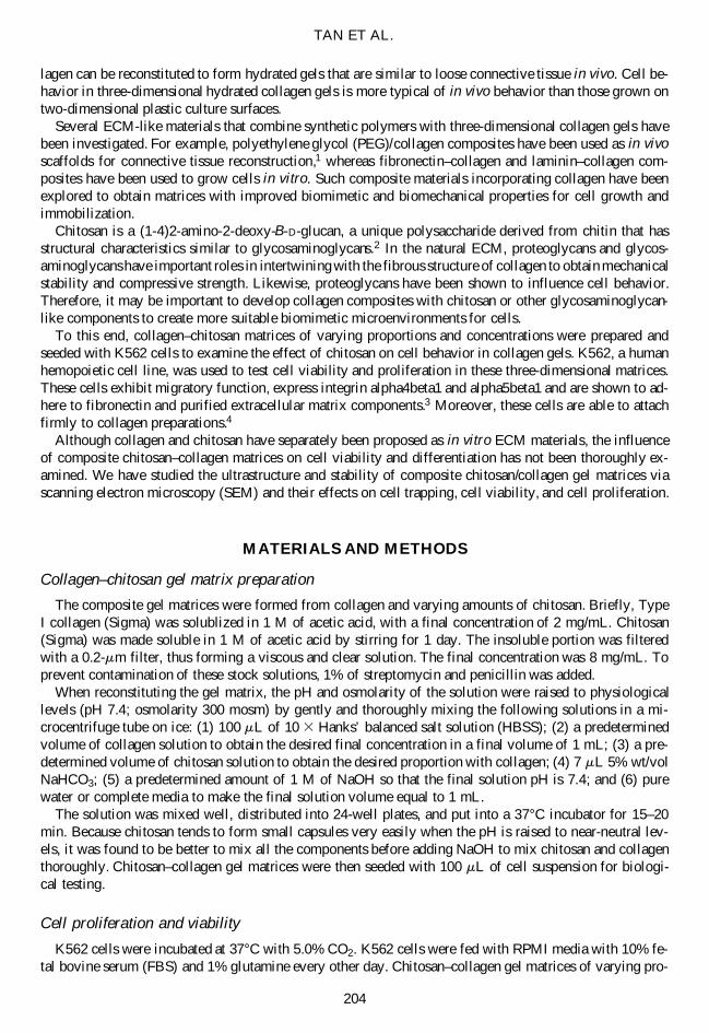
203 TISSUE ENGINEERING Volume 7, Number 2, 2001 Mary Ann Liebert, Inc. Evaluation of Nanostructured Composite Collagen–Chitosan Matrices for Tissue Engineering WEI TAN, B.S., 1 RAJ KRISHNARAJ, Ph.D., 2 and TEJAL A. DESAI, Ph.D. 1 ABSTRACT The development of suitable three-dimensional matrices for the maintenance of cellular vi- ability and differentiation is critical for applications in tissue engineering and cell biology. The structure and composition of the extracellular matrix (ECM) has been shown to mod- ulate cell behavior with respect to shape, movement, proliferation, and differentiation. Al- though collagen and chitosan have separately been proposed as in vitro ECM materials, the influence of chitosan–collagen composite matrices on cell morphology, differentiation, and function is not well studied. To this end, gel matrices of different proportions of collagen and chitosan were examined ultrastructurally and characterized for their ability to regulate cellular activity. A three-chamber system with circulating hydraulic fluids was used to eval- uate the gel stability under fluid force. Results indicated that overall matrix integrity in- creased with the proportion of chitosan. Scanning electron microscopy indicated that the ad- dition of chitosan greatly influences ultrastructure and changes collagen fiber cross-linking, reinforcing the structure and increasing pore size. K562 cells cultured in three-dimensional gels were examined for cell proliferation and differentiation. Although cell proliferation was inhibited with an increasing proportion of chitosan, cell function based on cytokine-release was greatly augmented. Results suggest that a hybrid chitosan–collagen matrix may have potential biological and mechanical benefits for use as a cellular scaffold. INTRODUCTION T HE DEVELOPMENT of suitable synthetic extracellular matrices (ECM) that can recreate three-dimensional cell–cell interactions and control tissue formation in vitro and in vivo is important for tissue engineer- ing and cell culture applications. Natural ECM can be used as a model for the development of synthetic matrices for tissue engineering. The two main classes of extracellular macromolecules that form the nat- ural ECM are proteoglycans and fibrous proteins. Proteoglycans, containing long unbranched polysaccha- ride side chains covalently tethered to a fibrous backbone, form three-dimensional networks of hydrated gels in which cells can be embedded. Structural fibrous proteins such as collagen provide the tensile strength of the ECM, whereas proteoglycans provide compressive strength. The ECM plays a critical role in tissue development, support, and cellular functions such as growth, differentiation, and motility. Currently, collagen is one of the most common ECM materials used for culturing cells in vitro. Colla- gen molecules assemble to form microfibrils, which, in turn, form fibrils to create the collagen fiber. Col- 1 Department of Bioengineering and 2 Department of Medicine, University of Illinois at Chicago, Chicago, Illinois.
-
Upload
independent -
Category
Documents
-
view
0 -
download
0
Transcript of Evaluation of nanostructured composite collagen-chitosan matrices for tissue engineering
203
TISSUE ENGINEERINGVolume 7 Number 2 2001Mary Ann Liebert Inc
Evaluation of Nanostructured CompositeCollagenndashChitosan Matrices for Tissue Engineering
WEI TAN BS1 RAJ KRISHNARAJ PhD2 and TEJAL A DESAI PhD1
ABSTRACT
The development of suitable three-dimensional matrices for the maintenance of cellular vi-ability and differentiation is critical for applications in tissue engineering and cell biologyThe structure and composition of the extracellular matrix (ECM) has been shown to mod-ulate cell behavior with respect to shape movement proliferation and differentiation Al-though collagen and chitosan have separately been proposed as in vitro ECM materials theinfluence of chitosanndashcollagen composite matrices on cell morphology differentiation andfunction is not well studied To this end gel matrices of different proportions of collagenand chitosan were examined ultrastructurally and characterized for their ability to regulatecellular activity A three-chamber system with circulating hydraulic fluids was used to eval-uate the gel stability under fluid force Results indicated that overall matrix integrity in-creased with the proportion of chitosan Scanning electron microscopy indicated that the ad-dition of chitosan greatly influences ultrastructure and changes collagen fiber cross-linkingreinforcing the structure and increasing pore size K562 cells cultured in three-dimensionalgels were examined for cell proliferation and differentiation Although cell proliferation wasinhibited with an increasing proportion of chitosan cell function based on cytokine-releasewas greatly augmented Results suggest that a hybrid chitosanndashcollagen matrix may havepotential biological and mechanical benefits for use as a cellular scaffold
INTRODUCTION
THE DEVELOPMENT of suitable synthetic extracellular matrices (ECM) that can recreate three-dimensionalcellndashcell interactions and control tissue formation in vitro and in vivo is important for tissue engineer-
ing and cell culture applications Natural ECM can be used as a model for the development of syntheticmatrices for tissue engineering The two main classes of extracellular macromolecules that form the nat-ural ECM are proteoglycans and fibrous proteins Proteoglycans containing long unbranched polysaccha-ride side chains covalently tethered to a fibrous backbone form three-dimensional networks of hydratedgels in which cells can be embedded Structural fibrous proteins such as collagen provide the tensile strengthof the ECM whereas proteoglycans provide compressive strength The ECM plays a critical role in tissuedevelopment support and cellular functions such as growth differentiation and motility
Currently collagen is one of the most common ECM materials used for culturing cells in vitro Colla-gen molecules assemble to form microfibrils which in turn form fibrils to create the collagen fiber Col-
1Department of Bioengineering and 2Department of Medicine University of Illinois at Chicago Chicago Illinois
lagen can be reconstituted to form hydrated gels that are similar to loose connective tissue in vivo Cell be-havior in three-dimensional hydrated collagen gels is more typical of in vivo behavior than those grown ontwo-dimensional plastic culture surfaces
Several ECM-like materials that combine synthetic polymers with three-dimensional collagen gels havebeen investigated For example polyethylene glycol (PEG)collagen composites have been used as in vivoscaffolds for connective tissue reconstruction1 whereas fibronectinndashcollagen and lamininndashcollagen com-posites have been used to grow cells in vitro Such composite materials incorporating collagen have beenexplored to obtain matrices with improved biomimetic and biomechanical properties for cell growth andimmobilization
Chitosan is a (1-4)2-amino-2-deoxy-B-D-glucan a unique polysaccharide derived from chitin that hasstructural characteristics similar to glycosaminoglycans2 In the natural ECM proteoglycans and glycos-aminoglycans have important roles in intertwining with the fibrous structure of collagen to obtain mechanicalstability and compressive strength Likewise proteoglycans have been shown to influence cell behaviorTherefore it may be important to develop collagen composites with chitosan or other glycosaminoglycan-like components to create more suitable biomimetic microenvironments for cells
To this end collagenndashchitosan matrices of varying proportions and concentrations were prepared andseeded with K562 cells to examine the effect of chitosan on cell behavior in collagen gels K562 a humanhemopoietic cell line was used to test cell viability and proliferation in these three-dimensional matricesThese cells exhibit migratory function express integrin alpha4beta1 and alpha5beta1 and are shown to ad-here to fibronectin and purified extracellular matrix components3 Moreover these cells are able to attachfirmly to collagen preparations4
Although collagen and chitosan have separately been proposed as in vitro ECM materials the influenceof composite chitosanndashcollagen matrices on cell viability and differentiation has not been thoroughly ex-amined We have studied the ultrastructure and stability of composite chitosancollagen gel matrices viascanning electron microscopy (SEM) and their effects on cell trapping cell viability and cell proliferation
MATERIALS AND METHODS
Collagenndashchitosan gel matrix preparation
The composite gel matrices were formed from collagen and varying amounts of chitosan Briefly TypeI collagen (Sigma) was solublized in 1 M of acetic acid with a final concentration of 2 mgmL Chitosan(Sigma) was made soluble in 1 M of acetic acid by stirring for 1 day The insoluble portion was filteredwith a 02-mm filter thus forming a viscous and clear solution The final concentration was 8 mgmL Toprevent contamination of these stock solutions 1 of streptomycin and penicillin was added
When reconstituting the gel matrix the pH and osmolarity of the solution were raised to physiologicallevels (pH 74 osmolarity 300 mosm) by gently and thoroughly mixing the following solutions in a mi-crocentrifuge tube on ice (1) 100 mL of 10 3 Hanksrsquo balanced salt solution (HBSS) (2) a predeterminedvolume of collagen solution to obtain the desired final concentration in a final volume of 1 mL (3) a pre-determined volume of chitosan solution to obtain the desired proportion with collagen (4) 7 mL 5 wtvolNaHCO3 (5) a predetermined amount of 1 M of NaOH so that the final solution pH is 74 and (6) purewater or complete media to make the final solution volume equal to 1 mL
The solution was mixed well distributed into 24-well plates and put into a 37degC incubator for 15ndash20min Because chitosan tends to form small capsules very easily when the pH is raised to near-neutral lev-els it was found to be better to mix all the components before adding NaOH to mix chitosan and collagenthoroughly Chitosanndashcollagen gel matrices were then seeded with 100 mL of cell suspension for biologi-cal testing
Cell proliferation and viability
K562 cells were incubated at 37degC with 50 CO2 K562 cells were fed with RPMI media with 10 fe-tal bovine serum (FBS) and 1 glutamine every other day Chitosanndashcollagen gel matrices of varying pro-
TAN ET AL
204
portions were seeded with 100 mL of cell suspension and were examined for 8 days Cell numbers werequantified with hemocytometry In addition AlamarBlue was used to track the cell proliferation of the cellscultured in three-dimensional matrices The method is similar to that described by Zhi-Jun et al5 Briefly10 volvol AlamarBlue was added into culture media (typically 25 mL added to 250 mL of culture me-dia) AlamarBlue reduction was then measured spectrophotometrically at 570 nm and 600 nm or fluoro-metrically Cell viability is measured using trypan blue exclusion The blue-dyed cells are subtracted fromthe living cell number Cell viability studies were carried out for 8 days
The stability of reconstituted matrices
The stability of the matrices under fluid flow was monitored using a three-chamber system Colla-genndashchitosan solutions prepared as described above were injected into the middle chamber The sample wasleft undisturbed for 30 min to allow gel formation After 30 min the sample was subjected to hydraulicflow by pumping media through the chamber at 40 mLmin After 4 h the gel thickness was measured andthe gel was fixed for SEM examination as described below
SEM of reconstituted gel matrices
Collagenndashchitosan extracellular matrices were prepared by physical mixing before gelation the mix-tures were incubated at 37degC to form gels The concentration of collagen was kept at 8 mgmL and in-fluence of chitosan concentration (8 mgmL 16 mgmL 24 mgmL and 32 mgmL) on collagen structurewas evaluated
Collagen gels and collagenndashchitosan gels with different proportions were fixed dehydrated and driedusing the following methods Gels were fixed with 25 glutaraldehyde in cacodylate buffer (pH 4) for 1 h at room temperature The specimens were washed three times with cacodylate buffer three times priorto dehydration through a series of graded alcohol solutions Instead of critical point drying small pieces ofmatrix were put into hexamethyldisilazane (HMDS Electron Microscopy Sciences) in small wells of a glassdish for 15 min Then pieces were transferred to a dry well and air-dried for about 15 min Matrix pieceswere mounted and silver paint was added to the edges of each specimen The resulting samples were sput-ter coated and examined using a scanning electron microscope
RESULTS
Ultrastructural examination of the matrix using SEM
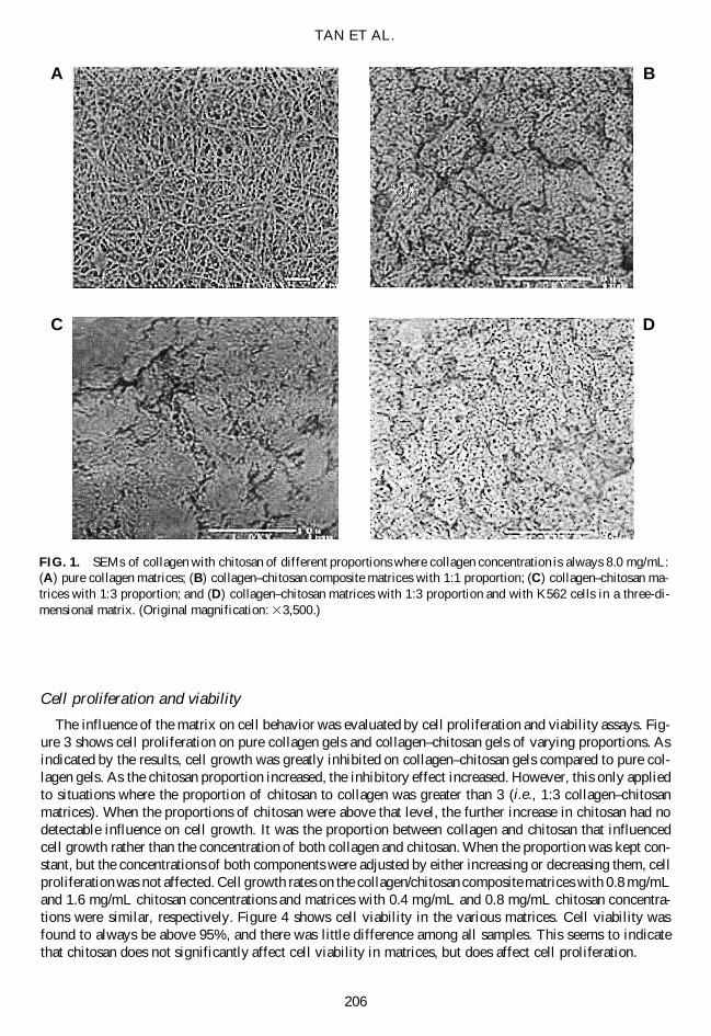
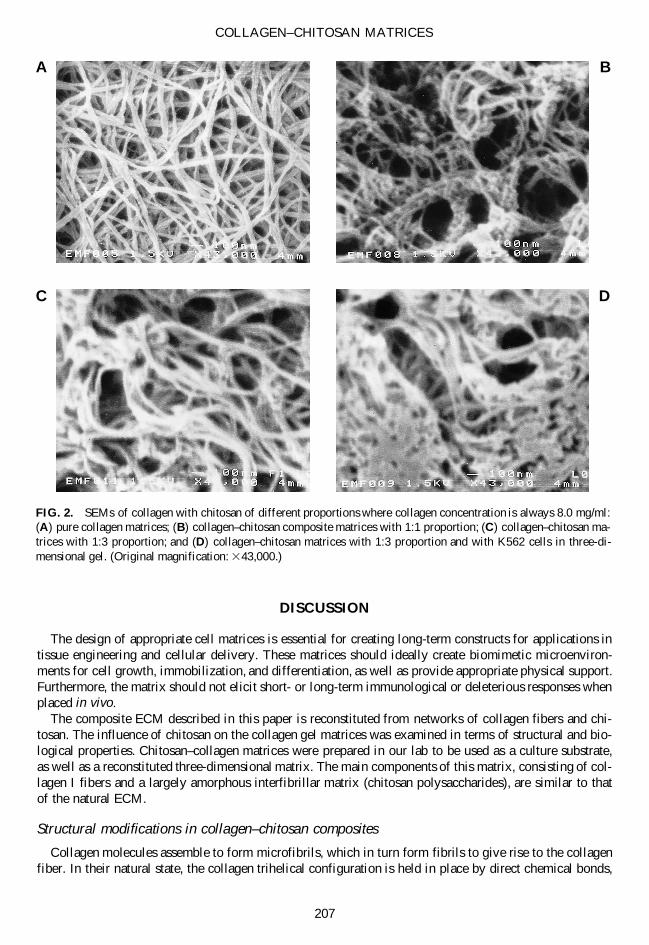
Using SEM to examine the ultrastructure of collagenndashchitosan gels at a magnification of 3500 (Fig 1)the matrix macrostructure and pore distribution could be seen With magnification set at 43000 (Fig 2)the detailed microstructure of the matrices could be more clearly examined The figures show that whenchitosan was introduced into collagen solution the formation of collagen fibers was not perturbed by thetiny chitosan clumps because the diameter of fibers in the pure collagen and those in gels with chitosanare very similar However fibrous organization was significantly changed by chitosan and pore sizes ofgel matrices were greatly affected by different chitosan concentrations
SEM micrographs of 13 collagenchitosan matrices without cells were very unclear because chitosancarries a positive charge and deviates the electron route However cells carry a negative charge and canneutralize the positive charge produced by chitosan thus the micrographs of the 13 collagenchitosan ma-trix with cells produced much clearer images than the unseeded matrix With increasing amounts of chi-tosan added to the collagen gel more collagen fibers were sequestered together and greater numbers ofclumps and knobs were formed as shown in Fig 2D The collagen fibers were more dense and the distri-bution of all fibers was more heterogeneous with the addition of chitosan Moreover the cell distributionbecame increasingly uneven in these matrices
The stability of the reconstituted matrices under fluid force was examined using the three-chambered sys-tem described earlier These tests revealed that the higher the chitosan concentration in collagenndashchitosangels the thicker the gel remained after circulating media for 4 h (Table 1) The testing was performed un-der the media flow rate of 14 mLmin and the circulation lasted for 4 h
COLLAGENndashCHITOSAN MATRICES
205
Cell proliferation and viability
The influence of the matrix on cell behavior was evaluated by cell proliferation and viability assays Fig-ure 3 shows cell proliferation on pure collagen gels and collagenndashchitosan gels of varying proportions Asindicated by the results cell growth was greatly inhibited on collagenndashchitosan gels compared to pure col-lagen gels As the chitosan proportion increased the inhibitory effect increased However this only appliedto situations where the proportion of chitosan to collagen was greater than 3 (ie 13 collagenndashchitosanmatrices) When the proportions of chitosan were above that level the further increase in chitosan had nodetectable influence on cell growth It was the proportion between collagen and chitosan that influencedcell growth rather than the concentration of both collagen and chitosan When the proportion was kept con-stant but the concentrations of both components were adjusted by either increasing or decreasing them cellproliferation was not affected Cell growth rates on the collagenchitosan composite matrices with 08 mgmLand 16 mgmL chitosan concentrations and matrices with 04 mgmL and 08 mgmL chitosan concentra-tions were similar respectively Figure 4 shows cell viability in the various matrices Cell viability wasfound to always be above 95 and there was little difference among all samples This seems to indicatethat chitosan does not significantly affect cell viability in matrices but does affect cell proliferation
TAN ET AL
206
FIG 1 SEMs of collagen with chitosan of different proportions where collagen concentration is always 80 mgmL(A) pure collagen matrices (B) collagenndashchitosan composite matrices with 11 proportion (C) collagenndashchitosan ma-trices with 13 proportion and (D) collagenndashchitosan matrices with 13 proportion and with K562 cells in a three-di-mensional matrix (Original magnification 33500)
D
B
C
A
DISCUSSION
The design of appropriate cell matrices is essential for creating long-term constructs for applications intissue engineering and cellular delivery These matrices should ideally create biomimetic microenviron-ments for cell growth immobilization and differentiation as well as provide appropriate physical supportFurthermore the matrix should not elicit short- or long-term immunological or deleterious responses whenplaced in vivo
The composite ECM described in this paper is reconstituted from networks of collagen fibers and chi-tosan The influence of chitosan on the collagen gel matrices was examined in terms of structural and bio-logical properties Chitosanndashcollagen matrices were prepared in our lab to be used as a culture substrateas well as a reconstituted three-dimensional matrix The main components of this matrix consisting of col-lagen I fibers and a largely amorphous interfibrillar matrix (chitosan polysaccharides) are similar to thatof the natural ECM
Structural modifications in collagenndashchitosan composites
Collagen molecules assemble to form microfibrils which in turn form fibrils to give rise to the collagenfiber In their natural state the collagen trihelical configuration is held in place by direct chemical bonds
COLLAGENndashCHITOSAN MATRICES
207
FIG 2 SEMs of collagen with chitosan of different proportions where collagen concentration is always 80 mgml(A) pure collagen matrices (B) collagenndashchitosan composite matrices with 11 proportion (C) collagenndashchitosan ma-trices with 13 proportion and (D) collagenndashchitosan matrices with 13 proportion and with K562 cells in three-di-mensional gel (Original magnification 343000)
D
B
C
A
hydrogen bonds and water-bridged cross-links Associated with collagen in native tissue are elastin pro-teoglycans and mucopolysaccharides attached covalently to protein cores These components are thoughtto modulate collagen fibrillogenesis fill space as well as bind and organize water
Our experiments revealed that with increasing amounts of chitosan added to the collagen gel more col-lagen fibers were sequestered together the collagen fibers were more dense and the distribution of all fiberswas more heterogeneous It has been shown that chitosan interacts with collagen in the mixture solutionforming ionic bonds when mixing6 Our study suggest these collagenndashchitosan interactions also exist in gelconditions and influence collagen fiber configurations Ultrastructural examination of the matrices showsmall chitosan beads tightly tethered to a collagen fiber backbone Several collagen fibers or different por-tions of one fiber seem to be woven and linked together through chitosan The microstructure created bythe addition of chitosan may prevent collagen fibers from shifting induce formation of interchain cross-links and reinforce the fibers by anchoring them in place The collagenndashchitosan interaction also influencesthe pore density and pore size of the matrix A greater concentration of chitosan in the matrix results in alower pore density and a smaller pore size of the matrix
TAN ET AL
208
FIG 3 K562 cell proliferation on collagenndashchitosan matrices of different proportions The mean values are used inthe plot Error bars represent the standard deviation (n 5 3)
TABLE 1 VOLUME OF THE MATRIX AS A FUNCTION OF WEIGHT COLLAGENCHITOSAN AFTER 4 H OF FLUID FLOW
Collagen chitosan(weight ) No chitosan 1 1 1 2 1 3
Volume after 4 hr 0 110 13ndash14 13original volume
The collagenndashchitosan gels are biological composites that have the crystalline chitosan component as themain support material in an amorphous interfibrillary collagen matrix This structure allows the colla-genndashchitosan gel to maintain structural integrity as compared to collagen alone under fluid flow as shownby our tests This structural integrity may be advantageous in applications requiring long-term maintenanceof three-dimensional cell-seeded matrices in vivo or in vitro
The ultrastructure of collagenndashchitosan composites greatly influences the microenvironment for cellgrowth When the collagen-to-chitosan ratio is more than 12 the greater amount of chitosan beads causescollagen fibers to clump together and fill in pores within the fiber structure This high-density microstruc-ture stabilizes the porous collagen structure and helps to maintain matrix microarchitecture
Microenvironments for cell growth in collagenndashchitosan gel matrices
Cell proliferation was inhibited in collagenndashchitosan gel matrices compared to pure collagen gels Thereason may be due to the fact that the addition of chitosan increases matrix density and cellndashmatrix con-tact therefore limiting the space for cell proliferation Cells become trapped more deeply into hydrated chi-tosan gels rather than just attached to collagen fibers The negativity and adhesivity of chitosan may resultin greater interaction between cells and the matrix However data on cell proliferation also suggests thatthis interaction saturates when the chitosan proportion goes beyond three times collagen in weight
The natural ECM is composed of a complex network of macromolecules that hold cells together in anorganized fashion to form complex tissues These organized networks allow cells within to migrate and in-teract with the matrix and one another Nutrients metabolites and hormones from neighboring blood ves-sels diffuse into the ECM to provide nourishment and mediate communication between the cells The in-
COLLAGENndashCHITOSAN MATRICES
209
FIG 4 K562 cell viability on collagen-chitosan matrices of different proportions The mean values are used in theplot Error bars represent the standard deviation (n 5 3)
creased pore size in chitosanndashcollagen matrices with lower proportions of chitosan may be beneficial forthe diffusion of nutrients and metabolites in three-dimensional matrices
Biocompatibility
Lastly the addition of chitosan does not take away from the beneficial properties of using collagen as asubstrate for cell culture or tissue engineering ECM Chitosan is biologically sourced biodegradable in vivoand biocompatible as well so that the addition of this component will not affect the overall biocompati-bility of the composite matrices
CONCLUSION
In this study gel matrices of different proportions of collagen and chitosan were examined ultrastruc-turally and characterized for their ability to regulate cellular activity The influence of chitosanndashcollagencomposite matrices on cell viability and proliferation was examined Addition of chitosan to traditional col-lagen-based matrices alters the cross-linking pattern of the collagen fibers increases the integrity of the ma-trix by ldquogluingrdquo the fibers together and alters cell proliferation Although cells maintain viability in all col-lagenndashchitosan composite matrices those matrices with a 11 collagenchitosan proportion seem mostsuitable as synthetic matrices because at higher chitosan levels the porosity of the matrix is low and cellproliferative capacity is minimal The addition of chitosan to the matrix is beneficial in that it promotesgreater cellndashmatrix interaction and leads to cell trapping and immobilization of cells The addition of chitosan also stabilizes the fibrous structure and prevents deformation of these matrices under fluid flow
REFERENCES
1 Tan J and Saltzman M Influence of synthetic polymers on neutrophil migration in three-dimensional collagengels J Biomed Mater Res 46 465 1999
2 Chandy T and Sharma C Chitosanmdashas a biomaterial Biomater Art Cells Art Org 18 1 19903 Turner ML Masek LC Hardy CL Parker AC and Sweetenham JW Comparative adhesion of human
haemopoietic cell lines to extracellular matrix components bone marrow stromal and endothelial cultures Br JHaematol 100 112 1998
4 Klein G Kibler C Schermutzki F Brown J Muller CA and Timpl R Cell binding properties of collagentype XIV for human hematopoietic cells Matrix Biol 16 307 1998
5 Zhi-Jun Y Sriranganathan N Vaught T Arastu SK and Ahmed SA A dye-based lymphocyte proliferationassay that permits multiple immunological analysis mRNA cytogenetic apoptosis and immunophenotyping stud-ies J Immun Meth 210 25 1997
6 Taravel MN and Domard A Relation between physicochemical characteristics of collagen and its interactionswith chitosan I Biomaterials 14 930 1993
Address reprint requests toTejal A Desai PhD
Department of Bioengineering (MC 063)University of Illinois at Chicago
851 South Morgan StreetChicago IL 60607
E-mail Tdesaiuicedu
TAN ET AL
lagen can be reconstituted to form hydrated gels that are similar to loose connective tissue in vivo Cell be-havior in three-dimensional hydrated collagen gels is more typical of in vivo behavior than those grown ontwo-dimensional plastic culture surfaces
Several ECM-like materials that combine synthetic polymers with three-dimensional collagen gels havebeen investigated For example polyethylene glycol (PEG)collagen composites have been used as in vivoscaffolds for connective tissue reconstruction1 whereas fibronectinndashcollagen and lamininndashcollagen com-posites have been used to grow cells in vitro Such composite materials incorporating collagen have beenexplored to obtain matrices with improved biomimetic and biomechanical properties for cell growth andimmobilization
Chitosan is a (1-4)2-amino-2-deoxy-B-D-glucan a unique polysaccharide derived from chitin that hasstructural characteristics similar to glycosaminoglycans2 In the natural ECM proteoglycans and glycos-aminoglycans have important roles in intertwining with the fibrous structure of collagen to obtain mechanicalstability and compressive strength Likewise proteoglycans have been shown to influence cell behaviorTherefore it may be important to develop collagen composites with chitosan or other glycosaminoglycan-like components to create more suitable biomimetic microenvironments for cells
To this end collagenndashchitosan matrices of varying proportions and concentrations were prepared andseeded with K562 cells to examine the effect of chitosan on cell behavior in collagen gels K562 a humanhemopoietic cell line was used to test cell viability and proliferation in these three-dimensional matricesThese cells exhibit migratory function express integrin alpha4beta1 and alpha5beta1 and are shown to ad-here to fibronectin and purified extracellular matrix components3 Moreover these cells are able to attachfirmly to collagen preparations4
Although collagen and chitosan have separately been proposed as in vitro ECM materials the influenceof composite chitosanndashcollagen matrices on cell viability and differentiation has not been thoroughly ex-amined We have studied the ultrastructure and stability of composite chitosancollagen gel matrices viascanning electron microscopy (SEM) and their effects on cell trapping cell viability and cell proliferation
MATERIALS AND METHODS
Collagenndashchitosan gel matrix preparation
The composite gel matrices were formed from collagen and varying amounts of chitosan Briefly TypeI collagen (Sigma) was solublized in 1 M of acetic acid with a final concentration of 2 mgmL Chitosan(Sigma) was made soluble in 1 M of acetic acid by stirring for 1 day The insoluble portion was filteredwith a 02-mm filter thus forming a viscous and clear solution The final concentration was 8 mgmL Toprevent contamination of these stock solutions 1 of streptomycin and penicillin was added
When reconstituting the gel matrix the pH and osmolarity of the solution were raised to physiologicallevels (pH 74 osmolarity 300 mosm) by gently and thoroughly mixing the following solutions in a mi-crocentrifuge tube on ice (1) 100 mL of 10 3 Hanksrsquo balanced salt solution (HBSS) (2) a predeterminedvolume of collagen solution to obtain the desired final concentration in a final volume of 1 mL (3) a pre-determined volume of chitosan solution to obtain the desired proportion with collagen (4) 7 mL 5 wtvolNaHCO3 (5) a predetermined amount of 1 M of NaOH so that the final solution pH is 74 and (6) purewater or complete media to make the final solution volume equal to 1 mL
The solution was mixed well distributed into 24-well plates and put into a 37degC incubator for 15ndash20min Because chitosan tends to form small capsules very easily when the pH is raised to near-neutral lev-els it was found to be better to mix all the components before adding NaOH to mix chitosan and collagenthoroughly Chitosanndashcollagen gel matrices were then seeded with 100 mL of cell suspension for biologi-cal testing
Cell proliferation and viability
K562 cells were incubated at 37degC with 50 CO2 K562 cells were fed with RPMI media with 10 fe-tal bovine serum (FBS) and 1 glutamine every other day Chitosanndashcollagen gel matrices of varying pro-
TAN ET AL
204
portions were seeded with 100 mL of cell suspension and were examined for 8 days Cell numbers werequantified with hemocytometry In addition AlamarBlue was used to track the cell proliferation of the cellscultured in three-dimensional matrices The method is similar to that described by Zhi-Jun et al5 Briefly10 volvol AlamarBlue was added into culture media (typically 25 mL added to 250 mL of culture me-dia) AlamarBlue reduction was then measured spectrophotometrically at 570 nm and 600 nm or fluoro-metrically Cell viability is measured using trypan blue exclusion The blue-dyed cells are subtracted fromthe living cell number Cell viability studies were carried out for 8 days
The stability of reconstituted matrices
The stability of the matrices under fluid flow was monitored using a three-chamber system Colla-genndashchitosan solutions prepared as described above were injected into the middle chamber The sample wasleft undisturbed for 30 min to allow gel formation After 30 min the sample was subjected to hydraulicflow by pumping media through the chamber at 40 mLmin After 4 h the gel thickness was measured andthe gel was fixed for SEM examination as described below
SEM of reconstituted gel matrices
Collagenndashchitosan extracellular matrices were prepared by physical mixing before gelation the mix-tures were incubated at 37degC to form gels The concentration of collagen was kept at 8 mgmL and in-fluence of chitosan concentration (8 mgmL 16 mgmL 24 mgmL and 32 mgmL) on collagen structurewas evaluated
Collagen gels and collagenndashchitosan gels with different proportions were fixed dehydrated and driedusing the following methods Gels were fixed with 25 glutaraldehyde in cacodylate buffer (pH 4) for 1 h at room temperature The specimens were washed three times with cacodylate buffer three times priorto dehydration through a series of graded alcohol solutions Instead of critical point drying small pieces ofmatrix were put into hexamethyldisilazane (HMDS Electron Microscopy Sciences) in small wells of a glassdish for 15 min Then pieces were transferred to a dry well and air-dried for about 15 min Matrix pieceswere mounted and silver paint was added to the edges of each specimen The resulting samples were sput-ter coated and examined using a scanning electron microscope
RESULTS
Ultrastructural examination of the matrix using SEM
Using SEM to examine the ultrastructure of collagenndashchitosan gels at a magnification of 3500 (Fig 1)the matrix macrostructure and pore distribution could be seen With magnification set at 43000 (Fig 2)the detailed microstructure of the matrices could be more clearly examined The figures show that whenchitosan was introduced into collagen solution the formation of collagen fibers was not perturbed by thetiny chitosan clumps because the diameter of fibers in the pure collagen and those in gels with chitosanare very similar However fibrous organization was significantly changed by chitosan and pore sizes ofgel matrices were greatly affected by different chitosan concentrations
SEM micrographs of 13 collagenchitosan matrices without cells were very unclear because chitosancarries a positive charge and deviates the electron route However cells carry a negative charge and canneutralize the positive charge produced by chitosan thus the micrographs of the 13 collagenchitosan ma-trix with cells produced much clearer images than the unseeded matrix With increasing amounts of chi-tosan added to the collagen gel more collagen fibers were sequestered together and greater numbers ofclumps and knobs were formed as shown in Fig 2D The collagen fibers were more dense and the distri-bution of all fibers was more heterogeneous with the addition of chitosan Moreover the cell distributionbecame increasingly uneven in these matrices
The stability of the reconstituted matrices under fluid force was examined using the three-chambered sys-tem described earlier These tests revealed that the higher the chitosan concentration in collagenndashchitosangels the thicker the gel remained after circulating media for 4 h (Table 1) The testing was performed un-der the media flow rate of 14 mLmin and the circulation lasted for 4 h
COLLAGENndashCHITOSAN MATRICES
205
Cell proliferation and viability
The influence of the matrix on cell behavior was evaluated by cell proliferation and viability assays Fig-ure 3 shows cell proliferation on pure collagen gels and collagenndashchitosan gels of varying proportions Asindicated by the results cell growth was greatly inhibited on collagenndashchitosan gels compared to pure col-lagen gels As the chitosan proportion increased the inhibitory effect increased However this only appliedto situations where the proportion of chitosan to collagen was greater than 3 (ie 13 collagenndashchitosanmatrices) When the proportions of chitosan were above that level the further increase in chitosan had nodetectable influence on cell growth It was the proportion between collagen and chitosan that influencedcell growth rather than the concentration of both collagen and chitosan When the proportion was kept con-stant but the concentrations of both components were adjusted by either increasing or decreasing them cellproliferation was not affected Cell growth rates on the collagenchitosan composite matrices with 08 mgmLand 16 mgmL chitosan concentrations and matrices with 04 mgmL and 08 mgmL chitosan concentra-tions were similar respectively Figure 4 shows cell viability in the various matrices Cell viability wasfound to always be above 95 and there was little difference among all samples This seems to indicatethat chitosan does not significantly affect cell viability in matrices but does affect cell proliferation
TAN ET AL
206
FIG 1 SEMs of collagen with chitosan of different proportions where collagen concentration is always 80 mgmL(A) pure collagen matrices (B) collagenndashchitosan composite matrices with 11 proportion (C) collagenndashchitosan ma-trices with 13 proportion and (D) collagenndashchitosan matrices with 13 proportion and with K562 cells in a three-di-mensional matrix (Original magnification 33500)
D
B
C
A
DISCUSSION
The design of appropriate cell matrices is essential for creating long-term constructs for applications intissue engineering and cellular delivery These matrices should ideally create biomimetic microenviron-ments for cell growth immobilization and differentiation as well as provide appropriate physical supportFurthermore the matrix should not elicit short- or long-term immunological or deleterious responses whenplaced in vivo
The composite ECM described in this paper is reconstituted from networks of collagen fibers and chi-tosan The influence of chitosan on the collagen gel matrices was examined in terms of structural and bio-logical properties Chitosanndashcollagen matrices were prepared in our lab to be used as a culture substrateas well as a reconstituted three-dimensional matrix The main components of this matrix consisting of col-lagen I fibers and a largely amorphous interfibrillar matrix (chitosan polysaccharides) are similar to thatof the natural ECM
Structural modifications in collagenndashchitosan composites
Collagen molecules assemble to form microfibrils which in turn form fibrils to give rise to the collagenfiber In their natural state the collagen trihelical configuration is held in place by direct chemical bonds
COLLAGENndashCHITOSAN MATRICES
207
FIG 2 SEMs of collagen with chitosan of different proportions where collagen concentration is always 80 mgml(A) pure collagen matrices (B) collagenndashchitosan composite matrices with 11 proportion (C) collagenndashchitosan ma-trices with 13 proportion and (D) collagenndashchitosan matrices with 13 proportion and with K562 cells in three-di-mensional gel (Original magnification 343000)
D
B
C
A
hydrogen bonds and water-bridged cross-links Associated with collagen in native tissue are elastin pro-teoglycans and mucopolysaccharides attached covalently to protein cores These components are thoughtto modulate collagen fibrillogenesis fill space as well as bind and organize water
Our experiments revealed that with increasing amounts of chitosan added to the collagen gel more col-lagen fibers were sequestered together the collagen fibers were more dense and the distribution of all fiberswas more heterogeneous It has been shown that chitosan interacts with collagen in the mixture solutionforming ionic bonds when mixing6 Our study suggest these collagenndashchitosan interactions also exist in gelconditions and influence collagen fiber configurations Ultrastructural examination of the matrices showsmall chitosan beads tightly tethered to a collagen fiber backbone Several collagen fibers or different por-tions of one fiber seem to be woven and linked together through chitosan The microstructure created bythe addition of chitosan may prevent collagen fibers from shifting induce formation of interchain cross-links and reinforce the fibers by anchoring them in place The collagenndashchitosan interaction also influencesthe pore density and pore size of the matrix A greater concentration of chitosan in the matrix results in alower pore density and a smaller pore size of the matrix
TAN ET AL
208
FIG 3 K562 cell proliferation on collagenndashchitosan matrices of different proportions The mean values are used inthe plot Error bars represent the standard deviation (n 5 3)
TABLE 1 VOLUME OF THE MATRIX AS A FUNCTION OF WEIGHT COLLAGENCHITOSAN AFTER 4 H OF FLUID FLOW
Collagen chitosan(weight ) No chitosan 1 1 1 2 1 3
Volume after 4 hr 0 110 13ndash14 13original volume
The collagenndashchitosan gels are biological composites that have the crystalline chitosan component as themain support material in an amorphous interfibrillary collagen matrix This structure allows the colla-genndashchitosan gel to maintain structural integrity as compared to collagen alone under fluid flow as shownby our tests This structural integrity may be advantageous in applications requiring long-term maintenanceof three-dimensional cell-seeded matrices in vivo or in vitro
The ultrastructure of collagenndashchitosan composites greatly influences the microenvironment for cellgrowth When the collagen-to-chitosan ratio is more than 12 the greater amount of chitosan beads causescollagen fibers to clump together and fill in pores within the fiber structure This high-density microstruc-ture stabilizes the porous collagen structure and helps to maintain matrix microarchitecture
Microenvironments for cell growth in collagenndashchitosan gel matrices
Cell proliferation was inhibited in collagenndashchitosan gel matrices compared to pure collagen gels Thereason may be due to the fact that the addition of chitosan increases matrix density and cellndashmatrix con-tact therefore limiting the space for cell proliferation Cells become trapped more deeply into hydrated chi-tosan gels rather than just attached to collagen fibers The negativity and adhesivity of chitosan may resultin greater interaction between cells and the matrix However data on cell proliferation also suggests thatthis interaction saturates when the chitosan proportion goes beyond three times collagen in weight
The natural ECM is composed of a complex network of macromolecules that hold cells together in anorganized fashion to form complex tissues These organized networks allow cells within to migrate and in-teract with the matrix and one another Nutrients metabolites and hormones from neighboring blood ves-sels diffuse into the ECM to provide nourishment and mediate communication between the cells The in-
COLLAGENndashCHITOSAN MATRICES
209
FIG 4 K562 cell viability on collagen-chitosan matrices of different proportions The mean values are used in theplot Error bars represent the standard deviation (n 5 3)
creased pore size in chitosanndashcollagen matrices with lower proportions of chitosan may be beneficial forthe diffusion of nutrients and metabolites in three-dimensional matrices
Biocompatibility
Lastly the addition of chitosan does not take away from the beneficial properties of using collagen as asubstrate for cell culture or tissue engineering ECM Chitosan is biologically sourced biodegradable in vivoand biocompatible as well so that the addition of this component will not affect the overall biocompati-bility of the composite matrices
CONCLUSION
In this study gel matrices of different proportions of collagen and chitosan were examined ultrastruc-turally and characterized for their ability to regulate cellular activity The influence of chitosanndashcollagencomposite matrices on cell viability and proliferation was examined Addition of chitosan to traditional col-lagen-based matrices alters the cross-linking pattern of the collagen fibers increases the integrity of the ma-trix by ldquogluingrdquo the fibers together and alters cell proliferation Although cells maintain viability in all col-lagenndashchitosan composite matrices those matrices with a 11 collagenchitosan proportion seem mostsuitable as synthetic matrices because at higher chitosan levels the porosity of the matrix is low and cellproliferative capacity is minimal The addition of chitosan to the matrix is beneficial in that it promotesgreater cellndashmatrix interaction and leads to cell trapping and immobilization of cells The addition of chitosan also stabilizes the fibrous structure and prevents deformation of these matrices under fluid flow
REFERENCES
1 Tan J and Saltzman M Influence of synthetic polymers on neutrophil migration in three-dimensional collagengels J Biomed Mater Res 46 465 1999
2 Chandy T and Sharma C Chitosanmdashas a biomaterial Biomater Art Cells Art Org 18 1 19903 Turner ML Masek LC Hardy CL Parker AC and Sweetenham JW Comparative adhesion of human
haemopoietic cell lines to extracellular matrix components bone marrow stromal and endothelial cultures Br JHaematol 100 112 1998
4 Klein G Kibler C Schermutzki F Brown J Muller CA and Timpl R Cell binding properties of collagentype XIV for human hematopoietic cells Matrix Biol 16 307 1998
5 Zhi-Jun Y Sriranganathan N Vaught T Arastu SK and Ahmed SA A dye-based lymphocyte proliferationassay that permits multiple immunological analysis mRNA cytogenetic apoptosis and immunophenotyping stud-ies J Immun Meth 210 25 1997
6 Taravel MN and Domard A Relation between physicochemical characteristics of collagen and its interactionswith chitosan I Biomaterials 14 930 1993
Address reprint requests toTejal A Desai PhD
Department of Bioengineering (MC 063)University of Illinois at Chicago
851 South Morgan StreetChicago IL 60607
E-mail Tdesaiuicedu
TAN ET AL
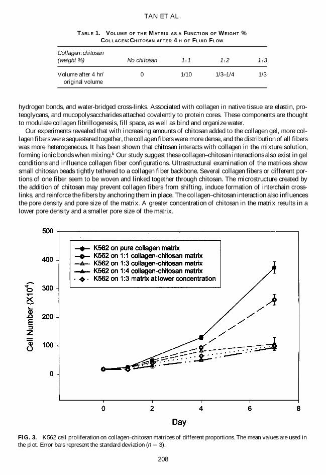
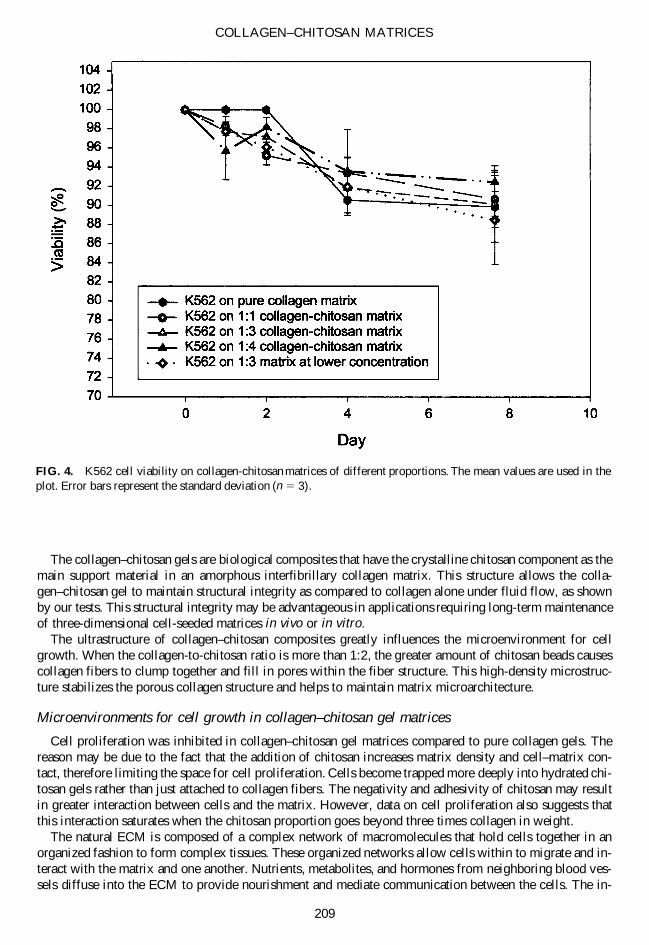