Targeted homozygous deletion of M-band titin in cardiomyocytes prevents sarcomere formation
Upload
independentCategory
view
3download
0
www.afm-journal.de
FULL P
APER
www.MaterialsViews.com
Shayanti Mukherjee , Jayarama Reddy Venugopal , * Rajeswari Ravichandran , Seeram Ramakrishna , and Michael Raghunath
Evaluation of the Biocompatibility of PLACL/Collagen Nanostructured Matrices with Cardiomyocytes as a Model for the Regeneration of Infarcted Myocardium
Pioneering research suggests various modes of cellular therapeutics and biomaterial strategies for myocardial tissue engineering. Despite several advantages, such as safety and improved function, the dynamic myocardial microenvironment prevents peripherally or locally administered therapeutic cells from homing and integrating of biomaterial constructs with the inf-arcted heart. The myocardial microenvironment is highly sensitive due to the nanoscale cues that it exerts to control bioactivities, such as cell migration, proliferation, differentiation, and angiogenesis. Nanoscale control of cardiac function has not been extensively analyzed in the fi eld of myocardial tissue engineering. Inspired by microscopic analysis of the ventricular organiza-tion in native tissue, a scalable in-vitro model of nanoscale poly( L -lactic acid)- co -poly( ε -caprolactone)/collagen biocomposite scaffold is fabricated, with nanofi bers in the order of 594 ± 56 nm to mimic the native myocar-dial environment for freshly isolated cardiomyocytes from rabbit heart, and the specifi cally underlying extracellular matrix architecture: this is done to address the specifi city of the underlying matrix in overcoming challenges faced by cellular therapeutics. Guided by nanoscale mechanical cues pro-vided by the underlying random nanofi brous scaffold, the tissue constructs display anisotropic rearrangement of cells, characteristic of the native cardiac tissue. Surprisingly, cell morphology, growth, and expression of an interactive healthy cardiac cell population are exquisitely sensitive to differences in the composition of nanoscale scaffolds. It is shown that suitable cell–material interactions on the nanoscale can stipulate organization on the tissue level and yield novel insights into cell therapeutic science, while providing mate-rials for tissue regeneration.
DOI: 10.1002/adfm.201002434
S. Mukherjee , Dr. M. Raghunath Division of Bioengineering 9 Engineering Drive 1, Block EA #03-12 National University of Singapore, Singapore S. Mukherjee , Dr. J. R. Venugopal , S. Ramakrishna HEM laboratory Nanoscience and Nanotechnology Initiative c/o Faculty of Engineering Block E3-05-29, 2 Engineering Drive 3, National University of Singapore Singapore Email: [email protected]
R. Ravichandran , S. RaDepartment of Mecha9 Engineering Drive 1,National University of S. Ramakrishna Institute of Materials RA-Star, Singapore Dr. M. Raghunath Department of BiocheMedical Drive Block MNational University of
© 2011 WILEY-VCH Verlag GmbH & Co. KGaA, WeinheimAdv. Funct. Mater. 2011, 21, 2291–2300
1. Introduction
Myocardial infarction (MI) is the single leading cause of deaths due to cardiovas-cular disease globally. [ 1 ] MI is caused when the supply of oxygen and nutrients to the cardiac muscle is impaired, usually due to occluded coronary arteries. A massive cell death occurs in the affected heart region, which forms a rather non-contractile scar characterized by mismatch of mechan-ical and electrical properties with native myocardium. The substantial cell loss in the myocardium leads to dilation of ven-tricular wall and remodeling of the heart and, eventually, to congestive heart failure (CHF). [ 2 ] Currently, more than 10 million people suffer from CHF in the USA, UK and southeastern Asia. Drugs alone might be able slow disease progression, but they cannot prevent it. [ 3 ] Current treatments for congestive heart failure use highly invasive methods, such as open chest surgery and transplantation. [ 4 ] Given the patient mor-bidity and complications involved with cur-rent procedures, it is desirable to develop minimally invasive technologies, such as injectable therapeutics.
Several modes of regenerating injured myocardium have been suggested over time, with pioneering research being
makrishna nical Engineering Block EA, 07-08 Singapore, Singapore
esearch and Engineering
mistry D7, #02-03, Yong Loo Lin School of Medicine Singapore, Singapore
2291wileyonlinelibrary.com
FULL
PAPER
2292
www.afm-journal.dewww.MaterialsViews.com
conducted in a variety of technologies including cell therapy using various cell types, injection of biomaterials, bioengineered patches, implantation of 3D construct generated in static and bioreactor culture. [ 5,6 ] Cellular therapy, involving the use of live cells to repair damaged myocardium, has been extensively studied in recent years, opening new horizons in the clinical fi eld. Cellular therapeutics essentially involve the direct delivery of suitable cell types, such as bone marrow cells (BMCs), mesen-chymal stem cells (MSCs), endothelial progenitor cells (EPCs), cardiac stem cells (CSCs), skeletal myoblast cells (SMCs), embryonic stem cells (ESCs), or induced pluripotent stem cells (iPS), to the damaged myocardial area. [ 7 ] Currently, more than 25 new clinical trials are in progress in the United States, and a similar number in Europe, using different cell types. [ 8,9 ] How-ever, recent experimental studies fail to answer many impor-tant aspects of cell therapy. Despite several advantages, such as safety and improved function, and positive results, such as increased healing, vascular density, increased regional circula-tion and cardiac function, the effi ciency of delivery and retention is lower than expected, and the retention and survival of cells at sites of delivery has been limited. [ 10 ] Once the scarred area has evolved, remodeling and collagen deposition changes the histological microenvironment of infarcted areas. This changed microenvironment may allow peripherally or locally admin-istered stem cells to home and survive. In native tissue, cell growth and structural development is supported by an extracel-lular matrix (ECM) that consistently assists in coordinating the contractility and maintenance of cardiac shape and size, as well as the function of cardiomyocytes. [ 11 ] In addition to providing growth cues, the ECM responds actively to cellular actions. It also undergoes remodeling by cells during development, homeostasis and healing processes, which includes digestion of the old matrix and deposition of a fresh one. Essentially, the ECM is made up of 80% and 10% of collagen types I and III, respectively. [ 12 ] In the healthy heart, collagen serves to maintain normal cardiac architecture by surrounding and bridging myo-cytes, which consistently assist in coordinating the contractility and maintenance of cardiac shape and size as well as the func-tion of the cardiomyocytes. ECM proteins greatly infl uence bio-activities, such as cell migration, differentiation, proliferation, and angiogenesis. These effects are recognizable to cells only when the ECM is suitably modifi ed. [ 13 ] It is indeed an immense technological challenge to mimic the entire milieu of ECM arti-fi cially without evoking an immune response in such sensitive surrounding. From a tissue engineering perspective, applying physiological stress on immature constructs could be a way of mimicking the natural environment. [ 14 ] Many efforts by the scientifi c community in the fi eld of myocardial tissue engi-neering (MTE) are dedicated to identify materials possessing specifi c mechanical properties that play a pivotal role. [ 15 ] First of all, it is desirable that the mechanical performance of bioen-gineered scaffolds matches as closely as possible those of the heart extracellular matrix in terms of stiffness, since the scaf-fold should be fl exible enough to promote the contraction of the growing cells. [ 16 ] The stiffness of native heart tissue ranges from 10–20 kPa at early diastole and increases to 50 kPa at the end of diastole, which may shoot up to 200 kPa or more in inf-arcted hearts. [ 17,18 ] In addition, since the myocardial tissue issubjected to cyclical and constant deformation, materials are
© 2011 WILEY-VCH Verlag Gwileyonlinelibrary.com
requested to show elastomeric properties and possibly long-term elasticity.
However, it is likely that the structure and function of the in vivo cardiac tissues are regulated by much smaller, nanoscale cues provided by the ECM, which is responsible for extensive control over cell and tissue function. [ 19 ] It is therefore important to investigate the effects of fi ner control over the cell–biomaterial interface on the nanoscale, in facilitating the creation of truly biomimetic cardiac constructs that replicate the structural and functional aspects of the in vivo ventricular organization. In addition, the ability to robustly and reproducibly generate uniformly controlled (both structurally and functionally) and precisely defi ned engineered cardiac tissue will likely be neces-sary for eventual therapeutic products. This makes nanofabri-cation of biomaterials for MTE an attractive strategy. Ultrafi ne nanofi bers having an ECMlike topography can be achieved by electrospinning of biomaterials. [ 20 ] Such scaffolds of suitable biomaterials with nanoscale architecture provide larger surface areas to adsorb proteins and provide more binding sites to cell membrane receptors, unlike microscale and fl at surfaces. [ 21 ] At present, there are no successful models of bioengineered car-diac implant that can satisfactorily replicate the anatomy, physi-ology, and biological stability of a healthy heart wall. [ 22 ]
To address this challenge, here we report the development and analysis of a nanotopographically controlled in-vitro model using nanoscale poly( L -lactic acid)- co -poly( ε -caprolactone) (PLACL) and collagen-blend biopolymer scaffolds that mimic the native myocardial environment and, specifi cally, the under-lying ECM architecture. The result is a nanofi brous scaffold that is scalable, biocompatible and closely imitates the myocardial ECM, allowing healthy interaction with freshly isolated cardiac cells over a span of 15 days in controlled environment. We inves-tigated the mechanical properties of the nanofi brous PLACL/collagen scaffold, demonstrating that they promote higher organizational features than unblended PLACL counterparts. Moreover, strikingly, we found the proliferation of the cells, their morphological patterns and expression of cardiac specifi c markers such as alpha actinin, troponin T, connexin-43, and car-diac myosin heavy chain were highly sensitive to variation in biomaterial content as well as nanoscale topographic features, revealing an unexpected level of regulation in the organization.
2. Results and Discussion
2.1. Fabrication and Characterization of Electrospun Nanofi brous Scaffolds
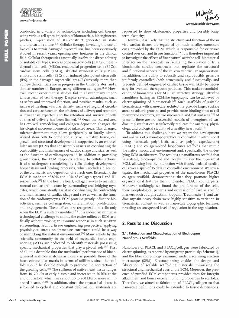
Nanofi bers of PLACL and PLACL/collagen were fabricated by electrospinning, as reported by our group previously ( Scheme 1 ), and the fi ber morphology examined under a scanning electron microscope (SEM). Electrospinning enables the design and fabrication of scalable scaffolding materials, mimicking the structural and mechanical cues of the ECM. Moreover, the pres-ence of purifi ed ECM components provides sites for integrin attachment and hence excellent binding properties to scaffolds. Therefore, we aimed at fabrication of PLACL/collagen so that nanoscale defi nitions could be extended to tissue dimensions.
mbH & Co. KGaA, Weinheim Adv. Funct. Mater. 2011, 21, 2291–2300
Scheme 1 . Myocardial tissue engineering aims at improving the structure and function of the in vivo cardiac tissue are regulated by much smaller, nanoscale cues provided by the ECM, which is responsible for extensive control over cell and tissue function.
FULL P
APER
www.afm-journal.dewww.MaterialsViews.com
We fabricated nanofi bers of PLACL/collagen and PLACL with precisely defi ned features, extending nanoscale control of the magnitude of fi ber diameter. Figure 1 shows SEM micro-graphs of electrospun PLACL and PLACL/collagen nanofi brous
Figure 1 . Fabrication of electrospun nanofi bers. Nanofi ber morphology sholess, uniform nanofi bers of: a) PLACL with diameter measuring 332 ± 31 nmcollagen measuring 594 ± 56 nm.
© 2011 WILEY-VCH Verlag GmAdv. Funct. Mater. 2011, 21, 2291–2300
scaffolds as porous, beadless, uniform nanofi bers with inter-connected pores with fi ber diameter in the range of 332 ± 31 nm and 594 ± 56 nm, respectively.
Biomaterial scaffolds are expected to provide a compliant
ws porous, bead-, and, b) PLACL/
bH & Co. KGaA, Wein
and highly hydrated environment, similar to soft tissues having high water content, thus facilitating diffusion of nutrients and cellular waste. Thus, tailoring the surface properties of the biomaterial in terms of hydrophilicity infl uences its biological performance. Hydrophilicity of our fabricated nanofi ber scaffolds to relative of the surfaces was ana-lyzed using water contact angle analysis. To analyze the wettability of the nanofi ber scaffolds, we measured the contact angle of water on the PLACL/collagen and PLACL using the sessile drop method ( Table 1 ). The PLACL/collagen nanofi bers had a contact angle of 0 ° compared to that of 120 ± 5 ° of PLACL nanofi bers. This observation high-lights the hypothesis that addition of an ECM component—collagen—imparts superior
heim 2293wileyonlinelibrary.com
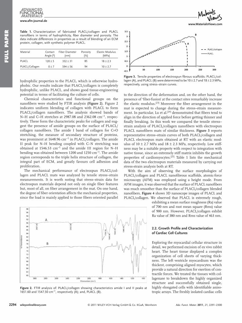
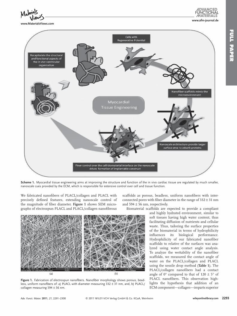
Figure 3 . Tensile properties of electrospun fi brous scaffolds: PLACL/col-lagen (A), and PLACL (B) were determined to be 10 ± 2.7 and 18 ± 2.3 MPa, respectively, using stress–strain curves.
Table 1. Characterization of fabricated PLACL/collagen and PLACL nanofi bers in terms of hydrophilicity, fi ber diameter and porosity. The data indicate differences in properties as a result of blending of an ECM protein, collagen, with synthetic polymer PLACL.
Material Contact Angle [º]
Fiber Diameter [nm]
Porosity [%]
Elastic Modulus [MPa]
PLACL 120 ± 5 332 ± 31 85 18 ± 2.3
PLACL/Collagen 0 ± 7 594 ± 56 94 10 ± 2.7
FULL
PAPER
2294
www.afm-journal.dewww.MaterialsViews.com
hydrophilic properties to the PLACL, which is otherwise hydro-phobic. Our results indicate that PLACL/collagen is completely hydrophilic, unlike PLACL, and shows good tissue-engineering potential in terms of facilitating the culture of cells.
Chemical characteristics and functional groups on the nanofi bers were studied by FTIR analysis ( Figure 2 ). Figure 2 indicates uniform blending of collagen with PLACL to form PLACL/collagen nanofi bers. The analysis showed bands of N–H and C–H stretches at 2987.08 and 2362.08 cm − 1 , respec-tively. These form the characteristic peaks for collagen and sug-gest the presence of amide groups on the surface of PLACL/collagen nanofi bers. The amide I band of collagen for C = O stretching, the measure of secondary structure of proteins, was prominent at 1648.96 cm − 1 in PLACL/collagen. The amide II peak for N–H bending coupled with C–N stretching was obtained at 1546.53 cm − 1 and the amide III region for N–H bending was obtained between 1200 and 1250 cm − 1 . The amide region corresponds to the triple helix structure of collagen, the integral part of ECM, and greatly favours cell adhesion and proliferation.
The mechanical performance of electrospun PLACL/col-lagen and PLACL mats was analyzed by tensile stress–strain measurements. It is worth noting that stress–strain data for electrospun materials depend not only on single fi ber features but, most of all, on fi ber arrangement in the mat. On one hand, the degree of fi ber orientation affects the mechanical properties, since the load is mainly applied to those fi bers oriented parallel
Figure 2 . FTIR analysis of: PLACL/collagen showing characteristics amide1651.60 and 1547.50 cm − 1 , respectively (A); and, PLACL (B).
© 2011 WILEY-VCH Verlag Gwileyonlinelibrary.com
to the direction of the deformation and, on the other hand, the presence of ‘fi ber-fusion’ at the contact sites remarkably increase the elastic modulus. [ 23 ] Moreover the fi ber arrangement in the mat is expected to change during the stress–strain measure-ment. In particular, Lu et al. [ 24 ] demonstrated that fi bers tend to align in the direction of applied force before getting thinner and fi nally breaking. In this work we compared the tensile stress–strain analysis of PLACL/collagen nanofi bers with electrospun PLACL nanofi bers mats of similar thickness. Figure 3 reports representative stress–strain curves of both PLACL/collagen and PLACL electrospun mats obtained at RT with an elastic mod-ulus of 10 ± 2.7 MPa and 18 ± 2.3 MPa, respectively. Low stiff-ness may be a suitable property with respect to integration with native tissue, since an extremely stiff matrix inhibits the growth properties of cardiomyocytes. [ 25 ] Table 1 lists the mechanical data of the two electrospun materials measured by carrying out stress-strain analysis both at RT.
With the aim of observing the surface morphologies of PLACL/collagen and PLACL nanofi brous scaffolds, atomic-force microscopy (AFM) was employed using a height mode. From AFM images, it was observed that the surface of PLACL nanofi bers was much smoother than the surface of PLACL/collagen blended nanofi bers. Figure 4 shows 3D nanoscope images of PLACL and PLACL/collagen. We observed that PLACL is extremely rough,
I and II peaks at
mbH & Co. KGaA, Wein
exhibiting a mean surface roughness (Ra) value of 700 nm and root mean square (Rms) value of 900 nm. However, PLACL/collagen exhibit Ra value of 380 nm and Rms value of 463 nm.
2.2. Growth Profi le and Characterization of Cardiac Cell Cultures
Exploring the myocardial cellular structure in detail, we performed excision of ex vivo rabbit heart. The heart tissue displayed a complex organization of cell sheets of varying thick-ness. The left ventricle myocardium was the thickest, comprising aligned myocytes, which provide a natural direction for exertion of con-tractile forces. We treated the tissues with col-lagenase to breakdown the highly organized structure and successfully obtained single, highly elongated cells with identifi able aniso-tropic arrays. The freshly isolated cardiac cells
heim Adv. Funct. Mater. 2011, 21, 2291–2300

Figure 4 . 3D AFM images of: a) PLACL/collagen (Ra = 380 nm and Rms = 463 nm) composite nanofi bers, and b) PLACL nanofi ber (Ra = 700 nm and Rms = 900 nm).
FULL P
APER
www.afm-journal.dewww.MaterialsViews.com
had a rod-shaped structure, as seen in Figure 5 , with clearly visible striations on the fi rst day. Our observations suggest that the alignment of the cells is strongly correlated to the ECM skeleton in the tissue, which might be largely responsible in
Figure 6 . Flow cytcardium immunosand b) cardiac myo
Figure 5 . a, b) Freshly isolated cardiac cells from rabbit heart: clearly vis-ible striations are seen on the fi rst day in the dispersed tissue. c) These cells remain in culture and have a distinct rod-shaped morphology.
© 2011 WILEY-VCH Verlag GmbH & Co. KGaA, WeinAdv. Funct. Mater. 2011, 21, 2291–2300
providing nanotopographic cues for guiding myocardial alignment during cardiovascular development. Therefore, precise control over various aspects of the microenvironment can be related to tissue organization and remod-eling. In order to obtain a quantitative idea of the functionality of the isolated cells, they were stained with cardiac specifi c protein markers and subjected to hydrodynamic fl ow using a fl ow cytometer (Dako) to count the per cell fl uorescence. The results in Figure 6 indicate that 82 ± 12% of the isolated cells were positive for the presence of sarcomeric alpha actinin and 25 ± 2% cardiac myosin heavy chain. Figure 7 shows the expression of sarcomeric alpha actinin and troponin T in the isolated cells that were seeded in tissue
ometry data of isolated cardiac cells from rabbit myo-tained for expression of: a) sarcomeric alpha actinin, sin heavy chain.
heim 2295wileyonlinelibrary.com

Figure 7 . Isolated cardiac cells stained for anti-alpha actinin, showing the cross-linked structure of actin fi laments (a–c) and anti-troponin T, that binds to tropomyosin (d–f) at Day 1 (a, d) Day 5 (d, e) and Day 14 (c, f).
FULL
PAPER
2296
www.afm-journal.dewww.MaterialsViews.com
culture plate and characterized at days 1, 5, and 15. The cells maintained the rod-shape from the end of day 1, with clear stri-ations of sarcomeric alpha actinin and troponin T. However, a few cells were seen adhered at day 5, although the majority of cells still appear to be rod-shaped with light striations of sar-comeric alpha actinin and troponin T. At day 15, we observed that the earlier rod-shaped fl oating cells were no longer obvious in the fl ask. Rather, a monolayer was formed in the tissue cul-ture fl ask out of the plated cardiac cells containing cardiomyo-cytes and co-isolated cardiac fi broblasts, which were devoid of their striated appearance hereafter. The monolayer expressed
© 2011 WILEY-VCH Verlag GmbH & Co. KGaA, Weiwileyonlinelibrary.com
sarcomeric alpha actinin and troponin T uniformly. These isolated adult cardiac cells were used as a model to understand the cellular interaction with biomaterial, since the mixed cell type represents a more natural myocardium-like environment. The myocardium is an ensemble of different cell types embedded in the complex well-defi ned structures of the ECM and arranged in nano-scale topographical and molecular patterns. Cardiomyocytes assemble and dissemble myofi brils prior to cell division and reas-semble after cell division. [ 26 ] Although the structure of the cardiac tissue is highly organ-ized in vivo, our results in Figure 7 support the isolated cardiac cells losing their native organization and adopting a random distri-bution when cultured in vitro using common cell-culture techniques. [ 27 ] Immunostaining of the isolated cardiac cells using striated muscle specifi c anti-alpha actinin and anti-troponin T revealed clear striations of alpha actinin and troponin T until day 5. However, the striations were no longer obvious at day 14, despite strong expression of protein in the cytoplasm of cardiac cells. It is known that the anisotropic architecture of the layers of myocardial and matrix fi bers affects coor-dination of mechanical contraction, as well as electrical propagation. [ 28 ] Our studies sup-port the knowledge that isolated cardiac cell cultures do not match the functionality of the native tissue due to the lack of proper envi-ronmental factors to allow cardiomyocytes to respond morphologically, mechanically and physiologically as they do in the native tissue. [ 29 ] Hence, we fi nd that development of functional cardiac tissue from any cell thera-peutics may require fabrication of substrates to mimic the native environment found in the heart in order to improve the effi cacy in terms of its mechanical and electrophysiolog-ical properties.
In order to characterize the growth of cells on nanofi bers, we tracked their metabolism over a span of 15 days with absorbance read-ings at regular intervals. Our results, shown in Figure 8 , indicate that growth prolifera-
tion of cardiac cells is enhanced by blended PLACL/collagen nanofi bers. The growth profi le of cells on PLACL collagen was statistically signifi cant ( P < 0.05) compared to that on PLACL devoid of collagen. Furthermore, we observed a statistically sig-nifi cant increment in cellular metabolism at day 15 compared to day 10 and day 5 on PLACL collagen ( P < 0.05). These statisti-cally signifi cant observations suggest that presence of an ECM component in scaffolding material provides a suitable micro-environment that supports cell growth and metabolism. This property may help exercise precise control over various aspects of tissue re-organization in the myocardium.
nheim Adv. Funct. Mater. 2011, 21, 2291–2300
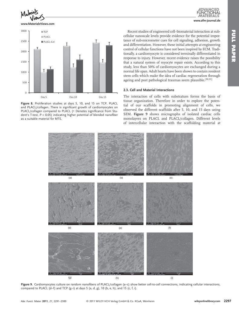
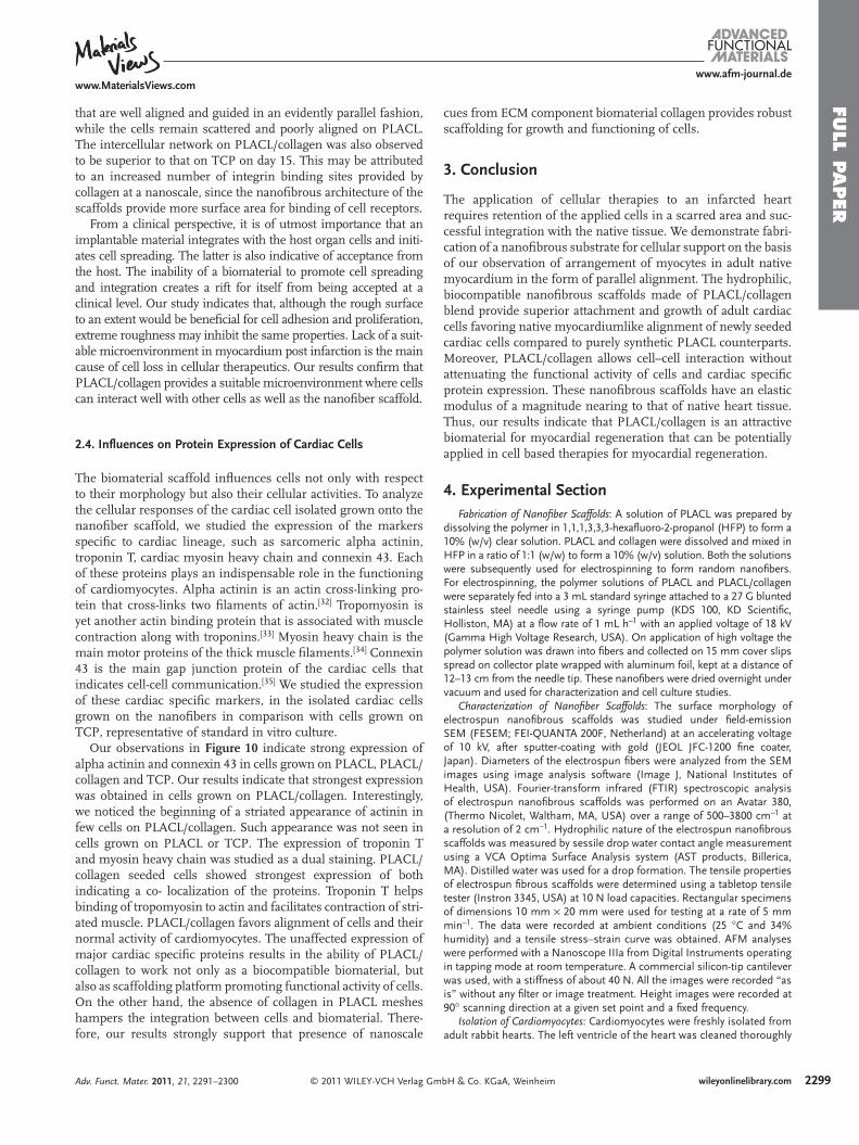
Figure 9 . Cardiomyocytes culture on random nanofi bers of PLACL/collagecompared to PLACL (d–f) and TCP (g–i) at days 5 (a, d, g), 10 (b, e, h), an
Figure 8 . Proliferation studies at days 5, 10, and 15 on TCP, PLACL and PLACL/collagen. There is signifi cant growth of cardiomyocytes on PLACL/collagen compared to PLACL ( ∗ Denotes signifi cance from Stu-dent’s T-test, P < 0.05) indicating higher potential of blended nanofi ber as a suitable material for MTE.
FULL P
APER
© 2011 WILEY-VCH Verlag G
www.afm-journal.dewww.MaterialsViews.com
Adv. Funct. Mater. 2011, 21, 2291–2300
Recent studies of engineered cell–biomaterial interaction at sub-cellular nano scale levels provide evidence for the potential impor-tance of sub-micrometer cues for cell signaling, adhesion, growth and differentiation. However, these initial attempts at engineering control of cellular functions have not been inspired by ECM. Tradi-tionally, a cardiomyocyte is considered terminally differentiated in response to injury. However, recent evidence raises the possibility that a natural system of myocyte repair exists. According to this study, less than 50% of cardiomyocytes are exchanged during a normal life span. Adult hearts have been shown to contain resident stem cells which make the idea of cardiac regeneration through ageing and post pathological traumas seem plausible. [ 30,31 ]
2.3. Cell and Material Interactions
The interaction of cells with substratum forms the basis of tissue organization. Therefore in order to explore the poten-tial of our scaffolds in promoting alignment of cells, we observed the different scaffolds after 5, 10, and 15 days using SEM. Figure 9 shows micrographs of isolated cardiac cells monolayers on PLACL and PLACL/collagen. Different levels of intercellular interaction with the scaffolding material at
n (a–c) show better cell-to-cell connections, indicating cellular interactions, d 15 (c, f, i).
mbH & Co. KGaA, Weinheim 2297wileyonlinelibrary.com

Figure 10 . Immunofl uorescence shows cardiomyocytes on random nanofi bers of PLACL/collagen, PLACL and TCP stained with for cardiac specifi c protein at day 15: a) cardiac myosin heavy chain–Alexa Fluor 594 and troponin T–Alexa Fluor 488 show colocalized expression, with PLACL/collagen showing the strongest expression; b) sarcomeric alpha actinin–Alexa Fluor 488 and connexin 43–Alexa Fluor 594, a few cells on PLACL collagen show re-appearance of a fribrillar pattern of alpha actinin expression (arrows).
FULL
PAPER
2298
www.afm-journal.dewww.MaterialsViews.com
different time points can be observed. It is evident from Figure 9 that the cells start to stabilize and align themselves by day 10 on PLACL/collagen. However, in the absence of collagen, the
© 2011 WILEY-VCH Verlag Gwileyonlinelibrary.com
cellular arrangement is rather random and scattered after the same period. By day 15, PLACL/collagen shows formation of car-diomyocyte monolayer interconnected by intercellular junctions
mbH & Co. KGaA, Weinheim Adv. Funct. Mater. 2011, 21, 2291–2300
FULL P
APER
www.afm-journal.dewww.MaterialsViews.com
that are well aligned and guided in an evidently parallel fashion, while the cells remain scattered and poorly aligned on PLACL. The intercellular network on PLACL/collagen was also observed to be superior to that on TCP on day 15. This may be attributed to an increased number of integrin binding sites provided by collagen at a nanoscale, since the nanofi brous architecture of the scaffolds provide more surface area for binding of cell receptors.
From a clinical perspective, it is of utmost importance that an implantable material integrates with the host organ cells and initi-ates cell spreading. The latter is also indicative of acceptance from the host. The inability of a biomaterial to promote cell spreading and integration creates a rift for itself from being accepted at a clinical level. Our study indicates that, although the rough surface to an extent would be benefi cial for cell adhesion and proliferation, extreme roughness may inhibit the same properties. Lack of a suit-able microenvironment in myocardium post infarction is the main cause of cell loss in cellular therapeutics. Our results confi rm that PLACL/collagen provides a suitable microenvironment where cells can interact well with other cells as well as the nanofi ber scaffold.
2.4. Infl uences on Protein Expression of Cardiac Cells
The biomaterial scaffold infl uences cells not only with respect to their morphology but also their cellular activities. To analyze the cellular responses of the cardiac cell isolated grown onto the nanofi ber scaffold, we studied the expression of the markers specifi c to cardiac lineage, such as sarcomeric alpha actinin, troponin T, cardiac myosin heavy chain and connexin 43. Each of these proteins plays an indispensable role in the functioning of cardiomyocytes. Alpha actinin is an actin cross-linking pro-tein that cross-links two fi laments of actin. [ 32 ] Tropomyosin is yet another actin binding protein that is associated with muscle contraction along with troponins. [ 33 ] Myosin heavy chain is the main motor proteins of the thick muscle fi laments. [ 34 ] Connexin 43 is the main gap junction protein of the cardiac cells that indicates cell-cell communication. [ 35 ] We studied the expression of these cardiac specifi c markers, in the isolated cardiac cells grown on the nanofi bers in comparison with cells grown on TCP, representative of standard in vitro culture.
Our observations in Figure 10 indicate strong expression of alpha actinin and connexin 43 in cells grown on PLACL, PLACL/collagen and TCP. Our results indicate that strongest expression was obtained in cells grown on PLACL/collagen. Interestingly, we noticed the beginning of a striated appearance of actinin in few cells on PLACL/collagen. Such appearance was not seen in cells grown on PLACL or TCP. The expression of troponin T and myosin heavy chain was studied as a dual staining. PLACL/collagen seeded cells showed strongest expression of both indicating a co- localization of the proteins. Troponin T helps binding of tropomyosin to actin and facilitates contraction of stri-ated muscle. PLACL/collagen favors alignment of cells and their normal activity of cardiomyocytes. The unaffected expression of major cardiac specifi c proteins results in the ability of PLACL/collagen to work not only as a biocompatible biomaterial, but also as scaffolding platform promoting functional activity of cells. On the other hand, the absence of collagen in PLACL meshes hampers the integration between cells and biomaterial. There-fore, our results strongly support that presence of nanoscale
© 2011 WILEY-VCH Verlag GmAdv. Funct. Mater. 2011, 21, 2291–2300
cues from ECM component biomaterial collagen provides robust scaffolding for growth and functioning of cells.
3. Conclusion
The application of cellular therapies to an infarcted heart requires retention of the applied cells in a scarred area and suc-cessful integration with the native tissue. We demonstrate fabri-cation of a nanofi brous substrate for cellular support on the basis of our observation of arrangement of myocytes in adult native myocardium in the form of parallel alignment. The hydrophilic, biocompatible nanofi brous scaffolds made of PLACL/collagen blend provide superior attachment and growth of adult cardiac cells favoring native myocardiumlike alignment of newly seeded cardiac cells compared to purely synthetic PLACL counterparts. Moreover, PLACL/collagen allows cell–cell interaction without attenuating the functional activity of cells and cardiac specifi c protein expression. These nanofi brous scaffolds have an elastic modulus of a magnitude nearing to that of native heart tissue. Thus, our results indicate that PLACL/collagen is an attractive biomaterial for myocardial regeneration that can be potentially applied in cell based therapies for myocardial regeneration.
4. Experimental Section Fabrication of Nanofi ber Scaffolds : A solution of PLACL was prepared by
dissolving the polymer in 1,1,1,3,3,3-hexafl uoro-2-propanol (HFP) to form a 10% (w/v) clear solution. PLACL and collagen were dissolved and mixed in HFP in a ratio of 1:1 (w/w) to form a 10% (w/v) solution. Both the solutions were subsequently used for electrospinning to form random nanofi bers. For electrospinning, the polymer solutions of PLACL and PLACL/collagen were separately fed into a 3 mL standard syringe attached to a 27 G blunted stainless steel needle using a syringe pump (KDS 100, KD Scientifi c, Holliston, MA) at a fl ow rate of 1 mL h − 1 with an applied voltage of 18 kV (Gamma High Voltage Research, USA). On application of high voltage the polymer solution was drawn into fi bers and collected on 15 mm cover slips spread on collector plate wrapped with aluminum foil, kept at a distance of 12–13 cm from the needle tip. These nanofi bers were dried overnight under vacuum and used for characterization and cell culture studies.
Characterization of Nanofi ber Scaffolds : The surface morphology of electrospun nanofi brous scaffolds was studied under fi eld-emission SEM (FESEM; FEI-QUANTA 200F, Netherland) at an accelerating voltage of 10 kV, after sputter-coating with gold (JEOL JFC-1200 fi ne coater, Japan). Diameters of the electrospun fi bers were analyzed from the SEM images using image analysis software (Image J, National Institutes of Health, USA). Fourier-transform infrared (FTIR) spectroscopic analysis of electrospun nanofi brous scaffolds was performed on an Avatar 380, (Thermo Nicolet, Waltham, MA, USA) over a range of 500–3800 cm − 1 at a resolution of 2 cm − 1 . Hydrophilic nature of the electrospun nanofi brous scaffolds was measured by sessile drop water contact angle measurement using a VCA Optima Surface Analysis system (AST products, Billerica, MA). Distilled water was used for a drop formation. The tensile properties of electrospun fi brous scaffolds were determined using a tabletop tensile tester (Instron 3345, USA) at 10 N load capacities. Rectangular specimens of dimensions 10 mm × 20 mm were used for testing at a rate of 5 mm min − 1 . The data were recorded at ambient conditions (25 ° C and 34% humidity) and a tensile stress–strain curve was obtained. AFM analyses were performed with a Nanoscope IIIa from Digital Instruments operating in tapping mode at room temperature. A commercial silicon-tip cantilever was used, with a stiffness of about 40 N. All the images were recorded “as is” without any fi lter or image treatment. Height images were recorded at 90 ° scanning direction at a given set point and a fi xed frequency.
Isolation of Cardiomyocytes : Cardiomyocytes were freshly isolated from adult rabbit hearts. The left ventricle of the heart was cleaned thoroughly
bH & Co. KGaA, Weinheim 2299wileyonlinelibrary.com
FULL
PAPER
2300
www.afm-journal.dewww.MaterialsViews.com
with phosphate buffered saline (PBS) (1 st Base) containing antibiotics (Sigma) for removal of fat tissue and clots and minced into fi ne pieces. The processed tissue was treated with 5 mg of collagenase (Sigma) for 20 min at 37 ° C. Freshly isolated cardiac cells were cultured in DMEM media supplemented with 10% FBS and 1% antibiotic and antimycotic solutions (Invitrogen Corp, USA) in a 75 cm 2 cell-culture fl ask. Cells were incubated at 37 ° C in a humidifi ed atmosphere containing 5% CO 2 for 6 days and the culture medium was changed once in every 2 days.
Cardiomyocyte Culture on Nanofi bers : Each of the nanofi brous scaffolds on 15 mm coverslip was placed in a 24-well plate and secured with a stainless steel ring to ensure complete contact of the scaffolds with wells. The specimens were sterilized under UV light, washed thrice with PBS and subsequently immersed in DMEM overnight before cell seeding. Cardiomyocytes grown in 75 cm 2 cell culture fl asks were detached by adding 1 mL of 0.25% trypsin (Sigma) containing 0.1% EDTA. Detached cells were centrifuged, counted via the trypan blue assay using a hemocytometer and seeded on the scaffolds at a density of 2.0 × 10 4 cells well − 1 and left in incubator for cell growth.
Cell Morphology Studies : Morphological studies of in vitro cultured cardiomyocytes on nanofi bers were performed after 5, 10, and 15 days of cell culture, by processing them for SEM studies. The scaffolds were rinsed twice with PBS and fi xed in 3% glutaraldehyde (Sigma) for 3 h. Thereafter, scaffolds were rinsed in DI water and dehydrated with increasing concentrations of ethanol (30%, 50%, 70% 90%, 100%) twice for 10 min each. Final washing with 100% ethanol was followed by treating the specimens with hexamethyldisilazane (HMDS). The HMDS (Sigma) was air-dried by keeping the samples in a fume hood. Finally, the scaffolds were sputter-coated with gold and observed under SEM to observe the morphology of cardiomyocytes.
Proliferation Studies : The cell adhesion and proliferation on the scaffolds were determined using the colorimetric MTS assay (CellTiter 96 AQueous One solution, Promega, Madison, WI). The reduction of yellow tetrazolium salt [3-(4,5-dimethylthiazol-2-yl)-5-(3-carboxymethoxyphenyl)-2(4-sulfophenyl)-2H-tetrazolium] in MTS to form purple formazan crystals by the dehydrogenase enzymes secreted by mitochondria of metabolically active cells forms the basis of this assay. The formazan dye shows absorbance at 492 nm and the amount of formazan crystals formed is directly proportional to the number of cells. For the MTS assay, samples were rinsed with PBS to remove unattached cells and incubated with 20% MTS reagent in a serum-free medium for a period of 3 h at 37 ° C. Absorbance of the obtained dye was measured at 490 nm using a spectrophotometric plate reader (FLUOstar OPTIMA, BMG lab Technologies).
Immunofl uorescence : Cardiomyocytes grown on nanofi bers were washed with PBS, and fi xed with 100% chilled methanol for 10 min. The cardiomyocytes were permeabilized with 0.1% Triton X-100, blocked for nonspecifi c staining using 3% BSA and then immunostained with anti-alpha actinin, anti-troponin T, anti-cardiac myosin heavy chain (Abcam) and anti-connexin 43(Sigma) for 1 h at room temperature. Then, they were revealed with anti-species-specifi c Alexa Fluor 488 or 594 secondary antibodies for 30 min and DAPI (Invitrogen). The samples were mounted using anti-quenching mounting medium (Vectashiled, Vector Laboratories) and observed under a confocal microscope (Olympus Fluoview FV1000). The images taken were analyzed using Olympus FV10-ASW imaging software.
Statistical Analysis : All the data presented are expressed as mean ± standard deviation (SD) and were analyzed using Student’s t -test for the calculation of signifi cance level of the data. Differences were considered statistically signifi cant at P < 0.05.
Acknowledgements The authors gratefully acknowledge the NRF-Technion (Grant No.: R-398-001-063-592), Division of Bioengineering, Nanoscience and Nanotechnology Initiative, National University of Singapore for fi nancial support.
Received: November 18, 2010 Published online: Published online: April 26, 2011
© 2011 WILEY-VCH Verlag Gwileyonlinelibrary.com
[ 1 ] J. He , L. G. Ogden , L. A. Bazzano , S. Vupputuri , C. Loria , P. K. Whelton , Arch. Intern. Med. 2001 , 161 , 996 .
[ 2 ] M. K. Baig , N. Mahon , W. J. McKenna , A. L. Caforio , R. O. Bonow , G. S. Francis , M. Gheorghiade , Heart Lung 1999 , 28 , 87 .
[ 3 ] M. Packer , J. Card. Fail. 2002 , 8 , 193 . [ 4 ] R. L. Kao , W. Browder , C. Li , Asian Cardiovasc. Thorac. Ann. 2009 , 17 , 89 . [ 5 ] H. Jawad , N. N. Ali , A. R. Lyon , Q. Z. Chen , S. E. Harding ,
A. R. Boccaccini , J. Tissue Eng. Regen. Med. 2007 , 1 , 327 . [ 6 ] J. Leor , Y. Amsalem , S. Cohen , Pharmacol. Ther. 2005 , 105 , 151 . [ 7 ] D. Kai , M. P. Prabhakaran , S. Liao , S. Ramakrishna , Nano Biomed.
2010 , 2 , 1 . [ 8 ] L. W. Miller , J. Cardiovasc. Trans. Res. 2008 , 1 , 185 . [ 9 ] J. M. Hare , J. H. Traverse , T. D. Henry , N. Dib , R. K. Strumpf ,
S. P. Schulman , G. Gerstenblith , A. N. DeMaria , A. E. Denktas , R. S. Gammon , J. B. Hermiller , Jr. , M. A. Reisman , G. L. Schaer , W. Sherman , J. Am. Coll. Cardiol. 2009 , 54 , 2277 .
[ 10 ] L. W. van Laake , R. Passier , P. A. Doevendans , C. L. Mummery , Circ. Res. 2008 , 102 , 1008 .
[ 11 ] E. C. Goldsmith , A. Hoffman , M. O. Morales , J. D. Potts , R. L. Price , A. McFadden , M. Rice , T. K. Borg , Dev. Dyn. 2004 , 230 , 787 .
[ 12 ] M. Eghbali , O. O. Blumenfeld , S. Seifter , P. M. Buttrick , L. A. Leinwand , T. F. Robinson , M. A. Zern , M. A. Giambrone . J. Mol. Cell. Cardiol. 1989 , 21 , 103 .
[ 13 ] J. E. Bishop , G. J. Laurent , Eur. Heart. J. 1995 , 16 , 38 . [ 14 ] G. M. Fomovsky , S. Thomopoulos , J. W. Holmes , J Mol. Cell. Cardiol.
2010 , 48 , 490 . [ 15 ] S. Mukherjee , J. Venugopal , R. Ravichandran , S. Ramakrishna ,
M. Raghunath , J. Mater. Chem. 2010 , 20 , 8819 . [ 16 ] T. Kofi dis , K. Müller-Stahl , A. Haverich , Meth. Mol. Med. 2007 , 140 , 273 . [ 17 ] S. F. Nagueh , G. Shah , Y. Wu , S. Lahmers , Circulation 2004 , 110 , 155 . [ 18 ] J. H. Omens , Prog. Biophys. Mol. Biol. 1998 , 69 , 559 . [ 19 ] J. Y. Lim , J. C. Hansen , C. A. Siedlecki , R. W. Hengstebeck , J. Cheng ,
N. Winograd , H. J. Donahue , Biomacromolecules 2005 , 6 , 3319 . [ 20 ] R. Murugan , S. Ramakrishna , Tissue Eng. 2007 , 8 , 1845 . [ 21 ] M. M. Stevens , J. H. George , Science 2005 , 310 , 1135 . [ 22 ] E. C. Martinez , T. Kofi dis . Expert. Rev. Cardiovasc. Ther. 2009 , 7 , 921 . [ 23 ] X. Wei , Z. Xia , S.-C. Wong , A. Baji . Int. J. Exp. Comput. Biomech.
2009 , 1 , 45 . [ 24 ] J.-W. Lu , Z. P. Zhang , X. Z. Ren , Y. Z. Chen , J. Yu , Z. X. Guo ,
Macromolecules 2008 , 41 , 3762 . [ 25 ] A. J. Engler , C. Carag-Krieger , C. P. Johnson , M. Raab , H. Y. Tang ,
D. W. Speicher , J. W. Sanger , J. M. Sanger , D. E. Discher , J. Cell. Sci. 2008 , 121 , 3794 .
[ 26 ] A. Du , J. M. Sanger , K. K. Linask , J. W. Sanger , Dev. Biol. 2003 , 257 , 382 . [ 27 ] J. S. Mitcheson , J. C. Hancox , A. J. Levi , Cardiovasc. Res. 1998 , 39 , 280 . [ 28 ] K. D. Costa , E. J. Lee , J. W. Holmes , Tissue. Eng. 2003 , 9 , 567 . [ 29 ] M. Wagner , M. A. Siddiqui , Exp. Biol. Med. 2007 , 232 , 852 . [ 30 ] E. Messina , L. D. Angelis , G. Frati , S. Morrone , S. Chimenti ,
F. Fiordaliso , M. Salio , M. Battaglia , M. V. Latronico , M. Coletta , E. Vivarelli , L. Frati , G. Cossu , A. Giacomello , Circ. Res. 2004 , 95 , 911 .
[ 31 ] C. Bearzi , M. Rota , T. Hosoda , C. Bearzi , M. Rota , T. Hosoda , J. Tillmanns , A. Nascimbene , A. De Angelis , S. Yasuzawa-Amano , I. Trofi mova , R. W. Siggins , N. Lecapitaine , S. Cascapera , A. P. Beltrami , D. A. D’Alessandro , E. Zias , F. Quaini , K. Urbanek , R. E. Michler , R. Bolli , J. Kajstura , A. Leri , P. Anversa , Proc. Natl. Acad. Sci. USA 2007 , 104 , 14068 .
[ 32 ] N. Cabello , R. Remelli , L. Canela , A. Soriguera , J. Mallol , E. I. Canela , M. J. Robbins , C. Lluis , R. Franco , R. A. McIlhinney , F. Ciruela , J. Biol. Chem. 2007 , 282 , 12143 .
[ 33 ] S. E. Hitchcock-DeGregori , N. J. Greenfi eld , A. Singh , Adv. Exp. Med. Biol. 2007 , 592 , 87 .
[ 34 ] K. Yamauchi-Takihara , M. J. Sole , J. Liew , D. Ing , C. C. Liew , Proc. Natl. Acad. Sci. USA. 1989 , 86 , 3504 .
[ 35 ] R. Schulz , G. Heusch , Cardiovasc. Res. 2004 , 62 , 335 .
mbH & Co. KGaA, Weinheim Adv. Funct. Mater. 2011, 21, 2291–2300
Copyright © 2022 FDOKUMEN