Depletion of CD4 T lymphocytes in human lymphoid tissue infected ex vivo with doxycycline-dependent...
6
Rapid Communication Depletion of CD4 T lymphocytes in human lymphoid tissue infected ex vivo with doxycycline-dependent HIV-1 Yana Kiselyeva a , Yoshinori Ito a , Rosangela G. Lima a,1 , Jean-Charles Grivel a , Atze T. Das b , Ben Berkhout b, * , Leonid B. Margolis a, * a Laboratory of Molecular and Cellular Biophysics, National Institute of Child Health and Human Development, Bethesda, MD 20892, USA b Department of Human Retrovirology, Academic Medical Center, University of Amsterdam, Amsterdam, The Netherlands Received 15 April 2004; returned to author for revision 17 May 2004; accepted 16 July 2004 Available online 21 August 2004 Abstract We investigated whether CD4 + Tcells that do not produce HIV-1 are killed in HIV-infected human lymphoid tissue. Tissue blocks were inoculated with high amount of doxycycline-dependent HIV-rtTA. Doxycycline triggered productive infection and loss of CD4 + T cells in these tissues, whereas without doxycycline, neither productive infection nor CD4 + T cell depletion was detected in spite of the massive presence of virions in the tissue and of viral DNA in the cells. Thus, HIV-1 alone is sufficient to deplete productively infected CD4 + T cells but is not sufficient to cause the death of uninfected or latently infected CD4 + T cells. Published by Elsevier Inc. Keywords: CD4 + T cell; Human lymphoid tissue; HIV-1 Introduction The hallmark of HIV infection is the depletion of CD4 + T lymphocytes. It is well established that HIV productive infection kills CD4 + T cells (reviewed in (Pantaleo et al., 1997)). However, CD4 + T cells that do not produce virus, but which reside in infected tissues, are killed as well. Such cells may be infected latently or may not be infected at all (bbystandersQ)(Badley et al., 1996; Banda et al., 1992; Gandhi et al., 1998; Herbein et al., 1998; Laurent-Crawford et al., 1995; Oyaizu et al., 1997). The mechanisms of cell killing by HIV-1 in general and of bystander cells in particular are not fully understood. While some published data suggest that cell interactions with viral particles or their components are sufficient to kill uninfected cells without productive infection, others suggest that general activation of the immune system is required for uninfected cells to become apoptotic (Badley et al., 2000; Casella and Finkel, 1997; Gougeon and Montagnier, 1993; Grossman et al., 2002; Mahlknecht and Herbein, 2001; Meyaard et al., 1992; Yang and Ashwell, 2001). Critical events of HIV disease occur in lymphoid tissue (Pantaleo and Fauci, 1995). Therefore, we addressed the problems of CD4 + T cell death in human lymphoid tissues infected ex vivo with HIV-1 (Glushakova et al., 1997). This system supports productive HIV-1 infection without exogenous stimulation, and such an infection results in a loss of CD4 + T cells (Glushakova et al., 1997). In the study we report here, we used a newly developed virus construct, HIV-rtTA, that can infect cells and establish an integrated provirus in the absence of doxycycline but whose transcription and replication is dependent on the presence of the antibiotic doxycycline (bdox-dependent HIV-rtTAQ) (Berkhout et al., 2002; Das et al., 2002; Verhoef et al., 2001). 0042-6822/$ - see front matter. Published by Elsevier Inc. doi:10.1016/j.virol.2004.07.014 * Corresponding authors. Ben Berkhout is to be contacted at Depart- ment of Human Retrovirology, University of Amsterdam, Academic Medical Center K3-110. Meibergdreef 15, 1105 AZ Amsterdam, The Netherlands. Fax: +31 20 691 6531. Leonid Margolis, Laboratory of Molecular and Cellular Biophysics, National Institute of Child Health and Human Development, NIH, Building 10, Room 9D58, 10 Center Drive, Bethesda, MD 20892. Fax: +1 301 480 0857. E-mail addresses: [email protected] (B. Berkhout)8 [email protected] (L.B. Margolis). 1 Current address: Department of Immunology, Oswaldo Cruz Foun- dation, Rio de Janeiro-RJ, Brazil. Virology 328 (2004) 1– 6 www.elsevier.com/locate/yviro
-
Upload
independent -
Category
Documents
-
view
0 -
download
0
Transcript of Depletion of CD4 T lymphocytes in human lymphoid tissue infected ex vivo with doxycycline-dependent...

www.elsevier.com/locate/yviro
Virology 328 (
Rapid Communication
Depletion of CD4 T lymphocytes in human lymphoid tissue infected
ex vivo with doxycycline-dependent HIV-1
Yana Kiselyevaa, Yoshinori Itoa, Rosangela G. Limaa,1, Jean-Charles Grivela, Atze T. Dasb,
Ben Berkhoutb,*, Leonid B. Margolisa,*
aLaboratory of Molecular and Cellular Biophysics, National Institute of Child Health and Human Development, Bethesda, MD 20892, USAbDepartment of Human Retrovirology, Academic Medical Center, University of Amsterdam, Amsterdam, The Netherlands
Received 15 April 2004; returned to author for revision 17 May 2004; accepted 16 July 2004
Available online 21 August 2004
Abstract
We investigated whether CD4+ T cells that do not produce HIV-1 are killed in HIV-infected human lymphoid tissue. Tissue blocks were
inoculated with high amount of doxycycline-dependent HIV-rtTA. Doxycycline triggered productive infection and loss of CD4+ T cells in
these tissues, whereas without doxycycline, neither productive infection nor CD4+ T cell depletion was detected in spite of the massive
presence of virions in the tissue and of viral DNA in the cells. Thus, HIV-1 alone is sufficient to deplete productively infected CD4+ T cells
but is not sufficient to cause the death of uninfected or latently infected CD4+ T cells.
Published by Elsevier Inc.
Keywords: CD4+ T cell; Human lymphoid tissue; HIV-1
Introduction
The hallmark of HIV infection is the depletion of CD4+ T
lymphocytes. It is well established that HIV productive
infection kills CD4+ T cells (reviewed in (Pantaleo et al.,
1997)). However, CD4+ Tcells that do not produce virus, but
which reside in infected tissues, are killed as well. Such cells
may be infected latently or may not be infected at all
(bbystandersQ) (Badley et al., 1996; Banda et al., 1992;
Gandhi et al., 1998; Herbein et al., 1998; Laurent-Crawford
et al., 1995; Oyaizu et al., 1997). The mechanisms of cell
0042-6822/$ - see front matter. Published by Elsevier Inc.
doi:10.1016/j.virol.2004.07.014
* Corresponding authors. Ben Berkhout is to be contacted at Depart-
ment of Human Retrovirology, University of Amsterdam, Academic
Medical Center K3-110. Meibergdreef 15, 1105 AZ Amsterdam, The
Netherlands. Fax: +31 20 691 6531. Leonid Margolis, Laboratory of
Molecular and Cellular Biophysics, National Institute of Child Health and
Human Development, NIH, Building 10, Room 9D58, 10 Center Drive,
Bethesda, MD 20892. Fax: +1 301 480 0857.
E-mail addresses: [email protected] (B. Berkhout)8
[email protected] (L.B. Margolis).1 Current address: Department of Immunology, Oswaldo Cruz Foun-
dation, Rio de Janeiro-RJ, Brazil.
killing by HIV-1 in general and of bystander cells in
particular are not fully understood. While some published
data suggest that cell interactions with viral particles or their
components are sufficient to kill uninfected cells without
productive infection, others suggest that general activation of
the immune system is required for uninfected cells to become
apoptotic (Badley et al., 2000; Casella and Finkel, 1997;
Gougeon and Montagnier, 1993; Grossman et al., 2002;
Mahlknecht and Herbein, 2001; Meyaard et al., 1992; Yang
and Ashwell, 2001). Critical events of HIV disease occur in
lymphoid tissue (Pantaleo and Fauci, 1995). Therefore, we
addressed the problems of CD4+ T cell death in human
lymphoid tissues infected ex vivo with HIV-1 (Glushakova et
al., 1997). This system supports productive HIV-1 infection
without exogenous stimulation, and such an infection results
in a loss of CD4+ T cells (Glushakova et al., 1997). In the
study we report here, we used a newly developed virus
construct, HIV-rtTA, that can infect cells and establish an
integrated provirus in the absence of doxycycline but whose
transcription and replication is dependent on the presence of
the antibiotic doxycycline (bdox-dependent HIV-rtTAQ)(Berkhout et al., 2002; Das et al., 2002; Verhoef et al., 2001).
2004) 1–6

Rapid Communication2
We used this construct to test whether uninfected cells
that have interacted with virions, or cells that are non-
productively infected, are depleted in the context of human
lymphoid tissue. We found that in the presence of high
amounts of virions and intracellular viral DNA, no depletion
of CD4+ T cells occurs in lymphoid tissue unless productive
replication is triggered by doxycycline. Thus, in the context
of human lymphoid tissue ex vivo, HIV infection kills
productively infected cells but is not sufficient to cause the
death of uninfected or latently infected CD4+ T cells.
Fig. 1. Doxycycline dependence of HIV-rtTA in ex vivo human lymphoid
tissue infected ex vivo. (a) Tissue blocks were inoculated with HIV-rtTA.
Doxycycline at the concentration of 0.5 Ag/ml was added to some of the
cultures at different time points after virus inoculation and supplemented
thereafter with each medium change every 3 days. For each donor tissue, 27
tissue blocks were inoculated and the medium was pooled for CA-p24
measurements. Each data point represents the average (FSEM). Tissue
blocks were inoculated with virus and cultured in the absence or presence of
doxycycline. Each bar represents the average (FSEM) of tissues from 13
donors. (b) Tissue blocks were inoculated with virus and doxycycline was
administered immediately or 6 days after inoculation. The mean values and
estimated SEM of triplicate measurements of pooled samples are presented.
Results and discussion
In the work that we report here, we (i) tested HIV-rtTA
dox-dependence in the context of human lymphoid tissue
and (ii) evaluated the effects of latent and productive HIV-
rtTA infection on CD4+ T cell depletion in human lymphoid
tissue ex vivo.
Dox dependence of HIV-rtTA in ex vivo-infected human
lymphoid tissue
To evaluate this dependence, we inoculated blocks of
human lymphoid tissue with HIV-rtTA and simultaneously
added doxycycline. The culturemediumwas changed every 3
days, and doxycycline was supplied with each medium
change. In the presence of 0.5 to 1.5 Ag/ml doxycycline, a
vigorous productive infection was detected in tissues
infected, and a dose as low as 0.1 Ag/ml triggered notable
viral replication (data not shown). For experiments described
hereafter, we chose the concentration of doxycycline of 0.5
Ag/ml. This dose supports efficient tissue productive infec-
tion by HIV-rtTA (although 4 times lower compared to the
wild-type virus) as evaluated from the increase of CA-p24 in
the medium (Fig. 1a). A less efficient replication of HIV-rtTA
compared to the wild-type virus was observed also in T cell
lines (Das et al., 2004) and may be due to the usage of
different regulatory systems by HIV-rtTA and wild-type
viruses. As was reported earlier for various HIV-1 variants,
viral replication became detectable after day 6 post-inocu-
lation and continued to increase to the end of experiment on
day 12–15. Without adding doxycycline, no productive
infection was detected in inoculated tissue blocks (Fig. 1a).
The amount of CA-p24 on day 3 (before the first medium
change) roughly corresponded to the amount of inoculum.
These amounts were similar in cultures not treated with
doxycycline and in cultures to which doxycycline was added
at the time of virus inoculation.
Next, we studied whether HIV replication could be turned
on in HIV-rtTA-inoculated tissues when doxycycline addi-
tion was delayed relative to viral inoculation. Tissue blocks
were inoculated with the virus on day 0 and cultured without
doxycycline for another 6 days. The amount of provirus was
evaluated by real-time PCR, and the HIV copy number was
normalized to the number of copies of the GAPDH gene. This
assay revealed that in tissues inoculated without doxycycline,
there were 1.4 � 10�4 F 0.8 � 10�4 genome equivalent of
viral DNA. The cultures were washed and the medium was
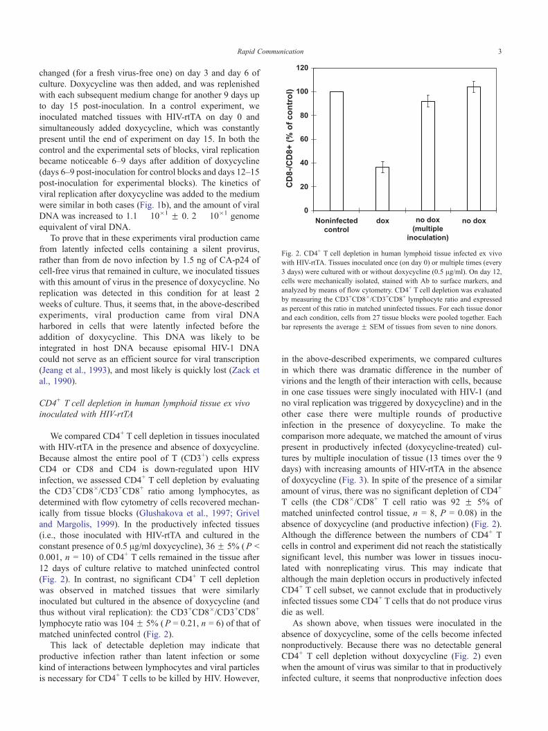
Fig. 2. CD4+ T cell depletion in human lymphoid tissue infected ex vivo
with HIV-rtTA. Tissues inoculated once (on day 0) or multiple times (every
3 days) were cultured with or without doxycycline (0.5 Ag/ml). On day 12,
cells were mechanically isolated, stained with Ab to surface markers, and
analyzed by means of flow cytometry. CD4+ T cell depletion was evaluated
by measuring the CD3+CD8�/CD3+CD8+ lymphocyte ratio and expressed
as percent of this ratio in matched uninfected tissues. For each tissue donor
and each condition, cells from 27 tissue blocks were pooled together. Each
bar represents the average F SEM of tissues from seven to nine donors.
Rapid Communication 3
changed (for a fresh virus-free one) on day 3 and day 6 of
culture. Doxycycline was then added, and was replenished
with each subsequent medium change for another 9 days up
to day 15 post-inoculation. In a control experiment, we
inoculated matched tissues with HIV-rtTA on day 0 and
simultaneously added doxycycline, which was constantly
present until the end of experiment on day 15. In both the
control and the experimental sets of blocks, viral replication
became noticeable 6–9 days after addition of doxycycline
(days 6–9 post-inoculation for control blocks and days 12–15
post-inoculation for experimental blocks). The kinetics of
viral replication after doxycycline was added to the medium
were similar in both cases (Fig. 1b), and the amount of viral
DNA was increased to 1.1 � 10�1 F 0. 2 � 10�1 genome
equivalent of viral DNA.
To prove that in these experiments viral production came
from latently infected cells containing a silent provirus,
rather than from de novo infection by 1.5 ng of CA-p24 of
cell-free virus that remained in culture, we inoculated tissues
with this amount of virus in the presence of doxycycline. No
replication was detected in this condition for at least 2
weeks of culture. Thus, it seems that, in the above-described
experiments, viral production came from viral DNA
harbored in cells that were latently infected before the
addition of doxycycline. This DNA was likely to be
integrated in host DNA because episomal HIV-1 DNA
could not serve as an efficient source for viral transcription
(Jeang et al., 1993), and most likely is quickly lost (Zack et
al., 1990).
CD4+ T cell depletion in human lymphoid tissue ex vivo
inoculated with HIV-rtTA
We compared CD4+ T cell depletion in tissues inoculated
with HIV-rtTA in the presence and absence of doxycycline.
Because almost the entire pool of T (CD3+) cells express
CD4 or CD8 and CD4 is down-regulated upon HIV
infection, we assessed CD4+ T cell depletion by evaluating
the CD3+CD8�/CD3+CD8+ ratio among lymphocytes, as
determined with flow cytometry of cells recovered mechan-
ically from tissue blocks (Glushakova et al., 1997; Grivel
and Margolis, 1999). In the productively infected tissues
(i.e., those inoculated with HIV-rtTA and cultured in the
constant presence of 0.5 Ag/ml doxycycline), 36 F 5% (P b
0.001, n = 10) of CD4+ T cells remained in the tissue after
12 days of culture relative to matched uninfected control
(Fig. 2). In contrast, no significant CD4+ T cell depletion
was observed in matched tissues that were similarly
inoculated but cultured in the absence of doxycycline (and
thus without viral replication): the CD3+CD8�/CD3+CD8+
lymphocyte ratio was 104 F 5% (P = 0.21, n = 6) of that of
matched uninfected control (Fig. 2).
This lack of detectable depletion may indicate that
productive infection rather than latent infection or some
kind of interactions between lymphocytes and viral particles
is necessary for CD4+ T cells to be killed by HIV. However,
in the above-described experiments, we compared cultures
in which there was dramatic difference in the number of
virions and the length of their interaction with cells, because
in one case tissues were singly inoculated with HIV-1 (and
no viral replication was triggered by doxycycline) and in the
other case there were multiple rounds of productive
infection in the presence of doxycycline. To make the
comparison more adequate, we matched the amount of virus
present in productively infected (doxycycline-treated) cul-
tures by multiple inoculation of tissue (13 times over the 9
days) with increasing amounts of HIV-rtTA in the absence
of doxycycline (Fig. 3). In spite of the presence of a similar
amount of virus, there was no significant depletion of CD4+
T cells (the CD8�/CD8+ T cell ratio was 92 F 5% of
matched uninfected control tissue, n = 8, P = 0.08) in the
absence of doxycycline (and productive infection) (Fig. 2).
Although the difference between the numbers of CD4+ T
cells in control and experiment did not reach the statistically
significant level, this number was lower in tissues inocu-
lated with nonreplicating virus. This may indicate that
although the main depletion occurs in productively infected
CD4+ T cell subset, we cannot exclude that in productively
infected tissues some CD4+ T cells that do not produce virus
die as well.
As shown above, when tissues were inoculated in the
absence of doxycycline, some of the cells become infected
nonproductively. Because there was no detectable general
CD4+ T cell depletion without doxycycline (Fig. 2) even
when the amount of virus was similar to that in productively
infected culture, it seems that nonproductive infection does

Fig. 3. Multiple inoculation of tissue with HIV-rtTA. Tissue blocks from
every donor were divided into three fractions. Tissue blocks from the first
fraction were inoculated once (on day 0) and cultured without doxycycline;
the second was also inoculated once (on day 0) and cultured with
doxycycline (0.5 Ag/ml) as described in the legend for Fig. 1; the third was
inoculated every 3 days with an increasing amount of virus and no
doxycycline was added. Twenty-seven tissue blocks from every donor were
inoculated for each condition and pooled culture medium was assayed for
CA-p24. Each data point represents average (FSEM) for tissues from three
to five donors.
Fig. 4. Depletion of CD4+ T cells by HIV-rtTA in the presence of
Nevirapine. Tissue blocks were inoculated with HIV-rtTA 11 times over the
period of 3 days in the absence of doxycycline. Nevirapine (5 AM) was
added on day 3 and continuously supplied till day 12 post HIV-rtTA
inoculation. Doxycycline (0.5 Ag/ml) was added on day 4 and continuously
present till day 12. CD4+ T cells (19.6%) were depleted as compared to the
number of CD4+ T cells in matched uninfected Nevirapine-treated control.
Rapid Communication4
not contribute significantly to the death of CD4+ T cells as
well. To control whether we would detect the depletion of
latently infected cells if they are depleted, we inoculated
tissues blocks with 2 ng of viral stock per block 11 times
over the period of 3 days in the absence of doxycycline;
then, we triggered viral replication by doxycycline and
prevented the spread of infection to new cells by adding an
inhibitory dose of a non-nucleoside reverse transcriptase
inhibitor Nevirapine 1.5 days before doxycycline treatment
(Grivel et al., 2003). Triggering viral production in these
infected cells resulted in a significant depletion of CD8� T
cells (the CD8�/CD8+ T cell ratio was 80.6% of that of
matched uninfected control, P = 0.05, Fig. 4). Thus, in the
absence of doxycycline, cells that are (nonproductively)
infected seem not to be depleted in human lymphoid tissues
inoculated with HIV-1. However, these cells are depleted
after doxycycline triggers their productive infection.
Thus, in ex vivo-infected human lymphoid tissue, neither
a massive presence of HIV-1 virions nor viral entry into the
cells and reverse transcription trigger significant depletion of
CD4+ T lymphocytes. However, when HIV-1 genome tran-
scription was triggered and the virus replicated, a significant
death of CD4+ T lymphocytes concomitantly occurred. This
is consistent with our published finding that CD4+ Tcells that
are not productively infected survive in productively infected
tissues (Grivel et al., 2003). Also, consistent with the present
results, aldrathiol-treated virions (which retain their con-
formation, but are not capable of performing reverse tran-
scription, and thus do not establish productive infection) do
not induce CD4+ T cell depletion in inoculated human
lymphoid tissues (Sylwester et al., 1998). In contrast to the
aldrathiol-treated virions, the construct used in the study we
report on here was able to reverse transcribe. Therefore, using
HIV-rtTA, we were able to demonstrate that not only
extracellular viral particles but also viral DNA in cells is
insufficient to induce significant CD4+ T cell depletion in the
absence of productive infection. Furthermore, our results are
in general agreement with the data published by the Sodroski
group (LaBonte et al., 2000), which has shown that gp120-
mediated killing in constitutively activated cell lines requires
expression of this protein on the cell surface, and thus gp120-
mediated cell death should not affect uninfected cells. Other
HIV-related cell alterations, such as down-regulation of CD4,
are also restricted to T cells productively infected with HIV-1
(Doms and Trono, 2000; Garcia and Miller, 1991; Hoxie et
al., 1986).
Further studies are needed to investigate if all CD4+ T
cells that die as the result of productive HIV infection of
tissues ex vivo produce virus. Alternatively, a minor fraction
of uninfected or latently infected cells could die as well. It
was suggested that in vivo general immunostimulation
contributes to lymphocyte death in HIV-infected individuals
(Muro-Cacho et al., 1995). Unlike lymphoid tissue in vivo,
which responds to HIV infection by general immunoacti-
vation and proliferation, blocks of lymphoid tissue ex vivo

Rapid Communication 5
are not activated upon infection (Malkevitch et al., 2001),
and cell proliferation in these blocks remains negligible
through the entire course of ex vivo infection (Grivel et al.,
2000). Thus, our ex vivo tissue allows to evaluate the
contribution of viral infection to the CD4+ T cell death in the
absence of tissue immunostimulation.
In conclusion, HIV is sufficient to deplete tissue of
productively infected CD4+ T cells, but is not sufficient to
cause a significant death of uninfected or latently infected
CD4+ T cells in the context of human lymphoid tissue, and
thus additional factors seem to contribute to bystander cell
death in vivo. Because, unlike in vivo, ex vivo-infected
tissues do not respond to HIV infection by general
immunostimulation (Grivel et al., 2003); such an immunos-
timulation may be the main additional factor for CD4+ T cell
depletion in HIV-infected individuals (Grossman et al.,
2002; Muro-Cacho et al., 1995).
Materials and methods
Dox-dependent HIV-rtTA was designed and described
earlier (Berkhout et al., 2002; Verhoef et al., 2001). Briefly,
in the full-length, infectious HIV-1 molecular clone pLAI
the TAR-Tat transcriptional axis was inactivated (by
mutation of multiple nucleotides in the single-stranded
bulge and loop domains of TAR, the binding sites for Tat
and cyclin T, respectively, and also by introduction of the
Tyr26Ala point mutation in Tat protein) and replaced by the
tetracycline-inducible tetO–rtTA system (Berkhout et al.,
2002; Das et al., 2002; Verhoef et al., 2001). The activity of
rtTA is critically dependent on doxycycline, a tetracycline
analogue. In this study, we used the HIV-rtTAF86Y A209T
variant with optimized tetO DNA binding sites and a more
doxycycline-sensitive and more potent rtTA variant that was
obtained by virus evolution (Das et al., 2004). For the
production of virus stocks, SupT1 cells were transfected
with the HIV-rtTAF86Y A209T molecular clone by electro-
poration. Briefly, 5 � 106 cells were grown at 378C and 5%
CO2 in RPMI1640 medium containing 10% fetal bovine
serum (FBS), 100 units/ml penicillin, 100 units/ml strepto-
mycin, washed in RPMI1640 with 20% FBS, and mixed
with 5 Ag of DNA in 250 Al RPMI1640 with 20% FBS.
Cells were electroporated in 0.4-cm cuvettes at 250 V and
960 AF, and subsequently resuspended in 5 ml RPMI1640
with 10% FBS and 1 Ag/ml doxycycline. Transfected cells
were mixed with 20 ml RPMI1640 with 10% FBS and 1 Ag/ml doxycycline containing 8 � 106 untransfected SupT1
cells at 3 days after transfection, and cultured until massive
syncytium was observed (7 days). Cells were removed by
centrifugation at 400 � g for 10 min. The virus containing
supernatant was filtered (0.2-Am filter) and stored in
aliquots at �808C. HIV-1 CA-p24 antigen was measured
by ELISA (Beckman-Coulter, Miami, FL).
HIV infection of human lymphoid tissue ex vivo was
performed as described earlier (Glushakova et al., 1997).
Briefly, human tonsils removed during routine tonsillectomy
were received within several hours of excision, washed
thoroughly with medium, and dissected into 2- to 3-mm
blocks. These tissue blocks were placed on top of collagen
sponge gels in culture medium at the air–liquid interface (9
blocks per well with 3 ml of culture medium) and, after
overnight incubation, were inoculated with dox-dependent
HIV-rtTA. In a typical experiment, 3–6 Al of clarified
medium containing 2–4 ng of CA-p24 were applied to the
top of each tissue block. Tissue was cultured in either the
presence or the absence of doxycycline. We assessed
productive HIV infection by measuring CA-p24 in the
culture medium, using p24 ELISA (Beckman-Coulter).
Specifically, the concentration of CA-p24 accumulated in
3 ml of culture medium bathing nine tissue blocks during
the 3 days between successive medium changes was used as
a measure of virus replication.
Flow cytometry was performed on cells mechanically
isolated from control and infected tissue blocks on day 12
after infection. Cells were stained with anti-CD3-fluorescein
isothiocyanate (FITC), anti-CD8-TriColor, and anti-CD4-
allophicocyanin (APC). Data were acquired and analyzed
with CellQuest software. To normalize for differences in
tissue block size and cellularity, CD4+ T cell depletion was
expressed as the ratio of the number of CD3+CD8� T cells
to the number of CD3+CD8+ T cells (Glushakova et al.,
1997; Grivel et al., 2003).
Real-time PCR assay was used to evaluate dox-dependent
HIV-rtTA DNA. Twenty-seven tissue blocks infected with
dox-dependent HIV-rtTA and 27 matched uninfected control
blocks were lysed in ATL buffer containing 10% proteinase
K (Qiagen Inc., USA) (100 Al per 10 mg block, 2 h at 558C).We extracted viral DNA using the DNeasy Tissue kit
(Qiagen) according to the manufacture’s instructions.
Extracted DNA (total volume of 200 Al) was diluted 10-
fold. For the real-time PCR assay, the following gag-specific
primer set was used: Gag forward primer, 5V-ATAATC-CACCTATCCCAGTAGGAGAAAT-3V; Gag reverse primer,
5V-TTGGTCCTTGTCTTATGTCCAGAAT-3V. The reaction
was performed in a 50 Al mixture of SYBR Green PCR
master mix (Applied Biosystems, Foster City, CA), each
primer at 300 nM, and 10 Al of DNA. Following activation ofthe Taq polymerase for 10 min at 958C, 45 cycles, each cycleconsisting of 15 s at 958C followed by 1 min at 608C were
carried out by the ABI Prism 7000 Sequence Detection
System (Applied Biosystems). To normalize the HIV DNA
copy number, we also measured GAPDH, using the follow-
ing specific primer set: GAPDH forward primer: 5V-GAAGGTGAAGGTCGGAGTAGTC-3V; GAPDH reverse
primer: 5V-GAAGATGGTGATGGGATTTC-3V. Serially
diluted plasmids that contained the target sequence were
used as a standard. Real-time fluorescent measurements were
analyzed by the attached software using the values obtained
from serially diluted positive control plasmids that contained
the target gene. Both control plasmids of Gag and those
GAPDH produced wide linear ranges of values from 10 to

Rapid Communication6
106 copies per reaction mixture. The absolute copy numbers
we detected in the present study was in the middle of this
range. We also analyzed the dissociation curve for each
amplification to confirm that there were no nonspecific PCR
products. Each sample was tested in triplicate and the mean of
the three values was used as the copy number of the sample.
Acknowledgments
We thank Monique Vink and Bep Klaver for their help
with the experiments. This research was sponsored in part by
the Technology Foundation STW (applied science division of
NWO and the technology program of the Ministry of
Economic Affairs, Utrecht, the Netherlands). The work of
Y.K. was sponsored, in part, by the HHS Biotechnology
Engagement Program.
References
Badley, A.D., McElhinny, J.A., Leibson, P.J., Lynch, D.H., Alderson, M.R.,
Paya, C.V., 1996. Upregulation of Fas ligand expression by human
immunodeficiency virus in human macrophages mediates apoptosis of
uninfected T lymphocytes. J. Virol. 70 (1), 199–206.
Badley, A.D., Pilon, A.A., Landay, A., Lynch, D.H., 2000. Mechanisms of
HIV-associated lymphocyte apoptosis. Blood 96 (9), 2951–2964.
Banda, N.K., Bernier, J., Kurahara, D.K., Kurrle, R., Haigwood, N.,
Sekaly, R.P., Finkel, T.H., 1992. Crosslinking CD4 by human
immunodeficiency virus gp120 primes T cells for activation-induced
apoptosis. J. Exp. Med. 176 (4), 1099–1106.
Berkhout, B., Verhoef, K., Marzio, G., Klaver, B., Vink, M., Zhou, X., Das,
A.T., 2002. Conditional virus replication as an approach to a safe live
attenuated human immunodeficiency virus vaccine. J. NeuroVirol. 8
(Suppl. 2), 134–137.
Casella, C.R., Finkel, T.H., 1997. Mechanisms of lymphocyte killing by
HIV. Curr. Opin. Hematol. 4 (1), 24–31.
Das, A.T., Zhou, X., Vink, M., Klaver, B., Berkhout, B., 2002. Conditional
live virus as a novel approach towards a safe live attenuated HIV
vaccine. Exp. Rev. Vaccines 1 (3), 293–301.
Das, A.T., Zhou, X., Monique, V., Klaver, B., Verhoef, K., Marzio, G.,
Berkhout, B., 2004. Viral evolution as a tool to improve the
tetracycline-regulated gene expression system. J. Biol. Chem. 279
(18), 1877–1882.
Doms, R.W., Trono, D., 2000. The plasma membrane as a combat zone in
the HIV battlefield. Genes Dev. 14 (21), 2677–2688.
Gandhi, R.T., Chen, B.K., Straus, S.E., Dale, J.K., Lenardo, M.J.,
Baltimore, D., 1998. HIV-1 directly kills CD4+ T cells by a Fas-
independent mechanism. J. Exp. Med. 187 (7), 1113–1122.
Garcia, J.V., Miller, A.D., 1991. Serine phosphorylation-independent down-
regulation of cell-surface CD4 by nef. Nature 350 (6318), 508–511.
Glushakova, S., Baibakov, B., Zimmerberg, J., Margolis, L.B., 1997.
Experimental HIV infection of human lymphoid tissue: correlation of
CD4+ T cell depletion and virus syncytium-inducing/non-syncytium-
inducing phenotype in histocultures inoculated with laboratory strains
and patient isolates of HIV type 1. AIDS Res. Hum. Retroviruses 13
(6), 461–471.
Gougeon, M.L., Montagnier, L., 1993. Apoptosis in AIDS. Science 260
(5112), 1269–1270.
Grivel, J.C., Margolis, L.B., 1999. CCR5- and CXCR4-tropic HIV-1 are
equally cytopathic for their T-cell targets in human lymphoid tissue.
Nat. Med. 5 (3), 344–346.
Grivel, J.C., Malkevitch, N., Margolis, L., 2000. Human immunodeficiency
virus type 1 induces apoptosis in CD4(+) but not in CD8(+) T cells in
Ex vivo-infected human lymphoid tissue [In Process Citation]. J. Virol.
74 (17), 8077–8084.
Grivel, J.C., Biancotto, A., Ito, Y., Lima, R.G., Margolis, L.B., 2003.
Bystander CD4+ T lymphocytes survive in HIV-infected human
lymphoid tissue. AIDS Res. Hum. Retroviruses 19 (3), 211–216.
Grossman, Z., Meier-Schellersheim, M., Sousa, A.E., Victorino, R.M.,
Paul, W.E., 2002. CD4+ T-cell depletion in HIV infection: are we closer
to understanding the cause? Nat. Med. 8 (4), 319–323.
Herbein, G., Van Lint, C., Lovett, J.L., Verdin, E., 1998. Distinct
mechanisms trigger apoptosis in human immunodeficiency virus type
1-infected and in uninfected bystander T lymphocytes. J. Virol. 72 (1),
660–670.
Hoxie, J.A., Alpers, J.D., Rackowski, J.L., Huebner, K., Haggarty, B.S.,
Cedarbaum, A.J., Reed, J.C., 1986. Alterations in T4 (CD4) protein
and mRNA synthesis in cells infected with HIV. Science 234 (4780),
1123–1127.
Jeang, K.T., Berkhout, B., Dropulic, B., 1993. Effects of integration and
replication on transcription of the HIV-1 long terminal repeat. J. Biol.
Chem. 268 (33), 24940–24949.
LaBonte, J.A., Patel, T., Hofmann, W., Sodroski, J., 2000. Importance of
membrane fusion mediated by human immunodeficiency virus enve-
lope glycoproteins for lysis of primary CD4-positive T cells. J. Virol. 74
(22), 10690–10698.
Laurent-Crawford, A.G., Coccia, E., Krust, B., Hovanessian, A.G., 1995.
Membrane-expressed HIV envelope glycoprotein heterodimer is a
powerful inducer of cell death in uninfected CD4+ target cells. Res.
Virol. 146 (1), 5–17.
Mahlknecht, U., Herbein, G., 2001. Macrophages and T-cell apoptosis in
HIV infection: a leading role for accessory cells? Trends Immunol. 22
(5), 256–260.
Malkevitch, N., McDermott, D.H., Yi, Y., Grivel, J.C., Schols, D., De
Clercq, E., Murphy, P.M., Glushakova, S., Collman, R.G., Margolis, L.,
2001. Coreceptor choice and T cell depletion by R5, X4, and R5X4
HIV-1 variants in CCR5-deficient (CCR5delta32) and normal human
lymphoid tissue. Virology 281 (2), 239–247.
Meyaard, L., Otto, S.A., Jonker, R.R., Mijnster, M.J., Keet, R.P., Miedema,
F., 1992. Programmed death of T cells in HIV-1 infection. Science 257
(5067), 217–219.
Muro-Cacho, C.A., Pantaleo, G., Fauci, A.S., 1995. Analysis of apoptosis
in lymph nodes of HIV-infected persons. Intensity of apoptosis
correlates with the general state of activation of the lymphoid tissue
and not with stage of disease or viral burden. J. Immunol. 154 (10),
5555–5566.
Oyaizu, N., Adachi, Y., Hashimoto, F., McCloskey, T.W., Hosaka, N.,
Kayagaki, N., Yagita, H., Pahwa, S., 1997. Monocytes express Fas
ligand upon CD4 cross-linking and induce CD4+ T cells apoptosis: a
possible mechanism of bystander cell death in HIV infection.
J. Immunol. 158 (5), 2456–2463.
Pantaleo, G., Fauci, A.S., 1995. New concepts in the immunopathogenesis
of HIV infection. Annu. Rev. Immunol. 13, 487–512.
Pantaleo, G., Graziosi, C., Fauci, A.S., 1997. Virologic and immunologic
events in primary HIV infection. Springer Semin. Immunopathol. 18
(3), 257–266.
Sylwester, A.W., Grivel, J.C., Fitzgerald, W., Rossio, J.L., Lifson, J.D.,
Margolis, L.B., 1998. CD4(+) T-lymphocyte depletion in human
lymphoid tissue ex vivo is not induced by noninfectious human
immunodeficiency virus type 1 virions. J. Virol. 72 (11), 9345–9347.
Verhoef, K., Marzio, G., Hillen, W., Bujard, H., Berkhout, B., 2001. Strict
control of human immunodeficiency virus type 1 replication by a
genetic switch: Tet for Tat. J. Virol. 75 (2), 979–987.
Yang, Y., Ashwell, J.D., 2001. Exploiting the apoptotic process for
management of HIV: are we there yet? Apoptosis 6 (1–2), 139–146.
Zack, J.A., Arrigo, S.J., Weitsman, S.R., Go, A.S., Haislip, A., Chen, I.S.,
1990. HIV-1 entry into quiescent primary lymphocytes: molecular
analysis reveals a labile, latent viral structure. Cell 61 (2), 213–222.