EXPERIMENTAL ACUTE KIDNEY INJURY
12
EXPERIMENTAL ACUTE KIDNEY INJURY SP066 WNT10A OVEREXPRESSION IN KIDNEY FIBROBLASTS INDUCES KIDNEY FIBROSIS IN ACUTE INTERSTITIAL NEPHRITIS Akihiro Kuma 1 , Sohsuke Yamada 1 , Tetsu Miyamoto 1 , Ryota Serino 1 , Masahito Tamura 1 , Yutaka Otsuji 1 and Kimitoshi Kohno 1 1 University of Occupational and Environmental Health, Kitakyushu, Japan Introduction and Aims: Acute interstitial nephritis (AIN) is a common cause of acute kidney injury (AKI). In severe cases, AIN may progress to chronic kidney disease or end-stage renal disease. We previously reported that WNT10A is a novel angio/ stromagenic factor in wound healing and organ fibrosis. In this study, we investigated the role of WNT10A in fibrotic progression of AIN. Methods: Kidney biopsy specimens from 25 AKI patients (all men, ≥60 years) treated in our hospital between 2007 and 2013 were examined for WNT proteins, α-SMA, and fibronectin expression by immunohistochemistry. The relationship between each WNT proteins expression level and estimated glomerular filtration rate (eGFR) was evaluated by the Mann-Whitney U test. COS1 cells (kidney fibroblasts from African green monkey) were transfected with a WNT10A expression plasmid or a siRNA targeting peroxiredoxin 5 (PRDX5). The effects of WNT10A overexpression and PRDX5 knockdown on proliferation and hydrogen peroxide induced cytotoxicity were measured by WST-8 assay. Results: The 10 patients exhibiting WNT10A expression in biopsy tissue had significantly lower eGFR values (median, 11.12 mL/min per 1.73 m 2 ; range, 7.16−28.15 mL/min per 1.73 m 2 ) than the 15 patients exhibiting no detectable WNT10A expression (34.70, 8.37−134.58; p = 0.0033). There was no significant relationship between eGFR and the expression level of any other WNT protein examined (WNT-1, -3, and -4). Overexpression of WNT10A in COS1 cells enhanced proliferation, fibronectin expression, PRDX5 expression, and resistance to hydrogen peroxide, while PRDX5 downregulation sensitized COS1 cells to hydrogen peroxide. Conclusions: WNT10A expression may promote fibrotic progression and kidney dysfunction in AIN. Blockade of WNT10A expression may be a feasible therapeutic strategy against kidney fibrosis. SP067 ADAMTS13-VON WILLEBRAND FACTOR (VWF) AXIS IS INVOLVED IN THE PATHOPHYSIOLOGY IN RENAL ISCHEMIA REPERFUSION INJURY Won Yong Cho 1 , Myung-Gyu Kim 1 , Sang-Kyung Jo 1 and Hyung Kyu Kim 1 1 Korea University Anam Hospital, Seoul, Republic of Korea Introduction and Aims: Renal ischemia-reperfusion injury (IRI) is one of the major causes of acute kidney injury (AKI), and inflammatory response is known to be a key mechanism in renal IRI. Recently, the ADAMTS13-Von Willebrand factor (vWF) axis has been suggested to play a causal role in the pathophysiology of IRI in different organs; however, its action in renal IRI is uncertain yet. Here, we investigated whether ultralarge vWF (UlvWF)-multimer was involved in the development of AKI and a potential role of ADAMTS13, a protease cleaving vWF multimer in renal IRI. Methods: We performed renal IRI in ADAMTS13 knockout (KO) mice or wild type (WT) mice. After 24hrs, tissue ULvWF-multimer was detected by Western blot analysis, and functional and histological damage were assessed. Results: In the immunohisotochemistry, vWF was detected in both medulla and cortex of injured kidney and significantly increased in the kidney of ADAMTS13 KO than in WT mice. Western blot analysis also showed increased expression of ULvWF-muiltimer in ischemic kidney, and the expression was significantly higher in ADAMTS13 KO mice than WT mice. The higher level of vWF-multimer in ADAMTS13 KO mice was correlated with more functional deterioration and severe tubular injury, suggesting an important role for VWF in the development of renal IRI. In addition, the number of Gr-1 (+) neutrophils increased in the kidney of ADAMTS13 KO mice compared to that of WT mice, whereas F4/80 macrophages were not. Conclusions: Our data shows that ADAMTS13-Von Willebrand factor (vWF)axis could be involved in the pathophysiology in renal IRI. This is first report showing the involvement of UlvWF-multimer in renal IRI indicating that rhADAMTS13 could be of therapeutic value to limit renal ischemia/reperfusion injury. SP068 EFFECTS OF CILASTATIN ON GENTAMICIN-INDUCED RENAL DAMAGE. IN VITRO AND IN VIVO EVIDENCE Juan C. Jado 1 , Blanca Humanes 1 , Virginia Lopez-Parra 1 , Sonia Camano 1 , Jose M. Lara 1 , Emilia Cercenado 1 , Alberto Tejedor 1,2 and Alberto Lázaro 1 1 Hospital General Universitario Gregorio Marañón, Madrid, Spain, 2 Universidad Complutense, Madrid, Spain Introduction and Aims: Gentamicin is an aminoglycoside antibiotic that is widely used in human clinical practice to treat life-threatening Gram-negative infections. Unfortunately it causes nephrotoxicity in 10-20% of therapeutic courses, side effect that seriously limits its use. Thus, a therapeutic approach to protect or recover renal damage would have very important clinical consequences. We have found that cilastatin, a renal dehydrodipeptidase I inhibitor has protective effects on tubular cells and animals from cisplatin-induced renal damage. In this study, we have investigated the potential use of cilastatin as nephroprotector on gentamicin-induced renal injury in vitro and in vivo. Methods: Primary proximal tubular epithelial cells (PTECs) obtained from pigs renal cortex were treated with increasing concentrations of gentamicin in the presence or absence of cilastatin (200 μg/ml) for 24 hours. Apoptosis was assessed by oligonucleosomes formation, apoptotic cellular morphology and cell detachment quantification by flow cytometry. On the other hand, male Wistar rats were divided into 4 groups: control rats, cilastatin-control rats, gentamicin-injected rats (80 mg/kg, daily i. p.), cilastatin-treated gentamicin-injected rats (150 mg/kg, daily, i.p.). Nephrotoxicity was assessed 9 days after the first dose of gentamicin treatment, by measuring serum creatinine, BUN, creatinine clearance, proteinuria and renal morphology. Renal apoptosis was measured by determination of TUNEL-positive cells and apoptotic mediator’s levels of extrinsic (death receptor) and the intrinsic (mitochondrial) pathways of apoptosis. Gentamicin uptake into PTECs and kidneys was measured by TDX specific assay. Results: Direct microscopy observation showed as gentamicin induced dose-dependent apoptosis on cultured PTECs with DNA fragmentation, and cell detachment. Cilastatin administration reduced these apoptotic events for every gentamicin concentration and decreased severe morphological changes induced by this antibiotic. The in vivo analysis displayed similar results. Gentamicin-treated rats showed significant elevations in BUN, creatinine and proteinuria, with severe morphological changes such as vacuolization and hyaline cast in the tubular lumen. The treatment with cilastatin resulted in amelioration of renal functions as shown by reduction of urea, creatinine and proteinuria and improved histological damage in gentamicin-treated animals. Cilastatin also reduced renal caspase 3-activation, bax, bax/Bcl-2 ratio, Fas ligand and TUNEL-positive apoptotic cells, previously increased by gentamicin. On the other hand, cilastatin partially attenuated gentamicin uptake by PTECs and kidney tissue. Conclusions: This study provides evidence that cilastatin reduces in vitro and in vivo gentamicin nephrotoxicity by ameliorating apoptosis and might represent a novel strategy in the prevention of gentamicin-induced acute renal injury. The mechanism of the beneficial effect could be attributed, at least in part, to a decrease in drug accumulation by the cells. SP069 DNASE 1 TREATMENT REDUCES RENAL I/R INJURY IN MICE Marcel Jansen 1 1 Academic Medical Center Amsterdam, Amsterdam, Netherlands Introduction and Aims: Acute kidney injury (AKI) is an important clinical problem among hospitalized patients in the ICU. It is associated with high mortality and increased risk for progression towards end stage renal disease (ESRD). AKI is often triggered by ischemia reperfusion (I/R), which leads to a complex interplay between coagulation, tubular epithelial and endothelial cell injury and sterile inflammation, resulting in renal tissue injury. Renal I/R injury is characterized by a massive influx and activation of neutrophils early after reperfusion, which play a crucial role in the patho physiology of post ischemic renal failure.It is known that upon activation, neutrophils can release nuclear material known as neutrophil extracellular traps (NETs). Initially described as a part of antimicrobial defense, recent studies indicate that NETs are also involved in the pathogenesis of non-infectious diseases. Deoxyribonuclease 1 (DNase 1) has been shown to dismantle the NET scaffold and administration of DNase 1 has a protective effect in vivo in murine models of e.g. myocardial I/R injury and acute lung injury. The presence of NETs in I/R injured renal tissue and the effect of DNAse 1 therapy on renal I/R injury is not known to date. In this study we investigated the presence of NETs in renal tissue after I/R and assessed the effect of DNAse 1 therapy on renal I/R injury and renal function in mice. © The Author 2014. Published by Oxford University Press on behalf of ERA-EDTA. All rights reserved. Nephrology Dialysis Transplantation 29 (Supplement 3): iii90–iii101, 2014 doi:10.1093/ndt/gfu143 at University of New Hampshire on May 29, 2014 http://ndt.oxfordjournals.org/ Downloaded from
-
Upload
independent -
Category
Documents
-
view
1 -
download
0
Transcript of EXPERIMENTAL ACUTE KIDNEY INJURY

EXPERIMENTAL ACUTE KIDNEY INJURY
SP066 WNT10AOVEREXPRESSION IN KIDNEY FIBROBLASTSINDUCES KIDNEY FIBROSIS IN ACUTE INTERSTITIALNEPHRITIS
Akihiro Kuma1, Sohsuke Yamada1, Tetsu Miyamoto1, Ryota Serino1,Masahito Tamura1, Yutaka Otsuji1 and Kimitoshi Kohno11University of Occupational and Environmental Health, Kitakyushu, Japan
Introduction and Aims: Acute interstitial nephritis (AIN) is a common cause of acutekidney injury (AKI). In severe cases, AIN may progress to chronic kidney disease orend-stage renal disease. We previously reported that WNT10A is a novel angio/stromagenic factor in wound healing and organ fibrosis. In this study, we investigatedthe role of WNT10A in fibrotic progression of AIN.Methods: Kidney biopsy specimens from 25 AKI patients (all men, ≥60 years) treatedin our hospital between 2007 and 2013 were examined for WNT proteins, α-SMA, andfibronectin expression by immunohistochemistry. The relationship between each WNTproteins expression level and estimated glomerular filtration rate (eGFR) was evaluatedby the Mann-Whitney U test. COS1 cells (kidney fibroblasts from African greenmonkey) were transfected with a WNT10A expression plasmid or a siRNA targetingperoxiredoxin 5 (PRDX5). The effects of WNT10A overexpression and PRDX5knockdown on proliferation and hydrogen peroxide induced cytotoxicity weremeasured by WST-8 assay.Results: The 10 patients exhibiting WNT10A expression in biopsy tissue hadsignificantly lower eGFR values (median, 11.12 mL/min per 1.73 m2; range, 7.16−28.15mL/min per 1.73 m2) than the 15 patients exhibiting no detectable WNT10Aexpression (34.70, 8.37−134.58; p = 0.0033). There was no significant relationshipbetween eGFR and the expression level of any other WNT protein examined (WNT-1,-3, and -4). Overexpression of WNT10A in COS1 cells enhanced proliferation,fibronectin expression, PRDX5 expression, and resistance to hydrogen peroxide, whilePRDX5 downregulation sensitized COS1 cells to hydrogen peroxide.Conclusions:WNT10A expression may promote fibrotic progression and kidneydysfunction in AIN. Blockade of WNT10A expression may be a feasible therapeuticstrategy against kidney fibrosis.
SP067 ADAMTS13-VONWILLEBRAND FACTOR (VWF) AXIS ISINVOLVED IN THE PATHOPHYSIOLOGY IN RENAL ISCHEMIAREPERFUSION INJURY
Won Yong Cho1, Myung-Gyu Kim1, Sang-Kyung Jo1 and Hyung Kyu Kim1
1Korea University Anam Hospital, Seoul, Republic of Korea
Introduction and Aims: Renal ischemia-reperfusion injury (IRI) is one of the majorcauses of acute kidney injury (AKI), and inflammatory response is known to be a keymechanism in renal IRI. Recently, the ADAMTS13-Von Willebrand factor (vWF) axishas been suggested to play a causal role in the pathophysiology of IRI in differentorgans; however, its action in renal IRI is uncertain yet. Here, we investigated whetherultralarge vWF (UlvWF)-multimer was involved in the development of AKI and apotential role of ADAMTS13, a protease cleaving vWF multimer in renal IRI.Methods:We performed renal IRI in ADAMTS13 knockout (KO) mice or wild type(WT) mice. After 24hrs, tissue ULvWF-multimer was detected by Western blotanalysis, and functional and histological damage were assessed.Results: In the immunohisotochemistry, vWF was detected in both medulla and cortexof injured kidney and significantly increased in the kidney of ADAMTS13 KO than inWT mice. Western blot analysis also showed increased expression ofULvWF-muiltimer in ischemic kidney, and the expression was significantly higher inADAMTS13 KO mice than WT mice. The higher level of vWF-multimer inADAMTS13 KO mice was correlated with more functional deterioration and severetubular injury, suggesting an important role for VWF in the development of renal IRI.In addition, the number of Gr-1 (+) neutrophils increased in the kidney ofADAMTS13 KO mice compared to that of WT mice, whereas F4/80 macrophages werenot.Conclusions: Our data shows that ADAMTS13-Von Willebrand factor (vWF)axiscould be involved in the pathophysiology in renal IRI. This is first report showing theinvolvement of UlvWF-multimer in renal IRI indicating that rhADAMTS13 could beof therapeutic value to limit renal ischemia/reperfusion injury.
SP068 EFFECTS OF CILASTATIN ON GENTAMICIN-INDUCED RENALDAMAGE. IN VITRO AND IN VIVO EVIDENCE
Juan C. Jado1, Blanca Humanes1, Virginia Lopez-Parra1, Sonia Camano1, JoseM. Lara1, Emilia Cercenado1, Alberto Tejedor1,2 and Alberto Lázaro11Hospital General Universitario Gregorio Marañón, Madrid, Spain, 2UniversidadComplutense, Madrid, Spain
Introduction and Aims: Gentamicin is an aminoglycoside antibiotic that is widely usedin human clinical practice to treat life-threatening Gram-negative infections.Unfortunately it causes nephrotoxicity in 10-20% of therapeutic courses, side effect thatseriously limits its use. Thus, a therapeutic approach to protect or recover renal damagewould have very important clinical consequences. We have found that cilastatin, a renaldehydrodipeptidase I inhibitor has protective effects on tubular cells and animals fromcisplatin-induced renal damage. In this study, we have investigated the potential use ofcilastatin as nephroprotector on gentamicin-induced renal injury in vitro and in vivo.Methods: Primary proximal tubular epithelial cells (PTECs) obtained from pigs renalcortex were treated with increasing concentrations of gentamicin in the presence orabsence of cilastatin (200 µg/ml) for 24 hours. Apoptosis was assessed byoligonucleosomes formation, apoptotic cellular morphology and cell detachmentquantification by flow cytometry. On the other hand, male Wistar rats were divided into4 groups: control rats, cilastatin-control rats, gentamicin-injected rats (80 mg/kg, daily i.p.), cilastatin-treated gentamicin-injected rats (150 mg/kg, daily, i.p.). Nephrotoxicity wasassessed 9 days after the first dose of gentamicin treatment, by measuring serumcreatinine, BUN, creatinine clearance, proteinuria and renal morphology. Renalapoptosis was measured by determination of TUNEL-positive cells and apoptoticmediator’s levels of extrinsic (death receptor) and the intrinsic (mitochondrial) pathwaysof apoptosis. Gentamicin uptake into PTECs and kidneys was measured by TDX specificassay.Results: Direct microscopy observation showed as gentamicin induced dose-dependentapoptosis on cultured PTECs with DNA fragmentation, and cell detachment. Cilastatinadministration reduced these apoptotic events for every gentamicin concentration anddecreased severe morphological changes induced by this antibiotic. The in vivo analysisdisplayed similar results. Gentamicin-treated rats showed significant elevations in BUN,creatinine and proteinuria, with severe morphological changes such as vacuolization andhyaline cast in the tubular lumen. The treatment with cilastatin resulted in ameliorationof renal functions as shown by reduction of urea, creatinine and proteinuria andimproved histological damage in gentamicin-treated animals. Cilastatin also reducedrenal caspase 3-activation, bax, bax/Bcl-2 ratio, Fas ligand and TUNEL-positive apoptoticcells, previously increased by gentamicin. On the other hand, cilastatin partiallyattenuated gentamicin uptake by PTECs and kidney tissue.Conclusions: This study provides evidence that cilastatin reduces in vitro and in vivogentamicin nephrotoxicity by ameliorating apoptosis and might represent a novelstrategy in the prevention of gentamicin-induced acute renal injury. The mechanism ofthe beneficial effect could be attributed, at least in part, to a decrease in drugaccumulation by the cells.
SP069 DNASE 1 TREATMENT REDUCES RENAL I/R INJURY IN MICE
Marcel Jansen11Academic Medical Center Amsterdam, Amsterdam, Netherlands
Introduction and Aims: Acute kidney injury (AKI) is an important clinical problemamong hospitalized patients in the ICU. It is associated with high mortality andincreased risk for progression towards end stage renal disease (ESRD). AKI is oftentriggered by ischemia reperfusion (I/R), which leads to a complex interplay betweencoagulation, tubular epithelial and endothelial cell injury and sterile inflammation,resulting in renal tissue injury. Renal I/R injury is characterized by a massive influx andactivation of neutrophils early after reperfusion, which play a crucial role in the pathophysiology of post ischemic renal failure.It is known that upon activation, neutrophilscan release nuclear material known as neutrophil extracellular traps (NETs). Initiallydescribed as a part of antimicrobial defense, recent studies indicate that NETs are alsoinvolved in the pathogenesis of non-infectious diseases. Deoxyribonuclease 1 (DNase1) has been shown to dismantle the NET scaffold and administration of DNase 1 has aprotective effect in vivo in murine models of e.g. myocardial I/R injury and acute lunginjury. The presence of NETs in I/R injured renal tissue and the effect of DNAse 1therapy on renal I/R injury is not known to date. In this study we investigated thepresence of NETs in renal tissue after I/R and assessed the effect of DNAse 1 therapyon renal I/R injury and renal function in mice.
© The Author 2014. Published by Oxford University Press on behalf of ERA-EDTA. All rights reserved.
Nephrology Dialysis Transplantation 29 (Supplement 3): iii90–iii101, 2014doi:10.1093/ndt/gfu143
at University of N
ew H
ampshire on M
ay 29, 2014http://ndt.oxfordjournals.org/
Dow
nloaded from

Methods:We subjected two groups of C57Bl/6J mice to renal I/R injury by clampingboth renal arteries for 25 minutes followed by a reperfusion phase of 1 day. 24 hoursand 1 hour prior to the operation and shortly after the operation we treated one groupwith 200 uL 20 mg/kg BW DNAse 1,and the other group with 200 uL 0.9 % NaCl.Mice were sacrificed after 24 hours.Upon sacrifice, kidneys were isolated and snap frozen into liquid nitrogen orformalin-fixed.Blood was collected by heart puncture.As marker for NET formation in tissue we performed a double staining for citrullinatedhistone H3 (H3Cit) and granulocyte marker Ly6G. As marker for NETs in plasma weperformed an ELISA for nucleosomes.Results: Renal I/R tissue shows presence of NETs in both mouse groups.Mice treated with DNAse 1 show a trend towards fewer NETs in tissue and significantlower levels of nucleosomes in plasma (P<0,05) compared to NaCl treated mice.Furthermore, mice treated with DNAse 1 have significantly reduced creatinine(P<0,05) and urea plasma levels (P<0,05). Moreover they show less expression of nGALmRNA (P<0,05) and MPO (P<0,05) in renal tissue.Conclusions: Our data indicate the presence of NETs formation in murine renal tissueafter renal I/R.Furthermore, we show that DNAse 1 treatment protects against acuteinflammation, renal I/R injury and loss of renal function.
SP070 ENDOTHELIAL DYSFUNCTION AND RENAL FIBROSISIN SEPSIS-INDUCED ACUTE KIDNEY INJURY:POSSIBLEROLE OF LPS BINDING PROTEIN
Giuseppe Castellano1, Alessandra Stasi1, Angelica Intini1, Margherita Gigante1,Anna Maria Di Palma1, Chiara Divella1, Giuseppe Stefano Netti2,Clelia Prattichizzo2, Paola Pontrelli3, Antonio Crovace4, Francesco Staffieri4,Enrico Fiaccadori5, Nicola Brienza6, Giuseppe Grandaliano2, Giovanni B. Pertosa1and Loreto Gesualdo1
1Department of Emergency and Organ Transplantation, Univ. of Bari, Bari, Italy,2Department of Medical and Surgical Sciences, Univ. of Foggia, Foggia, Italy,3Department of Emergency and Organ Transplantation, Bari, Italy, 4VeterinarySurgery Unit, D.E.T.O., Univ. of Bari, Valenzano, Italy, 5Department of Clinical andExperimental Medicine, Univ. of Parma, Parma, Italy, 6Anesthesia and IntensiveCare Unit, D.E.T.O., Univ. of Bari Italy, Bari, Italy
Introduction and Aims: The pathophysiology of sepsis-induced acute kidney injury(AKI) is characterized by a complex activation of the host immune system and renalresident cells by pathogen derived pro-inflammatory products. The occurrence of renalfibrosis in this setting has been poorly investigated and is usually associated with laterdevelopment of chronic kidney disease. Aim of the present study is to investigate thepossible association between EC dysfunction and acute development of tissue fibrosisin a swine model of LPS-induced AKI. Moreover we studied the possible effects ofcoupled plasma filtration adsorption (CPFA) in this setting.Methods: After 3 h from LPS infusion, 8 pigs were treated with CPFA for 6 h; 8 controlpigs receive no treatment. Renal biopsies were performed before (T0) and 9 hours (T9)after LPS infusion. LPS Binding Protein (LBP) levels were quantified in sera by ELISA.Endothelial cells (ECs) were stimulated for 24h with LPS 4ug/ml and cultured inpresence of 1% different swine sera for 12h and were analyzed by FACS.Results: In a swine model of LPS-induced AKI, we found that acute tubulo-interstitialfibrosis occurred by 9h from LPS injection (Masson’s Trichrome: 51.54±15.33, vsT0 p=0.04). Acute fibrosis was associated with dysfunctional α-SMApos ECs(T9:18.10±1.58 vs T0, p=0.003) characterized by active proliferation (Ki67pos)(T9: 12.5±0.7 vs T0, p=0.001), without apoptosis (Caspase 3neg). As in vivo, LPSactivation led to EC dysfunction in vitro with significant Vimentin and N-cadherinexpression and increased synthesis of mRNA for collagen I (normalized mRNAexpression: 2.10±0.5 vs basal 0.31±0.09, p=0.03). Therapeutic intervention by CPFAsignificantly prevented acute fibrosis in septic pigs (Masson’s Trichrome: 25.28±3.51 vsT9 LPS, p=0.04), by preserving EC phenotype both in peritubular capillaries and renalarteries (α-SMApos ECs 5.58±0.52 vs T9 LPS, p=0.0002). We found that the removalof LBP from septic plasma (0.21±0.03 ug/ml vs septic plasma:9.6±0.69, p=0.0001) wascritical to abrogate the effects of LPS on EC dysfunction in vitro, by blockingLPS-induced collagen I production (normalized mRNA expression, ctr sera:1.03±0.28vs septic sera: 4.17±0.72 , p=0.003).Conclusions: Our data indicated the EC dysfunction might be pivotal in the acutedevelopment of tubulo-interstitial fibrosis in LPS-induced AKI. Selective removal ofthe LPS adaptor proteins LBP might represent a future therapeutic option to preventEC dysfunction and tissue fibrosis in sepsis-induced AKI.
SP071 THE EFFECTOF ERYTHROPOIETIN ON THE EXPRESSIONOF CYTOCHROME CAND FAS/FASL IN AN EXPERIMENTALACUTE RENAL ISCHEMIA/REPERFUSION MODEL
Kyriaki Xanthopoulou1, Ioannis Tsouchnikas1, Georgios Ouzounidis1,Georgia Kokaraki2, Rosa Lagoudaki2, Constantina Simeonidou2,Georgios Karkavelas2, Evangelia Spandou2 and Dimitrios Tsakiris11General Hospital of Veria, Veria, Greece, 2Aristotle University of Thessaloniki,Thessaloniki, Greece
Introduction and Aims: The role of cytochrome c (cyt c) and Fas/FasL in apoptosisare well established, however their participation in apoptotic signaling pathways inacute kidney ischemia-reperfusion (I/R) injury are not completely understood.Erythropoietin (EPO), through its antiapoptotic action, was found to be renoprotectivein experimental models of I/R- induced acute kidney injury (AKI). The aim of thisstudy was to investigate the effect of EPO on the expression of cyt c and Fas/FasL in anexperimental model of I/R AKI at different time points after reperfusion.Methods:Male Wistar rats randomly divided into two groups: the I/R group (n=12)and the EPO-I/R group (500 U/Kg,i.p. 20 min prior to ischemia, n=15) were subjectedto bilateral renal ischemia 45min. Each group was allocated in three subgroupsaccording to the timing the animals were sacrificed at 6, 24 and 48 hrs afterreperfusion. Rats subjected to identical surgical procedure without occlusion of renalpedicles were used as sham-operated group (n=6). Renal injury was assessed bymeasurement of serum biochemical markers (urea and creatinine) and histologicalgrading. Expression of cyt c and Fas/FasL mRNAwas evaluated by RT-PCR andprotein expression of cyt c by immunohistochemistry.Results: EPO-I/R group had significantly lower serum urea and creatinine levelscompared to I/R group at 6, 24 and 48 hours of reperfusion (p<0.05). Histologicalevaluation revealed significantly less tubular damage in the EPO-treated groupcompared to I/R group (p<0.001).Fas/FasL and cyt c mRNAwere detected in normalkidneys. Fas/FasL mRNA expression was not altered by I/R and EPO administration,although differences in absolute numbers were observed. Cyt c mRNAwas significantlyincreased in I/R group compared to sham group at all time points (p<0.05). EPOadministration caused a significant reduction of cyt c mRNA expression compared toI/R group at 48hrs (p<0.05). A pronounced upregulation of cyt c protein expressionwas observed in I/R group, localized mainly in renal cortical tubules, compared tosham group. EPO administration markedly reduced cyt c staining compared to I/Rgroup. In both groups immunoreactivity became gradually weaker from 6 to 48hrs.Conclusions: EPO pretreatment reduced I/R-induced tubular injury and amelioratedrenal function. Acute renal I/R injury and renoprotective action of EPO were notobserved to correlate directly with changes in gene expression of Fas and FasL. Earlyinduction (6hr) of cyt c expression following I/R might contribute to severe tissuedamage observed at later time points. EPO reduced Cyt c immunostaining throughoutreperfusion and gene expression at later stages suggesting a potential antiapoptoticaction in I/R-induced renal injury through the endogenous pathway of apoptosis.
SP072 THE EXPRESSION OF NEURONAL NOS IN AN ACUTE RENALISCHEMIA/REPERFUSION INJURY EXPERIMENTALMODEL:THE EFFECTOF ERYTHROPOIETIN
Kyriaki Xanthopoulou1, Ioannis Tsouchnikas1, Georgios Ouzounidis1,Georgia Kokaraki2, Constantina Simeonidou2, Georgios Karkavelas2,Evangelia Spandou2, Konstantinos Kallaras2 and Dimitrios Tsakiris11General Hospital of Veria, Veria, Greece, 2Aristotle University of Thessaloniki,Thessaloniki, Greece
Introduction and Aims: Neuronal NOS (nNOS) is involved in normal renal functionregulation, although the individual mechanisms are not fully clarified. nNOSexpression is affected under ischemic conditions but its role in acute renal ischemia/reperfusion (I/R) injury remains unknown. Furthermore, it has not been studiedwhether nNOS is implicated in erythropoietin (EPO) renoprotective action. The aim ofthis study was to investigate the effect of EPO on the expression of nNOS in anexperimental model of acute renal I/R at different time points after reperfusion.Methods:Male Wistar rats randomly divided into two groups: I/R group (n=12) andEPO I/R group (500 U/Kg, i.p, 20 min prior to ischemia, n=15) were subjected tobilateral renal ischemia 45min. Each group was allocated in three subgroups accordingto the timing the animals were sacrificed at 6, 24 and 48 hrs after reperfusion. Ratssubjected to identical surgical procedure without occlusion of renal pedicles were usedas sham-operated group (n=6). Renal injury was assessed by measurement of serumbiochemical markers (urea and creatinine) and histological grading. Expression ofnNOS was evaluated by RT-PCR and immunohistochemistry.Results: EPO I/R group had significantly lower serum biochemical markers comparedto I/R at 6, 24 and 48 hrs of reperfusion (p<0.05). Histological evaluation revealedsignificantly less tubular damage in the EPO I/R group compared to I/R (p<0.001).Acute renal I/R decreased nNOS mRNA at all time points of reperfusion compared tosham-operated group (p<0.01). A slight increase was observed in the later stageswithout restoring to normal levels even at 48hrs. EPO pretreatment delayed nNOSmRNA reduction as the onset of the reduction was detected at 24hrs (p<0.001).Comparison of nNOS mRNA expression between groups revealed significantly highermRNA expression in the EPO I/R group at 6hrs (p=0.02). nNOS protein expressionwas observed in normal kidneys, mainly in macula densa regions and diffusely in renaltubules. Following I/R an obvious decrease in nNOS staining was detected in the earlystages of reperfusion, which seemed to be gradually restored at 48hrs. EPOadministration resulted in higher nNOS protein expression at 6 and 24hrs compared toI/R group. Immunoreactivity was similar in both groups at 48hrs of reperfusionregardless of the administration of EPO.Conclusions: EPO pretreatment reduced I/R-induced tubular injury and amelioratedrenal function. nNOS is expressed in normal kidneys. Severely damaged renal regionsexhibited a weak expression of nNOS indicating that nNOS reduction could beinvolved in renal injury. A gradual increase in nNOS expression in the later stages ofreperfusion might be correlated with the recovery of renal function. As EPOpretreatment resulted in higher levels of nNOS expression compared to I/R group in
Nephrology Dialysis Transplantation Abstracts
Volume 29 | Supplement 3 | May 2014 doi:10.1093/ndt/gfu143 | iii
at University of N
ew H
ampshire on M
ay 29, 2014http://ndt.oxfordjournals.org/
Dow
nloaded from

the early stages of reperfusion, it could be suggested that restoration of nNOS levelsis a possible contributory mechanism of EPO renoprotective effect in acute renal I/Rinjury.
SP073 OAT1/3 RESTORATION PROTECTS AGAINST RENALDAMAGE AFTER ISCHEMIC AKI
Reinhard Schneider1, Marcus Meusel2,3, Boris B. Betz1, Christopher Held1,Kerstin Möller-Ehrlich4, Maike Büttner-Herold5, Christoph Wanner1, Gekle Michael6
and Christoph Sauvant61University Hospital Wuüzburg, Würzburg, Germany, 2University Hospital Dresden,Dresden, Germany, 3Universitätsklinik Dresden, Dresden, Germany, 4Center ofExperimental Molecular Medicine (ZEMM), Würzburg, Germany, 5UniversityHospital Erlangen, Erlangen, Germany, 6University Halle/Saale, Halle/Saale,Germany
Introduction and Aims: Expression of proximal tubular organic anion transportersOAT1 and OAT3 is reduced by prostaglandin E2 (PGE2) after renal ischemia andreperfusion (I/R) injury. We hypothesized that impaired expression of Oat1/3 isdecisively involved in the deterioration of renal function after I/R injury. Therefore, weadministered probenecid, which blocks proximal tubular indomethacin uptake, toabolish the indomethacin mediated restoration of OAT1/3 regulation and its beneficialeffect on renal functional and morphological damage.Methods: Ischemic AKI was induced in rats by bilateral clamping of renal arteries for45 min with 24h follow up. Low-dose indomethacin (1mg/kg) was given i.p. at the endof ischemia. Probenecid (50mg/kg) was administered i.p. 20 min later. PAH-netsecretion, inulin- and PAH clearance as well as PGE2 clearance were determined. OATexpression, MCP1, caspase3 activity, iNOS expression and NO generation weremeasured. Pathohistomophologic damage of proximal tubules was scored and ED1infiltration quantified.Results: Indomethacin restored the expression of OAT1/3, PAH net secretion andPGE2 clearance. Additionally, indomethacin improved kidney function as measured byGFR and morphology, whereas in opposite it reduced renal cortical apoptosis andnitric oxide production. Notably, indomethacin did not affect parameters ofinflammation in kidney tissue. On the other hand, probenecid blocked theindomethacin induced restoration of Oat1/3 and moreover abrogated all beneficialeffects regarding renal function and morphology.Conclusions: Our study indicates that the beneficial effect of low-dose indomethacin iniAKI is not due to its anti-inflammatory potency, but in contrast to its restoration ofOAT1/3 expression and function. Inhibition of proximal tubular indomethacin uptakeabrogates the beneficial effect of indomethacin by resetting the PGE2 mediated Oat1/3impairment, thus re-establishing renal damage. This provides evidence for amechanistic effect of OAT1/3 in a new model of the induction of renal damagefollowing ischemic AKI.
SP074 THE ANTIDEPRESSANT FLUVOXAMINE IS PROTECTIVEAGAINST RENAL ISCHEMIA REPERFUSION INJURY
Adam Hosszu1,2, Zsuzsanna Antal1,2, Judit Hodrea1,2, Sandor Koszegi1,2,Nora F. Banki2, Laszlo Wagner2, Lilla Lenart1,2, Adam Vannay3, Attila J. Szabo2
and Andrea Fekete1,21MTA-SE „Lendület” Diabetes Research Group, Budapest, Hungary,2Semmelweis University, Budapest, Hungary, 3MTA-SE Pediatrics and NephrologyResearch Group, Budapest, Hungary
Introduction and Aims: Previously we showed that pretreatment with the Sigma-1receptor (S1R) agonist fluvoxamine (FLU) improved postischemic survival andresulted in milder deterioration of renal function and kidney damage. Here we studiedthe effect of FLU on the S1R-Akt-NOS signaling pathway and on intrarenalvasoregulation.Methods:Male Wistar rats were subjected to unilateral renal ischemia followed by 24hours of reperfusion (T24). 30 min prior to the ischemia (I/R) groups were treated i.p.either with vehiculum (VEH); FLU; FLU+S1R antagonist NE-100 (FN); FLU+non-selective NOS blocker L-NAME; FLU+selective endothelial (e)NOS blockerL-NIO or FLU+selective neuronal (n)NOS blocker 7-NI. Controls were sham-operatedanimals. Renal S1R, pAkt, peNOS and nNOS protein levels were measured at differenttime-points. Alteration of intrarenal capillary diameters was determined in vivo usingmultiphoton microscopy. In vitro experiments were performed on HK2 humanproximal tubular epithelial cells treated with 10μM FLU.Results: In controls FLU had an acute vasodilatative effect which was suspended byL-NAME and 7-NI and even reversed by L-NIO (FLU+L-NAME Δd=0.23 μm; FLU+7-NI Δd=0.86 μm; FLU+L-NIO Δd=-0.57 μm vs. FLU Δd=2.18 μm). S1R, pAkt andpeNOS protein levels were elevated 30min after FLU treatment, while nNOS expressionwas minimal. At T24 I/R induced renal vasoconstriction was ameliorated by FLU (C:9.86 ± 1.23 µm; VEH: 8.29 ± 1.29 µm; FLU: 10.64 ± 2.53 µm; FN: 7.88 ± 1.67 µm). Thisincrease was neutralized by all NOS blockers. S1R; pAkt; peNOS and nNOS levels weremore elevated in the FLU group compared to VEH and FN. S1R expression of HK2cells was elevated after 30 min, 2 hours and 12 hours of FLU treatment. FLU induced aslight increase in eNOS phosphorylation after 30 min, that became more robust after2 and 12 hours.
Conclusions: The S1R agonist FLU pretreatment directly acts on proxmimal tubularcells through the activation of the S1R - NOS system in a time and NOS isoformspecific manner. Thereby FLU - used in the long-term treatment of depression withoutnotable side-effects - improves postischemic renal perfusion and is renoprotective inI/R. Based on this data one can hope to find a new therapeutic target in the treatmentof renal I/R damage through the modulation of the S1R.Grants: LP008/2011, OTKA- NK84087/2010, - K100909, -K108688,KMR12-1-2012-007.
SP075 REVERSALOF RADIOCONTRASTMEDIUM TOXICITY INHUMAN RENAL PROXIMALTUBULAR CELLS BY VITIS SPP.EXTRACT
Ashour Michael1, Teresa Faga1, Michele Navarra2 and Michele Andreucci11“Magna Graecia” University, Catanzaro, Italy, 2University of Messina, Messina,Italy
Introduction and Aims: Radiocontrast media (RCM) are commonly used in medicalpractice but their use may lead to contrast-induced nephropathy. The toxicity of RCMmay be due to several factors, with effects on renal haemodynamics and direct celltoxicity believed to be the main ones. Direct cell toxicity has been shown by thedecrease in cell viability and an increase in cell apoptosis in several cell models. Wehave observed that HK-2 cells (an HRPTC line) pre-incubated with a Vitis spp. (whitegrape) juice extract (denoted as WGJe)dramatically reduced the decrease in cellviability induced by the RCM sodium diatrizoate (NaD). A possible mechanism forthis apparent protective effect was also investigated.Methods: HK-2 cells were incubated with NaD (75mg Iodine/ml) in the presence/absence of WGJe (250 µg of a spray-dried extract was added per ml of cell culturemedium). After 2.5h, the culture medium was removed and replaced with freshmedium. After a further 30h cell viability was measured using the MTT (Thiazolyl bluetetrazolium bromide) chemical reduction assay. HK-2 cells were also incubated withNaD (75mgI/ml) for 30 and 60 minutes in the presence/absence of WGJe, after whichthe cells were harvested and whole cell lysates prepared and subjected to SDS-PAGE/Western blotting.Results:HK-2 cell viability, as determined by the chemical reduction of MTT, wasreduced by nearly 75%, but in the presence of WGJe the decrease observed was only 15%.Analysis of the cell lysates byWestern blotting showed that NaD caused a decrease in thephosphorylation (activation) of the pro-survival/anti-apoptotic kinase Akt at Ser473, butin the presence ofWGJe, the phosphorylation status of this kinase was maintained.Conclusions: Vitis spp. extract can protect renal cells against cell death induced by RCM.Though the exact mechanism at present may not be fully known, maintaining thepro-survival kinase Akt in an active form may contribute to this.
SP076 MECHANISMS OF THE RENAL PROTECTION BYCYCLOSPORINE AFTER AN ISCHEMIA REPERFUSION : ROLEOF HSP70 AND CYCLOPHILINE D.
Sandrine Lemoine1, Bruno Pillot2, Maud Rabeyrin3, Annie Varennes3,Michel Ovize4 and Laurent Juillard3
1Hôpital Edouard Herriot, Hospices Civils de Lyon, Lyon, France, 2Université deLyon - INSERM Carmen 1060, Lyon, France, 3Hospices Civils de Lyon - HôpitalEdouard Herriot, Lyon, France, 4Hospices Civils de Lyon - Hôpital Louis Pradel,Lyon, France
Introduction and Aims: The aim of this study is to show that the preconditioning withCsA provides a protection against renal ischemia reperfusion (IR) by two pathwaysallowing together the delay of mPTP opening: the HSP70 pathways and by the directinhibition of cyclophiline D (cypD).Methods:We performed a right unilateral nephrectomy with a 30-minutes contralateralclamping of the left renal artery in C57BL6 male mice. First, we measured in 3 groups (asham group, a control group and a pre-conditionning group with 10 mg/kg of CsA(preCsA) injected 10 min before ischemia) the calcium overload (CRC) in order to testmPTP, the oxydative phosphorylation to test respiratory chain, the renal expression ofHSP70 and GSK 3-b by western blotting at 20 min of reperfusion. Secondly, we performedthe same ischemia in CyclophilineD deficient mice to evaluate the role of CypD in mPTP.Finally, we used the inhibitor of HSP70 (Quercetin, 100mg/kg, 2h before ischemia) in micepreconditioned with CsA to evaluate the possible role of HSP70 in mPTP.Results: At 20 min of reperfusion, we showed a delay of mPTP opening in the preCsAgroup compared to control group. We showed also a delay of mPTP opening in thecypD-KO group. However, we found no protection in the oxidative phosphorylation thatis dramatically decreased at 20 min of reperfusion in all groups, as previously reported.Renal HSP70 and GSK3 phosphorylated protein (P-GSK3b) levels were higher in thepreCsA group than in the control group, suggesting an enhancement of these 2 proteinsby CsA. The pretreatment with Quercetin decreased HSP70 and P-GSK3b levelscompared to preCsA group and eliminated the preconditionning protection with anearly opening of mPTP.Conclusions:Our study suggested that preCsA promotes two pathways of protection, theHSP70 pathways, mediated by P-GSK3B and by inhibition of cyclophilline D. These 2pathways allow delaying the mPTP opening and protects against IR, suggesting thatmPTP is the key event in the renal lesion of IR.
Abstracts Nephrology Dialysis Transplantation
iii | Abstracts Volume 29 | Supplement 3 | May 2014
at University of N
ew H
ampshire on M
ay 29, 2014http://ndt.oxfordjournals.org/
Dow
nloaded from

SP077 DIRECT EFFECTOF LPS ON GLOMERULAR RENINANGIOTENSIN SYSTEM (RAS) IN EXTRACORPOREALRENAL PERFUSION
Luciane Gomes Santana1, Waldemar Silva Almeida1 and Nestor Schor11UNIFESP, São Paulo, Brazil
Introduction and Aims: Recent experimental observations suggest that, at least in theearly phases of septic acute kidney injury (AKI), significant changes involving theglomerular hemodynamics and results in the loss of glomerular filtration rate (GFR)occur. A variety of substances are released in the systemic circulation as a consequenceof endotoxemia, leading to accented mutate in local renin angiotensin system.Cytokines increase the synthesis of nitric oxide, urging to marked reduction in vascularresistance, compensatory responses including increased of angiotensin II (Ang II).Increased sympathetic tone and renin-angiotensin system (RAS) activity induce renalhypoperfusion and ischemic damage certainly contributes to sepsis-related acutekidney injuries. In our laboratory demonstrated the inhibition of enzymatic activity ofrenin by the direct effect of LPS in immortalized human mesangial cell (HMC). Ifreproducible in vivo, inhibition of the RAS site by LPS have clinical importance giventhe role usually played by Ang II on glomerular cells (epithelial, endothelial andmesangial) and autoregulation of glomerular hemodynamicsMethods: FITC-LPS was infused into the aorta above the renal arteries of male Wistarrats weighing approximatey 250g. 1-2 hours after systemic injection, kidneys wereremoved and cortical glomerular cells were isolated through sieving technique and itsstepwise gradient centrifugation in Ficoll solution and the supernatant incubated inDNAse , collagenase and Fetal Bovine Serum to confirm the presence of LPS-FITC inglomerular tissue homogenate, by flow cytometry. Before, the glomerular fixedmaterial in the lamina technique of spin cell was analyzed to verify the absence ofcirculating blood cells. Histological evaluation by light microscopy performed in thecontrol group underwent extracorporeal perfusion was carried out to demonstrate thattubules and glomeruli had maintained its integrity.To exclude systemic effects of LPSon the kidneys did the extracorporeal perfusion for 1-2 hours with preservationEuro-Collins (EC) solution and solution EC + LPS of E. coli concentrations of 0.10 mg/ml, 0.100 mg/ml and 1mg/ml , and the kidney subsequently stored at -80°C fordetermination of Ang I /II and renin activity. Homogenate of renal cortex and medullaare isolated and incubated with proteinase inhibitors and decapeptides excess fordetermination of Ang I /II and to measure renin activity in high performance liquidchromatography (HPLC). Furthermore, fragments of kidney tissue will be fixed andanalyzed by immunohistochemistry (IHC) for RAS.Results: The evaluation of the blades showed absence of blood circulating cells inglomerular homogenate, n=8. The fluorescence in flow cytometry showed a highermean fluorescence intensity of glomerular cells in the renal cortex from animals treatedwith FITC-LPS,n=4, compared with animals control groups, n=4.Histologicalevaluation by light microscopy performed in the control group underwentextracorporeal perfusion showed that tubules and glomeruli had maintained itsintegrity. From measuring HPLC, we ruled out Ang I and II degradation, as levels oftheir metabolic fragments were also significantly decreased by LPS. ACE activity wasslightly increased following LPS administratin. On the other hand, renin activity wassignificantly inhibited, as Ang I concentration elevation following exogenousangiotensinogen administration was blunted by LPS(70%)Conclusions: So far we proved that circulating LPS-FITC is retained in renalglomerular tissue. We seek to determine that LPS, by direct action, can inhibit the RASand be held responsible for changes in glomerular microcirculation and initial declineglomerular filtration rate in severe sepsis.
SP078 PROTECTIVE EFFECTOF SILDENAFIL CITRATE IN ISCHEMICACUTE KIDNEY INJURY DEPENDS ON RENAL DAMAGEEXTENSION
Mirian Watanabe1, Cassiane Dezoti Fonseca1, Edson A. Pessoa2, MarianaHayashi Mendonça1, Sheila Marques Fernandes1, Fernanda Teixeira Borges2and MariadeFatima Fernandes Vattimo11University of Sao Paulo, Sao Paulo, Brazil, 2Federal University of Sao Paulo,Sao Paulo, Brazil
Introduction and Aims: The renal ischemia-reperfusion injury pathophysiologycalprocesses include generation of reactive oxygen and reactive nitrogen species (ROS andRNS) besides the induction of protection mechanisms, such as the expression of hemeoxygenase-1 (HO-1) enzyme. HO-1 is a protective enzyme against diverse insults inassorted tissues. Sildenafil citrate (SIL), a phosphodiesterase type-5 inhibitor, catalyzesthe breakdown of cGMP.This study evaluated the effect of SIL in protecting kidneyfunction in a time dependent ischemic AKI animal model.Methods: Adult, male, Wistar rats weighing between 260 - 310 g were divided intofollowing groups: SHAM, Ischemia 30 min (renal pedicles clamping for 30 min),Ischemia 30 + SIL (SIL 0.25 mg/kg 60 min before renal ischemia), Ischemia 45 min(renal pedicles clamping for 45 min), Ischemia 45 + SIL. Renal function (serumcreatinine-sCr, inulin clearance-iClear, sodium fractional excretion-FENa), renal bloodflow (RBF), oxidative injury (urinary peroxides - PU, thiobarbituric acid reactivesubstances - TBARS, nitric oxide - NO and thiols in renal tissue), RNA extraction andquantitative PCR of HO-1 and kidney histological analysis (fractional interstitial area -FIA and tubuleinterstitial injury) were evaluated.
Results: SIL treatment in 30 and 45 min ischemia models ameliorated renal functionand RBF, decreased the oxidation metabolites, NO levels and the expression of HO-1.Histology studies showed that SIL reduced FIA and tubuleinterstitial injury at bothischemia models.Conclusions: The study concludes that the functional and histological damageextension induced by the time of ischemia determines SIL protective effect in AKI,which mechanisms involve HO-1 and NO.
SP079 KIDNEYOXYGENATION DURING ACUTE SALINE VOLUMELOADING IN UNANESTHETIZED RATS
Connie P.C. Ow1, Francesco Tassone1, Maarten P. Koeners2, Simon C. Malpas2and Roger G Evans11Monash University, Melbourne, Australia, 2University of Auckland, Auckland,New Zealand
Introduction and Aims: Administration of radiocontrast agents in a clinical setting isaccompanied by the risk of development of acute kidney injury (AKI). Radiocontrastadministration is associated with renal medullary tissue hypoxia, which is thought, inturn, to contribute to the pathogenesis of AKI. Individuals at risk of AKI are acutelyvolume loaded prior to radiocontrast administration. This reduces their risk of AKI.We hypothesized that acute volume loading increases outer medullary oxygen tension(PO2), which might explain its efficacy in the clinical setting as a prophylactictreatment to prevent radiocontrast-induced AKI.Methods: Experiments were conducted using 10 - 12 week old male Sprague-Dawleyrats (n= 18). A carbon paste electrode (270 μm diameter) attached to the telemetrytransmitter was inserted 4 mm below the kidney surface, into the outer medulla, underisoflurane anesthesia. This allowed continuous measurement of outer medullary PO2.The left carotid artery and the right jugular vein were catheterized to allowmeasurement of mean arterial pressure and infusion of saline and radioactive markersrespectively. After a 1 week recovery period, freely moving rats received isotonic salineacutely, at a rate of 1 ml/kg/min over 40 min, either in a metabolic cage whereclearance studies were conducted or in their home cage where outer medullary tissuePO2 was measured. Glomerular filtration rate (GFR) was measured by the clearance of[3H]-inulin and effective renal plasma flow (eRPF) was measured by the clearance of[14C]-paraminohippurate.Results: Compared to a 165 min control period, urine flow (n=18) and sodiumexcretion (n=7) had increased by 176±40% and 194±40% respectively during theperiod 15 to 180 min after volume loading was completed. However, outer medullarytissue PO2 did not change significantly (n=5, 2±3% change). GFR (n=9) was increasedby 67±31% and total sodium reabsorption (n=4) tended to increase (P=0.075, 107±36%) Neither mean arterial pressure, heart rate, hematocrit, eRPF nor effective renalblood flow changed significantly in response to acute saline loading.Conclusions: The absence of a detectable increase in outer medullary tissue PO2 inresponse to acute saline loading could be explained by the absence of increased renaloxygen delivery (as indicated by unchanged total renal blood flow and hematocrit) andincreased, rather than decreased, renal oxygen consumption (as indicated by increasedtotal sodium reabsorption). Effects of saline loading on responses of outer medullaryPO2 to radiocontrast administration remain to be determined.
SP080 EFFECTOF TADALAFIL IN A RATMODELOF RENALISCHEMIA-REPERFUSION INJURY
Chiara Alfarano1, Maria-Alba Guardia1, Philippe Lluel1 and Stefano Palea11Urosphere, Toulouse, France
Introduction and Aims: The pathophysiology of ischemic acute kidney injury is verycomplex and still not completely understood. Experimental models of renalischemia-reperfusion (IR) injury in uninephrectomised rodents are widely used tostudy the effect of therapeutic and preventing strategies. Previous studies demonstratedthat Tadalafil, a selective phosphodiesterase type 5 inhibitor, attenuates renal IR injury
SP078 Renal function, oxidative index. αp<0.05 vs SHAM, β vs Isc30 and δ vs Isc45.
SHAM(n=7)
Isc30(n=6)
Isc30+SIL(n=6)
Isc45(n=5)
Isc45+SIL(n=5)
sCr(mg/dl) 0.3± 0.1 2.1±0.3α 0.8±0.2αβδ 2.9±0.2α 2.4±0.2αiClear(ml/min) 0.63±0.04 0.32
±0.04α0.50±0.04αβ 0.16
±0.02αβ0.31±0.03αδ
FENa(%) 0.29±0.03 2.85±0.50 0.68±0.11 9.17±1.38αβ
4.16.12αδ
RBF(ml/min) 10.4±0.8 4.0±0.3α 7.5±1.0β 2.3±0.1α 5.4±0.5αδUP (nmol/g Cr) 1.7±0.1 5.9±0.2α 2.3±0.3βδ 9.7±1.8αβ 4.0±0.6δThiols(nmol/mg
prot)170±16 117±11α 111±11α 129±17α 68±12αδ
TBARS(nmol/gCr)
57±7 114±16α 48±2β 117±33α 45±7βδ
NO(μmol/g Cr) 30.2±2.2 76.6±6.6α 33.5±3.5β 76.2±14.5α 46.9±5.3δ
Nephrology Dialysis Transplantation Abstracts
Volume 29 | Supplement 3 | May 2014 doi:10.1093/ndt/gfu143 | iii
at University of N
ew H
ampshire on M
ay 29, 2014http://ndt.oxfordjournals.org/
Dow
nloaded from
by decreasing leukocytes infiltration (1) or by reducing urinary injury markers (2). Theaim of our study was to evaluate the effect of Tadalafil on kidney function andhistological lesions after IR injury in uninephrectomised rats.Methods: In uninephrectomised male Sprague Dawley rats, we induced IR injury byclamping the left kidney pedicle for 45 minutes followed by reperfusion for 24 hours.Renal function was determined by plasma and urine creatinine and ureameasurements. Kidney histological lesions were evaluated by hematoxilin eosinstaining and histopathological analysis. Tadalafil or its vehicle (0.5 % methylcelluloseand 0.05 % Tween 80 in distilled water) were administrated by gavage (10 mg/kg p.o.)at 24 hours and 15 minutes prior to ischemia. Sham-operated animals underwent thesame surgical procedure and vehicle treatement without clamping of kidney pedicle.Results: Our results showed that, 24 hours after reperfusion, renal IR injury innephrectomised rats induced a significant increase of controlateral kidney weight (IR:1.8 ± 0.08 g vs sham:1.4 ± 0.06 g, P<0.01) as well as an increase of plasma levels forcreatinine (IR: 272.6 ± 24.02 μM vs sham: 43.3 ± 1.4 μM, P<0.001) and urea (IR: 26.53± 2.40 mM vs sham: 4.58 ± 0.11 mM, P<0.001) compared to sham-operated animals.In parallel, we also observed a significant decrease of creatinine excretion in urine (IR:4903 ± 857 vs sham: 13455 ± 1820 μmol/mg of proteins, P<0.01) and urea excretion(IR: 308.8 ± 56.9 vs sham: 1138.0 ± 138.9 mmol/mg of proteins, P<0.001) in IR ratscompared to sham, showing an impaired kidney function. Tadalafil treatment had noeffect on both creatinine and urea plasma levels and excretion in urine, as well as onkidney weight compared to IR vehicle treated animals. Histological analysis revealedadequate and successful lesion induction in IR animals compared to sham, includingtubular degeneration and necrosis, tubular dilatation and cast formation. In contrastwith previously published literature, our result showed that Tadalafil had no effects ontubular lesions.Conclusions: Previous studies demonstrated that Tadalafil attenuates renal IR inducedacute kidney injury. However, these data were obtained in the early phase ofreperfusion (60-240 minutes). In the same experimental conditions, our study showsthat Tadalafil has no beneficial effect at 24 hours of reperfusion, suggesting theimportant role of therapeutic window for drug treatments.1) Oruc O et al, Acta Histochemica, 112:377-344, 2010.2) Sohotnik R et al, Am J Physiol Renal Physiol, 304:F1099-104, 2013.
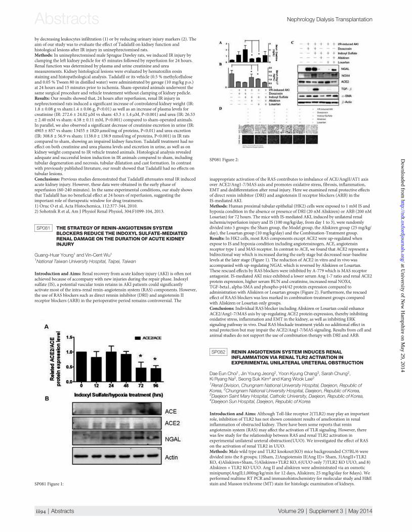
SP081 THE STRATEGYOF RENIN-ANGIOTENSIN SYSTEMBLOCKERS REDUCE THE INDOXYL SULFATE-MEDIATEDRENAL DAMAGE ON THE DURATION OF ACUTE KIDNEYINJURY
Guang-Huar Young1 and Vin-Cent Wu11National Taiwan University Hospital, Taipei, Taiwan
Introduction and Aims: Renal recovery from acute kidney injury (AKI) is often notachieved because of accompany with new injuries during the repair phase. Indoxylsulfate (IS), a potential vascular toxin retains in AKI patients could significantlyactivate most of the intra-renal renin-angiotensin system (RAS) components. However,the use of RAS blockers such as direct rennin inhibitor (DRI) and angiotensin IIreceptor blockers (ARB) in the perioperative period remains controversial. The
inappropriate activation of the RAS contributes to imbalance of ACE/AngII/AT1 axisover ACE2/Ang1-7/MAS axis and promotes oxidative stress, fibrosis, inflammation,EMT and dedifferentation after renal injury. Here we examined renal protective effectsof direct renin inhibitor (DRI) and angiotensin II receptor blockers (ARB) in theIS-mediated AKI.Methods:Human proximal tubular epithelial (HK2) cells were exposed to 1 mM IS andhypoxia condition in the absence or presence of DRI (20 nM Aliskiren) or ARB (200 nMLosartan) for 72 hours. The mice with IS-mediated AKI, induced by unilateral renalischemia/reperfusion injury and IS (100 mg/kg/day, from day 1 to 3), were randomlydivided into 5 groups: the Sham group, the Model group, the Aliskiren group (25 mg/kg/day), the Losartan group (10 mg/kg/day) and the Combination-Treatment group.Results: In HK2 cells, most RAS components except ACE2 were up-regulated whenexpose to IS and hypoxia condition including angiotensinogen, ACE, angiotensinreceptor type 1 andMAS receptor. In contrast to ACE, we found that ACE2 represent abidirectional way which is increased during the early stage but decreased near-baselinelevels at the later stage (Figure 1). The reduction of ACE2 in vitro and in vivo wasaccompanied with up-regulating NGAL which is reversed by Aliskiren or Losartan.These rescued effects by RAS blockers were inhibited by A-779 which is MAS receptorantagonist. IS-mediated AKI mice exhibited a lower serum Ang 1-7 ratio and renal ACE2protein expression, higher serum BUN and creatinine, increased renal NOX4,TGF-beta1, alpha-SMA and phospho-p44/42 protein expression compared toadministration with Aliskiren or Losartan groups (Figure 2). Furthermore, the rescuedeffect of RAAS blockers was less marked in combination-treatment groups comparedwith Aliskiren or Losartan only groups.Conclusions: Individual RAS blocker including Aliskiren or Losartan could enhanceACE2/Ang1-7/MAS axis by up-regulating ACE2 protein expression, thereby inhibitingoxidative stress, inflammation and EMT in the kidney, as well as inhibiting ERKsignaling pathway in vivo. Dual RAS blockade treatment yields no additional effect inrenal protection but may impair the ACE2/Ang1-7/MAS signaling. Results from cell andanimal studies do not support the use of combination therapy with DRI and ARB.
SP082 RENIN ANGIOTENSIN SYSTEM INDUCES RENALINFLAMMATION VIA RENALTLR2 ACTIVATION INEXPERIMENTAL UNILATERAL URETERALOBSTRUCTION
Dae Eun Choi1, Jin Young Jeong2, Yoon Kyung Chang3, Sarah Chung2,Ki Ryang Na2, Seong Suk Kim4 and Kang Wook Lee21Renal Division, Chungnam National University Hospital, Daejeon, Republic ofKorea, 2Chungnam National University Hospital, Daejeon, Republic of Korea,3Daejeon Saint Mary Hospital, Catholic University, Daejeon, Republic of Korea,4Daejeon Sun Hospital, Daejeon, Republic of Korea
Introduction and Aims: Although Toll-like receptor 2(TLR2) may play an importantrole, inhibition of TLR2 has not shown consistent results of amelioration in renalinflammation of obstructed kidney. There have been some reports that reninangiotensin system (RAS) may affect the activation of TLR signaling. However, therewas few study for the relationship between RAS and renal TLR2 activation inexperimental unilateral ureteral obstruction(UUO). We investigated the effect of RASon the activation of renal TLR2 in UUO.Methods: Male wild type and TLR2 knokout(KO) mice backgrounded C57BL/6 weredivided into the 8 groups; 1)Sham, 2)Angiotensin II(Ang II)+ Sham, 3)AngII+TLR2KO, 4)Aliskiren+Sham, 5)Aliskiren+TLR2 KO, 6)UUO only 7)TLR2 KO UUO, and 8)Aliskiren + TLR2 KO UUO. Ang II and aliskiren were administrated via an osmoticminipump(AngII;1,000ng/kg/min for 12 days, Aliskiren; 25 mg/kg/day for 8days). Weperformed realtime RT PCR and immunohistochemistry for molecular study and H&Estain and Masson trichrome (MT) stain for histologic examination of kidneys.SP081 Figure 1:
SP081 Figure 2:
Abstracts Nephrology Dialysis Transplantation
iii | Abstracts Volume 29 | Supplement 3 | May 2014
at University of N
ew H
ampshire on M
ay 29, 2014http://ndt.oxfordjournals.org/
Dow
nloaded from

Results: Ang II increased the renal mRNA expression of TLR2 in wild type mice (p <0.05). Ang II-infused TLR2 KO mice kidney showed significantly lower mRNAexpressions of osteopontin(OPN) and TGF-β compared to those of Ang II-infusedwild type mice. In TLR2 KO UUO kidneys, there were no differences of MCP-1, OPNand TGF-βmRNA expressions and renal histology compared with UUO kidneys ofwild type mice(p < 0.05, p < 0.05, respectively). The renal renin mRNA expression inTLR2 KO UUO was significantly higher than that of UUO kidneys of wild type mice(p < 0.05). Renin inhibition by aliskiren decreased the mRNA expressions of MCP-1,OPN and TGF-β, all of which were upregulated in TLR2 KO UUO kidneys (all, p <0.05). Aliskiren also significantly reduced renal tissue injury score, MT-stained area,and immunostained area of TGF-β in TLR2 KO UUO kidneys (p < 0.05).Conclusions: Although TLR2 inhibition did not attenuate renal inflammation,inhibition of RAS attenuates renal inflammation in TLR2 KO UUO kidneys. It isspeculated that RAS may modulate renal TLR2 activation in experimental unilateralureteral obstruction.
SP083 NAFAMOSTATMESYLATE ATTENUATESISCHEMIA-REPERFUSION INDUCED RENAL INJURYVIA INHIBITION OF APOPTOSIS
Dae Eun Choi1, Jin Young Jeong2, Sarah Chung1, Yoon Kyung Chang3,Ki Ryang Na1, Seong Suk Kim4 and Kang Wook Lee51Renal Division, Department of Internal Medicine, Chungnam National UniversityHospital, Daejeon, Republic of Korea, 2Renal Division, Chungnam NationalUniversity Hospital, Daejeon, Republic of Korea, 3Department of Internal Medicine,Daejeon Saint Mary Hospital, Catholic University, Daejeon, Republic of Korea,4Department of Internal Medicine, Daejeon Sun Hospital, Daejeon, Republic ofKorea, 5Chungnam National University Hospital, Daejeon, Republic of Korea
Introduction and Aims: It has been reported that nafamostat mesylate (NM) inhibitedinflammatory injury via inhibition of complementary activation in ischemic heart, liverand intestine. However, it has been little known that NM inhibits the apoptosis inischemia-reperfusion (IR) injured kidney. We investigated whether NM attenuates IRrenal injury and involves apoptosis inhibition.Methods:We used HK-2 cell and male C57BL/6 mice. C57Bl/6 mice were divided intofour groups; Sham, Nafamostat mesylate(NM,2mg/kg)+Sham, IR injury( IR injury;reperfusion 27 minutes after clamping of both renal artery and vein), and NM+IRinjury. Kidneys were harvested 24hr after IR injury. BUN and serum creatinine(s-Cr)were measured 24 hrs after IR injury. We performed real time RT-PCR andimmunohistochemistry for molecular study and H&E stain and Masson trichrome(MT) stain for histologic examination. For in vitro study, HK-2 cell were divided asinto three groups; Control, IR-HK-2(HK-2 cellswere incubated for 6 hours withmineral paraffin oil for ischemic injury) and IR-HK-2+NM(2nM) groups. Cellsurvival and the magnitude of apoptosis were evaluated.Results: BUN, serum creatinine level and renal tissue injury score in NM+IR injuredmice were significantly lower than those of control IR mice (all, p<0.01). NM treatmentsignificantly improved cell survival in ischemic HK-2 cells (p<0.01). Renal Bax proteinand mRNA expression were significantly increased in IR injured kidneys and ischemicHK-2 cells. NM treatment significantly decreased renal Bax expression (p<0.05). RenalBcl-2 protein and mRNA expression were significantly decreased in IR kidney andischemic HK-2 cells compared to those of sham and control groups. NM treatmentincreased renal Bcl-2 expression in IR injured kidneys and ischemic HK-2 cells(p<0.05). TUNEL positive cells were significantly lower in NM+IR injured kidneys,comparing to control IR injured mice (p<0.05).Conclusions: In conclusion, NM attenuates ischemia-reperfusion renal injury viainhibition of apoptosis.
SP084 MELITTIN INDUCED ACUTE KIDNEY INJURY THROUGHTNF-A/NF-KB/MITOCHONDRIA DEPENDENTAPOPTOTICPATHWAY
Yingying Yang1, Ling Zhang1, Ping Fu1, Yuliang Zhao1 and Xuemei Zhang11West China Hospital of Sichuan University, Chengdu, China
Introduction and Aims:Mechanism of acute kidney injury (AKI) following multiplebee stings is not clear. Melittin, a main component of bee venom, can induce apoptosisin many cell types. We aimed to investigate the mechanism of melittin inducedapoptosis in renal tubular epithelial cells through TNF-α/NF-κB signaling pathway.Methods: AKI model was established by injecting melittin (4.0 μg/g) through thecaudal veins of BALB/c mice (n=60). AKI was defined as creatinine (Cr) increased over2 fold of that in control group. Blood and kidney samples were collected at 0h, 2h, 6h,12h, 24h and 48h. Cr, urea nitrogen and hemoglobin were measured by Abbott i-STATblood-gas analyzer. Creatine kinase was measured by immunochemiluminometricassays. Serum TNF-α was measured by ELISA method. Apoptosis of renal tubule cellswas detected by TUNEL staining and transmission electron microscope. Expression ofcaspase-3, caspase-8, caspase-9, Bcl-2, Bax, and cytochrome c was detected by RT-PCRand/or immunohistochemical method. Expression of IκB and p-IκB was detected byWestern Blot.Results: AKI was diagnosed at 6h after injection of melittin (Cr: 1.1±0.3 mg/dl at 6h vs.0.4±0.1 mg/dl at 0h, p<0.05). Serum TNF-α level increased significantly since 2h after
injection. Apoptosis was detected since 2h, and was most notable at 12h. Western Blotshowed that p-IκB/IκB ratio was significantly decreased in melittin group during 6h to24h, indicating that NF-κB signal pathway was inhibited. RT-PCR showed that theBcl-2/Bax ratio was significantly lower in melittin group than control group, indicatingthat the mitochondrial dependent apoptosis pathway was activated.Conclusions:We successfully established melittin induced AKI in mice. Melittin couldinduce mitochondrial dependent apoptosis in renal tubular epithelial cells throughactivating TNF-α and inhibiting NF-κB signaling pathway.
SP085 ENHANCED NITRIC OXIDE PRODUCTION AMELIORATESACUTE KIDNEY INJURY IN EXPERIMENTAL ARISTOLOCHICACID NEPHROPATHY
Inès Jadot1, Anne-Emilie Declèves1,2, Vanessa Colombaro1, Blanche Martin1,Virginie Voisin1, Isabelle Habsch1, Eric Deprez2, Joëlle Nortier2 and Nathalie Caron11University of Namur, Namur, Belgium, 2Université Libre de Bruxelles, Brussels,Belgium
Introduction and Aims: Aristolochic acid nephropathy (AAN), a progressivetubulointerstitial injury of toxic origin, is characterized by early and transient acutetubular necrosis, followed by fibrosis and tubular atrophy leading to loss of renalfunction. A reduced NO production has been demonstrated, which may disturb theregulation of renal function. The present study tested the hypothesis that L-Argininesupplementation may restore renal function and reduce renal injury after aristolochicacid (AA) intoxication.Methods: C57BL/6J male mice were randomly subjected to ip injection of either sterilesaline solution (control) or AAI (2,5mg/Kg) for 5 days. To determine if the renalAA-induced injuries were related to NO reduction, L-Arginine (L-Arg), a substrate forNO synthase, was supplemented in drinking water.Results:Mice intoxicated with AAI displayed polyuria, significantly increased plasmacreatinine level, proteinuria and FENa+ (P<0.05). As compared to controls, histologicalanalyses showed severe proximal tubular cell necrosis, renal inflammation andincreased oxidative stress (P<0.05). These lesions were associated with a significantreduction of NO bioavailability, measured by the urinary nitrite/nitrate (NOx)excretion. L-Arg supplementation in AA-treated mice significantly improved kidneyfunction, as attested by significant reductions in plasma creatinine level, urine volume,proteinuria and FENa+ (see Table). Moreover, L-Arg treatment resulted in a significantreduction of tubular cell necrosis score, as well as in reduced renal inflammation andoxidative stress along with a normalized NO bioavailability (see Table). These resultssuggest that a preservation of the NO concentration leads to a kidney protection inAAN.
SP085 Renal functional parameters and NOx excretion in experimental groups
Control (n=6) AA (n=8)AA+L-Arg(n=8)
Plasma creatinine level(mg/dl) 0.16±0.01 0.58±0.03* 0.40±0.03&Urine volume(ml/24h) 0.54±0.09 1.70±0.34* 1.01±0.13Urinary protein level(mg/mg CRE) 1.58±0.14 22.10±2.03* 15.11±1.6&Fractional excretion of sodium - FE Na (%) 0.30±0.02 1.43±0.26* 0.82±0.06&Urinary NOx level (μmol/mg CRE) 2.53±0.15 1.49±0.20* 2.71±0.20&
Statistical analysis : ANOVA test. *P<0.05: AA vs Control, &P<0.05 : AA+L-Arg vs AA.
SP084
Nephrology Dialysis Transplantation Abstracts
Volume 29 | Supplement 3 | May 2014 doi:10.1093/ndt/gfu143 | iii
at University of N
ew H
ampshire on M
ay 29, 2014http://ndt.oxfordjournals.org/
Dow
nloaded from

Conclusions: In the present mouse model of acute AAN, NO seems to act as a strongmediator of renal function. Increased NO bioavailability by L-Arg supplementationwas demonstrated beneficial in improving renal injury in AAN.
SP086 CELL CYCLE ARREST IS ASSOCIATEDWITH ACQUIREDRESISTANCE TORECHALLENGE INJURY IN RATSRECOVERED FROM RENAL DAMAGEWITH URANYLACETATE
Takamasa Iwakura1, Tomoyuki Fujikura1, Naro Ohashi1, Hideo Yasuda1and Yoshihide Fujigaki1,21Hamamatsu University School of Medicine, Hamamatsu, Japan, 2TeikyoUniversity School of Medicine, Tokyo, Japan
Introduction and Aims: Rats that recovered from renal damage with uranyl acetate(UA), which mainly injures S3 segment of proximal tubule (PT), can develop resistanceto subsequent UA insult with less morphological and functional damages. Weinvestigated cell cycle status and progression in this acquired resistance model.Methods: Normal rats and rats 14 days after recovering from renal damage, which wasinduced by 1 mg/kg of UA, were injected with 38 mg/kg of lead acetate (a proliferativestimulus) or 4 mg/kg of UA. Isolated tubular cells after the second stimuli wereseparated into PT and distal tubule (DT) cells by Percoll density-gradientcentrifugation. The cell cycle status was analyzed by flow cytometry. G0 and G1 phasecells were separated by Hoechst33342/Pyronin Y method or immunohistochemistryfor Cdt1 (a G1 phase marker).Results: Cell cycle status in PT cells acquiring resistance was comparable to that innormal PT cells. When compared to the control group, delayed G0-G1 transition andless increased S phase cell number were found in response to lead acetate in theresistance group, indicating repressing S phase progression. PT cells stayed at G1 phasefor longer time and increased G2/M phase cell number without increased S phase cellnumber were found in response to UA in resistance group, comparing to controlgroup.Conclusions: G1 arrest was induced in PT cells recovered from renal damage with UAin response to proliferative stimulus or injurious stimulus with UA. Induction ofinhibiting proliferation pathway may closely link to inhibiting cell death pathway in PTcells with the acquired resistance.
SP087 THE PURIFIED MICRONIZED FLAVONOID FRACTION EFFECTIN THE SEPSIS-ASSOCIATED ACUTE KIDNEY INJURY
Carolina Ferreira Vasco1, Mirian Watanabe1, Cassiane Dezoti Fonseca1
and Maria De Fatima Fernandes Vattimo11University of Sao Paulo - School of Nursing, Sao Paulo, Brazil
Introduction and Aims: Sepsis is a severe inflammatory response accompanied by adepression in immunological function which causes multiple organ injury.Sepsis-associated acute kidney injury (AKI) pathophysiology induces renalvasoconstriction, ischemia and acute tubular apoptosis. The concept of renalvasoconstriction and kidney ischemia as a key pathogenic factor is certainly valid forsepsis models. Sepsis-associated AKI may result from global renal hypoperfusion,which is locally mediated by the upregulation of pro-inflammatory cytokines (TNF-αand IL-6), with subsequent tubular cell apoptosis also in distan organs. The purifiedmicronized flavonoid fraction is being mentioned to improve venous tone and reducecapillary hyperpermeability by protecting the microcirculation from inflammatoryprocesses. The aim of this study is to evaluate the effect of the purified micronizedflavonoid fraction (Diosmin) on the creatinine clearance, urinary peroxides and thekidney histology in sepsis associated AKI.Methods: Adult male Wistar rats, weighing 250-300g were used. Renal function(creatinine clearance, Clcr), urinary peroxides (UP, FOX-2), kidney, lung and intestinaltract levels of TNFα and IL-6 (ELISA) and kidney histology were evaluated. Sepsis wasinduced by cecal ligature and puncture (CLP). Groups: Sham (without CLP); Sepsis(CLP); Sepsis+Diosmin (Diosmin 3mg/Kg 30 minutes before CLP).Results: A similar quantitative inflammatory response in distant organs and in thekidneys was observed in CLP animals. CLP induced a decrease in mean arterialpressure, body temperature and creatinine clearance, accompanied by a prominent
increase in UP. These paramethers were significantly changed in the pretreatedDiosmin group. In the kidney histology, loss of brush border was evident in sepsisgroup and it was observed a severe dilation of the tubular lumen after CLP.Conclusions: Sepsis is an injury repair stereotyped response across remote organs.Distant organ inflammation can be considered a significantly mediator ofsepsis-associated AKI as shown in this study. These data provide a new insight aboutDiosmin attenuating injury effect in the sepsis AKI.
SP088 THERAPEUTIC S100A9 BLOCKADE IN EXPERIMENTALGLOMERULONEPHRITIS
Juliana Draibe11Centre for Nephrology, UCL, London, United Kingdom
Introduction and Aims: The S100A9 protein has been identified as one targetmolecule of paquinimod, a quinoline-3-carboximide compound, disrupting bindingthis protein to the proinflammatory receptor toll-like receptor 4. The S100A9 inassociation with S100A8 forms the heterodimer S100A8/S100A9, named calprotectin.Calprotectin is found abundantly in both neutrophils and monocytes and has beendemonstrated to be upregulated in patients with vasculitis ANCA positive (Pepper et al2013, Kidney International). Moreover, our group demonstrated that S100A9knock-out mice have significantly less glomerular disease than wild type counterpartsusing the nephrotoxic nephritis (NTN) model of immune mediatedglomerulonephritis (Pepper et al submitted). The aim of this study was to assess theefficacy of paquinimod in prevention and treatment of murine NTN model usinghistological, urinary and serum markersMethods: 21 C57BL/6 mice had NTN induced. Animals were pre-immunized withsheep IgG at day -5, followed by intravenous injection of sheep nephrotoxic serum atday 0. Paquinimod (25mg/kg), dissolved in drinking water, was started at day 0(prevention group) or day 2 (treatment group). Mice were sacrificed 8 days later. 24hours prior the sacrifice, mice were placed in individual metabolic cages and urine wascollected. Proteinuria was quantified using the sulphosalicylic acid assay. Creatinine,
SP087 Renal function, inflammatory response. α p<0.05 vs SHAM, β vs Sepsis.
SHAM Sepsis Sepsis+Diosmin
Cr Cl (ml/min) 0.60±0.01 0.20±0.06α 0.35±0.03βUP (nmol/g Cr) 3.5±0.7 13.9±5.1α 7.2±3.1βTBARS (nmol/g Cr) 4.4±2.2 13.0±3.4α 7.5±2.8βIL−6 intestine 11.9±1.6 14.2±3.1 11.7±4.5IL−6 kidney 13.3±2.0 19.4±5.8 21.5±9.1IL−6 lung 00±00 3.9±3.2α 0.7±0.8TNF−α intestine 4.2±0.9 4.3±2.5 1.3±0.9βTNF−α kidney 9.2±2.7 9.7±1.4 7.9±1.3TNF−α lung 0.2±0.3 0.2±0.1 00±00
SP088 Figure 1: Immunohistochemistry of renal calprotectin expression
SP088 Figure 2: Calprotectin score in control and treated groups
Abstracts Nephrology Dialysis Transplantation
iii | Abstracts Volume 29 | Supplement 3 | May 2014
at University of N
ew H
ampshire on M
ay 29, 2014http://ndt.oxfordjournals.org/
Dow
nloaded from

urea, ALT and albumin were measured in the serum. Kidneys were removed, fixed informalin and embedded in paraffin for periodic acid-Schiff staining andimmunohistochemical staining for infiltrating macrophages and calprotectinexpression. Histological analysis included enumerating the percentages of crescentsand degree of glomerular thrombosis on the PAS stained sections.Results: By day 8, 4 mice had died; 2 from the control group, 1 from the preventiongroup and 1 from the treatment group. In relation to proteinuria, Creatinine, Urea,ALT or albumin we could not find any statiscally significant differences between the 3groups. Analyzing the renal histology in the PAS staining, we could not find anysignificant differences in the percentages of crescents or the thrombosis score, but wecould see higher levels in the prevention group.We found an increase of thecalprotectin expression in the prevention group that was statistically significantcomparing to control group (p: 0.05) and slight increase in the treatment group (notstatiscally different). In the macrophages staining we could not find any significantdifferences in the expression of MAC2 antibody between the three groups, but again aslight increase in the groups that we administered paquinimod.Conclusions: Our results show that using a high dosing of paquinimod we did notsuccessfully prevent or treat mice with nephrotoxic nephritis as assessed by levels ofglomerular thrombosis, serum creatinine and urea, number of crescents and degree ofproteinuria. Interestingly, an increase in glomerular macrophages and calprotectinexpression was found in the paquinimod treated cohorts, in keeping with previousreports of temporary augmentation of inflammation during early paquinimodtreatment. One explanation maybe the use of LPS in this model in association with aS100A9 inhibition, which may have resulted in an increase of S100A8 expression, andloss of S100A9 regulation of S100A8 and a subsequent higher influx of inflammatoryrenal cells.
SP089 EVALUATION OF THE ANTIOXIDANTAND RENOPROTECTIVEEFFECTS OF ELLAGIC ACID ON ISCHEMIA / REPERFUSIONINDUCED NEPHROPATHY IN RATS
Yas¸Ar Yıldırım1, Özlem Aba1, Zülfükar Yılmaz1, Ali Kemal Kadiroglu1,Mehmet Emin Yılmaz1, Mesut Gül1, Aydın Ketani1 and Leyla Çolpan11Dicle University, Faculty of Medicine, Diyarbakar, Turkey
Introduction and Aims: Renal ischemia-reperfusion injury (IRI) is one of theimportant cause of acute kidney injury (AKI). Reactive oxygen species andinflammatory cytokines play major role in the pathogenesis of IRI. Ellagic acid (EA), aphenolic compound, have shown to exert antioxidants, anti-inflammatory,anticarcinogenic, antihyperlipidemic effects. We aim to evaluate, the effect of EllagicAcid on Renal Ischemia / Reperfusion induced nephropathy in rats.Methods: Twenty-eight male Sprague-Dawley rats were divided into four groups;control, control + EA, IR, and IR + EA. EA (85 mg/kg, perorally) was administered 60
min prior to the ischemia. Rats were unilaterally nephrectomized and subjected to 45min of renal pedicle occlusion followed by 60 min of reperfusion. Both groups weresubsequently studied by renal function tests, oxidant and antioxidant parameters, andkidney histology.Results: Serum/kidney TAC, NO and Paraoxonase levels were significantly higher,while serum urea and creatinine, serum/kidney MDA and TOS were significantly lowerin IR + EA group compared to IR group (p<0.05). Renal function tests, oxidant andantioxidant parameters of the groups are presented in Table 1. Histopathologicexamination revealed that less deterioration in glomerular structure, atrophic structure,and diffuse hydropic degeneration in the proximal and distal tubules in IR+EA groupcompared to IR group (score 2 vs. score 3, respectively, Figure 1, 2).Conclusions: Ellagic acid contributed to the amelioration of renalischemia-reperfusion injury by reducing oxidative stres and preventing histologicalinjuries.
SP090 POLYMYXIN B TOXICITY IN LLC−PK1 CELLS IS MEDIATEDBY THE HEME OXYGENASE 1 ENZYME
Luciana Barros de Moura Neiva1, Fernanda Teixeira Borges2, CassianeDezoti Fonseca1, Mirian Watanabe1 and Maria De Fatima Fernandes Vattimo11University of Sao Paulo, Sao Paulo, Brazil, 2Federal University of Sao Paulo,Sao Paulo, Brazil
Introduction and Aims: In the acute kidney injury, the mechanisms of defense act as“protector genes”, as the protein heat shock 32 (HSP 32) known as heme oxygenase 1(HO 1). The polymyxin B (PMB) is a nephrotoxic antimicrobial. The aim of this studywas to distinguish the role of the HO 1 enzyme in the PMB toxicity in LLC−PK1 cells.Methods: The cells were submitted to the following treatments: Control (CTL− 0μM);PMB (375μM); PMB +Hemin (25μM of hemin, one hour before PMB); PMB + ZnPP(10μM of ZnPP, one hour before PMB). All groups were evaluated in 72 hours. Thefollowing parameters were analysed: cellular viability, apoptosis, lactate dehydrogenase(LDH), lipid peroxidation (MDA), genic expression of HO 1 (RT−PCR)and nitricoxide (NO, Griess).Results: PMB decreased the cellular viability and increased LDH and apoptosis(p<0.05) Hemin improved viability and reduced apoptosis (p<0.05). No changes wereobserved in MDA and HO-1 genic expression. Additionally, PMB itself or with Heminor ZnPP increased nitric oxide (NO) (p<0.05).Conclusions: The PMB was confirmed as a cytotoxic drug by increasing apoptosis, celldamage and reducing viability. Its association with Hemin or ZnPP improved cellularviability by reducing apoptosis, confirming the heme system role associated with NO inthis model of toxicity.
SP091 CONDITIONEDMEDIUM FROMMESENCHYMAL STEMCELLS PROTECTS HUMAN PROXIMALTUBULAR CELLSLESIONS BY LPS
Jéssica Suller Garcia1, Andreia Silva de Oliveira1, Marcelo Andery Naves1,Fernanda Teixeira Borges1 and Nestor Schor11Escola Paulista de Medicina, Federal University of São Paulo (UNIFESP),São Paulo, Brazil
Introduction and Aims: Recent studies emphasize the contribution of mesenchymalstem cells (MSCs) in the regeneration related to acute kidney injury (AKI). MSCsmitigate the damage and / or accelerate the repair and participate in theimmunomodulation processes, probably by paracrine pathways. This protection
SP089 Table 1. Results of renal function tests, oxidant and antioxidant parameters
ParametersControl(n=7)
Control + EA(n=7) IR(n=7) IR + EA(n=7)
SerumUrea (mg/dl) 43,5±7,41 40,85±5,87 108,71±5,87 68,14±15,44Creatinine (mg/dl) 0,60±0,06 0,50±0,06 a 1,04±0,12 0,78±0,11MDA (mmol/ml) 34,42±12,28 15,85±3,07 a 107,14±40,92 58,28±11,22bTOS (µmol /L) 60,86±49,77 22,03±5,94 a 331,06±99,34 194,08±41,57bTAC (mmol /L) 0,76±0,32 1,48±0,35 a 3,25±4,76 11,87±2,72bNO (µM) 4,57±2,07 60,42±10,73 a 29,85±18,09 140,14±138,36bParaoxonase (U/L) 8,76±4,56 16,98±4,46 a 36,01±17,42 60,68±11,93bKidney TissueMDA (mmol/ml) 117,57±13,18 60,87±14,33 a 185,14±7,08 83,71±10,98bTOS (µmol /L) 213,56±59,60 113,57±16,63 a 398,63±109,76 233,06±54,36bTAC (mmol /L) 8,40±6,86 35,41±14,39 a 6,17±4,44 34,04±22,11bNO (µM) 35,57±11,90 60,42±10,73 a 89,00±47,03 203,57±100,03bParaoxonase (U/L) 27,25±8,88 38,82±8,58 a 44,62±19,03 74,47±9,87b
Note: MDA: malondialdehit; TOS: total oxidant status; TAC: total antioxidant capacity;NO: nitric oxidea P<0.05 vs control group, b P<0.05 vs IR group
SP089 Figure 1:
SP089 Figure 2:
SP090 PMB toxicity and HO-1. α p<0.05 vs Control; β vs PMB.
Groups/n Control(8) PMB(9) PMB+Hemin(7) PMB+ZnPP(7)
Viability(%) 81±5 40±2α 55±1αβ 53±3αβApoptosis(%) 8±2 36±3α 22±1αβ 24±2αβLDH(%) 1.7±0.1 22.6±1.7α 21.3±0.7α 22.9±1.2αNO(µM/mg protein) 0.6±0.1 1.4±0.3 2.9±0.3αβ 2.0±0.1α
Nephrology Dialysis Transplantation Abstracts
Volume 29 | Supplement 3 | May 2014 doi:10.1093/ndt/gfu143 | iii
at University of N
ew H
ampshire on M
ay 29, 2014http://ndt.oxfordjournals.org/
Dow
nloaded from

involves the conditioned medium (CM) that contains soluble factors or microvesicles(MVs). We studied the paracrine effect of MSCs in vitro models of AKI induced by LPSin culture.Methods: Human proximal tubular cells (HK2) cells were cultured and treated for24-96 h with LPS (100mg/ml), with or without CM from MSCs. The CM was alsopreviously treated with LPS (100mg/ml) or Gentamicin (GENTA) (2 mM) for 48 hbefore the experiments and it was employed in frozen form (CMc). Cell proliferationwas evaluated by MTT assay.Results:We observed an increase in cell proliferation in the CM+LPS group comparedwith the group treated only with LPS (0.12 ± 0.02 DO vs 0.10 ± 0.02 DO, p < 0.05), andthe major proliferation happened with the CMc+LPS (0.16 ± 0.04 DO, p < 0.05). Whencomparing the groups that received CM stimulated previously it was observed higherproliferation in CM (with GENTA)+LPS (0.16 ± 0.05 DO, p < 0.05) than in MC (withLPS)+LPS (0.09 ± 0.02 DO, p < 0.05). The latter result was very similar to the LPSgroup. Comparing only the different CM with HK2 cells in culture, it was measuredgreater proliferation in MC (with LPS) (0.27 ± 0.07 DO, p < 0.05) than with CM andCMc+GENTA (0.21 ± 0.03 DO vs. 0.20 ± 0.06 DO, p < 0.05). MC (with GENTA)group showed the lowest proliferation compared with others (0.17 ± 0.02 DO, p <0.05).Conclusions: These preliminary results suggested that CM from MSCs exerts aprotective effect by stimulating cell proliferation during the treatment with LPS. Also, itis possible to “conditioned” the CM and stimulate the higher production of thepotential factors produced by MSCs in culture. Moreover, results may indicate that it ispossible to minimize AKI induced by LPS without introducing stem cells but only withtheir CMs, avoiding potential harmful effects of cell therapy. It is clear that morestudies are needed to enable the use of CMs stimulated and frozen.
SP092 INVESTIGATING RENAL HANDLING OF HEPCIDIN: IS THEREA ROLE FOR THEMEGALIN RECEPTOR?
Rachel P. L. Van Swelm1,1, Jack F. M. Wetzels1, Vivienne G. M. Verweij1, CobyM. M. Laarakkers1, Jeanne C. L. M. Pertijs1, Dorine W Swinkels1and Rosalinde Masereeuw1
1Radboudumc, Nijmegen, The Netherlands
Introduction and Aims: Hepcidin is an iron regulating hormone. Recent studiessuggest that urine hepcidin may protect against acute kidney injury after coronaryartery bypass surgery by attenuating tubular injury. However, renal handling ofhepcidin is unknown.Methods: A single dose of 10 µg human hepcidin-25 (hhep25) was injectedintraperitoneally in C57Bl/6 mice and urine, plasma and tissue were collected after 1hor 24h.Results: Hhep25 was measured in plasma at 1h after injection, but was undetectable24h after injection. Presence of hhep25 in plasma led to a compensatory decrease inendogenous hepcidin-1 concentration in plasma (from 5.6±1.4 to 1.4±0.5 nM, p<0.05)and a 10-fold reduction in hepatic hepcidin-1 mRNA expression level (p<0.05)compared to control, showing that hhep25 is active in mice. In urine, hhep25 waspresent 1h after injection and the amount of excreted hhep25 increased over 24h,demonstrating glomerular filtration. Immunohistochemistry revealed clear hhep25staining in the proximal tubules at 1h after injection, which was not present at 24h afterinjection. Detection of the hhep25 isoforms hhep22 and hhep20 in urine, but not inplasma, shows that hhep25 degradation takes place in the kidney. Administration ofgelofusin 5 min prior to hhep25 injection resulted in increased urinary excretion ofhhep25 (from 2.4±1.6 to 10.4±5.9 pmol, p<0.05) and its isoforms (hhep22: from 4.8±3.1 to 16.7±7.1pmol, p<0.01; hhep20: from 1.0±0.3 to 2.1±0.7 pmol, p<0.05),suggesting proximal tubular reabsorption via megalin.Conclusions: Hhep25 is excreted in urine via glomerular filtration, after which it canbe degraded and/or reabsorbed via megalin.
SP093 BIOMARKERS OF TRANSITION FROMCYCLOSPORINE-INDUCED RENAL DYSFUNCTION TONEPHROTOXICITY
José Sereno1, Paulo Rodrigues-Santos2, Helena Vala3, Petronila Rocha-Pereira4,João Fernandes1, Alice Santos-Silva5, Frederico Teixeira1 and Flávio Reis61Faculty of Medicine, University of Coimbra, Coimbra, Portugal, 2Institute ofImmunology, Faculty of Medicine, University of Coimbra, Coimbra, Portugal, 3Esavand Educational, Technologies and Health Study Center, Polytechnic Institute ofViseu, Viseu, Portugal, 4Research Centre for Health Sciences, Beira InteriorUniversity, Covilhã, Portugal, 5Faculty of Pharmacy - University of Porto, Porto,Portugal, 6Faculty of Medicine - University of Coimbra, Coimbra, Portugal
Introduction and Aims: Calcineurin inhibitors, in particular Cyclosporin A (CsA),remain the cornerstone of immunosuppressive regimens in many transplantationcentres worldwide, regardless of drug-induced nephrotoxicity. The pathogenesis ofCsA-induced nephropathy remains to be fully elucidated, but it seems to be affected bythe duration of drug exposure. This experimental animal study aimed to clarify thepathways involved in short and long-term CsA-induced nephrotoxicity and theputative biomarkers of transition from cyclosporine-induced renal dysfunction tonephrotoxicity.
Methods: The study comprised 24 male Wistar rats, divided in two models: Short- andLong-term treatments (3 and 9 weeks, respectively). Each model included two ratgroups (n=6 each), receiving orally: Control group - vehicle; CsA group - 5 mg/Kg BW/day. Renal function was assessed on serum, urine and kidney tissue samples, throughcreatinine, BUN, TBARs and NGAL measures, including clearances. Renal tissue wasalso used to evaluate the gene expression (qRT-PCR) profile of proliferation/fibrosismarkers (TGF-β1, PCNA, Ki67, mTOR, TP53 and NF-κβ), as wells as to determine theprotein expression by immunohistochemistry of CTGF, KIM-1, mTOR, PCNA, NF-κβand TGF-β1. Hematoxilin & eosin, periodic acid of schiff and masson’s trichromestaining were used to evaluate glomerular, tubular and vascular kidney lesions. Statisticalanalysis: ANOVA and Bonferroni post hoc test; p<0.05 was considered significant.Results: In short-term treatment, creatinine and BUN levels increased and clearancesdecreased, accompanied by GFR reduction, but without kidney lesions. Short-term CsAexposure induced PCNA, TGF-β1, NF-κβ and TP53 kidney mRNA up-regulation. Inlong-term exposure, renal dysfunction (BUN and creatinine clearances) wasaccompanied by glomerular and tubulointerstitial lesions, associated with remarkablemTOR and Mki67 up-regulation, but without urine and serum NGAL correlation.Moreover, immunohistochemical analysis showed that lesions and expression profile ofsome proteins were positively associated.Conclusions: CsA-induced nephrotoxicity is aggravated over time and the resultsindicate that distinct mechanisms and biomarkers are involved in regulating short orlong-term toxicity. Functional impairment starts earlier but it is aggravated with time,while renal lesions only appeared after the long-term exposure, accompanied withsignificant mRNA up-regulation of Mki67 and mTOR. These findings reinforce therationale for the early substitution of CsA by less nephrotoxic agents, with themTORinhibitors as a validated choice, in order to prevent chronic CsA-inducednephrotoxicity.
SP094 CAFFEIC ACID PHENETHYL ESTER PROTECTS AGAINSTAMPHOTERICIN B INDUCED NEPHROTOXICITY IN RATMODEL
Atila Altuntas1, Haci Ramazan Yilmaz2, Aysegül Altuntas3, Efkan Uz4,Murat Demir1, Alparslan Gökcimen1, Dilek Senol Bayram1, Oguzhan Aksu4and Mehmet Tugrul Sezer51Süleyman Demirel University School of Medicine, Isparta, Turkey, 2MevlanaUniversity School of Medicine, Konya, Turkey, 3Isparta State Hospital, Isparta,Turkey, 4Süleyman Demirel University School of Medicine, Isparta, Turkey,5Süleyman Demirel University School of Medicine Department of Internal Medicine,Division of Nephrology, Isparta, Turkey
Introduction and Aims: The present study was conducted to investigate whethercaffeic acid phenethyl ester (CAPE), an active component of propolis extract, has aprotective effect on amphotericin B induced nephrotoxicity in rat models.Methods:Male Wistar-Albino rats were randomly divided into four groups: (I) controlgroup (n = 10); (II) CAPE group (n = 9) received 10 µmol/kg CAPE intraperitoneally(i.p.); (III) amphotericin B group (n = 7) received one dose of 50 mg/kg amphotericinB; and (IV) amphotericin B plus CAPE group (n = 7) received 10 µmol/kg CAPE i.p.and one dose of 50 mg/kg amphotericin B. CAPE started one day before theadministration of amphotericin B and continued for 7 days. The left kidney wasevaluated histopathologically for nephrotoxicity. Levels of malondialdehyde (MDA),nitric oxide (NO), enzyme activities including catalase (CAT) and superoxidedismutase (SOD) were measured in the right kidney.Results:Histopatological damage was prominent in the amphotericin B groupcompared to controls, and the severity of damage was lowered by CAPEadministration. The activity of SOD, MDA, and NO levels increased and catalaseactivity decreased in the amphotericin B group compared to the control group(P=0.0001, P=0.003, P=0.0001, P=0.0001 respectively). Amphotericin B plus CAPEtreatment caused a significant decrease in MDA, NO levels, and SOD activity (P=0.04,P=0.002, P=0.0001 respectively) and caused an increase in CAT activity compared withamphotericin B treatment alone (P=0.005).Conclusions: Amphotericin B toxicity remains high despite developments in drugformulations. The main strategies are based on prevention. The role of oxidative stressin amphotericin B toxicity is clear. Therefore, administration of CAPE seems to be analternative agent for the management of amphotericin B toxicity. Further clinicalstudies are necessary for confirmation of these positive effects in clinical settings.
SP095 SIRT2 REGULATES LIPOPOLYSACCHARIDE-INDUCE RENALINFLAMMATION
Kyung Hee Yang1, Yu Jin Jung1, Dal Kim1, Ae Sin Lee1, Sik Lee1, KyungPyo Kang1, Sung Kwang Park1 and Won Kim1
1Chonbuk National University Medical School, Jeonju, Republic of Korea
Introduction and Aims: SIRT2 an NAD-dependent histone deacetylase, playsimportant roles in genomic instability, carcinogenesis and genomic instability.However, its role in renal inflammatory injury has not yet been demonstrated.Methods: In this study, we explored the expression pattern of CXCL-2 and CCL-2 inkidney tissues from sirt2-/-and sirt2+/+ mice and in MPT cell lines after treatment withlipopolysaccharide.
Abstracts Nephrology Dialysis Transplantation
iii | Abstracts Volume 29 | Supplement 3 | May 2014
at University of N
ew H
ampshire on M
ay 29, 2014http://ndt.oxfordjournals.org/
Dow
nloaded from

Results: CXCL-2 and CCL-2 was significantly down-regulated at both the mRNA andprotein levels in kidney of sirt2-/- mice than sirt2+/+ mice after lipopolysaccharide.CXCL-2 and CCL-2 expression was decreased in MPT cells treated with theSIRT2-siRNA compared with the blank control. Overexpression of SIRT2 usingadenovirus significantly increased the expression of CXCL-2 and CCL-2 inlipopolysaccharide-treated MPT cells. Moreover, down-regulation of SIRT2 in thekidney increased the acetylation of p65 of nuclear factor kappa- B.Conclusions: Decrease of SIRT2 was associated with increase of renal proinflammatorycytokine CXCL-2 and CCL-2 and regulation of SIRT2 might be an important target forrenal inflammatory injury.
SP096 INFUSION OF RECOMBINANT IL-6 IN HEALTHY HUMANSELEVATES PLASMA NGAL CONCENTRATIONSWITHOUTAREDUCTION IN RENAL FUNCTION
Naushad A. Junglee1,2, Catrin R. Searell2, Mahdi M. Jibani2 and JamieH. Macdonald1
1Bangor University, Bangor, United Kingdom, 2Ysbyty Gwynedd, Bangor, UnitedKingdom
Introduction and Aims: Interleukin-6 (IL-6) is believed to be an important cytokinein acute kidney injury (AKI) and may be both a marker and mediator of AKI. Plasmaconcentrations of IL-6 correlate with poor outcomes. We determined whether IL-6 perse was able to influence an AKI biomarker (NGAL) and kidney function in healthyhumans.Methods: A three-hour rhIL-6 infusion was administered to six males at a rate of 5 μg/h. Plasma IL-6, NGAL, creatinine, and cystatin C concentrations were measured at 0h,0.5h, 3h and post-infusion at 24h and 48h. Tympanic temperature was recorded at 0hand 3h.
Results: IL-6 concentrations were 0.7 {0.6, 1.2} {interquartile range} pg/ml at 0h andpeaked at 3h to 159.7 {114.6, 186.7} pg/ml (figure 1). By 48 hours this fell to 8.2 {6.8,13.0} pg/ml (main effect of time, p = 0.007). Plasma NGAL concentrations were 24.3(13.2) ng/ml at 0h and peaked at the end of the infusion at 3h to 70.6 (49.3) ng/ml(figure 2). At 48 hours, this fell to 31.7 (16.5) ng/ml (main effect of time, p = 0.025).Plasma creatinine and cystatin C concentrations were unchanged. Tympanictemperature rose from 36.9 (0.2) 0C to 37.5 (0.6) 0C at 0h and 3h, respectively (p =0.046). Plasma NGAL was positively correlated with tympanic temperature (r = 0.945,p = 0.004).Conclusions: Although we achieved IL-6 concentrations found in clinical models ofAKI, elevations in plasma NGAL were below the range typically associated with AKIand no changes to renal function were evident. This suggests IL-6 per se is notresponsible for AKI or kidney dysfunction but other physiological aberrations areneeded.
SP097 N-ACETYLCYSTEINE EFFECTIVELY AMELIORATE HEATSTROKE INDUCED ACUTE KIDNEY INJURY
Chia-Chao Wu1, Chun-Chi Chen2, Kuo-Cheng Lu3 and Yuh-Feng Lin41Division of Nephrology, Department of Medicine, Tri-Service General Hospital,Taipei, Taiwan, 2Division of Nephrology, Department of Medicine, Tri-ServiceGeneral Hospital, National Defense Medical Center, Taipei, Taiwan, 3Division ofNephrology, Department of Medicine, Cardinal Tien Hospital, School of Medicine,Fu Jen Catholic University, Taipei, Taiwan, 4Division of Nephrology, Department ofMedicine, Shuang Ho Hospital, Graduate Institute of Clinical Medicine, TaipeiMedical University, Taipei, Taiwan
Introduction and Aims: The heat-related illness has become more prevalent andcontributed to increased morbidity and mortality in the world with global warming.Heat stroke (HS) is the most severe and potentially fetal heat-related illness. However,specific and effective therapeutic strategies are not yet available to date. Heat stress isknown to generate ROS which play a central role in disease process leading to multipleorgans damage including acute kidney injury (AKI). We assessed the efficacy ofN-acetylcysteine (NAC), a thiol-containing free radical scavenger and antioxidant,therapy for HS induced AKI.Methods: Adult male Sprague-Dawley rats, weight between 325 ± 15 g (age 9~10weeks) exposed to a Ta of 40°C under anesthesia in pre-warming chamber (withrelative humidity of 55%) for 60 mins were performed to induce experimental HS. Therats are randomly allocated into four groups. Two experimental HS groups of ratspre-treated with either saline, or NAC and another two control groups of rats treatedidentically kept at room temperature of 24.0°C used as normothermic controls. OralNAC, 10mg/mL, in drinking water was used. All physiological and biochemicalvariables are measured during the observation. Disease severity was verified by withserum and urine metabolic profiles and with renal histopathology. The expression ofcytokines and oxidative stress markers, cell apoptosis, and the associated mechanismswere also determined.Results:HS rat treated with NAC displayed a better survival rate and thehemodynamics were more stable including body temperature, mean arterial pressureand heart rates. NAC also significantly ameliorate reduce severity of AKI based onbiochemical (levels of blood urea nitrogen, creatinine) and histopathlogical evidence.The NGAL-positive cells and TUNEL-positive apoptotic cells in the kidney were alsosignificantly reduced in theNAC-treated HS rat. Oxidative stresses markers (AdvancedOxidation Protein Products, AOPP and Malondialdehyde, MDA) in the serum weresignificantly reduced in NAC -treated HS rat. Cytokines studies ndicated that NACsignificantly modulate serum proinflammatory, and anti-inflammatory cytokines. Theeffect on other organs revealed similar pattern as kidney.Conclusions:Our studies have demonstrated NAC administration significantly attenuatedorgan damage and enhanced survival in HS rats. These protective effects may be associatedwith its anti-inflammatory capacity and anti-oxidant activity. Our study suggests that NACcould be potentially therapeutic for clinical HS induced AKI in the future.
SP098 EXERCISE AND CALORIE RESTRICTION ATTENUATESCISPLATIN-INDUCED ACUTE KIDNEY INJURY
Gabriel R Estrela1, Frederick Wasinski1, Rafael Pereira1, Denise Malheiros2, NielsO.S. Camara2 and Ronaldo C. Araujo11Federal University of São Paulo, São Paulo, Brazil, 2University of São Paulo, SãoPaulo, Brazil
Introduction and Aims: Cisplatin treatment has been adopted in somechemotherapies; however, this drug can induce acute kidney injury due its ability tonegatively affect renal function. Inflammation and apoptosis is the general cause ofcisplatin-inducing acute kidney injury. Several works showed that exercise and calorierestriction are two good tools to modulate inflammatory response, diminishingapoptosis and expression levels of pro-inflammatory cytokines. Its well estabilishedthat those interventions can revert the inflammation in cardiovascular disease, obesity,diabetes and many others.Methods: To examine the role of exercise and calorie restriction in cisplatinnephrotoxicity C57/Bl6 mice were separated in 5 different groups: Cisplatin treatmentgroup that was submmited to 20mg/kg of cisplatin; Exercise group that was submmited
SP096 Figure 1: Effect of rhiL-6 infusion upon plasma IL-6 concentrations. Data aremedians (thick lines) and ranges. Open circles and asterixes are outliers. There was asignificant effect of time (P=0.007). #, difference compared to 0 hours sample point.
SP096 Figure 2: Effect of rhiL-6 infusion upon plasma NGAL concentrations. Data aremeans and standard deviations. Dotted line indicates upper limit of normal range forplasma NGAL (106 ng/ml). There was a significant effect of time (P=0.025). #,difference compared to 0 h sample point.
Nephrology Dialysis Transplantation Abstracts
Volume 29 | Supplement 3 | May 2014 doi:10.1093/ndt/gfu143 | iii
at University of N
ew H
ampshire on M
ay 29, 2014http://ndt.oxfordjournals.org/
Dow
nloaded from

to swimming training and treated with 20mg/kg of cisplatin; Calorie Restriction group,that was submmited to 30% of food restriction and treated with 20mg/kg of cisplatin;Exercise + Calorie Restriction group that was submmited to swimming training plus30% of food restriction and was treated with 20mg/kg of cisplatin: and Control groupthat received saline 0.9%.Results: Cisplatin treatment causes body weight loss and diminishes food intake, thecalorie restriction and exercise + calorie restriction groups were able to reverse this sideeffect. Moreover cisplatin administration leads to renal dysfunction augmentingcreatinine and urea levels, all three treatments were capable to maintain creatininelevels at basal state and only the exercise group was not able to prevent the higher levelsof urea. Additionally those treatments can attenuates the acute tubular necrosis andmRNA expression of pro-inflammatory cytokines levels.Conclusions: Our data suggest that exercise and calorie restriction arenon-pharmacological interventions to prevent the nephrotoxicity caused by cisplatinadministration.
SP099 EFFECTOF XANTHINE OXIDASE INHIBITORS (XOI) IN RATSSUBMITTED TO LPS
Maria Fatima Ramos1, Clevia dos Santos Passos2, Clara Versolato Razvickas2,Fernanda Borges2, Milene Ormanji2 and Nestor Schor21UNIFESP, Sâo Paulo, Brazil, 2UNIFESP, São Paulo, Brazil
Introduction and Aims: The systemic inflammatory response of sepsis is marked withthe production of reactive oxygen species, follow to instability components pro- andantioxidant. The antioxidant system is composed among others by glutathione(intracellular) and uric acid (intravascular). The latter is one of the most importantcirculating antioxidant. The higher consumption of antioxidants is observed in severesepsis with multiple organ failure. Thus, it was studied the role of the xanthine oxidaseinhibitors XOi (allopurinol and febuxostat) in the LPS model of sepsis in rats madehyperuricemic by oxonic acid (OxAc) administration.Methods:Mean arterial pressure (MAP) was measured by tail method and uric acid(UrAc) by uricase method. The LPS was administered i.p. (10 mg/Kg) every 24 hoursuntil the 3rd day. XOi were given by gavage each 24 hours for 3 days. Allopurinol(ALLO,2 mg/ kg) and febuxostat (Febux,1 mg / kg ) were used in an equivalent dose asin clinical use . To increase UrAc it was administered OxAc by gavage (750mg/Kg/day)during 5 days. UrAc measurements were done at baseline and at 6Th day. The animalswere divided into 9 groups (n = 6): 1-Control, 2-ALLO treated animals, 3-Febuxtreated, 4-LPS group, 5-LPS+ALLO, 6-LPS+Febux, 7-OxAc treated animals, 8-OxAc+LPS+ALLO and 9-OxAc+LPS+Febux.Data were evaluated by Student's t test andsurvival curves (Chi square) considering the value of p ≥ 0.05. The data were reportedas mean and standard deviation.Results: ALLO administration with LPS treatment caused a significant increases in themortality , inducing death in 28 of 34 animals (82%) , while the same did not occurredwith LPS+ Febux ( 11/17; 65% ) or LPS alone (10 /16; 63%) , p<0.05. Administration ofOxAc after 5 days increased the UrAc in the plasma from 2.98 ± 0.17 to 3.76 ± 0.59 mg/dl(p= 0,015) and also induced MAP to rise from 125 ± 8 to 149 ± 29 mmHg (p<0.02).Surprisingly no mortality was observed in the group OxAc+LPS+Febux (p<0.02). Therewas a significant lower number of deaths when we associated OxAc +LPS+ALLO in sepsis
(12 /18; 67%) compared with LPS+ALLO (28/ 34; 82%). It was also interesting that AcOx+LPS+ALLO (67%) vs. LPS+ALLO (82%), p= 0.08, did not differed in the mortality,despite the tendency of AcOx to protect agains ALLO in this protocol.Conclusions: In this study the administration of ALLO in LPS treated animals, decreasedsignificantly the chance of survival. This effect was not observed with Febux. On the otherhand elevation of plasma UrAc by the OxAc protected the animals from the LPS. Thus,high plasma UrAc, by its potential antioxidant capacity, decreases the effects of LPS andthe use of OxAc, increasing the blood uric acid, elevate the chance of survival. Morestudies are needed to elucidate if the administration of the xanthine oxidase inhibitors canworsen the sepsis and to evaluate different effects among XOi (ALLO vs. Febux) in thisexperimental model.
SP100 PROTECTIVE EFFECTOF MITOCHONDRIA-TARGETEDANTIOXIDANTS IN AN ACUTE BACTERIAL INFECTION
Egor Plotnikov1, Maria Morosanova2, Irina Pevzner2, Ljubava Zorova3,Vasily Manskikh2, Maxim Skulachev4, Vladimir Skulachev1 and Dmitry Zorov11Lomonosov Moscow State University, A. N. Belozersky Institute ofPhysico-Chemical Biology, Moscow, Russian Federation, 2Lomonosov MoscowState University, Faculty of Bioengineering and Bioinformatics, Moscow, RussianFederation, 3Lomonosov Moscow State University, International Laser Center,Moscow, Russian Federation, 4Institute of Mitoengineering, Lomonosov MoscowState University, Moscow, Russian Federation
Introduction and Aims: The main approach to treat acute pyelonephritis is antibiotictherapy. However, the pathology is accompanied by inflammation and oxidative stressphenomena that can also be a target for intervention when direct antibacterialmeasures are impossible or inefficient. In our study, in vitro and in vivo models ofexperimental pyelonephritis were used to define the role of mitochondria in thispathology and to find a way to alleviate the kidney damage.Methods:We used an in vivo experimental model of APN where bacteria areintroduced in the bladder of rat. The inoculum (5 mL per kg, 1 × 10⁸ cfu/mL of ratfecal bacterial composition) was injected slowly to avoid any leakage into the bladder.On the second day after the injection, blood samples were taken, and kidneys wereexcised for the mitochondria isolation, determination of MDA in the tissue, Westernblotting, and histopathological examination. Inflammation Modeling in Vitro. Renalcells and leukocytes (neutrophils) were cocultivated for 24 or 48 h with bacterial lysateor LPS. Cell death (both necrotic and apoptotic) rate was evaluated using Annexin-VFITC Kit (Invitrogen).Results: In APN, we found an excessive ROS generation strongly depending on theactivities of neutrophils, monocytes, and mesangial cells migrating to the primaryinflammatory loci. This traffic results in elevation of the levels and activities ofmyeloperoxidase that persists after transformation to chronic type. After activationinitiated by interaction with bacterial products, leukocytes secrete ROS, NO, andTNFα. The suppression of leukocytic NAD(P)H oxidation yields a dramatic decreaseof ROS within renal cells, which may serve as an argument to support the idea thatleukocytes are specifically responsible for excessive ROS production in thepyelonephritic kidney. A number of agents involved in different signaling mechanismswere tested for their ability to alleviate oxidative stress in renal cells. We found that theantioxidant Trolox reduced oxidative stress-related phenomena, apparently bydiminishing transfer of ROS from leukocytes to kidney cells. Protective effects ofmitochondria-targeted antioxidants (SkQ1, SkQR1) and of an inhibitor of GSK-3β (Li+) may be explained by the targeted depletion of mitochondrial ROS and theprevention of the mitochondrial permeability transition, respectively. Oxidative stressobserved in pyelonephritic kidney was accompanied by a reduced level ofmitochondrial B-cell lymphoma 2 (Bcl-2). Importantly, renal cell death and animalmortality were both alleviated by mitochondria-targeted antioxidant 10(60-plastoquinonyl) decylrhodamine 19 (SkQR1).Conclusions:We emphasize that, in a majority of pathological steps, prodeath andprosurvival signaling is related to mitochondrial function. Mitochondria-targetedantioxidants appear to be effective antipyelonephritic drugs with high potency toprevent renal dysfunction in the cases where direct antibacterial measures are restricted(in children, pregnant women, individuals with primary immunodeficiency, etc.) orinefficient (in cases of antibioticresistant bacterial strains).
SP101 THE PROTECTIVE EFFECTOF THE PURIFIED MICRONIZEDFLAVONOID FRACTION IN THE SEPSIS-ASSOCIATED ACUTEKIDNEY INJURY (AKI)
Carolina Ferreira Pinto1, Mirian Watanabe1, Cassiane Dezoti Fonseca1and MariaFatima Vattimo11University of Sao Paulo, Sao Paulo, Brazil
Introduction and Aims: Sepsis is characterized by a severe inflammatory responseaccompanied by depression in immunological function which causes multiple organinjury. Sepsis-associated AKI pathophysiology changes from renal vasoconstriction,ischemia and acute tubular apoptosis. The concept of renal vasoconstriction andkidney ischemia as a key pathogenic factor is certainly valid for sepsis models.Sepsis-associated AKI may result from global renal hypoperfusion, which is locallymediated by the upregulation of pro-inflammatory cytokines (TNF-α and IL-6), with
SP098 Figure 2:
SP098 Figure 1:
Abstracts Nephrology Dialysis Transplantation
iii | Abstracts Volume 29 | Supplement 3 | May 2014
at University of N
ew H
ampshire on M
ay 29, 2014http://ndt.oxfordjournals.org/
Dow
nloaded from

subsequent tubular cell apoptosis also in distan organs. The purified micronizedflavonoid fraction is being mentioned to improve venous tone and reduce capillaryhyperpermeability by protecting the microcirculation from inflammatory processes.The aim of this study is to evaluate the effect of the purified micronized flavonoidfraction (Diosmin) on the creatinine clearance, urinary peroxides and the kidneyhistology in sepsis associated AKI.Methods: Adult male Wistar rats, weighing 250-300g were used. Renal function(creatinine clearance, Clcr), urinary peroxides (UP, FOX-2), kidney, lung and intestinaltract of levels of TNFα and IL-6 (ELISA) and kidney histology were evaluated. Sepsiswas induced by cecal ligature and puncture (CLP). Groups: Sham (without CLP);Sepsis (CLP); Sepsis+Diosmin (Diosmin 3mg/Kg 30 minutes before CLP).
Results: A similar quantitative inflammatory response in distant organs and in thekidneys was observed in CLP animals. CLP induced a decrease in mean arterialpressure, body temperature and creatinine clearance, accompanied by a prominentincrease in UP. These paramethers were significantly changed in the pretreatedDiosmin group. In the kidney histology, loss of brush border was evident in sepsisgroup and it was observed a severe dilation of the tubular lumen after CLP.Conclusions: Sepsis is an injury repair stereotyped response across remote organs.Distant organ inflammation can be considered a significantly mediator ofsepsis-associated AKI. These data provide a new insight about Diosmin attenuatingeffect in the sepsis AKI.
Nephrology Dialysis Transplantation Abstracts
Volume 29 | Supplement 3 | May 2014 doi:10.1093/ndt/gfu143 | iii
at University of N
ew H
ampshire on M
ay 29, 2014http://ndt.oxfordjournals.org/
Dow
nloaded from