Diffuse Brain Injury Induces Acute Post-Traumatic Sleep
10
Diffuse Brain Injury Induces Acute Post-Traumatic Sleep Rachel K. Rowe 1,2,4,8 , Martin Striz 5 , Adam D. Bachstetter 9 , Linda J. Van Eldik 4,8,9 , Kevin D. Donohue 6 , Bruce F. O’Hara 5,8 , Jonathan Lifshitz 1,2,3,4,7,8 * 1 Barrow Neurological Institute at Phoenix Children’s Hospital, Phoenix, Arizona, United States of America, 2 Department of Child Health, University of Arizona College of Medicine–Phoenix, Phoenix, Arizona, United States of America, 3 Phoenix Veteran Affairs Healthcare System, Phoenix, Arizona, United States of America, 4 Department of Anatomy and Neurobiology, College of Medicine, University of Kentucky, Lexington, Kentucky, United States of America, 5 Department of Biology, College of Arts and Sciences, University of Kentucky, Lexington, Kentucky, United States of America, 6 Department of Electrical and Computer Engineering, College of Engineering, University of Kentucky, Lexington, Kentucky, United States of America, 7 Department of Physical Medicine & Rehabilitation, University of Kentucky College of Medicine, Lexington, Kentucky, United States of America, 8 Spinal Cord and Brain Injury Research Center (SCoBIRC), University of Kentucky College of Medicine, Lexington, Kentucky, United States of America, 9 Sanders-Brown Center on Aging, University of Kentucky College of Medicine, Lexington, Kentucky, United States of America Abstract Objective: Clinical observations report excessive sleepiness immediately following traumatic brain injury (TBI); however, there is a lack of experimental evidence to support or refute the benefit of sleep following a brain injury. The aim of this study is to investigate acute post-traumatic sleep. Methods: Sham, mild or moderate diffuse TBI was induced by midline fluid percussion injury (mFPI) in male C57BL/6J mice at 9:00 or 21:00 to evaluate injury-induced sleep behavior at sleep and wake onset, respectively. Sleep profiles were measured post-injury using a non-invasive, piezoelectric cage system. In separate cohorts of mice, inflammatory cytokines in the neocortex were quantified by immunoassay, and microglial activation was visualized by immunohistochemistry. Results: Immediately after diffuse TBI, quantitative measures of sleep were characterized by a significant increase in sleep (.50%) for the first 6 hours post-injury, resulting from increases in sleep bout length, compared to sham. Acute post- traumatic sleep increased significantly independent of injury severity and time of injury (9:00 vs 21:00). The pro- inflammatory cytokine IL-1b increased in brain-injured mice compared to sham over the first 9 hours post-injury. Iba-1 positive microglia were evident in brain-injured cortex at 6 hours post-injury. Conclusion: Post-traumatic sleep occurs for up to 6 hours after diffuse brain injury in the mouse regardless of injury severity or time of day. The temporal profile of secondary injury cascades may be driving the significant increase in post-traumatic sleep and contribute to the natural course of recovery through cellular repair. Citation: Rowe RK, Striz M, Bachstetter AD, Van Eldik LJ, Donohue KD, et al. (2013) Diffuse Brain Injury Induces Acute Post-Traumatic Sleep. PLoS ONE 8(12): e82507. doi:10.1371/journal.pone.0082507 Editor: Fernando de Castro, Hospital Nacional de Paraple ´jicos – SESCAM, Spain Received April 26, 2013; Accepted October 24, 2013; Published December 26, 2013 Copyright: ß 2013 Rowe et al. This is an open-access article distributed under the terms of the Creative Commons Attribution License, which permits unrestricted use, distribution, and reproduction in any medium, provided the original author and source are credited. Funding: Support provided by National Institutes of Health (NIH) grants R21 NS072611 (JL/BFO), R01 NS065052 (JL), R01 NS064247 (LVE), KSCHIRT 10-5A, F32 AG037280 (ADB). RKR was supported by a Research Supplement to Promote Diversity by the National Institute of Neurological Disorders and Stroke (NINDS). The funders had no role in study design, data collection and analysis, decision to publish, or preparation of the manuscript. Competing Interests: Co-authors Bruce O’Hara and Kevin Donohue are principal owners of Signal Solutions, Inc. Signal Solutions manufactures and supports the non-invasive sleep monitoring cages used in these studies. There are no further patents, products in development, or marketed products to declare. This does not alter the authors’ adherence to all the PLOS ONE policies on sharing data and materials. * E-mail: [email protected] Introduction Traumatic brain injury (TBI) is a major cause of death and disability throughout the world with little pharmacological treatment for the individuals who suffer from lifelong neurological morbidities associated with TBI. Brain injury can lead to both short and long-term impairment, including cognitive [1], and behavioral [2] deficits as well as increasing the risk for developing neurodegenerative disease [3] and/or psychiatric disorders [4]. Little can be done to mitigate the mechanical disruption associated with the primary insult and the biochemical cascades initiated shortly after the time of injury can impair physiological function and ultimately worsen long-term outcome [5]. Clinical studies have provided evidence to support the hypothesis that brain injury contributes to chronic sleep distur- bances as well as leads to excessive daytime sleepiness [6–9]. Far less is known about the acute relationship between TBI and sleep. Immediately after TBI, secondary injury mechanisms may impair physiological functions associated with the homeostatic regulation of sleep. For example, secondary injury processes result in glia activation and initiation of marked inflammatory responses. Injury-induced inflammation is mediated by the production of cytokines, such as pro-inflammatory interleukin-1 beta (IL-1b), which can have dual roles as sleep regulatory substances (SRSs) [10,11]. Elevated cytokine signaling has been observed across experimental models and human TBI, highlighting their involve- ment in pathological and reparative processes triggered by injury [12–15]. However, cytokines which are SRSs can also modulate sleep-wake behavior, primarily enhancing sleep by acting on sleep circuits of the brain [11,16]. PLOS ONE | www.plosone.org 1 December 2013 | Volume 8 | Issue 12 | e82507
-
Upload
independent -
Category
Documents
-
view
2 -
download
0
Transcript of Diffuse Brain Injury Induces Acute Post-Traumatic Sleep
Diffuse Brain Injury Induces Acute Post-Traumatic SleepRachel K. Rowe1,2,4,8, Martin Striz5, Adam D. Bachstetter9, Linda J. Van Eldik4,8,9, Kevin D. Donohue6,
Bruce F. O’Hara5,8, Jonathan Lifshitz1,2,3,4,7,8*
1 Barrow Neurological Institute at Phoenix Children’s Hospital, Phoenix, Arizona, United States of America, 2 Department of Child Health, University of Arizona College of
Medicine–Phoenix, Phoenix, Arizona, United States of America, 3 Phoenix Veteran Affairs Healthcare System, Phoenix, Arizona, United States of America, 4 Department of
Anatomy and Neurobiology, College of Medicine, University of Kentucky, Lexington, Kentucky, United States of America, 5 Department of Biology, College of Arts and
Sciences, University of Kentucky, Lexington, Kentucky, United States of America, 6 Department of Electrical and Computer Engineering, College of Engineering, University
of Kentucky, Lexington, Kentucky, United States of America, 7 Department of Physical Medicine & Rehabilitation, University of Kentucky College of Medicine, Lexington,
Kentucky, United States of America, 8 Spinal Cord and Brain Injury Research Center (SCoBIRC), University of Kentucky College of Medicine, Lexington, Kentucky, United
States of America, 9 Sanders-Brown Center on Aging, University of Kentucky College of Medicine, Lexington, Kentucky, United States of America
Abstract
Objective: Clinical observations report excessive sleepiness immediately following traumatic brain injury (TBI); however,there is a lack of experimental evidence to support or refute the benefit of sleep following a brain injury. The aim of thisstudy is to investigate acute post-traumatic sleep.
Methods: Sham, mild or moderate diffuse TBI was induced by midline fluid percussion injury (mFPI) in male C57BL/6J miceat 9:00 or 21:00 to evaluate injury-induced sleep behavior at sleep and wake onset, respectively. Sleep profiles weremeasured post-injury using a non-invasive, piezoelectric cage system. In separate cohorts of mice, inflammatory cytokines inthe neocortex were quantified by immunoassay, and microglial activation was visualized by immunohistochemistry.
Results: Immediately after diffuse TBI, quantitative measures of sleep were characterized by a significant increase in sleep(.50%) for the first 6 hours post-injury, resulting from increases in sleep bout length, compared to sham. Acute post-traumatic sleep increased significantly independent of injury severity and time of injury (9:00 vs 21:00). The pro-inflammatory cytokine IL-1b increased in brain-injured mice compared to sham over the first 9 hours post-injury. Iba-1positive microglia were evident in brain-injured cortex at 6 hours post-injury.
Conclusion: Post-traumatic sleep occurs for up to 6 hours after diffuse brain injury in the mouse regardless of injury severityor time of day. The temporal profile of secondary injury cascades may be driving the significant increase in post-traumaticsleep and contribute to the natural course of recovery through cellular repair.
Citation: Rowe RK, Striz M, Bachstetter AD, Van Eldik LJ, Donohue KD, et al. (2013) Diffuse Brain Injury Induces Acute Post-Traumatic Sleep. PLoS ONE 8(12):e82507. doi:10.1371/journal.pone.0082507
Editor: Fernando de Castro, Hospital Nacional de Paraplejicos – SESCAM, Spain
Received April 26, 2013; Accepted October 24, 2013; Published December 26, 2013
Copyright: � 2013 Rowe et al. This is an open-access article distributed under the terms of the Creative Commons Attribution License, which permitsunrestricted use, distribution, and reproduction in any medium, provided the original author and source are credited.
Funding: Support provided by National Institutes of Health (NIH) grants R21 NS072611 (JL/BFO), R01 NS065052 (JL), R01 NS064247 (LVE), KSCHIRT 10-5A, F32AG037280 (ADB). RKR was supported by a Research Supplement to Promote Diversity by the National Institute of Neurological Disorders and Stroke (NINDS). Thefunders had no role in study design, data collection and analysis, decision to publish, or preparation of the manuscript.
Competing Interests: Co-authors Bruce O’Hara and Kevin Donohue are principal owners of Signal Solutions, Inc. Signal Solutions manufactures and supportsthe non-invasive sleep monitoring cages used in these studies. There are no further patents, products in development, or marketed products to declare. This doesnot alter the authors’ adherence to all the PLOS ONE policies on sharing data and materials.
* E-mail: [email protected]
Introduction
Traumatic brain injury (TBI) is a major cause of death and
disability throughout the world with little pharmacological
treatment for the individuals who suffer from lifelong neurological
morbidities associated with TBI. Brain injury can lead to both
short and long-term impairment, including cognitive [1], and
behavioral [2] deficits as well as increasing the risk for developing
neurodegenerative disease [3] and/or psychiatric disorders [4].
Little can be done to mitigate the mechanical disruption associated
with the primary insult and the biochemical cascades initiated
shortly after the time of injury can impair physiological function
and ultimately worsen long-term outcome [5].
Clinical studies have provided evidence to support the
hypothesis that brain injury contributes to chronic sleep distur-
bances as well as leads to excessive daytime sleepiness [6–9]. Far
less is known about the acute relationship between TBI and sleep.
Immediately after TBI, secondary injury mechanisms may impair
physiological functions associated with the homeostatic regulation
of sleep. For example, secondary injury processes result in glia
activation and initiation of marked inflammatory responses.
Injury-induced inflammation is mediated by the production of
cytokines, such as pro-inflammatory interleukin-1 beta (IL-1b),
which can have dual roles as sleep regulatory substances (SRSs)
[10,11]. Elevated cytokine signaling has been observed across
experimental models and human TBI, highlighting their involve-
ment in pathological and reparative processes triggered by injury
[12–15]. However, cytokines which are SRSs can also modulate
sleep-wake behavior, primarily enhancing sleep by acting on sleep
circuits of the brain [11,16].
PLOS ONE | www.plosone.org 1 December 2013 | Volume 8 | Issue 12 | e82507

Secondary injury mechanisms of TBI deplete adenosine-59-
triphosphate (ATP) causing failure of energy-dependent mem-
brane ion pumps, increase reactive oxygen species (ROS), increase
intracellular concentrations of free radicals caused by activation of
lipid peroxidases, as well as increase inflammatory mediating
cytokines [17,18]. A low energy state, high ROS, and the increase
of certain cytokines have all been implicated in increased sleep
propensity and sleep duration [19,20]. Thus, secondary injury
processes following TBI have the potential to induce post-
traumatic sleep. The magnitude and duration of the induction of
post-traumatic sleep are unknown. Further, as these cellular
processes continue, chronic sleep issues may develop.
The biological function of sleep remains controversial, however,
prevailing hypotheses suggest the function of sleep is restorative,
conservative, and adaptive [21,22]. In the absence of sleep,
humans exhibit deficits in attention, memory, learning, and higher
cognitive processes [23]. Sleep is regulated by homeostatic
processes as well as circadian processes [24] such that injury
may disrupt the signaling required to maintain a healthy sleep
profile. If sleep is regenerative in function, then acute post-
traumatic sleep may improve outcome from brain injury. This
study is the first of its kind to investigate acute sleep following
diffuse brain injury.
To characterize acute sleep patterns in brain-injured mice, a
non-invasive sleep monitoring cage system was used to continu-
ously record post-traumatic sleep. The sleep monitoring cage
system allows reliable, immediate and continuous sleep monitoring
by using piezoelectric materials configured as highly-sensitive
pressure detectors incorporated into the bottom of the animal cage
to determine rest-activity based on motion and breathing patterns
[25]. Sleep is discriminated from wake every 2 seconds from
tapered 8 second overlapping windows based on classification
algorithms that exploit the limited mouse sleeping postures and
distinct respiratory patterns consistent with sleep, compared to rest
and activity [25,26]. This technology is also adapted to measure
the polycyclic sleep pattern of mice, accounting for short
interruptions and brief arousals in sleep. The cage system
recognizes short bouts (a single sleep episode) lasting only seconds
as well as extended bouts lasting longer than a minute.
Sleep can complicate the understanding of injury processes
following TBI [9]. The investigation of post-traumatic sleep may
lead to rational interventions to mitigate damage. The present
study was designed to examine injury-induced alterations in acute
sleep following TBI. Clinical observations indicate that patients
report excessive sleepiness immediately following TBI [6]. In view
of these observations, we hypothesized that diffuse brain injury
would induce acute post-traumatic sleep in the mouse. The lack of
biomedical research surrounding the controversial question
whether one should sleep or be frequently awoken immediately
following brain injury adds to the importance of investigating post-
traumatic sleep, specifically in the acute period. In neurosurgical
wards TBI patients are frequently awoken during the first day after
injury to check for possible worsening of their consciousness, a
condition that requires immediate action. In more mild conditions,
the practice of keeping a brain-injured individual awake is
controversial. Here we demonstrate significant increases in acute
sleep post-injury regardless of injury severity or time of day. Post-
traumatic sleep occurs during the same time as increased cortical
expression of SRS cytokines and inflammation.
Methods
Animals and Ethics StatementMale C57BL/6J mice (Harlan Laboratories, Inc., Indianapolis,
IN) were used for all experiments (n = 75). The animals were
housed in a 14 h light/10 h dark cycle at a constant temperature
(23uC62uC) with food and water available ad libitum according to
the Association for Assessment and Accreditation of Laboratory
Animal Care International. Animals were acclimated to their
environment following shipment for at least three days prior to any
experiments. After surgery, animals were evaluated daily for post-
operative care by a physical examination and documentation of
each animal’s condition. All studies were approved by the
University of Kentucky Institutional Animal Care and Use
Committee (IACUC Protocol Number: 2007-0142). All surgery
was performed under isoflurane anesthesia, and efforts were made
to minimize suffering.
Midline Fluid Percussion Injury (mFPI)Adult male C57BL/6J mice (20–24 g) were subjected to midline
fluid percussion injury (mFPI) consistent with methods previously
described [27]. Animal numbers are indicated in the results section
and figure legends for individual studies. Mice were anesthetized
using 5% isoflurane in 100% oxygen for five minutes and the head
of the animal was placed in a stereotaxic frame with continuously
delivered isoflurane at 2.5% via nosecone. While anesthetized, the
animal’s body temperature was maintained using a DeltaphaseHisothermal heating pad (Braintree Scientific Inc., Braintree, MA).
A midline incision was made exposing bregma and lambda, and
fascia was removed from the surface of the skull. A trephine (3 mm
outer diameter) was used for the craniotomy, centered on the
sagittal suture between bregma and lambda without disruption of
the dura. An injury cap prepared from the female portion of a
Luer-Loc needle hub was fixed over the craniotomy using
cyanoacrylate gel and methyl-methacrylate (Hygenic Corp.,
Akron, OH). The incision was sutured at the anterior and
posterior edges and topical Lidocaine ointment was applied. The
injury cap was closed using a Luer-Loc cap and animals were
placed in a heated recovery cage and monitored until ambulatory
before being returned to their sleep cage.
For injury induction 24 hours post-surgery, animals were re-
anesthetized with 5% isoflurane delivered for five minutes. The
cap was removed from the injury-hub assembly and the
craniotomy was visually inspected through the hub. The hub
was then filled with normal saline and attached to the male end of
the fluid percussion device (Custom Design and Fabrication,
Virginia Commonwealth University, Richmond, VA). An injury of
moderate severity (1.2–1.3 atm), mild severity (0.8 atm) or sham
injury was administered by releasing the pendulum onto the fluid-
filled cylinder. Sham-injured animals underwent the same
procedure except the pendulum was not released. Animals were
monitored for the presence of a forearm fencing response, and
righting reflex times were recorded for the injured animals as
indicators of injury severity [28]. The righting reflex time is the
total time from the initial impact until the animal spontaneously
rights itself. The fencing response is a tonic posturing character-
ized by extension and flexion of opposite arms that has been
validated as an overt indicator of injury force magnitude [28]. The
injury hub was removed and the brain was inspected for uniform
herniation and integrity of the dura. Animals in which the dura
was compromised were excluded from all studies as technical
failures. The incision was cleaned using saline and closed using
sutures. Moderate brain-injured animals had righting reflex
recovery times greater than six minutes and a positive fencing
Post-Traumatic Sleep following TBI
PLOS ONE | www.plosone.org 2 December 2013 | Volume 8 | Issue 12 | e82507

response, and mild injured animals had righting reflex times
between two and four minutes and no fencing response. Sham
injured animals recovered within 10 seconds. After spontaneously
righting, animals were placed in a heated recovery cage and
monitored until ambulatory before being returned to their sleep
cage (approximately 5 to 15 minutes). Adequate measures were
taken to minimize pain or discomfort.
Sleep RecordingsThe non-invasive sleep cage system (Signal Solutions, Lexing-
ton, KY) used in this study consisted of 16 separate units that could
simultaneously monitor the sleep and wake states over several
days. The cage system classified sleep and wake behavior based on
methods previously described [25,26]. Each cage unit housed the
mice individually with separate 767 inch walled compartments
and attached food and water structures [25]. The cages had open
bottoms that allowed them to be placed on a base with a
Polyvinylidine Difluoride (PVDF) sensor on the cage floor [25].
The PVDF sensors were coupled to an input differential amplifier
and pressure signals were generated and classified by the non-
invasive high-throughput classifier as motions consistent with
either wake activity or the inactivity associated with sleep [25].
Sleep was characterized primarily by periodic pressure measure-
ments with regular amplitudes, typical of respiration from a still
mouse. In contrast, movements characteristic of wake were both
the absence of the characteristic sleep signal and higher amplitude,
irregular spiking associated with volitional movements. The
piezoelectric signals were classified by the automated sleep scoring
system in two second epochs as ‘‘sleep’’ or ‘‘wake’’. The tapered
segmentation window was advanced every two seconds and
features associated with the characteristics just described were
computed. A linear discriminant classifier based on these features
was applied to assign a binary label given to each point associated
with the center of the window [25]. Data collected from the cage
system were binned over specified time periods (e.g. 5 minutes,
1 hour) using a rolling average of the percent sleep, as well as
binned by length of individual bouts of sleep and the median bout
lengths were calculated.
Mice were acclimated to the cages and sensors were tested for 8
days prior to injury (Figure 1A). The animals were removed from
their home cages for the midline craniotomy surgery and were
placed back into their specific cage following the surgery. The
following day the mice were removed from their home cage and
were subjected to mFPI or sham injury. As soon as the mice were
ambulatory (approximately 5 to 15 minutes), they were returned
to their original sleep cage and the sleep recordings started to
measure sleep continuously for 7 days [25]. Previous validation of
the sleep cages in agreement with human observation resulted in
classification rates of 90% or higher [25]. This sleep cage system
was a valid method for monitoring acute sleep in injured animals
without confounding the injury. Placement of invasive EEG
equipment for recordings can compromise the dura and adds
external weight to the head of the rodent possibly exacerbating the
brain-injury.
A multivariate analysis of variance (ANOVA) was used to
compare the mean percent sleep of brain-injured mice to
uninjured shams over time. Statistical significance was assigned
when p,0.05.
Tissue preparation and cytokine measurementAt selected time points (1, 3, 9, 12, 24, 48, 168 hours) post-
injury or sham operation, mice were given an overdose of sodium
pentobarbital and transcardially perfused with phosphate buffered
saline (PBS) (Figure 1A). Mice were decapitated and the brains
were dissected on ice and snap frozen in liquid nitrogen then
stored at 280uC until used. The protein levels of a panel of
inflammatory cytokines were measured in the neocortex by Meso
Scale Discovery (MSD) multiplex immunoassay (sector imager
2400, Meso Scale Discovery; Gaithersburg Maryland) (Figure 1B)
as previously described [29]. Brain cortex was homogenized using
high shear homogenizer (Omni TH115), in a 1:10 (w/v) of ice-cold
lysis buffer consisting of PBS containing 1 mg/ml Leupeptin,
1 mM PMSF, and 1 mM EDTA. The cortical homogenate was
centrifuged at 14,0006g for 20 minutes at 4uC in a microcen-
trifuge. Fifty microliters of the resulting supernatant was loaded
per well of the custom MSD plate, and IL-1b levels were
determined by MSD assay (Mouse Proinflammatory 7-Plex Ultra-
Sensitive (K15012C)). IL-1b levels in the cortex were normalized
to the total amount of protein in the sample loaded as determined
by BCA Protein Assay (Pierce).
Cytokine levels of IL-1b were compared between uninjured
sham and brain-injured mice. Increases in cytokine levels in the
brain-injured mice were analyzed over a time course using a one-
way ANOVA and selected comparisons were made using the
Bonferroni post hoc test.
Tissue preparation for immunohistochemistryAt 6 hours post-injury or sham operation, mice were given an
overdose of sodium pentobarbital and transcardially perfused with
4% paraformaldehyde after a (PBS) flush. Brains were removed
and placed in 4% paraformaldehyde overnight. Brains were
immersed in serial dilutions (10%, 20%, and 30%) of sucrose for
24 hours each. The brains were removed from the 30% sucrose
and frozen at 220uC. After freezing, brains were sectioned in the
coronal plane at 20 mm, mounted onto glass slides, and stored at
280uC.
IBA-1 immunohistochemistrySlides were removed from 280uC, placed in an oven at 60uC
for approximately 4 hours and then rinsed three times for
5 minutes each in PBS. Next, the slides were incubated in 4%
goat serum blocking solution for 1 hour. The slides were incubated
with the primary antibody (rabbit anti-ionized calcium binding
adaptor molecule 1 (IBA-1), 1:1000, Wako Chemicals 0199-
19741) and stored at room temperature overnight. Slides were
rinsed three times in PBS and the secondary antibody (biotinylated
horse anti-rabbit, 1:250, Vector Laboratories) was added then
slides were incubated on a rocker at room temperature for 1 hour.
The slides were washed in PBS three times for 5 minutes each and
tertiary stain was applied (streptavidin Alexa� Fluor 594, 1:1000,
Jackson Laboratories) and slides were incubated for 1 hour at
room temperature. Lastly, slides were rinsed three times in PBS
and coverslipped with Fluoromount-G anti-fade medium (South-
ern Biotech). The cortex was examined for microglia activation in
response to brain-injury using a Zeiss Axio Scope with attached
digital camera.
Results
Diffuse TBI induces acute post-traumatic sleep in themouse
Immediately after diffuse TBI, mean percent sleep was
significantly increased in brain-injured animals compared to sham
for the first 6 hours post-injury (F(1, 45) = 6.545, p = 0.00007)
(Figure 2A). After 6 hours post-injury, the mean percent sleep of
the injured mice (n = 31) normalized and was indistinguishable
from sleep in the sham (n = 16) through 7 days post-injury (data
not shown). A more detailed analysis was performed by calculating
Post-Traumatic Sleep following TBI
PLOS ONE | www.plosone.org 3 December 2013 | Volume 8 | Issue 12 | e82507
the mean percent sleep over five minute intervals for the first hour
post-injury to examine the increase in sleep observed acutely post-
injury. The mean percent sleep showed a significant time-
dependent increase in sleep over the first hour post-injury
(F(11,495) = 8.22, p,0.0001) (Figure 2B) until maximum sleep
was reached. In addition, analysis over the first hour-post injury
showed a significant group effect (F(1,45) = 37.00, p,0.0001)
(Figure 2B) indicating TBI induced a significant increase in mean
percent sleep compared to the uninjured sham. Increase in acute
post-traumatic sleep in the diffuse brain-injured mouse was
associated with increased median bout lengths of sleep
(Figure 2C). We observed a significant increase in the median
bout length of brain-injured mice compared to sham for the first
4 hours post-injury (F(1,45) = 2.9138, p = 0.032). This increase in
bout length indicated that the increase in mean percent sleep
observed acutely post-injury (Figure 2A) could result from mice
sleeping for longer durations during each bout, as opposed to
sleeping more bouts after diffuse TBI.
Post-traumatic sleep bouts are interrupted by volitionalmovement and arousal similar to uninjured shams
Immediately after diffuse TBI, mean percent sleep was
significantly increased in brain-injured mice compared to unin-
jured sham. To analyze the signals used to classify sleep, the raw
piezoelectric sensor data was extracted and compared between
brain-injured and sham mice within the first hour post-injury.
Uninjured sham mice showed a periodic rhythm associated with
the motion of breathing, approximately 3 Hz, with regular
amplitude typical of sleep in the mouse (Figure 3A). These sleep
bouts were interrupted by higher frequency and amplitude signals
that correspond to movements consistent with wake activity
(Figure 3A). The sleep-wake classifier plotted with the decision
threshold (Figure 3, below raw signal) were classified as sleep
activity above the threshold and as wake activity below the
threshold. In brain-injured mice, sleep activity showed similar
rhythmic breathing classified as sleep (Figure 3B). As in the
uninjured sham, sleep was interrupted by high amplitude and
frequency signals corresponding to volitional movement. Inter-
ruptions of sleep bouts by volitional movement indicate the brain-
injured animals terminate sleep bouts in a similar manner to
uninjured mice, suggesting that brain-injured mice are responsive,
capable of movement, and not in a comatose state of unrespon-
siveness. As shown, sleep bouts in brain-injured mice are longer in
duration than in uninjured mice (Figure 3B).
Increase in acute post-traumatic sleep in the diffusebrain-injured mouse was independent of injury time ofday
Sham or brain injury was administered at transitional time
points (9:00 or 21:00) in the light/dark cycle (Figure 1A). We
observed an increase in the mean percent sleep of brain-injured
mice compared to sham when mice were injured at 9:00, following
the onset of the light cycle (Figure 4A). Brain-injury resulted in a
significant increase in mean percent sleep for the first 3 hours
following injury as compared to the mean percent sleep of sham
(F(1,25) = 15.95, p = 0.0005). Similarly, we observed an increase in
post-traumatic sleep when mice were subjected to injury at 21:00,
following the onset of the dark cycle (Figure 4B). We recorded a
significant increase in mean percent sleep for the first 3 hours
following brain-injury compared to uninjured shams
(F(1,17) = 4.42, p = 0.0506). Regardless of injury time of day,
post-traumatic sleep was increased to comparable levels (45–65%)
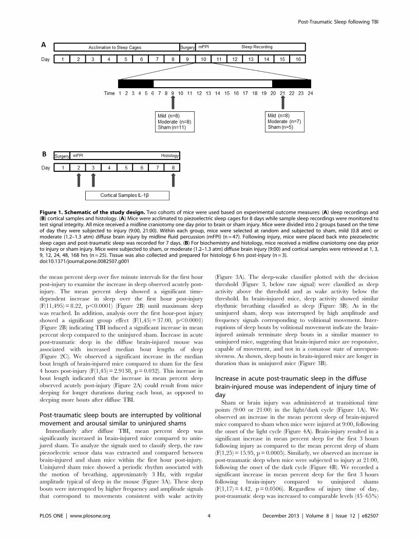
Figure 1. Schematic of the study design. Two cohorts of mice were used based on experimental outcome measures: (A) sleep recordings and(B) cortical samples and histology. (A) Mice were acclimated to piezoelectric sleep cages for 8 days while sample sleep recordings were monitored totest signal integrity. All mice received a midline craniotomy one day prior to brain or sham injury. Mice were divided into 2 groups based on the timeof day they were subjected to injury (9:00, 21:00). Within each group, mice were selected at random and subjected to sham, mild (0.8 atm) ormoderate (1.2–1.3 atm) diffuse brain injury by midline fluid percussion (mFPI) (n = 47). Following injury, mice were placed back into piezoelectricsleep cages and post-traumatic sleep was recorded for 7 days. (B) For biochemistry and histology, mice received a midline craniotomy one day priorto injury or sham injury. Mice were subjected to sham, or moderate (1.2–1.3 atm) diffuse brain injury (9:00) and cortical samples were retrieved at 1, 3,9, 12, 24, 48, 168 hrs (n = 25). Tissue was also collected and prepared for histology 6 hrs post-injury (n = 3).doi:10.1371/journal.pone.0082507.g001
Post-Traumatic Sleep following TBI
PLOS ONE | www.plosone.org 4 December 2013 | Volume 8 | Issue 12 | e82507

and became indistinguishable from sleep in the sham after 3 hours
post-injury. In contrast, diurnal pressures associated with the
change in the light/dark cycle were evident on the mean percent
sleep of uninjured sham animals, as expected. The mean percent
sleep of uninjured sham mice in the 9:00 group was significantly
higher than the mean percent sleep of sham mice in the 21:00
group (F(1,15) = 6.303, p = 0.0240). This finding is representative
of the nocturnal activity of mice.
Increase in acute post-traumatic sleep in the diffusebrain-injured mouse was independent of injury severity
Two levels of experimental injury severity were used to test the
effects of injury severity on post-traumatic sleep. We define injury
severity as mild (0.8 atm) and moderate (1.2–1.3 atm) according to
the righting-reflex and the fencing response (see methods). Post-
traumatic sleep was not significantly different between mild and
moderate brain-injured mice (Figure 5). A significant increase in
post-traumatic sleep was observed acutely following both mild and
moderate injury compared to the uninjured sham
(F(2,44) = 3.4773, p = 0.00037). No significant difference was
found between mild brain-injured mice and moderate brain-
injured mice, indicating the significant increase in post-traumatic
sleep is independent of injury severity.
Secondary injury responses temporally associate with theincrease in acute post-traumatic sleep in the diffusebrain-injured mouse
Following brain injury there is an up-regulation of pro-
inflammatory cytokines [30] that include IL-1b [12,18]. IL-1b is
a cytokine with sleep regulatory substance activity [10,16,31]
which could partially explain post-traumatic sleep. A temporal
profile of IL-1b indicated that cortical levels increased rapidly
following moderate injury as compared to uninjured sham
(Figure 6A). Levels of IL-1b peak at or near 9 hours post-injury
and return to baseline levels by 12 hours post-injury. There was a
significant increase in IL-1b levels in brain-injured animals
compared to sham (F(7,21) = 6.474, p = 0.0004) and selected
comparisons using the Bonferroni post-hoc analysis indicated a
significant increase between sham and brain-injured mice at 1, 3
and 9 hours post-injury.
Microglia morphology, an indicator of microglia activation, was
examined after diffuse brain injury in the mouse using Iba-1
immunohistochemistry. Iba-1 labels all microglia, however,
distinct morphological differences in Iba-1 stained microglia were
observed in brain-injured (mild and moderate injury, 09:00,
Figure 6 C,D) compared to uninjured (Figure 6B) cortex at
6 hours post-injury. Microglia in brain-injured cortex showed
morphologies consistent with activated microglia, including
amoeboid cell bodies with thick, densely labeled processes
(denoted by arrowheads). In contrast, thin, highly ramified
processes of ramified microglia (denoted by arrows) were present
in the uninjured sham cortex.
Figure 2. Diffuse TBI in the mouse disrupts acute post-traumatic sleep parameters compared to uninjured sham. (A)A multivariate ANOVA showed a significant increase in mean percentsleep over the first 6 hours post-injury compared to the uninjured sham(mean 6SEM; sham n = 16; injured n = 31; F(1, 45) = 6.545, p = 0.00007).After 6 hours post-injury, the mean percent sleep of injured micenormalized to sham mean percent sleep levels and remainedcomparable for 7 days post-injury (data not shown). (B) A detailedanalysis of the acute post-traumatic sleep (in the first hour) followingdiffuse TBI indicated a significant time dependent effect on the increasein sleep. A multivariate ANOVA of the rolling average of the meanpercent sleep over 5 min intervals showed post-traumatic sleepsignificantly increased over the first hour post-injury with a significanteffect of time (mean 6SEM; sham n = 16; injured n = 31; F(11,495) = 8.22,p,0.0001) and group (mean 6SEM; sham n = 16; injured n = 31;F(1,45) = 37.00, p,0.0001). Bonferroni post hoc analysis was used (*,p,0.05). (C) Acutely post-injury, the brain-injured mice showed an
increase in median bout length compared to shams. A multivariateANOVA revealed an increase in bout length significant over the first4 hours post-injury (mean 6SEM; sham n = 16; injured n = 31;F(1,45) = 2.9138, p = 0.032). This increase in bout length suggested thatthe increase in mean percent sleep observed acutely post-injury couldresult from mice sleeping for longer durations, as opposed to sleepingmore bouts after diffuse TBI.doi:10.1371/journal.pone.0082507.g002
Post-Traumatic Sleep following TBI
PLOS ONE | www.plosone.org 5 December 2013 | Volume 8 | Issue 12 | e82507

Discussion
Brain injury survivors report varying degrees of sleep distur-
bances [32], however, the contribution of acute post-traumatic
sleep to the injury itself remains unclear. To achieve this long-term
goal, we undertook the present study to measure the acute sleep
response to diffuse TBI, which we term post-traumatic sleep. We
chose to focus on acute sleep post-injury, because sleep itself may
be restorative and aid in the recovery of function following injury.
By non-invasively recording sleep immediately following diffuse
brain injury, we were able to document the induction of post-
traumatic sleep. Altogether, our data, for the first time, support the
hypothesis that diffuse brain injury promotes acute post-traumatic
sleep in the mouse, and secondary injury related cellular processes
coincide with this increase.
Acute sleep measured using novel non-invasive cagesystem
Current sleep research associated with TBI has focused on
chronic sleep disorders in the sequelae of human injury [32–34].
The lack of studies investigating acute sleep following TBI
heightens the importance of studies in this field, because evidence
promoting or disrupting sleep after TBI may change the standard
of care for brain-injury patients. By investigating the role of post-
traumatic sleep, interventions can be developed to mitigate
damage. This study used a non-invasive sleep monitoring cage
system which reliably measures injury-induced alterations in sleep
[25] immediately following injury. Without the surgical proce-
dures required for EEG recordings, we avoided the contraindica-
tions of an electrode in the brain at the time of injury which would
create contusion or cavitation and most importantly allowed for
post-injury sleep to be measured within minutes of the initial
injury. The piezoelectric sensor cages, in conjunction with
computer algorithms, recorded the sleep of each individual mouse
(injury and sham) and created a detailed sleep profile that included
mean percent sleep and median bout length. The cage system
allows for sleep profiles of brain-injured and sham mice to be
measured in exactly the same way. We found that post-traumatic
sleep was significantly increased after brain injury, regardless of
Figure 3. Representative sleep-wake recordings in the first hour post-injury showed sleep bouts interrupted by brief arousal andmovement. Uninjured sham mice showed a periodic rhythm of breathing motion (,3 Hz) with regular amplitude typical of sleep, interrupted byhigh frequency and amplitude signals corresponding to movement consistent with an awake mouse (A). Diffuse brain-injured mice showed similarrhythmic breathing classified as sleep interrupted by frequency and amplitude variations corresponding to movement during interbout intervals ofsleep (B). The red lines represent the raw piezoelectric sensor data over a one minute (top) or 25 second (bottom) interval. The discontinuous blueline indicates the decision classifier over two second intervals to classify sleep activity from wake activity. The broken green line delineates thethreshold (in arbitrary units) to determine sleep activity (above the threshold) from wake activity (below the threshold).doi:10.1371/journal.pone.0082507.g003
Post-Traumatic Sleep following TBI
PLOS ONE | www.plosone.org 6 December 2013 | Volume 8 | Issue 12 | e82507

time of day or injury severity, with longer median sleep bouts
underlying the overall increased post-traumatic sleep.
Despite the limitation of not being able to discriminate stages of
sleep (rapid-eye-movement-sleep, slow wave sleep), the piezoelec-
tric cage system is capable of accurately distinguishing wake from
sleep [26]. While the sleep cage system has been well validated in
distinguishing sleep from wake with comparisons between EEG/
EMG and human observation in normal mice [25], it is possible
that sleep could be over-estimated in TBI mice. If this were true,
we would expect percent sleep times to be greatest in the first hour
after injury and for moderate TBI to show a greater sleep increase
than mild TBI. Since TBI mice were able to make postural
adjustments and voluntary movements that signal wake, post-
traumatic sleep is likely to be sleep, rather than a more severe
condition. While the post-traumatic sleep bears many hallmarks of
normal sleep, we cannot rule out that the increased sleep time is in
part due to non-convulsive seizures with behavioral and temporal
dynamics similar to sleep. We discount non-convulsive status
epilepticus (NCSE) as a component of post-traumatic sleep
because the development of epilepsy following experimental brain
injury does not occur within the first week post-injury [35].
Following severe lateral fluid percussion in rats, EEG recordings
indicated no injury-induced seizure within the first six hours post-
injury [36]. Furthermore, EEG/EMG signal features used to
determine sleep stages in normal mice may be compromised after
brain injury and affect sleep-wake scoring. It should also be noted
EEG/EMG recordings can result in false positive or negative sleep
determination leading to possible error when used as the sole
arbiter of sleep. For the purposes of this study, sleep determination
did not rely fundamentally on EEG/EMG measures, but more
simply is associated with a reversible perceptual disengagement
from the environment, marked by a suspension of voluntary bodily
functions. Ongoing work with the sleep cage system suggests that
signal processing can distinguish REM from NREM sleep based
on more irregular breathing during REM stages. Future work will
determine whether the increases we observed in post-traumatic
sleep arise from increases in REM or NREM sleep, or most likely
from increases in both.
Acute post-traumatic sleep increased regardless of timeof injury
In order to explore the impact of diffuse TBI on natural sleep,
mice were subjected to injury at two time points in their circadian
rhythms. By conducting the injuries at the light/dark transition,
we investigated whether post-traumatic sleep was a result of the
brain injury or an interaction with natural biological tendencies to
sleep. The 9:00 time point followed the onset of the light cycle, a
time when nocturnal mice were expected to sleep. The 21:00 time
point followed the onset of the dark cycle, a time when nocturnal
mice are most active. Sleep patterns of uninjured mice showed
circadian related pressures. Acute post-traumatic sleep significant-
ly increased in comparison to the sleep of uninjured shams
independent of the time of day mice were subjected to injury. This
degree of increase in sleep following TBI is similar to the mean
percent sleep of mice following 6 hours of sleep deprivation [37].
It is possible that the circadian clock itself or its outputs are
dysregulated by TBI [34], which would contribute to injury-
induced sleep being independent of the time at which the injury
Figure 4. Significant increase in post-traumatic sleep isindependent of the time of day of the injury. Mice subjected tomild or moderate injury at 9:00 (A), following the dark/light transitionshowed significant increases in acute post-traumatic sleep compared touninjured sham. A multivariate ANOVA and Bonferroni post-hocanalysis was used (mean 6SEM; sham n = 12; injured n = 17;F(1,25) = 15.95); *, p,0.05). Mice subjected to mild or moderate injuryat 21:00 (B), following the light/dark transition also showed significantincreases in acute post-traumatic sleep compared to sham. Amultivariate ANOVA and Bonferroni post-hoc analysis was used (mean6SEM; sham n = 5; injured n = 14; F(1,17) = 4.42; *, p,0.05). An increasein sleep is observed acutely following TBI and is observed over thecourse of the first 3 hours in injured mice compared to sham. After3 hours, sleep began to normalize in the injured animals and becameindistinguishable from sleep in the sham. Mean percent sleep ofuninjured sham mice in the 9:00 group was significantly higher than themean percent sleep of sham mice in the 21:00 group (F(1,15) = 6.303,p = 0.0240), as expected.doi:10.1371/journal.pone.0082507.g004
Figure 5. The significant increase in post-traumatic sleep isobserved acutely following both mild and moderate injury. Amultivariate ANOVA showed a significant increase in mean percentsleep between injured mice and uninjured shams over the first 6 hourspost-injury with no significant difference between mildly injured micecompared to moderately injured mice (mean 6SEM; sham n = 16; mildn = 16; moderate n = 15; F(2,44) = 3.4773, p = 0.00037).doi:10.1371/journal.pone.0082507.g005
Post-Traumatic Sleep following TBI
PLOS ONE | www.plosone.org 7 December 2013 | Volume 8 | Issue 12 | e82507
occurs. However, immediate permanent pathology is unlikely, as
sleep parameters return to sham levels beyond 6 hours post-injury.
Acute post-traumatic sleep increased independent ofinjury severity
We also examined the relationship between injury severity and
post-traumatic sleep. Severity of the initial injury is considered a
major determining factor for magnitude of secondary injury
processes and outcome following TBI [38], which led to the
hypothesis that injury severity would directly impact post-
traumatic sleep. Contrary to our hypothesis, both mildly and
moderately brain-injured mice showed similar significant increases
in post-traumatic sleep compared to sham values. Even mild injury
significantly increased sleep, which urges continued investigation
into the contribution of post-traumatic sleep to the natural course
of the injury. The possibility exists to induce an even milder injury,
which may have less impact on post-traumatic sleep; however this
may reduce/eliminate all other cellular hallmarks of TBI as well.
Unfortunately, because mildly and moderately injured mice have
comparable increases in acute post-traumatic sleep, the utility of
sleep to serve as a diagnostic biomarker for injury severity is
limited.
Our data also exclude the possibility of an injury-induced coma,
as an extreme manifestation of sleep. Severe TBI can lead to
coma, however, our brain-injured mice exhibit a brief period of
non-responsiveness measured by the suppression of the righting
reflex. Also, the maximum median bout length, measured in
seconds, was 30 seconds, followed by periods of wake activity,
which excludes the possibility of an injury-induced coma since
mice voluntarily woke between sleep bouts. These periods of wake
activity during the interbout interval between sleep activity were
clearly shown by the piezoelectric sensor data.
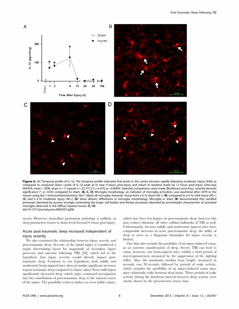
Figure 6. (A) Temporal profile of IL-1b. The temporal profile indicated that levels in the cortex increase rapidly following moderate injury (9:00) ascompared to uninjured sham. Levels of IL-1b peak at or near 9 hours post-injury and return to baseline levels by 12 hours post-injury (One-wayANOVA, mean 6SEM; sham n = 7; injured n = 22; F(7,21) = 6.474; p = 0.0004). Selected comparisons were made (Bonferroni post-hoc), asterisk denotessignificance (*, p,0.05) compared to sham. (B, C, D) Microglia morphology, an indicator of microglia activation, was examined after mFPI in themouse using Iba-1 immunohistochemistry. Iba-1 labels all microglia, however, tissue from a 6 hr sham (406) (B) compared to a 6 hr mild injury (406)(C) and a 6 hr moderate injury (406) (D) show distinct differences in microglia morphology. Microglia in sham (B) demonstrated thin ramifiedprocesses (denoted by arrows) strongly contrasting the larger cell bodies and thicker processes (denoted by arrowheads) characteristic of activatedmicroglia observed in the diffuse injured mouse (C, D).doi:10.1371/journal.pone.0082507.g006
Post-Traumatic Sleep following TBI
PLOS ONE | www.plosone.org 8 December 2013 | Volume 8 | Issue 12 | e82507

Inflammation as a secondary injury mechanismassociated with overall increases in acute post-traumaticsleep
TBI is characterized by two pathological phases: cellular injury
resulting from primary impact and the ensuing secondary injury
mediated by pathological processes [17]. Secondary injury occurs
over time post-injury with a more gradual onset beginning minutes
to hours after impact and contributes to the clinical morbidities
associated with TBI. Post-traumatic sleep in 5 minute intervals
showed that the increase in mean percent sleep over the first hour
post-injury is time dependent (Figure 2B). If the primary impact
solely contributed to post-traumatic sleep, then an immediate
increase in post-traumatic sleep to a maximum level would have
been observed. The secondary injury cascades that play a role in
inducing sleep following diffuse TBI likely include post-traumatic
signaling that activate glia, as evidenced by increased production
of pro-inflammatory cytokines, such as IL-1b, in both animal
models and human head injury patients [18,30].
Activated microglia can contribute to the production of IL-1bafter TBI. Once produced, IL-1b acts locally to affect neuronal
assemblies, altering their functional status, as well as acting on
sleep regulatory circuits [11]. Our group has accumulated
evidence for circuit disruption, dismantling and reorganization
in the diffuse-injured cortex, particularly the whisker sensory
circuit of the rat [39–43]. Microglia likely act as the effectors of
circuit disruption [40] by producing cytokines and as a
consequence influence the functional state of those circuits. The
impact of microglia can then extend beyond local circuits to sleep
regulatory circuits and ultimately induce sleep [10,16,44]. In the
injured mouse brain, these microglial signaling processes that
influence sleep last 6 hours post-injury and remain to be
determined in the human condition.
Our data show that IL-1b was upregulated in the cortex
following diffuse TBI, and previous studies have reported that
humans undergoing IL-1b therapy report excessive sleepiness
[11]. Injections of IL-1b enhance NREM sleep [16] and
application of IL-1b to the somatosensory cortex leads to
enhanced EEG delta wave activity [45]. These data indicate a
mechanistic link between IL-1b and sleep. In the injured cortex,
IL-1b continues to increase, peaking at or near 9 hours post-injury
and returning to sham levels by 12 hours post-injury (Figure 6A),
similar to the increase in mean percent sleep in brain-injured mice.
Collectively, these data suggest that post-traumatic sleep may
involve inflammatory mediated processes and the upregulation of
pro-inflammatory cytokines that can act as sleep regulatory
substances. Our immunohistological staining indicated activation
of microglia in the cortex of mild and moderate brain-injured mice
compared to uninjured sham at 6 hours post-injury, coinciding
with elevated cytokine levels in moderate injury and the end of
post-traumatic sleep. Activated microglia produce pro-inflamma-
tory cytokines, including those with dual roles as SRSs (IL-1b, IL-
6, TNFa) [46], as indicated by pharmacological inhibition of
microglia reducing levels of pro-inflammatory cytokines. The
infiltration and activation of microglia may be a potential source of
sleep regulatory factors in the injured brain [40,47,48], regardless
of injury severity. We argue that increases in sleep following TBI
may result from the inflammatory response associated with the
secondary injury in which elevated cytokine levels are associated
with activation of microglia after brain injury. Future studies are
needed to examine the mechanistic relationship between changes
in cytokine levels and sleep.
Conclusion
The current study demonstrated that acute sleep was increased
following diffuse TBI, and injury-induced cellular cascades may
contribute to this increase. The increase in sleep was independent
of time of day that the injury occurred and the injury severity.
Increases in median bout length contributed to the overall increase
in sleep observed post-injury. Further studies need to determine
the cellular benefit or detriment of acute post-traumatic sleep on
recovery following TBI (and other neurological conditions) by
disrupting acute sleep. Understanding the role of acute post-
traumatic sleep on outcome can begin to answer the controversial
question, ‘‘Should one sleep, be frequently awoken or left
uninterrupted after a concussion?’’
Acknowledgments
The authors wish to thank Kelley Hall for contributing to the surgical
preparation of the diffuse brain-injured mice. Thank you to Jordan
Harrison and Jenna Ziebell for their assistance on the immunohistochem-
istry and technological techniques.
Author Contributions
Conceived and designed the experiments: RR JL. Performed the
experiments: RR AB. Analyzed the data: RR AB MS KD BO.
Contributed reagents/materials/analysis tools: JL KD BO LV. Wrote
the paper: RR JL. Manuscript edits and revisions: BO AB KD LV.
References
1. Albensi BC, Janigro D (2003) Traumatic brain injury and its effects on synaptic
plasticity. Brain Inj 17: 653–663.
2. Yeates KO, Taylor HG, Wade SL, Drotar D, Stancin T, et al. (2002) A
prospective study of short- and long-term neuropsychological outcomes after
traumatic brain injury in children. Neuropsychology 16: 514–523.
3. Masel BE, DeWitt DS (2010) Traumatic brain injury: a disease process, not an
event. J Neurotrauma 27: 1529–1540.
4. Arciniegas DB, Topkoff J, Silver JM (2000) Neuropsychiatric Aspects of
Traumatic Brain Injury. Curr Treat Options Neurol 2: 169–186.
5. Gentleman D (1999) Improving outcome after traumatic brain injury–progress
and challenges. Br Med Bull 55: 910–926.
6. Castriotta RJ, Wilde MC, Lai JM, Atanasov S, Masel BE, et al. (2007)
Prevalence and consequences of sleep disorders in traumatic brain injury. J Clin
Sleep Med 3: 349–356.
7. Baumann CR, Werth E, Stocker R, Ludwig S, Bassetti CL (2007) Sleep-wake
disturbances 6 months after traumatic brain injury: a prospective study. Brain
130: 1873–1883.
8. Kempf J, Werth E, Kaiser PR, Bassetti CL, Baumann CR (2010) Sleep-wake
disturbances 3 years after traumatic brain injury. J Neurol Neurosurg Psychiatry
81: 1402–1405.
9. Baumann CR (2012) Traumatic Brain Injury and Disturbed Sleep and
Wakefulness. Neuromolecular medicine 14(3): 205–212.
10. Krueger JM, Majde JA (1995) Cytokines and Sleep. International Archives of
Allergy and Immunology 106: 97–100.
11. Krueger JM, Rector DM, Churchill L (2007) Sleep and Cytokines. Sleep Med
Clin 2: 161–169.
12. Frugier T, Morganti-Kossmann MC, O’Reilly D, McLean CA (2010) In situ
detection of inflammatory mediators in post mortem human brain tissue after
traumatic injury. J Neurotrauma 27: 497–507.
13. Morganti-Kossmann MC, Rancan M, Otto VI, Stahel PF, Kossmann T (2001)
Role of cerebral inflammation after traumatic brain injury: a revisited concept.
Shock 16: 165–177.
14. Semple BD, Bye N, Rancan M, Ziebell JM, Morganti-Kossmann MC (2010)
Role of CCL2 (MCP-1) in traumatic brain injury (TBI): evidence from severe
TBI patients and CCL22/2 mice. J Cereb Blood Flow Metab 30: 769–782.
15. Ziebell JM, Morganti-Kossmann MC (2010) Involvement of pro- and anti-
inflammatory cytokines and chemokines in the pathophysiology of traumatic
brain injury. Neurotherapeutics 7: 22–30.
16. Krueger JM, Obal F, Fang JD, Kubota T, Taishi P (2001) The role of cytokines
in physiological sleep regulation. Role of Neural Plasticity in Chemical
Intolerance 933: 211–221.
17. Werner C, Engelhard K (2007) Pathophysiology of traumatic brain injury.
Br J Anaesth 99: 4–9.
Post-Traumatic Sleep following TBI
PLOS ONE | www.plosone.org 9 December 2013 | Volume 8 | Issue 12 | e82507

18. Fan L, Young PR, Barone FC, Feuerstein GZ, Smith DH, et al. (1995)
Experimental brain injury induces expression of interleukin-1 beta mRNA in therat brain. Brain Res Mol Brain Res 30: 125–130.
19. Dworak M, McCarley RW, Kim T, Kalinchuk AV, Basheer R (2010) Sleep and
brain energy levels: ATP changes during sleep. The Journal of neuroscience: theofficial journal of the Society for Neuroscience 30: 9007–9016.
20. Chikahisa S, Sei H (2011) The role of ATP in sleep regulation. Frontiers inneurology 2: 87.
21. Chokroverty S (2010) Overview of sleep & sleep disorders. Indian J Med Res
131: 126–140.22. Tononi G, Cirelli C (2006) Sleep function and synaptic homeostasis. Sleep Med
Rev 10: 49–62.23. McCoy JG, Strecker RE (2011) The cognitive cost of sleep lost. Neurobiol Learn
Mem 96: 564–582.24. Borbely AA, Achermann P (1999) Sleep homeostasis and models of sleep
regulation. Journal of biological rhythms 14: 557–568.
25. Donohue KD, Medonza DC, Crane ER, O’Hara BF (2008) Assessment of anon-invasive high-throughput classifier for behaviours associated with sleep and
wake in mice. Biomed Eng Online 7: 14.26. Flores AE, Flores JE, Deshpande H, Picazo JA, Xie XS, et al. (2007) Pattern
recognition of sleep in rodents using piezoelectric signals generated by gross
body movements. IEEE Trans Biomed Eng 54: 225–233.27. Lifshitz J (2008) Fluid Percussion Injury. In: J Chen ZX, Xu X-M, Zhang J,
editors. Animal Models of Acute Neurological Injuries. Totowa, NJ: TheHumana Press Inc.
28. Hosseini AH, Lifshitz J (2009) Brain injury forces of moderate magnitude elicitthe fencing response. Med Sci Sports Exerc 41: 1687–1697.
29. Bachstetter AD, Xing B, de Almeida L, Dimayuga ER, Watterson DM, et al.
(2011) Microglial p38alpha MAPK is a key regulator of proinflammatorycytokine up-regulation induced by toll-like receptor (TLR) ligands or beta-
amyloid (Abeta). J Neuroinflammation 8: 79.30. Helmy A, Carpenter KL, Menon DK, Pickard JD, Hutchinson PJ (2011) The
cytokine response to human traumatic brain injury: temporal profiles and
evidence for cerebral parenchymal production. J Cereb Blood Flow Metab 31:658–670.
31. Fang J, Wang Y, Krueger JM (1998) Effects of interleukin-1 beta on sleep aremediated by the type I receptor. Am J Physiol 274: R655–660.
32. Orff HJ, Ayalon L, Drummond SP (2009) Traumatic brain injury and sleepdisturbance: a review of current research. J Head Trauma Rehabil 24: 155–165.
33. Verma A, Anand V, Verma NP (2007) Sleep disorders in chronic traumatic
brain injury. J Clin Sleep Med 3: 357–362.
34. Boone DR, Sell SL, Micci MA, Crookshanks JM, Parsley M, et al. (2012)
Traumatic brain injury-induced dysregulation of the circadian clock. PLoS One7: e46204.
35. D’Ambrosio R, Fairbanks JP, Fender JS, Born DE, Doyle DL, et al. (2004) Post-
traumatic epilepsy following fluid percussion injury in the rat. Brain 127: 304–314.
36. Kharatishvili I, Nissinen JP, McIntosh TK, Pitkanen A (2006) A model ofposttraumatic epilepsy induced by lateral fluid-percussion brain injury in rats.
Neuroscience 140: 685–697.
37. Huber R, Deboer T, Tobler I (2000) Effects of sleep deprivation on sleep andsleep EEG in three mouse strains: empirical data and simulations. Brain research
857: 8–19.38. Curry P, Viernes D, Sharma D (2011) Perioperative management of traumatic
brain injury. International journal of critical illness and injury science 1: 27–35.39. Hall KD, Lifshitz J (2010) Diffuse traumatic brain injury initially attenuates and
later expands activation of the rat somatosensory whisker circuit concomitant
with neuroplastic responses. Brain Res 1323: 161–173.40. Cao T, Thomas TC, Ziebell JM, Pauly JR, Lifshitz J (2012) Morphological and
genetic activation of microglia after diffuse traumatic brain injury in the rat.Neuroscience 225: 65–75.
41. Lifshitz J, Lisembee AM (2012) Neurodegeneration in the somatosensory cortex
after experimental diffuse brain injury. Brain Struct Funct 217: 49–61.42. McNamara KC, Lisembee AM, Lifshitz J (2010) The whisker nuisance task
identifies a late-onset, persistent sensory sensitivity in diffuse brain-injured rats.J Neurotrauma 27: 695–706.
43. Lifshitz J, Kelley BJ, Povlishock JT (2007) Perisomatic thalamic axotomy afterdiffuse traumatic brain injury is associated with atrophy rather than cell death.
J Neuropathol Exp Neurol 66: 218–229.
44. Krueger JM (2008) The Role of Cytokines in Sleep Regulation. CurrentPharmaceutical Design 14: 3408–3416.
45. Yasuda T, Yoshida H, Garcia-Garcia F, Kay D, Krueger JM (2005) Interleukin-1beta has a role in cerebral cortical state-dependent electroencephalographic
slow-wave activity. Sleep 28: 177–184.
46. Wisor JP, Schmidt MA, Clegern WC (2011) Evidence for neuroinflammatoryand microglial changes in the cerebral response to sleep loss. Sleep 34: 261–272.
47. Alder J, Fujioka W, Lifshitz J, Crockett DP, Thakker-Varia S (2011) Lateral fluidpercussion: model of traumatic brain injury in mice. J Vis Exp. doi: 10.3791/
3063.48. Jin X, Ishii H, Bai Z, Itokazu T, Yamashita T (2012) Temporal changes in cell
marker expression and cellular infiltration in a controlled cortical impact model
in adult male C57BL/6 mice. PLoS One 7: e41892.
Post-Traumatic Sleep following TBI
PLOS ONE | www.plosone.org 10 December 2013 | Volume 8 | Issue 12 | e82507