Sleep quality and sleep disruptive factors in adult patients in ...

Role of Corticosterone on Sleep Homeostasis Induced byREM Sleep Deprivation in RatsRicardo Borges Machado*, Sergio Tufik, Deborah Suchecki
Psychobiology Department, Universidade Federal de Sao Paulo, Sao Paulo, Brazil
Abstract
Sleep is regulated by humoral and homeostatic processes. If on one hand chronic elevation of stress hormones impair sleep,on the other hand, rapid eye movement (REM) sleep deprivation induces elevation of glucocorticoids and time of REM sleepduring the recovery period. In the present study we sought to examine whether manipulations of corticosterone levelsduring REM sleep deprivation would alter the subsequent sleep rebound. Adult male Wistar rats were fit with electrodes forsleep monitoring and submitted to four days of REM sleep deprivation under repeated corticosterone or metyrapone (aninhibitor of corticosterone synthesis) administration. Sleep parameters were continuously recorded throughout the sleepdeprivation period and during 3 days of sleep recovery. Plasma levels of adrenocorticotropic hormone and corticosteronewere also evaluated. Metyrapone treatment prevented the elevation of corticosterone plasma levels induced by REM sleepdeprivation, whereas corticosterone administration to REM sleep-deprived rats resulted in lower corticosterone levels thanin non-sleep deprived rats. Nonetheless, both corticosterone and metyrapone administration led to several alterations onsleep homeostasis, including reductions in the amount of non-REM and REM sleep during the recovery period, althoughcorticosterone increased delta activity (1.0–4.0 Hz) during REM sleep deprivation. Metyrapone treatment of REM sleep-deprived rats reduced the number of REM sleep episodes. In conclusion, reduction of corticosterone levels during REM sleepdeprivation resulted in impairment of sleep rebound, suggesting that physiological elevation of corticosterone levelsresulting from REM sleep deprivation is necessary for plentiful recovery of sleep after this stressful event.
Citation: Machado RB, Tufik S, Suchecki D (2013) Role of Corticosterone on Sleep Homeostasis Induced by REM Sleep Deprivation in Rats. PLoS ONE 8(5): e63520.doi:10.1371/journal.pone.0063520
Editor: Gianluca Tosini, Morehouse School of Medicine, United States of America
Received November 27, 2012; Accepted April 3, 2013; Published May 7, 2013
Copyright: � 2013 Machado et al. This is an open-access article distributed under the terms of the Creative Commons Attribution License, which permitsunrestricted use, distribution, and reproduction in any medium, provided the original author and source are credited.
Funding: This work was supported by Associacao Fundo de Incentivo a Pesquisa (AFIP) and Fundacao de Amparo a Pesquisa do Estado de Sao Paulo-FAPESP(Grant #98/14303-3). Ricardo Borges Machado was the recipient of PhD fellowship from FAPESP (04/02213-2). Deborah Suchecki and Sergio Tufik are therecipients of a scholarship from the National Research Council (CNPq). The funders had no role in study design, data collection and analysis, decision to publish, orpreparation of the manuscript.
Competing Interests: The authors have declared that no competing interests exist.
* E-mail: [email protected]
Introduction
Sleep homeostasis is regulated by humoral, circadian and
homeostatic factors. Among the humoral factors, stress hormones
are of great importance, given the negative influence that certain
forms of chronic stress have on sleep, both in humans [1,2,3] and
animals [4,5,6,7]. For instance, corticotropin-releasing hormone
(CRH), the main triggering neuropeptide of the hypothalamic-
pituitary-adrenal (HPA) axis, is a major regulator of waking, and
inhibits non-REM (NREM) sleep by acting both at the hypotha-
lamic and extra-hypothalamic levels [8,9,10,11], and REM sleep
even in REM sleep deprived rats [12] In regard to the effects of
corticosterone on sleep, administration of high doses of cortico-
sterone increases sleep latency, waking time after sleep onset and
number of awakening episodes [13,14], and reduces the time of
NREM sleep [15], whereas inhibition of corticosterone synthesis,
by acute administration of metyrapone, suppresses both REM and
non-REM sleep [16,17,18,19]. Because metyrapone inhibits 11-b-
hydroxilase, the enzyme that converts 11-deoxicortisol/11-deox-
icorticosterone to cortisol/corticosterone [20] there is a reduction
of corticosterone negative feedback at the hypothalamic and
pituitary levels, resulting in increased CRH activity. Adrenalecto-
my, likewise, reduces the potency of lower frequency bands (1.0 to
4.0 Hz) and increases the potency of higher ones (9.0 to 12.0 Hz),
which is promptly reversed by corticosterone supplementation
[15].
Regarding the homeostatic regulation, it is manifested after long
periods of forced awakening, after which a period of compensatory
sleep ensues, with augmented NREM and REM sleep [21,22]. In
rodents, brief (3 to 6 h) periods of total sleep deprivation result in
rebound of NREM sleep [23], whereas longer periods (12 to 24 h)
result in REM sleep rebound [24,25]. Four days of unremitting
REM sleep-deprivation (REMSD) produces a specific rebound of
REM sleep, and negligible rebound of NREM [26,27], most likely
because this method allows NREM sleep to take place [26]. In
addition, long periods of total sleep deprivation may also results
either in no or negative (under basal levels) NREM sleep rebound
(see [28] for review). Changes in sleep microarchitecture, involving
both low and high frequencies bands, have been reported after
total or partial sleep deprivation procedures, in humans [29,30,31]
and animals [25,32,33,34].
Recently, we showed that physiological elevation of corticoste-
rone in REM sleep-deprived rats exposed to stress during the
deprivation period, has modulatory effects on sleep, inasmuch as
intermediate levels favors the expression of REM sleep rebound,
whereas low or high levels impair sleep rebound [12]. CRH
administration, on the contrary, inhibits REM sleep rebound, even
in REM sleep-deprived rats that are prone to exhibit this
PLOS ONE | www.plosone.org 1 May 2013 | Volume 8 | Issue 5 | e63520

phenomenon [35]. Given that in rats REM sleep deprivation
activates the hypothalamic-pituitary-adrenal (HPA) axis [36,37,38]
and that stress hormones, in turn, modulate sleep, in the present
study we evaluated the outcomes of manipulating corticosterone
levels (by chronically treating different groups of rats with
metyrapone or corticosterone) throughout a protocol of forced
wakefulness on sleep macro-and microstructure in rats. ACTH
and corticosterone plasma concentrations were determined as a
means to ensure that the pharmacological manipulations produced
the expected neuroendocrine effects.
Methods
Ethics StatementsThe study protocol was approved by the Research Ethics
Committee of the Universidade Federal de Sao Paulo (CEP 0125/
04) in accordance with international guidelines for care in animal
research.
SubjectsMale adult Wistar rats (350–450 g) from our own animal facility
were used (eight to ten animals per group). A constant 12 h light-
dark cycle (fluorescent white lamps-lights on at 7:00 h) and
temperature (2262uC) was maintained in all experimental rooms
throughout the experimental protocol. Animals had free access to
food and water during the entire study.
Electrophysiological proceduresElectrodes to monitor the sleep-wake cycle were implanted
under ketamine + xylazine anesthesia (DopalemH and AnasedanH,
Vetbrands, Brazil; 90.0 and 10.5 mg/kg, i.p., respectively): two
bipolar electrodes placed ipsilaterally with stainless-steel micro-
screws (316 nickel-chromium alloy, generic manufacturer, Brazil;
0.9 mm of diameter and of 2.0 mm of length) were used for EEG
monitoring: one pair in the right lateral parieto-parietal (for
minimum theta activity EEG) and the other, in the left medial
fronto-parietal (for maximum theta activity EEG) areas [39,40].
One pair of insulated nickel-chromium flexible fine wire electrodes
(California Fine WireH, USA) was implanted in the dorsal neck
muscle for EMG recording. After the surgical procedure, a broad
spectrum antibiotics association (PentabioticoH, Fort-Dodge,
Brazil) and sodium diclofenac (VoltaremH, Novartis, Brazil) were
injected, intra-muscle, and the animal was allowed to recover from
surgery for 15 days.
Animals were habituated to the recording cables and to the
Faraday’s chambers for 3 days before baseline sleep recording,
which was performed for two consecutive days (2624 h) and the
values presented were obtained by averaging these two days. After
the baseline recording, in the period that preceded REMSD,
animals were adapted to the sleep deprivation chambers for 30
minutes per day for three consecutive days. Animals were
continuously recorded during the sleep deprivation and recovery
periods.
Electrophysiological signals were recorded on a digital poly-
graph (Neurofax QP 223A, Nihon Kohden, Japan). After
conventional amplification, the EEG signals were conditioned
through analogical filters, using cut off frequencies of 1.0 Hz and
35.0 Hz, and were then sampled at 200 Hz using a 16 bits A/D
converter. Recordings were displayed on 10 s epochs and
submitted off-line to visual scoring routine, as described previously
[41]. The following parameters were compared within each (light
or dark, separately) recording period throughout the study: Total
sleep time (percentage of time spent in sleep during the
corresponding recorded period); non REM (NREM) sleep–
percentage of time spent in NREM phase during the each of the
12 h recording period. In rats, as in humans, NREM sleep is not a
homogeneous state, therefore, in the present study, it was sub-
classified in low amplitude, LNREM (EEG amplitude varies
between 20.0 and 30.0 mV) and high amplitude, HNREM
(average of EEG voltage above 30.0 mV, because in rodents, the
EEG amplitude is the main hallmark [39,42,43]); REM Sleep –
percentage of time spent in REM sleep throughout the recording
time; Number of REM episodes (sum of all events of REM sleep);
Mean Length of REM Episodes (average duration, in minutes, of
REM sleep bouts during each 12 h recording period); Bouts of
waking (number of bouts of waking longer than 2.0 min.). Waking
periods were also divided in quiet wake (QW) and active wake
(AW), mainly as a function of EMG activity.
Fast Fourier Transform (Hanning window) was computed on
256 points (corresponding to each vigilance state) with a resolution
of 0.78 Hz. Non-overlapping bands were set giving 0.5 Hz bins
from 1.0 to 5.0 Hz, and 1.0 Hz bins from 5.1 Hz to 25.0 Hz, and
those above 25.0 Hz were discarded from analysis. EEG epochs
containing noise or artifacts were excluded from the analysis by
visual inspection and/or spectral tools (e.g. if average power
exceeded 2000 mVolts2 over a 1.0 25.0 Hz frequency range). Slow
Wave Activity-SWA was calculated as mean power density on 1.0–
4.0 Hz (delta band) and the Accumulated Slow Wave Activity-
ASWA reflects the sum of total SWA occurring during all NREM
sleep episodes in each 12 h recording period. The lateral parieto-
parietal deviation was used for NREM sleep EEG spectral analysis
due its high delta activity and good correlation with sleep phases
[39,40].
Sleep deprivation procedure and drug administrationThat was accomplished by the single platform method, in which
the animal is placed, individually, onto a narrow cylindrical
platform, 6.5 cm in diameter surrounded by water about 1 cm
below the platform surface. This method is well known for
producing selective REM sleep-deprivation and reduction of non-
REM sleep [26,44]. Twice a day (at 7:00 h and 19:00 h) during
the four days of sleep deprivation and immediately after the end of
the deprivation period (at 7:00 h), corticosterone (crystalline,
Sigma, USA; 5 mg/kg; s.c.; finely pulverized and suspended in
corn oil) or metyrapone (powder, Aldrich, Germany; 100 mg/kg;
i.p., diluted in warm propyleneglycol) were administered, making
up for nine administrations. Different solvents were chosen based
on their compatibility with the drugs and their low toxicity for the
animals, and different administration routes also were chosen
according with best pharmacokinetics drug properties for the
desired effects (e.g. increase or decrease on plasma corticosterone,
obtained from preliminary pilot studies). The sleep pattern of these
two groups was compared to that of REM sleep-deprived rats
treated with sterile saline (1 mL/kg; i.p.) under the same schedule.
After four days of sleep deprivation, rats were allowed to sleep
freely in their individual home cages (recovery period) for three
days.
Blood hormonesTrunk blood was obtained by decapitation approximately 2 h
after the last injection, from matched groups, run simultaneously
with the sleep study (see Fig. 1). During these 2 h, animals were
prevented from sleeping, by being placed back into the deprivation
chambers. Blood was collected in chilled vials containing
K2EDTA (0.46 mM) and centrifuged at 2300 rpm, at 4uC for
20 min; plasma was collected and frozen for further analysis.
Plasma ACTH was determined by sequential immunometric assay
(DPC Immulite, USA) and corticosterone levels, by specific
Corticosterone and REM Sleep Rebound
PLOS ONE | www.plosone.org 2 May 2013 | Volume 8 | Issue 5 | e63520

radioimmunoassay (INC Biomedicals, USA). Endogenous and
exogenous corticosterone were not differentiated, given the non
specificity of the antibody employed. All assays were performed in
duplicate.
Statistical analysisGeneral analysis of Hormone levels were done by the General
Linear Model with a two-way factorial ANOVA, with main factors
Group (Home-cage control [CTL] and REM sleep-deprivation
[REMSD]) and Treatment (Saline [SAL], Corticosterone
[CORT], Metyrapone [MET]). For the sleep parameters, data
were analyzed in two steps: the first one to compare different days
of REM sleep deprivation with baseline sleep and the second one,
different days of recovery with baseline sleep. Data was analyzed
separately for light and dark phases (12 h analysis blocks), by a
two-way ANOVA for repeated measures, with main factors
Treatment (SAL, CORT, MET) and Days (repeated measure:
Baseline, REM sleep-deprivation days [Dep 1, Dep 2, Dep 3 and
Dep 4]; and Baseline, Recovery days [Rec 1, Rec 2 and Rec 3]).
Post-hoc analyses were performed by the Newman-Keuls test for
factorial ANOVA and Test of Bonferroni for repeated ANOVA.
The level of significance was set at p#0.05.
Results
Hormones of the HPA axisACTH. Main effects of group (F1,50 = 6.23, p#0.05) and
treatment (F2,50 = 38.86, p#0.00001) were observed. REMSD
increased ACTH levels compared to CTL animals (77.84%,
p#0.02). In respect to treatment, CORT-treated rats displayed
lower levels of ACTH (85.10%, p#0.05), whereas MET-treated
animals secreted more ACTH (333.76%, p#0.0002) than SAL-
treated animals (Fig. 2).
Corticosterone. Main effects of treatment (F2,52 = 97,95;
p#0.00001) and an interaction between treatment and group
(F2,52 = 15.01, p#0.00005) were shown. Analysis of the interaction
revealed that SAL-treated REMSD rats secreted more CORT
than their respective CTL group (136.61%, p#0.01). Conversely,
REMSD rats treated with corticosterone displayed lower CORT
levels than their respective CTL group (235.01%, p#0.0005),
whereas no difference between these groups was seen with
metyrapone. Comparison of the treatments within groups showed
that for CTL rats, CORT treatment resulted in higher CORT
levels (204.99%, p#0.0005), whereas there was no difference
between MET- and SAL-treated rats. As for REMSD rats, CORT
treatment increased, whereas MET decreased CORT plasma
levels, compared to SAL-treated rats (244.49%, p#0.001) (Fig. 2).
Sleep Parameters (only REM sleep-deprived animals)Total Sleep Time. Light phase: During the deprivation
period, there were a main effect of Day (F4,84 = 149.84,
p#0.000001) and an interaction between Day and Treatment
(F8,84 = 3.009, p#0.006). Analysis of this interaction with Test of
Bonferroni revealed a reduction of percentage of time spent
sleeping (p#0.00001). Analysis of the recovery period showed
main effect of Day (F3,63 = 7.993; p#0.0002) and an interaction
between Day and Treatment (F6,63 = 3.841; p#0.003). Post hoc
analysis indicated that only SAL-treated rats slept more in the first
recovery day than baseline (within-group comparison: 39.96%,
p#0.0003) (Fig. 3).
Dark phase: A main effect of Day was observed (F4,84 = 13.895,
p#0.000001); compared to baseline sleep, total sleep was reduced
throughout the sleep deprivation period (234.32%, p#0.05).
Analysis of the recovery period showed a main effect of Day
(F3,63 = 11.724; p#0.000003), and all groups slept more in the first
recovery night than in the baseline (38.11%, p#0.0001) (Fig. 3).NREM Sleep. Light phase: There was a main effect of Day
(F4,84 = 99.086, p#0.000001) and an interaction between Treat-
ment and Day (F8,84 = 2.894, p#0.007) for the comparison
between REM sleep deprivation period and baseline. Pairwise
comparisons with the test of Bonferroni showed a reduction of
percentage of NREM sleep during the deprivation period, for all
rats, regardless of treatment (p#0.00001). Comparison of recovery
days and baseline sleep revealed a main effect of Day
(F3,63 = 4.765; p#0.005) and an interaction between Day and
Treatment (F3,63 = 4.411; p#0.001). MET-treated rats was the
only group that showed negative NREM rebound (below baseline
levels) on REC 1 (233.08%, p#0.001) (Fig. 3).
Dark phase: A main effect of Day was observed (F4,84 = 9.265,
p#0.000003); for all treatments NREM sleep was reduced on the
Figure 1. Experimental Design. Colorful bars indicate the procedure to which each group was submitted within treatments. CTL, home-cagecontrol; REMSD, REM sleep deprivation; Sleep Rec, Sleep recovery.doi:10.1371/journal.pone.0063520.g001
Corticosterone and REM Sleep Rebound
PLOS ONE | www.plosone.org 3 May 2013 | Volume 8 | Issue 5 | e63520

2nd, 3rd and 4th nights of sleep deprivation (227.08%, p#0.004;
30.57%, p#0.0007 and 238.47% p#0.000009, respectively).
Analysis of the recovery period showed a main effect of Day
(F3,63 = 3.949; p#0.02), with greater percentage of NREM in the
first recovery day, compared to REC 2 and REC 3 (20.47% and
20.57%, respectively; p#0.04) (Fig. 3).
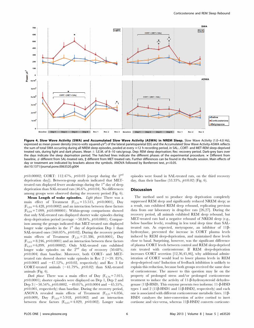
Slow Wave Activity during NREM Sleep. Light phase: There
were main effects of Treatment (F2,21 = 9.296, p#0.002), Day
(F4,84 = 6.418, p#0.0002) and an interaction between these factors
(F8,84 = 3.468, p#0.002). Analysis of this interaction showed that in
CORT-treated animals NREM slow wave activity was higher on
the 3rd day of the sleep deprivation period than baseline (94.05%,
p#0.0002). In addition, SWA was higher on day 2 and 3 than on
day 1 of REM sleep deprivation (68.78%, p#0.05 and 102.63%,
p#0.0.0001) in this same group. No differences among the
treatments were observed during the sleep recovery period (Fig. 4).
Dark phase: There were main effects of Treatment (F2,20 = 8.29,
p#0.003), Day (F4,80 = 6.896, p#0.0001) but no interaction
between these factors regarding the sleep deprivation period. Post
hoc analysis indicated that during sleep deprivation, NREM SWA
increased in CORT-treated animals when compared to SAL- and
MET-treated rats (68.22%, p#0.004 and 53.50%, p#0.02).
NREM SWA was augmented on 3rd and 4th days of sleep
deprivation compared to baseline (64.37%, p#0.002; 87.63%,
p#0.00008); the SWA levels were also higher on the 4th day than
on the 1st day of sleep deprivation (28.63%, p#0.04). Again, no
differences were observed during the sleep recovery period (Fig. 4).
Accumulated Slow Wave Activity during NREM Sleep. Light
phase: Main effects of Treatment (F2,21 = 6.286, p#0.008) and Day
(F4,84 = 8.75, p#0.000001) were detected for the sleep deprivation
period. Regarding the Treatment effect, in CORT-treated animals
ASWA was 31.27% higher than SAL- (p#0.04) and 41.82% higher
than MET-treated rats (p#0.01). As for the effect of Day, during REM
sleep-deprivation the animals displayed less ASWA than their
respective baselines (246.34%, in average, p#0.005). As for the
recovery period, there was a main effect of Day (F3,63) = 2.759;
p#0.05), with a significant reduction on the last recovery day when
compared to baseline (229.74%, p#0.04) (Fig. 4).
Figure 2. ACTH and Corticosterone Plasma Levels in Response to Treatments with Corticosterone or Metyrapone. CTL, home-cagecontrol; REMSD, REM sleep deprivation. Data is presented as mean 6 S.E.M. of 8–10 rats/group. Different from respective CTL groups; # differentfrom SAL-treated rats; ` different from MET-treated rats; lines above bars indicate main effects (group or treatment). ANOVA followed by Newman-Keuls test; p#0.05.doi:10.1371/journal.pone.0063520.g002
Corticosterone and REM Sleep Rebound
PLOS ONE | www.plosone.org 4 May 2013 | Volume 8 | Issue 5 | e63520

Dark phase: A main effect of Treatment was observed during
REM sleep deprivation (F2,21 = 5.932, p#0.009) and CORT-
treated rats exhibited more ASWA than SAL- (42.69%, p#0.03)
and MET-treated rats (46.07%, p#0.02). No differences among
groups were observed during the recovery period (Fig. 4).
REM Sleep. Due to the complete suppression of REM sleep
induced by the platform technique, analysis of the data included
only baseline sleep and the recovery period.
Light phase: The two-way ANOVA for repeated measures
detected a main effect of Day (F3,63 = 63.577, p#0.00001), in
which the animals showed increased REM during Rec 1 and Rec
2 compared to baseline (160.88%, p#0.00001; 40.47%, p#0.03;
respectively) (Fig. 5).
Dark phase: Again, a main effect of Day was observed
(F3,63 = 27.55, p#0.00001), with increased REM sleep
(p#0.0005) only on Rec 1 when compared to baseline levels
(Fig. 5).
Number of REM Sleep Episodes. Light phase: A main effect
of Day (F3,63 = 29.69, p#0.000001) and an interaction between
Treatment and Day was detected (F6,63 = 3.045, p#0,02). Within-
group comparisons revealed that SAL- and CORT-treated
animals exhibited more REM sleep events during Rec 1 than
baseline (107.78%, p#0.00005 and 65.45%; p#0.01, respectively).
No differences were found among the groups (Fig. 5).
Dark phase: A main effect of Day was revealed (F3,63 = 27.55,
p#0.00001) and the rats displayed more REM sleep events during
Rec 1 (151.27%, p#0.00001) than baseline sleep (Fig. 5).
Mean Length of REM Sleep Episodes. Light phase: An
interaction between Treatment and Day was revealed
(F6,63 = 2.193, p#0.05); Bonferroni test showed that both CORT-
and MET-treated groups exhibited longer REM sleep episodes in
Rec 1 than baseline (CORT: 85.14%, p#0.000001; MET:
41.67%, p#0.03). There were no between group differences
(Fig. 5).
Dark phase: A main effect of Day was observed (F3,57 = 10.75,
p#0.00005) and all groups exhibited longer REM sleep episodes in
Rec 1 than baseline (27.12%, p#0.0007), than Rec 2 (37.24%,
p#0.00002) and than Rec 3 (30.52%, p#0.0003) (Fig. 5).
Number of awakenings. Light phase: There was a main effect
of Treatment (F2,21 = 47.51, p#0.000001), of Day (F4,84 = 65.533,
p#0.000001) and an interaction between these factors
(F8,84 = 8.248, p#0.000001). Analysis of this interaction revealed
that awakenings longer than 2.0 min were higher during all days
of sleep deprivation than baseline for SAL- and CORT-treated
rats (average, SAL: 303.66%, p#0.000001; CORT: 90.89%,
p#0.01). For MET-treated rats, increased number of awakenings
above baseline occurred on Dep 2 and Dep 3 (average, 124.64%,
p#0.0001). Throughout the deprivation period, MET-treated rats
exhibited fewer awakening events than SAL-treated rats
(247.59%, p#0.02). CORT-treated animals showed less awaken-
ing events on Dep 4 when compared with SAL-treated rats
(238.05%, p#0.04). Analysis of the sleep recovery period showed
a main effect of Treatment (F2,21 = 17.335, p#0.0001). Post-hoc
analysis revealed that CORT-treated rats had more awakening
events than SAL-treated (58.32%, p#0.00003) and than MET-
treated rats (16.73%, p#0.04), which, in turn had more events
than SAL-treated ones (31.82%, p#0.01) (Fig. 6).
Dark phase: Main effects of Treatment (F2,21 = 20.328,
p#0.00002), Day (F4,84 = 24.713, p#0.000001) and an interaction
between these factors (F8,84 = 2.635, p#0.02) were shown
throughout the deprivation period. SAL- and CORT-treated rats
displayed more awakenings longer than 2.0 min (SAL: 240.58%,
Figure 3. Total Sleep Time and NREM Sleep. Results are expressed as percentage of recording time (>12 h), in SAL-, CORT- and MET-REM sleep-deprived treated rats, during light and dark phases. Data is presented as mean 6 S.E.M. of 8–10 rats/group. Dep: REM sleep deprivation; Rec: recoveryperiod. Dark-grey bars over the days indicate the sleep deprivation period. The hatched lines indicate the different phases of the experimentalprocedure. Different from baseline. Main effect of day is indicated by brackets above the symbols. Further differences can be found in the Resultssession. ANOVA followed by Bonferroni test, p#0.05.doi:10.1371/journal.pone.0063520.g003
Corticosterone and REM Sleep Rebound
PLOS ONE | www.plosone.org 5 May 2013 | Volume 8 | Issue 5 | e63520
p#0.00002; CORT: 112.47%, p#0.03 [except during the 2nd
deprivation day]). Between-group analysis indicated that MET-
treated rats displayed fewer awakenings during the 1st day of sleep
deprivation than SAL-treated rats (46.6%, p#0.04). No differences
among groups were observed during the recovery period (Fig. 6).Mean Length of wake episodes. Light phase: There was a
main effect of Treatment (F2,21 = 15.515, p#0.0001), Day
(F4,84 = 6.428, p#0.0002) and an interaction between these factors
(F8,84 = 7.099, p#0.000001). Within-group comparisons showed
that only SAL-treated rats displayed shorter wake episodes during
sleep deprivation period (average 258.84%, p#0.0001). Compar-
ison among the groups indicated that MET-treated rats displayed
longer wake episodes in the 1st day of deprivation Dep 1 than
SAL-treated ones (160.05%, p#0.02). During the recovery period
main effects of Treatment (F2,21 = 21.386, p#0.0001), Day
(F3,63 = 8.246, p#0.0001) and an interaction between these factors
(F6,63 = 6,899, p#0.00002). Only SAL-treated rats exhibited
longer wake episodes on the 2nd day of recovery (50.68%;
p#0.004) than baseline. Moreover, both CORT- and MET-
treated rats showed shorter wake episodes in Rec 2 (259. 85%,
p#0.0003 and 247.12%, p#0.02, respectively) and Rec 3 for
CORT-treated animals (241.79%, p#0.02) than SAL-treated
animals (Fig. 6).
Dark phase: There was a main effect of Day (F4,76 = 7.015,
p#0.0001); shorter episodes were displayed on Dep 1, Dep 2 and
Dep 3 (250.54%, p#0.0002, 249.01%, p#0.0004 and 245.55%,
p#0.001, respectively) than baseline. During the recovery period,
ANOVA revealed main effects of Treatment (F2,21 = 6.056,
p#0.009), Day (F3,63 = 5.918, p#0.002) and an interaction
between these factors (F6,63 = 4.029, p#0.002). Longer wake
episodes were found in SAL-treated rats, on the third recovery
day, than their baseline (53.33%, p#0.02) (Fig. 6).
Discussion
The method used to produce sleep deprivation completely
suppressed REM sleep and significantly reduced NREM sleep; as
a result, rats exhibited REM sleep rebound, replicating previous
data from our laboratory in drug-free rats [26,27]. During the
recovery period, all animals exhibited REM sleep rebound, but
MET-treated rats had a negative rebound of NREM sleep (e.g.,
below baseline levels), resulting in less total sleep time than SAL-
treated rats. As expected, metyrapone, an inhibitor of 11b-
hydroxylase, prevented the increase in CORT plasma levels
induced by REM sleep-deprivation, and maintained these levels
close to basal. Surprising, however, was the significant difference
of plasma CORT levels between control and REM sleep-deprived
rats treated with corticosterone. If REM sleep-deprivation
increases CORT secretion [12,36,45,46], why additional admin-
istration of CORT would lead to lower plasma levels in REM
sleep-deprived rats? Induction of feedback inhibition is unlikely to
explain this reduction, because both groups received the same dose
of corticosterone. The answer to this question may lie on the
property of prolonged stress and/or prolonged corticosterone
treatment to induce the activity of 11-b-hydroxysteroid dehydro-
genase (11b-HSD). This enzyme presents two isoforms: 11-b-HSD
types 1 and 2 (11b-HSD1 and 11b-HSD2, respectively) and each
one is associated with different corticosterone metabolic fates. 11b-
HSD1 catalyzes the inter-conversion of active cortisol to inert
cortisone and vice-versa, whereas 11b-HSD2 converts corticoste-
Figure 4. Slow Wave Activity (SWA) and Accumulated Slow Wave Activity (ASWA) in NREM Sleep. Slow Wave Activity (1.0–4.0 Hz),expressed as mean power density (micro-volts squared-mV2) of the lateral parietoparietal EEG and the Accumulated Slow Wave Activity-ASWA reflectsthe sum of total SWA occurring during all NREM sleep episodes, pooled at every >12 h recording period, in SAL-, CORT- and MET-REM sleep-deprivedtreated rats, during light and dark phases. Mean 6 S.E.M. of 8–10 rats/group. Dep: REM sleep deprivation; Rec: recovery period. Dark-grey bars overthe days indicate the sleep deprivation period. The hatched lines indicate the different phases of the experimental procedure. Different frombaseline, # different from SAL-treated rats, ` different from MET-treated rats. Further differences can be found in the Results session. Main effects ofday or treatment are indicated by brackets above the symbols. ANOVA followed by Bonferroni test, p#0.05.doi:10.1371/journal.pone.0063520.g004
Corticosterone and REM Sleep Rebound
PLOS ONE | www.plosone.org 6 May 2013 | Volume 8 | Issue 5 | e63520

rone to its inactive 11-keto metabolite [47]. Both stress and
corticosterone administration induce the activity of 11b-HSD2
[48,49] and we raised the possibility that combination of both, as
occurred in our protocol, could lead to a stronger effect than each
manipulation alone. This hypothesis, however, has still to be
tested.
The ACTH profile was the opposite to that of CORT, reflecting
the effects of the negative feedback mechanism. Thus, metyra-
pone, which inhibits CORT synthesis, leads to increased CRH
and ACTH levels, thus being considered a pharmacological
stressor [50]. In addition, metyrapone also increases glucose
plasma levels and activates the expression of Fos in several brain
structures, including frontal cortex, amygdala and thalamic and
hypothalamic nuclei [51]. On the contrary, corticosterone
administration increased CORT levels, albeit less so in REM
sleep-deprived than in control rats, stimulating the negative
feedback, which led to low levels of ACTH.
Repeated metyrapone treatment prolonged waking bouts
during sleep deprivation, and yet, there was an impairment of
NREM sleep rebound in the first light period of recovery. Single
administration of this drug has also been shown to increase waking
time, by reducing NREM and REM sleep times, but in this case,
the sleep impairment is followed by a homeostatic sleep rebound
[19]. The effects hereby reported could be explained by increased
CRH activity resulting from sustained blockade of corticosterone
synthesis. Numerous evidence supports this idea: CRH i.c.v.
administration in rats reduces pentobarbital-induced sleep and
increases neuronal excitability; the former being reversed by pre-
treatment with anti-CRH antibody [52]. Mice trained to escape a
shock exhibit REM sleep rebound, which is blocked by i.c.v.
infusion of CRH [6]; CRH receptor-1 (CRH-R1) knock-out mice
do not exhibit as intense REM sleep blockade as control mice in
response to CRH i.c.v. infusion [53]. Indeed, these receptors
appear to be responsible for the sleep impairing effects of CRH,
since administration of their antagonist, R121919, improves
Figure 5. REM Sleep, REM Sleep Episodes and Mean Length of REM Sleep Episodes. REM Sleep results are expressed as percentage ofrecording time (>12 h), in SAL-, CORT- and MET-REM sleep-deprived treated rats, during light and dark phases. REMS Episodes were expressed asabsolute number and Mean length of REM Episodes were expressed in minutes. Mean 6 S.E.M. of 8–10 rats/group. Dep: REM sleep deprivationperiod; Rec: recovery period. Dark-grey bars over the days indicate the sleep deprivation period. The hatched lines indicate the different phases of theexperimental procedure. Different from baseline. Main effect of day is indicated by brackets above the symbols. ANOVA followed by Bonferronitest, p#0.05.doi:10.1371/journal.pone.0063520.g005
Corticosterone and REM Sleep Rebound
PLOS ONE | www.plosone.org 7 May 2013 | Volume 8 | Issue 5 | e63520

NREM sleep in an animal model of depression [8] and in
depressive patients [54]. CRH receptors are located in thalamic
neurons [55,56] and they inhibit spontaneous activity of reticular
neurons [57], which are responsible for synchronization of cortical
low frequency EEG seen during NREM sleep [58]. Finally, REM
sleep-deprived rats repeatedly treated with CRH also exhibit
impairment of sleep homeostasis, with shorter bouts of REM sleep
than vehicle-treated rats [35] and reduced power spectrum of low
frequency bands (1.0 to 6.0 Hz) [59]. Therefore, increased CRH
activity induced by repeated metyrapone treatment could explain
the reduction in NREM and REM sleep rebound observed in the
present study.
Corticosterone-treated rats also exhibited less NREM sleep
during the entire sleep deprivation period. In non-REM sleep-
deprived rats, corticosterone doses similar to the one employed in
the present study causes a major reduction of NREM sleep for up
to 24 h after hormone administration [14]. This effect seems to be
dependent on the glucocorticoid concentration acting upon
GABAA receptors, since either adrenalectomy or dexamethasone
reduces, whereas moderate levels increase the affinity of these
receptors [60]. GABA, in turn, inhibits thalamic-cortical connec-
tions, resulting in generation of EEG synchronized activity,
including spikes and slow waves [58,61,62,63]. Importantly, the
thalamus, and its reticular nucleus, is rich in type II glucocorticoid
receptors [64,65,66], thus, being a natural target for these steroids.
Despite NREM sleep inhibition, CORT-treated rats showed a
robust increase of SWA and cumulated SWA from the second
deprivation day on. SWA during NREM sleep is directly
correlated with sleep intensity and is a marker of homeostatic
sleep recovery [67]. A recent study, however, claims that sleep
deprivation-induced corticosterone secretion is not associated to
increased SWA during sleep recovery [68]. Still, corticosterone
appears to be, at least in part, involved because adrenalectomy
reduces 1.0 to 4.0 Hz power, which is reversed by low levels of
corticosterone [15]. Interestingly, adrenalectomy also reduces
brain glycogen, which is restored by hormone replacement [69];
brain glycogen is directly related with EEG power [70,71] and one
of the functions attributed to sleep is to restore brain glycogen
levels after prolonged periods of waking [72]. Delta activity in
NREM sleep accumulates and increases during the course of
wakefulness (including forced wakefulness/sleep deprivation),
being dissipated within the first hours of the recovery period
[24,73,74]. In fact, in CORT-treated rats, the observed increase in
SWA took place during the sleep deprivation period and in the
first hours of recovery (light phase, data not shown). Despite the
negative NREM sleep rebound observed during the recovery in
MET-treated rats, some studies indicate that total or partial sleep
deprivation of various lengths also result in negative NREM sleep
rebounds following a small initial positive rebound [28,75,76]. We
have reason to believe that this effect on NREM sleep is not only
due to the homeostatic pressure for REM sleep, but rather the
result of the pharmacological manipulation herein employed, since
saline-treated sleep-deprived rats did not exhibit the negative
NREM rebound.
Corticosterone treatment increased the number and length of
REM sleep episodes during sleep recovery. Activation of type II
glucocorticoid receptors by this steroid activates type 2 pro-
convertase that cleaves pro-opiomelanocortin (POMC) to cortico-
tropin-like intermediate lobe peptide (CLIP) [77,78,79], which has
a well-established role in prolonging REM sleep episodes
[80,81,82]. In a previous study we showed that REMSD
associated with repeated stress resulted in REM sleep episodes
Figure 6. Episodes of awakening and Mean Length of Wake Episodes. Results of Episodes of Awakening longer than 2.0 min. are expressedas absolute number and Mean Length of Wake Episodes, in minutes, during the light and dark phases, in SAL-, CORT- and MET-REM sleep-deprivedtreated rats. Mean 6 S.E.M. of 8–10 rats/group. Dep: REM sleep deprivation period; Rec: recovery period. Dark-grey bars over the days indicate thesleep deprivation period. The hatched lines indicate the different phases of the experimental procedure. Main effects of day or treatment areindicated by brackets above the symbols. Different from baseline, # different from SAL-treated rats. ANOVA followed by Bonferroni test, p#0.05.doi:10.1371/journal.pone.0063520.g006
Corticosterone and REM Sleep Rebound
PLOS ONE | www.plosone.org 8 May 2013 | Volume 8 | Issue 5 | e63520

that were two to three times longer than baseline. This procedure
also led to corticosterone levels that were intermediate between
control animals that were not manipulated (lowest levels) and non-
deprived animals submitted to the repeated stress (highest levels)
[12]. Glucocorticoid influence on REM sleep follows an inverted
U shape curve, with very low or very high levels resulting in
impairment, whereas optimal concentrations lead to increase of
this sleep phase [83,84].
In conclusion, the present study showed that either high
(corticosterone treatment) or low (metyrapone treatment) circulat-
ing levels of corticosterone appear to be detrimental to sleep
recovery following 96 h of REM sleep-deprivation. These results
reinforce the notion that adequate stress response, and conse-
quently, glucocorticoid levels, is essential for maximal expression
of sleep rebound (both at the macro- and micro-structure levels),
which, in turn, is thought to be part of the behavioral repertoire
necessary for full recovery after stressful situations [85,86,87,88].
Acknowledgments
The authors are in debt to Adriana Fernandes Faria for helping with
ACTH hormone determinations.
Author Contributions
Conceived and designed the experiments: RBM ST DS. Performed the
experiments: RBM. Analyzed the data: RBM DS. Contributed reagents/
materials/analysis tools: ST. Wrote the paper: RBM DS.
References
1. Morin CM, Rodrigue S, Ivers H (2003) Role of stress, arousal, and coping skills
in primary insomnia. Psychosom Med 65: 259–267.
2. Ross RJ, Ball WA, Sanford LD, Morrison AR, Dinges DF, et al. (1999) Rapid
eye movement sleep changes during the adaptation night in combat veterans
with posttraumatic stress disorder. Biol Psychiatry 45: 938–941.
3. Cartwright RD, Wood E (1991) Adjustment disorders of sleep: the sleep effects
of a major stressful event and its resolution. Psychiatry Res 39: 199–209.
4. Kant GJ, Pastel RH, Bauman RA, Meininger GR, Maughan KR, et al. (1995)
Effects of chronic stress on sleep in rats. Physiol Behav 57: 359–365.
5. Philbert J, Pichat P, Beeske S, Decobert M, Belzung C, et al. (2011) Acute
inescapable stress exposure induces long-term sleep disturbances and avoidance
behavior: a mouse model of post-traumatic stress disorder (PTSD). Behav Brain
Res 221: 149–154.
6. Sanford LD, Yang L, Wellman LL, Liu X, Tang X (2010) Differential effects of
controllable and uncontrollable footshock stress on sleep in mice. Sleep 33: 621–
630.
7. Cui R, Li B, Suemaru K, Araki H (2007) Differential effects of psychological and
physical stress on the sleep pattern in rats. Acta Med Okayama 61: 319–327.
8. Lancel M, Muller-Preuss P, Wigger A, Landgraf R, Holsboer F (2002) The
CRH1 receptor antagonist R121919 attenuates stress-elicited sleep disturbances
in rats, particularly in those with high innate anxiety. J Psychiatr Res 36: 197–
208.
9. Chang FC, Opp MR (1998) Blockade of corticotropin-releasing hormone
receptors reduces spontaneous waking in the rat. Am J Physiol 275: R793–802.
10. Chang FC, Opp MR (2001) Corticotropin-releasing hormone (CRH) as a
regulator of waking. Neurosci Biobehav Rev 25: 445–453.
11. Opp MR (1997) Rat strain differences suggest a role for corticotropin-releasing
hormone in modulating sleep. Physiol Behav 63: 67–74.
12. Machado RB, Tufik S, Suchecki D (2008) Chronic stress during paradoxical
sleep deprivation increases paradoxical sleep rebound: association with prolactin
plasma levels and brain serotonin content. Psychoneuroendocrinology 33: 1211–
1224.
13. Vazquez-Palacios G, Velazquez-Moctezuma J (2000) Effect of electric foot
shocks, immobilization, and corticosterone administration on the sleep-wake
pattern in the rat. Physiol Behav 71: 23–28.
14. Vazquez-Palacios G, Retana-Marquez S, Bonilla-Jaime H, Velazquez-Mocte-
zuma J (2001) Further definition of the effect of corticosterone on the sleep-wake
pattern in the male rat. Pharmacol Biochem Behav 70: 305–310.
15. Bradbury MJ, Dement WC, Edgar DM (1998) Effects of adrenalectomy and
subsequent corticosterone replacement on rat sleep state and EEG power
spectra. Am J Physiol 275: R555–565.
16. Milcu SM, Nicolescu-Catargi A (1967) Deep sleep phase alterations in patients
with loss of the adrenal secretory rhythm and in hypophysectomized patients.
(The effects of hypothalamo-hypophyseal stressing by metopyron). Electro-
encephalogr Clin Neurophysiol 22: 574.
17. Jahn H, Kiefer F, Schick M, Yassouridis A, Steiger A, et al. (2003) Sleep
endocrine effects of the 11-beta-hydroxysteroiddehydrogenase inhibitor metyr-
apone. Sleep 26: 823–829.
18. Neylan TC, Lenoci M, Maglione ML, Rosenlicht NZ, Metzler TJ, et al. (2003)
Delta sleep response to metyrapone in post-traumatic stress disorder.
Neuropsychopharmacology 28: 1666–1676.
19. Drouet JB, Rousset C, Maury R, Michel V, Buguet A, et al. (2011) Single
administration of metyrapone modifies sleep-wake patterns in the rat.
Eur J Pharmacol 652: 60–64.
20. Igaz P, Tombol Z, Szabo PM, Liko I, Racz K (2008) Steroid biosynthesis
inhibitors in the therapy of hypercortisolism: theory and practice. Curr Med
Chem 15: 2734–2747.
21. Dement W (1960) The effect of dream deprivation. Science 131: 1705–1707.
22. Ferrara M, De Gennaro L, Bertini M (1999) Selective slow-wave sleep (SWS)
deprivation and SWS rebound: do we need a fixed SWS amount per night?
Sleep Res Online 2: 15–19.
23. Tobler I, Borbely AA (1990) The effect of 3-h and 6-h sleep deprivation on sleep
and EEG spectra of the rat. Behav Brain Res 36: 73–78.
24. Tobler I, Borbely AA (1986) Sleep EEG in the rat as a function of prior waking.
Electroencephalogr Clin Neurophysiol 64: 74–76.
25. Schwierin B, Borbely AA, Tobler I (1999) Prolonged effects of 24-h total sleep
deprivation on sleep and sleep EEG in the rat. Neurosci Lett 261: 61–64.
26. Machado RB, Hipolide DC, Benedito-Silva AA, Tufik S (2004) Sleep
deprivation induced by the modified multiple platform technique: quantification
of sleep loss and recovery. Brain Res 1004: 45–51.
27. Suchecki D, Duarte Palma B, Tufik S (2000) Sleep rebound in animals deprived
of paradoxical sleep by the modified multiple platform method. Brain Res 875:
14–22.
28. Rechtschaffen A, Bergman BM, Gilliland MA, Bauer K (1999) Effects of
method, duration, and sleep stage on rebounds from sleep deprivation in the rat.
Sleep 22: 11–31.
29. Borbely AA, Baumann F, Brandeis D, Strauch I, Lehmann D (1981) Sleep
deprivation: effect on sleep stages and EEG power density in man.
Electroencephalogr Clin Neurophysiol 51: 483–495.
30. Brunner DP, Dijk DJ, Tobler I, Borbely AA (1990) Effect of partial sleep
deprivation on sleep stages and EEG power spectra: evidence for non-REM and
REM sleep homeostasis. Electroencephalogr Clin Neurophysiol 75: 492–499.
31. Dijk DJ, Hayes B, Czeisler CA (1993) Dynamics of electroencephalographic
sleep spindles and slow wave activity in men: effect of sleep deprivation. Brain
Res 626: 190–199.
32. Lancel M, van Riezen H, Glatt A (1992) The time course of sigma activity and
slow-wave activity during NREMS in cortical and thalamic EEG of the cat
during baseline and after 12 hours of wakefulness. Brain Res 596: 285–295.
33. Lancel M, van Riezen H, Glatt A (1992) Enhanced slow-wave activity within
NREM sleep in the cortical and subcortical EEG of the cat after sleep
deprivation. Sleep 15: 102–118.
34. Endo T, Schwierin B, Borbely AA, Tobler I (1997) Selective and total sleep
deprivation: effect on the sleep EEG in the rat. Psychiatry Res 66: 97–110.
35. Machado RB, Tufik S, Suchecki D (2010) Modulation of Sleep Homeostasis by
Corticotropin Releasing Hormone in REM Sleep-Deprived Rats.
Int J Endocrinol 2010: 326151.
36. Galvao MD, Sinigaglia-Coimbra R, Kawakami SE, Tufik S, Suchecki D (2009)
Paradoxical sleep deprivation activates hypothalamic nuclei that regulate food
intake and stress response. Psychoneuroendocrinology.
37. Coenen AM, van Luijtelaar EL (1985) Stress induced by three procedures of
deprivation of paradoxical sleep. Physiol Behav 35: 501–504.
38. Tobler I, Murison R, Ursin R, Ursin H, Borbely AA (1983) The effect of sleep
deprivation and recovery sleep on plasma corticosterone in the rat. Neurosci Lett
35: 297–300.
39. Timo-Iaria C, Negrao N, Schmidek WR, Hoshino K, Lobato de Menezes CE,
et al. (1970) Phases and states of sleep in the rat. Physiol Behav 5: 1057–1062.
40. Rosenberg RS, Bergmann BM, Rechtschaffen A (1976) Variations in slow wave
activity during sleep in the rat. Physiol Behav 17: 931–938.
41. Machado RB, Suchecki D, Tufik S (2005) Sleep homeostasis in rats assessed by a
long-term intermittent paradoxical sleep deprivation protocol. Behav Brain Res
160: 356–364.
42. Bergmann BM, Mistlberger RE, Rechtschaffen A (1987) Period-amplitude
analysis of rat electroencephalogram: stage and diurnal variations and effects of
suprachiasmatic nuclei lesions. Sleep 10: 523–536.
43. Bergmann BM, Winter JB, Rosenberg RS, Rechtschaffen A (1987) NREM sleep
with low-voltage EEG in the rat. Sleep 10: 1–11.
44. Grahnstedt S, Ursin R (1985) Platform sleep deprivation affects deep slow wave
sleep in addition to REM sleep. Behav Brain Res 18: 233–239.
45. Tiba PA, Oliveira MG, Rossi VC, Tufik S, Suchecki D (2008) Glucocorticoids
are not responsible for paradoxical sleep deprivation-induced memory
impairments. Sleep 31: 505–515.
Corticosterone and REM Sleep Rebound
PLOS ONE | www.plosone.org 9 May 2013 | Volume 8 | Issue 5 | e63520

46. Suchecki D, Antunes J, Tufik S (2003) Palatable solutions during paradoxical
sleep deprivation: reduction of hypothalamic-pituitary-adrenal axis activity andlack of effect on energy imbalance. J Neuroendocrinol 15: 815–821.
47. Ma X, Lian QQ, Dong Q, Ge RS (2011) Environmental inhibitors of 11beta-
hydroxysteroid dehydrogenase type 2. Toxicology 285: 83–89.48. Igarreta P, Calvo JC, Damasco MC (1999) Activity of renal 11betahydroxyster-
oid dehydrogenase 2 (11betaHSD2) in stressed animals. Life Sci 64: 2285–2290.49. Zallocchi M, Matkovic L, Damasco MC (2004) Adrenal 11-beta hydroxysteroid
dehydrogenase activity in response to stress. Can J Physiol Pharmacol 82: 422–
425.50. Rotllant D, Armario A (2005) A single dose of metyrapone caused long-term
dysregulation of the hypothalamic-pituitary-adrenal axis in the rat. Neuroscience130: 427–434.
51. Rotllant D, Ons S, Carrasco J, Armario A (2002) Evidence that metyrapone canact as a stressor: effect on pituitary-adrenal hormones, plasma glucose and brain
c-fos induction. Eur J Neurosci 16: 693–700.
52. Burade VS, Jain MR, Khan FA, Saha SG, Subhedar N (1996) Involvement ofcorticosteroid-like neurosteroids in pentobarbital-induced sleep. Neuroreport 8:
139–141.53. Romanowski CP, Fenzl T, Flachskamm C, Wurst W, Holsboer F, et al. (2010)
Central deficiency of corticotropin-releasing hormone receptor type 1 (CRH-R1)
abolishes effects of CRH on NREM but not on REM sleep in mice. Sleep 33:427–436.
54. Held K, Kunzel H, Ising M, Schmid DA, Zobel A, et al. (2004) Treatment withthe CRH1-receptor-antagonist R121919 improves sleep-EEG in patients with
depression. J Psychiatr Res 38: 129–136.55. De Souza EB (1995) Corticotropin-releasing factor receptors: physiology,
pharmacology, biochemistry and role in central nervous system and immune
disorders. Psychoneuroendocrinology 20: 789–819.56. De Souza EB, Insel TR, Perrin MH, Rivier J, Vale WW, et al. (1985)
Corticotropin-releasing factor receptors are widely distributed within the ratcentral nervous system: an autoradiographic study. J Neurosci 5: 3189–3203.
57. Eberly LB, Dudley CA, Moss RL (1983) Iontophoretic mapping of corticotro-
pin-releasing factor (CRF) sensitive neurons in the rat forebrain. Peptides 4:837–841.
58. Steriade M, McCormick DA, Sejnowski TJ (1993) Thalamocortical oscillationsin the sleeping and aroused brain. Science 262: 679–685.
59. Ehlers CL, Reed TK, Henriksen SJ (1986) Effects of corticotropin-releasingfactor and growth hormone-releasing factor on sleep and activity in rats.
Neuroendocrinology 42: 467–474.
60. Majewska MD, Bisserbe JC, Eskay RL (1985) Glucocorticoids are modulators ofGABAA receptors in brain. Brain Res 339: 178–182.
61. McCormick DA, Bal T (1997) Sleep and arousal: thalamocortical mechanisms.Annu Rev Neurosci 20: 185–215.
62. von Krosigk M, Bal T, McCormick DA (1993) Cellular mechanisms of a
synchronized oscillation in the thalamus. Science 261: 361–364.63. Huntsman MM, Porcello DM, Homanics GE, DeLorey TM, Huguenard JR
(1999) Reciprocal inhibitory connections and network synchrony in themammalian thalamus. Science 283: 541–543.
64. Morimoto M, Morita N, Ozawa H, Yokoyama K, Kawata M (1996)Distribution of glucocorticoid receptor immunoreactivity and mRNA in the
rat brain: an immunohistochemical and in situ hybridization study. Neurosci
Res 26: 235–269.65. Sousa RJ, Tannery NH, Lafer EM (1989) In situ hybridization mapping of
glucocorticoid receptor messenger ribonucleic acid in rat brain. Mol Endocrinol3: 481–494.
66. Reul JM, de Kloet ER (1986) Anatomical resolution of two types of
corticosterone receptor sites in rat brain with in vitro autoradiography andcomputerized image analysis. J Steroid Biochem 24: 269–272.
67. Achermann P, Borbely AA (2003) Mathematical models of sleep regulation.
Front Biosci 8: s683–693.68. Mongrain V, Hernandez SA, Pradervand S, Dorsaz S, Curie T, et al. (2010)
Separating the contribution of glucocorticoids and wakefulness to the molecular
and electrophysiological correlates of sleep homeostasis. Sleep 33: 1147–1157.69. Gip P, Hagiwara G, Sapolsky RM, Cao VH, Heller HC, et al. (2004)
Glucocorticoids influence brain glycogen levels during sleep deprivation.Am J Physiol Regul Integr Comp Physiol 286: R1057–1062.
70. Ratcheson RA, Blank AC, Ferrendelli JA (1981) Regionally selective metabolic
effects of hypoglycemia in brain. J Neurochem 36: 1952–1958.71. Feise G, Kogure K, Busto KR, Scheinberg P, Reinmuth OM (1977) Effect of
insulin hypoglycemia upon cerebral energy metabolism and EEG activity in therat. Brain Res 126: 263–280.
72. Benington JH, Heller HC (1995) Restoration of brain energy metabolism as thefunction of sleep. Prog Neurobiol 45: 347–360.
73. Borbely AA, Achermann P (1999) Sleep homeostasis and models of sleep
regulation. J Biol Rhythms 14: 557–568.74. Franken P, Chollet D, Tafti M (2001) The homeostatic regulation of sleep need
is under genetic control. J Neurosci 21: 2610–2621.75. Benington JH, Heller HC (1999) Implications of sleep deprivation experiments
for our understanding of sleep homeostasis. Sleep 22: 1033–1043.
76. Rechtschaffen A, Bergmann BM (1999) Sleep rebounds and their implicationsfor sleep stage substrates: A response to Benington and Heller. Sleep 22: 1038–
1043.77. Day R, Schafer MK, Watson SJ, Chretien M, Seidah NG (1992) Distribution
and regulation of the prohormone convertases PC1 and PC2 in the rat pituitary.Mol Endocrinol 6: 485–497.
78. Ortego J, Wollmann G, Coca-Prados M (2002) Differential regulation of gene
expression of neurotensin and prohormone convertases PC1 and PC2 in thebovine ocular ciliary epithelium: possible implications on neurotensin processing.
Neurosci Lett 333: 49–53.79. Lee SN, Peng B, Desjardins R, Pintar JE, Day R, et al. (2007) Strain-specific
steroidal control of pituitary function. J Endocrinol 192: 515–525.
80. Chastrette N, Cespuglio R (1985) Influence of proopiomelanocortin-derivedpeptides on the sleep-waking cycle of the rat. Neurosci Lett 62: 365–370.
81. Wetzel W, Balschun D, Janke S, Vogel D, Wagner T (1994) Effects of CLIP(corticotropin-like intermediate lobe peptide) and CLIP fragments on paradox-
ical sleep in rats. Peptides 15: 237–241.82. Wetzel W, Wagner T, Vogel D, Demuth HU, Balschun D (1997) Effects of the
CLIP fragment ACTH 20–24 on the duration of REM sleep episodes.
Neuropeptides 31: 41–45.83. Garcia-Borreguero D, Wehr TA, Larrosa O, Granizo JJ, Hardwick D, et al.
(2000) Glucocorticoid replacement is permissive for rapid eye movement sleepand sleep consolidation in patients with adrenal insufficiency. J Clin Endocrinol
Metab 85: 4201–4206.
84. Marinesco S, Bonnet C, Cespuglio R (1999) Influence of stress duration on thesleep rebound induced by immobilization in the rat: a possible role for
corticosterone. Neuroscience 92: 921–933.85. Mellman TA, Bustamante V, Fins AI, Pigeon WR, Nolan B (2002) REM sleep
and the early development of posttraumatic stress disorder. Am J Psychiatry 159:1696–1701.
86. Pawlyk AC, Jha SK, Brennan FX, Morrison AR, Ross RJ (2005) A rodent model
of sleep disturbances in posttraumatic stress disorder: the role of context afterfear conditioning. Biol Psychiatry 57: 268–277.
87. Liu X, Tang X, Sanford LD (2003) Fear-conditioned suppression of REM sleep:relationship to Fos expression patterns in limbic and brainstem regions in
BALB/cJ mice. Brain Res 991: 1–17.
88. Suchecki D, Tiba PA, Machado RB (2012) REM Sleep Rebound as an AdaptiveResponse to Stressful Situations. Front Neurol 3: 41.
Corticosterone and REM Sleep Rebound
PLOS ONE | www.plosone.org 10 May 2013 | Volume 8 | Issue 5 | e63520
Copyright © 2022 FDOKUMEN