VEGF Promotes Malaria-Associated Acute Lung Injury in Mice
10
VEGF Promotes Malaria-Associated Acute Lung Injury in Mice Sabrina Epiphanio 1,2.¤ , Marta G. Campos 1,2. , Ana Pamplona 1,2. , Daniel Carapau 1 , Ana C. Pena 1 , Ricardo Ataı´de 1 , Carla A. A. Monteiro 3 , Nuno Fe ´ lix 3 , Artur Costa-Silva 4 , Claudio R. F. Marinho 5 , Se ´ rgio Dias 2,6 , Maria M. Mota 1,2 * 1 Unidade de Mala ´ria, Instituto de Medicina Molecular, Universidade de Lisboa, Lisboa, Portugal, 2 Instituto Gulbenkian de Cie ˆncia, Oeiras, Portugal, 3 Faculdade de Medicina Veterina ´ria de Lisboa, Universidade Te ´cnica de Lisboa, Portugal, 4 Servic ¸o de Anatomia Patolo ´ gica, Hospital de Santa Maria e Faculdade de Medicina de Lisboa, Portugal, 5 Departamento de Parasitologia, Universidade de Sa ˜o Paulo, Sa ˜o Paulo, Brasil, 6 Angiogenesis Laboratory, Centro Investigac ¸a ˜o em Patobiologia Molecular, Instituto Portugue ˆs de Oncologia Francisco Gentil, Centro Regional de Oncologia de Lisboa, Lisboa, Portugal Abstract The spectrum of the clinical presentation and severity of malaria infections is broad, ranging from uncomplicated febrile illness to severe forms of disease such as cerebral malaria (CM), acute lung injury (ALI), acute respiratory distress syndrome (ARDS), pregnancy-associated malaria (PAM) or severe anemia (SA). Rodent models that mimic human CM, PAM and SA syndromes have been established. Here, we show that DBA/2 mice infected with P. berghei ANKA constitute a new model for malaria-associated ALI. Up to 60% of the mice showed dyspnea, airway obstruction and hypoxemia and died between days 7 and 12 post-infection. The most common pathological findings were pleural effusion, pulmonary hemorrhage and edema, consistent with increased lung vessel permeability, while the blood-brain barrier was intact. Malaria-associated ALI correlated with high levels of circulating VEGF, produced de novo in the spleen, and its blockage led to protection of mice from this syndrome. In addition, either splenectomization or administration of the anti-inflammatory molecule carbon monoxide led to a significant reduction in the levels of sera VEGF and to protection from ALI. The similarities between the physiopathological lesions described here and the ones occurring in humans, as well as the demonstration that VEGF is a critical host factor in the onset of malaria-associated ALI in mice, not only offers important mechanistic insights into the processes underlying the pathology related with malaria but may also pave the way for interventional studies. Citation: Epiphanio S, Campos MG, Pamplona A, Carapau D, Pena AC, et al. (2010) VEGF Promotes Malaria-Associated Acute Lung Injury in Mice. PLoS Pathog 6(5): e1000916. doi:10.1371/journal.ppat.1000916 Editor: Mary M. Stevenson, McGill University, Canada Received October 12, 2009; Accepted April 20, 2010; Published May 20, 2010 Copyright: ß 2010 Epiphanio et al. This is an open-access article distributed under the terms of the Creative Commons Attribution License, which permits unrestricted use, distribution, and reproduction in any medium, provided the original author and source are credited. Funding: This work was partially supported by Fundac ¸a ˜o para a Cie ˆncia e a Tecnologia (FCT) (POCTI/SAU-IMI/57946/2004 to M.M.M.), the European Science Foundation (EURYI 2004 to M.M.M.) and the Gemi Fund (to M.M.M.). S.E., M.G.C., and A.C.P. were supported by FCT fellowships (SFRH/BPD/31598/2006, SFRH/BD/ 10034/2002 and SFRH/BPD/31598/2006, respectively). M.M.M. is a fellow of the EMBO Young Investigator Program and is a Howard Hughes Medical Institute International Scholar. The funders had no role in study design, data collection and analysis, decision to publish, or preparation of the manuscript. Competing Interests: The authors have declared that no competing interests exist. * E-mail: [email protected] . These authors contributed equally to this work. ¤ Current address: Departamento de Cie ˆncias Biolo ´ gicas, Universidade Federal de Sa ˜o Paulo, Diadema, Brasil Introduction Malaria is one of the most devastating diseases in the world today. The total burden of disease has recently been estimated to be higher than 500 million episodes annually being responsible for 18% of all childhood deaths in sub-Saharan Africa, equivalent to 800,000 deaths each year. It is caused by Apicomplexan parasites of the genus Plasmodium, which are transmitted through the bite of a female Anopheles mosquito. Infection begins when an infected mosquito bites a mammalian host and deposits Plasmodium sporozoites under the skin. These then enter the circulatory system to reach the liver where they infect hepatocytes leading to the release of thousands of merozoites into the bloodstream, initiating the symptomatic stage of the infection (reviewed in [1,2]). In endemic areas, many infections in semi-immune and immune children and adults present themselves as uncomplicated febrile illness. In more severe disease, non-immune individuals may exhibit a number of syndromes including severe anemia (SA), cerebral malaria (CM) or respiratory distress (ALI/ARDS) [1]. While CM is the most studied form of severe P. falciparum malaria, ALI/ARDS are not only important complications in severe P. falciparum malaria but have been also described in P. vivax and P. ovale malaria. Malaria-associated ALI/ARDS causes high mortal- ity and is more common in adults than in children and pregnant women, with non-immune individuals being more prone to develop this condition [3]. Malaria-associated pathogenesis is considered multi-factorial, with both host and Plasmodium factors playing critical roles [1,4]. Nevertheless, the mechanisms responsible for severe malaria’s high morbidity and mortality remain poorly understood [5]. This explains why no therapeutic strategies attempting to control the onset of severe malaria have been successfully developed. Laboratory mice infected with natural species of rodent malaria are indispensable tools in the search for pathways involved in the different syndromes developed during infection [6]. Here, we report on a rodent model for malaria-associated ALI. Thirty to PLoS Pathogens | www.plospathogens.org 1 May 2010 | Volume 6 | Issue 5 | e1000916
-
Upload
independent -
Category
Documents
-
view
3 -
download
0
Transcript of VEGF Promotes Malaria-Associated Acute Lung Injury in Mice

VEGF Promotes Malaria-Associated Acute Lung Injury inMiceSabrina Epiphanio1,2.¤, Marta G. Campos1,2., Ana Pamplona1,2., Daniel Carapau1, Ana C. Pena1, Ricardo
Ataıde1, Carla A. A. Monteiro3, Nuno Felix3, Artur Costa-Silva4, Claudio R. F. Marinho5, Sergio Dias2,6,
Maria M. Mota1,2*
1 Unidade de Malaria, Instituto de Medicina Molecular, Universidade de Lisboa, Lisboa, Portugal, 2 Instituto Gulbenkian de Ciencia, Oeiras, Portugal, 3 Faculdade de
Medicina Veterinaria de Lisboa, Universidade Tecnica de Lisboa, Portugal, 4 Servico de Anatomia Patologica, Hospital de Santa Maria e Faculdade de Medicina de Lisboa,
Portugal, 5 Departamento de Parasitologia, Universidade de Sao Paulo, Sao Paulo, Brasil, 6 Angiogenesis Laboratory, Centro Investigacao em Patobiologia Molecular,
Instituto Portugues de Oncologia Francisco Gentil, Centro Regional de Oncologia de Lisboa, Lisboa, Portugal
Abstract
The spectrum of the clinical presentation and severity of malaria infections is broad, ranging from uncomplicated febrileillness to severe forms of disease such as cerebral malaria (CM), acute lung injury (ALI), acute respiratory distress syndrome(ARDS), pregnancy-associated malaria (PAM) or severe anemia (SA). Rodent models that mimic human CM, PAM and SAsyndromes have been established. Here, we show that DBA/2 mice infected with P. berghei ANKA constitute a new modelfor malaria-associated ALI. Up to 60% of the mice showed dyspnea, airway obstruction and hypoxemia and died betweendays 7 and 12 post-infection. The most common pathological findings were pleural effusion, pulmonary hemorrhage andedema, consistent with increased lung vessel permeability, while the blood-brain barrier was intact. Malaria-associated ALIcorrelated with high levels of circulating VEGF, produced de novo in the spleen, and its blockage led to protection of micefrom this syndrome. In addition, either splenectomization or administration of the anti-inflammatory molecule carbonmonoxide led to a significant reduction in the levels of sera VEGF and to protection from ALI. The similarities between thephysiopathological lesions described here and the ones occurring in humans, as well as the demonstration that VEGF is acritical host factor in the onset of malaria-associated ALI in mice, not only offers important mechanistic insights into theprocesses underlying the pathology related with malaria but may also pave the way for interventional studies.
Citation: Epiphanio S, Campos MG, Pamplona A, Carapau D, Pena AC, et al. (2010) VEGF Promotes Malaria-Associated Acute Lung Injury in Mice. PLoS Pathog 6(5):e1000916. doi:10.1371/journal.ppat.1000916
Editor: Mary M. Stevenson, McGill University, Canada
Received October 12, 2009; Accepted April 20, 2010; Published May 20, 2010
Copyright: � 2010 Epiphanio et al. This is an open-access article distributed under the terms of the Creative Commons Attribution License, which permitsunrestricted use, distribution, and reproduction in any medium, provided the original author and source are credited.
Funding: This work was partially supported by Fundacao para a Ciencia e a Tecnologia (FCT) (POCTI/SAU-IMI/57946/2004 to M.M.M.), the European ScienceFoundation (EURYI 2004 to M.M.M.) and the Gemi Fund (to M.M.M.). S.E., M.G.C., and A.C.P. were supported by FCT fellowships (SFRH/BPD/31598/2006, SFRH/BD/10034/2002 and SFRH/BPD/31598/2006, respectively). M.M.M. is a fellow of the EMBO Young Investigator Program and is a Howard Hughes Medical InstituteInternational Scholar. The funders had no role in study design, data collection and analysis, decision to publish, or preparation of the manuscript.
Competing Interests: The authors have declared that no competing interests exist.
* E-mail: [email protected]
. These authors contributed equally to this work.
¤ Current address: Departamento de Ciencias Biologicas, Universidade Federal de Sao Paulo, Diadema, Brasil
Introduction
Malaria is one of the most devastating diseases in the world
today. The total burden of disease has recently been estimated to
be higher than 500 million episodes annually being responsible for
18% of all childhood deaths in sub-Saharan Africa, equivalent to
800,000 deaths each year. It is caused by Apicomplexan parasites
of the genus Plasmodium, which are transmitted through the bite of
a female Anopheles mosquito. Infection begins when an infected
mosquito bites a mammalian host and deposits Plasmodium
sporozoites under the skin. These then enter the circulatory
system to reach the liver where they infect hepatocytes leading to
the release of thousands of merozoites into the bloodstream,
initiating the symptomatic stage of the infection (reviewed in [1,2]).
In endemic areas, many infections in semi-immune and
immune children and adults present themselves as uncomplicated
febrile illness. In more severe disease, non-immune individuals
may exhibit a number of syndromes including severe anemia (SA),
cerebral malaria (CM) or respiratory distress (ALI/ARDS) [1].
While CM is the most studied form of severe P. falciparum malaria,
ALI/ARDS are not only important complications in severe P.
falciparum malaria but have been also described in P. vivax and P.
ovale malaria. Malaria-associated ALI/ARDS causes high mortal-
ity and is more common in adults than in children and pregnant
women, with non-immune individuals being more prone to
develop this condition [3].
Malaria-associated pathogenesis is considered multi-factorial,
with both host and Plasmodium factors playing critical roles [1,4].
Nevertheless, the mechanisms responsible for severe malaria’s high
morbidity and mortality remain poorly understood [5]. This
explains why no therapeutic strategies attempting to control the
onset of severe malaria have been successfully developed.
Laboratory mice infected with natural species of rodent malaria
are indispensable tools in the search for pathways involved in the
different syndromes developed during infection [6]. Here, we
report on a rodent model for malaria-associated ALI. Thirty to
PLoS Pathogens | www.plospathogens.org 1 May 2010 | Volume 6 | Issue 5 | e1000916

60% of the DBA/2 mice infected with P. berghei ANKA showed
not only dyspnea before death but also airway obstruction,
hypoxemia, pleural effusion, pulmonary hemorrhage and edema,
and increased lung vessel permeability. In this model, ALI is
associated with high levels of circulating VEGF and its blockade
during infection led to protection of mice from this syndrome,
opening new avenues to the treatment of this form of severe
malaria.
Results
Infection of DBA/2 mice with P. berghei ANKA constitutesa rodent model for malaria-associated acute lung injury(ALI)
With the aim of identifying host factors involved in the onset of
distinct severe malaria syndromes, we investigated the cause of death
of different mouse strains infected with the same rodent Plasmodium
strain. Infection of 3 different mouse strains, C57BL/6, BALB/c and
DBA/2 mice, with P. berghei ANKA-infected red blood cells (iRBCs)
showed 3 significantly distinct patterns of survival curves (P,0.05 for
C57BL/6 versus DBA/2, P,0.01 for DBA/2 versus BALB/c and
P,0.001 for C57BL/6 versus BALB/c). As previously described, all
C57BL/6 mice infected with P. berghei ANKA succumbed within 6–9
days (n = 7, Figure 1A) due to the development of a complex
neurological syndrome consisting of hemi- or paraplegia, head
deviation, tendency to roll-over on stimulation, ataxia and convul-
sions. Given its similarities to human CM, this neurological syndrome
is referred to as experimental cerebral malaria (ECM) (reviewed in [7]).
On the other hand, BALB/c mice are much less susceptible to
developing ECM when infected with P. berghei ANKA. Thus, none of
these mice died with ECM (n = 9) dying later (after 15 days of
infection) with hyperparasitemia (HP) (.50% of infected red blood
cells) (Figure 1A) without exhibiting any neurological symptoms.
Author Summary
Malaria remains a major source of morbidity and mortalitythroughout the tropical regions of the world causing up to1 million deaths every year, mainly in children. Althoughinfection with malaria parasites is common, only 1 to 2% ofinfections lead to severe life-threatening disease charac-terized by a range of clinical features including coma,severe anemia, respiratory distress, metabolic acidosis, ormultiorgan failure. Animal models of infection are indis-pensable tools to better understand the dynamic host-parasite interactions that lead to the onset of differentinfection outcomes. We now show that DBA/2 miceinfected with P. berghei ANKA constitute a rodent modelfor malaria-associated acute lung injury (ALI). Up to 60% ofthese infected mice develop respiratory problems includ-ing dyspnea, airway obstruction and hypoxemia and diesoon after. The most common pathological findings werepleural effusion, pulmonary hemorrhage and edema,features common to human malaria patients that showlife-threatening respiratory distress. Malaria-associated ALIin this model correlates with high levels of circulatingvascular permeability factor, VEGF, and its blockage bydifferent means leads to protection from ALI. The existenceof such a model of disease will certainly contribute to abetter understanding of malaria-associated pathology andpossibly to the design of novel intervention strategies.
Figure 1. Infection of C57BL/6, BALB/c and DBA/2 mice with P. berghei ANKA. (A) Survival and (B) parasitemia curves are shown for C57BL/6 (B6)(n = 7), BALB/c (n = 9) and DBA mice (n = 9) and mice. Parasitemias are shown as mean 6 standard deviation. (C, D) Penh (enhanced pause) values for non-infected (NI) versus P. berghei ANKA infected DBA mice (n = 21). (E) Respiratory frequency values for non-infected (NI) versus P. berghei ANKA-infected DBAmice (n = 21). ALI and HP groups were defined at the end of each experiment according to cause of death. (F) PaO2/FI(O2) values for non-infected (NI)versus P. berghei ANKA infected DBA mice (n = 6). Values for ALI mice were obtained after the onset of ALI symptoms. Values for HP mice were obtainedafter day 12 of infection and on mice not displaying ALI symptoms. Results are shown as mean concentration 6 standard deviation. (*P,0.05).doi:10.1371/journal.ppat.1000916.g001
VEGF Promotes Acute Lung Injury in Severe Malaria
PLoS Pathogens | www.plospathogens.org 2 May 2010 | Volume 6 | Issue 5 | e1000916
DBA/2 mice infected with P. berghei ANKA showed a pattern of
survival distinct from the previous two strains. These mice died
between days 7 and 20 after infection (Figure 1A). Thorough
examination allowed us to discriminate two different phenotypes in P.
berghei ANKA-infected DBA/2 mice: one that occurred in mice that
died up to day 12 after infection and the other that occurred in mice
that succumbed from day 12 onwards. The mice that died after 12
days of infection showed signs of severe anemia, consistent with their
high levels of parasitemia (.50%, Figure 1B). This is similar to the
HP phenotype, also observed for BALB/c mice. Importantly, none of
the DBA/2 mice that died between days 7–12 after infection showed
any symptoms of ECM (as observed for C57BL/6 mice). Instead,
these mice showed dyspnea before death and airway obstruction, as
determined by enhanced pause (Penh). These mice show significantly
higher Penh values as well as lower respiratory frequency, than non-
infected and P. berghei ANKA-infected DBA/2 mice that died later
with HP (Figure 1C, D, E). Importantly, these mice are hypoxemic
after the onset of the symptoms, with PaO2/fraction of inspired
oxygen (FIO2) values below 300 mmHg and significantly lower than
non-infected and P. berghei ANKA-infected DBA/2 mice without
symptoms (P,0.001; Figure 1F). Post-mortem studies revealed that the
main pulmonary necroscopic finding observed in 100% of these mice
was pleural effusion. Analysis of the pleural fluid from these mice
(n = 10) revealed to be an exsudate (high total protein content,
59.4611.7 mg/ml, showing specific-gravity .1.020, 1.03060.004)
that contained inflammatory cells such as neutrophils (57.6611.7%),
lymphocytes (28.5615.1%), monocytes and macrophages (13.86
6.8%), as well as both infected and non-infected red blood cells.
ALI and ARDS are both disorders of the lung with similar features
to those described above for P. berghei ANKA infected DBA mice,
such as dyspnea and respiratory insufficiency (as first symptoms) as
well as inflammatory infiltrates and hypoxemia. Importantly, ALI
and ARDS differ only in the degree of hypoxemia, defined as PaO2/
FiO2 #300 mmHg (for ALI) or #200 mmHg (for ARDS). Thus, P.
berghei ANKA infected DBA/2 mice, which show all these features
including hypoxemia with PaO2/fraction of inspired oxygen (FIO2)
values between 200 and 300 mmHg, represent a model of malaria-
associated ALI. Importantly, we also noted that none of these features
were observed in DBA/2 mice infected with other Plasmodium strains,
including P. berghei NK65, P. chabaudi chabaudi AS and P. yoelii yoelii
17X (data not shown) suggesting that the onset of malaria-associated
ALI in mice depends on the specific P. berghei ANKA-DBA/2
combination.
Given that these mice die within a similar time scale as C57BL/
6 mice infected with P. berghei ANKA, we sought to determine the
main differences between the ECM and malaria-associated ALI
syndromes. The main CNS (central nervous system) necroscopic
and histological findings in P. berghei ANKA-infected C57BL/6
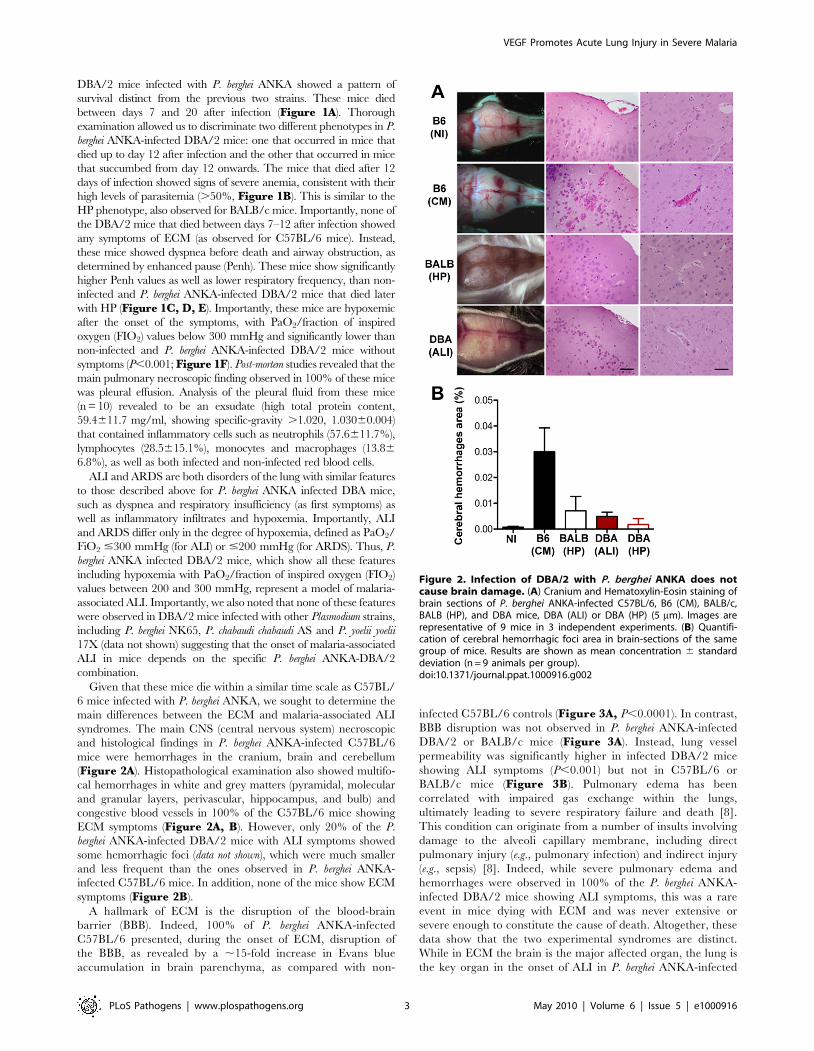
mice were hemorrhages in the cranium, brain and cerebellum
(Figure 2A). Histopathological examination also showed multifo-
cal hemorrhages in white and grey matters (pyramidal, molecular
and granular layers, perivascular, hippocampus, and bulb) and
congestive blood vessels in 100% of the C57BL/6 mice showing
ECM symptoms (Figure 2A, B). However, only 20% of the P.
berghei ANKA-infected DBA/2 mice with ALI symptoms showed
some hemorrhagic foci (data not shown), which were much smaller
and less frequent than the ones observed in P. berghei ANKA-
infected C57BL/6 mice. In addition, none of the mice show ECM
symptoms (Figure 2B).
A hallmark of ECM is the disruption of the blood-brain
barrier (BBB). Indeed, 100% of P. berghei ANKA-infected
C57BL/6 presented, during the onset of ECM, disruption of
the BBB, as revealed by a ,15-fold increase in Evans blue
accumulation in brain parenchyma, as compared with non-
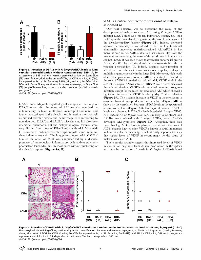
infected C57BL/6 controls (Figure 3A, P,0.0001). In contrast,
BBB disruption was not observed in P. berghei ANKA-infected
DBA/2 or BALB/c mice (Figure 3A). Instead, lung vessel
permeability was significantly higher in infected DBA/2 mice
showing ALI symptoms (P,0.001) but not in C57BL/6 or
BALB/c mice (Figure 3B). Pulmonary edema has been
correlated with impaired gas exchange within the lungs,
ultimately leading to severe respiratory failure and death [8].
This condition can originate from a number of insults involving
damage to the alveoli capillary membrane, including direct
pulmonary injury (e.g., pulmonary infection) and indirect injury
(e.g., sepsis) [8]. Indeed, while severe pulmonary edema and
hemorrhages were observed in 100% of the P. berghei ANKA-
infected DBA/2 mice showing ALI symptoms, this was a rare
event in mice dying with ECM and was never extensive or
severe enough to constitute the cause of death. Altogether, these
data show that the two experimental syndromes are distinct.
While in ECM the brain is the major affected organ, the lung is
the key organ in the onset of ALI in P. berghei ANKA-infected
Figure 2. Infection of DBA/2 with P. berghei ANKA does notcause brain damage. (A) Cranium and Hematoxylin-Eosin staining ofbrain sections of P. berghei ANKA-infected C57BL/6, B6 (CM), BALB/c,BALB (HP), and DBA mice, DBA (ALI) or DBA (HP) (5 mm). Images arerepresentative of 9 mice in 3 independent experiments. (B) Quantifi-cation of cerebral hemorrhagic foci area in brain-sections of the samegroup of mice. Results are shown as mean concentration 6 standarddeviation (n = 9 animals per group).doi:10.1371/journal.ppat.1000916.g002
VEGF Promotes Acute Lung Injury in Severe Malaria
PLoS Pathogens | www.plospathogens.org 3 May 2010 | Volume 6 | Issue 5 | e1000916
DBA/2 mice. Major histopathological changes in the lungs of
DBA/2 mice after the onset of ALI are characterized by
inflammatory cellular infiltration (neutrophil-dominant and
foamy macrophages in the alveolar and interstitial sites) as well
as marked alveolar edema and hemorrhage. It is interesting to
note that both DBA/2 and BALB/c mice showing HP also show
interstitial pneumonia but the histopathological features were
very distinct from those of DBA/2 mice with ALI. Mice with
HP showed a thickened alveolar septum with some mononu-
clear inflammatory cells. The lung pattern observed in C57BL/
6 after the onset of ECM was characterized by a discrete
presence of mononuclear inflammatory cells and/or polymor-
phonuclear leucocytes but, in most cases without thickening of
the alveolar septum (Figure 4A, B).
VEGF is a critical host factor for the onset of malaria-associated ALI
Our next objective was to determine the cause of the
development of malaria-associated ALI, using P. berghei ANKA-
infected DBA/2 mice as a model. Pulmonary edema, i.e., fluid
build-up in the lung alveoli, originates in the loss of the integrity of
the alveolar-capillary barrier (Figure 3B). Indeed, increased
alveolar permeability is considered to be the key functional
abnormality underlying malaria-associated ALI/ARDS in hu-
mans, as seen in ALI/ARDS due to other causes. However, the
mechanisms underlying the onset of this syndrome in humans are
still not known. It has been shown that vascular endothelial growth
factor, VEGF, plays a critical role in angiogenesis but also in
vascular permeability [9]. Indeed, systemic overexpression of
VEGF has been shown to cause widespread capillary leakage in
multiple organs, especially in the lungs [10]. Moreover, high levels
of VEGF in plasma were found in ARDS patients [11]. To address
the role of VEGF in malaria-associated ALI, VEGF levels in the
sera of P. berghei ANKA-infected DBA/2 mice were measured
throughout infection. VEGF levels remained constant throughout
infection, except for the mice that developed ALI, which showed a
significant increase in VEGF levels by day 7 after infection
(Figure 5A). The systemic increase in VEGF in the sera seems to
originate from de novo production in the spleen (Figure 5B), as
shown by the correlation between mRNA levels in the spleen and
serum protein levels (Figure 5C). No major alterations of VEGF
levels were observed in DBA/2 mice infected with P. berghei NK65,
P. c. chabaudi AS or P. yoelii yoelii 17X, similarly to C57BL/6 and
BALB/c mice infected with P. berghei ANKA, none of which
developed ALI symptoms (Figure 5D). Altogether, these data
show that high VEGF levels in plasma correlate with the onset of
ALI in malaria-infected mice. VEGF is known to cause an increase
in lung vascular permeability, which strongly supports the idea
that higher levels of VEGF in serum might be the cause of
malaria-associated ALI.
These results strongly suggest that increased levels of VEGF
in circulation originate from de novo production in the spleen
and may be the cause of death in P. berghei ANKA-infected
Figure 3. Infection of DBA/2 with P. berghei ANKA leads to lungvascular permeabilization without compromising BBB. (A, B)Assessment of BBB and lung vascular permeabilization by Evans Blue(EB) quantification, during the onset of ECM, i.e. C57BL/6 mice, B6 (CM),hyperparasitemia, i.e. BALB/c mice, BALB (HP), and ALI, i.e. DBA mice,DBA (ALI). Evans Blue quantification is shown as mean mg of Evans Blue(EB) per g of brain or lung tissue 6 standard deviation (n = 5–11 animalsper group).doi:10.1371/journal.ppat.1000916.g003
Figure 4. Infection of DBA/2 with P. berghei ANKA constitutes a rodent model for malaria-associated acute lung injury (ALI). (A–C)Hematoxylin-Eosin staining of lung sections (5 mm) and quantification of edema and haemorrhages, using a blinded scoring system (1-mild, 4-severe),during the onset of ECM, i.e. C57BL/6 mice, B6 (CM), hyperparasitemia, i.e. BALB/c mice, BALB (HP), and ALI, i.e. DBA mice, DBA (ALI). Images arerepresentative of 9 mice in 3 independent experiments. The bar corresponds to 100 mm.doi:10.1371/journal.ppat.1000916.g004
VEGF Promotes Acute Lung Injury in Severe Malaria
PLoS Pathogens | www.plospathogens.org 4 May 2010 | Volume 6 | Issue 5 | e1000916

DBA/2 mice that develop ALI. Thus, we next asked whether
P. berghei ANKA-infected splenectomized DBA/2 mice would
be protected from developing ALI. The results clearly show
that the spleen is required for the onset of malaria-associated
ALI, which correlates with VEGF levels in circulation
(Figura 6A–C).
Infected DBA/2 mice that did not develop ALI not only
showed unaltered levels of VEGF in the sera (Figure 5A), but
Figure 5. VEGF correlates with ALI onset during malaria infection. (A) Levels of VEGF protein in the sera of P. berghei ANKA-infected DBA mice withALI and hyperparasitemia (HP), compared to non-infected mice (NI). Results are shown as mean concentration 6 standard deviation (n = 6, 23 and 46 mousesera per group, for NI, ALI and HP, respectively). (B) Expression of VEGF mRNA levels in the lung, spleen, liver and kidney of P. berghei ANKA-infected DBAmice with ALI and HP (n = 15 animals per group), when compared to non-infected mice. (C) Correlation between VEGF protein levels in the serum andmRNA expression of VEGF in the spleen of P. berghei ANKA-infected DBA mice with ALI. (D) Levels of VEGF protein in the sera of different strains of mice(C57BL/6, BALB/c and DBA) infected with different Plasmodia (P. berghei ANKA, P. berghei NK65 - PbNK, P. yoelii yoelii 17X – Py, and P. chabaudi chabaudi AS -Pc) (n = 10 animals per group). Results are shown as mean concentration 6 standard deviation. (*P,0.001).doi:10.1371/journal.ppat.1000916.g005
Figure 6. Spleen is required for the onset of malaria-associated ALI. (A) Survival and (B) parasitemia of splenectomised and control P.berghei ANKA-infected DBA mice. (C) Levels of VEGF in the serum of non-infected (NI) DBA mice, P. berghei ANKA-infected DBA mice with ALI andsplenectomised P. berghei ANKA-infected DBA mice were taken on the same day as control DBA infected mice developed ALI. Results are shown asmean concentration 6 standard deviation. (*P,0.01).doi:10.1371/journal.ppat.1000916.g006
VEGF Promotes Acute Lung Injury in Severe Malaria
PLoS Pathogens | www.plospathogens.org 5 May 2010 | Volume 6 | Issue 5 | e1000916
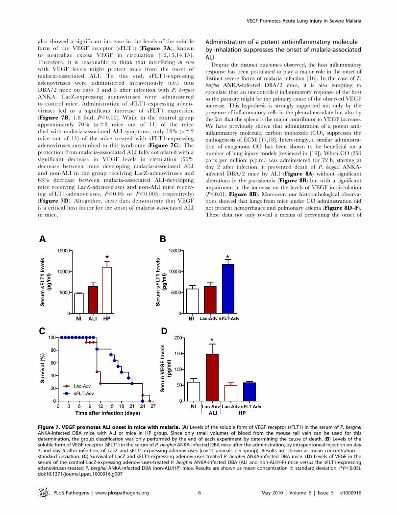
also showed a significant increase in the levels of the soluble
form of the VEGF receptor (sFLT1) (Figure 7A), known
to neutralize excess VEGF in circulation [12,13,14,15].
Therefore, it is reasonable to think that interfering in vivo
with VEGF levels might protect mice from the onset of
malaria-associated ALI. To this end, sFLT1-expressing
adenoviruses were administered intravenously (i.v.) into
DBA/2 mice on days 3 and 5 after infection with P. berghei
ANKA. LacZ-expressing adenoviruses were administered
to control mice. Administration of sFLT1-expressing adeno-
viruses led to a significant increase of sFLT1 expression
(Figure 7B, 1.8 fold, P,0.05). While in the control group
approximately 70% (n = 8 mice out of 11) of the mice
died with malaria-associated ALI symptoms, only 18% (n = 2
mice out of 11) of the mice treated with sFLT1-expressing
adenoviruses succumbed to this syndrome (Figure 7C). The
protection from malaria-associated ALI fully correlated with a
significant decrease in VEGF levels in circulation (66%
decrease between mice developing malaria-associated ALI
and non-ALI in the group receiving LacZ-adenoviruses and
63% decrease between malaria-associated ALI-developing
mice receiving LacZ-adenoviruses and non-ALI mice receiv-
ing sFLT1-adenoviruses, P,0.05 or P,0.005, respectively)
(Figure 7D). Altogether, these data demonstrate that VEGF
is a critical host factor for the onset of malaria-associated ALI
in mice.
Administration of a potent anti-inflammatory moleculeby inhalation suppresses the onset of malaria-associatedALI
Despite the distinct outcomes observed, the host inflammatory
response has been postulated to play a major role in the onset of
distinct severe forms of malaria infection [16]. In the case of P.
berghei ANKA-infected DBA/2 mice, it is also tempting to
speculate that an uncontrolled inflammatory response of the host
to the parasite might be the primary cause of the observed VEGF
increase. This hypothesis is strongly supported not only by the
presence of inflammatory cells in the pleural exsudate but also by
the fact that the spleen is the major contributor to VEGF increase.
We have previously shown that administration of a potent anti-
inflammatory molecule, carbon monoxide (CO), suppresses the
pathogenesis of ECM [17,18]. Interestingly, a similar administra-
tion of exogenous CO has been shown to be beneficial on a
number of lung injury models (reviewed in [19]). When CO (250
parts per million; p.p.m.) was administered for 72 h, starting at
day 2 after infection, it prevented death of P. berghei ANKA-
infected DBA/2 mice by ALI (Figure 8A) without significant
alterations in the parasitemia (Figure 8B) but with a significant
impairment in the increase on the levels of VEGF in circulation
(P,0.01; Figure 8B). Moreover, our histopathological observa-
tions showed that lungs from mice under CO administration did
not present hemorrhages and pulmonary edema (Figure 8D–F).
These data not only reveal a means of preventing the onset of
Figure 7. VEGF promotes ALI onset in mice with malaria. (A) Levels of the soluble form of VEGF receptor (sFLT1) in the serum of P. bergheiANKA-infected DBA mice with ALI or mice in HP group. Since only small volumes of blood from the mouse tail vein can be used for thisdetermination, the group classification was only performed by the end of each experiment by determining the cause of death. (B) Levels of thesoluble form of VEGF receptor (sFLT1) in the serum of P. berghei ANKA-infected DBA mice after the administration, by intraperitoneal injection on day3 and day 5 after infection, of LacZ and sFLT1-expressing adenoviruses (n = 11 animals per group). Results are shown as mean concentration 6standard deviation. (C) Survival of LacZ and sFLT1-expressing adenoviruses treated P. berghei ANKA-infected DBA mice. (D) Levels of VEGF in theserum of the control LacZ-expressing adenoviruses-treated P. berghei ANKA-infected DBA (ALI and non-ALI/HP) mice versus the sFLT1-expressingadenoviruses-treated P. berghei ANKA-infected DBA (non-ALI/HP) mice. Results are shown as mean concentration 6 standard deviation. (*P,0.05).doi:10.1371/journal.ppat.1000916.g007
VEGF Promotes Acute Lung Injury in Severe Malaria
PLoS Pathogens | www.plospathogens.org 6 May 2010 | Volume 6 | Issue 5 | e1000916

malaria-associated ALI but also strongly suggest that, as for ECM,
the host inflammatory response may play an important role in the
onset of this severe malaria syndrome.
Discussion
Once thought to be near eradication, malaria is now one of the
most prevalent infectious diseases worldwide, with a toll of nearly 1
million deaths per year in regions where infection is endemic.
These deaths are at the most severe end of a scale of pathologies
affecting approximately 500 million people per year and can be
due to the onset of distinct syndromes. These include cerebral
malaria (CM), acute lung injury (ALI), acute respiratory distress
syndrome (ARDS), and severe anemia, among other pathologies.
The outcome of infection is influenced by the genetics of both host
and parasite [4,20]. This is particularly visible in rodent models of
infection, as different strains of mice infected with different
Plasmodium strains develop a variety of pathologies, ranging from
lethal to self-resolving [18,21]. Rodent models that mimic certain
aspects of the human CM, anemia syndromes and pregnancy-
associated malaria have been established [6,22,23]. We now report
that DBA/2 mice infected with P. berghei ANKA constitute a model
for malaria-associated ALI, where the cause of death is respiratory
failure. It is important to note that P. berghei ANKA-infected DBA/
2 mice have been previously described as CM-resistant [24].
However, another report has described these mice as a resolving
CM model. Interestingly, the authors also noted changes in
vascular permeability in DBA/2 mice during what they called
‘‘mild cerebral malaria’’ phase. They further state that the even
distribution of these changes suggests a response to a circulating
factor, although they do not speculate on which factor that might
be [25]. Our present detailed pathological study, of the brain and
the lungs of P. berghei ANKA- infected DBA/2 mice, indicates that
the cause of death of these mice is respiratory failure.
In humans, while patients with uncomplicated malaria usually
present fever and non-specific symptoms, severe and complicated
malaria is characterized by multiorgan involvement including
ALI/ARDS. Recent years have witnessed a shift in the profile of
patients with complicated malaria (reviewed in [3]). Multi-organ
system failure and respiratory complications are being increasingly
reported not only for P. falciparum infections but also for malaria
caused by P. vivax [26,27,28,29], P. ovale [30] and P. malariae [31],
usually considered benign Plasmodium species. In fact, it has been
suggested that as many as 5% of patients with uncomplicated
malaria and 20–30% of patients with severe and complicated
malaria requiring intensive care unit admission may develop ALI/
ARDS, often after treatment has been initiated [3]. Pregnant
women with severe P. falciparum infection are particularly prone to
developing ALI/ARDS, which is associated with high mortality
[32,33]. It is therefore of the utmost importance that a rodent
model of such syndrome becomes available. Moreover, post-mortem
studies on human patients dying with severe P. falciparum malaria
have revealed histopathological findings, such as heavy edematous
lungs and hemorrhages [34,35], very similar to the ones we
describe here for P. berghei ANKA-infected DBA/2 mice
developing malaria-associated ALI. Mild lung pathology has been
previously reported in C57BL/6 mice infected with P. berghei
ANKA [36,37]. Our present study confirms that C57BL/6 mice
Figure 8. Exposure to CO suppresses onset of malaria-associated ALI. (A–B) Effect of CO inhalation on P. berghei ANKA-infected DBA micesurvival (A) and parasitemia (B). CO inhalation (starting at day 2 after infection and during 72 h) was compared to normal atmosphere. Parasitemiasare shown as mean 6 standard deviation (n = 10 animals per group). (C) Levels of VEGF in the sera of P. berghei ANKA-infected DBA mice exposed toair or CO. Results are shown as mean concentration 6 standard deviation (n = 3 mice per group). (*P,0.01) (D–F) Quantification of edema andhaemorrhages of hematoxylin-eosin-stained lung sections of non-infected DBA mice (NI DBA) versus P. berghei ANKA-infected DBA mice exposed toair, I DBA (ALI), or CO, I DBA+CO, using a blinded score system (1-mild, 4-severe). Images are representative of 6 mice in 2 independent experiments.doi:10.1371/journal.ppat.1000916.g008
VEGF Promotes Acute Lung Injury in Severe Malaria
PLoS Pathogens | www.plospathogens.org 7 May 2010 | Volume 6 | Issue 5 | e1000916

died with a significant loss of the integrity of the BBB, causing all
the ECM symptoms observed prior to death, but also showed
some level of lung pathology. However, none of those mice
presented pleural effusion or exsudate in their pleural cavities.
Moreover, while pulmonary edema and hemorrhages were
observed in 100% of the P. berghei ANKA-infected DBA/2 mice
showing ALI symptoms, this was a rare event in P. berghei ANKA-
infected C57BL/6 mice and was never severe enough to constitute
the cause of death. Plasmodium blood stage infection is known to
cause multi-organ pathology but the level of pathology varies from
organ to organ depending on the host-Plasmodium combination.
Here, we clearly show that infection of C57BL/6 or DBA/2 mice
with P. berghei ANKA results into two distinct models of severe
malaria; the former developing a neurological syndrome while the
latter causing death due to respiratory failure in approximately
half of the infected mice.
Importantly, our data also show that a host factor plays a
critical role in the establishment of malaria-associated ALI.
Indeed, the present data demonstrates that P. berghei ANKA only
causes malaria-associated ALI in DBA/2 mice. Interestingly,
DBA/2 mice have been shown to respond quite strongly to
angiogenic stimuli [38] and this might be the reason why a
proportion of these mice are not able to control the levels of
VEGF, leading to the onset of ALI during a P. berghei ANKA
infection. It should also be noted that a model named ‘‘malaria
lung syndrome’’, where C3H/z mice infected with P. berghei K173
also die very early in infection and show notably edematous lungs
and pleural effusion, has been described more than 25 years ago
[39]. Although it would be very interesting to test the levels of
VEGF in these mice, the unavailability of this strain of mice from
the major animal houses makes this experiment very difficult to
perform.
But why is VEGF responsible for the onset of malaria-
associated ALI? VEGF has long been known for its activity as a
regulator of vessel permeability [13]. In fact VEGF was primarily
termed vascular permeability factor, for its ability to induce
vascular leakage, rather than for its growth factor activity [40].
VEGF increases vascular permeability 50,000 times more
efficiently than does histamine [41]. Interestingly, VEGF also
plays a central role in the formation and maintenance of lung
vasculature [42]. However, when VEGF levels are altered, lung
disease frequently follows. Plasma VEGF levels in subjects with
non-malaria ALI/ARDS are strongly elevated compared to
controls and values higher than two-fold have been associated
with mortality [11]. The association between VEGF levels and
mortality due to respiratory failure does not mean that VEGF
effects are restricted to the lung, but simply highlights the
importance of vascular integrity for lung function. Another
example in which VEGF and lung injury are involved in response
to a pathogenic microorganism has recently been reported [43].
Pseudomonas aeruginosa is a pathogenic bacterium that colonizes the
lungs and may lead to lung disease in immunocompromized
patients. Interestingly, while aerosol delivery of this bacterium
causes fatal disease in DBA/2 mice, other mouse strains are able
to resolve infection. DBA/2 mice display progressive deteriora-
tion of lung pathology with extensive alveolar exsudate and
edema formation together with significantly increase levels of
VEGF that seem to result from an uncontrolled host inflamma-
tory response [43]. Indeed, a cross-talk between angiogenesis and
inflammation has long been proposed [44]. Similarly, P. berghei
ANKA-infected DBA/2 mice treated with a potent anti-
inflammatory molecule prior to the onset of ALI show
significantly reduced levels of VEGF in sera and are fully
protected from this syndrome of severe malaria.
Numerous studies have measured VEGF levels in malaria
patients [45,46,47] but none of these studies included a group of
individuals for which the cause of death was ALI/ARDS. On the
other hand, it was recently shown that P. falciparum-infected red
blood cells induce VEGF secretion from human mast cells, a cell
population highly represented in the spleen [48]. Importantly,
while ALI affects pregnant women infected with P. falciparum [32],
the VEGF pathway seems to play an important role during
chronic placental malaria and hypertension in first-time mothers
[49]. It remains to be established whether these observations are in
any way connected. The similarities between the physiopatholog-
ical lesions described in the rodent model reported here and those
occurring in humans pave the way for a better understanding of
the malaria-associated pathology and may contribute to the design
of novel rational intervention strategies.
Methods
MiceC57BL/6, BALB/c and DBA-2 mice were bred and housed in
the specific pathogen-free facilities of the Instituto de Gulbenkian
de Ciencia. The mice were then transferred to the Instituto de
Medicina Molecular at least 72 h prior to experimentation. All
protocols were approved by the Animal Care Committee of the
Instituto de Medicina Molecular, following Institutional, National,
and European Union guidelines.
Parasites, infection and disease assessmentP. berghei ANKA, P. berghei NK65, P. yoelii 17X or P. chabaudi AS
were used after one in vivo passage in C57BL/6, BALB/c or DBA-
2 mice. Mice were infected via intraperitoneal (ip) inoculation with
106–107 infected red blood cells. Infected mice were monitored
twice daily for clinical symptoms of ECM including hemi- or
paraplegia, head deviation, tendency to roll-over on stimulation,
ataxia and convulsions or ALI, including dyspnea. Parasitemia was
determined by Giemsa staining followed by microscopic counting
and expressed as percentage of infected red blood cells.
HistopathologyBrains or lungs were harvested from mice under different
experimental conditions when clinical signs of ECM, ALI or HP
were noticed. Tissues were fixed in buffered 10% (v/v) formaldehyde
for paraffin embedding and Hematoxylin-Eosin staining.
Determination of airway obstructionPulmonary function was assessed in unrestrained conscious mice
placed in a barometric plethysmographic chamber (Buxco
Electronics, Sharon, CT), where respiratory parameters were
measured every day for 10 minutes. Since these measurements can
be performed every day in the same group of mice, the group
classification was only performed by the end of each experiment
after determining the cause of death. The enhanced pause (Penh),
a dimensionless value indicative of airway obstruction, as well as
respiratory frequency, were used to determine respiratory
resistance and were calculated as previously described [50].
Measurement of PaO2 in arterial bloodMice were gently heated in their cages with a heat lamp to
increase peripheral blood flow. The mice were then restrained
in a restraining device, and the ventral artery of the tail was
nicked by carefully plunging a small scalpel blade diagonally
into the artery. Heparin was swabbed onto the skin before it was
cut to minimize clotting. About 100 mL of blood was collected
in a lithium-heparin (50 IU/ml) containing capillary tube Blood
VEGF Promotes Acute Lung Injury in Severe Malaria
PLoS Pathogens | www.plospathogens.org 8 May 2010 | Volume 6 | Issue 5 | e1000916
in the capillary tube was mixed by placing a small metal
fragment into the tube and then passing a magnet along the
length of the tube several times. The samples were analyzed
immediately with i-STAT cartridge CG8+ (pH, PCO2, PO2,
Na, K, iCA, Glu, Hct) using the i-STATH System Analyzer
(Abbott Laboratories).
BBB and lung permeabilityMice were injected intravenously (iv) with 0.2 ml of 1–2%
Evans Blue (Sigma) when clinical symptoms of ECM, ALI or HP
were noticed. Mice were sacrificed one hour later and brains or
lungs were weighted and placed in formamide (2 ml) (Merck)
(37uC, 48 h) to extract Evans Blue dye from the tissue.
Absorbance was measured at l= 620 nm (Bio Rad SmartSpec
3000). Evans Blue concentration was calculated from a standard
curve and is expressed as mg of Evans Blue per g of brain or lung
tissue.
CO exposureMice were placed in a gastight 60 L capacity chamber and
exposed to CO for the times indicated, as described elsewhere
[18]. Briefly, 1% CO (Aga Linde) was mixed with air in a stainless
steel cylinder to obtain a final concentration of 250 ppm. CO was
provided continuously at a flow rate of ,12 L/min. CO
concentration was monitored using a CO analyzer (Interscan
Corporation, Chatsworth). Controls were maintained in a similar
chamber without CO.
Protein levels determinationMouse VEGF and sFLT1 levels in plasma or serum samples
were determined using a commercial ELISA kit (R&D Systems)
following the manufacturer’s instructions. Once again, and since
only small volumes of blood from the mouse tail vein can be used
for this determination, the group classification was only
performed by the end of each experiment after determining the
cause of death.
Quantitative RT-PCRExtraction of total RNA from lungs, spleen, liver and kidney,
from mice with ALI and HP symptoms, was performed using
RNeasy Mini Kit (Qiagen), according to the manufacturer’s
instructions. Non-infected mice were used as controls and as
baseline levels. After extraction, RNA concentration and quality
were determined using a NanoDrop ND-100 spectrophotometer
(NanoDrop Technologies). One microgram of total RNA was
reverse-transcribed to single-strand cDNA using the AMV
Reverse Transcriptase protocol (Roche Applied Science). VEGF
transcripts in the cDNA pool obtained from the reverse
transcriptase reaction were quantified by real-time quantitative
fluorogenic PCR. SYBR Green PCR Master Mix (Applied
Biosystems) was used to quantify gene expression according to the
manufacturer’s instructions.
RNA expression levels were calculated using the ABIPrism 7000
SDS Software, and normalized against the expression levels of the
housekeeping gene hypoxanthine guanine phosphoribosyltransfer-
ase (HPRT).
Adenovirus productionAn adenoviral vector carrying the sFLT1 gene was produced
using the same LR Clonase II enzyme recombination reaction as
described above, but using the pAd/CMV/V5-DEST Gateway
vector (Ad; Invitrogen) as destination vector. Once the sFLT1-
containing Ad vector was established, an adenoviral stock was
produced. A vector containing the LacZ gene was used as a
control. After purification from the enzymatic reaction, the Pac I-
digested vectors were transfected into 293A cells, with Lipofecta-
mine 2000 (Invitrogen) as the transfection reagent in Opti-MEM I
Medium (Gibco/Invitrogen) without serum. Cells were incubated
overnight in a 5% CO2 incubator at 37uC. Media were replaced
the following day with complete medium (DMEM with 10%
Foetal Calf Serum, 2 mM glutamine, 0.1 mM non essential
aminoacids and 100 U/mL penicillin, 0.1 mg/mL streptomycin).
Forty-eight hours post-transfection, cells were trypsinized and
transfered to sterile 10 cm tissue culture plates containing 10 mL
complete medium. Media were replaced every other day until day
8, when visible regions of cytopathic effect (CPE) were observed.
Infection was allowed to proceed for an additional 2 days until
,80% CPE was observed. Adenovirus-containing cells were
harvested by squirting cells off the plate with a pipette. A crude
viral lysate was prepared by 3 consecutive freeze-thaw cycles (30
minutes at 280uC, followed by 15 minutes at 37uC). This crude
lysate was further amplified by infection of 293A cells. After 3
days, amplified viral stocks were obtained using the freeze-thaw
procedure described before. Amplified adenoviral stocks were
titered using 293A cells and stored at 2 80uC until use.
Statistical analysisFor samples in which n.5, statistical analysis were performed
using unpaired Student t or ANOVA parametric tests. Normal
distributions were confirmed using the Kolmogorov-Smirnov test.
For samples in which n,5, statistical analysis were performed
using Kruskall-Wallis or Wilcoxon non-parametric tests. All
survival curves were compared using Student t, Mann-Whitney e
Kolmogorov-Smirnov tests. P,0.05 was considered significant.
Acknowledgments
We thank Nuno Sepulveda (Instituto Gulbenkian de Ciencia) for statistical
analysis, Silvia Portugal for help with the animal experiments and Bruno
Silva-Santos and Miguel Prudencio (IMM) for critically reviewing the
manuscript. We also thank Sonia Martins de Oliveira (Instituto de
Histologia e Biologia do Desenvolvimento, Faculdade de Medicina de
Lisboa) and Mario Alberto Ferreira Matos (Departamento de Anatomia
Patologica, Hospital Santa Maria) for help in histopathology studies. We
also acknowledge M. Russo and Eliane Gomes (Departamento de
Imunologia, Universidade de Sao Paulo, Brasil) for expert technical
assistance with the Buxco Electronics machine.
Author Contributions
Conceived and designed the experiments: SE MGC AP SD MMM.
Performed the experiments: SE MGC AP DC ACP RA CRFM. Analyzed
the data: SE MGC AP DC ACS CRFM SD MMM. Contributed
reagents/materials/analysis tools: CAAM NF. Wrote the paper: SE MGC
AP SD MMM.
References
1. Haldar K, Murphy SC, Milner DA, Taylor TE (2007) Malaria: mechanisms of
erythrocytic infection and pathological correlates of severe disease. Annu Rev
Pathol 2: 217–249.
2. Prudencio M, Rodriguez A, Mota MM (2006) The silent path to thousands of
merozoites: the Plasmodium liver stage. Nat Rev Microbiol 4: 849–856.
3. Mohan A, Sharma SK, Bollineni S (2008) Acute lung injury and acute
respiratory distress syndrome in malaria. J Vector Borne Dis 45: 179–
193.
4. Prudencio M, Rodrigues CD, Mota MM (2007) The relevance of host genes in
malaria. SEB Exp Biol Ser 58: 47–91.
VEGF Promotes Acute Lung Injury in Severe Malaria
PLoS Pathogens | www.plospathogens.org 9 May 2010 | Volume 6 | Issue 5 | e1000916

5. Pamplona A, Hanscheid T, Epiphanio S, Mota MM, Vigario AM (2009)
Cerebral malaria and the hemolysis/methemoglobin/heme hypothesis: shed-
ding new light on an old disease. Int J Biochem Cell Biol 41: 711–716.
6. Lamb TJ, Brown DE, Potocnik AJ, Langhorne J (2006) Insights into the
immunopathogenesis of malaria using mouse models. Expert Rev Mol Med 8:
1–22.
7. Schofield L, Grau GE (2005) Immunological processes in malaria pathogenesis.
Nat Rev Immunol 5: 722–735.
8. Luh SP, Chiang CH (2007) Acute lung injury/acute respiratory distress
syndrome (ALI/ARDS): the mechanism, present strategies and future
perspectives of therapies. J Zhejiang Univ Sci B 8: 60–69.
9. Connolly DT (1991) Vascular permeability factor: a unique regulator of blood
vessel function. J Cell Biochem 47: 219–223.
10. Kaner RJ, Ladetto JV, Singh R, Fukuda N, Matthay MA, et al. (2000) Lung
overexpression of the vascular endothelial growth factor gene induces pulmonary
edema. Am J Respir Cell Mol Biol 22: 657–664.
11. Thickett DR, Armstrong L, Christie SJ, Millar AB (2001) Vascular endothelial
growth factor may contribute to increased vascular permeability in acute
respiratory distress syndrome. Am J Respir Crit Care Med 164: 1601–1605.
12. Barleon B, Siemeister G, Martiny-Baron G, Weindel K, Herzog C, et al. (1997)
Vascular endothelial growth factor up-regulates its receptor fms-like tyrosine
kinase 1 (FLT-1) and a soluble variant of FLT-1 in human vascular endothelial
cells. Cancer Res 57: 5421–5425.
13. Ferrara N, Chen H, Davis-Smyth T, Gerber HP, Nguyen TN, et al. (1998)
Vascular endothelial growth factor is essential for corpus luteum angiogenesis.
Nat Med 4: 336–340.
14. Goldman CK, Kendall RL, Cabrera G, Soroceanu L, Heike Y, et al. (1998)
Paracrine expression of a native soluble vascular endothelial growth factor
receptor inhibits tumor growth, metastasis, and mortality rate. Proc Natl Acad
Sci U S A 95: 8795–8800.
15. Keyt BA, Berleau LT, Nguyen HV, Chen H, Heinsohn H, et al. (1996) The
carboxyl-terminal domain (111–165) of vascular endothelial growth factor is
critical for its mitogenic potency. J Biol Chem 271: 7788–7795.
16. Clark IA, Alleva LM, Mills AC, Cowden WB (2004) Pathogenesis of malaria and
clinically similar conditions. Clin Microbiol Rev 17: 509–539, table of contents.
17. Pamplona A, Clark IA, Mota MM (2007) Severe malaria increases the list of
heme oxygenase-1-protected diseases. Future Microbiol 2: 361–363.
18. Pamplona A, Ferreira A, Balla J, Jeney V, Balla G, et al. (2007) Heme
oxygenase-1 and carbon monoxide suppress the pathogenesis of experimental
cerebral malaria. Nat Med 13: 703–710.
19. Jin Y, Choi AM (2005) Cytoprotection of heme oxygenase-1/carbon monoxide
in lung injury. Proc Am Thorac Soc 2: 232–235.
20. Su X, Hayton K, Wellems TE (2007) Genetic linkage and association analyses
for trait mapping in Plasmodium falciparum. Nat Rev Genet 8: 497–506.
21. Mota MM, Brown KN, Holder AA, Jarra W (1998) Acute Plasmodium
chabaudi chabaudi malaria infection induces antibodies which bind to the
surfaces of parasitized erythrocytes and promote their phagocytosis by
macrophages in vitro. Infect Immun 66: 4080–4086.
22. Lamikanra AA, Brown D, Potocnik A, Casals-Pascual C, Langhorne J, et al.
(2007) Malarial anemia: of mice and men. Blood 110: 18–28.
23. Neres R, Marinho CR, Goncalves LA, Catarino MB, Penha-Goncalves C (2008)
Pregnancy outcome and placenta pathology in Plasmodium berghei ANKA
infected mice reproduce the pathogenesis of severe malaria in pregnant women.
PLoS One 3: e1608.
24. Delahaye NF, Coltel N, Puthier D, Flori L, Houlgatte R, et al. (2006) Gene-
expression profiling discriminates between cerebral malaria (CM)-susceptible
mice and CM-resistant mice. J Infect Dis 193: 312–321.
25. Neill AL, Hunt NH (1992) Pathology of fatal and resolving Plasmodium berghei
cerebral malaria in mice. Parasitology 105 (Pt 2): 165–175.
26. Agarwal R, Nath A, Gupta D (2007) Noninvasive ventilation in Plasmodium
vivax related ALI/ARDS. Intern Med 46: 2007–2011.
27. Kumar S, Melzer M, Dodds P, Watson J, Ord R (2007) P. vivax malaria
complicated by shock and ARDS. Scand J Infect Dis 39: 255–256.
28. Lomar AV, Vidal JE, Lomar FP, Barbas CV, de Matos GJ, et al. (2005) Acute
respiratory distress syndrome due to vivax malaria: case report and literaturereview. Braz J Infect Dis 9: 425–430.
29. Price L, Planche T, Rayner C, Krishna S (2007) Acute respiratory distress
syndrome in Plasmodium vivax malaria: case report and review of the literature.Trans R Soc Trop Med Hyg 101: 655–659.
30. Lee EY, Maguire JH (1999) Acute pulmonary edema complicating ovalemalaria. Clin Infect Dis 29: 697–698.
31. Lozano F, Leal M, Lissen E, Munoz J, Bautista A, et al. (1983) [P. falciparum
and P. malariae malaria complicated by pulmonary edema with disseminatedintravascular coagulation]. Presse Med 12: 3004–3005.
32. Taylor WR, Canon V, White NJ (2006) Pulmonary manifestations of malaria:recognition and management. Treat Respir Med 5: 419–428.
33. Taylor WR, White NJ (2002) Malaria and the lung. Clin Chest Med 23:457–468.
34. James MF (1985) Pulmonary damage associated with falciparum malaria: a
report of ten cases. Ann Trop Med Parasitol 79: 123–138.35. Tong MJ, Ballantine TV, Youel DB (1972) Pulmonary function studies in
Plasmodium falciparum malaria. Am Rev Respir Dis 106: 23–29.36. Lovegrove FE, Gharib SA, Pena-Castillo L, Patel SN, Ruzinski JT, et al. (2008)
Parasite burden and CD36-mediated sequestration are determinants of acute
lung injury in an experimental malaria model. PLoS Pathog 4: e1000068.37. Piguet PF, Kan CD, Vesin C, Rochat A, Donati Y, et al. (2001) Role of CD40-
CVD40L in mouse severe malaria. Am J Pathol 159: 733–742.38. Shaked Y, Bertolini F, Man S, Rogers MS, Cervi D, et al. (2005) Genetic
heterogeneity of the vasculogenic phenotype parallels angiogenesis; Implicationsfor cellular surrogate marker analysis of antiangiogenesis. Cancer Cell 7:
101–111.
39. Weiss ML, Kubat K (1983) Plasmodium berghei: a mouse model for the‘‘sudden death’’ and ‘‘malarial lung’’ syndromes. Exp Parasitol 56: 143–151.
40. Senger DR, Galli SJ, Dvorak AM, Perruzzi CA, Harvey VS, et al. (1983) Tumorcells secrete a vascular permeability factor that promotes accumulation of ascites
fluid. Science 219: 983–985.
41. Zebrowski BK, Yano S, Liu W, Shaheen RM, Hicklin DJ, et al. (1999) Vascularendothelial growth factor levels and induction of permeability in malignant
pleural effusions. Clin Cancer Res 5: 3364–3368.42. Papaioannou AI, Kostikas K, Kollia P, Gourgoulianis KI (2006) Clinical
implications for vascular endothelial growth factor in the lung: friend or foe?Respir Res 7: 128.
43. Wilson KR, Napper JM, Denvir J, Sollars VE, Yu HD (2007) Defect in early
lung defence against Pseudomonas aeruginosa in DBA/2 mice is associated withacute inflammatory lung injury and reduced bactericidal activity in naive
macrophages. Microbiology 153: 968–979.44. Mor F, Quintana FJ, Cohen IR (2004) Angiogenesis-inflammation cross-talk:
vascular endothelial growth factor is secreted by activated T cells and induces
Th1 polarization. J Immunol 172: 4618–4623.45. Armah HB, Wilson NO, Sarfo BY, Powell MD, Bond VC, et al. (2007)
Cerebrospinal fluid and serum biomarkers of cerebral malaria mortality inGhanaian children. Malar J 6: 147.
46. Jain V, Armah HB, Tongren JE, Ned RM, Wilson NO, et al. (2008) Plasma IP-10, apoptotic and angiogenic factors associated with fatal cerebral malaria in
India. Malar J 7: 83.
47. Yeo TW, Lampah DA, Gitawati R, Tjitra E, Kenangalem E, et al. (2008)Angiopoietin-2 is associated with decreased endothelial nitric oxide and poor
clinical outcome in severe falciparum malaria. Proc Natl Acad Sci U S A 105:17097–17102.
48. Furuta T, Kimura M, Watanabe N Elevated levels of vascular endothelial
growth factor (VEGF) and soluble vascular endothelial growth factor receptor(VEGFR)-2 in human malaria. Am J Trop Med Hyg 82: 136–139.
49. Muehlenbachs A, Mutabingwa TK, Edmonds S, Fried M, Duffy PE (2006)Hypertension and maternal-fetal conflict during placental malaria. PLoS Med 3:
e446.
50. Hamelmann E, Schwarze J, Takeda K, Oshiba A, Larsen GL, et al. (1997)Noninvasive measurement of airway responsiveness in allergic mice using
barometric plethysmography. Am J Respir Crit Care Med 156: 766–775.
VEGF Promotes Acute Lung Injury in Severe Malaria
PLoS Pathogens | www.plospathogens.org 10 May 2010 | Volume 6 | Issue 5 | e1000916