Inpatient Mortality in Children With Clinically Diagnosed Malaria As Compared With Microscopically...
12
Inpatient Mortality in Children With Clinically Diagnosed Malaria As Compared With Microscopically Confirmed Malaria Robert O. Opoka, MMed, MPH * , Zongqi Xia, MD, PHD † , Paul Bangirana, MSc ‡ , and Chandy C. John, MD, MS § * Department of Paediatrics and Child Health, Mulago Hospital/Makerere University Medical School, Kampala, Uganda † Department of Neurology, Harvard Medical School, Massachusetts General Hospital, Brigham and Women’s Hospital, Boston, MA ‡ Department of Mental Health and Community Psychology, Makerere University Institute of Psychology, Kampala, Uganda § Global Pediatrics Program, Department of Pediatrics, University of Minnesota, Minneapolis, MN Abstract Background—Inpatient treatment for malaria without microscopic confirmation of the diagnosis occurs commonly in sub-Saharan Africa. Differences in mortality in children who are tested by microscopy for Plasmodium falciparum infection as compared with those not tested are not well characterized. Methods—A retrospective chart review was conducted of all children up to 15 years of age admitted to Mulago Hospital, Kampala, Uganda from January 2002 to July 2005, with a diagnosis of malaria and analyzed according to microscopy testing for P. falciparum. Results—A total of 23,342 children were treated for malaria during the study period, 991 (4.2%) of whom died. Severe malarial anemia in 7827 (33.5%) and cerebral malaria in 1912 (8.2%) were the 2 common causes of malaria-related admissions. Children who did not receive microscopy testing had a higher case fatality rate than those with a positive blood smear (7.5% versus 3.2%, P < 0.001). After adjustment for age, malaria complications, and comorbid conditions, children who did not have microscopy performed or had a negative blood smear had a higher risk of death than those with a positive blood smear [odds ratio (OR): 3.49, 95% confidence interval (CI): 2.88–4.22, P < 0.001; and OR: 1.59, 95% CI: 1.29–1.96, P < 0.001, respectively]. Conclusions—Diagnosis of malaria in the absence of microscopic confirmation is associated with significantly increased mortality in hospitalized Ugandan children. Inpatient diagnosis of malaria should be supported by microscopic or rapid diagnostic test confirmation. Keywords malaria; microscopy; Plasmodium falciparum; mortality; children The diagnosis of malaria in children attending a tertiary unit in an endemic area is not straightforward. Clinical manifestations of malaria overlap with those of other common infections, making definitive diagnosis difficult. 1-5 A history of fever and positive blood smear on light microscopy is the standard for malaria diagnosis and basis of treatment, but in practice this is not often adhered to. Microscopy is often not used even when it is available 6 and has Address for correspondence: Dr. Robert Opika Opoka, Department of Paediatrics and Child Health, Mulago Hospital/Makerere University Medical School, Box 7051 Kampala, Uganda. E-mail: [email protected]. NIH Public Access Author Manuscript Pediatr Infect Dis J. Author manuscript; available in PMC 2009 April 1. Published in final edited form as: Pediatr Infect Dis J. 2008 April ; 27(4): 319–324. doi:10.1097/INF.0b013e31815d74dd. NIH-PA Author Manuscript NIH-PA Author Manuscript NIH-PA Author Manuscript
Transcript of Inpatient Mortality in Children With Clinically Diagnosed Malaria As Compared With Microscopically...

Inpatient Mortality in Children With Clinically Diagnosed MalariaAs Compared With Microscopically Confirmed Malaria
Robert O. Opoka, MMed, MPH*, Zongqi Xia, MD, PHD†, Paul Bangirana, MSc‡, and ChandyC. John, MD, MS§
*Department of Paediatrics and Child Health, Mulago Hospital/Makerere University Medical School,Kampala, Uganda †Department of Neurology, Harvard Medical School, Massachusetts General Hospital,Brigham and Women’s Hospital, Boston, MA ‡Department of Mental Health and Community Psychology,Makerere University Institute of Psychology, Kampala, Uganda §Global Pediatrics Program, Departmentof Pediatrics, University of Minnesota, Minneapolis, MN
AbstractBackground—Inpatient treatment for malaria without microscopic confirmation of the diagnosisoccurs commonly in sub-Saharan Africa. Differences in mortality in children who are tested bymicroscopy for Plasmodium falciparum infection as compared with those not tested are not wellcharacterized.
Methods—A retrospective chart review was conducted of all children up to 15 years of age admittedto Mulago Hospital, Kampala, Uganda from January 2002 to July 2005, with a diagnosis of malariaand analyzed according to microscopy testing for P. falciparum.
Results—A total of 23,342 children were treated for malaria during the study period, 991 (4.2%)of whom died. Severe malarial anemia in 7827 (33.5%) and cerebral malaria in 1912 (8.2%) werethe 2 common causes of malaria-related admissions. Children who did not receive microscopy testinghad a higher case fatality rate than those with a positive blood smear (7.5% versus 3.2%, P < 0.001).After adjustment for age, malaria complications, and comorbid conditions, children who did not havemicroscopy performed or had a negative blood smear had a higher risk of death than those with apositive blood smear [odds ratio (OR): 3.49, 95% confidence interval (CI): 2.88–4.22, P < 0.001;and OR: 1.59, 95% CI: 1.29–1.96, P < 0.001, respectively].
Conclusions—Diagnosis of malaria in the absence of microscopic confirmation is associated withsignificantly increased mortality in hospitalized Ugandan children. Inpatient diagnosis of malariashould be supported by microscopic or rapid diagnostic test confirmation.
Keywordsmalaria; microscopy; Plasmodium falciparum; mortality; children
The diagnosis of malaria in children attending a tertiary unit in an endemic area is notstraightforward. Clinical manifestations of malaria overlap with those of other commoninfections, making definitive diagnosis difficult.1-5 A history of fever and positive blood smearon light microscopy is the standard for malaria diagnosis and basis of treatment, but in practicethis is not often adhered to. Microscopy is often not used even when it is available6 and has
Address for correspondence: Dr. Robert Opika Opoka, Department of Paediatrics and Child Health, Mulago Hospital/Makerere UniversityMedical School, Box 7051 Kampala, Uganda. E-mail: [email protected].
NIH Public AccessAuthor ManuscriptPediatr Infect Dis J. Author manuscript; available in PMC 2009 April 1.
Published in final edited form as:Pediatr Infect Dis J. 2008 April ; 27(4): 319–324. doi:10.1097/INF.0b013e31815d74dd.
NIH
-PA Author Manuscript
NIH
-PA Author Manuscript
NIH
-PA Author Manuscript

varying sensitivity and specificity according to the expertise of the personnel at the site oftesting.7-9 As a result it is not uncommon for malaria to be diagnosed and treated withoutmicroscopic confirmation or despite a negative blood smear.
The practice of diagnosis and treating children presumptively for malaria can result in excessivereporting of malaria cases,7 under-reporting of diseases that mimic malaria symptoms,10increased true or perceived antimalaria drug resistance, treatment of smear negative cases asmalaria6 and misallocation of resources, including overuse of artemisinin-based combinationtherapy.11 Few studies have examined the effect on inpatient mortality of diagnosing andtreating malaria without microscopic confirmation.
To assess how mortality differs in children with a diagnosis of malaria according to status ofmicroscopy testing for P. falciparum, we analyzed by retrospective chart review the differencesin inpatient mortality between clinically and microscopically diagnosed malaria in all malaria-related admissions in children in the national referral hospital in Uganda during a 3½ yearperiod.
MATERIALS AND METHODSStudy Type
A retrospective chart review was conducted of all malaria-related pediatric admissions aged0–15 years to Mulago Hospital, Kampala, Uganda between January 2002 and July 2005.
Study SiteMulago hospital is a 1500 bed national referral hospital in Uganda and a teaching hospital forMakerere University Medical School. It serves Kampala, the capital city, and surroundingdistricts in the central region of Uganda, which is an area of seasonal malaria transmission.12 Approximately 20,000 children are admitted to Mulago Hospital annually, of whom about30% are treated for malaria.13
Data CollectionAll inpatient charts of children up to 15 years of age with a diagnosis of malaria were retrievedusing the International Classification of Diseases (ICD 9) record codes and reviewed. Thediagnoses recorded on the files are the final diagnoses for which the patient was treated duringhospitalization. Information was collected from the patient’s charts on the child’s age, whetherdiagnostic microscopy for malaria had been performed (and if so, the results of microscopy),type of malaria complication, if any, other listed diagnoses, and outcome of hospitalization.Clinical diagnoses were recorded as listed in the chart (eg, cerebral malaria, severe malarialanemia, pneumonia), with or without supporting laboratory data. Accordingly, for these clinicaldata, WHO criteria might not be met (eg, cerebral malaria might be recorded in children withimpaired consciousness rather than coma, and severe malarial anemia might be recordedwithout laboratory measurement of hemoglobin level). Although this means that not all clinicaldiagnoses are supported by strict inclusion criteria, our data do reflect the practice anddiagnostic categorization in many hospitals in sub-Saharan Africa, where lack of resources andother factors often translate into diagnoses based purely on clinical criteria. Microscopy resultswere recorded as either not done, or negative or positive for Plasmodium species. The side-laboratory where all the malaria microscopy is done uses Field’s Method.14 To allow fordetection of even scanty parasitemia, thick films are routinely used. Although the specificPlasmodium parasite cannot be identified on thick films, malaria causing admission in this areais overwhelmingly caused by P. falciparum, so all cases are reported and treated as cases ofP. falciparum malaria. The laboratory is open from 7 AM to 10 PM, during which period themajority of patients are admitted. Patients admitted after 10 PM have malaria blood smears done
Opoka et al. Page 2
Pediatr Infect Dis J. Author manuscript; available in PMC 2009 April 1.
NIH
-PA Author Manuscript
NIH
-PA Author Manuscript
NIH
-PA Author Manuscript

the next morning. In addition to the side-laboratory the hospital has a main laboratory that iscapable of doing other diagnostic tests like blood counts, urinalysis, blood cultures and analysisof cerebral spinal fluid.
This study was approved by the Institutional Review Boards for Human Studies at MakerereUniversity Faculty of Medicine, Case Western Reserve University and the University ofMinnesota.
Data AnalysisData entry was done in Filemaker Pro 7. Analysis was done with STATA 9.2 (StataCorporation, Austin, TX). Frequencies were compared using the χ2 test or the χ2 test for trend,as appropriate. Multivariate logistic regression was used to assess the risk of mortality forspecific malaria complications, comorbid diagnoses, and microscopy testing status. P valuesof <0.05 were considered significant.
RESULTSMalaria Admissions and Microscopy Testing
A total of 23,342 children 15 years of age and under were admitted with a diagnosis of malaria.A peripheral blood smear for malarial parasites was done in 18,536 of these children (79%),of whom 12,256 (66%) had a positive result. Mean age did not differ between children whowere microscopy positive [mean age, 2.6 years; standard deviation (SD), 2.7], and those whowere microscopy negative (mean age, 2.6 years; SD, 2.8), but children who were not tested bymicroscopy were older (mean age, 3.1 years; SD, 3.6; P < 0.001 as compared with microscopypositive children). Gender frequencies were similar in the 3 groups, with a slight preponderanceof males in all the groups (54%, 53%, and 52% male for children who were microscopy positive,microscopy negative, and did not have microscopy testing for P. falciparum, respectively).Children who did not have a smear done on average died earlier after admission than those thathad a smear done (1.9 days versus 2.6 days, P = 0.01).
Malaria Seasonality and Overall Malaria-Attributed Morbidity and MortalityFrom January 2002 to July 2005, malaria-related admissions accounted for 30.4% of hospitaladmissions in children 15 years of age and younger and malaria-related deaths accounted for18.1% of total inpatient pediatric deaths. There was a significant increase in the proportion ofadmissions with a diagnosis of malaria from 2002–2004, but there was no significant increasein the proportion of inpatient deaths attributed to malaria during that period (Table 1).
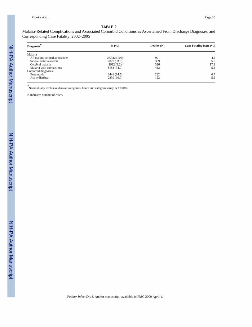
Case Fatality Rates for Malaria Complications and Comorbid ConditionsThe 3 most common complications of malaria were severe malaria anemia (SMA), cerebralmalaria (CM), and malaria associated with convulsions (Table 2). Pneumonia and acutediarrhea were the 2 most common comorbid diagnoses in children with malaria-relatedadmissions (Table 2). Other comorbid conditions occurring in >1% of children included upperrespiratory tract infection (1.3%), urinary tract infection (1.1%), and sickle cell disease (1.1%).The overall case fatality rate of malaria-related admissions during the study period was 4.2%.SMA and CM accounted for 72% of all malaria-related deaths.
Malaria-Associated Mortality According to Microscopic Confirmation of DiagnosisCase fatality rate (CFR) was assessed according to microscopy testing: blood smear for P.falciparum positive, negative, or not performed. The CFR for children with no microscopyperformed was more than twice that of children who were smear positive (7.5% versus 3.2%,respectively; P < 0.001), and children with no microscopy performed had higher CFR for each
Opoka et al. Page 3
Pediatr Infect Dis J. Author manuscript; available in PMC 2009 April 1.
NIH
-PA Author Manuscript
NIH
-PA Author Manuscript
NIH
-PA Author Manuscript

malaria complication and comorbid diagnosis than children who were smear positive (Table3). Similarly, among children in whom microscopy was performed, the CFR overall was higherin children who were microscopy negative than microscopy positive (3.8% versus 3.2%; P =0.03). This was largely because of a greater CFR for CM in microscopy negative as comparedwith microscopy positive children (17.7% versus 12.1%; P = 0.01; Table 3).
An increased risk of death was seen in children with CM in all 3 groups, and an increased riskof death for SMA was seen in children who were microscopy positive compared with childrenwithout these diagnoses. In addition, increased mortality was seen in children with a diagnosisof pneumonia in all 3 groups and with a diagnosis of acute diarrhea in children who were smearnegative or who did not have microscopy testing performed (Table 4). After adjustment forage, malaria complications and comorbid conditions, lack of microscopy testing and negativemicroscopy testing remained highly significant independent risk factors for death in childrenadmitted with a diagnosis of malaria (OR for no microscopy testing: 3.49, 95% CI: 2.88–4.23,P < 0.001; OR for microscopy negative: 1.59, 95% CI: 1.29 –1.96, P < 0.001; both as comparedwith children who were microscopy positive).
DISCUSSIONIn malaria endemic regions, there is a tendency to treat all fevers as malaria, particularly inhigh risk groups such as young children. The World Health Organization (WHO) guidelinesfor diagnosis and management of severe falciparum malaria,15 which have been adopted foruse here in Uganda, encourage the use of peripheral blood smear to confirm the diagnosis ofmalaria whenever possible. In routine clinical practice diagnosis and treatment of severefalciparum malaria without blood smear confirmation (presumptive diagnosis and treatment)is not uncommon even where facilities for microscopy do exist. For example, in the presentstudy a blood smear was not done in 21% of the malaria-related admissions. The most strikingfinding of the present study was the more than 3-fold increase in the risk of death in childrenwho were given a diagnosis of malaria without microscopy testing for P. falciparum comparedwith children who were microscopy positive for P. falciparum. Furthermore, when microscopywas performed on children suspected for malaria, more than a third of children weremicroscopy negative but were still diagnosed and treated for malaria, and these children alsohad a 1.5-fold increased risk of mortality as compared with microscopy-positive children.
Our study findings on mortality in microscopy negative as compared with positive childrenare similar to those of a prospective study conducted in Tanzania,8 which also documented ahigher case fatality rate in microscopy negative than in microscopy positive patients. TheTanzanian study was a prospective study with specific criteria for the malaria complicationsassessed, and so was able to define diagnoses with a degree of precision not possible in thepresent retrospective study. However, the design of the present study allowed us to answer aquestion of significant clinical importance in sub-Saharan Africa: how is lack of microscopytesting associated with outcome in patients admitted with a clinical diagnosis of malaria?
The higher mortality in children diagnosed and treated for malaria without microscopicconfirmation is likely due at least in part to misdiagnosis and a lack of treatment for conditionsother than malaria. We were not able to record complete treatment details on the patients inthis study, so we do not know how many of the children with a diagnosis of malaria receivedantibiotics in addition to antimalarials. At Mulago Hospital, antibiotics are usually given onlyto those with a listed diagnosis of a bacterial illness (eg, pneumonia or sepsis), so we suspectthat most of the children with a clinical diagnosis of malaria were not treated with antibiotics.Other causes of febrile illnesses in children, like bacteremia, meningitis, encephalitis or otherdiagnoses, may be obscured by the focus on malaria. A recent study in post neonatal infants(1–12 months) in Nigeria reported that 38.2% of children presenting with febrile illness had
Opoka et al. Page 4
Pediatr Infect Dis J. Author manuscript; available in PMC 2009 April 1.
NIH
-PA Author Manuscript
NIH
-PA Author Manuscript
NIH
-PA Author Manuscript
bacteremia16 and in another study 40% of children with febrile illness who were smear negativehad bacteremia compared with 12% in smear positives.17 Consideration should be given tothe possibility of both antimalarial therapy and empiric antibiotic therapy in the managementof severely ill children with febrile illnesses in malarial areas.
The risk of severe disease or death from malaria in children in malaria endemic areas supportsthe approach of presumptive treatment of febrile children in facilities where microscopy is notavailable.18,19 However, in this tertiary referral center, microscopy was readily available andis free of charge, yet it was frequently not performed, or when performed, not used inassignment of a diagnosis. Among children who had no microscopy testing performed,mortality was particularly increased in children with cerebral malaria, with a case fatality rateof 33.6% and an almost 7-fold increase in the risk of death in this group as compared with acase fatality rate of 12.1% and a 6-fold risk of death in children with cerebral malaria confirmedby blood smear. A case fatality rate this high strongly suggests the possibility of misdiagnosis,and underscores the critical need for microscopy testing in children thought to have severemalaria.
Among children with a negative blood smear, there was only a minor increase in the casefatality rate compared with children with a positive smear (3.8% versus 3.2%, P = 0.03), butafter controlling for other potential confounding factors such as age, type of malariacomplication and comorbid diagnosis, the increased risk of death was highly significant (OR:1.59, 95% CI: 1.29–1.96, P < 0.001). The increased risk of death in children with negativeblood smears is also most likely the result of misdiagnosis and lack of treatment for the realcause of illness in these children. However, it is possible that true infection was undetected insome children because of prior antimalarial treatment. In Uganda antimalarials are oftenpurchased from drug shops or pharmacies: a study conducted at 7 sites around the countryfound chloroquine metabolites in 32–80% of the general population.20 Much of this informaluse of antimalarial medications is associated with inappropriate dosing,21,22 which can leadto delayed parasite clearance and presentation to the hospital. This could lead to a more severelyill population on presentation and increased mortality in children who were smear negative. Inaddition, use of ineffective medications, even at the correct dose, may lead to persistentsymptoms despite low level parasitemia. There is also a need for public health policy makersto enforce the dispensing and proper use of antimalarials by the public.21
The reliance on microscopy to make a diagnosis of malaria has been subject of much debate.11,23,24 Accurate microscopy requires considerable experience and expertise and low levelparasitemias are sometimes not detected.25 Clinicians’ confidence in the accuracy ofmicroscopy may vary, depending on the area of testing and the expertise of microscopists inthis area. A lack of confidence in microscopy results may lead clinicians to prescribeantimalarial drugs even when the blood smear is negative to cover for potential missed casesof malaria. For example, a recent Kenyan study documented that although the results of theroutine negative blood slides were correct 92.7% of the time, 78.5% of patients with negativeblood slides were prescribed antimalarial medications.26 An alternative to microscopy is theuse of rapid diagnostic tests, or newer technologies like polymerase chain reaction methodsseveral of which have been shown to be sensitive, specific and stable under operationalconditions.27,28 Rapid diagnostic tests in particular, are generally easier to perform thanmicroscopy, requiring significantly less expertise, and may detect parasitemia in individualswho have taken antimalarial medications.29 Clinician education will be critical in any effortto improve accurate treatment of malaria, as recent studies from Tanzania and Zambiadocument that patients with negative rapid diagnostic tests were treated for malaria just asfrequently as patients with negative microscopy results.11,30
Opoka et al. Page 5
Pediatr Infect Dis J. Author manuscript; available in PMC 2009 April 1.
NIH
-PA Author Manuscript
NIH
-PA Author Manuscript
NIH
-PA Author Manuscript
Pneumonia and acute diarrhea were the most common comorbid conditions associated withmalaria and were both strong predictors of mortality, as has been documented in previousstudies.31,32 The diagnosis of pneumonia in a child with malaria might be a coexistingbacterial or viral respiratory illness, but the diagnosis might also be given to a child withmalaria-related respiratory distress, which is associated with a high mortality rate.31 Likewise,acute diarrhea might be a feature of clinical malaria,4 or the result of concurrent diarrhealdisease from an enteric pathogen.33 The increased mortality associated with these conditionsemphasizes the importance of prompt evaluation as to the cause of the condition, andappropriate cause-specific treatment after evaluation.
Drug resistance to the first-line drugs recommended for malaria at the time of this study(chloroquine and sulfadoxine-pyrimethamine) is the most likely cause of the increasedfrequency of malaria-related admissions in children from 2001–2004.34-36 The national drugpolicy for treatment of uncomplicated malaria in Uganda has since been changed toartemisinin-based combination therapy.37 This change was implemented widely in 2006, andit remains to be seen if this will result in a decrease in the proportion of admissions with adiagnosis of malaria.
The present study used the diagnoses assigned by the many different inpatient cliniciansadmitting these children. These diagnoses were not necessarily validated by rigorous oruniform criteria. For this reason, we cannot definitively conclude that all children with adiagnosis of cerebral malaria, for example, actually had cerebral malaria. Indeed, it is likelythat a significant proportion assigned this diagnosis without blood smear confirmation hadanother cause of their coma. But the latter case points out the practical relevance of chart reviewstudy such as the present study: clinical diagnosis of malaria, often without microscopyconfirmation, is not infrequent in hospitals and clinics throughout sub-Saharan Africa. Ourstudy results provide evidence that lack of testing in children with a diagnosis of malaria isassociated with significantly increased rates of death, and argue for microscopy or rapiddiagnostic testing of all children admitted with a diagnosis of malaria and evaluation of othercauses of disease in children with negative results. Based on the findings of previous studies,however, it is likely that reductions in mortality will only be accomplished with clinicianeducation on the utility of these tests, the risks of specific malaria complications and theimportance of evaluation for alternative diagnoses.
AcknowledgementsThis research was supported by a grant from the National Institutes of Health Fogarty International Center (grant R21TW-006794) to Dr. John and a Swedish International Development Agency (SIDA) Makerere University MedicalSchool/Mulago Hospital Program for the Development of Medical Research grant to Dr. Opoka.
References1. English M, Punt J, Mwangi I, McHugh K, Marsh K. Clinical overlap between malaria and severe
pneumonia in African children in hospital. Trans R Soc Trop Med Hyg 1996;90:658–662. [PubMed:9015508]
2. Berkley J, Mwarumba S, Bramham K, Lowe B, Marsh K. Bacteraemia complicating severe malariain children. Trans R Soc Trop Med Hyg 1999;93:283–286. [PubMed: 10492760]
3. Berkley JA, Mwangi I, Mellington F, Mwarumba S, Marsh K. Cerebral malaria versus bacterialmeningitis in children with impaired consciousness. QJM 1999;92:151–157. [PubMed: 10326074]
4. English M, Berkley J, Mwangi I, et al. Hypothetical performance of syndrome-based management ofacute paediatric admissions of children aged more than 60 days in a Kenyan district hospital. BullWorld Health Organ 2003;81:166–173. [PubMed: 12764512]
Opoka et al. Page 6
Pediatr Infect Dis J. Author manuscript; available in PMC 2009 April 1.
NIH
-PA Author Manuscript
NIH
-PA Author Manuscript
NIH
-PA Author Manuscript

5. Berkley JA, Maitland K, Mwangi I, et al. Use of clinical syndromes to target antibiotic prescribing inseriously ill children in malaria endemic area: observational study. BMJ 2005;330:995. [PubMed:15797893]
6. Reyburn H, Ruanda J, Mwerinde O, Drakeley C. The contribution of microscopy to targetingantimalarial treatment in a low transmission area of Tanzania. Malar J 2006;5:4. [PubMed: 16423307]
7. Amexo M, Tolhurst R, Barnish G, Bates I. Malaria misdiagnosis: effects on the poor and vulnerable.Lancet 2004;364:1896–1898. [PubMed: 15555670]
8. Reyburn H, Mbatia R, Drakeley C, et al. Overdiagnosis of malaria in patients with severe febrile illnessin Tanzania: a prospective study. BMJ 2004;329:1212. [PubMed: 15542534]
9. El-Nageh MM. Coordination for better laboratory services. World Health Forum 1996;17:200–202.[PubMed: 8936282]
10. O’Dempsey TJ, McArdle TF, Laurence BE, Lamont AC, Todd JE, Greenwood BM. Overlap in theclinical features of pneumonia and malaria in African children. Trans R Soc Trop Med Hyg1993;87:662–665. [PubMed: 8296367]
11. Reyburn H, Mbakilwa H, Mwangi R, et al. Rapid diagnostic tests compared with malaria microscopyfor guiding outpatient treatment of febrile illness in Tanzania: randomised trial. BMJ 2007;334:403.[PubMed: 17259188]
12. Idro R, Aloyo J, Mayende L, Bitarakwate E, John CC, Kivumbi GW. Severe malaria in children inareas with low, moderate and high transmission intensity in Uganda. Trop Med Int Health2006;11:115–124. [PubMed: 16398762]
13. Idro R, Aloyo J. Manifestations, quality of emergency care and outcome of severe malaria in mulagohospital, Uganda. Afr Health Sci 2004;4:50–57. [PubMed: 15126192]
14. Warhurst DC, Williams JE. ACP Broadsheet no. 148. Laboratory diagnosis of malaria. J Clin PatholJuly;1996 49:533–538. [PubMed: 8813948]
15. Diagnosis and Management of Severe Falciparum Malaria. World Health Organisation; Jun. 2002Trial edition
16. Ayoola OO, Adeyemo AA, Osinusi K. Concurrent bacteraemia and malaria in febrile Nigerian infants.Trop Doct 2005;35:34–36. [PubMed: 15712544]
17. Evans JA, Adusei A, Timmann C, et al. High mortality of infant bacteraemia clinicallyindistinguishable from severe malaria. QJM 2004;97:591–597. [PubMed: 15317928]
18. Snow RW, Nahlen B, Palmer A, Donnelly CA, Gupta S, Marsh K. Risk of severe malaria amongAfrican infants: direct evidence of clinical protection during early infancy. J Infect Dis1998;177:819–822. [PubMed: 9498474]
19. Breman JG, Alilio MS, Mills A. Conquering the intolerable burden of malaria: what’s new, what’sneeded: a summary. Am J Trop Med Hyg 2004;71:1–15. [PubMed: 15331814]
20. Talisuna AO, Langi P, Bakyaita N, et al. Intensity of malaria transmission, antimalarial-drug use andresistance in Uganda: what is the relationship between these three factors? Trans R Soc Trop MedHyg 2002;96:310–317. [PubMed: 12174786]
21. Talisuna AO, Staedke SG, D’Alessandro U. Pharmacovigilance of antimalarial treatment in Africa:is it possible? Malar J 2006;5:50. [PubMed: 16780575]
22. Abuya TO, Mutemi W, Karisa B, Ochola SA, Fegan G, Marsh V. Use of over-the-counter malariamedicines in children and adults in three districts in Kenya: implications for private medicine retailerinterventions. Malar J 2007;6:57. [PubMed: 17493270]
23. Zurovac D, Larson BA, Akhwale W, Snow RW. The financial and clinical implications of adultmalaria diagnosis using microscopy in Kenya. Trop Med Int Health 2006;11:1185–1194. [PubMed:16903882]
24. Bates I, Bekoe V, Asamoa-Adu A. Improving the accuracy of malaria-related laboratory tests inGhana. Malar J 2004;3:38. [PubMed: 15516269]
25. Coleman RE, Maneechai N, Rachaphaew N, et al. Comparison of field and expert laboratorymicroscopy for active surveillance for asymptomatic Plasmodium falciparum and Plasmodium vivaxin western Thailand. Am J Trop Med Hyg 2002;67:141–144. [PubMed: 12389937]
26. Zurovac D, Rowe AK. Quality of treatment for febrile illness among children at outpatient facilitiesin sub-Saharan Africa. Ann Trop Med Parasitol 2006;100:283–296. [PubMed: 16762109]
Opoka et al. Page 7
Pediatr Infect Dis J. Author manuscript; available in PMC 2009 April 1.
NIH
-PA Author Manuscript
NIH
-PA Author Manuscript
NIH
-PA Author Manuscript

27. Murray CK, Bell D, Gasser RA, Wongsrichanalai C. Rapid diagnostic testing for malaria. Trop MedInt Health 2003;8:876–883. [PubMed: 14516298]
28. Swan H, Sloan L, Muyombwe A, et al. Evaluation of a real-time polymerase chain reaction assay forthe diagnosis of malaria in patients from Thailand. Am J Trop Med Hyg 2005;73:850–854. [PubMed:16282292]
29. Moody A. Rapid diagnostic tests for malaria parasites. Clin Microbiol Rev 2002;15:66–78. [PubMed:11781267]
30. Hamer DH, Ndhlovu M, Zurovac D, et al. Improved diagnostic testing and malaria treatment practicesin Zambia. JAMA 2007;297:2227–2231. [PubMed: 17519412]
31. Marsh K, Forster D, Waruiru C, et al. Indicators of life-threatening malaria in African children. NEngl J Med 1995;332:1399–1404. [PubMed: 7723795]
32. Schellenberg D, Menendez C, Kahigwa E, et al. African children with malaria in an area of intensePlasmodium falciparum transmission: features on admission to the hospital and risk factors for death.Am J Trop Med Hyg 1999;61:431–438. [PubMed: 10497986]
33. Nkuo-Akenji TK, Menang ON. Prevalence of falciparum malaria together with acute diarrhoea inchildren residing in a malaria endemic zone. Afri J Health Sci 2005;12:26–30.
34. Bjorkman A, Bhattarai A. Public health impact of drug resistant Plasmodium falciparum malaria.Acta Trop 2005;94:163–169. [PubMed: 15893289]
35. Kamya MR, Bakyaita NN, Talisuna AO, Were WM, Staedke SG. Increasing antimalarial drugresistance in Uganda and revision of the national drug policy. Trop Med Int Health 2002;7:1031–1041. [PubMed: 12460394]
36. Dorsey G, Kamya MR, Ndeezi G, et al. Predictors of chloroquine treatment failure in children andadults with falciparum malaria in Kampala, Uganda. Am J Trop Med Hyg 2000;62:686–692.[PubMed: 11304055]
37. Uganda Malaria Control Programme. Management of Uncomplicated Malaria; A Practical Guide forHealth Workers. Ministry of Health; 2005.
Opoka et al. Page 8
Pediatr Infect Dis J. Author manuscript; available in PMC 2009 April 1.
NIH
-PA Author Manuscript
NIH
-PA Author Manuscript
NIH
-PA Author Manuscript

NIH
-PA Author Manuscript
NIH
-PA Author Manuscript
NIH
-PA Author Manuscript
Opoka et al. Page 9TA
BLE
1M
alar
ia-R
elat
ed A
dmis
sion
s an
d M
orta
lity
in C
hild
ren
15 Y
ears
of A
ge a
nd Y
oung
er in
Mul
ago
Hos
pita
l, K
ampa
la, U
gand
a, J
anua
ry20
02–J
uly
2005
Cas
es20
0220
0320
0420
05*
Ove
rall
P† 200
2–20
04
Mal
aria
adm
issi
ons
6040
6440
6396
4466
23,3
42To
tal p
edia
tric
adm
issi
ons
25,9
3920
,638
19,2
1610
,996
76,7
89Pr
opor
tiona
l mor
bidi
ty (%
)23
.331
.333
.340
.630
.4<0
.001
Mal
aria
dea
ths
248
273
305
165
991
Tota
l ped
iatri
c de
aths
1437
1598
1654
774
5463
Prop
ortio
nal m
orta
lity
(%)
17.3
17.1
18.4
21.3
18.1
0.55
* From
Janu
ary
to Ju
ly 2
005,
not
incl
uded
in th
e an
alys
is fo
r tre
nd.
† χ2
test
for t
rend
.
Pediatr Infect Dis J. Author manuscript; available in PMC 2009 April 1.
NIH
-PA Author Manuscript
NIH
-PA Author Manuscript
NIH
-PA Author Manuscript
Opoka et al. Page 10
TABLE 2Malaria-Related Complications and Associated Comorbid Conditions as Ascertained From Discharge Diagnoses, andCorresponding Case Fatality, 2002–2005
Diagnosis* N (%) Deaths (N) Case Fatality Rate (%)
Malaria All malaria-related admissions 23,342 (100) 991 4.2 Severe malaria anemia 7827 (33.5) 389 5.0 Cerebral malaria 1912 (8.2) 326 17.1 Malaria with convulsions 8154 (34.9) 412 5.1Comorbid diagnoses Pneumonia 3441 (14.7) 232 6.7 Acute diarrhea 2330 (10.0) 122 5.2
*Nonmutually exclusive disease categories, hence sub categories may be >100%.
N indicates number of cases.
Pediatr Infect Dis J. Author manuscript; available in PMC 2009 April 1.

NIH
-PA Author Manuscript
NIH
-PA Author Manuscript
NIH
-PA Author Manuscript
Opoka et al. Page 11TA
BLE
3C
ase
Fata
lity
Rat
es fo
r Mal
aria
-Rel
ated
Com
plic
atio
ns a
nd C
omor
bid
Con
ditio
ns, A
ccor
ding
to M
icro
scop
y Te
stin
g fo
r Pla
smod
ium
falc
ipar
um
Cas
esSm
ear
Posi
tive
N =
12,
295
Smea
r N
egat
ive
N =
630
5Sm
ear
Not
Don
e N
= 4
822
Cas
es N
Dea
ths N
(%)
Cas
es N
Dea
ths N
(%)
P*C
ases
ND
eath
s N (%
)P†
All
mal
aria
adm
issi
ons
12,2
5539
0 (3
.2)
6280
239
(3.8
)0.
0347
9836
1 (7
.5)
<0.0
01SM
A45
0417
8 (4
.0)
2041
89 (4
.4)
0.44
1278
122
(9.5
)<0
.001
CM
1194
145
(12.
1)37
867
(17.
7)0.
0133
611
3 (3
3.6)
<0.0
01M
alar
ia w
ith c
onvu
lsio
ns46
3717
7 (3
.8)
1968
87 (4
.4)
0.25
1545
148
(9.6
)<0
.001
Com
orbi
d co
nditi
ons
Pn
eum
onia
1301
77 (5
.9)
1265
59 (4
.7)
0.16
874
96 (1
1.0)
<0.0
01
Acu
te d
iarr
hea
673
32 (4
.8)
907
40 (4
.4)
0.75
749
49 (6
.5)
0.12
* χ2
test
for f
requ
ency
of d
eath
, sm
ear n
egat
ive
vs. s
mea
r pos
itive
.
† χ2
test
for f
requ
ency
of d
eath
, sm
ear n
ot d
one
vs. s
mea
r pos
itive
.
N in
dica
tes n
umbe
r of c
ases
; SM
A, s
ever
e m
alar
ial a
nem
ia; C
M, c
ereb
ral m
alar
ia.
Pediatr Infect Dis J. Author manuscript; available in PMC 2009 April 1.

NIH
-PA Author Manuscript
NIH
-PA Author Manuscript
NIH
-PA Author Manuscript
Opoka et al. Page 12TA
BLE
4R
isk
of M
orta
lity
Ass
ocia
ted
With
Age
, Mal
aria
Com
plic
atio
ns, a
nd C
omor
bid
Con
ditio
ns, A
ccor
ding
to
Mic
rosc
opy
Test
ing
for
Plas
mod
ium
falc
ipar
um
Ris
k Fa
ctor
Smea
r Po
sitiv
e N
= 1
2,25
6Sm
ear
Neg
ativ
e N
= 6
,280
Smea
r N
ot D
one
N =
4,7
99
OR
(95%
CI)
*P*
OR
(95%
CI)
*P*
OR
(95%
CI)
*P*
Age
(yr)
1.01
(0.9
6–1.
06)
0.69
0.93
(0.8
7–1.
00)
0.05
0.96
(0.9
1–1.
02)
0.18
Mal
aria
com
plic
atio
ns
SMA
1.44
(1.1
2–1.
85)
0.00
50.
87 (0
.48–
1.57
)0.
641.
05 (0
.65–
1.69
)0.
84
CM
6.01
(4.6
4–7.
78)
<0.0
016.
41 (3
.47–
11.8
5)<0
.001
7.74
(4.5
4–12
.30)
<0.0
01
Mal
aria
with
con
vuls
ions
1.17
(0.9
0–1.
51)
0.25
0.76
(0.4
9–1.
19)
0.23
0.85
(0.5
8–1.
23)
0.38
Com
orbi
d co
nditi
ons
Pn
eum
onia
2.75
(1.9
2–3.
94)
<0.0
012.
38 (1
.46–
3.88
)0.
001
2.44
(1.4
3–3.
89)
<0.0
01
Acu
te d
iarr
hea
1.14
(0.4
9–2.
65)
0.75
3.15
(1.3
6–7.
31)
0.01
3.10
(1.6
4–5.
85)
0.00
1
* Mul
tivar
iate
logi
stic
regr
essi
on a
naly
sis,
each
fact
or a
djus
ted
for a
ll ot
her r
isk
fact
ors.
N in
dica
tes n
umbe
r of c
ases
; OR
, odd
s rat
io; 9
5% C
I, 95
% c
onfid
ence
inte
rval
; SM
A, s
ever
e m
alar
ial a
nem
ia; C
M, c
ereb
ral m
alar
ia.
Pediatr Infect Dis J. Author manuscript; available in PMC 2009 April 1.