Use of Secondary Metabolites of Wood-Decaying Fungi to ...
17
Citation: Waszczuk, U.; Zapora, E.; Berezovska, D.; Stocki, M.; Wolkowycki, M.; Malewski, T.; Hsiang, T.; Oszako, T.; Borowik, P. Use of Secondary Metabolites of Wood-Decaying Fungi to Reduce Damping off Disease. Forests 2022, 13, 1208. https://doi.org/10.3390/ f13081208 Academic Editor: Young-Seuk Park Received: 1 June 2022 Accepted: 24 July 2022 Published: 1 August 2022 Publisher’s Note: MDPI stays neutral with regard to jurisdictional claims in published maps and institutional affil- iations. Copyright: © 2022 by the authors. Licensee MDPI, Basel, Switzerland. This article is an open access article distributed under the terms and conditions of the Creative Commons Attribution (CC BY) license (https:// creativecommons.org/licenses/by/ 4.0/). Article Use of Secondary Metabolites of Wood-Decaying Fungi to Reduce Damping off Disease Urszula Waszczuk 1 , Ewa Zapora 1 , Daria Berezovska 2 , Marcin Stocki 1 , Marek Wolkowycki 1 , Tadeusz Malewski 3 , Tom Hsiang 4 , Tomasz Oszako 5, * and Piotr Borowik 6 1 Faculty of Civil Engineering and Environmental Sciences, Institute of Forest Sciences, Bialystok University of Technology, Wiejska 45E, 15-351 Bialystok, Poland; [email protected] (U.W.); [email protected] (E.Z.); [email protected] (M.S.); [email protected] (M.W.) 2 Department of Biochemistry and Pharmacogenomics, Faculty of Pharmacy, Medical University of Warsaw, Banacha 1 Street, 02-097 Warsaw, Poland; [email protected] 3 Museum and Institute of Zoology, Polish Academy of Science, ul. Wilcza 64, 00-679 Warszawa, Poland; [email protected] 4 Environmental Sciences, University of Guelph, Guelph, ON N1G 2W1, Canada; [email protected] 5 Departement of Forest Protection, Forest Research Institute, ul. Braci Le´ snej 3, 05-090 S ˛ ekocin Stary, Poland 6 Faculty of Physics, Warsaw University of Technology, ul. Koszykowa 75, 00-662 Warszawa, Poland; [email protected] * Correspondence: [email protected] Abstract: Phytopathogenic fungi can cause plant diseases that are difficult to control, including mass mortality of some tree species. The Fusarium oxysporum complex (sensu lato) is one of the most dangerous groups of phytopathogenic fungi, causing the death of conifer species, including Pinus sylvestris seedlings in forest and ornamental nurseries. Recently, non-chemical methods of plant protection have become the basis of integrated pest management (IPM) in the European Union (EC Directive). The possibility of protection of pine seedlings against the pathogen F. oxysporum using active substances from wood-destroying fungi commonly found in forests was examined. Methanolic extracts of Fomitopsis pinicola, Ganoderma applanatum, and Trametes versicolor were found to contain substances effective in both prevention and treatment of infected seedlings. G. applanatum and T. versicolor showed particular biological activity in increasing plant resistance. Efficacy, especially of the extract of F. pinicola, increased with concentration. Further field trials are needed to confirm the results obtained in laboratory tests on plant protection. Keywords: biological control; white and brown rot fungi; Fomitopsis pinicola; Trametes versicolor; Ganoderma applanatum; Fusarium oxysporum 1. Introduction The species complex known as Fusarium oxysporum includes a wide range of organisms, some of which are saprophytic and others endophytic, but there are a considerable number of pathogens, especially in nurseries of conifers. Other published papers have also reported the pathogenicity of F. oxysporum in forestry. Fusarium oxysporum has long been recognized in nurseries as the most important causal agent of root and hypocotyl rots [1]. The role of the various Fusarium spp. has often been questioned, but species such as F. oxysporum, and F. solani can cause death of Pinus seedlings [2]. Fusarium root disease is one of the most common diseases of conifer seedlings worldwide. In addition to root disease, F. oxysporum and other Fusarium species are often responsible for root death at earlier stages of seedling development [3]. Since the fungal complex F. oxysporum causes serious plant diseases worldwide, often leading to crop failure and economic decline in agricultural countries, the search for new methods of crop protection is of great importance, especially in the context of the new EU Directive on Integrated Pest Management (IPM) against pests and diseases, where all Forests 2022, 13, 1208. https://doi.org/10.3390/f13081208 https://www.mdpi.com/journal/forests
-
Upload
khangminh22 -
Category
Documents
-
view
0 -
download
0
Transcript of Use of Secondary Metabolites of Wood-Decaying Fungi to ...

Citation: Waszczuk, U.; Zapora, E.;
Berezovska, D.; Stocki, M.;
Wołkowycki, M.; Malewski, T.;
Hsiang, T.; Oszako, T.; Borowik, P.
Use of Secondary Metabolites of
Wood-Decaying Fungi to Reduce
Damping off Disease. Forests 2022, 13,
1208. https://doi.org/10.3390/
f13081208
Academic Editor: Young-Seuk Park
Received: 1 June 2022
Accepted: 24 July 2022
Published: 1 August 2022
Publisher’s Note: MDPI stays neutral
with regard to jurisdictional claims in
published maps and institutional affil-
iations.
Copyright: © 2022 by the authors.
Licensee MDPI, Basel, Switzerland.
This article is an open access article
distributed under the terms and
conditions of the Creative Commons
Attribution (CC BY) license (https://
creativecommons.org/licenses/by/
4.0/).
Article
Use of Secondary Metabolites of Wood-Decaying Fungi toReduce Damping off DiseaseUrszula Waszczuk 1 , Ewa Zapora 1 , Daria Berezovska 2 , Marcin Stocki 1 , Marek Wołkowycki 1 ,Tadeusz Malewski 3 , Tom Hsiang 4 , Tomasz Oszako 5,* and Piotr Borowik 6
1 Faculty of Civil Engineering and Environmental Sciences, Institute of Forest Sciences,Białystok University of Technology, Wiejska 45E, 15-351 Białystok, Poland; [email protected] (U.W.);[email protected] (E.Z.); [email protected] (M.S.); [email protected] (M.W.)
2 Department of Biochemistry and Pharmacogenomics, Faculty of Pharmacy, Medical University of Warsaw,Banacha 1 Street, 02-097 Warsaw, Poland; [email protected]
3 Museum and Institute of Zoology, Polish Academy of Science, ul. Wilcza 64, 00-679 Warszawa, Poland;[email protected]
4 Environmental Sciences, University of Guelph, Guelph, ON N1G 2W1, Canada; [email protected] Departement of Forest Protection, Forest Research Institute, ul. Braci Lesnej 3, 05-090 Sekocin Stary, Poland6 Faculty of Physics, Warsaw University of Technology, ul. Koszykowa 75, 00-662 Warszawa, Poland;
[email protected]* Correspondence: [email protected]
Abstract: Phytopathogenic fungi can cause plant diseases that are difficult to control, includingmass mortality of some tree species. The Fusarium oxysporum complex (sensu lato) is one of themost dangerous groups of phytopathogenic fungi, causing the death of conifer species, includingPinus sylvestris seedlings in forest and ornamental nurseries. Recently, non-chemical methods ofplant protection have become the basis of integrated pest management (IPM) in the European Union(EC Directive). The possibility of protection of pine seedlings against the pathogen F. oxysporumusing active substances from wood-destroying fungi commonly found in forests was examined.Methanolic extracts of Fomitopsis pinicola, Ganoderma applanatum, and Trametes versicolor were found tocontain substances effective in both prevention and treatment of infected seedlings. G. applanatum andT. versicolor showed particular biological activity in increasing plant resistance. Efficacy, especially ofthe extract of F. pinicola, increased with concentration. Further field trials are needed to confirm theresults obtained in laboratory tests on plant protection.
Keywords: biological control; white and brown rot fungi; Fomitopsis pinicola; Trametes versicolor;Ganoderma applanatum; Fusarium oxysporum
1. Introduction
The species complex known as Fusarium oxysporum includes a wide range of organisms,some of which are saprophytic and others endophytic, but there are a considerable numberof pathogens, especially in nurseries of conifers. Other published papers have also reportedthe pathogenicity of F. oxysporum in forestry. Fusarium oxysporum has long been recognizedin nurseries as the most important causal agent of root and hypocotyl rots [1]. The role ofthe various Fusarium spp. has often been questioned, but species such as F. oxysporum,and F. solani can cause death of Pinus seedlings [2]. Fusarium root disease is one of the mostcommon diseases of conifer seedlings worldwide. In addition to root disease, F. oxysporumand other Fusarium species are often responsible for root death at earlier stages of seedlingdevelopment [3].
Since the fungal complex F. oxysporum causes serious plant diseases worldwide, oftenleading to crop failure and economic decline in agricultural countries, the search for newmethods of crop protection is of great importance, especially in the context of the newEU Directive on Integrated Pest Management (IPM) against pests and diseases, where all
Forests 2022, 13, 1208. https://doi.org/10.3390/f13081208 https://www.mdpi.com/journal/forests

Forests 2022, 13, 1208 2 of 17
non-chemical methods are to be used before pesticides. This approach is expected to reducepollution and improve quality of life. The Fusarium spp. complexes also cause problemsin Polish forestry, for example, as one of the causes of massive bud dieback in oaks [4] oras a serious threat to nursery plants. It is also predicted that the number of plant speciesaffected by Fusarium spp. could increase dramatically [5–7].
Pathogenic Fusarium strains are among the top 10 most destructive fungal pathogensin the world [8,9]. Moreover, the range of host plants on which the pathogens can developincludes more than 120 plant species, even excluding those naturally occurring in forestecosystems. In Poland, Fusarium oxysporum sensu lato was considered in forestry to causea disease of the seedlings of many forest-forming species called damping-off. Only re-cently have developments in molecular biology allowed identification at the DNA leveland shown that several fungal species belong to this complex [10]. These Fusarium spp.generally occur as saprotrophs in the soil, and most strains that can colonize plant roots arecommensal endophytes that do not affect plant health [9] and are often used as biologicalcontrol agents [11]. However, there are pathogenic strains among them that are commonlyassociated with agricultural crops [12–14]. Although most researchers believe that theimpact of these organisms on forest pathology is very low, there are examples that confirmtheir pathogenicity to forest-dwelling species [15]. This is probably due to the transfer ofvirulent strains from agriculture to forest nurseries [16,17]. Natural transmission routesmay include water taken from watercourses or other natural reservoirs and used to irrigateplants in nurseries. Another example is birds, whose role as vectors has been confirmed forPhytophthora species, which are also involved in conifer seedling mortality [18]. Currently,F. oxysporum is considered a species complex that can cause serious problems not onlyin agriculture and horticulture, but also in forestry, as it is involved in the mortality ofseedlings of Scots pine (Pinus sylvestris) in nurseries, but also kills the buds of oak shoots [4]or other forest tree species such as beech or ash [19,20]. In nurseries, F. oxysporum sensu latocauses pre-emergence and post-emergence damping off seedlings with root death and stemcankers [19,21–32].
The use of chemical products such as conventional pesticides to control invasivepest species [33–35] has several drawbacks, such as environmental degradation, non-target effects and cost [34]. Therefore, the challenge is to develop new and eco-friendlyalternatives for the safe control of diseases, and which pose low risk to human healthand the environment. Biological control strategies can be more cost-effective, efficient,environmentally friendly, and sustainable [33]. Thus, biological control of pathogens hasbecome an essential part of integrated pests management (IPM) in forests [34]. Scientistsare conducting research to evaluate forest responses to these practices at different scales toimprove outcomes and reduce the use of pesticides [34].
Preventive measures are an effective means of controlling fusariosis and consist ofinducing or increasing plant resistance through the use of biopreparations and/or seedtreatments [35]. Agrios’s [36] research on the role of phenolic compounds emphasizesthat the ability to synthesize them is related to the acquisition of resistance by the plant.Wood-decomposing fungi produce many biologically active compounds, including pheno-lic compounds [36–38]. Atanasova-Penichon et al. [39] reviewed the antioxidant secondarymetabolites with possible involvement in resistance to Fusarium. Their fungicidal activitywas characterized against various Fusarium species, and the authors proposed a rank-ing of phenolic acids for their toxicity to F. graminearum as follows: chlorogenic acid <p-hydroxybenzoic acid < caffeic acid < syringic acid < p-coumaric acid < ferulic acid. Thispathogen is not common in forests, but it is important in agriculture, and the limitation ofits population would be a food biosafety issue. The antifungal activity of p-hydroxybenzoicacid and cinnamic acid in Ganoderma lucidum against Aspergillus fumigatus, A. ochraceus,A. niger was also reported by Heleno et al. [40]. Culture filtrate of Trametes versicolor alsoinhibited the growth of F. langsethiae [41], F. oxysporum, and Botrytis cinerea [42]. Similarly,antifungal activities of Fomitopsis pinicola extracts against Fusarium inflexum and Fusariumheterosporium have been found [43].

Forests 2022, 13, 1208 3 of 17
Biocontrol is a tool in plant protection and is a part of the rapidly developing biologicalpest management, which includes a wide range of different methods, including beneficialsoil microorganisms. Biological pest control is a way to protect plants in nurseries orecosystems from harmful pathogens and involves the use of compounds that may have awide range of biological activities, including antifungal, cytotoxic, insecticidal, etc. [35,44].The intensive use of synthetic pesticides in the control of plant pests and diseases is acause for concern, mainly because of the toxicity and carcinogenicity of their chemicalcompounds and their persistence in the environment. There is a need to explore newerforms of crop protection that could replace traditional fungicides, which is in line with theprinciple of IPM currently promoted by the European Commission. With the promotionof organic farming, IPM is one of the tools for pest control with low pesticide use andmust be implemented by all professional users. Annex III of the EU directive states that“Sustainable biological, physical and other non-chemical methods must be preferred tochemical methods if they provide satisfactory pest control”.
Fungi are the only organisms capable of decomposing wood, and the changes indecomposing material owing to fungal activity creating niches to other organisms. This isrelated to two important features: the ability to biosynthesize secondary metabolites withstrong biological effects and the possession of extensive enzymatic pathways that allowsthem to carry out complex biotransformation reactions. Most wood-destroying fungi arewhite rot and brown rot fungi belonging to the phylum Basidiomycota.
Bioactive substances from fungi can generally be divided into two groups: high molec-ular weight compounds, which include primarily polysaccharides and proteins and lowmolecular weight compounds, such as steroles, terpenoids, or phenols [45]. In interactionsin complex environment, secondary metabolites play a crucial role. The wide spectrum ofaction includes antibacterial, antiviral, antifungal, immunomodulatory, signalling and cyto-toxic activities [46,47]. Nevertheless, the function of many fungal secondary metabolitesremains still unknown.
Fungi can be the basis to produce innovative biopreparations of natural origin. Bioac-tive substances from macrofungi can inhibit the growth and germination of spores ofother fungi [35]. Chemical substances that increase the resistance of plants or are harmfulto pathogenic fungi show the usefulness of fungal extracts in disease management [48].Among fungal antioxidants, particularly important are phenolic acids, including caffeicacid, ferulic acid, p-coumaric acid, o-coumaric acid, p-hydroxybenzoic acid, syringic acid,and vanillic acid, which may support plant immune system [49,50].
Numerous studies have reported the ability of secondary metabolites to induce plantresistance [51]. Polysaccharides are essential for pathogenic mechanisms and for immuneresponses in fungal infections [52–55]. In turn, exopolysaccharides (EPSs) are the ac-tive fungal biomacromolecules of soil organic matter that can help protect plants fromenvironmental stresses or unwanted interactions with other organisms [56]. Therefore,biopreparations that induce plant resistance to phytopathogens may be produced usingextracts from wood-decaying fungi, these may contain polysaccharides as well as phenoliccompounds or other active substances.
Three basidiomycetous species of wood decay fungi were selected for the study:Ganoderma applanatum, Fomilopsis pinocola, and Trametes versicolor. The purpose of this studywas to find out whether these fungi, owing to their purported carcinostatic properties andenhancement of human immunity, can also protect plants against root pathogens of the F.oxysporum, which can cause a major disease in plant nurseries.
The use of bioactive compounds from wood-decay fungi could represent a new re-search direction in the field of biological protection. Preparations of natural origin couldboth inhibit the development of pathogenic soil fungi and limit the undesirable effects ofxenobiotic pollution of the natural environment.
We considered the following two hypotheses in the present study:

Forests 2022, 13, 1208 4 of 17
(a) Fusarium infection and the lesions develop in spite of the application of wood-decayfungi extracts, which may contain both non-volatile compounds (NVOC) and volatilecompounds (VOC).
(b) At a certain concentration, extracts from wood-decay fungi effectively restrict Fusariuminfection or disease development in Scots pine seedlings.
2. Materials and Methods2.1. Extracts from Wood Decaying Fungi2.1.1. Origin of Wood-Decaying Fungi
Fruiting bodies of F. pinicola (host: Picea abies), G. applanatum (host: Quercus robur),and T. versicolor (host: Quercus robur) were collected from the trunks of infected host treespecies in Białowieza Forest (Hajnówka Forest District, Poland). Species were identifiedmorphologically following the identification key of Ryvanen at al. [57]. These are commonspecies that do not raise taxonomic questions. Therefore, standard identification methodswere used in this case. The preparations were made under a binocular magnifying glass at10–25× magnification. Opta-Tech LAB 40 light microscope with phase contrast and NikonEclipse Ni with Nomarski contrast were used to observe the microscopic features. Fungiwere identified based on their distinctive characteristic macroscopic features [57,58] basedon dichotomous keys used in standard fungal taxonomy, analyzing macroscopic featuresof the fruiting bodies (1) and elements of the microscopic structure of the hymenium (2).
For archival purposes, dried fruiting bodies were stored in the Fungarium of theScientific Research Centre in Hajnówka of the Institute of Forest Sciences (acronym BLS).
2.1.2. Preparation of Extracts from Wood Decaying Fungi
Fresh fruiting bodies of F. pinicola, G. applanatum and T. versicolor were chopped intoapproximately 1 cm3, and 100 g of each fungus were weighed out. The procedure ofextraction was as described below and it was the same for each of the fungi. The rawmaterial was transferred to screw-capped bottle with 200 mL 99.8% methanol. The 24 hextraction process was repeated three times. Then, the extracts obtained in three processeswere combined and filtered through a 5 cm diameter pleated paper filter Circles Whatmanno. 1 (460 × 570 mm). The methanol was evaporated during 90 min with used a vacuumevaporator, and the dry residue was used for chemical and biological analysis; such atechnique has been used before with success [59].
In addition, fungal extracts were deposited in the Fungi Extract Bank-a scientificcollections of the Institute of Forest Sciences (https://fungiextractbank.com/en/, accessedon 1 October 2021), Białystok University of Technology, Poland. The Fungi Extract Bank isa collection of extracts from several hundred species of Macromycetes fungi—mostly frompolypore fungi (saprotrophs and parasites).
2.1.3. Fungal Extracts Analysis by Gas Chromatography-Mass Spectrometry
Samples for GC/MS analysis were prepared as follows: 10 mg of the methanolicfungal extract (from reference and from fresh samples) was diluted with 1 mL of 99.8%(anhydrous) pyridine and 100 µL of N,O-bis(trimethylsilyl)-trifluoroacetamide (BSTFA)was added. The mixture was heated at 60 °C for 30 min. The silylated samples wereseparated using an Agilent 7890A gas chromatograph equipped with an Agilent 5975Cmass selective detector. Injection of 1 µL of sample was performed using an Agilent 7693Aautosampler. Separation was performed on a HP-5MS (30 m × 0.25 mm × 0.25 µm filmthickness) fused silica column at a helium flow rate of 1 mL/min. The injector operatedin a split (1:10). The injector temperature was 300 °C. The initial column temperaturewas 50 °C and increased to 325 °C at 3 °C/min; the final temperature was maintained for10 min. The ion source temperature was 230 °C and the quadrupole temperature was 150 °C.Electron ionization mass spectrometry (EIMS) was performed at an ionization energy of70 eV. Detection was performed in full-scan mode from 41 to 800 a.m.u. After integration,

Forests 2022, 13, 1208 5 of 17
the contribution of each component to the total ion current (% of TIC) was calculated.The retention indices of the analytes were determined from the retention times of alkanes.
The mass spectral data and calculated retention indices were used to identify com-ponents. Mass spectrometric identification was performed using an automated systemof GC-MS data processing provided by NIST—The National Institute of Standards andTechnology and data from “Identification of Biologically and Environmentally SignificantOrganic Compounds Mass Spectra and Retention Indices Library of Trimethylsilyl Deriva-tives”. The retention indices of the registered compounds were compared with those in theNIST database (https://webbook.nist.gov/, accessed on 1 October 2021).
2.2. Preparation of Pathogens for Testing
An isolate IBL279f of F. oxysporum [12] GenBank accession number MF162321.1 (strainIBL279f) came from the collection of the Forest Research Institute in S ekocin Stary, Poland,and originated from a pine nursery that had experienced root disease issues. The isolate ofF. oxysporum was grown in a Petri dish (90 mm) sealed with parafilm. Next, 1 cm2 piecesof the mycelium were cut out with a sterile dissecting needle. These pieces of myceliumwere transferred individually to sterile Petri dishes (90 mm) on a previously prepared PDAmedium. The concentration of prepared PDA was 39 g/L. The plates containing the thusprepared refreshed Fusarium culture were also protected with parafilm to prevent the entryof unwanted microorganisms that could contaminate the culture. After 7 days of growingin light conditions, the hyphal plugs on the medium at room temperature, the growncolonies were stored in the refrigerator at 10 °C. Some of the grown colonies were examinedfor colony appearance, spore production, and other morphological characteristics, and onesample of each was stained with 2 drops of Melzer reagent (a solution of 0.75%–1.25%iodine and 2.50%–3.75% potassium iodide in a mixture of 50% water and 50% chloralhydrate) to better visualize some structures. The stained slides were fixed by briefly heating(a few seconds) over a flame from the bottom of the slide to remove air bubbles and viewedunder a dissecting and light microscope (40× magnification). These cultures were thesource of the 1 cm diameter hyphal plugs used in the pine seedling experiment.
2.3. Pinus Sylvestris Seedlings for Use in In Vitro Studies2.3.1. Pine Seedlings Being Prepared for In Vitro Tests
The seeds of Scots pine came from the forest district Borne Sulinowo, Poland. The seedswere sown on sterile, moist tissue paper in Petri dishes covered with the upper lid. De-pending on the need of seeds, moisture was supplied by watering with a small amountof sterilized distilled water (10 mL) every time there was a visible sign of a water loss.In addition, in order to speed up the germination process (as this did not take place on thegerminator), lighting in the form of lamps (58 W) was used. The natural cycle of the daywas kept: 12 h with light (day), 12 h without light (night). After 2 weeks, 3 seedlings perdish were moved. The transfer of the seedlings to the new dishes was carried out understerile conditions. The working surface and tweezers were sterilised with 70% propanol.
2.3.2. Use of Extracts for Seedling Protection
In vitro tests were divided into two control treatments and four test treatments. Ger-minated 20-day-old seedlings of P. sylvestris were used for these tests. One drop (about2 mL) of each fungal extract was placed onto root tips of each seedling in the followingconcentrations: 5%, 10%, 25%, 50% and 100%. The plants were observed after seven days innatural light and room temperature (25–26 °C), and the inhibition of pathogen developmentwas assessed in comparison to a control.
2.3.3. Control and Test Treatments
Control treatment was inoculated with the pathogen by applying hyphal plugs (1 cm2
square piece of myceluium with PDA) and was not treated with extracts. Inhibition studieswere performed in Petri dishes by placing 1 cm2 square pieces of F. oxysporum hyphal plugs

Forests 2022, 13, 1208 6 of 17
(cut out of PDA culture) on tips of plant roots (laying on sterile moist paper). In eachinoculation treatment, 3 seedlings were used per extract concentration. Extracts wereapplied as droplets or by dipping roots into extract solution. Depending on the treatment,either one drop of the extract was applied on the F. oxysporum mycelium or the pineseedlings were dipped in the extract.
The four test treatments differed in the method of application of the extract and in thetiming of application before or after infection (prevention versus cure). In two treatments(I and II), one drop of the extract were applied using a sterile disposable pipette. In twoother treatments (III and IV), the roots of seedlings were dipped into the extract. Thesetreatments are described in Table 1.
Table 1. Treatments of extracts application to the seedlings’ roots.
Treatment Description
I One drop of the extract was applied to the root tip 24 h after the seedlingwas inoculated with a 1 cm2 sized mycelial plug of F. oxysporum.
II One drop of the extract was applied to the root tip 24 h before each seedlingwas inoculated with a 1 cm2 sized mycelial plug of F. oxysporum.
III Seedling roots were dipped in the extract for 1 min and inoculated with a1 cm2 sized mycelial plug of F. oxysporum immediately after removal fromthe extract.
IV Seedling roots were dipped in the extract for 1 min and inoculated with a1 cm2 mycelial plug of F. oxysporum 24 h after that.
2.3.4. Assessment of Disease Symptoms on Pine Seedlings
After 7 days, the length of necrosis on roots was measured, together with an evaluationof seedling health (root length, discoloration). The differences in the measurements wereexamined in terms of statistical significance. Additionally, the chemical composition of theextracts was examined by a mass spectrometer to determine which substances could beresponsible for the obtained effects.
2.4. Statistical Analysis
Data processing and statistical analysis of data were performed using SAS 9.4 (SASInstitute, Cary, NC, USA) software using the SAS Enterprise Guide user interface andSAS/Stat procedures [60]. A t-test was conducted to verify statistical significance of dif-ferences between two populations, at p < 0.05, with PROC TTEST. Analysis of variance(ANOVA) was used for pariwise multiple comparisons with Tukey’s HSD (honestly signifi-cant difference) test, at p < 0.05, with PROC GLM. PROC REG was used to estimate linearregression coefficients and corresponding p-values.
3. Results3.1. Chemical Composition of Extracts from Wood Decaying Fungi
Chemical composition of methanol extracts from F. pinicola, G. applanatum and T.versicolor were analyzed by GC-MS and the results are listed in tables in SupplementaryMaterials. The main components of fungi extracts were carbohydrates as well as fatty acidsand fatty acid esters. The carbohydrate content in F. pinicola, G. applanatum and T. versicolorextracts was 54.8, 35.8 and 72.7%, respectively. The content of fatty acid and fatty acidesters was 8.5, 37.4 and 10.4%, respectively.
Hydroxy acids were found in all analyzed extracts from fungi fruiting bodies. The F.pinicola extract contained lactic acid, and the G. applanatum extract furthermore had glycolicacid as well as methyl esters of malic acid and 2-hydroxyoctadecanoic acid. The T. versicolorextract was rich in hydroxy acids such as malic acid (0.89%), citric acid (0.68%) and lacticacid (0.19%). Dicarboxylic acids (succinic acid, fumaric acid, azelaic acid) and aromaticacids (4-hydroxybenzoic acid, vanillic acid, benzoic acid) were only identified in methanol
Forests 2022, 13, 1208 7 of 17
extract from T. versicolor. This extract was also distinguished by free amino acid content(5%). The highest content of sterols (18.17%) was in the G. applanatum extract.
3.2. Effects of Extract Concentrations on the Length of Pine Root Lesion
The main objective of the study was to verify whether the application of the extractsreduced compared to the control the length of the root lesion (necrosis) of the 20-day-oldpine seedlings and whether this depended on the extract concentration.
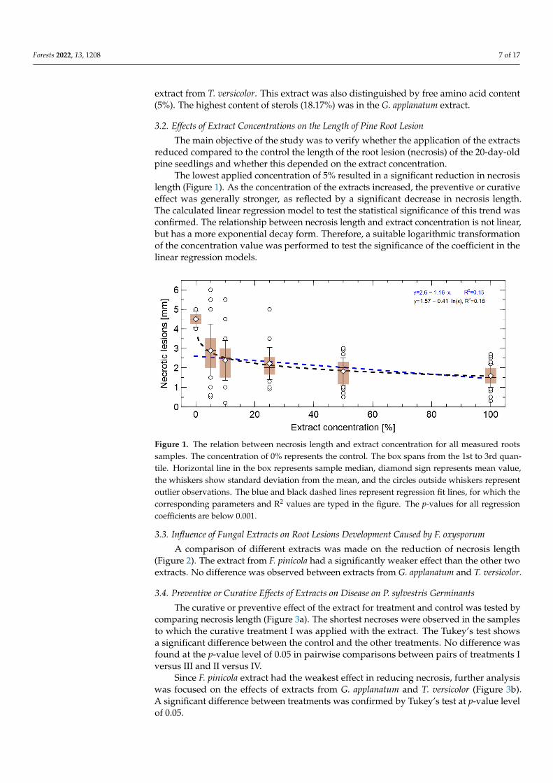
The lowest applied concentration of 5% resulted in a significant reduction in necrosislength (Figure 1). As the concentration of the extracts increased, the preventive or curativeeffect was generally stronger, as reflected by a significant decrease in necrosis length.The calculated linear regression model to test the statistical significance of this trend wasconfirmed. The relationship between necrosis length and extract concentration is not linear,but has a more exponential decay form. Therefore, a suitable logarithmic transformationof the concentration value was performed to test the significance of the coefficient in thelinear regression models.
Figure 1. The relation between necrosis length and extract concentration for all measured rootssamples. The concentration of 0% represents the control. The box spans from the 1st to 3rd quan-tile. Horizontal line in the box represents sample median, diamond sign represents mean value,the whiskers show standard deviation from the mean, and the circles outside whiskers representoutlier observations. The blue and black dashed lines represent regression fit lines, for which thecorresponding parameters and R2 values are typed in the figure. The p-values for all regressioncoefficients are below 0.001.
3.3. Influence of Fungal Extracts on Root Lesions Development Caused by F. oxysporum
A comparison of different extracts was made on the reduction of necrosis length(Figure 2). The extract from F. pinicola had a significantly weaker effect than the other twoextracts. No difference was observed between extracts from G. applanatum and T. versicolor.
3.4. Preventive or Curative Effects of Extracts on Disease on P. sylvestris Germinants
The curative or preventive effect of the extract for treatment and control was tested bycomparing necrosis length (Figure 3a). The shortest necroses were observed in the samplesto which the curative treatment I was applied with the extract. The Tukey’s test showsa significant difference between the control and the other treatments. No difference wasfound at the p-value level of 0.05 in pairwise comparisons between pairs of treatments Iversus III and II versus IV.
Since F. pinicola extract had the weakest effect in reducing necrosis, further analysiswas focused on the effects of extracts from G. applanatum and T. versicolor (Figure 3b).A significant difference between treatments was confirmed by Tukey’s test at p-value levelof 0.05.

Forests 2022, 13, 1208 8 of 17
Figure 2. Necrosis length for various extracts: C–control, F.–Fomitopsis pinicola, G.–Ganoderma applana-tum, and T.–Trametes versicolor. Treatments, for which statistically significant difference at p < 0.05was found using Tukey’s multiple comparisons test, are marked by different characters in the bottompart of the chart.
Figure 3. Necrosis lengths for the four treatments of extracts application (I—IV) and control (C).In panel (a), all measured samples are presented, in (b), samples for which the extract of Fomitopsispinicola was applied have been excluded. Treatments, for which a statistically significant difference atp < 0.05 was found using Tukey’s multiple comparisons test, are marked by different characters inthe bottom part of the chart.
The next step of the analysis concerned the combination of pairs of factors that dis-tinguished the different sample types. The aim of this test was to determine whether thetrend toward decreasing necrosis length with increasing extract concentration persistedwhen the data were analyzed for subgroups defined by the treatment of extract application(preventive or curative) and the type of extract (fungal species) (Figure 4). The observeddecreases in lesion length along with extract concentration were significant for all cases, ex-cept for treatment I. The p-value for the slope coefficient of 0.08 for treatment I indicate thatsuch relation is weak and the effect of the extracts on the fungi may be similar regardless ofthe applied extract concentration.
Necrosis length by type of extract used (fungal species) and treatment of extractapplication (preventive, curative) are shown in Figure 5, necrosis length was most reducedin treatment I when G. applanatum or T. versicolor extracts were applied. The Tukey’smultiple comparison test performed for these data confirms statistical significance atp-value level of 0.05 between treatment I, Ganoderma and Trametes extracts and other variantsexcept the cases: treatment II Ganoderma, treatment III Fomitopsis and Tramentes.

Forests 2022, 13, 1208 9 of 17
Figure 4. The relation between necrosis length and extract concentration in the control (C) and extracttreatments. Comparison of (a) treatments with various extract application, and (b) various types ofextracts. The dashed lines represent fitted regression lines y = a + b · ln(x). The R2 of the models,regression coefficients and corresponding p-values are typed in subfigures.
Figure 5. Necrosis length for various extracts: C–control, F.–Fomitopsis pinicola, G.–Ganoderma applana-tum, T.–Trametes versicolor and various extracts treatments (I—IV). Treatments, for which statisticallysignificant difference at p < 0.05 was found using Tukey’s multiple comparisons test, are marked bydifferent characters in the bottom part of the chart.
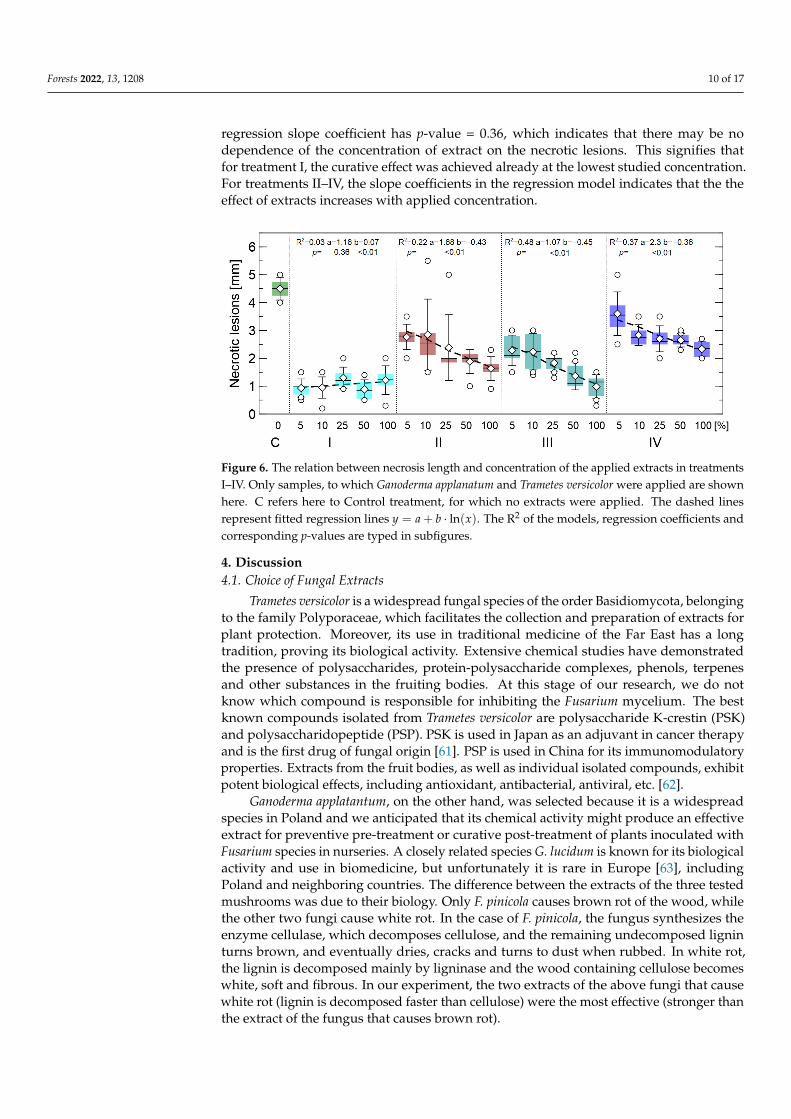
Since treatment I had the best effect for the extracts G. applanatum and T. versicolor,various concentrations of extracts from these two fungi were tested for treatments I–IV.Such analysis is presented in Figure 6. For treatment I, there was no difference betweenlength and extract concentration. Such behavior is visible in this figure, also the calculated
Forests 2022, 13, 1208 10 of 17
regression slope coefficient has p-value = 0.36, which indicates that there may be nodependence of the concentration of extract on the necrotic lesions. This signifies thatfor treatment I, the curative effect was achieved already at the lowest studied concentration.For treatments II–IV, the slope coefficients in the regression model indicates that the theeffect of extracts increases with applied concentration.
Figure 6. The relation between necrosis length and concentration of the applied extracts in treatmentsI–IV. Only samples, to which Ganoderma applanatum and Trametes versicolor were applied are shownhere. C refers here to Control treatment, for which no extracts were applied. The dashed linesrepresent fitted regression lines y = a + b · ln(x). The R2 of the models, regression coefficients andcorresponding p-values are typed in subfigures.
4. Discussion4.1. Choice of Fungal Extracts
Trametes versicolor is a widespread fungal species of the order Basidiomycota, belongingto the family Polyporaceae, which facilitates the collection and preparation of extracts forplant protection. Moreover, its use in traditional medicine of the Far East has a longtradition, proving its biological activity. Extensive chemical studies have demonstratedthe presence of polysaccharides, protein-polysaccharide complexes, phenols, terpenesand other substances in the fruiting bodies. At this stage of our research, we do notknow which compound is responsible for inhibiting the Fusarium mycelium. The bestknown compounds isolated from Trametes versicolor are polysaccharide K-crestin (PSK)and polysaccharidopeptide (PSP). PSK is used in Japan as an adjuvant in cancer therapyand is the first drug of fungal origin [61]. PSP is used in China for its immunomodulatoryproperties. Extracts from the fruit bodies, as well as individual isolated compounds, exhibitpotent biological effects, including antioxidant, antibacterial, antiviral, etc. [62].
Ganoderma applatantum, on the other hand, was selected because it is a widespreadspecies in Poland and we anticipated that its chemical activity might produce an effectiveextract for preventive pre-treatment or curative post-treatment of plants inoculated withFusarium species in nurseries. A closely related species G. lucidum is known for its biologicalactivity and use in biomedicine, but unfortunately it is rare in Europe [63], includingPoland and neighboring countries. The difference between the extracts of the three testedmushrooms was due to their biology. Only F. pinicola causes brown rot of the wood, whilethe other two fungi cause white rot. In the case of F. pinicola, the fungus synthesizes theenzyme cellulase, which decomposes cellulose, and the remaining undecomposed ligninturns brown, and eventually dries, cracks and turns to dust when rubbed. In white rot,the lignin is decomposed mainly by ligninase and the wood containing cellulose becomeswhite, soft and fibrous. In our experiment, the two extracts of the above fungi that causewhite rot (lignin is decomposed faster than cellulose) were the most effective (stronger thanthe extract of the fungus that causes brown rot).

Forests 2022, 13, 1208 11 of 17
Fomitopsis pinicola is a common brown-rot fungal species found in coniferous forests intemperate regions throughout Europe and Asia. F. pinicola has been intensively studiedbecause it has anti-tumor, anti-fungal, antioxidant, immunomodulation, and neuroprotec-tive activities [64]. However, in our experiment, its extracts showed the weakest biologicalactivity, so it will not be included in future in vivo studies. However, it is the most commonspecies in the local forests, developing on dead spruce trees as a result of infestation byIps typographus.
4.2. Chemical Composition of Fungal Extracts
As for the chemical composition of fungal extracts, polysaccharides are mainly respon-sible for their biological activity, but in the case of methanolic extracts, there are usuallynot too many of them [65]. This is because they are bound to the cell wall of the fungusand their amount is much higher when hot water extracts are used [66]. Therefore, in thefuture, we can consider preparing extracts with hot water to check if the biological activityis higher than observed now. In the methanolic extracts, mainly low molecular weightcompounds are found. Quite a number of compounds have antioxidant, antibacterial,antiviral and immunomodulatory potential [47]. Some of these compounds are also foundin fruits or herbs, while others are found only in fungi and in small amounts. A compoundmay also be specific (like a fingerprint) to a fungal species. Wood decay fungi produceligninolytic and cellulolytic enzymes, e.g., laccases, peroxidases, and manganases, but theseare not usually obtained by extraction. It is noteworthy that extract-based drugs usuallycontain whole fractions rather than single compounds. Hundreds of bioactive compoundscan act synergistically and multidirectionally. Thus, to understand the effect of an extract,the chemical properties must be carefully evaluated. Therefore, it is important to keep inmind that the GC-MS analysis of extracts may not identify about 20% of the compounds,especially macromolecular compounds such as polysaccharides, for which other methodsmust be used. For this reason, we focused on demonstrating the efficacy of the preventiveand curative effects of the studied fungal extracts against a dangerous pathogen in forestry,agriculture and horticulture—the fungus F. oxysporum. In the future, we will try to find bio-logically active compounds in the fungal extracts, but this was not the aim of our researchat this stage.
4.3. Preventive Measures
The current study with fungal extracts also showed a protective effect against F. oxys-porum. GC-MS analysis showed that the most abundant compounds in the species studiedwere carbohydrates (35.72–72.65) (see supplemental materials). This is consistent withseveral publications indicating the antifungal activity of such compounds [65,67–69]. Thesecond common group detected in this study was fatty acids, which accounted for 8.54%,37.40% and 10.35% of the extracts from F. pinicola, G. applanatum and T. versicolor, respec-tively. Potent antifungal effects of monoacylglycerols, short-chain and long-chain fattyacids were reported [70–73].
Induced resistance, which relies on the host’s natural defenses, is an effective measureto control plant diseases and meets the strategic needs of pesticide use and agriculturalproduct safety worldwide. Ganoderma lucidum polysaccharide (GLP), the main activemolecule of G. lucidum, is widely used in functional foods and clinical medicine [74,75].However, there are few reports on the use of GLP for plant disease prevention and control.Cör et al. [75] reported the direct antibacterial effect of GLP against certain plant pathogensand foodborne fungi and bacteria. GLP can also induce systemic resistance to Botrytiscinerea in tomato plants and promote tomato seed germination and seedling growth [76].GLP spray and irrigation root treatments can promote cotton growth. After soaking inGLP, both seedling height and Fusarium wilt resistance of cotton increased to some extent.The increased expression of genes related to the jasmonic acid pathway suggests that thispathway may be important for plant resistance induced by GLP plant resistance [77].

Forests 2022, 13, 1208 12 of 17
4.3.1. Protective Effect of Fungal Extracts
Methanol extracts of G. applanatum and T. versicolor showed the best results in inhibitingF. oxysporum. Probably, the active compounds contained in them have a direct toxic effecton the hyphae limiting the development of the pathogen, but most importantly they induceplant resistance. According to Oviasogie et al. [78], the antifungal activity of Ganodermaspecies is due to bioactive substances such as β-triterperinoids, polysaccharides, proteins,amino acids, nucleotides, alkaloids, steroids, lactones, fatty acids and enzymes [78,79].On the other hand, Boh et al. [80] demonstrated the bioactive effects of lucidic acid andganodermic acid. They also isolated ganodermic acid, ganolucidic acid, applanoxic acid,a group of triterpenoid aldehydes (ganoderals), and a group of triterpenoid alcohols(ganoderols) from G. applanatum fruiting bodies [80]). Nagaraj et al. [81] showed thatthis fungus produces phytochemicals such as saponins, phenols, steroids, glycosides,terpenoids, and flavonoids. The studies of Jonathan and Awtona [82] confirmed theproperties of fungi of the genus Ganoderma in limiting the development of F. oxysporum [82].Other studies on T. versicolor confirm the possibility of using extracts from this fungal speciesto inhibit pathogenic soil fungi, including Fusarium fungi [83]. Similarly, Waithaka et al. [84]point out the great potential of using T. versicolor extracts to limit the development ofF. oxysporum.
Test results are also affected by many variables, for example, results can vary depend-ing on the solvent used for the extract (methanol, ethanol, hexane, water extract). For thisreason, scientists argue about how the extracts are used and what their concentrationsare [83,85,86]. Even the substrate on which a particular fungal species grows has an influ-ence on the content of certain secondary metabolites as well as their amount [87]. This inturn is associated with potentially different properties of the biologically active compounds.
4.3.2. Potential Applications
The curative treatment (Treatment I) seemed to be more efficacious than the preventivetreatment (Treatment II) and the two dipping treatments.
Perhaps the protection provided by a 24 h post-inoculation treatment (Treatment I)might have better coincided with the stimulation of defense responses in the plant, and alsohad some direct inhibitory effects on hyphal growth, which had reached that part of theroots. In the curative treatment scenario, we speculate that after F. oxysporum inoculum isapplied, there is some initial stimulation of root cell defense, which is enhanced systemicallywhen the treatment is applied 24 h later. In the absence of the pathogen, the substancescontained in the extracts may induce resistance, but since wood-destroying fungi are notstrong pathogens acting against live host plant cells and likely do not produce phytotoxins,this elicits a weaker response from the plant. However, when the plant cells are in directcontact with a pathogen, the defense response should be stronger. This combined withdirect activity against the fungal hyphae may lead to decreased disease.
The potential of using wood-decomposing fungi to produce active ingredients thatincrease plant resistance is enormous [88]. To achieve optimal results, many variables mustbe considered, and the research must be conducted in many iterations, which means it willrequire many years of work. However, biologically active compounds can be used not onlyto increase the resistance of the plants themselves, but also to treat seeds (bioimprinting)from which the disease-resistant seedlings will grow [89–92]. This is even more valuable assoil-borne fungi gradually mutate and form new varieties that are more resistant to existingmethods of protection, particularly the chemicals used [7,93–95]. Due to the ongoingnegative phenomenon of environmental chemization, there is a need to search for new,environmentally friendly methods of crop protection.
5. Conclusions
Methanolic extracts of wood-destroying material showed a positive effect in pro-tecting pines as active preparations against soil-borne pathogen F. oxysporum. The fungi

Forests 2022, 13, 1208 13 of 17
G. applanatum and T. versicolor are potential biological control agents whose secondarymetabolites can be used against the pathogen F. oxysporum.
In summary:
• All three fungal extracts studied showed a curative or preventive effect, resulting in areduction in the length of root necrosis caused by infection with F. oxysporum.
• The strongest effects were obtained with extracts of G. applanatum and T. versicolor.• The strongest effect was obtained in treatment I, in which one drop of the extract was
applied to the root tip 24 h after infection (curative).• The full preventive effect of the application of these two extracts in treatment I was
achieved at a 5% concentration of the extract—further increasing the concentrationdid not change the results.
Further studies are needed to understand the mechanism of action of the extracts. Fieldtrials (in vivo) are needed to confirm the results of plant protection efficacy. In addition,isolation and testing of the active compounds responsible for the effect of the extracts couldbe one of the future topics of research.
Supplementary Materials: The following are available online at https://www.mdpi.com/article/10.3390/f13081208/s1.
Author Contributions: Conceptualization, T.O., U.W. and E.Z.; methodology, T.O., M.S. and E.Z.;software, P.B., D.B. and M.W.; validation, P.B., D.B., M.W. and M.S.; formal analysis, U.W., M.S.,P.B. and D.B.; investigation, U.W. and M.S.; resources, E.Z., M.S. and M.W.; data curation, P.B., D.B.and M.S.; writing—original draft preparation, U.W., E.Z. and P.B.; writing—review and editing, T.O.,T.M., T.H. and E.Z.; visualization, P.B.; supervision, T.O. and E.Z.; project administration, T.O. andE.Z.; funding acquisition, M.S., E.Z., M.W. and T.O. All authors have read and agreed to the publishedversion of the manuscript.
Funding: Studies was carried out within the framework: WZ/WB-INL/2/2021 and financed fromthe science funds for Ministry of Science and Higher Education.
Institutional Review Board Statement: Not applicable.
Informed Consent Statement: Not applicable.
Data Availability Statement: Not applicable.
Conflicts of Interest: The authors declare no conflict of interest.
References1. Gordon, T.R.; Swett, C.L.; Wingfield, M.J. Management of Fusarium diseases affecting conifers. Crop Prot. 2015, 73, 28–39.
[CrossRef]2. Brown, B.; Wylie, F. Diseases and pests of Australian forest nurseries: Past and present. In Proceedings of the First Meeting of IUFRO
Working Party S. 2.07–09 (Diseases and Insects in Forest Nurseries); Southerland, J.R., Glover, S., Eds.; Forestry Canada, Pacific andYukon Region, Pacific Forestry Centre: Victoria, BC, Canada, 1991; pp. 3–15.
3. Smith, R.S., Jr. Charcoal root disease. Macrophomina phaseoli (Maubl) Ashby. In Forest Nursery Diseases in the United States; Agric.Handb; Peterson, G.; Smith., R.T.C., Jr., Eds.; USDA Forest Service: Washington, DC, USA, 1975; Volume 470, pp. 11–13.
4. Wit, M.; Sierota, Z.; Oszako, T.; Mirzwa-Mróz, E.; Wakulinski, W. Fusarium spp. on the above-ground organs of dying oaks-a newthreat? Sylwan 2015, 159, 403–410. (In Polish) [CrossRef]
5. Armstrong, G.; Armstrong, J.K. Formae speciales and races of Fusarium oxysporum causing wilt diseases. In Fusarium: Disease,Biology, and Taxonomy; Nelson, P.E., Toussoun, T.A., Cook, R.J., Eds.; The Pennsylvania State University Press: University Park,PA, USA, 1981; pp. 391–399.
6. Michielse, C.B.; Rep, M. Pathogen profile update: Fusarium oxysporum. Mol. Plant Pathol. 2009, 10, 311–324. [CrossRef][PubMed]
7. Dean, R.A.; Lichens-Park, A.; Kole, C. Genomics of Plant-Associated Fungi and Oomycetes: Dicot Pathogens; Springer:Berlin/Heidelberg, Germany, 2014. [CrossRef]
8. Dean, R.; Van Kan, J.A.; Pretorius, Z.A.; Hammond-Kosack, K.E.; Di Pietro, A.; Spanu, P.D.; Rudd, J.J.; Dickman, M.; Kahmann,R.; Ellis, J.; et al. The Top 10 fungal pathogens in molecular plant pathology. Mol. Plant Pathol. 2012, 13, 414–430. [CrossRef][PubMed]

Forests 2022, 13, 1208 14 of 17
9. de Lamo, F.J.; Takken, F.L. Biocontrol by Fusarium oxysporum using endophyte-mediated resistance. Front. Plant Sci. 2020, 11, 37.[CrossRef]
10. Fisher, M.C.; Henk, D.A.; Briggs, C.J.; Brownstein, J.S.; Madoff, L.C.; McCraw, S.L.; Gurr, S.J. Emerging fungal threats to animal,plant and ecosystem health. Nature 2012, 484, 186–194. [CrossRef] [PubMed]
11. Edel-Hermann, V.; Brenot, S.; Gautheron, N.; Aimé, S.; Alabouvette, C.; Steinberg, C. Ecological fitness of the biocontrol agentFusarium oxysporum Fo47 in soil and its impact on the soil microbial communities. FEMS Microbiol. Ecol. 2009, 68, 37–45.[CrossRef]
12. Davydenko, K.; Nowakowska, J.A.; Kaluski, T.; Gawlak, M.; Sadowska, K.; García, J.M.; Diez, J.J.; Okorski, A.; Oszako, T. AComparative Study of the Pathogenicity of Fusarium circinatum and other Fusarium Species in Polish Provenances of P. sylvestrisL. Forests 2018, 9, 560. [CrossRef]
13. Okorski, A.; Milewska, A.; Pszczółkowska, A.; Karpiesiuk, K.; Kozera, W.; Dabrowska, J.A.; Radwinska, J. Prevalence of Fusariumfungi and Deoxynivalenol Levels in Winter Wheat Grain in Different Climatic Regions of Poland. Toxins 2022, 14, 102. [CrossRef]
14. Kulik, T.; Jestoi, M.; Okorski, A. Development of TaqMan assays for the quantitative detection of Fusarium avenaceum/Fusariumtricinctum and Fusarium poae esyn1 genotypes from cereal grain. FEMS Microbiol. Lett. 2011, 314, 49–56. [CrossRef] [PubMed]
15. Jankowiak, R.; Stepniewska, H.; Bilanski, P.; Taerum, S.J. Fungi as potential factors limiting natural regeneration of pedunculateoak (Quercus robur) in mixed-species forest stands in Poland. Plant Pathol. 2022, 71, 805–817. [CrossRef]
16. Oszako, T.; Sikora, K.; Belbahri, L.; Nowakowska, J.A. Molecular detection of oomycetes species in water courses. Folia For. Pol.2016, 58, 246–251. [CrossRef]
17. Orłowski, L.B.; Trzewik, A.; Ptaszek, M.; Orlikowska, T. Relationship between source of water, occurrence, and pathogenicity ofPhytophthora plurivora. Acta Mycol. 2012, 47, 1. [CrossRef]
18. Malewski, T.; Brzezinska, B.; Belbahri, L.; Oszako, T. Role of avian vectors in the spread of Phytophthora species in Poland. Eur. J.Plant Pathol. 2019, 155, 1363–1366. [CrossRef]
19. Stepniewska, H.; Jankowiak, R.; Bilanski, P.; Hausner, G. Structure and Abundance of Fusarium Communities Inhabiting theLitter of Beech Forests in Central Europe. Forests 2021, 12, 811. [CrossRef]
20. Kowalski, T.; Bilanski, P. Fungi Detected in the Previous Year’s Leaf Petioles of Fraxinus excelsior and Their Antagonistic Potentialagainst Hymenoscyphus fraxineus. Forests 2021, 12, 1412. [CrossRef]
21. Okorski, A.; Pszczółkowska, A.; Okorska, S.; Fordonski, G. First Report of Fagus sylvatica Infection by Fusarium avenaceum inForest Container Nurseries in Northeastern Poland. Plant Dis. 2015, 99, 420–420. [CrossRef] [PubMed]
22. James, R.L.; Dumroese, R.K. Investigations of Fusarium diseases within Inland Pacific Northwest forest nurseries. In Proceedingsof the 53rd Western International Forest Disease Work Conference, USDA Forest Service, Intermountain Region, Ogden, UT, USA,15–19 October 2007; pp. 3–11.
23. Dumroese, R.K.; James, R.L. Root diseases in bareroot and container nurseries of the Pacific Northwest: Epidemiology,management, and effects on outplanting performance. New For. 2005, 30, 185–202. [CrossRef]
24. Montecchio, L. Damping-off of beech seedlings caused by Fusarium avenaceum in Italy. Plant Dis. 2005, 89, 1014–1014. [CrossRef]25. Lilja, A.; Poteri, M.; Petäistö, R.L.; Rikala, R.; Kurkela, T.; Kasanen, R. Fungal diseases in forest nurseries in Finland. Silva Fenn.
2010, 44, 147. [CrossRef]26. Peterson, M. Fusarium species-a British Columbia perspective in forest seedling production. In National Proceedings: Forest and
Conservation Nursery Associations-2007. Proc. RMRS-P-57; Dumroese, R., Riley, L., Eds.; US Department of Agriculture, ForestService, Rocky Mountain Research Station: Fort Collins, CO, USA, 2008; Volume 57, pp. 109–125.
27. Pinto, P.M.; Alonso, J.A.P.; Fernández, V.P.; Casero, J.J.D. Fungi isolated from diseased nursery seedlings in Spain. New For. 2006,31, 41–56. [CrossRef]
28. Menkis, A.; Vasiliauskas, R.; Taylor, A.; Stenström, E.; Stenlid, J.; Finlay, R. Fungi in decayed roots of conifer seedlings in forestnurseries, afforested clear-cuts and abandoned farmland. Plant Pathol. 2006, 55, 117–129. [CrossRef]
29. Zakeri, A.; Hamzeharghani, H.; Banihashemi, Z.; Saadati, S. Pathogenic fungi associated with pre-and post-emergence seedlingblight of pine and cypress in Fars Province, Iran. For. Pathol. 2011, 41, 438–443. [CrossRef]
30. Lazreg, F.; Belabid, L.; Sanchez, J.; Gallego, E.; Bayaa, B. Pathogenicity of Fusarium spp. associated with diseases of Aleppo-pineseedlings in Algerian forest nurseries. J. For. Sci. 2014, 60, 115–120. [CrossRef]
31. Fajardo, M.A.; León, J.D.; Correa, G.A.; Morales, J.G. The Causal Agent of Damping-off in Pinus patula (Schiede) and Pinustecunumanii (Schwerdtf.). Floresta E Ambiente 2019, 26. [CrossRef]
32. Okorski, A.; Oszako, T.; Nowakowska, J.A.; Pszczolkowska, A. The possibilities of biologically protecting plants against diseasesin nurseries, with special consideration of Oomycetes and Fusarium fungi. Lesne Pr. Badaw. 2014, 75. [CrossRef]
33. Klapwijk, M.J.; Bylund, H.; Schroeder, M.; Björkman, C. Forest management and natural biocontrol of insect pests. Forestry 2016,89, 253–262. [CrossRef]
34. Balla, A.; Silini, A.; Cherif-Silini, H.; Chenari Bouket, A.; Moser, W.K.; Nowakowska, J.A.; Oszako, T.; Benia, F.; Belbahri, L. TheThreat of Pests and Pathogens and the Potential for Biological Control in Forest Ecosystems. Forests 2021, 12, 1579. [CrossRef]
35. Spremo, N.R.; Tesanovic, K.D.; Rakic, M.S.; Janjuševic, L.N.; Ignjatov, M.; Bjelic, D.; Karaman, M. Antifungal activity ofmacrofungi extracts on phytopathogenic fungal strains of genera Fusarium sp. and Alternaria sp. Zb. Matice Srp. Za Prir. Nauk.2017, 133, 231–240. [CrossRef]
Forests 2022, 13, 1208 15 of 17
36. Agrios, G.N. Plant diseases caused by Mollicutes: Phytoplasmas and spiroplasmas. In Plant Pathology; Agrios, G.N., Ed.;Academic Press: New York, NY, USA, 1997; pp. 457–470.
37. Zjawiony, J.K. Biologically active compounds from Aphyllophorales (polypore) fungi. J. Nat. Prod. 2004, 67, 300–310. [CrossRef]38. Alves, M.; Ferreira, I.; Dias, J.; Teixeira, V.; Martins, A.; Pintado, M. A Review on Antifungal Activity of Mushroom (Basid-
iomycetes) Extracts and Isolated Compounds. Curr. Top. Med. Chem. 2013, 13, 2648–2659. [CrossRef]39. Atanasova-Penichon, V.; Barreau, C.; Richard-Forget, F. Antioxidant secondary metabolites in cereals: Potential involvement in
resistance to Fusarium and mycotoxin accumulation. Front. Microbiol. 2016, 7, 566. [CrossRef]40. Heleno, S.A.; Ferreira, I.C.; Esteves, A.P.; Ciric, A.; Glamoclija, J.; Martins, A.; Sokovic, M.; Queiroz, M.J.R. Antimicrobial and
demelanizing activity of Ganoderma lucidum extract, p-hydroxybenzoic and cinnamic acids and their synthetic acetylatedglucuronide methyl esters. Food Chem. Toxicol. 2013, 58, 95–100. [CrossRef] [PubMed]
41. Parroni, A.; Bellabarba, A.; Beccaccioli, M.; Scarpari, M.; Reverberi, M.; Infantino, A. Use of the secreted proteome of Trametesversicolor for controlling the cereal pathogen Fusarium langsethiae. Int. J. Mol. Sci. 2019, 20, 4167. [CrossRef]
42. Schalchli, H.; Hormazabal, E.; Becerra, J.; Birkett, M.; Alvear, M.; Vidal, J.; Quiroz, A. Antifungal activity of volatile metabolitesemitted by mycelial cultures of saprophytic fungi. Chem. Ecol. 2011, 27, 503–513. [CrossRef]
43. Guler, P.; Akata, I.; Kutluer, F. Antifungal activities of Fomitopsis pinicola (Sw.: Fr) Karst and Lactarius vellereus (Pers.) Fr. Afr. J.Biotechnol. 2009, 8, 3811–3813.
44. Xu, K.; Li, X.Q.; Zhao, D.L.; Zhang, P. Antifungal secondary metabolites produced by the fungal endophytes: Chemical diversityand potential use in the development of biopesticides. Front. Microbiol. 2021, 12, 689527. [CrossRef] [PubMed]
45. Matuszewska, A.; Jaszek, M.; Stefaniuk, D.; Ciszewski, T.; Matuszewski, Ł. Anticancer, antioxidant, and antibacterial activities oflow molecular weight bioactive subfractions isolated from cultures of wood degrading fungus Cerrena unicolor. PLoS ONE 2018,13, e0197044. [CrossRef] [PubMed]
46. Zhong, J.J.; Xiao, J.H. Secondary metabolites from higher fungi: Discovery, bioactivity, and bioproduction. Biotechnol. China I2009, 113, 79–150. [CrossRef]
47. Brizuela, M.; García, L.; Pérez, L.; Mansur, M. Basidiomycetes: A new source of secondary metabolites. Rev. Iberoam. Micol. 1998,15, 69–74. [PubMed]
48. Thambugala, K.M.; Daranagama, D.A.; Phillips, A.J.; Kannangara, S.D.; Promputtha, I. Fungi vs. fungi in biocontrol: An overviewof fungal antagonists applied against fungal plant pathogens. Front. Cell. Infect. Microbiol. 2020, 10, 604923. [CrossRef] [PubMed]
49. Firáková, S.; Šturdíková, M.; Múcková, M. Bioactive secondary metabolites produced by microorganisms associated with plants.Biologia 2007, 62, 251–257. [CrossRef]
50. Stuper-Szablewska, K.; Przybylska-Balcerek, A.; Rogozinski, T.; Szwajkowska-Michałek, L. Phenolic Compounds in Trees andShrubs of Central Europe. Appl. Sci. 2020, 10, 6907. [CrossRef]
51. Tamm, L.; Thürig, B.; Fliessbach, A.; Goltlieb, A.; Karavani, S.; Cohen, Y. Elicitors and soil management to induce resistanceagainst fungal plant diseases. NJAS-Wagening. J. Life Sci. 2011, 58, 131–137. [CrossRef]
52. Šrobárová, A.; Kogan, G.; Tamas, L.; Machová, E. Protective activity of the fungal polysaccharides against fusariosis. Plant Prot.Sci. 2002, 38, 617–619. [CrossRef]
53. Roeder, A.; Kirschning, C.J.; Rupec, R.A.; Schaller, M.; Weindl, G.; Korting, H.C. Toll-like receptors as key mediators in innateantifungal immunity. Med. Mycol. 2004, 42, 485–498. [CrossRef] [PubMed]
54. Havrlentová, M.; Gregusová, V.; Šliková, S.; Nemecek, P.; Hudcovicová, M.; Kuzmová, D. Relationship between the Content ofβ-D-Glucans and Infection with Fusarium Pathogens in Oat (Avena sativa L.) Plants. Plants 2020, 9, 1776. [CrossRef] [PubMed]
55. Ruiz-Herrera, J.; Ortiz-Castellanos, L. Cell wall glucans of fungi. A review. Cell Surf. 2019, 5, 100022. [CrossRef]56. Stadnik, M.J.; Freitas, M.B.d. Algal polysaccharides as source of plant resistance inducers. Trop. Plant Pathol. 2014, 39, 111–118.
[CrossRef]57. Ryvarden, L.; Melo, I. Poroid fungi of Europe. In Synopsis Fungorum; Fungiflora A/S: Oslo, Norway, 2017; Volume 37.58. Karasinski, D.; Wołkowycki, M. An annotated and illustrated catalogue of polypores (Agaricomycetes) of the Białowieza Forest
(NE Poland). Pol. Bot. J. 2015, 60, 217–292. [CrossRef]59. Sadowska, A.; Zapora, E.; Sawicka, D.; Niemirowicz-Laskowska, K.; Surazynski, A.; Sułkowska-Ziaja, K.; Kała, K.; Stocki, M.;
Wołkowycki, M.; Bakier, S.; et al. Heterobasidion annosum induces apoptosis in DLD-1 cells and decreases colon cancer growthin in vivo model. Int. J. Mol. Sci. 2020, 21, 3447. [CrossRef] [PubMed]
60. Cody, R. SAS Statistics by Example; SAS Press: Cary, NC, USA, 2011.61. Tsukagoshi, S.; Hashimoto, Y.; Fujii, G.; Kobayashi, H.; Nomoto, K.; Orita, K. Krestin (PSK). Cancer Treat. Rev. 1984, 11, 131–155.
[CrossRef]62. Sułkowska-Ziaja, K.; Muszynska, B.; Sałaciak, K.; Gawalska, A. Trametes versicolor (L.) Lloyd as a source of biologically active
compounds with a wide spectrum of action and application. Postepy Fitoter. 2016, 17, 274–281. (In Polish)63. Lohmeyer, T.R.; Künkele, U. Grzyby. Rozpoznawanie i Zbieranie; Parragon: Warszawa, Poland, 2006. (In Polish)64. Liu, S.; Han, M.L.; Xu, T.M.; Wang, Y.; Wu, D.M.; Cui, B.K. Taxonomy and phylogeny of the Fomitopsis pinicola complex with
descriptions of six new species from East Asia. Front. Microbiol. 2021, 12, 644979. [CrossRef] [PubMed]65. Zhang, J.; Liu, Y.; Tang, Q.; Zhou, S.; Feng, J.; Chen, H. Polysaccharide of Ganoderma and its bioactivities. In Ganoderma and
Health. Advances in Experimental Medicine and Biology; Lin, Z., Yang, B., Eds.; Springer: Singapore, 2019; Volume 1181, pp. 107–134.[CrossRef]

Forests 2022, 13, 1208 16 of 17
66. Leong, Y.K.; Yang, F.C.; Chang, J.S. Extraction of polysaccharides from edible mushrooms: Emerging technologies and recentadvances. Carbohydr. Polym. 2021, 251, 117006. [CrossRef]
67. Ferreira, I.C.; Heleno, S.A.; Reis, F.S.; Stojkovic, D.; Queiroz, M.J.R.; Vasconcelos, M.H.; Sokovic, M. Chemical features ofGanoderma polysaccharides with antioxidant, antitumor and antimicrobial activities. Phytochemistry 2015, 114, 38–55. [CrossRef][PubMed]
68. Yamaç, M.; Bilgili, F. Antimicrobial activities of fruit bodies and/or mycelial cultures of some mushroom isolates. Pharm. Biol.2006, 44, 660–667. [CrossRef]
69. Hao, L.; Sheng, Z.; Lu, J.; Tao, R.; Jia, S. Characterization and antioxidant activities of extracellular and intracellular polysaccharidesfrom Fomitopsis pinicola. Carbohydr. Polym. 2016, 141, 54–59. [CrossRef]
70. Altieri, C.; Bevilacqua, A.; Cardillo, D.; Sinigaglia, M. Antifungal activity of fatty acids and their monoglycerides against Fusariumspp. in a laboratory medium. Int. J. Food Sci. Technol. 2009, 44, 242–245. [CrossRef]
71. Fischer, C.L.; Blanchette, D.R.; Brogden, K.A.; Dawson, D.V.; Drake, D.R.; Hill, J.R.; Wertz, P.W. The roles of cutaneous lipids inhost defense. Biochim. Biophys. Acta (BBA)-Mol. Cell Biol. Lipids 2014, 1841, 319–322. [CrossRef]
72. Frank, C.L.; Ingala, M.R.; Ravenelle, R.E.; Dougherty-Howard, K.; Wicks, S.O.; Herzog, C.; Rudd, R.J. The effects of cutaneousfatty acids on the growth of Pseudogymnoascus destructans, the etiological agent of white-nose syndrome (WNS). PLoS ONE2016, 11, e0153535. [CrossRef] [PubMed]
73. Gołebiowski, M.; Urbanek, A.; Oleszczak, A.; Dawgul, M.; Kamysz, W.; Bogus, M.I.; Stepnowski, P. The antifungal activity offatty acids of all stages of Sarcophaga carnaria L. (Diptera: Sarcophagidae). Microbiol. Res. 2014, 169, 279–286. [CrossRef] [PubMed]
74. Yang, Y.; Zhang, H.; Zuo, J.; Gong, X.; Yi, F.; Zhu, W.; Li, L. Advances in research on the active constituents and physiologicaleffects of Ganoderma lucidum. Biomed. Dermatol. 2019, 3, 6. [CrossRef]
75. Cör, D.; Knez, Ž.; Knez Hrncic, M. Antitumour, antimicrobial, antioxidant and antiacetylcholinesterase effect of Ganodermalucidum terpenoids and polysaccharides: A review. Molecules 2018, 23, 649. [CrossRef] [PubMed]
76. Skalicka-Wozniak, K.; Szypowski, J.; Los, R.; Siwulski, M.; Sobieralski, K.; Glowniak, K.; Malm, A. Evaluation of polysaccharidescontent in fruit bodies and their antimicrobial activity of four Ganoderma lucidum (W Curt.: Fr.) P. Karst. strains cultivated ondifferent wood type substrates. Acta Soc. Bot. Pol. 2012, 81, 17–21. [CrossRef]
77. Zhang, Z.; Diao, H.; Wang, H.; Wang, K.; Zhao, M. Use of Ganoderma Lucidum polysaccharide to control cotton fusarium wilt,and the mechanism involved. Pestic. Biochem. Physiol. 2019, 158, 149–155. [CrossRef]
78. Oviasogie, F.; Akpaja, E.; Gbona, K.; Akonoafua, E. Antimicrobial properties of Ganoderma applanatum (Pers.) Pat. from Benin city,Nigeria. Niger. J. Agric. Food Environ. 2015, 11, 65–69.
79. Kim, Y.S.; Rym, K.H.; Lee, C.K.; Han, S.S. Antimicrobial activity of Elfvingia applanata extract alone and in combination withsome antibiotics. Yakhak Hoeji 1994, 38, 742–748.
80. Boh, B.; Hodžar, D.; Dolnicar, D.; Berovic, M.; Pohleven, F. Isolation and quantification of triterpenoid acids from Ganodermaapplanatum of Istrian origin. Food Technol. Biotechnol. 2000, 38, 11–18.
81. Nagaraj, K.; Mallikarjun, N.; Naika, R.; Venugopal, T. Phytochemical analysis and in vitro antimicrobial potential of Ganodermaapplanatum (Pers.) Pat. of Shivamogga district-Karnataka, India. Int. J. Pharm. Sci. Rev. Res 2013, 23, 36–41.
82. Jonathan, S.; Awotona, F. Studies on antimicrobial potentials of three Ganoderma species. Afr. J. Biomed. Res. 2010, 13, 131–139.83. Mendieta, E.H.; Ramos, M.A.; Miranda, A.S.; Granados, C.R. Evaluation of Crude Extracts of Trametes versicolor (L.: Fr.) Pilát to
Control of Phytopathogenic Fungi. Int. J. Plant Res. 2019, 9, 9–13. [CrossRef]84. Waithaka, P.N.; Gathuru, E.M.; Githaiga, B.M.; Onkoba, K.M. Antimicrobial Activity of Mushroom (Agaricus bisporus) and
Fungal (Trametes gibbosa) Extracts from Mushrooms and Fungi of Egerton Main Campus, Njoro Kenya. J. Biomed. Sci. 2017, 6, 3.[CrossRef]
85. Kawagishi, H.; Nomura, A.; Yumen, T.; Mizuno, T.; Hagiwara, T.; Nakamura, T. Isolation and properties of a lectin from thefruiting bodies of Agaricus blazei. Carbohydr. Res. 1988, 183, 150–154. [CrossRef]
86. Fujita, R.; Liu, J.; Shimizu, K.; Konishi, F.; Noda, K.; Kumamoto, S.; Ueda, C.; Tajiri, H.; Kaneko, S.; Suimi, Y.; et al. Anti-androgenicactivities of Ganoderma lucidum. J. Ethnopharmacol. 2005, 102, 107–112. [CrossRef] [PubMed]
87. Dresch, P.; Rosam, K.; Grienke, U.; Rollinger, J.M.; Peintner, U. Fungal strain matters: Colony growth and bioactivity of theEuropean medicinal polypores Fomes fomentarius, Fomitopsis pinicola and Piptoporus betulinus. Amb Express 2015, 5, 4. [CrossRef][PubMed]
88. Sivanandhan, S.; Khusro, A.; Paulraj, M.G.; Ignacimuthu, S.; Al-Dhabi, N.A. Biocontrol properties of basidiomycetes: Anoverview. J. Fungi 2017, 3, 2. [CrossRef] [PubMed]
89. Redman, R.S.; Sheehan, K.B.; Stout, R.G.; Rodriguez, R.J.; Henson, J.M. Thermotolerance generated by plant/fungal symbiosis.Science 2002, 298, 1581. [CrossRef]
90. Waller, F.; Achatz, B.; Baltruschat, H.; Fodor, J.; Becker, K.; Fischer, M.; Heier, T.; Hückelhoven, R.; Neumann, C.; von Wettstein, D.;et al. The endophytic fungus Piriformospora indica reprograms barley to salt-stress tolerance, disease resistance, and higher yield.Proc. Natl. Acad. Sci. USA 2005, 102, 13386–13391. [CrossRef]
91. Bilal, L.; Asaf, S.; Hamayun, M.; Gul, H.; Iqbal, A.; Ullah, I.; Lee, I.J.; Hussain, A. Plant growth promoting endophytic fungiAsprgillus fumigatus TS1 and Fusarium proliferatum BRL1 produce gibberellins and regulates plant endogenous hormones.Symbiosis 2018, 76, 117–127. [CrossRef]

Forests 2022, 13, 1208 17 of 17
92. Hill, R.; Llewellyn, T.; Downes, E.; Oddy, J.; MacIntosh, C.; Kallow, S.; Panis, B.; Dickie, J.B.; Gaya, E. Seed Banks as IncidentalFungi Banks: Fungal Endophyte Diversity in Stored Seeds of Banana Wild Relatives. Front. Microbiol. 2021, 12, 508. [CrossRef][PubMed]
93. Houterman, P.M.; Speijer, D.; Dekker, H.L.; de Koster, C.G.; Cornelissen, B.J.; Rep, M. The mixed xylem sap proteome of Fusariumoxysporum-infected tomato plants. Mol. Plant Pathol. 2007, 8, 215–221. [CrossRef]
94. Ma, L.J.; Van Der Does, H.C.; Borkovich, K.A.; Coleman, J.J.; Daboussi, M.J.; Di Pietro, A.; Dufresne, M.; Freitag, M.; Grabherr, M.;Henrissat, B.; et al. Comparative genomics reveals mobile pathogenicity chromosomes in Fusarium. Nature 2010, 464, 367–373.[CrossRef] [PubMed]
95. Schmidt, S.M.; Houterman, P.M.; Schreiver, I.; Ma, L.; Amyotte, S.; Chellappan, B.; Boeren, S.; Takken, F.L.; Rep, M. MITEs inthe promoters of effector genes allow prediction of novel virulence genes in Fusarium oxysporum. BMC Genom. 2013, 14, 119.[CrossRef] [PubMed]