Onstage Rumors, Offstage Sounds: Contemporary Opera of Li Yu
Upload
independentCategory
view
0download
0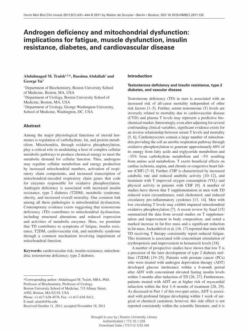
Horm Mol Biol Clin Invest 2011;8(1):431–444 © 2011 by Walter de Gruyter • Berlin • Boston. DOI 10.1515/HMBCI.2011.132
Androgen defi ciency and mitochondrial dysfunction: implications for fatigue, muscle dysfunction, insulin resistance, diabetes, and cardiovascular disease
Abdulmaged M. Traish 1,2, *, Bassima Abdallah 2 and George Yu 3
1 Department of Biochemistry , Boston University School of Medicine, Boston, MA , USA 2 Department of Urology , Boston University School of Medicine, Boston, MA , USA 3 Department of Urology , George Washington University, School of Medicine, Washington, DC , USA
Abstract
Among the major physiological functions of steroid hor-mones is regulation of carbohydrate, fat, and protein metab-olism. Mitochondria, through oxidative phosphorylation, play a critical role in modulating a host of complex cellular metabolic pathways to produce chemical energy to meet the metabolic demand for cellular function. Thus, androgens may regulate cellular metabolism and energy production by increased mitochondrial numbers, activation of respi-ratory chain components, and increased transcription of mitochondrial-encoded respiratory chain genes that code for enzymes responsible for oxidative phosphorylation. Androgen defi ciency is associated with increased insulin resistance, type 2 diabetes (T2DM), metabolic syndrome, obesity, and increased overall mortality. One common link among all these pathologies is mitochondrial dysfunction. Contemporary evidence exists suggesting that testosterone defi ciency (TD) contributes to mitochondrial dysfunction, including structural alterations and reduced expression and activities of metabolic enzymes. Here, we postulate that TD contributes to symptoms of fatigue, insulin resis-tance, T2DM, cardiovascular risk, and metabolic syndrome through a common mechanism involving impairment of mitochondrial function.
Keywords: cardiovascular risk; insulin resistance; mitochon-dria; testosterone defi ciency; type 2 diabetes.
Introduction
Testosterone defi ciency and insulin resistance, type 2
diabetes, and vascular disease
Testosterone defi ciency (TD) in men is associated with an increased risk of all-cause mortality independent of other risk factors [1 – 5] . Further, serum testosterone (T) levels are inversely related to mortality due to cardiovascular disease (CVD) and plasma T levels may represent a predictive bio-chemical marker. Interestingly, even after adjusting for several confounding clinical variables, signifi cant evidence exists for an inverse relationship between serum T levels and mortality [5, 6] . Cardiomyocytes contain a large number of mitochon-dria providing the cell an aerobic respiration pathway through oxidative phosphorylation to generate approximately 60 % of its energy from fatty acids and triglyceride metabolism and ∼ 35 % from carbohydrate metabolism and ∼ 5 % resulting from amino acid metabolism. T exerts benefi cial effects on cardiac ischemia, angina, and chronic or congestive heart fail-ure (CHF) [7 – 9] . Further, CHF is characterized by increased catabolic rate and reduced anabolic activity [10 – 12] , and treatment with T improved oxygen consumption (VO 2 ) and physical activity in patients with CHF [9] . A number of studies have shown that T supplementation in men with TD reduced waist circumference, total cholesterol, and reduced circulatory pro-infl ammatory cytokines [13, 14] . Men with low circulating T levels may exhibit impaired mitochondrial oxidative phosphorylation [7] . A recent review by Saad [15] summarized the data from several studies on T supplemen-tation and improvement in body composition, and noted a marked increase in fat-free mass and a signifi cant reduction in fat mass. Jockenh ö vel et al. [16, 17] reported that men with TD receiving T therapy consistently report reduced fatigue. This treatment is associated with concomitant stimulation of erythropoiesis and improvement in hematocrit levels [18] .
A number of prospective studies have shown that low T is a precursor of the later development of type 2 diabetes mel-litus (T2DM) [19 – 25] . Patients with prostate cancer (PCa) who were treated with androgen deprivation therapy (ADT) developed glucose intolerance within a 6-month period after ADT with concomitant elevated fasting insulin levels within 3 months after induction of TD [26, 27] . Furthermore, patients treated with ADT are at higher risk of myocardial infarction within the fi rst 3 – 6 months of treatment [28, 29] . As discussed in Part 1 of this two-part series, ADT is associ-ated with profound fatigue developing within 1 week of sur-gical or chemical castration; however, this side effect is not reported consistently within the scientifi c literature, and it is
*Corresponding author: Abdulmaged M. Traish, MBA, PhD, Professor of Biochemistry, Professor of Urology, Boston University School of Medicine, 715 Albany Street, A502, Boston, MA 02118, USA Phone: + 1-617-638-4578, Fax: + 1-617-638-5412, E-mail: [email protected] Received October 11, 2011; accepted November 18, 2011
Brought to you by | Boston University LibraryAuthenticated | 172.16.1.226
Download Date | 7/31/12 3:53 AM

432 Traish et al.: Testosterone defi ciency and mitochondrial dysfunction
viewed as a “ quality of life issue. ” Thus, due to its subjec-tive nature, no attempts have been made to objectively assess fatigue [30, 31] (see also Induced Testosterone Defi ciency: From Clinical Presentation of Fatigue, Erectile Dysfunction and Muscle Atrophy to Insulin Resistance and Diabetes Part 1 of this two part series).
Bjorntorp [32] showed a signifi cant relationship between low T and insulin resistance (IR). In recent studies, Pitteloud et al. [33] and Yialamas et al. [34] showed that acute sex steroid with-drawal reduces insulin sensitivity in young healthy men with idiopathic hypogonadotropic hypogonadism. They noted that the acuity of TD, in the absence of changes in body mass index or leptin levels, suggests that sex steroids modulate insulin sen-sitivity in the absence of changes in body composition. TD in the long term leads to IR, metabolic fat deposits, and T2DM [35] . As discussed in Part 1 of this two-part series, an extreme form of TD is seen in surgically or chemically castrated men with advanced PCa who experience an immediate onset of fatigue, muscle strength loss, and erectile dysfunction. These symptoms may be part of the spectrum of cellular changes due to a com-mon denominator that is “ mitochondrial dysfunction. ”
Here, we advance the hypothesis that in TD, “ the common denominator between the development of IR in the long term and the immediate experience of fatigue may lie in the rela-tionship between T and peak oxygen utilization (VO 2 max) and mitochondrial oxidative phosphorylation effi ciency and gene expression of proteins and enzymes involved in this critical metabolic pathway. ”
Pitteloud et al. [33] showed that T levels correlate inversely with IR and positively with VO 2 max and OXPHOS-CR gene expression. Medical castration of healthy normal men decreases lipid oxidation and resting energy expenditures [36] . Androgens modulate mitochondrial functions, and reduced androgen levels contribute to ineffi ciency of energy utilization [37] . Thus, it is reasonable to propose that mito-chondrial function controls our sense of energy and vitality, and the “ pick-up-and-go ” mentality, as well as possibly infl u-ences the pathogenesis of IR, T2DM, and CVD.
In subjects with IR, evidence exists for reduced expression of peroxisome proliferator-activated receptor γ (PPAR γ ) co-activator 1- α (PGC-1 α ) and down-regulation of the OXPHOS genes in skeletal muscle mitochondria [38, 39] . Morino et al. [40, 41] showed declining mitochondrial function in elderly men compared with young men. Whether this is related to the lower T levels in elderly men remains unknown. Elderly men exhibit a 40 % decline in mitochondrial oxidative phosphoryla-tion capacity, which may contribute to the development of IR.
The proposed link between mitochondrial dysfunction and increased IR, T2DM, increased body fat, decreased lean mus-cle mass, low energy levels, ineffi cient metabolism, increased low-grade infl ammation, metabolic syndrome (MetS), and increased obesity may contribute to accelerated aging, CVD and even premature death. Although the proposed link between mitochondrial dysfunction and various pathological processes are evident as mitochondria are abundant in meta-bolically active tissues and cells, including the brain, skel-etal muscle, heart, liver, and kidney, there are limited studies investigating androgen regulation of mitochondrial function.
Mitochondria are the sites of energy production from vari-ous fuel sources, such as carbohydrates, lipids, and proteins. A highly regulated set of complex biochemical pathways are involved in fuel oxidation to transform stored fuels into chemical energy in the form of ATP. During the process of energy production, reactive oxygen species (ROS) are also produced, and if not neutralized properly, these ROS will result in damage of mitochondrial DNA, proteins, and lipids. Mitochondrial dysfunction subsequent to mitochondrial DNA damage forms a vicious cycle whereby reductions in func-tional mitochondrial proteins leads to an increased accumula-tion of ROS and free radicals, which in turn causes further mitochondrial DNA damage.
Mitochondria are not only enclosed within their own membranes, but also possess their own DNA. The outermost compartment of mitochondria is its relatively permeable outer membrane. The outer membrane of the mitochondrion contains enzymes involved in the transport of lipids into the innermost compartment, the matrix, where they are used in the production of energy. The matrix is enclosed within the highly convoluted inner mitochondrial membrane. The series of folds and tubules that make up the inner mitochondrial membrane are known as cristae, and harbor the enzymes involved in ATP production. As such, it seems obvious that highly active cells possess a more complex inner mitochondrial membrane. ATP production occurs within the matrix where enzymes, such as ATP synthase are found. Some of these important enzymes are encoded by mitochondrial DNA, also found in the matrix.
Altered mitochondrial morphology has been associated with membrane potential heterogeneity [42, 43] and increased oxi-dative stress, whereby decreased membrane potential leads to increases in ROS production. Changes in mitochondrial mor-phology promote the opening of the mitochondrial permeability transition pores, a critical step that leads to reduced mitochon-drial membrane potential accompanied by increased release of cytochrome c and ultimately committing cells to apoptosis. Studies have implicated mitochondrial fragmentation as the precursor to mitochondrial permeability transition, which is rec-ognized as the “ point of no return ” for almost all signal transduc-tion cascades leading to apoptosis. Therefore, we hypothesize that in cardiomyocytes, TD may increase oxidative stress and apoptosis leading to mitochondrial dysfunction [33, 44] .
The question remains whether androgens regulate the num-ber of mitochondria, as well as the expression of proteins and enzymes, which are involved in energy production. If this were the case, then this suggests that androgen defi ciency contributes to mitochondrial dysfunction and disruption of normal cellular function, including production of ATP and promoting cell death. In the following sections, we discuss the relationship between TD and mitochondrial function and its implication in IR, T2DM, and CVD.
Effects of androgens on mitochondrial biogenesis/
morphology
Considerable information is available regarding the effect of T on mitochondrial biogenesis and morphology (see Table 1 and Figures 1 and 2). In a recent study, T up-regulated
Brought to you by | Boston University LibraryAuthenticated | 172.16.1.226
Download Date | 7/31/12 3:53 AM

Traish et al.: Testosterone defi ciency and mitochondrial dysfunction 433
serine-threonine kinase (Akt) phosphorylation and mito-chondrial transcription factor A (Tfam) expression, exerting an anti-apoptotic effect against doxorubicin (Dox)-induced cardiotoxicity in cardiac myoblast cells [46] . Low levels of T are associated with reduced expression of PGC-1 α in muscle [33] . Furthermore, androgen receptor (AR)-defi cient mice express low levels of PGC-1 α [47] . Electron micro-scopic examination revealed prominent vacuole formation of myocardial mitochondria in Dox-treated male AR knockout (ARKO) mice. In addition, cardiac oxidative stress and apop-tosis of cardiomyocytes were more prominently increased by Dox treatment in male ARKO mice than in male wild-type (WT) mice [46] .
The expression of a key mitochondrial transcriptional factor (Tfam) in cardiac tissues of male WT mice was not affected signifi cantly by Dox treatment, while its expres-sion was reduced signifi cantly by almost half in male ARKO mice treated with Dox, suggesting an important role for the AR in modulating mitochondrial function [46] . Orchiectomy induced a severe decrease in levator ani muscle weight associ-ated with increased apoptosis and accompanied by condensed mitochondria [66] . Reduced T induced anti-apoptotic protein expression, including Bcl-2 and survivin, in the androgen-sensitive human prostate adenocarcinoma cell line LNCaP in which depolarization of mitochondrial membrane potential was reported [67] .
Evidence of high-grade swelling of mitochondria with loss of matrix density, disturbances of mitochondrial cristae, and disruption of mitochondrial membranes has been demonstrated in rat embryos exposed to maternal diabetes in vivo or to high concentrations of glucose, pyruvate, β -hydroxybutyrate, or α -ketoisocaproate in vitro [68] . Maternal diabetes caused swelling of the mitochondria in the embryonic neuroepithe-lium. The swollen mitochondria were characterized by mark-edly increased size, pale matrix, short distended cristae, and occasional disruptions of their membranes [68] . We have also demonstrated mitochondrial swelling in trabecular smooth muscle of penile tissue from castrated male animals [69] (Figure 3 ). It has been hypothesized that mitochondrial swell-ing is the result of peroxidation of the mitochondrial membrane lipids, which occurs through a free-radical chain reaction and can be inhibited by antioxidants that block this reaction [70] .
Effects of androgens on mitochondrial enzyme
expression and activities
It has been shown that the anabolic response of the mouse gastrocnemius and soleus muscles to T is accompanied by a notable increase in the activity of mitochondrial cytochrome c oxidase as well as four lysosomal hydrolases [9] . T admin-istration potentiates the exercise-induced increments in cyto-chrome c oxidase activity in the heart and soleus muscles, and has been shown to slightly increase cytochrome c oxidase activity in the fast-twitch extensor digitorum longus muscles of sedentary and exercised rats [48] . Previous fi ndings reported higher (23 % ) cytochrome c oxidase activities in the glycolytic fragment of the gastrocnemius muscle and an ∼ 17 % increase in the soleus of sedentary male compared with sedentary female
mice. Orchiectomy abolished this sex difference, which was restored following T administration [71, 72] .
T administration stimulates pyruvate dehydrogenase (PDH) activity and citrate production from pyruvate in the presence of oxaloacetate in castrated rats (Figure 1 ) [49] . The administration of androgens to castrated rats caused increased specifi c activity of a number of mitochondrial enzymes in the epididymis, including succinate dehydrogenase (SDH), gly-cerol phosphate dehydrogenase, and pyruvate carboxylase [42, 45] . T propionate induced a substantial increase in specifi c activity of the inner-mitochondrial-membrane enzyme cyto-chrome c oxidase in both red and white skeletal muscle as well as mouse kidney, heart, and aorta, without affecting the outer-mitochondrial-membrane enzyme monoamine oxidase [9] . T also increases both oxaloacetate and acetyl-CoA pro-duction, which results in increased citrate synthesis, in an as yet to be determined mechanism that enhances PDH activity [49] . In prostate-derived cell lines, transcriptional regulation of DNA-encoded mitochondrial enzyme aconitase, which inter-converts citrate and isocitrate in the citric acid cycle, is mediated by the AR [50] . In skeletal muscle, it has been demonstrated that T levels correlated positively with both VO 2 max and UQCRB expression, a key enzyme in the oxidative phospho-rylation pathway, suggesting that an association exists between serum T levels and mitochondrial function [33] .
Effects of androgens on oxidative metabolism
A recent study reported that androgens stimulate the utilization of glucose to undergo a metabolic conversion for production of ATP as well as lipogenesis in androgen-dependent PCa cells [51] . In a study investigating the effect of T treatment in patients with CHF, T supplementation improved glucose metabolism as well as functional capacity and large-muscle strength [9] .
Androgen replacement therapy accelerates the conversion of fi ber types from fast to slow oxidation [52] , and increases the number and size of type I slow oxidative fi bers [53] . These fi ndings may explain the clinical observation of reduced mus-cle fatigability in response to T replacement, and the noted improved capacity of skeletal muscles may be attributed to an enhanced aerobic potential [9] .
Effects of androgens on fatty acid metabolism
T markedly stimulates hormone-sensitive lipolysis while inhibiting lipoprotein lipase (LPL) activity [75, 76] . This effect may enhance fatty acid release in tissues, causing an increase in plasma free fatty acid content and interfering with normal glucose utilization in muscles [77] . An increased fl ux of fatty acids may decrease muscle glycogen synthase activity [78] and increase muscle triglyceride stores [79] . T increases levels of fatty acid-binding protein (FABP) [48] . FABP plays an important role in the delivery of fatty acids to mitochondria for oxidation [80] . While the effect of T on FABP expression depends on the muscle type, it is clear that T treatment produces a positive infl uence on FABP avail-ability [77] . T treatment in sedentary and exercised mice increased FABP content in cardiac muscle [48] .
Brought to you by | Boston University LibraryAuthenticated | 172.16.1.226
Download Date | 7/31/12 3:53 AM

434 Traish et al.: Testosterone defi ciency and mitochondrial dysfunction
Table 1 Studies relating role of androgens in mitochondrial biogenesis, structural integrity, and biochemical function.
Function Study Observations Comments
Mitochondrial biogenesis/morphology
Ikeda et al., 2010 [46]
T counteracts Dox-induced cardiotoxicity partly through activation of the Akt pathway and up-regulation of Tfam to protect cardiomyocytes from mitochondrial damage and apoptosis
T up-regulates Akt phosphorylation and Tfam expression
Pitteloud et al., 2005 [33]
Low serum T levels appear to be associated with an adverse metabolic profi le and impaired mitochondrial function promoting IR
Low levels of T are associated with decreased expression of PGC-1 α
Fan et al., 2005 [47]
AR plays an important role in metabolism affecting energy balance, and loss of AR has a negative impact on adiposity and insulin sensitivity
AR-defi cient mice express low levels of PGC-1 α
Caminiti et al., 2009 [9]
T replacement therapy improves exercise capacity, muscle strength, and glucose metabolism in men with moderately severe CHF, and these effects are thought to be mediated by metabolic and peripheral effects
T causes an increase in muscle mitochondrial cytochrome c oxidase activity
Mitochondrial enzyme expression and activity
van Breda et al., 1992 [48]
FABP contents and mitochondrial activities of heart and skeletal muscle are affected by training and T, and these effects are different for heart and skeletal muscles
T potentiates training-induced increase in cytochrome c oxidase activity
Koenig et al., 1980 [44]
Androgens regulate mitochondrial cytochrome c oxidase and lysosomal hydrolases in mouse skeletal muscle
Orchiectomy abolishes sex difference in cytochrome c oxidase activity
Costello and Franklin, 1993 [49]
T stimulates PDH activity and citrate production from pyruvate in the presence of oxaloacetate (OAA). T increases both OAA and acetyl-CoA production, which results in increased citrate synthesis
T enhances PDH activity and citrate production
Brooks, 1979 [42]
Castration resulted in a decrease in the concentration of nearly all enzymes associated with the tricarboxylic acid cycle (TCA), and administration of T restored concentrations to values similar to those in animals maintained by endogenous androgen. The most marked change was in that of pyruvate carboxylase (PC)
Androgen increased levels of SDH, glycerol 3-phosphate dehydrogenase (GPDH), and PC in castrated rats
Juang et al., 2004 [50]
Dihydrotestosterone regulates mitochondrial aconitase gene expression
Transcriptional regulation of aconitase is mediated by AR
Moon et al., 2011 [51]
Androgens increase glucose utilization for de novo lipogenesis in LNCaP cells through the activation of hexokinase 2 and activation of the cardiac isoform of 6-phosphofructo-2-kinase/fructose-2,6-bisphosphatase
Androgens stimulate glucose utilization, ATP production, and lipogenesis
Oxidative metabolism
Czesla et al., 1997 [52]
Administration of androgens prevents severe muscle atrophy, and improves and accelerates fast to slow fi ber type conversion necessary for successful cardiomyoplasty
Androgen replacement therapy (ART) accelerates conversion of fi ber types from fast to slow oxidative
Ustunel et al., 2003 [53]
T induces protein synthesis in gastrocnemius muscle fi bers, and induces changes in shape and size, and also can change the appearance and the number of fi bers
ART increases number and size of type I slow oxidative fi bers
Bjorntorp, 1991 [32]
Administration of T in moderate doses to middle-aged men leads to adaptations of the metabolism of adipose tissue expected to be followed by a diminution of this mass
T stimulates hormone-sensitive lipolysis
Fatty acid metabolism
Lanfranco et al., 2004 [54]
Hypogonadal men show higher adiponectin levels, which are reduced by T replacement therapy, suggesting that T exerts a regulatory role on adiponectin secretion in humans
T therapy can decrease adiponectin in hypogonadal men
Basaria et al., 2006 [55]
Men with PCa receiving ADT are at risk for developing IR and hyperglycemia, thus leading to their increased risk of CVD
ADT contributes to development of MetS in men
MetS Mauvais-Jarvis, 2011 [56]
An inverse relationship exists between total serum T and visceral adipose tissue in males with MetS
Estrogen and androgen receptors are potential targets in the prevention of age-related metabolic disorders
Khaw and Barrett-Connor, 1992 [57]
T mobilizes the abdominal depot in males Androgen plays a role in fat metabolism and adiposity
Brought to you by | Boston University LibraryAuthenticated | 172.16.1.226
Download Date | 7/31/12 3:53 AM

Traish et al.: Testosterone defi ciency and mitochondrial dysfunction 435
Function Study Observations Comments
Pitteloud et al., 2005 [33]
Low serum T levels are associated with an adverse metabolic profi le and suggest a novel unifying mechanism for the previously independent observations that low T levels and impaired mitochondrial function promote IR in men
In males, high levels of T are associated with improved insulin sensitivity
Lichtenstein et al., 1987 [58]
Insulin and T may have an interdependent regulatory effect on lipid metabolism and the effect of T on ischemic heart disease appears to be primarily mediated through its association with insulin
An inverse relationship exists between levels of T and fasting insulin levels in males independent of age, obesity, or body fat distribution
IR Pasquali et al., 1991 [59]
Androgens play an important role in sugar and fat metabolism, and reduced T levels in diabetic men may contribute to the vicious cycle of altered glucose and lipid metabolism
Males with T2DM have lower levels of T than weight-matched non-diabetic controls
Simon et al., 1992 [60]
Total plasma T decreased with each decade of age, and insulin increased with each decade of age. In these cross-sectional data, this signifi cant graded inverse association between T and insulin was independent of age
A potential inverse relationship between T and IR may exist in diabetic men
Andersson et al., 1994 [61]
T and sex hormone-binding globulin (SHBG) were lower in the diabetic men than in control groups
Men with T2DM have low levels of SHBG and low T values
Barrett-Connor, 1992 [62]
Men with diabetes have signifi cantly lower plasma levels of free and total T, and lower levels of endogenous T in diabetic men are associated with diabetic dyslipidemia
There is a positive relationship between total T levels and insulin sensitivity in normal males
Wang et al., 2011 [63]
The presence of low T and/or SHBG predicts the development of MetS and T2DM
Men with obesity, MetS, and T2DM have low total and free T and low SHBG
Haffner et al., 1994 [20]
Total whole-body glucose disposal is negatively associated with waist-to-hip ratio and positively associated with total T and SHBG
Higher waist-to-hip ratio and lower T are strongly associated with a decrease in total and non-oxidative whole-body glucose disposal in men
Birkeland et al., 1993 [64]
There is a positive relationship between total T levels and insulin sensitivity in diabetic males
IR is associated with decreased levels of SHBG levels in men with T2DM
Heufelder et al., 2009 [65]
T treatment of hypogonadal diabetic males markedly improves insulin sensitivity
T coupled with lifestyle modifi cations improves insulin sensitivity and reduces hemoglobin A1C (HbA1C) in diabetic men
Akt, serine-threonine kinase; IR, insulin resistance; PGC-1α, coactivator 1-alpha; AR,androgen receptor; CHF, congestive heart failure; FABP, fatty acid-binding protein; PDH, pyruvate dehydrogenase; SDH, succinate dehydrogenase; PCa, prostate cancer; ADT, androgen deprivation therapy; CVD, cardiovascular disease; MetS, metabolic syndrome; T2DM, type 2 diabetes.
(Table 1 continued)
It has been reported that the high levels of adiponectin observed in men with TD can be reduced by T therapy [54] . T infusion decreases adiponectin levels in mice [81] , most likely by an AR-mediated mechanism [47] . It remains unclear whether AR-mediated suppression of adiponectin refl ects increased adi-ponectin sensitivity or a decreased number of adipocytes [56] . ADT contributes to the development of MetS in men [55, 56] . An inverse relationship exists between total serum T and the visceral adipose tissue in men with MetS [57] . This relationship was observed in age-related TD [14] , inherited TD [82] , and ADT during treatment [55] . It seems to follow that in men, high T levels are associated with improved insulin sensitivity [33] .
TD, mitochondrial dysfunction, and IR
Wang et al. [63, 74] recently reviewed the relationship between TD and T2DM. Cross-sectional studies have demon-strated an inverse relationship between T concentrations and fasting insulin levels in men independent of age, obesity, and body fat distribution [58 – 60, 83, 84] . The well-recognized observation that men with T2DM have lower T levels than weight-matched non-diabetic control subjects suggests a link between T and T2DM [61, 62, 74] . Moreover, several large prospective studies have demonstrated that low T levels are predictive of T2DM development in men [19 – 25] . In addition,
Brought to you by | Boston University LibraryAuthenticated | 172.16.1.226
Download Date | 7/31/12 3:53 AM
436 Traish et al.: Testosterone defi ciency and mitochondrial dysfunction
two studies have shown a positive relationship between total levels of T and insulin sensitivity in normal [20, 21] as well as diabetic men [64] . Although the data with total T is well established, the relationship with free T is controversial. For instance, data on the relationship between free T levels and insulin sensitivity in some studies show a weakly positive relationship [20, 21] and other studies report no correlation [64, 85] . However, T treatment of men with T2DM and TD resulted in marked improvement of insulin sensitivity [65] .
Morino et al. [40, 41] reported a 38 % reduction in the number of mitochondrial density in patients with IR. This observation was consistent with previous studies reporting lower mitochon-drial number in patients with T2DM [86] . Another study showed other morphological changes, such as impaired subsarcolem-mal fraction in obese patients and patients with T2DM [87] . In agreement with these observations, it has been shown that the expression of cytochrome c oxidase I, SDH, and PDH is mark-edly reduced in subjects with IR [40, 41] . This reduction in skel-etal muscle mitochondrial number may be responsible for the
diminished rates of mitochondrial oxidative phosphorylation, which predisposes to intramyocellular lipid accumulation [41] .
Mitochondrial dysfunction has commonly been observed in muscles of patients with T2DM [86] . Several clinical stud-ies in the past decade have reported that mitochondrial dys-function, including the reduction in mitochondrial density and OXPHOS effi ciency, is associated with T2DM [74] . IR in T2DM is associated with reduced oxidative capacity in skeletal muscle [88, 89] as well as diminished expression of a set of nuclear genes involved in oxidative metabolism [38, 39, 90, 91] . A signifi cant reduction in the total activity of the mitochondrial electron transport chain has been observed in the skeletal muscle of T2DM patients compared with that of lean and healthy controls [86] . Furthermore, the specifi c activities of NADH-oxidase/cardiolipin, NADH-oxidase/cit-rate, and NADH-oxidase/ β -hydroxyacetylcoAdehydrogenase ( β -HAD) ratios are reduced by two- to three-fold in patients with T2DM [92] . The frequency of common and large-scale deletion in mitochondrial DNA was found to be higher in
PGC-1α
Mitochondriabiogenesis
Tfam Akt-P FABPPDH
SDH
OAAAcetyl-CoA
TCAFA
Testosterone
ApoptosisNRF-1Oxidation
Oxphos
ROS
Insulinsensitivity
Cyt cOxidaseAconitase
LipolysisLPL activity
FA releasePlasmaFFA
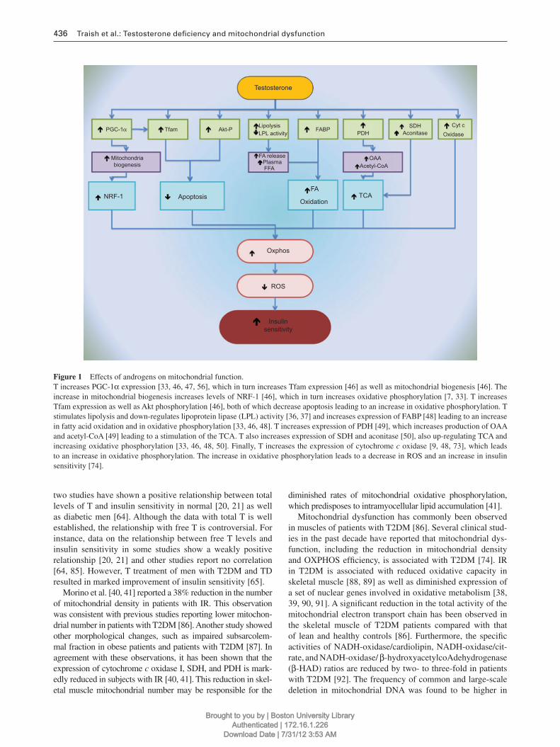
Figure 1 Effects of androgens on mitochondrial function. T increases PGC-1 α expression [33, 46, 47, 56] , which in turn increases Tfam expression [46] as well as mitochondrial biogenesis [46] . The increase in mitochondrial biogenesis increases levels of NRF-1 [46] , which in turn increases oxidative phosphorylation [7, 33] . T increases Tfam expression as well as Akt phosphorylation [46] , both of which decrease apoptosis leading to an increase in oxidative phosphorylation. T stimulates lipolysis and down-regulates lipoprotein lipase (LPL) activity [36, 37] and increases expression of FABP [48] leading to an increase in fatty acid oxidation and in oxidative phosphorylation [33, 46, 48] . T increases expression of PDH [49] , which increases production of OAA and acetyl-CoA [49] leading to a stimulation of the TCA. T also increases expression of SDH and aconitase [50] , also up-regulating TCA and increasing oxidative phosphorylation [33, 46, 48, 50] . Finally, T increases the expression of cytochrome c oxidase [9, 48, 73] , which leads to an increase in oxidative phosphorylation. The increase in oxidative phosphorylation leads to a decrease in ROS and an increase in insulin sensitivity [74] .
Brought to you by | Boston University LibraryAuthenticated | 172.16.1.226
Download Date | 7/31/12 3:53 AM

Traish et al.: Testosterone defi ciency and mitochondrial dysfunction 437
skeletal muscle of diabetic patients than in age- and sex-matched controls [93] . The impairment of oxidative phospho-rylation as well as fatty acid metabolism has been observed in skeletal muscles of insulin-resistant offspring of patients with T2DM compared with age-matched insulin-sensitive controls [94] . Mogensen et al. [95] reported that ADP-stimulated res-piration was diminished in obese subjects with T2DM with a compensatory increase in type 2 muscle fi bers. Mouse mod-els have been used to confi rm that mitochondrial dysfunc-tion can cause T2DM [74] . Other studies reported a decline in mitochondrial number and content of mitochondrial DNA and respiratory enzymes. Derangement of the mitochondrial network was also noted. These fi ndings are consistent with the decline in oxidative phosphorylation and β -oxidation in adipose tissue of mice with T2DM [96] .
Oxidative stress and mitochondrial dysfunction
Mitochondrial dysfunction contributes to overproduction of ROS and dysregulation of the insulin signaling pathway lead-ing to IR in muscle [74] . Overproduction of ROS caused by an imbalance of antioxidant enzymes and defective oxida-tive phosphorylation may damage intracellular components and impair normal cellular function [97] . Oxidative stress may activate multiple serine threonine kinase (Akt) cascades, including p38 mitogen activated protein kinase and Jun N-terminal kinase (JNK), and can act on a number of poten-tial targets in the insulin signaling pathway, such as insulin receptor and the family of insulin receptor substrate (IRS) proteins [74] . Furthermore, inactivation of phosphoinositol-3-kinase has been shown to impair the translocation of the
Androgen hormonesT and 5α-DHT
AR
Ca··CaMK
ARE
ARE NRF
TFAM protein
NucleusTFAM geneprotein
ARE PGC-1α NRF NRF protein
protein
AR
AR AP-1
mt RNA
MRC proteins
Mitochondrion
Mitochondrial biogenesisApoptosis
Fatty acid oxidationOxidative phosphorylationReactive oxygen species
Insulin sensitivity
protein
O2-
ATP
protein
PGC-1α
MEFCREBTF-X
PGC-1α protein
PGC-lα gene
mt ARENRF gene
XOPHOS genes
proteinprc
mt proteins
Figure 2 Modulation of mitochondrial function by androgens. T and 5- α -dihydrotestosterone (5α-DHT) bind with high affi nity to the AR. The ligand-AR complex modulates the expression of several nuclear and mitochondrial target genes that, together, promote fatty acid oxidation, oxidative phosphorylation, and mitochondrial biogenesis, attenuating the generation or build up of ROS resulting in an improvement in insulin sensitivity [33] . In the mitochondrion, the bound AR also up-regulates the expression of cytochrome c oxidase [9] . In the nucleus, the ligand-bound AR complex increases PGC-1 α expression [46] , which in turn up-regulates Tfam expression [46] . These pathways lead to mitochondrial biogenesis, which increases NRF-1 levels [46] , the later in turn facilitates oxidative phosphorylation and increased expression of oxidative phosphorylation genes encoding PDH, SDH, GPDH, and PC [42, 49] .
Brought to you by | Boston University LibraryAuthenticated | 172.16.1.226
Download Date | 7/31/12 3:53 AM

438 Traish et al.: Testosterone defi ciency and mitochondrial dysfunction
insulin-dependent glucose transporter GLUT4 to the plasma membrane, leading to a decreased glucose uptake by muscle in response to insulin [97] .
With advancing age, muscles exhibit lower mitochon-drial number and lower effi cacy of energy production [98] . It has been suggested that the lower effi ciency in muscle mitochondria in the elderly is caused by ROS damage to the inner mitochondrial membrane, resulting in uncoupling of the electron transport chain [99] . The decreased effi ciency and impaired energy production capacity of muscle mitochondria that develops with aging may be partially reversed [100, 101] . This suggests that mitochondrial dysfunction is not entirely due to irreversible mutations in mitochondrial DNA. Physical activity may stimulate mitochondrial biogenesis as well as improve the effi ciency of existing mitochondria, possibly by reversing oxidative damage [98] .
Impairment of mitochondrial function may be accom-panied by a diminished activity of enzymes involved in β -oxidation of fatty acids, leading to an increase in intra-cellular lipid content [74] . Hotta et al. [102] reported that the plasma level of adiponectin, the adipokine released by adipose tissue, is diminished in obese patients or patients with T2DM. In mouse models, it has been established that adiponectin defi ciency leads to IR [103] . This decrease in adiponectin expression in adipocytes with mitochondrial dysfunction occurs by activation of the JNK pathway [104] . Thus, mitochondrial dysfunction not only causes insulin insensitivity but also impairs the secretion of adipokines, by adipocytes, which in turn compromises other tissues with regard to glucose utilization [74] .
Mootha et al. [38] reported decreased maximal aerobic capacity and reduced expression of mitochondrial genes involved in oxidative phosphorylation in men with impaired glucose tolerance and T2DM. Patti et al. [39] demonstrated a decreased expression of enzymes involved in oxidative phos-phorylation in a group of T2DM patients as well as insulin-resistant fi rst-degree relatives of T2DM subjects with normal glucose tolerance. Using magnetic resonance spectroscopy, it has been shown that decreased mitochondrial oxidative phosphorylation activity is the cause of age-related IR [105] . In addition, the lean offspring of patients with T2DM also exhibited mitochondrial dysfunction [94] . A recent study per-formed in an animal model bred to exhibit low aerobic capac-ity also implicated a causative role of impaired mitochondrial function in the development of the CVD risk profi le associ-ated with MetS [106] .
Molecular mechanism of androgen action in
mitochondrial function
Androgens regulate gene transcription through a genomic pathway in which the AR, a ligand-activated transcription factor, binds the hormone and translocates into the nucleus where it interacts with specifi c DNA sequence known as androgen response element (ARE) in its target genes [107] . Evidence accumulating over the past two decades has also implicated rapid ( non-genomic ) responses to androgens, through a mechanism dependent or independent of AR
action [108] . While limited information is available regard-ing the interaction of AR with mitochondria, it has been con-fi rmed that the AR interacts directly with the Vb subunit of cytochrome c oxidase [73] . In prostatic cells, the AR medi-ates the translocation of the pro-apoptotic factor Bax to the mitochondria in a transcriptionally dependent mechanism that is yet to be elucidated [109] . In contrast, direct receptor-independent activation of inner mitochondrial membrane ATP-sensitive K + channels has been observed following T administration in cardiomyocytes [110] .
Several biochemical factors play an integral role in mito-chondrial biogenesis. These include the PPAR γ co-activator PGC-1 α , a transcriptional factor co-activator [41] , 5 ′ adeno-sine monophosphate-activated protein (AMP) kinase, which elicits its effects through myocyte enhancer factor-2 and cyclic AMP response element-binding protein-mediated increased PGC-1 α expression [111 – 113] . Thus, increased expression of PGC-1 α leads to an increase in target genes, such as the nuclear respiratory factor-1 ( NRF-1 ), a transcrip-tion factor that stimulates many nuclear-encoded mitochon-drial genes including OXPHOS genes and Tfam , which bind to the D-loop of the mitochondrial genome and increase tran-scription of mitochondrial genes and replication of mitochon-drial DNA [114] .
It has been suggested that the mechanism of IR induced by TD involves a down-regulation of the transcription factor PGC-1 α in skeletal muscle [56] . As a stimulator of mitochon-drial biogenesis as well as skeletal muscle oxidative fi bers, PGC-1 α is a molecular marker of insulin sensitivity, and a decrease in its expression has been observed in patients with T2DM [38] . Low T levels are associated with low PGC-1 α expression in muscle [33] . This is supported by studies in mouse models in which similar association has been observed between TD and low levels of PGC-1 α in tissues [47] . The reduced expression of PGC-1 α compromises mitochondrial activity and energy production. Thus, it seems likely that TD promotes IR, at least partially, through an AR-dependent mechanism that involves a decrease in PGC-1 α -mediated oxidative and insulin-sensitive muscle fi bers [56] .
T substitution in orchiectomized rats improved recovery of myocardial function after ischemia. While this effect might have been partly related to acute coronary vasodilation by T, the investigators of one study hypothesized that T may also exhibit direct cytoprotective actions on the myocardium. The results showed that T acutely and directly depolarized and oxi-dized cardiac mitochondria in a K + -dependent, ATP-sensitive, and AR-independent manner (non-genomic pathway). By patch clamping the cardiac inner mitochondrial membrane, it was demonstrated that T induced activation of mitochondrial K + channels, which were inhibited by ATP, 5-hydroxydecai-noic acid, and glibenclamide, but exhibited no effect on sar-coplasmic K ATP channels. T protected cardiomyocytes from ischemic cell death [110] .
A recent study set out to clarify whether the AR system exerts a cardioprotective effect against Dox-induced cardio-toxicity. Electron microscopic examination of mitochondria in the hearts of animals treated with Dox revealed prominent mitochondrial damage, such as vacuolization in the ARKO
Brought to you by | Boston University LibraryAuthenticated | 172.16.1.226
Download Date | 7/31/12 3:53 AM

Traish et al.: Testosterone defi ciency and mitochondrial dysfunction 439
mice model compared with that of the WT mice. A basal mito-chondrial dysfunction was noted in the myocardium of male ARKO mouse heart, in the absence of Dox treatment. This was attributed to loss of AR-mediated signaling, which may play a critical role in cardiac oxidative stress. Furthermore, superoxide production in response to Dox treatment of male ARKO mice was markedly enhanced compared with that of male WT mice, but the number of apoptotic cells in the ven-tricular tissues was signifi cantly larger in male ARKO mice than in male WT mice. The expression of a key mitochondrial transcription factor (Tfam) in cardiac tissues of male WT mice was not affected signifi cantly by Dox treatment while its expression was reduced by almost half in male ARKO mice treated with Dox. The results of this study suggest that the AR system may counteract Dox-induced cardiotoxicity partly through activation of the Akt pathway and up-regulation of Tfam to protect cardiomyocytes from mitochondrial damage and apoptosis [46] .
A recent study comparing mitochondrial function of young obese and non-obese individuals showed more mitochondrial dysfunction in the cardiomyocytes of obese patients with excessive oxidative stress, mitochondrial damage, and apop-tosis. This may be partially explained by the noted reduction in the expression of NRF-1 and its target Tfam [115] and may account for decreased expression of the complex I pro-tein ND6 [116] . Since MetS and obesity are associated with reduced T levels, it is possible that reduced circulating andro-gens, which contribute to increased infl ammatory cytokines, may play a role in mitochondrial dysfunction.
Mitochondrial dysfunction and diseases
TD is increasingly recognized not only among older men but also in young men and in cancer survivors, who underwent ADT. However, the impact of TD on the quality of life remains poorly established. Among the common complaints of TD is fatigue, reduced energy, loss of self-esteem, and sexual dys-function [30] . T replacement therapy in men with TD showed improvement in mood, sexual function, reduced depres-sion and anxiety, increased concentration, self-confi dence, improved mood, and decreased fatigue within a few weeks [17] . The mechanisms by which androgens affect many of these physiological processes are not clearly understood. One underlying hypothesis is that androgens modulate mitochon-drial function and this may be a common link between TD and the various symptoms noted in men with TD. However, no specifi c mechanism has been provided to relate androgen defi ciency to mitochondrial dysfunction.
Mitochondria play a critical role in cellular function by regulating biochemical pathways involved in lipid, protein, and carbohydrate metabolism as well as cell survival and apoptosis. Understanding androgen modulation of mito-chondrial function is critical to understanding the role of TD in the pathophysiology of T2DM, IR, and CVD. As shown in Figure 1 , androgen regulation of expression and activity of oxidative phosphorylation enzymes in the mito-chondria may represent one critical mechanism, among others, in smooth and skeletal muscle in the control of
cellular processes. This regulation by androgens facilitates the increase in production of cellular energy depending on the physiological conditions, such as physical demand, stress, or acute illness.
As depicted in Figure 2 , androgens regulate cellular metabolism and energy production through molecular and cellular mechanisms involving both genomic and non-ge-nomic pathways and mitochondrial OXPHOS genes. Binding of activated AR complexes with nuclear and mitochondrial OXPHOS gene response elements has been demonstrated [117 – 119] . Another proposed pathway of androgen action is through direct interaction of AR complexes with AREs of OXPHOS genes in the mitochondria. Additional mechanisms may involve indirect interactions with ARE in the nucleus to activate transcription of genes encoding transcription factors, such as NRF and PGC-1 α , which in turn activate OXPHOS genes in the mitochondria [120, 121] .
Testosterone effect on mitochondrial functions
As depicted in Figure 1 , T up-regulates a host of enzymes and transcriptional factors. For example, T increases the expres-sion of PGC-1 α , in which the latter modulates Tfam tran-scriptional factor activity as well as stimulates mitochondrial biogenesis, leading to an increase in the expression of NRF-1. T also increases the expression and activity of Akt, hormone-activated lipases, FABP, cytochrome c oxidase, SDH, and PDH. These enzymes coordinate a host of integrated path-ways leading to increased mitochondrial biogenesis and to increased energy production. Furthermore, the expression of nuclear transcription factors, which in turn control the expres-sion of nuclear-encoded mitochondrial proteins, is also regu-lated by androgens.
Depletion of mitochondrial DNA by chronic treatment with ethidium bromide causes loss of response to insulin due to impaired insulin signaling pathways, and repletion of mito-chondrial DNA restores insulin sensitivity of muscle cells [122] . In addition to this genetic mechanism of action, treat-ment with respiratory inhibitors has revealed a decrease in insulin-stimulated glucose uptake as well as inactivation of Akt and IRS-1 of the insulin signaling pathway [123] . One proposed mechanism by which mitochondrial dysfunction causes IR involves impairment in insulin secretion by β -islet cells in response to a decreased intracellular Ca 2 + concentra-tion. It has been suggested that the ATP/ADP ratio is dimin-ished in β -islet cells with mitochondrial defects, rendering the cell incapable of inducing closure of ATP-dependent K + chan-nels or depolarization of the membrane [74] .
Mitochondrial dysfunction is implicated in a number of pathophysiological processes in disease states, such as diabe-tes, IR, and CVD. Recent studies have demonstrated a strong association between TD, IR, diabetes, and CVD. However, the molecular and cellular mechanisms linking TD to these pathologies remain under investigation. Yialamas et al. [34] and Pittleoud et al. [33] have suggested that TD has a direct effect on glucose utilization in cases of increased IR. VO 2 max and OXPHOS-CR gene expression were shown to corre-late positively with T levels [33] . In animal studies, castration
Brought to you by | Boston University LibraryAuthenticated | 172.16.1.226
Download Date | 7/31/12 3:53 AM

440 Traish et al.: Testosterone defi ciency and mitochondrial dysfunction
Figure 3 Effects of androgen deprivation on mitochondrial morphology. Penile corpus cavernosum tissue sections obtained from 2-week castrated mature male rabbits (top) or from sham-operated animals (bottom) were fi xed, embedded in plastic, and sectioned for electron microscopic examinations as described previously [69] . Note that the trabecular smooth muscle cells from castrated animals have disorga-nized contours and have accumulated vacuoles and fl occulating sub-stances. Also note that the mitochondria (arrow) were increased in number and appear swollen (see magnifi ed box at top right corner). In contrast, in the section from the sham-operated animals (bottom), the smooth muscle appears normal with no distortions or accumula-tion of vacuoles, and the mitochondria (arrow) appear normal with no increase in the number or swelling (see magnifi ed box in the top right corner).
is also associated with IR and decreased glycogen synthase activity [37] . Investigations in obese rats, a genetic model of obesity and T2DM, strongly suggest a role for androgens in regulating mitochondrial function [37] . Interestingly, medical castration of young healthy men with gonadotropin-releas-ing hormone agonist resulted in reduced lipid oxidation and diminished resting energy expenditure [36] . These observa-tions suggest that TD may contribute to IR by a mechanism that involves attenuated or altered fatty acid oxidation. T modulation of OXPHOS gene expression may represent an important therapeutic modality for preventing or treating mitochondrial dysfunction in men with TD.
Summary
Androgens regulate fuel metabolism through mitochondrial function and modulate mitochondrial biogenesis, expression of mitochondrial enzymes, and oxidative phosphorylation. Androgen defi ciency contributes to the pathophysiology of fatigue, IR, diabetes, and in turn CVD through the com-mon link “ mitochondrial dysfunction. ” Research on the molecular basis of androgen action in the mitochondria may provide novel strategies for the development of phar-macotherapeutic agents for the management of the afore-mentioned pathologies .
Acknowledgments
This work was supported solely by the Department of Urology, Boston University School of Medicine.
References
1. Khaw KT, Dowsett M, Folkerd E, Bingham S, Wareham N, Luben R, Welch A, Day N. Endogenous testosterone and mortal-ity due to all causes, cardiovascular disease, and cancer in men: European prospective investigation into cancer in Norfolk (EPIC-Norfolk) Prospective Population Study. Circulation 2007;116:2694 – 701.
2. Laughlin GA, Barrett-Connor E, Bergstrom J. Low serum tes-tosterone and mortality in older men. J Clin Endocrinol Metab 2008;93:68 – 75.
3. Tivesten A, Vandenput L, Labrie F, Karlsson MK, Ljunggren O, Mellstr ö m D, Ohlsson C. Low serum testosterone and estra-diol predict mortality in elderly men. J Clin Endocrinol Metab 2009;94:2482 – 8.
4. Shores MM, Matsumoto AM, Sloan KL, Kivlahan DR. Low serum testosterone and mortality in male veterans. Arch Intern Med 2006;166:1660 – 5.
5. Haring R, V ö lzke H, Felix SB, Schipf S, D ö rr M, Rosskopf D, Nauck M, Sch ö fl C, Wallaschofski H. Prediction of metabolic syndrome by low serum testosterone levels in men: results from the study of health in Pomerania. Diabetes 2009;58:2027 – 31.
6. Malkin CJ, Channer KS, Jones TH. Testosterone and heart fail-ure. Curr Opin Endocrinol Diabetes Obes 2010;17:262 – 8.
7. Jones TH. Testosterone defi ciency: a risk factor for cardiovascu-lar disease ? Trends Endocrinol Metab 2010;21:496 – 503.
8. Malkin CJ, Pugh PJ, Morris PD, Asif S, Jones TH, Channer KS. Low serum testosterone and increased mortality in men with coronary heart disease. Heart 2010;96:1821 – 5.
9. Caminiti G, Volterrani M, Iellamo F, Marazzi G, Massaro R, Miceli M, Mammi C, Piepoli M, Fini M, Rosano GM. Effect of long-acting testosterone treatment on functional exercise capac-ity, skeletal muscle performance, insulin resistance, and barore-fl ex sensitivity in elderly patients with chronic heart failure a double-blind, placebo-controlled, randomized study. J Am Coll Cardiol 2009;54:919 – 27.
10. Coats AJ, Clark AL, Piepoli M, Volterrani M, Poole-Wilson PA. Symptoms and quality of life in heart failure: the muscle hypoth-esis. Br Heart J 1994;72:S36 – 9.
11. Piepoli M, Clark AL, Volterrani M, Adamopoulos S, Sleight P, Coats AJ. Contribution of muscle afferents to the hemodynamic, autonomic, and ventilatory responses to exercise in patients with
Brought to you by | Boston University LibraryAuthenticated | 172.16.1.226
Download Date | 7/31/12 3:53 AM

Traish et al.: Testosterone defi ciency and mitochondrial dysfunction 441
chronic heart failure: effects of physical training. Circulation 1996;93:940 – 52.
12. Piepoli MF, Kaczmarek A, Francis DP, Davies LC, Rauchhaus M, Jankowska EA, Anker SD, Capucci A, Banasiak W, Ponikowski P. Reduced peripheral skeletal muscle mass and abnormal refl ex physiology in chronic heart failure. Circulation 2006;114:126 – 34.
13. Traish AM, Kypreos KE. Testosterone and cardiovascular disease: an old idea with modern clinical implications. Atherosclerosis 2011;214:244 – 8.
14. Zitzmann M. Testosterone defi ciency, insulin resistance and the metabolic syndrome. Nat Rev Endocrinol 2009;5:673 – 81.
15. Saad F. The relationship between testosterone defi ciency and frailty in elderly men. Horm Mol Biol Clin Invest 2010;4:529 – 38.
16. Jockenh ö vel F, Minnemann T, Schubert M, Freude S, H ü bler D, Schumann C, Christoph A, Ernst M. Comparison of long-acting testosterone undecanoate formulation versus testosterone enan-thate on sexual function and mood in hypogonadal men. Eur J Endocrinol 2009;160:815 – 9.
17. Jockenh ö vel F, Minnemann T, Schubert M, Freude S, H ü bler D, Schumann C, Christoph A, Gooren L, Ernst M. Timetable of effects of testosterone administration to hypogonadal men on variables of sex and mood. Aging Male 2009;12:113 – 8.
18. Kalinchenko SY, Tishova YA, Mskhalaya GJ, Gooren LJ, Giltay EJ, Saad F. Effects of testosterone supplementation on markers of the metabolic syndrome and infl ammation in hypogonadal men with the metabolic syndrome: the double-blinded place-bo-controlled Moscow study. Clin Endocrinol (Oxf) 2010;73:602 – 12.
19. Tibblin G, Adlerberth A, Lindstedt G, Bj ö rntorp P. The pituitary-gonadal axis and health in elderly men: a study of men born in 1913. Diabetes 1996;45:1605 – 9.
20. Haffner SM, Karhap ä ä P, Mykk ä nen L, Laakso M. Insulin resis-tance, body fat distribution, and sex hormones in men. Diabetes 1994;43:212 – 9.
21. Haffner SM, Shaten J, Stern MP, Smith GD, Kuller L. Low lev-els of sex hormone-binding globulin and testosterone predict the development of non-insulin-dependent diabetes mellitus in men. MRFIT Research Group. Multiple Risk Factor Intervention Trial. Am J Epidemiol 1996;143:889 – 97.
22. Stellato RK, Feldman HA, Hamdy O, Horton ES, McKinlay JB. Testosterone, sex hormone-binding globulin, and the devel-opment of type 2 diabetes in middle-aged men: prospective results from the Massachusetts male aging study. Diabetes Care 2000 ; 23:490 – 4.
23. Oh JY, Barrett-Connor E, Wedick NM, Wingard DL. Endogenous sex hormones and the development of type 2 diabetes in older men and women: the Rancho Bernardo study. Diabetes Care 2002;25:55 – 60.
24. Svartberg J, Jenssen T, Sundsfjord J, Jorde R. The associations of endogenous testosterone and sex hormone-binding globulin with glycosylated hemoglobin levels, in community dwelling men. The Tromso Study. Diabetes Metab 2004;30:29 – 34.
25. Laaksonen DE, Niskanen L, Punnonen K, Nyyss ö nen K, Tuomainen TP, Valkonen VP, Salonen R, Salonen JT. Testosterone and sex hormone-binding globulin predict the meta-bolic syndrome and diabetes in middle-aged men. Diabetes Care 2004;27:1036 – 41.
26. Smith JC, Bennett S, Evans LM, Kynaston HG, Parmar M, Mason MD, Cockcroft JR, Scanlon MF, Davies JS. The effects of induced hypogonadism on arterial stiffness, body composition, and metabolic parameters in males with prostate cancer. J Clin Endocrinol Metab 2001;86:4261 – 7.
27. Dockery F, Bulpitt CJ, Agarwal S, Donaldson M, Rajkumar C. Testosterone suppression in men with prostate cancer leads to an increase in arterial stiffness and hyperinsulinaemia. Clin Sci (Lond) 2003;104:195 – 201.
28. D ’ Amico AV, Denham JW, Crook J, Chen MH, Goldhaber SZ, Lamb DS, Joseph D, Tai KH, Malone S, Ludgate C, Steigler A, Kantoff PW. Infl uence of androgen suppression therapy for prostate cancer on the frequency and timing of fatal myocardial infarctions. J Clin Oncol 2007;25:2420 – 5.
29. Tsai HK, D ’ Amico AV, Sadetsky N, Chen MH, Carroll PR. Androgen deprivation therapy for localized prostate cancer and the risk of cardiovascular mortality. J Natl Cancer Inst 2007;99:1516 – 24.
30. Greenfi eld DM, Walters SJ, Coleman RE, Hancock BW, Snowden JA, Shalet SM, DeRogatis LR, Ross RJ. Quality of life, self-esteem, fatigue, and sexual function in young men after cancer: a controlled cross-sectional study. Cancer 2010;116:1592 – 601.
31. O ’ Connor PJ. Evaluation of four highly cited energy and fatigue mood measures. J Psychosom Res 2004;57:435 – 41.
32. Bjorntorp P. Metabolic implications of body fat distribution. Diabetes Care 1991;14:1132 – 43.
33. Pitteloud N, Mootha VK, Dwyer AA, Hardin M, Lee H, Eriksson KF, Tripathy D, Yialamas M, Groop L, Elahi D, Hayes FJ. Relationship between testosterone levels, insulin sensitivity, and mitochondrial function in men. Diabetes Care 2005;28:1636 – 42.
34. Yialamas MA, Dwyer AA, Hanley E, Lee H, Pitteloud N, Hayes FJ. Acute sex steroid withdrawal reduces insulin sensitivity in healthy men with idiopathic hypogonadotropic hypogonadism. J Clin Endocrinol Metab 2007;92:4254 – 9.
35. Traish AM, Miner MM, Morgentaler A, Zitzmann M. Testosterone defi ciency. Am J Med 2011;124:578 – 87.
36. Mauras N, Hayes V, Welch S, Rini A, Helgeson K, Dokler M, Veldhuis JD, Urban RJ. Testosterone defi ciency in young men: marked alterations in whole body protein kinetics, strength, and adiposity. J Clin Endocrinol Metab 1998;83:1886 – 92.
37. Ryu JW, Kim MS, Kim CH, Song KH, Park JY, Lee JD, Kim JB, Lee KU. DHEA administration increases brown fat uncoupling protein 1 levels in obese OLETF rats. Biochem Biophys Res Commun 2003;303:726 – 31.
38. Mootha VK, Lindgren CM, Eriksson KF, Subramanian A, Sihag S, Lehar J, Puigserver P, Carlsson E, Ridderstr å le M, Laurila E, Houstis N, Daly MJ, Patterson N, Mesirov JP, Golub TR, Tamayo P, Spiegelman B, Lander ES, Hirschhorn JN, Altshuler D, Groop LC. PGC-1 α -responsive genes involved in oxidative phospho-rylation are coordinately downregulated in human diabetes. Nat Genet 2003;34 : 267 – 73.
39. Patti ME, Butte AJ, Crunkhorn S, Cusi K, Berria R, Kashyap S, Miyazaki Y, Kohane I, Costello M, Saccone R, Landaker EJ, Goldfi ne AB, Mun E, DeFronzo R, Finlayson J, Kahn CR, Mandarino LJ. Coordinated reduction of genes of oxidative metabolism in humans with insulin resistance and diabetes: potential role of PGC1 and NRF1. Proc Natl Acad Sci USA 2003;100:8466 – 71.
40. Morino K, Petersen KF, Dufour S, Befroy D, Frattini J, Shatzkes N, Neschen S, White MF, Bilz S, Sono S, Pypaert M, Shulman GI. Reduced mitochondrial density and increased IRS-1 serine phosphorylation in muscle of insulin-resistant offspring of type 2 diabetic parents. J Clin Invest 2005;115:3587 – 93.
41. Morino K, Petersen KF, Shulman GI. Molecular mechanisms of insulin resistance in humans and their potential links with mito-chondrial dysfunction. Diabetes 2006;55:S9 – 15.
42. Brooks DE. Infl uence of androgens on the weights of the male accessory reproductive organs and on the activities of
Brought to you by | Boston University LibraryAuthenticated | 172.16.1.226
Download Date | 7/31/12 3:53 AM

442 Traish et al.: Testosterone defi ciency and mitochondrial dysfunction
mitochondrial enzymes in the epididymis of the rat. J Endocrinol 1979;82:293 – 303.
43. Collins TJ, Berridge MJ, Lipp P, Bootman MD. Mitochondria are morphologically and functionally heterogeneous within cells. EMBO J 2002;21:1616 – 27.
44. Koenig H, Goldstone A, Lu CY. Testosterone-mediated sexual dimorphism of the rodent heart. Ventricular lysosomes, mito-chondria, and cell growth are modulated by androgens. Circ Res 1982;50:782 – 7.
45. Brooks DE. Activity and androgenic control of enzymes associ-ated with the tricarboxylic acid cycle, lipid oxidation and mito-chondrial shuttles in the epididymis and epididymal spermatozoa of the rat. Biochem J 1978;174:741 – 52.
46. Ikeda Y, Aihara K, Akaike M, Sato T, Ishikawa K, Ise T, Yagi S, Iwase T, Ueda Y, Yoshida S, Azuma H, Walsh K, Tamaki T, Kato S, Matsumoto T. Androgen receptor counteracts Doxorubicin-induced cardiotoxicity in male mice. Mol Endocrinol 2010;24 : 1338 – 48.
47. Fan W, Yanase T, Nomura M, Okabe T, Goto K, Sato T, Kawano H, Kato S, Nawata H. Androgen receptor null male mice develop late-onset obesity caused by decreased energy expenditure and lipolytic activity but show normal insulin sensitivity with high adiponectin secretion. Diabetes 2005;54:1000 – 8.
48. van Breda E, Keizer HA, Vork MM, Surtel DA, de Jong YF, van der Vusse GJ, Glatz JF. Modulation of fatty-acid-binding protein content of rat heart and skeletal muscle by endurance training and testosterone treatment. Pfl ugers Arch 1992;421:274 – 9.
49. Costello LC, Franklin RB. Testosterone regulates pyruvate dehy-drogenase activity of prostate mitochondria. Horm Metab Res 1993;25:268 – 70.
50. Juang HH, Hsieh ML, Tsui KH. Testosterone modulates mito-chondrial aconitase in the full-length human androgen receptor-transfected PC-3 prostatic carcinoma cells. J Mol Endocrinol 2004;33:121 – 32.
51. Moon JS, Jin WJ, Kwak JH, Kim HJ, Yun MJ, Kim JW, Park SW, Kim KS. Androgen stimulates glycolysis for de novo lipid synthesis by increasing the activities of hexokinase 2 and 6-phos-phofructo-2-kinase/fructose-2,6-bisphosphatase 2 in prostate cancer cells. Biochem J 2011;433:225 – 33.
52. Czesla M, Mehlhorn G, Fritzsche D, Asmussen G. Cardio-myoplasty improvement of muscle fi bre type transformation by anabolic steroid. J Mol Cell Cardiol 1997;29:2989 – 96.
53. Ustunel I, Akkoyunlu G, Demir R. The effect of testosterone on gastrocnemius muscle fi bres in growing and adult male and female rats: a histochemical, morphometric and ultrastructural study. Anat Histol Embryol 2003;32:70 – 9.
54. Lanfranco F, Zitzmann M, Simoni M, Nieschlag E. Serum adiponectin levels in hypogonadal males: infl uence of testo-sterone replacement therapy. Clin Endocrinol (Oxf) 2004;60:500 – 7.
55. Basaria S, Muller DC, Carducci MA, Egan J, Dobs AS. Hyperglycemia and insulin resistance in men with prostate carcinoma who receive androgen-deprivation therapy. Cancer 2006;106:581 – 8.
56. Mauvais-Jarvis F. Estrogen and androgen receptors: regulators of fuel homeostasis and emerging targets for diabetes and obesity. Trends Endocrinol Metab 2011;22:24 – 33.
57. Khaw KT, Barrett-Connor E. Lower endogenous androgens pre-dict central adiposity in men. Ann Epidemiol 1992;2:675 – 82.
58. Lichtenstein MJ, Yarnell JW, Elwood PC, Beswick AD, Sweetnam PM, Marks V, Teale D, Riad-Fahmy D. Sex hormones, insulin, lipids, and prevalent ischemic heart disease. Am J Epidemiol 1987;126:647 – 57.
59. Pasquali R, Casimirri F, Cantobelli S, Melchionda N, Morselli Labate AM, Fabbri R, Capelli M, Bortoluzzi L. Effect of obesity and body fat distribution on sex hormones and insulin in men. Metabolism 1991;40:101 – 4.
60. Pasquali R, Casimirri F, Cantobelli S, Melchionda N, Morselli Labate AM, Fabbri R, Capelli M, Bortoluzzi L. Interrelation between plasma testosterone and plasma insulin in healthy adult men: the Telecom Study. Diabetologia 1992;35:173 – 7.
61. Andersson B, Marin P, Lissner L, Vermeulen A, Bjorntorp P. Testosterone concentrations in women and men with NIDDM. Diabetes Care 1994;17:405 – 11.
62. Barrett-Connor E. Lower endogenous androgen levels and dys-lipidemia in men with non-insulin-dependent diabetes mellitus. Ann Intern Med 1992;117:807 – 11.
63. Wang C, Jackson G, Jones TH, Matsumoto AM, Nehra A, Perelman MA, Swerdloff RS, Traish A, Zitzmann M, Cunningham G. Low testosterone associated with obesity and the metabolic syndrome contributes to sexual dysfunction and cardiovascular disease risk in men with type 2 diabetes. Diabetes Care 2011;34:1669 – 75.
64. Birkeland KI, Hanssen KF, Torjesen PA, Vaaler S. Level of sex hormone-binding globulin is positively correlated with insulin sensitivity in men with type 2 diabetes. J Clin Endocrinol Metab 1993;76:275 – 8.
65. Heufelder AE, Saad F, Bunck MC, Gooren L. Fifty-two-week treatment with diet and exercise plus transdermal testosterone reverses the metabolic syndrome and improves glycemic con-trol in men with newly diagnosed type 2 diabetes and subnormal plasma testosterone. J Androl 2009;30:726 – 33.
66. Boissonneault G. Evidence of apoptosis in the castration-induced atrophy of the rat levator ani muscle. Endocr Res 2001;27:317 – 28.
67. Shukla Y, Prasad S, Tripathi C, Singh M, George J, Kalra N. In vitro and in vivo modulation of testosterone mediated alterations in apoptosis related proteins by [6]-gingerol. Mol Nutr Food Res 2007;51:1492 – 502.
68. Yang CY, Lam HC, Lee HC, Wei YH, Lu CC, Han TM, Tsai JL, Chuang YH, Lee JK. MELAS syndrome associated with diabetes mellitus and hyperthyroidism: a case report from Taiwan. Clin Endocrinol (Oxf) 1995;43:235 – 9.
69. Traish A, Kim N. The physiological role of androgens in penile erection: regulation of corpus cavernosum structure and function. J Sex Med 2005;2:759 – 70.
70. Niki E. Antioxidants in relation to lipid peroxidation. Chem Phys Lipids 1987;44:227 – 53.
71. Koenig H, Goldstone A, Lu CY. Androgens regulate mitochon-drial cytochrome c oxidase and lysosomal hydrolases in mouse skeletal muscle. Biochem J 1980;192:349 – 53.
72. Koenig H, Goldstone A, Blume G, Lu CY. Testosterone-mediated sexual dimorphism of mitochondria and lysosomes in mouse kid-ney proximal tubules. Science 1980;209:1023 – 6.
73. Beauchemin AM, Gottlieb B, Beitel LK, Elhaji YA, Pinsky L, Trifi ro MA. Cytochrome c oxidase subunit Vb interacts with human androgen receptor: a potential mechanism for neurotoxicity in spinobulbar muscular atrophy. Brain Res Bull 2001;56:285 – 97.
74. Wang CH, Wang CC, Wei YH. Mitochondrial dysfunction in insulin insensitivity: implication of mitochondrial role in type 2 diabetes. Ann N Y Acad Sci 2010;1201:157 – 65.
75. M å rin P, L ö nn L, Andersson B, Od é n B, Olbe L, Bengtsson BA, Bj ö rntorp P. Assimilation of triglycerides in subcutaneous and intraabdominal adipose tissues in vivo in men: effects of testos-terone. J Clin Endocrinol Metab 1996;81 : 1018 – 22.
76. Christofi lis MA, Remacle-Bonnet M, Atlan-Gepner C, Garrouste F, Vialettes B, Fuentes P, Guidicelli R, Pommier G. Study of
Brought to you by | Boston University LibraryAuthenticated | 172.16.1.226
Download Date | 7/31/12 3:53 AM
Traish et al.: Testosterone defi ciency and mitochondrial dysfunction 443
serum big-insulin-like growth factor (IGF)-II and IGF binding proteins in two patients with extrapancreatic tumor hypoglyce-mia, using a combination of Western blotting methods. Eur J Endocrinol 1998;139:317 – 22.
77. Saleh J, Sniderman AD, Cianfl one K. Regulation of plasma fatty acid metabolism. Clin Chim Acta 1999;286:163 – 80.
78. Boden G. Role of fatty acids in the pathogenesis of insulin resis-tance and NIDDM. Diabetes 1997;46:3 – 10.
79. McGarry JD, Dobbins RL, Stein DT. [Fatty acids, insulin resis-tance and pancreatic β cell function]. J Annu Diabetol Hotel Dieu 1998;1 – 10. PMID: 9773605.
80. Peeters RA, Veerkamp JH. Does fatty acid-binding protein play a role in fatty acid transport ? Mol Cell Biochem 1989;88:45 – 9.
81. Nishizawa H, Shimomura I, Kishida K, Maeda N, Kuriyama H, Nagaretani H, Matsuda M, Kondo H, Furuyama N, Kihara S, Nakamura T, Tochino Y, Funahashi T, Matsuzawa Y. Androgens decrease plasma adiponectin, an insulin-sensitizing adipocyte-derived protein. Diabetes 2002;51:2734 – 41.
82. Bojesen A, Kristensen K, Birkebaek NH, Fedder J, Mosekilde L, Bennett P, Laurberg P, Frystyk J, Flyvbjerg A, Christiansen JS, Gravholt CH. The metabolic syndrome is frequent in Klinefelter ’ s syndrome and is associated with abdominal obesity and hypogo-nadism. Diabetes Care 2006;29:1591 – 8.
83. Phillips GB. Relationship between serum sex hormones and glucose, insulin and lipid abnormalities in men with myocardial infarction. Proc Natl Acad Sci USA 1977;74:1729 – 33.
84. Seidell JC, Bjorntorp P, Sjostrom L, Kvist H, Sannerstedt R. Visceral fat accumulation in men is positively associated with insulin, glucose, and C-peptide levels, but negatively with testos-terone levels. Metabolism 1990;39:897 – 901.
85. Abate N, Haffner SM, Garg A, Peshock RM, Grundy SM. Sex steroid hormones, upper body obesity, and insulin resistance. J Clin Endocrinol Metab 2002;87:4522 – 7.
86. Kelley DE, He J, Menshikova EV, Ritov VB. Dysfunction of mitochondria in human skeletal muscle in type 2 diabetes. Diabetes 2002;51:2944 – 50.
87. Ritov VB, Menshikova EV, He J, Ferrell RE, Goodpaster BH, Kelley DE. Defi ciency of subsarcolemmal mitochondria in obe-sity and type 2 diabetes. Diabetes 2005;54:8 – 14.
88. Simoneau JA, Kelley DE. Altered glycolytic and oxidative capacities of skeletal muscle contribute to insulin resistance in NIDDM. J Appl Physiol 1997;83:166 – 71.
89. Ukropcova B, Sereda O, de Jonge L, Bogacka I, Nguyen T, Xie H, Bray GA, Smith SR. Family history of diabetes links impaired substrate switching and reduced mitochondrial content in skel-etal muscle. Diabetes 2007;56:720 – 7.
90. Heilbronn LK, Gan SK, Turner N, Campbell LV, Chisholm DJ. Markers of mitochondrial biogenesis and metabolism are lower in overweight and obese insulin-resistant subjects. J Clin Endocrinol Metab 2007;92:1467 – 73.
91. Mensink M, Hesselink MK, Russell AP, Schaart G, Sels JP, Schrauwen P. Improved skeletal muscle oxidative enzyme activ-ity and restoration of PGC-1 α and PPAR β / δ gene expression upon rosiglitazone treatment in obese patients with type 2 diabe-tes mellitus. Int J Obes (Lond) 2007;31:1302 – 10.
92. Ritov VB, Menshikova EV, Azuma K, Wood R, Toledo FG, Goodpaster BH, Ruderman NB, Kelley DE. Defi ciency of elec-tron transport chain in human skeletal muscle mitochondria in type 2 diabetes mellitus and obesity. Am J Physiol Endocrinol Metab 2010;298:E49 – 58.
93. Liang P, Hughes V, Fukagawa NK. Increased prevalence of mito-chondrial DNA deletions in skeletal muscle of older individuals with impaired glucose tolerance: possible marker of glycemic stress. Diabetes 1997;46:920 – 3.
94. Petersen KF, Dufour S, Befroy D, Garcia R, Shulman GI. Impaired mitochondrial activity in the insulin-resistant offspring of patients with type 2 diabetes. N Engl J Med 2004;350:664 – 71.
95. Mogensen M, Sahlin K, Fernstr ö m M, Glintborg D, Vind BF, Beck-Nielsen H, H ø jlund K. Mitochondrial respiration is decreased in skeletal muscle of patients with type 2 diabetes. Diabetes 2007;56:1592 – 9.
96. Mogensen M, Sahlin K, Fernstr ö m M, Glintborg D, Vind BF, Beck-Nielsen H, H ø jlund K. Mitochondria are impaired in the adipocytes of type 2 diabetic mice. Diabetologia 2006;49:784 – 91.
97. Erol A. Insulin resistance is an evolutionarily conserved physi-ological mechanism at the cellular level for protection against increased oxidative stress. Bioessays 2007;29:811 – 8.
98. Evans JL. The secret life of mitochondria. Your cells ’ micro-scopic powerhouses for longevity, appearance and performance. Xymogen EP, 2009.
99. Conley KE, Marcinek DJ, Villarin J. Mitochondrial dysfunction and age. Curr Opin Clin Nutr Metab Care 2007;10:688 – 92.
100. Conley KE, Jubrias SA, Esselman PC. Oxidative capacity and ageing in human muscle. J Physiol 2000;526:203 – 10.
101. Jubrias SA, Esselman PC, Price LB, Cress ME, Conley KE. Large energetic adaptations of elderly muscle to resistance and endurance training. J Appl Physiol 2001;90:1663 – 70.
102. Hotta K, Funahashi T, Arita Y, Takahashi M, Matsuda M, Okamoto Y, Iwahashi H, Kuriyama H, Ouchi N, Maeda K, Nishida M, Kihara S, Sakai N, Nakajima T, Hasegawa K, Muraguchi M, Ohmoto Y, Nakamura T, Yamashita S, Hanafusa T, Matsuzawa Y. Plasma concentrations of a novel, adipose-spe-cifi c protein, adiponectin, in type 2 diabetic patients. Arterioscler Thromb Vasc Biol 2000;20:1595 – 9.
103. Li K, Li L, Yang GY, Liu H, Li SB, Boden G. Effect of short hairpin RNA-mediated adiponectin/Acrp30 down-regulation on insulin signaling and glucose uptake in the 3T3-L1 adipocytes. J Endocrinol Invest 2010;33:96 – 102.
104. Koh EH, Park JY, Park HS, Jeon MJ, Ryu JW, Kim M, Kim SY, Kim MS, Kim SW, Park IS, Youn JH, Lee KU. Essential role of mitochondrial function in adiponectin synthesis in adipocytes. Diabetes 2007;56:2973 – 81.
105. Petersen KF, Befroy D, Dufour S, Dziura J, Ariyan C, Rothman DL, DiPietro L, Cline GW, Shulman GI. Mitochondrial dysfunc-tion in the elderly: possible role in insulin resistance. Science 2003;300:1140 – 2.
106. Wisl ø ff U, Najjar SM, Ellingsen O, Haram PM, Swoap S, Al-Share Q, Fernstr ö m M, Rezaei K, Lee SJ, Koch LG, Britton SL. Cardiovascular risk factors emerge after artifi cial selection for low aerobic capacity. Science 2005;307:418 – 20.
107. Beato M. Gene regulation by steroid hormones. Cell 1989;56:335 – 44.
108. Foradori CD, Weiser MJ, Handa RJ. Non-genomic actions of androgens. Front Neuroendocrinol 2008;29:169 – 81.
109. Lin Y, Kokontis J, Tang F, Godfrey B, Liao S, Lin A, Chen Y, Xiang J. Androgen and its receptor promote Bax-mediated apoptosis. Mol Cell Biol 2006;26:1908 – 16.
110. Er F, Michels G, Gassanov N, Rivero F, Hoppe UC. Testosterone induces cytoprotection by activating ATP-sensitive K + chan-nels in the cardiac mitochondrial inner membrane. Circulation 2004;110:3100 – 7.
111. Akimoto T, Ribar TJ, Williams RS, Yan Z. Skeletal muscle adaptation in response to voluntary running in Ca 2 + /calmodulin-dependent protein kinase IV-defi cient mice. Am J Physiol Cell Physiol 2004;287:C1311 – 9.
112. Bergeron R, Ren JM, Cadman KS, Moore IK, Perret P, Pypaert M, Young LH, Semenkovich CF, Shulman GI. Chronic activation of
Brought to you by | Boston University LibraryAuthenticated | 172.16.1.226
Download Date | 7/31/12 3:53 AM

444 Traish et al.: Testosterone defi ciency and mitochondrial dysfunction
AMP kinase results in NRF-1 activation and mitochondrial bio-genesis. Am J Physiol Endocrinol Metab 2001;281:E1340 – 6.
113. Winder WW, Holmes BF, Rubink DS, Jensen EB, Chen M, Holloszy JO. Activation of AMP-activated protein kinase increases mitochondrial enzymes in skeletal muscle. J Appl Physiol 2000;88:2219 – 26.
114. Scarpulla RC. Nuclear control of respiratory gene expression in mammalian cells. J Cell Biochem 2006;97:673 – 83.
115. Niemann B, Chen Y, Teschner M, Li L, Silber RE, Rohrbach S. Obesity induces signs of premature cardiac aging in younger patients: the role of mitochondria. J Am Coll Cardiol 2011;57:577 – 85.
116. Abel ED. Obesity stresses cardiac mitochondria even when you are young. J Am Coll Cardiol 2011;57:586 – 9.
117. Gavrilova-Jordan LP, Price TM. Actions of steroids in mito-chondria. Semin Reprod Med 2007;25:154 – 64.
118. Psarra AM, Sekeris CE. Steroid and thyroid hormone receptors in mitochondria. IUBMB Life 2008;60:210 – 23.
119. Scheller K, Sekeris CE. The effects of steroid hor-mones on the transcription of genes encoding enzymes of oxidative phosphorylation. Exp Physiol 2003;88:129 – 40.
120. Scarpulla RC. Nuclear control of respiratory chain expres-sion in mammalian cells. J Bioenerg Biomembr 1997;29:109 – 19.
121. Pillar TM, Seitz HJ. Thyroid hormone and gene expression in the regulation of mitochondrial respiratory function. Eur J Endocrinol 1997;136:231 – 9.
122. Park SY, Lee W. The depletion of cellular mitochondrial DNA causes insulin resistance through the alteration of insulin receptor substrate-1 in rat myocytes. Diabetes Res Clin Pract 2007;77:S165 – 71.
123. Lim JH, Lee JI, Suh YH, Kim W, Song JH, Jung MH. Mitochondrial dysfunction induces aberrant insulin signal-ling and glucose utilisation in murine C2C12 myotube cells. Diabetologia 2006;49:1924 – 36.
Brought to you by | Boston University LibraryAuthenticated | 172.16.1.226
Download Date | 7/31/12 3:53 AM
Copyright © 2022 FDOKUMEN