Loss of Runx1 perturbs adult hematopoiesis and is associated with a myeloproliferative phenotype
12
doi:10.1182/blood-2004-08-3280 Prepublished online March 22, 2005; 2005 106: 494-504 Gary Gilliland Rowan, David P. Curley, Jeffery L. Kutok, Koichi Akashi, Ifor R. Williams, Nancy A. Speck and D. Joseph D. Growney, Hirokazu Shigematsu, Zhe Li, Benjamin H. Lee, Jennifer Adelsperger, Rebecca myeloproliferative phenotype Loss of Runx1 perturbs adult hematopoiesis and is associated with a http://bloodjournal.hematologylibrary.org/content/106/2/494.full.html Updated information and services can be found at: (4217 articles) Neoplasia (3131 articles) Hematopoiesis and Stem Cells (1086 articles) Gene Expression Articles on similar topics can be found in the following Blood collections http://bloodjournal.hematologylibrary.org/site/misc/rights.xhtml#repub_requests Information about reproducing this article in parts or in its entirety may be found online at: http://bloodjournal.hematologylibrary.org/site/misc/rights.xhtml#reprints Information about ordering reprints may be found online at: http://bloodjournal.hematologylibrary.org/site/subscriptions/index.xhtml Information about subscriptions and ASH membership may be found online at: Copyright 2011 by The American Society of Hematology; all rights reserved. Washington DC 20036. by the American Society of Hematology, 2021 L St, NW, Suite 900, Blood (print ISSN 0006-4971, online ISSN 1528-0020), is published weekly For personal use only. by guest on June 4, 2013. bloodjournal.hematologylibrary.org From
Transcript of Loss of Runx1 perturbs adult hematopoiesis and is associated with a myeloproliferative phenotype

doi:10.1182/blood-2004-08-3280Prepublished online March 22, 2005;2005 106: 494-504
Gary GillilandRowan, David P. Curley, Jeffery L. Kutok, Koichi Akashi, Ifor R. Williams, Nancy A. Speck and D. Joseph D. Growney, Hirokazu Shigematsu, Zhe Li, Benjamin H. Lee, Jennifer Adelsperger, Rebecca myeloproliferative phenotypeLoss of Runx1 perturbs adult hematopoiesis and is associated with a
http://bloodjournal.hematologylibrary.org/content/106/2/494.full.htmlUpdated information and services can be found at:
(4217 articles)Neoplasia � (3131 articles)Hematopoiesis and Stem Cells �
(1086 articles)Gene Expression �Articles on similar topics can be found in the following Blood collections
http://bloodjournal.hematologylibrary.org/site/misc/rights.xhtml#repub_requestsInformation about reproducing this article in parts or in its entirety may be found online at:
http://bloodjournal.hematologylibrary.org/site/misc/rights.xhtml#reprintsInformation about ordering reprints may be found online at:
http://bloodjournal.hematologylibrary.org/site/subscriptions/index.xhtmlInformation about subscriptions and ASH membership may be found online at:
Copyright 2011 by The American Society of Hematology; all rights reserved.Washington DC 20036.by the American Society of Hematology, 2021 L St, NW, Suite 900, Blood (print ISSN 0006-4971, online ISSN 1528-0020), is published weekly
For personal use only. by guest on June 4, 2013. bloodjournal.hematologylibrary.orgFrom

HEMATOPOIESIS
Loss of Runx1 perturbs adult hematopoiesis and is associatedwith a myeloproliferative phenotypeJoseph D. Growney, Hirokazu Shigematsu, Zhe Li, Benjamin H. Lee, Jennifer Adelsperger, Rebecca Rowan,David P. Curley, Jeffery L. Kutok, Koichi Akashi, Ifor R. Williams, Nancy A. Speck, and D. Gary Gilliland
Homozygous loss of function of Runx1(Runt-related transcription factor 1 gene)during murine development results in anembryonic lethal phenotype character-ized by a complete lack of definitive hema-topoiesis. In light of recent reports ofdisparate requirements for hematopoietictranscription factors during developmentas opposed to adult hematopoiesis, weused a conditional gene-targeting strat-egy to effect the loss of Runx1 function inadult mice. In contrast with the criticalrole of Runx1 during development, Runx1
was not essential for hematopoiesis inthe adult hematopoietic compartment,though a number of significant hemato-poietic abnormalities were observed.Runx1 excision had lineage-specific ef-fects on B- and T-cell maturation andpronounced inhibition of common lym-phocyte progenitor production. Runx1 ex-cision also resulted in inefficient plateletproduction. Of note, Runx1-deficient micedeveloped a mild myeloproliferative phe-notype characterized by an increase inperipheral blood neutrophils, an increase
in myeloid progenitor populations, andextramedullary hematopoiesis composedof maturing myeloid and erythroid ele-ments. These findings indicate that Runx1deficiency has markedly different conse-quences during development comparedwith adult hematopoiesis, and they pro-vide insight into the phenotypic manifes-tations of Runx1 deficiency in hematopoi-etic malignancies. (Blood. 2005;106:494-504)
© 2005 by The American Society of Hematology
Introduction
RUNX1 (also known as AML1, CBFA2, and PEPB2A) encodes aheterodimeric partner of CBF� (core-binding factor beta gene), andtogether they constitute a transcription factor in the core-bindingfactor (CBF) family. Runx1-CBF� is required for hematopoieticstem cell emergence,1,2 and it regulates a broad spectrum of genesin the myeloid and lymphoid lineages, including IL3 (interleukin 3gene), CSF2 (colony-stimulating factor 2 [granulocyte-macro-phage] gene), CSF1R (colony-stimulating factor 1 receptor gene),CD4 (CD4 antigen gene), and Tcrd (T-cell receptor delta chaingene).3-10 Mice that are deficient in either Runx1 or Cbfb lackdefinitive hematopoiesis and die during midgestation, emphasizingthe important role that CBF plays in development.11-14 Recentexpression studies using internal ribosomal entry site–green fluores-cence protein (IRES-GFP) or lacZ knock-in mice further demon-strate the wide expression pattern of Runx1 throughout the maturehematopoietic system and suggest lineage-specific requirementsfor Runx1 expression in adult hematopoietic development.15,16
Several lines of evidence suggest that loss-of-function muta-tions in RUNX1 contribute to the pathogenesis of myelodysplasticsyndrome (MDS) and acute myeloid leukemia (AML). First,RUNX1 and its heterodimeric partner, CBF�, are among the mostcommon targets of chromosomal translocations in human leukemia. Three examples—t(8;21)(q22;q22),17-21 inv16(p13q22),22 and
t(12;21)(p13;q22),23-25 giving rise to the RUNX1-ETO (eight totwenty-one), CBFB-MYH11, and ETV6-RUNX1 fusion proteins,respectively—account for approximately 25% of adult AML and25% of pediatric acute lymphoblastic leukemia (ALL).26-34 Second,familial platelet disorder with propensity to develop acute myeloidleukemia (FPD/AML; MIM 301699) is an autosomal dominantdisorder that is caused by loss-of-function mutations in RUNX135,36
and that has phenotypic similarities to MDS, including peripheralblood cytopenias, decreased colony-forming activity in myeloidprogenitors, qualitative and quantitative platelet defects, and highlikelihood of progression to AML during the lifetime of affectedpersons. Third, mutations in RUNX1 have been identified insporadic leukemias at a frequency of approximately 3% to 5% andat a lower frequency in MDS. Mutations in RUNX1 are morecommon in undifferentiated myeloid leukemias (French-American-British [FAB] subtype M0), occurring at a frequency of approxi-mately 25%, and in AML associated with trisomy 21.37 Mostsporadic cases of AML with loss-of-function mutations in RUNX1have biallelic mutations.38
Translocations that target CBF result in the expression offusion proteins lacking the ability to transactivate expression ofhematopoietic target genes.39-42 Furthermore, homologous recom-bination in which the RUNX1/ETO and CBFB/MYH11 alleles
From the Division of Hematology and the Department of Pathology, Brighamand Women’s Hospital, Boston; Cancer Immunology and AIDS, Dana-FarberCancer Institute, Boston; Howard Hughes Medical Institute, Harvard MedicalSchool, Boston, MA; Department of Biochemistry, Dartmouth Medical School,Hanover, NH; and Department of Pathology, Emory University, Atlanta, GA.
Submitted August 26, 2004; accepted March 16, 2005. Prepublished online asBlood First Edition Paper, March 22, 2005; DOI 10.1182/blood-2004-08-3280.
Supported in part by the American Cancer Society (postdoctoral fellowshipgrant PF-02-133-01-LIB) (J.D.G.), the Public Health Service (grantR01CA58343) (N.A.S.), the National Cancer Institute (grant CA66996), theNational Institute of Diabetes and Digestive and Kidney Diseases (grant
50564), and the Leukemia and Lymphoma Society (D.G.G.). The transgenicmouse facility at Dartmouth is supported in part by a core grant from the NorrisCotton Cancer Center. D.G.G is an Investigator for the Howard HughesMedical Institute.
Reprints: D. Gary Gilliland or Joseph D. Growney, Division of Hematology, Brighamand Women’s Hospital, 1 Blackfan Circle, Boston, MA 02115; e-mail:[email protected] or [email protected].
The publication costs of this article were defrayed in part by page chargepayment. Therefore, and solely to indicate this fact, this article is herebymarked ‘‘advertisement’’ in accordance with 18 U.S.C. section 1734.
© 2005 by The American Society of Hematology
494 BLOOD, 15 JULY 2005 � VOLUME 106, NUMBER 2
For personal use only. by guest on June 4, 2013. bloodjournal.hematologylibrary.orgFrom

were “knocked-in” to the Runx1 or Cbfb loci, respectively,resulted in midgestation embryonic lethality with a phenotypethat was nearly identical to that of mice with homozygousdeficiency of either Runx1 or Cbfb, demonstrating that RUNX1/ETO and CBFB/MYH11 are dominant-negative Runx1 and Cbfballeles.43-45 Collectively, these data indicate that loss of functionof CBF, either because of chromosomal translocations orbecause of loss-of-function point mutations, contributes to thepathogenesis of AML in part by interfering with normalhematopoietic differentiation programs.
Thus, there is a paradox that RUNX1 function is required fordefinitive hematopoiesis, yet RUNX1 loss of function is associatedwith acute leukemias in which hematopoietic progenitors haveself-renewal capacity. There are now a number of examples ofdisparate requirements for hematopoietic transcription factorsduring development compared with adult hematopoiesis. Forexample, Scl (Tal1) (the T-cell lymphocyte leukemia 1 gene) isessential for hematopoietic development during embryogenesis butis dispensable for adult hematopoiesis.46-48 To further characterizethe role of Runx1 in adult hematopoiesis and in leukemia, wegenerated a conditional Runx1 allele that would allow for analysisof the role of Runx1 in the adult hematopoietic compartment.
Materials and methods
Generation of the conditional Runx1 mouse strain
A 4.3-kilobase (kb) XbaI-AvrII genomic fragment upstream of exon 4 ofRunx1 and a 3.0-kb AflII-XhoI fragment downstream of exon 4 wereintroduced as the 5� and the 3� homologous region, respectively, of the
targeting vector. A LoxP site was generated by oligo synthesis (Operon,Huntsville, AL) and was inserted into the junction between the 5�homologous region and the AvrII-AflII genomic fragment containing exon4. A second LoxP site was introduced from 1 of 2 LoxP sites in theNeo(LoxP) cassette (Figure 1A). The targeting vector was linearized withNotI and electroporated into J1 ES cells, and Runx1tm3Spe/� (Runx1F/�) F1mice were generated by standard protocols. The 5� and 3� targeting probeswere used to screen for homologous recombination events and to confirmthe correct targeting. The 5� probe recognizes a 7.3-kb BamHI fragment inthe wild-type Runx1 allele and a 4.7-kb BamHI fragment in the targetedallele. The 3� probe recognizes an 11.5-kb SspI fragment in the wild-typeallele and a 13.5-kb fragment in the targeted allele (Figure 1A).
Runx1F/F and Runx1F/F—Tg(Mx1-Cre) colony generation
All mice were housed in a pathogen-free animal facility in microisolatorcages. Runx1F/� mice were backcrossed against C57BL/6 mice (Taconic,Germantown, NY) for 3 generations, then intercrossed to obtain Runx1F/F
mice. Tg(Mx1 [myxovirus resistance 1 gene]–Cre) mice49 were similarlybackcrossed onto C57BL/6. Runx1F/F mice were mated to Runx1�/�—Tg(Mx1-Cre) mice to generate Runx1F/�—Tg(Mx1-Cre) mice. Runx1F/�—Tg(Mx1-Cre) mice were mated to Runx1F/F mice to generate Runx1F/F—Tg(Mx1-Cre) mice. Runx1F/F—Tg(Mx1-Cre) mice were mated to Runx1F/F
mice to generate Runx1F/F—Tg(Mx1-Cre) and Runx1F/F littermates forphenotypic analysis. To evaluate the Runx1F allele in the Runx1 null(designated Runx1tm1Spe or Runx1rd) background,11 Runx1F/F—Tg(Mx1-Cre)mice were crossed to Runx1�/rd mice backcrossed 7 generations ontoBALB/c (Taconic) to generate CXB6F1 Runx1F/rd—Tg(Mx1-Cre) mice andlittermate controls (Runx1F/rd, Runx1F/�—Tg(Mx1-Cre), and Runx1F/�). InRunx1F/rd—Tg(Mx1-Cre) mice, a single Runx1 Cre-mediated excision eventresults in a Runx1 null genotype.
Tail genomic DNA was obtained using a Puregene DNA isolation kit(Gentra Systems, Minneapolis, MN). Mice were genotyped for the Runx1F
Figure 1. Inducible Runx1 excision in adult mice induces thrombocytopenia. (A) Schematic representation of Runx1 gene-targeting strategy used to flank exon 4 withLoxP-targeting sites. B indicates BamHI; S, SspI; E4, Runt domain exon 4. (filled boxes) 5� and 3� targeting probes. (dashed arrows) BamHI-digested genomic DNA fragmentsfor wild-type (7.3-kb), Floxed (2.8-kb), and excised (2.5-kb) DNA detected with excision (�) probe (open boxes) located 3� to the distal LoxP site. (B) Southern blot analysis ofBamHI-digested genomic DNA from bone marrow [B] and spleen [S] cells of representative mice killed at 14, 35, and 91 days after pIpC injection. The dose of pIpC is indicated.The blots are probed with � probe, which detects the Floxed (2.8-kb) and excised (2.5-kb) Runx1 alleles. Numbers indicate percentage excision. (C) Mean � SD of total WBCcounts, total red blood cell (RBC) counts, total platelet counts, percentage lymphocytes, and percentage neutrophils in PB of pIpC-treated Runx1F/F—Tg(Mx1-Cre) (redsymbols) and Runx1F/F (black symbols) mice. Mice were bled 12 days before pIpC injection and 14, 21, 28, 35, 49, 91, 119, and 154 days after pIpC injection. The number ofanimals evaluated for each genotype at the respective time points is N (Runx1F/F—Tg(Mx1-Cre) � 14, 14, 12, 8, 10, 9, 8, 7 and 6; N (Runx1F/F) � 7, 5, 7, 7, 5, 7, 6, 6, and 6.
Runx1 LOSS IN ADULT MURINE HEMATOPOIESIS 495BLOOD, 15 JULY 2005 � VOLUME 106, NUMBER 2
For personal use only. by guest on June 4, 2013. bloodjournal.hematologylibrary.orgFrom

allele by polymerase chain reaction (PCR). Reactions (20 �L) wereperformed with 80 �M forward (Rdintron 5�, GAGTCCCAGCTGTCAAT-TCC 3�) and reverse (Rdexon4 5�, GGTGATGGTCAGAGTGAAGC 3�)primers, 250 �M dNTPs, 1.5 mM MgCl2, 2.5 U Taq (Invitrogen) and 5 to50 ng DNA. DNA was denatured at 94°C for 3 minutes, then amplified by40 cycles at 94°C for 30 seconds, 60°C for 1 minute, and 72°C for 1 minute.Runx1rd alleles were similarly genotyped with Rdintron and RD29 (5�)TCGCAGCGCATCGCCTTCTA 3�). The Mx1-Cre genotype was per-formed by Southern blot analysis of BamHI-digested genomic tail DNA.
Induction of Mx1-Cre expression and Runx1F/F excisionin adult mice
Eight- to 12-week-old mice were injected intraperitoneally with sterilepolyinosinic-polycytidylic acid (pIpC) (Sigma, St Louis, MO) dissolved at2 mg/mL in phosphate-buffered saline (PBS; Invitrogen, Carlsbad, CA).One cohort of mice (3 Runx1F/F and 4 Runx1F/F—Tg(Mx1-Cre)) wasinjected every other day for 3 injections with 125 �L (250 �g/dose).49
Runx1F excision was variable with this dose (Figure 1B). Therefore, allstudies were performed with mice receiving 7 injections, every other day, of300 �L (600 �g/dose).46 All times after pIpC induction are counted fromthe first day of injection (day 0). Mice were anesthetized with methoxyflu-rane or isoflurane according to institutional guidelines (Harvard University,Boston, MA) and retro-orbital eye bleeds were performed. Automated cellcounts were performed at Children’s Hospital (Boston, MA) using murine-specific software.
Runx1F/F excision evaluation
Excision of floxed Runx1 alleles after Cre expression was evaluated usingSouthern blot or PCR analysis. Single-cell suspensions of bone marrow(BM), spleen, or thymus cells were prepared as described previously.50,51
Genomic DNA (10-20 �g) was digested with BamHI, separated in 0.7%agarose gel, and transferred to Hybond-N� membranes (AmershamBiosciences, Piscataway, NJ). The excision (�) probe (Figure 1A) is locatedwithin Runx1 intron 4, 3� to the most distal LoxP site, and hybridizes toRunx1� (7.3 kb), Runx1F (2.8 kb), and Runx1F-Excised (designated Runx1�)(2.5 kb) genomic DNA (Figure 1A-B) but not to Runx1rd. Probe templateDNA was generated in PCR with the primers RX1FldelF (5�) TGCGCTTA-CAGAATGTCAGG (3�) and RX1FldelR (5�) CATGACCATGATTGCAG-GAG 3�. Alternatively, excision was evaluated by 3-primer PCR, performedas described earlier in this paragraph with 80 �M Rdintron and 40 �M eachof Rdexon4 and LoxPrev113 (5�) CCAAGATAGTCCTTAACGGTCG 3�).
Histopathology
Histopathologic examination was conducted as previously described.51
Critical analyses were performed by a hematopathologist blinded togenotype (J.L.K.). Histologic images were obtained on a Nikon EclipseE400 microscope (Nikon, Tokyo, Japan) equipped with a SPOT RT colordigital camera model 2.1.1 (Diagnostic Instruments, Sterling Heights, MI).The microscope was equipped with a 10�/22 ocular lens. Low powerimages (�100) were obtained with a 10�/0.25 objective lens. High powerimages (�600) were obtained with a 60�/1.4 objective lens with oil (Trak300; Richard Allan Scientific, Kalamazoo, MI). Images were cropped inAdobe Photoshop 6.0 (Adobe Systems, San Jose, CA) and composed inAdobe Illustrator for CS (Adobe Systems).
In vitro colony assays
Myeloid colony-plating assays were performed in methylcellulose-basedmedium (M3434) containing 3 U/mL erythropoietin (EPO), 10 ng/mLrecombinant murine interleukin-3 (rmIL-3), 10 ng/mL rmIL-6, and 50ng/mL recombinant murine stem cell factor (rmSCF), as per the manufactur-er’s protocols (StemCell Technologies, Vancouver, BC, Canada). Cellswere plated in 3 dilutions (2 � 104, 1 � 104, 5 � 103 cells/dish) induplicate and were incubated for 12 days at 37°C. Colonies were counted atdilutions in which 30 to 60 colonies per plate were observed. Similar platingefficiencies were observed with fresh and previously frozen BM cells.Colonies were scored by morphology and Wright-Giemsa–stained slides of
cytospins. Individual colonies were washed and suspended in 200 �L PBS.One hundred microliters of this suspension was plated on slides with aCytospin4 centrifuge (Thermo Shandon, Pittsburgh, PA) for 5 minutes at36g. The remaining cells were suspended in 50 �L water and boiled for 5minutes, and 5 �L was used in a PCR. Megakaryocyte plating assays wereperformed in collagen-containing medium according to the manufacturer’sprotocol (MegaCult-C; StemCell Technologies). Slides were fixed andstained for 6 hours with acetylcholine iodide (Sigma, St Louis, MO), as perthe MegaCult-C protocol, and were counterstained with Harris hematoxylinsolution (Sigma).
Bone marrow transplantation
Single-cell suspensions of BM cells were prepared as described previ-ously.50,51 Cells were stored in 90% fetal calf serum and 10% dimethylsulfoxide (DMSO; Sigma) in liquid nitrogen. Cells were thawed at 37°Cand washed in PBS, and viable cells were counted by trypan blue dyeexclusion (Sigma). Thawed cells consistently demonstrated viability of40% to 50% after thawing (trypan blue dye exclusion). For competitiverepopulation assays, competitor BM was obtained from wild-type B6.SJL(CD45.1�) mice (Jackson Laboratories, Bar Harbor, ME) on the day oftransplantation. Total (6 � 105) viable BM cells (0.6 mL) at a test-competitor ratio of 4:1 were injected into the lateral tail veins of lethallyirradiated (2 � 650 cGy) female recipient B6.SJL mice. Blood wasobtained by retro-orbital bleeding at 4, 8, and 19 weeks after transplantationfor flow cytometric analysis. For noncompetitive transplantations, 1 � 106
total BM cells in 0.6 mL were injected. Mice were housed in microisolatorcages with autoclaved chow and acidified water.
Flow cytometric analysis
Sorting of myeloid progenitors was accomplished by staining BM cellsfrom mice 17 weeks (fresh) or 27 and 39 weeks (frozen) after pIpCinduction with purified rat anti–IL-7R chain monoclonal antibodies(A7R34) (e-Bioscience, San Diego, CA) and purified or phycoerythrin(PE)–Cy5-conjugated rat antibodies specific for the following lineagemarkers: CD3 (CT-CD3), CD4 (RM4-5), CD8 (5H10), B220 (6B2), Gr-1(8C5), Ter119, and CD19 (6D5) (Caltag, Burlingame, CA). IL-7R�Lin�
cells were removed with sheep anti–rat immunoglobulin G (IgG)–conjugated magnetic beads (Dynabeads M-450; Dynal A.S., Oslo, Nor-way), and the remaining cells were stained with PE-Cy5–conjugated goatanti–rat IgG (Caltag). Cells were then stained with R-phycoerythrin(PE)–conjugated anti–FcRII/III (2.4G2), fluorescein-isothiocyanate (FITC-)–conjugated anti–CD34 (RAM34), allophycocyanin (APC)–conjugatedanti–c-Kit (2B8), and biotinylated anti–Sca-1 (E13-161-7) monoclonalantibodies (BD PharMingen, San Diego, CA), followed by avidin-APC-Cy7 (Caltag). Myeloid progenitors were sorted as IL-7R�Lin�Sca-1�c-Kit�CD34�FcRII/IIIlo (common myeloid progenitor [CMP]), IL-7R�Lin�Sca-1�c-Kit�CD34�FcRII/IIIhi (granulocyte-monocyteprogenitor [GMP]), and as IL-7R�Lin�Sca-1�c-Kit�CD34�FcRII/IIIlo megakaryocyte-erythrocyte progenitor [MEPs], as described previ-ously.52 Hematopoietic stem cells (HSCs) and common lymphocyteprogenitor cells (CLPs) were sorted as IL-7R�Lin�Sca-1hic-Kithi andIL-7R�Lin�Sca-1loc-Kitlo populations, respectively.53
Additional flow cytometric analysis was performed on a 4-colorFACScalibur cytometer (Becton Dickinson, Mountain View, CA) andanalyzed using CellQuest software. All antibodies were obtained from BDPharMingen. Cells were preincubated with 1 �g purified rat anti–mouseCD16/CD32 before staining. Competitive reconstitution was assayed bystaining 250 �L fresh red blood cell–lysed peripheral blood (PB) with 1 �gfluorescein isothiocyanate (FITC)–mouse anti–mouse CD45.1 and 1 �gPerCP-Cy5.5 mouse anti–mouse CD45.2 of scatter-gated nucleated cells.Additional flow cytometric analyses were performed on previously frozencell suspensions. Double-negative (DN) thymocytes were stained withFITC-rat anti–mouse CD8a and PerCP–Cy5.5-rat anti–mouse CD4 or withFITC-anti–CD45.1, PerCP–Cy5.5-rat anti–mouse CD4, PerCP–Cy5.5-ratanti–mouse CD8a, PE-rat anti–mouse CD44, and APC-rat anti–mouseCD25. CD25 and CD44 stainings were evaluated in CD45�CD4�CD8�
gated cells. The percentage of myeloid cells in spleen and BM was
496 GROWNEY et al BLOOD, 15 JULY 2005 � VOLUME 106, NUMBER 2
For personal use only. by guest on June 4, 2013. bloodjournal.hematologylibrary.orgFrom
determined by staining with FITC–anti-CD45.2, PE–rat anti–mouse CD11b(Mac-1), 7AAD-PerCP-Cy5.5 (for viability), and APC–rat anti–mouseLy-6G and Ly-6C (Gr-1) or PE–rat anti–mouse c-KIT and APC–ratanti–mouse Ter119. Freshly derived BM cells from 2 femurs and 2 tibiaswere cultured in Dulbecco modified Eagle medium (DMEM) � 10%fetal calf serum with 10 ng/mL recombinant murine thrombopoietin(rmTPO; Sigma) and 50 ng/mL rhIL-11 (StemCell Technologies) for 4days before flow cytometric analysis of DNA content. BM megakaryo-cytes were stained with FITC–rat anti–mouse CD41, followed by 50�g/mL propidium iodide in 0.1% sodium citrate buffer and thenincubated with 50 �g/mL RNAase (Qiagen, Valencia, CA), as previ-ously described.54
Results
Excision of a conditional Runx1 allele in adulthematopoietic tissues
To test the effects of loss of Runx1 on adult hematopoietic tissues,we constructed a conditional knock-out allele of the Runx1 locus inmice that could be inactivated using the Cre-LoxP conditionaltargeting system (Figure 1A). This allows for tissue-specificdeletion of a LoxP-flanked (floxed) exon of Runx1 after tissue-specific expression of Cre recombinase. Excision of the floxedRunx1 allele (Runx1tm3Spe, Runx1F) by the early embryonic Credeleter strain Tg(EIIa-Cre)55 recapitulated the embryonic lethalphenotype associated with homozygosity for a previously derivedRunx1 allele (Runx1rd), in which neo replaces exon 411 (data notshown). In transgenic (Tg) Mx1-Cre mice, the interferon-induciblepromoter Mx1 allows for expression of Cre recombinase in thehematopoietic system in response to interferon or interferon-inducing agents such as pIpC.49 Runx1F/F and Runx1F/F—Tg(Mx1-Cre) littermates on the C57BL/6 background were induced forexcision at 8 to 12 weeks of age by intraperitoneal injection ofpIpC, and PB counts were monitored for 22 weeks. We compared 2pIpC dosing regimens, 3 doses of 250 �g every other day and 7doses of 600 �g every other day. The latter dosing regimen induceda more than 90% excision of the Runx1F allele throughout theobservation period, as demonstrated by Southern blot analysis, andwas subsequently used for all further studies (Figure 1B; data notshown). Although Runx1 is essential for the establishment ofdefinitive hematopoiesis during development, we observed, as didIchikawa et al,56 that adult hematopoietic development was maintainedafter the excision of Runx1F (Figure 1C). There were, however, severaldistinctive hematopoietic abnormalities affecting all lineages. Withinthis cohort of mice, these findings included an average approximately80% decrease in platelets, an approximately 39% decrease in total whiteblood cell (WBC) counts, an approximately 22% decrease in thepercentage of lymphocytes, and an approximately 28% increase in the
percentage of neutrophils during the course of the observation period.We made similar observations after Runx1 excision in Runx1F/rd—Tg(Mx1-Cre) mice on a C57BL/6 � BALB/c F1 (C�B6F1) back-ground (data not shown). PB counts of Runx1 heterozygous mice(Runx1F/rd and Runx1F/�—Tg(Mx1-Cre) were not significantly differentfrom Runx1F/� mice (data not shown).
Effects of Runx1 excision on the hematopoietic stemcell compartment
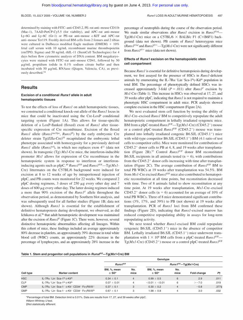
Because Runx1 is essential for definitive hematopoiesis during develop-ment, we first assayed for the presence of HSCs in Runx1-deficientanimals by enumerating the IL-7R�Lin�Sca-1hic-Kithi population intotal BM. The percentage of phenotypically defined HSCs was in-creased approximately 3-fold (P � .011) after Runx1F excision byMx1-Cre (Table 1). This increase in HSCs was observed at 17, 27, and39 weeks after pIpC, indicating that Runx1 is not required to maintain aphenotypic HSC compartment in adult mice. PCR analysis showedcomplete excision in the HSC compartment (Figure 2A).
We next evaluated stem cell function by testing the ability ofMx1-Cre–excised Runx1 BM to competitively repopulate the adulthematopoietic compartment in lethally irradiated syngeneic mice.BM from a pIpC-treated Runx1F/F—Tg(Mx1-Cre) (CD45.2�) mouseor a control pIpC-treated Runx1F/F (CD45.2�) mouse was trans-planted into lethally irradiated congenic B6.SJL (CD45.1�) micewith wild-type competitor B6.SJL (CD45.1�) BM (4:1 ratio of testcells to competitor cells). Mice were monitored for contributions ofCD45.2� donor cells to PB at 4, 8, and 19 weeks after transplanta-tion (Figure 2B).57 Control Runx1F/F donor BM reconstitutedB6.SJL recipients in all animals tested (n � 6), with contributionsfrom the CD45.2� donor cells increasing with time after transplan-tation (Figure 2C). The average contribution of Runx1F/F BM tototal PB WBCs at 19 weeks after transplantation was 54.5%. BMfrom Mx1-Cre excised Runx1F/F mice also contributed to hematopoi-etic reconstitution at all time points, but reconstitution decreasedwith time. Two of 8 animals failed to show reconstitution at anytime point. At 19 weeks after transplantation, Mx1-Cre–excisedCD45.2� donor cells (n � 6) accounted for an average of 10% oftotal PB WBCs. Three of 8 mice demonstrated significant contribu-tions (3%, 17%, and 39%) to PB (not shown) at 19 weeks aftertransplantation. PCR of Runx1 loci from BM confirmed thesefindings (Figure 2D), indicating that Runx1-excised marrow hasreduced competitive repopulating ability in assays for long-termrepopulating activity.
We next tested whether Runx1-excised BM could repopulatesyngeneic B6.SJL (CD45.1�) mice in the absence of competitorBM. Lethally irradiated B6.SJL (CD45.1�) mice underwent trans-plantation with 1 � 106 BM cells from a pIpC-treated Runx1F/F—Tg(Mx1-Cre) (CD45.2�) mouse or a control pIpC-treated Runx1F/F
Table 1. Stem and progenitor cell populations in RunxIF/F—Tg(Mx1-Cre) BM
Cell type Marker content
Genotype
Runx1F/F Runx1F/F—Tg(Mx1-Cre)
BM, %, mean� SD*
No.mice
BM, %, mean� SD*
No.mice Fold change P†
HSC IL-7R�Lin�Sca-1hi c-Kithi 0.34 � 0.1 4 0.99 � 0.5 6 2.9 .011
CLP IL-7R�Lin�Sca-1lo c-Kitlo 0.07 � 0.01 4 �0.01 � �0.01 6 �7.0 .019
CMP IL-7R�Lin�Sca-1� c-Kit� CD34� FcRII/III� 0.37 � 0.1 3 0.30 � 0.2 4 �0.8 .377‡
GMP IL-7R�Lin�Sca-1� c-Kit� CD34� FcRII/IIIhi 0.67 � 0.1 3 1.98 � 0.8 4 3.0 .032
*Percentage of total BM. Detection limit is 0.01%. Data are results from 17, 27, and 39 weeks after pIpC.†Mann-Whitney U test.‡Not statistically different.
Runx1 LOSS IN ADULT MURINE HEMATOPOIESIS 497BLOOD, 15 JULY 2005 � VOLUME 106, NUMBER 2
For personal use only. by guest on June 4, 2013. bloodjournal.hematologylibrary.orgFrom

(CD45.2�) mouse. Mice receiving Runx1F/F—Tg(Mx1-Cre) mar-row (n � 4) were viable at 14 weeks after transplantation, indicat-ing that Runx1-excised cells are competent for reconstitution(Figure 2E). The alterations in PB counts observed in primaryanimals (Figure 1C), including an 80% decrease in platelet counts,49% decrease in WBC counts, and relative changes in thedifferential, were observed in mice that underwent transplantationwith excised Runx1F/F—Tg(Mx1-Cre) BM. (Table 2). In nonex-cised recipients, approximately 1.3% of the PB cells were derivedfrom the host (CD45.1�), whereas approximately 12.1% of the PBcells in excised recipients were derived from the host (Table 2).Thus, even under these stringent selective conditions for HSCrepopulation, Runx1-excised HSCs are less competitive underrepopulating conditions than wild-type HSCs.
Effects of Runx1 excision on the lymphoid lineage
Mature B and T cells were present in pIpC-treated Runx1F/F—Tg(Mx1-Cre) mice, but the lymphocyte count in PB was reducedby an average of approximately 50% (Figure 1C). There was anaverage 19.7-fold (P � .007) reduction in the percentage of matureB cells (CD19�B220�) in the BM of excised mice (Figure 3A).Pre-B (IgM�NK1.1�Lin�CD43�B220�) and pro-B (IgM�
NK1.1�Lin�CD43�B220�) cell precursors were nearly undetect-able (less than 0.1%) in excised mice (Figure 3A), consistent withan early block in B-cell maturation.
There were also marked defects in T-lineage development inMx1-Cre–excised Runx1 animals. The total cellularity of the
Figure 2. Stem cells from pIpC-treated Runx1F/F—Tg(Mx1-Cre) mice are re-duced in competitive repopulation ability. (A) Ethidium bromide (EtBr)–stained3% agarose gel of 3-primer PCR of Runx1 loci from sorted HSCs derived from freshBM 17 weeks after pIpC (Table 1) and unfractionated BM and PB. 1 � Runx1F/F;2 � Runx F/F—Tg(Mx1-Cre). Mr � 1-kb ladder, sizes indicated (bp). Control samplesinclude tail DNA from Runx1�/�, Runx1F/�, and Runx1F/F mice and from Runx1�/�,Runx1F/�, and Runx1�/� embryos. Runx1� alleles were generated by matingRunx1F/F mice to Tg(EIIa-Cre) mice and intercrossing Runx1F/� mice. (B) Compositeshowing gating strategy and CD45.1/CD45.2 staining of PB from representative micethat underwent transplantation with Runx1F/F or Runx1F/F—Tg(Mx1-Cre) and competi-tor marrow 8 weeks after transplantation. Green indicates lymphocytic (R2), and redindicates granulocytic (R3) fractions based on scatter gating (SSC, side scatter; FSC,forward scatter). Gate assignments were confirmed by back-gating of controlstainings with Gr1�–, Mac1�–, CD3�–, T-cell receptor � (TCR��)–, CD19�–, orB220�–stained PB (not shown). The percentage contribution to the granulocytic andlymphocytic lineages of PB from each marrow isotype (CD45.1/CD45.2) wasdetermined. (C) Plotted is the mean (�SD) percentage contribution of donor-derivedCD45.2� cells in PB from mice that underwent transplantation with Runx1F/F—Tg(Mx1-Cre) (red symbols; n � 6) or Runx1F/F (black symbols; n � 6) and competitor bonemarrow to total WBCs, granulocytes, and lymphocytes of recipient mice at 4, 8, and19 weeks after transplantation. Two of 8 mice receiving Runx1F/F—Tg(Mx1-Cre)marrow that failed to show contribution of CD45.2� marrow to PB of recipient mice atany time point are excluded. (D) Nineteen weeks after transplantation, EtBr-stained3% agarose gel of 3-primer PCR of Runx1 loci of BM from mice that underwentcompetitive transplantation. (E) EtBr-stained 3% agarose gel of 3-primer PCR ofRunx1 loci from BM of mice that underwent transplantation without competitormarrow.
Table 2. Hematopoietic effects of Runx1 excision are transplantable
PB counts†
Donor cell genotype*
Runx1F/F,mean � SD
Runx1F/F-Tg(Mx1-Cre),mean � SD
WBCs, �103/�L 6.94 � 2.3 3.55 � 0.4
RBCs, �106/�L 7.53 � 1.7 9.46 � 0.5
Platelets, �103/�L 1808 � 200 364 � 140
Neutrophils, % 32.9 � 4.6 40.6 � 2.6
Lymphocytes, % 60.0 � 4.6 49.0 � 3.1
Lineage and origin of PB cells‡
WBC donor derived, CD45.2�, %§ 98.7 � 0.5 87.9 � 3.4
PB T cells
CD3� %� 3.6 � 1.8 9.0 � 1.7
CD3� donor derived, %¶ 70.9 � 22.0 7.0 � 4.5
PB B cells
B220�, %� 35.4 � 7.3 0.6 � 0.3
B220� donor derived, %¶ 100.0 � 0.0 97.1 � 4.5
PB myeloid cells
Mac1�, %� 6.4 � 2.1 17.1 � 3.7
Mac1� donor derived, %¶ 99.6 � 0.2 99.1 � 0.4
Gr1�/Mac1�, %� 40.5 � 71.2 67.8 � 32.9
Gr1�/Mac1� donor derived, %¶ 99.8 � 0.3 98.1 � 0.8
Gr1�, %� 1.0 � 0.4 9.8 � 1.3
Gr1� donor derived, %¶ 45.5 � 34.7 4.9 � 1.7
Results are for 2 Runx1F/F mice (2 others were lost to fighting) and 4Runx1F/F-Tg(Mx1-Cre) mice.
*Genotypes of donor cells (CD45.2�) transplanted into lethally irradiated B6.SJL(CD45.1�) recipient mice. Mice were killed 14 weeks after transplantation.
†Results from automated blood counts.‡Cells gated by scatter, CD45.1, and CD45.2.§Indicated is the percentage of total WBCs (pan-CD45�) that were donor derived
(CD45.1� and CD45.2�).�Indicated is the percentage of total WBCs (pan-CD45�) that were CD3, B220,
Mac-1, or Gr-1 positive.¶Indicated is the percentage of total CD3-, B220-, Mac-1-, Mac-1/Gr-1-, or
Gr-1-positive WBCs (pan-CD45�) that were donor derived (CD45.2�).
498 GROWNEY et al BLOOD, 15 JULY 2005 � VOLUME 106, NUMBER 2
For personal use only. by guest on June 4, 2013. bloodjournal.hematologylibrary.orgFrom

thymus was reduced approximately 10-fold compared with litter-mate controls (not shown). Although there was 90% or greaterexcision of Runx1F allele in BM, the percentage of excised alleleswas 50% or less in thymocytes from either Runx1F/F—Tg(Mx1-Cre) (not shown) or Runx1F/rd—Tg(Mx1-Cre) mice (Figure 3B).This suggests that Mx1-Cre expression in the thymus is lower thanin BM. To distinguish between the effects of homozygous andheterozygous excision, we evaluated thymic T-cell maturation inRunx1F/rd—Tg(Mx1-Cre) (CXB6F1) and littermate mice. Therewas a 37% decrease (P � .033) in the relative percentage ofCD4�CD8� (double-positive [DP]) thymocytes in Mx1-Cre ex-cised Runx1F/rd mice compared with Runx1F/� mice (Figure 3C).We did not observe a significant expansion of the CD4�CD8�
(DN) population. However, this was likely obscured by theapproximately 40% increase (P � .024) in CD4�CD8� (SP4) cellscompared with Runx1F/� mice. This increase in SP4 cells likelyresulted from the requirement for Runx1 repression of CD4expression during thymocyte maturation from the DN to the DPstage.6,7 There was an approximate 3-fold decrease in the percent-ages of DN3 (CD44�CD25�) (P � .033) and DN4 (CD44�CD25�)(P � .011) cells relative to Runx1F/� thymocytes (Figure 3D).Relative to Runx1F/rd and Runx1F/�—Tg(Mx1-Cre) mice, the per-centage of DN3 and DN4 cells were reduced 4.3-fold (P � .018)and 2.3-fold (P � .018) in Runx1F/rd—Tg(Mx1-Cre) mice,respectively.
To determine the earliest step in lymphocyte developmentrequiring Runx1, we enumerated the number of BM-derived CLPs(IL-7R�Lin�Sca-1loc-Kitlo) in Runx1F/F—Tg(Mx1-Cre)–excisedmice. This revealed a greater than 7-fold decrease (P � .019) in thepercentage of CLPs in the BM of Runx1-excised mice (Figure 3E;Table 1). We next evaluated lymphocyte commitment in mice aftertransplantation. In the competitive reconstitution assay, there was acomplete lack of contribution of excised Runx1F/F—Tg(Mx1-Cre)donor cells to the lymphocyte lineages (Figure 2C). In noncompeti-
tive repopulation assays, Runx1F/F—Tg(Mx1-Cre) contributed to T-and B-cell lineages to a limited extent (Table 2). The percentage ofCD3� T cells in Runx1-excised recipients was increased 2.5-foldcompared with nonexcised recipients. However, 93% of CD3� Tcells were derived from the host BM (CD45.1�). Strikingly,peripheral blood B220� B cells were decreased 58-fold in excisedrecipients compared with nonexcised recipients. In contrast to theT-cell lineage, 97% of PB B cells were donor derived. Together,these findings suggest that Runx1 is not required for lymphocytematuration but that the loss of Runx1 inhibits lymphocyte matura-tion at early and late stages of development.
Effects of Runx1 excision on the myeloid lineage
The marked defects in lymphocyte maturation observed in Runx1-excised mice are sharply contrasted with the effects of Runx1excision on the myeloid lineage. All myeloid lineages, includinggranulocytes, monocytes, erythrocytes, and platelets, were repre-sented in Mx1-Cre–excised Runx1F/F (Figure 1C) and Runx1F/rd
(data not shown) animals. No significant abnormalities wereobserved in the erythroid lineage. However, we observed signifi-cant defects in the megakaryocytic lineage. Although there was noclinically evident bleeding diathesis, within 14 days of pIpCinjection there was an approximately 80% reduction in average PBplatelet counts in Runx1F/F—Tg(Mx1-Cre) (Figure 1C) and Runx1F/rd—Tg(Mx1-Cre) (not shown) mice compared with littermate controls.Histologic examination of BM from pIpC-treated Runx1F/F—Tg(Mx1-Cre) (Figure 4A-B) and Runx1F/rd—Tg(Mx1-Cre) (not shown)mice showed a marked decrease in the number of maturemegakaryocytes compared with control mice, suggesting that thedecrement in platelet count was attributed to decreased megakaryo-cyte maturation. This defect was observed in animals that under-went transplantation, indicating that the megakaryocyte maturationdefect is cell autonomous (Figure 4C). Propidium iodide staining
Figure 3. Lymphoid development is inhibited inRunx1-excised mice. (A) Representative B220/CD19and B220/CD43 staining of BM from Runx1F/F andRunx1F/F—Tg(Mx1-Cre) mice. CD43 staining is shownfor cells gated as IgM�NKK1.1�Lin�. Numbers indicatepercentage of cells gated in each respective quadrant.(B) EtBr-stained 3% agarose gels with 3-primer PCRproducts of Runx1 loci in PB, spleen (Sp), BM, andthymocytes (Th) of mice 154 days after pIpC. Genotypesare as indicated. Control samples are as in Figure 2. Theprimers do not amplify a product from the Runx1rd allele.(C) Shown are CD4 and CD8 staining from representa-tive animals of genotypes, Runx1 F/�, Runx1F/rd, andRunx1F/rd—Tg(Mx1-Cre). Numbers indicate percentageof cells gated in each respective quadrant. (D) Shown areCD25 and CD44 staining of CD45.2�CD4�CD8� gatedthymocytes from representative animals, as in panel C.(E) Composite of high-speed flow cytometric analysis ofBM from representative Runx1F/F and Runx1F/F—Tg(Mx1-Cre) mice. Numbers indicate percentages of total BM forindicated populations. Gated populations indicated in-clude IL-7R�Lin� (R1), IL-7R�Lin� (R2) (which in-cludes immature B cells), and CLP (R3), as indicated inTable 1. PI indicates propidium iodide.
Runx1 LOSS IN ADULT MURINE HEMATOPOIESIS 499BLOOD, 15 JULY 2005 � VOLUME 106, NUMBER 2
For personal use only. by guest on June 4, 2013. bloodjournal.hematologylibrary.orgFrom

showed reduced polyploidization of CD41� BM cells (Figure 4D).However, we paradoxically observed that in vitro megakaryocytecolony-plating efficiency was increased by an average of 2.9(� 0.6)–fold (Figure 4E). Furthermore, megakaryocytic coloniesdisplayed an increased number of acetylcholinesterase-positivecells per colony (Figure 4F-G).
Immunophenotypic analysis of myeloid progenitors derivedfrom BM showed that the CMP population (IL-7R�Lin�Sca-1�c-Kit�CD34�FcRII/III�) was not significantly altered in Runx1-excised mice. However, the GMP population (IL-7R�Lin�Sca-1�c-Kit�CD34�FcRII/IIIhi) was expanded approximately 3-fold(P � .032) in Runx1-excised mice (Table 1). These observationsare consistent with the increased production of myeloid colonies invitro (Figure 5A). Unfractionated BM derived from pIpC-treatedRunx1F/F—Tg(Mx1-Cre) mice had an approximately 3.4-fold in-crease in total myeloid colonies relative to pIpC-treated Runx1F/F
mice. Microscopic evaluation of Wright-Giemsa–stained cytospinsof granulocyte-macrophage (GM) and mixed-lineage coloniesindicated that Runx1-excised colonies gave rise to the full range ofmyeloid cell types observed in control colonies (Figure 5B). GMand mixed-lineage colonies were increased 3.9- and 3.2-foldcompared with controls, respectively. In contrast, there was nosignificant change in erythroid blast-forming unit (BFU-E) colonynumber. Evaluation of individual colonies by PCR indicatedcomplete Runx1 excision in 92% of evaluable colonies frompIpC-treated Runx1F/F—Tg(Mx1-Cre) mice (Figure 5C) comparedwith Runx1F/F controls.
Perhaps the most unexpected finding in adult Runx1-excisedmice was a mild myeloid expansion in hematopoietic tissues.Histopathologic examination of Runx1-excised BM showed subtlemyeloid hyperplasia with a consistent increase in the ratio ofmaturing myeloid to erythroid forms compared with controls(Figure 4A-B). Runx1-excised marrows were hypercellular, yield-ing twice as many cells as control marrows. Similar findings wereobserved in mice after noncompetitive transplantation (Figure 4C).
Figure 4. Megakaryocyte maturation is inefficient in pIpC-treated Runx1-excised mice. BM from representative (A)Runx1F/F and (B) Runx1F/F—Tg(Mx1-Cre) mice 143 days afterpIpC injection shows an absence of normal megakaryocytes inRunx1-excised mice (hematoxylin and eosin [H&E] staining;original magnification, � 600). (C) BM from representative Runx1F/F—Tg(Mx1-Cre) recipient mice 98 days after transplantation (H&E;original magnification, � 600). (A-C) Yellow arrows indicate represen-tative megakaryocytes. Red and green arrowheads indicate represen-tative erythroid and myeloid elements, respectively. Sections demon-strate a notable absence of megakaryocytes and an increased ratio ofmaturing myeloid-to-erythroid forms in Runx1-excised versus nonex-cised marrows. (D) Histograms depicting number of cultured Runx1F/F
and Runx1F/F—Tg (Mx1-Cre) bone marrow cells stained with pro-pidium iodide to show ploidy. Colors indicate CD41 gates: green,CD41�; red, CD41�. (E, left) Plotted are mean � SD numbers ofacetylcholinesterase-positive colonies per 5 � 104 BM cells platedfrom pIpC-treated Runx1F/F (�) and Runx1F/F—Tg(Mx1-Cre) (f)mice. Results shown are for 3 experiments performed in quadrupli-cate. 1 and 3, fresh marrow; 2, previously frozen marrow. (Right)Plotted is the average fold increase for Runx1F/F—Tg(Mx1-Cre) (f)relative to Runx1F/F (�) in the 3 experiments (P � .034). Representa-tive megakaryocyte colonies from (F) Runx1F/F and (G) Runx1F/F—Tg(Mx1-Cre) mice show acetylcholinesterase staining (brown) ofmegakaryocytic cells (original magnification, � 100).
Figure 5. Mature and progenitor myeloid cell populations are expanded inpIpC-treated Runx1-excised mice. (A) Plotted are the mean � SD of total (P � .05),myeloid (P � .05), BFU-E (P � .5), and mixed-lineage colonies (P � �.05) per2 � 104 BM cells plated in vitro from pIpC-treated Runx1F/F (�) and Runx1F/F—Tg(Mx1-Cre) (f) mice. Results are the average of 3 experiments performed induplicate. (B) Representative cytospin from mixed-lineage colony derived fromRunx1F/F—Tg(Mx1-Cre) BM (Wright-Giemsa staining; original magnification, � 600).Red arrowheads indicate representative erythroid elements. Green arrowheadsdenote myeloid and monocytic forms. (C) Representative EtBr-stained 3% agarosegel of Runx1 PCR products from single GM or mixed-lineage colonies derived fromRunx1F/F—Tg(Mx1-Cre) (lanes 1-20) and Runx1F/F (lanes 21-25) mice. Of 70Runx1F/F—Tg(Mx1-Cre) colonies evaluated, 9 (12.8%) failed to show any amplifica-tion, 5 of 61 (8.2%) indicated partial excision, and 56 of 61 (91.8%) were completelyexcised. Tail and embryo genomic DNA controls are as indicated. (D) Gr1 and Mac1staining of BM from representative CXB6F1 mice 154 days after pIpC induction.Numbers indicate percentages of cells gated in each respective quadrant.
500 GROWNEY et al BLOOD, 15 JULY 2005 � VOLUME 106, NUMBER 2
For personal use only. by guest on June 4, 2013. bloodjournal.hematologylibrary.orgFrom

The histopathologic findings were supported by a 2.4-fold expan-sion of Mac1� cells in excised BM of Runx1F/F—Tg(Mx1-Cre)(P � .021) (data not shown) and Runx1F/rd—Tg(Mx1-Cre)(P � .021) (Figure 5D) compared with controls.
Gross and histopathologic findings consistent with a myelopro-liferative phenotype included splenomegaly attributable to extramed-ullary hematopoiesis in the spleen, with an increase in spleenweight of 1.4- to 2.1-fold in Runx1-excised mice compared withcontrols (Table 3). Histopathologic examination of spleen sectionsshowed effacement of splenic architecture with a mild expansion ofthe red pulp resulting from extramedullary hematopoiesis com-posed predominantly of maturing myeloid forms and admixederythroid elements, with only scant to rare megakaryocytes (Figure6A-B). Liver sections from Runx1-excised mice also showedextramedullary hematopoiesis composed of maturing myeloidelements in a perivascular distribution (Figure 6C-D). Spleens ofmice that underwent transplantation with excised Runx1F/F—Tg(Mx1-Cre) marrow in the absence of wild-type competitor cellsalso demonstrated extramedullary hematopoiesis (Figure 6E-F).Flow cytometric analysis of single cell suspensions derived fromspleens of primary Runx1-excised animals and animals that under-went transplantation with Runx1-excised BM confirmed the in-crease of myeloid lineage cells in the spleen (Figure 6G; Table 4).In Runx1F/F—Tg(Mx1-Cre) mice and mice after transplantation,but not in Runx1F/rd—Tg(Mx1-Cre) mice, there was a significantexpansion of CD45�Ter119� cells in the spleen (Figure 6G; Table4). Taken together, these data indicate that Runx1 is not required forneutrophil development, but its loss of function contributes to amyeloproliferative phenotype.
Discussion
Runx1 deficiency during development results in a failure of HSCemergence and lethal central nervous system (CNS) hemorrhage,but the data presented here indicate that Runx1 is not required in theadult murine hematopoietic compartment for HSC maintenance orsurvival of adult mice. However, there were a number of mild tosevere hematopoietic abnormalities, including a block in lymphoiddevelopment, reduced platelet production, and development of amyeloproliferative phenotype.
Runx1-excised adult mice have an expanded, immunophenotypi-cally defined HSC compartment. These mice survive with afollow-up of more than 5 months, indicating that there is long-termHSC activity in these animals. This finding is surprising in light ofthe hematopoietic defect observed in Runx1-deficient embryos. We
Figure 6. Runx1-excised mice display myeloid expansion in spleen and liver.Spleen (A-B) and liver (C-D) sections from representative (A,C) Runx1F/F and (B,D)Runx1F/F—Tg(Mx1-Cre) mice 143 days after pIpC (H&E; original magnification,�100). Insets’ original magnification, � 600. (E-F) Spleen sections from representa-tive mice 14 weeks after transplantation with (E) Runx1F/F and (F) Runx1F/F—Tg(Mx1-Cre) marrow (H&E; original magnification, � 100). Insets’ original magnification,�600. (A-F) Yellow arrows indicate representative lymphocytes. Red and greenarrowheads indicate representative erythroid and myeloid elements, respectively.Sections demonstrate the presence of extramedullary hematopoiesis in the liver andsplenic red pulp in Runx1-excised mice, absent in nonexcised control animals. (G)Gr1/Mac1 and CD45.2/Ter119 staining of splenocytes of indicated genotype fromrepresentative primary mice or mice that underwent transplantation. Numbersindicate percentage of cells gated in each respective quadrant.
Table 3. Spleen weights of pIpC-treated mice
Genotype No. mice Days after pIpCSpleen weight, mg,
mean � SD Fold increase
Runx1F/F 7 60-273 153 � 48 —
Runx1F/F—Tg(Mx1-Cre) 9 91-273 318‡ � 195 2.08
Runx1F/� 4 154 122 � 26 —
Runx1F/rd and Runx1F/�—Tg(Mx1-Cre) 5 154 129 � 14 —
Runx1F/rd—Tg(Mx1-Cre) 6 154 185§ � 42 1.52�/1.43¶
Runx1F/F recipients* 2 98† 84 � 21 —
Runx1F/F—Tg(Mx1-Cre) recipients* 4 98† 128 � 10 1.53
*Mice that underwent transplantation with Runx1F/F or Runx1F/FTg(Mx1-Cre) marrow in the absence of competitor marrow.†Days after transplantation.‡Comparison with Runx1F/F (P � .01; Mann-Whitney U test).§Comparison with Runx1F/� (P � .033). Comparison with Runx1F/rd and Runx1F/�—Tg(Mx1-Cre) (P � .068; Mann- Whitney U test).�Comparison with Runx1F/�.¶Comparison with Runx1F/rd and Runx1F/�—Tg(Mx1-Cre).
Runx1 LOSS IN ADULT MURINE HEMATOPOIESIS 501BLOOD, 15 JULY 2005 � VOLUME 106, NUMBER 2
For personal use only. by guest on June 4, 2013. bloodjournal.hematologylibrary.orgFrom

note that homozygous loss of RUNX1 has been observed inhumans with AML, though not in those with acute lymphoidleukemia.38 These observations suggest that AML cells withself-renewal ability and limited ability to differentiate also do notrequire RUNX1. Our findings that Runx1 is not required for HSCmaintenance and survival in the adult are consistent with thepersistence of RUNX1-deficient cells in human leukemias. BMtransplantation studies indicate that Runx1-excised HSCs arecompetent for long-term repopulation. However, competitive re-population studies indicate a significant reduction in Runx1-deficient HSC ability to compete with wild-type HSCs duringrepopulation.
Runx1 is required for efficient lymphoid maturation at multiplestages of differentiation. We observed a significant reduction in thenumber mature PB B cells and BM-derived B-cell precursors inprimary Runx1-excised mice. This suggests a significant block inB-cell maturation, though it is unclear at which stage of B-cellmaturation Runx1 functions. In the T-cell lineage, we report aspecific block in T-cell maturation during the transition from theDN2 (CD44�CD25�) to the DN3 (CD44�CD25�) stage.56 Thisis consistent with reports describing a tightly regulated expres-sion pattern for Runx1 and CBF� in developing thymo-cytes.6,7,15,16,58 However, in contrast to the findings of Ichikawaet al,56 we observed a significant reduction in bone marrow CLP,suggesting that Runx1 is also important during the earlieststages of lymphoid lineage development. The difference in CLPproduction we observed compared with the observations ofIchikawa et al56 may be the result of higher pIpC dosing in ourstudy, resulting in more efficient excision, although otherexplanations are possible. Noncompetitive reconstitution assaysindicate that Runx1 is not required for commitment to andmaturation of the lymphoid lineage. It is clear that the loss ofRunx1 severely reduces the efficiency of CLP production andlymphocyte maturation. This conclusion is supported by theclinical observation that RUNX1 loss is not observed in patientswith acute lymphoblastic leukemia (ALL).
In contrast to the pronounced inhibition of the lymphoidlineage, excision of Runx1 did not inhibit maturation of themyeloid lineage. Runx1 loss had no apparent effect on the erythroidlineage, but it did have a significant effect on megakaryocyticmaturation. Runx1 is required for normal maturation of the
megakaryocyte lineage but not for the establishment of this lineage.These differential requirements for Runx1 in erythroid andmegakaryocyte maturation correlate with reports that the expres-sion of Runx1 and its heterodimeric binding partner, CBF�, aremarkedly decreased in erythroid lineages15,16,59 but are maintainedin megakaryocytes.16,59,60 Humans with FPD/AML syndrome haveheterozygous loss of RUNX1, with an associated quantitative andqualitative platelet defect, and a proclivity to develop AML.35 Theplatelet defect is reminiscent of the abnormalities observed in micewith homozygous loss of Runx1 and suggests that there may becompensation in mice for monoallelic loss of Runx1.
RUNX1 loss in humans is most often associated with M0(undifferentiated) AML, suggesting that RUNX1 loss may havedeleterious effects on hematopoietic differentiation.61,62 In contrast,Runx1-excised mice demonstrate expansion of the myeloid lineageby several phenotypic and functional criteria, with no evidence of ablock in myeloid development. The specific expansion of the GMP,but not the CMP, population suggests that the myeloid expansionobserved in Runx1-excised animals results from the loss ofnegative regulatory functions of Runx1 in the GMP population. Thestrain-specific differences in expansion of CD45�—Ter119� cellsin the spleens of Runx1-excised mice indicate that the myeloidexpansion is not secondary to a compensatory drive for increasedplatelet production. Although these findings are surprising, therehave been several other indications that Runx1 is not essential foradult hematopoiesis in mice. For example, expression of theleukemogenic fusion protein RUNX1-ETO, a dominant-negativeinhibitor of CBF, during development results in an embryoniclethal phenotype with a lack of definitive hematopoiesis that isnearly identical to that observed in Runx1-deficient mice.44,45
However, expression of Runx1-ETO from a conditional allele inthe adult hematopoietic compartment has only subtle effects onhematopoiesis, characterized by enhanced replating efficiency ofmyeloid progenitors in vitro and an increased susceptibility todevelop leukemia after N-ethyl-N-nitrosurea (ENU) mutagen-esis.63 Runx1-ETO or Runx1-deficient animals do not developleukemia, suggesting that Runx1 loss of function must be comple-mented with additional leukemogenic genetic events for thedevelopment of a leukemia phenotype. The myeloproliferativephenotype associated with Runx1 excision suggests that Runx1 lossmay constitute a preleukemic state, and it may explain the
Table 4. Myeloid expansion in spleens of Runx1-excised mice
GenotypeNo.
miceTime after
pIpC, dGr1� splenocytes,mean � SD, %* (P)
Gr1�Mac1�
splenocytes,mean � SD, %* (P)
Mac1�
splenocytes,mean � SD, %* (P)
Ter119�/CD45�
splenocytes, mean� SD, %* (P)†
Runx1F/F‡ 4 186-190 2.9 � 0.6 (.999�¶) 1.6 � 0.4 (.008�) 6.1 � 1.4 (.014�) 2.9 � 1.6 (.023�)
Runx1F/F—Tg(Mx1-Cre)‡ 7 186-190 2.7 � 1.9 10.2 � 4.7 14.9 � 8.8 20.6 � 15.5
Runx1F/� 4 154 5.7 � 1.5 (.020#) 2.5 � 0.2 (.020#) 1.8 � 0.2 (.020#) 2.0 � 1.3 (.136¶#)
Runx1F/�—Tg(Mx1-Cre) and Runx1F/rd 5 154 7.5 � 1.2 (.465¶**) 5.4 � 2.8 (.029**) 12.0 � 0.6 (.006**) 3.1 � 2.1 (.715¶**)
Runx1F/rd—Tg(Mx1-Cre) 6 154 8.3 � 1.2 12.7 � 5.3 4.0 � 0.5 3.8 � 2.0
Runx1F/F (recipient)‡ 2 96§ 3.1 � 1.2 (.098¶††) 2.5 � 0.1 (.040††) 1.9 � 0.5 (.061¶††) 2.8 � 1.7 (.003††)
Runx1F/F—Tg(Mx1-Cre) (recipient)‡ 4 96§ 5.8 � 1.6 7.4 � 2.2 3.9 � 1.0 50.0 � 9.3
*Numbers indicate mean percentage Mac1�, Gr1�, or Mac1�Gr1� of total splenocytes. Primary pIpC-treated splenocytes were stained with CD45.2- FITC, Mac1-PE,7AAD-PerCP-Cy5.5, and Gr1-APC and were gated by scatter and 7AAD negativity. Splenocytes from animals that underwent transplantation were stained with CD45.2-FITC,Mac1-PE, CD45.2-PerCP-Cy5.5, and Gr1-APC and were scatter gated.
†Numbers indicate mean percentage CD45�Ter119� of total splenocytes. Splenocytes were stained with CD45.2-FITC, c-Kit-PE, 7AAD-PerCP- Cy5.5, and Ter119-APC.Splenocytes were gated by scatter and 7AAD negativity.
‡Representative data in Figure 6.§Days after transplantation.�Mann-Whitney U test for comparison of primary Runx1F/F—Tg(Mx1-Cre) to primary Runx1F/F animals.¶Not statistically significant.#Mann-Whitney U test for comparison of primary Runx1F/rd—Tg(Mx1-Cre) to primary Runx1F/� animals.**Mann-Whitney U test for comparison of primary Runx1F/rd—Tg(Mx1-Cre) to primary Runx1F/�—Tg(Mx1-Cre) and Runx1F/rd animals.††Unpaired t test for comparison of mice that underwent transplantation with Runx1F/F—Tg(Mx1-Cre) to mice that underwent transplantation with Runx1F/F animals.
502 GROWNEY et al BLOOD, 15 JULY 2005 � VOLUME 106, NUMBER 2
For personal use only. by guest on June 4, 2013. bloodjournal.hematologylibrary.orgFrom

proclivity for the development of myeloid leukemias in the contextof RUNX1 deficiency in humans. The ability of Runx1-deficientmyeloid lineage cells to differentiate normally suggests that theblock in differentiation observed in leukemias with loss ofRUNX1 function cannot be attributed to the loss of RUNX1alone and that it must be due to other genetic events.
Acknowledgments
We thank Woo Jong Song, Terryl Stacy, Vivienne Rebel, and HannoHock for their insightful discussions and contributions to thecompletion of this work.
References
1. North T, Gu TL, Stacy T, et al. Cbfa2 is requiredfor the formation of intra-aortic hematopoieticclusters. Development. 1999;126:2563-2575.
2. Cai Z, de Bruijn M, Ma X, et al. Haploinsufficiencyof AML1 affects the temporal and spatial genera-tion of hematopoietic stem cells in the mouse em-bryo. Immunity. 2000;13:423-431.
3. Cameron S, Taylor DS, TePas EC, Speck NA,Mathey-Prevot B. Identification of a critical regu-latory site in the human interleukin-3 promoter byin vivo footprinting. Blood. 1994;83:2851-2859.
4. Otto F, Lubbert M, Stock M. Upstream and down-stream targets of RUNX proteins. J Cell Biochem.2003;89:9-18.
5. de Bruijn MF, Speck NA. Core-binding factors inhematopoiesis and immune function. Oncogene.2004;23:4238-4248.
6. Taniuchi I, Osato M, Egawa T, et al. Differentialrequirements for Runx proteins in CD4 repressionand epigenetic silencing during T lymphocyte de-velopment. Cell. 2002;111:621-633.
7. Woolf E, Xiao C, Fainaru O, et al. Runx3 andRunx1 are required for CD8 T cell developmentduring thymopoiesis. Proc Natl Acad Sci U S A.2003;100:7731-7736.
8. Lauzurica P, Zhong XP, Krangel MS, Roberts JL.Regulation of T cell receptor delta gene rear-rangement by CBF/PEBP2. J Exp Med. 1997;185:1193-1201.
9. Takahashi A, Satake M, Yamaguchi-Iwai Y, et al.Positive and negative regulation of granulocyte-macrophage colony-stimulating factor promoteractivity by AML1-related transcription factor,PEBP2. Blood. 1995;86:607-616.
10. Zhang DE, Fujioka K, Hetherington CJ, et al.Identification of a region which directs the mono-cytic activity of the colony-stimulating factor 1(macrophage colony-stimulating factor) receptorpromoter and binds PEBP2/CBF (AML1). MolCell Biol. 1994;14:8085-8095.
11. Wang Q, Stacy T, Binder M, Marin-Padilla M,Sharpe AH, Speck NA. Disruption of the Cbfa2gene causes necrosis and hemorrhaging in thecentral nervous system and blocks definitive he-matopoiesis. Proc Natl Acad Sci U S A. 1996;93:3444-3449.
12. Wang Q, Stacy T, Miller JD, et al. The CBFbetasubunit is essential for CBFalpha2 (AML1) func-tion in vivo. Cell. 1996;87:697-708.
13. Okuda T, van Deursen J, Hiebert SW, GrosveldG, Downing JR. AML1, the target of multiple chro-mosomal translocations in human leukemia, isessential for normal fetal liver hematopoiesis.Cell. 1996;84:321-330.
14. Sasaki K, Yagi H, Bronson RT, et al. Absence offetal liver hematopoiesis in mice deficient in tran-scriptional coactivator core binding factor beta.Proc Natl Acad Sci U S A. 1996;93:12359-12363.
15. Lorsbach RB, Moore J, Ang SO, Sun W, Lenny N,Downing JR. Role of RUNX1 in adult hematopoi-esis: analysis of RUNX1-IRES-GFP knock-inmice reveals differential lineage expression.Blood. 2004;103:2522-2529.
16. North TE, Stacy T, Matheny CJ, Speck NA, deBruijn MF. Runx1 is expressed in adult mousehematopoietic stem cells and differentiating my-eloid and lymphoid cells, but not in maturing ery-throid cells. Stem Cells. 2004;22:158-168.
17. Miyoshi H, Shimizu K, Kozu T, Maseki N, Kaneko
Y, Ohki M. t(8;21) breakpoints on chromosome 21in acute myeloid leukemia are clustered within alimited region of a single gene, AML1. Proc NatlAcad Sci U S A. 1991;88:10431-10434.
18. Miyoshi H, Kozu T, Shimizu K, et al. The t(8;21)translocation in acute myeloid leukemia results inproduction of an AML1-MTG8 fusion transcript.EMBO J. 1993;12:2715-2721.
19. Nucifora G, Birn DJ, Erickson P, et al. Detectionof DNA rearrangements in the AML1 and ETOloci and of an AML1/ETO fusion mRNA in patientswith t(8;21) acute myeloid leukemia. Blood. 1993;81:883-888.
20. Erickson P, Gao J, Chang KS, et al. Identificationof breakpoints in t(8;21) acute myelogenous leu-kemia and isolation of a fusion transcript, AML1/ETO, with similarity to Drosophila segmentationgene, runt. Blood. 1992;80:1825-1831.
21. Nisson PE, Watkins PC, Sacchi N. Transcription-ally active chimeric gene derived from the fusionof the AML1 gene and a novel gene on chromo-some 8 in t(8;21) leukemic cells. Cancer GenetCytogenet. 1992;63:81-88.
22. Liu P, Tarle SA, Hajra A, et al. Fusion betweentranscription factor CBF beta/PEBP2 beta and amyosin heavy chain in acute myeloid leukemia.Science. 1993;261:1041-1044.
23. Golub TR, Barker GF, Bohlander SK, et al. Fusionof the TEL gene on 12p13 to the AML1 gene on21q22 in acute lymphoblastic leukemia. Proc NatlAcad Sci U S A. 1995;92:4917-4921.
24. Romana SP, Mauchauffe M, Le Coniat M, et al.The t(12;21) of acute lymphoblastic leukemia re-sults in a tel-AML1 gene fusion. Blood. 1995;85:3662-3670.
25. Shurtleff SA, Buijs A, Behm FG, et al. TEL/AML1fusion resulting from a cryptic t(12;21) is the mostcommon genetic lesion in pediatric ALL and de-fines a subgroup of patients with an excellentprognosis. Leukemia. 1995;9:1985-1989.
26. Tien HF, Wang CH, Lin MT, et al. Correlation ofcytogenetic results with immunophenotype, geno-type, clinical features, and ras mutation in acutemyeloid leukemia: a study of 235 Chinese pa-tients in Taiwan. Cancer Genet Cytogenet. 1995;84:60-68.
27. Langabeer SE, Walker H, Gale RE, et al. Fre-quency of CBF beta/MYH11 fusion transcripts inpatients entered into the U.K. MRC AML trials:the MRC Adult Leukaemia Working Party. Br JHaematol. 1997;96:736-739.
28. Grimwade D, Walker H, Oliver F, et al. The impor-tance of diagnostic cytogenetics on outcome inAML: analysis of 1,612 patients entered into theMRC AML 10 trial: the Medical Research CouncilAdult and Children’s Leukaemia Working Parties.Blood. 1998;92:2322-2333.
29. Langabeer SE, Walker H, Rogers JR, et al. Inci-dence of AML1/ETO fusion transcripts in patientsentered into the MRC AML trials: MRC Adult Leu-kaemia Working Party. Br J Haematol. 1997;99:925-928.
30. Rowe D, Cotterill SJ, Ross FM, et al. Cytogeneti-cally cryptic AML1-ETO and CBF beta-MYH11gene rearrangements: incidence in 412 cases ofacute myeloid leukaemia. Br J Haematol. 2000;111:1051-1056.
31. Loh ML, Silverman LB, Young ML, et al. Inci-dence of TEL/AML1 fusion in children with re-
lapsed acute lymphoblastic leukemia. Blood.1998;92:4792-4797.
32. Liang DC, Chou TB, Chen JS, et al. High inci-dence of TEL/AML1 fusion resulting from a cryp-tic t(12;21) in childhood B-lineage acute lympho-blastic leukemia in Taiwan. Leukemia. 1996;10:991-993.
33. Harbott J, Viehmann S, Borkhardt A, Henze G,Lampert F. Incidence of TEL/AML1 fusion geneanalyzed consecutively in children with acutelymphoblastic leukemia in relapse. Blood. 1997;90:4933-4937.
34. Borkhardt A, Cazzaniga G, Viehmann S, et al.Incidence and clinical relevance of TEL/AML1fusion genes in children with acute lymphoblasticleukemia enrolled in the German and Italian multi-center therapy trials: Associazione Italiana Ema-tologia Oncologia Pediatrica and the Berlin-Frankfurt-Munster Study Group. Blood. 1997;90:571-577.
35. Song WJ, Sullivan MG, Legare RD, et al. Haplo-insufficiency of CBFA2 causes familial thrombo-cytopenia with propensity to develop acute my-elogenous leukaemia. Nat Genet. 1999;23:166-175.
36. Michaud J, Wu F, Osato M, et al. In vitro analysesof known and novel RUNX1/AML1 mutations indominant familial platelet disorder with predispo-sition to acute myelogenous leukemia: implica-tions for mechanisms of pathogenesis. Blood.2002;99:1364-1372.
37. Preudhomme C, Warot-Loze D, Roumier C, et al.High incidence of biallelic point mutations in theRunt domain of the AML1/PEBP2 alpha B gene inMo acute myeloid leukemia and in myeloid malig-nancies with acquired trisomy 21. Blood. 2000;96:2862-2869.
38. Osato M, Asou N, Abdalla E, et al. Biallelic andheterozygous point mutations in the runt domainof the AML1/PEBP2alphaB gene associated withmyeloblastic leukemias. Blood. 1999;93:1817-1824.
39. Fenrick R, Amann JM, Lutterbach B, et al. BothTEL and AML-1 contribute repression domains tothe t(12;21) fusion protein. Mol Cell Biol. 1999;19:6566-6574.
40. Hiebert SW, Sun W, Davis JN, et al. The t(12;21)translocation converts AML-1B from an activatorto a repressor of transcription. Mol Cell Biol.1996;16:1349-1355.
41. Lutterbach B, Westendorf JJ, Linggi B, et al. ETO,a target of t(8;21) in acute leukemia, interactswith the N-CoR and mSin3 corepressors. Mol CellBiol. 1998;18:7176-7184.
42. Westendorf JJ, Yamamoto CM, Lenny N, Down-ing JR, Selsted ME, Hiebert SW. The t(8;21) fu-sion product, AML-1-ETO, associates withC/EBP-alpha, inhibits C/EBP-alpha-dependenttranscription, and blocks granulocytic differentia-tion. Mol Cell Biol. 1998;18:322-333.
43. Castilla LH, Wijmenga C, Wang Q, et al. Failureof embryonic hematopoiesis and lethal hemor-rhages in mouse embryos heterozygous for aknocked-in leukemia gene CBFB-MYH11. Cell.1996;87:687-696.
44. Yergeau DA, Hetherington CJ, Wang Q, et al.Embryonic lethality and impairment of haemato-poiesis in mice heterozygous for an AML1-ETOfusion gene. Nat Genet. 1997;15:303-306.
45. Okuda T, Cai Z, Yang S, et al. Expression of a
Runx1 LOSS IN ADULT MURINE HEMATOPOIESIS 503BLOOD, 15 JULY 2005 � VOLUME 106, NUMBER 2
For personal use only. by guest on June 4, 2013. bloodjournal.hematologylibrary.orgFrom

knocked-in AML1-ETO leukemia gene inhibits theestablishment of normal definitive hematopoiesisand directly generates dysplastic hematopoieticprogenitors. Blood. 1998;91:3134-3143.
46. Mikkola HK, Klintman J, Yang H, et al. Haemato-poietic stem cells retain long-term repopulatingactivity and multipotency in the absence of stem-cell leukaemia SCL/tal-1 gene. Nature. 2003;421:547-551.
47. Hall MA, Curtis DJ, Metcalf D, et al. The criticalregulator of embryonic hematopoiesis, SCL, isvital in the adult for megakaryopoiesis, erythro-poiesis, and lineage choice in CFU-S12. ProcNatl Acad Sci U S A. 2003;100:992-997.
48. Curtis DJ, Hall MA, Van Stekelenburg LJ, Robb L,Jane SM, Begley CG. SCL is required for normalfunction of short-term repopulating hematopoieticstem cells. Blood. 2004;103:3342-3348.
49. Kuhn R, Schwenk F, Aguet M, Rajewsky K. Induc-ible gene targeting in mice. Science. 1995;269:1427-1429.
50. Liu Q, Schwaller J, Kutok J, et al. Signal trans-duction and transforming properties of the TEL-TRKC fusions associated with t(12;15)(p13;q25)in congenital fibrosarcoma and acute myelog-enous leukemia. EMBO J. 2000;19:1827-1838.
51. Schwaller J, Frantsve J, Aster J, et al. Transfor-mation of hematopoietic cell lines to growth-factorindependence and induction of a fatal myelo- and
lymphoproliferative disease in mice by retrovirallytransduced TEL/JAK2 fusion genes. EMBO J.1998;17:5321-5333.
52. Akashi K, Traver D, Miyamoto T, Weissman IL. Aclonogenic common myeloid progenitor that givesrise to all myeloid lineages. Nature. 2000;404:193-197.
53. Kondo M, Weissman IL, Akashi K. Identification ofclonogenic common lymphoid progenitors inmouse bone marrow. Cell. 1997;91:661-672.
54. Jackson CW, Brown LK, Somerville BC, LylesSA, Look AT. Two-color flow cytometric measure-ment of DNA distributions of rat megakaryocytesin unfixed, unfractionated marrow cell suspen-sions. Blood. 1984;63:768-778.
55. Lakso M, Pichel JG, Gorman JR, et al. Efficient invivo manipulation of mouse genomic sequencesat the zygote stage. Proc Natl Acad Sci U S A.1996;93:5860-5865
56. Ichikawa M, Asai T, Saito T, et al. AML-1 is re-quired for megakaryocytic maturation and lym-phocytic differentiation, but not for maintenanceof hematopoietic stem cells in adult hematopoi-esis. Nat Med. 2004;10:299-304.
57. Spangrude GJ, Scollay R. Differentiation of he-matopoietic stem cells in irradiated mouse thymiclobes: kinetics and phenotype of progeny. J Im-munol. 1990;145:3661-3668.
58. Kundu M, Liu PP. Cbf beta is involved in matura-
tion of all lineages of hematopoietic cells duringembryogenesis except erythroid. Blood Cells MolDis. 2003;30:164-169.
59. Kundu M, Chen A, Anderson S, et al. Role of Cbfbin hematopoiesis and perturbations of the leuke-mogenic fusion gene Cbfb-MYH11. Blood. 2002;100:2449-2456
60. Elagib KE, Racke FK, Mogass M, Khetawat R,Delehanty LL, Goldfarb AN. RUNX1 and GATA-1coexpression and cooperation in megakaryocyticdifferentiation. Blood. 2003;101:4333-4341.
61. Roumier C, Eclache V, Imbert M, et al. M0 AML,clinical and biologic features of the disease, in-cluding AML1 gene mutations: a report of 59cases by the Groupe Francais d’HematologieCellulaire (GFHC) and the Groupe Francais deCytogenetique Hematologique (GFCH). Blood.2003;101:1277-1283.
62. Langabeer SE, Gale RE, Rollinson SJ, MorganGJ, Linch DC. Mutations of the AML1 gene inacute myeloid leukemia of FAB types M0 and M7.Genes Chromosomes Cancer. 2002;34:24-32.
63. Higuchi M, O’Brien D, Kumaravelu P, Lenny N,Yeoh EJ, Downing JR. Expression of a condi-tional AML1-ETO oncogene bypasses embryoniclethality and establishes a murine model of hu-man t(8;21) acute myeloid leukemia. Cancer Cell.2002;1:63-74.
504 GROWNEY et al BLOOD, 15 JULY 2005 � VOLUME 106, NUMBER 2
For personal use only. by guest on June 4, 2013. bloodjournal.hematologylibrary.orgFrom