Vascular Endothelial Growth Factor and Angiopoietin-1 Stimulate Postnatal Hematopoiesis by...
10
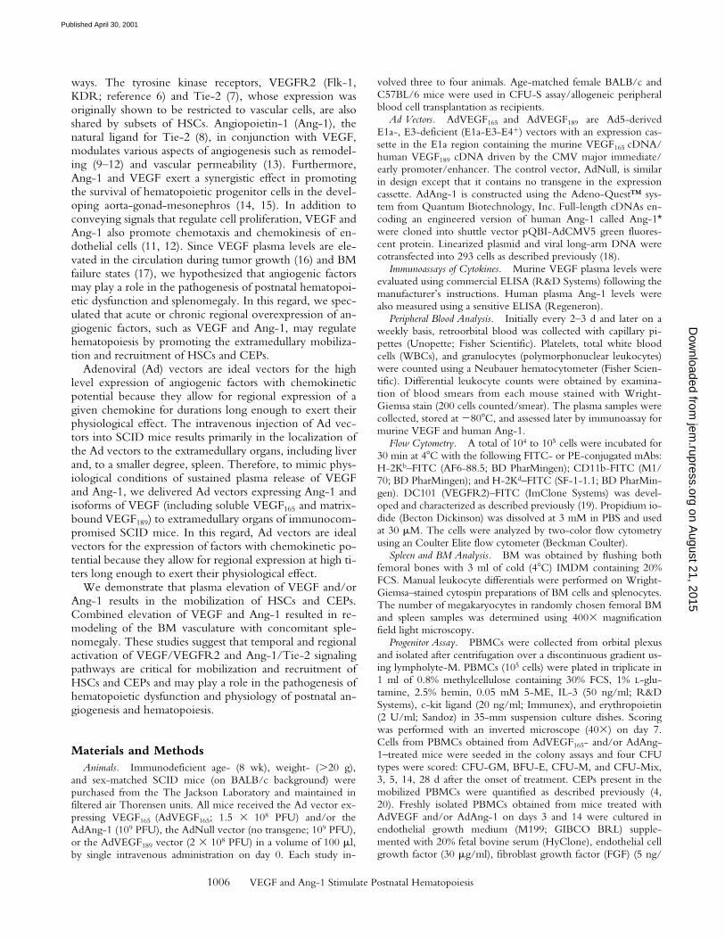
J. Exp. Med. The Rockefeller University Press • 0022-1007/2001/05/1005/10 $5.00 Volume 193, Number 9, May 7, 2001 1005–1014 http://www.jem.org/cgi/content/full/193/9/1005 1005 Vascular Endothelial Growth Factor and Angiopoietin-1 Stimulate Postnatal Hematopoiesis by Recruitment of Vasculogenic and Hematopoietic Stem Cells By Koichi Hattori,* § Sergio Dias,* Beate Heissig,* Neil R. Hackett, ‡ David Lyden, § Masatoshi Tateno, i Daniel J. Hicklin, ¶ Zhenping Zhu, ¶ Larry Witte, ¶ Ronald G. Crystal, ‡ Malcolm A.S. Moore, § and Shahin Rafii* From the *Division of Hematology-Oncology and the ‡ Division of Pulmonary, Critical Care Medicine and Genetic Medicine Program, Cornell University Medical College, New York, New York 10021; the § Sloan-Kettering Institute for Cancer Research, New York, New York 10021; the i Department of Pathology, Sapporo City General Hospital, Sapporo 060-8604, Japan; and the ¶ ImClone Systems Inc., New York, New York 10014 Abstract Tyrosine kinase receptors for angiogenic factors vascular endothelial growth factor (VEGF) and angiopoietin-1 (Ang-1) are expressed not only by endothelial cells but also by subsets of he- matopoietic stem cells (HSCs). To further define their role in the regulation of postnatal he- matopoiesis and vasculogenesis, VEGF and Ang-1 plasma levels were elevated by injecting re- combinant protein or adenoviral vectors expressing soluble VEGF 165 , matrix-bound VEGF 189 , or Ang-1 into mice. VEGF 165 , but not VEGF 189 , induced a rapid mobilization of HSCs and VEGF receptor (VEGFR)2 1 circulating endothelial precursor cells (CEPs). In contrast, Ang-1 induced delayed mobilization of CEPs and HSCs. Combined sustained elevation of Ang-1 and VEGF 165 was associated with an induction of hematopoiesis and increased marrow cellularity followed by proliferation of capillaries and expansion of sinusoidal space. Concomitant to this vascular remodeling, there was a transient depletion of hematopoietic activity in the marrow, which was compensated by an increase in mobilization and recruitment of HSCs and CEPs to the spleen resulting in splenomegaly. Neutralizing monoclonal antibody to VEGFR2 com- pletely inhibited VEGF 165 , but not Ang-1–induced mobilization and splenomegaly. These data suggest that temporal and regional activation of VEGF/VEGFR2 and Ang-1/Tie-2 signaling pathways are critical for mobilization and recruitment of HSCs and CEPs and may play a role in the physiology of postnatal angiogenesis and hematopoiesis. Key words: vascular endothelial growth factor • angiopoietin-1 • angiogenesis • hematopoiesis • tyrosine kinase receptors Introduction In the adult, the majority of hematopoietic stem cells (HSCs) 1 (1) and circulating endothelial precursor cells (CEPs; references 2–4) reside in the bone marrow (BM). However, upon stimulation with various cytokines, HSCs and CEPs can be mobilized to the peripheral circulation and colonize other hematopoietic organs, such as the spleen. In addition, BM failure states secondary to BM fibrosis (my- elofibrosis) or certain metastatic neoplastic diseases are asso- ciated with relocalization of HSCs from BM to extramed- ullary hematopoietic macroenvironments, including the spleen resulting in splenomegaly (5). However, the exact identities of the chemocytokines responsible for the mobili- zation and the mechanism of extramedullary recruitment of BM-derived HSCs and CEPs are not well established. Given the common prenatal origin of CEPs and HSCs from hemangioblasts, both hematopoietic and vasculogenic cells share several survival and chemotactic signaling path- Address correspondence to Shahin Rafii, Cornell University Medical College, Division of Hematology-Oncology, 1300 York Ave., Rm. C-606, New York, NY 10021. Phone: 212-746-2070; Fax: 212-746- 8866; E-mail: [email protected] 1 Abbreviations used in this paper: Ad, adenovirus; Ang-1, angiopoietin-1; BM, bone marrow; BMMC, BM mononuclear cell; CEP, circulating en- dothelial precursor cell; FGF, fibroblast growth factor; HSC, hematopoi- etic stem cell; VEGF, vascular endothelial growth factor; vWF, von Wil- lebrand factor; WBC, white blood cell. on August 21, 2015 jem.rupress.org Downloaded from Published April 30, 2001
-
Upload
independent -
Category
Documents
-
view
3 -
download
0
Transcript of Vascular Endothelial Growth Factor and Angiopoietin-1 Stimulate Postnatal Hematopoiesis by...

J. Exp. Med.
The Rockefeller University Press • 0022-1007/2001/05/1005/10 $5.00Volume 193, Number 9, May 7, 2001 1005–1014http://www.jem.org/cgi/content/full/193/9/1005
1005
Vascular Endothelial Growth Factor and Angiopoietin-1 Stimulate Postnatal Hematopoiesis by Recruitment of Vasculogenic and Hematopoietic Stem Cells
By Koichi Hattori,
*
§
Sergio Dias,
*
Beate Heissig,
*
Neil R. Hackett,
‡
David Lyden,
§
Masatoshi Tateno,
i
Daniel J. Hicklin,
¶
Zhenping Zhu,
¶
Larry Witte,
¶
Ronald G. Crystal,
‡
Malcolm A.S. Moore,
§
and Shahin Rafii
*
From the
*
Division of Hematology-Oncology and the
‡
Division of Pulmonary, Critical Care Medicine and Genetic Medicine Program, Cornell University Medical College, New York, New York 10021;
the
§
Sloan-Kettering Institute for Cancer Research, New York, New York 10021; the
i
Department of Pathology, Sapporo City General Hospital, Sapporo 060-8604, Japan; and the
¶
ImClone Systems Inc., New York, New York 10014
Abstract
Tyrosine kinase receptors for angiogenic factors vascular endothelial growth factor (VEGF) andangiopoietin-1 (Ang-1) are expressed not only by endothelial cells but also by subsets of he-matopoietic stem cells (HSCs). To further define their role in the regulation of postnatal he-matopoiesis and vasculogenesis, VEGF and Ang-1 plasma levels were elevated by injecting re-
combinant protein or adenoviral vectors expressing soluble VEGF
165
, matrix-bound VEGF
189
,or Ang-1 into mice. VEGF
165
, but not VEGF
189
, induced a rapid mobilization of HSCs and
VEGF receptor (VEGFR)2
1
circulating endothelial precursor cells (CEPs). In contrast, Ang-1induced delayed mobilization of CEPs and HSCs. Combined sustained elevation of Ang-1 andVEGF
165
was associated with an induction of hematopoiesis and increased marrow cellularityfollowed by proliferation of capillaries and expansion of sinusoidal space. Concomitant to thisvascular remodeling, there was a transient depletion of hematopoietic activity in the marrow,which was compensated by an increase in mobilization and recruitment of HSCs and CEPs tothe spleen resulting in splenomegaly. Neutralizing monoclonal antibody to VEGFR2 com-pletely inhibited VEGF
165
, but not Ang-1–induced mobilization and splenomegaly. These datasuggest that temporal and regional activation of VEGF/VEGFR2 and Ang-1/Tie-2 signalingpathways are critical for mobilization and recruitment of HSCs and CEPs and may play a rolein the physiology of postnatal angiogenesis and hematopoiesis.
Key words: vascular endothelial growth factor • angiopoietin-1 • angiogenesis • hematopoiesis • tyrosine kinase receptors
Introduction
In the adult, the majority of hematopoietic stem cells
(HSCs)
1
(1) and circulating endothelial precursor cells(CEPs; references 2–4) reside in the bone marrow (BM).However, upon stimulation with various cytokines, HSCs
and CEPs can be mobilized to the peripheral circulation andcolonize other hematopoietic organs, such as the spleen. Inaddition, BM failure states secondary to BM fibrosis (my-elofibrosis) or certain metastatic neoplastic diseases are asso-ciated with relocalization of HSCs from BM to extramed-ullary hematopoietic macroenvironments, including thespleen resulting in splenomegaly (5). However, the exactidentities of the chemocytokines responsible for the mobili-zation and the mechanism of extramedullary recruitment ofBM-derived HSCs and CEPs are not well established.
Given the common prenatal origin of CEPs and HSCsfrom hemangioblasts, both hematopoietic and vasculogeniccells share several survival and chemotactic signaling path-
Address correspondence to Shahin Rafii, Cornell University Medical
College, Division of Hematology-Oncology, 1300 York Ave., Rm.C-606, New York, NY 10021. Phone: 212-746-2070; Fax: 212-746-8866; E-mail: [email protected]
1
Abbreviations used in this paper:
Ad, adenovirus; Ang-1, angiopoietin-1;BM, bone marrow; BMMC, BM mononuclear cell; CEP, circulating en-dothelial precursor cell; FGF, fibroblast growth factor; HSC, hematopoi-etic stem cell; VEGF, vascular endothelial growth factor; vWF, von Wil-lebrand factor; WBC, white blood cell.
on August 21, 2015
jem.rupress.org
Dow
nloaded from
Published April 30, 2001
1006
VEGF and Ang-1 Stimulate Postnatal Hematopoiesis
ways. The tyrosine kinase receptors, VEGFR2 (Flk-1,KDR; reference 6) and Tie-2 (7), whose expression wasoriginally shown to be restricted to vascular cells, are alsoshared by subsets of HSCs. Angiopoietin-1 (Ang-1), thenatural ligand for Tie-2 (8), in conjunction with VEGF,modulates various aspects of angiogenesis such as remodel-ing (9–12) and vascular permeability (13). Furthermore,Ang-1 and VEGF exert a synergistic effect in promotingthe survival of hematopoietic progenitor cells in the devel-oping aorta-gonad-mesonephros (14, 15). In addition toconveying signals that regulate cell proliferation, VEGF andAng-1 also promote chemotaxis and chemokinesis of en-dothelial cells (11, 12). Since VEGF plasma levels are ele-vated in the circulation during tumor growth (16) and BMfailure states (17), we hypothesized that angiogenic factorsmay play a role in the pathogenesis of postnatal hematopoi-etic dysfunction and splenomegaly. In this regard, we spec-ulated that acute or chronic regional overexpression of an-giogenic factors, such as VEGF and Ang-1, may regulatehematopoiesis by promoting the extramedullary mobiliza-tion and recruitment of HSCs and CEPs.
Adenoviral (Ad) vectors are ideal vectors for the highlevel expression of angiogenic factors with chemokineticpotential because they allow for regional expression of agiven chemokine for durations long enough to exert theirphysiological effect. The intravenous injection of Ad vec-tors into SCID mice results primarily in the localization ofthe Ad vectors to the extramedullary organs, including liverand, to a smaller degree, spleen. Therefore, to mimic phys-iological conditions of sustained plasma release of VEGFand Ang-1, we delivered Ad vectors expressing Ang-1 andisoforms of VEGF (including soluble VEGF
165
and matrix-bound VEGF
189
) to extramedullary organs of immunocom-promised SCID mice. In this regard, Ad vectors are idealvectors for the expression of factors with chemokinetic po-tential because they allow for regional expression at high ti-ters long enough to exert their physiological effect.
We demonstrate that plasma elevation of VEGF and/orAng-1 results in the mobilization of HSCs and CEPs.Combined elevation of VEGF and Ang-1 resulted in re-modeling of the BM vasculature with concomitant sple-nomegaly. These studies suggest that temporal and regionalactivation of VEGF/VEGFR2 and Ang-1/Tie-2 signalingpathways are critical for mobilization and recruitment ofHSCs and CEPs and may play a role in the pathogenesis ofhematopoietic dysfunction and physiology of postnatal an-giogenesis and hematopoiesis.
Materials and Methods
Animals.
Immunodeficient age- (8 wk), weight- (
.
20 g),and sex-matched SCID mice (on BALB/c background) werepurchased from the The Jackson Laboratory and maintained infiltered air Thorensen units. All mice received the Ad vector ex-pressing VEGF
165
(AdVEGF
165
; 1.5
3
10
8
PFU) and/or theAdAng-1 (10
9
PFU), the AdNull vector (no transgene; 10
9
PFU),or the AdVEGF
189
vector (2
3
10
8
PFU) in a volume of 100
m
l,by single intravenous administration on day 0. Each study in-
volved three to four animals. Age-matched female BALB/c andC57BL/6 mice were used in CFU-S assay/allogeneic peripheralblood cell transplantation as recipients.
Ad Vectors.
AdVEGF
165
and AdVEGF
189
are Ad5-derivedE1a-, E3-deficient (E1a-E3-E4
1
) vectors with an expression cas-sette in the E1a region containing the murine VEGF
165
cDNA/human VEGF
189
cDNA driven by the CMV major immediate/early promoter/enhancer. The control vector, AdNull, is similarin design except that it contains no transgene in the expressioncassette. AdAng-1 is constructed using the Adeno-Quest™ sys-tem from Quantum Biotechnology, Inc. Full-length cDNAs en-coding an engineered version of human Ang-1 called Ang-1
*
were cloned into shuttle vector pQBI-AdCMV5 green fluores-cent protein. Linearized plasmid and viral long-arm DNA werecotransfected into 293 cells as described previously (18).
Immunoassays of Cytokines.
Murine VEGF plasma levels wereevaluated using commercial ELISA (R&D Systems) following themanufacturer’s instructions. Human plasma Ang-1 levels werealso measured using a sensitive ELISA (Regeneron).
Peripheral Blood Analysis.
Initially every 2–3 d and later on aweekly basis, retroorbital blood was collected with capillary pi-pettes (Unopette; Fisher Scientific). Platelets, total white bloodcells (WBCs), and granulocytes (polymorphonuclear leukocytes)were counted using a Neubauer hematocytometer (Fisher Scien-tific). Differential leukocyte counts were obtained by examina-tion of blood smears from each mouse stained with Wright-Giemsa stain (200 cells counted/smear). The plasma samples werecollected, stored at
2
80
8
C, and assessed later by immunoassay formurine VEGF and human Ang-1.
Flow Cytometry.
A total of 10
4
to 10
5
cells were incubated for30 min at 4
8
C with the following FITC- or PE-conjugated mAbs:H-2K
b
–FITC (AF6-88.5; BD PharMingen); CD11b-FITC (M1/70; BD PharMingen); and H-2K
d
–FITC (SF-1-1.1; BD PharMin-gen). DC101 (VEGFR2)–FITC (ImClone Systems) was devel-oped and characterized as described previously (19). Propidium io-dide (Becton Dickinson) was dissolved at 3 mM in PBS and usedat 30
m
M. The cells were analyzed by two-color flow cytometryusing an Coulter Elite flow cytometer (Beckman Coulter).
Spleen and BM Analysis.
BM was obtained by flushing bothfemoral bones with 3 ml of cold (4
8
C) IMDM containing 20%FCS. Manual leukocyte differentials were performed on Wright-Giemsa–stained cytospin preparations of BM cells and splenocytes.The number of megakaryocytes in randomly chosen femoral BMand spleen samples was determined using 400
3
magnificationfield light microscopy.
Progenitor Assay.
PBMCs were collected from orbital plexusand isolated after centrifugation over a discontinuous gradient us-ing lympholyte-M. PBMCs (10
5
cells) were plated in triplicate in1 ml of 0.8% methylcellulose containing 30% FCS, 1%
L
-glu-tamine, 2.5% hemin, 0.05 mM 5-ME, IL-3 (50 ng/ml; R&DSystems), c-kit ligand (20 ng/ml; Immunex), and erythropoietin(2 U/ml; Sandoz) in 35-mm suspension culture dishes. Scoringwas performed with an inverted microscope (40
3
) on day 7.Cells from PBMCs obtained from AdVEGF
165
- and/or AdAng-1–treated mice were seeded in the colony assays and four CFUtypes were scored: CFU-GM, BFU-E, CFU-M, and CFU-Mix,3, 5, 14, 28 d after the onset of treatment. CEPs present in themobilized PBMCs were quantified as described previously (4,20). Freshly isolated PBMCs obtained from mice treated withAdVEGF and/or AdAng-1 on days 3 and 14 were cultured inendothelial growth medium (M199; GIBCO BRL) supple-mented with 20% fetal bovine serum (HyClone), endothelial cellgrowth factor (30
m
g/ml), fibroblast growth factor (FGF) (5 ng/
on August 21, 2015
jem.rupress.org
Dow
nloaded from
Published April 30, 2001

1007
Hattori et al.
ml, human recombinant basic FGF; Sigma-Aldrich), heparin (5U/ml), penicillin (100 U/mL), streptomycin (100
m
g/ml), andfungizone (0.25
m
g/ml). These cells were placed on 12-well gel-atin-coated plates. After 2–3 wk, the endothelial colonies wereidentified by metabolic uptake of DiI-acetylated-LDL (DiI-Ac-LDL; PerImmune, Inc.) by incubating the wells with 1
m
g/ml ofhuman DiI-Ac-LDL for 5 h and visualized by fluorescence mi-croscopy. For von Willebrand factor (vWF) staining, cells wererinsed with HBSS and fixed immediately with ethanol for 10min. DiI-Ac-LDL
1
vWF
1
colonies formed in the first 3 d aftervector administration were scored as early outgrowth endothelialcolonies, whereas endothelial colonies formed after 14 d of cul-ture were considered as late-outgrowth colonies (CFU-ECs).
CFU-S Assay.
Mobilized PBMCs were obtained from days3, 5, 7, 14, 21, and 28 after the onset of treatment with variousAd vectors. For each data point, three recipient mice were irradi-ated with 9 Gy from a
137
Cs
g
-ray source at a dose rate of
z
0.90Gy/min to prevent the production of endogenous spleen colo-nies. Irradiated mice were injected intravenously via the tail veinwith 10
5
PBMCs within several hours after the completion of ir-radiation. The mice were killed 12 d later, and their spleens wereremoved and fixed in Bouin’s solution. The number of macro-scopic spleen colonies was then scored.
Repopulating Assay.
After vector administration, the periph-eral blood from SCID mice was collected on day 7. PBMCs (10
6
cells) were transplanted into lethally irradiated (9.5 Gy) recipientC57BL/6 mice. As a control, PBMCs (10
6
cells) from AdNull-treated SCID mice were transplanted into irradiated C57BL/6mice by intravenous injection at day 0. Chimerism was deter-mined by FACS
®
analysis (donor SCID mice, H-2K
d
; recipientC57BL/6 mice, H-2K
b
). The chimeric mice were killed at 90,120, and 150 d after transplantation and BM mononuclear cells(BMMCs) were stained for flow cytometry analysis with H-2K
d
–FITC (donor type). The representative percentages of positivepopulations in BMMCs are shown at day 150 (see Fig. 2 C). Thedata from age-matched normal C57BL/6 mice are also repre-sented as control. A total of 10
4
cells were incubated for 30 minat 4
8
C with FITC- or PE-conjugated mAbs, H-2K
b
–FITC (AF6-88.5; BD PharMingen) and H-2K
d
–FITC (SF-1-1.1; BDPharMingen). The percentage of positive cells was analyzedby flow cytometry using an Elite flow cytometer (BeckmanCoulter). Survival was monitored every day until 150 d after vec-tor administration. As control, BMMCs (10
6
) from SCID micewere transplanted into irradiated C57BL/6 mice by intravenousinjection at day 0.
Histopathology.
Tissues were fixed in 10% buffered formalinand paraffin embedded. Sections were stained with hematoxylinand eosin and examined under microscopy.
Delivery of Neutralizing VEGFR2 mAb to Mice.
A group ofSCID mice were treated with 1.5
3
10
8
PFU of AdVEGF
165
, 1.5
3
10
8
PFU of AdVEGF
165
and 10
9
PFU of AdAng-1, or 10
9
PFU ofAdNull intravenously on day 0. Some AdVEGF
165
-, AdVEGF
165
-and AdAng-1–, or AdNull-treated SCID mice received 800
m
gof anti-VEGFR2 (clone DC101) mAb intraperitoneally at 2-d in-tervals from either day 0 or 2.
Recombinant VEGF Induces Splenomegaly in Mice.
A group ofBALB/c mice were treated with 100 ng/mice recombinantVEGF or PBS intraperitoneally daily. Recombinant VEGF waspurchased from Immunotech.
Statistical Evaluation.
The results are expressed as mean
6
SEM. Statistical analyses were performed using the unpaired two-tailed Student’s
t
test. Survival rates were compared between thetwo groups by the log rank test.
Results
AdAng-1 and AdVEGF
165
but Not AdVEGF
189
PromoteMobilization of Hematopoietic Cell
s. Intravenous administra-tion of Ad vectors expressing VEGF
165
(AdVEGF
165
, 1.5
3
10
8
PFU) resulted in a peak VEGF plasma level (1,640
6
233 pg/ml) 24 h after injection and a return to pretreatmentlevel on day 35. Doses .1.5 3 108 PFU of AdVEGF165
were significantly toxic and resulted in the death of thetreated mice (data not shown). There were no signs of tox-icity in mice treated with AdNull vectors at doses up to 109
PFU. In AdAng-1–treated mice (109 PFU), plasma levels ofAng-1 peaked on day 3 after injection (74.1 6 0.7 ng/ml),returning to the pretreatment level after 8 wk. Plasma VEGFand Ang-1 were not detectable in AdNull-treated mice.
To examine the effects of acute and chronic elevation ofcirculating VEGF165 or Ang-1 levels in the mobilization ofCEPs and HSCs, SCID mice were injected intravenouslywith AdVEGF165 (1.5 3 108 PFU) and/or AdAng-1 (109
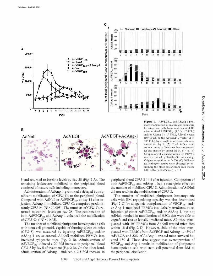
PFU), or AdVEGF189 (2 3 108 PFU). Subsequently, thenumber of leukocytes, hematopoietic progenitor cells, andHSCs were determined in the peripheral blood. Controlmice were injected with 109 PFU of AdNull vector. Com-pared with AdNull-treated mice, AdVEGF165-treated micehad a fourfold increase in WBCs on day 3, returning to thelevel of AdNull-treated mice by 5 wk after injection (Fig. 1A). In contrast, administration of AdAng-1 into SCID miceresulted in only a small increase in WBCs, peaking at day17 and returning to control levels by 7 wk. However, thecombination of AdVEGF165 and AdAng-1 resulted in asynergistic sevenfold increase in WBCs 5 d after vector ad-ministration. As expected, injection of AdVEGF189, whichresults in production of matrix-bound VEGF189, did notresult in leukocyte mobilization.
Populations of blast-like cells that are usually localized tothe BM were detected at high levels in the peripheral circu-lation of the AdVEGF165- and/or AdAng-1–treated mice.These cells displayed scant cytoplasm and large nuclei, remi-niscent of BM-derived immature hematopoietic progenitorand precursor cells (Fig. 1 B). Compared with AdNull-treated mice, AdVEGF165- and AdVEGF165 plus AdAng-1–treated mice had a dramatic increase in the WBC percentageof blast-like cells (Others; Fig. 1 B) and monocytes duringdays 2–14, returning almost to baseline 3 wk after the start oftreatment (Fig. 1 C). The intravenous administration ofAdAng-1 to SCID mice also resulted in increased circulationof blast-like cells, peaking at day 16 and returning to baselineon day 49 (Fig. 1 C). There were no significant changes inthe leukocyte levels of AdNull-treated mice.
AdVEGF165 and AdAng-1 Induced Mobilization of Hemato-poietic Progenitor Cells with Stem Cell Potential. The admin-istration of AdVEGF165 induced mobilization of hemato-poietic progenitors to the peripheral circulation. Theseprogenitors comprised colony-forming units (CFU-Cs)such as CFU-M, CFU-GM, BFU-E, and CFU-Mix(CFU-GEMM). Compared with AdNull-treated mice, atdays 3 and 5 the majority of the mobilized CFU-Cs con-sisted of CFU-GM (*P , 0.005). CFU-GMs peaked at day
on August 21, 2015
jem.rupress.org
Dow
nloaded from
Published April 30, 2001
1008 VEGF and Ang-1 Stimulate Postnatal Hematopoiesis
5 and returned to baseline levels by day 28 (Fig. 2 A). Theremaining leukocytes mobilized to the peripheral bloodconsisted of mature cells including monocytes.
Administration of AdAng-1 promoted a delayed but sig-nificant mobilization of CFU-Cs to the peripheral blood.Compared with AdNull or AdVEGF165, at day 14 after in-jection, AdAng-1–mobilized CFU-Cs comprised predomi-nantly CFU-M (*P , 0.005). The numbers of CFU-Cs re-turned to control levels on day 28. The combination ofboth AdVEGF165 and AdAng-1 enhanced the mobilizationof CFU-Cs (**P , 0.05).
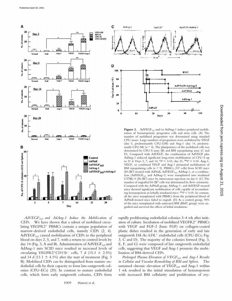
The number of mobilized pluripotent hematopoietic cellswith stem cell potential, capable of forming spleen colonies(CFU-S), was measured by injecting AdVEGF165 and/orAdAng-1 or, as control, AdNull-mobilized PBMCs intoirradiated syngeneic mice (Fig. 2 B). Administration ofAdVEGF165 induced a 20-fold increase in peripheral bloodCFU-S by day 5 of treatment (Fig. 2 B). On the other hand,administration of AdAng-1 induced a 2.5-fold increase in
peripheral blood CFU-S 14 d after injection. Coinjection ofboth AdVEGF165 and AdAng-1 had a synergistic effect onthe number of mobilized CFU-S. Administration of AdNulldid not result in the mobilization of CFU-S.
The number of mobilized pluripotent hematopoieticcells with BM-repopulating capacity was also determined(Fig. 2 C) by allogeneic transplantation of VEGF165- and/or Ang-1–mobilized PBMCs into lethally irradiated mice.Injection of either AdVEGF165 and/or AdAng-1, but notAdNull, resulted in mobilization of HSCs that were able toengraft and rescue lethally irradiated mice. All mice trans-planted with 106 PBMCs from AdNull-treated mice diedwithin 18 d (Fig. 2 D). However, 56% of the mice trans-planted with PBMCs from AdVEGF and AdAng-1, 45% ofAdVEGF, and 22% of AdAng-1–treated mice survived be-yond 150 d. These data suggest that overexpression ofVEGF165 and Ang-1 results in mobilization of pluripotenthematopoietic cells with stem cell potential from BM tothe peripheral circulation.
Figure 1. AdVEGF165 and AdAng-1 pro-mote mobilization of mature and immaturehematopoietic cells. Immunodeficient SCIDmice received AdVEGF165 (1.5 3 108 PFU)and/or AdAng-1 (109 PFU), AdNull vector(109 PFU), or the AdVEGF189 vector (2 3108 PFU) by a single intravenous adminis-tration on day 0. (A) Total WBCs werecounted using a Neubauer hematocytome-ter and stained by crystal violet. n = 6. (B)Morphological characterization of PBMCswas determined by Wright-Giemsa staining.Original magnification: 3200. (C) Differen-tial leukocyte counts were obtained by ex-amining the blood smears from each mouse(200 cells counted/smear). n = 4.
on August 21, 2015
jem.rupress.org
Dow
nloaded from
Published April 30, 2001
1009 Hattori et al.
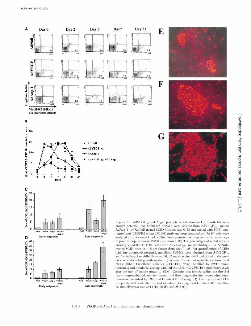
AdVEGF165 and AdAng-1 Induce the Mobilization ofCEPs. We have shown that a subset of mobilized circu-lating VEGFR21 PBMCs contain a unique population ofmarrow-derived endothelial cells, namely CEPs (2, 4).AdVEGF165 caused mobilization of CEPs to the peripheralblood on days 2, 5, and 7, with a return to control levels byday 14 (Fig. 3, A and B). Administration of AdVEGF165 andAdAng-1 into SCID mice resulted in increased levels ofcirculating VEGFR21CD11b2 cells, 7 d (15.3 6 2.5%)and 14 d (11.1 6 4.1%) after the start of treatment (Fig. 3B). Mobilized CEPs can be distinguished from mature en-dothelial cells by their capacity to form late-outgrowth col-onies (CFU-ECs) (20). In contrast to mature endothelialcells, which form early outgrowth colonies, CEPs form
rapidly proliferating endothelial colonies 3–4 wk after initi-ation of culture. Incubation of mobilized VEGFR21 PBMCswith VEGF and FGF-2 (basic FGF) on collagen-coatedplastic dishes resulted in the generation of early and lateoutgrowth DiI-Ac-LDL1 endothelial cells (CFU-ECs; Fig.3, C and D). The majority of the colonies formed (Fig. 3,E, F, and G) were composed of late outgrowth endothelialcells, suggesting that VEGF and Ang-1 promote the mobi-lization of BM-derived CEPs.
Prolonged Plasma Elevation of VEGF165 and Ang-1 Resultsin Cellular and Vascular Remodeling of BM and Spleen. Thesustained chronic elevation of VEGF165 and Ang-1 within1 wk resulted in the initial stimulation of hematopoiesiswith increased BM cellularity and proliferation of ery-
Figure 2. AdVEGF165 and/or AdAng-1 induce peripheral mobili-zation of hematopoietic progenitor cells and stem cells. (A) Thenumber of mobilized progenitors was determined using standardCFU assays. Large numbers of progenitors were mobilized by VEGF(day 5, predominantly CFU-GM) and Ang-1 (day 14, predomi-nantly CFU-M) (n = 4). The pluripotency of the mobilized cells wasdetermined by CFU-S assay (B) and BM repopulating assay (C andD). Compared with AdVEGF, the combination of AdVEGF plusAdAng-1 induced significant long-term mobilization of CFU-S upto 21 d. Days 3, 7, and 14, *P , 0.01; day 21, **P , 0.05. Ang-1,VEGF, or combined VEGF and Ang-1 promoted mobilization ofBM repopulating cells (n = 9). PBMCs (106 cells) from SCID mice(H-2Kd) treated with AdNull, AdVEGF165, AdAng-1, or a combina-tion (AdVEGF165 and AdAng-1) were transplanted into irradiatedC57BL/6 (H-2Kb) mice by intravenous injection on day 0. (C) Thenumber of engrafted H-2Kd cells was determined by flow cytometry.Compared with the AdNull group, AdAng-1– and AdVEGF-treatedmice showed significant mobilization of cells capable of reconstitut-ing hematopoiesis in lethally irradiated mice. **P , 0.05. In contrast,all the mice transplanted with PBMCs from the peripheral blood ofAdNull-treated mice failed to engraft. (D) As a control group, 90%of the mice transplanted with untreated BM (BMT group) were en-grafted and survived the effects of lethal irradiation.
on August 21, 2015
jem.rupress.org
Dow
nloaded from
Published April 30, 2001
1010 VEGF and Ang-1 Stimulate Postnatal Hematopoiesis
Figure 3. AdVEGF165 and Ang-1 promote mobilization of CEPs with late out-growth potential. (A) Mobilized PBMCs were isolated from AdVEGF165- and/orAdAng-1– or AdNull-treated SCID mice on days 0–28 and stained with FITC-con-jugated anti-VEGFR2 (clone DC101) mAb and propidium iodide. (A) 104 cells wereanalyzed on a Beckman Coulter Elite flow cytometer, and representative percentagesof positive populations in PBMCs are shown. (B) The percentages of mobilized cir-culating VEGFR21CD11b2 cells from AdVEGF165- and/or AdAng-1– or AdNull-treated SCID mice (n = 3) are shown from days 0 –28. For quantification of CEPswith late outgrowth potential, mobilized PBMCs were obtained from AdVEGF165
and/or AdAng-1 or AdNull-treated SCID mice on days 0–21 and plated in the pres-ence of endothelial growth medium (reference 13) on collagen/fibronectin-coatedplastic dishes. Endothelial colonies (CFU-ECs) were identified by vWF immu-nostaining and metabolic labeling with DiI-Ac-LDL. (C) CFU-ECs proliferated 2 wkafter the start of culture (mean 6 SEM). Colonies that formed within the first 3 d(early outgrowth) and colonies formed 14 d (late outgrowth) after vector administra-tion were quantified by vWF and DiI-Ac-LDL labeling. (D) The majority of CFU-EC proliferated 3 wk after the start of culture, forming focal DiI-Ac-LDL1 endothe-lial monolayers as seen at 14 (E), 20 (F), and 25 d (G).
on August 21, 2015
jem.rupress.org
Dow
nloaded from
Published April 30, 2001
1011 Hattori et al.
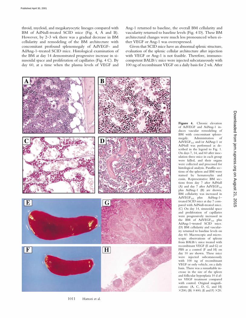
throid, myeloid, and megakaryocytic lineages compared withBM of AdNull-treated SCID mice (Fig. 4, A and B).However, by 2–3 wk there was a gradual decrease in BMcellularity and remodeling of the BM architecture withconcomitant profound splenomegaly of AdVEGF- andAdAng-1–treated SCID mice. Histological examination ofthe BM at day 14 demonstrated progressive increase in si-nusoidal space and proliferation of capillaries (Fig. 4 C). Byday 60, at a time when the plasma levels of VEGF and
Ang-1 returned to baseline, the overall BM cellularity andvascularity returned to baseline levels (Fig. 4 D). These BMarchitectural changes were much less pronounced when ei-ther VEGF or Ang-1 was overexpressed.
Given that SCID mice have an abnormal splenic structure,evaluation of the splenic cellular architecture after injectionwith VEGF or Ang-1 is not feasible. Therefore, immuno-competent BALB/c mice were injected subcutaneously with100 ng of recombinant VEGF on a daily basis for 2 wk. After
Figure 4. Chronic elevationof AdVEGF and AdAng-1 in-duces vascular remodeling ofBM with concomitant spleno-megaly. Administration ofAdVEGF165 and/or AdAng-1 orAdNull was performed as de-scribed in the legend to Fig. 1.On days 7, 14, and 60 after inoc-ulation three mice in each groupwere killed, and their organswere collected and processed forhistological analysis. Paraffin sec-tions of the spleen and BM werestained by hematoxylin andeosin. Representative BM sec-tions from day 7 after AdNull(A) and day 7 after AdVEGF165
plus AdAng-1 (B) are shown.BM cellularity was increased inAdVEGF165 plus AdAng-1–treated SCID mice at day 7 com-pared with AdNull-treated mice.(C) On day 14, sinusoidal spaceand proliferation of capillarieswere progressively increased inthe BM of AdVEGF165 plusAdAng-1–treated SCID mice.(D) BM cellularity and vascular-ity returned to baseline levels onday 60. Macroscopic and micro-scopic observations of spleensfrom BALB/c mice treated withrecombinant VEGF (E and G) orPBS as a control (F and H) onday 10 are shown. These micewere injected subcutaneouslywith 100 ng of recombinantVEGF or only vehicle, on a dailybasis. There was a remarkable in-crease in the size of the spleenand follicular hyperplasia 10 d af-ter VEGF treatment comparedwith control. Original magnifi-cations: (A, C, D, G, and H)3200; (B) 3400; (E and F) 320.
on August 21, 2015
jem.rupress.org
Dow
nloaded from
Published April 30, 2001
1012 VEGF and Ang-1 Stimulate Postnatal Hematopoiesis
2 wk, at the time when there was a relative decrease in BMcellularity (1.9 6 0.3 3 107/femur from VEGF-treated micecompared with 2.5 6 0.1 3 107/femur for normal BALB/cmice, P , 0.01), there was a remarkable increase in spleensize (Fig. 4, E and F). Compared with the control group(Fig. 4 H), there was increased cellularity and follicular hy-perplasia in the spleen of VEGF-treated group (Fig. 4 G;0.3 6 0.2 3 108 compared with 2.4 6 0.3 3 108 for normalBALB/c mice, P , 0.005). Splenomegaly and follicular hy-perplasia were reversed after cessation of treatment.
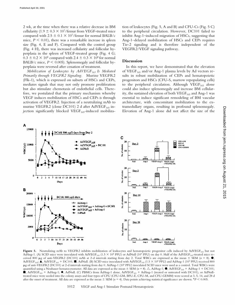
Mobilization of Leukocytes by AdVEGF165 Is MediatedPrimarily through VEGFR2 Signaling. Murine VEGFR2(Flk-1), which is expressed on subsets of HSCs and CEPs,mediates signals that may not only promote proliferationbut also stimulate chemotaxis of endothelial cells. There-fore, we postulated that the primary mechanism wherebyVEGF induces mobilization of HSCs and CEPs is throughactivation of VEGFR2. Injection of a neutralizing mAb tomurine VEGFR2 (clone DC101) 2 d after AdVEGF165 in-jection significantly blocked VEGF165-induced mobiliza-
tion of leukocytes (Fig. 5, A and B) and CFU-Cs (Fig. 5 C)to the peripheral circulation. However, DC101 failed toinhibit Ang-1–induced migration of HSCs, suggesting thatAng-1–delayed mobilization of HSCs and CEPs requiresTie-2 signaling and is therefore independent of theVEGFR2/VEGF signaling pathway.
DiscussionIn this report, we have demonstrated that the elevation
of VEGF165 and/or Ang-1 plasma levels by Ad vectors re-sults in robust mobilization of CEPs and hematopoieticprogenitors and HSCs (CFU-S, marrow repopulating cells)to the peripheral circulation. Although VEGF165 alonecould also induce splenomegaly and increase BM cellular-ity, the sustained elevation of both VEGF165 and Ang-1 wasessential to induce significant remodeling of BM vasculararchitecture, with concomitant mobilization to the ex-tramedullary organs, resulting in profound splenomegaly.Elevation of Ang-1 alone did not affect the size of the
Figure 5. Neutralizing mAb to VEGFR2 inhibits mobilization of leukocytes and hematopoietic progenitor cells induced by AdVEGF165 but notAdAng-1. (A) SCID mice were inoculated with AdVEGF165 (1.5 3 108 PFU) or AdNull (109 PFU) on day 0. Half of the AdVEGF165-treated mice re-ceived 800 mg of anti-VEGFR2 (DC101) mAb at 2-d intervals starting from day 2. Total WBCs are expressed as the mean 6 SEM (n = 8). d,AdVEGF165; m, AdVEGF165 1 DC101; j, AdNull. (B) SCID mice inoculated with AdVEGF165 (1.5 3 108 PFU) and AdAng-1 (109 PFU) received 800mg of anti-VEGFR2 (DC101) at 2-d intervals starting from day 0. AdAng-1 (109 PFU) inoculated SCID mice were used as a control. Total WBCs werequantified using a Neubauer hematocytometer. All data are expressed as the mean 6 SEM (n = 8). n, AdAng-1; d, AdVEGF165 1 AdAng-1 1 DC101;j, AdVEGF165 1 AdAng-1; r, AdNull. (C) PBMCs from AdAng-1 alone, AdVEGF165 1 AdAng-1 (treated or untreated with DC101), or AdNull-treated mice were seeded into the colony assays and four types of CFU (CFU-GM, BFU-E, CFU-M, and CFU-GEMM) were scored at 3, 5, 14, and 28 dafter the onset of treatment. All data are expressed as the mean 6 SEM (n = 4). Data points achieving statistical significance are shown. *P , 0.005.
on August 21, 2015
jem.rupress.org
Dow
nloaded from
Published April 30, 2001

1013 Hattori et al.
spleen or cellularity of the BM, suggesting that combinedsignaling through VEGFR2/VEGF and Ang1/Tie-2 sig-naling pathways plays a key role in the regulation of organ-specific hematopoietic activity.
The overexpression of VEGF165 induces a rapid (24–48 h)mobilization of CEPs and HSCs, suggesting that the ef-fect of VEGF165 is mediated through a chemokinetic pro-cess. However, plasma elevation of Ang-1 requires at least7 d to induce mobilization of CEPs and HSCs. The physi-ological effect of VEGF is most likely not mediatedthrough BM endothelial cell leakiness since studies haveshown that Ang-1 prevents VEGF-induced vascular per-meability (13, 18). In this regard, Ang-1 should have inhib-ited the VEGF-induced mobilization of the HSCs andCEPs. In contrast, Ang-1 promoted the mobilization ofHSCs and CEPs and synergized with VEGF in augmentingits effect. Collectively, these data suggest that VEGF aloneor in combination with Ang-1 induce mobilization by di-rectly interacting with stem cells or promoting remodelingof the BM vasculature. It is conceivable that Ang-1 may al-ter the adhesion molecule repertoire of BM endothelialcells or HSCs, resulting in enhanced mobilization. Alterna-tively, Ang-1 may sustain or promote the survival of Tie-21
HSCs and CEPs, inducing cell cycle shifts that may favortheir mobilization to the peripheral circulation.
VEGF has been shown to exert its pleiotropic effects onendothelial cells through its receptors VEGFR1 andVEGFR2. VEGFR2 has been shown to be the principalmitogenic receptor for VEGF (21, 22), whereas VEGFR1conveys signals for vascular remodeling. However,VEGFR1 is expressed by various mature hematopoieticcells, including dendritic and monocytic cells (23, 24),whereas VEGFR2 is expressed by HSCs and CEPs (4).Therefore, VEGF165-induced mobilization of mature cellssuch as monocytes may be mediated though VEGFR1,whereas mobilization of HSCs is most likely mediatedthrough VEGFR2 (6). Neutralizing mAb to VEGFR2 in-hibited VEGF165-induced mobilization and splenomegaly,suggesting that interaction of VEGF165 with VEGFR2 isessential for mobilization and recruitment of HSCs. De-layed mobilization in response to elevation in circulatingAng-1 is most likely induced by interaction with eitherTie-21 on the stem cells (19) or because of remodeling ofthe BM microenvironment.
Most in vivo experiments assessing the chemoattractantproperties of VEGF165 have been performed with injectionof recombinant VEGF165. However, given that the half-lifeof VEGF165 in the circulation is only several minutes, it hasbeen difficult to evaluate the role of chronic VEGF165 re-lease, as it may occur during tumor growth and metastasisor myelodysplastic syndromes. Likewise, recombinantAng-1 is unstable and previous studies using Ang-1 proteinhave failed to demonstrate a functional endpoint (21). It issuggested that in contrast to VEGF165, whereby injection ofrecombinant protein can induce mobilization of stem cells,Ang-1 protein does not have enough biochemical stabilityto exert any biological effect. Furthermore, given the de-layed mobilization seen with Ang-1 treatment, injection of
AdAng-1 into SCID mice as opposed to immunocompe-tent mice was the only means of sustaining the level ofAng-1 long enough in vivo to observe its effects. There-fore, the use of Ad vectors as regional delivery vehicles forVEGF165 and Ang-1 provides ideal tools for studying therole of these angiogenic factors in mobilization and recruit-ment of HSCs and CEPs. In addition, given that VEGF189
is matrix bound, Ad vectors producing VEGF189 provide anideal mechanism to deliver this matrix-bound isoform ofVEGF regionally to the extramedullary hematopoietic mi-croenvironments such as the liver.
We show that long-term overexpression of bothVEGF165 and Ang-1 is essential to induce an initial increasein BM cellularity followed by remodeling of the BM archi-tecture, with depletion of sinusoidal spaces of hematopoieticcells and a parallel increase in BM vascularization and sple-nomegaly. These results suggest that regional expression ofVEGF165 and Ang-1 in extramedullary organs may act syn-ergistically to modulate postnatal hematopoiesis by inducingextramedullary shifts in hematopoiesis or remodeling of vas-cular architecture of sinusoidal BM endothelial cells. Re-cently, it has been shown that plasma VEGF levels are sig-nificantly elevated in myelodysplastic syndromes (17),where the BM microenvironment is damaged by fibrotictissue. This raises the possibility that elevation of VEGF mayserve as one of the primary adaptive responses to compen-sate for BM myelofibrosis, by promoting the relocalizationand recruitment of HSCs to other hematopoietic microen-vironments such as the spleen, resulting in splenomegaly.
Mobilization of CEPs to the peripheral circulation mayplay a critical role in regulating postnatal angiogenesis andvasculogenesis. Based on the data presented here, high lev-els of VEGF produced by tumors may result in the mobili-zation of BM-derived CEPs to the peripheral circulationand enhance their recruitment into the tumor vasculature.Given that CEPs may also contribute to vascular healing,overexpression of VEGF165 may promote the peripheralblood mobilization and recruitment of CEPs to the injuredvascular bed, thereby accelerating wound healing.
As evidenced by the relative plasma levels of Ang-1 andVEGF, higher levels of Ang-1 are necessary to induce mo-bilization of HSCs and CEPs. Indeed, at doses .1.5 3 108
PFU of AdVEGF165, elevations of plasma VEGF were asso-ciated with significant toxicity and resulted in the death ofthe treated mice. Ang-1 does not exert any major physio-logical effect at the lower doses (5 3 108 PFU); however,when administered at higher doses (109 PFU) it had a po-tent synergistic effect with VEGF in mobilizing HSCs andCEPs. These differences in the dose response may be dueto the variable receptor densities or differential physiologi-cal effects of the VEGF receptors and Tie-2. It is also con-ceivable that expression of VEGFR2 (Flk-1) on HSCs andCEPs is significantly higher than Tie-2.
These data set forth a novel paradigm for efficient mobi-lization of HSCs and CEPs to the peripheral circulation.This strategy facilitates recovery of large numbers of pluri-potent HSCs and CEPs, which may be used for transplan-tation or treatment of a wide variety of genetic and malig-
on August 21, 2015
jem.rupress.org
Dow
nloaded from
Published April 30, 2001
1014 VEGF and Ang-1 Stimulate Postnatal Hematopoiesis
nant disorders. Whether transplantation of mobilized CEPsin conjunction with mobilized HSCs will exert a synergis-tic effect to enhance engraftment and reconstitution of he-matopoiesis is the subject of future studies.
We are grateful to Drs. John Rudge, Ella Ioffe, and George D.Yancopoulos (Regeneron Pharmaceutical, Inc.) for providing theAd vectors and Ang-1 ELISA data and for their helpful discussionsduring the preparation of this manuscript. We thank Harry G. Sat-terwhite, Dr. George Lam, Mannix S. Quitriano, Maureen Sulli-van, Koji Shido, and Sinae R. Park for their helpful assistance andMargaret Choy for critical reading.
S. Rafii is supported by National Heart, Lung, and Blood Institute(NHLBI) grants R01 HL58707, R01 HL61849, Program ProjectU01 HL66952 (Project 2), Pilot Project P01 HL59312, the DorothyRodbell Foundation for Sarcoma Research, and the Rich Founda-tion. M.A.S. Moore is supported by NHLBI grant R01 HL61401and the Gar Reichman Fund of Cancer Research Institute. R.G.Crystal is supported in part by NHLBI grant R01 HL57318, Pro-gram Project U01 HL66952-01, the Will Rogers Memorial Fund,and Gen Vec, Inc. (Gaithersburg, MD). K. Hattori is the recipient ofa fellowship from the Uehara Memorial Foundation (Tokyo, Japan).B. Heissig is the recipient of a fellowship from the Dr. MildredScheel Stiftung für Krebsforschung (Bonn, Germany).
Submitted: 28 September 2000Accepted: 12 March 2001Revised: 14 March 2001
References1. Moore, M.A. 1997. Stem cell proliferation: ex vivo and in
vivo observations. Stem Cells. 1:239–248.2. Rafii, S. 2000. Circulating endothelial precursors: mystery,
reality, and promise. J. Clin. Invest. 105:17–19.3. Takahashi, T., C. Kalka, H. Masuda, D. Chen, M. Silver, M.
Kearney, M. Magner, J.M. Isner, and T. Asahara. 1999. Isch-emia- and cytokine-induced mobilization of bone marrow-derived endothelial progenitor cells for neovascularization.Nat. Med. 5:434–438.
4. Peichev, M., A.J. Naiyer, D. Pereira, Z. Zhu, W.J. Lane, M.Williams, M.C. Oz, D.J. Hicklin, L. Witte, M.A. Moore, andS. Rafii. 2000. Expression of VEGFR-2 and AC133 by circu-lating human CD34(1) cells identifies a population of func-tional endothelial precursors. Blood. 95:952–958.
5. Frey, B.M., S. Rafii, M. Teterson, D. Eaton, R.G. Crystal,and M.A. Moore. 1998. Adenovector-mediated expression ofhuman thrombopoietin cDNA in immune-compromisedmice: insights into the pathophysiology of osteomyelofibrosis.J. Immunol. 160:691–699.
6. Ziegler, B.L., M. Valtieri, G.A. Porada, R. De Maria, R.Muller, B. Masella, M. Gabbianelli, I. Casella, E. Pelosi, T.Bock, et al. 1999. KDR receptor: a key marker defining he-matopoietic stem cells. Science. 285:1553–1558.
7. Hashiyama, M., A. Iwama, K. Ohshiro, K. Kurozumi, K. Ya-sunaga, Y. Shimizu, Y. Masuho, I. Matsuda, N. Yamaguchi,and T. Suda. 1996. Predominant expression of a receptor ty-rosine kinase, TIE, in hematopoietic stem cells and B cells.Blood. 87:93–101.
8. Davis, S., T.H. Aldrich, P.F. Jones, A. Acheson, D.L. Comp-ton, V. Jain, T.E. Ryan, J. Bruno, C. Radziejewski, P.C.Maisonpierre, and G.D. Yancopoulos. 1996. Isolation of an-giopoietin-1, a ligand for the TIE2 receptor, by secretion-trapexpression cloning. Cell. 87:1161–1169.
9. Suri, C., P.F. Jones, S. Patan, S. Bartunkova, P.C. Maisonpi-erre, S. Davis, T.N. Sato, and G.D. Yancopoulos. 1996.Requisite role of angiopoietin-1, a ligand for the TIE2 recep-tor, during embryonic angiogenesis. Cell. 87:1171–1180.
10. Holash, J., D. Maisonpierre, D. Compton, P. Boland, C.R.Alexander, D. Zagzag, G.D. Yancopoulos, and S.J. Wiegland.1999. Vessel cooption, regression, and growth in tumors me-diated by angiopoietins and VEGF. Science. 284:1994–1998.
11. Koblizek, T.I., C. Weiss, G.D. Yancopoulos, U. Deutsch,and W. Risau. 1998. Angiopoietin-1 induces sprouting an-giogenesis in vitro. Curr. Biol. 8:529–532.
12. Witzenbichler, B., P.C. Maisonpierre, P. Jones, G.D. Yanco-poulos, and J.M. Isner. 1998. Chemotactic properties of an-giopoietin-1 and -2, ligands for the endothelial-specific re-ceptor tyrosine kinase Tie2. J. Biol. Chem. 273:18514–18521.
13. Thurston, G., C. Suri, K. Smith, J. McClain, T.N. Sato, G.D.Yancopoulos, and D.M. McDonald. 1999. Leakage-resistantblood vessels in mice transgenically overexpressing angiopoi-etin-1. Science. 286:2511–2514.
14. Hamaguchi, I., X.L. Huang, N. Takakura, J. Tada, Y.Yamaguchi, H. Kodama, and T. Suda. 1999. In vitro he-matopoietic and endothelial cell development from cells ex-pressing TEK receptor in murine aorta-gonad-mesonephrosregion. Blood. 93:1549–1556.
15. Huang, X.L., N. Takakura, and T. Suda. 1999. In vitro ef-fects of angiopoietins and VEGF on hematopoietic and en-dothelial cells. Biochem. Biophys. Res. Commun. 264:133–138.
16. Damert, A., M. Machein, G. Breier, M.Q. Fujita, D. Hana-han, W. Risau, and K.H. Plate. 1997. Up-regulation of vas-cular endothelial growth factor expression in a rat glioma isconferred by two distinct hypoxia-driven mechanisms. CancerRes. 57:3860–3864.
17. Aguayo, A., E. Estey, H. Kantarjian, T. Mansouri, C. Gidel,M. Keating, F. Giles, Z. Estrov, B. Barlogie, and M. Albitar.1999. Cellular vascular endothelial growth factor is a predic-tor of outcome in patients with acute myeloid leukemia.Blood. 94:3717–3721.
18. Thurston, G., J.S. Rudge, E. Ioffe, H. Zhou, L. Ross, S.D.Croll, N. Glazer, J. Holash, D.M. McDonald, and G.D. Yan-copoulos. 2000. Angiopoietin-1 protects the adult vasculatureagainst plasma leakage. Nat. Med. 6:460–463.
19. Hsu, H.C., H. Ema, M. Osawa, Y. Nakamura, T. Suda, andH. Nakauchi. 2000. Hematopoietic stem cells express Tie-2receptor in the murine fetal liver. Blood. 96:3757–3762.
20. Lin, Y., D.J. Weisdorf, A. Solovey, and R.P. Hebbel. 2000.Origins of circulating endothelial cells and endothelial out-growth from blood. J. Clin. Invest. 105:71–77.
21. Shalaby, F., J. Rossant, T.P. Yamaguchi, M. Gertsenstein,X.F. Wu, M.L. Breitman, and A.C. Schuh. 1995. Failure ofblood-island formation and vasculogenesis in Flk-1-deficientmice. Nature. 376:62–66.
22. Fong, G.H., J. Rossant, M. Gertsenstein, and M.L. Breitman.1995. Role of the Flt-1 receptor tyrosine kinase in regulatingthe assembly of vascular endothelium. Nature. 376:66–70.
23. Barleon, B., S. Sozzani, D. Zhou, H.A. Weich, A. Manto-vani, and D. Marme. 1996. Migration of human monocytesin response to vascular endothelial growth factor (VEGF) ismediated via the VEGF receptor flt-1. Blood. 87:3336–3343.
24. Clauss, M., H. Weich, G. Breier, U. Knies, W. Rockl, J.Waltenberger, and W. Risau. 1996. The vascular endothelialgrowth factor receptor Flt-1 mediates biological activities.Implications for a functional role of placenta growth factor inmonocyte activation and chemotaxis. J. Biol. Chem. 271:17629–17634.
on August 21, 2015
jem.rupress.org
Dow
nloaded from
Published April 30, 2001