Cell Factors Stimulate Human Immunodeficiency Virus Type 1 Reverse Transcription In Vitro
13
JOURNAL OF VIROLOGY, Feb. 2008, p. 1425–1437 Vol. 82, No. 3 0022-538X/08/$08.000 doi:10.1128/JVI.01808-07 Copyright © 2008, American Society for Microbiology. All Rights Reserved. Cell Factors Stimulate Human Immunodeficiency Virus Type 1 Reverse Transcription In Vitro David Warrilow, 1 Luke Meredith, 1,4 Adam Davis, 2 Christopher Burrell, 2,3 Peng Li, 2,3 and David Harrich 1,4 * Division of Immunology and Infectious Disease, Queensland Institute of Medical Research, Brisbane, Queensland 4006, Australia 1 ; Infectious Diseases Laboratories, Institute of Medical and Veterinary Science, Frome Rd., Adelaide, South Australia 5000, Australia 2 ; School of Molecular and Biomedical Sciences, University of Adelaide, North Tce., Adelaide 5005, Australia 3 ; and Griffith Medical Research College, a joint program of Griffith University and the Queensland Institute of Medical Research, Herston, Queensland 4006, Australia 4 Received 16 August 2007/Accepted 13 November 2007 After fusion of the human immunodeficiency virus type 1 (HIV-1) envelope with the host cell membrane, the HIV-1 core enters the cell cytoplasm. Core components are then restructured to form the reverse transcription complex (RTC); the biochemical details of this process are currently unclear. To investigate early RTC formation, we characterized the endogenous reverse transcription activity of virions, which was less efficient than reverse transcription during cell infection and suggested a requirement for a cell factor. The addition of detergent to virions released reverse transcriptase and capsid, and reverse transcription products became susceptible to the action of exogenous nucleases, indicating virion disruption. Disruption was coincident with the loss of the endogenous reverse transcription activity of virions, particularly late reverse transcription products. Consistent with this observation, the use of a modified “spin thru” method, which uses brief detergent exposure, also disrupted virions. The addition of lysates made from mammalian cell lines (Jurkat, HEK293T, and NIH 3T3 cells) to virions delipidated by detergent stimulated late reverse transcription efficiency. A complex with reverse transcription activity that was slower sedimenting than virions on a velocity gradient was greatly stimulated to generate full-length reverse transcription products and was associated with only relatively small amounts of capsid. These experiments suggest that cell factors are required for efficient reverse transcription of HIV-1. Early in human immunodeficiency virus type 1 (HIV-1) rep- lication, the core enters the cytoplasm after cell fusion of the viral envelope. The core then undergoes restructuring, leading to the formation of the reverse transcription complex (RTC) during a process commonly referred to as uncoating (31). The viral proteins, including matrix, Vpr, integrase, reverse trans- criptase (RT), genomic RNA (reviewed in reference 44), and possibly host cell proteins (30) such as Gemin2 (22), are thought to constitute part of the RTC. The role of capsid in RTC formation is unclear. Functional RTCs isolated from cells infected with HIV-1 particles lack capsid (14), and only trace amounts were reported to be asso- ciated with preintegration complexes (PIC) (28). Deoxyribo- nucleotides added to HIV-1 virions are taken up and incorpo- rated into reverse transcription products, a process referred to as endogenous reverse transcription (ERT) (sometimes re- ferred to as natural endogenous reverse transcription) (24, 47, 49, 50). ERT in HIV-1, which can be initiated without added detergent, differs from ERT in other retroviruses, which re- quire nonionic detergent or compounds that can partially or completely solubilize the virion envelope (6, 19, 32). Deter- gent-free ERT is reported to partially disrupt the structure of the core (48). The loss of capsid prior to RTC formation and the disruption of cores during ERT suggest that the virion- derived core was restructured postentry. In support of this idea, an “uncoating” activity that released capsid from HIV-1 cores and was required for RT activation was attributed to a cellular factor (5). Hence, capsid is thought to dissociate soon after entry and, therefore, is generally not considered part of the HIV-1 RTC. Other evidence suggests a more complex model than simply a requirement for the release of capsid for RTC formation. First, the release of capsid is not a prerequisite for RT activa- tion, as reverse transcription occurs in intact virions during ERT. Second, RTCs were found, by using immunofluorescent microscopy, to be associated with capsid (27). Experiments with capsid mutants suggest that the HIV-1 core must remain intact for some period of time after virus entry (16, 17). Hence, it is possible that capsid may be associated with RTC, at least in the initial period postentry, but the association is weak and easily disrupted by biochemical procedures. A requirement for a regulated process of uncoating has been invoked to reconcile the lack of evidence for a physical association of capsid with the RTC, contrary to the genetic and microscopic data (10, 36). In other words, core structure is either wholly or partially maintained to complete some replication step, after which disassembly is completed. HIV particles with irregular core morphologies (38) and altered stabilities (15) are unable to undergo reverse transcrip- tion in cells. For some of these mutants, the amount of RT in the core was less than in wild-type particles and there was greater retention of capsid (39). The above findings suggest * Corresponding author. Mailing address: Division of Immunology and Infectious Disease, Queensland Institute of Medical Research, Post Office Royal Brisbane Hospital, Queensland 4029, Australia. Phone: (61-7) 3845-3679. Fax: (61-7) 3362-0107. E-mail: [email protected]. Published ahead of print on 28 November 2007. 1425
Transcript of Cell Factors Stimulate Human Immunodeficiency Virus Type 1 Reverse Transcription In Vitro
JOURNAL OF VIROLOGY, Feb. 2008, p. 1425–1437 Vol. 82, No. 30022-538X/08/$08.00�0 doi:10.1128/JVI.01808-07Copyright © 2008, American Society for Microbiology. All Rights Reserved.
Cell Factors Stimulate Human Immunodeficiency Virus Type 1Reverse Transcription In Vitro�
David Warrilow,1 Luke Meredith,1,4 Adam Davis,2 Christopher Burrell,2,3
Peng Li,2,3 and David Harrich1,4*Division of Immunology and Infectious Disease, Queensland Institute of Medical Research, Brisbane, Queensland 4006, Australia1;
Infectious Diseases Laboratories, Institute of Medical and Veterinary Science, Frome Rd., Adelaide, South Australia 5000, Australia2;School of Molecular and Biomedical Sciences, University of Adelaide, North Tce., Adelaide 5005, Australia3; and
Griffith Medical Research College, a joint program of Griffith University and the Queensland Institute ofMedical Research, Herston, Queensland 4006, Australia4
Received 16 August 2007/Accepted 13 November 2007
After fusion of the human immunodeficiency virus type 1 (HIV-1) envelope with the host cell membrane, theHIV-1 core enters the cell cytoplasm. Core components are then restructured to form the reverse transcriptioncomplex (RTC); the biochemical details of this process are currently unclear. To investigate early RTCformation, we characterized the endogenous reverse transcription activity of virions, which was less efficientthan reverse transcription during cell infection and suggested a requirement for a cell factor. The addition ofdetergent to virions released reverse transcriptase and capsid, and reverse transcription products becamesusceptible to the action of exogenous nucleases, indicating virion disruption. Disruption was coincident withthe loss of the endogenous reverse transcription activity of virions, particularly late reverse transcriptionproducts. Consistent with this observation, the use of a modified “spin thru” method, which uses briefdetergent exposure, also disrupted virions. The addition of lysates made from mammalian cell lines (Jurkat,HEK293T, and NIH 3T3 cells) to virions delipidated by detergent stimulated late reverse transcriptionefficiency. A complex with reverse transcription activity that was slower sedimenting than virions on a velocitygradient was greatly stimulated to generate full-length reverse transcription products and was associated withonly relatively small amounts of capsid. These experiments suggest that cell factors are required for efficientreverse transcription of HIV-1.
Early in human immunodeficiency virus type 1 (HIV-1) rep-lication, the core enters the cytoplasm after cell fusion of theviral envelope. The core then undergoes restructuring, leadingto the formation of the reverse transcription complex (RTC)during a process commonly referred to as uncoating (31). Theviral proteins, including matrix, Vpr, integrase, reverse trans-criptase (RT), genomic RNA (reviewed in reference 44), andpossibly host cell proteins (30) such as Gemin2 (22), arethought to constitute part of the RTC.
The role of capsid in RTC formation is unclear. FunctionalRTCs isolated from cells infected with HIV-1 particles lackcapsid (14), and only trace amounts were reported to be asso-ciated with preintegration complexes (PIC) (28). Deoxyribo-nucleotides added to HIV-1 virions are taken up and incorpo-rated into reverse transcription products, a process referred toas endogenous reverse transcription (ERT) (sometimes re-ferred to as natural endogenous reverse transcription) (24, 47,49, 50). ERT in HIV-1, which can be initiated without addeddetergent, differs from ERT in other retroviruses, which re-quire nonionic detergent or compounds that can partially orcompletely solubilize the virion envelope (6, 19, 32). Deter-gent-free ERT is reported to partially disrupt the structure ofthe core (48). The loss of capsid prior to RTC formation and
the disruption of cores during ERT suggest that the virion-derived core was restructured postentry. In support of thisidea, an “uncoating” activity that released capsid from HIV-1cores and was required for RT activation was attributed to acellular factor (5). Hence, capsid is thought to dissociate soonafter entry and, therefore, is generally not considered part ofthe HIV-1 RTC.
Other evidence suggests a more complex model than simplya requirement for the release of capsid for RTC formation.First, the release of capsid is not a prerequisite for RT activa-tion, as reverse transcription occurs in intact virions duringERT. Second, RTCs were found, by using immunofluorescentmicroscopy, to be associated with capsid (27). Experimentswith capsid mutants suggest that the HIV-1 core must remainintact for some period of time after virus entry (16, 17). Hence,it is possible that capsid may be associated with RTC, at leastin the initial period postentry, but the association is weak andeasily disrupted by biochemical procedures. A requirement fora regulated process of uncoating has been invoked to reconcilethe lack of evidence for a physical association of capsid withthe RTC, contrary to the genetic and microscopic data (10, 36).In other words, core structure is either wholly or partiallymaintained to complete some replication step, after whichdisassembly is completed.
HIV particles with irregular core morphologies (38) andaltered stabilities (15) are unable to undergo reverse transcrip-tion in cells. For some of these mutants, the amount of RT inthe core was less than in wild-type particles and there wasgreater retention of capsid (39). The above findings suggest
* Corresponding author. Mailing address: Division of Immunology andInfectious Disease, Queensland Institute of Medical Research, Post OfficeRoyal Brisbane Hospital, Queensland 4029, Australia. Phone: (61-7)3845-3679. Fax: (61-7) 3362-0107. E-mail: [email protected].
� Published ahead of print on 28 November 2007.
1425
two possible interpretations: either mutant core is defective foruncoating, which results in a block to reverse transcription or,alternatively, the maintenance of a specific core structure isrequired for reverse transcription. In support of the latterinterpretation, virions treated with the nonhydrolyzable ATPanalogue ATP-�-S had irregular core morphology and weredeficient in ERT and in reverse transcription in infected cells(21). The maintenance of a specific core structure in the cyto-plasm may support the RTC complex and protect the RNAgenome and product cDNA from host cell degradation.
Cell proteins may assist the uncoating or reverse transcrip-tion process (5, 29). Consistent with this idea, other host cellproteins are known to regulate early replication. For example,a number of host restriction factors which can block retroviralinfection have now been characterized (reviewed in references9 and 34). Target cell cyclophilin A is important for efficientHIV-1 replication by possibly blocking an unidentified restric-tion factor (25, 33, 37). A number of host proteins are knownto be associated with the RTC/PIC. The barrier-to-autointe-gration factor, high-mobility-group protein, and LEDGEF/p75assist integration (reviewed in reference 43). PML and Ini-1are recruited by the PIC (42), and the latter stimulates tran-scription (4); and the survival motor neuron-interacting pro-tein 1, Gemin2, modulates reverse transcription (22). In addi-tion, HIV-1 infection of somatic cell mutants was blocked at anearly stage of replication (18), and additional functions willlikely be attributed to other host cell proteins. Recent workwith two different cell systems suggests the existence of a hostcell factor requirement (13, 35). In vitro reverse transcriptionsystems also suggest a cell factor requirement during reversetranscription (24). Hence, cell factors may be required to com-plete early replication.
In this paper, we describe an in vitro reconstitution system tostudy RTC activity and to determine the effects of exogenouscell factors. As a starting point, HIV-1 virion preparationshighly active for ERT activity were treated with detergents andthe effect on reverse transcription was observed. Exposure tothe nonionic detergent Triton X-100 reduced ERT activity inparallel with envelope removal and disruption of the corestructure. When mammalian cell lysates were added to virionsin which the envelope had been removed by mild detergenttreatment, endogenous synthesis of late products was in-creased, suggesting that a cell factor assists the core’s imma-ture RTC. A complex was formed that sedimented more slowlythan whole virions on a velocity gradient. These observationssuggest that one or more cell factors may assist core uncoating.
MATERIALS AND METHODS
Cell lines and virus culture. The MAGI CXCR4-expressing cell line (NationalInstitutes of Health [NIH] AIDS research and reference reagent program) wasgrown in Dulbecco’s modified Eagle’s medium supplemented with 10% heat-inactivated newborn bovine serum, penicillin-streptomycin, glutamine, 0.2 mg/mlgeneticin, 0.1 mg/ml hygromycin B, and 1 �g/ml puromycin. All other cells weregrown in RPMI 1640 supplemented with 10% newborn bovine serum and pen-icillin-streptomycin. All cell lines were incubated at 37°C in 5% CO2 (standardconditions). A stock of HIVNL4.3 (2) was generated by transfection of the cor-responding proviral DNA using Lipofectamine 2000 (Invitrogen, Carlsbad, CA)into HEK293T cells according to the manufacturer’s recommendations. Cellculture supernatants were removed at 48 h posttransfection and centrifuged(200 � g, 10 min), and the supernatant was filtered (0.45 �m) and stored in 1-mlaliquots at �80°C. To generate a MAGI cell line-derived virus stock, cells wereplated to give approximately 50% confluence and then incubated overnight. The
cells were infected with HIVNL4.3 (250 ng p24) and incubated under standardconditions for 2 h. The culture medium was then removed, and the cells werewashed four times with sterile phosphate-buffered saline (PBS). Fresh mediumwas then added (Dulbecco’s modified Eagle’s medium with 10% fetal calf serum,0.2 mg/ml G418, 0.1 mg/ml hygromycin, 1 �g/ml puromycin, and 1% penicillin-streptomycin), and the cells were incubated under standard conditions for 6 days.The supernatant was then harvested, centrifuged (1,000 rpm, 10 min), andfiltered (0.45 �m). Virus was concentrated by ultracentrifugation (100,000 � g,2 h, 4°C) on a 20% sucrose cushion, and the pellet was resuspended overnight in1/10 of the culture volume of sterile PBS and stored in 50-�l aliquots at �80°Cuntil needed. The plasmids used as described above were made available throughthe NIH AIDS research and reference reagent program.
ERT assays. Reverse transcription products were generated by the addition ofsucrose cushion-purified virus particles (equivalent to 10 ng p24) to a mixturecontaining 10 mM Tris, pH 7.4, 10 mM MgCl2, and 200 �M of each deoxynucleo-side triphosphate in RPMI 1640 medium (final volume of 50 �l) for up to 18 to20 h at 37°C; DNase I (500 U/ml), Triton X-100, and other optional additiveswere included as indicated in the text at the concentrations shown. When de-tergent was used, it was the last component added. A no-nucleotide controlreaction mixture was always included. If relevant, lysate or fractions were addedto 1/10 of the final volume unless indicated (5 to 20 �g of total protein). Productswere extracted, once with an equal amount of phenol:chloroform:isoamyl alcohol(25:24:1) and once with chloroform. The extracts were ethanol precipitated,washed with 70% ethanol, dried, and resuspended in 100 �l of 0.1 mM EDTA.Purified reaction products (5 �l) were added to the reaction mixture containing0.4 �M of each primer, Sybr green I, 30 U/ml platinum Taq polymerase, 20 mMTris-HCl, pH 8.4, 50 mM KCl, 3 mM MgCl2, 200 �M of each deoxynucleosidetriphosphate, 20 U/liter uracil-N-glycosylase (Invitrogen, Carlsbad, CA) in a finalvolume of 15 �l. A no-DNA control (5 �l of 0.1 mM EDTA, pH 8.0) was alsoincluded. The standard primer sets used for amplification were as follows: forstrong-stop DNA, forward primer (5�-dGGTCTCTCTGGTTAGACCA-3�) andreverse primer (5�-dAAGCAGTGGGTTCCCTAGTTAG-3�); for first-strandtransfer DNA, forward primer (5�-dAGCAGCTGCTTTTTGCCTGTACT) andreverse primer (5�-dACACAACAGACGGGCACACAC); for full-length minus-strand DNA, forward primer (5�-dCAAGTAGTGTGTGCCCGTCTGTT) andreverse primer (5�-dCCTGCGTCGAGAGAGCTCCTCTGG); and for second-strand transfer DNA, forward primer (5�-dAGCAGCTGCTTTTTGCCTGTACT) and reverse primer (5�-dCCTGCGTCGAGAGAGCTCCTCTGG). Themixtures were subjected to 1 cycle of 2 min at 50°C and 2 min at 95°C and 40cycles of 15 s at 95°C and 30 s at 65°C on a Rotor-Gene 3000 thermocycler(Corbett) set to collect Sybr fluorescent signal after the 65°C step. The copynumber (proviral equivalents) was determined by reference to a standard curveprepared by dilution of plasmid pNL4.3. A no-nucleotide control was alwaysincluded, and either the results were negligible or the data were discarded.
Cell infection. MAGI cells (2 � 105) were added to the wells of 6-well plates,with triplicate wells for each sample. An uninfected control was also included.The cells were incubated at 37°C overnight. A control sample of HIVNL4.3 washeat inactivated at 80°C for 15 min. HIVNL4.3 particles, either infectious orheat-inactivated control virus (virus equivalent to 200 ng p24), were added to thecells in duplicate, and they were incubated for 18 h. The cells were washed withPBS three times and then trypsinized and resuspended in 1 ml PBS. The cellswere then centrifuged (14,000 rpm, 1 min), washed once with PBS, and recen-trifuged. The cells were resuspended in TE (250 �l; 10 mM Tris, pH 7.4, 10 mMEDTA). These mixtures were diluted 1:10 in 0.1 mM EDTA for the real-timePCR. Cytoplasmic DNA was prepared by the method of Hirt (23). Briefly, lysisbuffer (250 �l; 10 mM Tris, pH 7.4, 10 mM EDTA, 1.2% sodium dodecyl sulfate,100 �g/ml proteinase K) was added and the lysate incubated at 37°C for 2 h. Aconcentration of 5 M NaCl (125 �l) was added, and the mixture was incubatedovernight on ice. The lysate was centrifuged (14,000 rpm, 10 min). The super-natant was extracted, once with phenol:chloroform:isoamyl alcohol (25:24:1; 250�l) and once with chloroform (250 �l). The extracts were ethanol precipitated,washed with 70% ethanol, dried, and resuspended in 100 �l of 0.1 mM EDTA.Reverse transcription products were analyzed by quantitative PCR as describedabove.
Detergent treatment and p24 and RT colorimetric assays. Triton X-100 de-tergent was added at room temperature to virions at the concentrations indicatedin the text. The samples were then immediately diluted to 10 ml in PBS andcentrifuged at 100,000 � g for 2 h (Beckman Sw41Ti rotor; 28,000 rpm, 4°C).Prior to centrifugation, the supernatant and pellet fractions were assayed for p24antigen by using a RETROtek HIV-1 p24 antigen enzyme-linked immunosor-bent assay (ELISA) (Zeptometrix) according to the manufacturer’s instructions.For RT analysis, after the addition of detergent to virions, an RT colorimetricassay was used to measure RT activity (Roche) released from virions using a
1426 WARRILOW ET AL. J. VIROL.
poly(A):oligo(dT)15 template. The manufacturer’s instructions were followed,with the exception that a detergent-free reaction buffer, which was identical tothe supplied buffer in all other respects, was used.
“Spin thru” method. The “spin thru” method was performed as describedpreviously with a minor modification (1): the equilibrium gradient describedabove was used, but an additional layer (200 �l) containing either 15% OptiPrepand 0.03% (vol/vol) Triton X-100 or no detergent (no-detergent control) wasplaced above the gradient. A second buffer layer (200 �l) containing 10%OptiPrep was laid above the Triton-containing layer, and the sample was laidon top.
Preparation of human cell lysates and S100 and P100 fractions. Cells grownto near confluence were washed once with sterile PBS. Adherent cells wereinduced to shed by the addition of 1 mM EDTA in sterile PBS, followed byincubation for 20 min. Cold PBS (five times the packed cell pellet volumes) wasadded to the dishes, and the cells were gently resuspended. The cells werecentrifuged (1,000 rpm, 10 min), the supernatant was discarded, and the pelletwas suspended in lysis buffer (five times the packed cell pellet volume of 10 mMTris, pH 7.4, 1.5 mM MgCl2, 10 mM KCl, 1� complete protease inhibitorcocktail [Roche], and 0.5 mM �-mercaptoethanol). Protease inhibitor cocktailwas omitted in extracts prepared for protease digestion experiments. The cellswere centrifuged as described above, the supernatant was discarded, and thepellet was resuspended in two times the packed cell pellet volume of lysis buffer.The cells were lysed with 10 rapid strokes of a Dounce homogenizer, 1 ml at atime. The lysate was cleared by centrifugation in a refrigerated microfuge (4°C,12,000 rpm, 10 min). One-half of the crude preparation was removed and storedat �80°C. The remaining part of the preparation was centrifuged at 100,000 �g for 1 h in an Sw60Ti rotor. The supernatant (S100) was removed for storage at�80°C. The pellet (P100) was resuspended overnight in the original volume oflysis buffer and stored at �80°C. To prepare a dialyzed fraction, the crude lysatewas dialyzed for 4 h against 1,000 times the volume of lysis buffer using amembrane with a molecular mass cutoff of 3,500 Da, with the buffer beingchanged at 2 h. The protein concentration was measured by using a commerciallyavailable Bradford assay (Bio-Rad, CA). The final protein concentration variedwith the cell line and from preparation to preparation but was typically 1 mg/mlfor a crude Jurkat lysate and 0.5 mg/ml for the S100 and P100 fractions, 4 mg/mlfor the 293T crude lysate, 2 mg/ml for the NIH 3T3 crude lysate, and 3 mg/ml forthe Vero crude lysate.
Protease and nuclease treatment of cell lysates. Lysates were treated witheither proteinase K (S100 fraction; 20 �g/ml in 1 mM CaCl2, 10 mM Tris, pH 7.4,and 1 mM dithiothreitol) or RNase A (crude lysate; 50 �g/ml) for 1 h at 37°C.Protease treatment was stopped by the addition of inhibitor (2 mM EGTA and4 mM Pefabloc) followed by incubation at 37°C for 2 h, and RNase treatment wasstopped by the addition of RNasin (2 U/�l). The control reaction mixturescontained lysate only, lysate with either protease/RNase or inhibitor, or lysatewith protease/RNase and inhibitor added simultaneously without incubation at37°C (no-incubation control). Treated samples and controls (20-�l volume) wereadded to standard ERT reaction mixtures as described above, and the reactionproducts were extracted and analyzed by quantitative real-time PCR.
Equilibrium density and velocity gradient ultracentrifugation. For equilib-rium density gradients, OptiPrep (Axis-Shield) was diluted from 60% to 20% in5% steps in buffer (5 mM Tris, pH 7.4, 20 mM NaCl, 1 mM MgCl2, and 0.5 mM�-mercaptoethanol). OptiPrep was added in layers of 400 �l, starting with the60% layer at the bottom and progressively increasing up to 20%. The layers wereallowed to diffuse at room temperature for 4 h to form a continuous densitygradient. The sample (400 �l) was placed on top of the gradient and centrifuged(Beckman Sw60Ti; 34,000 rpm, 4°C, 20 h). Fractions (400 �l) were collectedfrom the top of the tube. The fractions were assayed for endogenous RT activity,RT activity, and p24 antigen, and the density of each fraction was determined byweighing part of the sample (100 �l). Velocity gradient ultracentrifugation wasperformed as described above but using 28% to 14% OptiPrep in 2% steps witha 60% cushion at the bottom, and the gradient was centrifuged for 2 h at 34,000rpm in an Sw60Ti rotor.
RESULTS
Intravirion reverse transcription is inefficient in compari-son to reverse transcription in cell infection. Our laboratoryand others have previously studied the property of HIV-1virions of undergoing detergent-free ERT (24, 47, 49, 50). Forthis work, to improve sensitivity, we used a concentrated virionpreparation produced by ultracentrifugation over a sucrose
cushion. These virus stocks, which were highly active for re-verse transcription, were then characterized.
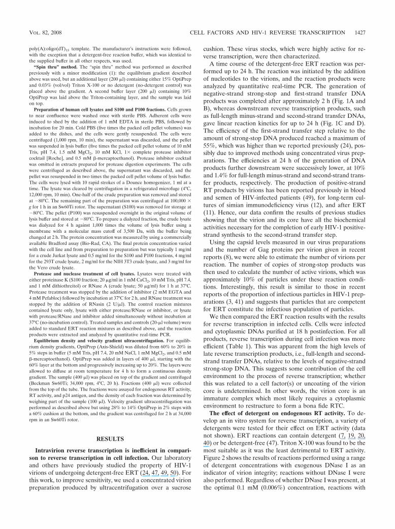
A time course of the detergent-free ERT reaction was per-formed up to 24 h. The reaction was initiated by the additionof nucleotides to the virions, and the reaction products wereanalyzed by quantitative real-time PCR. The generation ofnegative-strand strong-stop and first-strand transfer DNAproducts was completed after approximately 2 h (Fig. 1A andB), whereas downstream reverse transcription products, suchas full-length minus-strand and second-strand transfer DNAs,gave linear reaction kinetics for up to 24 h (Fig. 1C and D).The efficiency of the first-strand transfer step relative to theamount of strong-stop DNA produced reached a maximum of55%, which was higher than we reported previously (24), pos-sibly due to improved methods using concentrated virus prep-arations. The efficiencies at 24 h of the generation of DNAproducts further downstream were successively lower, at 10%and 1.4% for full-length minus-strand and second-strand trans-fer products, respectively. The production of positive-strandRT products by virions has been reported previously in bloodand semen of HIV-infected patients (49), for long-term cul-tures of simian immunodeficiency virus (12), and after ERT(11). Hence, our data confirm the results of previous studiesshowing that the virion and its core have all the biochemicalactivities necessary for the completion of early HIV-1 positive-strand synthesis to the second-strand transfer step.
Using the capsid levels measured in our virus preparationsand the number of Gag proteins per virion given in recentreports (8), we were able to estimate the number of virions perreaction. The number of copies of strong-stop products wasthen used to calculate the number of active virions, which wasapproximately 10% of particles under these reaction condi-tions. Interestingly, this result is similar to those in recentreports of the proportion of infectious particles in HIV-1 prep-arations (3, 41) and suggests that particles that are competentfor ERT constitute the infectious population of particles.
We then compared the ERT reaction results with the resultsfor reverse transcription in infected cells. Cells were infectedand cytoplasmic DNAs purified at 18 h postinfection. For allproducts, reverse transcription during cell infection was moreefficient (Table 1). This was apparent from the high levels oflate reverse transcription products, i.e., full-length and second-strand transfer DNAs, relative to the levels of negative-strandstrong-stop DNA. This suggests some contribution of the cellenvironment to the process of reverse transcription; whetherthis was related to a cell factor(s) or uncoating of the virioncore is undetermined. In other words, the virion core is animmature complex which most likely requires a cytoplasmicenvironment to restructure to form a bona fide RTC.
The effect of detergent on endogenous RT activity. To de-velop an in vitro system for reverse transcription, a variety ofdetergents were tested for their effect on ERT activity (datanot shown). ERT reactions can contain detergent (7, 19, 20,40) or be detergent-free (47). Triton X-100 was found to be themost suitable as it was the least detrimental to ERT activity.Figure 2 shows the results of reactions performed using a rangeof detergent concentrations with exogenous DNase I as anindicator of virion integrity; reactions without DNase I werealso performed. Regardless of whether DNase I was present, atthe optimal 0.1 mM (0.006%) concentration, reactions with
VOL. 82, 2008 CELL FACTORS AND HIV-1 REVERSE TRANSCRIPTION 1427
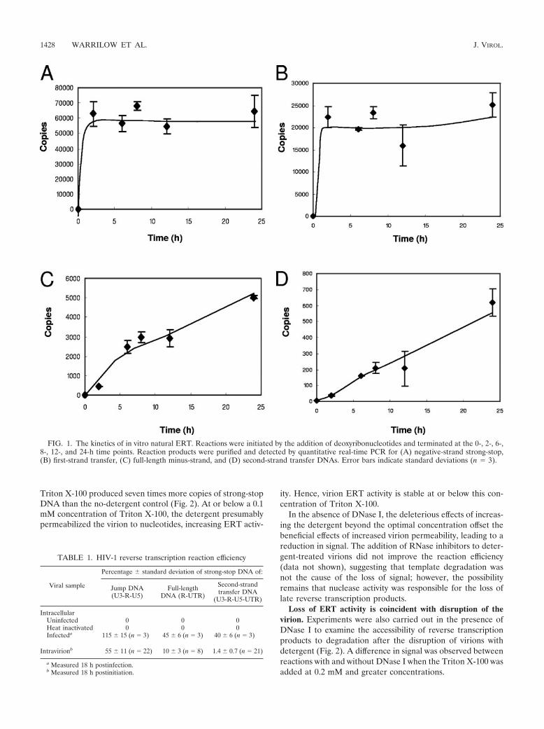
Triton X-100 produced seven times more copies of strong-stopDNA than the no-detergent control (Fig. 2). At or below a 0.1mM concentration of Triton X-100, the detergent presumablypermeabilized the virion to nucleotides, increasing ERT activ-
ity. Hence, virion ERT activity is stable at or below this con-centration of Triton X-100.
In the absence of DNase I, the deleterious effects of increas-ing the detergent beyond the optimal concentration offset thebeneficial effects of increased virion permeability, leading to areduction in signal. The addition of RNase inhibitors to deter-gent-treated virions did not improve the reaction efficiency(data not shown), suggesting that template degradation wasnot the cause of the loss of signal; however, the possibilityremains that nuclease activity was responsible for the loss oflate reverse transcription products.
Loss of ERT activity is coincident with disruption of thevirion. Experiments were also carried out in the presence ofDNase I to examine the accessibility of reverse transcriptionproducts to degradation after the disruption of virions withdetergent (Fig. 2). A difference in signal was observed betweenreactions with and without DNase I when the Triton X-100 wasadded at 0.2 mM and greater concentrations.
FIG. 1. The kinetics of in vitro natural ERT. Reactions were initiated by the addition of deoxyribonucleotides and terminated at the 0-, 2-, 6-,8-, 12-, and 24-h time points. Reaction products were purified and detected by quantitative real-time PCR for (A) negative-strand strong-stop,(B) first-strand transfer, (C) full-length minus-strand, and (D) second-strand transfer DNAs. Error bars indicate standard deviations (n � 3).
TABLE 1. HIV-1 reverse transcription reaction efficiency
Viral sample
Percentage standard deviation of strong-stop DNA of:
Jump DNA(U3-R-U5)
Full-lengthDNA (R-UTR)
Second-strandtransfer DNA
(U3-R-U5-UTR)
IntracellularUninfected 0 0 0Heat inactivated 0 0 0Infecteda 115 15 (n � 3) 45 6 (n � 3) 40 6 (n � 3)
Intravirionb 55 11 (n � 22) 10 3 (n � 8) 1.4 0.7 (n � 21)
a Measured 18 h postinfection.b Measured 18 h postinitiation.
1428 WARRILOW ET AL. J. VIROL.
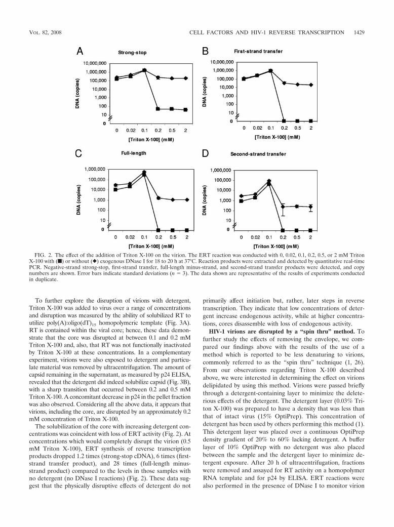
To further explore the disruption of virions with detergent,Triton X-100 was added to virus over a range of concentrationsand disruption was measured by the ability of solubilized RT toutilize poly(A):oligo(dT)15 homopolymeric template (Fig. 3A).RT is contained within the viral core; hence, these data demon-strate that the core was disrupted at between 0.1 and 0.2 mMTriton X-100 and, also, that RT was not functionally inactivatedby Triton X-100 at these concentrations. In a complementaryexperiment, virions were also exposed to detergent and particu-late material was removed by ultracentrifugation. The amount ofcapsid remaining in the supernatant, as measured by p24 ELISA,revealed that the detergent did indeed solubilize capsid (Fig. 3B),with a sharp transition that occurred between 0.2 and 0.5 mMTriton X-100. A concomitant decrease in p24 in the pellet fractionwas also observed. Considering all the above data, it appears thatvirions, including the core, are disrupted by an approximately 0.2mM concentration of Triton X-100.
The solubilization of the core with increasing detergent con-centrations was coincident with loss of ERT activity (Fig. 2). Atconcentrations which would completely disrupt the virion (0.5mM Triton X-100), ERT synthesis of reverse transcriptionproducts dropped 1.2 times (strong-stop cDNA), 6 times (first-strand transfer product), and 28 times (full-length minus-strand product) compared to the levels in those samples withno detergent (no DNase I reactions) (Fig. 2). These data sug-gest that the physically disruptive effects of detergent do not
primarily affect initiation but, rather, later steps in reversetranscription. They indicate that low concentrations of deter-gent increase endogenous activity, while at higher concentra-tions, cores disassemble with loss of endogenous activity.
HIV-1 virions are disrupted by a “spin thru” method. Tofurther study the effects of removing the envelope, we com-pared our findings above with the results of the use of amethod which is reported to be less denaturing to virions,commonly referred to as the “spin thru” technique (1, 26).From our observations regarding Triton X-100 describedabove, we were interested in determining the effect on virionsdelipidated by using this method. Virions were passed brieflythrough a detergent-containing layer to minimize the delete-rious effects of the detergent. The detergent layer (0.03% Tri-ton X-100) was prepared to have a density that was less thanthat of intact virus (15% OptiPrep). This concentration ofdetergent has been used by others performing this method (1).This detergent layer was placed over a continuous OptiPrepdensity gradient of 20% to 60% lacking detergent. A bufferlayer of 10% OptiPrep with no detergent was also placedbetween the sample and the detergent layer to minimize de-tergent exposure. After 20 h of ultracentrifugation, fractionswere removed and assayed for RT activity on a homopolymerRNA template and for p24 by ELISA. ERT reactions werealso performed in the presence of DNase I to monitor virion
FIG. 2. The effect of the addition of Triton X-100 on the virion. The ERT reaction was conducted with 0, 0.02, 0.1, 0.2, 0.5, or 2 mM TritonX-100 with (f) or without (}) exogenous DNase I for 18 to 20 h at 37°C. Reaction products were extracted and detected by quantitative real-timePCR. Negative-strand strong-stop, first-strand transfer, full-length minus-strand, and second-strand transfer products were detected, and copynumbers are shown. Error bars indicate standard deviations (n � 3). The data shown are representative of the results of experiments conductedin duplicate.
VOL. 82, 2008 CELL FACTORS AND HIV-1 REVERSE TRANSCRIPTION 1429
disruption by determining the nuclease susceptibilities of thereverse transcription products.
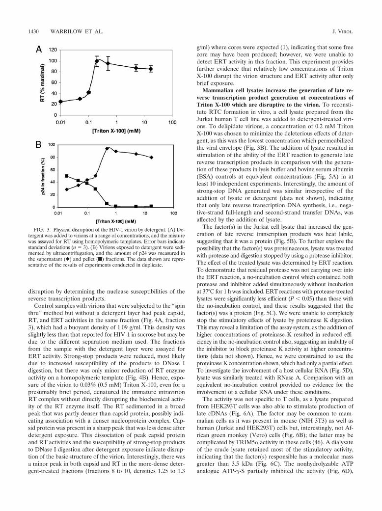
Control samples with virions that were subjected to the “spinthru” method but without a detergent layer had peak capsid,RT, and ERT activities in the same fraction (Fig. 4A, fraction3), which had a buoyant density of 1.09 g/ml. This density wasslightly less than that reported for HIV-1 in sucrose but may bedue to the different separation medium used. The fractionsfrom the sample with the detergent layer were assayed forERT activity. Strong-stop products were reduced, most likelydue to increased susceptibility of the products to DNase Idigestion, but there was only minor reduction of RT enzymeactivity on a homopolymeric template (Fig. 4B). Hence, expo-sure of the virion to 0.03% (0.5 mM) Triton X-100, even for apresumably brief period, denatured the immature intravirionRT complex without directly disrupting the biochemical activ-ity of the RT enzyme itself. The RT sedimented in a broadpeak that was partly denser than capsid protein, possibly indi-cating association with a denser nucleoprotein complex. Cap-sid protein was present in a sharp peak that was less dense afterdetergent exposure. This dissociation of peak capsid proteinand RT activities and the susceptibility of strong-stop productsto DNase I digestion after detergent exposure indicate disrup-tion of the basic structure of the virion. Interestingly, there wasa minor peak in both capsid and RT in the more-dense deter-gent-treated fractions (fractions 8 to 10, densities 1.25 to 1.3
g/ml) where cores were expected (1), indicating that some freecore may have been produced; however, we were unable todetect ERT activity in this fraction. This experiment providesfurther evidence that relatively low concentrations of TritonX-100 disrupt the virion structure and ERT activity after onlybrief exposure.
Mammalian cell lysates increase the generation of late re-verse transcription product generation at concentrations ofTriton X-100 which are disruptive to the virion. To reconsti-tute RTC formation in vitro, a cell lysate prepared from theJurkat human T cell line was added to detergent-treated viri-ons. To delipidate virions, a concentration of 0.2 mM TritonX-100 was chosen to minimize the deleterious effects of deter-gent, as this was the lowest concentration which permeabilizedthe viral envelope (Fig. 3B). The addition of lysate resulted instimulation of the ability of the ERT reaction to generate latereverse transcription products in comparison with the genera-tion of these products in lysis buffer and bovine serum albumin(BSA) controls at equivalent concentrations (Fig. 5A) in atleast 10 independent experiments. Interestingly, the amount ofstrong-stop DNA generated was similar irrespective of theaddition of lysate or detergent (data not shown), indicatingthat only late reverse transcription DNA synthesis, i.e., nega-tive-strand full-length and second-strand transfer DNAs, wasaffected by the addition of lysate.
The factor(s) in the Jurkat cell lysate that increased the gen-eration of late reverse transcription products was heat labile,suggesting that it was a protein (Fig. 5B). To further explore thepossibility that the factor(s) was proteinaceous, lysate was treatedwith protease and digestion stopped by using a protease inhibitor.The effect of the treated lysate was determined by ERT reaction.To demonstrate that residual protease was not carrying over intothe ERT reaction, a no-incubation control which contained bothprotease and inhibitor added simultaneously without incubationat 37°C for 1 h was included. ERT reactions with protease-treatedlysates were significantly less efficient (P 0.05) than those withthe no-incubation control, and these results suggested that thefactor(s) was a protein (Fig. 5C). We were unable to completelystop the stimulatory effects of lysate by proteinase K digestion.This may reveal a limitation of the assay system, as the addition ofhigher concentrations of proteinase K resulted in reduced effi-ciency in the no-incubation control also, suggesting an inability ofthe inhibitor to block proteinase K activity at higher concentra-tions (data not shown). Hence, we were constrained to use theproteinase K concentration shown, which had only a partial effect.To investigate the involvement of a host cellular RNA (Fig. 5D),lysate was similarly treated with RNase A. Comparison with anequivalent no-incubation control provided no evidence for theinvolvement of a cellular RNA under these conditions.
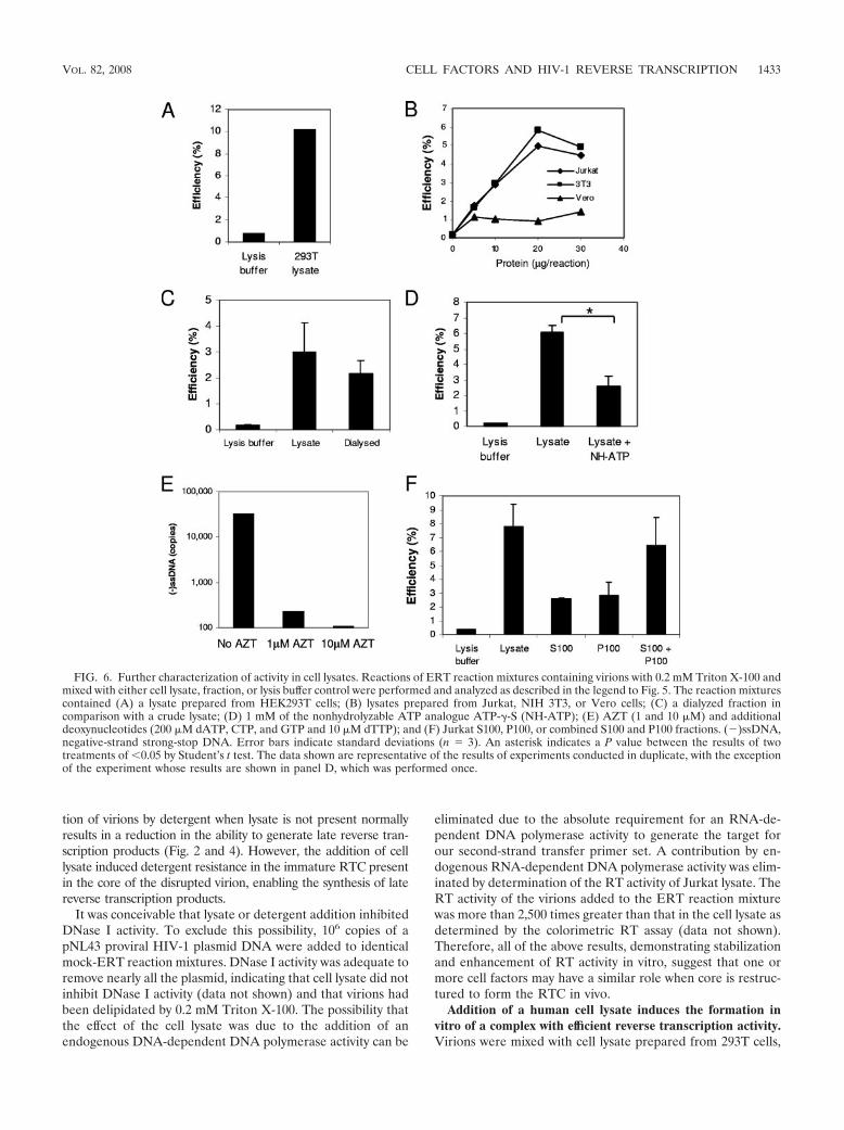
The activity was not specific to T cells, as a lysate preparedfrom HEK293T cells was also able to stimulate production oflate cDNAs (Fig. 6A). The factor may be common to mam-malian cells as it was present in mouse (NIH 3T3) as well ashuman (Jurkat and HEK293T) cells but, interestingly, not Af-rican green monkey (Vero) cells (Fig. 6B); the latter may becomplicated by TRIM5� activity in these cells (46). A dialysateof the crude lysate retained most of the stimulatory activity,indicating that the factor(s) responsible has a molecular massgreater than 3.5 kDa (Fig. 6C). The nonhydrolyzable ATPanalogue ATP-�-S partially inhibited the activity (Fig. 6D),
FIG. 3. Physical disruption of the HIV-1 virion by detergent. (A) De-tergent was added to virions at a range of concentrations, and the mixturewas assayed for RT using homopolymeric templates. Error bars indicatestandard deviations (n � 3). (B) Virions exposed to detergent were sedi-mented by ultracentrifugation, and the amount of p24 was measured inthe supernatant (}) and pellet (f) fractions. The data shown are repre-sentative of the results of experiments conducted in duplicate.
1430 WARRILOW ET AL. J. VIROL.

FIG. 4. Effect of “spin thru” method on HIV-1 ERT activity and structure. Fractions were prepared by the modified “spin thru” method, and their nascentERT activity and capsid (p24) and RT activities were measured. Data for preparations without (A) or with (B) a 0.03% (vol/vol) Triton X-100 layer are shown.(�)ssDNA, negative-strand strong-stop DNA. The data shown are representative of the results of two experiments.
1431
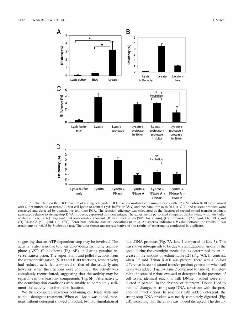
suggesting that an ATP-dependent step may be involved. Theactivity is also sensitive to 3�-azido-3�-deoxythymidine triphos-phate (AZT; Calbiochem) (Fig. 6E), indicating genuine re-verse transcription. The supernatant and pellet fractions fromthe ultracentrifugation (S100 and P100 fractions, respectively)had reduced activities compared to that of the crude lysate;however, when the fractions were combined, the activity wascompletely reconstituted, suggesting that the activity may beseparable into at least two components (Fig. 6F). Alternatively,the centrifugation conditions were unable to completely sedi-ment the activity into the pellet fraction.
We then compared reactions containing cell lysate with andwithout detergent treatment. When cell lysate was added, reac-tions without detergent showed a modest, twofold stimulation of
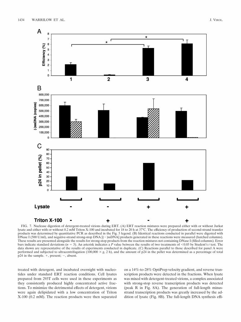
late cDNA products (Fig. 7A, lane 1 compared to lane 3). Thiswas shown subsequently to be due to stabilization of virions by thelysate during the overnight incubation, as determined by an in-crease in the amount of sedimentable p24 (Fig. 7C). In contrast,when 0.2 mM Triton X-100 was present, there was a 36-folddifference in second-strand transfer product generation when celllysate was added (Fig. 7A, lane 2 compared to lane 4). To deter-mine the state of virions exposed to detergent in the presence ofcell lysate, identical reactions with DNase I added were con-ducted in parallel. In the absence of detergent, DNase I led tominimal changes in strong-stop DNA, consistent with the pres-ence of intact virions. In reactions with added detergent, thestrong-stop DNA product was nearly completely digested (Fig.7B), indicating that the virion was indeed disrupted. The disrup-
FIG. 5. The effect on the ERT reaction of adding cell lysate. ERT reaction mixtures containing virions with 0.2 mM Triton X-100 were mixedwith either untreated or treated Jurkat cell lysate or control (lysis buffer or BSA) and incubated for 18 to 20 h at 37°C, and nascent products wereextracted and detected by quantitative real-time PCR. The reaction efficiency was calculated as the fraction of second-strand transfer productsgenerated relative to strong-stop DNA products, expressed as a percentage. The experiments performed compared Jurkat lysate with lysis buffercontrol and (A) BSA (100-�g/ml final concentration) control, (B) heat inactivation (90°C for 30 min), (C) proteinase K (20 �g/ml, 1 h, 37°C), and(D) RNase A (50 �g/ml, 1 h, 37°C). Error bars indicate standard deviations (n � 3). An asterisk indicates a P value between the results of twotreatments of 0.05 by Student’s t test. The data shown are representative of the results of experiments conducted in duplicate.
1432 WARRILOW ET AL. J. VIROL.
tion of virions by detergent when lysate is not present normallyresults in a reduction in the ability to generate late reverse tran-scription products (Fig. 2 and 4). However, the addition of celllysate induced detergent resistance in the immature RTC presentin the core of the disrupted virion, enabling the synthesis of latereverse transcription products.
It was conceivable that lysate or detergent addition inhibitedDNase I activity. To exclude this possibility, 106 copies of apNL43 proviral HIV-1 plasmid DNA were added to identicalmock-ERT reaction mixtures. DNase I activity was adequate toremove nearly all the plasmid, indicating that cell lysate did notinhibit DNase I activity (data not shown) and that virions hadbeen delipidated by 0.2 mM Triton X-100. The possibility thatthe effect of the cell lysate was due to the addition of anendogenous DNA-dependent DNA polymerase activity can be
eliminated due to the absolute requirement for an RNA-de-pendent DNA polymerase activity to generate the target forour second-strand transfer primer set. A contribution by en-dogenous RNA-dependent DNA polymerase activity was elim-inated by determination of the RT activity of Jurkat lysate. TheRT activity of the virions added to the ERT reaction mixturewas more than 2,500 times greater than that in the cell lysate asdetermined by the colorimetric RT assay (data not shown).Therefore, all of the above results, demonstrating stabilizationand enhancement of RT activity in vitro, suggest that one ormore cell factors may have a similar role when core is restruc-tured to form the RTC in vivo.
Addition of a human cell lysate induces the formation invitro of a complex with efficient reverse transcription activity.Virions were mixed with cell lysate prepared from 293T cells,
FIG. 6. Further characterization of activity in cell lysates. Reactions of ERT reaction mixtures containing virions with 0.2 mM Triton X-100 andmixed with either cell lysate, fraction, or lysis buffer control were performed and analyzed as described in the legend to Fig. 5. The reaction mixturescontained (A) a lysate prepared from HEK293T cells; (B) lysates prepared from Jurkat, NIH 3T3, or Vero cells; (C) a dialyzed fraction incomparison with a crude lysate; (D) 1 mM of the nonhydrolyzable ATP analogue ATP-�-S (NH-ATP); (E) AZT (1 and 10 �M) and additionaldeoxynucleotides (200 �M dATP, CTP, and GTP and 10 �M dTTP); and (F) Jurkat S100, P100, or combined S100 and P100 fractions. (�)ssDNA,negative-strand strong-stop DNA. Error bars indicate standard deviations (n � 3). An asterisk indicates a P value between the results of twotreatments of 0.05 by Student’s t test. The data shown are representative of the results of experiments conducted in duplicate, with the exceptionof the experiment whose results are shown in panel D, which was performed once.
VOL. 82, 2008 CELL FACTORS AND HIV-1 REVERSE TRANSCRIPTION 1433
treated with detergent, and incubated overnight with nucleo-tides under standard ERT reaction conditions. Cell lysatesprepared from 293T cells were used in these experiments asthey consistently produced highly concentrated active frac-tions. To minimize the detrimental effects of detergent, virionswere again delipidated with a low concentration of TritonX-100 (0.2 mM). The reaction products were then separated
on a 14%-to-28% OptiPrep velocity gradient, and reverse tran-scription products were detected in the fractions. When lysatewas mixed with detergent-treated virions, a complex associatedwith strong-stop reverse transcription products was detected(peak R in Fig. 8A). The generation of full-length minus-strand transcription products was greatly increased by the ad-dition of lysate (Fig. 8B). The full-length DNA synthesis effi-
FIG. 7. Nuclease digestion of detergent-treated virions during ERT. (A) ERT reaction mixtures were prepared either with or without Jurkatlysate and either with or without 0.2 mM Triton X-100 and incubated for 18 to 20 h at 37°C. The efficiency of production of second-strand transferproducts was determined by quantitative PCR as described in the Fig. 5 legend. (B) Identical reactions conducted in parallel were digested withDNase I (500 U/ml), and negative-strand strong-stop DNA [(�)ssDNA] products generated in these reactions were measured (hatched columns).These results are presented alongside the results for strong-stop products from the reaction mixtures not containing DNase I (filled columns). Errorbars indicate standard deviations (n � 3). An asterisk indicates a P value between the results of two treatments of 0.05 by Student’s t test. Thedata shown are representative of the results of experiments conducted in duplicate. (C) Reactions parallel to those described for panel A wereperformed and subjected to ultracentrifugation (100,000 � g, 2 h), and the amount of p24 in the pellet was determined as a percentage of totalp24 in the sample. �, present; �, absent.
1434 WARRILOW ET AL. J. VIROL.

ciency relative to the level of strong-stop DNA product infraction 3 was 71%, which compares with 45% efficiency ob-served for full-length products during cell infection after 18 h(Table 1). This was in contrast to the results for detergent-treated virions not incubated with lysate, where the strong-stopDNA synthesis remained in the top fraction (fraction 1) andhad no full-length DNA. In addition, a complex with the samesedimentation properties was capable of nascent reverse tran-scription product generation after fractionation (Fig. 8C), in-dicating that the complex is capable of synthesizing nascentfull-length DNA (the relatively low signal in Fig. 8C is due tothe small amounts of material used [5 �l of a 400-�l fraction]).These data demonstrate that a highly efficient RTC-like com-plex can be reconstituted in vitro. This complex was associatedwith only 0.5% of the total capsid in the virus preparation thatwas used (Fig. 8D), which is consistent with in vivo observa-tions of low amounts of capsid in the RTC.
DISCUSSION
These data confirm and extend previous work on intravirionreverse transcription in which we noted reduced strand trans-fer efficiency in ERT compared with the efficiency in cell in-
fection (24). Improvements to methodology meant that wewere able to measure later events in the HIV reverse transcrip-tion pathway (Fig. 1). This technical advance confirmed thatthe enzymatic activities sufficient for reverse transcription arepresent in the virion-derived cores. However, these experi-ments revealed that intravirion reverse transcription was inef-ficient in comparison with reverse transcription in cell infec-tion, as exemplified by the poor efficiency of the production ofintravirion late reverse transcription products (Table 1). Thisobservation suggests a requirement for a contribution from thehost cell environment for optimal HIV-1 reverse transcription,as was previously shown for avian sarcoma and leukosis virus(ASLV) (29). These data (Fig. 2 and 3) also suggested thatcore structure may need to be maintained for efficient reversetranscription; this is consistent with observations of an optimalstability for core which predicts that core remains intact forsome time postentry for efficient reverse transcription (17).
The addition of Triton X-100 at concentrations (0.5 mM)which completely disrupted virions reduced the generation oflate cDNA products to a greater extent than early products.Whether this was due to the loss of a virion component(s), suchas RT, or the exposure of core template RNA and productDNA to nucleases is unknown at present. One possibility sug-
FIG. 8. Reconstitution of the RTC in vitro. (A, B) Modified ERT reaction mixtures containing detergent-treated (0.2 mM Triton X-100) virionseither with (}) or without (Œ) HEK293T lysate or untreated virions with lysate (f) were incubated overnight with nucleotides. A no-nucleotidecontrol (●) was also included. Samples were then subjected to velocity gradient ultracentrifugation (14%-to-28% OptiPrep with a 60% cushion at100,000 � g for 2 h). Fractions (0.4 ml) were removed from the top of the gradient, and nucleic acids were extracted and detected using primersspecific for (A) strong-stop or (B) full-length reverse transcription products by real-time PCR. Virion (V) and in vitro-reconstituted (R) RTC dataare indicated. (C) As described above, but complexes were formed for 2 h and then fractionated as described above. Fractions (5 �l of each) wereassayed for nascent ERT activity, and full-length products were detected by real-time PCR. Data for samples containing detergent-treated virionswith (}) or without (Œ) lysate are shown. (D) p24 levels in velocity gradient fractions of the sample containing detergent-treated virions andHEK293T cell lysate. The data shown are representative of the results of experiments conducted in duplicate.
VOL. 82, 2008 CELL FACTORS AND HIV-1 REVERSE TRANSCRIPTION 1435
gested by these data is that capsid may remain associated withthe RTC for some part of the reverse transcription pathway.The core may provide a scaffold for the RTC and increase theeffective concentration of components important for the stepsof reverse transcription, facilitating strand transfers and theefficiency of the overall reaction. As reverse transcription canbe induced to occur in the virion, the core itself does notsterically constrain polymerase elongation. We recently re-ported the isolation without detergent of HIV-1 cores thatwere capable of reverse transcription in the absence of anuncoating activity (45). However, the elongation of viral DNAby RT could result in shedding of capsid, as suggested by theeffect of detergent-free ERT on virion morphology (48).
Consistent with the observations of the effects of detergent,the exposure of virions to Triton X-100 (0.5 mM, or 0.03%)through the modified “spin thru” method also resulted in theirdisruption. The reduction of early (strong-stop DNA) productswas most likely due to digestion by exogenous DNase I and notto direct inhibition of the enzymatic activity of the RT enzyme,which remained active in colorimetric assays using homopoly-meric templates. In addition, this concentration of detergentwas much lower than that used in lysis buffers used in com-mercial RT assays (0.5% Triton X-100). The efficiency of iso-lation of cores from HIV-1 particles has reportedly been im-proved by “spin thru” methods, and RT activity on exogenoustemplates has been detected in such fractions (39). For exam-ple, cores prepared by the “spin thru” method were recentlyused to demonstrate an uncoating activity in cell lysates (5). Aswe did not show core isolation, we cannot comment on the useof the method for this purpose. However, it was apparent thatvirions were sufficiently disrupted by brief exposure to 0.03%Triton X-100 as to make ERT products susceptible to exoge-nous nuclease digestion.
Cell lysates prepared from Jurkat, HEK293T, and NIH 3T3cells greatly increased ERT efficiency, i.e., the fraction of sec-ond-strand transfer products relative to strong-stop DNA, ex-pressed as a percentage (Fig. 6A and B). The factor(s) respon-sible is most likely proteinaceous in nature and was partiallyinhibited by nonhydrolyzable ATP. Whether a protein kinaseor other ATP binding protein is required is unknown. Thisactivity was segregated into soluble and insoluble components,each with partial activity, that when combined reconstitutedthe full activity (Fig. 6F). The level of improved reverse tran-scription efficiency was consistently as high as or higher thanthe reverse transcription efficiency observed in cell infectionwith respect to full-length HIV-1 DNA synthesis. For example,reverse transcription in cells was about 45% for full-lengthDNA synthesis (Table 1), in comparison with the peak fractionof the reconstituted material separated on the velocity gradi-ent, which had an efficiency of 71% for this product. In addi-tion, ERT reactions with added lysate were consistently moreefficient compared to the reactions in controls. Importantly, weruled out the possibility that a low-level cellular RNA-depen-dent DNA polymerase activity was capable of the observedrobust DNA synthesis. Overall, these data indicate that cellfactors were required for efficient reverse transcription.
We are the first to report an in vitro reconstitution assay forHIV-1 incorporating cellular factors. We hypothesize that oneor more cellular activities are required to form the reversetranscription elongation complex. The activity described here
differed from that reported to stimulate ASLV reverse tran-scription (29). First, a cytoplasmic factor was reported to stim-ulate ASLV RT initiation, which was not seen in this study.Second, ASLV RT elongation was stimulated by a nuclearextract, whereas HIV-1 RT elongation was stimulated by acytoplasmic factor(s). The cell factor(s) may do this in one oftwo ways. The recent identification of a host cell uncoatingactivity in a lysate prepared from activated T cells has providedweight to the suggestion that shedding of capsid is a prerequi-site for reverse transcription (5). The first possibility, then, isthat the cell lysate may have an activity that uncoats the coreand/or regulates the formation of the RTC. Our observation oflow amounts of capsid associated with the reconstituted com-plex is consistent with this model. However, as loss of corestructure leads to loss of the ability to generate late reversetranscription products (Fig. 2 and 3), another hypothesis is thatthe cell stimulatory factor stabilizes the whole or part of thecore. Alternatively, the cell factor may provide some essentialfunction for the RTC by becoming part of the complex.Gemin2 may become part of the RTC and stimulates bothearly and late reverse transcription products (22). The activitywe have identified differs from that of Gemin2 in that it spe-cifically stimulates late reverse transcription. Understandingthe precise mechanism of this novel activity will require furtherexperimentation.
It has been suggested that uncoating and reverse transcrip-tion proceed by the ordered disassembly of the core (10, 36),and cell factors may assist this process. Our observation that amammalian cell factor(s) enables the reconstitution of an ef-ficient RTC from delipidated virions is consistent with thismodel of uncoating. Our in vitro reconstitution system willenable us to address the many unanswered questions regardinguncoating and early RTC formation. We are in the process ofidentifying what factor(s) is responsible for this effect. Theidentification of such factor(s) would reveal a novel therapeu-tic target against HIV-1.
ACKNOWLEDGMENTS
We thank Andreas Suhrbier for useful suggestions about the manu-script.
This work was supported by the Australian National Health andMedical Research Council, project grant 298926.
REFERENCES
1. Accola, M. A., Å. Ohagen, and H. G. Gottlinger. 2000. Isolation of humanimmunodeficiency virus type 1 cores: retention of Vpr in the absence ofp6gag. J. Virol. 74:6198–6202.
2. Adachi, A., H. E. Gendelman, S. Koenig, T. Folks, R. Willey, A. Rabson, andM. A. Martin. 1986. Production of acquired immunodeficiency syndrome-associated retrovirus in human and nonhuman cells transfected with aninfectious molecular clone. J. Virol. 59:284–291.
3. Andreadis, S., T. Lavery, H. E. Davis, J. M. Le Doux, M. L. Yarmush, andJ. R. Morgan. 2000. Toward a more accurate quantitation of the activity ofrecombinant retroviruses: alternatives to titer and multiplicity of infection.J. Virol. 74:3431–3439.
4. Ariumi, Y., F. Serhan, P. Turelli, A. Telenti, and D. Trono. 2006. Theintegrase interactor 1 (INI1) proteins facilitate Tat-mediated human immu-nodeficiency virus type 1 transcription. Retrovirology 3:47.
5. Auewarakul, P., P. Wacharapornin, S. Srichatrapimuk, S. Chutipongtanate,and P. Puthavathana. 2005. Uncoating of HIV-1 requires cellular activation.Virology 337:93–101.
6. Bishop, D. H., R. Ruprecht, R. W. Simpson, and S. Spiegelman. 1971.Deoxyribonucleic acid polymerase of Rous sarcoma virus: reaction condi-tions and analysis of the reaction product nucleic acids. J. Virol. 8:730–741.
7. Boone, L. R., and A. M. Skalka. 1981. Viral DNA synthesized in vitro byavian retrovirus particles permeabilized with melittin. I. Kinetics of synthesisand size of minus- and plus-strand transcripts. J. Virol. 37:109–116.
1436 WARRILOW ET AL. J. VIROL.
8. Briggs, J. A., M. N. Simon, I. Gross, H. G. Krausslich, S. D. Fuller, V. M.Vogt, and M. C. Johnson. 2004. The stoichiometry of Gag protein in HIV-1.Nat. Struct. Mol. Biol. 11:672–675.
9. Cullen, B. R. 2006. Role and mechanism of action of the APOBEC3 familyof antiretroviral resistance factors. J. Virol. 80:1067–1076.
10. Dismuke, D. J., and C. Aiken. 2006. Evidence for a functional link betweenuncoating of the human immunodeficiency virus type 1 core and nuclearimport of the viral preintegration complex. J. Virol. 80:3712–3720.
11. Dornadula, G., S. Yang, R. J. Pomerantz, and H. Zhang. 2000. Partial rescueof the Vif-negative phenotype of mutant human immunodeficiency virus type1 strains from nonpermissive cells by intravirion reverse transcription. J. Vi-rol. 74:2594–2602.
12. Dornadula, G., H. Zhang, O. Bagasra, and R. J. Pomerantz. 1997. Naturalendogenous reverse transcription of simian immunodeficiency virus. Virol-ogy 227:260–267.
13. Dueck, M., and J. Guatelli. 2007. Evidence against a direct antiviral activityof the proteasome during the early steps of HIV-1 replication. Virology361:1–8.
14. Fassati, A., and S. P. Goff. 2001. Characterization of intracellular reversetranscription complexes of human immunodeficiency virus type 1. J. Virol.75:3626–3635.
15. Fitzon, T., B. Leschonsky, K. Bieler, C. Paulus, J. Schroder, H. Wolf, and R.Wagner. 2000. Proline residues in the HIV-1 NH2-terminal capsid domain:structure determinants for proper core assembly and subsequent steps ofearly replication. Virology 268:294–307.
16. Forshey, B. M., and C. Aiken. 2003. Disassembly of human immunodefi-ciency virus type 1 cores in vitro reveals association of Nef with the subviralribonucleoprotein complex. J. Virol. 77:4409–4414.
17. Forshey, B. M., U. von Schwedler, W. I. Sundquist, and C. Aiken. 2002.Formation of a human immunodeficiency virus type 1 core of optimal sta-bility is crucial for viral replication. J. Virol. 76:5667–5677.
18. Gao, G., and S. P. Goff. 1999. Somatic cell mutants resistant to retrovirusreplication: intracellular blocks during the early stages of infection. Mol.Biol. Cell 10:1705–1717.
19. Garapin, A. C., J. P. McDonnell, W. Levinson, N. Quintrell, L. Fanshier, andJ. M. Bishop. 1970. Deoxyribonucleic acid polymerase associated with Roussarcoma virus and avian myeloblastosis virus: properties of the enzyme andits product. J. Virol. 6:589–598.
20. Gilboa, E., S. Goff, A. Shields, F. Yoshimura, S. Mitra, and D. Baltimore.1979. In vitro synthesis of a 9 kbp terminally redundant DNA carrying theinfectivity of Moloney murine leukemia virus. Cell 16:863–874.
21. Gurer, C., A. Hoglund, S. Hoglund, and J. Luban. 2005. ATP�S disruptshuman immunodeficiency virus type 1 virion core integrity. J. Virol. 79:5557–5567.
22. Hamamoto, S., H. Nishitsuji, T. Amagasa, M. Kannagi, and T. Masuda.2006. Identification of a novel human immunodeficiency virus type 1 integraseinteractor, Gemin2, that facilitates efficient viral cDNA synthesis in vivo. J. Vi-rol. 80:5670–5677.
23. Hirt, B. 1967. Selective extraction of polyoma DNA from infected mouse cellcultures. J. Mol. Biol. 26:365–369.
24. Hooker, C. W., and D. Harrich. 2003. The first strand transfer reaction ofHIV-1 reverse transcription is more efficient in infected cells than in cell-freenatural endogenous reverse transcription reactions. J. Clin. Virol. 26:229–238.
25. Keckesova, Z., L. M. J. Ylinen, and G. J. Towers. 2006. Cyclophilin A rendershuman immunodeficiency virus type 1 sensitive to Old World monkey butnot human TRIM5� antiviral activity. J. Virol. 80:4683–4690.
26. Kotov, A., J. Zhou, P. Flicker, and C. Aiken. 1999. Association of Nef withthe human immunodeficiency virus type 1 core. J. Virol. 73:8824–8830.
27. McDonald, D., M. A. Vodicka, G. Lucero, T. M. Svitkina, G. G. Borisy, M.Emerman, and T. J. Hope. 2002. Visualization of the intracellular behaviorof HIV in living cells. J. Cell Biol. 159:441–452.
28. Miller, M. D., C. M. Farnet, and F. D. Bushman. 1997. Human immunode-ficiency virus type 1 preintegration complexes: studies of organization andcomposition. J. Virol. 71:5382–5390.
29. Narayan, S., and J. A. Young. 2004. Reconstitution of retroviral fusion anduncoating in a cell-free system. Proc. Natl. Acad. Sci. USA 101:7721–7726.
30. Nermut, M. V., and A. Fassati. 2003. Structural analyses of purified humanimmunodeficiency virus type 1 intracellular reverse transcription complexes.J. Virol. 77:8196–8206.
31. Nisole, S., and A. Saib. 2004. Early steps of retrovirus replicative cycle.Retrovirology 1:9.
32. Novak, U., R. Friedrich, and K. Moelling. 1979. Elongation of DNA com-plementary to the 5� end of the avian sarcoma virus genome by the virion-associated RNA-dependent DNA polymerase. J. Virol. 30:438–452.
33. Sokolskaja, E., L. Berthoux, and J. Luban. 2006. Cyclophilin A and TRIM5�independently regulate human immunodeficiency virus type 1 infectivity inhuman cells. J. Virol. 80:2855–2862.
34. Sokolskaja, E., and J. Luban. 2006. Cyclophilin, TRIM5, and innate immu-nity to HIV-1. Curr. Opin. Microbiol. 9:404–408.
35. Srichatrapimuk, S., and P. Auewarakul. 2007. Resistance of monocyte toHIV-1 infection is not due to uncoating defect. Virus Res. 126:277–281.
36. Stremlau, M., M. Perron, M. Lee, Y. Li, B. Song, H. Javanbakht, F. Diaz-Griffero, D. J. Anderson, W. I. Sundquist, and J. Sodroski. 2006. Specificrecognition and accelerated uncoating of retroviral capsids by the TRIM5�restriction factor. Proc. Natl. Acad. Sci. USA 103:5514–5519.
37. Stremlau, M., B. Song, H. Javanbakht, M. Perron, and J. Sodroski. 2006.Cyclophilin A: an auxiliary but not necessary cofactor for TRIM5alpharestriction of HIV-1. Virology 351:112–120.
38. Tang, S., T. Murakami, B. E. Agresta, S. Campbell, E. O. Freed, and J. G.Levin. 2001. Human immunodeficiency virus type 1 N-terminal capsid mu-tants that exhibit aberrant core morphology and are blocked in initiation ofreverse transcription in infected cells. J. Virol. 75:9357–9366.
39. Tang, S., T. Murakami, N. Cheng, A. C. Steven, E. O. Freed, and J. G. Levin.2003. Human immunodeficiency virus type 1 N-terminal capsid mutantscontaining cores with abnormally high levels of capsid protein and virtuallyno reverse transcriptase. J. Virol. 77:12592–12602.
40. Temin, H. M., and S. Mizutani. 1970. RNA-dependent DNA polymerase invirions of Rous sarcoma virus. Nature 226:1211–1213.
41. Thomas, J. A., D. E. Ott, and R. J. Gorelick. 2007. Efficiency of humanimmunodeficiency virus type 1 postentry infection processes: evidenceagainst disproportionate numbers of defective virions. J. Virol. 81:4367–4370.
42. Turelli, P., V. Doucas, E. Craig, B. Mangeat, N. Klages, R. Evans, G.Kalpana, and D. Trono. 2001. Cytoplasmic recruitment of INI1 and PML onincoming HIV preintegration complexes: interference with early steps ofviral replication. Mol. Cell 7:1245–1254.
43. Van Maele, B., K. Busschots, L. Vandekerckhove, F. Christ, and Z. Debyser.2006. Cellular co-factors of HIV-1 integration. Trends Biochem. Sci. 31:98–105.
44. Warrilow, D., and D. Harrich. 2007. HIV-1 replication from after cell entryto the nuclear periphery. Curr. HIV Res. 5:293–299.
45. Warrilow, D., D. Stenzel, and D. Harrich. 2007. Isolated HIV-1 core is activefor reverse transcription. Retrovirology 4:77.
46. Yap, M. W., S. Nisole, C. Lynch, and J. P. Stoye. 2004. Trim5� proteinrestricts both HIV-1 and murine leukemia virus. Proc. Natl. Acad. Sci. USA101:10786–10791.
47. Zhang, H., G. Dornadula, P. Alur, M. A. Laughlin, and R. J. Pomerantz.1996. Amphipathic domains in the C terminus of the transmembrane protein(gp41) permeabilize HIV-1 virions: a molecular mechanism underlying nat-ural endogenous reverse transcription. Proc. Natl. Acad. Sci. USA 93:12519–12524.
48. Zhang, H., G. Dornadula, J. Orenstein, and R. J. Pomerantz. 2000. Mor-phologic changes in human immunodeficiency virus type 1 virions secondaryto intravirion reverse transcription: evidence indicating that reverse tran-scription may not take place within the intact viral core. J. Hum. Virol.3:165–172.
49. Zhang, H., G. Dornadula, and R. J. Pomerantz. 1996. Endogenous reversetranscription of human immunodeficiency virus type 1 in physiological mi-croenviroments: an important stage for viral infection of nondividing cells.J. Virol. 70:2809–2824.
50. Zhang, H., G. Dornadula, and R. J. Pomerantz. 1998. Natural endogenousreverse transcription of HIV-1. J. Reprod. Immunol. 41:255–260.
VOL. 82, 2008 CELL FACTORS AND HIV-1 REVERSE TRANSCRIPTION 1437