Using Ozone To Stimulate Oxygen Utilization “It's all about NAD”
35
Using Ozone To Stimulate Oxygen Utilization “It’s all about NAD” Frank Shallenberger, MD, HMD, ABAAM The Nevada Center of Anti-Aging Medicine Carson City, Nevada Ph. 775-884-3990 Fax 775-884-2202 [email protected]
-
Upload
khangminh22 -
Category
Documents
-
view
2 -
download
0
Transcript of Using Ozone To Stimulate Oxygen Utilization “It's all about NAD”

Using Ozone To Stimulate Oxygen Utilization“It’s all about NAD”
Frank Shallenberger, MD, HMD, ABAAMThe Nevada Center of Anti-Aging Medicine
Carson City, NevadaPh. 775-884-3990 Fax 775-884-2202

Outline1.
Aging and the diseases of aging are caused
primarily by decreased oxygen utilization.
2.
This decrease leads to the excessive free radical production that results in degenerative disease.
3.
Decreased ozygen utilization is caused by both pre‐mitochondrial and mitochondrial factors.
4.
Decreased oxygen utilization exerts its effects by causeing a decrease in the NAD/NADH ratio.
5.
Ozone therapy is effective for so many diseases including the infirmaties of aging because it
normalizes this ratio.

Aging and the diseases of aging are caused primarily by decreased
oxygen utilization

It Works – But How?
• Coronary artery and cardiovascular
disease.• Claudication.• Gangrene.• Pain
• Macular degeneration.
• Aging.• Oncology.• Chronic viral
infection.

Oxygen – The Forgotten Nutrient
• The most critical nutrient.
• It’s not what you take in, it’s what you utilize.• The difference between you at 20 and you at 70.• The key to the treatment and prevention of
disease is optimum oxygen utilization.
• Local and systemic oxygen utilization.
• The good news – oxygen utilization can be measured and improved.

Oxygen Utilization (Aerobic Capacity)
• The process whereby oxygen metabolizes either fat or glucose into water, heat, NAD (nicotinamide adenine dinucleotide), and ATP.
• Oxygen works through NAD and ATP (and to a lesser degree NADP and FAD). These “oxygen intermediates”
are the bottom line for all
cellular function.

EOxygen
LungsHeart
Circulation
Fat Glucose
T3, Cortisol, DHEA, Nutrients, Toxins
Oxygen Utilization
FreeRadicals

Aging and Oxygen UtilizationNothing is as consistent and as predictable as the gradual, linear decline in oxygen utilization
seen in all aging populations.

“Meta-analysis of the age associated decline in maximal aerobic capacity in men: relation to training status.”
• Maximal aerobic capacity means maximal oxygen utilization.
• “Maximal aerobic capacity [not VO2max] is an independent risk factor for cardiovascular disease, cognitive dysfunction, and all cause mortality.”
• Even highly trained marathon runners showed a decrease in oxygen utilization. This means that oxygen utilization is the determining factor in aging not VO2max
• VO2 max is neither global nor sensitive enough.
Wilson & Tanaka, Am. J. Physiol. Heart Circ. Physiol. Vol. 278: 829-834, 2000

Premature ageing in mice expressing defective mitochondrial DNA polymerase.
• Mice are genetically manipulated to develop mtDNA mutations at a rapid rate. This results in an accelerated
reduction in oxygen utilization over their lifespan.
• Significantly reduced lifespan.• Premature onset of age‐related phenotypes such as lean
body mass loss, alopecia, kyphosis, anemia, osteoporosis, reduced fertility, and cardiomegaly.
• These results provide a causative link between decreased oxygen utilization and aging.
Trifunovic A, Wredenberg A, et al.Nature.
2004 May 27;429(6990):417‐23.

Uncoupled and surviving: individual mice with high metabolism have greater mitochondrial uncoupling and live longer.
• Examined associations between longevity and individual variations in resting oxygen utilization in a
cohort of mice. • A positive association between oxygen utilization and
lifespan was noted.• Mice in the upper quartile of oxygen utilization lived
36% longer
than mice in the lowest quartile.
Speakman JR, Talbot DA, et al.Aging Cell. 2004 Jun;3(3):87‐95.

Oxygen Utilization and Disease
• Varanasi SS, Francis RM, et al. Mitochondrial DNA deletion associated oxidative stress and severe male osteoporosis. Osteoporos Int.
1999;10(2):143‐9.• Liang FQ, Godley BF. Oxidative stress‐induced
mitochondrial DNA damage in human retinal pigment epithelial cells: a possible mechanism
for RPE aging and age‐related macular degeneration. Exp Eye Res. 2003
Apr;76(4):397‐403.

Oxygen Utilization and Disease
• Patwari P, Lee RT. Thioredoxins, mitochondria, and hypertension. Am J Pathol. 2007
Mar;170(3):805‐8.
• Eerola E, Pulkki K, et al. Abnormal mitochondria in cultured synovial fibroblasts
in rheumatoid and reactive arthritis? Br J Rheumatol. 1988;27 Suppl 2:128‐31.

Oxygen Utilization and Disease
• Modica‐Napolitano JS, Kulawiec M, et al. Mitochondria and human cancer. Curr Mol
Med. 2007 Feb;7(1):121‐31.
• Gerbitz KD, Gempel K, Brdiczka D. Mitochondria and diabetes. Genetic,
biochemical, and clinical implications of the cellular energy circuit. Diabetes. 1996
Feb;45(2):113‐26.

Oxygen Utilization and Disease
• Biskup S, Moore DJ. Detrimental deletions: mitochondria, aging and Parkinson's
disease.Bioessays. 2006 Oct;28(10):963‐7.
• Moreira PI, Cardoso SM, et al. The key role of mitochondria in Alzheimer's
disease. J
Alzheimers Dis. 2006 Jul;9(2):101‐10.

Oxygen Utilization and Disease
• Tsutsui H. Oxidative stress in heart failure: the role of mitochondria.
Intern Med. 2001 Dec;40(12):1177‐82.
• Marin‐Garcia J, Goldenthal MJ. Heart mitochondria
signaling pathways: appraisal of
an emerging field. J Mol Med. 2004 Sep;82(9):565‐78

VO2Max
Glucose
Glucose
Glucose
Anaerobic Anaerobic
Anaerobic
Healthy Mito-Decay&
Disease
The Stages Of The Aging Process
Fat
FatFatFat
Glucose
Anaerobic
MaxOU
AsymptomaticFunctional symptoms“Healthy for your age”
MaxATPFat

Decreased oxygen utilization causes degeneration changes secondary to excessive free
radical activity.

Free Radical Damage is Caused By Decreased Oxygen Utilization
• Decreased oxygen utilization creates a “functional hypoxia”
which 1) accelerates free
radical formation, and 2) exhausts anti‐oxidant buffering capacity.
“Decreased oxygen utilization is toxic to the cell
by exacerbating free radical generation in membranes
housing electron transfer assemblies.”Antioxidant Adaptation
Levine & Kidd

Decreased Oxygen
UtilizationDecrease
d Free Radical
Buffering
Increased Free
Radical Formatio
n MitochondrialDecay
Aging DegenerativeDisease

Decreased oxygen utilization is caused by both pre‐mitochondrial
and mitochondrial factors.

Pre‐Mitochondrial1. Decreased lypolysis.2. Hypoglycemia3. Ischemia.3. Hypoxia.4. Decreased methylation.
5. Inflammation
Mitochondrial1.
Toxicity and infections.
2. Stress.3. Nutritional
deficiencies.4. Hormonal
deficiencies.5. Decreased fitness.
What Causes Decreased Oxygen Utilization?

Decreased oxygen utilization exerts its negative effects by causing a
decrease in the NAD/NADH ratio.

Cardiolipin(Lipoic/carnitine)
Glucose LiverFatStores
Fattyacids
Pyruvate + LactateLactate
CO2CO2
NAD
Carnitine
Lipoic, B1NAD
KrebsCycle
B2, B3, B6, B12,Folic, TMG, Mg,
Catabolic & Anabolic hormones
CO2CO2
3O2+NADH +2H2O +
ATP
ATPATP
Insulin+ +
LungsCirculationCoagulation
2,3 DPGantacids
Hypervent-ilation
TFA’s, CoQ10, B12, folic,Heavy metals, “uncouplers”
A-CoA
Aminos
Triglyc.
--
Cortisol+++
Ketones
T3Epin
NADNADH
ADPPOOL
NAD

Nicotinamide adenine dinucleotide, a metabolic regulator of transcription, longevity and disease
• “NAD has emerged as a putative metabolic regulator of transcription, longevity and several
age‐associated diseases, including diabetes, cancer and neurodegenerative diseases.”
• “Calorie restriction (CR) has been shown to decrease the incidence or delay the onset of
some of these diseases.”• “Studies in yeast suggest that CR functions by
increasing the NAD level and/or the NAD/NADH ratio.”
Lin SJ, Guarente L. Current Opinion in Cell Biology 2003, 15:241–246

NAD and Cell Signaling
• NAD is rate limiting for ADP‐ribosylation. ADP‐ ribosylation reactions are involved in cell
signaling and the control of many cell processes in the cell nucleus, including DNA
repair, apoptosis, and telomere maintenance.
• Another function of NAD in cell signaling is as a precursor of cyclic ADP‐ribose, which
regulates intracellular calcium channels.

NAD and Sirtuins• Sir stands for Silent Information Regulator genes. Sir2 is short
for
Silent mating type Information Regulatior‐2. So sirtuins are Sir2‐ homologs. Sirtuins act by removing acetyl groups from proteins in the presence of NAD.
• Sirtuins are hypothesized to play a key role in an organism's response to stresses (such as heat or starvation) and to be
responsible for the lifespan‐extending effects of calorie restriction.
• The sirtuins regulate nuclear transcription through deacetylating histones and altering nucleosome structure. These activities of
sirtuins are particularly interesting because of their importance in the regulation of aging.
• Sirtuins are NAD‐dependent, and are thus classified as "NAD ‐ dependent deacetylases.

Ozone therapy is effective for so many diseases including the
infirmaties of aging because it normalizes the NAD/NADH ratio.


Ozone Forms Peroxides• Free radicals only in a
pH greater than 8. • Reacts ionically with
double bonds to produce peroxides
called ozonides. • Most ozonides are
formed from the short chained lipids in cell
membranes.
• Ozonides are stable for days to weeks, easily
penetrate cell membranes, and are
selectively reactive.• Once in the cells, these
ozonides oxidize NADH to NAD.

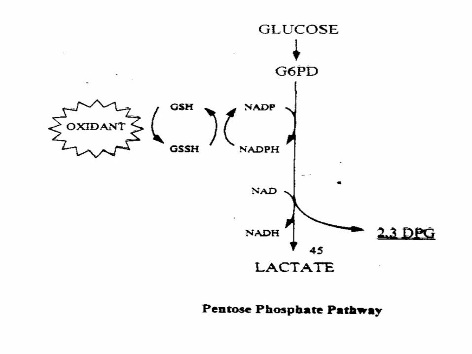
It’s All About The NAD/NADH Ratio• Oxygen does not directly catalyze cellular reactions. It indirectly
catalyzes them with NAD.
• The normal cytosol ratio of NAD/NADH is 700, guaranteeing an emphasis on oxidation.
• When NAD catalyzes a reaction, it is converted to NADH.
• The problem with decreased oxygen utilization is that is results
in decreased levels of NAD.
• As the NAD/NADH ratio decreases, all
cellular activity slows down.
• NADH is removed in order to achieve a healthy NAD/NADH ratio.
• The decrease in NADH further decreases oxygen utilization.
• Ozone therapy, by oxidizing NADH to NAD corrects the ratio and thus improves oxygen utilization by stimulating increasing levels of NADH.
• Oxidation therapies are enhanced with the addition of oral NADH.

Pharmacological Stimulation of NADH Oxidation Ameliorates Obesity and Related Phenotypes in Mice.
• The NAD/NADH ratio “plays a crucial role in cellular energy metabolism, and dysregulated NAD/NADH ratio is implicated in metabolic syndrome.”
• Used beta‐lapachone to oxidize NADH in diet‐induced obesity mice.
• NADH oxidation “strongly provoked mitochondrial fatty acid oxidation in vitro and in vivo, and dramatically ameliorated their key symptoms such as
increased adiposity, glucose intolerance, dyslipidemia, and fatty liver.”
• “The treated mice also showed higher expressions of the genes related to mitochondrial energy metabolism (PGC‐1alpha, NRF‐1) and caloric
restriction (Sirt1), consistent with the increased mitochondrial
biogenesis and energy expenditure.”
• “Conclusions: Pharmacological activation of NADH oxidation by NQO1 resolves obesity and related phenotypes in mice, opening the possibility
that it may provide the basis for a new therapy for the treatment of metabolic syndrome.”
Hwang JH, Kim DK, Jo EJ, et al. Diabetes Apr;58(4):965‐74. Epub 2009 Jan 9

It Happens Locally
• Chronic localized pain is caused by localized areas of chronically decreased oxygen utilization.
• Vicious cycle starts with trauma or infection.• Edema, inflammation, hyper‐coagulation, and
endothelial damage lead to localized decreased oxygen utilization.
• Decreased oxygen utilization disables the healing mechanisms, and condition becomes chronic
resulting in permanent edema, inflammation, hyper‐coagulation, endothelial damage, and pain.

Take Home Message
The most effective way to maximize the effects of ozone
therapy is to combine it with other therapies aimed at
eliminating the causes of decreased oxygen utilization