Molecular and Functional Characterization of Hv1 Proton Channel in Human Granulocytes

IntroductionThe outermost leaflet of the outer membrane of Gram-negativebacterial cell wall consists of lipopolysaccharide (LPS), acomplex molecule which induces a dysregulated innateimmune response. Because of the pathophysiological effects ofLPSs and their action on a number of different cell types, it isimportant to get a picture as complete as possible of the earlyevents involved in these responses. This picture may not beas simple as was thought a few years ago. The structuraldifferences of LPSs produced by distinct microorganisms,and the distinct patterns of molecular sensors for LPSs onmembranes of different cell types, seems to justify theexpectation that the initial interactions and early eventsinvolved in the cellular effects of LPSs depend on both theparticular LPS considered, and the cell type that is exposed toit.
It has been established that the activation ofmonocytes/macrophages by enterobacterial LPS requires atleast four molecules: the serum LPS-binding protein LBP(Tobias et al., 1995), the GPI-anchored cell-surface proteinCD14 (Wright et al., 1990; Golenbock et al., 1993), the type Itransmembrane Toll-like receptor 4 (TLR4) (Poltorak et al.,1998), and the extracellular molecule MD2 that physicallyassociates with TLR4 on the cell surface (Shimazu et al.,1999). Some investigators have presented this as the paradigm
accounting for cellular responses to LPS. However, other celltypes, and particularly granulocytes, also play a crucial role inhost protection. Furthermore, the considerable diversity ofbacteria should incite caution in granting the status ofrepresentative model to a small group of bacteria such asenterobacteria. Therefore, it is important at the present stage ofresearch in this area to examine what can occur when other celltypes are exposed to other types of LPSs.
Concerning the responses of other cell types, it has beenreported that Kupffer cells, the resident macrophages of theliver and most abundant tissue macrophages in the body, do notconstitutively express CD14 (Tracy et al., 1995). They produceTNF-α via an LBP-independent and CD14-independentpathway, when stimulated by nanogram amounts ofenterobacterial LPS (Lichtman et al., 1998). Furthermore,when CD14-deficient mice are injected with low doses of LPS,their liver produces normal levels of acute phase proteins(Haziot et al., 1998). In addition, we reported in previousstudies that another mouse cell type, the bone marrowgranulocyte (BMG), is sensitive to very low concentrations ofenterobacterial LPS, although it does not constitutively expressCD14 (Girard et al., 1993). Exposure of BMGs to this type ofLPS actually induces a CD14-independent and serum-independent stimulation of the cells, and leads to theexpression of CD14 (Pedron et al., 1999; Girard et al., 1997),
293
Lipopolysaccharide (LPS) derived from enterobacteriaelicit in several cell types cellular responses that arerestricted in the use of Toll-like receptor 4 (TLR4) asthe principal signal-transducing molecule. A tendency toconsider enterobacterial LPS as a prototypic LPS ledsome authors to present this mechanism as a paradigmaccounting for all LPSs in all cell types. However, thestructural diversity of LPS does not allow such a generalstatement. By using LPSs from bacteria that do not belongto the Enterobacteriaceae, we show that in bone marrowcells (BMCs) the LPS of Rhizobium speciesSin-1 and ofthree strains of Legionella pneumophila require TLR2
rather than TLR4 to elicit the expression of CD14.In addition, exposure of BMCs from TLR4-deficient(C3H/HeJ) mice to the lipid A fragment of the BordetellapertussisLPS inhibits their activation by the Legionellalipid A. The data show selective action of different LPSs viadifferent TLRs, and suggest that TLR2 can interact withmany lipid A structures, leading to either agonistic orspecific antagonistic effects.
Key words: Lipopolysaccharide, Toll-like receptors, Bone marrow,CD14, Innate immunity
Summary
Lipopolysaccharides from Legionella and Rhizobiumstimulate mouse bone marrow granulocytes via Toll-like receptor 2Robert Girard 1, Thierry Pedron 2, Satoshi Uematsu 3, Viviane Balloy 4, Michel Chignard 4, Shizuo Akira 3 andRichard Chaby 5,*1Lymphocyte development, URA-1961 of the National Center for Scientific Research, Pasteur Institute, Paris, France2Pathogénie Microbienne Moléculaire, Unité INSERM U389, Institut Pasteur, Paris, France3Department of Host Defense, Research Institute for Microbial Diseases, Osaka University, Osaka, Japan4Défense Innée et Inflammation, Unité associée IP/Inserm Z485, Institut Pasteur, Paris, France5Endotoxin Group, UMR-8619 of the National Center for Scientific Research, University of Paris-Sud, Orsay, France*Author for correspondence (e-mail: [email protected])
Accepted 8 October 2002Journal of Cell Science 116, 293-302 © 2003 The Company of Biologists Ltddoi:10.1242/jcs.00212
Research Article
JCS ePress online publication date 20 November 2002

294
and downregulation of L-selectin (Pedron et al., 2001) andTNF-receptor 2 (T.P., R.G. and R.C., unpublished).
Regarding some LPSs that are structurally and/orfunctionally different from those isolated from enterobacteria,it has been shown that they induce a response in macrophagesand B lymphocytes from TLR4-deficient mice (C3H/HeJmice), which are unresponsive to enterobacterial LPSs. This isparticularly the case with the LPSs from Pseudomonasaeruginosa (Pier et al., 1981), Porphyromonas gingivalis(Tanamoto et al., 1997; Ogawa et al., 1996) and Prevotellaintermedia (Kirikae et al., 1999). In a recent study (Pedron etal., 2000), we found that LPSs from Rhizobiaceaeand theirstructurally atypical lipid A moieties can stimulate BMGs fromTLR4-deficient mice (C3H/HeJ and C57BL/10 ScCr) andinduce the expression of CD14 in these cells.
It was therefore important to determine whether some LPSs,because of particular structural features of their lipid A moiety,can stimulate cells via a toll-like receptor distinct from TLR4or some other receptor unrelated to the toll family. Recentpublications proposed that LPSs from Porphyromonasgingivalis (Hirschfeld et al., 2001) and Leptospira interrogans(Werts et al., 2001) may activate macrophages via a TLR2-dependent mechanism. TLR2 is classically considered asmainly involved in the recognition of Gram-positive bacteriaand mycobacteria (Yoshimura et al., 1999; Flo et al., 2000). Itis also required in innate host defense to Borrelia burgdorferi,an atypical Gram-negative bacterium that lacks LPS butabundantly produces lipoproteins (Wooten et al., 2002). Inaddition to lipoproteins (Hirschfeld et al., 1999), porins canalso activate immune cells by engaging TLR2 (Massari et al.,2002).
In the present study, we examined the contributions of TLR4and TLR2 for the stimulation of BMGs by structurally atypicalLPSs. We focused on LPSs for which a reliable background ofstructural data is available. As a follow-up to our previousstudies (Pedron et al., 2000), we first examined the stimulationinduced by the rough-type LPS of a plant pathogen: RhizobiumspeciesSin-1. We also examined LPSs from different strainsof Legionella pneumophila, a human pathogen commonlyassociated with water-based aerosols and one of the top threecauses of nosocomial pneumonias.
Materials and MethodsAnimals and cell cultureC57BL/10 ScSn and C57BL/6 mice were purchased from Harlan(Gannat, France). C57BL/10 ScCr mice were a gift from MarinaFreudenberg (Freiburg, Germany). Homozygous mutant mice forTLR2 (TLR2–/–) generated by gene targeting, as described previously(Takeuchi et al., 1999), were back-crossed eight times with C57Bl/6to ensure similar genetic backgrounds. C3H/HeOU and C3H/HeJmice were bred at the Pasteur Institute (Paris, France). 8- to 10-week-old female mice were used in all experiments. Bone marrow cells(BMCs) were collected from mouse femurs. Culture media wasRPMI-1640 (Sigma, St Louis, MO) containing 20 mM Hepes, 1 mMsodium pyruvate, 2 mM L-glutamine, 100 IU/ml penicillin and 100µg/ml streptomycin. Fetal calf serum was from Gibco (Grand Island,NY).
Lipopolysaccharides and lipid AThe rough-type LPSs from Salmonella minnesotaRe-595 and fromEscherichia coliJ5 were from Sigma. The LPS from Bordetella
pertussis(LPS-Bp) and the lipid A fraction of the latter (BpLA), wereprepared as described previously (Tahri-Jouti et al., 1990). The LPSfrom Rhizobiumspecies Sin-1 (‘rough’ chemotype) and its lipid Afragment, were provided by R. W. Carlson (University of Georgia,Athens, GA). The LPS was extracted using hot phenol/water(Westphal and Jan, 1965) and purified by gel-filtrationchromatography in the presence of deoxycholate (Reuhs et al., 1994).LPSs from three strains of Legionella pneumophilaserogroup 1(Philadelphia strain CS338, wild-type OLDA strain RC1 and phasevariant OLDA strain 811) were prepared as described previously(Lüneberg et al., 1998; Zou et al., 1999; Kooistra et al., 2002b). Thelipid A fragments of the LegionellaLPSs were prepared by hydrolysisof 10 mg of LPS in 1 ml of 0.1 M sodium acetate/acetic acid buffer(pH 4.4) for 4 hours at 100°C. The lipid A fraction was recovered bycentrifugation (2500 g, 15°C, 30 minutes), followed by two washesof the pellet by resuspension in apyrogenic water and centrifugationas described above. The washed pellets were lyophilized.
For removal of lipoprotein contaminants, LPSs and lipid A werere-extracted twice by phenol in the presence of sodium deoxycholateas recommended (Hirschfeld et al., 2000). The absence ofcontaminants in the LPS and lipid A samples was assessed by SDS-PAGE analysis of the samples and staining the gels with silver nitrate(Rabilloud et al., 1994).
ReagentsMALP-2, prepared as described (Aliprantis et al., 1999), was fromAlexis (San Diego, CA). Tripalmitoyl pentapeptide was from Bachem(Bubendorf, Switzerland). Rabbit LBP was kindly provided byRichard Ulevitch (Scripps Research Institute, La Jolla, CA). Mouserecombinant LBP was from Biometec (Greifswald, Germany). The ratanti-mouse CD14 monoclonal antibody (Rm-C5-3) was fromPharMingen (San Diego, CA). In FACS experiments, FITC-labeled orbiotin-labeled goat anti-rat Ig antibodies (Southern BiotechnologyAssociates, Birmingham, AL), and FITC-labeled goat anti-hamster Igantibody (Caltag, Burlingame, CA), were used as secondaryantibodies, and biotin-labeled antibodies were stained with FITC-labeled streptavidin (Amersham-Pharmacia Biotech, Little Chalfont,UK). In western blot experiments, the biotin-labeled antibodywas stained with a streptavidin-peroxydase conjugate (SouthernBiotechnology Associates, Birmingham, AL). AutoradiographyHyperfilm MP, and all electrophoresis reagents, including molecularweight standards (rainbow markers), were from Amersham.
FACS analysisBone marrow cells (5×10⁵ cells in 400 µl CM without FCS) wereincubated at 37°C with (10 ng/ml) or without LPS. When used,inhibitors were added to cell cultures 30 to 60 minutes before LPS.For detection of membrane antigens, the cells were incubated first (30minutes, 4°C) with the primary antibody, and stained by reincubation(30 minutes, 4°C) with a labeled secondary antibody. Stained cellswere layered on a 50% FCS solution, centrifuged and the cell pelletwas resuspended in 0.5 ml of staining buffer (PBS, 5% FCS and0.02% sodium azide) containing propidium iodide (0.2 µg/ml) to staindead cells. Fluorescent cells were detected by analysis (5000 cells persample) on a FACS flow cytometer (FACScan, Becton-DickinsonElectronic Laboratories, Mountain View, CA) using Cell QuestSoftware. Dead cells, which incorporated propidium iodide, weregated out of analysis. Cells with a fluorescence intensity higher thanthe maximal level of auto-fluorescence were scored as fluorescentcells.
Preparation of peritoneal macrophages and TNF-α assayMice were injected intraperitoneally with 2 ml of 4% thioglycollate(Difco, Detroit, MI). Three days later, peritoneal exudate cells were
Journal of Cell Science 116 (2)

295TLR-2 dependent stimulation by atypical LPSs
isolated from the peritoneal cavity. Then the cells were cultured for 2hours and adherent cells were used as peritoneal macrophages.Peritoneal macrophages (5×10⁴) were cultured in RPMI-1640medium supplemented with 10% FCS and exposed to LPS (100ng/ml) for 24 hours. Concentrations of TNF-α in culture supernatantswere determined by ELISA (Genzyme-Techne, Minneapolis, MN).
Expression vectorspFLAG-TLR2 and pFLAG-TLR6 were obtained as describedpreviously (Takeuchi et al., 2001).
Luciferase assayHuman embryonic kidney (HEK) 293 cells were transientlytransfected with the vectors indicated above, together with a pELAMluciferase reporter plasmid (Takeuchi et al., 2001) and a pRL-TK(Promega, Madison, WI) for normalization of transfection efficiencyby lipofectamine 2000 (Invitrogen). Twenty-four hours aftertransfection, the cells were stimulated with the LPS from Rhizobiumspecies Sin1 (100 ng/ml) for 8 hours. Then, the cells were lysed andluciferase activity was measured using the dual-luciferase reporterassay system (Promega) according to the manufacturer’s instructions.
SDS-PAGE analysis of membrane CD14Membrane proteins were extracted from the cell pellet with 1%CHAPS in 300 mM NaCl, 50 mM Tris, pH 7.5, supplemented with acocktail of proteases inhibitors (aprotinin 10 µg/ml, PMSF 1 mM,pepstatin and leupeptin at 2 µg/ml and iodoacetamide 2 mM.).Solubilized proteins were analyzed by SDS-PAGE in 10%polyacrylamide slab gels according to the method of Laemmli.Molecular mass markers from 14.3 to 220 kDa were run in parallel.Gels were fixed in transfer buffer (20 mM Tris, 150 mM glycine, 20%methanol) and proteins were transferred onto PVDF membranes(Millipore, Bedford, MA) with a semidry blotting system at 30 voltsfor 90 minutes. Membranes were blocked (18 hours at 20°C) with 2%BSA in PBS, and incubated (1 hour, 20°C) with the rat anti-mouseantibody rmC5-3 (1:1000 in PBS containing 2% BSA). The blots werewashed with 0.1% Tween-20 in PBS, and then incubated for 1 hourat 20°C with a biotin-labeled goat-anti-rat antibody (1:2500 in thesame buffer). After extensive washing and incubation withperoxidase-labeled streptavidine (1:20,000 in 2% nonfat milk in PBS),sites with peroxidase activity were detected by chemiluminescencewith the Super Signal system (Pierce, Rockford, IL) according to theguidelines of the manufacturer.
ResultsTLR2-mediated expression of CD14 induced by the lipidA of Rhizobium species Sin-1We established in a previous study (Pedron et al., 2000) thatthe lipid A region of several LPSs from Rhizobiaceae canstimulate BMGs of the TLR4-deficient mouse strains C3H/HeJand C57BL/10ScCr, and induce the expression of CD14 inthese cells. This suggests that LPSs from Rhizobiaceae arerecognized by another member of the Toll family, or by anotherreceptor. We first analyzed the influence of the LPS isolatedfrom one of these Rhizobiaceae, R. speciesSin1, onmacrophages. This LPS was repurified by gel-filtrationchromatography in the presence of deoxycholate (Reuhs et al.,1994). Its protein content, measured using a bicinchoninic acidassay kit from Pierce (Rockford, IL) was lower than 0.5%. Wefound that, unlike BMG, mouse peritoneal macrophages werenot activated by this LPS: 100 ng/ml of the Rhizobium LPS
did not induce detectable levels of TNF-α, whereas the sameamount of the Salmonella minnesotaRe-595 LPS induced aproduction of 615 pg/ml of the cytokine. To determine whetherthe RhizobiumLPS can trigger cell activation via TLRs distinctfrom TLR4, HEK293 cells were transfected with TLR2- and/orTLR6-expression vectors along with an ELAM luciferasereporter plasmid, and exposed for 8 hours to 100 ng/ml of theRhizobium speciesSin1 LPS. The results in Fig. 1A indicatethat, in this model, cell activation by the RhizobiumLPSrequires the expression of both TLR2 and TLR6. Asexemplified by this cooperation between two TLRs, anothermembrane component present in granulocytes and absent frommacrophages may also be required for cell activation by someatypical LPSs, and thus explain why the RhizobiumLPSstimulates BMGs but does not activate macrophages at lowconcentrations.
To analyze the effect of the RhizobiumLPS in a morephysiological cellular system than a transfected cell line, were-examined the response of BMGs, and particularly the roleof TLR2 in this response. We compared the expression ofCD14 in BMGs from normal (C3H/HeOU) and TLR2-
Fig. 1.TLR dependence of cellular responses to the LPS from R.species. (A) HEK293 cells transfected with the TLR2 and/or TLR6expression vectors together with a pELAM luciferase reporterplasmid and pRL-TK were exposed 24 hours later to the RhizobiumspeciesSin1 LPS (100 ng/ml) for 8 hours. TLR-dependent activationwas measured by the dual-luciferase reporter assay. (B) BMGs fromC3H/HeOU and TLR2–/– mice were incubated with lipid A fractions(1 µg/ml) isolated from the LPSs of Bordetella pertussisandRhizobium speciesSin-1. Cell lysates (105 cells in 1% CHAPS,supplemented with a mixture of protease inhibitors) were analyzedfor CD14 by SDS-PAGE and western blotting.
296
deficient (TLR2–/–) mice. The lipid A fragments of LPSs fromBordetella pertussisand Rhizobium speciesSin-1 were usedas inducers because they represent the biologically activeregion of the LPSs, and also because eventual lipoproteincontaminants of LPSs are removed during the preparation ofthe lipid A fragments (as observed by SDS-PAGE analysis ofa number of such lipid A preparations). Therefore, BMGs wereexposed to lipid A for 24 hours at 37°C, in the absence ofserum. The results of the analysis of CD14 expression bywestern blot (Fig. 1B) indicated that, in normal cells, the twolipid A fractions induced the expression of large amounts ofCD14. The B. pertussislipid A was also active in TLR2–/–
cells. In contrast, a much lower expression of CD14 wasinduced by the lipid A from R. speciesSin-1 in TLR2–/– cells.This result explains why this atypical LPS was active in TLR4-deficient mice (Pedron et al., 2000), and shows that this effectis mediated by TLR2.
LPSs from Legionella induced CD14 expression inBMGs from normal miceThe structure of the Rhizobiumlipid A (Fig. 2) is atypical.When compared with the Bordetella pertussislipid A, whichacts via TLR4, the Rhizobiumlipid A exhibits three majordifferences: the absence of phosphate groups, a modified sugarbackbone (a glucosamine unit is replaced by 2-aminogluconicacid), and the presence of a very long fatty acid chain [28:0(27-hydroxy) residue] (Bhat et al., 1994; Basu et al., 1999). Insearching for other LPSs with at least one of these atypicalstructural features, we chose the LegionellaLPSs because theyalso contain a long fatty acid chain [27:0(1,27-dioic) or28:0(27-oxo) residue]. The three strains of Legionellapneumophilaused (CS338, RC1 and 811) have similar lipid Astructures, that differ only in their heterogeneity in theproportion and length of the different fatty acid chains (Fig. 2)(Zähringer et al., 1995; Kooistra et al., 2002a). We exposedBMGs from C3H/HeOU mice to various concentrations of thethree LegionellaLPSs, and analyzed by FACS the expressionof CD14. The results (Fig. 3) show that the three LPSs fromLegionella can induce cell surface expression of CD14,although the concentrations of these LPSs required for thisresponse (1 µg/ml) were much higher than that at which the B.pertussisLPS induced a similar effect (0.01 µg/ml).
Stimulation of BMGs from TLR4-deficient mice by theLegionella LPSsTo examine the role of TLR4 in the activation of BMGs byLegionella LPSs, we compared the expression of CD14 byBMGs from C3H/HeOU, C3H/HeJ and C57BL/10ScCr mice.In a first set of experiments, the cells were analyzed by FACSafter exposure for 24 hours to 1 or 10 µg/ml of the LPSs.Several experiments gave similar results. As shown in onerepresentative experiment (Fig. 4A), the three LegionellaLPSsinduced CD14 expression in C3H/HeJ and C57BL/10ScCr
Journal of Cell Science 116 (2)
Fig. 2.Structures of the lipid A regions of LPSs used in this study.The structure of the lipid A region of R. speciesSin-1 is notcompletely determined but, as that from some other Rhizobiaceae(Bhat et al., 1994), it is devoid of phosphate, has 2-aminogluconatein place of glucosamine-1-phosphate, and contains the very longchain of 27-hydroxyoctacosanoic acid (27-OHC28:0) that may beester-linked in the N-acyloxylacyl residue of the distal glucosamineunit (Basu et al., 1999). The lipid A regions of the LPSs from threestrains of Legionella pneumophila(Zähringer et al., 1995; Kooistra etal., 2002a) are built upon the same structural model (two residues of2,3-diaminoglucose substituted with one very long and five shorterfatty acids), with variations limited to the length or substituents ofthe fatty acid chains in positions 2 and 2′: R1=H or OH; R2=OH orCH3; n=18-22 (in strains CS338 and RC1), or n=16-18 (in strain811).
Fig. 3. Influence of LegionellaLPSs on BMGs of LPS-responsivemice. BMGs (5×10⁵ cells) from C3H/HeOU mice were incubated for24 hours at 37°C with various concentrations of LPSs from B.pertussis(d), and from the L. pneumophilastrains CS338 (s), RC1(.), and 811 (j). CD14 was detected by incubation with the anti-CD14 antibody rmC5-3 (2.5 µg/ml, 30 minutes, 0°C), and furtherincubation (30 minutes, 0°C) with a FITC-labeled goat anti-rat IgmAb. The percentage of fluorescent cells was determined by FACSanalysis of the gated granulocyte population.

297TLR-2 dependent stimulation by atypical LPSs
mice, whereas the B. pertussisLPS did not. The most efficientactivator was the LPS from the L. pneumophilaCS338 strain.In a second set of experiments, expression of CD14 wasanalyzed by western blot. The results (Fig. 4B) were consistentwith those obtained by FACS, and confirmed that the threeLPSs of Legionellaare efficient activators of BMGs from micewith a TLR4 defect.
Influence of the Legionella LPSs on TLR2-deficientmacrophages and BMGsThe capacity of LegionellaLPS to activate BMGs from TLR4-defective mice suggests that another receptor is involved. Byanalogy with the the results obtained above with the RhizobiumLPS, we used TLR2-deficient (TLR2–/–) mice to examine therole of TLR2. We analyzed first the responses of macrophages.We found (Table 1) that thioglycollate-elicited peritonealmacrophages from TLR2–/– mice did not respond to the TLR2-dependent mycobacterial lipopeptide MALP-2, and producednornal levels of TNF-α in response to Salmonella andBordetellaLPSs. In contrast, these cells were not activated bythe LegionellaLPSs.
The stimulation of BMGs by the LegionellaLPSs was thenexamined. The expression of CD14 BMGs from TLR2–/– micewas analyzed by FACS, and compared with that of BMGs fromnormal mice of the same genetic background (TLR2+/+).Several experiments gave similar results. As shown in onerepresentative experiment (Fig. 5A), the LPS isolated from thestrain CS338 of L. pneumophilawas completely unable toinduce CD14 expression in TLR2–/– cells. CD14 expressionwas detectable in TLR2–/– cells exposed to the two otherLegionellaLPSs, but the responses were lower than those ofTLR2+/+ cells, and were only induced by high concentrationsof these LPSs (10 µg/ml). These residual responses mayindicate that a receptor distinct from TLR2 reacts with these
LPSs or with a contaminant present in the LPSpreparations. To ensure that contaminations cannotbe put forward, the three LegionellaLPSs were re-extracted twice by phenol in the presence of sodiumdeoxycholate (Hirschfeld et al., 2000). Because oftheir hydrophobic nature, the LegionellaLPSs arefairly soluble in phenol, so that only 10% of theLPSs were recovered in the aqueous phase.However, the ability of these purified (lipoprotein-depleted) samples to induce CD14 in BMGs ofdifferent mouse strains was not different from thatof the non-extracted samples. Analysis of CD14
expression by western blot (Fig. 5B) confirms that this cellresponse to Legionella LPSs is markedly reduced, but notcompletely abolished, in TLR2–/– BMGs.
Effects of lipid A fragments in TLR4- and TLR2-deficientcellsSome cellular activities of LPS preparations are due to theirpolysaccharide region, or are modulated by that region. Otherbacterial components (peptides, lipoproteins) present ascontaminants in LPS preparations can also take part in cellresponses (Hirschfeld et al., 2000). However, it is generallyaccepted that LPS effects are those mediated by the lipid Aregion of that molecule. To check that this was indeed the casefor the effects of the LegionellaLPSs mentioned above, weprepared the lipid A fragments of these molecules byhydrolysis for 4 hours at 100°C in a pH 4.4 buffer and washing
Fig. 4. CD14 expression in BMGs from normal andTLR4-deficient mice. BMGs from C3H/HeOU,C3H/HeJ and C57BL/10ScCr mice were incubated for24 hours at 37°C with the B. pertussisLPS, or with LPSsfrom the different strains of L. pneumophila. (A) CD14expression in BMGs incubated with 1 or 10 µg/ml of theLPSs was detected by FACS analysis of the gatedgranulocyte population, as described in the legend toFig. 3. The dashed line represents the level of CD14 innon-stimulated cells. (B) CD14 expression in BMGsincubated with 10 µg/ml of the LPSs was analyzed bywestern blot in cell lysates, as described in the legend toFig. 1.
Table 1. LPS-induced TNF-α production by macrophagesof wild-type and TLR2–/– mice
TNF-α produced (pg/ml)Concentration
Inducer (ng/ml) Wild-type mice TLR2–/– mice
None – 0±0 0±0MALP-2 3 558±23 0±0LPS Sm-595 100 615±64 800±200LPS Bp-414 100 388±40 346±78LPS Rs-Sin1 100 0±0 0±0LPS Lp-338 100 386±28 0±0LPS Lp-811 100 225±34 0±0LPS Lp-RC1 100 1061±174 0±0
Thioglycollate-elicited peritoneal macrophages (105 cells/well) from wild-type, and TLR2–/– mice, were incubated for 24 hours with MALP-2 (3 ng/ml)or with different LPSs (100 ng/ml). The production of TNF-α was determinedby ELISA. Data are the means±s.d. of three experiments.

298
the insoluble material with water. Analysis of these lipid Apreparations by SDS-PAGE stained with silver nitrate indicatedan almost complete absence of protein contamination (proteincontent lower than 0.01%, as determined by comparison ofband intensities of protein standards). BMGs from C3H/HeJ,TLR2+/+, and TLR2–/– mice were then exposed to these lipidA preparations, and CD14 expression was analyzed by westernblot. The results (Fig. 6) were similar to those induced by theunfragmented LPS preparations: the three lipids were active inC3H/HeJ cells, and the activities were markedly reduced inTLR2–/– cells.
Because LegionellaLPSs are fairly soluble in phenol (seeabove), whereas their lipid A fraction is not, the lipid A isolatedfrom Legionella pneumophilaRC1 was re-extracted twice byphenol in the presence of sodium deoxycholate, to remove
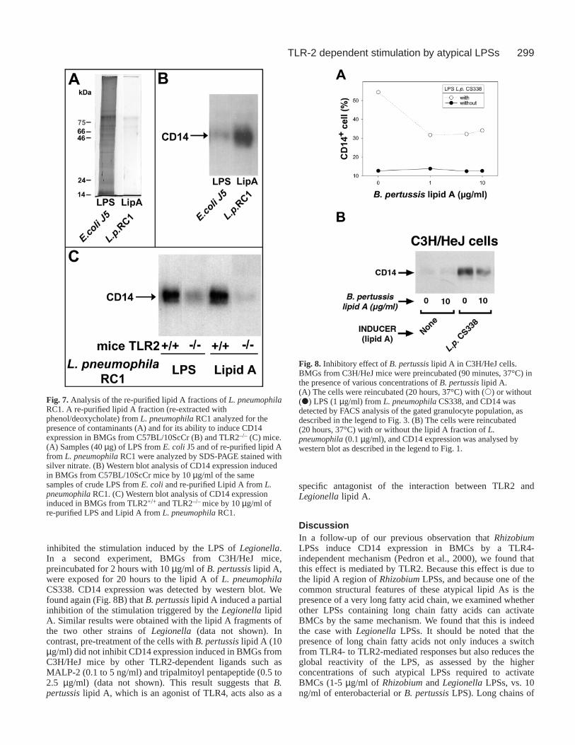
residual amounts of unhydrolyzed LPS. This purified lipid A,in which the level of protein contamination was considerablylower than that of a standard E. coli LPS (Fig. 7A), wasconsiderably more active in BMCs from TLR4-defective mice(Fig. 7B). This confirms that pure lipid A from LegionellapneumophilaRC1 induces a TLR4-independent effect. We alsofound that the residual activation of TLR2–/– BMGs inducedby the repurified LPS is markedly reduced with the repurifiedlipid A (Fig. 7C). This observation suggests that the lipid A ofLegionella pneumophilaRC1 stimulates BMGs exclusively viaTLR2, whereas the unfragmented LPS, which contains anadditional polysaccharide region, stimulates BMGs via TLR2and another receptor.
Dose-response experiments indicated that even lowconcentrations (100 ng/ml) of L. pneumophilalipid A caninduce detectable levels of CD14 in BMGs from TLR4-deficient (C3H/HeJ) mice. The presence of fetal calf serum(10%), or of rabbit or recombinant mouse LBP (10 µg/ml) didnot increase this response (data not shown).
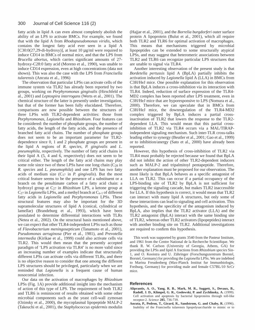
Antagonist effect of Bordetella pertussis lipid AWe established in a previous study (Pedron et al., 2000) thatthe lipid A fragment of the B. pertussisLPS inhibits theexpression of CD14 induced by the lipid A of RhizobiumspeciesSin-1 in BMGs from C3H/HeJ mice. As we hadpreviously demonstrated that Rhizobiumand LegionellaLPSsare both TLR2-dependent stimulators of BMG, it wasimportant to determine whether B. pertussislipid A can alsoinhibit the TLR2-dependent response induced by theLegionellaLPSs. In a first experiment, BMGs from C3H/HeJmice, preincubated for 90 minutes with various concentrationsof B. pertussislipid A, were exposed for 20 hours to the LPSof L. pneumophilaCS338. CD14 expression was detected byFACS. The results (Fig. 8A) show that 1 µg/ml of lipid A
Journal of Cell Science 116 (2)
Fig. 5. Analysis of CD14 expression in BMGs from TLR2+/+ andTLR2–/– mice. BMGs from TLR2+/+ and TLR2–/– mice wereincubated for 24 hours at 37°C with the B. pertussisLPS, or withLPSs from the different strains of L. pneumophila. (A) CD14expression in BMGs incubated with 1 or 10 µg/ml of the LPSs wasdetected by FACS analysis of the gated granulocyte population, asdescribed in the legend to Fig. 3. The dashed line represents the levelof CD14 in non-stimulated cells. (B) CD14 expression in BMGsincubated with 10 µg/ml of re-purified LPSs (re-extracted withphenol/deoxycholate) was analyzed by western blot in cell lysates, asdescribed in the legend to Fig. 1.
Fig. 6. CD14 expressioninduced by lipid A fractionsin BMGs from TLR4- andTLR2-deficient mice. BMGsfrom C3H/HeJ, TLR2+/+ andTLR2–/– mice wereincubated for 24 hours at37°C with lipid A from B.pertussis(10 µg/ml), or fromthe different strains of L.pneumophila(50 µg/ml).CD14 expression wasanalyzed by western blot incell lysates, as described inthe legend to Fig. 1.
299TLR-2 dependent stimulation by atypical LPSs
inhibited the stimulation induced by the LPS of Legionella.In a second experiment, BMGs from C3H/HeJ mice,preincubated for 2 hours with 10 µg/ml of B. pertussislipid A,were exposed for 20 hours to the lipid A of L. pneumophilaCS338. CD14 expression was detected by western blot. Wefound again (Fig. 8B) that B. pertussislipid A induced a partialinhibition of the stimulation triggered by the LegionellalipidA. Similar results were obtained with the lipid A fragments ofthe two other strains of Legionella (data not shown). Incontrast, pre-treatment of the cells with B. pertussislipid A (10µg/ml) did not inhibit CD14 expression induced in BMGs fromC3H/HeJ mice by other TLR2-dependent ligands such asMALP-2 (0.1 to 5 ng/ml) and tripalmitoyl pentapeptide (0.5 to2.5 µg/ml) (data not shown). This result suggests that B.pertussislipid A, which is an agonist of TLR4, acts also as a
specific antagonist of the interaction between TLR2 andLegionellalipid A.
DiscussionIn a follow-up of our previous observation that RhizobiumLPSs induce CD14 expression in BMCs by a TLR4-independent mechanism (Pedron et al., 2000), we found thatthis effect is mediated by TLR2. Because this effect is due tothe lipid A region of RhizobiumLPSs, and because one of thecommon structural features of these atypical lipid As is thepresence of a very long fatty acid chain, we examined whetherother LPSs containing long chain fatty acids can activateBMCs by the same mechanism. We found that this is indeedthe case with Legionella LPSs. It should be noted that thepresence of long chain fatty acids not only induces a switchfrom TLR4- to TLR2-mediated responses but also reduces theglobal reactivity of the LPS, as assessed by the higherconcentrations of such atypical LPSs required to activateBMCs (1-5 µg/ml of Rhizobiumand LegionellaLPSs, vs. 10ng/ml of enterobacterial or B. pertussisLPS). Long chains of
Fig. 7. Analysis of the re-purified lipid A fractions of L. pneumophilaRC1. A re-purified lipid A fraction (re-extracted withphenol/deoxycholate) from L. pneumophilaRC1 analyzed for thepresence of contaminants (A) and for its ability to induce CD14expression in BMGs from C57BL/10ScCr (B) and TLR2–/– (C) mice.(A) Samples (40 µg) of LPS from E. coliJ5 and of re-purified lipid Afrom L. pneumophilaRC1 were analyzed by SDS-PAGE stained withsilver nitrate. (B) Western blot analysis of CD14 expression inducedin BMGs from C57BL/10ScCr mice by 10 µg/ml of the samesamples of crude LPS from E. coliand re-purified Lipid A from L.pneumophilaRC1. (C) Western blot analysis of CD14 expressioninduced in BMGs from TLR2+/+ and TLR2–/– mice by 10 µg/ml ofre-purified LPS and Lipid A from L. pneumophilaRC1.
Fig. 8. Inhibitory effect of B. pertussislipid A in C3H/HeJ cells.BMGs from C3H/HeJ mice were preincubated (90 minutes, 37°C) inthe presence of various concentrations of B. pertussislipid A.(A) The cells were reincubated (20 hours, 37°C) with (s) or without(d) LPS (1 µg/ml) from L. pneumophilaCS338, and CD14 wasdetected by FACS analysis of the gated granulocyte population, asdescribed in the legend to Fig. 3. (B) The cells were reincubated(20 hours, 37°C) with or without the lipid A fraction of L.pneumophila(0.1 µg/ml), and CD14 expression was analysed bywestern blot as described in the legend to Fig. 1.
300
fatty acids in lipid A can even almost completely abolish theability of an LPS to activate BMCs. For example, we foundthat with the lipid A from Rhodopseudomonas viridis, whichcontains the longest fatty acid ever seen in a lipid A[C30:0(27,29-di-hydroxy)], at least 10 µg/ml were required toinduce CD14 in BMCs of normal mice, and that the LPS fromBrucella abortus, which carries significant amounts of 27-hydroxy-C28:0 fatty acid (Moreno et al., 1990), was unable toinduce CD14 expression, even at high concentrations (data notshown). This was also the case with the LPS from Francisellatularensis(Ancuta et al., 1996)
The observation that particular LPSs can activate cells of theimmune system via TLR2 has already been reported by twogroups, working on Porphyromonas gingivalis(Hirschfeld etal., 2001) and Leptospira interrogans(Werts et al., 2001). Thechemical structure of the latter is presently under investigation,but that of the former has been fully elucidated. Therefore,comparisons are now possible between the structures ofthree LPSs with TLR2-dependent activities: those fromPorphyromonas, Legionellaand Rhizobium. Four features canbe considered: the number of phosphate groups, the number offatty acids, the length of the fatty acids, and the presence ofbranched fatty acid chains. The number of phosphate groupsdoes not seem to be an important parameter for TLR2-dependence since 0, 1 and 2 phosphate groups are present inthe lipid A regions of R. species, P. gingivalis and L.pneumophila, respectively. The number of fatty acid chains intheir lipid A (5, 4 and 6, respectively) does not seem to becritical either. The length of the fatty acid chains may playsome role since two of these LPSs have one long chain (C28 inR. speciesand L. pneumophila) and one LPS has two fattyacids of medium size (C17 in P. gingivalis). But the mostcritical feature seems to be the presence of a substituent or abranch on the penultimate carbon of a fatty acid chain: ahydroxyl group at C27 in RhizobiumLPS, a ketone group atC27 in LegionellaLPSs, and a methyl branch at Cn-1of differentfatty acids in Legionella and PorphyromonasLPSs. Thesestructural features may also be important for the 3Dsupramolecular structures of lipid A (conical, cylindrical orlamellar) (Brandeburg et al., 1993), which have beenpostulated to determine differential interactions with TLRs(Netea et al., 2002). On the structural basis mentioned above,we can expect that other TLR4-independent LPSs such as thoseof Flavobacterium meningosepticum(Tanamoto et al., 2001),Pseudomonas aeruginosa(Pier et al., 1981), and Prevotellaintermedia(Kirikae et al., 1999) could also activate cells viaTLR2. This would then mean that the presently acceptedparadigm of ‘LPS activation via TLR4’ is no more valid sincean increasing number of examples indicate that structurallydifferent LPSs can activate cells via different TLRs, and thereis no objective reason to consider that one among the differentLPS structures should be privileged, particularly when we arereminded that Legionella is a frequent cause of humannosocomial infection.
Our data on the activation of macrophages by RhizobiumLPSs (Fig. 1A) provide additional insight into the mechanismof action of this type of LPS. The requirement of both TLR2and TLR6 is reminiscent of results obtained with some othermicrobial components such as the yeast cell-wall zymosan(Ozinsky et al., 2000), the mycoplasmal lipopeptide MALP-2(Takeuchi et al., 2001), the Staphylococcus epidermismodulin
(Hajjar et al., 2001), and the Borrelia burgdorferiouter surfaceprotein A lipoprotein (Bulut et al., 2001), which all requireboth TLR2 and TLR6 for optimal activation of macrophages.This means that mechanisms triggered by microbiallipopeptides can be extended to some structurally atypicalLPSs, and may suggest that heteromeric associations betweenTLR2 and TLR6 can recognize particular LPS structures thatare unable to signal via TLR4.
Another interesting observation of the present study is thatBordetella pertussislipid A (BpLA) partially inhibits theactivation induced by Legionellalipid A (LLA) in BMCs fromC3H/HeJ mice. One possible explanation for this observationis that BpLA induces a cross-inhibition via its interaction withTLR4. Indeed, reduction of surface expression of the TLR4-MD2 complex has been reported after LPS treatment, even inC3H/HeJ mice that are hyporesponsive to LPS (Nomura et al.,2000). Therefore, we can speculate that in BMCs fromC3H/HeJ mice, the downregulation of the TLR4-MD2complex triggered by BpLA induces a partial cross-inactivation of TLR2 that lowers the response to the TLR2-dependent LLA. This would mean that this partial cross-inhibition of TLR2 via TLR4 occurs via a MAL/TIRAP-independent signaling mechanism. Such inter-TLR cross-talksleading either to synergy (Beutler et al., 2001; Gao et al., 1999)or to inhibition/anergy (Sato et al., 2000) have already beenreported.
However, this hypothesis of cross-inhibition of TLR2 viaTLR4 must probably be rejected because we found that BpLAdid not inhibit the action of other TLR2-dependent inducerssuch as MALP-2 and tripalmitoyl pentapeptide. Therefore,another explanation must be proposed for our observation. Themost likely is that BpLA behaves as a specific antagonist ofLLA on TLR2. This can occur if a partial occupancy of theLPS-binding site of TLR2 by BpLA is not sufficient fortriggering the signaling cascade, but makes TLR2 inaccessiblefor LLA. If this hypothesis is correct, it would mean that TLR2can interact with many lipid A structures, but only some ofthese interactions can lead to signaling and cell activation. Thishypothesis, and the specificity of the antagonism induced byBpLA, also implies that the TLR2 activator (LLA) and theTLR2 antagonist (BpLA) interact with the same binding siteof TLR2, whereas other TLR2 activators (lipopeptides) interactwith another binding site on TLR2. Additional investigationsare required to confirm this hypothesis.
This work was supported by grants 3540 from the Pasteur Institute,and 1961 from the Centre National de la Recherche Scientifique. Wethank R. W. Carlson (University of Georgia, Athens, GA) forproviding the LPSs and lipid A fractions fromRhizobium speciesSin-1, and O. Kooistra and U. Zähringer (Forschungszentrum Borstel,Borstel, Germany) for providing the LegionellaLPSs. We are indebtedto Marina Freudenberg (Max-Planck Institut fur Immunbiologie,Freiburg, Germany) for providing male and female C57BL/10 ScCrmice.
ReferencesAliprantis, A. O., Yang, R. B., Mark, M. R., Suggett, S., Devaux, B.,
Radolf, J. D., Klimpel, G. R., Godowski, P. and Zychlinsky, A. (1999).Cell activation and apoptosis by bacterial lipoproteins through toll-likereceptor-2. Science285, 736-739.
Ancuta, P., Pedron, T., Girard, R., Sandstrom, G. and Chaby, R. (1996).Inability of the Francisella tularensis lipopolysaccharide to mimic or to
Journal of Cell Science 116 (2)

301TLR-2 dependent stimulation by atypical LPSs
antagonize the induction of cell activation by endotoxins. Infect. Immun. 64,2041-2046.
Basu, S. S., White, K. A., Que, N. L. and Raetz, C. R. (1999). A deacylasein Rhizobium leguminosarummembranes that cleaves the 3-O-linked beta-hydroxymyristoyl moiety of lipid A precursors. J. Biol. Chem. 274, 11150-11158.
Beutler, E., Gelbart, T. and West, C. (2001). Synergy between TLR2 andTLR4: a safety mechanism. Blood Cells Mol. Dis. 27, 728-730.
Bhat, U. R., Forsberg, L. S. and Carlson, R. W. (1994). Structure of lipid Acomponent of Rhizobium leguminosarumbv. phaseoli lipopolysaccharide.Unique nonphosphorylated lipid A containing 2-amino-2-deoxygluconate,galacturonate, and glucosamine. J. Biol. Chem. 269, 14402-14410.
Brandenburg, K., Mayer, H., Koch, M. H., Weckesser, J., Rietschel, E. T.and Seydel, U. (1993). Influence of the supramolecular structure of freelipid A on its biological activity. Eur J. Biochem. 218, 555-563.
Bulut, Y., Faure, E., Thomas, L., Equils, O. and Arditi, M. (2001).Cooperation of Toll-like receptor 2 and 6 for cellular activation by solubletuberculosis factor and Borrelia burgdorferi outer surface protein Alipoprotein: role of Toll-interacting protein and IL-1 receptor signalingmolecules in Toll-like receptor 2 signaling. J. Immunol. 167, 987-994.
Flo, T. H., Halaas, O., Lien, E., Ryan, L., Teti, G., Golenbock, D. T.,Sundan, A. and Espevik, T. (2000). Human toll-like receptor 2 mediatesmonocyte activation by Listeria monocytogenes, but not by group Bstreptococci or lipopolysaccharide. J. Immunol. 164, 2064-2069.
Gao, J. J., Zuvanich, E. G., Xue, Q., Horn, D. L., Silverstein, R. andMorrison, D. C. (1999). Bacterial DNA and LPS act in synergy in inducingnitric oxide production in RAW 264.7 macrophages. J. Immunol. 163, 4095-4099.
Girard, R., Pedron, T. and Chaby, R. (1993). Endotoxin-induced expressionof endotoxin binding sites on murine bone marrow cells. J. Immunol. 150,4504-4513.
Girard, R., Pedron, T. and Chaby, R. (1997). Functional lipopolysaccharidereceptors of low affinity are constitutively expressed on mouse bone marrowcells. Immunology91, 391-398.
Golenbock, D. T., Liu, Y., Millham, F. H., Freeman, M. W. and Zoeller, R.A. (1993). Surface expression of human CD14 in Chinese hamster ovaryfibroblasts imparts macrophage-like responsiveness to bacterial endotoxin.J. Biol. Chem. 268, 22055-22059.
Hajjar, A. M., O’Mahony, D. S., Ozinsky, A., Underhill, D. M., Aderem,A., Klebanoff, S. J. and Wilson, C. B. (2001). Functional interactionsbetween toll-like receptor (TLR) 2 and TLR1 or TLR6 in response tophenol-soluble modulin. J. Immunol. 166, 15-19.
Haziot, A., Lin, X. Y., Zhang, F. and Goyert, S. M. (1998). The inductionof acute phase proteins by lipopolysaccharide uses a novel pathway that isCD14-independent. J. Immunol. 160, 2570-2572.
Hirschfeld, M., Kirschning, C. J., Schwandner, R., Wesche, H., Weis, J.H., Wooten, R. M. and Weis, J. J. (1999). Inflammatory signaling byBorrelia burgdorferi lipoproteins is mediated by toll-like receptor 2. J.Immunol. 163, 2382-2386.
Hirschfeld, M., Ma, Y., Weis, J. H., Vogel, S. N. and Weis, J. J. (2000).Repurification of lipopolysaccharide eliminates signaling through bothhuman and murine toll-like receptor 2. J. Immunol. 165, 618-622.
Hirschfeld, M., Weis, J. J., Toshchakov, V., Salkowski, C. A., Cody, M. J.,Ward, D. C., Qureshi, N., Michalek, S. M. and Vogel, S. N. (2001).Signaling by toll-like receptor 2 and 4 agonists results in differential geneexpression in murine macrophages. Infect. Immun. 69, 1477-1482.
Kirikae, T., Nitta, T., Kirikae, F., Suda, Y., Kusumoto, S., Qureshi, N. andNakano, M. (1999). Lipopolysaccharides (LPS) of oral black-pigmentedbacteria induce tumor necrosis factor production by LPS-refractoryC3H/HeJ macrophages in a way different from that of Salmonella LPS.Infect. Immun. 67, 1736-1742.
Kooistra, O., Knirel, Y. A., Lüneberg, E., Frosch, M. and Zähringer, U.(2002a). Phase variation in Legionelle pneumophilaserogroup 1, subgroupOLDA, strain RC1 influences lipid A structure. In Legionella – Proceedingsof the 5th International Conference(ed. R. Marre, Y. Abu Kwaik, C. Barltett,N. Cianciotto, B. S. Fields, M. Frosch, J. Hacker and B. C. Lück), pp. 68-73. Washington: ASM Press.
Kooistra, O., Lüneberg, E., Knirel, Y. A., Frosch, M. and Zähringer, U.(2002b). N-Methylation in polylegionaminic acid is associated with thephase-variable epitope of Legionella pneumophila serogroup 1lipopolysaccharide. Identicication of 5-(N,N-dimethylacetimidoyl)amino-and 5-acetimidoyl(N-methyl)amino-7-acetamido-3,5,7,9-tetradeoxynon-2-ulosonic acid in the O-chain polysaccharide. Eur. J. Biochem. 269, 560-572.
Lichtman, S. N., Wang, J. and Lemasters, J. J. (1998). LPS receptor CD14participates in release of TNF-alpha in RAW 264.7 and peritoneal cells butnot in kupffer cells. Am. J. Physiol. 275, G39-G46.
Lüneberg, E., Zähringer, U., Knirel, Y. A., Steinmann, D., Hartmann, M.,Steinmetz, I., Rohde, M., Kohl, J. and Frosch, M. (1998). Phase-variableexpression of lipopolysaccharide contributes to the virulence of Legionellapneumophila. J. Exp. Med. 188, 49-60.
Massari, P., Henneke, P., Ho, Y., Latz, E., Golenbock, D. T. and Wetzler,L. M. (2002). Immune stimulation by neisserial porins is toll-like receptor2 and MyD88 dependent. J. Immunol. 168, 1533-1537.
Moreno, E., Stackebrandt, E., Dorsch, M., Wolters, J., Busch, M. andMayer, H. (1990). Brucella abortus16S rRNA and lipid A reveal aphylogenetic relationship with members of the alpha-2 subdivision of theclass Proteobacteria. J. Bacteriol. 172, 3569-3576.
Netea, M. G., van Deuren, M., Kullberg, B. J., Cavaillon, J. M. and vander Meer, J. W. (2002). Does the shape of lipid A determine the interactionof LPS with Toll- like receptors? Trends Immunol. 23, 135-139.
Nomura, F., Akashi, S., Sakao, Y., Sato, S., Kawai, T., Matsumoto, M.,Nakanishi, K., Kimoto, M., Miyake, K., Takeda, K. et al. (2000).Endotoxin tolerance in mouse peritoneal macrophages correlates with down-regulation of surface toll-like receptor 4 expression. J. Immunol. 164, 3476-3479.
Ogawa, T., Shimauchi, H., Uchida, H. and Mori, Y. (1996). Stimulation ofsplenocytes in C3H/HeJ mice with Porphyromonas gingivalislipid A incomparison with enterobacterial lipid A. Immunobiology196, 399-414.
Ozinsky, A., Underhill, D. M., Fontenot, J. D., Hajjar, A. M., Smith, K. D.,Wilson, C. B., Schroeder, L. and Aderem, A. (2000). The repertoire forpattern recognition of pathogens by the innate immune system is defined bycooperation between toll-like receptors. Proc. Natl. Acad. Sci. USA97,13766-13771.
Pedron, T., Girard, R. and Chaby, R. (1999). Exogenous cyclic AMP,cholera toxin, and endotoxin induce expression of the lipopolysaccharidereceptor CD14 in murine bone marrow cells: role of purinoreceptors. Clin.Diagn. Lab. Immunol. 6, 885-890.
Pedron, T., Girard, R., Jeyaretnam, B., Carlson, R. W. and Chaby, R.(2000). The lipid A region of lipopolysaccharides from Rhizobiaceaeactivates bone marrow granulocytes from lipopolysaccharide-hyporesponsive C3H/HeJ and C57BL/10ScCr mice. Immunology101, 262-270.
Pedron, T., Girard, R. and Chaby, R. (2001). Down-modulation of L-selectinby lipopolysaccharide is not required for lipopolysaccharide-inducedexpression of CD14 in mouse bone marrow granulocytes. Infect. Immun. 69,4287-4294.
Pier, G. B., Markham, R. B. and Eardley, D. (1981). Correlation of thebiologic responses of C3H/HEJ mice to endotoxin with the chemical andstructural properties of the lipopolysaccharides from Pseudomonasaeruginosa and Escherichia coli. J. Immunol. 127, 184-191.
Poltorak, A., He, X., Smirnova, I., Liu, M. Y., Huffel, C. V., Du, X.,Birdwell, D., Alejos, E., Silva, M., Galanos, C. et al. (1998). DefectiveLPS signaling in C3H/HeJ and C57BL/10ScCr mice: mutations in Tlr4gene. Science282, 2085-2088.
Rabilloud, T., Vuillard, L., Gilly, C. and Lawrence, J. J. (1994). Silver-staining of proteins in polyacrylamide gels: a general overview. Cell. Mol.Biol. 40, 57-75.
Reuhs, B. L., Kim, J. S., Badgett, A. and Carlson, R. W. (1994). Productionof cell-associated polysaccharides of Rhizobium fredii USAD205 ismodulated by apigenin and host root extract. Mol. Plant Microbe Interact.7, 240-244.
Sato, S., Nomura, F., Kawai, T., Takeuchi, O., Muhlradt, P. F., Takeda, K.and Akira, S. (2000). Synergy and cross-tolerance between toll-likereceptor (TLR) 2- and TLR4-mediated signaling pathways. J. Immunol. 165,7096-7101.
Shimazu, R., Akashi, S., Ogata, H., Nagai, Y., Fukudome, K., Miyake, K.and Kimoto, M. (1999). MD-2, a molecule that confers lipopolysaccharideresponsiveness on Toll-like receptor 4. J. Exp. Med. 189, 1777-1782.
Tahri-Jouti, M. A., Mondange, M., le Dur, A., Auzanneau, F. I., Charon,D., Girard, R. and Chaby, R. (1990). Specific binding oflipopolysaccharides to mouse macrophages–II. Involvement of distinct lipida substructures. Mol. Immunol. 27, 763-770.
Takeuchi, O., Hoshino, K., Kawai, T., Sanjo, H., Takada, H., Ogawa, T.,Takeda, K. and Akira, S. (1999). Differential roles of TLR2 and TLR4 inrecognition of gram-negative and gram-positive bacterial cell wallcomponents. Immunity11, 443-451.
Takeuchi, O., Kawai, T., Muhlradt, P. F., Morr, M., Radolf, J. D.,

302
Zychlinsky, A., Takeda, K. and Akira, S. (2001). Discrimination ofbacterial lipoproteins by Toll-like receptor 6. Int. Immunol. 13, 933-940.
Tanamoto, K., Azumi, S., Haishima, Y., Kumada, H. and Umemoto, T.(1997). The lipid A moiety of Porphyromonas gingivalislipopolysaccharidespecifically mediates the activation of C3H/HeJ mice. J. Immunol. 158,4430-4436.
Tanamoto, K., Kato, H., Haishima, Y. and Azumi, S. (2001). Biologicalproperties of lipid A isolated from Flavobacterium meningosepticum. Clin.Diagn. Lab. Immunol. 8, 522-527.
Tobias, P. S., Soldau, K., Gegner, J. A., Mintz, D. and Ulevitch, R. J. (1995).Lipopolysaccharide binding protein-mediated complexation oflipopolysaccharide with soluble CD14. J. Biol. Chem. 270, 10482-10488.
Tracy, T. F., Jr and Fox, E. S. (1995). CD14-lipopolysaccharide receptor activityin hepatic macrophages after cholestatic liver injury. Surgery118, 371-377.
Werts, C., Tapping, R. I., Mathison, J. C., Chuang, T. H., Kravchenko, V.,Saint Girons, I., Haake, D. A., Godowski, P. J., Hayashi, F., Ozinsky, A.et al. (2001). Leptospiral lipopolysaccharide activates cells through a TLR2-dependent mechanism. Nat. Immunol. 2, 346-352.
Westphal, O. and Jann, K. (1965). Bacterial lipopolysaccharides. MethodsCarbohydr. Chem. 5, 83-91.
Wooten, R. M., Ma, Y., Yoder, R. A., Brown, J. P., Weis, J. H., Zachary,J. F., Kirschning, C. J. and Weis, J. J. (2002). Toll-like receptor 2 isrequired for innate, but not acquired, host defense to Borrelia burgdorferi.J. Immunol. 168, 348-355.
Wright, S. D., Ramos, R. A., Tobias, P. S., Ulevitch, R. J. and Mathison,J. C. (1990). CD14, a receptor for complexes of lipopolysaccharide (LPS)and LPS binding protein. Science249, 1431-1433.
Yoshimura, A., Lien, E., Ingalls, R. R., Tuomanen, E., Dziarski, R. andGolenbock, D. (1999). Recognition of Gram-positive bacterial cell wallcomponents by the innate immune system occurs via Toll-like receptor 2. J.Immunol. 163, 1-5.
Zähringer, U., Knirel, Y. A., Lindner, B., Helbig, J. H., Sonesson,A., Marre, R. and Rietschel, E. T. (1995). The lipopolysaccharideof Legionella pneumophilaserogroup 1 (strain Philadelphia 1):chemical structure and biological significance. Prog. Clin. Biol. Res. 392,113-139.
Zou, C. H., Knirel, Y. A., Helbig, J. H., Zahringer, U. and Mintz, C. S.(1999). Molecular cloning and characterization of a locus responsible for Oacetylation of the O polysaccharide of Legionella pneumophilaserogroup 1lipopolysaccharide. J. Bacteriol. 181, 4137-4141.
Journal of Cell Science 116 (2)
Copyright © 2022 FDOKUMEN


![[Current Topics in Microbiology and Immunology] || Modulation of the Ubiquitination Machinery by Legionella](https://static.fdokumen.com/doc/165x107/63328412f0080405510482a9/current-topics-in-microbiology-and-immunology-modulation-of-the-ubiquitination.jpg)