CC chemokine receptor-2A is frequently overexpressed in glioblastoma
Upload
independentCategory
view
3download
0
Gastrointestinal, Hepatobiliary and Pancreatic Pathology
Single-Dose Gamma-Irradiation InducesUp-Regulation of Chemokine Gene Expression andRecruitment of Granulocytes into the Portal Area butNot into Other Regions of Rat Hepatic Tissue
Ihtzaz Ahmed Malik,* Federico Moriconi,*Nadeem Sheikh,* Naila Naz,* Sajjad Khan,*Jozsef Dudas,* Tumen Mansuroglu,*Clemens Friedrich Hess,† Margret Rave-Frank,†
Hans Christiansen,† and Giuliano Ramadori*From the Division of Gastroenterology and Endocrinology,
Department of Internal Medicine,* and Department of Radiation
Oncology,† University Hospital, Georg-August-University,
Gottingen, Germany
Liver damage is a serious clinical complication of�-irradiation. We therefore exposed rats to single-dose �-irradiation (25 Gy) that was focused on theliver. Three to six hours after irradiation, an in-creased number of neutrophils (but not mononuclearphagocytes) was observed by immunohistochemistryto be attached to portal vessels between and aroundthe portal (myo)fibroblasts (smooth muscle actin andThy-1� cells). MCP-1/CCL2 staining was also detectedin the portal vessel walls , including some cells ofthe portal area. CC-chemokine (MCP-1/CCL2 andMCP-3/CCL7) and CXC-chemokine (KC/CXCL1, MIP-2/CXCL2, and LIX/CXCL5) gene expression was sig-nificantly induced in total RNA from irradiated livers.In laser capture microdissected samples, an early (1to 3 hours) up-regulation of CCL2, CXCL1, CXCL8,and CXCR2 gene expression was detected in the por-tal area but not in the parenchyma; with the excep-tion of CXCL1 gene expression. In addition, treat-ment with an antibody against MCP-1/CCL2 beforeirradiation led to an increase in gene expression ofinterferon-� and IP-10/CXCL10 in liver tissue withoutinfluencing the recruitment of granulocytes. Indeed,the CCL2, CXCL1, CXCL2, and CXCL5 genes werestrongly expressed and further up-regulated in liver(myo)fibroblasts after irradiation (8 Gy). Taken to-gether, these results suggest that �-irradiation of theliver induces a transient accumulation of granulocyteswithin the portal area and that (myo)fibroblasts of the
portal vessels may be one of the major sources of thechemokines involved in neutrophil recruitment. More-over, inhibition of more than one chemokine (eg,CXCL1 and CXCL8) may be necessary to reduce leuko-cytes recruitment. (Am J Pathol 2010, 176:000–000; DOI:10.2353/ajpath.2010.090505)
Radiation therapy has played a minor role in the treat-ment of patients with liver cancer or liver metastasesbecause the liver has been considered sensitive to radi-ation. Indeed, radiation-induced liver disease is a seriousclinical complication,1 due chiefly to radiation-inducedinflammation. Radiation-induced liver damage seems tobe worse if the diseased liver is irradiated.2
Chemokines are thought to be responsible for recruit-ing inflammatory cells. They are actively involved in in-flammation, tissue repair, and development of fibrosis.3
The chemokine family is divided into four main groupsbased on their structure and chemotactic activity for spe-cific leukocyte populations: C, CC, CXC, and CX3C. Thesubset of CXC chemokines containing a glycine-leucine-arginine (ELR) motif, which immediately precedes theCXC residues, selectively targets neutrophils. Althoughthere are seven ELR� CXC chemokines in the humangenome, only four have been identified in the murinegenome: keratinocyte-derived chemokine (KC)/CXCL1,macrophage-inflammatory protein-2 (MIP-2)/CXCL2, li-popolysaccharide-induced chemokine (LIX)/CXCL5, andCXCL15/lungkine.4–6
The CXC (or �) chemokines, such as interleukin-8(IL-8)/CXCL8, CXCL9/MIG, CXCL10/IP-10, CXCL11/ITAC,
Supported by grants from the Deutsche Krebshilfe (Project 108774) andBundesamt fur Strahlenschutz (StSch4546).
Accepted for publication December 15, 2009.
Supplemental material for this article can be found on http://ajp.amjpathol.org.
Address reprint requests to Giuliano Ramadori, M.D., Department ofInternal Medicine, University Hospital Gottingen, Robert-Koch-Str. 40,37075 Gottingen, Germany. E-mail: [email protected].
The American Journal of Pathology, Vol. 176, No. 4, April 2010
Copyright © American Society for Investigative Pathology
DOI: 10.2353/ajpath.2010.090505
1
Uncorrected Version. Published on February 25, 2010 as DOI:10.2353/ajpath.2010.090505
Copyright 2010 by the American Society for Investigative Pathology.
and CXCL12/SDF1, have the potential to activate andattract neutrophils and T lymphocytes,7 whereas the CC(or �) chemokines, such as monocyte chemoattractantprotein-1 (MCP-1)/CCL2, MIP-1�/CCL3, MIP-1�/CCL4,MIP-3�/CCL20, and MIP-3�/CCL19, are predominantlychemoattractants for multiple leukocyte subtypes, includ-ing monocytes, eosinophils, basophils, T lymphocytes,dendritic cells, natural killer cells, and, to a lesser extent,neutrophils.8 Neutrophil recruitment is regulated by acomplex array of signals,9 including activated comple-ment and the CXC family chemokines IL-8/CXCL8 orCINC-1, MIP-2/CXCL2, cytokine-induced neutrophil che-moattractant (KC/CXCL1/Gro-�), and LIX/CXCL5.10,11
This process is regulated at multiple levels, but it mayalso depend in part on the local production of chemoat-tractant cytokines (interferon-� �IFN-��, tumor necrosisfactor-�, etc) or chemokines that function to modulate theactivity of cell-surface adhesion receptors as well as todirect migration of targeted cells into the tissue site.10,12
Among the most thoroughly characterized chemokinesare the MCPs. MCPs attract cells through activation oftheir cognate receptor, CC-chemokine receptor 2 (CCR2).MCP-1/CCL2 is expressed in the monocytes, neutrophils,endothelial cells, epithelial cells, fibroblasts, and hepato-cytes.13,14 Mice that are genetically deficient in CCR2(CCR2�/� mice) exhibit markedly reduced tissue recruit-ment of monocytes in autoimmune encephalitis,15 tubercu-losis,16 and atherosclerosis.17 Previous reports showedmore liver injury in mice that lack CCR2, the receptor forCCL2, compared with wild-type mice, and this susceptibilitywas related to an increase in levels of IFN-� and tumornecrosis factor-�.18 MCP-1/CCL2 and MCP-3/CCL7 are theCCR2 agonists and have a well-established role in recruit-ing monocytes to sites of inflammation. Furthermore, re-duced mobilization of monocytes from the bone marrow tothe peripheral circulation in CCR2-deficient mice duringperitonitis has been reported.19
CXCL1, CXCL2, and CXCL5 (their receptor is CXCR2)are CXC chemokines that promote chemotaxis of inflam-matory cells to sites of inflammation. Induction of CXCL2and CXCL5 was observed in myocardial cells in an isch-emia-reperfusion rat model and also after lipopolysac-charide treatment.11 CXCL2 has been shown able toattract neutrophils to the site of inflammation.20 Localexpression of CXCL1 and of CXCL2 is important for neu-trophil-dependent hepatic injury induced by ischemiaand reperfusion in mice.21
In previous work, we have shown that single-dose�-irradiation of rat liver changes the gene expression ofseveral proteins including those of iron metabolism.22,23
Additionally, up-regulation of the genes of some proin-flammatory chemokines (CINC-1/CXCL8, IP-10/CXCL10,ITAC/CXCL11, MCP-1/CCL2, MIG/CXCL9, MIP-1�/CCL3,MIP-1�/CCL4, MIP-3�/CCL20, MIP-3�/CCL19, and SDF1/CXCL12) in �-irradiated rat liver were observed, butgross histology did not show significant disturbance ofthe liver architecture by massive infiltration of inflamma-tory cells.24 Our aim for this work was to study the recruit-ment of inflammatory cells in different regions of rat livertissue by means of immunohistology and to extend theanalysis on additional chemokines known to be involved
in recruitment of inflammatory cells. By means of immu-nohistology, we now show that irradiation induces a mildtransient recruitment of granulocytes into the portal ves-sels but not into the sinusoids of the liver parenchyma. Atthe same time, a strong induction mainly of MCP-1/CCL2and MCP-3/CCL7 was observed but also accompaniedby an increase of CXCL2, CXCL5, and CXCL8 geneexpression in the portal area. Recruitment of granulo-cytes into the portal field was also accompanied by up-regulation of CXCR2 gene expression in this region. How-ever, no increase of mononuclear phagocytes wasobserved at any time point after rat liver irradiation. Nor-mal liver seems to be quite resistant to high single-dose�-irradiation.
Materials and Methods
Animals
Male Wistar rats of about 170 to 200 g body weight werepurchased from Harlan-Winkelmann (Brochen, Ger-many). The rats were kept under standard conditions with12-hour light and dark cycles and ad libitum access tofresh water and food pellets. We adhered to the univer-sity’s policies and relevant guidelines for care and use oflaboratory animals. The treatment of the rats was re-viewed, approved, and overseen by the local ethics com-mittee of the University of Gottingen and the public au-thority on animal welfare.
Antibodies
A rabbit polyclonal antibody directed against rat neutro-phil elastase (NE) was purchased from Calbiochem(Merck, Germany), and a mouse monoclonal antibodydirected against rat CD11b/c came from BD Pharmingen(San Diego, CA). A mouse anti-rat ectodysplasin-1 (ED1)monoclonal antibody (Serotec, Duesseldorf, Germany), arabbit monoclonal anti-smooth muscle actin (SMA; Epito-mics, Burlingame, CA), an anti-rat Thy-1 (BD Pharmin-gen), a goat polyclonal antiserum directed against MCP-1/CCL2 (R and D Systems, Wiesbaden, Germany), andan Armenian hamster monoclonal antibody directedagainst MCP-1 (Biolegend, Munchen, Germany) werepurchased. Anti-rat ED1, NE, and MCP-1/CCL2 werestained by using the peroxidase method as previouslydescribed.25 Anti-CD11b/c, anti-ED1, anti-SMA, and an-ti-Thy-1 antibodies were used for double immunofluores-cence staining as described before.26
Whole Liver Irradiation In Vivo
The in vivo experiments were performed as describedpreviously22: planning computed tomography was per-formed with a scanner (Somatom Balance; Siemens Med-ical Solutions, Erlangen, Germany) in each rat to delin-eate the livers of the animals. The rats were anesthetizedintraperitoneally with 90 mg ketamine per kilogram ofbody weight (Intervet, Unterschleissheim, Germany) and7.5 mg/kg of 2% xylazine (Serumwerk Bernburg, Bern-
2 Malik et alAJP April 2010, Vol. 176, No. 4
burg/Saale, Germany). The margins of the liver weremarked on the skin of the animals, and a dose distributionwas calculated. The livers were irradiated selectively with6 mV photons (dose rate of 2.4 Gy/min) by using a VarianClinac 600 C accelerator (Varian, Palo Alto, CA); 25 Gywas delivered by using an anterior-posterior/posterior-anterior (ap/pa) treatment technique. Treated animals(three for each time point) and sham-irradiated controls(three for each time point) were sacrificed humanely 1, 3,6, 12, 24, and 48 hours after irradiation.
In a separate experiment, a single dose of 250 �g/kgmonoclonal anti-rat MCP-1/CCL2 antibody suspended in0.5 ml sterile sodium chloride (0.9%) was injected intra-peritoneally into the rat 30 minutes before irradiation.Animals were divided into four groups. Group 1 receivedonly irradiation (25 Gy); group 2 received only MCP-1/CCL2 antibody; group 3 received irradiation (25 Gy) andMCP-1/CCL2 antibody; and group 4 comprised saline-injected and sham-irradiated animals that served as con-trols. Treated animals (two for each time point) and sham-irradiated controls (two for each time point) weresacrificed humanely 3 and 6 hours after irradiation. Allgroups were anesthetized and irradiated as describedabove.
Measurement of MIP-2/CXCL2 and LiverEnzyme at Serum Level
At time points ranging from 1 to 48 hours after irradiationof the rat livers, blood samples from the inferior vena cavawere collected from control and irradiated rats and usedfor detection of CXCL2 in the serum of rats by usingMIP-2/CXCL2 enzyme-linked immunosorbent assay kitsfrom Biosource (Karlsruhe, Germany). The samples con-tained serum removed 1, 3, 6, 12, 24, and 48 hours afterirradiation and were processed according to the suppli-ers’ instructions. Furthermore, alkaline phosphatase wasdetected by using a standard p-nitrophenolphosphatephotometric assay (Roche, Mannheim, Germany). Aspar-tate aminotransferase (AST) and alanine aminotransfer-ase (ALT) activities were measured by using analysis kits(DiaSys, Karben, Germany) as instructed.
Liver Immunohistology and Cell Counting
Five-micrometer cryostat sections were performed, air-dried, fixed with acetone (�20°C, 10 minutes), andstored at �20°C. After inhibition of endogenous peroxi-dase by incubating the slides with PBS containing glu-cose/glucose oxidase/sodium azide, they were treatedwith fetal calf serum (FCS) for 30 minutes to minimizenonspecific staining. The sections were incubated in ahumidified chamber with the first antibody directedagainst NE, ED1, ED2, CD11b/c, and CCL2 diluted inPBS at 1:100 for 1 hour at room temperature. Negativecontrols were obtained by incubating with isotype-spe-cific mouse/rabbit/goat IgGs instead of the specific pri-mary antibody. After washing, the slides were coveredwith peroxidase-conjugated anti-rabbit/anti-mouse/anti-goat immunoglobulins preabsorbed with normal rat se-
rum to avoid cross-reactivity. Slides were washed andincubated with PBS containing 3,3�-diaminobenzidine(0.5 mg/ml) and H2O2 (0.01%) for 10 minutes to visualizeimmune complexes. Nuclei were counterstained withMeyer’s hemalum solution before slides were mountedwith coverslips. The number of positive cells (NE, ED1,ED2, CD11b/c, and CCL2) was counted at each timepoint in the portal areas from 10 different portal vessels inthe liver.
RNA Isolation, Quantitative Real-Time RT-PCR,and Northern Blot Analysis
Total RNA from the rat livers was isolated after homoge-nisation in guanidinium isothiocyanate (Sigma, Deisen-hofen, Germany) by using the CsCl ultracentrifugationmethod as previously described.27 For real-time PCR,reverse transcription of RNA samples was performed byusing the Superscript kit from Invitrogen (Groningen,Netherlands) and following the manufacturer’s instruc-tions. Real-time PCR analysis of cDNA was performed at60°C to 95°C for 45 cycles in the Sequence DetectionSystem of ABI Prism 7000 (Applied Biosystems, Darm-stadt, Germany) following the manufacturers’ instructionsand by using SYBR Green Reaction Master Mix (ABIPrism; Applied Biosystems) and the primers listed inTable 1. All primers were synthesized by Invitrogen. Inevery RNA sample, �-actin mRNA was measured as thehousekeeping gene (ubiquitin C was also measured withvery similar results). Values were then compared withthose obtained by using the control-RNA obtained fromsham-irradiated rats from each animal series. The resultswere normalized to the housekeeping gene and foldchange expression was calculated by using thresholdcycle (Ct) values. During a real-time RT-PCR, the numberof cycles when expression of any specific gene understudy reaches a predetermined threshold of detection isknown as the Ct value. The Ct value (threshold cycle) isdefined as the fractional cycle number at which the fluo-rescence passes the fixed threshold. The higher the Ctvalue is for the specific gene at a given time, the lower theabundance is for the specific mRNA. For calculation ofthe relative changes, gene expression measured insham-irradiated animals sacrificed at the same timepoints as irradiated animals was set at 1.
For Northern blot analysis, total RNA (5 �g/lane) wassize-fractionated by electrophoresis in 1% agarose-form-aldehyde gels, transferred to nylon membranes (Amer-sham Pharmacia Biotech, Freiburg, Germany) by usingthe capillary transfer system, and cross-linked by UVlight. Rat CXCL1 cDNA was generated by PCR from rathepatic RNA with the primers listed in Table 1. Hybrid-ization was performed at 68°C for 2 hours with random-primed �32P-labeled dCTP labeled cDNA probes for KC/CXCL1 (Amersham Pharmacia Biotech). PCR productswere visualized by gel electrophoresis (1% agarose) andethidium-bromide staining.
Role of Chemokines after �-Irradiation 3AJP April 2010, Vol. 176, No. 4
Laser Capture Microdissection
Liver sections were dissected from frozen tissues (threefor each time point and three regions per section) of ratliver after irradiation. The structure of the portal area andliver parenchyma were microdissected by laser capturemicrodissection (Carl Zeiss Jena, Germany) according tothe previously described protocol.28 Briefly, 10-�m-thickliver cryostat sections were cut and placed on RNase freeslides. Liver sections were fixed by using different con-centrations of ethanol and stained shortly by RNase freehematoxylin. Fixed sections were immediately used forlaser capture microdissection by using the UV laser-based PALM MicroBeam system (PALM MicrolaserTechnologies AG, Bernried, Germany). After the desiredcell areas were selected microscopically, a pulsed337-nm UV laser controlled by the PALM Robo Softwareautomatically cut around the selected area and then cat-apulted it into a collection device. For RNA analysis, thecap of a 500-�l microfuge tube was filled with 20 �l ofmineral oil for sample collection. RNA was isolated byusing an Invisorb RNA kit 1 (Invitek, Berlin, Germany) asper instructions. The cDNA and PCR were performed asdescribed above.
Isolation of Liver Parenchymal andNonparenchymal Cells and In VitroTreatment (Irradiation)
Parenchymal Cell Isolation and Cell CultureConditions
Liver parenchymal (hepatocytes) cells were isolatedfrom normal animals according to protocol describedpreviously.29 The purity of the isolated cell populationswas determined by phase-contrast microscopy and byimmunocytochemistry by using antibodies against lami-nin or glial fibrillary acidic protein to identify stellate cells(both from Sigma) or ED1 and ED2 (gift from C. Dijkstra)for macrophages. The cells were incubated at 37°C in a95% air and 5% CO2 atmosphere. Dulbecco’s modifiedEagle’s medium (Biochrom, Berlin, Germany) was sup-
plemented with 10% FCS (PAA, Colbe, Germany), 1nmol/L insulin (Roche), and 100 nmol/L dexamethasone(Sigma). On the first day after isolation, hepatocytes wereirradiated (8 Gy) with 6 mV photons at a dose rate of 2.4Gy/min by using a Varian Clinac 600 C accelerator asdescribed previously.24
Liver Nonparenchymal Cells Isolation
Liver (myo)fibroblasts isolation and cell culture conditions.Rat liver (myo)fibroblasts (LMFs) were isolated and cul-tured as described previously.26 In brief, cells were cul-tured in Dulbecco’s modified Eagle’s medium (Biochrom)supplemented with 15% FCS, 100 U/ml penicillin, 100�g/ml streptomycin, and 1% L-glutamine. The culturemedium was replaced 2 days after plating and then everysecond day. The cultured cells were maintained at 37°Cin a 5% CO2 atmosphere with 100% humidity. Passagewas performed when cells reached confluence, and cellswere used 4 days after the first passage. Isolated LMFswere irradiated with 8 Gy as described above. RNA fromall in vitro experiments was extracted 1, 3, 6, 12, and 24hours after irradiation. Sham-irradiated cells served ascontrols in all experiments.
Kupffer cells isolation and cell culture conditions. Kupffercells were isolated according to the method describedbefore.30 Briefly, liver macrophages were plated by using200,000 cells per milliliter of culture medium (M-199 �Bio-chrom� supplemented with 10% FCS �PAA�). Purity of thecell isolation was determined by ED1/ED2 staining. Themedium was replaced 24 hours after isolation directlybefore irradiation. The cultured cells were maintained at37°C in a 5% CO2 atmosphere with 100% humidity. Iso-lated Kupffer cells were irradiated with 8 Gy as describedabove. RNA from all in vitro experiments was extracted 1,3, 6, 12, and 24 hours after irradiation. Sham-irradiatedcells served as controls in all experiments.
Statistical Analysis
The data were analyzed by using Graph Pad Prism 4software (San Diego, CA). All experimental errors are
Table 1. Rat Primer Sequences Used in This Study
Primer Forward Reverse
MIP-2/CXCL2 5�-ATCCAGAGCTTGACGGTGAC-3� 5�-AGGTACGATCCAGGCTTCCT-3�CXCR2 5�-CCAAGCTGATCAAGGAGACC-3� 5�-GGGGTTAAGACAGCTGTGGA-3�LIX/CXCL5 5�-CTCAAGCTGCTCCTTTCTCG-3� 5�-GCGATCATTTTGGGGTTAAT-3�KC/CXCL1 5�-GGCAGGGATTCACTTCAAGA-3� 5�-GCCATCGGTGCAATCTATCT-3�MCP-1/CCL2 5�-AGGCAGATGCAGTTAATGCCC-3� 5�-ACACCTGCTGCTGGTGATTCTC-3�MCP-3/CCL7 5�-GCATGGAAGTCTGTGCTGAA-3� 5�-CCGTTCCTACCCCTTAGGAC-3�CCR2 5�-CTTGTGGCCCTTATTTTCCA-3� 5�-AGATGAGCCTCACAGCCCTA-3�NE 5�-CTTTGAGAACGGCTTTGACC-3� 5�-CACATTGAGCTCTTGGAGCA-3�PECAM-1 5�-TCAGCTGCCAGTCAGTAAATGG-3� 5�-TCTGGAAGTTGCTCTTTGCTCTT-3�IL-8/CXCL8 5�-CCCCCATGGTTCAGAAGATTG-3� 5�-TTGTCAGAAGCCAGCGTTCAC-3�IFN-� 5�-GAACTGGCAAAAGGACGGTA-3� 5�-CTGATGGCCTGGTTGTCTTT-3�IP-10/CXCL10 5�-CTGTCGTTCTCTGCCTCGTG-3� 5�-GGATCCCTTGAGTCCCACTCA-3�IL-1� 5�-TACCTATGTCTTGCCCGTGGAG-3� 5�-ATCATCCCACGAGTCACAGAGG-3�IL-6 5�-GTCAACTCCATCTGCCCTTCAG-3� 5�-GGCAGTGGCTGTCAACAACAT-3��-actin 5�-ACCACCATGTACCCAGGCATT-3� 5�-CCACACAGAGTACTTGCGCTCA-3�Ubiquitin C 5�-CACCAAGAAGGTCAAACAGGAA-3� 5�-AAGACACCTCCCCATCAAACC-3�
4 Malik et alAJP April 2010, Vol. 176, No. 4
shown as SEM. Statistical significance was calculated byone-way analysis of variance and Dunnett post hoc test.Significance was accepted at P � 0.05.
Results
Changes of Liver Enzymes and of MIP-2/CXCL2Serum Levels in Irradiated Rats
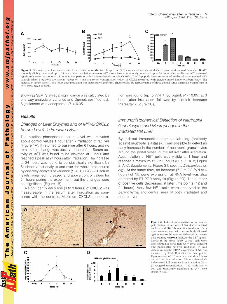
The alkaline phosphatase serum level was elevatedabove control values 1 hour after � irradiation of rat liver(Figure 1A). It returned to baseline after 6 hours, and noremarkable change was observed thereafter. Serum ac-tivity of AST was found to be elevated at 1 hour andreached a peak at 24 hours after irradiation. The increaseat 24 hours was found to be statistically significant byStudent’s t-test analysis and over the whole-time-courseby one-way analysis of variance (P � 0.0004). ALT serumlevels remained increased and above control values for24 hours during the experiment, but the changes werenot significant (Figure 1B).
A significantly early rise (1 to 3 hours) of CXCL2 wasmeasureable in the serum after irradiation as com-pared with the controls. Maximum CXCL2 concentra-
tion was found (up to 774 � 80 pg/ml; P � 0.05) at 3hours after irradiation, followed by a quick decreasethereafter (Figure 1C).
Immunohistochemical Detection of NeutrophilGranulocytes and Macrophages in theIrradiated Rat Liver
By indirect immunohistochemical labeling (antibodyagainst neutrophil elastase), it was possible to detect anearly increase in the number of neutrophil granulocytesaround the portal vessel of the rat liver after irradiation.Accumulation of NE� cells was visible at 1 hour andreached a maximum at 3 to 6 hours (60.2 � 16.8; Figure2, A–C; Supplemental Figure S1, see http://ajp.amjpathol.org). At the same time, an increase (7.2 � 2.3-fold at 6hours) of NE gene expression at RNA level was alsodetected by RT-PCR analysis (Figure 2D). The numberof positive cells decreased at later time points (12 and24 hours). Very few NE� cells were observed in theparenchyma and central area of both irradiated andcontrol livers.
Figure 1. Serum enzyme levels in rats after liver irradiation. A: Alkaline phosphatase (AP) serum level was elevated after 1 hour but decreased thereafter. B: ALTwas only slightly increased up to 24 hours after irradiation, whereas AST serum level continuously increased up to 24 hours after irradiation. AST increasedsignificantly to its maximum at 24 hours in comparison with sham-irradiated controls. C: MIP-2/CXCL2-peptide levels in serum of irradiated rats compared withcontrols (sham-irradiated) are shown. Values on y axis are serum concentration values of CXCL2 measured with enzyme-linked immunosorbent assay. Theincrease in serum levels 1 to 3 hours after irradiation was statistically significant. These results are representative of three animal series (statistically significant at*P � 0.05; mean � SEM).
Figure 2. Indirect immunodetection of neutro-phil elastase in sections of (A) sham-irradiatedrat liver and (B) 6 hours after irradiation. Sec-tions were stained with an antibody directedagainst neutrophil elastase followed by peroxi-dase staining (arrows indicate the NE� granu-locytes in the portal field). C: NE� cells werealso counted in portal field (N � 10) at differenttime points after rat liver irradiation. D: Foldchange in hepatic mRNA expression of NE wasmeasured by RT-PCR at different time points.Up-regulation of NE was detected after 1 hourand reached its maximum at 6 hours, after whichit decreased following rat liver irradiation (N �3). Original magnification, 200. Scale bar �100 �m. Statistically significant at *P � 0.05(mean � SEM).
Role of Chemokines after �-Irradiation 5AJP April 2010, Vol. 176, No. 4
Identification of Recruited Inflammatory Cells byImmunoflourescence Double Staining
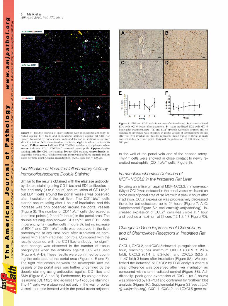
Similar to the results obtained with the elastase antibody,by double staining using CD11b/c and ED1 antibodies, afast and early (3 to 6 hours) accumulation of CD11b/c�
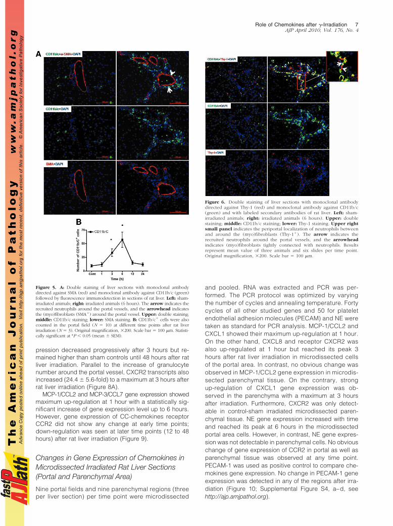
but ED1� cells around the portal vessels was observedafter irradiation of the rat liver. The CD11b/c� cellsstarted accumulating after 1 hour of irradiation, and thisincrease was only observed around the portal vessels(Figure 3). The number of CD11b/c� cells decreased atlater time points (12 and 24 hours) in the portal area. Thedouble staining also showed CD11b/c� and ED1� cellsin parenchyma (Kupffer cells; Figure 3), but no increaseof ED1� and CD11b/c� cells was observed in the liverparenchyma at any time point after irradiation as com-pared with sham-irradiated controls. Compared with theresults obtained with the CD11b/c antibody, no signifi-cant change was observed in the number of tissuephagocytes when the antibody against ED2 was used(Figure 4, A–D). These results were confirmed by count-ing the cells around the portal area (Figure 4, E and F).The tight relationship between the neutrophils and thevessels of the portal area was further underlined by thedouble staining using antibodies against CD11b/c andSMA (Figure 5, A and B). Furthermore, by using antibod-ies against CD11b/c and against Thy-1 (double staining),Thy-1� cells were observed not only in the wall of portalvessels but also located within the portal tracts adjacent
to the wall of the portal vein and of the hepatic artery.Thy-1� cells were showed in close contact to newly re-cruited neutrophils (CD11b/c� cells; Figure 6).
Immunohistochemical Detection ofMCP-1/CCL2 in the Irradiated Rat Liver
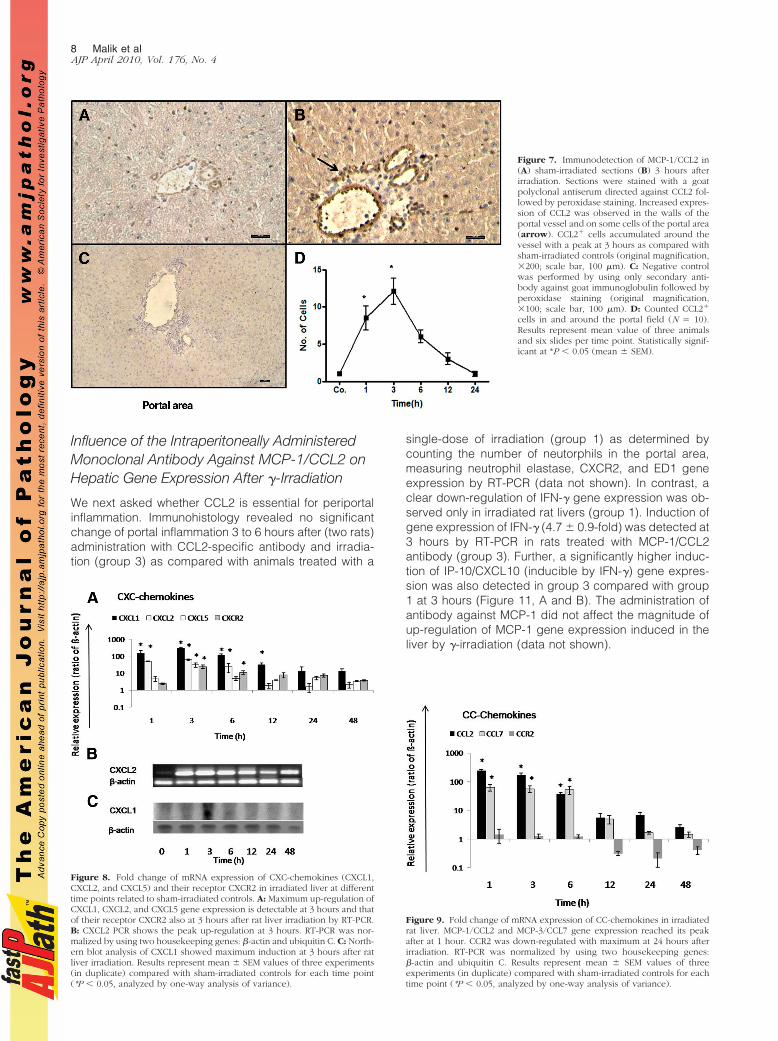
By using an antiserum against MCP-1/CCL2, immune-reac-tivity of CCL2 was detected in the portal vessel walls and onsome cells of portal area of rat liver with a peak 3 hours afterirradiation. CCL2 expression was progressively decreasedthereafter but detectable up to 24 hours (Figure 7, A–C;Supplemental Figure S2, see http://ajp.amjpathol.org). In-creased expression of CCL2� cells was visible at 1 hourand reached a maximum at 3 hours (12.1 � 1.7; Figure 7D).
Changes in Gene Expression of Chemokinesand of Chemokines-Receptors in Irradiated RatLiver
CXCL1, CXCL2, and CXCL5 showed up-regulation after 1hour, reaching their maximum CXCL1 (308.9 � 28.8-fold), CXCL2 (61.4 � 5.3-fold), and CXCL5 (52.5 �11.47-fold) 3 hours after irradiation (Figure 8A). We con-firmed the induction of CXCL2 by PCR analysis where aclear difference was observed after liver irradiation ascompared with sham-irradiated control (Figure 8B). Ad-ditionally, peak gene expression of CXCL1 (at 3 hours)was observed by RT-PCR and confirmed by Northern blotanalysis (Figure 8C; Supplemental Figure S3 see http://ajp.amjpathol.org). CXCL1, CXCL2, and CXCL5 gene ex-
Figure 3. Double staining of liver sections with monoclonal antibody di-rected against ED1 (red) and monoclonal antibody against rat CD11b/c(green) followed by fluorescence immunodetection in sections of rat liverafter irradiation. Left: sham-irradiated animals; right: irradiated animals (6hours). Yellow arrow indicates ED1 CD11b/c resident macrophages; whitearrow indicates ED1� CD11b/c� recruited neutrophils. Upper: doublestaining; middle: CD11b/c staining; lower: ED1 staining (arrowheads in-dicate the portal area). Results represent mean value of three animals and sixslides per time point. Original magnification, 200. Scale bar � 100 �m.
Figure 4. ED1 and ED2� cells in rat liver after irradiation. A: sham-irradiatedED1 cells (C) 6 hours after treatment. B: sham-irradiated ED2 cells (D) 6hours after treatment. ED1� (E) and ED2� (F) cells were also counted and nosignificant difference was observed in portal vessels at different time pointsafter rat liver irradiation. Results represent mean value of three animalsand six slides per time point. Original magnification, 100. Scale bar �100 �m.
6 Malik et alAJP April 2010, Vol. 176, No. 4
pression decreased progressively after 3 hours but re-mained higher than sham controls until 48 hours after ratliver irradiation. Parallel to the increase of granulocytenumber around the portal vessel, CXCR2 transcripts alsoincreased (24.4 � 5.6-fold) to a maximum at 3 hours afterrat liver irradiation (Figure 8A).
MCP-1/CCL2 and MCP-3/CCL7 gene expression showedmaximum up-regulation at 1 hour with a statistically sig-nificant increase of gene expression level up to 6 hours.However, gene expression of CC-chemokines receptorCCR2 did not show any change at early time points;down-regulation was seen at later time points (12 to 48hours) after rat liver irradiation (Figure 9).
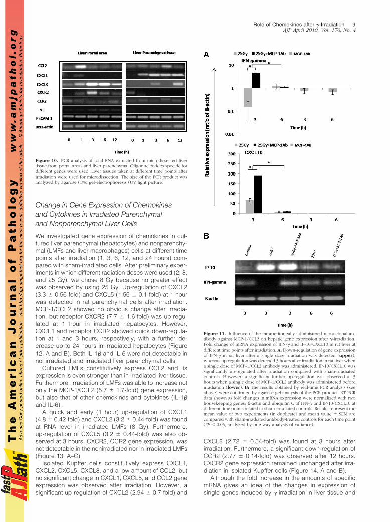
Changes in Gene Expression of Chemokines inMicrodissected Irradiated Rat Liver Sections(Portal and Parenchymal Area)
Nine portal fields and nine parenchymal regions (threeper liver section) per time point were microdissected
and pooled. RNA was extracted and PCR was per-formed. The PCR protocol was optimized by varyingthe number of cycles and annealing temperature. Fortycycles of all other studied genes and 50 for plateletendothelial adhesion molecules (PECAM) and NE weretaken as standard for PCR analysis. MCP-1/CCL2 andCXCL1 showed their maximum up-regulation at 1 hour.On the other hand, CXCL8 and receptor CXCR2 wasalso up-regulated at 1 hour but reached its peak 3hours after rat liver irradiation in microdissected cellsof the portal area. In contrast, no obvious change wasobserved in MCP-1/CCL2 gene expression in microdis-sected parenchymal tissue. On the contrary, strongup-regulation of CXCL1 gene expression was ob-served in the parenchyma with a maximum at 3 hoursafter irradiation. Furthermore, CXCR2 was only detect-able in control-sham irradiated microdissected paren-chymal tissue. NE gene expression increased with timeand reached its peak at 6 hours in the microdissectedportal area cells. However, in contrast, NE gene expres-sion was not detectable in parenchymal cells. No obviouschange of gene expression of CCR2 in portal as well asparenchymal tissue was observed at any time point.PECAM-1 was used as positive control to compare che-mokines gene expression. No change in PECAM-1 geneexpression was detected in any of the regions after irra-diation (Figure 10; Supplemental Figure S4, a–d, seehttp://ajp.amjpathol.org).
Figure 5. A: Double staining of liver sections with monoclonal antibodydirected against SMA (red) and monoclonal antibody against CD11b/c (green)followed by fluorescence immunodetection in sections of rat liver. Left: sham-irradiated animals; right: irradiated animals (6 hours). The arrow indicates therecruited neutrophils around the portal vessels, and the arrowhead indicatesthe (myo)fibroblasts (SMA�) around the portal vessel. Upper: double staining;middle: CD11b/c staining; lower: SMA staining. B: CD11b/c� cells were alsocounted in the portal field (N � 10) at different time points after rat liverirradiation (N � 3). Original magnification, 200. Scale bar � 100 �m. Statisti-cally significant at *P � 0.05 (mean � SEM).
Figure 6. Double staining of liver sections with monoclonal antibodydirected against Thy-1 (red) and monoclonal antibody against CD11b/c(green) and with labeled secondary antibodies of rat liver. Left: sham-irradiated animals; right: irradiated animals (6 hours). Upper: doublestaining; middle: CD11b/c staining; lower: Thy-1 staining. Upper rightsmall panel indicates the periportal localization of neutrophils betweenand around the (myo)fibroblasts (Thy-1�). The arrow indicates therecruited neutrophils around the portal vessels, and the arrowheadindicates (myo)fibroblasts tightly connected with neutrophils. Resultsrepresent mean value of three animals and six slides per time point.Original magnification, 200. Scale bar � 100 �m.
Role of Chemokines after �-Irradiation 7AJP April 2010, Vol. 176, No. 4
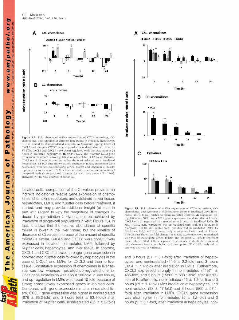
Influence of the Intraperitoneally AdministeredMonoclonal Antibody Against MCP-1/CCL2 onHepatic Gene Expression After �-Irradiation
We next asked whether CCL2 is essential for periportalinflammation. Immunohistology revealed no significantchange of portal inflammation 3 to 6 hours after (two rats)administration with CCL2-specific antibody and irradia-tion (group 3) as compared with animals treated with a
single-dose of irradiation (group 1) as determined bycounting the number of neutorphils in the portal area,measuring neutrophil elastase, CXCR2, and ED1 geneexpression by RT-PCR (data not shown). In contrast, aclear down-regulation of IFN-� gene expression was ob-served only in irradiated rat livers (group 1). Induction ofgene expression of IFN-� (4.7 � 0.9-fold) was detected at3 hours by RT-PCR in rats treated with MCP-1/CCL2antibody (group 3). Further, a significantly higher induc-tion of IP-10/CXCL10 (inducible by IFN-�) gene expres-sion was also detected in group 3 compared with group1 at 3 hours (Figure 11, A and B). The administration ofantibody against MCP-1 did not affect the magnitude ofup-regulation of MCP-1 gene expression induced in theliver by �-irradiation (data not shown).
Figure 7. Immunodetection of MCP-1/CCL2 in(A) sham-irradiated sections (B) 3 hours afterirradiation. Sections were stained with a goatpolyclonal antiserum directed against CCL2 fol-lowed by peroxidase staining. Increased expres-sion of CCL2 was observed in the walls of theportal vessel and on some cells of the portal area(arrow). CCL2� cells accumulated around thevessel with a peak at 3 hours as compared withsham-irradiated controls (original magnification,200; scale bar, 100 �m). C: Negative controlwas performed by using only secondary anti-body against goat immunoglobulin followed byperoxidase staining (original magnification,100; scale bar, 100 �m). D: Counted CCL2�
cells in and around the portal field (N � 10).Results represent mean value of three animalsand six slides per time point. Statistically signif-icant at *P � 0.05 (mean � SEM).
Figure 8. Fold change of mRNA expression of CXC-chemokines (CXCL1,CXCL2, and CXCL5) and their receptor CXCR2 in irradiated liver at differenttime points related to sham-irradiated controls. A: Maximum up-regulation ofCXCL1, CXCL2, and CXCL5 gene expression is detectable at 3 hours and thatof their receptor CXCR2 also at 3 hours after rat liver irradiation by RT-PCR.B: CXCL2 PCR shows the peak up-regulation at 3 hours. RT-PCR was nor-malized by using two housekeeping genes: �-actin and ubiquitin C. C: North-ern blot analysis of CXCL1 showed maximum induction at 3 hours after ratliver irradiation. Results represent mean � SEM values of three experiments(in duplicate) compared with sham-irradiated controls for each time point(*P � 0.05, analyzed by one-way analysis of variance).
Figure 9. Fold change of mRNA expression of CC-chemokines in irradiatedrat liver. MCP-1/CCL2 and MCP-3/CCL7 gene expression reached its peakafter at 1 hour. CCR2 was down-regulated with maximum at 24 hours afterirradiation. RT-PCR was normalized by using two housekeeping genes:�-actin and ubiquitin C. Results represent mean � SEM values of threeexperiments (in duplicate) compared with sham-irradiated controls for eachtime point (*P � 0.05, analyzed by one-way analysis of variance).
8 Malik et alAJP April 2010, Vol. 176, No. 4
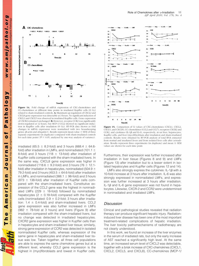
Change in Gene Expression of Chemokinesand Cytokines in Irradiated Parenchymaland Nonparenchymal Liver Cells
We investigated gene expression of chemokines in cul-tured liver parenchymal (hepatocytes) and nonparenchy-mal (LMFs and liver macrophages) cells at different timepoints after irradiation (1, 3, 6, 12, and 24 hours) com-pared with sham-irradiated cells. After preliminary exper-iments in which different radiation doses were used (2, 8,and 25 Gy), we chose 8 Gy because no greater effectwas observed by using 25 Gy. Up-regulation of CXCL2(3.3 � 0.56-fold) and CXCL5 (1.56 � 0.1-fold) at 1 hourwas detected in rat parenchymal cells after irradiation.MCP-1/CCL2 showed no obvious change after irradia-tion, but receptor CXCR2 (7.7 � 1.6-fold) was up-regu-lated at 1 hour in irradiated hepatocytes. However,CXCL1 and receptor CCR2 showed quick down-regula-tion at 1 and 3 hours, respectively, with a further de-crease up to 24 hours in irradiated hepatocytes (Figure12, A and B). Both IL-1� and IL-6 were not detectable innonirradiated and irradiated liver parenchymal cells.
Cultured LMFs constitutively express CCL2 and itsexpression is even stronger than in irradiated liver tissue.Furthermore, irradiation of LMFs was able to increase notonly the MCP-1/CCL2 (5.7 � 1.7-fold) gene expression,but also that of other chemokines and cytokines (IL-1�and IL-6).
A quick and early (1 hour) up-regulation of CXCL1(4.8 � 0.42-fold) and CXCL2 (3.2 � 0.44-fold) was foundat RNA level in irradiated LMFs (8 Gy). Furthermore,up-regulation of CXCL5 (3.2 � 0.44-fold) was also ob-served at 3 hours. CXCR2, CCR2 gene expression, wasnot detectable in the nonirradiated nor in irradiated LMFs(Figure 13, A–C).
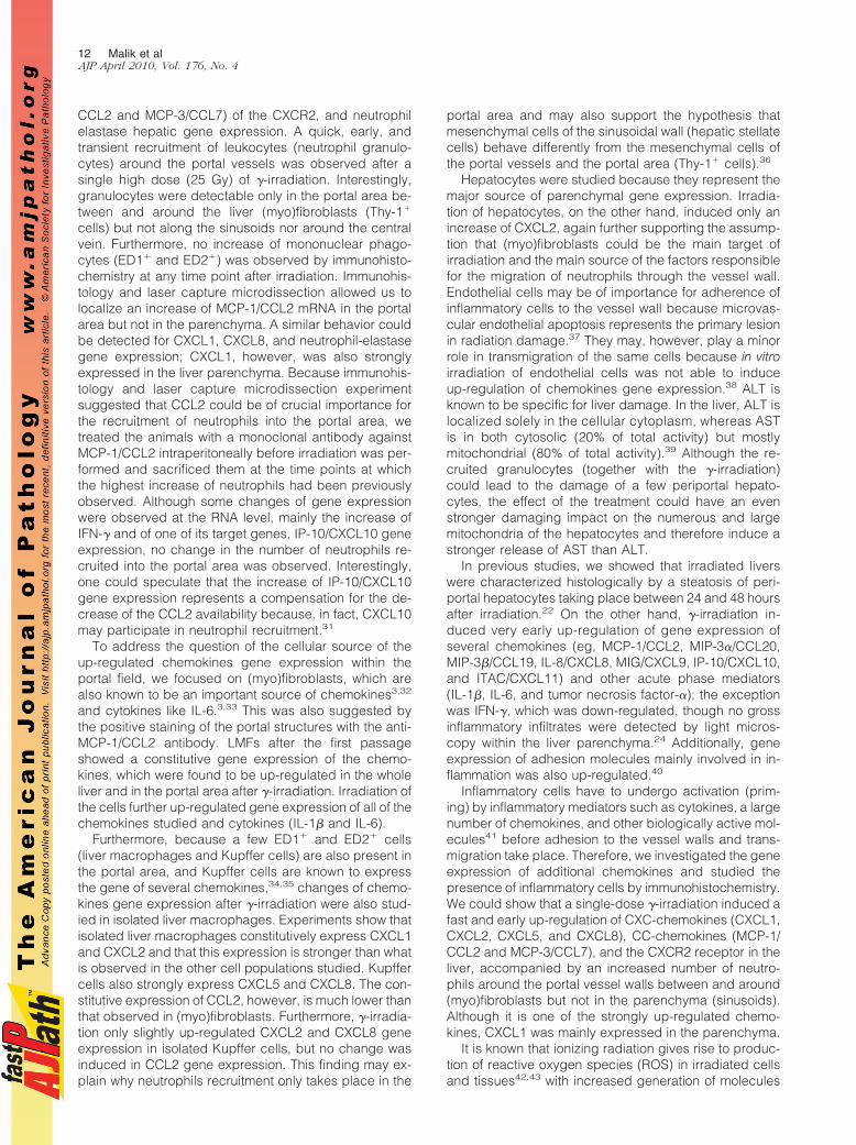
Isolated Kupffer cells constitutively express CXCL1,CXCL2, CXCL5, CXCL8, and a low amount of CCL2, butno significant change in CXCL1, CXCL5, and CCL2 geneexpression was observed after irradiation. However, asignificant up-regulation of CXCL2 (2.94 � 0.7-fold) and
CXCL8 (2.72 � 0.54-fold) was found at 3 hours afterirradiation. Furthermore, a significant down-regulation ofCCR2 (2.77 � 0.14-fold) was observed after 12 hours.CXCR2 gene expression remained unchanged after irra-diation in isolated Kupffer cells (Figure 14, A and B).
Although the fold increase in the amounts of specificmRNA gives an idea of the changes in expression ofsingle genes induced by �-irradiation in liver tissue and
Figure 10. PCR analysis of total RNA extracted from microdissected livertissue from portal areas and liver parenchyma. Oligonucleotides specific fordifferent genes were used. Liver tissues taken at different time points afterirradiation were used for microdissection. The size of the PCR product wasanalyzed by agarose (1%) gel-electrophoresis (UV light picture).
Figure 11. Influence of the intraperitoneally administered monoclonal an-tibody against MCP-1/CCL2 on hepatic gene expression after �-irradiation.Fold change of mRNA expression of IFN-� and IP-10/CXCL10 in rat liver atdifferent time points after irradiation. A: Down-regulation of gene expressionof IFN-� in rat liver after a single dose irradiation was detected (upper),whereas up-regulation was detected 3 hours after irradiation in rat liver whena single dose of MCP-1/CCL2 antibody was administered. IP-10/CXCL10 wassignificantly up-regulated after irradiation compared with sham-irradiatedcontrols. However, a significant further up-regulation was observed at 3hours when a single dose of MCP-1/CCL2 antibody was administered beforeirradiation (lower). B: The results obtained by real-time PCR analysis (seeabove) were confirmed by agarose gel analysis of the PCR product. RT-PCRdata shown as fold changes in mRNA expression were normalized with twohousekeeping genes: �-actin and ubiquitin C of IFN-� and IP-10/CXCL10 atdifferent time points related to sham-irradiated controls. Results represent themean value of two experiments (in duplicate) and mean value � SEM arecompared with sham-irradiated antibody-treated controls for each time point(*P � 0.05, analyzed by one-way analysis of variance).
Role of Chemokines after �-Irradiation 9AJP April 2010, Vol. 176, No. 4
isolated cells, comparison of the Ct values provides anindirect indicator of relative gene expression of chemo-kines, chemokine receptors, and cytokines in liver tissue,hepatocytes, LMFs, and Kupffer cells before treatment, ifstarted, and may provide additional insight (at least inpart with regard to why the magnitude of changes in-duced by �-irradiation in vivo cannot be achieved byirradiation of single-cell populations in vitro; Figure 15). Infact, it shows that the relative abundance of specificmRNA is lower in the liver tissue, but the kinetics ofdecrease of Ct values (increase of the amount of specificmRNA) is similar. CXCL5 and CXCL8 were constitutivelyexpressed in isolated nonirradiated LMFs followed byKupffer cells, hepatocytes, and liver tissue. In contrast,CXCL1 and CXCL2 showed stronger gene expression innonirradiated Kupffer cells followed by hepatocytes in thecase of CXCL1 and LMFs for CXCL2 and then to livertissue. Constitutive expression of chemokines in liver tis-sue was low, whereas irradiated up-regulated chemo-kines gene expression was about 100-fold in liver tissue,and up-regulation in LMFs was about 10-fold because ofstrong constitutively expressed genes in isolated cells.Compared with gene expression in sham-irradiated liv-ers, CXCL1 gene expression was higher in nonirradiated(676 � 65.2-fold) and 3 hours (668 � 83.1-fold) afterirradiation of Kupffer cells, nonirradiated (35 � 5.2-fold)
and 3 hours (21 � 3.1-fold) after irradiation of hepato-cytes, and nonirradiated (11.5 � 2.2-fold) and 3 hours(33.4 � 7.1-fold) after irradiation in LMFs. Furthermore,CXCL2 expressed strongly in nonirradiated (11571 �465-fold) and 3 hours (15862 � 683.1-fold) after irradia-tion of Kupffer cells, nonirradiated (15 � 1.2-fold) and 3hours (29 � 3.1-fold) after irradiation of hepatocytes, andnonirradiated (98 � 17-fold) and 3 hours (565 � 97.1-fold) after irradiation in LMFs. CXCL5 gene expressionwas also higher in nonirradiated (5 � 1.2-fold) and 3hours (9 � 3.1-fold) after irradiation in hepatocytes, non-
Figure 12. Fold change of mRNA expression of CXC-chemokines, CC-chemokines, and cytokines at different time points in irradiated hepatocytes(8 Gy) related to sham-irradiated controls. A: Maximum up-regulation ofCXCL2 and receptor CXCR2 gene expression was detectable at 1 hour byRT-PCR. CXCL1 and CXCL5 were down-regulated with the maximum at 24hours in irradiated hepatocytes. B: MCP-1/CCL2 and receptor CCR2 geneexpression maximum down-regulation was detectable at 12 hours. Cytokine(IL-1� nor IL-6) was detected in neither the nonirradiated nor in irradiatedhepatocytes. RT-PCR data shown as fold changes in mRNA expression werenormalized with two housekeeping genes: �-actin and ubiquitin C. Resultsrepresent the mean value � SEM of three separate experiments (in duplicate)compared with sham-irradiated controls for each time point (*P � 0.05,analyzed by one-way analysis of variance).
Figure 13. Fold change of mRNA expression of CXC-chemokines, CC-chemokines, and cytokines at different time points in irradiated (myo)fibro-blasts (LMFs; 8 Gy) related to sham-irradiated controls. A: Maximum up-regulation of CXCL1 and CXCL2 gene expression was detectable at 1 hour.CXCL5 was up-regulated with maximum at 3 hours in irradiated LMFs. B:MCP-1/CCL2 gene expression was up-regulated with peak at 1 hour. Bothreceptors (CXCR2 and CCR2) were not detected in irradiated LMFs. C:Cytokines, IL-1� and IL-6, were early up-regulated with peak at 1 hour.RT-PCR data shown as fold changes in mRNA expression were normalizedwith two housekeeping genes: �-actin and ubiquitin C. Results representmean value � SEM of three separate experiments (in duplicate) comparedwith sham-irradiated controls for each time point (*P � 0.05, analyzed byone-way analysis of variance).
10 Malik et alAJP April 2010, Vol. 176, No. 4
irradiated (65.5 � 8.2-fold) and 3 hours (668.4 � 64.6-fold) after irradiation in LMFs, and nonirradiated (101.1 �8-fold) and 3 hours (118 � 13-fold) after irradiation ofKupffer cells compared with the sham-irradiated livers. Inthe same way, CXCL8 gene expression was higher innonirradiated (118.6 � 9.2-fold) and 3 hours (76 � 12.1-fold) after irradiation in hepatocytes, nonirradiated (504.9 �79.2-fold) and 3 hours (453.5 � 64.6-fold) after irradiationin LMFs, and nonirradiated (388.1 � 98-fold) and 3 hours(873 � 138-fold) after irradiation of Kupffer cells com-pared with the sham-irradiated livers. Constitutive ex-pression of the CCL2 gene was the highest in nonirradi-ated LMFs (229 � 18-fold) followed by nonirradiatedhepatocytes (2 � 0.18-fold) compared with the Kupffercells (nonirradiated: 0.9 � 0.2-fold; 3 hours after irradia-tion: 1.4 � 0.4-fold) and sham-irradiated livers. CCL2gene expression was also further increased in LMFs(982 � 78-fold at 3 hours) and livers (Figure 9) afterirradiation compared with the sham-irradiated livers, butno change was detected in irradiated hepatocytes.CXCR2 gene expression was higher in Kupffer cells thanin hepatocytes and sham-irradiated liver tissue; similarly,strong gene expression of CCR2 was detected in isolatednonirradiated Kupffer cells, whereas expression of thesame gene in hepatocytes and sham-irradiated liver tis-sue was low. Taken together, all three cell populationsare able to express the same chemokine genes but at adifferent level, whereby CCL2 gene expression is thehighest in (myo)fibroblasts and lowest in Kupffer cells.
Furthermore, their expression was further increased afterirradiation in liver tissue (Figures 8 and 9) and LMFs(Figure 13) after irradiation but to a lesser extent in iso-lated hepatocytes and Kupffer cells (Figures 12 and 14).
LMFs also strongly express the cytokines: IL-1� with a10-fold increase at 3 hours after irradiation. IL-6 was alsostrongly expressed in nonirradiated LMFs, and expres-sion was further increased at 3 hours after irradiation.IL-1� and IL-6 gene expression was not found in hepa-tocytes. Likewise, CXCR-2 and CCR2 were undeterminedin nonirradiated and irradiated LMFs.
Discussion
Clinical and pathological studies revealed that radiationtherapy can produce significant hepatic injury. Radiation-induced liver disease has been one of the most importanttreatment-related complications of hepatic irradiation.The liver toxicity pathomechanisms of radiotherapy arenot clearly understood.
In this work, we found an increase of the liver enzymesin the serum of irradiated rats, but only the early increaseof AST reached a significantly high level. At the sametime, an increased serum level of CXCL2 was detectable,together with a brisk increase of CXC-chemokines (CXCL1,CXCL2, CXCL5, and CXCL8), CC-chemokines (MCP-1/
Figure 14. Fold change of mRNA expression of CXC-chemokines andCC-chemokines at different time points in irradiated Kupffer cells (8 Gy)related to sham-irradiated controls. A: Maximum up-regulation of CXCL2 andCXCL8 gene expression was detectable at 3 hours. No significant induction ofCXCL1 and CXCL5 was observed in irradiated Kupffer cells. Gene expressionof CXCR2 remained unchanged. B: However, receptor CCR2 was significantlydown-regulated at 12 hours, but MCP-1/CCL2 showed no significant induc-tion in Kupffer cells after irradiation (8 Gy). RT-PCR data shown as foldchanges in mRNA expression were normalized with two housekeepinggenes: �-actin and ubiquitin C. Results represent mean value � SEM of threeseparate experiments (in duplicate) compared with sham-irradiated controlsfor each time point (*P � 0.05, analyzed by one-way analysis of variance).
Figure 15. Comparison of Ct values of CXC-chemokines (CXCL1, CXCL2,CXCL5, and CXCL8), CC-chemokines (CCL2 and CCL7), receptors CXCR2 andCCR2, and cytokines (IL-1� and IL-6), respectively, in rat liver, hepatocytes,Kupffer cells, and liver (myo)fibroblasts after irradiation and sham-irradiatedcontrols. Results were obtained by RT-PCR analysis of total RNA extractedfrom normal and irradiated livers and from isolated liver cells after �-irradi-ation. Results represent three experiments (in duplicate) and mean � SEMvalues are shown for each time point.
Role of Chemokines after �-Irradiation 11AJP April 2010, Vol. 176, No. 4
CCL2 and MCP-3/CCL7) of the CXCR2, and neutrophilelastase hepatic gene expression. A quick, early, andtransient recruitment of leukocytes (neutrophil granulo-cytes) around the portal vessels was observed after asingle high dose (25 Gy) of �-irradiation. Interestingly,granulocytes were detectable only in the portal area be-tween and around the liver (myo)fibroblasts (Thy-1�
cells) but not along the sinusoids nor around the centralvein. Furthermore, no increase of mononuclear phago-cytes (ED1� and ED2�) was observed by immunohisto-chemistry at any time point after irradiation. Immunohis-tology and laser capture microdissection allowed us tolocalize an increase of MCP-1/CCL2 mRNA in the portalarea but not in the parenchyma. A similar behavior couldbe detected for CXCL1, CXCL8, and neutrophil-elastasegene expression; CXCL1, however, was also stronglyexpressed in the liver parenchyma. Because immunohis-tology and laser capture microdissection experimentsuggested that CCL2 could be of crucial importance forthe recruitment of neutrophils into the portal area, wetreated the animals with a monoclonal antibody againstMCP-1/CCL2 intraperitoneally before irradiation was per-formed and sacrificed them at the time points at whichthe highest increase of neutrophils had been previouslyobserved. Although some changes of gene expressionwere observed at the RNA level, mainly the increase ofIFN-� and of one of its target genes, IP-10/CXCL10 geneexpression, no change in the number of neutrophils re-cruited into the portal area was observed. Interestingly,one could speculate that the increase of IP-10/CXCL10gene expression represents a compensation for the de-crease of the CCL2 availability because, in fact, CXCL10may participate in neutrophil recruitment.31
To address the question of the cellular source of theup-regulated chemokines gene expression within theportal field, we focused on (myo)fibroblasts, which arealso known to be an important source of chemokines3,32
and cytokines like IL-6.3,33 This was also suggested bythe positive staining of the portal structures with the anti-MCP-1/CCL2 antibody. LMFs after the first passageshowed a constitutive gene expression of the chemo-kines, which were found to be up-regulated in the wholeliver and in the portal area after �-irradiation. Irradiation ofthe cells further up-regulated gene expression of all of thechemokines studied and cytokines (IL-1� and IL-6).
Furthermore, because a few ED1� and ED2� cells(liver macrophages and Kupffer cells) are also present inthe portal area, and Kupffer cells are known to expressthe gene of several chemokines,34,35 changes of chemo-kines gene expression after �-irradiation were also stud-ied in isolated liver macrophages. Experiments show thatisolated liver macrophages constitutively express CXCL1and CXCL2 and that this expression is stronger than whatis observed in the other cell populations studied. Kupffercells also strongly express CXCL5 and CXCL8. The con-stitutive expression of CCL2, however, is much lower thanthat observed in (myo)fibroblasts. Furthermore, �-irradia-tion only slightly up-regulated CXCL2 and CXCL8 geneexpression in isolated Kupffer cells, but no change wasinduced in CCL2 gene expression. This finding may ex-plain why neutrophils recruitment only takes place in the
portal area and may also support the hypothesis thatmesenchymal cells of the sinusoidal wall (hepatic stellatecells) behave differently from the mesenchymal cells ofthe portal vessels and the portal area (Thy-1� cells).36
Hepatocytes were studied because they represent themajor source of parenchymal gene expression. Irradia-tion of hepatocytes, on the other hand, induced only anincrease of CXCL2, again further supporting the assump-tion that (myo)fibroblasts could be the main target ofirradiation and the main source of the factors responsiblefor the migration of neutrophils through the vessel wall.Endothelial cells may be of importance for adherence ofinflammatory cells to the vessel wall because microvas-cular endothelial apoptosis represents the primary lesionin radiation damage.37 They may, however, play a minorrole in transmigration of the same cells because in vitroirradiation of endothelial cells was not able to induceup-regulation of chemokines gene expression.38 ALT isknown to be specific for liver damage. In the liver, ALT islocalized solely in the cellular cytoplasm, whereas ASTis in both cytosolic (20% of total activity) but mostlymitochondrial (80% of total activity).39 Although the re-cruited granulocytes (together with the �-irradiation)could lead to the damage of a few periportal hepato-cytes, the effect of the treatment could have an evenstronger damaging impact on the numerous and largemitochondria of the hepatocytes and therefore induce astronger release of AST than ALT.
In previous studies, we showed that irradiated liverswere characterized histologically by a steatosis of peri-portal hepatocytes taking place between 24 and 48 hoursafter irradiation.22 On the other hand, �-irradiation in-duced very early up-regulation of gene expression ofseveral chemokines (eg, MCP-1/CCL2, MIP-3�/CCL20,MIP-3�/CCL19, IL-8/CXCL8, MIG/CXCL9, IP-10/CXCL10,and ITAC/CXCL11) and other acute phase mediators(IL-1�, IL-6, and tumor necrosis factor-�); the exceptionwas IFN-�, which was down-regulated, though no grossinflammatory infiltrates were detected by light micros-copy within the liver parenchyma.24 Additionally, geneexpression of adhesion molecules mainly involved in in-flammation was also up-regulated.40
Inflammatory cells have to undergo activation (prim-ing) by inflammatory mediators such as cytokines, a largenumber of chemokines, and other biologically active mol-ecules41 before adhesion to the vessel walls and trans-migration take place. Therefore, we investigated the geneexpression of additional chemokines and studied thepresence of inflammatory cells by immunohistochemistry.We could show that a single-dose �-irradiation induced afast and early up-regulation of CXC-chemokines (CXCL1,CXCL2, CXCL5, and CXCL8), CC-chemokines (MCP-1/CCL2 and MCP-3/CCL7), and the CXCR2 receptor in theliver, accompanied by an increased number of neutro-phils around the portal vessel walls between and around(myo)fibroblasts but not in the parenchyma (sinusoids).Although it is one of the strongly up-regulated chemo-kines, CXCL1 was mainly expressed in the parenchyma.
It is known that ionizing radiation gives rise to produc-tion of reactive oxygen species (ROS) in irradiated cellsand tissues42,43 with increased generation of molecules
12 Malik et alAJP April 2010, Vol. 176, No. 4
such as superoxide radical, hydroxyl radical, and H2O2
due to oxidative stress.43 Furthermore, a link betweenROS production after whole body exposure to �-irradia-tion and hepatic injury has been reported in mice.43,44
ROS contribute to the expression of a variety of differentinflammatory cytokines, chemokines, and adhesion mol-ecules45 by activating redox-sensitive transcription fac-tors such as nuclear factor-�B.46
We speculate that ROS act as signaling moleculesleading to modulation of crucial events such as elevationof the inflammasome, which is a multiprotein complexthat promotes secretion of the proinflammatory genesthrough activation of caspase-1.47
On the other hand, radiation-induced injury in the vascu-lar endothelium could in turn decrease oxygenation in nor-mal tissue and lead to hypoxia.48,49 Reduced tissue oxygentension induces activation of signaling pathways, whichalso promotes cell survival largely mediated by a transcrip-tion factor such as hypoxia inducible factor-1 (manuscript inpreparation).50 Further, hypoxia-induced ROS could alsoinduce chemokines and cytokine synthesis as well as up-regulation of adhesion molecules gene(s).45,51,52
In our current study, we showed for the first time alocalized up-regulation of several chemokines, such asMCP-1/CCL2, CXCL1, and IL-8/CXCL8, which is accom-panied by a recruitment of the inflammatory cells limitedto that area. It has been shown that MCP-1/CCL2 servesas an indirect mediator to attract neutrophils through theproduction of LTB4 in a murine model of septic peritoni-tis.53 Additionally, neutralization of MCP-1/CCL2 activityin a lipopolysaccharide model of inflammation resultednot only in decreased monocytes accumulation within theintestinal muscularis, but polymorphonuclear leukocytesinfiltration was also significantly reduced.54 ROS pro-duced by ischemia/reperfusion injury significantly in-creased the mRNA expression of MCP-1/CCL2 in theheart,55 as has also been shown for the hepatotoxincarbon tetrachloride,56 as well as whole lung irradiation inmice.57 IL-8/CXCL8 has been considered to be of crucialimportance for recruitment of granulocytes in differenttissues.7 In previous work,25 however, we were surprisedto find a massive up-regulation of CXCL8 gene expres-sion in the liver of rats treated with turpentine oil intra-muscularly without detecting any granulocyte infiltration.Because LMFs constitutively express most of the chemo-kines and cytokines up-regulated by �-irradiation in theliver and in isolated cells, they could be responsible forrecruitment of neutrophil around the portal vessels. To-gether with the data presented here, our experience sug-gests that contemporary up-regulation of gene expres-sion of several chemokines (mainly in myofibroblasts)may be necessary to induce recruitment of inflammatorycells in a localized area beginning around the walls ofvessels. In fact, intraperitoneal administration of MCP-1/CCL2 antibody 30 minutes before irradiation reverted theinhibition of IFN-� gene expression in the liver but did notcontribute to a significant reduction of the number ofneutrophils. However, we cannot rule out that a higheramount of antibody administration earlier or later, beforeor during irradiation, may inhibit recruitment of neutro-phils into the portal field. The data may suggest that
contemporary administration of antibodies specific for thedifferent chemokines is necessary to reduce neutrophilsmigration into the portal area. Based on these results, wecan speculate that CCL2 has an inhibitory effect on IFN-�gene expression, as has been observed in T-cells.58 Thereason for the lack of increase of macrophages and theconsequence of this phenomenon will be a matter for futureinvestigation.
In conclusion, we found recruited neutrophils but nomononuclear phagocytes attached to the portal vesselsand to portal (myo)fibroblasts in the liver of irradiatedrats. Furthermore, fast and early induction of gene ex-pression of several chemokines (CXCL1, CXCL2, CXCL5,CXCL8, CCL2, and CCL7) and the chemokines receptorCXCR2 gene expression in irradiated liver tissue andmicrodissected cells of the portal area were observed.Several chemokines may be necessary in neutrophilsrecruitment, adhesion, and transmigration induced by�-irradiation in the rat liver. The induction of the mediatorsin cells of portal area (mainly myofibroblasts) may hap-pen through molecules such as ROS. Because tissuedamage observed under these conditions is of limitedextent, the often reported sensitivity of liver tissue to�-irradiation may be due to the frequently concomitantadministration of chemotherapy or to the presence ofchronic liver disease.
Acknowledgments
We wish to acknowledge the invaluable help of Mrs SonjaHeyroth and Mrs Anke Herbst for their expert technicalassistance.
This work is dedicated to Professor Dr. Meyer zumBuschenfelde on the occasion of his 80th birthday.
References
1. Shim SJ, Seong J, Lee IJ, Han KH, Chon CY, Ahn SH: Radiation-induced hepatic toxicity after radiotherapy combined with chemother-apy for hepatocellular carcinoma. Hepatol Res 2007, 37:906–913
2. Cheng JC, Wu JK, Huang CM, Huang DY, Cheng SH, Lin YM, Jian JJ,Yang PS, Chuang VP, Huang AT: Radiation-induced liver diseaseafter radiotherapy for hepatocellular carcinoma: clinical manifestationand dosimetric description. Radiother Oncol 2002, 63:41–45
3. Marra F: Chemokines in liver inflammation and fibrosis. Front Biosci2002, 7:d1899–d1914
4. Bozic CR, Kolakowski LF Jr, Gerard NP, Garcia-Rodriguez C, vonUexkull-Guldenband C, Conklyn MJ, Breslow R, Showell HJ, GerardC: Expression and biologic characterization of the murine chemokineKC. J Immunol 1995, 154:6048–6057
5. Rossi DL, Hurst SD, Xu Y, Wang W, Menon S, Coffman RL, Zlotnik A:Lungkine, a novel CXC chemokine, specifically expressed by lungbronchoepithelial cells. J Immunol 1999, 162:5490–5497
6. Wolpe SD, Sherry B, Juers D, Davatelis G, Yurt RW, Cerami A:Identification and characterization of macrophage inflammatory pro-tein 2. Proc Natl Acad Sci USA 1989, 86:612–616
7. Harris JG, Flower RJ, Watanabe K, Tsurufuji S, Wolitzky BA, PerrettiM: Relative contribution of the selectins in the neutrophil recruitmentcaused by the chemokine cytokine-induced neutrophil chemoattrac-tant (CINC). Biochem Biophys Res Commun 1996, 221:692–696
8. Ajuebor MN, Flower RJ, Hannon R, Christie M, Bowers K, Verity A,Perretti M: Endogenous monocyte chemoattractant protein-1 recruitsmonocytes in the zymosan peritonitis model. J Leukoc Biol 1998,63:108–116
Role of Chemokines after �-Irradiation 13AJP April 2010, Vol. 176, No. 4
9. Frangogiannis NG, Smith CW, Entman ML: The inflammatory re-sponse in myocardial infarction. Cardiovasc Res 2002, 53:31–47
10. Baggiolini M: Chemokines and leukocyte traffic. Nature 1998,392:565–568
11. Chandrasekar B, Smith JB, Freeman GL: Ischemia-reperfusion of ratmyocardium activates nuclear factor-KappaB and induces neutrophilinfiltration via lipopolysaccharide-induced CXC chemokine. Circula-tion 2001, 103:2296–2302
12. Gerard C, Rollins BJ: Chemokines and disease. Nat Immunol 2001,2:108–115
13. Woo CW, Siow YLOK: Homocysteine induces monocyte chemoat-tractant protein-1 expression in hepatocytes mediated via activatorprotein-1 activation. J Biol Chem 2008, 283: 1282–1292
14. Yoshimura T, Takahashi M: IFN-gamma-mediated survival enableshuman neutrophils to produce MCP-1/CCL2 in response to activationby TLR ligands. J Immunol 2007, 179:1942–1949
15. Izikson L, Klein RS, Charo IF, Weiner HL, Luster AD: Resistance toexperimental autoimmune encephalomyelitis in mice lacking the CCchemokine receptor (CCR)2. J Exp Med 2000, 192:1075–1080
16. Peters W, Scott HM, Chambers HF, Flynn JL, Charo IF, Ernst JD:Chemokine receptor 2 serves an early and essential role in resistanceto Mycobacterium tuberculosis. Proc Natl Acad Sci USA 2001,98:7958–7963
17. Boring L, Gosling J, Cleary M, Charo IF: Decreased lesion formationin CCR2�/� mice reveals a role for chemokines in the initiation ofatherosclerosis. Nature 1998, 394:894–897
18. Hogaboam CM, Bone-Larson CL, Steinhauser ML, Matsukawa A,Gosling J, Boring L, Charo IF, Simpson KJ, Lukacs NW, Kunkel SL:Exaggerated hepatic injury due to acetaminophen challenge in micelacking C-C chemokine receptor 2. Am J Pathol 2000, 156:1245–1252
19. Tsou CL, Peters W, Si Y, Slaymaker S, Aslanian AM, Weisberg SP,Mack M, Charo IF: Critical roles for CCR2 and MCP-3 in monocytemobilization from bone marrow and recruitment to inflammatory sites.J Clin Invest 2007, 117:902–909
20. Tessier PA, Naccache PH, Clark-Lewis I, Gladue RP, Neote KS,McColl SR: Chemokine networks in vivo: involvement of C-X-C andC-C chemokines in neutrophil extravasation in vivo in response toTNF-alpha. J Immunol 1997, 159:3595–3602
21. Lentsch AB, Yoshidome H, Cheadle WG, Miller FN, Edwards MJ:Chemokine involvement in hepatic ischemia/reperfusion injury inmice: roles for macrophage inflammatory protein-2 and Kupffer cells.Hepatology 1998, 27:507–512
22. Christiansen H, Batusic D, Saile B, Hermann RM, Dudas J, Rave-Frank M, Hess CF, Schmidberger H, Ramadori G: Identification ofgenes responsive to gamma radiation in rat hepatocytes and rat liverby cDNA array gene expression analysis. Radiat Res 2006,165:318–325
23. Christiansen H, Sheikh N, Saile B, Reuter F, Rave-Frank M, HermannRM, Dudas J, Hille A, Hess CF, Ramadori G: x-Irradiation in rat liver:consequent upregulation of hepcidin and downregulation of hemojuvelinand ferroportin-1 gene expression. Radiology 2007, 242:189–197
24. Moriconi F, Christiansen H, Raddatz D, Dudas J, Hermann RM,Rave-Frank M, Sheikh N, Saile B, Hess CF, Ramadori G: Effect ofradiation on gene expression of rat liver chemokines: in vivo and invitro studies. Radiat Res 2008, 169:162–169
25. Sheikh N, Tron K, Dudas J, Ramadori G: Cytokine-induced neutrophilchemoattractant-1 is released by the noninjured liver in a rat acute-phase model. Lab Invest 2006, 86:800–814
26. Dudas J, Mansuroglu T, Batusic D, Saile B, Ramadori G: Thy-1 is anin vivo and in vitro marker of liver myofibroblasts. Cell Tissue Res 2007,329:503–514
27. Ramadori G, Sipe JD, Dinarello CA, Mizel SB, Colten HR: Pretrans-lational modulation of acute phase hepatic protein synthesis by mu-rine recombinant interleukin 1 (IL-1) and purified human IL-1. J ExpMed 1985, 162:930–942
28. Espina V, Wulfkuhle JD, Calvert VS, VanMeter A, Zhou W, Coukos G,Geho DH, Petricoin EF, III, Liotta LA: Laser-capture microdissection.Nat Protoc 2006, 1:586–603
29. Ramadori G, Moebius U, Dienes HP, Meuer S, Meyer Zum Buschen-felde KH: Lymphocytes from hepatic inflammatory infiltrate kill rathepatocytes in primary culture. Comparison with peripheral bloodlymphocytes. Virchows Arch B Cell Pathol Incl Mol Pathol 1990,59:263–270
30. Tello K, Christiansen H, Gurleyen H, Dudas J, Rave-Frank M, Hess
CF, Ramadori G, Saile B: Irradiation leads to apoptosis of Kupffercells by a Hsp27-dependant pathway followed by release of TNF-alpha. Radiat Environ Biophys 2008, 47:389–397
31. Taub DD, Longo DL, Murphy WJ: Human interferon-inducible pro-tein-10 induces mononuclear cell infiltration in mice and promotes themigration of human T lymphocytes into the peripheral tissues andhuman peripheral blood lymphocytes-SCID mice. Blood 1996,87:1423–1431
32. Sprenger H, Kaufmann A, Garn H, Lahme B, Gemsa D, Gressner AM:Induction of neutrophil-attracting chemokines in transforming rat he-patic stellate cells. Gastroenterology 1997, 113:277–285
33. Knittel T, Kobold D, Saile B, Grundmann A, Neubauer K, Piscaglia F,Ramadori G: Rat liver myofibroblasts and hepatic stellate cells: dif-ferent cell populations of the fibroblast lineage with fibrogenic poten-tial. Gastroenterology 1999, 117:1205–1221
34. Krohn N, Kapoor S, Enami Y, Follenzi A, Bandi S, Joseph B, Gupta S:Hepatocyte transplantation-induced liver inflammation is driven bycytokines-chemokines associated with neutrophils and Kupffer cells.Gastroenterology 2009, 136:1806–1817
35. Mosher B, Dean R, Harkema J, Remick D, Palma J, Crockett E:Inhibition of Kupffer cells reduced CXC chemokine production andliver injury. J Surg Res 2001, 99:201–210
36. Dudas J, Mansuroglu T, Batusic D, Ramadori G: Thy-1 is expressedin myofibroblasts but not found in hepatic stellate cells following liverinjury. Histochem Cell Biol 2009, 131:115–127
37. Maj JG, Paris F, Haimovitz-Friedman A, Venkatraman E, Kolesnick R,Fuks Z: Microvascular function regulates intestinal crypt response toradiation. Cancer Res 2003, 63:4338–4341
38. Gaugler MH, Squiban C, Claraz M, Schweitzer K, Weksler B, Gour-melon P, Van der Meeren A: Characterization of the response ofhuman bone marrow endothelial cells to in vitro irradiation. Br JHaematol 1998, 103:980–989
39. Rej R: Aminotransferases in disease. Clin Lab Med 1989, 9:667–68740. Moriconi F, Malik I, Ahmad G, Dudas J, Rave-Frank M, Vorwerk H,
Hille A, Hess CF, Ramadori G, Christiansen H: Effect of irradiation ongene expression of rat liver adhesion molecules: in vivo and in vitrostudies. Strahlenther Onkol 2009, 185:460–468
41. Ramaiah SK, Jaeschke H: Role of neutrophils in the pathogenesis ofacute inflammatory liver injury. Toxicol Pathol 2007, 35:757–766
42. Leach JK, Van TG, Lin PS, Schmidt-Ullrich R, Mikkelsen RB: Ionizingradiation-induced, mitochondria-dependent generation of reactiveoxygen/nitrogen. Cancer Res 2001, 61:3894–3901
43. Riley PA: Free radicals in biology: oxidative stress and the effects ofionizing radiation. Int J Radiat Biol 1994, 65:27–33
44. An JH, Kim J, Seong J: Redox signaling by ionizing radiation in mouseliver. Ann NY Acad Sci 2004, 1030:86–94
45. Lin BR, Yu CJ, Chen WC, Lee HS, Chang HM, Lee YC, Chien CT,Chen CF: Green tea extract supplement reduces D-galactosamine-induced acute liver injury by inhibition of apoptotic and proinflamma-tory signaling. J Biomed Sci 2009, 16:35
46. Droge W: Free radicals in the physiological control of cell function.Physiol Rev 2002, 82:47–95
47. Franchi L, Eigenbrod T, Munoz-Planillo R, Nunez G: The inflammasome:a caspase-1-activation platform that regulates immune responses anddisease pathogenesis. Nat Immunol 2009, 10:241–247
48. Kiani MF, Ansari R, Gaber MW: Oxygen delivery in irradiated normaltissue. J Radiat Res (Tokyo) 2003, 44:15–21
49. Stone HB, McBride WH, Coleman CN: Modifying normal tissue dam-age postirradiation: report of a workshop sponsored by the RadiationResearch Program, National Cancer Institute, Bethesda, Maryland,September 6–8, 2000. Radiat Res 2002, 157:204–223
50. Semenza GL, Agani F, Feldser D, Iyer N, Kotch L, Laughner E, Yu A:Hypoxia, HIF-1, and the pathophysiology of common human dis-eases. Adv Exp Med Biol 2000, 475:123–130
51. Bremer C, Bradford BU, Hunt KJ, Knecht KT, Connor HD, Mason RP,Thurman RG: Role of Kupffer cells in the pathogenesis of hepaticreperfusion injury. Am J Physiol 1994, 267:G630–G636
52. Ghezzi P, Dinarello CA, Bianchi M, Rosandich ME, Repine JE, WhiteCW: Hypoxia increases production of interleukin-1 and tumor necro-sis factor by human mononuclear cells. Cytokine 1991, 3:189–194
53. Matsukawa A, Hogaboam CM, Lukacs NW, Lincoln PM, Strieter RM,Kunkel SL: Endogenous monocyte chemoattractant protein-1 (MCP-1)
14 Malik et alAJP April 2010, Vol. 176, No. 4
protects mice in a model of acute septic peritonitis: cross-talk betweenMCP-1 and leukotriene B4. J Immunol 1999, 163:6148–6154
54. Turler A, Schwarz NT, Turler E, Kalff JC, Bauer AJ: MCP-1 causesleukocyte recruitment and subsequently endotoxemic ileus in rat.Am J Physiol Gastrointest Liver Physiol 2002, 282:G145–G155
55. Morimoto H, Hirose M, Takahashi M, Kawaguchi M, Ise H,Kolattukudy PE, Yamada M, Ikeda U: MCP-1 induces cardioprotec-tion against ischaemia/reperfusion injury: role of reactive oxygenspecies. Cardiovasc Res 2008, 78:554–562
56. Czaja MJ, Geerts A, Xu J, Schmiedeberg P, Ju Y: Monocyte chemoat-
tractant protein 1 (MCP-1) expression occurs in toxic rat liver injuryand human liver disease. J Leukoc Biol 1994, 55:120–126
57. Ao X, Zhao L, Davis MA, Lubman DM, Lawrence TS, Kong FM:Radiation produces differential changes in cytokine profiles inradiation lung fibrosis sensitive and resistant mice. J HematolOncol 2009, 2:6
58. Hogaboam CM, Lukacs NW, Chensue SW, Strieter RM, Kunkel SL:Monocyte chemoattractant protein-1 synthesis by murine lung fi-broblasts modulates CD4� T cell activation. J Immunol 1998,160:4606 – 4614
Role of Chemokines after �-Irradiation 15AJP April 2010, Vol. 176, No. 4
Copyright © 2022 FDOKUMEN