Adjustment of Host Cells for Accommodation of Symbiotic Bacteria: Vacuole Defunctionalization, HOPS...
27
Adjustment of Host Cells for Accommodation of Symbiotic Bacteria: Vacuole Defunctionalization, HOPS Suppression, and TIP1g Retargeting in Medicago C W OPEN Aleksandr Gavrin, a,1 Brent N. Kaiser, b Dietmar Geiger, c Stephen D. Tyerman, b Zhenqyu Wen, b Ton Bisseling, a,d and Elena E. Fedorova a,2 a Laboratory of Molecular Biology, Wageningen University, 6708PB Wageningen, The Netherlands b School of Agriculture, Food, and Wine, Waite Research Institute, University of Adelaide, Adelaide, South Australia 5064, Australia c Institute for Molecular Plant Physiology and Biophysics, University of Würzburg, D-97082 Würzburg, Germany d College of Science, King Saud University, Riyadh 11451, Saudi Arabia In legume–rhizobia symbioses, the bacteria in infected cells are enclosed in a plant membrane, forming organelle-like compartments called symbiosomes. Symbiosomes remain as individual units and avoid fusion with lytic vacuoles of host cells. We observed changes in the vacuole volume of infected cells and thus hypothesized that microsymbionts may cause modifications in vacuole formation or function. To examine this, we quantified the volumes and surface areas of plant cells, vacuoles, and symbiosomes in root nodules of Medicago truncatula and analyzed the expression and localization of VPS11 and VPS39, members of the HOPS vacuole-tethering complex. During the maturation of symbiosomes to become N 2 -fixing organelles, a developmental switch occurs and changes in vacuole features are induced. For example, we found that expression of VPS11 and VPS39 in infected cells is suppressed and host cell vacuoles contract, permitting the expansion of symbiosomes. Trafficking of tonoplast-targeted proteins in infected symbiotic cells is also altered, as shown by retargeting of the aquaporin TIP1g from the tonoplast membrane to the symbiosome membrane. This retargeting appears to be essential for the maturation of symbiosomes. We propose that these alterations in the function of the vacuole are key events in the adaptation of the plant cell to host intracellular symbiotic bacteria. INTRODUCTION Legumes can establish symbioses with the N 2 -fixing bacteria that are collectively named rhizobia. The symbiosis leads to the for- mation of a new organ, the root nodule. Unique in higher plants, the nodule cells contain thousands of bacteria, which are kept in individual membrane compartments provided by the host. The membrane-bound bacterial units are called symbiosomes and show structural similarities to microbes housed in mammalian pathogenic vacuoles (Brumell and Scidmore, 2007; Isberg et al., 2009; von Bargen et al., 2009). However, unlike mammals, le- gumes have specialized cells that promote intracellular bacteria accommodation, whereas in mammalian tissues such cells do not exist. In nitrogen-fixing infected cells, symbiosomes do not fuse with the lytic vacuole and remain as individual units within the cyto- sol. The mechanisms that inhibit this fusion and subsequently enhance lytic clearance in senescing infected cells are unknown. To clarify the mechanisms of symbiotic cell adaptation to intra- cellular bacteria, we first quantified cell, vacuole, and micro- symbiont surface–volume dynamics during nodule development. This showed that vacuole modification plays a crucial role in symbiotic cell progression. We hypothesized that the maintenance of symbiosomes requires a major adjustment of the vacuole for- mation pathway and tonoplast-targeted trafficking. Therefore, we characterized the vacuoles of host cells during intracellular bac- terial accommodation. We selected for our studies the model legume Medicago truncatula. M. truncatula nodules have a persistent meristem; as a result, the nodule is composed of zones representing sub- sequent stages of development. The apical part of the nodule consists of the meristem and the infection zone. At this site, bacteria are released from infection threads into the host cell cytoplasm. Upon release, bacteria are surrounded by a host cell–derived membrane to form symbiosomes. The release re- quires a specific exocytotic pathway (Ivanov et al., 2012), and the symbiosomes continue to share some properties of the plasma membrane during their lifespan (Catalano et al., 2007). After release, rhizobia grow, divide, and gradually colonize the entire host cell. Next, mature infected cells form in the so-called fixation zone. In these cells, the rhizobial enzyme nitrogenase is induced, al- lowing the bacteria to reduce atmospheric nitrogen to ammonia, and the bacterial differentiation process is terminated (Vasse et al., 1990; Maagd et al., 1994; Farkas et al., 2014). At later stages 1 Current address: Plant Molecular Biology Laboratory, School of Bio- logical Science, University of Sydney, Sydney NSW 2006, Australia. 2 Address correspondence to [email protected]. The author responsible for distribution of materials integral to the findings presented in this article in accordance with the policy described in the Instructions for Authors (www.plantcell.org) is: Elena E. Fedorova (elena. [email protected]). C Some figures in this article are displayed in color online but in black and white in the print edition. W Online version contains Web-only data. OPEN Articles can be viewed online without a subscription. www.plantcell.org/cgi/doi/10.1105/tpc.114.128736 This article is a Plant Cell Advance Online Publication. The date of its first appearance online is the official date of publication. The article has been edited and the authors have corrected proofs, but minor changes could be made before the final version is published. Posting this version online reduces the time to publication by several weeks. The Plant Cell Preview, www.aspb.org ã 2014 American Society of Plant Biologists. All rights reserved. 1 of 14
-
Upload
uni-wuerzburg -
Category
Documents
-
view
3 -
download
0
Transcript of Adjustment of Host Cells for Accommodation of Symbiotic Bacteria: Vacuole Defunctionalization, HOPS...
Adjustment of Host Cells for Accommodation of SymbioticBacteria: Vacuole Defunctionalization, HOPS Suppression,and TIP1g Retargeting in MedicagoC W OPEN
Aleksandr Gavrin,a,1 Brent N. Kaiser,b Dietmar Geiger,c Stephen D. Tyerman,b Zhenqyu Wen,b Ton Bisseling,a,d
and Elena E. Fedorovaa,2
a Laboratory of Molecular Biology, Wageningen University, 6708PB Wageningen, The Netherlandsb School of Agriculture, Food, and Wine, Waite Research Institute, University of Adelaide, Adelaide, South Australia 5064, Australiac Institute for Molecular Plant Physiology and Biophysics, University of Würzburg, D-97082 Würzburg, GermanydCollege of Science, King Saud University, Riyadh 11451, Saudi Arabia
In legume–rhizobia symbioses, the bacteria in infected cells are enclosed in a plant membrane, forming organelle-likecompartments called symbiosomes. Symbiosomes remain as individual units and avoid fusion with lytic vacuoles of hostcells. We observed changes in the vacuole volume of infected cells and thus hypothesized that microsymbionts may causemodifications in vacuole formation or function. To examine this, we quantified the volumes and surface areas of plant cells,vacuoles, and symbiosomes in root nodules of Medicago truncatula and analyzed the expression and localization of VPS11and VPS39, members of the HOPS vacuole-tethering complex. During the maturation of symbiosomes to become N2-fixingorganelles, a developmental switch occurs and changes in vacuole features are induced. For example, we found thatexpression of VPS11 and VPS39 in infected cells is suppressed and host cell vacuoles contract, permitting the expansion ofsymbiosomes. Trafficking of tonoplast-targeted proteins in infected symbiotic cells is also altered, as shown by retargeting ofthe aquaporin TIP1g from the tonoplast membrane to the symbiosome membrane. This retargeting appears to be essentialfor the maturation of symbiosomes. We propose that these alterations in the function of the vacuole are key events in theadaptation of the plant cell to host intracellular symbiotic bacteria.
INTRODUCTION
Legumes can establish symbioses with the N2-fixing bacteria thatare collectively named rhizobia. The symbiosis leads to the for-mation of a new organ, the root nodule. Unique in higher plants,the nodule cells contain thousands of bacteria, which are keptin individual membrane compartments provided by the host. Themembrane-bound bacterial units are called symbiosomes andshow structural similarities to microbes housed in mammalianpathogenic vacuoles (Brumell and Scidmore, 2007; Isberg et al.,2009; von Bargen et al., 2009). However, unlike mammals, le-gumes have specialized cells that promote intracellular bacteriaaccommodation, whereas in mammalian tissues such cells donot exist.
In nitrogen-fixing infected cells, symbiosomes do not fuse withthe lytic vacuole and remain as individual units within the cyto-sol. The mechanisms that inhibit this fusion and subsequently
enhance lytic clearance in senescing infected cells are unknown.To clarify the mechanisms of symbiotic cell adaptation to intra-cellular bacteria, we first quantified cell, vacuole, and micro-symbiont surface–volume dynamics during nodule development.This showed that vacuole modification plays a crucial role insymbiotic cell progression. We hypothesized that the maintenanceof symbiosomes requires a major adjustment of the vacuole for-mation pathway and tonoplast-targeted trafficking. Therefore, wecharacterized the vacuoles of host cells during intracellular bac-terial accommodation.We selected for our studies the model legume Medicago
truncatula. M. truncatula nodules have a persistent meristem; asa result, the nodule is composed of zones representing sub-sequent stages of development. The apical part of the noduleconsists of the meristem and the infection zone. At this site,bacteria are released from infection threads into the host cellcytoplasm. Upon release, bacteria are surrounded by a hostcell–derived membrane to form symbiosomes. The release re-quires a specific exocytotic pathway (Ivanov et al., 2012), andthe symbiosomes continue to share some properties of theplasma membrane during their lifespan (Catalano et al., 2007).After release, rhizobia grow, divide, and gradually colonize theentire host cell.Next, mature infected cells form in the so-called fixation zone.
In these cells, the rhizobial enzyme nitrogenase is induced, al-lowing the bacteria to reduce atmospheric nitrogen to ammonia,and the bacterial differentiation process is terminated (Vasseet al., 1990; Maagd et al., 1994; Farkas et al., 2014). At later stages
1Current address: Plant Molecular Biology Laboratory, School of Bio-logical Science, University of Sydney, Sydney NSW 2006, Australia.2 Address correspondence to [email protected] author responsible for distribution of materials integral to the findingspresented in this article in accordance with the policy described in theInstructions for Authors (www.plantcell.org) is: Elena E. Fedorova ([email protected]).C Some figures in this article are displayed in color online but in black andwhite in the print edition.W Online version contains Web-only data.OPENArticles can be viewed online without a subscription.www.plantcell.org/cgi/doi/10.1105/tpc.114.128736
This article is a Plant Cell Advance Online Publication. The date of its first appearance online is the official date of publication. The article has been
edited and the authors have corrected proofs, but minor changes could be made before the final version is published. Posting this version online
reduces the time to publication by several weeks.
The Plant Cell Preview, www.aspb.org ã 2014 American Society of Plant Biologists. All rights reserved. 1 of 14
of maturation, the symbiosome membrane acquires tonoplastand late endosomal identity markers (Behnia and Munro, 2005),including the small GTPase Rab7 and vacuolar SNAREs (Limpenset al., 2009). Symbiosomes have some vacuolar properties, butthey do not fuse with the vacuole in nitrogen-fixing infected cells.To test our hypothesis that the pathway of vacuole formation ininfected cells is impaired, we examined the expression and lo-calization of proteins belonging to the tethering complex HOPS(for homotypic fusion and vacuole protein sorting complex). HOPSis the key regulator involved in formation of the vacuole (Nickersonet al., 2009; Balderhaar and Ungermann, 2013). In yeast, the HOPScomplex consists of six vacuolar sorting proteins (VPS): VPS11,VPS16, VPS18, VPS33, VPS39, and VPS41. The HOPS complexensures specificity during the fusion of membranes with the vac-uole (Balderhaar and Ungermann, 2013). In plants, HOPS proteinsalso function in vacuole formation and localize to the tonoplastand prevacuolar compartments. A null mutation of VPS16 causesembryonic lethality in Arabidopsis thaliana (Rojo et al., 2001, 2003).
To test whether default tonoplast-targeted trafficking is com-promised in infected cells, we investigated the localization ofthe vacuolar aquaporin TIP1g. Aquaporins are membrane pro-teins that facilitate the transport of small molecules such as water,glycerol, and ammonia (Kaldenhoff and Fischer, 2006; Chaumontand Tyerman, 2014). Higher plant aquaporins are subdivided intofive main subfamilies: the plasma membrane intrinsic proteins,the tonoplast intrinsic proteins (TIPs), the Nodulin26-like intrinsicproteins, the small basic intrinsic proteins, and the X intrinsicproteins (Forrest and Bhave, 2007; Maurel et al., 2009; Wudicket al., 2009; Hwang et al., 2010).
The transition from infection to nitrogen fixation requires adevelopmental switch that controls the adaptation of host cellsto the accommodation of nitrogen-fixing rhizobia. At this tran-sition, rhizobial nif genes are expressed and the HOPS complexis turned off, a process strictly correlated with the collapse anddefunctionalization of vacuoles in the infected cells. As a conse-quence, the tonoplast aquaporin TIP1g is retargeted towardsymbiosomes. This retargeting is in line with the loss of fusionspecificity of tonoplast-targeted vesicles due to temporary sup-pression of HOPS. Retargeting appears to be essential for thefunctional maturation of symbiosomes and the maintenance ofturgor pressure in the infected cells. Our study shows that amajor adjustment in vacuole formation and tonoplast targetingoccurs during the development of the infected cells, and wediscuss how this contributes to the mechanisms by whichsymbiosomes are maintained in infected cells of root nodules.
RESULTS
Host Cell Architecture during the Development ofInfected Cells
To determine the adaptation of infected cells to accommodateintracellular rhizobia, we quantified the volume and surface areaof cells, symbiosomes, and vacuoles. For this, we used confocalmicroscopy and 3D reconstruction in combination with Imaris7.5 software to image transgenic roots expressing green fluo-rescent protein (GFP)–tagged SYP22 (vacuolar SNARE), whichspecifically labels the vacuoles (Limpens et al., 2009). Nodules
were stained with propidium iodide (PI) to contrast the cell wall,nuclei, and symbiosomes. Z-stacks obtained by confocal mi-croscopy were used for 3D reconstruction and quantification ofthe volumes occupied by vacuoles and symbiosomes. Infectedand noninfected cells were analyzed in subsequent develop-mental stages. An overview of nodule zones and the develop-mental stages of symbiosomes is provided as a cartoon inFigure 1A and as light microscopy images in Figures 1B to 1E.Infected and noninfected cells were analyzed at four stagesof development: (1) young (distal) cells of the infection zone(Figures 1A to 1C and 1F); (2) the most proximal cell layer of theinfection zone; (3) the (adjacent) first cell layer of the fixationzone (interzone 2/3 according to Vasse et al., 1990) (Figures 1A,1B, 1D, and 1G); and (4) more mature infected cells of the fix-ation zone (Figures 1A, 1B, 1E, and 1H). For 3D reconstruction,Z-stacks covering whole cells were obtained for at least eightinfected/noninfected cells at each selected developmental stage(Figures 1F to 1H). Figure 1I shows an example of 3D recon-structions of infected cells.Young infected cells of the infection zone are relatively small,
compared with mature cells, and ;65% of their cell volume isoccupied by vacuoles. The vacuole size is comparable with thatof noninfected cells (Figures 1C, 1F, and 2A). In the most prox-imal (oldest) layer of the infection zone, cells have increased insize, with ;60% of the cell volume occupied by the vacuole and30% by the symbiosomes. In the adjacent cell layer, the first celllayer of the nitrogen-fixing zone, the vacuoles of infected cellscollapse as their volume decreases (Figures 1D, 1G, and 2A).The infected cells have only slightly increased in size in this tran-sition layer. By contrast, the absolute volume of the vacuoles wasreduced 4-fold and the absolute volume of symbiosomes in-creased 2.5-fold to occupy;65% of the cell volume (Figure 2A).Hence, the absolute volume of the vacuoles in this zone de-creased and the total volume of microsymbiont increased.In the cell layers after the termination of microsymbiont ex-
pansion, infected cells have doubled in size. The vacuoles areagain clearly present; hence, the vacuole volume returns to thepreinfection level. However, vacuoles remain flaccid in the ma-ture infected cells of the fixation zone (Figures 1E and 1H). Thechange in vacuole volume suggests that during the transitionfrom infection to fixation zone the vacuoles are maintained andonly their volume has been reduced (Figures 1D and 1E). Todetermine whether this is the case, we quantified the surface ofthe vacuoles using Imaris 7.5 software (Reisen et al., 2005). Thisshowed that the surface of the tonoplast indeed remainedsimilar, whereas the volume of vacuoles decreased 4-fold (Fig-ure 2B). This experiment was repeated twice, and both ex-periments showed a similar change in the vacuole volume ininfected cells. The total vacuole volume in mature infected cellsis ;30% of total cell volume, whereas for plant cells it is nor-mally ;80 to 90% (Reisen et al., 2005). This change in vacuolemorphology prompted us to determine whether other vacuolarproperties are also modified.
Vacuoles of Infected Cells Are Not Acidic
Functional lytic vacuoles have a characteristic acidic pH. Todetermine the pH in infected cells, we stained them with Neutral
2 of 14 The Plant Cell

Red (NR), a supravital stain and pH marker. In its unprotonatedform, NR penetrates the plasma membrane and tonoplast andaccumulates in acidic compartments, resulting in red staining ofthese compartments. However, the detection of NR staining inroot nodules by light microscopy (Fedorova and Brown, 2007;Sujkowska et al., 2011) has disadvantages, due to the multilayerstructure of nodule sections. Therefore, hand sections of thenodules were analyzed by confocal microscopy, which permitsthe observation of a single cell layer and shows the cell zoneswith different pH. NR accumulated in the vacuoles of non-infected cells but not in vacuoles of infected cells (Figures 3Aand 3B). To verify this, we stained two different sets of nodules(n = 10 to 12 nodules per experiment), obtaining the same resultfor each. Our results indicate that the pH of infected cell vac-uoles is not acidic. We suggest that this nonacidic vacuolar pHis already established in cells where bacteria are released fromthe infection thread and is maintained throughout the de-velopment of infected cells, whereas vacuoles of neighboringuninfected cells remain acidic. Vacuoles in infected cells oftencontained some rhizobia. In nodules stained with NR, the bac-teria had red fluorescence, most likely due to the dye sticking todead bacteria (see Discussion). This strongly suggests that NR
enters the vacuoles of infected cells but does not change colorbecause the pH of these vacuoles is not acidic. The difference inpH status between noninfected and infected cells was furtherverified by using the ratiometric probe LysoSensor Yellow/BlueDND-160 (Han and Burgess, 2010). In its acidic form, this dyefluoresces brightly yellow and in neutral pH the fluorescence be-comes blue. The nodules were hand-sectioned, immediatelyloaded with LysoSensor Yellow/Blue, and observed. Vacuoles ofnoninfected cells emitted fluorescence corresponding to an acidicpH 5 to 5.5 according to the calibration (Supplemental Figures 1Aand 1B). By contrast, vacuoles of infected cells emitted fluores-cence corresponding to neutral pH (Supplemental Figures 1A and1B). This analysis was repeated twice with 10 to 12 nodules eachtime. Twenty-five infected and 25 uninfected cells from zone III ofwild-type nodules were analyzed for each experiment.
The HOPS Complex Is Temporarily Repressed in YoungNodule Infected Cells of the Fixation Zone
The HOPS complex is essential for the tethering and fusionof vesicles to vacuoles, a process that ensures the specificityof fusion (Balderhaar and Ungermann, 2013). To determine the
Figure 1. Nodule Zonation, Stages of Symbiosome Development, and 3D Reconstructed Cells of M. truncatula Nodules.
(A) Scheme of the nodule zones: M, meristem; zII, infection zone; zIII, zone of nitrogen fixation. The stages of symbiotic cells in development are shownnext to each other (1 to 4). Zones described here correspond to the light microscopy image of the nodule(B) The arrow points to the zone of transition between the infection and fixation zones. The vacuoles of infected cells in this zone are highlighted by red.Here, the arrows point to the cells in consequent stages of development given in high-magnification images ([C] to [E]) and as examples of 3Dreconstruction ([F] to [H]).(C) and (F) Cells from the zone of bacteria release.(D) and (G) The transition zone (interzone 2/3). It is recognized by the appearance of starch grains.(E) and (H) Mature infected cells. Flaccid vacuoles of a mature symbiotic cell have a “scalloped” surface, whereas vacuoles of an uninfected cell areroundish and turgid.(I) An example of Imaris rendering based on a confocal Z-stack of a GFP-SYP22–expressing nodule. Frames show the reconstructed images ofvacuoles from the neighboring cells. Samples were contrasted by PI (red).IC, infected cell; It, infection thread; s, symbiosomes; SG, starch grains; UC, uninfected cell; Uv, vacuole of uninfected cell; v, vacuole. Bar in (B) = 100µm; bar in (C) = 25 µm; bars in (D) and (E) = 75 µm; bar in (H) = 12.5 µm; bar in (F) = 5 µm; bar in (G) = 10 µm; bar in (I) = 50 µm.
Adjustment of Host Cell for Symbiosis 3 of 14

role of the HOPS complex in symbiotic cell development, twoM. truncatula genes encoding subunits of the HOPS complex,VPS11 and VPS39, were analyzed.
The promoter regions (2.5 kb upstream of the translation start)of VPS11 and VPS39 were fused to b-glucuronidase (GUS) andintroduced intoM. truncatula roots by Agrobacterium rhizogenes–mediated transformation. Analysis of GUS staining in transgenicroot nodules expressing ProVPS11:GUS (Figures 4A and 4B) orProVPS39:GUS (Figures 4E and 4F) showed that these promoterswere active in the meristem, in cells of the peripheral tissues, andin the infection zone. Surprisingly, the promoters were not ex-pressed in the infected cells at the transition of the infection to thefixation zone (interzone 2/3 according to Vasse et al., 1990) butwere expressed in the neighboring noninfected cells (Figures 4Aand 4E).
To check whether the repression of the HOPS complex isspecific to the legume–rhizobia symbiosis, we inoculated thetransgenic roots expressing ProVPS11:GUS or ProVPS39:GUSwith Glomus intraradices, fungi that form arbuscular mycorrhizalsymbiosis in the roots. VPS11 and VPS39 promoters were ac-tive in the inner root cortical cells where the fungus forms the
arbuscules (Supplemental Figures 2A and 2B). The vacuoles inthe cells, containing arbuscules, also retained their acidic pH(Supplemental Figure 2C). Hence, we conclude that the sup-pression of HOPS does not occur in the mycorrhizal interaction.The repression of VPS genes is rather unusual, as the absence
of HOPS is generally lethal (Rojo et al., 2001, 2003). Therefore, weanalyzed the distribution of VPS11 and VPS39 proteins in nod-ules. We used nodules formed on transgenic roots expressingProVPS11:GFP-VPS11 and ProVPS39:GFP-VPS39 harvested 14and 35 d after inoculation (DAI). As VPS proteins are not abundantin the cells, the analysis required enhancement of the GFP signalwith anti-GFP antiserum. Hand sections of transgenic noduleswere analyzed by confocal microscopy. At 14 DAI, nodulesshowed GFP-VPS signal in endosomes and the tonoplast ofcells in the zone where bacteria are released. However, the la-beling was not detected in infected cells of the fixation zone,while neighboring uninfected cells displayed the signal. VPSproteins were absent or their level was drastically reduced in allinfected cells of the fixation zone (Figures 4C and 4G). Thissuggests that the maintenance of symbiosomes in the fixationzone correlates with the temporary repression of the HOPStethering complex in infected cells.In senescing nodule cells, lytic compartments form de novo
(Dupont et al., 2012). This likely requires a HOPS complex. Con-focal and immunogold labeling electron microscopy (EM) ofsenescing nodules at 35 DAI showed that the GFP-VPS proteinsindeed accumulate in infected cells undergoing lysis (Figures 4Dand 4H). Reappearance of the VPS proteins in infected cellsduring senescence implies that the fusion of symbiosomes andthe formation of lytic vacuole–like structures, typical for nodulesenescence, require the presence of a functional HOPS tether-ing complex.To verify the correct localization of GFP-VPS proteins, we ex-
amined their colocalization with VTI11, a member of the vacuo-lar SNAREpin complex, in ProVPS11:GFP-VPS11 and ProVPS39:GFP-VPS39 transgenic roots and nodules. VTI11 is involved inmembrane fusion to the tonoplast and interaction with theHOPS complex (Balderhaar and Ungermann, 2013). Using anM. truncatula–specific anti-VTI11 antibody (Limpens et al., 2009),we showed that VTI11 was generally colocalized with GFP-tagged VPS proteins (Supplemental Figures 3A and 3B), con-firming that GFP-VPS–tagged proteins maintain their correctlocalization in ProVPS11:GFP-Vps11 and ProVPS39:GFP-VPS39transgenic roots and nodules.To validate the localization of VPS11 on the tonoplast, we
used a construct where GFP-VPS11 was expressed under thecontrol of the Ubiquitin3 promoter (ProUBQ3). The strong ex-pression under the control of the UBQ3 promoter permittedclear visualization of GFP-VPS11 on the tonoplast membrane(Supplemental Figure 3C).
Functional Analysis of VPS Proteins
RNA interference (RNAi) was used to analyze the function of VPS.Initially, the cauliflower mosaic virus 35S promoter (Pro35S) wasused to direct expression of the constructs, but transgenic rootsexpressing either Pro35S:RNAi-VPS39 or Pro35S:RNAi-VPS11were not obtained. This suggested that the VPS proteins are
Figure 2. Quantification of Cell, Vacuole, and Symbiosome Volume andVacuole Surface Area from Imaris 3D Reconstructed Z-Stacks of Infec-ted Cells.
(A) Dynamics of vacuole, symbiosome (SBs), and cell volume. *P < 0.001,**P < 0.05.(B) Dynamics of vacuole surface area.Error bars indicate SD calculated from the data sets[See online article for color version of this figure.]
4 of 14 The Plant Cell

essential for root growth and development, confirming previousstudies (Rojo et al., 2001). Therefore, we used the promoter ofNodulin E12 (ProE12), which is active only in nodule primordiaand in the youngest part of the infection zone of the nodule (Vijnet al., 1995), to express the RNAi constructs. The level of silencing,
as measured by quantitative RT-PCR (qRT-PCR) is shown inSupplemental Figures 4A and 4B.Nodules from ProE12:RNAi-VPS11 and ProE12:RNAi-VPS39
transgenic roots showed a higher number of small nonfusedvacuoles (8.126 3.8 across three sections from individual ProE12:
Figure 3. NR Staining to Determine Vacuolar pH in Nodule Cells.
(A) Infection zone.(B) Fixation zone.Confocal images display the red color of the acidotrophic dye showing acid compartments–vacuoles. Symbiosomes were counterstained by SytoxGreen. inf, infected cells; R, bacteria release; UC uninfected cells. Asterisks indicate dead bacteria stained by NR inside the vacuole lumen. Bar in (A) =20 µm; bar in (B) = 50 µm.
Figure 4. Expression of ProVPS11:GUS and ProVPS39:GUS in Young (14-DAI) Nodules and the Localization of GFP-VPS11 and GFP-VPS39 in Matureand Senescent Infected Cells Undergoing Lysis.
(A) and (B) GUS staining shows the expression of VPS11 in all cells of the apical part of nodules and only in uninfected cells in the zone of nitrogenfixation.(C) and (G) GFP-VPS labels young infected cells during bacteria release and only noninfected cells in the fixation zone.(D) and (H) VPS11 appears on symbiosomes in senescent infected cells where symbiosomes are scattered in a disintegrated cytoplasm, a bacteria ina vacuole-like structure formed by the fusion of senescent symbiosomes. VPS signal is marked by 15-nm gold particles in (H).(E) and (F) GUS staining shows the expression of VPS39 in all cells of the apical part of nodules and only in uninfected cells in the zone of nitrogenfixation.Membranes are contrasted by staining with FM4-64 in (C), (D), and (G). B, bacteroid; IC, infected cell; It, infection thread; UC, uninfected cell; zII,infection zone; zIII, fixation zone. Arrowheads indicate starch grains of infected cells in the first cell layer of the fixation zone. Bars in (A) and (D) = 75 µm;bars in (B) and (E) = 25 µm; bars in (C), (D), and (G) = 5 µm; bar in (H) = 200 nm.
Adjustment of Host Cell for Symbiosis 5 of 14

RNAi-VPS39 nodules; Figures 5A and 5B) relative to empty vectorcontrols (2.40 6 1.21 [P < 0.005]; Figures 5C and 5D). In twoseparate experiments, where a total of 20 random nodules wereexamined, the vacuole phenotype was detected in nine nodules.The significant increase of nonfused vacuole numbers supportsthe hypothesis that the identified VPS genes encode a part ofa HOPS tethering complex that controls homotypic and het-erotypic fusion of vacuoles. This raised the question of whethervesicles normally targeted to the tonoplast are mistargeted in in-fected cells or, alternatively, the specificity of their fusion is lost. Totest this, we selected and used a tonoplast aquaporin as a markerfor vesicle targeting.
MtTIP1g as a Membrane Marker for Vacuolar ProteinTargeting in Infected Cells
TIPs have been used as vacuolar markers in a variety of plantspecies (Gattolin et al., 2009). M. truncatula TIPs were identifiedbased on homology with Arabidopsis TIPs. Seven homologswere found in the available genomic and cDNA sequences. Asshown in Supplemental Figure 5A, all of them cluster with Arab-idopsis TIPs in a phylogenetic analysis (Johanson et al., 2001;Tamura et al., 2011).
The predicted amino acid sequences of M. truncatula TIPswere aligned and compared with aquaporins that previously hadbeen functionally analyzed. The M. truncatula TIPs share withthese aquaporins six a-helical transmembrane domains and fiveinterhelical loops (predicted by TMHMM; http://www.cbs.dtu.dk/services/TMHMM/) and two highly conserved NPA (for Asn-Pro-Ala) motifs (Forrest and Bhave, 2007) (Supplemental Figure5B). To select an M. truncatula TIP for the study, we first deter-mined the expression of TIP genes in nodules (14 DAI) com-pared with roots using qRT-PCR analysis (Supplemental Figure6). Most examined TIP genes had low expression in nodules,with the exception of TIP1g, which displayed a high level ofexpression in nodules. ClustalW alignments against other TIPaquaporins suggest that TIP1g is most likely a tonoplast intrinsicprotein. This conclusion was supported by the functional char-acterization of TIP1g in Xenopus laevis oocytes.
Water permeability by TIP1g was measured using an X. laevisoocyte heterologous expression system. We confirmed the lo-calization of TIP1g to the oocyte plasma membrane by injectingthe oocytes with a yellow fluorescent protein (YFP)–tagged ver-sion of TIP1g (Supplemental Figure 7). Water permeability wasmeasured by incubating oocytes in a 5-fold-diluted ND96 solution(osmolarity of 47 mosmol/kg). Oocyte volume change was derivedfrom images captured at 3-s intervals for 2 min using a dissectingmicroscope. The value of the water permeability coefficient forTIP1g-expressing oocytes was 0.00413 6 0.00125 cm/s, whilewater-injected and AQP1-expressing oocytes exhibited waterpermeability coefficients of 0.00096 6 0.00093 and 0.00878 60.0023 cm/s, respectively. On average, 10 oocytes were testedfor each trial (Supplemental Figure 8). These results suggest thatTIP1g is a functional water transporter. Whole-oocyte currentmeasurements revealed that TIP1g does not transport malate orammonia (Supplemental Figures 9A and 9B).
To determine whether TIP1g is targeted to the tonoplast, weanalyzed its subcellular localization in young nodule cells. TIP1g
was fused to the N terminus of GFP and expressed under thecontrol of either the ProUBQ3 or Leghemoglobin (ProLB) pro-moter. Confocal microscopy of ProUBQ3:GFP-TIP1g–expressingnodules showed that it is located on the tonoplast (Figure 6A).Under the control of the ProLB promoter, GFP-TIP1g signalwas found on the tonoplast and symbiosomes of infectednodule cells (Figure 6B). Collectively, these data show that TIP1gcan be used as a marker to study the targeting of tonoplast-residing proteins as well as those localized to the symbiosomemembrane.
TIP1g Is Retargeted to the Symbiosome Membrane
To examine the targeting of TIP1g in infected cells ofM. truncatulanodules, a new construct was made, with the GFP-TIP1gfusion driven by the TIP1g promoter (ProTIP1g). ProTIP1g:GUSexpression analysis showed that this promoter is active in allzones of the nodule, including the fixation zone (Figure 6C). Anti-GFP antibodies were used to enhance the GFP-TIP1g signal.Confocal microscopy of transgenic nodules expressing ProTIP1g:GFP-TIP1g showed that TIP1g is located in the tonoplast of in-fected and noninfected cells in the infection zone, although thelevel of signal was not high (Figure 6D). The signal of GFP-TIP1gwas abundant in infected cells of the fixation zone (Figures 6Dand 6E). In these cells, it appeared that TIP1g is localized on thesymbiosome membrane, indicating that this protein might beretargeted toward the symbiosomes from its default route to thetonoplast. EM and immunogold labeling of ProTIP1g:GFP-TIP1g–expressing nodules confirmed that GFP-TIP1g is present on thesymbiosome membrane (Figure 6F). According to the estimationfrom five EM frames, 80% of the symbiosomes showed GFPlabeling.
Silencing of TIP1g Affects Symbiosome Development
Since TIP1g is targeted to the symbiosome, we investigatedwhether this is essential for symbiosome maturation. RNAiconstructs under the control of either the Pro35S or ProLBpromoter were introduced into M. truncatula hairy roots. Thesepromoters are active in different zones of the nodule. Pro35Sis active in the meristem and in the proximal infection zone,and ProLB is most active in the fixation zone, which con-tains mature, N2-fixing symbiosomes, although expressionalready begins in the distal part of the infection zone (Yanget al., 1991).To check whether the partial silencing of TIP1g affects nitro-
gen fixation in symbiosomes, transgenic roots were inoculatedwith a Sinorhizobium meliloti line expressing GFP under the con-trol of the nifH promoter (nifH:GFP). In nodules that are not fixingnitrogen, the rhizobial nifH gene is not induced, allowing dis-crimination between Fix+ and Fix2 nodules on ProLB:RNAi-TIP1gtransgenic roots by analysis of GFP fluorescence in infected cells(Supplemental Figure 10). The Fix+ and Fix2 nodules (14 DAI)from transgenic ProLB:RNAi-TIP1g roots were harvested sep-arately and analyzed by confocal and light microscopy. In con-trol nodules, the expression of nifH:GFP was clearly detectable(Figure 7A) at the transition between the zone of infection andthe zone of nitrogen fixation (interzone 2/3) (Vasse et al., 1990).
6 of 14 The Plant Cell

The first cell layer of the fixation zone can be distinguished bythe appearance of starch grains on the periphery of the infectioncells (Figure 7B). Previously, this zone was named interzone 2/3,as it was presumed that symbiosomes of these cell layers werenot able to fix nitrogen (Vasse et al., 1990). However, as nifH isspecifically induced at this transition, this shows that, func-tionally, it belongs to the fixation zone (Figures 7A and 7B). Fix+
ProLB:RNAi-TIP1g transgenic nodules showed an identical pat-tern to the control nodules. However, in Fix2 transgenic nodules,the zone of nitrogen fixation did not develop, nifH:GFP was notdetectable, and the zone of transition, which contains un-developed symbiosomes, expanded to four to six cell layers(Figure 7C). The Fix+ and Fix2 transgenic nodules were harvested
separately, and the expression level of TIP1g was estimatedby qRT-PCR (Supplemental Figure 11). In Fix2 nodules, TIP1gexpression was 5% of that in control nodules, while Fix+ noduleshave expression similar to that in control nodules (SupplementalFigure 11).RNAi-TIP1g transgenic nodules (14 DAI) were analyzed by
light microscopy. No effect on nodule development was ob-served in Fix+ ProLB:RNAi-TIP1g and Pro35S:RNAi-TIP1gtransgenic root nodules. However, each Fix2 nodule (12 nodulesanalyzed) from ProLB:RNAi-TIP1g–expressing roots displayedan interruption in the development of symbiosomes: maturationof symbiosomes from elongation stage 3 to nitrogen-fixing stage4 (classification of Vasse et al., 1990) did not occur (Figures 8A,
Figure 5. Silencing of VPS39 Disturbs the Formation of the Central Vacuole in the Infected Cell.
(A) In the zone of infection, infected cells have numerous small vacuoles.(B) Magnification of (A).(C) and (D) Empty vector control.v, vacuole. Bars in (A) and (C) = 25 µm; bars in (B) and (D) = 5 µm.[See online article for color version of this figure.]
Adjustment of Host Cell for Symbiosis 7 of 14

8B, 8E, and 8F). This suggests that TIP1g is required specificallyfor symbiosome maturation between stages 3 and 4. In somenodules, small vacuoles in the nitrogen fixation zone were ob-served (Figure 8F). Sections of the nodules also displayed morepronounced defects, including the distortion of plant cell turgorand the detachment of the plasma membrane from the cell wall ofsymbiotic cells. These differences in phenotype may reflect thelevel of silencing of TIP1g (Figure 8B).
EM analysis of the effects of TIP1g silencing on nodule ul-trastructure showed that symbiosomes from Fix2 ProLB:RNAi-TIP1g transgenic root nodules reached only stage 2 or 3 in theirmaturation (Figures 8C and 8D), even in nodules where plasmamembrane–cell wall detachments were not observed by lightmicroscopy (Figures 8G and 8H). In most symbiotic cells, theproliferation of endoplasmic reticulum (ER) occurred and the ERlumen widened. Structures similar to autophagosomes werepresent in symbiotic cells (Figure 8G). These autophagosomeswere situated peripherally in the host cell cytoplasm, forminga vacuole-like structure adjacent to the cell wall. ER membranesencased symbiosomes and parts of host cell cytoplasm. Weconsider that in these cells, the ER is involved in the formationof membranes to isolate the autophagic bodies, as was shownrecently for animal cell omegasomes (Li et al., 2012). Someinfected cells displayed an electron-dense cytoplasm that in-dicates premature senescence. In some cells, “secondary”bacterial release occurred, with rhizobia entering alreadypopulated cells, a process that may be facilitated by low turgorpressure and gaps between the cell wall and the plasmamembrane. Bacteria released into these cells have to beconsidered saprophytic and not symbiotic, as the cell wasalready undergoing lytic clearance (Figure 8H). Since sym-biosomes in the RNAi nodules did not reach functional matu-rity as in control nodules (Figures 8I to 8L), they were not ableto fix atmospheric nitrogen and the symbiosis appears to beterminated prematurely.
DISCUSSION
We report here that host cell architecture and vacuole formationare modified in nodule infected cells. In root nodules in thetransition from the zone of infection to the zone of nitrogenfixation, several vacuolar changes occur. The host cell vacuolevolume is reduced by 75%, and this is accompanied by tono-plast folding. The HOPS complex in infected cells is temporarilyrepressed, and the integral tonoplast aquaporin TIP1g accu-mulates at relatively high levels and is retargeted to the sym-biosomes. The collapse of the vacuoles in this transition maypermit the expansion of bacteria in the host cell cytoplasm at theexpense of vacuole volume.The cell volume change related to the modulation of the vac-
uole volume is not uncommon for plant cells. This process is beststudied in guard cells. Stomatal opening is partly caused byvacuolar convolution (Zouhar and Rojo, 2009; Bak et al., 2013).The conditions that induce stomatal closing result in fragmen-tation of the central vacuole and loss of guard cell turgor. Lossof vacuole volume and tonoplast folding have also been de-scribed in cells exposed to hyperosmotic stress (Reisen et al.,2005; Brett and Merz, 2008). Hence, cell architecture remodeling,which is mediated by the change in volume, may be a commonreaction during plant development. Vermeer et al. (2014) showedthat volume loss in endodermal cells, and vacuole remodeling viafragmentation, take place during lateral root formation in Arabi-dopsis. They considered this to be a coordinated response re-flecting the mechanical stresses at neighboring cell layers, whichmight affect tissue and organ patterning.The collapse of the vacuole in the transition zone ofM. truncatula
root nodules is most likely due to the combination of suppressionof the HOPS complex, high water demand from proliferatingbacteria, and a functionally compromised nonacidic vacuole. Itmay also reflect the reaction of nodule cells to mechanical stressdue to rhizobial expansion. We assume that the pathway of
Figure 6. Localization of TIP1g.
(A) Confocal image of ProUBQ3:GFP-TIP1g–expressing nodules. GFP-TIP1g labels the tonoplast of the apical nodule cells.(B) Confocal image of ProLB:GFP-TIP1g–expressing nodules. GFP-TIP1g labels the tonoplast and symbiosome membrane in infected cells of thefixation zone.(C) TIP1g promoter activity. Histochemical GUS staining is observed throughout the developing and fixing nodule zones on young 14-DAI nodules.(D) Confocal image of the apical part of a ProTIP1g:GFP-TIP1g–expressing nodule. GFP-TIP1g labels the symbiosome membrane from the first celllayer of the fixation zone.(E) Magnification of the first cell layer of the fixation zone.(F) EM immunogold labeling with anti-GFP antibody. Signal (arrowhead) is present over the symbiosome membrane. The immunogold labeling has beenquantified from five different frames, each containing 6 to 8 symbiosomes (37 symbiosomes in total), and 85% of the symbiosomes showed thelabeling.Inf, infected cell; It, infection thread; zII, infection zone; zIII, fixation zone. Bars in (A), (B), and (D) = 20 µm; bar in (C) = 75 µm; bar in (E) = 5 µm; bar in(F) = 200 nm.
8 of 14 The Plant Cell

vacuole formation in infected nodule cells is modified and trafficto the tonoplast is impaired. This is confirmed by the accumu-lation of TIP1g and its retargeting to the symbiosome membranein the transition between the infection and fixation zones. It isinteresting that the increase of TIP protein content and the re-targeting of TIP aquaporin to other endomembranes from thetonoplast were previously reported as a result of osmotic stressin leaves ofMesembryanthemum crystallinum (Vera-Estrella et al.,2004). In that case, it was shown to be a developmental adap-tation by which cells are preprogrammed for such a response.By analogy, symbiotic cells may also have evolved such re-programming mechanisms.
To clarify the role of the HOPS tethering complex during thedevelopment of symbiosis, we studied VPS11 and VPS39, whichencode putative HOPS subunits. The expression of both VPS11and VPS39 was temporarily switched off at the transition of theinfection to the fixation zone and the proteins were not detectedin infected cells of the fixation zone, while neighboring uninfectedcells display the signal. The mechanism for this quite specificblock of expression remains to be elucidated, but the analysis ofthe expression of VPS11 and VPS39 in arbuscular mycorrhizalsymbiosis shows that this block is nodule specific. Functionalsilencing of VPS11 and VPS39 in the infection zone results ininfected cells that have an increased number of small nonfusedvacuoles instead of a large central vacuole. Furthermore, bothVPS proteins are localized on endosomes and tonoplast mem-branes. We considered that the localization of VPS11 and VPS39is similar to the localization reported for VPS proteins in yeast andArabidopsis (Rojo et al., 2003; Balderhaar and Ungermann, 2013).We did not find GFP-tagged VPS proteins on symbiosomes dur-ing the infection stage, despite the presence of the protein in thecell. The explanation is that the identity of young symbiosomes issimilar to that of the plasma membrane (Ivanov et al., 2012), andthe vacuolar identity is acquired at later stages (Limpens et al.,2009). In the zone of fixation, VPS proteins are not detected.However, during the termination of symbiosis and the senes-cence of infected cells, VPS proteins accumulate. In these cells,symbiosomes lose the ability to fix nitrogen and are transformedinto lytic compartments that fuse and form vacuole-like units (Van
de Velde et al., 2006; Dupont et al., 2012). These results show thatthe vacuolar fusion machinery controls symbiosome lysis.The retargeting of TIP1g indicates a loss of specificity of
tonoplast-targeted vesicle fusion, probably due to the absence ofthe HOPS complex. The TIP1g RNAi experiments indicate thatthis retargeting is essential for symbiosome expansion and ma-turation to the nitrogen-fixing stage, as partial silencing of TIP1gcauses a block in symbiosome maturation. The presence ofTIP1g on symbiosomes might have a direct function in sym-biosome maturation, for example by enhancing the availabilityof water. A similar block in symbiosome development was ob-served after silencing of the small GTPase Rab7, a nonintegraltonoplast protein involved in membrane fusion (Limpens et al.,2009). This underlines the importance of retargeting of tonoplastproteins to symbiosomes.In TIP1g RNAi experiments, the nodules with high levels of
silencing (95%) are Fix2. In these nodules, the symbiosis is notfunctional, and the symbiosomes do not mature to reach thenitrogen-fixing stage. In the majority of infected cells of TIP1g:RNAi, nodules in which nifH expression is blocked, a massiveburst of autophagy occurs. Macroautophagy involves the for-mation of autophagosomes, double-membrane structures thatsequester part of the cytoplasm or organelles (Bassham et al.,2006; Avila-Ospina et al., 2014). The mechanisms for the forma-tion of the autophagosome isolation membrane are not com-pletely understood, but its formation always begins with the ERsurrounding the part of the cytoplasm that later appears insidethe autophagosome. Recently, Uemura et al. (2014) suggestedthe mechanisms for transformation of the ER to autophagic iso-lation membranes. In TIP1g:RNAi–expressing infected cells, wehave observed the formation of autophagic bodies, and massiveproliferation of ER, surrounding the symbiosomes and part ofthe cytoplasm. Therefore, it is quite possible that formationof the isolation membrane of autophagic bodies from the ER inthe nodules is similar to the process described for animal cells(Li et al., 2012; Uemura et al., 2014).The spatial and temporal patterns of autophagic body for-
mation in TIP1g:RNAi nodules are quite different from macro-autophagy events in wild-type nodules. In wild-type nodules, the
Figure 7. nifH Expression Is Not Induced in ProLB:RNAi-TIP1g Transgenic Nodules.
(A) and (B) nifH expression is switched on in the first cell layer of the fixation zone in control nodules. (B) is a magnification of (A); the rectanglehighlights starch grains in the infected cell of the first cell layer of the fixation zone.(C) In ProLB:RNAi-TIP1g–expressing nodules, the zone of nitrogen fixation is not developed, such that the zone of transition, which contains smallundeveloped symbiosomes, is expanded to four to six cell layers.Hand sections of M. truncatula nodules formed by S. meliloti (nifH:GFP) were counterstained by FM4-64. zII, infection zone; zIII, fixation zone. Bar in(A) = 100 µm; bars in (B) and (C) = 20 µm.
Adjustment of Host Cell for Symbiosis 9 of 14

formation of autophagic bodies occurs in young infected cellsand in senescing cells, but not in the efficient nitrogen-fixingzone. In these cells, some parts of the cytoplasm or small freshlyreleased symbiosomes, being in close proximity with the vacuole,become engulfed in the vacuole lumen (Fedorova and Brown,2007). In some cases, such as nodules formed on the defective innitrogen fixation mutant, where symbiosomes do not mature,autophagic bodies become quite numerous (Wang et al., 2010),but they do not specifically entrap symbiosomes.
In wild-type root nodules, the quick expansion of the micro-symbiont helps to maintain the tight contact of the plasmamembrane with the cell wall even after the collapse of the vac-uole. However, in TIP1g:RNAi–infected cells when symbiosomegrowth and maturation was blocked, the plasma membranecontact with the cell wall was impaired. It is possible that this maycause the starvation of these cells. The arrest of symbiosomedevelopment and their consequent lysis might also be partly dueto the starvation.
In other plants, for example in Arabidopsis, single andmultiple knockouts of TIP aquaporins are not lethal (Wudick
et al., 2009). The severe phenotype, which we observed in TIP1g:RNAi nodules, is most likely due to the precarious situationof symbiotic cells, which lack a functional vacuole but at thesame time are “burdened” by thousands of symbiosomes.This phenotype suggests that infected cells do not have a ro-bust mechanism to maintain contact between the plasmamembrane and the cell wall and thus may be easily subjected todisruption of water transport from the apoplast to the infectedcell.The presence of tonoplast or late endosome proteins on
symbiosomes raises the question of how they can fuse to thismembrane in the absence of a HOPS tethering complex. Weshowed that VPS11, which encodes the core subunit of theHOPS complex, is temporarily repressed in the first cell layer ofthe fixation zone. This subunit is shared with another tetheringcomplex, CORVET (Balderhaar and Ungermann, 2013). There-fore, it is unlikely that CORVET can replace the HOPS complexin the symbiotic cells of the fixation zone. How the fusion ofvesicles is regulated in mature symbiosomes in the zone offixation remains to be solved.
Figure 8. TIP1g Is Required for Symbiosome Development and Infected Cell Turgidity.
(A), (B), (E), and (F) Light microscopy of ProLB:RNAi-TIP1g 14-DAI nodules. Symbiosome development in transgenic nodules did not proceed furtherthan stage 3. Detachment of the plasma membrane from the cell wall in infected cells is indicated by arrows. Secondary release of bacteria in infectedcells is indicated by arrowheads.(C), (D), (G), and (H) Electron microscopy of ProLB:RNAi-TIP1g 14-DAI nodules. Symbiosomes reached only developmental stage 2 or 3, and cellsshowed extreme ER proliferation (C). Detachment of plasma membrane formed a gap between the plasma membrane and the cell wall (D). The asteriskindicates autophagic bodies containing small parts of the cytoplasm (G). Secondary release of rhizobia into already populated cells is indicated in (H).(I) and (J) Light microscopy of empty vector control 14-DAI nodules.(K) and (L) EM of empty vector control 14-DAI nodules. A young infected cell populated by symbiosomes in stage 2 to 3 is shown in (K), and matureradially aligned symbiosomes are shown in (L). The asterisk indicates starch grains.Bars in (A), (E), and (I) = 75 µm; bars in (B), (F), and (J) = 25 µm; bar in (C) = 1 µm; bars in (D) and (G) = 0.5 µm; bars in (H), (K), and (L) = 2 µm.[See online article for color version of this figure.]
10 of 14 The Plant Cell

The nonacidic pH of the vacuole in the symbiotic cells re-duces its lytic properties and may affect endocytotic trafficking(Dettmer et al., 2006). The alteration in endocytosis probablysuppresses the fusion of symbiosomes with themselves as wellas with other endosomes and young vacuoles. This is similar tothe suppression of fusion of bacteria-containing vacuoles withlysosomes in mammalian cells, as this also involves the de-acidification of phagosomes as well as lysosomes (Huynh andGrinstein, 2007; von Bargen et al., 2009). A further consequenceof an increased vacuolar pH is that it may lead to impaired malateuptake by the vacuole, as the inward transport is dependent onthe acidic pH of the vacuolar lumen (Hurth et al., 2005; Etienneet al., 2013). Malate is a primary carbon source for symbiosomes(White et al., 2007); thus, vacuole deacidification may boost theavailability of malate for symbiosomes by negatively affectingthe transport of malate into the vacuole. The mechanism by whichthe vacuolar pH is increased remains to be determined, but itseems that a V-ATPase, regulating vacuolar pH, could be in-volved (Xu et al., 2010; Schnitzer et al., 2011; Tarsio et al., 2011).
In conclusion, we report here that infected cell adjustment toaccommodate rhizobia involves the suppression and defunction-alization of its vacuole and retargeting of some tonoplast proteins tosymbiosomes. The temporary suppression of the HOPS tetheringcomplex is most likely part of the mechanism that facilitates sym-biosome expansion and maintenance as nitrogen-fixing organelles.
METHODS
Plant Materials, Transformation, and Inoculation
Agrobacterium rhizogenes–based root transformation ofMedicago truncatulacv Jemalong A17 was performed according to Limpens et al. (2005). Rootswere inoculated by Sinorhizobium meliloti 2011 and with S. meliloti ex-pressing nifH:GFP.M. truncatula plants that were inoculated withGlomusintraradices were cocultivated with Allium schoenoprasum nurse plants ina sand/hydrobead mixture saturated with Hoagland medium according toIvanov et al. (2012).
Cloning
VPS11 and VPS39 open reading frames and their 2.5-kb 59 regulatorysequence were amplified via PCR from 7-DAI nodule cDNA and genomicDNA, respectively, using Phusion high-fidelity polymerase (Finnzymes).Primers are listed in Supplemental Table 1.
Coding sequences of VPS11 and VPS39were directionally cloned withSmaI-KpnI and KpnI-BamHI, respectively, into a modified pENTR vector(pENTR2) containing a multiple cloning site. Entry clones for VPS11 andVPS39 promoters were generated by TOPO cloning (Invitrogen). TheGateway cloning system (Invitrogen) was used to create genetic con-structs for RNAi, promoter-GUS, andGFP fusion. pENTR clones of VPS11and VPS39 were recombined into the following destination vectors usingLR Clonase (Invitrogen): pE12-pK7WGF2-R (containing the M. truncatulaENOD12 promoter) (Limpens et al., 2009), pKGW-GGRR, and UBQ3-pK7WGF2-R, creating N-terminal GFP-X fusions. GFP-VPS11 and GFP-VPS39 translational fusions driven by their own promoters were generatedby multiple cloning of gene and promoter sequences into pKGW-MGW.
The TIP1g open reading frame and its 2-kb 59 regulatory sequencewere amplified via PCR from 7-DAI nodule cDNA and genomic DNA,respectively, using Phusion high-fidelity polymerase (Finnzymes). TIP1gpENTR clones were recombined into the following destination vectorsusing LR Clonase (Invitrogen): pLBpK7WGF2-R (containing the Pisum
sativum LB promoter) (Limpens et al., 2009), pKGWGGRR, UBQ3-pK7WGF2-R, and LB-pK7WGF2-R, creating N-terminal GFP-X fusions.The GFP-TIP1g translational fusion driven by its own promoter wasgenerated by multiple cloning of gene and promoter sequences intopKGW-MGW. Primers are listed in Supplemental Table 1.
GUS Staining, Sectioning, and Light Microscopy
Transgenic roots and nodules were collected and washed twice in 0.1 Msodium phosphate buffer, pH 7.2, incubated in GUS buffer under vacuumat room temperature for 30 min to allow the buffer to replace oxygen in thetissue, incubated at 37°C for 2 h or overnight to enable the enzymaticreaction, and embedded for sectioning following the Technovit 7100 pro-tocol (Technovit). Sections were mounted on microscope slides, counter-stained with ruthenium red, and analyzed using Nikon Optiphot-2 and LeicaDM 5500 Flu microscopes.
The selection of transgenic roots and nodules was performed on a LeicaMZFLIII fluorescence macroscope equipped with filter cubes for the de-tection of GFP, YFP, red fluorescent protein, excitation/emission EGFP(excitation, 470/40D; 495 emission, 525/50), EYFP (excitation, 510/20D; 530emission, 560/40), and DsRED (excitation, 545/30 D; 570 emission, 620/60).
Confocal Microscopy
Confocal imaging of GFP-fused proteins was done on transgenic hand-sectioned nodules by using a Zeiss LSM 5Pascal confocal laser-scanningmicroscope (Carl Zeiss) and the Zeiss Meta LSM 510 microscope. Forenhancement of GFP-VPS11, GFP-VPS39, and GFP-TIP1g signal, drivenby their own promoters, we used polyclonal rabbit anti-GFP antibody(Molecular Probes) (1:100 dilution) and secondary anti-rabbit Alexa 488antibody (Molecular Probes) (1:200 dilution); normal goat serum or 3%BSA was used for blocking. Sections were counterstained with FM4-64(30 mg/mL) or PI. In cases where the constructs were created using strongpromoters like ubiquitin, the localization was observed without enhance-ment. For immunolocalization of VTI11-specific M. truncatula, anti-VTI11antibodies developed in rabbit (diluted 1:100) were used (Limpens et al.,2009), and as a secondary antibody, an anti-rabbit CY3 antibody (MolecularProbes) (diluted 1:200) was used. To enhance the GFP signal in this ex-periment, anti-GFP antibodies developed in mouse (1:50) were used, fol-lowed by secondary anti-mouse Alexa 488 (1:200). A mix of 0.5% skim-milkpowder with 2% BSA was used as a blocking solution. The protocol for NRstaining was adapted from Dubrovsky et al. (2006). Confocal microscopysettings with the window for NR excitation/emission (564/595 to 615 nm)were used. Bacteroids were contrasted by staining with SYTO 16 (green).
3D Reconstruction and Volume-Surface Measurements
3D reconstruction of Z-stacks obtained by confocal microscopy of rootnodules expressing GFP-SYP22 driven by the ProUBQ3 or ProLB pro-moter or wild-type root nodules counterstained by Sytox Green (MolecularProbes) was used for quantitative estimation of the volume and surface areaof the vacuole, bacteroids, and cell. Bacteroids and nuclei were counter-stained either by PI (red fluorescence) or Sytox Green. To obtain 3D re-constructions, confocal image stacks (50 images with 0.5 mm of Z-step)were imported to Imaris 7.5 (Bitplane). After baseline subtraction, a sub-region with a cell of interest was defined. The isosurface module was usedto reconstitute the 3D pictures. Volume and surface area measurementswere performed using the Imaris MeasurementPro module.
Sample Preparation for Light Microscopy and EM, andEM Immunodetection
The protocol for tissue processing was described previously (Limpenset al., 2009). Semithin sections (0.6 mm) for light microscopy and thin
Adjustment of Host Cell for Symbiosis 11 of 14

sections (60 nm) for EM of transgenic nodules were cut using a LeicaUltracut microtome. Nickel grids with the sections were blocked in normalgoat serum with 1% milk or 2% BSA in PBS and incubated with theprimary antibody at the dilutions given above. Goat anti-rabbit antibodycoupled with 10-nm gold (BioCell) (1:50 dilution) was used as secondaryantibody. Sections were examined using a JEOL JEM 2100 transmissionelectron microscope equipped with a Gatan US4000 4K34K camera.
Expression in Xenopus laevis Oocytes
The cDNA of TIP1g was cloned into the expression vector pGEMHEusing the restriction enzymes BamHI and XbaI. To linearize the plasmid,pGEMHE-TIP1g was digested with NheI. Complementary RNA (cRNA)was transcribed using 1 mg of linearized DNA with the mMESSAGEmMACHINE kit (Ambion). X. laevis oocytes were surgically removed anddefolliculated, then injected with 30 ng of cRNA or water, using a mi-croinjector (Drummond Nanoject II automatic nanoliter injector; Drum-mond Scientific). After injection, oocytes were incubated in ND96 for 48 hat 18°C. To measure water permeability, oocytes were transferred to a5-fold-diluted solution of ND96 (osmolarity of 47 mosmol/kg). The volumechange in the oocyte was derived from images captured at 3-s intervalsfor 2 min using a dissecting microscope with IC Capture 2.0 software (TheImaging Source) as AVI format video files. ImageJ software (http://rsbweb.nih.gov/ij/) was used to calculate the change in the total area of theoocytes captured in the AVI video file. The rate of oocyte swelling wasplotted as V/V0 versus time, where V is a volume at a certain time point andV0 is the initial volume. Water permeability coefficient values for oocytesinjected with cRNA encoding TIP1g, AQP1, or water were determined asdescribed by Fetter et al. (2004). The significance of results was analyzedby Tukey’s multiple comparison test.
qRT-PCR Analysis
Total RNA was extracted from roots and different zones of 14-DAI rootnodules using the E.Z.N.A. Plant RNA Mini Kit (Omega Bio-Tek) andtranscribed into cDNA using the iScript cDNA synthesis kit (Bio-Rad).Real-time PCR was set up in a 20-mL reaction system using iQ SYBRGreen Supermix (Bio-Rad). Gene-specific primers were designed withPrimer-3-Plus software (Untergasser et al., 2007). Gene expression profileswere normalized against the transcription level of the reference geneUBQ10. Primers are listed in Supplemental Table 1.
Accession Numbers
Sequence data from this article can be found in the Phytozome andGenBank websites and databases under the following accessionnumbers: VPS11, Medtr1g019500; VPS39, Medtr5g020140; and TIP1g,Medtr4g063090.
Supplemental Data
The following materials are available in the online version of this article.
Supplemental Figure 1. LysoSensor Yellow/Blue Staining to DetermineVacuolar pH in Nodule Cells.
Supplemental Figure 2. The Expression of ProVPS11:GUS andProVPS39:GUS in Transgenic Roots Inoculated by G. intraradices.
Supplemental Figure 3. Colocalization of Endosome/Vacuole Molec-ular Marker VTI11 with VPS Proteins.
Supplemental Figure 4. The Level of Gene Silencing of VPS11 andVPS39 by RNAi.
Supplemental Figure 5. Transporters from M. truncatula and Arabi-dopsis.
Supplemental Figure 6. Expression of M. truncatula TIP Genes.
Supplemental Figure 7. YFP-TIP1g Localizes on the Plasma Mem-brane of Injected Oocytes.
Supplemental Figure 8. Water-Channel Activity of TIP1g.
Supplemental Figure 9. TIP1g Does Not Transport Ammonia andMalate.
Supplemental Figure 10. The Induction of the Rhizobial nifH Gene,Detectable Due to GFP Fluorescence, Permits Discrimination betweenFix+ and Fix2 Nodules on ProLB:RNAi-TIP1g Transgenic Roots.
Supplemental Figure 11. The Level of TIP1g Gene Silencing in Fix+
and Fix2 Nodules.
Supplemental Table 1. Primers Used for Cloning and PCR Analysis ofVPS11, VPS39, and Tonoplast Aquaporins.
Supplemental Methods 1. Calibration of LysoSensor Yellow/Blue,Targeting of TIP1g in X. laevis Oocytes, NH3 Flux Experiment, and C14Malate Flux.
Supplemental Data Set 1. Text File of Alignment Used for Phyloge-netic Analysis Shown in Supplemental Figure 5.
ACKNOWLEDGMENTS
We thank T.W.J. Gadella (University of Amsterdam) for help with the anal-ysis of Imaris 3D reconstructed images. We thank P. Smith (University ofSydney) for help in editing the article. We thank our colleagues Norbertde Ruijter for assistance with confocal imaging, Rene Geurts for help-ful discussions, and Jan Hontelez for providing the line of S. melilotiexpressing nifH:GFP. A.G. received a Ph.D. fellowship from the EPSSchool of Biological Sciences (Wageningen University).
AUTHOR CONTRIBUTIONS
A.G. and E.E.F. designed and performed the experiments and partici-pated in the writing and editing of the article. B.N.K. and D.G. performedthe analysis of MtTIP1g aquaporin water transport capacity and partici-pated in editing of the article. S.D.T. and Z.W. participated in the work withX. laevis oocytes and performed the analysis of malate and ammoniatransport. T.B. participated in designing the experiments and writing andediting of the article.
Received June 11, 2014; revised August 11, 2014; accepted August 21,2014; published September 12, 2014.
REFERENCES
Avila-Ospina, L., Moison, M., Yoshimoto, K., and Masclaux-Daubresse,C. (2014). Autophagy, plant senescence, and nutrient recycling. J.Exp. Bot. 65: 3799–3811.
Bak, G., Lee, E.-J., Lee, Y., Kato, M., Segami, S., Sze, H., Maeshima,M., Hwang, J.-U., and Lee, Y. (2013). Rapid structural changes andacidification of guard cell vacuoles during stomatal closure requirephosphatidylinositol 3,5-bisphosphate. Plant Cell 25: 2202–2216.
Balderhaar, H.J., and Ungermann, C. (2013). CORVET and HOPStethering complexes—Coordinators of endosome and lysosome fusion.J. Cell Sci. 126: 1307–1316.
Bassham, D.C., Laporte, M., Marty, F., Moriyasu, Y., Ohsumi, Y.,Olsen, L.J., and Yoshimoto, K. (2006). Autophagy in developmentand stress responses of plants. Autophagy 2: 2–11.
12 of 14 The Plant Cell

Behnia, R., and Munro, S. (2005). Organelle identity and the sign-posts for membrane traffic. Nature 438: 597–604.
Brett, C.L., and Merz, A.J. (2008). Osmotic regulation of Rab-mediatedorganelle docking. Curr. Biol. 18: 1072–1077.
Brumell, J.H., and Scidmore, M.A. (2007). Manipulation of rabGTPase function by intracellular bacterial pathogens. Microbiol.Mol. Biol. Rev. 71: 636–652.
Catalano, C.M., Czymmek, K.J., Gann, J.G., and Sherrier, D.J.(2007). Medicago truncatula syntaxin SYP132 defines the symbio-some membrane and infection droplet membrane in root nodules.Planta 225: 541–550.
Chaumont, F., and Tyerman, S.D. (2014). Aquaporins: Highly regu-lated channels controlling plant water relations. Plant Physiol. 164:1600–1618.
Dettmer, J., Hong-Hermesdorf, A., Stierhof, Y.-D., and Schumacher, K.(2006). Vacuolar H+-ATPase activity is required for endocytic and se-cretory trafficking in Arabidopsis. Plant Cell 18: 715–730.
Dubrovsky, J.G., Guttenberger, M., Saralegui, A., Napsucialy-Mendivil,S., Voigt, B., Baluska, F., and Menzel, D. (2006). Neutral red as a probefor confocal laser scanning microscopy studies of plant roots. Ann. Bot.(Lond.) 97: 1127–1138.
Dupont, L., Alloing, G., Pierre, O., Msehli, S.E., Hopkins, J., andHérouart, D.P.F. (2012). The legume root nodule: From symbioticnitrogen fixation to senescence. In Senescence, T. Nagata, ed (In-Tech), pp. 137–168.
Etienne, A., Génard, M., Lobit, P., Mbeguié-A-Mbéguié, D., andBugaud, C. (2013). What controls fleshy fruit acidity? A review of malateand citrate accumulation in fruit cells. J. Exp. Bot. 64: 1451–1469.
Farkas, A., Maróti, G., Durgő, H., Györgypál, Z., Lima, R.M.,Medzihradszky, K.F., Kereszt, A., Mergaert, P., and Kondorosi,É. (2014). Medicago truncatula symbiotic peptide NCR247 con-tributes to bacteroid differentiation through multiple mechanisms.Proc. Natl. Acad. Sci. USA 111: 5183–5188.
Fedorova, E.E., and Brown, S. (2007). Cytochemistry of proteolyticactivity and pH status of vacuoles in Medicago truncatula rootnodules. Russ. J. Plant Physiol. 54: 25–31.
Fetter, K., Van Wilder, V., Moshelion, M., and Chaumont, F. (2004).Interactions between plasma membrane aquaporins modulate theirwater channel activity. Plant Cell 16: 215–228.
Forrest, K.L., and Bhave, M. (2007). Major intrinsic proteins (MIPs) inplants: A complex gene family with major impacts on plant phe-notype. Funct. Integr. Genomics 7: 263–289.
Gattolin, S., Sorieul, M., Hunter, P.R., Khonsari, R.H., and Frigerio,L. (2009). In vivo imaging of the tonoplast intrinsic protein family inArabidopsis roots. BMC Plant Biol. 9: 133.
Han, J., and Burgess, K. (2010). Fluorescent indicators for in-tracellular pH. Chem. Rev. 110: 2709–2728.
Hurth, M.A., Suh, S.J., Kretzschmar, T., Geis, T., Bregante, M.,Gambale, F., Martinoia, E., and Neuhaus, H.E. (2005). ImpairedpH homeostasis in Arabidopsis lacking the vacuolar dicarboxylatetransporter and analysis of carboxylic acid transport across thetonoplast. Plant Physiol. 137: 901–910.
Huynh, K.K., and Grinstein, S. (2007). Regulation of vacuolar pH andits modulation by some microbial species. Microbiol. Mol. Biol. Rev.71: 452–462.
Hwang, J.H., Ellingson, S.R., and Roberts, D.M. (2010). Ammoniapermeability of the soybean nodulin 26 channel. FEBS Lett. 584:4339–4343.
Isberg, R.R., O’Connor, T.J., and Heidtman, M. (2009). The Legionellapneumophila replication vacuole: Making a cosy niche inside host cells.Nat. Rev. Microbiol. 7: 13–24.
Ivanov, S., Fedorova, E.E., Limpens, E., De Mita, S., Genre, A.,Bonfante, P., and Bisseling, T. (2012). Rhizobium-legume symbiosis
shares an exocytotic pathway required for arbuscule formation. Proc.Natl. Acad. Sci. USA 109: 8316–8321.
Johanson, U., Karlsson, M., Johansson, I., Gustavsson, S., Sjövall,S., Fraysse, L., Weig, A.R., and Kjellbom, P. (2001). The completeset of genes encoding major intrinsic proteins in Arabidopsis pro-vides a framework for a new nomenclature for major intrinsic pro-teins in plants. Plant Physiol. 126: 1358–1369.
Kaldenhoff, R., and Fischer, M. (2006). Aquaporins in plants. ActaPhysiol. (Oxf.) 187: 169–176.
Li, L., et al. (2012). The invasion of tobacco mosaic virus RNA inducesendoplasmic reticulum stress-related autophagy in HeLa cells. Biosci.Rep. 32: 171–186.
Limpens, E., Ivanov, S., van Esse, W., Voets, G., Fedorova, E., andBisseling, T. (2009). Medicago N2-fixing symbiosomes acquire theendocytic identity marker Rab7 but delay the acquisition of vacuolaridentity. Plant Cell 21: 2811–2828.
Limpens, E., Mirabella, R., Fedorova, E., Franken, C., Franssen, H.,Bisseling, T., and Geurts, R. (2005). Formation of organelle-like N2-fixing symbiosomes in legume root nodules is controlled by DMI2.Proc. Natl. Acad. Sci. USA 102: 10375–10380.
Maagd, R.A.d., Yang, W.-C., Roo, G.-d., Mulders, I.H.M., Roest, H.P., Spaink, H.P., Bisseling, T., and Lugtenberg, B.J.J. (1994).Down-regulation of expression of the Rhizobium leguminosarumouter membrane protein gene ropA occurs abruptly in interzone II–IIIof pea nodules and can be uncoupled from nif gene activation. Mol.Plant Microbe Interact. 7: 279–281.
Maurel, C., Santoni, V., Luu, D.-T., Wudick, M.M., and Verdoucq, L.(2009). The cellular dynamics of plant aquaporin expression andfunctions. Curr. Opin. Plant Biol. 12: 690–698.
Nickerson, D.P., Brett, C.L., and Merz, A.J. (2009). Vps-C complexes:Gatekeepers of endolysosomal traffic. Curr. Opin. Cell Biol. 21: 543–551.
Reisen, D., Marty, F., and Leborgne-Castel, N. (2005). New insightsinto the tonoplast architecture of plant vacuoles and vacuolar dy-namics during osmotic stress. BMC Plant Biol. 5: 13.
Rojo, E., Gillmor, C.S., Kovaleva, V., Somerville, C.R., and Raikhel,N.V. (2001). VACUOLELESS1 is an essential gene required for vacuoleformation and morphogenesis in Arabidopsis. Dev. Cell 1: 303–310.
Rojo, E., Zouhar, J., Kovaleva, V., Hong, S., and Raikhel, N.V. (2003).The AtC-VPS protein complex is localized to the tonoplast and theprevacuolar compartment in Arabidopsis. Mol. Biol. Cell 14: 361–369.
Schnitzer, D., Seidel, T., Sander, T., Golldack, D., and Dietz, K.-J.(2011). The cellular energization state affects peripheral stalk sta-bility of plant vacuolar H+-ATPase and impairs vacuolar acidifica-tion. Plant Cell Physiol. 52: 946–956.
Sujkowska, M., Górska-Czekaj, M., Bederska, M., and Borucki, W.(2011). Vacuolar organization in the nodule parenchyma is impor-tant for the functioning of pea root nodules. Symbiosis 54: 1–16.
Tamura, K., Peterson, D., Peterson, N., Stecher, G., Nei, M., andKumar, S. (2011). MEGA5: Molecular evolutionary genetics analysisusing maximum likelihood, evolutionary distance, and maximumparsimony methods. Mol. Biol. Evol. 28: 2731–2739.
Tarsio, M., Zheng, H., Smardon, A.M., Martínez-Muñoz, G.A., andKane, P.M. (2011). Consequences of loss of Vph1 protein-containingvacuolar ATPases (V-ATPases) for overall cellular pH homeostasis. J.Biol. Chem. 286: 28089–28096.
Uemura, T., Yamamoto, M., Kametaka, A., Sou, Y.S., Yabashi, A.,Yamada, A., Annoh, H., Kametaka, S., Komatsu, M., and Waguri,S. (2014). A cluster of thin tubular structures mediates transformation ofthe endoplasmic reticulum to autophagic isolation membrane. Mol. Cell.Biol. 34: 1695–1706.
Untergasser, A., Nijveen, H., Rao, X., Bisseling, T., Geurts, R., andLeunissen, J.A.M. (2007). Primer3Plus, an enhanced web interfaceto Primer3. Nucleic Acids Res. 35: W71–W74.
Adjustment of Host Cell for Symbiosis 13 of 14
Van de Velde, W., Guerra, J.C.P., De Keyser, A., De Rycke, R.,Rombauts, S., Maunoury, N., Mergaert, P., Kondorosi, E., Holsters, M.,and Goormachtig, S. (2006). Aging in legume symbiosis: A molecular viewon nodule senescence inMedicago truncatula. Plant Physiol. 141: 711–720.
Vasse, J., de Billy, F., Camut, S., and Truchet, G. (1990). Correlationbetween ultrastructural differentiation of bacteroids nitrogen fixa-tion in alfalfa nodules. J. Bacteriol. 172: 4295–4306.
Vera-Estrella, R., Barkla, B.J., Bohnert, H.J., and Pantoja, O.(2004). Novel regulation of aquaporins during osmotic stress.Plant Physiol. 135: 2318–2329.
Vermeer, J.E.M., von Wangenheim, D., Barberon, M., Lee, Y.,Stelzer, E.H.K., Maizel, A., and Geldner, N. (2014). A spatial ac-commodation by neighboring cells is required for organ initiation inArabidopsis. Science 343: 178–183.
Vijn, I., Christiansen, H., Lauridsen, P., Kardailsky, I., Quandt, H.-J.,Broer, I., Drenth, J., Ostergaard Jensen, E., van Kammen, A., andBisseling, T. (1995). A 200 bp region of the pea ENOD12 promoter issufficient for nodule-specific and nod factor induced expression. PlantMol. Biol. 28: 1103–1110.
von Bargen, K., Polidori, M., Becken, U., Huth, G., Prescott, J.F., andHaas, A. (2009). Rhodococcus equi virulence-associated protein A is
required for diversion of phagosome biogenesis but not for cytotoxicity.Infect. Immun. 77: 5676–5681.
Wang, D., Griffitts, J., Starker, C., Fedorova, E., Limpens, E.,Ivanov, S., Bisseling, T., and Long, S. (2010). A nodule-specificprotein secretory pathway required for nitrogen-fixing symbiosis.Science 327: 1126–1129.
White, J., Prell, J., James, E.K., and Poole, P. (2007). Nutrientsharing between symbionts. Plant Physiol. 144: 604–614.
Wudick, M.M., Luu, D.-T., and Maurel, C. (2009). A look inside: Lo-calization patterns and functions of intracellular plant aquaporins.New Phytol. 184: 289–302.
Xu, L., Shen, X., Bryan, A., Banga, S., Swanson, M.S., and Luo, Z.Q.(2010). Inhibition of host vacuolar H+-ATPase activity by a Legion-ella pneumophila effector. PLoS Pathog. 6: e1000822.
Yang, W.C., Horvath, B., Hontelez, J., Van Kammen, A., andBisseling, T. (1991). In situ localization of Rhizobium mRNAs inpea root nodules: nifA and nifH localization. Mol. Plant MicrobeInteract. 4: 464–468.
Zouhar, J., and Rojo, E. (2009). Plant vacuoles: Where did they comefrom and where are they heading? Curr. Opin. Plant Biol. 12: 677–684.
14 of 14 The Plant Cell

Supplemental Data. Garvin et al. (2014). Plant Cell 10.1105/tpc.114.128736

Supplemental Data. Garvin et al. (2014). Plant Cell 10.1105/tpc.114.128736
2

Supplemental Data. Garvin et al. (2014). Plant Cell 10.1105/tpc.114.128736
3
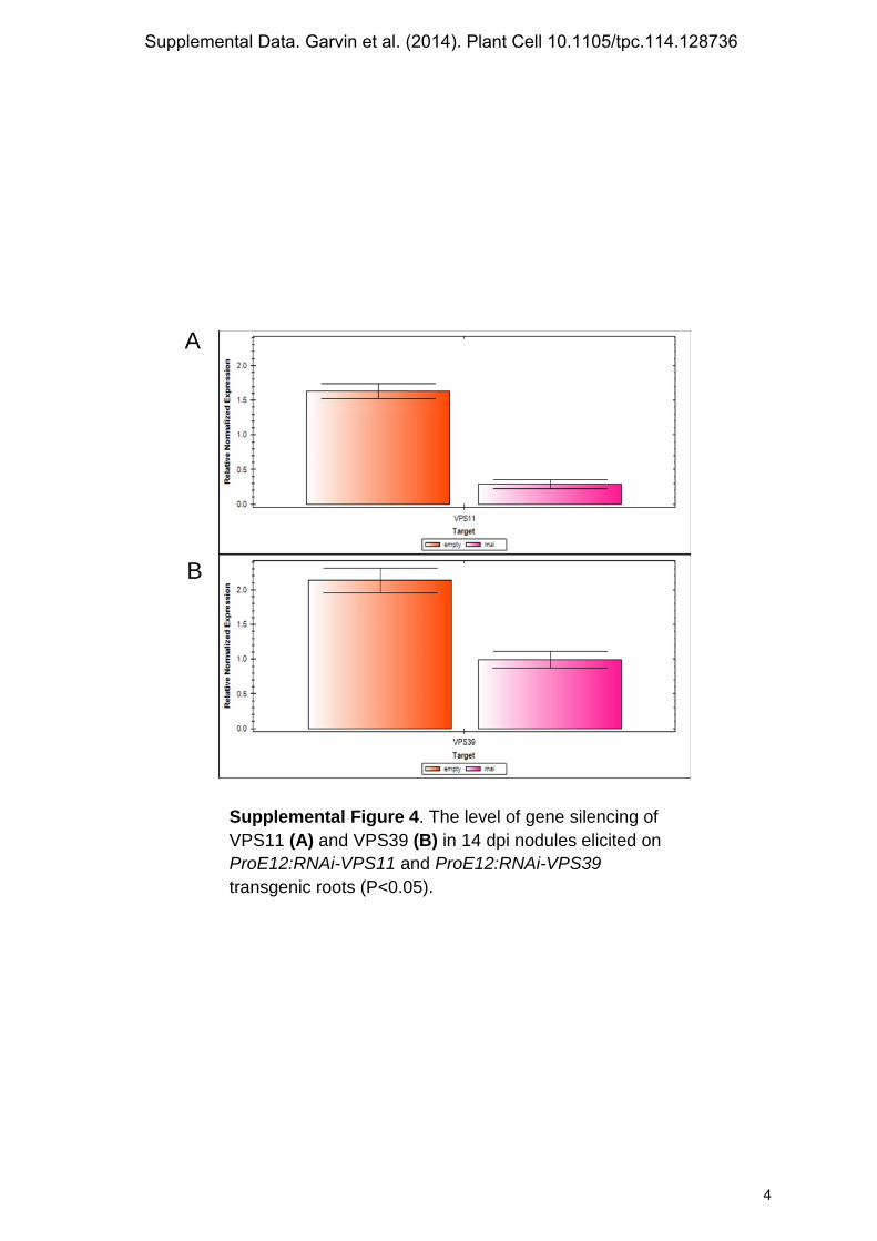
Supplemental Figure 4. The level of gene silencing of VPS11 (A) and VPS39 (B) in 14 dpi nodules elicited on ProE12:RNAi-VPS11 and ProE12:RNAi-VPS39 transgenic roots (P<0.05).
A
B
Supplemental Data. Garvin et al. (2014). Plant Cell 10.1105/tpc.114.128736
4

A
B At-TIP1;2 MPTRNIAIGGVQEEVYHPNALRAALAEFISTLIFVFAGSGSGIAFNKITDNGATTPSGLV 60 Os-γTIP MPIRNIAVG-SHQEVYHPGALKAALAEFISTLIFVFAGQGSGMAFSKLTGGGATTPAGLI 59 Mt-TIP1b MPIRNIAVG-TPQEATHPDTLKAGLAEFISTFIFVFAGSGSGIAYNKLTNDGAATPAGLI 59 Mt-TIP1g MPISRIAIG-NPSEFGKADALKAALAEFISMLIFVFAGEGSGMAYNKLTNNGAATPAGLV 59 Gh-γTIP1 MPISRIAVG-SPAEAGQADALKAALAEFISVLIFVFAGEGSGMAFNKLTDDGSSTPAGLV 59 Zm-TIP1-2 MPVSRIAVG-APGELSHPDTAKAAVAEFISTLIFVFAGSGSGMAFSKLTDGGAATPAGLI 59 Mt-TIP1e --MAKIALG-TTREATQPDCIQALIVEFIATFLFVFAGVGSAMTADKLSGD--------- 48
Supplemental Data. Garvin et al. (2014). Plant Cell 10.1105/tpc.114.128736
5

Mt-TIP1c -MA-GIAFG-RLDDSFSFGSIKAYIAEFISTLLFVFAGVGSAIAYGKLTSDAALDPAGLL 57 Mt-TIP1f -MGRGIAFG-RFDDSFSVSSIRAYVAEFISTLIFVFAGVGSAIAYAKLTSGAALDPAGLV 58 Mt-TIP1d --MVKIGFG-TFDDSFSAASLKAYLSEFIATLIFVFAGVGSAIAYNDLTSDAALDPAGLV 57 Mt-TIP1a -----------------------------------MTG--------KLMPDASLNPTSLV 17 At-TIP1;2 AAALAHAFGLFVAVSVGANISGGHVNPAVTFGVLLGGNITLLRGILYWIAQLLGSVAACF 120 Os-γTIP AAAVAHAFALFVAVSVGANISGGHVNPAVTFGAFVGGNITLFRGLLYWIAQLLGSTVACF 119 Mt-TIP1b SASIAHAFALFVAVSVGANISGGHVNPAVTFGAFVGGNITLLRGIVYIIAQLLGSIVASA 119 Mt-TIP1g AASLSHAFALFVAVSVGANISGGHVNPAVTFGAFIGGHITLIRGLLYWIAQLLGSVVACL 119 Gh-γTIP1 AAALAHALALFVAVSIGANISGGHVNPAVTFGAFVGGHITLVRSILYWIAQLLGSVVACF 119 Zm-TIP1-2 AASLAHALALFVAVSVGANISGGHVNPAVTFGAFVGGNISLLKALVYWVAQLLGSVVACL 119 Mt-TIP1e ----------------------------ALVGLFFVGHITIVRSILYWIDQLIASAAACY 80 Mt-TIP1c AVAVCHGFALFVAVAVGANISGGHVNPAVTFGLAVGGQITILTGIFYWIAQLLGSIVACF 117 Mt-TIP1f AVAVCHGFALFVAVSVGANISGGHVNPAVTFGLAIGGQITILTGIFYWIAQLLGSIVACF 118 Mt-TIP1d AVAVAHAFALFVGVAIAANISGGHLNPAVTFGLAIGGNITILTGLFYWIAQLLGSIVASL 117 Mt-TIP1a VGAIASAFALSSVLYIAWDISGGHVNPAVTFAMAVGGHISVPTALFYWVAQLIASVIACL 77 At-TIP1;2 LLSFATGGEPIPAFGLSAGVGSLNALVFEIVMTFGLVYTVYATAVDPKNGSLGTIAPIAI 180 Os-γTIP LLRFSTGGLATGTFGLTG-VSVWEALVLEIVMTFGLVYTVYATAVDPKKGSLGTIAPIAI 178 Mt-TIP1b LLVFVTAS-SVPAFGLSEGVGVGPALVLEIVMTFGLVYTVYATAVDPKKGNIGIIAPIAI 178 Mt-TIP1g LLKIATGGLETSAFSLSSGVGATNALVFEIVMTFGLVYTVYATAVDPKNGSLGTIAPIAI 179 Gh-γTIP1 LLKFSTGGMTTSAFSLSSGVGAWNAVVFEIVMTFGLVYTVYATAVDPKKGNIGIIAPIAI 179 Zm-TIP1-2 LLKIATGGAALGAFSLSAGVGAMNAVVLEMVMTFGLVYTVYATAVDPKKGDLGVIAPIAI 179 Mt-TIP1e LLHYLSGGLTTPAHTLASGVGYTQGVVWEIVLTFSLLFTVYATMVDPKKGALAGLGPTLV 140 Mt-TIP1c LLQFVTGGLETPIHSVAAEVGPIGGVVTEIIITFGLVYTVYATAADPKKGSIGTIAPIAI 177 Mt-TIP1f LLKYATGGLTIPIHSVASGVGAGEGVVTEIIITFGLVYTVYATAADPKKGSLGTIAPIAI 178 Mt-TIP1d LLNYVT-AKSVPTHGVAAGLNPIAGLVFEIIITFGLVYTVYATAADPKKGSIGTIAPIAI 176 Mt-TIP1a FLKVIVVGMHVPTYSIAEEMTGFGASILEGILTFVLVYTIYA-ARDTRRGQVSSTGTLVI 136 At-TIP1;2 GFIVGANILAGGAFSGASMNPAVAFGPAVVSWTWTNHWVYWAGPLIGGGLAGIIYDFVFI 240 Os-γTIP GFIVGANILVGGAFDGASMNPAVSFGPALVSWSWESQWVYWVGPLIGGGLAGVIYEVLFI 238 Mt-TIP1b GFIVGANILVGGAFTGASMNPAVSFGPAVVSWSWSNHWVYWAGPLIGGGIAGLVYEVLFI 238 Mt-TIP1g GFIVGANILAGGAFDGASMNPAVSFGPAVVSWTWANHWVYWVGPLIGSAIAAVVYETFFI 239 Gh-γTIP1 GFIVGANILAGGAFDGASMNPAVSFGPAVVSWTWDNHWVYWLGPFIGSAIAAIVYEVFFI 239 Zm-TIP1-2 GFIVGANILAGGAFDGASMNPAVSFGPAVVTGVWENHWVYWVGPLAGAAIAALVYDIIFI 239 Mt-TIP1e GFVVGANILAGGAFSAASMNPARSFGPALVSGNWTDHWVYWVGPLIGGGLAGFIYENFFI 200 Mt-TIP1c GFIVGANILAAGPFSGGSMNPARSFGPAVVSGNFHDNWIYWAGPLIGGGLAGLIYGNVFM 237 Mt-TIP1f GLIVGANILAAGPFSGGSMNPARSFGPAVLSGDYHNNWIYWVGPLIGGGLAGVIYSYVFM 238 Mt-TIP1d GFVVGANILAAGPFSGGSMNPARSFGPAVVSGNFADNWIYWVGPLIGGGLAGLIYGDVFI 236 Mt-TIP1a GLIAGASVLAAGPFSGGSINPACAFGSASIAGTFRNQAVYWVGPLIGAVVAGLLYDNVLF 196 At-TIP1;2 DENAHEQLP--TTDY--- 253 Os-γTIP SH-THEQLP--TTDY--- 250 Mt-TIP1b NS-THEQLP--TTDY--- 250 Mt-TIP1g TPSSYEQLP--VTDY--- 252 Gh-γTIP1 APSTYEEVP--SADF--- 252 Zm-TIP1-2 GQRPHQQLPTTAADY--- 254 Mt-TIP1e NR-DHVPLAVDEESY--- 214 Mt-TIP1c HT-EHAPLS---SDF--- 248 Mt-TIP1f PS-DHVPLA---SDF--- 249 Mt-TIP1d GS-YTPAPA---SEF--- 247 Mt-TIP1a PSQNSDCIRGVSDGTVRV 214
C
Gene name Corresponding gene locus Mt-TIP1a Medtr2g060360 Mt-TIP1b AJ251652.1 Mt-TIP1c Medtr8g013680 Mt-TIP1d Medtr5g012810 Mt-TIP1e Medtr1g006490 Mt-TIP1f Medtr2g101370 Mt-TIP1g Medtr4g063090 At-TIP1;1 AT2G36830 At-TIP1;2 AT3G26520 At-TIP1;3 AT4G01470
∆∆∆
∆∆∆
Supplemental Data. Garvin et al. (2014). Plant Cell 10.1105/tpc.114.128736
6

At-TIP2;1 AT3G16240 At-TIP2;2 AT4G17340 At-TIP2;3 AT5G47450 At-TIP3;1 AT1G73190 At-TIP3;2 AT1G17810 At-TIP4;1 AT2G25810 At-TIP5;1 AT3G47440 At-PIP1;1 At3g61430 At-PIP1;2 At2g45960 At-PIP1;3 At1g01620 At-PIP1;4 At4g00430 At-PIP1;5 At4g23400 At-PIP2;1 At3g53420 At-PIP2;2 At2g37170 At-PIP2;3 At2g37180 At-PIP2;4 At5g60660 At-PIP2;5 At3g54820 At-PIP2;6 At2g39010 At-PIP2;7 At4g35100 At-PIP2;8 At2g16850 At-NIP1;1 At4g19030 At-NIP1;2 At4g18910 At-NIP2;1 At2g34390 At-NIP3;1 At1g31885 At-NIP4;1 At5g37810 At-NIP4;2 At5g37820 At-NIP5;1 At4g10380 At-NIP6;1 At1g80760 At-NIP7;1 At3g06100 At-SIP1;1 At3g04090 At-SIP1;2 At5g18290 At-SIP2;1 At3g56950
D Gene name Corresponding gene locus References
At-TIP1;2 AT3G26520 Bourguignon et al., 2007 Gh-γTIP1 EF470294 Zhang et al., 2008 Os-γTIP RICYK333 Uchimiya et al., 1994 Zm-TIP1-2 AF326500 Jung et al., 2001 Supplemental Figure 5. (A) Phylogenetic comparison of transporters from M. truncatula and A. thaliana. Classification and naming of A. thaliana MIPs members as in Johanson et al. (2001). Phylogenetic analyses (bootstrap values of 500 replicates) were conducted using MEGA version 5 (Tamura et al., 2011). (B) ClastalW alignment of predicted amino acid sequence of M. truncatula aquaporins with another well studied water transporters. Transmembrane domains are shown with a line below the alignment; triangles indicate the NPA selectivity filter. (C) Accession numbers of gene sequences used in TIPs phylogenetic analysis. (D) Gene sequences used for multiple alignments of amino acid sequences
Supplemental Data. Garvin et al. (2014). Plant Cell 10.1105/tpc.114.128736
7
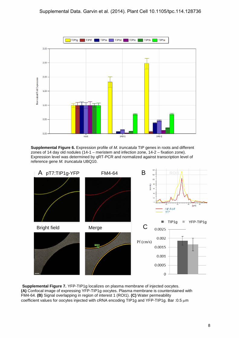
Supplemental Figure 6. Expression profile of M. truncatula TIP genes in roots and different zones of 14 day old nodules (14-1 – meristem and infection zone, 14-2 – fixation zone). Expression level was determined by qRT-PCR and normalized against transcription level of reference gene M. truncatula UBQ10. B Pf (cm/s)
Pf (cm/s)
Supplemental Figure 7. YFP-TIP1g localizes on plasma membrane of injected oocytes. (A) Confocal image of expressing YFP-TIP1g oocytes. Plasma membrane is counterstained with FM4-64. (B) Signal overlapping in region of interest 1 (ROI1). (C) Water permeability coefficient values for oocytes injected with cRNA encoding TIP1g and YFP-TIP1g. Bar :0.5 µm
A pT7:TIP1g-YFP FM4-64
Bright field Merge
TIP1g YFP-TIP1g
B
C
Supplemental Data. Garvin et al. (2014). Plant Cell 10.1105/tpc.114.128736
8

A B A
1
1,005
1,01
1,015
1,02
1,025
1,03
1,035
1,04
3 6 9 12 15 18 21 24 27 30
Time (sec)
Rel
ativ
e vo
lum
e V
/V0
MtTIP1g wc AQP1
Supplemental Figure 8. Water-channel activity of TIP1g. (A) The relative volume of oocytes injected with cRNA encoding TIP1g, human AQP1 or water following exposure to hypotonic media. The rate of oocytes swelling is plotted as V/V0 versus time, where V is a volume at the certain time point and V0 is the initial volume. (B) Water permeability coefficient values for oocytes injected with cRNA encoding TIP1g, AQP1 or water. The difference was significant, that show that TIP1g is a functional water transporter.
Water MtTIP1g AQP1 0.000
0.005
0.010
0.015
P f (
c m
/
s )
c
b
a
A B
A B
Supplemental Figure 9. TIP1g does not transport ammonia and malate. (A) NH3 flux experiment. 15 N content of TIP1g injected oocytes were same with water control in all pH tested. This result suggests that TIP1g is not a NH3 transporter. (B) Malate flux experiment. TIP1g injected oocytes in comparison with water control, show no significant differences, TIP1g does not transport malate.
Supplemental Data. Garvin et al. (2014). Plant Cell 10.1105/tpc.114.128736
9

Supplemental Data. Garvin et al. (2014). Plant Cell 10.1105/tpc.114.128736
10

Supplemental Figure 11. The level of TIP1g gene silencing in Fix+ and Fix-
nodules harvested from ProLB:RNAi-TIP1g transgenic roots in comparison with an empty vector control. The difference between the control and Fix- nodules is significant (P<0.05).
Supplemental Data. Garvin et al. (2014). Plant Cell 10.1105/tpc.114.128736
11

Supplemental Table 1: primers used for cloning and PCR analysis of VPS11, VPS39 and TIP aquaporins
Gene specific primers for cloning VPS11 and VPS39: VPS11-F TCCCCGGGATGTATCAATGGCGGAAGTT; VPS11-R GGGGTACCTCAGAAG-CCACTGCTAGATGAT;
VPS39-F CGGGATCCATGGTGCACAGTGCGTACG;
VPS39-R GGGGTACCTCATCGCTTCCTCAACTGA;.
Promoter specific primers for VPS11 and VPS39 : Pro VPS11- F CACCTATTCAAATTGAAAAAACACAGAATATT;
Pro VPS11- R CGCCGCCGCGAT;
Pro VPS39- F CACCAAGGATTCAAACCCCGATCA;
Pro VPS39- R TTTGGTTACGAAATA-TTGAAGTTGA;
Primers for the analysis of the expression level by q-PCR for VPS11 and VPS39 : VPS11- F TCAAGCAACGCAACTTCCTG;
VPS11-R TCAGGCACAAAGCTGATTGC;
Vps39-F AATCTACTCGCCGGAAACAG;
Vps39-R TGACACAACAGGCTTCTTCG;
Gene specific primers for cloning TIP1g: TIP1g-F CACCATGCCGATTTCTAGAATTGCA;
Tip1g-R TTAATAATCCGTGACAGGTAACTG;
Promoter specific primers for TIP1g: ProTIP1g- F CACCGCTTGTCTTGATTTCATGGATTG;
ProTIP1g- R TGTTTTATATTTTTTCTTTTCTCAAAGA;
Primers for the analysis of the expression level by q-PCR of TIP genes in roots and different zones of14 day old nodules: TIP1a-F ATTTACGCTGCAAGGGACAC;
TIP1a- R CACATGCAGGGTTGATTGAC;
TIP1b-F CGTTGACCCAAAGAAGGGTA;
TIP1b-R AAGTCCAGCAATTCCACCAC;
TIP1c-F GTCACCTTTGGATTGGCTGT;
TIP1c-R TCAGCTGCTGTGGCATAAAC;
TIP1d-F TTTTGTGTTCGCTGGAGTTG;
TIP1d-R ACAGCTGGGTTCAAATGTCC;
TIP1e-F CCCAAAGAAAGGAG-CACTTG;
TIP1e-R CCAACCCAATAAACCCAATG;
TIP1f-F GGTTCCATAG-TGGCATGCTT;
TIP1f-R AGCTGCTGTGGCATACACTG;
TIP1g-F AACACCAG-CAGGGTTGGTAG;
TIP1g-R CAAGCAACTGAGCAATCCAA.
Supplemental Data. Garvin et al. (2014). Plant Cell 10.1105/tpc.114.128736
DOI 10.1105/tpc.114.128736; originally published online September 12, 2014;Plant Cell
and Elena E. FedorovaAleksandr Gavrin, Brent N. Kaiser, Dietmar Geiger, Stephen D. Tyerman, Zhenqyu Wen, Ton Bisseling
MedicagoHOPS Suppression, and TIP1g Retargeting in Adjustment of Host Cells for Accommodation of Symbiotic Bacteria: Vacuole Defunctionalization,
This information is current as of December 5, 2014
Supplemental Data http://www.plantcell.org/content/suppl/2014/09/03/tpc.114.128736.DC1.html
Permissions https://www.copyright.com/ccc/openurl.do?sid=pd_hw1532298X&issn=1532298X&WT.mc_id=pd_hw1532298X
eTOCs http://www.plantcell.org/cgi/alerts/ctmain
Sign up for eTOCs at:
CiteTrack Alerts http://www.plantcell.org/cgi/alerts/ctmain
Sign up for CiteTrack Alerts at:
Subscription Information http://www.aspb.org/publications/subscriptions.cfm
is available at:Plant Physiology and The Plant CellSubscription Information for
ADVANCING THE SCIENCE OF PLANT BIOLOGY © American Society of Plant Biologists