Effect of dietary salt on feeding, digestion, growth and osmoregulation in teleost fish
Upload
independentCategory
view
0download
0
New Insights into the Roles of Acidocalcisomes and theContractile Vacuole Complex in Osmoregulation in Protists
Roberto Docampo, Veronica Jimenez, Noelia Lander, Zhu-Hong Li, and Sayantanee NiyogiCenter for Tropical and Emerging Global Diseases and Department of Cellular Biology, Universityof Georgia, Athens, GA 30602-7411
AbstractWhile free-living protists are usually subjected to hyposmotic environments, parasitic protists arealso in contact with hyperosmotic habitats. Recent work in one of these parasites, Trypanosomacruzi, has revealed that its contractile vacuole complex, which usually collects and expels excesswater as a mechanism of regulatory volume decrease after hyposmotic stress, has also a role in cellshrinking when the cells are submitted to hyperosmotic stress. Trypanosomes also have an acidiccalcium store rich in polyphosphate (polyP), named the acidocalcisome, which is involved in theirresponse to osmotic stress. Here, we review newly emerging insights on the role ofacidocalcisomes and the contractile vacuole complex in the cellular response to hyposmotic andhyperosmotic stresses. We also review the current state of knowledge on the composition of theseorganelles and their other roles in calcium homeostasis and protein trafficking.
1. IntroductionVolume regulation is a homeostatic mechanism present in all cells. With exception of thosehaving a cell wall, most cells respond to osmotic changes by swelling or shrinking throughthe activation of a number of transporters and metabolic pathways that return the cells totheir original volume, and gene expression changes that adapt the cells to the newenvironmental conditions. These physiological adaptations to osmotic stress have beenstudied extensively in a wide variety of vertebrate cell types.
Upon exposure to a reduction in external osmolarity, cells initially swell but soon regainnearly normal volume by a process that has been termed the Regulatory Volume Decrease(RVD), which is accompanied by the efflux of various inorganic ions (such as Na+ and K+)and organic osmolytes, including glycerophosphorylcholine, sorbitol, inositol, betaine, andamino acids. During the response to hyposmotic stress, all of these can be released to theextracellular medium to various degrees in different cell types (Rohloff and Docampo,2008). By far the most functionally significant efflux, in terms of total contribution to RVD,seems to involve the amino acids, particularly the β-amino acid taurine. Efflux ishypothesized to occur through a non-specific, volume-sensitive organic osmolyte anionchannel (VSOAC) that can mediate the efflux of both organic osmolytes and inorganic ions(Lang et al., 1998b). The molecular candidates for this VSOAC channel are numerous,although it is unlikely that only a single channel is responsible for all observations acrossmultiple cell types (Furst et al., 2002). It should also be kept in mind that, in many of thebest-characterized vertebrate systems, the contribution of inorganic ion efflux to the RVDfar exceeds that of organic osmolytes (Lang et al., 1998b).
*Corresponding author: Roberto Docampo, Department of Cellular Biology and Center for Tropical and Emerging Global Disease,350B Paul D. Coverdell Center, University of Georgia, Athens, GA 30602. Tel.: 706-542-8104; Fax: 706-542-9493;[email protected].
NIH Public AccessAuthor ManuscriptInt Rev Cell Mol Biol. Author manuscript; available in PMC 2014 January 01.
Published in final edited form as:Int Rev Cell Mol Biol. 2013 ; 305: . doi:10.1016/B978-0-12-407695-2.00002-0.
NIH
-PA Author Manuscript
NIH
-PA Author Manuscript
NIH
-PA Author Manuscript
On the other hand, upon exposure to an elevation in external osmolarity, cells shrink andthen regain normal volume by a process called Regulatory Volume Increase (RVI).However, this RVI response seems not to be a general process; a number of cell types frommammals have been shown not to regain its original volume after hyperosmotic stress, atleast within an hour (see (O'Neill, 1999) for a review). Following these initial changes geneexpression changes also occur in a variety of cells, which are necessary for their adaptationto the new osmotic conditions (Alfieri and Petronini, 2007; Causton et al., 2001; Gasch etal., 2000).
In contrast to vertebrate cells most protists live in environments of low osmolarity and theirmechanism of volume regulation appears to differ. While protists with a rigid cell wall resistswelling in these environments, several protists devoid of cell wall possess a contractilevacuole complex (CVC), which accumulates and expels excess water. Some protists alsoneed to deal with high osmolarity conditions under certain circumstances, and a role for theCVC under these conditions has also been demonstrated (Li et al., 2011).
Recent work in several protists has revealed a link between the CVC and the organellesnamed the acidocalcisomes (Docampo et al., 2011). Acidocalcisomes are acidic calciumstores rich in polyphosphate (polyP), a polymer of few to hundreds of phosphate units,whose function in osmoregulation has been better studied in trypanosomatid parasites(Docampo and Moreno, 2011). These parasites alternate between an insect vector and amammalian host, where the parasites are exposed not only to low but also to highosmolarities.
This review provides an overview of the cellular and molecular events underlying the role ofacidocalcisomes and the CVC in volume homeostasis in Trypanosoma cruzi, the etiologicalagent of Chagas disease, with additional references to similarities and differences with otherprotists.
2. Acidocalcisomes in Protists2.1. History
In 1992 a research article on calcium homeostasis in Dictyostelium discoideum (Rooney andGross, 1992) reported the presence of a Ca2+-ATPase in organelles called the acidosomes,which were thought to be part of the contractile vacuole apparatus of this slime mold. Thename acidosome was used because they are acidic as indicated by their sensitivity tonigericin (a K+/H+ ionophore). Nigericin-sensitive calcium compartments had also beendescribed in Leishmania donovani (Philosoph and Zilberstein, 1989), as well as inTrypanosoma brucei (Ruben et al., 1991). We therefore tested, in permeabilized cells,whether it was possible to detect a Ca2+-ATPase activity in the calcium-containing acidiccompartment of T. brucei. We demonstrated the presence of proton uptake sensitive tovacuolar ATPase (V-H+-ATPase) inhibitors, Ca2+ uptake sensitive to vanadate (Ca2+-ATPase), and the presence of organelles in these parasites that stained with Acridine Orangeand were responsible for the responses to inhibitors and ionophores. We named theseorganelles the acidocalcisomes to indicate that they are acidic and contain calcium (Vercesiet al., 1994). Further work in T. cruzi, now using intact cells loaded with Fura-2, a calciumdye indicator, allowed the physiological characterization of these organelles (Docampo etal., 1995).
An important aspect of this early work was the identification of acidocalcisomes at theultrastructural level. The best candidates were the polyP granules. These had been describedvery early in trypanosomes when they were known as volutin granules (Swellengrebel,1908). Ormerod’s work in the 1950s characterized them very well from a morphological
Docampo et al. Page 2
Int Rev Cell Mol Biol. Author manuscript; available in PMC 2014 January 01.
NIH
-PA Author Manuscript
NIH
-PA Author Manuscript
NIH
-PA Author Manuscript
point of view (Ormerod, 1958). Although they were known as polyP granules nobody hadever documented in trypanosomes that they actually contained polyP. However, work byVickerman and Tetley (Vickerman and Tetley, 1977), and later by Dvorak et al. (Dvorak etal., 1988) and LeFurgey et al. (LeFurgey et al., 1990), using X-ray microanalysis, haddescribed the presence of large amounts of calcium and phosphorus in these granules. Bydoing incubations of intact trypanosomes, with and without nigericin, and using quickfreezing, ultracryomicrotomy, and electron probe microanalysis, the problem was solved:the granules that contained calcium increased their K+ concentration after nigericintreatment indicating that they were acidic and that polyP granules and acidocalcisomes werethe same entity (Scott et al., 1997).
The nature of the abundant phosphorus compounds present in acidocalcisomes was still amystery, but the use of 31P-NMR led to the identification of very large amounts ofpyrophosphate (PPi) in T. cruzi, which is preferentially localized in the acidocalcisomes(Urbina et al., 1999). This was followed by the identification of short and long chain polyPin cells and isolated acidocalcisomes of different trypanosomatids using 31P-NMR (Morenoet al., 2002; Moreno et al., 2000) and biochemical techniques (Ruiz et al., 2001b).
While this work was going on, the gene of the first pump described in acidocalcisomes, theCa2+-ATPase, was cloned and the protein co-localized with the vacuolar H+-ATPase (Lu etal., 1998), and was later also cloned and characterized in T. brucei (Luo et al., 2004). Thepresence of a V-H+-ATPase made the acidocalcisomes look very similar to the plant vacuolethat was known to contain both a V-H+-ATPase and a V-H+-pyrophosphatase. This, togetherwith the finding of large amount of PPi in trypanosomes suggested that perhapsacidocalcisomes also had a V-H+-PPase. In fact, a PPi-driven proton uptake was found inpermeabilized cells, and the enzyme was localized to acidocalcisomes using antibodiesagainst the V-H+-PPase from plants (Scott et al., 1998). The discovery of this enzyme,which at the time was known to be present only in bacteria and plants, was also importantbecause it was the marker needed to purify the organelle, a process that was developed in T.cruzi (Scott et al., 1998), and later used to isolate acidocalcisomes from T. brucei (Rodrigueset al., 1999a) and L. donovani (Rodrigues et al., 1999b). The gene encoding for this enzymein T. cruzi was then cloned and functionally expressed in yeast (Hill et al., 2000), and wasalso studied in T. brucei (Lemercier et al., 2002).
The recent years of acidocalcisome research have been very exciting. The isolation methodfor these organelles was improved (Salto et al., 2008; Scott and Docampo, 2000; Yagisawaet al., 2009); acidocalcisomes were isolated and characterized in other trypanosomatids(Mendoza et al., 2002; Miranda et al., 2004a; Miranda et al., 2004b; Miranda et al., 2004c;Moraes Moreira et al., 2005; Soares Medeiros et al., 2005), Apicomplexan parasites(Marchesini et al., 2000; Moreno and Zhong, 1996; Ruiz et al., 2004b; Soares Medeiros etal., 2011), Chlamydomonas reinhardtii (Ruiz et al., 2001a), D. discoideum (Marchesini etal., 2002), the bacteria Agrobacterium tumefaciens (Seufferheld et al., 2003) andRhodospirillum rubrum (Seufferheld et al., 2004), human platelets (Ruiz et al., 2004a), mastcells (Moreno-Sanchez et al., 2012), insect (Motta et al., 2009; Ramos et al., 2011), chicken(Ramos et al., 2010b), and sea urchin (Ramos et al., 2010a) eggs; their chemicalcomposition investigated (Ferella et al., 2008; Salto et al., 2008); a number of pumps,channels and exchangers in their membranes were biochemically characterized and theirgenes cloned, and expressed (Fang et al., 2007; Huang, 2013; Montalvetti et al., 2004;Rohloff et al., 2004); their biogenesis was studied (Besteiro et al., 2008; de Jesus et al.,2010; Huang et al., 2011; Madeira da Silva and Beverley, 2010); and the investigation oftheir functional roles was started (Docampo et al., 2011). Some of these studies will be thesubject of this review.
Docampo et al. Page 3
Int Rev Cell Mol Biol. Author manuscript; available in PMC 2014 January 01.
NIH
-PA Author Manuscript
NIH
-PA Author Manuscript
NIH
-PA Author Manuscript
2.2. Structure and compositionAcidocalcisomes of protists are in general spherical, and can be detected with dyes thataccumulate in acidic vesicles like Acridine Orange (Docampo et al., 1995) or LysoSensor(Seufferheld et al., 2003), or with techniques that detect polyP, such as staining with DAPI(Ruiz et al., 2004a) or with antibodies against the polymer (Ramos et al., 2010a). Theirnumber varies in different species and could reach hundreds in some cells. By standardelectron microscopy they appear as empty vacuoles or vacuoles containing a thin layer ofdense material or an inclusion that sticks to the inner face of the membrane (Fig. 1). Theelectron dense material inside acidocalcisomes is better preserved with the use ofcryomethods (Scott et al., 1997) where the organelles seem completely filled by an electrondense material. Protist acidocalcisomes are usually about 0.2 µm diameter and distributed atrandom (Docampo et al., 2005). Leishmania amazonensis (~0.6 µm) (Rodrigues et al.,2002) and Tetrahymena pyriformis (up to 2–3 µm) (Rosenberg, 1966; Rosenberg and Munk,1969) possess very large acidocalcisomes.
Besides polyP, acidocalcisomes of protists also contain orthophosphate (Pi), and PPi. Thesephosphorus compounds are in close association to cations (sodium, potassium, magnesium,calcium, zinc and iron) and basic amino acids (Docampo et al., 2005; Rohloff et al., 2003).Trypanosomatids are very rich in very short chain polyP such as polyP3, polyP4, and polyP5(Moreno et al., 2000). Taking into account its total concentration and the relative volume ofacidocalcisomes in some of these cells (about 1–2% of the total cell volume), theintraorganellar concentration would be in the molar range (~ 3 M) (Docampo et al., 2005).
The membrane of the acidocalcisome possesses pumps, antiporters, and channels. Ca2+-ATPases were found in several protists, and the genes encoding some of them were clonedand expressed in yeast to demonstrate their function (Lu et al., 1998; Luo et al., 2004; Luo etal., 2001). The proteins are closely related to the family of plasma membrane calciumATPases (PMCA). In T. brucei, knockdown of the acidocalcisome Ca2+-ATPase by RNAiresults in reduced levels of mobilizable calcium from these stores and impaired growth (Luoet al., 2004). Ablation of the acidocalcisome Ca2+-ATPase from T. gondii also affectsgrowth, and the cells have a serious defect in invasion and virulence (Luo et al., 2001).
Two proton pumps were found in acidocalcisomes of protists. One is the vacuolartype H+-ATPase, a macromolecular complex of 14 subunits (Bowman et al., 2009; Lu et al., 1998;Marchesini et al., 2002; Rodrigues et al., 2000; Ruiz et al., 2001a; Yagisawa et al., 2009),and the other is the V-H+-PPase, a single subunit protein that uses PPi instead of ATP totransport protons (Drozdowicz et al., 2003; Rodrigues et al., 1999a; Ruiz et al., 2001a; Scottet al., 1998; Yagisawa et al., 2009). Only the gene for the T. cruzi V-H+-PPase could befunctionally expressed in yeast (Hill et al., 2000). In addition, the N-terminal region of theT. cruzi V-H+-PPase can enhance the functional expression of other V-H+-PPases in yeast(Drake et al., 2010).
There is biochemical evidence for the presence of Na+/H+ and Ca2+/H+ antiporters inacidocalcisomes of some trypanosomatids (Rodrigues et al., 1999b; Vercesi and Docampo,1996; Vercesi et al., 1997; Vercesi et al., 2000) and T. gondii (Rohloff et al., 2011), andmolecular evidence of a Ca2+/H+ antiporter in acidocalcisomes of Neurospora crassa(Bowman et al., 2009). An homolog to a zinc transporter was detected in T. cruziacidocalcisomes (Ferella et al., 2008). A water channel or aquaporin was also found inacidocalcisomes of T. cruzi (Rohloff et al., 2004). In contrast to the aquaporins of T. brucei,this protein is unable to transport glycerol when expressed in Xenopus oocytes (Montalvettiet al., 2004).
Docampo et al. Page 4
Int Rev Cell Mol Biol. Author manuscript; available in PMC 2014 January 01.
NIH
-PA Author Manuscript
NIH
-PA Author Manuscript
NIH
-PA Author Manuscript
Recently, an inositol 1,4,5-trisphosphate receptor was found in the acidocalcisomes of T.brucei, which is the long sought channel for Ca2+ release from these organelles (Huang,2013). Finally, a vacuolar transporter chaperone complex (VTC complex) was also detectedin the acidocalcisome membrane of trypanosomatids (Fang et al., 2007), T. gondii (Rooneyet al., 2011) and the red alga Cyanidioschyzon merolae (Yagisawa et al., 2009). Thecomplex is apparently formed by two proteins, VTC1 (in trypanosomes) or VTC2 (in T.gondii) and VTC4, of which VTC4 is the catalytic subunit that synthesizes and translocatespolyP into the acidocalcisomes (Hothorn et al., 2009).
An exopolyphosphatase (Rodrigues et al., 2002), a soluble inorganic pyrophosphatase(Lemercier et al., 2004), and a metacaspase (Lee et al., 2007) have also been localized toacidocalcisomes by immunofluorescence analyses. An acid phosphatase activity was alsodetected using cytochemical methods (Gomes et al., 2006). A protein with significantsequence identity to proteins of peptidase family M13 (CMP249C), a prenylated Rabreceptor (CMJ260C), an ABC transporter (CMS401C), and an o-methyltransferase(CMT369C) were found in acidocalcisomes of C. merolae, using specific antibodies orexpression of HA-tagged proteins (Yagisawa et al., 2009). The enzymes and transportersidentified in acidocalcisomes of protists are listed in Table 1.
2.3. Evolutionary distributionWhen the characteristic features of acidocalcisomes were revealed, it became apparent thatthey were morphologically and chemically similar to the “granules” described more than100 years ago as “metachromatic granules”, because they had the ability to turn purple basicblue dyes (Babes, 1895). These were also called “volutin granules” because they wereinitially found in Spirillum volutans (Meyer, 1904). Volutin granules were renamed polyPgranules after Wiame found that the number of granules in yeast correlated with the amountof polyP (Wiame, 1947). Volutin or polyphosphate granules were found in a number ofeukaryotic microbes using the “Meyer test”, based on their methachromasy, includingcoccidia (Kunze, 1907), trypanosomes (Swellengrebel, 1908), and Sarcosporidia(Erdnmann, 1910).
Early reports (Friedberg and Avigad, 1968; Jensen, 1968) suggested the presence of amembrane surrounding the bacterial granule, but since this contradicted current thought thatbacteria lack an endomembrane system, for many years they were assumed to lack aninternal structure or limiting membrane (Shively, 1974; Shively et al., 1988). However, thepresence of a membrane in acidocalcisomes of eukaryotes suggested that this was probablynot the case. The finding of enzymes and transporters in the surrounding membrane of theseorganelles was fundamental in understanding their potential function and origin, and thesestudies started after their description in trypanosomatid and Apicomplexan parasites(Docampo et al., 2005).
Work in Agrobacterium tumefaciens (Seufferheld et al., 2003) and Rhodospirillum rubrum(Seufferheld et al., 2004) demonstrated that acidocalcisomes in bacteria are also membrane-bounded. Evidence for the presence of a limiting membrane included: [1] its detection byelectron microscopy of intact bacteria and subcellular fractions; [2] the staining of theorganelles by dyes that accumulate in acidic compartments, such as LysoSensor, andcycloprodigiosin; and [3] the detection in the acidocalcisome membranes of a vacuolarproton pyrophosphatase (V-H+-PPase), which contains several transmembrane domains, byimmunofluorescence and immunoelectron microscopy and by subcellular fractionation. Themore recent discovery of acidocalcisome-like organelles in human platelets (Ruiz et al.,2004a) and mast cells (Moreno-Sanchez et al., 2012) established that acidocalcisomes arethe only membrane-bounded organelle present from bacteria to humans (Docampo et al.,2005) and suggested that the this is an organelle that either evolved before bacterial and
Docampo et al. Page 5
Int Rev Cell Mol Biol. Author manuscript; available in PMC 2014 January 01.
NIH
-PA Author Manuscript
NIH
-PA Author Manuscript
NIH
-PA Author Manuscript
eukaryotic lineages diverged, or appeared independently by convergent evolution (Docampoet al., 2010; Seufferheld et al., 2011).
2.4. BiogenesisAcidocalcisomes of eukaryotes are considered lysosome-related organelles (LROs). Theseare a group of organelles with similarities to lysosomes, as for example melanosomes, lyticgranules, major histocompatibiliy complex (MHC) class II compartments, platelet densegranules, basophil granules, and neutrophil azurophil granules (Dell'Angelica et al., 2000).Human platelet dense granules (Ruiz et al., 2004a) and mast cell granules (Moreno-Sanchezet al., 2012) are considered acidocalcisomes because they are acidic calcium stores rich inpolyP.
One of the main differences with lysosomes is that acidocalcisomes do not accumulateendocytic tracers, such as transferrin (Scott et al., 1997), horseradish peroxidase (Coppens etal., 1993) or FM4-64 (Mullin et al., 2001). However, a common origin cannot be ruled outbecause a L. major mutant deficient in sphingolipid synthesis was found to be defective inbiogenesis of both multivesicular bodies (or late endosomes) and acidocalcisomes (Zhang etal., 2005c).
Adaptor protein (AP) complexes are important mediators for vesicular transport ofmembrane proteins between cellular compartments, such as Golgi complex, endosomes,lysosomes and plasma membrane (Boehm and Bonifacino, 2002). Five main basic APcomplexes have been described: AP-1 to AP-5 (Hirst et al., 2013). Each of these complexesis composed of two large, one medium and one small subunits or adaptins. AP-3 is involvedin sorting of proteins to lysosomes and lysosome-related organelles from the Golgi (Peden etal., 2004) or from endosomes (Theos et al., 2005).
To study whether biogenesis of acidocalcisomes in T. brucei is linked to the expression ofAP-3 function we investigated the effects of ablation of its large β and δ subunits (Tbβ3 andTbδ) by RNAi (Huang et al., 2011). In contrast to the results reported in L. major, whereknockout of the δ subunit of AP-3 did not affect growth in culture or acidocalcisomebiogenesis (Besteiro et al., 2008) knockdown of the β3 or δ subunits of the AP-3 complexaffected growth in vitro, and led to a decrease in the number of acidocalcisomes in bothprocyclic (PCF) and bloodstream form (BSF) trypanosomes (Huang et al., 2011). Thesephenotypic changes were revealed by immmunofluorescence and electron microscopyassays, and by the decrease in their acidic calcium, PPi, and polyP content (Huang et al.,2011). However, as occurs with T. brucei mutants, L. major promastigotes mutants deficientin functional acidocalcisomes, were also less virulent in vivo (Besteiro et al., 2008).
Although the mechanism for the phenotypic differences between L. major and T. brucei invitro is unknown, a possible explanation is the formation of partial adaptor complexes in L.major constituted by just two subunits, a phenomenon that has been described for mousemodels deficient in the AP-3 δ or β3 chain (Peden et al., 2002). Our results using C-terminaltagged β3 and δ subunits of AP-3 suggest similar predominant endosomal localization in T.brucei procyclic form trypanosomes. However, we also observed partial co-localization withmarkers of the trans-Golgi network (TGN) and with acidocalcisomes, suggesting that, as itwas indicated for other LROs, acidocalcisome integral membrane proteins can follow apathway from the TGN to endosomes rather than from the TGN to the plasma membraneand then to endosomes (Huang et al., 2011). The traffic of proteins from the Golgi complexto acidocalcisomes is also supported by results with L. donovani. Dominant negative ADP-ribosylation factor like-1 (ARL-1) L. donovani had no VP1 in their acidocalcisomes (Sahinet al., 2008).
Docampo et al. Page 6
Int Rev Cell Mol Biol. Author manuscript; available in PMC 2014 January 01.
NIH
-PA Author Manuscript
NIH
-PA Author Manuscript
NIH
-PA Author Manuscript
An important role of target of rapamycin (TOR) kinase 3 (TOR3 kinase) in the biogenesis ofacidocalcisomes of T. brucei (de Jesus et al., 2010) and L. major (Madeira da Silva andBeverley, 2010) has also been described. However, while knockdown of TOR3 in T bruceiled to increases in polyP and PPi levels, larger acidocalcisomes, and more sensitivity togrowth under hyperosmotic conditions (de Jesus et al., 2010), TOR3 knockout mutantpromastigotes of L. major had smaller acidocalcisomes with apparently less polyP, and theparasites were less responsive to hyposmotic stress (Madeira da Silva and Beverley, 2010).The results suggest that TOR3 could have different roles in acidocalcisome biogenesis ofeach parasite.
2.5. Functional rolesStorage of phosphorus compounds (Pi, PPi and polyP) and cations (calcium, magnesium,sodium, potassium, zinc and iron) is one of the main roles of acidocalcisomes from differentprotists. This storage in an intracellular compartment reduces the osmotic effect of largepools of these compounds in the cytosol.
PolyP has roles in development, sporulation and predation (Zhang et al., 2005a; Zhang et al.,2005b), in stress adaptation (Castro et al., 1999; Castro et al., 1995; Pick and Weiss, 1991;Pick et al., 1991; Weiss et al., 1991) and in osmoregulation in different protists (Li et al.,2011; Ruiz et al., 2001b). Several protist parasites are less virulent when they contain loweramounts of polyP in their acidocalcisomes (Lemercier et al., 2004; Luo et al., 2005). Therecent discovery that polyP has critical roles in blood clotting (Smith et al., 2006) andinflammation (Muller et al., 2009) suggests that polyP present in microorganisms could beinvolved in their pathogenicity.
The discovery of an inositol 1,4,5-trisphosphate receptor (IP3R) in acidocalcisomes of T.brucei (Huang, 2013) indicates that these organelles have a significant role in Ca2+
signaling. The IP3R is the primary target responsible for the initiation of intracellular Ca2+
signaling in most eukaryotic cells. Ca2+ release via IP3Rs stimulates activities critical forlife. In this regard, it has been reported that acidocalcisome Ca2+ has a role in host cellinvasion. Depletion of acidocalcisome Ca2+ by pretreatment of invasive stages of T. cruziwith ionomycin plus nigericin or ionomycin plus NH4Cl, inhibited invasion of host cells(Fernandes et al., 2006; Neira et al., 2002). In T gondii, knockout of TgA1, the enzymenecessary for pumping Ca2+ into the organelle results in de-regulation of cytosolic calcium,altered microneme secretion, and decreased virulence (Luo et al., 2004).
Acidocalcisomes also appear to have a role in regulation of intracellular pH. RNAiexperiments in T. brucei to reduce the acidocalcisome V-H+-PPase activity resulted in theirinability to recover their normal pH when they were exposed to an external basic pH >7.4,and the same cells recovered from intracellular acidification at a slower rate and to a moreacidic final intracellular pH (Lemercier et al., 2002).
Acidocalcisomes have also an important role in osmoregulation. There is rapid hydrolysis orsynthesis of acidocalcisome polyP during hypo- or hyperosmotic stress, respectively, in T.cruzi (Li et al., 2011; Ruiz et al., 2001b), as well as changes in sodium and chloride contentin acidocalcisomes of L. major in response to acute hyposmotic stress (LeFurgey et al.,2001). Cyclic AMP levels increase when epimastigotes of T. cruzi are subjected tohyposmotic stress (Rohloff et al., 2004). Acidocalcisomes, which have an aquaporin(TcAQP1), traffic towards the contractile vacuole after hyposmotic stress, as revealed bydirect observation of cells expressing TcAQP1 tagged with green fluorescent protein (GFP).This traffic is stimulated by cAMP analogs or phosphodiesterase (PDE) inhibitors and isinhibited by adenylyl cyclase inhibitors, and results in fusion of acidocalcisomes with thecontractile vacuole and translocation of TcAQP1 (Rohloff et al., 2004). It has been proposed
Docampo et al. Page 7
Int Rev Cell Mol Biol. Author manuscript; available in PMC 2014 January 01.
NIH
-PA Author Manuscript
NIH
-PA Author Manuscript
NIH
-PA Author Manuscript
that the stimulus of cell swelling causes a spike in intracellular cAMP through an as yetunidentified adenylyl cyclase, resulting in fusion of acidocalcisomes with the contractilevacuole and translocation of aquaporin. This process helps the elimination of water by thecontractile vacuole and is terminated by the action of the PDE (Rohloff and Docampo, 2008;Schoijet et al., 2011).
3. The Contractile Vacuole3.3. History
The first description of a contractile vacuole complex is attributed to Lazzaro Spallanzani(Spallanzani, 1776), who noted a pulsatile star-shaped organelle in a free-swimmingorganism, presumably a Paramecium, and he postulated that it was involved in respiration.Numerous descriptions of this organelle in a wide variety of amoeba, algae, flagellates, andciliates, followed since then. Their role in osmoregulation was defined by Kitching, using avariety of fresh-water and marine protists, in a series of articles published since 1934(Kitching, 1934). The CVC is also present in single cell stages of some multicellular fungiand some cells of freshwater sponges but is absent from other multicellular species.
The first description of a contractile vacuole complex in trypanosomes, including T. cruzi,was from Clark (Clark, 1959). Linder and Staehelin (Linder and Staehelin, 1979) provided amodel for fluid secretion involving an exocytic mechanism for the CVC of thetrypanosomatid Leptomonas collosoma, and besides a few morphological descriptions (i.e.,Leishmania amazonensis (Molyneux et al., 1975), Bodo sp. (Attias et al., 1996), thepresence and functions of this organelle in trypanosomatids were mostly ignored until webegan our studies on osmoregulation in these parasites (Rohloff et al., 2004).
3.2. Structure and compositionSeveral electron microscopic studies determined that the structure of the contractile vacuoleis bipartite, consisting of a central vacuole or bladder and a surrounding loose network oftubules and vesicles named the spongiome (Allen and Naitoh, 2002). The morphologicalcharacteristics of the CVC of a number of protists have been carefully reviewed in aprevious issue of this series (Allen and Naitoh, 2002), and we will limit our discussion torecent results on their structure and composition.
The three-dimensional architecture of the CVC of epimastigotes of T. cruzi was recentlydescribed (Girard-Dias et al., 2012). Using high-pressure freezing and freeze substitution theultrastructure of T. cruzi was analyzed by serial electron tomography. A well-conservedCVC, containing a turgid central vacuole and spongiome tubules forming an interconnectednetwork was observed (Fig. 2).
A number of proteins have been localized in the CVC of a variety of protists (Table 2). Asoccurs with acidocalcisomes, two proton pumps, a vacuolar proton ATPase (V-H+-ATPase,(Fok et al., 1993; Heuser et al., 1993; Nishihara et al., 2008; Rooney and Gross, 1992; Ruizet al., 2001a; Ulrich et al., 2011) and a vacuolar H+ pyrophosphatase (V-H+-PPase or VP1,(Rohloff et al., 2004; Ruiz et al., 2001a; Ulrich et al., 2011) have been localized to thecontractile vacuole of different protists. However, the pH of the CVC does not appear to bevery acidic (for example, one study (Stock et al., 2002) calculated the pH of the CV in P.multimicronucleatum to be 6.4) and it has been proposed that the proton gradient is morelikely a factor in the mechanism of fluid accumulation rather than in acidifying the organelle(Heuser et al., 1993).
The presence of several proteins related to Ca2+ signaling underscores the role of the CVCin this process (see below): a Ca2+-ATPase (PAT1) in D. discoideum (Marchesini et al.,
Docampo et al. Page 8
Int Rev Cell Mol Biol. Author manuscript; available in PMC 2014 January 01.
NIH
-PA Author Manuscript
NIH
-PA Author Manuscript
NIH
-PA Author Manuscript
2002; Moniakis et al., 1999), an IP3R in Paramecium tetraurelia (Ladenburger et al., 2006),the Ca2+ binding proteins calmodulin (CaM) (Zhu and Clarke, 1992; Zhu et al., 1993) andcopine A (Damer et al., 2007) in D. discoideum, and also CaM in P. multimicronucleatum(Fok et al., 2008) and T. cruzi (Ulrich et al., 2011), and the P2X receptors in D. discoideum(Fountain et al., 2007; Ludlow et al., 2009; Sivaramakrishnan and Fountain, 2012a, b).These receptors are Ca2+ permeable ligand-gated ion channels activated by ATP. Othertransporters, such as an ammonium (NH4
+) transporter (AmtB, (Kirsten et al., 2008)), ahomologous to the Rh protein in red blood cells (RH50, (Benghezal et al., 2001)) that couldact as CO2 transporter (Kustu and Inwood, 2006), in D. discoideum, and polyamine(TcPOT1, (Hasne et al., 2010)) and phosphate transporters (TcPHO1, (Ulrich et al., 2011)),in T. cruzi, have also been localized to the CVC.
Although a water channel was postulated to be involved in water accumulation by the CVC(Allen and Naitoh, 2002) only recently an aquaporin was discovered, first in the organelle ofT. cruzi (Montalvetti et al., 2004; Rohloff et al., 2004), and later in those of L. major(Figarella et al., 2007), A. proteus (Nishihara et al., 2008), and C. reinhardtii (Komsic-Buchmann et al., 2012).
A number of proteins involved in trafficking and vacuolar fusion have also been identified.Rab8a (Du et al., 2008), 11a (Harris et al., 2001), 11c (Du et al., 2008), and 14 (Harris andCardelli, 2002) have been localized to the CVC of D. discoideum, and Rab11 and 32 to theCVC of T. cruzi (Ulrich et al., 2011). Disgorgin was identified as a Rab8a GTPaseactivating protein (GAP) and functions in the CV cycle (Du et al., 2008). Drainin is a Rab-GAP like protein (although apparently inactive) that regulates CVC discharge (Becker et al.,1999; Du et al., 2008).
The Dictyostelium homologue to one of the Chediak-Higashi syndrome (CHS) proteins orlarge vacuole sphere A (LvsA) labels the CVC bladder when it reaches its maximal diameterand remains associated throughout the discharge phase until it concentrates in a patch at theplasma membrane (Gerald et al., 2002). Clathrin also contributes to CVC function and hasbeen detected in the bladder of the complex of D. discoideum (Heuser, 2006; Stavrou andO'Halloran, 2006). Clathrin-coated vesicles on CVC bladders contain adaptor proteinsAP180, AP-2 and epsin, and the SNARE, Vamp7B (Wen et al., 2009). AP180 and aVMAP7 homologue, and two SNAREs have also been found in the bladder and thespongiome of the CVC of T. cruzi, respectively (Ulrich et al., 2011). A homolog toSNAP-25, another SNARE, was also detected in the CVC of P. tetraurelia (Schilde et al.,2008). Sec15 and Sec8, two components of the exocyst complex, have also been localized tothe CVC of D. discoideum especially during their discharge (Essid et al., 2012).
Another group of proteins are related to the cytoskeleton and involved in membranetubulation and motility. The MEGAPs (mental retardation GTPase-activating proteins) areGAPs that localize to the tubules of the CVC of D. discoideum and transiently to the bladderwhen it is distended (Heath and Insall, 2008a, b). In addition, two myosins, a type I myosin(myosin 1c, (Zhu and Clarke, 1992)) and type V myosin (MyoJ, (Jung et al., 2009)) havebeen localized to the CVC of D. discoideum. MyoJ is required for the normal steady statedistribution of membranes in the actin-rich cortex and to drive the actin-based corticalmotility of the membrane tubules that arise from collapsed bladder membranes after waterdischarge (Jung et al., 2009). In addition to this role of myosin in CVC motility it has alsobeen described that tubules and vesicles of the spongiome move bidirectionally between thecortex and the microtubule-organizing center (MTOC) via plus and minus end-directedmitrotubule motors (Jung et al., 2009).
Docampo et al. Page 9
Int Rev Cell Mol Biol. Author manuscript; available in PMC 2014 January 01.
NIH
-PA Author Manuscript
NIH
-PA Author Manuscript
NIH
-PA Author Manuscript
Two proteins, golvesin and dajumin, have been used as markers of different compartmentsin D. discoideum. Golvesin is a protein that localizes in endosomes and the contractilevacuole, but localizes in the Golgi apparatus when its C-terminal region is blocked with GFP(Gerisch et al., 2004; Schneider et al., 2000). Dajumin-GFP is the only integral membraneprotein to unequivocally identify constituents of the CVC (Gabriel et al., 1999). However,little is known about the function of these proteins. Another very large phosphorylatedprotein of unidentified function has been found in the CVC of Crithidia luciliae thermophila(Baqui et al., 2000)
Several proteins with enzymatic activity localize to the CVC of different protists: an alkalinephosphatase activity in D. discoideum (Nolta and Steck, 1994; Zhu and Clarke, 1992) and T.cruzi (Rohloff et al., 2004) has been used as marker for subcellular fractionation studies; aphosphodiesterase C, which is involved in the termination of cyclic AMP stimulation afterhyposmotic stress was found in T. cruzi (Schoijet et al., 2011); an acetazolamide-sensitivecarbonic anhydrase activity was detected in D. discoideum and could be important for thefilling of the bladder (Marchesini et al., 2002). An unconventional protein kinase (proteinkinase alpha) that contains an N-terminal von Willebrand factor A (vWFA)-like motif(vWFA kinase) and is able to autophosphorylate and bind to calmodulin is enriched inmembranes of the CVC and Golgi-like structures of D. discoideum (Betapudi and Egelhoff,2009; Betapudi et al., 2005). A metacaspase has recently been localized to the CVC of D.discoideum, when overexpressed (Saheb et al., 2012).
Two soluble proteins, DdCAD-1 (Sesaki et al., 1997; Siu et al., 2011; Sriskanthadevan et al.,2009; Sriskanthadevan et al., 2011) and discoidin 1 (Sriskanthadevan et al., 2009) aretransported to the cell surface previous residence in the lumen of the CVC. Polyphosphate(polyP) has also been detected by DAPI staining in the lumen of the CVC of C. reinhardtii(Ruiz et al., 2001a), and D. discoideum (Marchesini et al., 2002), while orthophosphate (Pi)was enriched in subcellular fractions of T. cruzi containing the CVC (Rohloff et al., 2004),suggesting hydrolysis of polyP during fractionation. This argues against the idea that thebladders are empty inside and that they contain only water or a dilute electrolyte and favorsan early hypothesis suggesting that contractile vacuoles might be filled with an expandablehydrocolloid that accumulates and retains water (Heywood, 1978).
3.3. BiogenesisUltrastructural studies in D. discoideum have suggested that the CVC is a post-Golgicompartment (Gabriel et al., 1999; Heuser et al., 1993). Biochemical and molecular dataalso support the Golgi-related origin of many of its constituents. For example, it has beendemonstrated in D. discoideum that overexpressed golvesin, can be detected usingmonoclonal antibodies and is present in vesicles of various sizes including the CVC(Schneider et al., 2000). When the protein is CVC and that the C-terminal region of theprotein is important for this translocation.
There is evidence that the transport of proteins from the Golgi apparatus to the CVC isthrough budding of clathrin-coated vesicles from the trans-Golgi network, as occurs inmammalian cells (Boehm and Bonifacino, 2001). The function of clathrin and its adaptors inthe biogenesis of the contractile vacuole can be inferred from studies showingmislocalization of contractile vacuole components in AP-1 and AP-2 knockouts (Lefkir etal., 2003; Sosa et al., 2012). Studies in D. discoideum in which the µ1 adaptin of AP-1 wasknocked out revealed that the contractile vacuole proteins Rh50 and dajumin-GFP weremislocalized to punctate structures inside the cell (Lefkir et al., 2003; Sosa et al., 2012)while the contractile vacuole was completely absent (Lefkir et al., 2003).
Docampo et al. Page 10
Int Rev Cell Mol Biol. Author manuscript; available in PMC 2014 January 01.
NIH
-PA Author Manuscript
NIH
-PA Author Manuscript
NIH
-PA Author Manuscript
By simultaneously imaging fluorescently tagged clathrin and AP-2, Macro et al. (Macro etal., 2012) recently described that the CVC marker dajumin-GFP is trafficked via the plasmamembrane and identified it as a cargo that is internalized by clathrindependent, AP-2independent mechanisms. The finding that clathrin mediated endocytosis is required forinternalization of CVC proteins from the cell membrane explains the CV biogenesis defectin D. discoideum cells lacking clathrin. Osmoregulation phenotypes of varying severity areobserved in both clathrin light chain (clc-) (Wang et al., 2003) and heavy chain (chcA-)knockouts (O'Halloran and Anderson, 1992), as well as in knockouts of AP180 (Stavrou andO'Halloran, 2006), the α, β1/2, or µ2 subunits of AP-2 (Sosa et al., 2012; Wen et al., 2009),and the µ1 subunit of AP1 (Lefkir et al., 2003). Clathrin-mediated endocytosis has thereforea role in the biogenesis and/or maintenance of the contractile vacuole by functioning inretrieval of proteins from the cell surface indicating that the plasma membrane is anotherpotential source of membrane for the contractile vacuole (Macro et al., 2012).
3.4. Role in osmoregulationThe CVC accumulates excess water from the cell resulting in swelling of the bladder, whicheventually enters in contact with the plasma membrane expelling this excess water from thecell. This is a homeostatic process in organisms possessing a CVC, and it has a periodicitythat varies from species to species and changes after modifications in the osmoticenvironmental conditions. The CVC of T. cruzi, for example, contracts every min and a half(Clark, 1959).
Several questions need to be considered: How water is accumulated? Which is the ioniccomposition of the CV? Which is the origin of the membrane that forms the swollenbladders and how the vacuole bladder contracts? How the bladder enters in contact with theplasma membrane?
How water is accumulated was unknown until the presence of an aquaporin or water channelwas described in the CVC of different protists (Figarella et al., 2007; Komsic-Buchmann etal., 2012; Montalvetti et al., 2004; Nishihara et al., 2008). A water channel had beenpreviously postulated to be involved in the CVC function (Allen and Naitoh, 2002) andcalculations of water permeability in the CVC of Amoeba proteus suggested that theirmembrane was equipped with water channels (Nishihara et al., 2004). Experiments in T.cruzi clearly established a role for the CVC-located aquaporin, TcAQP1, in the cellularresponse to both hyposmotic and hyperosmotic stresses (Li et al., 2011). Knockdown of theexpression of TcAQP1 reduced the regulatory volume decrease (RVD) after hyposmoticstress and the shrinking of the cells after hyperosmotic stress (Li et al., 2011). WhenTcAQP1 is knocked down and the cells are submitted to hyposmotic stress, water entry intothe CVC is diminished and the cells have reduced capacity to pump water into the mediumand recover their original volume (shrink) (Li et al., 2011). Conversely, when these cells aresubmitted to hyperosmotic stress the contractile vacuole is deficient in water accumulationand the cells cannot shrink by eliminating water into the medium as the wild type cells do(Li et al., 2011). Therefore TcAQP1 is important for cell shrinking during volume recoveryafter hyposmotic stress and during the initial phase of the response to hyperosmotic stress.These studies were also the first to reveal a role for the CVC under hyperosmotic stress.
The question regarding the ionic composition of the CVC is more difficult to answer. Earlymicropuncture studies in C. chaos and A. proteus showed that the CVC is hyposmolarrespective to the cytosol (Riddick, 1968; Schmidt-Nielsen and Schrauger, 1963) but Heuseret al. (Heuser et al., 1993) argued that the cell cannot afford to expend valuable osmolytes tomitigate this osmotic gradient. They speculated that expendable metabolic byproducts, suchas ammonia (NH3) and bicarbonate (HCO3
−), might be important sources for generating anosmotic gradient across the contractile vacuole membrane and that the vacuolar H+-ATPase
Docampo et al. Page 11
Int Rev Cell Mol Biol. Author manuscript; available in PMC 2014 January 01.
NIH
-PA Author Manuscript
NIH
-PA Author Manuscript
NIH
-PA Author Manuscript
would provide the electrochemical gradient needed for water accumulation. NH3 ismembrane permeable and would be retained by transformation into ammonium (NH4
+) withthe protons provided by the H+-V-ATPase (or the H+-V-PPase, where available). An anionchannel would transport HCO3
−. Some evidence for this pathway was found in D.discoideum (Marchesini et al., 2002). Incubation of amoebas in the presence of the anionexchanger inhibitor H2DIDS (Cabantchik and Greger, 1992) or the V-H+-ATPase inhibitorbafilomycin A1 (Bowman et al., 1988) produced a dose-dependent prolongation of their CVcontraction cycle, measured as the interval between two contractile vacuole discharges in thesame cell (Marchesini et al., 2002). In addition, the carbonic anhydrase inhibitoracetazolamide prolonged the contraction cycle of the CV (Marchesini et al., 2002).Furthermore, carbonic anhydrase activity was present in CV fractions and most of theacetazolamidesensitive carbonic anhydrase activity co-localized with the contractile vacuolemarkers (Marchesini et al., 2002). Interestingly, evidence for an ammonium (NH4
+)transporter (AmtB) (Kirsten et al., 2008) and a potential CO2 transporter (RH50) (Benghezalet al., 2001) in the CVC of D. discoideum has also been provided. Although the orientationof these transporters is not known their presence suggest that NH4
+ and CO2 could return tothe cytosol once the water is eliminated by the CV discharge. A model is shown in Fig. 3.
An alternative, although not excluding, possibility was considered for T. cruzi, where it waspostulated that fusion of acidocalcisomes with the CVC and concomitant hydrolysis ofpolyP would lead to an increase in phosphate and cations in the bladder, which would resultin water accumulation (Rohloff and Docampo, 2008). After water elimination, cations andphosphate would return to the cytosol. The presence of a phosphate transporter in the CVC(Ulrich et al., 2011) and cation exchangers that could be transferred from the acidocalcisomemembranes together with aquaporin upon their fusion (Rohloff et al., 2004) would favor thishypothesis. Since similar associations of acidocalcisomes with the CVC of D. discoideum(Marchesini et al., 2002) and C. reinhardtii (Ruiz et al., 2001a) have been described, thesepossibilities are not necessarily mutually exclusive. See Fig. 4 for a model.
The CV bladder does not burst during diastole and to increase the surface area duringexpansion, the membrane is provided by the tubules and vesicles that form the spongiome. Ithas been proposed (Clarke et al., 2002; Gerisch et al., 2002) that the contraction phase is notdue to muscle-like contraction since no F-actin or myosin are present in the bladder (Heuseret al., 1993) but to an asymmetry in the phospholipids of the network. Filling the bladderswould place a strain on the membrane, which has a tendency to tubulate, and this tendencywould be responsible for driving the emptying process.
Finally, the mechanism that the CV bladder uses to make contact with the plasma membraneappears to differ in different protists. In Paramecium the CVC is highly differentiated andthe intracellular position of the CVC (2 per cell) is fixed and a permanent surfaceindentation of the plasma membrane called the CV pore is recognizable (Allen and Naitoh,2002). The situation is significantly different with the CVC of D. discoideum; the form ofwhich is always changing and its position is not fixed in the cell. However, there is evidencethat the CV discharge occurs by “kiss and run” exocytosis, where the discharging entityretains its identity, so its membrane does not need to be recycled via a compensatoryendocytic event, except under non-physiological circumstances (Heuser, 2006). In thisregard, the GTPase Rab8a interacts with the exocyst complex in tethering of the CV to theplasma membrane, and its fusion and attachment, thereby regulating the “kiss and run”exocytosis (Essid et al., 2012). This hypothesis is compatible with the presence ofporosomes, which are supramolecular cup-shaped lipoprotein structures at the cellmembrane, where membrane-bound secretory vesicles transiently dock and fuse to releaseintravesicular contents to the outside during secretion (Jena, 2013). Finally, the CV oftrypanosomes is attached to the side of the flagellar pocket by a specialized electron-dense
Docampo et al. Page 12
Int Rev Cell Mol Biol. Author manuscript; available in PMC 2014 January 01.
NIH
-PA Author Manuscript
NIH
-PA Author Manuscript
NIH
-PA Author Manuscript

region that has been termed the adhesion plaque, first described in L. collosoma (Linder andStaehelin, 1979) and more recently found in T. cruzi (Girard-Dias et al., 2012) (Fig. 2).
3.5. Role in calcium homeostasisContractile vacuoles are considered acidic calcium stores (Patel and Docampo, 2010) andthey have been proposed to be involved in Ca2+ secretion and signaling.
In D. discoideum expression of the CVC Ca2+-ATPase PAT1 is upregulated, in acalcineurin-dependent manner, when the cells are grown in calcium-rich medium (Moniakiset al., 1999). Conditions that impair CVC function reduce the rate of Ca2+ secretion andantisense patA RNA or calcineurin antagonists affect the growth of cells in high Ca2+
medium (Moniakis et al., 1999). The results suggest a role of the CVC in Ca2+ sequestrationand excretion pathways, especially under conditions of high extracellular Ca2+. Inagreement with these results, isolated CVs from D. discoideum have been shown to take upCa2+ (Malchow et al., 2006). It has also been proposed that the CVC P2X receptors of D.discoideum are Ca2+ release channels (Ludlow et al., 2009) that are stimulated by changes inluminal ATP, which would be translocated into the CVC through and ATP-specifictransporter (Sivaramakrishnan and Fountain, 2012b).
Ca2+ release from the CVC was observed in P. tetraurelia when the InsP3R located in thisorganelle was stimulated by uncaging InsP3 (Ladenburger et al., 2006), suggesting a role ofthe CVC in Ca2+ signaling. Peptides corresponding to a homolog of the InsP3R were alsofound in the proteomic analysis of the CVC of T. cruzi but the role of this channel in T.cruzi is not yet known (Ulrich et al., 2011).
3.6. Role in protein traffickingThere is evidence that the CVC of different protists could act as a trafficking hub, receivingand delivering proteins to the plasma membrane. Although it has been indicated thatnormally there is no much mixing or ‘scrambling’ of contractile vacuole and plasmamembranes (Heuser, 2006), transfer of membrane proteins from the CVC to the plasmamembrane has been observed in several instances. For example, V-H+-ATPase and CaMtranslocate to the plasma membrane of D. discoideum when cells are starved duringstationary phase (Heuser et al., 1993). The Ca2+-ATPase PAT1 moves to the plasmamembrane when D. discoideum is incubated at high Ca2+ concentrations (Moniakis et al.,1999). The CVC polyamine transporter of T. cruzi (TcPOT1) is transferred to the plasmamembrane when the incubation medium is deficient in polyamines (Hasne et al., 2010). Theadhesive protein DdCAD-1 is also targeted to the cell surface via the CVC in D. discoideum(Sesaki et al., 1997) by an unconventional protein-trafficking pathway, being imported to theCV after forming vesicular structures in its lumen (Sriskanthadevan et al., 2009). Inaddition, it is remarkable that Rab11, a protein that localizes in recycling endosomes in mostcells, localizes to the CVC of T. cruzi (Ulrich et al., 2011) and D. discoideum (Harris et al.,2001), suggesting that this compartment could share the function of recycling endosomesthat usually send proteins back to the plasma membrane. The studies described above(Biogenesis) showing that the CVC marker dajumin-GFP is trafficked via the cellmembrane and is internalized by a clathrin-dependent mechanism suggest that clathrin-mediated endocytosis may also function as a back-up mechanism in case of transfer ofproteins from the contractile vacuole to the plasma membrane (Macro et al., 2012).
Docampo et al. Page 13
Int Rev Cell Mol Biol. Author manuscript; available in PMC 2014 January 01.
NIH
-PA Author Manuscript
NIH
-PA Author Manuscript
NIH
-PA Author Manuscript
4. Volume control in T. cruzi4.4. Need for osmoregulation
The life cycle of T. cruzi involves four major developmental stages that alternate between aninsect vector and a mammalian host. The parasite enters the mammalian host when theinsect vector defecates in the vicinity of the bite, and the natural infective stage, themetacyclic trypomastigote, is carried into the wound by scratching, and then penetrates andinfects nearby cells. Once inside the host cells, metacyclic trypomastigotes differentiate intoamastigotes. These replicative forms multiply in the cytoplasm and, after several rounds ofreplication, differentiate back into trypomastigotes, which gain access into the bloodstreamand eventually invade new cells, thus perpetuating the infection. When the insect bites aninfected mammal, the trypomastigotes carried over with the blood meal differentiate intoepimastigotes, which are a free replicative form living in the insect intestine. In the rectum,where the insect’s urine is discharged, the epimastigotes differentiate to metacyclictrypomastigotes by a process termed metacyclogenesis, and these forms are able to start anew round of infection (Fig. 5).
During its developmental cycle in the mammalian and insect hosts, T. cruzi faces criticalenvironmental challenges and one that is especially dramatic is osmolarity. Each time thetrypomastigote stage passes through the kidney of its mammalian host it must be able toresist 1,300–1,400 mOsm/Kg and return to isosmotic conditions of 300 mOsm/kg (Lang,2007). Some organs that are infected by T. cruzi (liver, spleen, lymphoid tissues) have alsohigher osmolarity than serum (330 vs 300 mOsm/kg) (Go et al., 2004). In the insect vector,the parasite is found in the epimastigote form and in this environment the osmolarity alsoincreases dramatically from the feces to the urine of the vector, and reaches values of up to1,000 mOsm/kg in the yellow rectal content (Kollien et al., 2001). In addition to the drasticchanges in osmolarity to which it is exposed, as all cells (Lang, 2007), T. cruzi need toregulate its volume continuously. Recent work has revealed that this parasite has veryunique mechanisms to deal with these challenges. The parasite acidocalcisomes and theCVC appear to have a central role in the adaptation to osmotic changes.
4.2. Response to hyposmotic stressA regulatory volume decrease mechanism is present in amastigotes, epimastigotes andtrypomastigotes of T. cruzi (Rohloff et al., 2003). This process is rapid and essentiallycomplete in all T. cruzi stages by 5 min. An amino acid efflux mechanism accounts forapproximately 50% of the regulatory volume decrease. A number of uncharged or acidicamino acids are mobilized during hyposmotic stress in all three stages and there is a markedabsence of mobilization of cationic amino acids. Glu, Gly, Pro, and Ala account for nearly90% of the total amino acids mobilized (Rohloff et al., 2003). These results suggest thatamino acid efflux in T. cruzi occurs through anion channels or transporters with propertiessimilar to those previously described in other cells (Lang et al., 1998a; Lang et al., 1998b;Vieira et al., 1996). Although acidocalcisomes have large amounts of amino acids, nearly90% of the amino acid pool of the acidocalcisome consists of Arg and Lys, minorcomponents of the amino acids released extracellularly during regulatory volume decrease(Rohloff et al., 2003).
A rise in intracellular Ca2+ occurs upon hyposmotic stress which is completely dependent onextracellular Ca2+ and, although it plays a role in modulating the early phase of amino acidefflux, is not a key determinant of the final outcome of the regulatory volume decrease(Rohloff et al., 2003). Na+, phosphate and inositol are not released extracellularly, while K+
efflux in epimastigotes could account for only about 7% of the regulatory volume decrease(Rohloff and Docampo, 2008).
Docampo et al. Page 14
Int Rev Cell Mol Biol. Author manuscript; available in PMC 2014 January 01.
NIH
-PA Author Manuscript
NIH
-PA Author Manuscript
NIH
-PA Author Manuscript
Taken together, these results showed that an osmolyte efflux mechanism alone does notentirely account for the regulatory volume decrease in T. cruzi, and that the function of theCVC is necessary for a complete RVD. It was found that cyclic AMP levels increase whenT. cruzi epimastigotes are subjected to hyposmotic stress and that modulators of cyclic AMPlevels and microtubule function affect trafficking of TcAQP1 from the acidocalcisomes tothe contractile vacuole (Rohloff et al., 2004). The results suggested that either amechanosensitive adenylyl cyclase is activated or a mechanosensitive channel leads to theinflux of ions, such as Ca2+, and activation of an adenylyl cyclase upon hyposmotic stress. Amodel was proposed (Rohloff et al., 2004) in which the stimulus of cell swelling causes aspike in intracellular cyclic AMP through an as yet unidentified adenyl cyclase, resulting ina microtubule-dependent fusion of acidocalcisomes with the contractile vacuole andtranslocation of an aquaporin. A simultaneous rise in ammonia (Rohloff and Docampo,2006), and its sequestration in acidocalcisomes as NH4
+, would activate an acidocalcisomeexopolyphosphatase, which cleaves polyphosphate (Ruiz et al., 2001b), releasing inorganicphosphate residues and also the various polyphosphate-chelated osmolytes, such as basicamino acids and calcium. The resulting osmotic gradient sequesters water, through the aid ofTcAQP1, which is subsequently ejected into the flagellar pocket. A cyclic AMPphosphodiesterase (PDE) would terminate the signaling pathway by hydrolyzing cAMP to5’-AMP (Rohloff et al., 2004) (Fig. 4).
Since this model was proposed, evidence was presented for the localization of a PDE C inthe CVC (spongiome) of the parasites (Schoijet et al., 2011). Inhibition of this enzyme bynewly developed compounds caused inhibition of the RVD and cell killing validating thisprocess as a target for chemotherapy (King-Keller et al., 2010). It was also found that a classIII phosphatidylinositol 3-kinase (TcPI3K), related to the yeast vacuolar protein sorting 34,Vps34p, was also important for RVD (Schoijet et al., 2008). Overexpression of TcPI3K wasshown to affect the RVD (Schoijet et al., 2008). In addition, several proteins important forvacuolar fusion (SNAREs) were shown to localize to the CVC (Table 2) and a putativephosphate transporter was found in the bladder of the CVC (Ulrich et al., 2011). Thistransporter could be involved in recycling of phosphate produced by the hydrolysis of poly Pduring RVD (Fig. 4).
4.3. Response to hyperosmotic stressHyperosmotic stress in most mammalian cells causes cell shrinkage due to osmotic efflux ofwater leading to increases in intracellular ionic strength (Alfieri and Petronini, 2007). Thisrapid reduction in cell volume is corrected by the regulatory volume increase (RVI), whichis mediated by ion transport systems, including the Na+-K+-Cl− co-transporter, the Na+/H+
exchanger and the Cl−/HCO3− exchanger (Lang et al., 1998a; McManus et al., 1995). The
increase of intracellular ions and the accompanying influx of water cause RVI. Cellscounteract the additional increase in ionic strength produced by the further uptake ofinorganic ions by substituting them by either the synthesis or uptake and cellularaccumulation of compatible osmolytes, such as neutral amino acids or their derivatives,polyols such as sorbitol and myo-inositol, and methylamines such as betaine (Alfieri andPetronini, 2007). Compatible osmolytes replace the inorganic ions without impairing normalbiochemical functions such as protein synthesis (Alfieri and Petronini, 2007). Compatibleosmolytes also protect the cells from apoptosis and modulate their adaptive responses(Alfieri et al., 2002; Kitamura et al., 1997).
In contrast to what happens with mammalian cells, when epimastigotes of T. cruzi aresubjected to hyperosmotic stress they shrink within a few seconds, but do not significantlyregain their normal volume, suggesting that there is no immediate inorganic ion and wateruptake (Li et al., 2011). There is an initial great increase in the size of the contractile vacuolesuggesting that water efflux is mediated through the CVC (Li et al., 2011). In spite of the
Docampo et al. Page 15
Int Rev Cell Mol Biol. Author manuscript; available in PMC 2014 January 01.
NIH
-PA Author Manuscript
NIH
-PA Author Manuscript
NIH
-PA Author Manuscript
initial increase in intracellular ionic strength they adapt well to these conditions, beingvirtually indistinguishable in terms of motility from control cells maintained in isosmoticbuffer (Li et al., 2011). However, within minutes of hyperosmotic stress there is a decreasein ammonium production and accumulation of amino acids, which then stabilize atconcentrations higher than those under isosmotic conditions (Li et al., 2011). Protein contentalso decreases within three hours suggesting protein degradation to increase the amino acidpool. PolyP synthesis is stimulated within minutes of hyperosmotic stress and thisstimulation results in a 3- and 2.35-fold increase in long-chain polyP content at 3 and 6hours, respectively, after hyperosmotic stress (Li et al., 2011). Taken together, these resultsare in agreement with a model in which reduced amino acid catabolism and increasedprotein degradation result in amino acid accumulation (Li et al., 2011). These amino acidsare the compatible osmolytes that replace the inorganic ions (Na+, K+) that are sequesteredtogether with newly formed polyP in the acidocalcisomes, thus reducing the cytosolic ionicstrength increased after water elimination, and preventing cell damage (Li et al., 2011) (Fig.6).
Treatment of the epimastigotes with low concentrations of HgCl2, a known inhibitor of T.cruzi aquaporin 1 (TcAQP1), or knockdown of TcAQP1 expression reduces the intensity ofshrinking after hyperosmotic stress while overexpression of TcAQP1 increased shrinking,suggesting that the CVC mediates water efflux during hyperosmotic challenge (Li et al.,2011). Shrinking is also probably due to cation elimination through a cation channel(TcCAT) that is translocated to the plasma membrane of epimastigotes submitted tohyperosmotic stress (Jimenez and Docampo, 2012). Inhibitors of TcCAT (BaCl2, 4-aminopyridine) inhibit shrinking of trypomastigotes under hyperosmotic stress (Jimenez andDocampo, 2012). Early synthesis of polyP and sequestration of inorganic ions inacidocalcisomes of epimastigotes and the simultaneous increase in compatible osmolytesprevents the deleterious effects of a cellular increase in ionic strength.
A second phase of recovery after hyperosmotic stress of epimastigotes is characterized byinduction of amino acid transporters (Li et al., 2011). The higher expression of genesencoding for amino acid transporters suggest that amino acids are the compatible osmolytesneeded to replace the inorganic ions sequestered by the stimulated synthesis of polyP in theacidocalcisomes (Li et al., 2011). Interestingly, some of these amino acid transporters havesimilarity to the sodium-dependent neutral amino acid transporter-2 (SNAT2) known asSystem A, which increases upon exposure of mammalian cells to hyperosmotic stress(Alfieri et al., 2002).
The response of epimastigotes to hyperosmotic stress is therefore different from thatobserved in mammalian cells or yeasts. An aquaporin and the contractile vacuole areinvolved in water efflux leading to cell shrinkage, and there is no early regulatory volumeincrease. The results suggest that the increase in ionic strength is counteracted by the earlysynthesis of polyP and sequestration of inorganic ions in acidocalcisomes. Amino acids arethe compatible osmolytes that replace the inorganic ions sequestered in acidocalcisomes,and they initially accumulate by a reduction in their catabolism, and later on by proteindegradation and by uptake through induced amino acid transporters. A model for thechanges occurring in T. cruzi during hyperosmotic stress is shown in Fig. 6.
5. Conclusions and Open QuestionsT. cruzi is exposed to environments of different osmolarities and has developed novelmechanisms to deal with these changes. A contractile vacuole complex (CVC) is importantnot only to expel water as a mechanism of regulatory volume decrease (RVD) but also tohelp in shrinking the cells when submitted to hyperosmotic stress. The CVC of T. cruzi also
Docampo et al. Page 16
Int Rev Cell Mol Biol. Author manuscript; available in PMC 2014 January 01.
NIH
-PA Author Manuscript
NIH
-PA Author Manuscript
NIH
-PA Author Manuscript
appears to have a role as a trafficking hub, and could be important for Ca2+ signaling. T.cruzi possesses acidic Ca2+ stores rich in polyP, which are termed the acidocalcisomes, andwhich participate in both the response to hyposmotic and hyperosmotic stresses. Roles forpolyP in the generation of inorganic osmolytes during hyposmotic stress and in sequesteringinorganic osmolytes to prevent the increase in the cytosolic ionic strength of the cells underhyperosmotic stress have been proposed in T. cruzi. Furthermore, a water channel oraquaporin is important not only for the filling of the CVC during hyposmotic stress but alsounder hyperosmotic stress to facilitate shrinking of the cells. Reduced levels of polyP areassociated to decreased ability to respond to osmotic stress and decreased pathogenicity invivo. A microtubule- and cAMP-dependent signaling pathway is stimulated by hyposmoticstress and results in the transfer of the aquaporin from acidocalcisomes to the CVC. A PI3Kis also involved in the response to hyposmotic stress. Acidocalcisomes alkalinize due toammonia accumulation and also increase their volume in response to hyposmotic stress. AT. cruzi PDE C was localized to the CVC and demonstrated to be essential for volumeregulation and survival of the parasite providing a novel target for chemotherapy.
AcknowledgmentsWe thank Wendell Girard Diaz and Kildare Miranda for the use of Figure 2, and Christina Moore for the drawingsof Figures 3–6. Work in our laboratory was supported in part by grants from the U.S. National Institute of Allergyand Infectious Diseases (AI068647 to RD and AI101167 to VJ), and a pre-doctoral fellowship from the AmericanHeart Association Southeast Affiliate (to NL).
ReferencesAlfieri RR, Cavazzoni A, Petronini PG, Bonelli MA, Caccamo AE, Borghetti AF, Wheeler KP.
Compatible osmolytes modulate the response of porcine endothelial cells to hypertonicity andprotect them from apoptosis. J Physiol. 2002; 540:499–508. [PubMed: 11956339]
Alfieri RR, Petronini PG. Hyperosmotic stress response: comparison with other cellular stresses.Pflugers Arch. 2007; 454:173–185. [PubMed: 17206446]
Allen RD, Naitoh Y. Osmoregulation and contractile vacuoles of protozoa. Int Rev Cytol. 2002;215:351–394. [PubMed: 11952235]
Attias M, Vommaro RC, de Souza W. Computer aided three-dimensional reconstruction of the free-living protozoan Bodo sp. (Kinetoplastida:Bodonidae). Cell Struct Funct. 1996; 21:297–306.[PubMed: 9118235]
Babes V. Beoachtungen über die metachromatischen körperchen, sporenbildung, verzwiegung,kolben- und kapsel-bildung pathogener bakterien. Zentrabl. Bakteriol. arasitenkd. Infektionskr.1895; 20:412–420.
Baqui MM, De Moraes N, Milder RV, Pudles J. A giant phosphoprotein localized at the spongiomeregion of Crithidia luciliae thermophila . J Eukaryot Microbiol. 2000; 47:532–537. [PubMed:11128704]
Becker M, Matzner M, Gerisch G. Drainin required for membrane fusion of the contractile vacuole inDictyostelium is the prototype of a protein family also represented in man. Embo J. 1999; 18:3305–3316. [PubMed: 10369671]
Benghezal M, Gotthardt D, Cornillon S, Cosson P. Localization of the Rh50-like protein to thecontractile vacuole in Dictyostelium . Immunogenetics. 2001; 52:284–288. [PubMed: 11220631]
Besteiro S, Tonn D, Tetley L, Coombs GH, Mottram JC. The AP3 adaptor is involved in the transportof membrane proteins to acidocalcisomes of Leishmania . J Cell Sci. 2008; 121:561–570. [PubMed:18252798]
Betapudi V, Egelhoff TT. Roles of an unconventional protein kinase and myosin II in amoeba osmoticshock responses. Traffic. 2009; 10:1773–1784. [PubMed: 19843280]
Betapudi V, Mason C, Licate L, Egelhoff TT. Identification and characterization of a novel alpha-kinase with a von Willebrand factor A-like motif localized to the contractile vacuole and Golgicomplex in Dictyostelium discoideum . Mol Biol Cell. 2005; 16:2248–2262. [PubMed: 15728726]
Docampo et al. Page 17
Int Rev Cell Mol Biol. Author manuscript; available in PMC 2014 January 01.
NIH
-PA Author Manuscript
NIH
-PA Author Manuscript
NIH
-PA Author Manuscript
Boehm M, Bonifacino JS. Adaptins: the final recount. Mol Biol Cell. 2001; 12:2907–2920. [PubMed:11598180]
Boehm M, Bonifacino JS. Genetic analyses of adaptin function from yeast to mammals. Gene. 2002;286:175–186. [PubMed: 11943473]
Bowman BJ, Draskovic M, Freitag M, Bowman EJ. Structure and distribution of organelles andcellular location of calcium transporters in Neurospora crassa . Eukaryot Cell. 2009; 8:1845–1855.[PubMed: 19801418]
Bowman EJ, Siebers A, Altendorf K. Bafilomycins: a class of inhibitors of membrane ATPases frommicroorganisms, animal cells, and plant cells. Proc Natl Acad Sci U S A. 1988; 85:7972–7976.[PubMed: 2973058]
Bush J, Nolta K, Rodriguez-Paris J, Kaufmann N, O'Halloran T, Ruscetti T, Temesvari L, Steck T,Cardelli J. A Rab4-like GTPase in Dictyostelium discoideum colocalizes with V-H+-ATPases inreticular membranes of the contractile vacuole complex and in lysosomes. J Cell Sci. 1994;107:2801–2812. [PubMed: 7876348]
Cabantchik ZI, Greger R. Chemical probes for anion transporters of mammalian cell membranes. Am JPhysiol. 1992; 262:C803–C827. [PubMed: 1566811]
Castro CD, Koretsky AP, Domach MM. NMR-Observed phosphate trafficking and polyphosphatedynamics in wild-type and vph1-1 mutant Saccharomyces cerevisae in response to stresses.Biotechnol Prog. 1999; 15:65–73. [PubMed: 9933515]
Castro CD, Meehan AJ, Koretsky AP, Domach MM. In situ 31P nuclear magnetic resonance forobservation of polyphosphate and catabolite responses of chemostat-cultivated Saccharomycescerevisiae after alkalinization. Appl Environ Microbiol. 1995; 61:4448–4453. [PubMed: 8534109]
Causton HC, Ren B, Koh SS, Harbison CT, Kanin E, Jennings EG, Lee TI, True HL, Lander ES,Young RA. Remodeling of yeast genome expression in response to environmental changes. MolBiol Cell. 2001; 12:323–337. [PubMed: 11179418]
Clark TB. Comparative morphology of four genera of trypanosomatidae. J. Protozool. 1959; 6:227–232.
Clarke M, Kohler J, Arana Q, Liu T, Heuser J, Gerisch G. Dynamics of the vacuolar H+-ATPase in thecontractile vacuole complex and the endosomal pathway of Dictyostelium cells. J Cell Sci. 2002;115:2893–2905. [PubMed: 12082150]
Coppens I, Baudhuin P, Opperdoes FR, Courtoy PJ. Role of acidic compartments in Trypanosomabrucei, with special reference to low-density lipoprotein processing. Mol Biochem Parasitol. 1993;58:223–232. [PubMed: 8479447]
Damer CK, Bayeva M, Kim PS, Ho LK, Eberhardt ES, Socec CI, Lee JS, Bruce EA, Goldman-YassenAE, Naliboff LC. Copine A is required for cytokinesis, contractile vacuole function, anddevelopment in Dictyostelium . Eukaryot Cell. 2007; 6:430–442. [PubMed: 17259548]
de Jesus TC, Tonelli RR, Nardelli SC, da Silva Augusto L, Motta MC, Girard-Dias W, Miranda K,Ulrich P, Jimenez V, Barquilla A, Navarro M, Docampo R, Schenkman S. Target of rapamycin(TOR)-like 1 kinase is involved in the control of polyphosphate levels and acidocalcisomemaintenance in Trypanosoma brucei . J Biol Chem. 2010; 285:24131–24140. [PubMed:20495004]
Dell'Angelica EC, Mullins C, Caplan S, Bonifacino JS. Lysosome-related organelles. Faseb J. 2000;14:1265–1278. [PubMed: 10877819]
Docampo R, de Souza W, Miranda K, Rohloff P, Moreno SN. Acidocalcisomes - conserved frombacteria to man. Nat Rev Microbiol. 2005; 3:251–261. [PubMed: 15738951]
Docampo R, Jimenez V, King-Keller S, Li ZH, Moreno SN. The role of acidocalcisomes in the stressresponse of Trypanosoma cruzi . Adv Parasitol. 2011; 75:307–324. [PubMed: 21820562]
Docampo R, Moreno SN. Acidocalcisomes. Cell Calcium. 2011; 50:113–119. [PubMed: 21752464]
Docampo R, Scott DA, Vercesi AE, Moreno SN. Intracellular Ca2+ storage in acidocalcisomes ofTrypanosoma cruzi . Biochem J. 1995; 310(Pt 3):1005–1012. [PubMed: 7575396]
Docampo R, Ulrich P, Moreno SN. Evolution of acidocalcisomes and their role in polyphosphatestorage and osmoregulation in eukaryotic microbes. Philos Trans R Soc Lond B Biol Sci. 2010;365:775–784. [PubMed: 20124344]
Docampo et al. Page 18
Int Rev Cell Mol Biol. Author manuscript; available in PMC 2014 January 01.
NIH
-PA Author Manuscript
NIH
-PA Author Manuscript
NIH
-PA Author Manuscript
Drake R, Serrano A, Perez-Castineira JR. N-terminal chimaeras with signal sequences enhance thefunctional expression and alter the subcellular localization of heterologous membrane-boundinorganic pyrophosphatases in yeast. Biochem J. 2010; 426:147–157. [PubMed: 20025609]
Drozdowicz YM, Shaw M, Nishi M, Striepen B, Liwinski HA, Roos DS, Rea PA. Isolation andcharacterization of TgVP1, a type I vacuolar H+-translocating pyrophosphatase from Toxoplasmagondii. The dynamics of its subcellular localization and the cellular effects of a diphosphonateinhibitor. J Biol Chem. 2003; 278:1075–1085. [PubMed: 12411435]
Du F, Edwards K, Shen Z, Sun B, De Lozanne A, Briggs S, Firtel RA. Regulation of contractilevacuole formation and activity in Dictyostelium . Embo J. 2008; 27:2064–2076. [PubMed:18636095]
Dvorak JA, Engel JC, Leapman RD, Swyt CR, Pella PA. Trypanosoma cruzi: elemental compositionheterogeneity of cloned stocks. Mol Biochem Parasitol. 1988; 31:19–26. [PubMed: 3054539]
Erdnmann R. Kern und metachromatische körper bei sarkosporidien. Arch. Protistenk. 1910; 20:239–243.
Essid M, Gopaldass N, Yoshida K, Merrifield C, Soldati T. Rab8a regulates the exocyst-mediated kiss-and-run discharge of the Dictyostelium contractile vacuole. Mol Biol Cell. 2012; 23:1267–1282.[PubMed: 22323285]
Fang J, Rohloff P, Miranda K, Docampo R. Ablation of a small transmembrane protein ofTrypanosoma brucei (TbVTC1) involved in the synthesis of polyphosphate alters acidocalcisomebiogenesis and function, and leads to a cytokinesis defect. Biochem J. 2007; 407:161–170.[PubMed: 17635107]
Ferella M, Nilsson D, Darban H, Rodrigues C, Bontempi EJ, Docampo R, Andersson B. Proteomics inTrypanosoma cruzi-localization of novel proteins to various organelles. Proteomics. 2008; 8:2735–2749. [PubMed: 18546153]
Fernandes AB, Neira I, Ferreira AT, Mortara RA. Cell invasion by Trypanosoma cruzi amastigotes ofdistinct infectivities: studies on signaling pathways. Parasitol Res. 2006; 100:59–68. [PubMed:16791632]
Figarella K, Uzcategui NL, Zhou Y, LeFurgey A, Ouellette M, Bhattacharjee H, Mukhopadhyay R.Biochemical characterization of Leishmania major aquaglyceroporin LmAQP1: possible role involume regulation and osmotaxis. Mol Microbiol. 2007; 65:1006–1017. [PubMed: 17640270]
Fok AK, Aihara MS, Ishida M, Allen RD. Calmodulin localization and its effects on endocytic andphagocytic membrane trafficking in Paramecium multimicronucleatum . J Eukaryot Microbiol.2008; 55:481–491. [PubMed: 19120793]
Fok AK, Clarke M, Ma L, Allen RD. Vacuolar H+-ATPase of Dictyostelium discoideum. Amonoclonal antibody study. J Cell Sci. 1993; 106:1103–1113. [PubMed: 8126094]
Fountain SJ, Parkinson K, Young MT, Cao L, Thompson CR, North RA. An intracellular P2Xreceptor required for osmoregulation in Dictyostelium discoideum . Nature. 2007; 448:200–203.[PubMed: 17625565]
Friedberg I, Avigad G. Structures containing polyphosphate in Micrococcus lysodeikticus . J Bacteriol.1968; 96:544–553. [PubMed: 5674060]
Furst J, Gschwentner M, Ritter M, Botta G, Jakab M, Mayer M, Garavaglia L, Bazzini C, RodighieroS, Meyer G, Eichmuller S, Woll E, Paulmichl M. Molecular and functional aspects of anionicchannels activated during regulatory volume decrease in mammalian cells. Pflugers Arch. 2002;444:1–25. [PubMed: 11976912]
Gabriel D, Hacker U, Kohler J, Muller-Taubenberger A, Schwartz JM, Westphal M, Gerisch G. Thecontractile vacuole network of Dictyostelium as a distinct organelle: its dynamics visualized by aGFP marker protein. J Cell Sci. 1999; 112:3995–4005. [PubMed: 10547360]
Gasch AP, Spellman PT, Kao CM, Carmel-Harel O, Eisen MB, Storz G, Botstein D, Brown PO.Genomic expression programs in the response of yeast cells to environmental changes. Mol BiolCell. 2000; 11:4241–4257. [PubMed: 11102521]
Gerald NJ, Siano M, De Lozanne A. The Dictyostelium LvsA protein is localized on the contractilevacuole and is required for osmoregulation. Traffic. 2002; 3:50–60. [PubMed: 11872142]
Docampo et al. Page 19
Int Rev Cell Mol Biol. Author manuscript; available in PMC 2014 January 01.
NIH
-PA Author Manuscript
NIH
-PA Author Manuscript
NIH
-PA Author Manuscript
Gerisch G, Benjak A, Kohler J, Weber I, Schneider N. GFP-golvesin constructs to study Golgitubulation and post-Golgi vesicle dynamics in phagocytosis. Eur J Cell Biol. 2004; 83:297–303.[PubMed: 15511087]
Gerisch G, Heuser J, Clarke M. Tubular-vesicular transformation in the contractile vacuole system ofDictyostelium . Cell Biol Int. 2002; 26:845–852. [PubMed: 12421575]
Girard-Dias W, Alcantara CL, Cunha ESN, de Souza W, Miranda K. On the ultrastructuralorganization of Trypanosoma cruzi using cryopreparation methods and electron tomography.Histochem Cell Biol. 2012; 138:821–831. [PubMed: 22872316]
Go WY, Liu X, Roti MA, Liu F, Ho SN. NFAT5/TonEBP mutant mice define osmotic stress as acritical feature of the lymphoid microenvironment. Proc Natl Acad Sci U S A. 2004; 101:10673–10678. [PubMed: 15247420]
Gomes SA, Fonseca de Souza AL, Silva BA, Kiffer-Moreira T, Santos-Mallet JR, Santos AL, Meyer-Fernandes JR. Trypanosoma rangeli: Differential expression of cell surface polypeptides and ecto-phosphatase activity in short and long epimastigote forms. Exp Parasitol. 2006; 112:253–262.[PubMed: 16442100]
Harris E, Cardelli J. RabD, a Dictyostelium Rab14-related GTPase, regulates phagocytosis andhomotypic phagosome and lysosome fusion. J Cell Sci. 2002; 115:3703–3713. [PubMed:12186956]
Harris E, Yoshida K, Cardelli J, Bush J. Rab11-like GTPase associates with and regulates the structureand function of the contractile vacuole system in Dictyostelium . J Cell Sci. 2001; 114:3035–3045.[PubMed: 11686306]
Hasne MP, Coppens I, Soysa R, Ullman B. A high-affinity putrescine-cadaverine transporter fromTrypanosoma cruzi . Mol Microbiol. 2010; 76:78–91. [PubMed: 20149109]
Heath RJ, Insall RH. Dictyostelium MEGAPs: F-BAR domain proteins that regulate motility andmembrane tubulation in contractile vacuoles. J Cell Sci. 2008a; 121:1054–1064. [PubMed:18334553]
Heath RJ, Insall RH. F-BAR domains: multifunctional regulators of membrane curvature. J Cell Sci.2008b; 121:1951–1954. [PubMed: 18525024]
Heuser J. Evidence for recycling of contractile vacuole membrane during osmoregulation inDictyostelium amoebae--a tribute to Gunther Gerisch. Eur J Cell Biol. 2006; 85:859–871.[PubMed: 16831485]
Heuser J, Zhu Q, Clarke M. Proton pumps populate the contractile vacuoles of Dictyosteliumamoebae. J Cell Biol. 1993; 121:1311–1327. [PubMed: 8509452]
Heywood P. Osmoregulation in the alga Vacuolaria virescens. Structure of the contractile vacuole andthe nature of its association with the Golgi apparatus. J Cell Sci. 1978; 31:213–224. [PubMed:670320]
Hill JE, Scott DA, Luo S, Docampo R. Cloning and functional expression of a gene encoding avacuolar-type proton-translocating pyrophosphatase from Trypanosoma cruzi . Biochem J. 2000;351:281–288. [PubMed: 10998372]
Hirst J, Irving C, Borner GH. Adaptor protein complexes AP-4 and AP-5: new players in endosomaltrafficking and progressive spastic paraplegia. Traffic. 2013; 14:153–164. [PubMed: 23167973]
Hothorn M, Neumann H, Lenherr ED, Wehner M, Rybin V, Hassa PO, Uttenweiler A, Reinhardt M,Schmidt A, Seiler J, Ladurner AG, Herrmann C, Scheffzek K, Mayer A. Catalytic core of amembrane-associated eukaryotic polyphosphate polymerase. Science. 2009; 324:513–516.[PubMed: 19390046]
Huang G, Bartlett PD, Thomas AP, Moreno SNJ, Docampo R. Acidocalcisomes of Trypanosomabrucei have an inositol 1,4,5-trisphosphate receptor that is required for growth and infectivity.Proc. Natl. Acad. Sci USA. 2013 in press.
Huang G, Fang J, Sant'Anna C, Li ZH, Wellems DL, Rohloff P, Docampo R. Adaptor protein-3(AP-3) complex mediates the biogenesis of acidocalcisomes and is essential for growth andvirulence of Trypanosoma brucei . J Biol Chem. 2011; 286:36619–36630. [PubMed: 21880705]
Jena BP. Porosome: the secretory NanoMachine in cells. Methods Mol Biol. 2013; 931:345–365.[PubMed: 23027011]
Docampo et al. Page 20
Int Rev Cell Mol Biol. Author manuscript; available in PMC 2014 January 01.
NIH
-PA Author Manuscript
NIH
-PA Author Manuscript
NIH
-PA Author Manuscript
Jensen TE. Electron microscopy of polyphosphate bodies in a blue-green alga, Nostoc pruniforme.Arch, Mikrobiol. 1968; 62:144–152.
Jimenez V, Docampo R. Molecular and electrophysiological characterization of a novel cation channelof Trypanosoma cruzi . PLoS Pathog. 2012; 8:e1002750. [PubMed: 22685407]
Jung G, Titus MA, Hammer JA 3rd. The Dictyostelium type V myosin MyoJ is responsible for thecortical association and motility of contractile vacuole membranes. J Cell Biol. 2009; 186:555–570. [PubMed: 19687255]
King-Keller S, Li M, Smith A, Zheng S, Kaur G, Yang X, Wang B, Docampo R. Chemical validationof phosphodiesterase C as a chemotherapeutic target in Trypanosoma cruzi, the etiological agentof Chagas' disease. Antimicrob Agents Chemother. 2010; 54:3738–3745. [PubMed: 20625148]
Kirsten JH, Xiong Y, Davis CT, Singleton CK. Subcellular localization of ammonium transporters inDictyostelium discoideum . BMC Cell Biol. 2008; 9:71. [PubMed: 19108721]
Kitamura H, Yamauchi A, Nakanishi T, Takamitsu Y, Sugiura T, Akagi A, Moriyama T, Horio M,Imai E. Effects of inhibition of myo-inositol transport on MDCK cells under hypertonicenvironment. Am J Physiol. 1997; 272:F267–F272. [PubMed: 9124406]
Kitching J. The physiology of contractive vacuoles: I. Osmotic relations. J. Exp. Biol. 1934; 11:364–381.
Kollien AH, Grospietsch T, Kleffmann T, Zerbst-Boroffka I, Schaub GA. Ionic composition of therectal contents and excreta of the reduviid bug Triatoma infestans . J Insect Physiol. 2001; 47:739–747. [PubMed: 11356421]
Komsic-Buchmann K, Stephan LM, Becker B. The SEC6 protein is required for contractile vacuolefunction in Chlamydomonas reinhardtii . J Cell Sci. 2012; 125:2885–2895. [PubMed: 22427688]
Kunze W. Uber orcheobius herpobdellae Schuberg et Kunze. Arch. Protistenk. 1907; 9:382–390.
Kustu S, Inwood W. Biological gas channels for NH3 and CO2: evidence that Rh (Rhesus) proteins areCO2 channels. Transfus Clin Biol. 2006; 13:103–110. [PubMed: 16563833]
Ladenburger EM, Korn I, Kasielke N, Wassmer T, Plattner H. An Ins(1,4,5)P3 receptor in Parameciumis associated with the osmoregulatory system. J Cell Sci. 2006; 119:3705–3717. [PubMed:16912081]
Lang F. Mechanisms and significance of cell volume regulation. J Am Coll Nutr. 2007; 26:613S–623S. [PubMed: 17921474]
Lang F, Busch GL, Ritter M, Volkl H, Waldegger S, Gulbins E, Haussinger D. Functional significanceof cell volume regulatory mechanisms. Physiol Rev. 1998a; 78:247–306. [PubMed: 9457175]
Lang F, Busch GL, Volkl H. The diversity of volume regulatory mechanisms. Cell Physiol Biochem.1998b; 8:1–45. [PubMed: 9547017]
Lee N, Gannavaram S, Selvapandiyan A, Debrabant A. Characterization of metacaspases with trypsin-like activity and their putative role in programmed cell death in the protozoan parasiteLeishmania . Eukaryot Cell. 2007; 6:1745–1757. [PubMed: 17715367]
Lefkir Y, de Chassey B, Dubois A, Bogdanovic A, Brady RJ, Destaing O, Bruckert F, O'Halloran TJ,Cosson P, Letourneur F. The AP-1 clathrin-adaptor is required for lysosomal enzymes sorting andbiogenesis of the contractile vacuole complex in Dictyostelium cells. Mol Biol Cell. 2003;14:1835–1851. [PubMed: 12802059]
LeFurgey A, Ingram P, Blum JJ. Elemental composition of polyphosphate-containing vacuoles andcytoplasm of Leishmania major . Mol Biochem Parasitol. 1990; 40:77–86. [PubMed: 2348832]
LeFurgey A, Ingram P, Blum JJ. Compartmental responses to acute osmotic stress in Leishmaniamajor result in rapid loss of Na+ and Cl. Comp Biochem Physiol A Mol Integr Physiol. 2001;128:385–394. [PubMed: 11223400]
Lemercier G, Dutoya S, Luo S, Ruiz FA, Rodrigues CO, Baltz T, Docampo R, Bakalara N. Avacuolar-type H+-pyrophosphatase governs maintenance of functional acidocalcisomes andgrowth of the insect and mammalian forms of Trypanosoma brucei . J Biol Chem. 2002;277:37369–37376. [PubMed: 12121996]
Lemercier G, Espiau B, Ruiz FA, Vieira M, Luo S, Baltz T, Docampo R, Bakalara N. Apyrophosphatase regulating polyphosphate metabolism in acidocalcisomes is essential forTrypanosoma brucei virulence in mice. J Biol Chem. 2004; 279:3420–3425. [PubMed: 14615483]
Docampo et al. Page 21
Int Rev Cell Mol Biol. Author manuscript; available in PMC 2014 January 01.
NIH
-PA Author Manuscript
NIH
-PA Author Manuscript
NIH
-PA Author Manuscript
Li ZH, Alvarez VE, De Gaudenzi JG, Sant'Anna C, Frasch AC, Cazzulo JJ, Docampo R.Hyperosmotic stress induces aquaporin-dependent cell shrinkage, polyphosphate synthesis, aminoacid accumulation, and global gene expression changes in Trypanosoma cruzi . J Biol Chem.2011; 286:43959–43971. [PubMed: 22039054]
Linder JC, Staehelin LA. A novel model for fluid secretion by the trypanosomatid contractile vacuoleapparatus. J Cell Biol. 1979; 83:371–382. [PubMed: 500785]
Lu HG, Zhong L, de Souza W, Benchimol M, Moreno S, Docampo R. Ca2+ content and expression ofan acidocalcisomal calcium pump are elevated in intracellular forms of Trypanosoma cruzi . MolCell Biol. 1998; 18:2309–2323. [PubMed: 9528801]
Ludlow MJ, Durai L, Ennion SJ. Functional characterization of intracellular Dictyostelium discoideumP2X receptors. J Biol Chem. 2009; 284:35227–35239. [PubMed: 19833731]
Luo S, Rohloff P, Cox J, Uyemura SA, Docampo R. Trypanosoma brucei plasma membrane-typeCa(2+)-ATPase 1 (TbPMC1) and 2 (TbPMC2) genes encode functional Ca2+-ATPases localizedto the acidocalcisomes and plasma membrane, and essential for Ca2+ homeostasis and growth. JBiol Chem. 2004; 279:14427–14439. [PubMed: 14724285]
Luo S, Ruiz FA, Moreno SN. The acidocalcisome Ca2+-ATPase (TgA1) of Toxoplasma gondii isrequired for polyphosphate storage, intracellular calcium homeostasis and virulence. MolMicrobiol. 2005; 55:1034–1045. [PubMed: 15686552]
Luo S, Vieira M, Graves J, Zhong L, Moreno SN. A plasma membrane-type Ca2+-ATPase co-localizes with a vacuolar H+-pyrophosphatase to acidocalcisomes of Toxoplasma gondii . Embo J.2001; 20:55–64. [PubMed: 11226155]
Macro L, Jaiswal JK, Simon SM. Dynamics of clathrin-mediated endocytosis and its requirement fororganelle biogenesis in Dictyostelium . J Cell Sci. 2012 in press.
Madeira da Silva L, Beverley SM. Expansion of the target of rapamycin (TOR) kinase family andfunction in Leishmania shows that TOR3 is required for acidocalcisome biogenesis and animalinfectivity. Proc Natl Acad Sci U S A. 2010; 107:11965–11970. [PubMed: 20551225]
Malchow D, Lusche DF, Schlatterer C, De Lozanne A, Muller-Taubenberger A. The contractilevacuole in Ca2+-regulation in Dictyostelium: its essential function for cAMP-induced Ca2+-influx.BMC Dev Biol. 2006; 6:31. [PubMed: 16787542]
Marchesini N, Luo S, Rodrigues CO, Moreno SN, Docampo R. Acidocalcisomes and a vacuolar H+-pyrophosphatase in malaria parasites. Biochem J 347 Pt. 2000; 1:243–253. [PubMed: 10727425]
Marchesini N, Ruiz FA, Vieira M, Docampo R. Acidocalcisomes are functionally linked to thecontractile vacuole of Dictyostelium discoideum . J Biol Chem. 2002; 277:8146–8153. [PubMed:11748243]
McManus ML, Churchwell KB, Strange K. Regulation of cell volume in health and disease. N Engl JMed. 1995; 333:1260–1266. [PubMed: 7566004]
Mendoza M, Mijares A, Rojas H, Rodriguez JP, Urbina JA, DiPolo R. Physiological andmorphological evidences for the presence acidocalcisomes in Trypanosoma evansi: single cellfluorescence and 31P NMR studies. Mol Biochem Parasitol. 2002; 125:23–33. [PubMed:12467971]
Meyer A. Orientierende untersuchungen ueber verbreitung. Morphologie, und chemie des volutins.Bot. Zeit. 1904; 62:113–152.
Miranda K, Docampo R, Grillo O, de Souza W. Acidocalcisomes of trypanosomatids have species-specific elemental composition. Protist. 2004a; 155:395–405. [PubMed: 15648720]
Miranda K, Docampo R, Grillo O, Franzen A, Attias M, Vercesi A, Plattner H, Hentschel J, de SouzaW. Dynamics of polymorphism of acidocalcisomes in Leishmania parasites. Histochem CellBiol. 2004b; 121:407–418. [PubMed: 15138840]
Miranda K, Rodrigues CO, Hentchel J, Vercesi A, Plattner H, de Souza W, Docampo R.Acidocalcisomes of Phytomonas francai possess distinct morphological characteristics andcontain iron. Microsc Microanal. 2004c; 10:647–655. [PubMed: 15525437]
Molyneux DH, Killick-Kendrick R, Ashford RW. Leishmania in phlebotomid sandflies. III. Theultrastructure of Leishmania mexicana amazonensis in the midgut and pharynx of Lutzomyialongipalpis . Proc R Soc Lond B Biol Sci. 1975; 190:341–357. [PubMed: 240164]
Docampo et al. Page 22
Int Rev Cell Mol Biol. Author manuscript; available in PMC 2014 January 01.
NIH
-PA Author Manuscript
NIH
-PA Author Manuscript
NIH
-PA Author Manuscript
Moniakis J, Coukell MB, Janiec A. Involvement of the Ca2+-ATPase PAT1 and the contractilevacuole in calcium regulation in Dictyostelium discoideum . J Cell Sci. 1999; 112:405–414.[PubMed: 9885293]
Montalvetti A, Rohloff P, Docampo R. A functional aquaporin co-localizes with the vacuolar protonpyrophosphatase to acidocalcisomes and the contractile vacuole complex of Trypanosoma cruzi .J Biol Chem. 2004; 279:38673–38682. [PubMed: 15252016]
Moraes Moreira BL, Soares Medeiros LC, Miranda K, de Souza W, Hentschel J, Plattner H, BarrabinH. Kinetics of pyrophosphate-driven proton uptake by acidocalcisomes of Leptomonas wallacei .Biochem Biophys Res Commun. 2005; 334:1206–1213. [PubMed: 16039991]
Moreno B, Rodrigues CO, Bailey BN, Urbina JA, Moreno SN, Docampo R, Oldfield E. Magic-anglespinning 31P NMR spectroscopy of condensed phosphates in parasitic protozoa: visualizing theinvisible. FEBS Lett. 2002; 523:207–212. [PubMed: 12123833]
Moreno B, Urbina JA, Oldfield E, Bailey BN, Rodrigues CO, Docampo R. 31P NMR spectroscopy ofTrypanosoma brucei, Trypanosoma cruzi, and Leishmania major. Evidence for high levels ofcondensed inorganic phosphates. J Biol Chem. 2000; 275:28356–28362. [PubMed: 10871617]
Moreno SN, Zhong L. Acidocalcisomes in Toxoplasma gondii tachyzoites. Biochem J. 1996; 313:655–659. [PubMed: 8573106]
Moreno-Sanchez D, Hernandez-Ruiz L, Ruiz FA, Docampo R. Polyphosphate is a novel pro-inflammatory regulator of mast cells and is located in acidocalcisomes. J Biol Chem. 2012;287:28435–28444. [PubMed: 22761438]
Motta LS, Ramos IB, Gomes FM, de Souza W, Champagne DE, Santiago MF, Docampo R, MirandaK, Machado EA. Proton-pyrophosphatase and polyphosphate in acidocalcisome-like vesiclesfrom oocytes and eggs of Periplaneta americana . Insect Biochem Mol Biol. 2009; 39:198–206.[PubMed: 19111615]
Muller F, Mutch NJ, Schenk WA, Smith SA, Esterl L, Spronk HM, Schmidbauer S, Gahl WA,Morrissey JH, Renne T. Platelet polyphosphates are proinflammatory and procoagulantmediators in vivo. Cell. 2009; 139:1143–1156. [PubMed: 20005807]
Mullin KA, Foth BJ, Ilgoutz SC, Callaghan JM, Zawadzki JL, McFadden GI, McConville MJ.Regulated degradation of an endoplasmic reticulum membrane protein in a tubular lysosome inLeishmania mexicana . Mol Biol Cell. 2001; 12:2364–2377. [PubMed: 11514622]
Neira I, Ferreira AT, Yoshida N. Activation of distinct signal transduction pathways in Trypanosomacruzi isolates with differential capacity to invade host cells. Int J Parasitol. 2002; 32:405–414.[PubMed: 11849637]
Nishihara E, Shimmen T, Sonobe S. Functional characterization of contractile vacuole isolated fromAmoeba proteus . Cell Struct Funct. 2004; 29:85–90. [PubMed: 15665503]
Nishihara E, Yokota E, Tazaki A, Orii H, Katsuhara M, Kataoka K, Igarashi H, Moriyama Y,Shimmen T, Sonobe S. Presence of aquaporin and V-ATPase on the contractile vacuole ofAmoeba proteus . Biol Cell. 2008; 100:179–188. [PubMed: 18004980]
Nolta KV, Steck TL. Isolation and initial characterization of the bipartite contractile vacuole complexfrom Dictyostelium discoideum . J Biol Chem. 1994; 269:2225–2233. [PubMed: 8294479]
O'Halloran TJ, Anderson RG. Clathrin heavy chain is required for pinocytosis, the presence of largevacuoles, and development in Dictyostelium . J Cell Biol. 1992; 118:1371–1377. [PubMed:1522112]
O'Neill WC. Physiological significance of volume-regulatory transporters. Am J Physiol. 1999;276:C995–C1011. [PubMed: 10329946]
Ormerod WE. A comparative study of cytoplasmic inclusions (volutin granules) in different species oftrypanosomes. J Gen Microbiol. 1958; 19:271–288. [PubMed: 13587893]
Patel S, Docampo R. Acidic calcium stores open for business: expanding the potential for intracellularCa2+ signaling. Trends Cell Biol. 2010; 20:277–286. [PubMed: 20303271]
Peden AA, Oorschot V, Hesser BA, Austin CD, Scheller RH, Klumperman J. Localization of the AP-3adaptor complex defines a novel endosomal exit site for lysosomal membrane proteins. J CellBiol. 2004; 164:1065–1076. [PubMed: 15051738]
Peden AA, Rudge RE, Lui WW, Robinson MS. Assembly and function of AP-3 complexes in cellsexpressing mutant subunits. J Cell Biol. 2002; 156:327–336. [PubMed: 11807095]
Docampo et al. Page 23
Int Rev Cell Mol Biol. Author manuscript; available in PMC 2014 January 01.
NIH
-PA Author Manuscript
NIH
-PA Author Manuscript
NIH
-PA Author Manuscript
Philosoph H, Zilberstein D. Regulation of intracellular calcium in promastigotes of the humanprotozoan parasite Leishmania donovani . J Biol Chem. 1989; 264:10420–10424. [PubMed:2732230]
Pick U, Weiss M. Polyphosphate Hydrolysis within acidic vacuoles in response to amine-inducedalkaline stress in the halotolerant alga Dunaliella salina . Plant Physiol. 1991; 97:1234–1240.[PubMed: 16668514]
Pick U, Zeelon O, Weiss M. Amine accumulation in acidic vacuoles protects the halotolerant algaDunaliella salina against alkaline stress. Plant Physiol. 1991; 97:1226–1233. [PubMed:16668513]
Ramos I, Gomes F, Koeller CM, Saito K, Heise N, Masuda H, Docampo R, de Souza W, MachadoEA, Miranda K. Acidocalcisomes as calcium- and polyphosphate-storage compartments duringembryogenesis of the insect Rhodnius prolixus Stahl. PLoS One. 2011; 6:e27276. [PubMed:22096545]
Ramos IB, Miranda K, Pace DA, Verbist KC, Lin FY, Zhang Y, Oldfield E, Machado EA, De SouzaW, Docampo R. Calcium- and polyphosphate-containing acidic granules of sea urchin eggs aresimilar to acidocalcisomes, but are not the targets for NAADP. Biochem J. 2010a; 429:485–495.[PubMed: 20497125]
Ramos IB, Miranda K, Ulrich P, Ingram P, LeFurgey A, Machado EA, de Souza W, Docampo R.Calcium- and polyphosphate-containing acidocalcisomes in chicken egg yolk. Biol Cell. 2010b;102:421–434. [PubMed: 20196772]
Riddick DH. Contractile vacuole in the amoeba Pelomyxa carolinensis . Am J Physiol. 1968; 215:736–740. [PubMed: 5671014]
Rodrigues CO, Ruiz FA, Vieira M, Hill JE, Docampo R. An acidocalcisomal exopolyphosphatasefrom Leishmania major with high affinity for short chain polyphosphate. J Biol Chem. 2002;277:50899–50906. [PubMed: 12393865]
Rodrigues CO, Scott DA, Bailey BN, De Souza W, Benchimol M, Moreno B, Urbina JA, Oldfield E,Moreno SN. Vacuolar proton pyrophosphatase activity and pyrophosphate (PPi) in Toxoplasmagondii as possible chemotherapeutic targets. Biochem J 349 Pt. 2000; 3:737–745. [PubMed:10903134]
Rodrigues CO, Scott DA, Docampo R. Characterization of a vacuolar pyrophosphatase inTrypanosoma brucei and its localization to acidocalcisomes. Mol Cell Biol. 1999a; 19:7712–7723. [PubMed: 10523660]
Rodrigues CO, Scott DA, Docampo R. Presence of a vacuolar H+-pyrophosphatase in promastigotes ofLeishmania donovani and its localization to a different compartment from the vacuolar H+-ATPase. Biochem J. 1999b; 340:759–766. [PubMed: 10359662]
Rohloff P, Docampo R. Ammonium production during hypo-osmotic stress leads to alkalinization ofacidocalcisomes and cytosolic acidification in Trypanosoma cruzi . Mol Biochem Parasitol.2006; 150:249–255. [PubMed: 17005261]
Rohloff P, Docampo R. A contractile vacuole complex is involved in osmoregulation in Trypanosomacruzi . Exp Parasitol. 2008; 118:17–24. [PubMed: 17574552]
Rohloff P, Miranda K, Rodrigues JC, Fang J, Galizzi M, Plattner H, Hentschel J, Moreno SN. Calciumuptake and proton transport by acidocalcisomes of Toxoplasma gondii . PLoS One. 2011;6:e18390. [PubMed: 21541023]
Rohloff P, Montalvetti A, Docampo R. Acidocalcisomes and the contractile vacuole complex areinvolved in osmoregulation in Trypanosoma cruzi . J Biol Chem. 2004; 279:52270–52281.[PubMed: 15466463]
Rohloff P, Rodrigues CO, Docampo R. Regulatory volume decrease in Trypanosoma cruzi involvesamino acid efflux and changes in intracellular calcium. Mol Biochem Parasitol. 2003; 126:219–230. [PubMed: 12615321]
Rooney EK, Gross JD. ATP-driven Ca2+/H+ antiport in acid vesicles from Dictyostelium . Proc NatlAcad Sci U S A. 1992; 89:8025–8029. [PubMed: 1387710]
Rooney EK, Gross JD, Satre M. Characterisation of an intracellular Ca2+ pump in Dictyostelium . CellCalcium. 1994; 16:509–522. [PubMed: 7712544]
Docampo et al. Page 24
Int Rev Cell Mol Biol. Author manuscript; available in PMC 2014 January 01.
NIH
-PA Author Manuscript
NIH
-PA Author Manuscript
NIH
-PA Author Manuscript
Rooney PJ, Ayong L, Tobin CM, Moreno SN, Knoll LJ. TgVTC2 is involved in polyphosphateaccumulation in Toxoplasma gondii. Mol Biochem Parasitol. 2011; 176:121–126. [PubMed:21195114]
Rosenberg H. The isolation and identification of “volutin” granules from Tetrahymena . Exp Cell Res.1966; 41:397–410. [PubMed: 4286434]
Rosenberg H, Munk N. Transport phenomena associated with the deposition and disappearance ofpyrophosphate granules in Tetrahymena pyriformis . Biochim Biophys Acta. 1969; 184:191–197.[PubMed: 4307180]
Ruben L, Hutchinson A, Moehlman J. Calcium homeostasis in Trypanosoma brucei. Identification of apH-sensitive non-mitochondrial calcium pool. J Biol Chem. 1991; 266:24351–24358. [PubMed:1722203]
Ruiz FA, Lea CR, Oldfield E, Docampo R. Human platelet dense granules contain polyphosphate andare similar to acidocalcisomes of bacteria and unicellular eukaryotes. J Biol Chem. 2004a;279:44250–44257. [PubMed: 15308650]
Ruiz FA, Luo S, Moreno SN, Docampo R. Polyphosphate content and fine structure ofacidocalcisomes of Plasmodium falciparum . Microsc Microanal. 2004b; 10:563–567. [PubMed:15525430]
Ruiz FA, Marchesini N, Seufferheld M, Govindjee Docampo R. The polyphosphate bodies ofChlamydomonas reinhardtii possess a proton-pumping pyrophosphatase and are similar toacidocalcisomes. J Biol Chem. 2001a; 276:46196–46203. [PubMed: 11579086]
Ruiz FA, Rodrigues CO, Docampo R. Rapid changes in polyphosphate content within acidocalcisomesin response to cell growth, differentiation, and environmental stress in Trypanosoma cruzi . JBiol Chem. 2001b; 276:26114–26121. [PubMed: 11371561]
Saheb E, Trzyna W, Bush J. An Acanthamoeba castellanii metacaspase associates with the contractilevacuole and functions in osmoregulation. Exp Parasitol. 2012 in press.
Sahin A, Espiau B, Tetaud E, Cuvillier A, Lartigue L, Ambit A, Robinson DR, Merlin G. TheLeishmania ARL-1 and Golgi traffic. PLoS One. 2008; 3:e1620. [PubMed: 18286177]
Salto ML, Kuhlenschmidt T, Kuhlenschmidt M, de Lederkremer RM, Docampo R. Phospholipid andglycolipid composition of acidocalcisomes of Trypanosoma cruzi . Mol Biochem Parasitol. 2008;158:120–130. [PubMed: 18207579]
Schilde C, Lutter K, Kissmehl R, Plattner H. Molecular identification of a SNAP-25-like SNAREprotein in Paramecium . Eukaryot Cell. 2008; 7:1387–1402. [PubMed: 18552286]
Schmidt-Nielsen B, Schrauger CR. Amoeba proteus: studying the contractile vacuole bymicropuncture. Science. 1963; 139:606–607. [PubMed: 17788304]
Schneider N, Schwartz JM, Kohler J, Becker M, Schwarz H, Gerisch G. Golvesin-GFP fusions asdistinct markers for Golgi and post-Golgi vesicles in Dictyostelium cells. Biol Cell. 2000;92:495–511. [PubMed: 11229601]
Schoijet AC, Miranda K, Girard-Dias W, de Souza W, Flawia MM, Torres HN, Docampo R, AlonsoGD. A Trypanosoma cruzi phosphatidylinositol 3-kinase (TcVps34) is involved inosmoregulation and receptor-mediated endocytosis. J Biol Chem. 2008; 283:31541–31550.[PubMed: 18801733]
Schoijet AC, Miranda K, Medeiros LC, de Souza W, Flawia MM, Torres HN, Pignataro OP, DocampoR, Alonso GD. Defining the role of a FYVE domain in the localization and activity of a cAMPphosphodiesterase implicated in osmoregulation in Trypanosoma cruzi . Mol Microbiol. 2011;79:50–62. [PubMed: 21166893]
Scott DA, de Souza W, Benchimol M, Zhong L, Lu HG, Moreno SN, Docampo R. Presence of a plant-like proton-pumping pyrophosphatase in acidocalcisomes of Trypanosoma cruzi . J Biol Chem.1998; 273:22151–22158. [PubMed: 9705361]
Scott DA, Docampo R. Characterization of isolated acidocalcisomes of Trypanosoma cruzi . J BiolChem. 2000; 275:24215–24221. [PubMed: 10816577]
Scott DA, Docampo R, Dvorak JA, Shi S, Leapman RD. In situ compositional analysis ofacidocalcisomes in Trypanosoma cruzi . J Biol Chem. 1997; 272:28020–28029. [PubMed:9346954]
Docampo et al. Page 25
Int Rev Cell Mol Biol. Author manuscript; available in PMC 2014 January 01.
NIH
-PA Author Manuscript
NIH
-PA Author Manuscript
NIH
-PA Author Manuscript
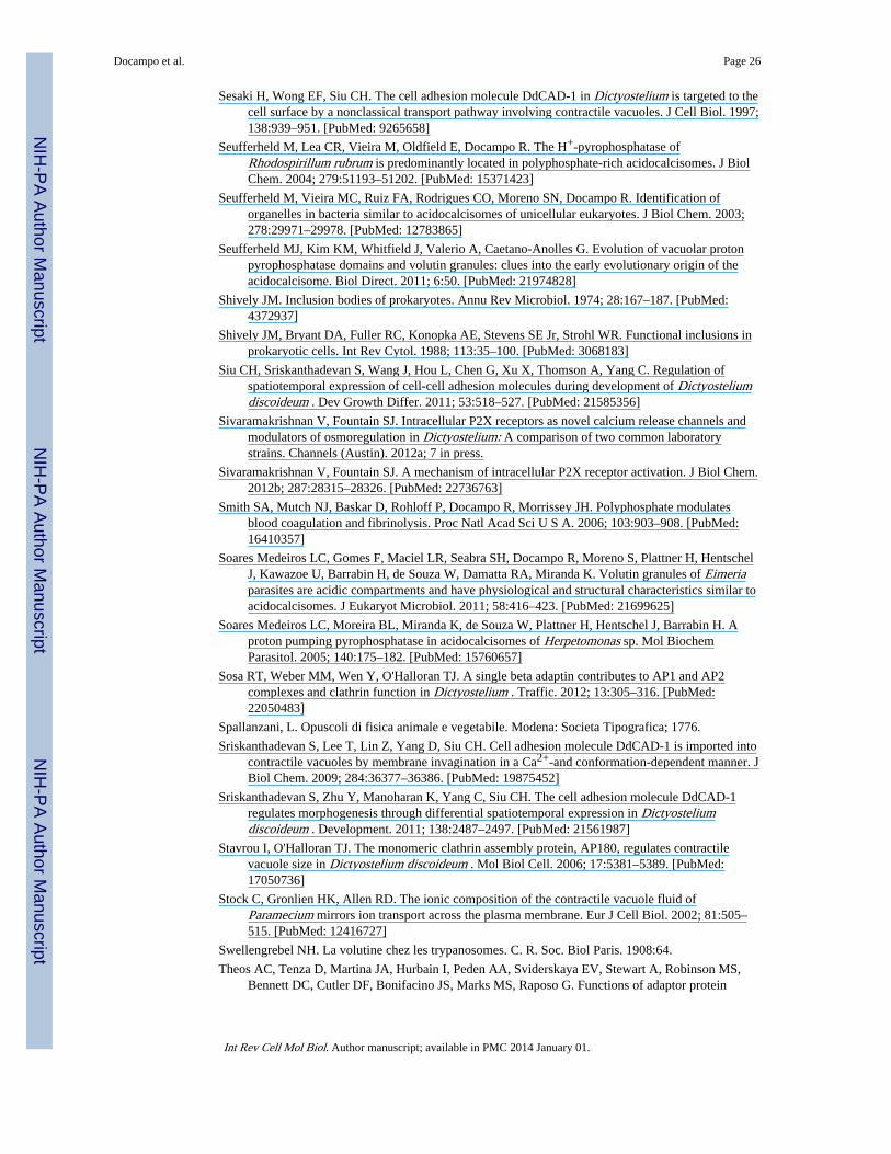
Sesaki H, Wong EF, Siu CH. The cell adhesion molecule DdCAD-1 in Dictyostelium is targeted to thecell surface by a nonclassical transport pathway involving contractile vacuoles. J Cell Biol. 1997;138:939–951. [PubMed: 9265658]
Seufferheld M, Lea CR, Vieira M, Oldfield E, Docampo R. The H+-pyrophosphatase ofRhodospirillum rubrum is predominantly located in polyphosphate-rich acidocalcisomes. J BiolChem. 2004; 279:51193–51202. [PubMed: 15371423]
Seufferheld M, Vieira MC, Ruiz FA, Rodrigues CO, Moreno SN, Docampo R. Identification oforganelles in bacteria similar to acidocalcisomes of unicellular eukaryotes. J Biol Chem. 2003;278:29971–29978. [PubMed: 12783865]
Seufferheld MJ, Kim KM, Whitfield J, Valerio A, Caetano-Anolles G. Evolution of vacuolar protonpyrophosphatase domains and volutin granules: clues into the early evolutionary origin of theacidocalcisome. Biol Direct. 2011; 6:50. [PubMed: 21974828]
Shively JM. Inclusion bodies of prokaryotes. Annu Rev Microbiol. 1974; 28:167–187. [PubMed:4372937]
Shively JM, Bryant DA, Fuller RC, Konopka AE, Stevens SE Jr, Strohl WR. Functional inclusions inprokaryotic cells. Int Rev Cytol. 1988; 113:35–100. [PubMed: 3068183]
Siu CH, Sriskanthadevan S, Wang J, Hou L, Chen G, Xu X, Thomson A, Yang C. Regulation ofspatiotemporal expression of cell-cell adhesion molecules during development of Dictyosteliumdiscoideum . Dev Growth Differ. 2011; 53:518–527. [PubMed: 21585356]
Sivaramakrishnan V, Fountain SJ. Intracellular P2X receptors as novel calcium release channels andmodulators of osmoregulation in Dictyostelium: A comparison of two common laboratorystrains. Channels (Austin). 2012a; 7 in press.
Sivaramakrishnan V, Fountain SJ. A mechanism of intracellular P2X receptor activation. J Biol Chem.2012b; 287:28315–28326. [PubMed: 22736763]
Smith SA, Mutch NJ, Baskar D, Rohloff P, Docampo R, Morrissey JH. Polyphosphate modulatesblood coagulation and fibrinolysis. Proc Natl Acad Sci U S A. 2006; 103:903–908. [PubMed:16410357]
Soares Medeiros LC, Gomes F, Maciel LR, Seabra SH, Docampo R, Moreno S, Plattner H, HentschelJ, Kawazoe U, Barrabin H, de Souza W, Damatta RA, Miranda K. Volutin granules of Eimeriaparasites are acidic compartments and have physiological and structural characteristics similar toacidocalcisomes. J Eukaryot Microbiol. 2011; 58:416–423. [PubMed: 21699625]
Soares Medeiros LC, Moreira BL, Miranda K, de Souza W, Plattner H, Hentschel J, Barrabin H. Aproton pumping pyrophosphatase in acidocalcisomes of Herpetomonas sp. Mol BiochemParasitol. 2005; 140:175–182. [PubMed: 15760657]
Sosa RT, Weber MM, Wen Y, O'Halloran TJ. A single beta adaptin contributes to AP1 and AP2complexes and clathrin function in Dictyostelium . Traffic. 2012; 13:305–316. [PubMed:22050483]
Spallanzani, L. Opuscoli di fisica animale e vegetabile. Modena: Societa Tipografica; 1776.
Sriskanthadevan S, Lee T, Lin Z, Yang D, Siu CH. Cell adhesion molecule DdCAD-1 is imported intocontractile vacuoles by membrane invagination in a Ca2+-and conformation-dependent manner. JBiol Chem. 2009; 284:36377–36386. [PubMed: 19875452]
Sriskanthadevan S, Zhu Y, Manoharan K, Yang C, Siu CH. The cell adhesion molecule DdCAD-1regulates morphogenesis through differential spatiotemporal expression in Dictyosteliumdiscoideum . Development. 2011; 138:2487–2497. [PubMed: 21561987]
Stavrou I, O'Halloran TJ. The monomeric clathrin assembly protein, AP180, regulates contractilevacuole size in Dictyostelium discoideum . Mol Biol Cell. 2006; 17:5381–5389. [PubMed:17050736]
Stock C, Gronlien HK, Allen RD. The ionic composition of the contractile vacuole fluid ofParamecium mirrors ion transport across the plasma membrane. Eur J Cell Biol. 2002; 81:505–515. [PubMed: 12416727]
Swellengrebel NH. La volutine chez les trypanosomes. C. R. Soc. Biol Paris. 1908:64.
Theos AC, Tenza D, Martina JA, Hurbain I, Peden AA, Sviderskaya EV, Stewart A, Robinson MS,Bennett DC, Cutler DF, Bonifacino JS, Marks MS, Raposo G. Functions of adaptor protein
Docampo et al. Page 26
Int Rev Cell Mol Biol. Author manuscript; available in PMC 2014 January 01.
NIH
-PA Author Manuscript
NIH
-PA Author Manuscript
NIH
-PA Author Manuscript
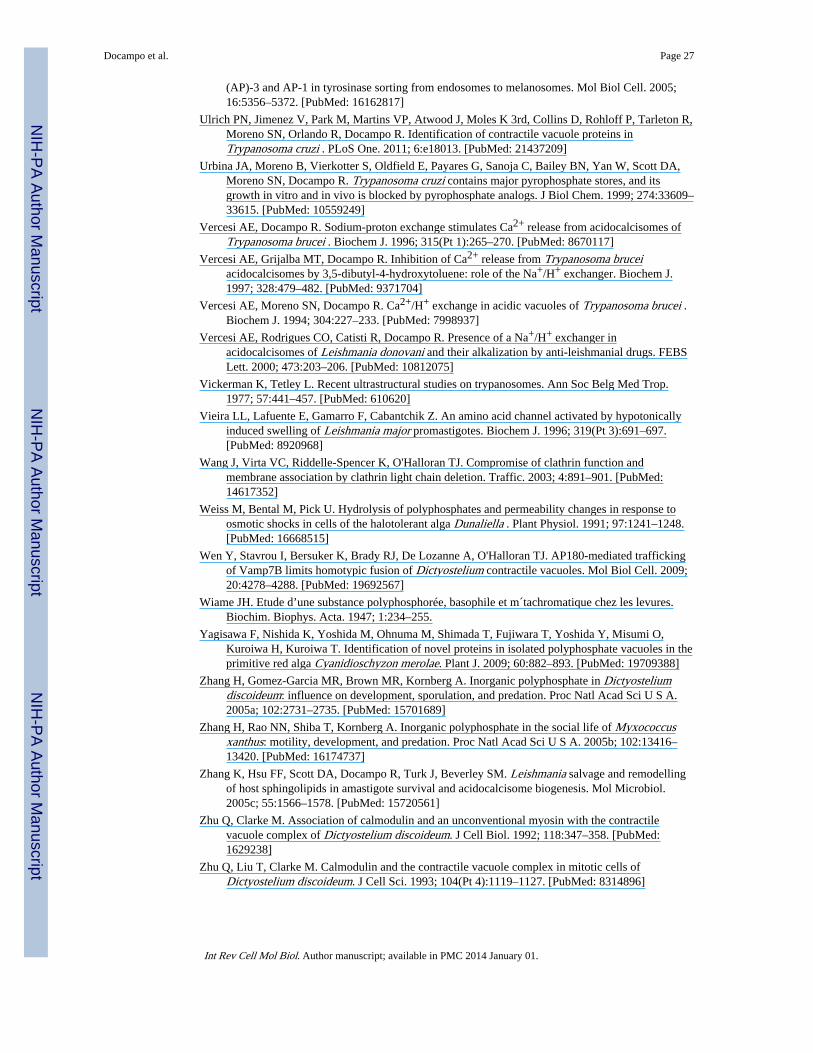
(AP)-3 and AP-1 in tyrosinase sorting from endosomes to melanosomes. Mol Biol Cell. 2005;16:5356–5372. [PubMed: 16162817]
Ulrich PN, Jimenez V, Park M, Martins VP, Atwood J, Moles K 3rd, Collins D, Rohloff P, Tarleton R,Moreno SN, Orlando R, Docampo R. Identification of contractile vacuole proteins inTrypanosoma cruzi . PLoS One. 2011; 6:e18013. [PubMed: 21437209]
Urbina JA, Moreno B, Vierkotter S, Oldfield E, Payares G, Sanoja C, Bailey BN, Yan W, Scott DA,Moreno SN, Docampo R. Trypanosoma cruzi contains major pyrophosphate stores, and itsgrowth in vitro and in vivo is blocked by pyrophosphate analogs. J Biol Chem. 1999; 274:33609–33615. [PubMed: 10559249]
Vercesi AE, Docampo R. Sodium-proton exchange stimulates Ca2+ release from acidocalcisomes ofTrypanosoma brucei . Biochem J. 1996; 315(Pt 1):265–270. [PubMed: 8670117]
Vercesi AE, Grijalba MT, Docampo R. Inhibition of Ca2+ release from Trypanosoma bruceiacidocalcisomes by 3,5-dibutyl-4-hydroxytoluene: role of the Na+/H+ exchanger. Biochem J.1997; 328:479–482. [PubMed: 9371704]
Vercesi AE, Moreno SN, Docampo R. Ca2+/H+ exchange in acidic vacuoles of Trypanosoma brucei .Biochem J. 1994; 304:227–233. [PubMed: 7998937]
Vercesi AE, Rodrigues CO, Catisti R, Docampo R. Presence of a Na+/H+ exchanger inacidocalcisomes of Leishmania donovani and their alkalization by anti-leishmanial drugs. FEBSLett. 2000; 473:203–206. [PubMed: 10812075]
Vickerman K, Tetley L. Recent ultrastructural studies on trypanosomes. Ann Soc Belg Med Trop.1977; 57:441–457. [PubMed: 610620]
Vieira LL, Lafuente E, Gamarro F, Cabantchik Z. An amino acid channel activated by hypotonicallyinduced swelling of Leishmania major promastigotes. Biochem J. 1996; 319(Pt 3):691–697.[PubMed: 8920968]
Wang J, Virta VC, Riddelle-Spencer K, O'Halloran TJ. Compromise of clathrin function andmembrane association by clathrin light chain deletion. Traffic. 2003; 4:891–901. [PubMed:14617352]
Weiss M, Bental M, Pick U. Hydrolysis of polyphosphates and permeability changes in response toosmotic shocks in cells of the halotolerant alga Dunaliella . Plant Physiol. 1991; 97:1241–1248.[PubMed: 16668515]
Wen Y, Stavrou I, Bersuker K, Brady RJ, De Lozanne A, O'Halloran TJ. AP180-mediated traffickingof Vamp7B limits homotypic fusion of Dictyostelium contractile vacuoles. Mol Biol Cell. 2009;20:4278–4288. [PubMed: 19692567]
Wiame JH. Etude d’une substance polyphosphorée, basophile et m´tachromatique chez les levures.Biochim. Biophys. Acta. 1947; 1:234–255.
Yagisawa F, Nishida K, Yoshida M, Ohnuma M, Shimada T, Fujiwara T, Yoshida Y, Misumi O,Kuroiwa H, Kuroiwa T. Identification of novel proteins in isolated polyphosphate vacuoles in theprimitive red alga Cyanidioschyzon merolae. Plant J. 2009; 60:882–893. [PubMed: 19709388]
Zhang H, Gomez-Garcia MR, Brown MR, Kornberg A. Inorganic polyphosphate in Dictyosteliumdiscoideum: influence on development, sporulation, and predation. Proc Natl Acad Sci U S A.2005a; 102:2731–2735. [PubMed: 15701689]
Zhang H, Rao NN, Shiba T, Kornberg A. Inorganic polyphosphate in the social life of Myxococcusxanthus: motility, development, and predation. Proc Natl Acad Sci U S A. 2005b; 102:13416–13420. [PubMed: 16174737]
Zhang K, Hsu FF, Scott DA, Docampo R, Turk J, Beverley SM. Leishmania salvage and remodellingof host sphingolipids in amastigote survival and acidocalcisome biogenesis. Mol Microbiol.2005c; 55:1566–1578. [PubMed: 15720561]
Zhu Q, Clarke M. Association of calmodulin and an unconventional myosin with the contractilevacuole complex of Dictyostelium discoideum. J Cell Biol. 1992; 118:347–358. [PubMed:1629238]
Zhu Q, Liu T, Clarke M. Calmodulin and the contractile vacuole complex in mitotic cells ofDictyostelium discoideum. J Cell Sci. 1993; 104(Pt 4):1119–1127. [PubMed: 8314896]
Docampo et al. Page 27
Int Rev Cell Mol Biol. Author manuscript; available in PMC 2014 January 01.
NIH
-PA Author Manuscript
NIH
-PA Author Manuscript
NIH
-PA Author Manuscript
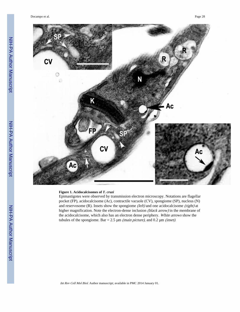
Figure 1. Acidocalcisomes of T. cruziEpimastigotes were observed by transmission electron microscopy. Notations are flagellarpocket (FP), acidocalcisome (Ac), contractile vacuole (CV), spongiome (SP), nucleus (N)and reservosome (R). Insets show the spongiome (left) and one acidocalcisome (right) athigher magnification. Note the electron-dense inclusion (black arrow) in the membrane ofthe acidocalcisome, which also has an electron dense periphery. White arrows show thetubules of the spongiome. Bar = 2.5 µm (main picture), and 0.2 µm (inset).
Docampo et al. Page 28
Int Rev Cell Mol Biol. Author manuscript; available in PMC 2014 January 01.
NIH
-PA Author Manuscript
NIH
-PA Author Manuscript
NIH
-PA Author Manuscript
Figure 2. The CVC in T. cruzi epimastigotes(A) Thin section of chemically fixed epimastigote showing the CVC. Note the collapsedaspect of the spongiome (arrows). (B) Thin section of a high pressure-freeze substitutedepimastigote in the G2 phase of the cell cycle showing the CV and the interconnectedtubules (arrows) that form the spongiome (Sp). (C) Virtual section showing the CV dockedto the flagellar pocket (FP) and the electron dense region between both structures (arrow andinset). (D) Virtual section and 3D model of the CVC and flagellar pocket (FP) where adeformation in the FP was observed (black arrows) and a tubule of the spongiome wasconnected to the central vacuole (white arrow). (E) 3D model of the CVC and FP showingthe interconnected spongiome connected to the CV (arrows) and concentrated in the anterior
Docampo et al. Page 29
Int Rev Cell Mol Biol. Author manuscript; available in PMC 2014 January 01.
NIH
-PA Author Manuscript
NIH
-PA Author Manuscript
NIH
-PA Author Manuscript
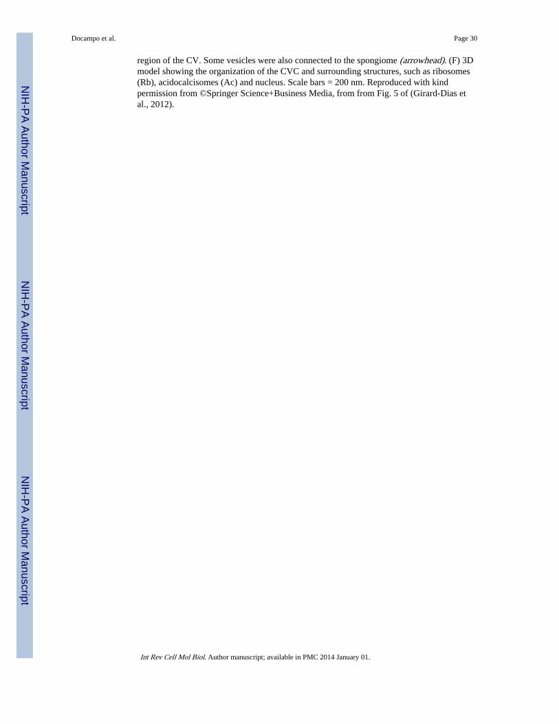
region of the CV. Some vesicles were also connected to the spongiome (arrowhead). (F) 3Dmodel showing the organization of the CVC and surrounding structures, such as ribosomes(Rb), acidocalcisomes (Ac) and nucleus. Scale bars = 200 nm. Reproduced with kindpermission from ©Springer Science+Business Media, from from Fig. 5 of (Girard-Dias etal., 2012).
Docampo et al. Page 30
Int Rev Cell Mol Biol. Author manuscript; available in PMC 2014 January 01.
NIH
-PA Author Manuscript
NIH
-PA Author Manuscript
NIH
-PA Author Manuscript
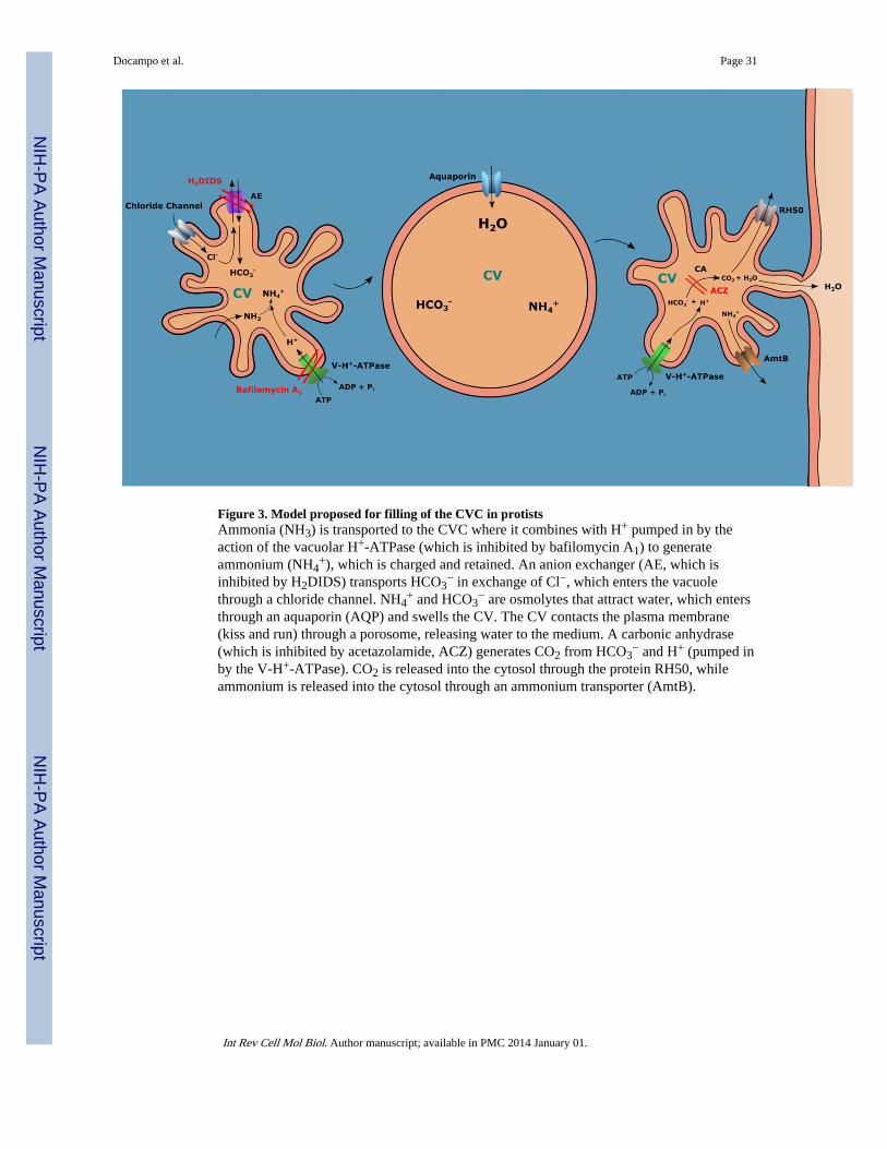
Figure 3. Model proposed for filling of the CVC in protistsAmmonia (NH3) is transported to the CVC where it combines with H+ pumped in by theaction of the vacuolar H+-ATPase (which is inhibited by bafilomycin A1) to generateammonium (NH4
+), which is charged and retained. An anion exchanger (AE, which isinhibited by H2DIDS) transports HCO3
− in exchange of Cl−, which enters the vacuolethrough a chloride channel. NH4
+ and HCO3− are osmolytes that attract water, which enters
through an aquaporin (AQP) and swells the CV. The CV contacts the plasma membrane(kiss and run) through a porosome, releasing water to the medium. A carbonic anhydrase(which is inhibited by acetazolamide, ACZ) generates CO2 from HCO3
− and H+ (pumped inby the V-H+-ATPase). CO2 is released into the cytosol through the protein RH50, whileammonium is released into the cytosol through an ammonium transporter (AmtB).
Docampo et al. Page 31
Int Rev Cell Mol Biol. Author manuscript; available in PMC 2014 January 01.
NIH
-PA Author Manuscript
NIH
-PA Author Manuscript
NIH
-PA Author Manuscript
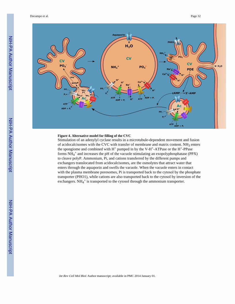
Figure 4. Alternative model for filling of the CVCStimulation of an adenylyl cyclase results in a microtubule-dependent movement and fusionof acidocalcisomes with the CVC with transfer of membrane and matrix content. NH3 entersthe spongiome and combined with H+ pumped in by the V-H+-ATPase or the H+-PPaseforms NH4
+ and increases the pH of the vacuole stimulating an exopolyphosphatase (PPX)to cleave polyP. Ammonium, Pi, and cations transferred by the different pumps andexchangers translocated from acidocalcisomes, are the osmolytes that attract water thatenters through the aquaporin and swells the vacuole. When the vacuole enters in contactwith the plasma membrane porosomes, Pi is transported back to the cytosol by the phosphatetransporter (PHO1), while cations are also transported back to the cytosol by inversion of theexchangers. NH4
+ is transported to the cytosol through the ammonium transporter.
Docampo et al. Page 32
Int Rev Cell Mol Biol. Author manuscript; available in PMC 2014 January 01.
NIH
-PA Author Manuscript
NIH
-PA Author Manuscript
NIH
-PA Author Manuscript
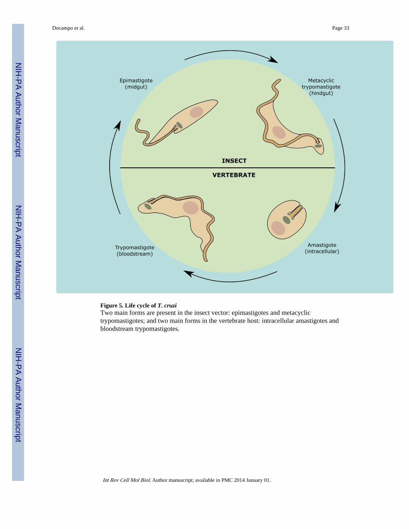
Figure 5. Life cycle of T. cruziTwo main forms are present in the insect vector: epimastigotes and metacyclictrypomastigotes; and two main forms in the vertebrate host: intracellular amastigotes andbloodstream trypomastigotes.
Docampo et al. Page 33
Int Rev Cell Mol Biol. Author manuscript; available in PMC 2014 January 01.
NIH
-PA Author Manuscript
NIH
-PA Author Manuscript
NIH
-PA Author Manuscript
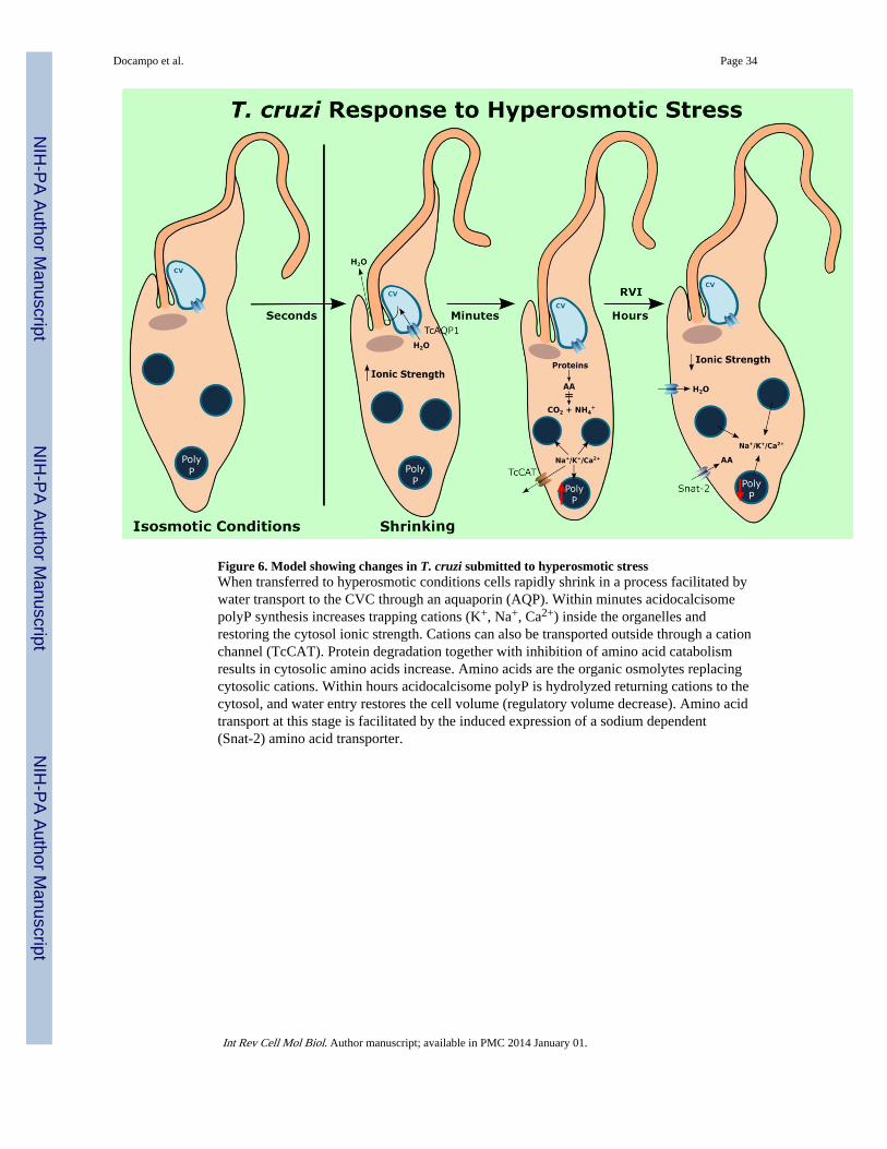
Figure 6. Model showing changes in T. cruzi submitted to hyperosmotic stressWhen transferred to hyperosmotic conditions cells rapidly shrink in a process facilitated bywater transport to the CVC through an aquaporin (AQP). Within minutes acidocalcisomepolyP synthesis increases trapping cations (K+, Na+, Ca2+) inside the organelles andrestoring the cytosol ionic strength. Cations can also be transported outside through a cationchannel (TcCAT). Protein degradation together with inhibition of amino acid catabolismresults in cytosolic amino acids increase. Amino acids are the organic osmolytes replacingcytosolic cations. Within hours acidocalcisome polyP is hydrolyzed returning cations to thecytosol, and water entry restores the cell volume (regulatory volume decrease). Amino acidtransport at this stage is facilitated by the induced expression of a sodium dependent(Snat-2) amino acid transporter.
Docampo et al. Page 34
Int Rev Cell Mol Biol. Author manuscript; available in PMC 2014 January 01.
NIH
-PA Author Manuscript
NIH
-PA Author Manuscript
NIH
-PA Author Manuscript
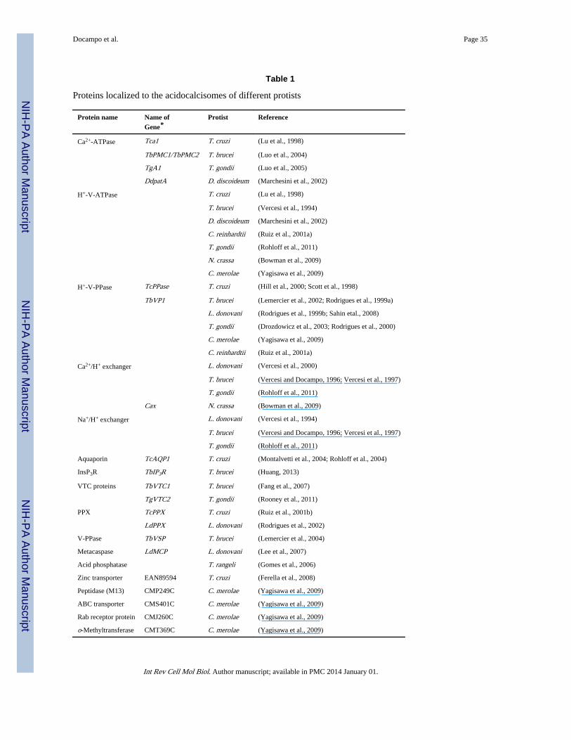
NIH
-PA Author Manuscript
NIH
-PA Author Manuscript
NIH
-PA Author Manuscript
Docampo et al. Page 35
Table 1
Proteins localized to the acidocalcisomes of different protists
Protein name Name ofGene*
Protist Reference
Ca2+-ATPase Tca1 T. cruzi (Lu et al., 1998)
TbPMC1/TbPMC2 T. brucei (Luo et al., 2004)
TgA1 T. gondii (Luo et al., 2005)
DdpatA D. discoideum (Marchesini et al., 2002)
H+-V-ATPase T. cruzi (Lu et al., 1998)
T. brucei (Vercesi et al., 1994)
D. discoideum (Marchesini et al., 2002)
C. reinhardtii (Ruiz et al., 2001a)
T. gondii (Rohloff et al., 2011)
N. crassa (Bowman et al., 2009)
C. merolae (Yagisawa et al., 2009)
H+-V-PPase TcPPase T. cruzi (Hill et al., 2000; Scott et al., 1998)
TbVP1 T. brucei (Lemercier et al., 2002; Rodrigues et al., 1999a)
L. donovani (Rodrigues et al., 1999b; Sahin etal., 2008)
T. gondii (Drozdowicz et al., 2003; Rodrigues et al., 2000)
C. merolae (Yagisawa et al., 2009)
C. reinhardtii (Ruiz et al., 2001a)
Ca2+/H+ exchanger L. donovani (Vercesi et al., 2000)
T. brucei (Vercesi and Docampo, 1996; Vercesi et al., 1997)
T. gondii (Rohloff et al., 2011)
Cax N. crassa (Bowman et al., 2009)
Na+/H+ exchanger L. donovani (Vercesi et al., 1994)
T. brucei (Vercesi and Docampo, 1996; Vercesi et al., 1997)
T. gondii (Rohloff et al., 2011)
Aquaporin TcAQP1 T. cruzi (Montalvetti et al., 2004; Rohloff et al., 2004)
InsP3R TbIP3R T. brucei (Huang, 2013)
VTC proteins TbVTC1 T. brucei (Fang et al., 2007)
TgVTC2 T. gondii (Rooney et al., 2011)
PPX TcPPX T. cruzi (Ruiz et al., 2001b)
LdPPX L. donovani (Rodrigues et al., 2002)
V-PPase TbVSP T. brucei (Lemercier et al., 2004)
Metacaspase LdMCP L. donovani (Lee et al., 2007)
Acid phosphatase T. rangeli (Gomes et al., 2006)
Zinc transporter EAN89594 T. cruzi (Ferella et al., 2008)
Peptidase (M13) CMP249C C. merolae (Yagisawa et al., 2009)
ABC transporter CMS401C C. merolae (Yagisawa et al., 2009)
Rab receptor protein CMJ260C C. merolae (Yagisawa et al., 2009)
o-Methyltransferase CMT369C C. merolae (Yagisawa et al., 2009)
Int Rev Cell Mol Biol. Author manuscript; available in PMC 2014 January 01.
NIH
-PA Author Manuscript
NIH
-PA Author Manuscript
NIH
-PA Author Manuscript
Docampo et al. Page 36
*When a gene name is indicated it means that it has been cloned, expressed and the protein product localized to acidocalcisomes.
Int Rev Cell Mol Biol. Author manuscript; available in PMC 2014 January 01.
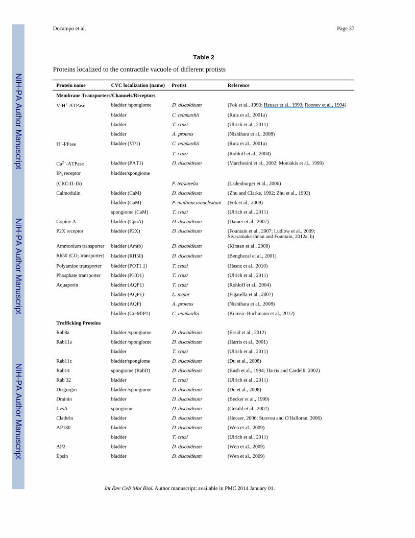
NIH
-PA Author Manuscript
NIH
-PA Author Manuscript
NIH
-PA Author Manuscript
Docampo et al. Page 37
Table 2
Proteins localized to the contractile vacuole of different protists
Protein name CVC localization (name) Protist Reference
Membrane Transporters/Channels/Receptors
V-H+-ATPase bladder /spongiome D. discoideum (Fok et al., 1993; Heuser et al., 1993; Rooney et al., 1994)
bladder C. reinhardtii (Ruiz et al., 2001a)
bladder T. cruzi (Ulrich et al., 2011)
bladder A. proteus (Nishihara et al., 2008)
H+-PPase bladder (VP1) C. reinhardtii (Ruiz et al., 2001a)
T. cruzi (Rohloff et al., 2004)
Ca2+-ATPase bladder (PAT1) D. discoideum (Marchesini et al., 2002; Moniakis et al., 1999)
IP3 receptor bladder/spongiome
(CRC-II-1b) P. tetraurelia (Ladenburger et al., 2006)
Calmodulin bladder (CaM) D. discoideum (Zhu and Clarke, 1992; Zhu et al., 1993)
bladder (CaM) P. multimicronucleatum (Fok et al., 2008)
spongiome (CaM) T. cruzi (Ulrich et al., 2011)
Copine A bladder (CpnA) D. discoideum (Damer et al., 2007)
P2X receptor bladder (P2X) D. discoideum (Fountain et al., 2007; Ludlow et al., 2009;Sivaramakrishnan and Fountain, 2012a, b)
Ammonium transporter bladder (Amtb) D. discoideum (Kirsten et al., 2008)
Rh50 (CO2 transporter) bladder (RH50) D. discoideum (Benghezal et al., 2001)
Polyamine transporter bladder (POT1.1) T. cruzi (Hasne et al., 2010)
Phosphate transporter bladder (PHO1) T. cruzi (Ulrich et al., 2011)
Aquaporin bladder (AQP1) T. cruzi (Rohloff et al., 2004)
bladder (AQP1) L. major (Figarella et al., 2007)
bladder (AQP) A. proteus (Nishihara et al., 2008)
bladder (CreMIP1) C. reinhardtii (Komsic-Buchmann et al., 2012)
Trafficking Proteins
Rab8a bladder /spongiome D. discoideum (Essid et al., 2012)
Rab11a bladder /spongiome D. discoideum (Harris et al., 2001)
bladder T. cruzi (Ulrich et al., 2011)
Rab11c bladder/spongiome D. discoideum (Du et al., 2008)
Rab14 spongiome (RabD) D. discoideum (Bush et al., 1994; Harris and Cardelli, 2002)
Rab 32 bladder T. cruzi (Ulrich et al., 2011)
Disgorgin bladder /spongiome D. discoideum (Du et al., 2008)
Drainin bladder D. discoideum (Becker et al., 1999)
LvsA spongiome D. discoideum (Gerald et al., 2002)
Clathrin bladder D. discoideum (Heuser, 2006; Stavrou and O'Halloran, 2006)
AP180 bladder D. discoideum (Wen et al., 2009)
bladder T. cruzi (Ulrich et al., 2011)
AP2 bladder D. discoideum (Wen et al., 2009)
Epsin bladder D. discoideum (Wen et al., 2009)
Int Rev Cell Mol Biol. Author manuscript; available in PMC 2014 January 01.
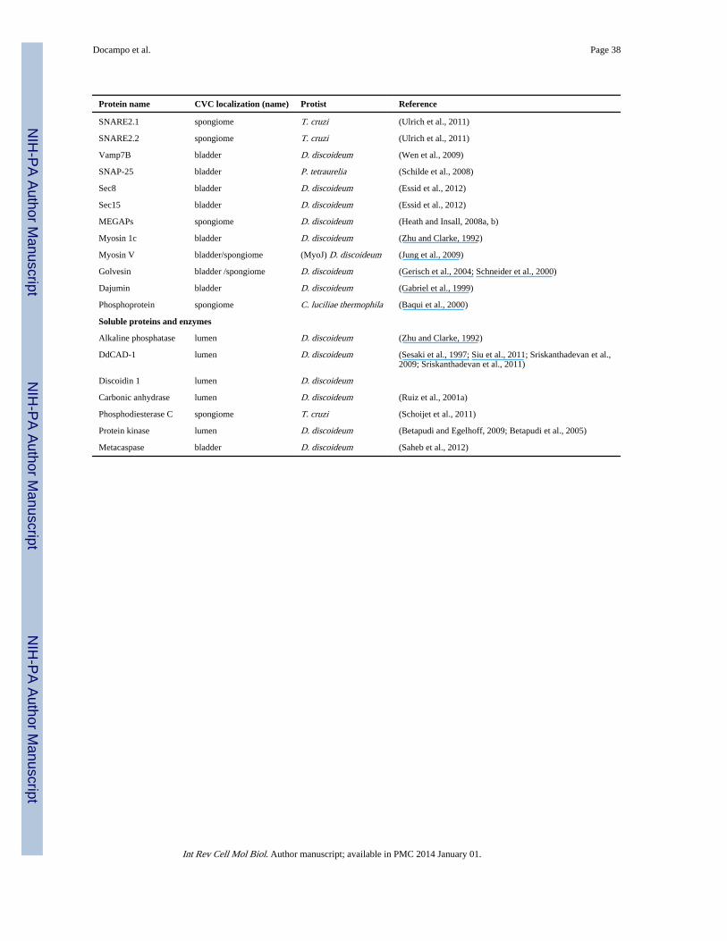
NIH
-PA Author Manuscript
NIH
-PA Author Manuscript
NIH
-PA Author Manuscript
Docampo et al. Page 38
Protein name CVC localization (name) Protist Reference
SNARE2.1 spongiome T. cruzi (Ulrich et al., 2011)
SNARE2.2 spongiome T. cruzi (Ulrich et al., 2011)
Vamp7B bladder D. discoideum (Wen et al., 2009)
SNAP-25 bladder P. tetraurelia (Schilde et al., 2008)
Sec8 bladder D. discoideum (Essid et al., 2012)
Sec15 bladder D. discoideum (Essid et al., 2012)
MEGAPs spongiome D. discoideum (Heath and Insall, 2008a, b)
Myosin 1c bladder D. discoideum (Zhu and Clarke, 1992)
Myosin V bladder/spongiome (MyoJ) D. discoideum (Jung et al., 2009)
Golvesin bladder /spongiome D. discoideum (Gerisch et al., 2004; Schneider et al., 2000)
Dajumin bladder D. discoideum (Gabriel et al., 1999)
Phosphoprotein spongiome C. luciliae thermophila (Baqui et al., 2000)
Soluble proteins and enzymes
Alkaline phosphatase lumen D. discoideum (Zhu and Clarke, 1992)
DdCAD-1 lumen D. discoideum (Sesaki et al., 1997; Siu et al., 2011; Sriskanthadevan et al.,2009; Sriskanthadevan et al., 2011)
Discoidin 1 lumen D. discoideum
Carbonic anhydrase lumen D. discoideum (Ruiz et al., 2001a)
Phosphodiesterase C spongiome T. cruzi (Schoijet et al., 2011)
Protein kinase lumen D. discoideum (Betapudi and Egelhoff, 2009; Betapudi et al., 2005)
Metacaspase bladder D. discoideum (Saheb et al., 2012)
Int Rev Cell Mol Biol. Author manuscript; available in PMC 2014 January 01.
Copyright © 2022 FDOKUMEN