Identification of a serine protease inhibitor which causes inclusion vacuole reduction and is lethal...
14
Identification of a serine protease inhibitor which causes inclusion vacuole reduction and is lethal to Chlamydia trachomatis Sarina Gloeckl, 1† Vanissa A. Ong, 1† Pooja Patel, 1† Joel D. A. Tyndall, 2 Peter Timms, 1 Kenneth W. Beagley, 1 John A. Allan, 3 Charles W. Armitage, 1 Lynne Turnbull, 4 Cynthia B. Whitchurch, 4 Melisa Merdanovic, 5 Michael Ehrmann, 5 James C. Powers, 6 Jozef Oleksyszyn, 7 Martijn Verdoes, 8 Matthew Bogyo 8 and Wilhelmina M. Huston 1 * 1 Institute of Health and Biomedical Innovation and School of Biomedical Sciences, Faculty of Health, Queensland University of Technology, Kelvin Grove, Qld 4059, Australia. 2 School of Pharmacy, University of Otago, Dunedin, New Zealand. 3 The Wesley Research Institute, The Wesley Reproductive Medicine and Gynaecological Surgery Unit, Auchenflower, Brisbane, Qld, Australia. 4 Microbial Imaging Facility, The iThree institute, University of Technology Sydney, Sydney, NSW 2007, Australia. 5 ZMB, Uni Duisburg-Essen, Essen, Germany. 6 School of Chemistry and Biochemistry, Georgia Institute of Technology, Atlanta, GA 30332, USA. 7 Division of Medicinal Chemistry and Microbiology, Faculty of Chemistry, Wroclaw University of Technology, Wybrzez ˙e Wyspian ´ skiego 27, 50-370 Wroclaw, Poland. 8 Department of Pathology, Stanford University School of Medicine, Stanford, CA 94305-5324, USA. Summary The mechanistic details of the pathogenesis of Chla- mydia, an obligate intracellular pathogen of global importance, have eluded scientists due to the scarcity of traditional molecular genetic tools to investigate this organism. Here we report a chemical biology strat- egy that has uncovered the first essential protease for this organism. Identification and application of a unique CtHtrA inhibitor (JO146) to cultures of Chla- mydia resulted in a complete loss of viable elementary body formation. JO146 treatment during the replica- tive phase of development resulted in a loss of Chla- mydia cell morphology, diminishing inclusion size, and ultimate loss of inclusions from the host cells. This completely prevented the formation of viable Chlamydia elementary bodies. In addition to its effect on the human Chlamydia trachomatis strain, JO146 inhibited the viability of the mouse strain, Chlamydia muridarum, both in vitro and in vivo. Thus, we report a chemical biology approach to establish an essential role for Chlamydia CtHtrA. The function of CtHtrA for Chlamydia appears to be essential for maintenance of cell morphology during replicative the phase and these findings provide proof of concept that proteases can be targeted for antimicrobial therapy for intracel- lular pathogens. Introduction Chlamydia are obligate intracellular bacterial pathogens with significant clinical importance. Chlamydia (C.) tra- chomatis is the most common sexually transmitted bacte- rial infection world wide and the ocular infecting serovars are the most common cause of preventable blindness worldwide (World Health Organization, 2011). In spite of the substantial health burden due to the Chlamydia, com- paratively little is known about the organism’s functional pathogenesis and disease mechanisms. Members of the chlamydiae family are distinguished by an unusual bi-phasic developmental cycle that consists of a small, tough, spore-like, non-replicative extracellular form [termed the elementary body (EB)] and the intracel- lular replicative, metabolically active, non-infectious form [called the reticulate body (RB)]. The intracellular phase occurs exclusively in a unique vacuole known as the inclusion vacuole (reviewed Abelrahman and Belland, 2005). This developmental cycle and the obligate intracel- lular nature of the Chlamydia have hampered efforts to develop traditional techniques to genetically manipulate the organism. Although there have been recent develop- ments, including a chemical mutation approach to gener- ate chromosomal mutants (Kari et al., 2011) and Accepted 19 June, 2013. *For correspondence. E-mail w.huston@ qut.edu.au; Tel. (+61) 7 31386258; Fax (+61) 7 31386030. † S.G., V.A.O. and P.P. all contributed equally to this work. Molecular Microbiology (2013) 89(4), 676–689 ■ doi:10.1111/mmi.12306 First published online 12 July 2013 © 2013 John Wiley & Sons Ltd
-
Upload
independent -
Category
Documents
-
view
1 -
download
0
Transcript of Identification of a serine protease inhibitor which causes inclusion vacuole reduction and is lethal...
Identification of a serine protease inhibitor whichcauses inclusion vacuole reduction and is lethal toChlamydia trachomatis
Sarina Gloeckl,1† Vanissa A. Ong,1† Pooja Patel,1†
Joel D. A. Tyndall,2 Peter Timms,1
Kenneth W. Beagley,1 John A. Allan,3
Charles W. Armitage,1 Lynne Turnbull,4
Cynthia B. Whitchurch,4 Melisa Merdanovic,5
Michael Ehrmann,5 James C. Powers,6
Jozef Oleksyszyn,7 Martijn Verdoes,8
Matthew Bogyo8 and Wilhelmina M. Huston1*1Institute of Health and Biomedical Innovation andSchool of Biomedical Sciences, Faculty of Health,Queensland University of Technology, Kelvin Grove,Qld 4059, Australia.2School of Pharmacy, University of Otago, Dunedin,New Zealand.3The Wesley Research Institute, The WesleyReproductive Medicine and Gynaecological SurgeryUnit, Auchenflower, Brisbane, Qld, Australia.4Microbial Imaging Facility, The iThree institute,University of Technology Sydney, Sydney, NSW 2007,Australia.5ZMB, Uni Duisburg-Essen, Essen, Germany.6School of Chemistry and Biochemistry, GeorgiaInstitute of Technology, Atlanta, GA 30332, USA.7Division of Medicinal Chemistry and Microbiology,Faculty of Chemistry, Wrocław University of Technology,Wybrzeze Wyspianskiego 27, 50-370 Wrocław, Poland.8Department of Pathology, Stanford University School ofMedicine, Stanford, CA 94305-5324, USA.
Summary
The mechanistic details of the pathogenesis of Chla-mydia, an obligate intracellular pathogen of globalimportance, have eluded scientists due to the scarcityof traditional molecular genetic tools to investigatethis organism. Here we report a chemical biology strat-egy that has uncovered the first essential protease forthis organism. Identification and application of aunique CtHtrA inhibitor (JO146) to cultures of Chla-
mydia resulted in a complete loss of viable elementarybody formation. JO146 treatment during the replica-tive phase of development resulted in a loss of Chla-mydia cell morphology, diminishing inclusion size,and ultimate loss of inclusions from the host cells.This completely prevented the formation of viableChlamydia elementary bodies. In addition to its effecton the human Chlamydia trachomatis strain, JO146inhibited the viability of the mouse strain, Chlamydiamuridarum, both in vitro and in vivo. Thus, we report achemical biology approach to establish an essentialrole for Chlamydia CtHtrA. The function of CtHtrA forChlamydia appears to be essential for maintenanceof cell morphology during replicative the phase andthese findings provide proof of concept that proteasescan be targeted for antimicrobial therapy for intracel-lular pathogens.
Introduction
Chlamydia are obligate intracellular bacterial pathogenswith significant clinical importance. Chlamydia (C.) tra-chomatis is the most common sexually transmitted bacte-rial infection world wide and the ocular infecting serovarsare the most common cause of preventable blindnessworldwide (World Health Organization, 2011). In spite ofthe substantial health burden due to the Chlamydia, com-paratively little is known about the organism’s functionalpathogenesis and disease mechanisms.
Members of the chlamydiae family are distinguished byan unusual bi-phasic developmental cycle that consists ofa small, tough, spore-like, non-replicative extracellularform [termed the elementary body (EB)] and the intracel-lular replicative, metabolically active, non-infectious form[called the reticulate body (RB)]. The intracellular phaseoccurs exclusively in a unique vacuole known as theinclusion vacuole (reviewed Abelrahman and Belland,2005). This developmental cycle and the obligate intracel-lular nature of the Chlamydia have hampered efforts todevelop traditional techniques to genetically manipulatethe organism. Although there have been recent develop-ments, including a chemical mutation approach to gener-ate chromosomal mutants (Kari et al., 2011) and
Accepted 19 June, 2013. *For correspondence. E-mail [email protected]; Tel. (+61) 7 31386258; Fax (+61) 7 31386030. †S.G.,V.A.O. and P.P. all contributed equally to this work.
Molecular Microbiology (2013) 89(4), 676–689 ■ doi:10.1111/mmi.12306First published online 12 July 2013
© 2013 John Wiley & Sons Ltd
successful transformation of the organism with its ownplasmid (Wang et al., 2011), biological understanding ofthis organism remains limited relative to its economic andhealth impact.
The highly conserved protease HtrA (Chlamydia HtrA;CtHtrA) has been described as having a number of viru-lence functions in pathogenic bacteria (Hoy et al., 2010;Chitlaru et al., 2011). This protease has also beendescribed in C. trachomatis using in vitro and microscopymethods, and in Chlamydia it potentially functions both asa bacterial cell-associated protease and as a secretedvirulence factor (from 28 h post infection) (Huston et al.,2007; 2008; Wu et al., 2011). Our previous investigationsinto the biochemical mechanism of activation implicatedouter membrane protein sequences with activation of thechaperone form, suggesting a potential role in surfaceprotein assembly (Huston et al., 2011). Recently the pro-tease that has been considered to be the major chlamydialpathogenesis factor, CPAF, has been the focus of contro-versy as it appears that many of the functions attributed tothis protease may have been detected as an artefact of thesample preparation (Chen et al., 2012), including a keyfunction thought to be critical for viable infectious yield(Heuer et al., 2009).
Other chlamydial virulence factors have also beenuncovered using small molecule approaches, includingthe bacterial type III protein secretion inhibitor (Wolf et al.,2006), and a peptidomimetic to the type III secretionATPase protein interaction domain (Stone et al., 2011).These studies support the fact that chemical approachesare a powerful strategy to investigate the function of pro-teins within this unique organism. Here we screened forand found a novel selective inhibitor against a CtHtrA.Using this protease inhibitor, JO146, we demonstrated anessential role for CtHtrA for chlamydial developmentalcycle progression during the replicative phase. JO146treatment resulted in chlamydial cell morphology loss, thereduction in inclusion vacuoles and eventual completebacterial lethality as the result of failure to develop viableinfectious progeny (EBs).
Results
Selective and specific phosphonate inhibitors of CtHtrAare lethal to C. trachomatis when added during thereplicative phase of the developmental cycle
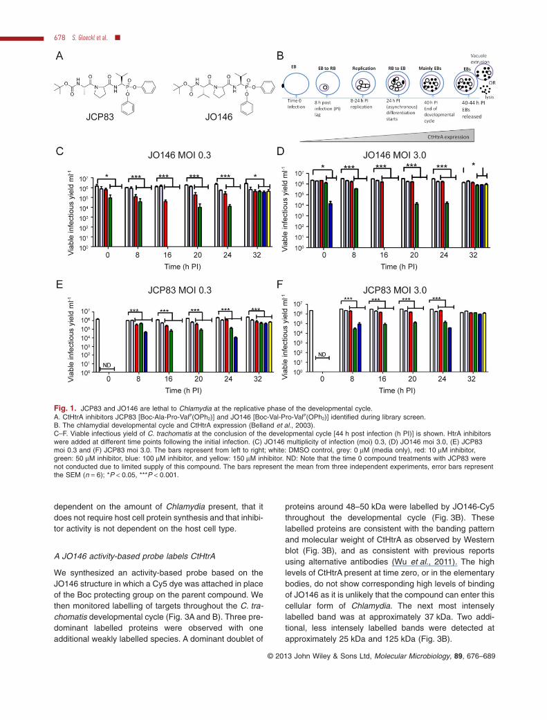
A library of 1090 serine protease inhibitors [previouslydescribed (Arastu-Kapur et al., 2008; Hall et al., 2011)]was screened against CtHtrA in vitro protease activity(detailed in Experimental procedures). An initial screenidentified two inhibitors, JO146 and JCP83 (IC50s:12.5 μM and 47.19 μM, Fig. 1A). Both are peptides with aC-terminal phosphonate ‘warhead’ electrophile which
reacts irreversibly with the protease active-site serineresidue (Oleksyszyn and Powers, 1991) (Jackson et al.,1998), and differ only in the P3 position (Val/Ala). Bothinhibitors have valine at the P1 position and proline at P2.The two inhibitors were quite selective towards CtHtrAwhen screened against a series of other serine and HtrAfamily proteases (Table 1, Fig. S1). Inhibition was onlyobserved for HTRA2 and elastase. Neither compoundwas cytotoxic to McCoy and HEp-2 cells, both commoncell lines used for Chlamydia culture. Specifically, wedetected no cell lysis or impaired metabolic turnover whenthe cells were incubated with JO146 or JCP83 for 8 h(Table S1, as described in the supporting data).
The two inhibitors were added to C. trachomatis HEp-2cultures at different time points throughout the develop-mental cycle [Fig. 1B: lag ∼ 8 h, replication at ∼ 2.4 h perdivision, EB formation from ∼ 24 h post infection (h PI)(Miyairi et al., 2006)]. Viable infectious elementary bodies(inclusion forming units) at completion of the developmen-tal cycle after each of these independent treatmentswas then determined. As shown in Fig. 1C–F, additionof JO146 or JCP83 resulted in complete or significant(P < 0.001) loss of viable elementary body formationwhen added during the replicative phase. The activity wasmost effective at higher doses; however, at 16 h PI even10 μM JO146 had a significant impact on viability with alower Chlamydia multiplicity of infection (moi) (Fig. 1C,P < 0.001). The host cell numbers were the same undereach condition so these data indicate that the amount ofChlamydia present (moi) associates with the effective-ness of the compounds.
The lethality of JO146 for C. trachomatis at 16 h PIdoes not require host cell protein synthesis, and is notinfluenced by the type of host cell
Chlamydia viable yield from cell culture improves when ahost cell protein synthesis inhibitor (cycloheximide) isused (Thomas et al., 1977). We tested the role of host cellprotein synthesis during JO146 inhibition (16 h PI) at anmoi of 0.3 in HEp-2 cells. The lethality of JO146 wasdetermined at 44 h PI and was found to be maintained inthe presence of cycloheximide (Fig. 2A).
Chlamydia trachomatis is capable of infection and rep-lication within a variety of epithelial cells. The effective-ness of JO146 against C. trachomatis when added at 16 hPI was evaluated in a variety of host cell types with viabil-ity determined at 44 h PI (moi 0.3). There were differentyields of C. trachomatis depending on the host cell typeinfected which was expected (Fig. 2B). There were sig-nificantly (P < 0.001) higher yields of viable Chlamydiawhen cultured in McCoy cells; however, JO146 was stilllethal for C. trachomatis at a concentration of 100 μM inMcCoy cells. These data suggest that JO146 activity is
Essential serine protease for Chlamydia 677
© 2013 John Wiley & Sons Ltd, Molecular Microbiology, 89, 676–689
dependent on the amount of Chlamydia present, that itdoes not require host cell protein synthesis and that inhibi-tor activity is not dependent on the host cell type.
A JO146 activity-based probe labels CtHtrA
We synthesized an activity-based probe based on theJO146 structure in which a Cy5 dye was attached in placeof the Boc protecting group on the parent compound. Wethen monitored labelling of targets throughout the C. tra-chomatis developmental cycle (Fig. 3A and B). Three pre-dominant labelled proteins were observed with oneadditional weakly labelled species. A dominant doublet of
proteins around 48–50 kDa were labelled by JO146-Cy5throughout the developmental cycle (Fig. 3B). Theselabelled proteins are consistent with the banding patternand molecular weight of CtHtrA as observed by Westernblot (Fig. 3B), and as consistent with previous reportsusing alternative antibodies (Wu et al., 2011). The highlevels of CtHtrA present at time zero, or in the elementarybodies, do not show corresponding high levels of bindingof JO146 as it is unlikely that the compound can enter thiscellular form of Chlamydia. The next most intenselylabelled band was at approximately 37 kDa. Two addi-tional, less intensely labelled bands were detected atapproximately 25 kDa and 125 kDa (Fig. 3B).
Fig. 1. JCP83 and JO146 are lethal to Chlamydia at the replicative phase of the developmental cycle.A. CtHtrA inhibitors JCP83 [Boc-Ala-Pro-ValP(OPh2)] and JO146 [Boc-Val-Pro-ValP(OPh2)] identified during library screen.B. The chlamydial developmental cycle and CtHtrA expression (Belland et al., 2003).C–F. Viable infectious yield of C. trachomatis at the conclusion of the developmental cycle [44 h post infection (h PI)] is shown. HtrA inhibitorswere added at different time points following the initial infection. (C) JO146 multiplicity of infection (moi) 0.3, (D) JO146 moi 3.0, (E) JCP83moi 0.3 and (F) JCP83 moi 3.0. The bars represent from left to right; white: DMSO control, grey: 0 μM (media only), red: 10 μM inhibitor,green: 50 μM inhibitor, blue: 100 μM inhibitor, and yellow: 150 μM inhibitor. ND: Note that the time 0 compound treatments with JCP83 werenot conducted due to limited supply of this compound. The bars represent the mean from three independent experiments, error bars representthe SEM (n = 6); *P < 0.05, ***P < 0.001.
678 S. Gloeckl et al. ■
© 2013 John Wiley & Sons Ltd, Molecular Microbiology, 89, 676–689
We conducted competitive binding assays to confirmthat the JO146-Cy5 activity-based probe bound to thesame targets as JO146. C. trachomatis infected and unin-fected HEp-2 cells were harvested at 22 h PI and a titrationof JO146 was added to either lysed or unlysed cell cultures.JO146-Cy5 was added 30 min later. The same proteinspreviously observed in Fig. 3A were also observed (lane 1,Fig. 3C). The binding of JO146-Cy5 to all of the proteinsexcept the ∼ 25 kDa species was competitively inhibitedby JO146 pre-incubation (Fig. 3C). The ∼ 25, ∼ 37 and∼ 125 kDa proteins were also present in the HEp-2 onlycultures. Thus, JO146 appears to bind to two mammalianproteins (∼ 125 kDa and ∼ 37 kDa) and to a doublet ofproteins corresponding to CtHtrA. CtHtrA JO146-Cy5binding was competitively inhibited by the addition ofJO146 to live, unlysed cultures supporting the compoundentering the inclusion vacuole as CtHtrA is only detectedinside the inclusion at this time point (Wu et al., 2011).
A JO146-biotin activity-based probe was used to isolatethe labelled proteins and confirm their identities by prot-eomics. Purified recombinant CtHtrA was incubated withthe JO146-biotin activity-based probed and streptavidin-magnetic bead binding was used to confirm that CtHtrAcanbe isolated using this methodology, and therefore that thisactivity-based probe binds to CtHtrA (Supporting dataFig. S2). We then applied this approach to chlamydial cellculture lysates to identify proteins labelled by JO146-biotin.The experiment was conducted at 24 h PI to maximize theyield of chlamydial material present and included controlsof HEp-2 lysates only and C. trachomatis infected HEp-2lysate without JO146-biotin. In the experiment where
JO146-biotin was isolated from a C. trachomatis infectedHEp-2 lysate we observed clumping and aggregation. Thismay suggest that when we are binding and isolating CtHtrAfrom a lysate, proteins which are bound by CtHtrA are alsoisolated (potentially specific or non-specific substrates).We have previously demonstrated that CtHtrA is a chaper-one and protease that can form large oligomeric cages tochaperone substrates (Huston et al., 2011). Therefore, it islikely that, under cell lysis conditions, CtHtrA binds to manyproteins which may not necessarily be specific substrates.Accordingly, several proteins were observed to be isolatedby the JO146-biotin activity-based probe streptavidin iso-lation which did not correspond to the proteins previouslydetected to be bound by JO146-Cy5 in Fig. 3 (see Fig. 4).CtHtrA was identified by mass spectrometry (as describedin Experimental procedures) (∼ 48 kDa) as the only bandwhich corresponds to this molecular mass with two pep-tides identified which correspond to CtHtrA (Mascot Score104, significance threshold P < 0.0005). Interestingly, wealso identified the chlamydial protein MOMP (major outermembrane protein) by mass spectrometry as the 37 kDaband (Mascot score 166, five peptides, significance thresh-old P < 0.0005); however, it is possible that this is a post-lysis artefact of CtHtrA binding rather than a genuineCtHtrA substrate or JO146-biotin target. Two host cellproteins (Myosin-9 and DHX9) were also present in theJO146-biotin pull-down (and not the negative controls);however, as suggested for MOMP it is also possible thatthese are pulled down by CtHtrA (possible artefact giventhe protein binding capacity of CtHtrA) rather than beingdirect JO146-biotin or CtHtrA substrates (Fig. 4). Regard-
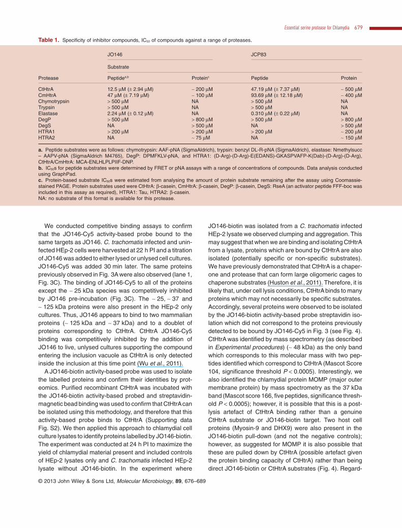
Table 1. Specificity of inhibitor compounds, IC50 of compounds against a range of proteases.
Protease
JO146 JCP83
Substrate
Peptidea,b Proteinc Peptide Protein
CtHtrA 12.5 μM (± 2.94 μM) ∼ 200 μM 47.19 μM (± 7.37 μM) ∼ 500 μMCmHtrA 47 μM (± 7.19 μM) ∼ 100 μM 93.69 μM (± 12.18 μM) ∼ 400 μMChymotrypsin > 500 μM NA > 500 μM NATrypsin > 500 μM NA > 500 μM NAElastase 2.24 μM (± 0.12 μM) NA 0.310 μM (± 0.22 μM) NADegP > 500 μM > 800 μM > 500 μM > 800 μMDegS NA > 500 μM NA > 500 μMHTRA1 > 200 μM > 200 μM > 200 μM ∼ 200 μMHTRA2 NA ∼ 75 μM NA ∼ 150 μM
a. Peptide substrates were as follows: chymotrypsin: AAF-pNA (SigmaAldrich), trypsin: benzyl DL-R-pNA (SigmaAldrich), elastase: Nmethylsucc– AAPV-pNA (SigmaAldrich M4765), DegP: DPMFKLV-pNA, and HTRA1: (D-Arg)-(D-Arg)-E(EDANS)-GKASPVAFP-K(Dab)-(D-Arg)-(D-Arg),CtHtrA/CmHtrA: MCA-ENLHLPLPIIF-DNP.b. IC50s for peptide substrates were determined by FRET or pNA assays with a range of concentrations of compounds. Data analysis conductedusing GraphPad.c. Protein-based substrate IC50s were estimated from analysing the amount of protein substrate remaining after the assay using Coomassie-stained PAGE. Protein substrates used were CtHtrA: β-casein, CmHtrA: β-casein, DegP: β-casein, DegS: RseA (an activator peptide FFF-boc wasincluded in this assay as required), HTRA1: Tau, HTRA2: β-casein.NA: no substrate of this format is available for this protease.
Essential serine protease for Chlamydia 679
© 2013 John Wiley & Sons Ltd, Molecular Microbiology, 89, 676–689

less, these results confirm that JO146-biotin binds toCtHtrA in a cell culture lysate.
The lethality of JO146 treatment relates to the timing ofthe chlamydial developmental cycle timing
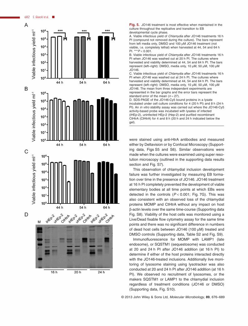
The observed requirement of JO146 addition to be at 16 hPI for complete lethality may be a consequence of thephase of chlamydial growth (i.e. replicative phase), oralternatively may reflect a short term stability of the com-pound. Hence, we conducted further investigations by,first, removing the compound during the culture experi-ments, and second, extended culturing prior to determin-ing viable infectious yield. JO146 was added to cultures at
16 h PI in an identical experiment to that shown in Fig. 1.JO146 was removed from the cultures by extensivewashing at 20 and 24 h PI and chlamydial viability wasdetermined at 44, 54 and 64 h PI. JO146 treatment at16 h PI was lethal for chlamydial viability, even afterextended culturing until 54 and 64 h PI (consistent withthe Fig. 1 data where viability was measured at 44 h PI;see Fig. 5A). JO146 treatment was still highly effective butnot completely lethal when washed out after 4 and 8 hafter treatment (20 and 24 h PI respectively) with 1–2 logreduction in viability (Fig. 5B and C). The loss of viabilitywhen the compound was washed out 4 and 8 h afteraddition was partially rescued (∼ 0.5 log) by extendedculture in the absence of the compound (to 54 and 64 hPI) (Fig. 5B and C). To further explore the developmentalcycle time point dependence of JO146 lethality, treat-ments of either isolated chlamydial EBs or host cells priorto commencing the infection was tested. In these experi-ments, some reduction in viability was observed with thehost cell JO146 treatment prior to infection (∼ 1 log; Sup-porting data Fig. S3).
We tested the compound’s stability during the mid-developmental cycle conditions where lethality wasobserved, by monitoring the in vitro stability of the JO146-Cy5 activity-based probe. The probe was added to cellculture lysates (16 h PI) cultured under identical condi-tions as used for the experiment shown in Fig. 1. Thecultures were then incubated for 4 and 8 h (37°C, 5%CO2) under the standard culture conditions and thelysates were harvested and examined by SDS-PAGE tomonitor JO146-Cy5 labelling. The JO146-Cy5 was stablethroughout this experiment (Fig. 5D), suggesting that thecritical nature of the timing of compound addition formaximum effectiveness related specifically to a develop-mental cycle feature of Chlamydia rather than compoundstability.
JO146 treatment results in diminishing chlamydialinclusion vacuole size and eventual loss of inclusionsover time in cell culture
We monitored inclusion vacuole size in real time usingwide field microscopy after JO146 was added to HEp-2C. trachomatis cultures at 16 h PI. The inclusions appearas non-stained or dark areas inside the cells which incontrol cultures (DMSO or media)increased in size overtime (Fig. 6, and Video S1). In contrast, during JO146treatment, the inclusions appeared to diminish in sizeand eventually could not be visualized (Fig. 6A). Thiswas quantified by measuring inclusion vacuole size andnumber of inclusion vacuoles present confirming that theinclusions decrease in size and number during JO146treatment (Fig. 6B and C). No significant difference wasdetected between DMSO and JO146 treatments for
Fig. 2. Host cell protein synthesis and host cell type do notinfluence the effectiveness of inhibition.A. Viable infectious yield of C. trachomatis at 44 h PI followingcycloheximide (black bars) addition to cultures prior to JO146addition at 16 h PI (moi 0.3). The white bars represent a controlwith no cycloheximide.B. Viable infectious yield at 44 h PI of C. trachomatis cultured withdifferent host cells treated with JO146 at 16 h PI. The barsrepresent the cell types McCoy, HEp-2, Ecc1, Beasb2b cells (left toright; moi 0.3).The mean from three independent experiments with the standarderror of the mean as error bars are shown on the graph (n = 27);*P < 0.05, ***P < 0.001.
680 S. Gloeckl et al. ■
© 2013 John Wiley & Sons Ltd, Molecular Microbiology, 89, 676–689

number of host cells present or lost throughout the entireduration of the video (Supporting data, Fig. S4).
Confocal microscopy shows that JO146 treatmentresults in decreasing inclusion size and loss ofchlamydial cellular morphology
The apparent diminishing size and eventual loss of inclu-sions from the cultures observed by real-time microscopywas further examined using immunocytochemistry andconfocal laser scanning microscopy. Cultures (moi 0.3)were fixed and labelled for MOMP, β-actin (phalloidin) andnucleus (DAPI) and examined at a series of time pointsfollowing JO146 addition at 16 h PI. Representativeimages are shown in Fig. 7A–E. The inclusions are muchsmaller at 24 h PI when treated with JO146 comparedwith the DMSO controls (Fig. 7F), and there are fewerchlamydial cells within the inclusions. In some cases nodiscernable cell shapes are apparent compared with therobust inclusion observed in the DMSO treated controls.The same observations were made when the Chlamydia
Fig. 3. Activity-based probe confirms JO146 binds C. trachomatisproteins including a band at the size corresponding to CtHtrA.A. JO146-Cy5 structure.B. JO146-Cy5 (20 μM) binding throughout the developmental cycle.CtHtrA and MOMP Western blots on the samples are shown belowthe Cy5 scanned gel. Lanes represent time (h PI).C. JO146 (concentrations indicated above in μM) was added tolysed or unlysed cultures (upper and lower gels respectively) priorto lysis and binding with JO146-Cy5 (20 μM). Lanes representC. trachomatis infected HEp-2 cells treated with increasingconcentrations of JO146 (1–5), uninfected HEp-2 cells with thesame concentrations (6–10), purified recombinant CtHtrA (11), andpurified recombinant S247A CtHtrA (active-site serine mutant) (12).Corresponding CtHtrA immunoblots are shown below each gel.
Fig. 4. JO146-biotin activity-based probe confirms JO146 isbound to CtHtrA. Samples from a dynabead-streptavidin pull-downof JO146-biotin from cell culture lysates are shown. The gelrepresents three individual pull-down experiments, indicated above,where the initial lysate, final wash and elution are shown on theCoomassie-stained 12% SDS-PAGE. Molecular weight markersizes are indicated to the left. The proteomic identification of theexcised bands from the C. trachomatis infected HEp-2 JO146-biotinpull-down lane is indicated to the right.
Essential serine protease for Chlamydia 681
© 2013 John Wiley & Sons Ltd, Molecular Microbiology, 89, 676–689
were stained using anti-HtrA antibodies and measuredeither by Deltavision or by Confocal Microscopy (Support-ing data, Figs S5 and S6). Similar observations weremade when the cultures were examined using super reso-lution microscopy (outlined in the supporting data resultssection and Fig. S7).
This observation of chlamydial inclusion developmentfailure was further investigated by measuring EB forma-tion over time in the presence of JO146. JO146 treatmentat 16 h PI completely prevented the development of viableelementary bodies at all time points at which EBs weredetected in the controls (P < 0.001; Fig. 7G). This wasalso consistent with an observed loss of the chlamydialproteins MOMP and CtHtrA without any impact on hostβ-actin levels over the same time-course (Supporting dataFig. S8). Viability of the host cells was monitored using aLive/Dead fixable flow cytometry assay for the same timepoints and there was no significant difference in numbersof dead host cells between JO146 (100 μM) treated andDMSO controls (Supporting data, Table S2 and Fig. S9).
Immunofluorescence for MOMP with LAMP1 (lateendosome), or SQSTM1 (sequestosome) was conductedat 20 and 24 h PI after JO146 addition (at 16 h PI) todetermine if either of the host proteins interacted directlywith the JO146-treated inclusions. Additionally live moni-toring of lysosome staining using lysotracker was alsoconducted at 20 and 24 h PI after JO146 addition (at 16 hPI). We observed no recruitment of lysosomes, or themakers SQSTM1 or LAMP1 to the chlamydial inclusionregardless of treatment conditions (JO146 or DMSO)(Supporting data, Fig. S10).
Fig. 5. JO146 treatment is most effective when maintained in theculture throughout the replicative and transition to EBdevelopmental cycle phase.A. Viable infectious yield of Chlamydia after JO146 treatments 16 hPI (compound not removed during the culture). The bars representfrom left media only, DMSO and 100 μM JO146 treatment (notvisible, i.e. completely lethal) when harvested at 44, 54 and 64 hPI. ***P < 0.001.B. Viable infectious yield of Chlamydia after JO146 treatments 16 hPI when JO146 was washed out at 20 h PI. The cultures whereharvested and viability determined at 44, 54 and 64 h PI. The barsrepresent (left–right): DMSO, media only, 10 μM, 50 μM, 100 μMJO146.C. Viable infectious yield of Chlamydia after JO146 treatments 16 hPI when JO146 was washed out at 24 h PI. The cultures whereharvested and viability determined at 44, 54 and 64 h PI. The barsrepresent (left–right): DMSO, media only, 10 μM, 50 μM, 100 μMJO146. The mean from three independent experiments arerepresented in the bar graphs and the error bars represent thestandard error of the mean (n = 27).D. SDS-PAGE of the JO146-Cy5 bound proteins in a lysateincubated under cell culture conditions for 4 (20 h PI) and 8 h (24 hPI). An in vitro stability assay was carried out where the JO146-Cy5activity-based probe was incubated with lysates of infected(iHEp-2), uninfected HEp-2 (Hep-2) and purified recombinantCtHtrA (CtHtrA) for 4 and 8 h (20 h and 24 h indicated below thegel).
682 S. Gloeckl et al. ■
© 2013 John Wiley & Sons Ltd, Molecular Microbiology, 89, 676–689

JO146 is effective in vivo using the mouse Chlamydiamuridarum model of disease
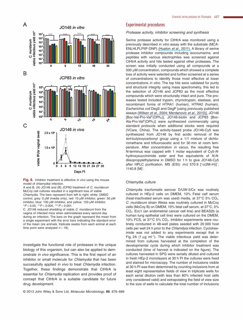
The effectiveness of JO146 treatment in vivo was evalu-ated using the Chlamydia muridarum mouse model ofgenital tract infection. JO146 and JCP83 inhibitedCmHtrA during in vitro assays, although with reducedpotency compared with CtHtrA (Table 1). JO146 andJCP83 treatment of C. muridarum infections in in vitromouse cell culture (McCoy cells) led to a ∼ 2–∼ 2.5 logreduction in viable infectious yield of elementary bodies,with JO146 being slightly more effective (Fig. 8A and B).The C. muridarum developmental cycle is complete within26–30 h, and again the most effective time (12 h PI) forJO146 treatment was consistent with the replicativephase. Similar reduced viability was observed againstC. muridarum when the compounds were tested using adifferent host cell (HEp-2) (P < 0.001; Supporting data,Fig. S11).
The impact of vaginal treatment with 50 mg kg−1 ofJO146 every second day for 14 days on uninfected miceand on the progression of a vaginal C. muridarum infec-tion was investigated. No toxicity was detected fromJO146 treatment of uninfected mice (described in thesupporting items). C. muridarum genital infections of pro-gesterone synchronized female BALB/C mice were testedby treating the mice every second day of infection withvaginal administration of DMSO, or 50 mg kg−1 JO146.Vaginal swabs were collected every third day and theamount of viable Chlamydia shed from the genital tractwas determined (Fig. 8C). In this experiment whichinvolved only six mice, there was a small but statisticallysignificant reduction (P < 0.05) in the total viable Chla-mydia shed from 50 mg kg−1 JO146 treatments comparedwith DMSO control.
Discussion
A chemical approach to inhibit the serine protease CtHtrAduring the chlamydial developmental cycle in human cellculture has demonstrated complete lethality for C. tra-chomatis. Specifically, treatment of cultures during thereplicative phase with a CtHtrA protease inhibitor (JO146)led to lethality with no viable elementary bodies detected.This coincided with a loss of chlamydial cell morphology,diminishing inclusion size and eventual loss of detectableinclusions in the cultures. The lethality occurred indepen-dently of the host cell type, host cell protein synthesis, inthe absence of any host cell toxicity or death, or anyactivation of the major pathogen protection pathways(lysosome or autophagy).
The compounds were identified by screening a libraryof serine protease inhibitor compounds using our previ-ously established CtHtrA protease assay (Huston et al.,
2011). The library consisted of a collection of variouselectrophillic molecules that form a covalent bond with theactive-site serine of serine proteases and hydrolyases.The screen identified two compounds (JO146 and JCP83)from two distinct synthetic sources with similar peptidesequences and identical primary electrophiles. Whenscreened against a variety of proteases in vitro, the com-pounds were found to be selective towards CtHtrA. It isimportant to note that the activity of JCP83 and JO146 invitro against protein substrates was not as effective as thein vivo activity observed against Chlamydia. It is not clearwhy this may be the case, although the in vitro substratesused were model proteins and not known substrates.JO146 and JCP83 were lethal against Chlamydia whenadded during the replicative phase of the chlamydialdevelopmental cycle. JO146 has a lower IC50 value com-pared with JCP83 from the protease assays and was alsomore effective on the in vitro cultures. Several observa-tions suggested a chlamydial target protein was requiredfor the lethality of this compound. The lethal impact ofJO146 was not associated with any host cellular toxicityor cell death. This lethality was impacted directly by themultiplicity of infection (amount of Chlamydia presentin the cultures), did not require active host cell proteinsynthesis, and was independent of the host cell type.Although other studies have resulted in several log reduc-tions in viability (Christian et al., 2011; Stone et al., 2011),this is the first time that any small molecule or inhibitorstrategy has resulted in complete lethality to Chlamydia.There is another chlamydial protease (CPAF, chlamydialsecreted protease activity factor) that may also be a criti-cal factor for chlamydial growth. CPAF has been identifiedas an important protease target for intracellular impair-ment of chlamydial growth. The authors used a caspase-1inhibitor (WEHD-fmk) that had previously been shown toinhibit the in vitro CPAF protease activity and demon-strated a 10-fold reduction in viable chlamydial yield whenthis compound was added at 24 h PI (Christian et al.,2011). The validation of this inhibitor acting directly viabinding to CPAF was the presence of golgin-84 cleavage(Christian et al., 2011). However, subsequent to this pub-lication, concerns have been raised about the role ofCPAF during cell culture due to the experimental methodsutilized. In particular, recent work determined thatgoglin-84 fragmentation may be an artefact of the chosenexperimental design (Chen et al., 2012). Therefore, itremains unclear if CPAF is an important protease forChlamydia.
The timing of compound addition during the chlamydialdevelopmental cycle had a significant impact on the effec-tiveness of JO146 treatment. JO146 was most effectivewhen added at 16 h PI, consistent with an effect on themiddle of the chlamydial replicative phase of develop-ment. JO146 was lethal when added at 16 h PI at all
Essential serine protease for Chlamydia 683
© 2013 John Wiley & Sons Ltd, Molecular Microbiology, 89, 676–689

684 S. Gloeckl et al. ■
© 2013 John Wiley & Sons Ltd, Molecular Microbiology, 89, 676–689

concentrations above 10 μM. A dose of 50 μM resulted ina 1–2 log reduction in yield when added at 6, 20 or 24 hPI, but was completely lethal at 16 h PI. While we knowthe chlamydial developmental cycle is asynchronous, thecompletion of replication by binary fission is quite rapid.Miyairi and co-workers comprehensively characterizedthe parameters of replication and EB formation for anumber of serovars and found that for serovar D, logarith-mic replication occurs from approximately 12–24 h PI witha marked halt of replication from approximately 24 h PIonwards (Miyairi et al., 2006). EB formation could bedetected from approximately 20 h PI onwards and gradu-ally increased until approximately 40 h PI (Miyairi et al.,2006). Therefore, EB formation is highly asynchronous;however, the replicative phase is tightly defined asbetween 12 and 24 h PI. The data presented here stronglysuggests that JO146 is only effective on chlamydial cellsthat are actively replicating or transitioning to EBs, as priortreatment of EBs (Fig. S2) or treatment late during thedevelopmental cycle was not effective. The compoundwas most effective at 16 h PI, exactly mid-replicativephase, at 12 h PI not all cells will be replicating and after24 h PI a significant proportion of the cells will be begin-ning to transition back to elementary bodies. The removalof JO146 at 24 h PI (8 h after administered) showed a 2.5log reduction in viability indicating that the most effectivephase of inhibition was throughout the replicative phaseuntil EB formation. There may be some other ‘off-target’impacts on the host cell which could explain the 0.5–1 logreductions in viability observed when JO146 was addedearly during the developmental cycle (8 h PI) or whensome loss of viability was observed during host cell pre-treatment (Supplementary data Fig. S2). However, onlythe 16 h PI treatment was lethal suggesting that the majorimpact of JO146 is specific to Chlamydia. Therefore,these data indicate that JO146 is inhibiting a specificfunction involved in replication that is essential for Chla-mydia. Interestingly, with extended cultures (54 and 64 hPI) and following removal of the compound at 24 h PI,there was some rescue of viability indicating that thecompound may be partially inducing chlamydial persis-tence (Fig. 5). HtrA has been described as a protease andchaperone with broad roles in protein maintenance andstress response in many bacteria (Clausen et al., 2011).
For some bacteria, substrates of HtrA are essential forviability and pathogenesis of the organism. Perhaps thebest described example of this is the Shigella proteinIcsA, which requires HtrA/DegP for its correct assembly.In the absence of HtrA Shigella does not correctly presentIcsA on the surface of the cell, resulting in the inability togenerate actin tails and virulence attenuation (Purdyet al., 2007). The mechanism of chlamydial deathobserved during this study was unique, with loss ofchlamydial cell structure within the inclusion as well asdiminishing the chlamydial inclusion size with eventualloss of any detectable inclusions. This correlated with aloss of viable elementary bodies. This chlamydial deathand inclusion loss appeared to relate directly to theobserved Chlamydia defects and not a host mediatedmechanism. Thus, it appears that addition of a CtHtrAinhibitor during the replicative phase of C. trachomatisdisrupts the chlamydial developmental cycle, by impact-ing reticulate body cellular morphology, resulting in thereduction in inclusion vacuole size, and ultimate loss ofinclusions from the host cell without viable elementarybody formation.
The use of an activity-based probe strategy enableddirect assessment of compound selectivity. The targetsof JO146 comprised a protein species corresponding toCtHtrA and two additional mammalian cell proteins. Thelabelled protein corresponding to CtHtrA was competi-tively inhibited by prior binding of JO146 during live cellculture, supporting that the compound is accessing thechlamydial inclusion as CtHtrA has only been detectedinside the inclusion vacuole at this time point (Wu et al.,2011). A biotin activity-based probe was used in anaffinity purification experiment which further validatedthat JO146 binds to CtHtrA. These experiments repre-sent the first use of an activity-based probe strategy forany protein within Chlamydia and provide key evidencethat our lead compound JO146 preferentially bindsCtHtrA.
Regardless of the mechanism of chlamydial death,JO146 was also effective in vivo. JO146 vaginal admin-istration during female mice genital tract infection signifi-cantly reduced the viability of C. muridarum. This in vivoeffectiveness is an exciting finding supporting theconcept that chemical strategies can be applied both to
Fig. 6. JO146 treatment leads to diminishing chlamydial inclusion vacuoles.A. Representative images of the same location in a slide culture of a C. trachomatis infection of HEp-2 cells labelled with CellTracker Blue.Treatment conditions are indicated above (JO146 100 μM) and time to the right. Arrows indicate one example inclusion vacuole for eachcondition. The figures have had contrast adjustment which was conducted on the whole image for each figure in the series. Representativevideos are provided as Video S1.B. Analysis of real-time microscopy of JO146 treatment-impact on C. trachomatis inclusion size.C. The number of visible inclusions in each field of view for each condition over time.The mean from three independent experiments have been calculated and are presented in the graph, the error bars represent the standarderror of the mean (n = 14 for the inclusion size, n = 4 for the inclusion numbers). An moi of 1 was used for the experiment.
Essential serine protease for Chlamydia 685
© 2013 John Wiley & Sons Ltd, Molecular Microbiology, 89, 676–689
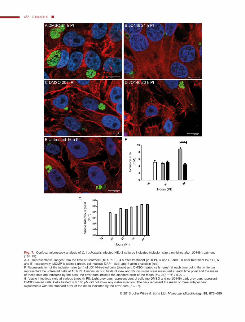
Fig. 7. Confocal microscopy analysis of C. trachomatis infected HEp-2 cultures indicates inclusion size diminishes after JO146 treatment(16 h PI).A–E. Representative images from the time of treatment (16 h PI, E), 4 h after treatment (20 h PI, C and D) and 8 h after treatment 24 h PI, Aand B) respectively. MOMP is stained green, cell nucleus DAPI (blue) and β-actin phalloidin (red).F. Representation of the inclusion size (μm) of JO146-treated cells (black) and DMSO-treated cells (grey) at each time point, the white barrepresented the untreated cells at 16 h PI. A minimum of 6 fields of view and 25 inclusions were measured at each time point and the meanof these data are indicated by the bars, the error bars indicate the standard error of the mean (n = 25); ***P < 0.001.G. Viable infectious yield at various times (h PI). Light grey bars represent control cells (no DMSO and no JO146) dark grey bars representDMSO-treated cells. Cells treated with 100 μM did not show any viable infection. The bars represent the mean of three independentexperiments with the standard error of the mean indicated by the error bars (n = 27).
686 S. Gloeckl et al. ■
© 2013 John Wiley & Sons Ltd, Molecular Microbiology, 89, 676–689
investigate the functional role of proteases in the uniquebiology of this organism, but can also be applied to dem-onstrate in vivo significance. This is the first report of aninhibitor or small molecule for Chlamydia that has beensuccessfully applied in vivo to treat Chlamydia infection.Together, these findings demonstrate that CtHtrA isessential for Chlamydia replication and provides proof ofconcept that CtHtrA is a suitable candidate for futuredrug development.
Experimental procedures
Protease activity, inhibitor screening and synthesis
Serine protease activity for CtHtrA was monitored using apreviously described in vitro assay with the substrate (MCA-ENLHLPLPIIF-DNP) (Huston et al., 2011). A library of serineprotease inhibitor compounds including isocoumarins, andpeptides with various electrophiles was screened againstCtHtrA activity and hits tested against other proteases. Thescreen was initially conducted using all compounds at a500 μM concentration, compounds which showed a completeloss of activity were selected and further screened at a seriesof concentrations to identify those most effective at lowerconcentrations in vitro. The top hits were validated for purityand structural integrity using mass spectrometry, this led tothe selection of JO146 and JCP83 as the most effectivecompounds which were structurally intact and pure. The pro-teases tested included trypsin, chymotrypsin, elastase, andrecombinant forms of HTRA1 (human), HTRA2 (human),Escherichia coli DegS and DegP [using previously publishedassays (Wilken et al., 2004; Merdanovic et al., 2010)]. JO146[Boc-Val-Pro-ValP(OPh2)], JO146-biotin and JCP83 [Boc-Ala-Pro-ValP(OPh2)] were synthesized commercially usingstandard protocols when additional stocks were required(VCare, China). The activity-based probe JO146-Cy5 wassynthesized from JO146 by first acidic removal of thetert-butyloxycarbonyl group using a 1/1 mixture of dichlo-romethane and trifluoroacetic acid for 30 min at room tem-perature. After concentration in vacuo, the resulting freeN-terminus was capped with 1 molar equivalent of Cy5-N-hydroxysuccinimide ester and five equivalents of N,N-diisopropylethylamine in DMSO for 1 h to give JO146-Cy5after HPLC purification. MS (ESI): m/z 570.9 [1⁄2(2M+H)]+,1140.8 [M]+.
Chlamydia culture
Chlamydia trachomatis serovar D/UW-3/Cx was routinelycultured in HEp-2 cells on DMEM, 10% Fetal calf serum(heat-inactivated serum was used) media, at 37°C 5% CO2.C. muridarum strain Weiss was routinely cultured in McCoycells (McCoy B) on DMEM, 10% fetal calf serum, at 37°C, 5%CO2. Ecc1 (an endometrial cancer cell line) and BEAS2b (ahuman lung epithelial cell line) were cultured on the DMEM,10% FCS, at 37°C 5% CO2. Inhibitor experiments were rou-tinely conducted in 48-well plates seeded with 20 000 hostcells per well 24 h prior to the Chlamdyia infection. Cyclohex-imide was not added to any experiments except that inFig. 2A (1 μg ml−1). The viable infectious yield was deter-mined from cultures harvested at the completion of thedevelopmental cycle during which inhibitor treatment wasconducted (time of harvest is indicated on the figure). Thecultures harvested in SPG were serially diluted and culturedin fresh HEp-2 monolayers at 30 h PI the cultures were fixedand stained for microscopy. The number of inclusions visibleat 30 h PI was then determined by counting inclusions from atleast eight representative fields of view in triplicate wells foreach serial dilution (with less than 80% infected host cellsonly considered valid) and extrapolating the field of view sizeto the size of wells to calculate the total number of inclusions
Fig. 8. Inhibitor treatment is effective in vivo using the mousemodel of chlamydial infection.A and B. (A) JO146 and (B) JCP83 treatment of C. muridarumMcCoy cell cultures resulted in a significant loss of viableChlamydia. The bars represent from left to right; white: DMSOcontrol, grey: 0 μM (media only), red: 10 μM inhibitor, green: 50 μMinhibitor, blue: 100 μM inhibitor, and yellow: 150 μM inhibitor.*P < 0.05, **P < 0.005, ***P < 0.001.C. JO146 reduced shedding of viable C. muridarum from thevagina of infected mice when administered every second dayduring an infection. The bars on the graph represent the mean froma single experiment with the error bars indicating the standard errorof the mean (six animals, triplicate swabs from each animal at eachtime point were analysed n = 18).
Essential serine protease for Chlamydia 687
© 2013 John Wiley & Sons Ltd, Molecular Microbiology, 89, 676–689

in the well, dilutions and volumes added to the wells werethen accounted for to give the viable IFU ml−1. Quantitativeanalysis of viability and morphological properties was con-ducted using GraphPad Prism, statistical analysis was rou-tinely conducted using two-way ANOVA and Bonferroni post-tests (typically relative to the DMSO control).
Microscopy
Chlamydia trachomatis cultures were examined using immu-nofluorescence using the Leica SP5 Confocal microscopewith antibodies against CtHtrA, MOMP (Biodesign), LAMP-1(AbChem) and SQSTM1 (AbCam), and secondary antibodiesconjugated to Alexafluor dyes (Invitrogen) (Huston et al.,2008). Live cell imaging was performed using a Leica AF6000widefield microscope. The CellTracker concentration wasoptimized so that it did not penetrate the chlamydial inclusion.1 μM CellTracker (Invitrogen) was added to cultures grown inglass-bottomed, chamber-welled slides 45 min before JO146addition. Images were constructed using the Leica applica-tion suite. Where indicated immunofluorescence was alsomonitored using a Deltavision (personal DV deconvolutionmicroscope) (Applied Precision, Issaquah, WA). 3D struc-tured illumination microscopy (3D-SIM) of the Chlamydiainclusions was conducted using a Deltavision OMX OMXImaging System with Blaze module as previously described(Strauss et al., 2012). Raw images were processed andreconstructed as previously described (Gustafsson et al.,2008; Schermelleh et al., 2008).
Activity gels, immunoblots and PAGE
Activity-based probe binding activity in cell culture and celllysates was monitored using polyacrylamide gel electropho-resis and scanning of the gels using the Li-Cor Odyssey at700 nm. Activity-based probe binding was conducted on cul-tures from T25 flasks at different time points, while the com-petitive binding assays were conducted on cultures from T80flasks harvested at 22 h PI. Western blots for CtHtrA andMOMP were conducted as previously described (Hustonet al., 2008).
Affinity purification and proteomics
JO146-biotin was used for affinity purification experiments.Streptavidin Dynabeads (Invitrogen, Australia) were used toaffinity purify JO146-biotin from cell culture lysates in accord-ance with the manufacturer’s instructions. Cells were har-vested, washed and then suspended in RIPA buffer (Peirce,Australia) and incubated on a turn wheel at room temperaturefor 1 h to lyse the cells. Debri and unlysed cells wereremoved from the suspension by centrifugation at 10 000 gfor 10 min. JO146-biotin or DMSO was added to the lysatesand incubated on a turn wheel at room temperature for30 min prior to addition of streptavidin Dynabeads. Thesamples were harvested using a magnetic block to allowbuffer changes, three PBS washes and four PBST washeswere conducted prior to elution of bound products from thebeads using 0.1% SDS and boiling. Samples were analysedby SDS-PAGE prior to gel excision for proteomics.
Gel excised bands were then analysed by the AustralianProteomics Analytical Facility. Gel slices were cut up, washedand dried prior to rehydration with 100 ng of trypsin in 25 mMammonium bicarbonate. After an overnight digestion at 37°C,peptides were extracted twice with a solution containing 50%acetonitrile and 5% formic acid. The sample was then concen-trated on a peptide trap column, prior to LC and in-line massspectrometry. LC eluent was subject to positive ion nanoflowelectrospray MS analysis in an information dependant acqui-sition mode (IDA). In the IDA mode a TOFMS survey scan wasacquired (m/z 350–1200, 0.5 s), with 10 largest multiplycharged ions (counts > 150) in the survey scan sequentiallysubjected to MS/MS analysis. MS/MS spectra were accumu-lated for 100 ms (m/z 100–1500) with rolling collision energy(ESI-QUAD-TOF). The peak lists of the LC/MS/MS data weregenerated using Analyst 2.0 MASCOT script and searched byMascot against Human and Bacteria databases using MS/MSIon search. Significance threshold for Human samples was(P < 0.01), Bacterial samples (P < 0.0005). This work wasundertaken at APAF the infrastructure provided by the Austral-ian Government through the National Collaborative ResearchInfrastructure Strategy (NCRIS).
Animal model
All animal work must have been conducted according to theAustralian Code of Practice for the Care and Use of Animalsfor Scientific Purposes, which has been embodied in theQueensland Animal Care and Protection Act 2001. Thepurpose of the Code is to ensure the humane care of animalsused for scientific purposes, including teaching. QUT isaccredited to conduct these activities. Animal ethics approvalwas granted from the QUT Animal Research Ethics Commit-tee Approval number 1100000607. Female BALB/c mice pro-vided by the Animal Resource Centre (Australia) wereinfected with 5 × 10⁴ C. muridarum intravaginally. Methodol-ogy is described in the supplementary data.
Acknowledgements
This work was funded by Australian NHMRC Project Grant553020, ARC Linkage Project LP11020007, The WesleyResearch Institute and QUT IHBI MCR Grant. C.B.W. is sup-ported by an NHMRC Senior Research Fellowship. Theauthors wish to acknowledge technical assistance fromConnor O’Meara with the animal experiments.
References
Abelrahman, Y.M., and Belland, R.J. (2005) The chlamydialdevelopmental cycle. FEMS Microbiol Rev 29: 949–959.
Arastu-Kapur, S., Ponder, E.L., Fonovic, U.P., Yeoh, S.,Yuan, F., Fonovic, M., et al. (2008) Identification of pro-teases that regulate erythrocyte rupture by the malariaparasite Plasmodium falciparum. Nat Chem Biol 4: 203–213.
Belland, R.J., Zhong, G., Crane, D.D., Hogan, D., Sturdevant,D., Sharma, J., et al. (2003) Genomic transcriptional pro-filing of the developmental cycle of Chlamydia trachomatis.Proc Natl Acad Sci USA 100: 8478–8483.
688 S. Gloeckl et al. ■
© 2013 John Wiley & Sons Ltd, Molecular Microbiology, 89, 676–689

Chen, A.L., Johnson, K.A., Lee, J.K., Sutterlin, C., and Tan,M. (2012) CPAF: a chlamydial protease in search of anauthentic substrate. PLoS Pathog 8: e1002842.
Chitlaru, T., Zaide, G., Ehrlich, S.D., Inbar, I., Cohen, O., andShafferman, A. (2011) HtrA is a major virulence determi-nant of Bacillus anthracis. Mol Microbiol 81: 1542–1559.
Christian, J.G., Heymann, J., Paschen, S.A., Vier, J.,Schauenburg, L., Rupp, J., et al. (2011) Targeting of achlamydial protease impedes intracellular bacterial growth.PLoS Pathog 7: e1002283.
Clausen, T., Kaiser, M., Huber, R., and Ehrmann, M. (2011)HTRA proteases: regulated proteolysis in protein qualitycontrol. Nat Rev Mol Cell Biol 12: 152–162.
Gustafsson, M.G., Shao, L., Carlton, P.M., Wang, C.J.,Golubovskaya, I.N., Cande, W.Z., et al. (2008) Three-dimensional resolution doubling in wide-field fluorescencemicroscopy by structured illumination. Biophys J 94: 4957–4970.
Hall, C.I., Reese, M.L., Weerapana, E., Child, M.A., Bowyer,P.W., Albrow, V.E., et al. (2011) Chemical genetic screenidentifies Toxoplasma DJ-1 as a regulator of parasitesecretion, attachment, and invasion. Proc Natl Acad SciUSA 108: 10568–10573.
Heuer, D., Rejman Lipinski, A., Machuy, N., Karlas, A.,Wehrens, A., Siedler, F., et al. (2009) Chlamydia causesfragmentation of the Golgi compartment to ensure repro-duction. Nature 457: 731–735.
Hoy, B., Lower, M., Weydig, C., Carra, G., Tegtmeyer, N.,Geppert, T., et al. (2010) Helicobacter pylori HtrA is a newsecreted virulence factor that cleaves E-cadherin to disruptintercellular adhesion. EMBO Rep 11: 798–804.
Huston, W.M., Swedberg, J.E., Harris, J.M., Walsh, T.P.,Mathews, S.A., and Timms, P. (2007) The temperatureactivated HtrA protease from pathogen Chlamydia tra-chomatis acts as both a chaperone and protease at 37degrees C. FEBS Lett 581: 3382–3386.
Huston, W.M., Theodoropoulos, C., Mathews, S.A., andTimms, P. (2008) Chlamydia trachomatis responds to heatshock, penicillin induced persistence, and IFN-gamma per-sistence by altering levels of the extracytoplasmic stressresponse protease HtrA. BMC Microbiol 8: 190.
Huston, W.M., Tyndall, J.D., Lott, W.B., Stansfield, S.H., andTimms, P. (2011) Unique residues involved in activation ofthe multitasking protease/chaperone HtrA from Chlamydiatrachomatis. PLoS ONE 6: e24547.
Jackson, D.S., Fraser, S.A., Ni, L.M., Kam, C.M., Winkler, U.,Johnson, D.A., et al. (1998) Synthesis and evaluation ofdiphenyl phosphonate esters as inhibitors of the trypsin-like granzymes A and K and mast cell tryptase. J MedChem 41: 2289–2301.
Kari, L., Goheen, M.M., Randall, L.B., Taylor, L.D., Carlson,J.H., Whitmire, W.M., et al. (2011) Generation of targetedChlamydia trachomatis null mutants. Proc Natl Acad SciUSA 108: 7189–7193.
Merdanovic, M., Mamant, N., Meltzer, M., Poepsel, S.,Auckenthaler, A., Melgaard, R., et al. (2010) Determinantsof structural and functional plasticity of a widely conservedprotease chaperone complex. Nat Struct Mol Biol 17: 837–843.
Miyairi, I., Mahdi, O.S., Ouellette, S., Belland, R.J., and
Byrne, G.I. (2006) Different growth rates of Chlamydiatrachomatis biovars reflect pathotype. J Infect Dis 194:350–357.
Oleksyszyn, J., and Powers, J.C. (1991) Irreversible inhibi-tion of serine proteases by peptide derivatives of (alpha-aminoalkyl)phosphonate diphenyl esters. Biochemistry 30:485–493.
Purdy, G.E., Fisher, C.R., and Payne, S.M. (2007) IcsAsurface presentation in Shigella flexneri requires the peri-plasmic chaperones DegP, Skp, and SurA. J Bacteriol 189:5566–5573.
Schermelleh, L., Carlton, P.M., Haase, S., Shao, L., Winoto,L., Kner, P., et al. (2008) Subdiffraction multicolor imagingof the nuclear periphery with 3D structured illuminationmicroscopy. Science 320: 1332–1336.
Stone, C.B., Bulir, D.C., Emdin, C.A., Pirie, R.M., Porfilio,E.A., Slootstra, J.W., and Mahony, J.B. (2011) Chlamydiapneumoniae CdsL regulates CdsN ATPase activity, anddisruption with a peptide mimetic prevents bacterial inva-sion. Front Microbiol 2: 21.
Strauss, M.P., Liew, A.T., Turnbull, L., Whitchurch, C.B.,Monahan, L.G., and Harry, E.J. (2012) 3D-SIM super reso-lution microscopy reveals a bead-like arrangement for FtsZand the division machinery: implications for triggeringcytokinesis. PLoS Biol 10: e1001389.
Thomas, B.J., Evans, R.T., Hutchinson, G.R., and Taylor-Robinson, D. (1977) Early detection of chlamydial inclu-sions combining the use of cycloheximide-treated McCoycells and immunofluorescence staining. J Clin Microbiol 6:285–292.
Wang, Y., Kahane, S., Cutcliffe, L.T., Skilton, R.J., Lambden,P.R., and Clarke, I.N. (2011) Development of a transforma-tion system for Chlamydia trachomatis: restoration of gly-cogen biosynthesis by acquisition of a plasmid shuttlevector. PLoS Pathog 7: e1002258.
Wilken, C., Kitzing, K., Kurzbauer, R., Ehrmann, M., andClausen, T. (2004) Crystal structure of the DegS stresssensor: how a PDZ domain recognizes misfolded proteinand activates a protease. Cell 117: 483–494.
Wolf, K., Betts, H.J., Chellas-Gery, B., Hower, S., Linton,C.N., and Fields, K.A. (2006) Treatment of Chlamydia tra-chomatis with a small molecule inhibitor of the Yersiniatype III secretion system disrupts progression of thechlamydial developmental cycle. Mol Microbiol 61: 1543–1555.
World Health Organization (2011) Global incidence and preva-lence of selected curable sexually transmitted infections –2008 [WWW document]. URL http://www.who.int/reproductivehealth/publications/rtis/stisestimates/en/index.html
Wu, X., Lei, L., Gong, S., Chen, D., Flores, R., and Zhong, G.(2011) The chlamydial periplasmic stress response serineprotease cHtrA is secreted into host cell cytosol. BMCMicrobiol 11: 87.
Supporting information
Additional supporting information may be found in the onlineversion of this article at the publisher’s web-site.
Essential serine protease for Chlamydia 689
© 2013 John Wiley & Sons Ltd, Molecular Microbiology, 89, 676–689