Host inflammatory response and development of complications of Chlamydia trachomatis genital...
11
244 J Microbiol Immunol Infect 2005;38:244-254 Corresponding author: Dr. Joseph U. Igietseme, NCID/SRP/CDC, Mailstop C17, 1600 Clifton Road, Atlanta, GA 30333, USA. E-mail: [email protected] The genus Chlamydia comprises obligate intracellular, Gram-negative-like bacteria that cause numerous oculo- genital and respiratory infections in humans, animals and birds. Trachoma, caused by C. trachomatis serovars A, B, Ba and C, is the world’s most common preventable Host inflammatory response and development of complications of Chlamydia trachomatis genital infection in CCR5-deficient mice and subfertile women with the CCR5delta32 gene deletion Erika L. Barr 1 , Sander Ouburg 2 , Joseph U. Igietseme 3,4 , Servaas A. Morré 2 , Edith Okwandu 1 , Francis O. Eko 4 , Godwin Ifere 4 , Tesfaye Belay 1 , Qing He 3,4 , Deborah Lyn 4 , Gift Nwankwo 1 , James Lillard 4 , Carolyn M. Black 3 , Godwin A. Ananaba 1 T cell immunity protects against diseases caused by the obligate intracellular bacterium Chlamydia trachomatis. Incidentally, host inflammatory response that includes T cells appears to also contribute to the pathogenesis of chlamydial diseases such as trachoma and tubal factor infertility (TFI). Therefore, designing effective prevention strategies requires a delineation of immune processes responsible for pathology and those mediating immunity, and identification of the immunogenetic factors predisposing to complication development. The chemokine receptor CCR5 is crucial for T cell activation and function since its deficiency causes suppression of T cell response. We investigated the hypothesis that the clearance of genital chlamydial infection in CCR5-deficient mice could be delayed in the short term; however, a beneficial effect could include protection against inflammation-related complications such as TFI. In a translational study in humans, we investigated the effect of a functional 32 bp deletion in the CCR5 gene on the risk of developing tubal pathology in Dutch Caucasian women with immunologic evidence [i.e., immunoglobulin G (IgG) responses] of chlamydial infection. When genitally-infected wild-type (WT) and CCR5 knockout (CCR5KO) mice were evaluated for microbiologic shedding of chlamydiae, there was a greater intensity of infection and delayed resolution in the knockout mice. However, compared to WT mice, the fertility of infected CCR5KO mice (measured by pregnancy rate) was only mildly affected in the short term and unaffected in the long term (70% vs 30% reduction in the short term, and 50 vs 0% in the long term, respectively). Immunobiologic analysis revealed that the diminished capacity of CCR5KO to control acute chlamydial infection correlated with the relatively low chemokine [interferon-inducible protein 10 (IP-10) and regulated upon activation normal cell expressed and secreted (RANTES)] and cytokine (mainly interferon-gamma and tumor necrosis factor-alpha) expression corresponding to a poor early T- helper I response. However, the reduced incidence of complications in the CCR5KO mice appears to correlate with the low activity of long term inflammatory mediators. Besides, the translational studies in humans revealed that among patients with positive anti-chlamydial IgG responses, tubal pathology correlated with a low incidence of CCR5delta32 deletion (7%), while women without tubal pathology had higher incidence of the CCR5delta32 deletion (31%) as compared to controls (19%). Thus, in mice and humans the inflammation associated with CCR5 function may predispose to development of complications of chlamydial infection, such as TFI. Key words: Chemokine receptors, chlamydia, gene deletion, genetic predisposition to disease, immunogenetics 1 Clark Atlanta University, Atlanta, GA, USA; 2 Department of Pathology, Laboratory of Immunogenetics, Section Immunogenetics of Infectious Diseases, VU University Medical Center, Amsterdam, The Netherlands; 3 Center for Disease Control and Prevention, Atlanta, GA; and 4 Morehouse School of Medicine, Atlanta, GA, USA Received: March 11, 2005 Revised: May 3, 2005 Accepted: May 19, 2005 blinding disease. It is epidemic in several developing nations, including Africa, south east Asia, and the Middle East, with an estimated 150 million people affected, of whom six million are irreversibly blinded or severely visually impaired [1]. Genital infections by the different genovars of C. trachomatis constitute the most common bacterial sexually transmitted dis- eases (STDs) in the United States and several other
-
Upload
morehouse-med -
Category
Documents
-
view
2 -
download
0
Transcript of Host inflammatory response and development of complications of Chlamydia trachomatis genital...
244
Chlamydia trachomatis genital infectionJ Microbiol Immunol Infect2005;38:244-254
Corresponding author: Dr. Joseph U. Igietseme, NCID/SRP/CDC,Mailstop C17, 1600 Clifton Road, Atlanta, GA 30333, USA.E-mail: [email protected]
The genus Chlamydia comprises obligate intracellular,Gram-negative-like bacteria that cause numerous oculo-genital and respiratory infections in humans, animalsand birds. Trachoma, caused by C. trachomatis serovarsA, B, Ba and C, is the world’s most common preventable
Host inflammatory response and development ofcomplications of Chlamydia trachomatis genital infection in
CCR5-deficient mice and subfertile women with theCCR5delta32 gene deletion
Erika L. Barr1, Sander Ouburg2, Joseph U. Igietseme3,4, Servaas A. Morré2, Edith Okwandu1,Francis O. Eko4, Godwin Ifere4, Tesfaye Belay1, Qing He3,4, Deborah Lyn4, Gift Nwankwo1, James Lillard4,
Carolyn M. Black3, Godwin A. Ananaba1
T cell immunity protects against diseases caused by the obligate intracellular bacterium Chlamydia trachomatis.Incidentally, host inflammatory response that includes T cells appears to also contribute to the pathogenesis ofchlamydial diseases such as trachoma and tubal factor infertility (TFI). Therefore, designing effective preventionstrategies requires a delineation of immune processes responsible for pathology and those mediating immunity, andidentification of the immunogenetic factors predisposing to complication development. The chemokine receptorCCR5 is crucial for T cell activation and function since its deficiency causes suppression of T cell response. Weinvestigated the hypothesis that the clearance of genital chlamydial infection in CCR5-deficient mice could be delayedin the short term; however, a beneficial effect could include protection against inflammation-related complicationssuch as TFI. In a translational study in humans, we investigated the effect of a functional 32 bp deletion in the CCR5gene on the risk of developing tubal pathology in Dutch Caucasian women with immunologic evidence [i.e.,immunoglobulin G (IgG) responses] of chlamydial infection. When genitally-infected wild-type (WT) and CCR5 knockout(CCR5KO) mice were evaluated for microbiologic shedding of chlamydiae, there was a greater intensity of infectionand delayed resolution in the knockout mice. However, compared to WT mice, the fertility of infected CCR5KO mice(measured by pregnancy rate) was only mildly affected in the short term and unaffected in the long term (70% vs 30%reduction in the short term, and 50 vs 0% in the long term, respectively). Immunobiologic analysis revealed that thediminished capacity of CCR5KO to control acute chlamydial infection correlated with the relatively low chemokine[interferon-inducible protein 10 (IP-10) and regulated upon activation normal cell expressed and secreted (RANTES)]and cytokine (mainly interferon-gamma and tumor necrosis factor-alpha) expression corresponding to a poor early T-helper I response. However, the reduced incidence of complications in the CCR5KO mice appears to correlate withthe low activity of long term inflammatory mediators. Besides, the translational studies in humans revealed thatamong patients with positive anti-chlamydial IgG responses, tubal pathology correlated with a low incidence ofCCR5delta32 deletion (7%), while women without tubal pathology had higher incidence of the CCR5delta32 deletion(31%) as compared to controls (19%). Thus, in mice and humans the inflammation associated with CCR5 functionmay predispose to development of complications of chlamydial infection, such as TFI.
Key words: Chemokine receptors, chlamydia, gene deletion, genetic predisposition to disease, immunogenetics
1Clark Atlanta University, Atlanta, GA, USA; 2Department of Pathology, Laboratory of Immunogenetics, SectionImmunogenetics of Infectious Diseases, VU University Medical Center, Amsterdam, The Netherlands; 3Center
for Disease Control and Prevention, Atlanta, GA; and 4Morehouse School of Medicine, Atlanta, GA, USA
Received: March 11, 2005 Revised: May 3, 2005 Accepted: May 19, 2005
blinding disease. It is epidemic in several developingnations, including Africa, south east Asia, and theMiddle East, with an estimated 150 million peopleaffected, of whom six million are irreversibly blindedor severely visually impaired [1]. Genital infectionsby the different genovars of C. trachomatis constitutethe most common bacterial sexually transmitted dis-eases (STDs) in the United States and several other
245
Barr et al
industrialized nations, including the United Kingdom,Germany, Japan, and France. Pelvic inflammatorydisease (PID) and tubal factor infertility (TFI) aremajor complications of genital infection, occurring inapproximately 40% and 10% of untreated infections,respectively, and constituting an enormous morbidityand socioeconomic burden of chlamydial infections [2-5]. The lymphogranuloma venereum (LGV) infectionsare invasive and often ulcerative with lymphatic tissueinvolvement (e.g., inguinal bubo) [6,7], which areendemic in certain developing nations includingparts of Africa, Asia, South America, and the Caribbean[8]. The recent epidemic outbreak of LGV diseaseamong male homosexuals in Europe is attractingconsiderable public health attention in many countries[9]. Ulcerative STDs in general are a major risk factorfor HIV acquisition. Moreover, reports suggesting thatgenital chlamydial infection may be on the rise [10,11],and could predispose to HIV-related acquired immuno-deficiency disease [12-18] and human papilloma virus-associated cervical dysplasia, have heightened theseconcerns [19].
There is an urgent need to develop interventionand prevention measures to control chlamydialinfections in the human population. However, a betterunderstanding of the pathobiology of the diseaseis crucial for efforts to design preventive measuresincluding the use of targeted immunomodulators andselective anti-inflammatory agents to control the onsetof pathologies, and the application of effective vaccinesas prophylaxis. Although T cell immunity is crucialfor chlamydial control [20-22], clinicopathologicand experimental studies have suggested that thepathogenesis of complications such as trachoma andTFI is due to the deleterious host inflammatory immuneresponse against the infectious agent. Thus, studiesdefining the key elements of protective immunity againstChlamydia, and establishing the parameters for vaccineselection and evaluation, are often confounded by thecomplexities of chlamydia biology, serovariation,infection manifestations and induction of paradoxicalimmune effectors that can be both protective andpathologic [23-27]. This double-edge effect poses adilemma to the effort to dissect the role of host immuneresponse on pathogenesis and immunity. In this respect,various inflammatory chemokines and cytokines andstrains of mice with differential susceptibilities tochlamydial infection have been evaluated to definesome of the immunopathogenic factors responsible forchlamydial disease [28-34]. Among other findings,
toll-like receptor 2 (TLR2) deficiency caused decreasedsecretion of specific inflammatory cytokines, and asignificant reduction in oviduct and mesosalpinxpathology even without affecting the course of theinfection in mice [30]. Also, cytokines and moleculesindicative of T cell activation were upregulated in activetrachoma [35]. These observations and others wouldsuggest that there are key immunobiologic pathways ofT cell activation with limited redundancies that oftencomplicate such studies. The chemokine receptor CCR5is a member of the 7-transmembrane, G protein-coupledreceptor superfamily, functioning as an importantchemokine receptor that is preferentially expressed oncertain leukocytes (monocytes, cyctotoxic T cells, andCD4, T-helper 1 (Th1) and dendritic cells), and bindingspecific chemokines [e.g., regulated upon activationnormal cell expressed and secreted (RANTES), macro-phage inflammatory protein 1 (MIP-1) alpha and MIP-1 beta] that activate and induce Th1-like cells [36-40].As a crucial receptor involved in T cell activation andfunction, a deficiency of CCR5 is associated with asuppression of T cell induction and leukocyte migrationunder certain infectious and non-infectious conditions[41-43], suggesting that it plays a role in both infection-related immune and inflammatory processes. The effectof a targeted suppression of the critical specific T cellresponse on both immune-mediated microbial clearanceand the development of complications of chlamydialinfection is largely unknown.
We investigated the hypothesis that the suppressionof T cell response against genital chlamydial infectionin CCR5-deficient mice could delay the clearance ofthe pathogen in the short-term, but it could also confera beneficial effect by protecting the animals fromcomplications such as TFI. In addition, in a translationalstudy in humans, we investigated if the functional 32 bpdeletion in the CCR5 gene (CCR5delta32) had an effecton the risk of developing tubal pathology in women withserologic evidence [immunglobulin G (IgG) responses]of C. trachomatis infection. The results supported theworking hypothesis and could pave the way for a moredetailed analysis and definition of the specific host-relatedimmune effectors and immunopathologic processesunderlying the pathogenesis of chlamydial disease.
Materials and Methods
Knockout and wild-type miceFemale CCR5 knockout (CCR5KO or CCR5–/–) andthe wild-type (WT) control (i.e., CCR5+/+) mice, on

246
Chlamydia trachomatis genital infection
(C57BL/6J) background, 5-8 weeks old, were obtainedfrom The Jackson Laboratory, Bar Harbor, MA, USA.All animals were fed with food and water ad libitum,and maintained in laminar flow racks under pathogen-free conditions of 12-h light and 12-h darkness.
Chlamydia stocks and antigensStocks of Chlamydia muridarum (the C. trachomatisagent of mouse pneumonitis or MoPn) used forinfections were prepared by propagating elementarybodies (EBs) in McCoy or HeLa cells, according tostandard procedures [44]. Chlamydial stock titerswere expressed as inclusion-forming units per milliliter(IFU/mL). Chlamydial antigens were prepared bygrowing the agent in HeLa cells and purifying EBs overrenografin gradients, followed by inactivation underultraviolet (UV) light for 3 h.
Animal InfectionGroups of CCR5KO and control mice were intra-vaginally infected with 106 IFU of MoPn. The status ofthe infection was monitored by periodic cervico-vaginalswabbing of individual animals and isolation ofchlamydiae in tissue culture [44]. Experiments wererepeated 2 times to give 10-12 mice per group.
Chlamydia-induced cell activation, cytokine andchemokine secretion by leukocytesThe profile of cytokines and chemokines secreted byleukocytes from chlamydial-infected CCR5KO andWT mice was compared by measuring the levels ofspecific cytokines and chemokines released followingin vitro restimulation of the total splenic cells withUV-inactivated chlamydiae. CCR5KO and control micewere genitally infected with MoPn as describedabove and at various times postinfection (7, 14, 21 and28 days) splenic cells were isolated from infectedmice and 2 × 10⁵ cells were stimulated with 10 µg/mLof chlamydial antigen and incubated at 37°C in 5%carbon dioxide incubators for 120 h. At the end ofthe incubation period, the supernatants were collectedand assayed for the Th1 cytokine interferon-gamma(IFN-γ), and the Th1 chemokines interferon-inducibleprotein 10 (IP-10) and RANTES, using a quantitativeenzyme-linked immunosorbent assay (CytoscreenTM
Immunoassay Kit; BioSource International, Camarillo,CA, USA) according to the supplier’s instructions.The concentration of the cytokine and chemokines ineach sample was obtained by extrapolation from astandard calibration curve generated simultaneously.
Data were calculated as the mean values (± standarddeviation) of triplicate cultures for each experiment.The results were derived from at least 3 independentexperiments.
Animal fertility studiesAnimals were infected with 106 IFU/mouse withMoPn. Two weeks and 5 weeks postinfection, groupsof animals were mated with male counterparts byplacing 3 females to 1 male, and subsequently observedand weighed daily for 19 days to determine pregnancy,as previously described [45]. The numbers of pregnantmice in the different groups were enumerated after 19days in each case.
CCR5delta32 gene deletion in women with subfertilityThe study cohort included 256 Dutch Caucasian womenwho presented with subfertility at the Research InstituteGrowth and Development (GROW) and the Departmentof Obstetrics and Gynaecology, Academisch ZiekenhuisMaastricht, The Netherlands. This subfertility cohorthas been described elsewhere [46]. Tubal pathologywas defined as extensive periadnexal adhesions and/ordistal occlusions of one or both tubes, and 50 womenhad severe tubal pathology based on these criteria.Chlamydial antibodies were assessed by indirectmicroimmunofluorescence (MIF) test for anti-C.trachomatis IgG antibodies, as described previously[47,48]. A positive C. trachomatis IgG MIF test wasdefined as a titer ≥1:32. A healthy Dutch Caucasiancontrol group (n = 145) was included to assess thegeneral frequency of the CCR5delta32 genotypes in theDutch Caucasian population. Genomic DNA wasextracted from blood using the MagNaPure LC isolatoraccording to the manufacturer’s instructions (RocheMolecular Biochemicals, Mannheim, Germany). Thehuman CCR5 gene delta 32 deletion was determinedby polymerase chain reaction (PCR), with the senseprimer CCR5-d32S: 5'-CAAAAAGAAGGTCTTCATTACACC-3' and anti-sense primer CCR5-d32AS: 5'-CCTGTGCCTCTTCTTCTCATTTCG-3' under thefollowing PCR conditions: 5 min at 94°C, followed by35 cycles of 60 sec at 94°C, 60 sec at 55°C, and 60 secat 72°C, and the cycling programme was followed by7 min at 72°C with final storage at 4°C (Perkin ElmerPE9700). The PCR product was electrophoresed on3% agarose and the following 3 fragment patterns wereidentified: WT CCR5 gene (1.1) 189 bp, homozygoteCCR5 mutant gene (2.2) 157 bp, and the heterozygoteCCR5 gene (1.2) 189 bp + 157 bp.
247
Barr et al
Statistical analysisThe levels of cytokines in samples from differentexperiments were analyzed and compared by performinga 1- or 2-tailed t test, and the relationship betweendifferent experimental groupings was assessed byanalysis of variance. Minimal statistical significancewas judged at p<0.05. The chi-squared test or Fisherexact test was used for comparison of CCR5d32genotype frequencies between patient (sub) groups and/or controls. Statistical Package for the Social Sciences(SPSS) 10.0 for Windows (SPSS Inc., Chicago, IL,USA) was used for statistical analysis.
Results
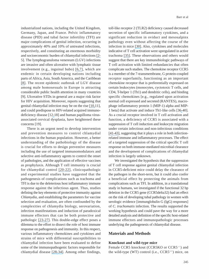
High intensity of infection and delayedclearance of genital chlamydial infection inCCR5KO miceCCR5 is a crucial chemokine receptor that supportsthe activation and induction of specific T cells duringinfectious and non-infectious inflammatory processes.We investigated the effect of CCR5 deficiency on theability of mice to control and clear genital chlamydialinfection. Fig. 1 shows results from studies thatcompared the course of genital chlamydial infection inCCR5KO and control (WT) mice. The data revealedthat within the first week of infection, there was nodifference in the level of infectivity of CCR5KO andWT mice. However, by the second and fourth weeks,the ability of CCR5KO mice to control the infectionwas compromised, with a higher intensity of infection
revealed by the isolation of higher chlamydiae from themice. By the fifth week, all WT mice had clearedthe infection but the CCR5KO mice remained infected(0.0 vs log
10 3.0 IFU/mL, respectively). The results
suggested that the deficiency of CCR5 could haveadversely affected the ability of the mice to elicit therequired T cell response that is known to clearchlamydiae in mice [20].
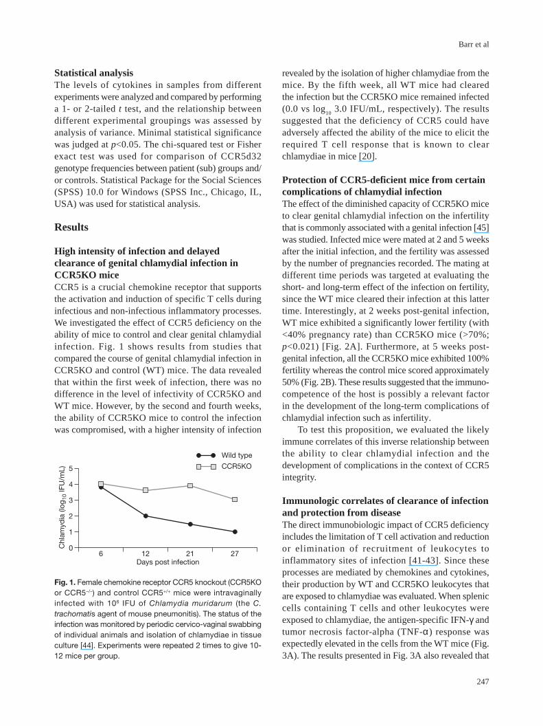
Protection of CCR5-deficient mice from certaincomplications of chlamydial infectionThe effect of the diminished capacity of CCR5KO miceto clear genital chlamydial infection on the infertilitythat is commonly associated with a genital infection [45]was studied. Infected mice were mated at 2 and 5 weeksafter the initial infection, and the fertility was assessedby the number of pregnancies recorded. The mating atdifferent time periods was targeted at evaluating theshort- and long-term effect of the infection on fertility,since the WT mice cleared their infection at this lattertime. Interestingly, at 2 weeks post-genital infection,WT mice exhibited a significantly lower fertility (with<40% pregnancy rate) than CCR5KO mice (>70%;p<0.021) [Fig. 2A]. Furthermore, at 5 weeks post-genital infection, all the CCR5KO mice exhibited 100%fertility whereas the control mice scored approximately50% (Fig. 2B). These results suggested that the immuno-competence of the host is possibly a relevant factorin the development of the long-term complications ofchlamydial infection such as infertility.
To test this proposition, we evaluated the likelyimmune correlates of this inverse relationship betweenthe ability to clear chlamydial infection and thedevelopment of complications in the context of CCR5integrity.
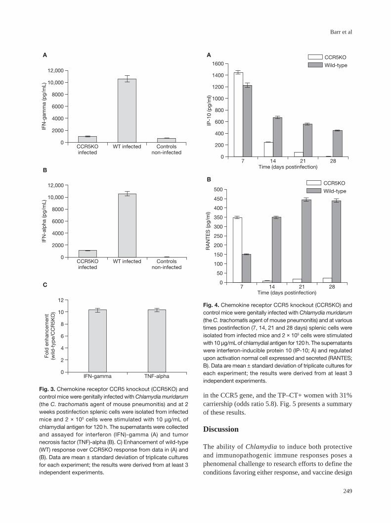
Immunologic correlates of clearance of infectionand protection from diseaseThe direct immunobiologic impact of CCR5 deficiencyincludes the limitation of T cell activation and reductionor elimination of recruitment of leukocytes toinflammatory sites of infection [41-43]. Since theseprocesses are mediated by chemokines and cytokines,their production by WT and CCR5KO leukocytes thatare exposed to chlamydiae was evaluated. When spleniccells containing T cells and other leukocytes wereexposed to chlamydiae, the antigen-specific IFN-γ andtumor necrosis factor-alpha (TNF-α) response wasexpectedly elevated in the cells from the WT mice (Fig.3A). The results presented in Fig. 3A also revealed that
0
1
2
3
4
5
6 12 21Days post infection
27
Wild type
CCR5KO
Chl
amyd
ia (l
og10
IFU
/mL)
Fig. 1. Female chemokine receptor CCR5 knockout (CCR5KOor CCR5–/–) and control CCR5+/+ mice were intravaginallyinfected with 106 IFU of Chlamydia muridarum (the C.trachomatis agent of mouse pneumonitis). The status of theinfection was monitored by periodic cervico-vaginal swabbingof individual animals and isolation of chlamydiae in tissueculture [44]. Experiments were repeated 2 times to give 10-12 mice per group.
248
Chlamydia trachomatis genital infection
the level of antigen-specific IFN-γ secreted by leukocytesfrom CCR5KO mice was not statistically different fromthe levels secreted by leukocytes from non-infectedmice, indicating that CCR5 is required for adequateactivation of Th1 response against chlamydia. When thelevels of the inflammatory chemokines RANTES andIP-10 secreted by chlamydia-exposed leukocytes frominfected CCR5KO and WT mice were compared, it wasalso found that the knockout mice exhibited a diminishedcapacity (Fig. 3 and Fig. 4), suggesting the deficiencyof CCR5 results in a compromised Th1 response. Fig.3C expresses the data in Fig. 3A and 3B as extent ofenhancement of WT response over CCR5KO response,to emphasize the significance of Th1 suppression dueto CCR5 deficiency.
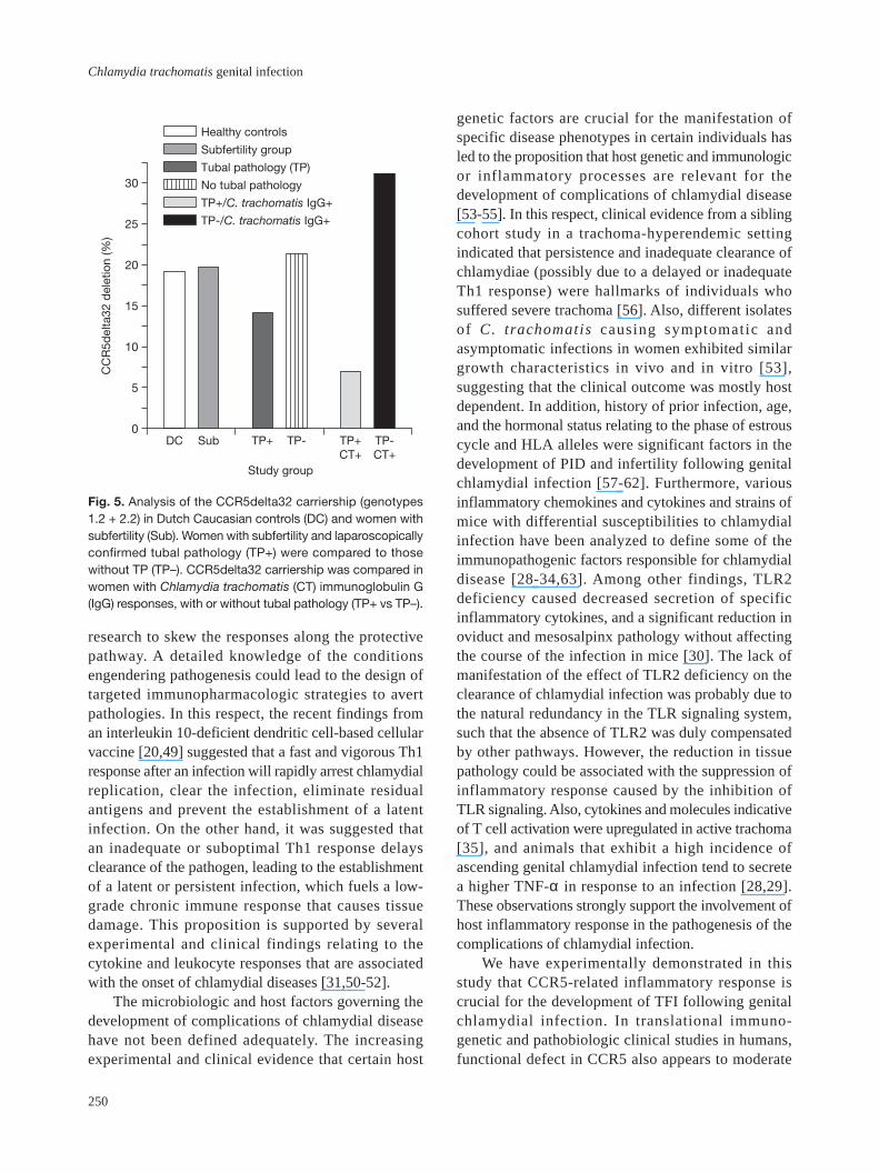
Inverse relation between CCR5delta32 genedeletion and development of tubal pathologyamong women with C. trachomatis infectionIn a translational study involving subfertile DutchCaucasian women, we investigated by PCR methodwhether a functional 32 bp deletion in the CCR5 genehad an effect on the risk of developing tubal pathologyin women with serologic evidence of C. trachomatisinfection based on IgG responses. Results revealed thatin the control women (n = 145) the CCR5delta32 WTgenotype (1.1) was present in 81%, the heterozygousgenotype 1.2 in 18%, and the 2.2 homozygous mutantgenotype in 1% (with a total of 19% being carriers ofthe mutant allele). Also, the subfertility cohort (n = 256)
had frequencies of the WT (1.1 = 80%), heterozygous(1.2 = 19.5%), and homozygous mutant genotypes(2.2 = 0.5%) essentially identical to the control women.Therefore, there was no difference in the frequency ofthe CCR5delta32 gene deletion among subfertile (20%)and control (19%) women.
However, in women with laparoscopicallyconfirmed tubal pathology (n = 50), cariership of thedelta32 deletion was lowered to 14%, while in womenwithout tubal pathology (n = 206) carriership of thedelta32 deletion remained at the normal level (21%).This suggested that the incidence of tubal pathology waslower in women carrying the CCR5 mutation. Since onlya proportion of the women with proven C. trachomatisinfection (MIF IgG responses) actually develop latecomplications such as tubal pathology, we wonderedwhether CCR5-mediated inflammatory response hasa role in the development of chlamydial-associatedcomplications. Therefore, to directly investigate therole of CCR5 status on the incidence of chlamydial-associated TFI, we hypothesized that as in mice, afunctional deficiency in CCR5 gene will moderate tubalpathology in chlamydia-infected women. To test thishypothesis, we compared the frequency of delta32deletion in women with C. trachomatis IgG responseswho developed tubal pathology (TP+CT+, n = 28) andwomen with C. trachomatis IgG responses without tubalpathology (TP–CT+, n=13). The results revealed thatthere was a significant difference between the TP+CT+women, with only 7% carriership of the 32 bp deletion
0
50
100
150
% P
regn
ant
W/T non-infected
CCR5KO non-infected
W/T
CCR5KO
0
40
80
120
20
60
100
W/T non-infected
CCR5KO non-infected
W/T
CCR5KO
% P
regn
ant
A B
Fig. 2. Animals were infected with 106 IFU of the Chlamydia muridarum (the C. trachomatis agent of mouse pneumonitis). Two (A)and 5 (B) weeks postinfection, groups of animals were mated with males, and subsequently observed for 19 days to determinepregnancy. The numbers of pregnant mice in the different groups were enumerated after 19 days in each case. Experimentswere repeated 3 times with 6 mice per experiment. W/T = wild-type control mice; CCR5KO = CCR5 knockout.
249
Barr et al
in the CCR5 gene, and the TP–CT+ women with 31%carriership (odds ratio 5.8). Fig. 5 presents a summaryof these results.
Discussion
The ability of Chlamydia to induce both protectiveand immunopathogenic immune responses poses aphenomenal challenge to research efforts to define theconditions favoring either response, and vaccine design
A
B
Fig. 3. Chemokine receptor CCR5 knockout (CCR5KO) andcontrol mice were genitally infected with Chlamydia muridarum(the C. trachomatis agent of mouse pneumonitis) and at 2weeks postinfection splenic cells were isolated from infectedmice and 2 × 10⁵ cells were stimulated with 10 µg/mL ofchlamydial antigen for 120 h. The supernatants were collectedand assayed for interferon (IFN)-gamma (A) and tumornecrosis factor (TNF)-alpha (B). C) Enhancement of wild-type(WT) response over CCR5KO response from data in (A) and(B). Data are mean ± standard deviation of triplicate culturesfor each experiment; the results were derived from at least 3independent experiments.
C
0
200
400
600
800
1000
1200
1400
1600
7 14 21Time (days postinfection)
28
IP-1
0 (p
g/m
l)
CCR5KO
Wild-type
0
50
100
150
200
250
300
350
400
450
500
7 14 21Time (days postinfection)
28
CCR5KO
Wild-type
RA
NTE
S (p
g/m
l)
Fig. 4. Chemokine receptor CCR5 knockout (CCR5KO) andcontrol mice were genitally infected with Chlamydia muridarum(the C. trachomatis agent of mouse pneumonitis) and at varioustimes postinfection (7, 14, 21 and 28 days) splenic cells wereisolated from infected mice and 2 × 10⁵ cells were stimulatedwith 10 µg/mL of chlamydial antigen for 120 h. The supernatantswere interferon-inducible protein 10 (IP-10; A) and regulatedupon activation normal cell expressed and secreted (RANTES;B). Data are mean ± standard deviation of triplicate cultures foreach experiment; the results were derived from at least 3independent experiments.
A
B
0
2000
4000
6000
8000
10,000
12,000
CCR5KOinfected
WT infected Controlsnon-infected
IFN
-gam
ma
(pg/
mL)
0
2000
4000
6000
8000
10,000
12,000
CCR5KOinfected
WT infected Controlsnon-infected
IFN
-alp
ha (p
g/m
L)
0
2
4
6
8
10
12
IFN-gamma TNF-alpha
Fold
enh
ance
men
t(w
ild-t
ype/
CC
R5K
O)
250
Chlamydia trachomatis genital infection
research to skew the responses along the protectivepathway. A detailed knowledge of the conditionsengendering pathogenesis could lead to the design oftargeted immunopharmacologic strategies to avertpathologies. In this respect, the recent findings froman interleukin 10-deficient dendritic cell-based cellularvaccine [20,49] suggested that a fast and vigorous Th1response after an infection will rapidly arrest chlamydialreplication, clear the infection, eliminate residualantigens and prevent the establishment of a latentinfection. On the other hand, it was suggested thatan inadequate or suboptimal Th1 response delaysclearance of the pathogen, leading to the establishmentof a latent or persistent infection, which fuels a low-grade chronic immune response that causes tissuedamage. This proposition is supported by severalexperimental and clinical findings relating to thecytokine and leukocyte responses that are associatedwith the onset of chlamydial diseases [31,50-52].
The microbiologic and host factors governing thedevelopment of complications of chlamydial diseasehave not been defined adequately. The increasingexperimental and clinical evidence that certain host
genetic factors are crucial for the manifestation ofspecific disease phenotypes in certain individuals hasled to the proposition that host genetic and immunologicor inflammatory processes are relevant for thedevelopment of complications of chlamydial disease[53-55]. In this respect, clinical evidence from a siblingcohort study in a trachoma-hyperendemic settingindicated that persistence and inadequate clearance ofchlamydiae (possibly due to a delayed or inadequateTh1 response) were hallmarks of individuals whosuffered severe trachoma [56]. Also, different isolatesof C. trachomatis causing symptomatic andasymptomatic infections in women exhibited similargrowth characteristics in vivo and in vitro [53],suggesting that the clinical outcome was mostly hostdependent. In addition, history of prior infection, age,and the hormonal status relating to the phase of estrouscycle and HLA alleles were significant factors in thedevelopment of PID and infertility following genitalchlamydial infection [57-62]. Furthermore, variousinflammatory chemokines and cytokines and strains ofmice with differential susceptibilities to chlamydialinfection have been analyzed to define some of theimmunopathogenic factors responsible for chlamydialdisease [28-34,63]. Among other findings, TLR2deficiency caused decreased secretion of specificinflammatory cytokines, and a significant reduction inoviduct and mesosalpinx pathology without affectingthe course of the infection in mice [30]. The lack ofmanifestation of the effect of TLR2 deficiency on theclearance of chlamydial infection was probably due tothe natural redundancy in the TLR signaling system,such that the absence of TLR2 was duly compensatedby other pathways. However, the reduction in tissuepathology could be associated with the suppression ofinflammatory response caused by the inhibition ofTLR signaling. Also, cytokines and molecules indicativeof T cell activation were upregulated in active trachoma[35], and animals that exhibit a high incidence ofascending genital chlamydial infection tend to secretea higher TNF-α in response to an infection [28,29].These observations strongly support the involvement ofhost inflammatory response in the pathogenesis of thecomplications of chlamydial infection.
We have experimentally demonstrated in thisstudy that CCR5-related inflammatory response iscrucial for the development of TFI following genitalchlamydial infection. In translational immuno-genetic and pathobiologic clinical studies in humans,functional defect in CCR5 also appears to moderate
Fig. 5. Analysis of the CCR5delta32 carriership (genotypes1.2 + 2.2) in Dutch Caucasian controls (DC) and women withsubfertility (Sub). Women with subfertility and laparoscopicallyconfirmed tubal pathology (TP+) were compared to thosewithout TP (TP–). CCR5delta32 carriership was compared inwomen with Chlamydia trachomatis (CT) immunoglobulin G(IgG) responses, with or without tubal pathology (TP+ vs TP–).
0DC Sub TP+ TP- TP+
CT+TP-CT+
5
10
15
20
25
30
CC
R5d
elta
32 d
elet
ion
(%)
Healthy controls
Subfertility group
Tubal pathology (TP)
No tubal pathology
TP+/C. trachomatis IgG+
TP-/C. trachomatis IgG+
Study group
251
Barr et al
the development of tubal pathologies associated withgenital chlamydial infection in women. These dataare corroborated by previous propositions that certainhost factors are relevant for the development of thecomplications of chlamydial infection. Specifically, the32 bp deletion in the CCR5 gene, which results in atruncated protein with impaired signal-transductioncapacity, was associated with resistance to humanimmunodeficiency virus type 1 (HIV-1) infection [64,65]. Recently, it has been suggested that heterozygosityfor CCR5delta32 was also associated with spontaneoushepatitis C viral clearance and with significantly lowerhepatic inflammatory scores [66]. Our experimentalstudies indicated that a deficiency of specific anti-chlamydial Th1 response led to a suppression of Th1response and delayed clearance of genital chlamydialinfection. However, CCR5KO mice were protected fromthe complication of the genital infection relating toinfertility. In addition, C. trachomatis-exposed womenwith CCR5delta32 deletions appear to be protected fromtubal pathology as well, suggesting a crucial role forCCR5-related specific Th1 and inflammatory responsesin the pathogenesis of infectious tubal pathologies.
Perhaps this study represents the first concurrentdemonstration of a strong causal relationship betweenCCR5-related specific Th1 and inflammatory responseand development of complications of genital chlamydialinfection in both animal models and humans. Theimplications include the fact that although Th1 responseis crucial for chlamydial control, there are hostconditions that could skew the response towardpathology. Such conditions may include the involvementof immunopathogenic chlamydial antigens [26] andvaccine design effort may focus on defining the existenceof clonotypic T cells that recognize such antigens ordevelop additional strategies to eliminate them frompromising vaccine candidates. T cell clones reactiveagainst specific chlamydial antigens have been isolatedfrom the synovial fluids of patients suffering chlamydia-induced reactive arthritis [67]. In addition, since an earlyand relatively robust T cell response is protective againstsubsequent development of complications of chlamydialinfection [20,28], these results from the CCR5KOsystem may suggest that the lack of an early T cellactivation caused the delay in resolution of the infection;however, the persisting suppression of T cell activationprevented the chronic host inflammatory response thatinduces pathologies. Therefore, an early treatment ofchlamydial infection with antimicrobials followed byspecifically targeted anti-inflammatory agents may hold
promise as a strategy for preventing the complicationsof chlamydial infection. Finally, it is pertinent to mentionthat although previous studies along this propositionyielded conflicting results [68-72], the selective use ofspecifically targeted anti-inflammatory agents incombination with antibiotics could prove useful in themanagement of chlamydial infections to avert pathology.In fact, inflammatory processes induced by severalspecies of Chlamydia could be suppressed by certainnon-steroidal anti-inflammatory drugs, including aspirinand indomethacin [73].
Acknowledgments
This research was supported by PHS grants(5P60MD000525, AI41231, GM08247, GM08248, andRR03034) from the National Institutes of Health andthe Center for Disease Control and Prevention (CDC).The authors wish to thank Jolande A. Land, MD, PhD,Research Institute Growth and Development (GROW)and Department of Obstetrics and Gynaecology,Academisch Ziekenhuis Maastricht, The Netherlandsfor providing the subfertility cohort and patientcharacteristics, and Prof. A. Salvador Peña, MD, PhD,FRCP, Head of the Laboratory of Immunogenetics,Vrije Universiteit Medical Center, Amsterdam, TheNetherlands, for fostering the excellent setting for theimmunogenetic studies and for valuable discussion.
References1. Schachter J. Infection and disease epidemiology. In Stephens
RS, ed. Chlamydia: intracellular biology, pathogenesis, and
immunity, Washington, DC: ASM; 1999:139-69.
2. Paavonen J, Wolner-Hanssen P. Chlamydia trachomatis:
a major threat to reproduction. Hum Reprod 1989;4:111-24.
3. Stamm WE, Guinan ME, Johnson C, Starcher T, Holmes KK,
McCormack WM. Effect of treatment regimens for Neisseria
gonorrhoeae on simultaneous infection with Chlamydia
trachomatis. N Engl J Med 1984;310:545-9.
4. Rees E. Treatment of pelvic inflammatory disease. Am J Obstet
Gynecol 1980;138:1042-7.
5. Westrom L, Joesoef R, Reynolds G, Hadgu A, Thompson SE.
Pelvic inflammatory disease and fertility: a cohort study of
1,844 women with laparoscopically verified disease and 657
control women with normal laparoscopy results. Sex Transm
Dis 1992;19:185-92.
6. Mahdi OS, Byrne GI, Kalayoglu M. Emerging strategies in
the diagnosis, prevention and treatment of chlamydial
infections. Expert Opin Ther Targets 2001;11:1253-65.
7. Schachter J, Osoba AO. Lymphogranuloma venereum. Br Med
252
Chlamydia trachomatis genital infection
Bull 1983;39:151-4.
8. Mabey D, Peeling RW. Lymphogranuloma venereum. Sex
Transm Infect 2002;78:90-2.
9. Nieuwenhuis RF, Ossewaarde JM, Gotz HM, Dees J, Thio HB,
Thomeer MG, et al. Resurgence of lymphogranuloma venereum
in Western Europe: an outbreak of Chlamydia trachomatis
serovar l2 proctitis in The Netherlands among men who have
sex with men. Clin Infect Dis 2004;39:996-1003.
10. Centers for Disease Control. Sexually transmitted disease
surveillance, 2000. US Department of Health and Human
Services, CDC, Atlanta, GA, 2001.
11. Johnson RE, Newhall WJ, Papp JR, Knapp JS, Black CM, Gift
TL, et al. Screening tests to detect Chlamydia trachomatis and
Neisseria gonorrhoeae infections - 2002. MMWR Recomm
Rep 2002;51(RR-15):1-38.
12. Thior I, Diouf G, Diaw IK, Sarr AD, Hsieh CC, Ndoye I, et al.
Sexually transmitted diseases and risk of HIV infection in men
attending a sexually transmitted diseases clinic in Dakar,
Senegal. Afr J Reprod Health 1997;1:26-35.
13. Wilkinson D, Rutherford G. Population-based interventions
for reducing sexually transmitted infections, including HIV
infection. Cochrane Database Syst Rev 2001;(2):CD001220.
14. Monno R, Maggi P, Carbonara S, Sibilio G, D’Aprile A,
Costa D, et al. Chlamydia trachomatis and Mycobacterium
tuberculosis lung infection in an HIV-positive homosexual man.
AIDS Patient Care STDs 2001;15:607-10.
15. Kilmarx PH, Mock PA, Levine WC. Effect of Chlamydia
trachomatis coinfection on HIV shedding in genital tract
secretions. Sex Transm Dis 2001;28:347-8.
16. Mcclelland RS, Wang CC, Mandaliya K, Overbaugh J, Reiner
MT, Panteleeff DD, et al. Treatment of cervicitis is associated
with decreased cervical shedding of HIV-1. AIDS 2001;15:
105-10.
17. Chesson HW, Pinkerton SD. Sexually transmitted diseases and
the increased risk for HIV transmission: implications for cost-
effectiveness analyses of sexually transmitted disease
prevention interventions. J Acquir Immune Defic Syndr 2000;
24:48-56.
18. Rotchford K, Strum AW, Wilkinson D. Effect of coinfection
with STDs and STD treatment on HIV shedding in genital-
tract secretions: systematic review and data synthesis. Sex
Transm Dis 2000;27:243-8.
19. Schachter J, Grayston JT. Epidemiology of human chlamydial
infections. In: Stephens RS, Byrne GI, Christiansen G, Clarke
IN, Grayston JT, Rank RG, et al, eds. Chlamydial infections,
Berkeley, pp. 3-10, 1998.
20. Igietseme JU, Eko FO, Black CM. Contemporary approaches
to designing and evaluating vaccines against Chlamydia. Expert
Rev Vaccines 2003;2:129-46.
21. Morrison RP, Caldwell HD. Immunity to murine chlamydial
genital infection. Infect Immun 2002;70:2741-51.
22. Loomis WP, Starnbach MN. T cell responses to Chlamydia
trachomatis. Curr Opin Microbiol 2002;5:87-91.
23. Wang SP, Grayston JT. Three new serovars of Chlamydia
trachomatis: Da, Ia, and L2a. J Infect Dis 1991;163:403-5.
24. Rockey DD, Lenart J, Stephens RS. Genome sequencing
and our understanding of chlamydiae. Infect Immun 2000;68:
5473-9.
25. Schachter J. Overview of Chlamydia trachomatis infection and
the requirements for a vaccine. Rev Infect Dis 1985;7:713-6.
26. LaVerda D, Kalayoglu MV, Byrne GI. Chlamydial heat shock
proteins and disease pathology: new paradigms for old
problems? Infect Dis Obstet Gynecol 1999;7:64-71.
27. Belay T, Eko FO, Ananaba GA, Bowers S, Moore T, Lyn D, et
al. Chemokine and chemokine receptor dynamics during genital
chlamydial infection. Infect Immun 2002;70:844-50.
28. Darville T, Andrews CW Jr, Sikes JD, Fraley PL, Rank RG.
Early local cytokine profiles in strains of mice with different
outcomes from chlamydial genital tract infection. Infect Immun
2001;69:3556-61.
29. Darville T, Andrews CW Jr, Laffoon KK, Shymasani W, Kishen
LR, Rank RG. Mouse strain-dependent variation in the course
and outcome of chlamydial genital tract infection is associated
with differences in host response. Infect Immun 1997;65:
3065-73.
30. Darville T, O’Niell JM, Andrews CW Jr, Nagarajan UM, Ojcius
DM. Toll-like receptor-2, but not Toll-like receptor-4, is essential
for development of oviduct pathology in chlamydial genital
tract infection. J Immunol 2003;171:6187-97.
31. Yang X, Gartner J, Zhu L, Wang S, Brunham RC. IL-10 gene
knockout mice show enhanced Th1-like protective immunity
and absent granuloma formation following Chlamydia
trachomatis lung infection. J Immunol 1999;162:1010-7.
32. Yang X, HayGlass KT, Brunham RC. Genetically determined
differences in IL-10 and IFN-γ responses correlate with
clearance of Chlamydia trachomatis mouse pneumonitis
infection. J Immunol 1996;156:4338-44.
33. Cohen CR, Brunham RC. Pathogenesis of Chlamydia induced
pelvic inflammatory disease. Sex Transm Infect 1999;75:
21-4.
34. Ito JI, Lyons JM, Airo-Brown LP. Variation in virulence among
oculogenital serovars of Chlamydia trachomatis in experimental
genital tract infection. Infect Immun 1990;58:2021-3.
35. Burton MJ, Bailey RL, Jeffries D, Mabey DC, Holland MJ.
Cytokine and fibrogenic gene expression in the conjuctivas of
subjects from a Gambian community where trachoma is
endemic. Infect Immun 2004;72:7352-6.
36. Bonecchi R, Bianchi G, Bordignon PP, D’Ambrosio D, Lang
R, Borsatti A, et al. Differential expression of chemokine
receptors and chemotactic responsiveness of type 1 T helper
253
Barr et al
cells (Th1s) and Th2s. J Exp Med 1998;187:129-34.
37. Loetscher P, Uguccioni M, Bordoli L, Baggiolini M, Moser B,
Chizzolini C, et al. CCR5 is characteristic of Th1 lymphocytes.
Nature 1998;391:344-5.
38. Luther SA, Cyster JG. Chemokines as regulators of T cell
differentiation. Nat Immunol 2001;2:102-7.
39. Makino Y, Cook DN, Smithies O, Hwang OY, Neilson EG,
Turka LA, et al. Impaired T cell function in RANTES-deficient
mice. Clin Immunol 2002;102:302-9.
40. Montavani A. The chemokine system: redundancy for robust
outputs. Immunol Today 1999;20:254-7.
41. Huffnagle GB, McNeil LK, McDonald RA, Murphy
JW, Toews GB, Maeda N, et al. Cutting edge: role of C-C
chemokine receptor 5 in organ-specific and innate im-
munity to Cryptococcus neoformans. J Immunol 1999;163:
4642-6.
42. Belnoue E, Kayibanda M, Deschemin JC, Viguier M, Mack
M, Kuziel WA, et al. CCR5 deficiency decreases susceptibility
to experimental cerebral malaria. Blood 2003; 101:4253-9.
43. Luangsay S, Kasper LH, Rachinel N, Minns LA, Mennechet
FJ, Vandewalle A, et al. CCR5 mediates specific migration of
Toxoplasma gondii-primed CD8 lymphocytes to inflammatory
intestinal epithelial cells. Gastroenterology 2003;125:491-500.
44. Ramsey KH, Newhall WJ, Rank RG. Humoral immune
response to chlamydial genital infection of mice with the agent
of mouse pneumonitis. Infect Immun 1989;57:2441-6.
45. de la Maza LM, Pal S, Khamesipour A, Peterson EM. Intra-
vaginal inoculation of mice with the Chlamydia trachomatis
mouse pneumonitis biovar results in infertility. Infect Immun
1994;62:2094-7.
46. Murillo LS, Land JA, Pleijster J, Bruggeman CA, Peña AS,
Morré SA. Interleukin-1B (IL-1B) and interleukin-1 receptor
antagonist (IL-1RN) gene polymorphisms are not associated
with tubal pathology and Chlamydia trachomatis-related tubal
factor subfertility. Hum Reprod 2003;18:2309-14.
47. den Hartog JE, Land JA, Stassen FR, Slobbe-van Drunen ME,
Kessels AG, Bruggeman CA. The role of chlamydia genus-
specific and species-specific IgG antibody testing in predicting
tubal disease in subfertile women. Hum Reprod 2004;19:1
380-4.
48. Land GA, Gijsen AP, Kessels AG, Slobbe ME, Bruggeman
CA. Performance of five serological chlamydia antibody tests
in subfertile women. Hum Reprod 2003;18:2621-7.
49. Igietseme JU, Ananaba GA, Bolier J, Bowers S, Moore T, Belay
T, et al. Suppression of endogenous IL-10 gene expression in
dendritic cells enhances antigen presentation for enhanced
specific Th1 induction: potential for cellular vaccine
development. J Immunol 2000;164:4212-9.
50. Wang S, Fan Y, Brunham RC, Yang X. IFN-gamma knockout
mice show Th2-associated delayed-type hypersensitivity and
the inflammatory cells fail to localize and control chlamydial
infection. Eur J Immunol 1999;29:3782-92.
51. Igietseme JU, Ananaba GA, Bolier J, Bowers S, Moore T, Belay
T, et al. The intercellular adhesion molecule type-1 is required
for rapid activation of T helper type 1 lymphocytes that control
early acute phase of genital chlamydial infection in mice.
Immunology 1999;98:510-9.
52. Moore T, Ekworomadu CO, Eko FO, MacMillan L, Ramey K,
Ananaba GA, et al. Fc receptor-mediated antibody regulation
of T cell immunity against intracellular pathogens. J Infect Dis
2003;188:617-24.
53. Lyons JM, Ito JI Jr, Morre SA. Chlamydia trachomatis serovar
E isolates from patients with different clinical manifestations
have similar courses of infection in a murine model: host factors
as major determinants of C. trachomatis mediated pathogenesis.
J Clin Pathol 2004;57:657-9.
54. Mahdi OS. Impact of host genetics on susceptibility to
human Chlamydia trachomatis disease. Br J Biomed Sci 2002;
59:128-32.
55. Babalola OE, Bage SD. The persistence of chlamydial
inclusions in clinically quiescent trachoma. West Afr J Med
1992;11:55-61.
56. Bobo LD, Novak N, Munoz B, Hsieh YH, Quinn TC, West S.
Severe disease in children with trachoma is associated with
persistent Chlamydia trachomatis infection. J Infect Dis 1997;
176:1524-30.
57. Pal S, Hui W, Peterson EM, de la Maza LM. Factors influencing
the induction of infertility in a mouse model of Chlamydia
trachomatis ascending genital infection. J Med Microbiol 1998;
47:599-605.
58. Pal S, Peterson EM, de la Maza LM. Susceptibility of mice to
vaginal infection with Chlamydia trachomatis mouse
pneumonitis is dependent on the age of the animal. Infect
Immun 2001;69:5203-6.
59. Geisler WM, Tang J, Wang C, Wilson CM, Kaslow RA.
Epidemiological and genetic correlates of incident Chlamydia
trachomatis infection in North American adolescents. J Infect
Dis 2004;190:1723-9.
60. Conway DJ, Holland MJ, Bailey RL, Campbell AE, Mahdi
OS, Jennings R, et al. Scarring trachoma is associated with
polymorphism in the tumor necrosis factor alpha (TNF-alpha)
gene promoter and with elevated TNF-alpha levels in tear fluid.
Infect Immun 1997;65:1003-6.
61. Gaur LK, Peeling RW, Cheang M, Kimani J, Bwayo J,
Plummer F, et al. Association of Chlamydia trachomatis heat-
shock protein 60 antibody and HLA class II DQ alleles. J Infect
Dis 1999;180:234-7.
62. Kimani J, Maclean IW, Bwayo JJ, MacDonald K, Oyugi J,
Maitha GM, et al. Risk factors for Chlamydia trachomatis pelvic
inflammatory disease among sex workers in Nairobi, Kenya. J
254
Chlamydia trachomatis genital infection
Infect Dis 1996;173:1437-44.
63. Kinnunen A, Surcel HM, Halttunen M, Tiitinen A, Morrison
RP, Morrison SG, et al. Chlamydia trachomatis heat shock
protein-60 induced interferon-gamma and interleukin-10
production in infertile women. Clin Exp Immunol 2003;131:
299-303.
64. Huang Y, Paxton WA, Wolinsky SM, Neumann AU, Zhang L,
He T, et al. The role of a mutant CCR5 allele in HIV-1
transmission and disease progression. Nat Med 1996;2:
1240-3.
65. Dean M, Carrington M, Winkler C, Huttley GA, Smith MW,
Allikmets R, et al. Genetic restriction of HIV-1 infection and
progression to AIDS by a deletion allele of the CKR5 structural
gene. Hemophilia Growth and Development Study, Multicenter
AIDS Cohort Study, Multicenter Hemophilia Cohort Study,
San Francisco City Cohort, ALIVE Study. Science 1996;273:
1856-62.
66. Goulding C, Murphy A, Macdonald G, Barrett S, Crowe J,
Hegarty J, et al. The CCR5{Delta}32 mutation: impact on
disease outcome in individuals with hepatitis C infection from
a single source. Gut 2005;54:1157-61.
67. Hassell AB, Reynolds DJ, Deacon M, Gaston JS, Pearce JH.
Identification of T-cell stimulatory antigens of Chlamydia
trachomatis using synovial fluid-derived T-cell clones.
Immunology 1993;73:513-9.
68. Verhoest P, Orfila J, Bissac E. Use of an experimental
Chlamydia trachomatis salpingitis model for evaluating the
effectiveness of antibiotics and anti-inflammatory agents on
fertility. J Gynecol Obstet Biol Reprod (Paris) 1997;26:
309-14.
69. Maichuk YF. Dexamethasone in the complex treatment of
Chlamydia conjunctivitis. Rev Int Trach Pathol Ocul Trop
Subtrop Sante Publique 1991;68:83-93.
70. Landers DV, Sung ML, Bottles K, Schachter J. Does addition
of anti-inflammatory agents to antimicrobial therapy reduce
infertility after murine chlamydial salpingitis? Sex Transm Dis
1993;20:121-5.
71. Blanco JD, Patterson RM, Ramzy I, Turner T. Clindamycin
and ibuprofen effects on chlamydial salpingitis in mice. Sex
Transm Dis 1989;16:192-4.
72. Patton DL, Sweeney YC, Bohannon NJ, Clark AM,
Hughes JP, Cappuccio A, et al. Effects of doxycycline and
antiinflammatory agents on experimentally induced chlamydial
upper genital tract infection in female macaques. J Infect Dis
1997;175:648-54.
73. Yoneda H, Miura K, Matsushima H, Sugi K, Murakami T,
Ouchi K, et al. Aspirin inhibits Chlamydia pneumoniae-induced
NF-kappa B activation, cyclo-oxygenase-2 expression and
prostaglandin E2 synthesis and attenuates chlamydial growth.
J Med Microbiol 2003;52:409-15.