Kinetics of Infection and Effects on Placental Cell Populations in a Murine Model of Chlamydia...
7
INFECTION AND IMMUNITY, 0019-9567/98/$04.0010 May 1998, p. 2128–2134 Vol. 66, No. 5 Copyright © 1998, American Society for Microbiology Kinetics of Infection and Effects on Placental Cell Populations in a Murine Model of Chlamydia psittaci-Induced Abortion ANTONIO J. BUENDI ´ A, 1 * JOAQUI ´ N SA ´ NCHEZ, 2 MARI ´ A C. MARTI ´ NEZ, 2 PAULINA CA ´ MARA, 2 JOSE A. NAVARRO, 2 ANNIE RODOLAKIS, 3 AND JESUS SALINAS 1 Departamento de Patologı ´a Animal (Microbiologı ´a e Inmunologı ´a) 1 and Departamento de Histologı ´a y Anatomı ´a Patolo ´gica, 2 Facultad de Veterinaria, Universidad de Murcia, 30100 Murcia, Spain, and Pathologie Infectiouse et Immunologie, Institut National de la Recherche Agronomique, 37380 Nouzilly, France 3 Received 14 November 1997/Returned for modification 30 December 1997/Accepted 13 February 1998 The anatomical progression of chlamydial infection was studied in different areas of the placenta, using a mouse model and two inoculation times: early pregnancy (day 7, group A) and midpregnancy (day 11, group B). The first population cells affected were decidual cells and neutrophils located just at the limits of the ma- ternal and fetal placenta. The following invaded area was the layer of giant cells. Complete colonization of the maternal placenta occurred after day 15 of pregnancy independently of the inoculation time, the metrial gland being the last area to be invaded; numerous granulated metrial gland (GMG) cells were infected. Finally, chla- mydial inclusions were observed in labyrinthine trophoblastic cells from day 18 of pregnancy onward. Since no fetal damage was observed, it seems that an indirect mechanism involving the lysis of GMG cells and neutro- phil infiltration of the decidua and metrial gland may be the pathogenic mechanism that leads to abortion. Chlamydia psittaci serotype 1 is an obligate intracellular bac- terium which can colonize very different types of placenta (ru- minant, porcine, human, and murine), causing abortion during the last third of the gestation period. The disease is especially important in small ruminants, the natural hosts of the bacte- rium, because of the economic losses it causes. However, the bacteria also present a potential zoonotic risk to pregnant wom- en, since several cases of human abortion followed by severe complications have been reported after exposure to goats or sheep in abattoirs or during lambing (11, 13, 28). Chlamydiae show a unique growth cycle (17): infection be- gins with adhesion of an infectious elementary body (EB) to a susceptible cell, followed by phagocytosis within a phagosome. The EB is transformed into an active metabolic form, the re- ticulate body (RB), which divides by binary fission. The cycle ends with reorganization of RBs into EBs and the release of EBs, causing infection in nearby cells. Previously we developed a model of chlamydial infection in pregnant mice (7, 21), in which intraperitoneal or intravenous inoculation with 10 5 to 10 6 PFU of C. psittaci serotype 1 caused abortion, usually by day 19 or 20 of pregnancy in nearly all of the pregnant mice inoculated. These late-term abortions ap- peared similar to those observed in cases of natural or exper- imentally induced abortion in small ruminants. In fact, this model has already been used to control the efficacy of com- mercial vaccines (20) and antigens suitable for vaccine devel- opment (10) or to measure the virulence of different strains (7, 21). In a preliminary study (23), we showed that C. psittaci in- fect the granulated metrial gland (GMG) cells, a lymphoid cell population, related to NK cells (18, 24), that are located during pregnancy mainly in the metrial gland and in the decidua basalis. These cells are the major lymphoid population associ- ated with pregnancy. Their function is not clearly understood but, like their morphology, seems to vary during pregnancy. A putative defensive function against intracellular pathogens has been proposed (29), since these cells contain granules with cytotoxic molecules (perforins and serine esterases). Whatever the case may be, their functionality and number seem to de- crease from day 15 of pregnancy onward (8, 9). Recently, several studies using different placental pathogens have been performed to show how the inoculation time influences the outcome of pregnancy (1, 2, 5, 16). However, there has been no description of the different areas of the placenta invaded by pathogens according to the time of infection, nor has it been observed whether these pathogens are able to invade GMG cells. The aim of this work was to study which of the placental areas and cell populations are invaded by C. psittaci in an at- tempt to clarify the pathological mechanisms that cause abor- tion and to see whether there was any relationship with the time that inoculation was carried out. MATERIALS AND METHODS Microorganism. The AB7 strain (21) of C. psittaci serotype 1 used in this study was isolated from an ovine abortion and was propagated in chicken embryo yolk sac in our laboratory. Chlamydiae were titered by a modification of the plaque- forming technique (3) on McCoy cells and stored at 280°C until use. Mice. Adult (56- to 60-day-old) OF1 mice (outbred) were obtained from the animal facility of the Institut de la Recherche Agronomique, Nouzilly, France. Virgin females were mated with males of the same strain. The presence of a vaginal plug was designated day 0 of pregnancy. The mice were then placed in individual cages and weighed daily. Experimental design. To evaluate the role of the time of inoculation in the development of chlamydial infection, the mice were divided into two groups: group A, consisting of mice inoculated at day 7 of pregnancy (early pregnancy), and group B, inoculated at day 11 of pregnancy (midpregnancy). Inoculation was carried out intraperitoneally with 5 3 10 5 PFU of C. psittaci in 0.2 ml of 0.1 M phosphate-buffered saline (PBS). Mice were killed at days 3, 5, 7, 9, 11, 13, 15, 17, and 21 postinfection (p.i.), four or more mice being sacrificed each time. Samples from the liver, spleen, and different areas of placenta were processed after necropsy for both light and electron microscopy. The control group con- sisted of 10 pregnant but uninoculated mice. Immunohistochemistry. Samples were collected and stained by the avidin- biotin-peroxidase complex (ABC) and immunogold techniques as previously described (23). Briefly, fetoplacental units from the mice were fixed in 10% formaldehyde in PBS, dehydrated, and embedded in paraffin wax at 56°C for light microscopy. To visualize chlamydial antigen on paraffin sections (5 mm), immunohistochemical staining was carried out with a biotinylated mouse mono- clonal anti-chlamydial lipopolysaccharide (LPS) antibody (22) (dilution, 1:25), and the ABC according to the instructions of the manufacturer (Vector Labo- * Corresponding author. Mailing address: Departamento Patologı ´a Animal (Microbiologı ´a e Inmunologı ´a), Facultad de Veterinaria, Uni- versidad de Murcia, Campus de Espinardo, Murcia 30100, Spain. Phone: (34) 68 364810. Fax: (34) 68 364147. E-mail: [email protected]. 2128
-
Upload
independent -
Category
Documents
-
view
3 -
download
0
Transcript of Kinetics of Infection and Effects on Placental Cell Populations in a Murine Model of Chlamydia...

INFECTION AND IMMUNITY,0019-9567/98/$04.0010
May 1998, p. 2128–2134 Vol. 66, No. 5
Copyright © 1998, American Society for Microbiology
Kinetics of Infection and Effects on Placental Cell Populations in aMurine Model of Chlamydia psittaci-Induced Abortion
ANTONIO J. BUENDIA,1* JOAQUIN SANCHEZ,2 MARIA C. MARTINEZ,2 PAULINA CAMARA,2
JOSE A. NAVARRO,2 ANNIE RODOLAKIS,3 AND JESUS SALINAS1
Departamento de Patologıa Animal (Microbiologıa e Inmunologıa)1 and Departamento de Histologıa y Anatomıa Patologica,2
Facultad de Veterinaria, Universidad de Murcia, 30100 Murcia, Spain, and Pathologie Infectiouse et Immunologie,Institut National de la Recherche Agronomique, 37380 Nouzilly, France3
Received 14 November 1997/Returned for modification 30 December 1997/Accepted 13 February 1998
The anatomical progression of chlamydial infection was studied in different areas of the placenta, using amouse model and two inoculation times: early pregnancy (day 7, group A) and midpregnancy (day 11, groupB). The first population cells affected were decidual cells and neutrophils located just at the limits of the ma-ternal and fetal placenta. The following invaded area was the layer of giant cells. Complete colonization of thematernal placenta occurred after day 15 of pregnancy independently of the inoculation time, the metrial glandbeing the last area to be invaded; numerous granulated metrial gland (GMG) cells were infected. Finally, chla-mydial inclusions were observed in labyrinthine trophoblastic cells from day 18 of pregnancy onward. Since nofetal damage was observed, it seems that an indirect mechanism involving the lysis of GMG cells and neutro-phil infiltration of the decidua and metrial gland may be the pathogenic mechanism that leads to abortion.
Chlamydia psittaci serotype 1 is an obligate intracellular bac-terium which can colonize very different types of placenta (ru-minant, porcine, human, and murine), causing abortion duringthe last third of the gestation period. The disease is especiallyimportant in small ruminants, the natural hosts of the bacte-rium, because of the economic losses it causes. However, thebacteria also present a potential zoonotic risk to pregnant wom-en, since several cases of human abortion followed by severecomplications have been reported after exposure to goats orsheep in abattoirs or during lambing (11, 13, 28).
Chlamydiae show a unique growth cycle (17): infection be-gins with adhesion of an infectious elementary body (EB) to asusceptible cell, followed by phagocytosis within a phagosome.The EB is transformed into an active metabolic form, the re-ticulate body (RB), which divides by binary fission. The cycleends with reorganization of RBs into EBs and the release ofEBs, causing infection in nearby cells.
Previously we developed a model of chlamydial infection inpregnant mice (7, 21), in which intraperitoneal or intravenousinoculation with 105 to 106 PFU of C. psittaci serotype 1 causedabortion, usually by day 19 or 20 of pregnancy in nearly all ofthe pregnant mice inoculated. These late-term abortions ap-peared similar to those observed in cases of natural or exper-imentally induced abortion in small ruminants. In fact, thismodel has already been used to control the efficacy of com-mercial vaccines (20) and antigens suitable for vaccine devel-opment (10) or to measure the virulence of different strains (7,21). In a preliminary study (23), we showed that C. psittaci in-fect the granulated metrial gland (GMG) cells, a lymphoid cellpopulation, related to NK cells (18, 24), that are located duringpregnancy mainly in the metrial gland and in the deciduabasalis. These cells are the major lymphoid population associ-ated with pregnancy. Their function is not clearly understoodbut, like their morphology, seems to vary during pregnancy. Aputative defensive function against intracellular pathogens
has been proposed (29), since these cells contain granules withcytotoxic molecules (perforins and serine esterases). Whateverthe case may be, their functionality and number seem to de-crease from day 15 of pregnancy onward (8, 9). Recently,several studies using different placental pathogens have beenperformed to show how the inoculation time influences theoutcome of pregnancy (1, 2, 5, 16). However, there has been nodescription of the different areas of the placenta invaded bypathogens according to the time of infection, nor has it beenobserved whether these pathogens are able to invade GMGcells.
The aim of this work was to study which of the placentalareas and cell populations are invaded by C. psittaci in an at-tempt to clarify the pathological mechanisms that cause abor-tion and to see whether there was any relationship with thetime that inoculation was carried out.
MATERIALS AND METHODS
Microorganism. The AB7 strain (21) of C. psittaci serotype 1 used in this studywas isolated from an ovine abortion and was propagated in chicken embryo yolksac in our laboratory. Chlamydiae were titered by a modification of the plaque-forming technique (3) on McCoy cells and stored at 280°C until use.
Mice. Adult (56- to 60-day-old) OF1 mice (outbred) were obtained from theanimal facility of the Institut de la Recherche Agronomique, Nouzilly, France.Virgin females were mated with males of the same strain. The presence of avaginal plug was designated day 0 of pregnancy. The mice were then placed inindividual cages and weighed daily.
Experimental design. To evaluate the role of the time of inoculation in thedevelopment of chlamydial infection, the mice were divided into two groups:group A, consisting of mice inoculated at day 7 of pregnancy (early pregnancy),and group B, inoculated at day 11 of pregnancy (midpregnancy). Inoculation wascarried out intraperitoneally with 5 3 105 PFU of C. psittaci in 0.2 ml of 0.1 Mphosphate-buffered saline (PBS). Mice were killed at days 3, 5, 7, 9, 11, 13, 15,17, and 21 postinfection (p.i.), four or more mice being sacrificed each time.Samples from the liver, spleen, and different areas of placenta were processedafter necropsy for both light and electron microscopy. The control group con-sisted of 10 pregnant but uninoculated mice.
Immunohistochemistry. Samples were collected and stained by the avidin-biotin-peroxidase complex (ABC) and immunogold techniques as previouslydescribed (23). Briefly, fetoplacental units from the mice were fixed in 10%formaldehyde in PBS, dehydrated, and embedded in paraffin wax at 56°C forlight microscopy. To visualize chlamydial antigen on paraffin sections (5 mm),immunohistochemical staining was carried out with a biotinylated mouse mono-clonal anti-chlamydial lipopolysaccharide (LPS) antibody (22) (dilution, 1:25),and the ABC according to the instructions of the manufacturer (Vector Labo-
* Corresponding author. Mailing address: Departamento PatologıaAnimal (Microbiologıa e Inmunologıa), Facultad de Veterinaria, Uni-versidad de Murcia, Campus de Espinardo, Murcia 30100, Spain. Phone:(34) 68 364810. Fax: (34) 68 364147. E-mail: [email protected].
2128

ratories, Burlingame, Calif.). A positive reaction was demonstrated by the pre-cipitation of diaminobenzidine tetrahydrochloride. Sections were subsequentlystained with hematoxylin or with the periodic acid-Schiff technique.
For electron microscopy, fetoplacental units were rapidly excised and im-mersed in a mixture of 2% paraformaldehyde and 1% glutaraldehyde in 0.1 MPBS. The metrial gland, decidua basalis, and labyrinth were cut into small piecesand fixed in the same fixative for 24 h at 4°C. After fixation, the tissues weredehydrated in ethanol and embedded in LR-White resin, soft grade (LondonResin Company, Basingstoke, England). Thick sections (1 mm) were cut, stainedwith toluidine blue, and examined with the light microscope to locate the in-fected cells. Ultrathin sections (80 nm) were then cut and immunostained withthe biotinylated anti-chlamydial LPS monoclonal antibody described above (di-lution, 1:25) as previously described (22). Briefly, sections were collected onnickel grids and incubated for 1 h at 37°C with the primary biotinylated anti-LPSantibody. After several washes in PBS, the sections were incubated for 30 min at37°C with streptavidin-gold (10 nm) (Sigma, Madrid, Spain), then washed inPBS, and counterstained in aqueous uranyl acetate and lead citrate.
Control sections were reacted by the ABC or immunogold technique. Thesecontrol sections either were treated with nonimmune mouse serum instead of theprimary antibody or were sections of tissue from noninfected mice. The controlsections were all negative.
Statistical analysis. The mean and standard deviation were calculated for eachgroup of animals. Nonparametric tests (Mann-Whitney U test) were used tocompare differences between groups A and B. Results were considered signifi-cant when P was ,0.05.
RESULTS
Pregnancy outcome. All the pregnant mice inoculated, if notpreviously sacrificed, aborted at days 19 to 20 of pregnancy (12to 13 days p.i. for group A; 8 to 9 days p.i. for group B), whilethe noninoculated control group had an average litter of 12 livebaby mice at days 21 to 22 of pregnancy.
Early stages of the infection. In our model, which involvedintraperitoneal inoculation, the placenta was not the primarytarget organ of C. psittaci, since positive immunoreaction wasnot detected until day 5 p.i. in the mice of group A and wasdetected only in some mice of group B at day 3 p.i. However,the spleen and liver of mice in both groups showed moderateimmunoreaction at day 3 p.i. (data not shown). The first pla-cental area to be colonized by C. psittaci was the decidua
basalis, at the very limit of the giant cells layer (day 3 in groupB; day 5 in group A), where chlamydial inclusion in decidualcells and immunolabelled neutrophils could be observed, al-ways near maternal vessels.
Progression of the infection. Progression of the infectiondepended on the day of inoculation (Tables 1 and 2). In groupA it was quite slow: at day 5 p.i. (Fig. 1a), chlamydial antigencould be seen in the decidua basalis and decidua parietalis,decidual cells and neutrophils being the cell populations af-fected. Electron microscopy showed the decidual cells to haveclassical chlamydial inclusions, while the neutrophils continuedto change as infection progressed in both groups. During thefirst stages of infection, the neutrophils contained immunola-belled remains that suggested a successful phagocytic function(Fig. 2a), while in more advanced stages they showed conden-sation of the nuclear chromatin and numerous EBs (Fig. 2b),reflecting a breakdown in the control of the infection. No signof active chlamydial reproduction was observed in neutrophils(typical inclusions with RBs and EBs).
By day 7 p.i. (Fig. 1b), the decidua capsularis had also beencolonized and very large inclusions appeared in the giant-celllayer (Fig. 2c), although no immunoreaction was found in themetrial gland. At day 9 p.i. (Fig. 1c), weak immunoreaction wasobserved in the metrial gland whereas the decidua basalisshowed moderate reaction, some GMG cells showing chlamyd-ial inclusions in both areas (Fig. 2d). At day 11 p.i., the immu-noreaction was moderate in the metrial gland and strong in thedecidua basalis, with many GMG cells infected. There werevery large inclusions in the decidual cells (Fig. 2e) and a highdegree of neutrophil infiltration of both areas. In the labyrinth,some inclusions were observed in trophoblast cells and alsoimmunolabelled neutrophils in small foci. By day 13 p.i., somemice of group A had aborted. These mice showed a strongimmunoreaction in the sites of previous attachment (Fig. 3a)and also in the uterus lumen, this positive immunoreactionremaining until day 17 p.i. Immunoreaction was not detected
TABLE 1. Distribution and intensity of the immunoreaction to C. psittaci antigen in the uterus, maternal placenta, fetal placenta,and fetus of mice inoculated on day 7 of pregnancy (group A)
TissueImmunoreaction (mean 6 SD of all mice in group)a at indicated day p.i.
3 (4b) 5 (5) 7 (4) 9 (6) 11 (4) 13 (4) 15c (5)
UterusEndometrium 0 0 0 0 0 0 0Epithelium 0 0 0 0.3 6 0.5* 0.5 6 0.5 0.7 6 0.5 0.8 6 0.4*Lumen 0 0 0 0* 0* 2 6 0d* 2.2 6 0.4*
Maternal placentaMetrial gland 0 0* 0* 0.5 6 0.5* 2.2 6 0.5 2.5 6 0.7 NPDecidua basalis 0* 1.2 6 0.4 1.5 6 0.5* 2.1 6 0.7 2.7 6 0.5 3 6 0 NPDecidua parietalis 0 0.8 6 0.4 1.2 6 0.5 2 6 0.6 2 6 0.7 2 6 0 NPDecidua capsularis 0 0* 0.7 6 0.5 1.1 6 0.4 1 6 0 1 6 0 NP
Fetal placentaGiant-cell layer 0 0* 0.7 6 0.5 1.3 6 0.5 1.5 6 0.5 1.5 6 0.7 NPLabyrinth 0 0 0* 0* 0.7 6 0.5 1 6 0 NP
Sites of previous placentalattachment
NP NP NP NP NP 3 6 0d* 2.2 6 0.4*
Fetus 0 0 0 0 0 0 NP
a 0, negative; 1, weakly positive; 2, moderately positive; 3, strongly positive; NP, tissue not present; p, significant differences (P , 0.05) with regard to group B at thesame day postinfection.
b Number of mice used.c All mice had aborted at day 15 p.i.d Values only for aborted mice in group.
VOL. 66, 1998 INFECTION IN MURINE MODEL OF C. PSITTACI-INDUCED ABORTION 2129

at day 21 p.i. Electron microscopy (Fig. 3b) showed that theimmunoreaction was localized in the debris of necrotic cells, indegenerated neutrophils, and in extracellular groups of EBs.The decidua basalis and metrial gland of the mice that had notaborted showed an image similar to that at day 11 p.i., withsubstantial infiltration of neutrophils.
In group B, the anatomical progression of the infection wasnoticeably faster (Table 2). By day 5 p.i. (Fig. 1d), the metrialgland already showed immunoreaction and several GMG cellsboth in the decidua basalis and metrial gland were infected.The giant-cell layer was also invaded. At day 7 p.i. (Fig. 1e), themetrial gland and decidua basalis showed a strong immunore-action, with substantial neutrophilic infiltration as well as nu-merous decidual and GMG cells with chlamydial inclusions.The infection stage was the equivalent to that at day 11 forgroup A; there were not significant differences in the immu-noreaction (P , 0.05). Likewise, day 9 p.i. (Fig. 1f) was theequivalent of day 13 in group A, both in aborted and in non-aborted mice. The positive immunoreaction in the site of pre-vious placental attachment remained in this group until day15 p.i. Immunoreaction was not detected at day 17 or 21 p.i.
There was no sign of immunoreaction in any fetus or ofhistopathological lesions in either group A or group B.
DISCUSSION
The first placental area to be invaded by C. psittaci was theboundary between the maternal and fetal placenta, which waspreviously identified as the first center of infection in the pla-centa for different intracellular bacteria such as Coxiella bur-netii (4), Listeria monocytogenes (19), and Brucella abortus (26).This border area between the maternal and fetal placenta is asuitable place for intracellular pathogen settlement since thereare very few or no maternal macrophages or T cells to facilitatethe survival of the fetal trophoblast (19). Neutrophils were theonly defensive cell population which reached the decidua to
any great extent as late as 13 p.i., by which time in the liver andspleen of the same animals they partially had been replaced bymacrophages beginning on day 5 p.i. (data not shown). How-ever, the neutrophils were unable to control chlamydial repli-cation, while their massive accumulation, degeneration, andlysis could cause significant necrosis in the decidua. Neutrophilinfiltration and extensive necrosis of the maternal-fetal junc-tions is a characteristic event of chlamydial infection of rumi-nant placenta (6), and similar lesions have been observed inseveral cases of gestational chlamydiosis in human beings, in-volving acute inflammatory cells in the intervillous spaces to-gether with inflammation of the decidual bed (13, 28).
Extensive neutrophil infiltration has been observed in mu-rine placental infection with other intracellular pathogens suchas Coxiella burnetii (4), L. monocytogenes (19), and B. abortus(26).
A comparison of the rate at which infection developed inboth groups indicated a delay of 4 days in group A; the metrialgland was not susceptible to chlamydia infection until day 15 ofpregnancy regardless of the time of inoculation, this momentof pregnancy coinciding with the time at which the GMG cellsstart to lose their functionality (8). This fact suggests that theability of chlamydiae to infect these cells and to completelycolonize the maternal placenta depends on the functional stateof the GMG cells. It has been suggested that the compositionand increased granularity of the mature GMG cells accompa-nied by cell death may be a part of a mechanism to facilitateseparation of the placenta from the uterine wall at parturition(8, 25), while the chlamydia-induced lysis of GMG cells and therelease of their granules could contribute to a premature pla-cental separation at abortion. In sheep, despite the differencesin the type of placenta and local immune response in relationto mouse, it is well known that endometrial tissues contain cellswhich are morphologically and functionally analogous to theGMG cells of pregnant rodent uterus although they are gdTCR1 CD81 (12). However, it is not known whether these
TABLE 2. Distribution and intensity of the immunoreaction to C. psittaci antigen in the uterus, maternal placenta, fetal placenta,and fetus of mice inoculated on day 11 of pregnancy (group B)
TissueImmunoreaction (mean 6 SD of all mice in group)a at indicated day p.i.
3 (5b) 5 (5) 7 (6) 9 (4) 11c (6) 13 (5) 15 (5)
UterusEndometrium 0 0 0 0 0 0 0Epithelium 0 0 0 1 6 0* 0.8 6 0.4 0.8 6 0.4 0*Lumen 0 0 0 2 6 0d* 2 6 0.6* 1.2 6 0.5* 0*
Maternal placentaMetrial gland 0 1.2 6 0.4* 2 6 0.6* 3 6 0* NP NP NPDecidua basalis 0.6 6 0.5* 1.4 6 0.5 2.3 6 0.5* 2.5 6 0.5 NP NP NPDecidua parietalis 0 0.8 6 0.4 1.5 6 0.6 1.5 6 0.7 NP NP NPDecidua capsularis 0 0.8 6 0.4* 0.8 6 0.4 1 6 0 NP NP NP
Fetal placentaGiant-cell layer 0 0.6 6 0.5* 0.8 6 0.4 1 6 0 NP NP NPLabyrinth 0 0 0.6 6 0.5* 1 6 0* NP NP NP
Sites of previous placentalattachment
NP NP NP 2.5 6 0.7d 2.6 6 0.5 2 6 0.7* 0.8 6 0.4*
Fetus 0 0 0 0 NP NP NP
a 0, negative; 1, weakly positive; 2, moderately positive; 3, strongly positive; NP, tissue not present; p, significant differences (P , 0.05) with regard to group A at thesame day postinfection.
b Number of mice used.c All mice had aborted at day 11 p.i.d Values only for aborted mice in group.
2130 BUENDIA ET AL. INFECT. IMMUN.
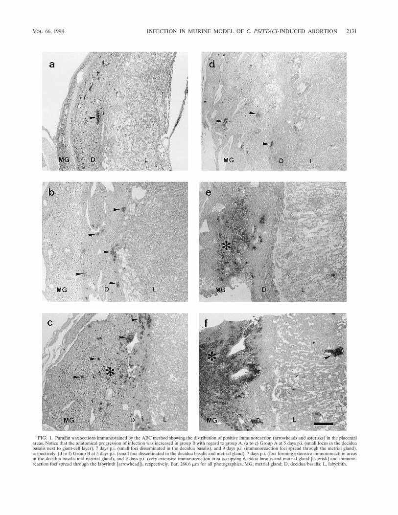
FIG. 1. Paraffin wax sections immunostained by the ABC method showing the distribution of positive immunoreaction (arrowheads and asterisks) in the placentalareas. Notice that the anatomical progression of infection was increased in group B with regard to group A. (a to c) Group A at 5 days p.i. (small focus in the deciduabasalis next to giant-cell layer), 7 days p.i. (small foci disseminated in the decidua basalis), and 9 days p.i. (immunoreaction foci spread through the metrial gland),respectively. (d to f) Group B at 5 days p.i. (small foci disseminated in the decidua basalis and metrial gland), 7 days p.i. (foci forming extensive immunoreaction areasin the decidua basalis and metrial gland), and 9 days p.i. (very extensive immunoreaction area occupying decidua basalis and metrial gland [asterisk] and immuno-reaction foci spread through the labyrinth [arrowhead]), respectively. Bar, 266.6 mm for all photographies. MG, metrial gland; D, decidua basalis; L, labyrinth.
VOL. 66, 1998 INFECTION IN MURINE MODEL OF C. PSITTACI-INDUCED ABORTION 2131

FIG. 2. Electron photomicrographs of the different cell populations affected by chlamydial infection labelled by the immunogold technique. (a) Neutrophil with remainsof immunolabelled material within phagolysosomes, day 14 of pregnancy (7 p.i.). Bar, 1.66 mm. (b) Neutrophils with degenerative changes and chromatin condensationcontaining numerous EBs, day 18 of pregnancy (11 p.i.). Bar, 1.66 mm. (c) Giant cell with a chlamydial inclusion, day 14 of pregnancy (7 p.i.). Bar, 1.98 mm. (d) GMG cellwith a young chlamydial inclusion, day 16 of pregnancy (9 p.i.). Bar, 2.64 mm. (e) Decidual cell with a very large chlamydial inclusion, day 18 of pregnancy (11 p.i.). Bar, 1.51 mm.
2132 BUENDIA ET AL. INFECT. IMMUN.

cells develop NK activity. During pregnancy, this large granu-lated lymphocyte subpopulation increases in the lumimal epi-thelium in the interplacentomal endometrium and may repre-sent up to 10% of the cells of this tissue (15). In pregnanthuman uterus, large granulated lymphocytes have been iden-tified as a subset of NK cells in the decidua (14) and seem tobe related to GMG cells. The role of these cell populations inchlamydial infection of ruminant or human placenta has notbeen determined.
In our experiments there was no sign of immunoreaction inany fetus or of any histopathological findings, which suggeststhat chlamydiae do not directly damage the fetus. However,Rodolakis et al. (21) isolated chlamydiae from the fetus by theplaque-forming method, although in very low numbers (103- to105-fold less than in placenta). This apparent disagreementcould be due to the poor sensitivity of the immunohistochem-ical technique, since it may be able to detect easily chlamydialinclusions but not scarce free EBs. In our model, chlamydiaeprobably reached the fetus from day 18 of pregnancy, when thelabyrinth was invaded, although they did not have time to causelesions since abortion occurred on days 19 to 20. In sheep,chlamydiae also reach the fetus during the last third of preg-nancy, causing focal necrosis in the liver and other organs (6).However, in this case the time elapsing between fetal infectionand abortion is greater (about 30 days). In chlamydial humanabortion, on the other hand, although bacteria were isolatedfrom both fetal and placental tissue, histopathological changeswere observed only in the placenta (28). The murine modeltherefore is quite similar to human chlamydial abortion.
A very similar occurrence has been observed with Coxiellaburnetii, where hepatic lesions have been found in fetus from na-turally infected sheep (27); however, Baumgartner and Bach-mann in an extensive immunohistochemical study (4) found nopositive immunoreaction or lesions in the fetuses of experi-mentally infected pregnant mice but found both lesions andpositive immunoreaction in surviving 9-day-old offspring,which suggests that fetal infection occurred just before or afterbirth.
In conclusion, our findings suggest that there is no importantdamage to the fetus prior to abortion, although two occur-rences that could represent an indirect form of damage to thefetus took place in the decidua basalis: lysis of GMG cells withthe subsequent release of granules containing cytotoxic pro-
teins and pronounced infiltration of this area by neutrophils.Both of these occurrences, together with the direct destructionof the decidual cells by chlamydiae, could lead to a malfunc-tioning of the maternal placenta and a premature breaking ofthe decidua basalis, coinciding with the late-term abortion in-duced by chlamydiae.
ACKNOWLEDGMENTS
This work was supported in part by Comision Interministerial deCiencia y Tecnologıa (CICYT) grant AGF97-0459. A. J. Buendıa wasthe recipient of a predoctoral grant from the Universidad de Murcia,Murcia, Spain.
We thank Ian J. Stewart for helpful suggestions at the outset of thisstudy and A. Souriau for help in the biotinylation of the antichlamydialmonoclonal antibody.
REFERENCES
1. Abzug, M. J., H. A. Rotbart, S. A. Magliato, and M. J. Levin. 1991. Evolutionof the placental barrier to fetal infection by murine enteroviruses. J. Infect.Dis. 163:1336–1341.
2. Awan, A. R., M. Baxi, and H. J. Field. 1995. EHV-1-induced abortion in miceand its relationship to stage of gestation. Res. Vet. Sci. 59:139–145.
3. Banks, J., B. Eddie, J. Schachter, and K. F. Meyer. 1970. Plaque formationby Chlamydia in L cells. Infect. Immun. 1:259–262.
4. Baumgartner, W., and S. Bachmann. 1992. Histological and immunocyto-chemical characterization of Coxiella burnetii-associated lesions in the mu-rine uterus and placenta. Infect. Immun. 60:5232–5241.
5. Brown, M. B., and D. A. Steiner. 1996. Experimental genital mycoplasmosis:time of infection influences pregnancy outcome. Infect. Immun. 64:2315–2321.
6. Buxton, D., R. M. Barlow, J. Finlayson, I. E. Anderson, and A. Mackellar.1990. Observations on the pathogenesis of Chlamydia psittaci infection ofpregnant sheep. J. Comp. Pathol. 102:221–237.
7. Buzoni-Gatel, D., and A. Rodolakis. 1983. A mouse model to comparevirulence of abortive and intestinal ovine strains of Chlamydia psittaci: influ-ence of the route of inoculation. Ann. Microbiol. 134:91–99.
8. Croy, B. A. 1994. Granulated metrial gland cells: hypothesis concerningpossible functions during murine gestation. J. Reprod. Immunol. 27:85–94.
9. Delgado, S. R., B. A. McBey, S. Yamashiro, J. Fujita, Y. Kiso, and B. A. Croy.1996. Accounting for the peripartum loss of granulated metrial gland cells, anatural killer cell population, from the pregnant mouse uterus. J. LeukocyteBiol. 59:262–269.
10. De Sa, C., A. Souriau, F. Bernard, J. Salinas, and A. Rodolakis. 1995. Anoligomer of the major outer membrane protein of Chlamydia psittaci isrecognized by monoclonal antibodies which protect mice from abortion.Infect. Immun. 63:4912–4916.
11. Hadley, K. M., D. Carrington, C. E. Frew, A. A. Gibson, and W. S. Hislop.1992. Ovine chlamydiosis in an abattoir worker. J. Infect. 25:105–109.
12. Hansen, P. J., and W.-J. Lee. 1996. Immunological aspects of pregnancy:
FIG. 3. Site of previous attachment at day 2 postabortion. (a) Paraffin wax section immunostained by the ABC technique. Bar, 266.6 mm. (b) Electron photo-micrograph labelled by the immunogold technique, showing groups of EBs, amorphous immunolabelled material, and cells with significant degenerative changes. Bar,1.66 mm.
VOL. 66, 1998 INFECTION IN MURINE MODEL OF C. PSITTACI-INDUCED ABORTION 2133
concepts and speculations using the sheep as a model. Anim. Reprod. Sci. 42:483–493.
13. Jorgensen, D. M. 1997. Gestational psittacosis in a Montana sheep rancher.Emerging Infect. Dis. 3:191–194.
14. King, A., P. Wooding, L. Gardner, and Y. W. Loke. 1993. Expression ofperforin, granzyme A and TIA-1 by human uterine CD561 NK cells impliesthey are activates and capable of effector functions. Hum. Reprod. 8:2061–2067.
15. Lee, C. S., E. Meeusen, K. Gogolin-Ewens, and M. R. Brandon. 1992. Quan-titative and qualitative changes in the intraepithelial lymphocyte populationin the uterus of nonpregnant and pregnant sheep. Am. J. Reprod. Immunol.28:90–96.
16. Long, M. T., and T. V. Bazler. 1996. Fetal loss in BALB/c mice infected withNeospora caninum. J. Parasitol. 82:608–611.
17. Moulder, J. W., T. P. Hatch, C.-C. Kuo, J. Schachter, and J. Storz. 1984.Genus I. Chlamydia, Jones, Rake and Stearns 1945, p. 729–739. In N. J. Krieg(ed.), Bergey’s manual of systematic bacteriology, vol. 1. Williams & Wilkins,Baltimore, Md.
18. Parr, E. L., L. H. Young, M. B. Parr, and J. D.-E. Young. 1990. Granulatedmetrial gland cells of pregnant mouse uterus are natural killer-like cells thatcontain perforin and serine esterases. J. Immunol. 145:2365–2372.
19. Redline, R. W., and C. Y. Lu. 1988. Specific defects in the anti-listerialimmune response in discrete regions of the murine uterus and placentaaccount for susceptibility to infection. J. Immunol. 140:3947–3955.
20. Rodolakis, A., L. Gestin, and A. Bertin. 1981. Methode de controle desvaccins contre la chlamydiose abortive ovine utilisant la souris gestante. Ann.Rech. Vet. 12:371–377.
21. Rodolakis, A., F. Bernard, and F. Lantier. 1989. Mouse models for evalua-tion of virulence of Chlamydia psittaci isolated from ruminants. Res. Vet. Sci.46:34–39.
22. Salinas, J., J. Sanchez, A. J. Buendıa, A. Souriau, A. Rodolakis, A. Bernabe,and F. Cuello. 1994. The LPS localization might explain the lack of protec-tion of LPS-specific antibodies in abortion-causing Chlamydia psittaci infec-tions. Res. Microbiol. 145:611–620.
23. Sanchez, J., A. J. Buendıa, J. Salinas, A. Bernabe, A. Rodolakis, and I. J.Stewart. 1996. Murine granulated metrial gland cells are susceptible toChlamydia psittaci infection in vivo. Infect. Immun. 64:3897–3900.
24. Stewart, I. J. 1991. Granulated metrial gland cells: pregnancy specific leu-kocytes? J. Leukocyte Biol. 50:198–207.
25. Straatsburg, I. H., and R. Gossrau. 1993. Enzyme histochemistry of theregressing rat decidua and metrial gland. Acta Histochem. 94:202–219.
26. Tobias, L., D. O. Cordes, and G. G. Schurig. 1993. Placental pathology of thepregnant mouse inoculated with Brucella abortus strain 2308. Vet. Pathol. 30:119–129.
27. Van Moll, P., W. Baumgartner, U. Eskens, and T. Hanichen. 1993. Immu-nocytochemical demonstration of Coxiella burnetii antigen in the fetal pla-centa of naturally infected sheep and cattle. J. Comp. Pathol. 109:295–301.
28. Wong, S. Y., E. S. Gray, D. Buxton, J. Finlayson, and F. W. A. Johnson. 1985.Acute placentitis and spontaneous abortion caused by Chlamydia psittaci: ahistological and ultrastructural study. J. Clin. Pathol. 38:707–711.
29. Zheng, L. M., S. V. Joag, M. B. Parr, E. L. Parr, and J. D.-E. Young. 1991.Perforin-expressing granulated metrial gland cells in murine deciduoma.J. Exp. Med. 174:1221–1227.
Editor: J. G. Cannon
2134 BUENDIA ET AL. INFECT. IMMUN.