Effect of TNF-α genetic variants and CCR5 Δ 32 on the vulnerability to HIV1 infection and disease...
10
Veloso et al. BMC Medical Genetics 2010, 11:63 http://www.biomedcentral.com/1471-2350/11/63 Open Access RESEARCH ARTICLE BioMed Central © 2010 Veloso et al; licensee BioMed Central Ltd. This is an Open Access article distributed under the terms of the Creative Commons Attribution License (http://creativecommons.org/licenses/by/2.0), which permits unrestricted use, distribution, and reproduction in any medium, provided the original work is properly cited. Research article Effect of TNF-α genetic variants and CCR5Δ32 on the vulnerability to HIV-1 infection and disease progression in Caucasian Spaniards Sergi Veloso 1,2,3 , Montserrat Olona 1,2,3 , Felipe García 4 , Pere Domingo 5,6 , Carlos Alonso-Villaverde 2,3,7 , Montserrat Broch 2,3,8 , Joaquim Peraire 1,2,3 , Consuelo Viladés 1,2,3 , Montserrat Plana 4 , Enric Pedrol 9 , Miguel López- Dupla 1,2,3 , Carmen Aguilar 1,2,3,8 , Mar Gutiérrez 5 , Agathe Leon 4 , Mariona Tasias 9 , Josep Ma Gatell 4,10 , Cristóbal Richart 1,2,3,8 and Francesc Vidal* 1,2,3 Abstract Background: Tumor necrosis factor alpha (TNF-α) is thought to be involved in the various immunogenetic events that influence HIV-1 infection. Methods: We aimed to determine whether carriage of the TNF-α-238G>A, -308G>A and -863 C>A gene promoter single nucleotide polymorphisms (SNP) and the CCR5Δ32 variant allele influence the risk of HIV-1 infection and disease progression in Caucasian Spaniards. The study group consisted of 423 individuals. Of these, 239 were uninfected (36 heavily exposed but uninfected [EU] and 203 healthy controls [HC]) and 184 were HIV-1-infected (109 typical progressors [TP] and 75 long-term nonprogressors [LTNP] of over 16 years' duration). TNF-α SNP and the CCR5Δ32 allele were assessed using PCR-RFLP and automatic sequencing analysis methods on white blood cell DNA. Genotype and allele frequencies were compared using the χ 2 test and the Fisher exact test. Haplotypes were compared by logistic regression analysis. Results: The distribution of TNF-α-238G>A, -308G>A and -863 C>A genetic variants was non-significantly different in HIV-1-infected patients compared with uninfected individuals: -238G>A, p = 0.7 and p = 0.3; -308G>A, p = 0.05 and p = 0.07; -863 C>A, p = 0.7 and p = 0.4, for genotype and allele comparisons, respectively. Haplotype analyses, however, indicated that carriers of the haplotype H3 were significantly more common among uninfected subjects (p = 0.04). Among the infected patients, the distribution of the three TNF-α genetic variants assessed was non-significantly different between TP and LTNP: -238G>A, p = 0.35 and p = 0.7; -308G>A, p = 0.7 and p = 0.6: -863 C>A, p = 0.2 and p = 0.2, for genotype and allele comparisons, respectively. Haplotype analyses also indicated non-significant associations. Subanalyses in the LTNP subset indicated that the TNF-α-238A variant allele was significantly overrepresented in patients who spontaneously controlled plasma viremia compared with those who had a detectable plasma viral load (genotype comparisons, p = 0.02; allele comparisons, p = 0.03). The CCR5Δ32 distribution was non-significantly different in HIV-1-infected patients with respect to the uninfected population (p = 0.15 and p = 0.2 for genotype and allele comparisons, respectively) and in LTNP vs TP (p = 0.4 and p = 0.5 for genotype and allele comparisons, respectively). Conclusions: In our cohort of Caucasian Spaniards, TNF-α genetic variants could be involved in the vulnerability to HIV- 1 infection. TNF-α genetic variants were unrelated to disease progression in infected subjects. The -238G>A SNP may modulate the control of viremia in LTNP. Carriage of the CCR5Δ32 variant allele had no effect on the risk of infection and disease progression. Background TNF-α is a pleiotropic cytokine that acts as an immune and inflammatory mediator. It is synthesized mainly by macrophages and T-cells and most of its actions take place by binding to two cell receptors: TNF-α R1 and R2 [1]. Investigations suggest that TNF-α is involved in the pathogenesis of HIV-1 infection since it is overproduced by infected individuals [2-4]. * Correspondence: [email protected] 1 Hospital Universitari de Tarragona Joan XXIII, Tarragona, Spain Full list of author information is available at the end of the article
-
Upload
independent -
Category
Documents
-
view
2 -
download
0
Transcript of Effect of TNF-α genetic variants and CCR5 Δ 32 on the vulnerability to HIV1 infection and disease...
Veloso et al. BMC Medical Genetics 2010, 11:63http://www.biomedcentral.com/1471-2350/11/63
Open AccessR E S E A R C H A R T I C L E
© 2010 Veloso et al; licensee BioMed Central Ltd. This is an Open Access article distributed under the terms of the Creative Commons
Research articleEffect of TNF-α genetic variants and CCR5Δ32 on the vulnerability to HIV-1 infection and disease progression in Caucasian SpaniardsSergi Veloso1,2,3, Montserrat Olona1,2,3, Felipe García4, Pere Domingo5,6, Carlos Alonso-Villaverde2,3,7, Montserrat Broch2,3,8, Joaquim Peraire1,2,3, Consuelo Viladés1,2,3, Montserrat Plana4, Enric Pedrol9, Miguel López-Dupla1,2,3, Carmen Aguilar1,2,3,8, Mar Gutiérrez5, Agathe Leon4, Mariona Tasias9, Josep Ma Gatell4,10, Cristóbal Richart1,2,3,8 and Francesc Vidal*1,2,3
AbstractBackground: Tumor necrosis factor alpha (TNF-α) is thought to be involved in the various immunogenetic events that influence HIV-1 infection.
Methods: We aimed to determine whether carriage of the TNF-α-238G>A, -308G>A and -863 C>A gene promoter single nucleotide polymorphisms (SNP) and the CCR5Δ32 variant allele influence the risk of HIV-1 infection and disease progression in Caucasian Spaniards. The study group consisted of 423 individuals. Of these, 239 were uninfected (36 heavily exposed but uninfected [EU] and 203 healthy controls [HC]) and 184 were HIV-1-infected (109 typical progressors [TP] and 75 long-term nonprogressors [LTNP] of over 16 years' duration). TNF-α SNP and the CCR5Δ32 allele were assessed using PCR-RFLP and automatic sequencing analysis methods on white blood cell DNA. Genotype and allele frequencies were compared using the χ 2 test and the Fisher exact test. Haplotypes were compared by logistic regression analysis.
Results: The distribution of TNF-α-238G>A, -308G>A and -863 C>A genetic variants was non-significantly different in HIV-1-infected patients compared with uninfected individuals: -238G>A, p = 0.7 and p = 0.3; -308G>A, p = 0.05 and p = 0.07; -863 C>A, p = 0.7 and p = 0.4, for genotype and allele comparisons, respectively. Haplotype analyses, however, indicated that carriers of the haplotype H3 were significantly more common among uninfected subjects (p = 0.04). Among the infected patients, the distribution of the three TNF-α genetic variants assessed was non-significantly different between TP and LTNP: -238G>A, p = 0.35 and p = 0.7; -308G>A, p = 0.7 and p = 0.6: -863 C>A, p = 0.2 and p = 0.2, for genotype and allele comparisons, respectively. Haplotype analyses also indicated non-significant associations. Subanalyses in the LTNP subset indicated that the TNF-α-238A variant allele was significantly overrepresented in patients who spontaneously controlled plasma viremia compared with those who had a detectable plasma viral load (genotype comparisons, p = 0.02; allele comparisons, p = 0.03). The CCR5Δ32 distribution was non-significantly different in HIV-1-infected patients with respect to the uninfected population (p = 0.15 and p = 0.2 for genotype and allele comparisons, respectively) and in LTNP vs TP (p = 0.4 and p = 0.5 for genotype and allele comparisons, respectively).
Conclusions: In our cohort of Caucasian Spaniards, TNF-α genetic variants could be involved in the vulnerability to HIV-1 infection. TNF-α genetic variants were unrelated to disease progression in infected subjects. The -238G>A SNP may modulate the control of viremia in LTNP. Carriage of the CCR5Δ32 variant allele had no effect on the risk of infection and disease progression.
BackgroundTNF-α is a pleiotropic cytokine that acts as an immuneand inflammatory mediator. It is synthesized mainly by
macrophages and T-cells and most of its actions takeplace by binding to two cell receptors: TNF-α R1 and R2[1]. Investigations suggest that TNF-α is involved in thepathogenesis of HIV-1 infection since it is overproducedby infected individuals [2-4].
* Correspondence: [email protected] Hospital Universitari de Tarragona Joan XXIII, Tarragona, SpainFull list of author information is available at the end of the article
BioMed Central Attribution License (http://creativecommons.org/licenses/by/2.0), which permits unrestricted use, distribution, and reproduction inany medium, provided the original work is properly cited.
Veloso et al. BMC Medical Genetics 2010, 11:63http://www.biomedcentral.com/1471-2350/11/63
Page 2 of 10
Like many other proinflammatory cytokines, the pro-duction of TNF-α is at least partially genetically deter-mined [5,6]. Several functional single nucleotidepolymorphisms (SNP) have been identified within theTNF-α gene cluster [5-8], the most widely studied ofwhich are a G>A transition at position -238, a G>A tran-sition at position -308, and a C>A transition at position -863 [9]. Several cohort studies have assessed the influ-ence of TNF-α SNP on the immunogenetics of HIV-1infection. The information available on how TNF-αgenetic variants affect vulnerability to infection is incon-sistent [10-14]: some studies have found no effect[11,13,14] while others have found a strong association[10,12]. Case-control studies have assessed the influenceof TNF-α genetic variants on HIV-1 disease progressionin infected patients [10-12,14-17]. While some of thesestudies have reported an association with progression[10-12,16,17], others have failed to find such an associa-tion [14,15]. A minority of untreated HIV-1-infectedpatients show an uncommon clinical form of infectioncharacterized by a non-progressive disease and, some-times, by self-limited viremia over the years. Thesepatients are known as long-term nonprogressors (LTNP)[18] and elite controllers [19], respectively. Little data isavailable on the influence of TNF-α SNP on these uncom-mon clinical forms of HIV-1 infection [11,16].
In this study we have assessed the influence of TNF-αSNP on the vulnerability to HIV-1 infection and, ininfected patients, on disease progression. For this pur-pose we analysed a cohort that we have already used forseveral immunogenetic studies on HIV-1 infection whichcontains a subset of repeatedly exposed but uninfectedindividuals and a carefully collected cohort of extremelong-term nonprogressors [20,21]. Given its modulatingrole in HIV-1-infection and disease progression reportedelsewhere [22], the CCR5Δ32 allele was also assessed.
MethodsDesign and settingThis was a multicenter cross-sectional population associ-ation study. All subjects were recruited from a prospec-tively collected cohort of almost 6000 HIV-1-infectedpatients treated at the HIV outpatients' clinics of the par-ticipating hospitals, which are located in an epidemiolog-ical setting in which intravenous drug use is one of themain causes of HIV-1-infection.
PopulationTwo subsets of HIV-1-infected patients were studied:LTNP and typical progressors (TP). They were recruitedbetween 2005 and 2007. Criteria for LTNP were: asymp-tomatic HIV-1 infection of over 16 years' duration; in theabsence of any antiretroviral treatment, a stable CD4+cell count persistently over 500 cells/μl; and a plasma
HIV-1 viral load repeatedly under 5000 copies/ml [18].LTNP were further divided into two subgroups depend-ing on whether the plasma viral load was detectable(LTNP-DVL) or persistently undetectable. LTNP in thelatter subgroup were called elite controllers (LTNP-EC)[19]. Patients were categorized as TP if they fulfilled allthe following criteria: a) the HIV-1 infection had pro-gressed to the advanced disease (that is to say class CHIV-1 disease had appeared according to the 1993 Cen-ters for Disease Control criteria [23]), b) the plasma HIV-1 viral load was over 35,000 copies/ml and, c) the CD4+T-cell count decreased over time and was below 350 cells/μl at least once in the first 10 years of infection. Almost allof them were on antiretroviral therapy when theyenrolled. For a few patients whose date of infection wasnot available, we assumed that it was the midpointbetween the first positive and the last negative HIV-1blood test [24,25]. We identified 75 patients within ourcohort who fulfilled the LTNP criteria. They all agreed toparticipate in the study, and we recruited a randomlyselected group of TP (n = 109) whose age (± 5 years) andgender were comparable with the LTNP. A group of 36repeatedly exposed but uninfected individuals (EU) wasalso evaluated. They were part of a cohort of individualsthat we have used in other studies and whose details andcharacteristics we have extensively described elsewhere[26,27]. For the control group we studied a sample ofhealthy subjects recruited from voluntary blood donors,whose age and gender were comparable with the patients.Table 1 shows details of the study population. All subjectsin our study were white Spaniards. Immigrants fromother countries, including those from other Europeancountries, and their descendents were excluded.Informed consent was obtained from each participant.The project was approved by the local ethical researchcommittees.
DNA and plasma samplesBlood samples with ethylene diamine tetra-acetic acidwere obtained from an antecubital vein. Five mL of wholeblood was used to determine the CD4+ T-cell count, and500 μl was used to isolate DNA with a MagNa Pure LCInstrument (Roche Diagnostics, Basel, Switzerland).Plasma for determining HIV-1 viral load was obtained bycentrifugation at 3500 g for 15 minutes at 4C°.
Laboratory methodsHIV-1 infectionThis was diagnosed using an enzymoimmunoanalysisand confirmed by a Western-Blot test.Plasma HIV-1 viral loadThis was determined by the HIV Cobas Ampliprep CAP-CTMHIV-1 using the COBAS AMPLICOR system
Veloso et al. BMC Medical Genetics 2010, 11:63http://www.biomedcentral.com/1471-2350/11/63
Page 3 of 10
(Roche Diagnostics, Basel, Switzerland). The cutoff forundetectable viral load was 50 copies/μl.Assessment of blood CD4+ T-cell countSamples were analyzed in a flow cytometer FAC Scan(Becton Dickinson Immunocytometry Systems, San José,CA, USA). The data acquired were analyzed using theMultiset program.
TNF-α genotype-238 TNF-α genotype (rs 361525)This polymorphism consists of a GT A substitution atposition -238 in the proximal promoter of the TNF-αgene. The primers used in PCR were: forward primer 5'AGAAGACCCCCCTCGGAACC3', modified in 3' forRFLP analysis with Msp I restriction endonuclease, and
reverse primer 5' ATCTGGAGGAAGCGGTAGTG 3'. Afragment of 152 bp was amplified at a final volume of 50μL, with 3 mM of MgCl2, 0.2 mM dNTPs, 0.2 μM of eachprimer and 1 unit of Taq polymerase. DNA was amplifiedfor 35 cycles: 95° for 30 seconds, 55° for 30 seconds and72° for 30 seconds. PCR products were digested withMspI and revealed a fragment of 152 bp for the A alleleand two fragments of 133 and 19 bp for the G allele.-308 TNF-α genotype (rs 1800629)This polymorphism consists of a GT A substitution atposition -308 in the proximal promoter of the TNF-αgene. The primers used in the PCR were5'AGGCAATAGGTTTTGAGGGCCAT3' and 5'TCCTCCCTGCTCCGATTCCG3'. Amplification was per-formed at a final volume of 50 μl containing 3 mM MgCl2,
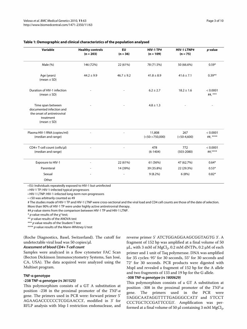
Table 1: Demographic and clinical characteristics of the population analysed
Variable Healthy controls(n = 203)
EU(n = 36)
HIV-1 TP#(n = 109)
HIV-1 LTNP#(n = 75)
p value
Male (%) 146 (72%) 22 (61%) 78 (71.5%) 50 (66.6%) 0.59*
Age (years)(mean ± SD)
44.2 ± 9.9 46.7 ± 9.2 41.8 ± 8.9 41.6 ± 7.1 0.39**
Duration of HIV-1 infection(mean ± SD)
- - 6.2 ± 2.7 18.2 ± 1.6 < 0.0001##, ***
Time span between documented infection and the onset of antiretroviral
treatment(mean ± SD)
- - 4.8 ± 1.3 - -
Plasma HIV-1 RNA (copies/ml)(median and range)
- - 11,808(<50->750,000)
267(<50-4,600)
< 0.0001##, ****
CD4+ T-cell count (cells/μl)(median and range)
- - 478(6-1404)
772(503-2080)
< 0.0001 ##,****
Exposure to HIV-1 - 22 (61%) 61 (56%) 47 (62.7%) 0.64*
Parenteral - 14 (39%) 39 (35.8%) 22 (29.3%) 0.53*
Sexual - - 9 (8.2%) 6 (8%) 0.82*
Other
• EU: Individuals repeatedly exposed to HIV-1 but uninfected• HIV-1 TP: HIV-1-infected typical progressors• HIV-1 LTNP: HIV-1-infected long-term non-progressors• <50 was arbitrarily counted as 49# The studies made of HIV-1 TP and HIV-1 LTNP were cross-sectional and the viral load and CD4 cell counts are those of the date of selection. More than 90% of HIV-1 TP were under highly active antiretroviral therapy.## p value stems from the comparison between HIV-1 TP and HIV-1 LTNP.* p value results of the χ2 test;** p value results of the ANOVA test*** p value results of the Student T test**** p value results of the Mann-Whitney U test
Veloso et al. BMC Medical Genetics 2010, 11:63http://www.biomedcentral.com/1471-2350/11/63
Page 4 of 10
0.5 mM of each nucleotide (Boehringer Mannheim™,Manheim, Germany), 0.2 μl of each oligonucleotide and 1U of Taq polymerase (Gibco BRL). DNA was amplifiedfor 35 cycles with denaturation at 94°C for 1 minute(min), annealing at 60°C for 1 min and extension at 72°Cfor 1 min. The first cycle was at 94°C for 3 min, at 60°Cfor 1 min and at 72°C for 1 min, followed by the 35 cyclesand, finally, a cycle at 94°C for 1 min, at 60°C for 1 minand at 72°C for 5 min. The PCR products were digested at37°C with NcoI for 24 hours, subjected to 2.5% agarosegel electrophoresis at 80 V and stained with ethidiumbromide. The 107 bp band corresponded to the A alleleand the set of 87 bp and 20 bp bands corresponded to theG allele.-863 TNF-α genotype (rs 1800630)This polymorphism consists of a CT A substitution atposition -863 in the proximal promoter of the TNF-αgene. The primers used in the PCR were5'GGCTCTGAGGAATGGGTTAC3' and 5'CTACATGGCCCTGTCTTCGTTACG3'. Amplification was per-formed at a final volume of 25 μl containing 1 mM MgCl2,0.2 mM of each nucleotide (Boehringer Mannheim™,Manheim, Germany), 1.2 μl of each oligonucleotide and0.5 U of Taq polymerase (Gibco BRL). DNA was ampli-fied for 35 cycles with denaturation at 94°C for 30 sec-onds, annealing at 62.3°C for 1 min and extension at 72°Cfor 2 min. The first cycle was at 94°C for 3 min, followedby the 35 cycles and, finally, an extension at 72°C for 5min. The amplified product was digested with Bsa AIrestriction enzyme (New England Biolabs) at 37°C for 24hours, electrophoresed on 2.5% agarose gel at 80 V andstained with ethidium bromide. The 126 pb band corre-sponded to the C wild-type allele and the set of 103 bpand 23 pb bands corresponded to the variant A allele.
CCR5Δ32 genotypeThe PCR primers used were 5'GCTCTCTCCCAGGAATCATC3' and antisense 5'TTCCCGAGTAGCAGATGACC3' with annealing at 60°C. The Genbank accessionnumber was AF031237.1. The PCR products were visual-ized on 2.5% agarose gel. The wild-type CCR5 gene led toa 174 bp fragment and the CCR5Δ32 variant resulted in a142 bp fragment.
Statistical analysisDescriptive data were expressed as the mean ± SD ormedian (range) for non-parametric distributions. Thedifferences in levels between groups in continuous vari-ables were compared using Student's t test, the Mann-Whitney U test, or ANOVA when necessary. To evaluatethe association between HIV-1 TP and the different cate-gories of uninfected subjects for the different genotypes,the odds ratio (OR) and 95% confidence interval (95%CI)were calculated. The Hardy-Weinberg equilibrium was
assessed by the chi-square goodness-of-fit test. Genotypedistribution and allele frequencies in the different groupswere compared by the χ2 test or Fisher's exact test whennecessary. To evaluate the association of TNF-α geneticvariants and CCR5Δ32 with the risk of HIV-1 infection,the differences in the distribution between HIV-1-infected, HC and EU were assessed. To evaluate the asso-ciation of TNF-α genetic variants and CCR5Δ32 with dis-ease progression in infected subjects, we compared HIV-1-infected TP vs HIV-1-infected LTNP. In the latter sub-set, we also compared LTNP-DVL and LTNP-EC. Analy-ses were performed using the SPSS/PC+ statisticalpackage (v. 12.0 for Windows; Chicago, Illinois, USA).Haplotype frequencies were estimated by the maximum-likelihood method. Haplotype frequencies were esti-mated by the PAC-likelihood method [28], which takesinto account the similarity of haplotypes and the fact thatlinkage disequilibrium decays with distance. The distri-butions of the different haplotypes between groups werecompared by logistic regression analysis. The commonesthaplotype (which we arbitrarily called haplotype H1) wastaken as a reference. Haplotype analyses were performedusing the PHASEv2.1 software [29,30]. A p value < 0.05was considered significant.
ResultsFour hundred and twenty-three individuals were studied:239 uninfected (203 healthy controls [HC] and 36 EU)and 184 HIV-1-infected. Of the infected individuals, mostof whom had acquired HIV-1 through intravenous druguse, 109 were TP and 75 were LTNP. Of the LTNP, 20were elite controllers. The age, gender and risk factors ofacquiring HIV-1 infection in the TP and LTNP were notsignificantly different. Most TP (>90%) were receivinghighly active antiretroviral therapy. Table 1 showsselected characteristics of the population studied. Asexpected, the duration of HIV-1 infection was signifi-cantly greater in LTNP than in TP. CD4+ T-cell countswere also greater and viral loads were lower.
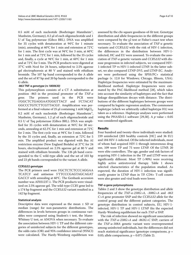
TNF-α gene polymorphismsTables 2 and 3 show the genotype distribution and allelefrequencies of the TNF-α-238G>A, -308G>A and -863C>A gene promoter SNP and the CCR5Δ32 allele for thecontrol group and the different patient categories. Thegenotype distribution in control subjects, EU, HIV-1-infected, HIV-1 TP and HIV-1 LTNP fits the expectedHardy-Weinberg equilibrium for each TNF-α SNP.
The risk of infection showed no significant associationswith the TNF-α-238G>A and -863G>C SNP; carriers ofthe TNF-α-308A genetic variant were overrepresentedamong uninfected individuals, but the differences did notreach statistical significance (genotype comparisons, p =0.05; allele comparisons, p = 0.07) (Table 2).
Veloso et al. BMC Medical Genetics 2010, 11:63http://www.biomedcentral.com/1471-2350/11/63
Page 5 of 10
Table 2: TNF-α and CCR5Δ32 genotype and allele frequencies in HC, EU and HIV-1-infected for assessment of associations
with the risk of HIV-1 infection.
Genotype and allele frequencies
HC(n = 203)
EU(n = 36)
HIV-1-infected(n = 184)
p value *
TNF-α-238 G>A
GG 147 (87.5%) 29 (80.5%) 161 (87.5%)
GA 20 (12%) 6 (16.7%) 22 (12%) 0.7
AA 1 (0.5%) 1 (2.8%) 1 (0.5%)
GA+AA 21 (12.5%) 7 (19.5%) 23 (12.5%) 0.5
Variant allele A 22 (6.5%) 8 (11.1%) 24 (6.5%) 0.3
TNF-α-308G>A
GG 122(70.9%) 24 (67%) 148 (80.4%)
GA 42(24.4%) 12 (33%) 31 (16.8%) 0.05
AA 8 (4.7%) 0 5 (2.8%)
GA+AA 50 (29.1%) 12 (33%) 36 (19.6%) 0.05
Variant allele A 58 (16.9%) 12 (16.7%) 41 (11.1%) 0.07
TNF-α-863 C>A
CC 116 (69.9%) 27 (75%) 137 (74.4%)
CA 44 (26.5%) 8 (22%) 45 (24.5%) 0.7
AA 6 (3.6%) 1 (3%) 2 (1.1%)
CA+AA 50 (30.1%) 9 (25%) 47 (25.5%) 0.6
Variant allele A 56 (16.8%) 10 (14%) 49 (13.3%) 0.4
CCR5Δ32
wt/wt 174 (87%) 31 (86.1%) 144 (78.3%)
wt/Δ32 26 (13%) 5 (13.9%) 40 (21.7%) 0.15
Δ32/Δ32 0 0 0
Δ32 allele 26 (6.5%) 5 (6.9) 40 (10.9%) 0.2
• HC: healthy controls• EU: individuals repeatedly exposed to HIV-1 but uninfected• wt indicates wild-type allele; Δ32, 32 bp deletion• Genotype and allele numbers do not match the HC studied because DNA for TNF-α and CCR5 genotyping was not available or could not be amplified in some people* χ2 test or Fisher exact test when necessary. p value arises from the comparison between HC, EU and HIV-1-infected.
Veloso et al. BMC Medical Genetics 2010, 11:63http://www.biomedcentral.com/1471-2350/11/63
Page 6 of 10
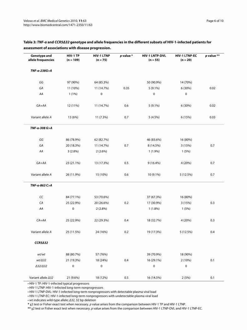
Table 3: TNF-α and CCR5Δ32 genotype and allele frequencies in the different subsets of HIV-1-infected patients for
assessment of associations with disease progression.
Genotype andallele frequencies
HIV-1 TP(n = 109)
HIV-1 LTNP(n = 75)
p value * HIV-1 LNTP-DVL(n = 55)
HIV-1 LTNP-EC(n = 20)
p value **
TNF-α-238G>A
GG 97 (90%) 64 (85.3%) 50 (90.9%) 14 (70%)
GA 11 (10%) 11 (14.7%) 0.35 5 (9.1%) 6 (30%) 0.02
AA 1 (1%) 0 0 0
GA+AA 12 (11%) 11 (14.7%) 0.6 5 (9.1%) 6 (30%) 0.02
Variant allele A 13 (6%) 11 (7.3%) 0.7 5 (4.5%) 6 (15%) 0.03
TNF-α-308 G>A
GG 86 (78.9%) 62 (82.7%) 46 (83.6%) 16 (80%)
GA 20 (18.3%) 11 (14.7%) 0.7 8 (14.5%) 3 (15%) 0.7
AA 3 (2.8%) 2 (2.6%) 1 (1.9%) 1 (5%)
GA+AA 23 (21.1%) 13 (17.3%) 0.5 9 (16.4%) 4 (20%) 0.7
Variant allele A 26 (11.9%) 15 (10%) 0.6 10 (9.1%) 5 (12.5%) 0.7
TNF-α-863 C>A
CC 84 (77.1%) 53 (70.6%) 37 (67.3%) 16 (80%)
CA 25 (22.9%) 20 (26.6%) 0.2 17 (30.9%) 3 (15%) 0.3
AA 0 2 (2.8%) 1 (1.8%) 1 (5%)
CA+AA 25 (22.9%) 22 (29.3%) 0.4 18 (32.7%) 4 (20%) 0.3
Variant allele A 25 (11.5%) 24 (16%) 0.2 19 (17.3%) 5 (12.5%) 0.4
CCR5Δ32
wt/wt 88 (80.7%) 57 (76%) 39 (70.9%) 18 (90%)
wt/Δ32 21 (19.3%) 18 (24%) 0.4 16 (29.1%) 2 (10%) 0.1
Δ32/Δ32 0 0 0 0
Variant allele Δ32 21 (9.6%) 18 (12%) 0.5 16 (14.5%) 2 (5%) 0.1
• HIV-1 TP: HIV-1-infected typical progressors• HIV-1 LTNP: HIV-1-infected long-term nonprogressors• HIV-1 LTNP-DVL: HIV-1-infected long-term nonprogressors with detectable plasma viral load• HIV-1 LTNP-EC: HIV-1-infected long-term nonprogressors with undetectable plasma viral load• wt indicates wild-type allele; Δ32, 32 bp deletion* χ2 test or Fisher exact test when necessary. p value arises from the comparison between HIV-1 TP and HIV-1 LTNP.** χ2 test or Fisher exact test when necessary. p value arises from the comparison between HIV-1 LTNP-DVL and HIV-1 LTNP-EC.
Veloso et al. BMC Medical Genetics 2010, 11:63http://www.biomedcentral.com/1471-2350/11/63
Page 7 of 10
Both genotype and allele analyses indicated that therewere no differences in the distribution of the genetic vari-ants between LTNP and TP (Table 3). When we analysedthe LTNP subset of patients and compared the elite con-trollers with those with detectable viral loads, we foundthat the TNF-α-238A genetic variant was significantlyoverrepresented in elite controllers: p = 0.02 for genotypeanalyses and p = 0.03 for allele analyses (Table 3). Theresults obtained in the full cohort for all these analyseswere maintained when the individuals carrying theCCR5Δ32 variant allele were excluded.
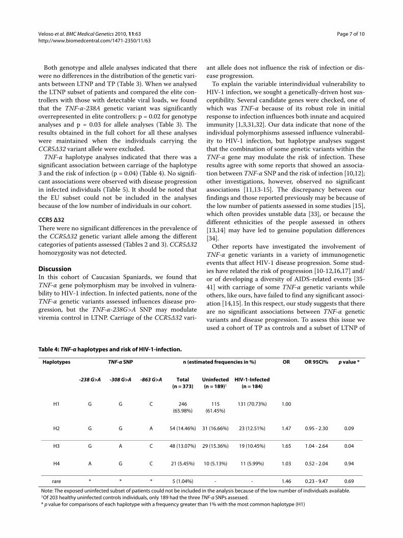
TNF-α haplotype analyses indicated that there was asignificant association between carriage of the haplotype3 and the risk of infection (p = 0.04) (Table 4). No signifi-cant associations were observed with disease progressionin infected individuals (Table 5). It should be noted thatthe EU subset could not be included in the analysesbecause of the low number of individuals in our cohort.
CCR5 Δ32There were no significant differences in the prevalence ofthe CCR5Δ32 genetic variant allele among the differentcategories of patients assessed (Tables 2 and 3). CCR5Δ32homozygosity was not detected.
DiscussionIn this cohort of Caucasian Spaniards, we found thatTNF-α gene polymorphism may be involved in vulnera-bility to HIV-1 infection. In infected patients, none of theTNF-α genetic variants assessed influences disease pro-gression, but the TNF-α-238G>A SNP may modulateviremia control in LTNP. Carriage of the CCR5Δ32 vari-
ant allele does not influence the risk of infection or dis-ease progression.
To explain the variable interindividual vulnerability toHIV-1 infection, we sought a genetically-driven host sus-ceptibility. Several candidate genes were checked, one ofwhich was TNF-α because of its robust role in initialresponse to infection influences both innate and acquiredimmunity [1,3,31,32]. Our data indicate that none of theindividual polymorphisms assessed influence vulnerabil-ity to HIV-1 infection, but haplotype analyses suggestthat the combination of some genetic variants within theTNF-α gene may modulate the risk of infection. Theseresults agree with some reports that showed an associa-tion between TNF-α SNP and the risk of infection [10,12];other investigations, however, observed no significantassociations [11,13-15]. The discrepancy between ourfindings and those reported previously may be because ofthe low number of patients assessed in some studies [15],which often provides unstable data [33], or because thedifferent ethnicities of the people assessed in others[13,14] may have led to genuine population differences[34].
Other reports have investigated the involvement ofTNF-α genetic variants in a variety of immunogeneticevents that affect HIV-1 disease progression. Some stud-ies have related the risk of progression [10-12,16,17] and/or of developing a diversity of AIDS-related events [35-41] with carriage of some TNF-α genetic variants whileothers, like ours, have failed to find any significant associ-ation [14,15]. In this respect, our study suggests that thereare no significant associations between TNF-α geneticvariants and disease progression. To assess this issue weused a cohort of TP as controls and a subset of LTNP of
Table 4: TNF-α haplotypes and risk of HIV-1-infection.
Haplotypes TNF-α SNP n (estimated frequencies in %) OR OR 95CI% p value *
-238 G>A -308 G>A -863 G>A Total (n = 373)
Uninfected (n = 189)1
HIV-1-Infected (n = 184)
H1 G G C 246 (65.98%)
115 (61.45%)
131 (70.73%) 1.00
H2 G G A 54 (14.46%) 31 (16.66%) 23 (12.51%) 1.47 0.95 - 2.30 0.09
H3 G A C 48 (13.07%) 29 (15.36%) 19 (10.45%) 1.65 1.04 - 2.64 0.04
H4 A G C 21 (5.45%) 10 (5.13%) 11 (5.99%) 1.03 0.52 - 2.04 0.94
rare * * * 5 (1.04%) - - 1.46 0.23 - 9.47 0.69
Note: The exposed uninfected subset of patients could not be included in the analysis because of the low number of individuals available.1Of 203 healthy uninfected controls individuals, only 189 had the three TNF-α SNPs assessed.* p value for comparisons of each haplotype with a frequency greater than 1% with the most common haplotype (H1)

Veloso et al. BMC Medical Genetics 2010, 11:63http://www.biomedcentral.com/1471-2350/11/63
Page 8 of 10
over 16 years' duration as cases. This extreme LTNP phe-notype was expressly chosen to prevent groups fromsuperposing. Of particular note, however, was that withinthe LTNP group we showed a significant associationbetween carriage of the TNF-α-238A variant allele andthe spontaneous "elite controller" phenotype. This repli-cates what was reported in an early study [16] but, sincethis finding is supported by a very small number ofpatients it should be further replicated in larger series ofthis uncommon subset of HIV-1-infected patients.
For CCR5Δ32, we found that there is no signal for nonprogression. This is in agreement with CCR5Δ32 having aprimary effect against rapid progression as pointed out bythe GRIV study [42,43]. We acknowledge, however, thatin order to see the effect of CCR5Δ32, the use of a largercohort and/or a Kaplan-Meier plot of patients' evolutionsince seroconversion under various endpoints could havebeen useful.
Our study has some limitations. First, the cross sec-tional nature of the design provides associations, not cau-sality. Second, the comparison of the LTNP with the TPsubsets is rather limited since it involves long-term nonprogression and not simple progression, and additionalanalyses with Kaplan-Meier curves (with various end-points such as death or AIDS 1993 criteria) could lead todifferent results. Finally, some of the subsets assessedwere small, particularly the exposed uninfected and elitecontrollers. This means that our analyses of these subsetsmay be too underpowered to detect other significantassociations.
ConclusionsIn summary, in a cohort of Caucasian Spaniards, poly-morphism within the TNF-α gene may be associated with
vulnerability to HIV-1 infection. In infected patients,none of the TNF-α genetic variants assessed influencesdisease progression but the TNF-α-238G>A SNP maymodulate the elite controller status. The CCR5Δ32 vari-ant allele influences neither the risk of infection nor dis-ease progression.
Competing interestsThe authors declare that they have no competing interests.
Authors' contributionsSV, FG, PD, CAV, EP, JMG, CR and FV designed the study. SV, JP, CV, MG, AL andMT collected and analysed the data. MB, MP and CA performed the geneticstudies. MO and MLD performed the statistical analyses. SV, MO, FG, PD, CAV, EP,JMG, CR and FV drafted the manuscript. All authors have read and approvedthe manuscript.
AcknowledgementsThis study has been partially financed by grants from the Fondo de Investiga-cion Sanitaria (07/0976, 08/1032); SAF 2008-02278; Fondos Europeos para el Desarrollo Regional (FEDER); Fundación para la Investigación y Prevención del Sida en España (FIPSE 06/36572 and 06/30610); Grups Reconeguts AGAUR (2009 SGR 959 and 2009 SGR 1061); Red de Investigación en Sida, Instituto de Salud Carlos III (RIS; RD06/006/0000, RD06/006/0022 and RD06/006/1004). Montserrat Plana was supported by contract FIS 03/00072 from the Fundació Privada Clínic per a la Recerca Biomèdica in collaboration with the Spanish Health Department. Pilar Hernandez helped us in the statistical analyses. John Bates kindly improved the English text. The constructive comments and criti-cisms of the three reviewers helped us to improve the manuscript and are greatly appreciated.
Author Details1Hospital Universitari de Tarragona Joan XXIII, Tarragona, Spain, 2IISPV, Tarragona, Spain, 3Universitat Rovira i Virgili, Tarragona, Spain, 4Hospital Clínic, Barcelona, Spain, 5Hospital de la Santa Creu i Sant Pau, Barcelona, Spain, 6Universitat Autònoma de Barcelona, Barcelona, Spain, 7Hospital Universitari de Sant Joan, Reus, Spain, 8CIBER Fisiopatologia de la Obesidad y Nutrición (CB06/03), Instituto de Salud Carlos III, Madrid, Spain, 9Hospital de Sant Pau i Santa Tecla, Tarragona, Spain and 10Universitat de Barcelona, Barcelona, Spain
Received: 21 July 2009 Accepted: 26 April 2010 Published: 26 April 2010This article is available from: http://www.biomedcentral.com/1471-2350/11/63© 2010 Veloso et al; licensee BioMed Central Ltd. This is an Open Access article distributed under the terms of the Creative Commons Attribution License (http://creativecommons.org/licenses/by/2.0), which permits unrestricted use, distribution, and reproduction in any medium, provided the original work is properly cited.BMC Medical Genetics 2010, 11:63
Table 5: TNF-α haplotypes and disease progression in HIV-1-infected patients.
Haplotypes TNF-α SNP n (estimated frequencies in %) OR OR 95CI% p value *
-238 G>A -308 G>A -863 G>A Total (n = 184)
HIV-1 TP (n = 109)
HIV-1 LTNP (n = 75)
H1 G G C 131 (70.73%) 80 (73.44%) 51 (66.70%) 1.00
H2 G G A 23 (12.51%) 12 (9.90%) 11 (16.04%) 1.83 0.92 - 3.64 0.09
H3 G A C 19 (10.45%) 12 (9.83%) 7 (9.95%) 1.20 0.60 - 2.41 0.61
H4 A G C 11 (5.99%) 5 (4.25%) 6 (7.30%) 1.84 0.68 - 4.95 0.23
rare * * * 0 (0.32%) - - 1.00 - 1.00
* p value for comparisons of each haplotype with a frequency greater than 1% with the most common haplotype (H1)HIV-1 TP: HIV-1-infected typical progressorsHIV-1 LTNP: HIV-1-infected long-term non-progressors

Veloso et al. BMC Medical Genetics 2010, 11:63http://www.biomedcentral.com/1471-2350/11/63
Page 9 of 10
References1. Bradley JR: TNF-mediated inflammatory disease. J Pathol 2008,
214:149-160.2. Ledru E, Lecoeur H, Garcia S, Debord T, Gougeon M: Differential
susceptibility to activation-induced apoptosis among peripheral Th1 subsets: correlation with Bcl-2 expression and consequences for AIDS pathogenesis. J Immunol 1998, 160:3194-3206.
3. Herbelin G, Aziz Khan K: Is HIV infection a TNF receptor signalling-driven disease? Trends Immunol 2007, 29:61-67.
4. Domingo P, Vidal F, Domingo JC, Veloso S, Sambeat MA, Torres F, Sirvent JJ, Vendrell J, Matias-Guiu X, Richart C, HIV-FRS Study Group: Tumour necrosis factor alpha in fat redistribution syndromes associated with combination antiretroviral therapy in HIV-1-infected patients: potential role in subcutaneous adipocyte apoptosis. Eur J Clin Invest 2005, 35:771-780.
5. Bouma G, Crusius JBA, Oudkerk Pool M, Kolkman JJ, von Blomberg BM, Kostense PJ, Giphart MJ, Schreuder GM, Meuwissen SG, Peña AS: Secretion of tumour necrosis factor and lymphotoxin in relation to polymorphisms in TNF genes and HLA-DR alleles. Relevance for inflammatory bowel disease. Scand J Immunol 1996, 43:456-463.
6. Hajeer AH, Hutchinson IV: Influence of TNFα gene polymorphisms on TNFα production and disease. Human Immunology 2001, 62:1191-1199.
7. Wilson AG, de Vries N, Pociot F, di Giovine FS, Putte LB van der, Duff GW: An allelic polymorphism within the human tumor necrosis factor alpha promoter region is strongly associated with HLA A1, B8 and DR3 alleles. J Exp Med 1993, 177:557-560.
8. D'Alfonso S, Richiardi PM: A polymorphic variation in a putative regulation box of the TNFA promoter region. Immunogenetics 1994, 39:150-154.
9. Auguet T, Vidal F, López-Dupla M, Broch M, Gutiérrez C, Olona M, Oltra C, Aguilar C, González E, Quer JC, Sirvent JJ, Richart C: A study on the TNF-α system in Caucasian Spanish patients with alcoholic liver disease. Drug Alcohol Depend 2008, 92:91-99.
10. Khoo SH, Pepper L, Snowden N, Hajeer AH, Vallely P, Wilkins EG, Mandal BK, Ollier WE: Tumor necrosis factor c2 micro satellite allele is associated with the rate of HIV progression. AIDS 1997, 11:423-428.
11. Knuchel MC, Spira TJ, Neumman AU, Xiao L, Rudolph DL, Phair J, Wolinsky SM, Koup RA, Cohen OJ, Folks TM, Lal RB: Analysis of a biallelic polymorphism in the tumor necrosis factor alpha promoter and HIV type 1 disease progression. AIDS Res Hum Retroviruses 1998, 14:305-309.
12. Konenkov VI, Smolnikova MV: Polymorphism of promotor sites of Interleukins-4 and -10 and tumour necrosis factor-α genes in HIV-infected patients. Bull Experiment Biol Med 2002, 4:389-391.
13. Wang C, Song W, Lovashevsky E, Wilson CM, Douglas SD, Mytilineos J, Schoenbaum EE, Tang J, Kaslow RA: Cytokine and chemokine gene polymorphisms among ethnically diverse North Americans with HIV-1 infection. J Acquir Immune Defic Syndr 2004, 35:446-454.
14. Erikstrup C, Kallestrup P, Zinyama-Gutsire RB, Gomo E, Butterworth AE, Pedersen BK, Ostrowski SR, Gerstoft J, Ullum H: Reduced mortality and CD4 cell loss among carriers of the interleukin-10-1082G allele in a Zimbabwean cohort of HIV-1-infected adults. AIDS 2007, 21:2283-2291.
15. Brinkmann BM, Keet IP, Miedema F, Verweij CL, Klein MR: Polymorphisms within the human tumor necrosis factor-alpha promoter region in human immunodeficiency virus type-1 seropositive persons. J Infect Dis 1997, 175:188-190.
16. Delgado JC, Leung JY, Baena A, Clavijo OP, Vittinghoff E, Buchbinder S, Wolinsky S, Addo M, Walker BD, Yunis EJ, Goldfeld AE: The -1063/-862-linked TNF promoter single-nucleotide polymorphisms are associated with the inability to control HIV-1 viremia. Immunogenetics 2003, 55:497-501.
17. Limou S, Le Clerck S, Coulonges C, Carpentier W, Dina C, Delaneau O, Labib T, Taing L, Sladek R, Deveau C, Ratsimandresy R, Montes M, Spadoni JL, Lelièvre JD, Lévy Y, Therwath A, Schächter F, Matsuda F, Gut I, Froguel P, Delfraissy JF, Hercberg S, Zagury JF, ANRS Genomic Group: Genomewide association study of an AIDS-nonprogression cohort emphasizes the role played by HLA genes (ANRS genomewide association study 02). J Infect Dis 2009, 199:419-426.
18. Petrucci A, Dorrucci M, Alliegro MB, Pezzotti P, Rezza G, Sinicco A, Lazzarin A, Angarano G: How many HIV-infected individuals may be defined as long-term nonprogressors? A report from the Italian Seroconversion Study. Italian Seroconversion Study Group. J Acquir Immune Defic Syndr 1997, 14:243-248.
19. Bailey JR, Lassen KJ, Yang HC, Quinn TC, Ray SC, Blankson JN, Siliciano RF: Neutralizing antibodies do not mediate suppression of human immunodeficiency virus type 1 in elite suppressors or selection of plasma virus variants in patients on highly active antiretroviral therapy. J Virol 2006, 80:4758-4770.
20. Peraire J, Vidal F, Plana M, Domingo P, Coll B, Viladés C, Garcia F, Veloso S, Gatell JM, Broch M: Polymorphism in the 3'untranslated region of the fractalkine (CX3CL1) gene and the risk of HIV-1 infection and disease progression. AIDS 2007, 23:891-893.
21. Viladés C, Broch M, Plana M, Domingo P, Alonso-Villaverde C, Pedrol E, Knobel H, Dalmau D, Peraire J, Gutiérrez C, López A, Sambeat MA, Olona M, Garcia F, Richart C, Gatell JM, Vidal F, Chemokines and Long-Term Nonprogressors Study Group: Effect of genetic variants of CCR2 and CCL2 on the natural history of HIV-1 infection. J Acquir Immune Defic Syndr 2007, 44:132-138.
22. Ioannidis JPA, Rosenberg PS, Goedert JJ, Ashton LJ, Benfield TL, Buchbinder SP, Coutinho RA, Eugen-Olsen J, Gallart T, Katzenstein TL, Kostrikis LG, Kuipers H, Louie LG, Mallal SA, Margolick JB, Martinez OP, Meyer L, Michael NL, Operskalski E, Pantaleo G, Rizzardi GP, Schuitemaker H, Sheppard HW, Stewart GJ, Theodorou ID, Ullum H, Vicenzi E, Vlahov D, Wilkinson D, Workman C, Zagury JF, O'Brien TR, International Meta-Analysis of HIV Host Genetics: Effects of CCR5, CCR2-64I and SDF-1 3'A alleles on HIV-1 disease progression: an international meta-analysis of individual-patient data. Ann Intern Med 2001, 135:782-795.
23. Castro KG, Ward JW, Slutsker L, Buehler JW, Jaffe HW, Berkelman RL: CDC 1993 revised classification system for HIV infection and expanded surveillance case definition for AIDS among adolescents and adults. MMWR 1992, 41(RR-17):1-20.
24. Vidal F, Peraire J, Domingo P, Broch M, Cairó M, Pedrol E, Montero M, Viladés C, Gutiérrez C, Sambeat MA, Fontanet A, Dalmau D, Deig E, Knobel H, Sirvent JJ, Richart C, Veloso S, Saumoy M, López-Dupla M, Olona M, Cadafalch J, Fuster M, Ochoa A, Soler A, Guelar A, González J, Chemokines and Long-Term Nonprogressive HIV-1 Infection Study Group: Polymorphism of RANTES chemokine gene promoter is not associated with long-term nonprogressive HIV-1 infection of more than 16 years. J Acquir Immune Defic Syndr 2006, 41:17-22.
25. McDermott D, Beecroft MJ, Kleeberger CA, Al-Sharif FM, Ollier WE, Zimmerman PA, Boatin BA, Leitman SF, Detels R, Hajeer AH, Murphy PM: Chemokine RANTES promoter polymorphism affects risk of both HIV infection and disease progression in the Multicenter AIDS Cohort Study. AIDS 2000, 14:821-826.
26. Soriano A, Martinez C, Garcia F, Plana M, Palou E, Lejeune M, Aróstegui JI, De Lazzari E, Rodriguez C, Barrasa A, Lorenzo JI, Alcamí J, del Romero J, Miró JM, Gatell JM, Gallart T: Plasma stromal cell-derived factor (SDF)-1 levels, SDF-1 3'A genotype, and expression of CXCR4 on T lymphocytes: their impact on resistance to human immunodeficiency virus type 1 infection and its progression. J Infect Dis 2002, 186:922-931.
27. Soriano A, Lozano F, Oliva H, García F, Nomdedéu M, De Lazzari E, Rodríguez C, Barrasa A, Lorenzo JI, Del Romero J, Plana M, Miró JM, Gatell JM, Vives J, Gallart T: Polymorphisms in the interleukin-4 receptor α chain gene influence susceptibility to HIV-1 infection and its progression to AIDS. Immunogenetics 2005, 57:644-654.
28. Li N, Stephens M: Modelling linkage disequilibrium, and identifying recombination hotspots using SNP data. Genetics 2003, 165:2213-2233.
29. Stephens M, Smith NJ, Donnelly P: A new statistical method for haplotype reconstruction from population data. Am J Hum Genet 2001, 68:978-989.
30. Stephens M, Scheet P: Accounting for decay of linkage disequilibrium in haplotype inference and missing data imputation. Am J Hum Genet 2005, 76:449-462.
31. Matsuyama T, Kobayashi N, Yamamoto N: Cytokines and HIV infection: is AIDS a tumor necrosis factor disease? AIDS 1991, 5:1405-1417.
32. Kedzierska K, Crowe SM: Cytokines and HIV-1: interactions and clinical implications. Antivir Chem Chemother 2001, 12:133-150.
33. Hendel H, Winkler C, An P, Roemer-Binns E, Nelson G, Haumont P, O'Brien S, Khalilli K, Zagury D, Rappaport J, Zagury JF: Validation of genetic case-control studies in AIDS and aplication to the CX3CR1 polymorphism. J Acquir Immune Defic Syndr 2001, 26:507-511.
34. Chakravati A: Kinship: Race relations. Nature 2009, 457:380-381.35. Quasney MW, Zhang Q, Sargent S, Mynatt M, Glass J, McArthur J:
Increased frequency of the tumor necrosis factor-α-308A allele in
http://www.ncbi.nlm.nih.gov/entrez/query.fcgi?cmd=Retrieve&db=PubMed&dopt=Abstract&list_uids=9531275
http://www.ncbi.nlm.nih.gov/entrez/query.fcgi?cmd=Retrieve&db=PubMed&dopt=Abstract&list_uids=8668926
http://www.ncbi.nlm.nih.gov/entrez/query.fcgi?cmd=Retrieve&db=PubMed&dopt=Abstract&list_uids=8426126
http://www.ncbi.nlm.nih.gov/entrez/query.fcgi?cmd=Retrieve&db=PubMed&dopt=Abstract&list_uids=7903959
http://www.ncbi.nlm.nih.gov/entrez/query.fcgi?cmd=Retrieve&db=PubMed&dopt=Abstract&list_uids=9084788
http://www.ncbi.nlm.nih.gov/entrez/query.fcgi?cmd=Retrieve&db=PubMed&dopt=Abstract&list_uids=9519891
http://www.ncbi.nlm.nih.gov/entrez/query.fcgi?cmd=Retrieve&db=PubMed&dopt=Abstract&list_uids=8985218
Veloso et al. BMC Medical Genetics 2010, 11:63http://www.biomedcentral.com/1471-2350/11/63
Page 10 of 10
adults with human immunodeficiency virus dementia. Ann Neurol 2001, 50:167-172.
36. Pemberton LA, Stone E, Price P, van Bockxmeer F, Brew BJ: The relationship between ApoE, TNFA, IL1a, IL1b and IL12b genes and HIV-1-associated dementia. HIV Med 2008, 9:677-680.
37. Sato-Matsumara KC, Berger J, Hainfeller JA, Mazal P, Budka H: Development of HIV encephalitis and TNF-α regulatory elements. J Neuroimmunol 1998, 91:89-92.
38. Diaz-Arrastia R, Gong Y, Kelly CJ, Gelman BB: Host genetic polymorphisms in human immunodeficiency virus-related neurologic disease. J Neurovirol 2004, 10(Suppl 1):67-73.
39. Foster CB, Lehrnbecher T, Samuels S, Stein S, Mol F, Metcalf JA, Wyvill K, Steinberg SM, Kovacs J, Blauvelt A, Yarchoan R, Chanock SJ: An IL-6 promoter polymorphism is associated with a lifetime risk of development of Kaposi sarcoma in men infected with human immunodeficiency virus. Blood 2000, 96:2562-2567.
40. Price P, Morahan G, Huang D, Stone E, Cheong KY, Castley A, Rodgers M, McIntyre MQ, Abraham LJ, French MA: Polymorphisms in cytokine genes define subpopulations of HIV-1 patients who experienced immune restoration diseases. AIDS 2002, 16:2043-2047.
41. Haas DW, Geraghty DE, Andersen J, Mar J, Motsinger AA, D'Aquila RT, Unutmaz D, Benson CA, Ritchie MD, Landay A, AIDS Clinical Trials Group: Immunogenetics of CD4 lymphocyte count recovery during antiretroviral therapy: an AIDS Clinical Trials Group Study. J Infect Dis 2006, 194:1098-1107.
42. Rappaport J, Cho YY, Hendel H, Scwartz EJ, Schachter F, Zagury JF: 32 bp CCR-5 gene deletion and resistance to fast progression in HIV-1 infected heterozygotes. Lancet 1997, 349:922-923.
43. Winkler CA, Hendel H, Carrington M, Smith MW, Nelso GW, O'Brien SJ, Phair J, Vlahov D, Jacobson LP, Rappaport J, Vasilescu A, Bertin.Maghit S, An P, Lu W, Andrieu JM, Schächter F, Therwath A, Zagury JF: Dominant effects of CCR2-CCR5 haplotypes in HIV-1 disease progression. J Acquir Immune Defic Syndr 2004, 37:1534-1538.
Pre-publication historyThe pre-publication history for this paper can be accessed here:http://www.biomedcentral.com/1471-2350/11/63/prepub
doi: 10.1186/1471-2350-11-63Cite this article as: Veloso et al., Effect of TNF-? genetic variants and CCR5?32 on the vulnerability to HIV-1 infection and disease progression in Caucasian Spaniards BMC Medical Genetics 2010, 11:63
http://www.ncbi.nlm.nih.gov/entrez/query.fcgi?cmd=Retrieve&db=PubMed&dopt=Abstract&list_uids=9846823


![Puschkin und Tiflis: Kaukasische Spuren (preprint) [Pushkin and Tbilisi: Caucasian Traces (preprint)]](https://static.fdokumen.com/doc/165x107/6315cc26511772fe45108470/puschkin-und-tiflis-kaukasische-spuren-preprint-pushkin-and-tbilisi-caucasian.jpg)