3Methyladenine blocks Toxoplasma gondii division prior to centrosome replication
12
Molecular & Biochemical Parasitology 173 (2010) 142–153 Contents lists available at ScienceDirect Molecular & Biochemical Parasitology 3-Methyladenine blocks Toxoplasma gondii division prior to centrosome replication Yubao Wang a , Anuradha Karnataki b,c , Marilyn Parsons b,c , Louis M. Weiss a,d , Amos Orlofsky a,∗ a Department of Pathology, Albert Einstein College of Medicine, Golding 704, 1300 Morris Park Avenue, Bronx, NY 10461, USA b Seattle Biomedical Research Institute, 307 Westlake Avenue North, Seattle, WA 98109, USA c Department of Global Health, University of Washington, Seattle, WA 98195, USA d Department of Medicine, Albert Einstein College of Medicine, Bronx, NY 10461, USA article info Article history: Received 21 December 2009 Received in revised form 21 May 2010 Accepted 25 May 2010 Available online 1 June 2010 Keywords: Toxoplasma Endodyogeny Cell cycle 3-Methyladenine Centrosome Apicoplast abstract The apicomplexan Toxoplasma gondii replicates by endodyogeny, in which replicated organelles assem- ble into nascent daughter buds within the maternal parasite. The mechanisms governing this complex sequence are not understood. We now report that the kinase inhibitor 3-methlyadenine (3-MA) efficiently blocks T. gondii replication. The inhibition could not be attributed to the effects of 3-MA on mammalian phosphatidylinositol 3-kinase and host cell autophagy. Furthermore, we show that accumulation of host lysosomes around the parasitophorous vacuoles was unaffected. Most 3-MA-treated parasites failed to form daughter buds or replicate DNA, indicating arrest in G1 or early S-phase. Some 3-MA-treated par- asites displayed abortive cell division, in which nuclear segregation to malformed daughter buds was incomplete or asymmetrical. Electron microscopy revealed the presence of residual body-like structures in many vacuoles, even in the absence of daughter buds. Most treated parasites had otherwise normal morphology and were able to resume replication upon drug removal. 3-MA-treated and control para- sites were similar with respect to the extent of Golgi body division and apicoplast elongation; however, treated parasites rarely possessed replicated centrosomes or apicoplasts. These data are suggestive of a generalized blockade of T. gondii cell cycle progression at stages preceding centrosome replication, rather than arrest at a specific checkpoint. We hypothesize that 3-MA treatment triggers a cell cycle pause pro- gram that may serve to protect parasites during periods, such as subsequent to egress, when cell cycle progression might be deleterious. Elucidation of the mechanism of 3-MA inhibition may provide insight into the control of parasite growth. © 2010 Elsevier B.V. All rights reserved. 1. Introduction Toxoplasma gondii, an apicomplexan obligate intracellular par- asite, infects about one third of the human population worldwide and causes severe disease in immunocompromised individuals [1]. Following the invasion of host cells and the establishment of a parasitophorous vacuole, Toxoplasma replicates by a mechanism termed endodyogeny, in which two daughter buds form complete cells and subsequently emerge from the mother parasite, the small unused portion of which forms a residual body. During this process, several organelles, including the Golgi apparatus, apicoplast, cen- trosomes, mitochondrion and nucleus, replicate and segregate into Abbreviations: 3-MA, 3-methyladenine; GFP, green fluorescent protein; HFF, human foreskin fibroblasts; IMC, inner membrane complex; NST, nuclear sugar transporter; PI3K, phosphatidylinositol 3-kinase; PDTC, pyrrolidone dithiocarba- mate; S+T-Red, S+T ACP -HcRed; YFP, yellow fluorescent protein. ∗ Corresponding author. Tel.: +1 718 430 2674; fax: +1 718 430 8541. E-mail address: [email protected] (A. Orlofsky). the daughter buds, while others, such as micronemes and rhoptries, form de novo. This sequence of events has recently been elucidated by a series of time-lapse microscopy studies [2–5]. The mechanisms controlling this process, however, are as yet unknown, although the existence of control points is supported by recent studies that use either forward genetic approaches [6] or pharmacologic agents [7] to block cell cycle progression. In addition to signals propagated within the parasite, these mechanisms may also be initiated via interactions with the host cell, which provides a critical source of nutrients. Many host cell organelles, including mitochondria, endoplasmic reticulum, centrosome and endocytic vesicles become closely associated with the parasitophorous vacuole [8,9]. Due to the unique features of endodyogeny, in comparison with the mam- malian cell cycle, the elucidation of the mechanisms controlling T. gondii cell division may be of considerable value with respect to the development of novel targets for intervention. We have previously reported that host cell autophagy con- tributes to the growth of T. gondii [10]. We now have examined the effect of 3-methyladenine (3-MA), an inhibitor of phosphatidyli- nositol 3-kinase (PI3K) widely used to suppress autophagy [11], on 0166-6851/$ – see front matter © 2010 Elsevier B.V. All rights reserved. doi:10.1016/j.molbiopara.2010.05.020
-
Upload
independent -
Category
Documents
-
view
1 -
download
0
Transcript of 3Methyladenine blocks Toxoplasma gondii division prior to centrosome replication
3r
Ya
b
c
d
a
ARRAA
KTEC3CA
1
aaFptcust
htm
0d
Molecular & Biochemical Parasitology 173 (2010) 142–153
Contents lists available at ScienceDirect
Molecular & Biochemical Parasitology
-Methyladenine blocks Toxoplasma gondii division prior to centrosomeeplication
ubao Wanga, Anuradha Karnatakib,c, Marilyn Parsonsb,c, Louis M. Weissa,d, Amos Orlofskya,∗
Department of Pathology, Albert Einstein College of Medicine, Golding 704, 1300 Morris Park Avenue, Bronx, NY 10461, USASeattle Biomedical Research Institute, 307 Westlake Avenue North, Seattle, WA 98109, USADepartment of Global Health, University of Washington, Seattle, WA 98195, USADepartment of Medicine, Albert Einstein College of Medicine, Bronx, NY 10461, USA
r t i c l e i n f o
rticle history:eceived 21 December 2009eceived in revised form 21 May 2010ccepted 25 May 2010vailable online 1 June 2010
eywords:oxoplasmandodyogenyell cycle-Methyladenineentrosomepicoplast
a b s t r a c t
The apicomplexan Toxoplasma gondii replicates by endodyogeny, in which replicated organelles assem-ble into nascent daughter buds within the maternal parasite. The mechanisms governing this complexsequence are not understood. We now report that the kinase inhibitor 3-methlyadenine (3-MA) efficientlyblocks T. gondii replication. The inhibition could not be attributed to the effects of 3-MA on mammalianphosphatidylinositol 3-kinase and host cell autophagy. Furthermore, we show that accumulation of hostlysosomes around the parasitophorous vacuoles was unaffected. Most 3-MA-treated parasites failed toform daughter buds or replicate DNA, indicating arrest in G1 or early S-phase. Some 3-MA-treated par-asites displayed abortive cell division, in which nuclear segregation to malformed daughter buds wasincomplete or asymmetrical. Electron microscopy revealed the presence of residual body-like structuresin many vacuoles, even in the absence of daughter buds. Most treated parasites had otherwise normalmorphology and were able to resume replication upon drug removal. 3-MA-treated and control para-
sites were similar with respect to the extent of Golgi body division and apicoplast elongation; however,treated parasites rarely possessed replicated centrosomes or apicoplasts. These data are suggestive of ageneralized blockade of T. gondii cell cycle progression at stages preceding centrosome replication, ratherthan arrest at a specific checkpoint. We hypothesize that 3-MA treatment triggers a cell cycle pause pro-gram that may serve to protect parasites during periods, such as subsequent to egress, when cell cycleeterioe gro
progression might be delinto the control of parasit
. Introduction
Toxoplasma gondii, an apicomplexan obligate intracellular par-site, infects about one third of the human population worldwidend causes severe disease in immunocompromised individuals [1].ollowing the invasion of host cells and the establishment of aarasitophorous vacuole, Toxoplasma replicates by a mechanismermed endodyogeny, in which two daughter buds form complete
ells and subsequently emerge from the mother parasite, the smallnused portion of which forms a residual body. During this process,everal organelles, including the Golgi apparatus, apicoplast, cen-rosomes, mitochondrion and nucleus, replicate and segregate intoAbbreviations: 3-MA, 3-methyladenine; GFP, green fluorescent protein; HFF,uman foreskin fibroblasts; IMC, inner membrane complex; NST, nuclear sugarransporter; PI3K, phosphatidylinositol 3-kinase; PDTC, pyrrolidone dithiocarba-
ate; S+T-Red, S+TACP-HcRed; YFP, yellow fluorescent protein.∗ Corresponding author. Tel.: +1 718 430 2674; fax: +1 718 430 8541.
E-mail address: [email protected] (A. Orlofsky).
166-6851/$ – see front matter © 2010 Elsevier B.V. All rights reserved.oi:10.1016/j.molbiopara.2010.05.020
us. Elucidation of the mechanism of 3-MA inhibition may provide insightwth.
© 2010 Elsevier B.V. All rights reserved.
the daughter buds, while others, such as micronemes and rhoptries,form de novo. This sequence of events has recently been elucidatedby a series of time-lapse microscopy studies [2–5]. The mechanismscontrolling this process, however, are as yet unknown, although theexistence of control points is supported by recent studies that useeither forward genetic approaches [6] or pharmacologic agents [7]to block cell cycle progression. In addition to signals propagatedwithin the parasite, these mechanisms may also be initiated viainteractions with the host cell, which provides a critical sourceof nutrients. Many host cell organelles, including mitochondria,endoplasmic reticulum, centrosome and endocytic vesicles becomeclosely associated with the parasitophorous vacuole [8,9]. Due tothe unique features of endodyogeny, in comparison with the mam-malian cell cycle, the elucidation of the mechanisms controlling T.gondii cell division may be of considerable value with respect to the
development of novel targets for intervention.We have previously reported that host cell autophagy con-tributes to the growth of T. gondii [10]. We now have examined theeffect of 3-methyladenine (3-MA), an inhibitor of phosphatidyli-nositol 3-kinase (PI3K) widely used to suppress autophagy [11], on

mical
teovT
2
2
heflaiotonGelHbobTp(avwfifiIdoN(2Rc
2
Gfibaadslfl
2
ccmwnp
(Invitrogen, Southern Biotechnology) for 1 h at room temperature.After extensive washing and staining with DAPI, coverslips weremounted with ProLong Gold anti-fade reagent (Invitrogen). Theprimary antibodies used were anti-V5 (Invitrogen), anti-LAMP1(BD Bioscience), anti-centrin (a kind gift of J. Salisbury), and anti-
Fig. 1. 3-MA inhibits T. gondii proliferation. (A) HFF infected with GFP-RH wereexposed to the indicated concentrations of 3-MA, or to wortmannin (100 nM) orLY294002 (10 �M). Parasites per infected cell were determined by flow cytometry(mean ± S.E., n = 3). (B) Flow cytometric histograms of parasite content of peritonealmacrophages infected with YFP-RH. After 4 h of infection, cultures were treated
Y. Wang et al. / Molecular & Bioche
he parasite. The results of these studies demonstrate that parasitendodyogeny is highly sensitive to 3-MA, independent of effectsn host cell autophagy, and suggest that the drug is likely to pro-ide a valuable tool for the elucidation of critical early events in theoxoplasma cell cycle.
. Materials and methods
.1. Parasites and cell culture
RH strain T. gondii and derived strains were maintained inuman foreskin fibroblasts (HFFs). Green fluorescent protein (GFP)-xpressing parasites (GFP-RH) have been described [12]. Yellowuorescent protein (YFP)-expressing parasites (YFP-RH) [13] werekind gift of B. Striepen (Univ. of Georgia). RH parasites express-
ng the apicoplast luminal marker, S+TACP-HcRed (S+T-Red) [14],r additionally expressing the apicoplast membrane protein FtsH1,agged with V5 and HA epitopes [15], were used for analysisf the apicoplast. A cell line expressing an HA-tagged form of aucleotide-sugar translocator (NST1) was used for analysis of theolgi apparatus (unpublished results). In some cases the cells alsoxpressed the Golgi marker GRASP55-YFP [16]. Fibroblast mono-ayers grown on coverslips were infected with the above cell lines.ost cells were cultured in DMEM (Invitrogen) containing 10% fetalovine serum (Hyclone). Macrophages were obtained by lavagef mice injected 4 days previously with 1 ml of 3% thioglycolateroth (Difco). Cells were cultured for 1 day prior to infection with. gondii. Multiplicity of infection was either 1 or 4, yielding com-arable inhibitor effects. Treatments with 3-MA (Sigma), LY294002LC Labs) or wortmannin (EMD) were initiated 3–4 h post-infections indicated, to permit completion of invasion and parasitophorousacuole formation. For plaque assay, infected HFF cultures in multi-ell plates were stained with crystal violet after paraformaldehydexation and entire wells were photographed. A set of 10 randomelds (excluding the outer 20% of the well radius) was designed in
mageJ and applied to replicate wells. The value for each well wasetermined as the mean number of plaques/field. For knockdownf Vps34, HeLa cells were transfected with either nonspecific siR-As (D-001810-0X, Dharmacon) or predesigned siRNA for hVps34
GCAAACUCUUCCACAUAGAdTdT, Sigma). Cells were reseeded at4 h post-transfection and infected on the following day with YFP-H at a multiplicity of infection of 4. Infected cells and uninfectedontrols were harvested for flow cytometry and immunoblotting.
.2. Flow cytometry
For analysis of intracellular parasite content, cells infected withFP-RH or YFP-RH parasites were trypsinized, washed with PBS,xed with 2% buffered paraformaldehyde, washed and analyzedy flow cytometry (FACSCalibur, Becton Dickinson). The data werenalyzed with FCS Express (De Novo Software). The number of par-sites per infected cell was calculated as the mean fluorescenceivided by the fluorescence of single extracellular parasites in theame sample, thereby controlling for differences in GFP expressionevels. A sorting analysis verified the linear relationship betweenuorescence and parasite number [17].
.3. Western blotting
Cells were lysed in RIPA buffer (1% NP-40, 0.5% sodium deoxy-holate, 0.1% SDS in PBS) supplemented with a protease inhibitor
ocktail (Sigma). Protein concentration was determined by theicrobicinchoninic acid assay (Pierce). Lysates (20 �g protein)ere subjected to SDS-PAGE on 15% gels and transferred toitrocellulose membranes. After blocking, the membranes wererobed with anti-beclin (Novus Biologicals), anti-LC3 (a kind giftParasitology 173 (2010) 142–153 143
of R. Kopito), or anti-�-Actin (Abcam), followed by horseradishperoxidase-conjugated secondary antibody (KPL), and detectionwith enhanced chemiluminescence (ECL, Amersham). For FtsH1immunoblots, intracellular V5-FtsH-HA-expressing parasites weretreated with 10 mM 3-MA for 20 h. Lysates from 107 parasites wereseparated on 7.5% SDS-PAGE gels followed by transfer to nitro-cellulose. Blots were blocked and incubated with antibodies aspreviously described [18]. Bound antibodies were detected usingthe LI-COR Odyssey.
2.4. Immunofluorescence and electron microscopy
For light microscopy, cells were seeded on coverslips in mul-tiwell plates. Cells were stained as described [14] or using thefollowing protocol. Cells were fixed with 4% paraformaldehyde andpermeabilized with 0.1% Triton X-100. After blocking with 10% fetalbovine serum in PBS, the samples were incubated with primaryantibodies diluted in 1% BSA washed and incubated with Alexa488-, Alexa 680-, FITC- or Cy5-conjugated secondary antibodies
with 10 mM 3-MA and analyzed for YFP content at either 7 h or 24 h post-infection.Dotted lines display the intensity of single parasites (extracellular). At 7 h, untreated(solid line) and 3-MA-treated cells (not shown) are similar. At 24 h, 3-MA-treatedcells (unshaded) showed reduced proliferation compared to control (shaded). (C)GFP-RH-infected HeLa or BALB/c 3T3 cells were treated for 20 h with 10 mM 3-MA,100 nM wortmannin, or 10 �M LY 294002.
144 Y. Wang et al. / Molecular & Biochemical Parasitology 173 (2010) 142–153
F ) Wilt specifi3 ith thP
Imgbmm(ATntcpwgwE
3
3
oaecscMaacbiwmt
ig. 2. The inhibitory effect of 3-MA is independent of host cell autophagy. (A and Breated for 20 h with 10 mM 3-MA. (C and D) HeLa cells were transfected with non-MA for 20 h. (A and C) Total cell lysates were subjected to Western blot analysis warasite content was determined by flow cytometry (mean ± S.E., n = 3).
MC1 (a kind gift of G. Ward and C. Beckers) [19]. Mouse anti-HAonoclonal antibody conjugated to Alexa 594 (10 �g/ml; Invitro-
en) was used to detect the HA tag. S+TACP-HcRed was detectedy intrinsic fluorescence. Images were collected on a fluorescenceicroscope (Olympus 1X81) or on a DeltaVision deconvolutionicroscope with an Olympus UPlan/Apo 100X 1.35 NA objective
images were deconvolved using the conservative ratio option).nalysis of DNA content by DAPI intensity was performed in ImageJ.o derive estimated DNA content relative to the 1n haploid value,uclear intensity, corrected for background, was normalized tohe mean of values derived from untreated vacuoles containinglosely adjoined parasites and presumed to have recently com-leted division. For electron microscopy, infected macrophagesere harvested, briefly centrifuged into a loose pellet, fixed with
lutaraldehyde/paraformaldehyde and processed for microscopyith the assistance of the Analytical Imaging Facility of the Albert
instein College of Medicine.
. Results
.1. 3-MA inhibits the proliferation of T. gondii
We initially investigated the effect of the PI3K inhibitor 3-MAn the growth of T. gondii in HFF. Intracellular parasite content wasssessed by flow cytometry using transgenic parasite strains thatxpress either GFP or YFP. The fluorescence intensity of infectedells was compared to that of single extracellular parasites in theame culture to determine the number of parasites per infectedell. As shown in Fig. 1A, overnight infection in the presence of 3-A resulted in a dose-dependent inhibition of intracellular parasite
ccumulation. A minimal effect was observed at 1–2 mM 3-MA andmaximal effect (70% reduction of parasite content) at 10 mM, a
oncentration previously shown to be required for optimal inhi-
ition of both autophagy and PI3K in mammalian cells [20]. Nonhibition of T. gondii proliferation was observed using two otheridely used inhibitors of mammalian PI3K, LY294002 and wort-annin, demonstrating that the anti-parasitic action of 3-MA is not
he result of inhibition of host cell PI3K. In earlier pilot experiments,
dtype or Atg5−/− embryonic fibroblasts were infected with GFP-RH for 4 h and thenc (NS) or Vps34 RNAi for 24 h, infected for 4 h with GFP-RH and then treated with
e indicated probes. ‘Toxo’, lysate prepared from extracellular tachyzoites. (B and D)
we had observed that all three inhibitors partially inhibit parasiteinvasion (data not shown). Therefore, in this experiment, 3-MA wasadded 4 h after the initiation of infection, in order to specificallyexamine effects on proliferation. The effect of 3-MA was not hostcell-specific, as similar inhibition was observed using either pri-mary macrophages (Fig. 1B), BALB/c-3T3, or HeLa (Fig. 1C) as hostcells. Fig. 1B illustrates the use of two time points to quantitatethe inhibition of parasite replication by 3-MA. The fold increase inparasites/cell between 7 and 24 h post-infection was 1.4 ± 0.02 for3-MA-treated cells compared to 4.8 ± 0.04 for control cultures (71%inhibition). Since tachyzoites are partly asynchronous at the timeof infection, residual growth during 3-MA treatment may reflect apreferential action of the drug at earlier stages of the cell cycle.
3.2. The inhibitory effect of 3-MA is independent of host cellautophagy
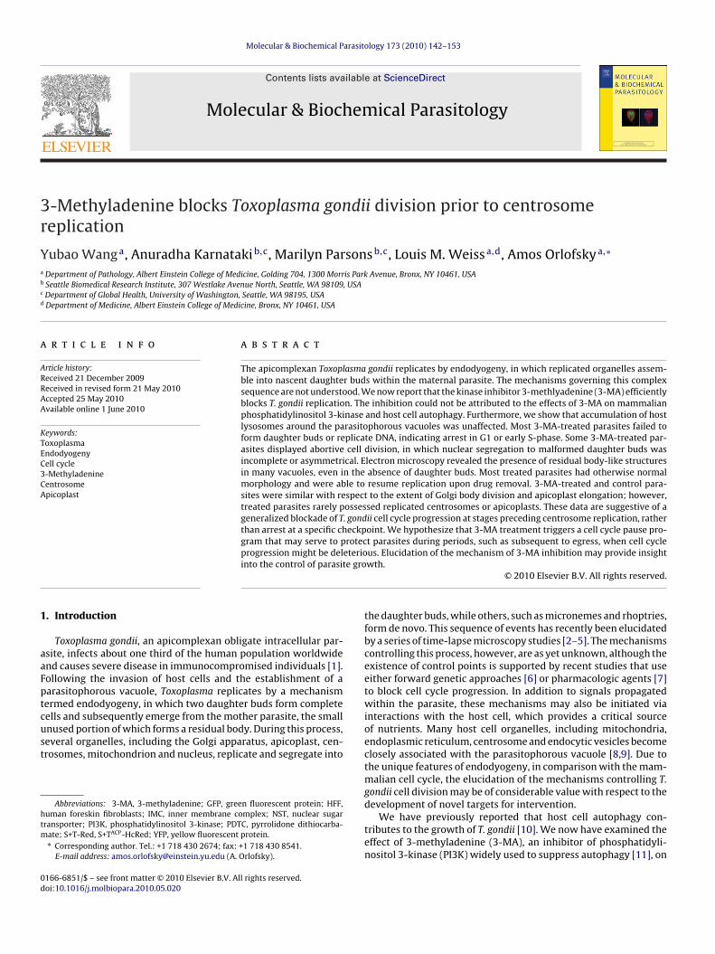
3-MA is a well-established inhibitor of macroautophagy inmammalian cells [11], and we have recently demonstrated thathost cell macroautophagy can be induced by T. gondii and enhancesparasite replication [10]. Therefore we considered whether theinhibitory effect of 3-MA on Toxoplasma proliferation might resultfrom its effect on host cell autophagy. Macroautophagy requiresthe presence of Atg5, which functions as part of a ubiquitin-likeconjugation system that results in the conversion of LC3 (Atg8)to a lipidated form (LC3-II) that associates with the developingautophagosome [21]. The extent of LC3 conversion is a widelyused indicator of autophagy, although this conversion can occureven during 3-MA blockade of autophagy [22]. We examined theeffect of 3-MA on T. gondii proliferation in the presence or absenceof host Atg5. As expected, Atg5−/− cells lacked LC3-II (Fig. 2A).However, Atg5 status had no effect on 3-MA inhibition of parasite
growth, demonstrating independence of this inhibition from hostcell autophagy (Fig. 2B). Similarly, we observed no alteration of 3-MA inhibition upon siRNA-mediated knockdown of Vps34, anotheressential component of the autophagic pathway [23] (Fig. 2Cand D).
Y. Wang et al. / Molecular & Biochemical Parasitology 173 (2010) 142–153 145
F were2 ousei olor in
3b
mbatteotw
ig. 3. 3-MA does not alter host cell lysosome distribution. HFF (A) or HeLa cells (B)0 h. Cells were fixed and labeled with anti-LAMP1 and Alexa488-conjugated anti-m
mages of the same field. Scale bar = 2 �m. (For interpretation of the references to c
.3. 3-MA does not affect the sequestration of host cell lysosomesy the parasitophorous vacuole
PI3Ks play an important role in endosomal trafficking in mam-alian cells [24]. In T. gondii-infected cells, host endolysosomes
ecome closely associated with the parasitophorous vacuole, andcquisition of these vesicles by the vacuole may play an impor-ant nutritive function for the parasite [25]. Since, in comparison
o LY294002 and wortmannin, 3-MA has additional effects on thendolysosomal system [26], it was possible that the effect of 3-MAn T. gondii proliferation was due to an inhibition of host lysosomerafficking to the vacuole. However, in both HFF and HeLa cells,e observed that the association of LAMP1-bearing vesicles withinfected with T. gondii for 4 h and then incubated with or without 10 mM 3-MA forIgG. Samples in (A) are stained with DAPI (blue). Insets in (B) display phase contrastthis figure legend, the reader is referred to the web version of the article.)
the parasitophorous vacuole was unaffected by 3-MA, even thoughparasite proliferation was efficiently inhibited (Fig. 3).
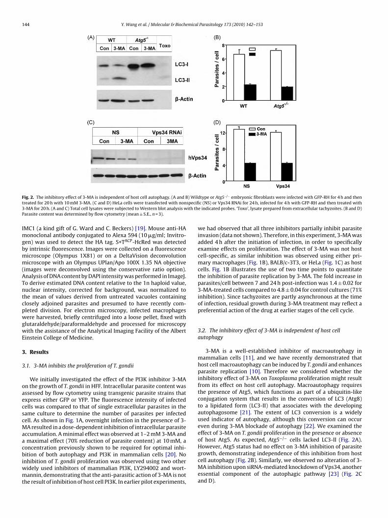
3.4. Morphology of 3-MA-treated parasites
The majority of T. gondii exposed to 3-MA retain normal size andshape by phase contrast microscopy (Fig. 3). To more definitivelydetermine the structural integrity of 3-MA-treated parasites, we
assessed electron micrographs of macrophages infected overnightin the presence or absence of the drug. 3-MA-treated vacuoles typ-ically contained only a single parasite, which displayed a normalorganization of organelles (Fig. 4A). Host mitochondria surroundingthe vacuole were considerably enlarged. Notably, many vacuoles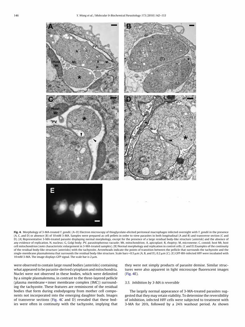
146 Y. Wang et al. / Molecular & Biochemical Parasitology 173 (2010) 142–153
Fig. 4. Morphology of 3-MA-treated T. gondii. (A–D) Electron microscopy of thioglycolate-elicited peritoneal macrophages infected overnight with T. gondii in the presence(A, C, and D) or absence (B) of 10 mM 3-MA. Samples were prepared as cell pellets in order to view parasites in both longitudinal (A and B) and transverse section (C andD). (A) Representative 3-MA-treated parasite displaying normal morphology, except for the presence of a large residual body-like structure (asterisk) and the absence ofany evidence of replication. N, nucleus; G, Golgi body; PV, parasitophorous vacuole; Mt, mitochondrion; A, apicoplast; R, rhoptry; M, microneme; C, conoid; host Mt, hostc Normo icate ts ale ba1
wwNb(ibnoi
ell mitochondrion (note characteristic enlargement in 3-MA-treated samples). (B)f the residual body-like structure (asterisks) with the tachyzoite. Arrowheads indingle-membrane plasmalemma that surrounds the residual body-like structure. Sc0 mM 3-MA. The image displays GFP signal. The scale bar is 2 �m.
ere observed to contain large round bodies (asterisks) containinghat appeared to be parasite-derived cytoplasm and mitochondria.uclei were not observed in these bodies, which were delimitedy a simple plasmalemma, in contrast to the three-layered pellicleplasma membrane + inner membrane complex (IMC)) surround-
ng the tachyzoite. These features are reminiscent of the residualodies that form during endodyogeny from mother cell compo-ents not incorporated into the emerging daughter buds. Imagesf transverse sections (Fig. 4C and D) revealed that these bod-es were often in continuity with the tachyzoite, implying thatal morphology and replication in control cells. (C and D) Examples of the continuityhe points of transition between the pellicle that surrounds the tachyzoite and thers = 0.5 �m (A, B, and D), 0.2 �m (C). (E) GFP-RH-infected HFF were incubated with
they were not simply products of parasite demise. Similar struc-tures were also apparent in light microscope fluorescent images(Fig. 4E).
3.5. Inhibition by 3-MA is reversible
The largely normal appearance of 3-MA-treated parasites sug-gested that they may retain viability. To determine the reversibilityof inhibition, infected HFF cells were subjected to treatment with3-MA for 20 h, followed by a 24 h washout period. As shown
mical
iwiob(sio
3f
aitr(boMiot
Fopabs
Y. Wang et al. / Molecular & Bioche
n Fig. 5A, vigorous parasite proliferation resumed during theashout period. This proliferation resulted in a 7.4-fold increase in
ntracellular parasite content, comparable to the 6.8-fold increasebserved in untreated cells during the first day of culture. The via-ility of 3-MA-treated parasites was confirmed by plaque assayFig. 5B). Parasitophorous vacuoles displayed a normal rosettetructure following inhibitor washout (Fig. 5C), further indicat-ng that for most vacuoles a complete reversal of inhibition wasbtained.
.6. 3-MA inhibits progression through S-phase and daughter budormation
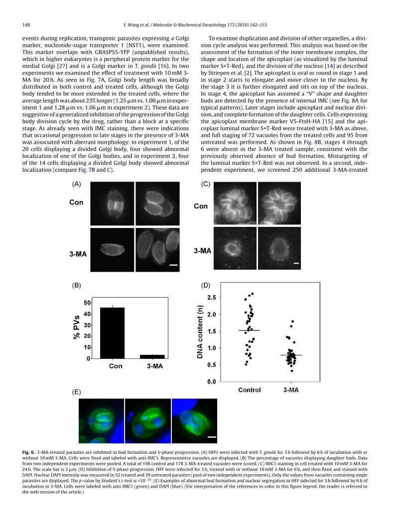
To locate the effect of 3-MA within the parasite cell cycle, wessessed daughter bud formation in 3-MA treated cells. To avoidnterference from secondary effects arising from prolonged drugreatment, the duration of treatment was limited to 6 h. Buds wereeadily detected in control cells by the presence of nascent IMCFig. 6A and B). In contrast, the frequency of budding was reducedy 95% in 3-MA-treated parasites (Fig. 6A and B) and few buds were
bserved even after 20 h of treatment (Fig. 6C). DAPI staining of 3-A-treated cells revealed an absence of nuclear growth or divisionn treated cells, suggesting an arrest either prior to or close to thenset of S-phase (data not shown). This was confirmed by quantita-ive analysis. In parasites treated with 3-MA for 6 h, the distribution
ig. 5. The inhibitory effect of 3-MA is reversible. (A) HFF cells were infected with YFP-Rr incubated in the presence or absence of 10 mM 3-MA for 20 h (day 1). Cells were thenresence or absence of 3-MA (day 2). Parasites per cell were determined by flow cytometnd incubated in the presence or absence of 10 mM 3-MA for 24 h. Culture was continuey fixation to determine plaque density (mean ± S.E. for replicate wells, n = 3). (C) YFP orcale bar is 5 �m.
Parasitology 173 (2010) 142–153 147
of DAPI intensity was markedly restricted compared with controlcells, consistent with an inability of treated parasites to progressthrough S-phase (Fig. 6D).
While most 3-MA-treated parasites were blocked near or priorto S-phase entry, a few parasites (<10%) displayed an abnormal pro-gression to a bud-forming stage (Fig. 6E). The buds in these parasiteswere characteristically asymmetrical and irregular: at least one ofthe buds typically had a pinched or otherwise deformed appear-ance. In some instances, only a single daughter bud was evident.Nuclear segregation was also highly irregular in these cells, and wascharacterized by asymmetric partitioning between the two buds,as well as the retention of considerable nuclear material by theposterior portion of the mother cell (residual body).
3.7. Early replicative events in 3-MA-inhibited T. gondii
During the T. gondii cell cycle, a number of parasite organelles,including the Golgi apparatus, the centrosome and the apicoplast,initiate replication in a series of clearly delineated events thatprecede the formation of daughter buds [2,3,5]. We therefore
investigated the effect of 3-MA on these early events usingtransgenic parasite strains expressing tagged Golgi or apicoplastproteins. Division of the Golgi body is one of the earliest organel-lar duplication events in T. gondii, occurring prior to division ofthe apicoplast [5]. To investigate the effects of 3-MA on earlyH for 4 h (multiplicity of infection = 4) and then either harvested for analysis (4 h)either harvested for analysis or else washed and incubated for a further 24 h in thery (mean ± S.E., n = 3). (B) HFF were infected for 4 h (multiplicity of infection = 0.01)d in the absence of 3-MA for a further 4 days (control) or 5 days (3-MA), followedphase contrast images of infected cells from the experiment shown in panel A. The
1 mical
emTwmeMdbaisbstw2lol
Fwf2Dpit
48 Y. Wang et al. / Molecular & Bioche
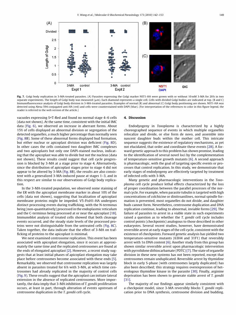
vents during replication, transgenic parasites expressing a Golgiarker, nucleotide-sugar transporter 1 (NST1), were examined.
his marker overlaps with GRASP55-YFP (unpublished results),hich in higher eukaryotes is a peripheral protein marker for theedial Golgi [27] and is a Golgi marker in T. gondii [16]. In two
xperiments we examined the effect of treatment with 10 mM 3-A for 20 h. As seen in Fig. 7A, Golgi body length was broadly
istributed in both control and treated cells, although the Golgiody tended to be more extended in the treated cells, where theverage length was about 23% longer (1.25 �m vs. 1.00 �m in exper-ment 1 and 1.28 �m vs. 1.06 �m in experiment 2). These data areuggestive of a generalized inhibition of the progression of the Golgiody division cycle by the drug, rather than a block at a specifictage. As already seen with IMC staining, there were indicationshat occasional progression to late stages in the presence of 3-MA
as associated with aberrant morphology: in experiment 1, of the0 cells displaying a divided Golgi body, four showed abnormalocalization of one of the Golgi bodies, and in experiment 2, fourf the 14 cells displaying a divided Golgi body showed abnormalocalization (compare Fig. 7B and C).
ig. 6. 3-MA-treated parasites are inhibited in bud formation and S-phase progression.ithout 10 mM 3-MA. Cells were fixed and labeled with anti-IMC1. Representative vacu
rom two independent experiments were pooled. A total of 198 control and 178 3-MA-tr4 h. The scale bar is 2 �m. (D) Inhibition of S-phase progression. HFF were infected forAPI. Nuclear DAPI intensity was measured in 52 treated and 39 untreated parasites (poolarasites are displayed. The p-value by Student’s t-test is <10−10. (E) Examples of abnorm
ncubation in 3-MA. Cells were labeled with anti-IMC1 (green) and DAPI (blue). (For intehe web version of the article.)
Parasitology 173 (2010) 142–153
To examine duplication and division of other organelles, a divi-sion cycle analysis was performed. This analysis was based on theassessment of the formation of the inner membrane complex, theshape and location of the apicoplast (as visualized by the luminalmarker S+T-Red), and the division of the nucleus [14] as describedby Striepen et al. [2]. The apicoplast is oval or round in stage 1 andin stage 2 starts to elongate and move closer to the nucleus. Bythe stage 3 it is further elongated and sits on top of the nucleus.In stage 4, the apicoplast has assumed a “V” shape and daughterbuds are detected by the presence of internal IMC (see Fig. 8A fortypical patterns). Later stages include apicoplast and nuclear divi-sion, and complete formation of the daughter cells. Cells expressingthe apicoplast membrane marker V5-FtsH-HA [15] and the api-coplast luminal marker S+T-Red were treated with 3-MA as above,and full staging of 72 vacuoles from the treated cells and 95 from
untreated was performed. As shown in Fig. 8B, stages 4 through6 were absent in the 3-MA treated sample, consistent with thepreviously observed absence of bud formation. Mistargeting ofthe luminal marker S+T-Red was not observed. In a second, inde-pendent experiment, we screened 250 additional 3-MA-treated(A) HFFs were infected with T. gondii for 3 h followed by 6 h of incubation with oroles are displayed. (B) The percentage of vacuoles displaying daughter buds. Dataeated vacuoles were scored. (C) IMC1 staining in cell treated with 10 mM 3-MA for3 h, treated with or without 10 mM 3-MA for 6 h, and then fixed and stained withof two independent experiments). Only the values from vacuoles containing singleal bud formation and nuclear segregation in HFF infected for 3 h followed by 6 h ofrpretation of the references to color in this figure legend, the reader is referred to
Y. Wang et al. / Molecular & Biochemical Parasitology 173 (2010) 142–153 149
Fig. 7. Golgi body replication in 3-MA-treated parasites. (A) Parasites expressing the Golgi marker NST1-HA were grown with or without 10 mM 3-MA for 20 h in twos d reprI mpled with Dr
v(d1d(bIainssattt
tcmdbaIetTfi
amtgpRat(etoc
eparate experiments. The length of Golgi body was measured (�m). Each diamonmmunofluorescence analysis of Golgi body division in 3-MA-treated parasites. Exaetected using Alexa 594-conjugated anti-HA (red) and cells were counterstainedeader is referred to the web version of the article.)
acuoles expressing S+T-Red and found no normal stage 4–6 cellsdata not shown). At the same time, consistent with the initial IMCata (Fig. 6), we observed an increase in aberrant forms. About5% of cells displayed an abnormal division or segregation of theetected organelles, a much higher percentage than normally seenFig. 8B). Some of these abnormal forms displayed bud formation,ut either nuclear or apicoplast division was deficient (Fig. 8D).n other cases the cells contained two daughter IMC complexesnd two apicoplasts but only one DAPI-stained nucleus, indicat-ng that the apicoplast was able to divide but not the nucleus (dataot shown). These results could suggest that cell cycle progres-ion is blocked by 3-MA at a stage prior to stage 4. Alternatively,ince the distribution of apicoplast stages prior to stage 4 did notppear to be altered by 3-MA (Fig. 8B), the results are also consis-ent with a generalized 3-MA-induced pause at stages 1–3, and inhis respect are similar to our observations of Golgi body replica-ion.
In the 3-MA-treated population, we observed some staining ofhe ER with the apicoplast membrane marker in about 10% of theells (data not shown), suggesting that localization of apicoplastembrane proteins might be impeded. V5-FtsH1-HA undergoes
istinct processing events during trafficking, with the N-terminuseing (non-quantitatively) processed in the endoplasmic reticulumnd the C-terminus being processed at or near the apicoplast [18].mmunoblot analysis of treated cells showed that both cleavagevents occurred, and the steady state levels of the processed pro-eins were not distinguishable from the untreated cells (Fig. 8C).aken together, the data indicate that the effect of 3-MA on traf-cking of proteins to the apicoplast is minimal.
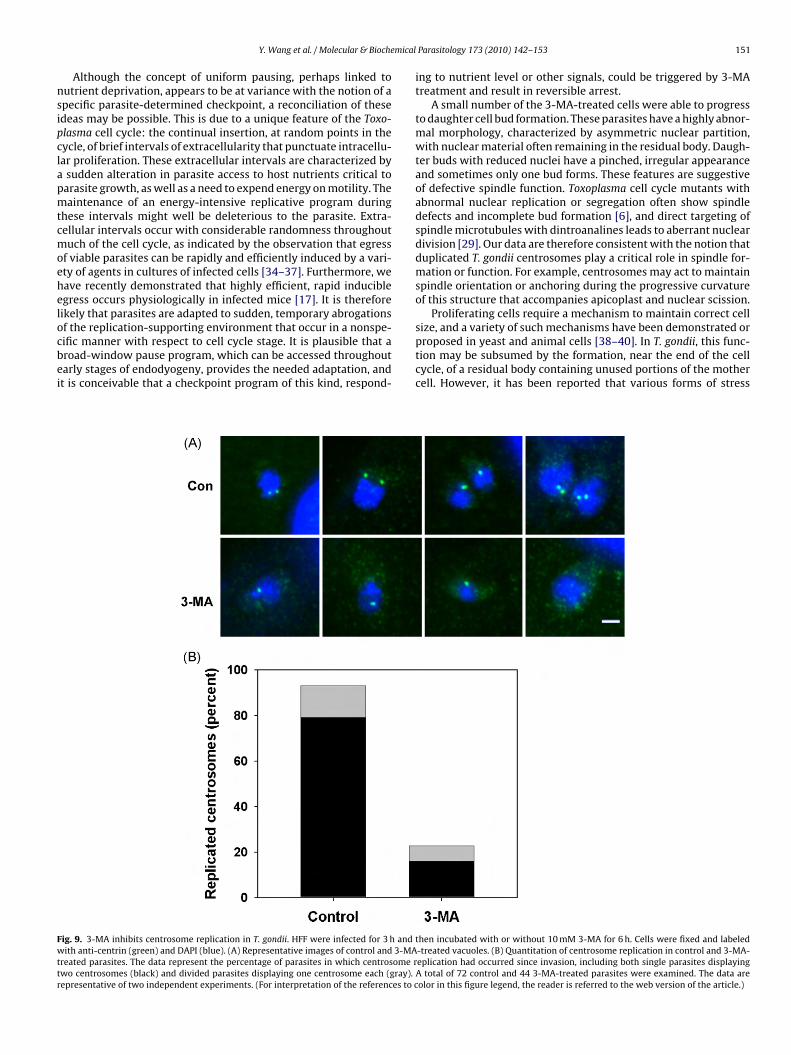
We next examined centrosome replication. This event has beenssociated with apicoplast elongation, since it occurs at approxi-ately the same time and the replicated centrosomes are found at
he ends of elongated apicoplast [2]. However, a recent study sug-ests that at least initial phases of apicoplast elongation may takelace before centrosomes become associated with these ends [5].emarkably, we observed that centrosome replication was largelybsent in parasites treated for 6 h with 3-MA, at which time cen-rosomes had already replicated in the majority of control cells
Fig. 9). These results suggest that the apicoplast can initiate lateralxtension in the absence of replicated centrosomes. More impor-antly, the data imply that 3-MA inhibition of T. gondii proliferationccurs, at least in part, through alteration of events upstream ofentrosome duplication in the T. gondii cell cycle.esents a single cell. Cells with divided Golgi bodies are indicated at top. (B and C)s of normal (B) and abnormal (C) Golgi body positioning are shown. NST1-HA was
API (blue). (For interpretation of the references to color in this figure legend, the
4. Discussion
Endodyogeny in Toxoplasma is characterized by a highlychoreographed sequence of events in which multiple organellesrelocalize and divide, or else form de novo, and assemble intonascent daughter buds within the mother cell. This intricatesequence suggests the existence of regulatory mechanisms, as yetnot elucidated, that order and coordinate these events [28]. A for-ward genetic approach to this problem has shown promise, leadingto the identification of several novel loci by the complementationof temperature-sensitive growth mutants [6]. A second approachis pharmacologic, with the goal of targeting specific events or pro-cesses that control replication. In this study, we demonstrate thatearly stages of endodyogeny are effectively targeted by treatmentof infected cells with 3-MA.
Many genetic and pharmacologic interventions in the Toxo-plasma cell cycle produce lethal effects characterized by the lossof proper coordination between the parallel processes of the nor-mal cycle. For example, when parasite tubulin is targeted with highconcentrations of colchicine or dinitroanilines, normal spindle for-mation is prevented, most organelles do not divide, and daughterbuds cannot form. Nevertheless, centrosome duplication and DNAreplication continue, leading to abnormal, inviable forms [29]. Thefailure of parasites to arrest in a viable state in such experimentsraised a question as to whether the T. gondii cell cycle includescontrol points (checkpoints) analogous to those described in othereukaryotes. Several recent studies, however, have demonstratedreversible arrest at early stages of the cell cycle, consistent with theexistence of checkpoints. Forward genetic analysis has yielded twotemperature-sensitive mutants (63H4 and 31F1) that reversiblyarrest with 1n DNA content [6]. Another study from this group hasshown similar reversible arrest upon pharmacologic interventionwith pyrrolidone dithiocarbamate (PDTC) [7]. The state of organelledivision in these new systems has not been reported, except thatcentrosomes remain unduplicated. Reversible arrest by thymidineblock in early S-phase (with centrosomes largely duplicated) hasalso been described; this strategy requires overexpression of het-erologous thymidine kinase in the parasite [30]. Finally, arginine
deprivation has been shown to generate stable arrest of T. gondii[31].The majority of our findings appear similarly consistent witha checkpoint model, since 3-MA reversibly blocks T. gondii repli-cation prior to DNA synthesis, centrosome division and budding.

1 mical
Hasowb31setwri[
FwcV3v
50 Y. Wang et al. / Molecular & Bioche
owever, this model predicts that 3-MA-treated parasites shouldccumulate at a single stage in the cell cycle. We did not observeuch an accumulation, with respect to either apicoplast progressionr Golgi body replication, even after 20 h of 3-MA treatment. Whilee cannot rule out that a partial accumulation might be revealed
y further analysis, the simplest interpretation of the results is that-MA induces a uniform pausing of cell cycle progression in stage–3 parasites. In the case of the Golgi apparatus, a slow progres-ion may still take place, as indicated by the increased mean lateralxtension of this organelle in drug-treated cells. It will be of interesto compare this phenotype to that of the reversible arrest obtainedith PDTC or the 63H4 and 31F1 mutants. Arrest with PDTC was
eported to result in parasite synchronization upon release, imply-ng that the blockade occurred within a narrow cell cycle window7].
ig. 8. Apicoplast replication in 3-MA-treated parasites. Parasites expressing the apicopere treated with 10 mM 3-MA for 20 h. Vacuoles were stained with anti-V5 (green), an
ells at stages 3 and 4. (B) Vacuole stage distribution in untreated and 3-MA-treated cel5-FtsH-HA. Full-length protein (FL) as well as N-terminally processed (NP) and C-termin-MA-treated parasites, showing defective duplication of apicoplasts. (For interpretationersion of the article.)
Parasitology 173 (2010) 142–153
How might uniform pausing occur in stages 1–3? Several of thereplicative events examined, including apicoplast progression andcentrosome division, involve organellar motility (the centrosomeis reported to migrate caudally prior to division [5]). Therefore it ispossible that a disruption by 3-MA of signaling required for suchmotility could affect multiple early events. Effects on organellarlocalization might be related to previously observed effects of 3-MA on vesicle trafficking [26]. Alternatively, 3-MA might act byinterfering with parasite acquisition of energy or nutrients. Anenergy-poor state of the 3-MA-treated parasite, perhaps the resultof defective acquisition of energy-rich nutrients, is suggested bythe prominence of enlarged host mitochondria adjoining the para-
sitophorous vacuole. Enlargement of mitochondria may representa compensatory mechanism responding to local ATP depletion[32,33].last membrane marker V5-FtsH1-HA and the apicoplast luminal marker, S+T-Redti-IMC1 (red) and DAPI (blue) and staged for cell cycle analysis. (A) Representativels (95 and 72 vacuoles, respectively). (C) Immunoblot analysis of cells expressingally processed (CP) cleavage products were detected. (D) Representative abnormalof the references to color in this figure legend, the reader is referred to the web
mical
nsipclapmtcmoehelocbei
Fwttr
Y. Wang et al. / Molecular & Bioche
Although the concept of uniform pausing, perhaps linked toutrient deprivation, appears to be at variance with the notion of apecific parasite-determined checkpoint, a reconciliation of thesedeas may be possible. This is due to a unique feature of the Toxo-lasma cell cycle: the continual insertion, at random points in theycle, of brief intervals of extracellularity that punctuate intracellu-ar proliferation. These extracellular intervals are characterized by
sudden alteration in parasite access to host nutrients critical toarasite growth, as well as a need to expend energy on motility. Theaintenance of an energy-intensive replicative program during
hese intervals might well be deleterious to the parasite. Extra-ellular intervals occur with considerable randomness throughoutuch of the cell cycle, as indicated by the observation that egress
f viable parasites can be rapidly and efficiently induced by a vari-ty of agents in cultures of infected cells [34–37]. Furthermore, weave recently demonstrated that highly efficient, rapid induciblegress occurs physiologically in infected mice [17]. It is thereforeikely that parasites are adapted to sudden, temporary abrogationsf the replication-supporting environment that occur in a nonspe-
ific manner with respect to cell cycle stage. It is plausible that aroad-window pause program, which can be accessed throughoutarly stages of endodyogeny, provides the needed adaptation, andt is conceivable that a checkpoint program of this kind, respond-ig. 9. 3-MA inhibits centrosome replication in T. gondii. HFF were infected for 3 h andith anti-centrin (green) and DAPI (blue). (A) Representative images of control and 3-MA
reated parasites. The data represent the percentage of parasites in which centrosome rwo centrosomes (black) and divided parasites displaying one centrosome each (gray).epresentative of two independent experiments. (For interpretation of the references to c
Parasitology 173 (2010) 142–153 151
ing to nutrient level or other signals, could be triggered by 3-MAtreatment and result in reversible arrest.
A small number of the 3-MA-treated cells were able to progressto daughter cell bud formation. These parasites have a highly abnor-mal morphology, characterized by asymmetric nuclear partition,with nuclear material often remaining in the residual body. Daugh-ter buds with reduced nuclei have a pinched, irregular appearanceand sometimes only one bud forms. These features are suggestiveof defective spindle function. Toxoplasma cell cycle mutants withabnormal nuclear replication or segregation often show spindledefects and incomplete bud formation [6], and direct targeting ofspindle microtubules with dintroanalines leads to aberrant nucleardivision [29]. Our data are therefore consistent with the notion thatduplicated T. gondii centrosomes play a critical role in spindle for-mation or function. For example, centrosomes may act to maintainspindle orientation or anchoring during the progressive curvatureof this structure that accompanies apicoplast and nuclear scission.
Proliferating cells require a mechanism to maintain correct cellsize, and a variety of such mechanisms have been demonstrated or
proposed in yeast and animal cells [38–40]. In T. gondii, this func-tion may be subsumed by the formation, near the end of the cellcycle, of a residual body containing unused portions of the mothercell. However, it has been reported that various forms of stressthen incubated with or without 10 mM 3-MA for 6 h. Cells were fixed and labeled-treated vacuoles. (B) Quantitation of centrosome replication in control and 3-MA-eplication had occurred since invasion, including both single parasites displayingA total of 72 control and 44 3-MA-treated parasites were examined. The data areolor in this figure legend, the reader is referred to the web version of the article.)

1 mical
d[at3sbbtrefrnpbdbta
oatticlpfasargattos
pcatpf
A
M((aBYsAt
R
[
[
[
[
[
[
[
[
[
[
[
[
[
[
[
[
[
[
[
[
[
[
[
52 Y. Wang et al. / Molecular & Bioche
uring endodyogeny can lead to expansion of this compartment5]. Remarkably, 3-MA-treated parasites, even in the absence ofny bud formation, often display a large appendage with proper-ies of a residual body. A possible explanation of this finding is that-MA treatment, while effectively inhibiting bud formation, centro-ome duplication and other replicative events, may not completelylock growth of the parasite. The parasite may continue to acquireiomass at a reduced rate during drug treatment, and then jettisonhis excess material by activating the pathway that generates theesidual body. The observation that PDTC-treated parasites gen-rate a large amount of ‘cytoplasmic discard’ shortly after releaserom the block [7] may reflect a similar phenomenon of volumeegulation. This notion is compatible with the hypothesis of a ‘hostutrient’ checkpoint advanced earlier; under this hypothesis, thearasite pauses organelle replication under unfavorable conditions,ut may not actively arrest general growth processes, which williminish naturally in the energy-poor extracellular milieu. It wille of interest to examine the modulation of parasite anabolic func-ion in the context of treatments or mutations that lead to cell cyclerrest.
In mammalian cells, 3-MA has been characterized as an inhibitorf PI3K, acting via the competitive inhibition of ATP binding [20],nd as a negative regulator of autophagy. We consider it unlikelyhat these effects account for the inhibition of parasite replica-ion, since the blockade is not replicated with other known PI3Knhibitors, is independent of the state of autophagy in the hostell and is not correlated with any alteration of host endolysosomeocalization to the PV. It is possible that 3-MA affects another hostrocess, or a parasite-encoded kinase, such as a PI3K homolog. Theamily of PI3K-related kinases includes target-of-rapamycin (Tor),central regulator of cell growth that has been shown to be sen-
itive to PI3K inhibitors, such as LY294002, that act (like 3-MA)s competitive inhibitors of the ATP binding site [41,42]. Proteinselated to both PI3K and mTOR are predicted from the T. gondiienome sequence. Inhibition of a PI3K-like kinase might lead tolterations in parasite vesicular trafficking, as observed in 3-MA-reated mammalian cells [26]. Finally, the drug may target a kinasehat participates in centrosome duplication. Evidence from otherrganisms suggests the involvement of multiple kinases in centro-ome duplication and separation [43].
In summary, this study identifies 3-methyladenine as a newharmacologic tool for the reversible blockade of T. gondii repli-ation. The findings are suggestive of a novel pause mechanismffecting multiple early stages of the cell cycle. The elucidation ofhe mechanism of the 3-MA blockade should provide insight intoathways governing the parasite cell cycle and identify new targetsor intervention in tachyzoite expansion.
cknowledgments
We are grateful to Drs. Michael Lenardo (NIH), Noboruizushima (Tokyo Medical and Dental University), Jeffrey Salisbury
Mayo Clinic), Gary Ward (University of Vermont), and Con BeckersUniversity of North Carolina) for the generous gifts of reagents. Weppreciate the discussion with Drs. Ana Maria Cuervo and Jonathanacker at the Albert Einstein College of Medicine. We also thank Ms.anfen Ma for her technical assistance. This study was financiallyupported by the National Institutes of Health grants (AI-55358 toO and R01 AI50506 to MP. The authors are solely responsible for
he content.
eferences
[1] Hall S, Ryan KA, Buxton D. The epidemiology of Toxoplasma infection. In: Joyn-son DH, Wreghitt TJ, editors. Toxoplasmosis: a comprehensive clinical guide.Cambridge: Cambridge University Press; 2001. p. 58–124.
[
[
Parasitology 173 (2010) 142–153
[2] Striepen B, Crawford MJ, Shaw MK, et al. The plastid of Toxoplasma gondii isdivided by association with the centrosomes. J Cell Biol 2000;151:1423–34.
[3] Hartmann J, Hu K, He CY, et al. Golgi and centrosome cycles in Toxoplasmagondii. Mol Biochem Parasit 2006;145:125–7.
[4] Hu K, Johnson J, Florens L, et al. Cytoskeletal components of an inva-sion machine—the apical complex of Toxoplasma gondii. PLoS Pathog2006;2:121–38.
[5] Nishi M, Hu K, Murray JM, et al. Organellar dynamics during the cell cycle ofToxoplasma gondii. J Cell Sci 2008;121:1559–68.
[6] Gubbels MJ, Lehmann M, Muthalagi M, et al. Forward genetic analysis of theapicomplexan cell division cycle in Toxoplasma gondii. PLoS Pathog 2008:4.
[7] de Felipe MMC, Lehmann MM, Jerome ME, et al. Inhibition of Toxoplasma gondiigrowth by pyrrolidine dithiocarbamate is cell cycle specific and leads to popu-lation synchronization. Mol Biochem Parasit 2008;157:22–31.
[8] Coppens I, Dunn JD, Romano JD, et al. Toxoplasma gondii sequesters lysosomesfrom mammalian hosts in the vacuolar space. Cell 2006;125:261–74.
[9] Sinai AP, Webster P, Joiner KA. Association of host cell endoplasmic reticu-lum and mitochondria with the Toxoplasma gondii parasitophorous vacuolemembrane: a high affinity interaction. J Cell Sci 1997;110:2117–28.
10] Wang YB, Weiss LM, Orlofsky A. Host cell autophagy is induced by Toxo-plasma gondii and contributes to parasite growth. J Biol Chem 2009;284:1694–701.
11] Seglen PO, Gordon PB. 3-Methyladenine: specific inhibitor ofautophagic/lysosomal protein degradation in isolated rat hepatocytes.Proc Natl Acad Sci USA 1982;79:1889–92.
12] Ma YF, Zhang YW, Kim K, et al. Identification and characterisation of a reg-ulatory region in the Toxoplasma gondii hsp70 genomic locus. Int J Parasitol2004;34:333–46.
13] Gubbels MJ, Li C, Striepen B. High-throughput growth assay for Toxo-plasma gondii using yellow fluorescent protein. Antimicrob Agents Chemother2003;47:309–16.
14] Karnataki A, Derocher A, Coppens I, et al. Cell cycle-regulated vesicular traffick-ing of Toxoplasma APT1, a protein localized to multiple apicoplast membranes.Mol Microbiol 2007;63:1653–68.
15] Karnataki A, Derocher AE, Coppens I, et al. A membrane protease is targetedto the relict plastid of Toxoplasma via an internal signal sequence. Traffic2007;8:1543–53.
16] Pelletier L, Stern CA, Pypaert M, et al. Golgi biogenesis in Toxoplasma gondii.Nature 2002;418:548–52.
17] Tomita T, Yamada T, Weiss LM, et al. Externally triggered egress is the majorfate of Toxoplasma gondii during acute infection. J Immunol 2009;183:6667–80.
18] Karnataki A, Derocher AE, Feagin JE, et al. Sequential processing of the Toxo-plasma apicoplast membrane protein FtsH1 in topologically distinct domainsduring intracellular trafficking. Mol Biochem Parasit 2009;166:126–33.
19] Mann T, Beckers C. Characterization of the subpellicular network, a filamentousmembrane skeletal component in the parasite Toxoplasma gondii. Mol BiochemParasit 2001;115:257–68.
20] Blommaart EFC, Krause U, Schellens JPM, et al. The phosphatidylinositol 3-kinase inhibitors wortmannin and LY294002 inhibit autophagy in isolated rathepatocytes. Eur J Biochem 1997;243:240–6.
21] Hanada T, Noda NN, Satomi Y, et al. The Atg12-Atg5 conjugate has anovel E3-like activity for protein lipidation in autophagy. J Biol Chem2007;282:37298–302.
22] Gimenez-Xavier P, Francisco R, Platini F, et al. LC3-I conversion to LC3-II doesnot necessarily result in complete autophagy. Int J Mol Med 2008;22:781–5.
23] Backer JM. The regulation and function of Class III PI3Ks: novel roles for Vps34.Biochem J 2008;410:1–17.
24] Lindmo K, Stenmark H. Regulation of membrane traffic by phosphoinositide3-kinases. J Cell Sci 2006;119:605–14.
25] Coppens I, Dunn JD, Romano JD, et al. Toxoplasma gondii sequesters lysosomesfrom mammalian hosts in the vacuolar space. Cell 2006;125(2):261–74.
26] Hirosako K, Imasato H, Hirota Y, et al. 3-Methyladenine specifically inhibitsretrograde transport of cation-independent mannose 6-phosphate/insulin-likegrowth factor II receptor from the early endosome to the TGN. Biochem BiophysRes Commun 2004;316:845–52.
27] Shorter J, Watson R, Giannakou ME, et al. GRASP55, a second mammalian GRASPprotein involved in the stacking of Golgi cisternae in a cell-free system. EMBOJ 1999;18:4949–60.
28] Gubbels MJ, White M, Szatanek T. The cell cycle and Toxoplasma gondii celldivision: tightly knit or loosely stitched? Int J Parasitol 2008;38:1343–58.
29] Morrissette NS, Sibley LD. Disruption of microtubules uncouples budding andnuclear division in Toxoplasma gondii. J Cell Sci 2002;115:1017–25.
30] Radke JR, White MW. A cell cycle model for the tachyzoite of Toxoplasmagondii using the Herpes simplex virus thymidine kinase. Mol Biochem Parasit1998;94:237–47.
31] Fox BA, Gigley JP, Bzik DJ. Toxoplasma gondii lacks the enzymes required for denovo arginine biosynthesis and arginine starvation triggers cyst formation. IntJ Parasitol 2004;34:323–31.
32] Bertoni-Freddari C, Mocchegiani E, Malavolta M, et al. Synaptic and mitochon-drial physiopathologic changes in the aging nervous system and the role of zinc
ion homeostasis. Mech Ageing Dev 2006;127:590–6.33] Bertonifreddari C, Fattoretti P, Casoli T, et al. Morphological plasticity of synap-tic mitochondria during aging. Brain Res 1993;628:193–200.
34] Stommel EW, Ely KH, Schwartzman JD, et al. Toxoplasma gondii: dithiol-inducedCa2+ flux causes egress of parasites from the parasitophorous vacuole. ExpParasitol 1997;87:88–97.

mical
[
[
[
[
[
[
Y. Wang et al. / Molecular & Bioche
35] Persson EK, Agnarson AM, Lambert H, et al. Death receptor ligation or exposureto perforin trigger rapid egress of the intracellular parasite Toxoplasma gondii.J Immunol 2007;179:8357–65.
36] Moudy R, Manning TJ, Beckers CJ. The loss of cytoplasmic potassium upon
host cell breakdown triggers egress of Toxoplasma gondii. J Biol Chem2001;276:41492–501.37] Arrizabalaga G, Ruiz F, Moreno S, et al. Ionophore-resistant mutant of Toxo-plasma gondii reveals involvement of a sodium/hydrogen exchanger in calciumregulation. J Cell Biol 2004;165:653–62.
38] Umen JG. The elusive sizer. Curr Opin Cell Biol 2005;17:435–41.
[
[
[
Parasitology 173 (2010) 142–153 153
39] Kellogg DR. Wee1-dependent mechanisms required for coordination of cellgrowth and cell division. J Cell Sci 2003;116:4883–90.
40] Grebien F, Dolznig H, Beug H, et al. Cell size control—new evidence for a generalmechanism. Cell Cycle 2005;4:418–21.
41] Vlahos CJ, Matter WF, Brown RF. A specific inhibitor of phosphatidylinositol
3-kinase. J Cell Biochem 1994:274.42] Walker EH, Pacold ME, Perisic O, et al. Structural determinants of phospho-inositide 3-kinase inhibition by wortmannin, LY294002, quercetin, myricetin,and staurosporine. Mol Cell 2000;6:909–19.
43] Azimzadeh J, Bornens M. Structure and duplication of the centrosome. J Cell Sci2007;120:2139–42.