Biologic and genetic characteristics of Toxoplasma gondii isolates in free-range chickens from...
7
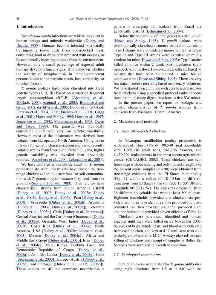
Biologic and genetic characteristics of Toxoplasma gondii isolates in free-range chickens from Nicaragua, Central America J.P. Dubey a, * , N. Sundar a , N. Pineda b , N.C. Kyvsgaard c , L.A. Luna b,d , E. Rimbaud b , J.B. Oliveira e , O.C.H. Kwok a , Y. Qi f , C. Su f a United States Department of Agriculture, Agricultural Research Service, Animal and Natural Resources Institute, Animal Parasitic Diseases Laboratory, Building 1001, Beltsville, MD 20705-2350, USA b Facultad de Ciencias Agrarias, Universidad de Ciencias Comerciales (UCC), Managua, Nicaragua c Department of Veterinary Pathobiology, The Royal Veterinary and Agricultural University, Stigbojlen 4, 1870, Frederiksberg C, Denmark d Centro de Investigacio ´n de Ganaderia Campesina (CIGAC), El Sauce – Leo ´n, Nicaragua e Laboratorio de Parasitologı ´a, Escuela de Medicina Veterinaria, Universidad Nacional, Apodo 86-3000, Heredia, Costa Rica f Department of Microbiology, The University of Tennessee, Knoxville, TN 37996-0845, USA Received 19 April 2006; received in revised form 15 June 2006; accepted 18 June 2006 Abstract The prevalence of Toxoplasma gondii in free-ranging chickens is a good indicator of the prevalence of T. gondii oocysts in the soil because chickens feed from the ground. The prevalence of T. gondii in 98 free-range chickens (Gallus domesticus) from Nicragua was determined. Antibodies to T. gondii were assayed by the modified agglutination test (MAT), and found in 84 (85.7%) of 98 chickens with titers of 1:5 in 10, 1:10 in eight, 1:20 in seven, 1:40 in nine, 1:80 in 11, 1:160 in one, 1:200 in 27, 1:400 in six, 1:800 four, and 1:3200 in one bird. Hearts and brains of 32 chickens with titers of 1:10 or less were pooled and fed to three T. gondii- free cats. Hearts and brains of 66 chickens with titers of 1:20 or higher were bioassayed in mice. Feces of cats were examined for oocysts. The cat fed tissues from eight chickens with titers of 1:10 shed T. gondii oocysts. The two cats fed tissues of 24 chickens with titers of 1:5 or less did not shed oocysts. T. gondii was isolated by bioassay in mice from 47 chickens with MAT titers of 1:20 or higher. All infected mice from six isolates died of toxoplasmosis. Overall, 41 of 170 (24.1%) mice that became infected after inoculation with chicken tissues died of toxoplasmosis. Genotyping of these 48 isolates (47 from mice and 1 from pooled tissues) using polymorphisms at the loci SAG1, SAG2, SAG3, BTUB and GRA6 revealed eight genotypes. Six isolates had Type I alleles, three isolate had Type II alleles and six isolates had Type III alleles at all loci. Four isolates had mixed infections. Two isolates have a unique allele at SAG1 locus and combination of I and III alleles at other loci. The rest 27 isolates contained the combination of Type I and III alleles and were divided into four genotypes. More than one genotypes were often isolated in chickens from the same household, indicating multiple genotypes were circulating in the same environment. This may explain the high frequency of mixed infections observed. High rate of mixed infection in intermediate hosts such as chickens may facilitate genetic exchange between different parasite lineages in definitive feline hosts. This is the first report of genetic characterization of T. gondii isolates from Nicragua, Central America. Published by Elsevier B.V. Keywords: Toxoplasma gondii; Chickens; Gallus domesticus; Free-range; Nicragua; Central America; Genotype www.elsevier.com/locate/vetpar Veterinary Parasitology 142 (2006) 47–53 * Corresponding author. Tel.: +1 301 504 8128; fax: +1 301 504 9222. E-mail address: [email protected] (J.P. Dubey). 0304-4017/$ – see front matter. Published by Elsevier B.V. doi:10.1016/j.vetpar.2006.06.016
-
Upload
independent -
Category
Documents
-
view
0 -
download
0
Transcript of Biologic and genetic characteristics of Toxoplasma gondii isolates in free-range chickens from...
www.elsevier.com/locate/vetpar
Veterinary Parasitology 142 (2006) 47–53
Biologic and genetic characteristics of Toxoplasma gondii
isolates in free-range chickens from Nicaragua, Central America
J.P. Dubey a,*, N. Sundar a, N. Pineda b, N.C. Kyvsgaard c, L.A. Luna b,d,E. Rimbaud b, J.B. Oliveira e, O.C.H. Kwok a, Y. Qi f, C. Su f
a United States Department of Agriculture, Agricultural Research Service, Animal and Natural Resources Institute,
Animal Parasitic Diseases Laboratory, Building 1001, Beltsville, MD 20705-2350, USAb Facultad de Ciencias Agrarias, Universidad de Ciencias Comerciales (UCC), Managua, Nicaragua
c Department of Veterinary Pathobiology, The Royal Veterinary and Agricultural University, Stigbojlen 4, 1870,
Frederiksberg C, Denmarkd Centro de Investigacion de Ganaderia Campesina (CIGAC), El Sauce – Leon, Nicaraguae Laboratorio de Parasitologıa, Escuela de Medicina Veterinaria, Universidad Nacional,
Apodo 86-3000, Heredia, Costa Ricaf Department of Microbiology, The University of Tennessee, Knoxville, TN 37996-0845, USA
Received 19 April 2006; received in revised form 15 June 2006; accepted 18 June 2006
Abstract
The prevalence of Toxoplasma gondii in free-ranging chickens is a good indicator of the prevalence of T. gondii oocysts in the
soil because chickens feed from the ground. The prevalence of T. gondii in 98 free-range chickens (Gallus domesticus) from
Nicragua was determined. Antibodies to T. gondii were assayed by the modified agglutination test (MAT), and found in 84 (85.7%)
of 98 chickens with titers of 1:5 in 10, 1:10 in eight, 1:20 in seven, 1:40 in nine, 1:80 in 11, 1:160 in one, 1:200 in 27, 1:400 in six,
1:800 four, and 1:3200 in one bird. Hearts and brains of 32 chickens with titers of 1:10 or less were pooled and fed to three T. gondii-
free cats. Hearts and brains of 66 chickens with titers of 1:20 or higher were bioassayed in mice. Feces of cats were examined for
oocysts. The cat fed tissues from eight chickens with titers of 1:10 shed T. gondii oocysts. The two cats fed tissues of 24 chickens
with titers of 1:5 or less did not shed oocysts. T. gondii was isolated by bioassay in mice from 47 chickens with MAT titers of 1:20 or
higher. All infected mice from six isolates died of toxoplasmosis. Overall, 41 of 170 (24.1%) mice that became infected after
inoculation with chicken tissues died of toxoplasmosis. Genotyping of these 48 isolates (47 from mice and 1 from pooled tissues)
using polymorphisms at the loci SAG1, SAG2, SAG3, BTUB and GRA6 revealed eight genotypes. Six isolates had Type I alleles,
three isolate had Type II alleles and six isolates had Type III alleles at all loci. Four isolates had mixed infections. Two isolates have a
unique allele at SAG1 locus and combination of I and III alleles at other loci. The rest 27 isolates contained the combination of Type
I and III alleles and were divided into four genotypes. More than one genotypes were often isolated in chickens from the same
household, indicating multiple genotypes were circulating in the same environment. This may explain the high frequency of mixed
infections observed. High rate of mixed infection in intermediate hosts such as chickens may facilitate genetic exchange between
different parasite lineages in definitive feline hosts. This is the first report of genetic characterization of T. gondii isolates from
Nicragua, Central America.
Published by Elsevier B.V.
Keywords: Toxoplasma gondii; Chickens; Gallus domesticus; Free-range; Nicragua; Central America; Genotype
* Corresponding author. Tel.: +1 301 504 8128; fax: +1 301 504 9222.
E-mail address: [email protected] (J.P. Dubey).
0304-4017/$ – see front matter. Published by Elsevier B.V.
doi:10.1016/j.vetpar.2006.06.016
J.P. Dubey et al. / Veterinary Parasitology 142 (2006) 47–5348
1. Introduction
Toxoplasma gondii infections are widely prevalent in
human beings and animals worldwide (Dubey and
Beattie, 1988). Humans become infected post-natally
by ingesting tissue cysts from undercooked meat,
consuming food or drink contaminated with oocysts, or
by accidentally ingesting oocysts from the environment.
However, only a small percentage of exposed adult
humans develop clinical signs. It is unknown whether
the severity of toxoplasmosis in immunocompetent
persons is due to the parasite strain, host variability, or
to other factors.
T. gondii isolates have been classified into three
genetic types (I, II, III) based on restriction fragment
length polymorphism (RFLP) (Ajzenberg et al.,
2002a,b, 2004; Aspinall et al., 2003; Boothroyd and
Grigg, 2002; da Silva et al., 2005; Dubey et al., 2004a,d;
Ferreira et al., 2004, 2006; Fuentes et al., 2001; Grigg
et al., 2001; Howe and Sibley, 1995; Howe et al., 1997;
Jungersen et al., 2002; Mondragon et al., 1998; Owen
and Trees, 1999). The parasite was previously
considered clonal with very low genetic variability.
However, most of the information was derived from
isolates from Europe and North America. Using newer
markers for genetic characterization and using recently
isolated strains from Brazil and French Guyana, higher
genetic variability was revealed than previously
reported (Ajzenberg et al., 2004; Lehmann et al., 2004).
We have initiated a worldwide study of T. gondii
population structure. For this we have chosen the free-
range chicken as the indicator host for soil contamina-
tion with T. gondii oocysts because they feed from the
ground (Ruiz and Frenkel, 1980). Thus far, we have
characterized strains from South America (Brazil
[Dubey et al., 2002; Dubey et al., 2003a; Dubey
et al., 2003d; Dubey et al., 2006a], Peru [Dubey et al.,
2004b], Venezuela [Dubey et al., 2005h], Argentina
[Dubey et al., 2003e; Dubey et al., 2005f]), Colombia
[Dubey et al., 2005d], Chile [Dubey et al., in press-a];
Central America and the Caribbean (Guatemala [Dubey
et al., 2005e], Grenada, West Indies [Dubey et al.,
2005b], Costa Rica [Dubey et al., 2006c), North
America (USA [Dubey et al., 2003c; Lehmann et al.,
2003], Mexico [Dubey et al., 2004c]), Africa and
Middle East (Egypt [Dubey et al., 2003b], Israel [Dubey
et al., 2004e], Mali, Kenya, Burkina Faso, and
Democratic Republic of Congo [Dubey et al.,
2005a]), Asia (Sri Lanka [Dubey et al., 2005g], India
[Sreekumar et al., 2003]), Europe (Austria [Dubey et al.,
2005c], and Portugal [Dubey et al., 2005, 2006b]).
These studies are still not complete, nevertheless, a
pattern is emerging that isolates from Brazil are
genetically distinct (Lehmann et al., 2004).
Before the recognition of three genotypes of T. gondii
(Howe and Sibley, 1995), T. gondii isolates were
phenotypically classified as mouse virulent or avirulent.
Type I strains were considered mouse virulent whereas
Type II and Type III strains were avirulent or mildly
virulent for mice (Howe and Sibley, 1995); Type I strains
killed all mice within 2 week post-inoculation (p.i.),
irrespective of the dose. However, these data are based on
isolates that have been maintained in mice for an
unknown time (Howe and Sibley, 1995). There are very
few data on mouse mortality based on primary isolations.
We have started to accumulate such data based on isolates
from chickens using a specified protocol (subcutaneous
inoculation of tissue digest into four to five SW mice).
In the present paper, we report on biologic and
genetic characteristics of T. gondii isolates from
chickens from Nicragua, Central America.
2. Materials and methods
2.1. Naturally-infected chickens
In Nicaragua smallholder poultry production is
wide-spread. Thus, 71% of 199,549 rural households
kept 1,269,116 adult hens, 241,296 roosters, and
1,479,204 replacements in the most recent agricultural
census (CENAGRO, 2002). These chickens are kept
free-range without fencing and only housed at night. For
the present study, samples (n = 98) were obtained from
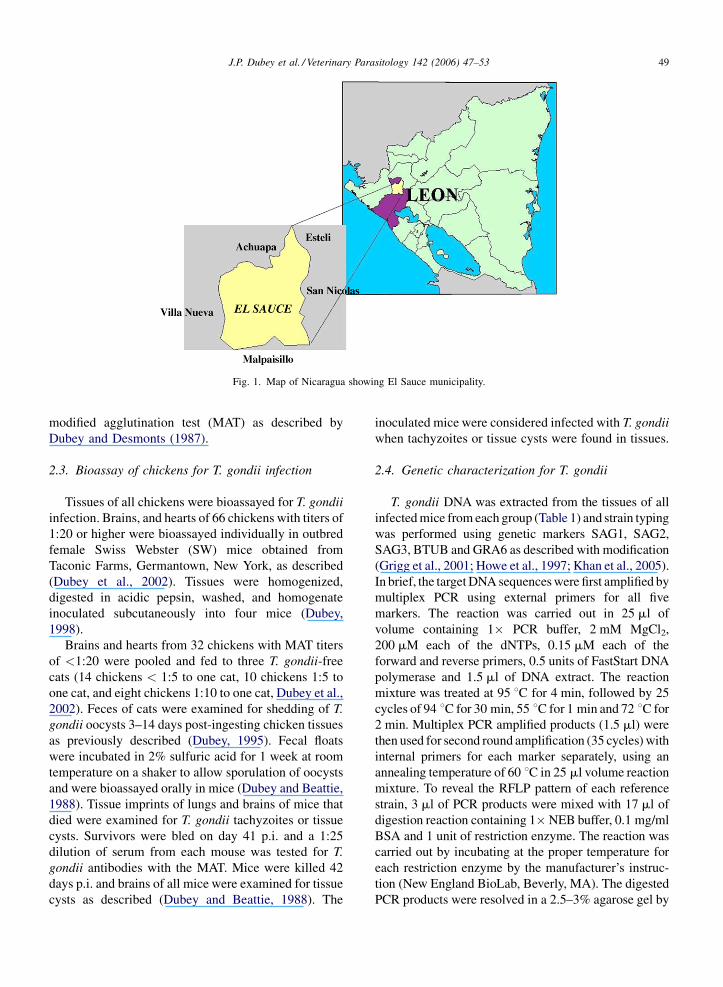
free-range chickens from the El Sauce municipality
(Fig. 1) within a radius of 10–15 km in different
directions from El Sauce town (latitude 12853013N and
longitude 86832017 W). The chickens originated from
36 different households that were at least 500 m apart.
Eighteen households provided one chicken, six pro-
vided two, three provided three, one provided four, two
provided five, two provided six, three provided eight
and one household provided eleven chickens (Table 1).
Chickens were purchased, identified and housed
together until they were killed on 28 November 2005.
Samples of brain, whole heart, and blood were collected
from each chicken, and kept at 4 8C until sent with cold
packs by air to Beltsville, MD. Two days elapsed between
killing of chickens and receipt of samples at Beltsville.
Samples were received in excellent condition.
2.2. Serological examination
Sera of chickens were tested for T. gondii antibodies
using eight dilutions, from 1:5 to 1: 640 with the
J.P. Dubey et al. / Veterinary Parasitology 142 (2006) 47–53 49
Fig. 1. Map of Nicaragua showing El Sauce municipality.
modified agglutination test (MAT) as described by
Dubey and Desmonts (1987).
2.3. Bioassay of chickens for T. gondii infection
Tissues of all chickens were bioassayed for T. gondii
infection. Brains, and hearts of 66 chickens with titers of
1:20 or higher were bioassayed individually in outbred
female Swiss Webster (SW) mice obtained from
Taconic Farms, Germantown, New York, as described
(Dubey et al., 2002). Tissues were homogenized,
digested in acidic pepsin, washed, and homogenate
inoculated subcutaneously into four mice (Dubey,
1998).
Brains and hearts from 32 chickens with MAT titers
of <1:20 were pooled and fed to three T. gondii-free
cats (14 chickens < 1:5 to one cat, 10 chickens 1:5 to
one cat, and eight chickens 1:10 to one cat, Dubey et al.,
2002). Feces of cats were examined for shedding of T.
gondii oocysts 3–14 days post-ingesting chicken tissues
as previously described (Dubey, 1995). Fecal floats
were incubated in 2% sulfuric acid for 1 week at room
temperature on a shaker to allow sporulation of oocysts
and were bioassayed orally in mice (Dubey and Beattie,
1988). Tissue imprints of lungs and brains of mice that
died were examined for T. gondii tachyzoites or tissue
cysts. Survivors were bled on day 41 p.i. and a 1:25
dilution of serum from each mouse was tested for T.
gondii antibodies with the MAT. Mice were killed 42
days p.i. and brains of all mice were examined for tissue
cysts as described (Dubey and Beattie, 1988). The
inoculated mice were considered infected with T. gondii
when tachyzoites or tissue cysts were found in tissues.
2.4. Genetic characterization for T. gondii
T. gondii DNA was extracted from the tissues of all
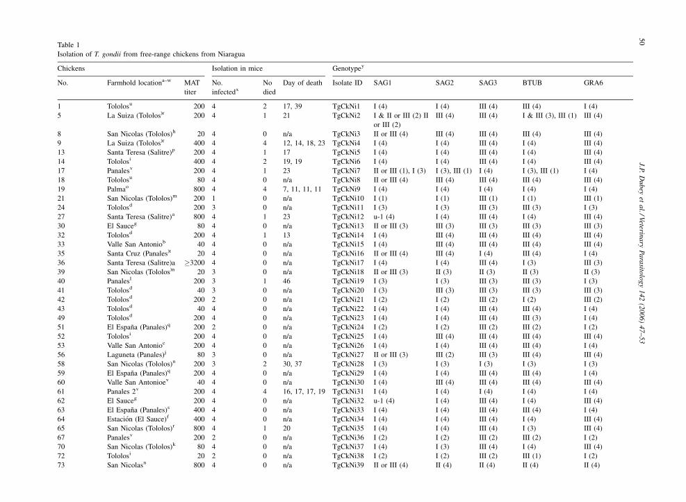
infected mice from each group (Table 1) and strain typing
was performed using genetic markers SAG1, SAG2,
SAG3, BTUB and GRA6 as described with modification
(Grigg et al., 2001; Howe et al., 1997; Khan et al., 2005).
In brief, the target DNA sequences were first amplified by
multiplex PCR using external primers for all five
markers. The reaction was carried out in 25 ml of
volume containing 1� PCR buffer, 2 mM MgCl2,
200 mM each of the dNTPs, 0.15 mM each of the
forward and reverse primers, 0.5 units of FastStart DNA
polymerase and 1.5 ml of DNA extract. The reaction
mixture was treated at 95 8C for 4 min, followed by 25
cycles of 94 8C for 30 min, 55 8C for 1 min and 72 8C for
2 min. Multiplex PCR amplified products (1.5 ml) were
then used for second round amplification (35 cycles) with
internal primers for each marker separately, using an
annealing temperature of 60 8C in 25 ml volume reaction
mixture. To reveal the RFLP pattern of each reference
strain, 3 ml of PCR products were mixed with 17 ml of
digestion reaction containing 1�NEB buffer, 0.1 mg/ml
BSA and 1 unit of restriction enzyme. The reaction was
carried out by incubating at the proper temperature for
each restriction enzyme by the manufacturer’s instruc-
tion (New England BioLab, Beverly, MA). The digested
PCR products were resolved in a 2.5–3% agarose gel by
J.P.
Du
bey
eta
l./Veterin
ary
Pa
rasito
log
y1
42
(20
06
)4
7–
53
50
Table 1
Isolation of T. gondii from free-range chickens from Niaragua
Chickens Isolation in mice Genotypey
No. Farmhold locationa–w MAT
titer
No.
infectedx
No
died
Day of death Isolate ID SAG1 SAG2 SAG3 BTUB GRA6
1 Tololosu 200 4 2 17, 39 TgCkNi1 I (4) I (4) III (4) III (4) I (4)
5 La Suiza (Tololos)r 200 4 1 21 TgCkNi2 I & II or III (2) II
or III (2)
III (4) III (4) I & III (3), III (1) III (4)
8 San Nicolas (Tololos)h 20 4 0 n/a TgCkNi3 II or III (4) III (4) III (4) III (4) III (4)
9 La Suiza (Tololos)r 400 4 4 12, 14, 18, 23 TgCkNi4 I (4) I (4) III (4) I (4) III (4)
13 Santa Teresa (Salitre)p 200 4 1 17 TgCkNi5 I (4) I (4) III (4) I (4) III (4)
14 Tololosi 400 4 2 19, 19 TgCkNi6 I (4) I (4) III (4) I (4) III (4)
17 Panalesv 200 4 1 23 TgCkNi7 II or III (1), I (3) I (3), III (1) I (4) I (3), III (1) I (4)
18 Tololosu 80 4 0 n/a TgCkNi8 II or III (4) III (4) III (4) III (4) III (4)
19 Palmao 800 4 4 7, 11, 11, 11 TgCkNi9 I (4) I (4) I (4) I (4) I (4)
21 San Nicolas (Tololos)m 200 1 0 n/a TgCkNi10 I (1) I (1) III (1) I (1) III (1)
24 Tololosd 200 3 0 n/a TgCkNi11 I (3) I (3) III (3) III (3) I (3)
27 Santa Teresa (Salitre)a 800 4 1 23 TgCkNi12 u-1 (4) I (4) III (4) I (4) III (4)
30 El Sauceg 80 4 0 n/a TgCkNi13 II or III (3) III (3) III (3) III (3) III (3)
32 Tololosd 200 4 1 13 TgCkNi14 I (4) III (4) III (4) III (4) III (4)
33 Valle San Antoniob 40 4 0 n/a TgCkNi15 I (4) III (4) III (4) III (4) III (4)
35 Santa Cruz (Panales)t 20 4 0 n/a TgCkNi16 II or III (4) III (4) I (4) III (4) I (4)
36 Santa Teresa (Salitre)a �3200 4 0 n/a TgCkNi17 I (4) I (4) III (4) I (3) III (3)
39 San Nicolas (Tololos)n 20 3 0 n/a TgCkNi18 II or III (3) II (3) II (3) II (3) II (3)
40 Panalesl 200 3 1 46 TgCkNi19 I (3) I (3) III (3) III (3) I (3)
41 Tololosd 40 3 0 n/a TgCkNi20 I (3) III (3) III (3) III (3) III (3)
42 Tololosd 200 2 0 n/a TgCkNi21 I (2) I (2) III (2) I (2) III (2)
43 Tololosd 40 4 0 n/a TgCkNi22 I (4) I (4) III (4) III (4) I (4)
49 Tololosd 200 4 0 n/a TgCkNi23 I (4) I (4) III (4) III (3) I (4)
51 El Espana (Panales)q 200 2 0 n/a TgCkNi24 I (2) I (2) III (2) III (2) I (2)
52 Tololosi 200 4 0 n/a TgCkNi25 I (4) III (4) III (4) III (4) III (4)
53 Valle San Antonioc 200 4 0 n/a TgCkNi26 I (4) I (4) III (4) III (4) I (4)
56 Laguneta (Panales)j 80 3 0 n/a TgCkNi27 II or III (3) III (2) III (3) III (4) III (4)
58 San Nicolas (Tololos)n 200 3 2 30, 37 TgCkNi28 I (3) I (3) I (3) I (3) I (3)
59 El Espana (Panales)q 200 4 0 n/a TgCkNi29 I (4) I (4) III (4) III (4) I (4)
60 Valle San Antonioev 40 4 0 n/a TgCkNi30 I (4) III (4) III (4) III (4) III (4)
61 Panales 2v 200 4 4 16, 17, 17, 19 TgCkNi31 I (4) I (4) I (4) I (4) I (4)
62 El Sauceg 200 4 0 n/a TgCkNi32 u-1 (4) I (4) III (4) I (4) III (4)
63 El Espana (Panales)s 400 4 0 n/a TgCkNi33 I (4) I (4) III (4) III (4) I (4)
64 Estacion (El Sauce)f 400 4 0 n/a TgCkNi34 I (4) I (4) III (4) I (4) III (4)
65 San Nicolas (Tololos)r 800 4 1 20 TgCkNi35 I (4) I (4) III (4) I (3) III (4)
67 Panalesv 200 2 0 n/a TgCkNi36 I (2) I (2) III (2) III (2) I (2)
70 San Nicolas (Tololos)k 80 4 0 n/a TgCkNi37 I (4) I (3) III (4) I (4) III (4)
72 Tololosi 20 2 0 n/a TgCkNi38 I (2) I (2) III (2) III (1) I (2)
73 San Nicolasn 800 4 0 n/a TgCkNi39 II or III (4) II (4) II (4) II (4) II (4)
J.P. Dubey et al. / Veterinary Parasitology 142 (2006) 47–53 517
5P
anal
es2
v2
00
44
13
,1
4,
14
,1
6T
gC
kN
i40
I(4
)I
(4)
I(4
)I
(4)
I(4
)
77
Pav
on
20
33
21
,2
3,
27
Tg
Ck
Ni
41
I(3
)I
(3)
I(3
)I
(3)
I(3
)
84
San
taT
eres
a(S
alit
re)w
20
04
14
0T
gC
kN
i42
IIo
rII
I(4
)II
(4)
II(4
)II
(4)
II(4
)
85
Pan
ales
2l
20
04
41
4,
15
,1
5,
19
Tg
Ck
Ni4
3I
(4)
I(4
)I
(4)
I(4
)I
(4)
92
El
Sau
ceg
�6
40
43
13
,1
4,
21
Tg
Ck
Ni4
4II
or
III
(4)
III
(4)
III
(4)
III
(3)
III
(3)
94
San
taC
ruz
(Pan
ales
)t1
60
40
n/a
Tg
Ck
Ni4
5II
or
III
(4)
III
(4)
I(4
)II
I(4
)I
(4)
97
To
lolo
si8
04
0n
/aT
gC
kN
i46
I(4
)I
(4)
III
(4)
III
(3),
I&
III
(1)
I(3
),I
&II
I(1
)
98
Pan
ales
l8
04
12
7T
gC
kN
i47
I(4
)I(
4)
I(2
),II
I(2
)I
&II
I(1
),I
(2),
III
(1)
I(4
)
Po
ole
dti
ssu
es1
0N
ot
app
lica
ble
Tg
Ck
Ni4
8II
or
III
III
III
III
III
(x)
Of
fou
rm
ice
inocu
late
d.
(a-w
)H
ou
seho
lds
des
ign
ated
by
dif
fere
nt
lett
ers.
(y)
Gen
oty
pin
gb
ased
on
DN
Afr
om
the
stat
edn
o.
of
mic
e.(u
-1)
isan
un
iqu
eal
lele
iden
tifi
edat
SA
G1
locu
s.
n/a
=n
ot
app
lica
ble
.
electrophoresis in the presence of 0.3 mg/ml ethidium
bromide and visualized under UV light. The primers and
enzymes used were stated previously (Dubey et al., in
press-b).
3. Results
Antibodies to T. gondii were found in 84 (85.7%) of
98 chickens with titers of 1:5 in 10, 1:10 in eight, 1:20 in
seven, 1:40 in nine, 1:80 in 11, 1:160 in one, 1:200 in 27,
1:400 in six, 1:800 four, and 1:3200 in one bird.
T. gondii was isolated by bioassay in mice from 47
chickens with MAT titers of 1:20 or higher. All infected
mice from six isolates died of toxoplasmosis. Overall,
41 of 170 (24.1%) mice that became infected after
inoculation with chicken tissues died of toxoplasmosis.
All mice that became infected after inoculation with
tissues from six chicken (9, 19, 61, 75, 77, and 85) died
of acute toxoplasmosis between 6 and 27 days p.i.
Genotyping of these 47 isolates using polymorphisms
at the loci SAG1, SAG2, SAG3, BTUB and GRA6
revealed eight genotypes. Six isolates (TgCkNi9, 28, 31,
40, 41, 43) had Type I alleles, three isolate (TgCkNi18,
39, 42) had Type II at all loci, and five isolates (TgCkNi3,
8, 13, 27, 44) had Type III alleles at all loci. Two isolates
(TgCkNi12, 32) have a unique allele at SAG1 locus and
combination of I and III alleles at other loci. The rest 27
isolates contained the combination of Type I and III
alleles and were divided into four genotypes. Of these 27
isolates, 11 isolates (TgCkNi1, 11, 19, 22, 23, 24, 26, 29,
33, 36, 38) have I, I, III, III and I alleles, nine isolates
(TgCkNi4, 5, 6, 10, 17, 21, 34, 35, 37) have I, I, III, I and
III alleles, two isolates (TgCkNi16, 45) have II or III, III,
I, III and I alleles, five isolates (TgCkNi14, 15, 20, 25, 30)
have I, III, III, III and III alleles at loci SAG1, SAG2,
SAG3, BTUB and GRA6, respectively. Four isolates
(TgCkNi2, 7, 46, 47) had mixed infections.
The cat (no. 230) fed pooled tissues from eight
chickens with titers of 1:10 shed T. gondii oocysts. The
two mice fed oocysts from cat 230 died of acute
toxoplasmosis 4 days later and numerous tachyzoites
were found in their mesenteric lymph nodes; these
tachyzoites were infective to mice by the subcutaneous
route. Genotyping of this isolate (TgCkNi48) revealed
the Type III alleles at all loci.
The two cats fed tissues of 24 chickens with titers of
1:5 or less did not shed oocysts.
4. Discussion
It is interesting to see that multiple genotypes were
identified in chickens from the same household. From
J.P. Dubey et al. / Veterinary Parasitology 142 (2006) 47–5352
one household in Tololos, six isolates (TgCkNi11, 14,
20, 21, 22, 23) were typed into three genotypes. Three
isolates (TgCkNi18, 28, 39) from a household in San
Nicolas were typed into two genotypes. Similar
phenomenon was observed in a few other locations.
This clearly indicate that more than one genotype are
circulating in a given area in El Sauce municipality.
This may explain the high frequency of mixed
infections observed. High rate of mixed infection in
intermediate hosts (such as chickens) will likely lead to
more frequent genetic exchange between different
parasite lineages when the intermediate hosts are preyed
by feline hosts, which in turn will facilitate the
evolution of T. gondii.
Phenotypically and genetically, T. gondii isolates
from chickens from Nicaragua were different from the
isolates from North America and Grenada, West Indies
but similar to those from Costa Rica. Most isolates from
chickens from Brazil and Colombia were lethal for mice
whereas isolates from North America and the Caribbean
did not kill inoculated mice. Genetically, none of T.
gondii isolates from Colombia and Brazil was SAG2
Type II, whereas most isolates from chickens from
North America and Grenada were Type II (Dubey et al.,
2003c; Lehmann et al., 2003). This is the first report of
genetic characterization of T. gondii isolates from
Nicaragua.
References
Ajzenberg, D., Cogne, N., Paris, L., Bessieres, M.H., Thulliez, P.,
Filisetti, D., Pelloux, H., Marty, P., Darde, M.L., 2002a. Genotype
of 86 Toxoplasma gondii isolates associated with human conge-
nital toxoplasmosis, and correlation with clinical findings. J.
Infect. Dis. 186, 684–689.
Ajzenberg, D., Banuls, A.L., Tibayrenc, M., Darde, M.L., 2002b.
Microsatellite analysis of Toxoplasma gondii shows considerable
polymorphism structured into two main clonal groups. Int. J.
Parasitol. 32, 27–38.
Ajzenberg, D., Banuls, A.L., Su, C., Dumetre, A., Demar, M., Carme,
B., Darde, M.L., 2004. Genetic diversity, clonality and sexuality in
Toxoplasma gondii. Int. J. Parasitol. 34, 1185–1196.
Aspinall, T.V., Guy, E.C., Roberts, K.E., Joynson, D.H.M., Hyde, J.E.,
Sims, P.F.G., 2003. Molecular evidence for multiple Toxoplasma
gondii infections in individual patients in England and Wales:
publichealth implications. Int. J. Parasitol. 33, 97–103.
Boothroyd, J.C., Grigg, M.E., 2002. Population biology of Toxo-
plasma gondii and its relevance to human infection: do different
strains cause different disease? Curr. Opin. Microbiol. 5, 438–442.
CENAGRO, 2002. Tercer Censo Nacional Agropecuario. Gobierno de
Nicaragua – Instituto Nacional de Estadisticas y Censos. (http://
www.inec.gob.ni).
da Silva, A.V., Pezerico, S.B., de Lima, V.Y., d’Arc Moretti, L.,
Pinheiro, J.P., Tanaka, E.M., Ribeiro, M.G., Langoni, H., 2005.
Genotyping of Toxoplasma gondii strains isolated from dogs with
neurological signs. Vet. Parasitol. 127, 23–27.
Dubey, J.P., 1995. Duration of immunity to shedding of Toxoplasma
gondii oocysts by cats. J. Parasitol. 81, 410–415.
Dubey, J.P., 1998. Refinement of pepsin digestion method for isolation
of Toxoplasma gondii from infected tissues. Vet. Parasitol. 74, 75–
77.
Dubey, J.P., Beattie, C.P., 1988. Toxoplasmosis of Animals and Man.
CRC Press, Boca Raton, Florida, pp. 1–220.
Dubey, J.P., Desmonts, G., 1987. Serological responses of equids fed
Toxoplasma gondii oocysts. Equine Vet. J. 19, 337–339.
Dubey, J.P., Graham, D.H., Blackston, C.R., Lehmann, T., Gennari,
S.M., Ragozo, A.M.A., Nishi, S.M., Shen, S.K., Kwok, O.C.H.,
Hill, D.E., Thulliez, P., 2002. Biological and genetic characterisa-
tion of Toxoplasma gondii isolates from chickens (Gallus domes-
ticus) from Sao Paulo, Brazil: unexpected findings. Int. J.
Parasitol. 32, 99–105.
Dubey, J.P., Graham, D.H., Silva, D.S., Lehmann, T., Bahia-Oliveira,
L.M.G., 2003a. Toxoplasma gondii isolates of free-ranging chick-
ens from Rio de Janeiro, Brazil: mouse mortality, genotype, and
oocyst shedding by cats. J. Parasitol. 89, 851–853.
Dubey, J.P., Graham, D.H., Dahl, E., Hilali, M., El-Ghaysh, A.,
Sreekumar, C., Kwok, O.C.H., Shen, S.K., Lehmann, T., 2003b.
Isolation and molecular characterization of Toxoplasma gondii
from chickens and ducks from Egypt. Vet. Parasitol. 114, 89–95.
Dubey, J.P., Graham, D.H., Dahl, E., Sreekumar, C., Lehmann, T.,
Davis, M.F., Morishita, T.Y., 2003c. Toxoplasma gondii isolates
from free-ranging chickens from the United States. J. Parasitol. 89,
1060–1062.
Dubey, J.P., Navarro, I.T., Graham, D.H., Dahl, E., Freire, R.L.,
Prudencio, L.B., Sreekumar, C., Vianna, M.C., Lehmann, T.,
2003d. Characterization of Toxoplasma gondii isolates from free
range chickens from Parana, Brazil. Vet. Parasitol. 117, 229–234.
Dubey, J.P., Venturini, M.C., Venturini, L., Piscopo, M., Graham,
D.H., Dahl, E., Sreekumar, C., Vianna, M.C., Lehmann, T., 2003e.
Isolation and genotyping of Toxoplasma gondii from free-ranging
chickens from Argentina. J. Parasitol. 89, 1063–1064.
Dubey, J.P., Graham, D.H., de Young, R.W., Dahl, E., Eberhard, M.L.,
Nace, E.K., Won, K., Bishop, H., Punkosdy, G., Sreekumar, C.,
Vianna, M.C.B., Shen, S.K., Kwok, O.C.H., Sumners, J.A.,
Demarais, S., Humphreys, J.G., Lehmann, T., 2004a. Molecular
and biologic characteristics of Toxoplasma gondii isolates from
wildlife in the United States. J. Parasitol. 90, 67–71.
Dubey, J.P., Levy, M., Sreekumar, C., Kwok, O.C.H., Shen, S.K.,
Dahl, E., Thulliez, P., Lehmann, T., 2004b. Tissue distribution and
molecular characterization of chicken isolates of Toxoplasma
gondii from Peru. J. Parasitol. 90, 1015–1018.
Dubey, J.P., Morales, E.S., Lehmann, T., 2004c. Isolation and geno-
typing of Toxoplasma gondii from free-ranging chickens from
Mexico. J. Parasitol. 90, 411–413.
Dubey, J.P., Parnell, P.G., Sreekumar, C., Vianna, M.C.B., de Young,
R.W., Dahl, E., Lehmann, T., 2004d. Biologic and molecular
charactaeristics of Toxoplasma gondii isolates from striped skunk
(Mephitis mephitis), Canada goose (Branta canadensis), blacked-
winged lory (Eos cyanogenia), and cats (Felis catus). J. Parasitol.
90, 1171–1174.
Dubey, J.P., Salant, H., Sreekumar, C., Dahl, E., Vianna, M.C.B.,
Shen, S.K., Kwok, O.C.H., Spira, D., Hamburger, J., Lehmann, T.,
2004e. High prevalence of Toxoplasma gondii in a commercial
flock of chickens in Israel, and public health implications of free-
range farming. Vet. Parasitol. 121, 317–322.
Dubey, J.P., Karhemere, S., Dahl, E., Sreekumar, C., Diabate, A.,
Dabire, K.R., Vianna, M.C.B., Kwok, O.C.H., Lehmann, T.,
2005a. First biologic and genetic characterization of Toxoplasma
J.P. Dubey et al. / Veterinary Parasitology 142 (2006) 47–53 53
gondii isolates from chickens from Africa (Democratic Republic
of Congo, Mali, Burkina Faso, and Kenya). J. Parasitol. 91, 69–72.
Dubey, J.P., Bhaiyat, M.I., de Allie, C., Macpherson, C.N.L., Sharma,
R.N., Sreekumar, C., Vianna, M.C.B., Shen, S.K., Kwok, O.C.H.,
Lehmann, T., 2005b. Isolation, tissue distribution, and molecular
characterization of Toxoplasma gondii from chickens in Grenada,
West Indies. J. Parasitol. 91, 557–560.
Dubey, J.P., Edelhofer, R., Marcet, P., Vianna, M.C.B., Kwok, O.C.H.,
Lehmann, T., 2005c. Genetic and biologic characteristics of
Toxoplasma gondii infections in free range chickens from Austria.
Vet. Parasitol. 133, 299–306.
Dubey, J.P., Gomez-Marin, J.E., Bedoya, A., Lora, F., Vianna, M.C.B.,
Hill, D., Kwok, O.C.H., Shen, S.K., Marcet, P.L., Lehmann, T.,
2005d. Genetic and biologic characteristics of Toxoplasma gondii
isolates in free-range chickens from Colombia, South America.
Vet. Parasitol. 134, 67–72.
Dubey, J.P., Lopez, B., Alveraz, M., Mendoza, C., Lehmann, T.,
2005e. Isolation, tissue distribution, and molecular characteriza-
tion of Toxoplasma gondii from free-range chickens from Gua-
temala. J. Parasitol. 91, 955–957.
Dubey, J.P., Marcet, P.L., Lehmann, T., 2005f. Characterization of
Toxoplasma gondii isolates from free-range chickens in Argentina.
J. Parasitol. 91, 1335–1339.
Dubey, J.P., Rajapakse, R.P.V.J., Ekanayake, D.K., Sreekumar, C.,
Lehmann, T., 2005g. Isolation and molecular characterization of
Toxoplasma gondii from chickens from Sri Lanka. J. Parasitol. 92,
1480–1482.
Dubey, J.P., Lenhart, A., Castillo, C.E., Alvarez, L., Marcet, P.,
Sreekumar, C., Lehmann, T., 2005h. Toxoplasma gondii infections
in chickens from Venezuela: isolation, tissue distribution, and
molecular characterization. J. Parasitol. 91, 1332–1334.
Dubey, J.P., Gennari, S.M., Labruna, M.B., Camargo, L.M.A., Vianna,
M.C.B., Marcet, P.L., Lehmann, T., 2006a. Characterization of
Toxoplasma gondii isolates in free-range chickens from Amazon,
Brazil. J. Parasitol. 92, 36–40.
Dubey, J.P., Vianna, M.C.B., Sousa, S., Canada, N., Meireles, C.S.,
Correia da Costa, J.M., Marcet, P.L., Lehmann, T., Darde, M.L.,
Thulliez, F.D., 2006b. Characterization of Toxoplasma gondii
isolates in free-range chickens from Portugal. J. Parasitol. 92,
184–186.
Dubey, J.P., Su, C., Oliveira, J., Morales, J.A., Bolanos, R.V., Sundar,
N., Kwok, O.C.H., Shen, S.K., 2006c. Biologic and genetic
characteristics of Toxoplasma gondii isolates in free-range chick-
ens from Costa Rica, Central America. Vet. Parasitol. 139, 29–36.
Dubey, J.P., Patitucci, A.N., Su, C., Sundar, N., Kwok, O.C.H., Shen,
S.K., in press-a. Characterization of Toxoplasma gondii isolates in
free-range chickens from Chile, South America. Vet. Parasitol.
Ferreira, A.M., Vitor, R.W.A., Carneiro, A.C.A.V., Brandao, G.P.,
Melo, M.N., 2004. Genetic viariability of Brazilian Toxoplasma
gondii strains detected by random amplified polymorphic DNA-
polymerase chain reaction (RAPD-PCR) and simple sequence
repeat anchored-PCR (SSR-PCR). Infect. Genet. Evol. 4, 131–
142.
Ferreira, A.M., Vitor, R.W.A., Gazzinelli, R.T., Melo, M.N., 2006.
Genetic analysis of natural recombinant Brazilian Toxoplasma
gondii strains by multilocus PCR-RFLP. Infect. Genet. Evol. 6,
22–31.
Fuentes, I., Rubio, J.M., Ramırez, C., Alvar, J., 2001. Genotypic
characterization of Toxoplasma gondii strains associated with
human toxoplasmosis in Spain: direct analysis from clinical
samples. J. Clin. Microbiol. 39, 1566–1570.
Grigg, M.E., Ganatra, J., Boothrooyd, J.C., Margolis, T.P., 2001.
Unusual abundance of atypical strains associated with human
ocular toxoplasmosis. J. Infect. Dis. 184, 633–639.
Howe, D.K., Sibley, L.D., 1995. Toxoplasma gondii comprises three
clonal lineages: correlation of parasite genotype with human
disease. J. Infect. Dis. 172, 1561–1566.
Howe, D.K., Honore, S., Derouin, F., Sibley, L.D., 1997. Determina-
tion of genotypes of Toxoplasma gondii strains isolated from
patients with toxoplasmosis. J. Clin. Microbiol. 35, 1411–
1414.
Jungersen, G., Jensen, L., Rask, M.R., Lind, P., 2002. Non-lethal
infection parameters in mice separate sheep type II Toxoplasma
gondii isolates by virulence. Comp. Immunol. Microbiol. Infect.
Dis. 25, 187–195.
Khan, A., Su, C., German, M., Storch, G.A., Clifford, D.B., Sibley,
L.D., 2005. Genotyping of Toxoplasma gondii strains from immu-
nocompromised patients reveals high prevalence of type I strains.
J. Clin. Microbiol. 43, 5881–5887.
Lehmann, T., Graham, D.H., Dahl, E., Sreekumar, C., Launer, F.,
Corn, J.L., Gamble, H.R., Dubey, J.P., 2003. Transmission
dynamics of Toxoplasma gondii on a pig farm. Infect. Genet.
Evol. 3, 135–141.
Lehmann, T., Graham, D.H., Dahl, E.R., Bahia-Oliveira, L.M.G.,
Gennari, S.M., Dubey, J.P., 2004. Variation in the structure of
Toxoplasma gondii and the roles of selfing, drift, and epistatic
selection in maintaining linkage disequilibria. Infect. Genet. Evol.
4, 107–114.
Mondragon, R., Howe, D.K., Dubey, J.P., Sibley, L.D., 1998. Geno-
typic analysis of Toxoplasma gondii isolates from pigs. J. Para-
sitol. 84, 639–641.
Owen, M.R., Trees, A.J., 1999. Genotyping of Toxoplasma gondii
associated with abortion in sheep. J. Parasitol. 85, 382–384.
Ruiz, A., Frenkel, J.K., 1980. Intermediate and transport hosts of
Toxoplasma gondii in Costa Rica. Am. J. Trop. Med. Hyg. 29,
1161–1166.
Sreekumar, C., Graham, D.H., Dahl, E., Lehmann, T., Raman, M.,
Bhalerao, D.P., Vianna, M.C.B., Dubey, J.P., 2003. Genotyping of
Toxoplasma gondii isolates from chickens from India. Vet. Para-
sitol. 118, 187–194.