Anionic sites on Toxoplasma gondii tissue cyst wall: Expression, uptake and characterization
Upload
independentCategory
view
0download
0
Toxoplasma gondii Is Dependent on Glutamine andAlters Migratory Profile of Infected Host Bone MarrowDerived Immune Cells through SNAT2 and CXCR4Pathways
I-Ping Lee1*, Andrew K. Evans1., Cissy Yang1., Melissa G. Works1, Vineet Kumar2, Zurine De Miguel1,
Nathan C. Manley1,3,4, Robert M. Sapolsky1,3,4,5
1Department of Biology, Stanford University, Stanford, California, United States of America, 2 School of Biological Sciences, Nanyang Technological University, Singapore,
Republic of Singapore, 3Department of Neurosurgery, Stanford University School of Medicine, Stanford, California, United States of America, 4 Stanford Stroke Center and
Stanford Institute for Neuro-Innovation and Translational Neurosciences, Stanford University School of Medicine, Stanford, California, United States of America,
5Department of Neurology and Neurological Sciences, Stanford University School of Medicine, Stanford, California, United States of America
Abstract
The obligate intracellular parasite, Toxoplasma gondii, disseminates through its host inside infected immune cells. Wehypothesize that parasite nutrient requirements lead to manipulation of migratory properties of the immune cell. Wedemonstrate that 1) T. gondii relies on glutamine for optimal infection, replication and viability, and 2) T. gondii-infectedbone marrow-derived dendritic cells (DCs) display both ‘‘hypermotility’’ and ‘‘enhanced migration’’ to an elevated glutaminegradient in vitro. We show that glutamine uptake by the sodium-dependent neutral amino acid transporter 2 (SNAT2) isrequired for this enhanced migration. SNAT2 transport of glutamine is also a significant factor in the induction of migrationby the small cytokine stromal cell-derived factor-1 (SDF-1) in uninfected DCs. Blocking both SNAT2 and C-X-C chemokinereceptor 4 (CXCR4; the unique receptor for SDF-1) blocks hypermotility and the enhanced migration in T. gondii-infectedDCs. Changes in host cell protein expression following T. gondii infection may explain the altered migratory phenotype; weobserved an increase of CD80 and unchanged protein level of CXCR4 in both T. gondii-infected and lipopolysaccharide(LPS)-stimulated DCs. However, unlike activated DCs, SNAT2 expression in the cytosol of infected cells was also unchanged.Thus, our results suggest an important role of glutamine transport via SNAT2 in immune cell migration and a possibleinteraction between SNAT2 and CXCR4, by which T. gondii manipulates host cell motility.
Citation: Lee I-P, Evans AK, Yang C, Works MG, Kumar V, et al. (2014) Toxoplasma gondii Is Dependent on Glutamine and Alters Migratory Profile of Infected HostBone Marrow Derived Immune Cells through SNAT2 and CXCR4 Pathways. PLoS ONE 9(10): e109803. doi:10.1371/journal.pone.0109803
Editor: Matthew Bogyo, Stanford University, United States of America
Received February 18, 2014; Accepted September 12, 2014; Published October 9, 2014
Copyright: � 2014 Lee et al. This is an open-access article distributed under the terms of the Creative Commons Attribution License, which permits unrestricteduse, distribution, and reproduction in any medium, provided the original author and source are credited.
Funding: This work was supported by The Stanley Medical Research Institute (grant number: 06R-1463). The funders had no role in study design, data collectionand analysis, decision to publish, or preparation of the manuscript.
Competing Interests: The authors have declared that no competing interests exist.
* Email: [email protected]
. These authors contributed equally to this work.
Introduction
Toxoplasma gondii, an obligate intracellular protozoan that is
capable of infecting nearly all warm-blooded animals, depends on
the host to meet its glucose and energy requirements. Recent
studies have shown that apicomplexan parasites catabolize both
glucose and the non-essential amino acid glutamine via the
tricarboxylic acid (TCA) cycle to generate energy; however,
disrupting the entry of glucose-derived intermediates into the TCA
cycle has no significant effect on the growth of asexual stages of
Plasmodium falciparum, indicating that glutamine serves as an
alternative carbon source for the cycle [1]. T. gondii has also been
shown to rely on glutamine for energy during glucose starvation,
and glutaminolysis is indispensable to its survival [2–4]. Rapidly
dividing tachyzoites of T. gondii may therefore impose a heavy
glutamine burden on the host, and we here propose that such
glutamine dependency may generate a specific means by which
the parasite can manipulate the function of host cells to induce
them to leave the circulation and enter tissues with high glutamine
level for the benefit of the parasite. T. gondii has been shown to
migrate to and encyst in brain, retina and muscle during chronic
infection, all places with abundant glutamine supply [5–9].
T. gondii has been demonstrated to manipulate the migratory
properties of infected immune cells, inducing a hypermotility state
in infected dendritic cells (DCs), which has been hypothesized to
promote dissemination of parasites throughout the body [10,11].
This phenomenon is dependent on Gi-protein signaling but the
specific Gi protein-coupled receptor has not been identified. C-X-
C chemokine receptor 4 (CXCR4), the unique receptor for small
cytokine stromal cell-derived factor-1 (SDF-1) on the surface of
immune cells, may be a strong candidate. The SDF-1-CXCR4
complex activates the downstream phosphoinositide 3-kinase
(PI3K) pathway involved in cell motility [12,13]. Importantly,
the PI3K pathway can also be induced by the glutamine
transporter (sodium-dependent neutral amino acid transporter 2,
SNAT2)/amino acid substrate complex in muscle cells [14,15].
PLOS ONE | www.plosone.org 1 October 2014 | Volume 9 | Issue 10 | e109803

SNAT2, the ubiquitously expressed subtype of the system A
transporter in mammalian cells, is the primary mediator of uptake
of aliphatic amino acids such as glutamine, glycine, and asparagine
[16]. Recent studies on cultured rat myoblasts have demonstrated
that SNAT2 exhibits a hybrid transporter-receptor (transceptor)
function that can sense extracellular amino acid concentration and
activate downstream PI3K pathway during nutrient stress [14,15].
Such amino acid transceptors are well documented in Drosophilaand yeast like Saccharomyces cerevisiae, but the role of SNAT2 in
mammalian immune cells has not been extensively studied. On the
other hand, it is well known that lymphocytes, monocytes and
macrophages have high utilization rates of glutamine, which is
involved in cell proliferation, expression of surface activation
markers and the production of cytokines [17,18].
In this study we hypothesized that T. gondii, due to its high
demand for glutamine, manipulates the dual transporter/receptor
function of SNAT2 in the host, and via interaction with the
CXCR4 signaling pathway causes enhanced migration of infected
immune cells to glutamine-enriched environments. In order to
determine the specific role of glutamine and the transporter
SNAT2 as variables influencing this migration, we used an in vitrotranswell system to investigate cell migration of normal and T.gondii-infected murine bone marrow-derived DCs to a range of L-
glutamine concentrations. We examined the role of SNAT2 and
CXCR4-PI3K-Rho kinase pathways in this migration. Our data
show that CXCR4 signaling-dependent migration is SNAT2-
dependent in normal DCs, and antagonizing both CXCR4 and
SNAT2 blocks the induced migration in T. gondii-infected cells.
Results
T. gondii is dependent on glutamine for optimalinfection, replication and viability in vitroType II strains of T. gondii (e.g., Prugniaud) are the most
prevalent in Europe and North America [19,20], are less virulent
in the mouse model but highly associated with human diseases
[21,22], and they alter host physiology and behavior [23,24].
Therefore, we first investigated if type II tachyzoites maintained in
human foreskin fibroblast (HFFs) monolayers depend on gluta-
mine for infection, replication, and viability in vitro. INFEC-
TION: By using live staining of SAG1 (red), the major surface
antigen of T. gondii, we were able to distinguish extra- (red and
green) and intracellular (green) tachyzoites and quantify the
efficiency in infecting HFFs (Fig. 1A). In the absence of glutamine,
no obvious change of the morphology of parasites was observed.
However, without glutamine, there were more extracellular
parasites in the examined fields of the coverslips over the course
of 6 hours and the average of infection efficiency of T. gondii wasreduced approximately two-thirds (Fig. 1B). REPLICATION:
HFF monolayers were firstly infected with T. gondii tachyzoitesin the presence of glutamine for 6 hours. Extracellular parasites
were then washed away with phosphate-buffered saline (PBS) and
infected HFFs were cultured in fresh medium containing 0 mM or
2 mM of L-glutamine for another 16 hours. In the presence of
glutamine, T. gondii was able to duplicate up to 4 times in
22 hours (Fig. 1C) while the replication ability was impaired with
,50% of the intracellular tachyzoites only having divided twice
after glutamine deprivation (Fig. 1D). The mean number of
replication cycles was quantified and figure 1E shows that the
replication ability of T. gondii was reduced about 25% in the
absence of glutamine. VIABILITY: Parasite viability was evalu-
ated by a tetrazolium dye reduction (MTT) assay, and after
4 hours of glutamine starvation, the viability of T. gondiidecreased 29% and could not be rescued by the stereoisomer D-
glutamine, which suggests that glutamine serves as an energy
source for T. gondii metabolic activity rather than a modulator of
extracellular redox environments (Fig. 1F). MTT assays showed
no significant effect of L-glutamine depletion on the HFF host cells
(Fig. S1), indicating that the virulence and growth of T. gondii arenot compromised by the host viability during glutamine starvation.
Infected DCs exhibit hypermotility and an enhancedmigration specific to high glutamine in vitroWe next used rat bone marrow-derived DC culture to study the
effect of T. gondii infection on cell migration. Bone marrow-
derived cells were isolated and cultured in complete medium
supplemented with cytokines for seven days (see methods), and the
phenotype of DCs was confirmed by flow cytometry (Fig. S2). DCs
were infected by tachyzoites (multiplicity of infection, MOI, of 1)
for 4 hours and then migration was examined by in vitro transwellsystem. Flow cytometric results show that the infection frequency
of DCs is about 75% (Fig. 2A and 2B). T. gondii infection resulted
in a roughly 2.5-fold increase in the number of DCs migrating to
control medium, a result that we will refer to as a ‘‘hypermotility
state’’ of infected DCs and one that has been reported previously
[10] (Fig. 2C). Moreover, the infected cells exhibited an additional
‘‘enhanced migration’’ towards medium containing glutamine
concentration above 0.5 mM (Fig. 2C). This enhanced migration
was beyond that of the hypermotility induced by infection alone
and was specific to glutamine relative to other amino acids,
including the amino acids that utilize the same transporter
(Fig. 2D). Importantly, uninfected DCs did not show induced
migration to any of the tested glutamine concentrations or amino
acids. This hypermotility and enhanced migration could not be
explained simply by the maturation of the DCs because challenge
with lipopolysaccharide (LPS) had no effect on migration to
glutamine or other amino acids (Table S1).
Enhanced migration of infected DCs towards glutamineis dependent on the glutamine transport via SNAT2 andavailability of intracellular glutamineA glutamine analog (2-(methylamino)-isobutyrate, MeAIB),
which is a specific substrate of SNAT2 and decreases the cellular
uptake of glutamine [25,26], was able to block the enhanced
migration to 0.5 mM glutamine but had no effect on the
hypermotility of infected DCs (Fig. 3A). A glutamine synthetase
inhibitor (methionine sulfoximine, MSO) also blocked the
enhanced migration to glutamine but not the hypermotility.
Extracellular glutamine was always present in the media during
the experiment (see methods), and thus, these results suggest that
glutamine uptake by SNAT2 and glutamine synthetase are both
required for the enhanced migration. In contrast, complete
glutamine starvation of the infected DCs for 2 hours prior to the
assay abolished both enhanced migration to glutamine and
hypermotility, indicating that glutamine resource is essential for
both hypermotility and enhanced migration of infected DCs.
Glutamine transport via SNAT2 is important forCXCR4-dependent migration of uninfected DCs to SDF-1aIn order to test if SNAT2 serves as a general factor influencing
immune cell migration, we examined another scenario in which
uninfected DCs migrate, in this case to SDF-1a, a migration that
has been shown to be dependent on CXCR4 signaling. Examining
SDF-1a-induced transmigration in uninfected DCs with the same
treatments described above, we demonstrated that glutamine
starvation, MeAIB or MSO significantly reduced the uninfected
DC migration to SDF-1a (Fig. 3B). Both a CXCR4 antagonist
T. gondii and Glutamine-Dependent Immune Cell Migration
PLOS ONE | www.plosone.org 2 October 2014 | Volume 9 | Issue 10 | e109803

(AMD3100) and a PI3K inhibitor (LY294002) have previously
been shown to block the SDF-1a-induced migration in bone
marrow-derived mesenchymal stem cells, T lymphocytes and
platelets [27–30]. We demonstrated that either of these two drugs,
Figure 1. T. gondii is dependent on glutamine for optimal infection, replication and viability in vitro. Experiments were conducted inhuman foreskin fibroblast (HFF) monolayers. (A) Representative confocal images on the top row show the intracellular tachyzoites expressing GFP(green) and one extracellular parasite staining with SAG1 (red, also indicated by the white arrow) in the presence of L-glutamine. A 636oil objectivewas used to examine the SAG1-positive parasite at higher magnification (bottom left corner). Pictures on the second row represent the SAG1 stainingof extracellular T. gondii in the absence of L-glutamine. The nuclei of HFFs were stained with DAPI (blue), and the merged images of the fluorescenceand the bright field shows that the HFF monolayer was confluent when the experiment was carried out. (B) Infection efficiency is defined as the ratioof intracellular parasites to total parasites in the examined field on the coverslip after 6 hours of infection. Infection efficiency was reduced ,65% byomitting L-glutamine (Student’s t-test, P,0.001). (C) Examples of the confocal images showing T. gondii replication in HFFs after 22 hours in thepresence of glutamine. The replication cycle numbers were indicated in white. (D) In the presence of L-glutamine, T. gondii tachyzoites were able toduplicate up to 4 times in 22 hours, but without L-glutamine, over 50% of parasites only divided twice. (E) In the absence of L-glutamine, meanreplication cycle number declined approximately 25% in 22 hours (Student’s t-test, P,0.05). (F) MTT assay was used to evaluate parasite viability.Glutamine starvation significantly decreased the viability of T. gondii by 29%, and D-glutamine was unable to substitute for L-glutamine (one-wayANOVA, F(2,89)=45.036, P,0.001). Asterisks indicate significant difference from medium containing 2 mM L-glutamine (post hoc Dunnett’s test, P,0.001). Data show mean value 6 SEM from three independent experiments.doi:10.1371/journal.pone.0109803.g001
T. gondii and Glutamine-Dependent Immune Cell Migration
PLOS ONE | www.plosone.org 3 October 2014 | Volume 9 | Issue 10 | e109803
as well as the specific Rho kinase inhibitor (Y27632), also
dramatically decreased the migratory ability of uninfected DCs
to SDF-1a (Fig. 3B). Thus, SDF-1a-induced migration of unin-
fected DCs is dependent on SNAT2 transport of glutamine,
functional glutamine synthetase as well as the CXCR4-PI3K-Rho
kinase pathways.
Both hypermotility and enhanced migration toglutamine of infected DCs are dependent on the CXCR4-PI3K-Rho kinase pathwaysWe then tested if these CXCR4-PI3K-Rho kinase-related
inhibitors could affect hypermotility and enhanced migration of
T. gondii-infected DCs in the presence of glutamine. After 2 hours
of T. gondii infection, DCs were treated for an additional 2 hours
with the same concentration of these inhibitors of the CXCR4-
PI3K-Rho kinase pathways described previously. Inhibiting PI3K
or Rho kinase blocked both the hypermotility and the enhanced
migration to glutamine in infected cells (Fig. 3C). The CXCR4
inhibitor, AMD3100, inhibited the enhanced migration to
glutamine and partially decreased the hypermotility, which
suggested that CXCR4 is not the only activator of the downstream
PI3K pathway that contributes to the migratory state of infected
cells. In order to determine if glutamine transport through SNAT2
could be coactivating this pathway and thereby contributing to
migration, we used the combination of MeAIB and AMD3100 to
completely block enhanced migration and reduced hypermotility
to close to the control level. The viability of intracellular T. gondii
Figure 2. Transmigration assay of T. gondii-infected DCs in vitro. Infection frequency of T. gondii-infected DC culture was analyzed by flowcytometery. (A) In the gating strategy, SSC (side scatter) measures intracellular granularity and FSC (forward scatter) measures cell sizes. (B) Therepresentative histogram shows the mean fluorescent intensity (x-axis) versus percentage of gated population (y-axis) for GFP. The pink histogramrepresents uninfected and the blue indicates infected DC culture. The infection frequency of DCs is about 75%. (C and D) DCs were infected with T.gondii for 4 hours and then cell migration was tested by Costar Transwell System. White bars represent uninfected DCs and black bars represent T.gondii-infected cells. Bar graphs depict mean values of migration6 SEM from three independent experiments performed in triplicate. Migration valueis defined as the number of migrated cells in each condition normalized by the number of migrated cells in spontaneous migration, the migration ofuninfected cells in the absence of factors (% of uninfected control). (C) ‘‘Hypermotility’’ state is defined as the increased migration after infectioncomparing to the spontaneous migration, the migration of uninfected control cells in the absence of factors. ‘‘Enhanced migration’’ to glutamine isthe increased migration responding to increasing glutamine concentrations (between 0.5 and 4.0 mM) and is beyond that of the hypermotilityinduced by infection alone. Two-way ANOVA revealed an interaction effect between T. gondii infection and glutamine concentration (F(5,37)=4.426;P = 0.003). Further one-way ANOVA examining effects of glutamine within infected groups revealed an effect of glutamine in T. gondii-infected (P,0.01; indicated by asterisks) but not uninfected DCs. Post-hoc pairwise comparison of means for glutamine concentrations within the T. gondii-infected DCs revealed an increase in migration relative to control at 0.5, 1.0, 2.0, and 4.0 mM concentrations (Fisher’s protected LSD, P,0.02;indicated by #), but not at 0.1 mM glutamine. (D) The specificity of enhanced migration to glutamine was tested by examining the cell migration inthe presence of glutamine verse other amino acids such as glycine (Gly), histidine (His), arginine (Arg), glutamic acid (Glu), aspartic acid (Asp), tyrosine(Tyr), tryptophan (Trp) or phenylalanine (Phe). Two-way ANOVA revealed an interaction effect between T. gondii infection and amino acids(F(9,70)= 2.765; P = 0.008). Further one way ANOVA examining effects of amino acids within infected groups revealed an effect of amino acids in T.gondii-infected (P,0.005; indicated by asterisks) but not uninfected DCs. Post-hoc pairwise comparison of means for amino acid condition within theinfected DCs revealed an increase in migration relative to the control within only the increased glutamine condition (Fisher’s protected LSD, P,0.001;indicated by #). The y-axis scale for both (C) and (D) is the same and shown at the far left.doi:10.1371/journal.pone.0109803.g002
T. gondii and Glutamine-Dependent Immune Cell Migration
PLOS ONE | www.plosone.org 4 October 2014 | Volume 9 | Issue 10 | e109803

T. gondii and Glutamine-Dependent Immune Cell Migration
PLOS ONE | www.plosone.org 5 October 2014 | Volume 9 | Issue 10 | e109803

following these pharmacological treatments was measured by
plaque assays, and none of the inhibitors affected virulence of the
parasites (Fig. S3), indicating that the decrease of induced
migration of infected DCs was not due to the loss of parasite
viability or stalled replication. Based on these results, we developed
a hypothesis that hypermotility and enhanced migration of T.gondii-infected DCs may be dependent on an interaction between
CXCR4 and glutamine transport via SNAT2 in activating the
PI3K-Rho kinase pathways.
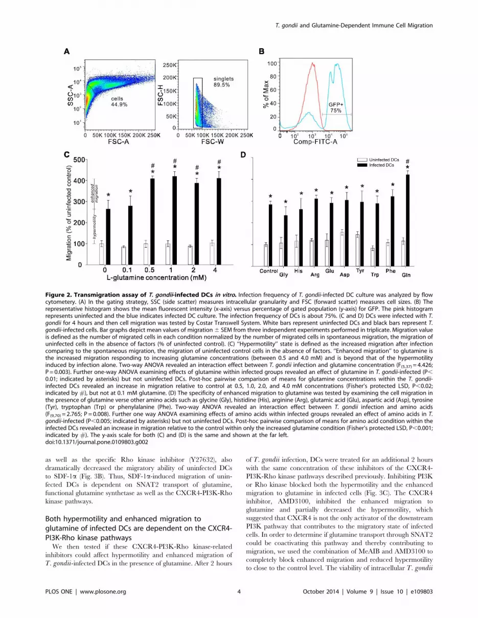
T. gondii infection activates DCs and the SNAT2 andCXCR4 expression in the host cell remains unchangedTo determine whether T. gondii affected CXCR4 and SNAT2
protein expression in host cells, we used Western blot to compare
the protein expression on surface and in cytosol in DCs either
exposed to live T. gondii or an alternative immune activator (LPS
+ tumor necrosis factor a, TNFa). We also examined CD80
protein expression as a marker of immune cell activation/
maturity. Figure 4A shows that both LPS + TNFa treatment
and T. gondii infection increased CD80 protein expression on the
surface of DCs, which suggests that both stimuli result in cell
activation and maturation. The quantification of the density of the
bands in the blots shows that although cell maturation with either
LPS + TNFa or T. gondii infection did not affect either surface or
cytosolic CXCR4 expression or surface SNAT2 expression, LPS +
TNFa treatment decreased SNAT2 in cytosol while T. gondii-infected DCs possessed similar levels as uninfected DCs (Fig.4B).
Discussion
T. gondii is an obligate intracellular pathogen that acquires
glutamine from the host to carry out functions necessary for its
metabolic activity, invasion, and replication. Here, we report that
infection, replication and viability in T. gondii are greatly
facilitated by glutamine availability. We demonstrate that in
infected immune cells (DCs) in vitro, T. gondii induces a) a
hypermotility state and, b) a specific enhanced migration to
environments with high glutamine levels (§0.5 mM). This
hypermotility and enhanced migration are dependent on SNAT2
transport of glutamine, the presence of functional glutamine
synthetase and the CXCR4-PI3K-Rho kinase pathways. Whereas
activation with LPS + TNFa results in downregulation of SNAT2
protein expression in DCs, this expression is unchanged with T.gondii infection and may contribute to the induced migration of T.gondii-infected cells. These findings support the hypothesis that T.gondii manipulates the migratory properties of the host cell to
increase the probability of migrating to places with sufficient
glutamine resource to meet its metabolic requirements.
The glutamine dependency of T. gondii is well documented.
The Toxoplasma TCA cycle, which utilizes glucose and
glutamine, generates energy for intracellular growth and replica-
tion; inhibition of the cycle leads to the death of parasites [2,4].
However, mutation of the T. gondii glucose transporter only has aminor effect on the growth of the parasite, and glutamine, but not
glucose, is indispensable to its survival [3]. Therefore, glutamino-
lysis is essential for the parasitic requirements of T. gondii.Recently a GABA shunt was observed for regulating the
catabolism of glutamine/glutamate in the Toxoplasma TCA cycle
[2], and DCs infected with live T. gondii have been demonstrated
to secrete GABA and exhibit hypermotility; this induced migration
may increase the chance of wide dissemination of the parasites
during acute infection [10,31,32]. Glutamine serves as the
precursor of GABA; thus it is plausible that the rapidly dividing
T. gondii imposes a high glutamine burden on the host, which
may manipulate the migratory phenotype of the infected immune
cells in order to sustain intracellular glutamine concentrations.
Our results support this idea as infected DCs exhibit the
hypermotility phenotype and enhanced migration to glutamine
(Fig. 2). On the other hand, although glutamine-induced migra-
tion of T. gondii-infected DCs is a new finding, it is not the first
amino acid that was reported to be a chemotaxis-inducing factor
for immune cells, as glutamate has been shown to attract
neutrophils to migrate to the inflamed/wound tissue via activating
the downstream PI3K pathway [33,34].
The hypermotility of T. gondii-infected DCs is related to the
parasite strain, and type II strain, as used in this study, exhibits the
strongest induction of hypermotility [11]. This hypermotility has
been suggested to be independent of CCR5, CCR7 or Toll/
interleukin-1 pathways but to involve a Gi protein signaling
transduction [10,32]. Chemokine-receptor complexes are known
to initiate signal transduction events leading to cellular responses
such as leukocyte chemotaxis and adhesion. CXCR4, one of 19
known chemokine receptors in mammals, couples primarily
through Gi proteins, and PI3K and Rho kinase pathways involved
in cell motility and orientation are downstream to activation of
CXCR4 [12,13,35,36]. While we also demonstrate the crucial
roles of PI3K and Rho kinase in this migration, as inhibition of
either pathway blocked enhanced migration to glutamine and
drastically reduced the hypermotility in T. gondii-infected DCs,
AMD3100, an antagonist that binds CXCR4 without engaging
receptor signaling [37,38], blocked enhanced migration to
glutamine but only partially decreased hypermotility. On the
other hand, the combination of AMD3100 and MeAIB, a specific
Figure 3. Pharmacological manipulation study of the induced migration of the T. gondii-infected and uninfected DCs. (A) Uninfectedand infected DCs were left untreated (no treatment) or were treated with MeAIB (the competitive inhibitor of glutamine transport via SNAT2) or MSO(the inhibitor of glutamine synthetase), and cell migration to control medium (without glutamine) or to medium containing 0.5 mM glutamine wasstudied. A pretreatment starving the DCs of glutamine was also used to examine the importance of glutamine source in hypermotility and enhancedmigration. (B) Uninfected control DCs were treated with MeAIB, MSO, inhibitors of CXCR4 (AMD3100), PI3K (LY294002) or Rho kinase (Y27632), or Glnstarvation for 2 hours before assessing migration to 100 ng/ml SDF-1a. Chemotactic index (CI) is defined as the fold increase in the number ofmigrating DCs to SDF-1a over the spontaneous migration. One-way ANOVA reveals an effect of pharmacological treatments on the SDF-1a-inducedmigration (F(6,44)=6.700, P,0.001). Asterisks indicate P,0.05 (Dunnett’s post hoc). (C) LY294002, Y27632, AMD3100 and the combination ofAMD3100 and MeAIB were used to study the potential mechanisms contributed to the hypermotility and the enhanced migration of T. gondii-infected DCs. White bars represent uninfected DCs, and gray and black bars represent infected cells in response to 0 mM and 0.5 mM glutamine,respectively. Bar graphs depict mean values of migration6 SEM from three independent experiments performed in triplicate. ‘‘No treatment’’ bars in(A) and (C) represent the same data. One way ANOVA examining effects of treatment across the uninfected control DCs revealed no effect oftreatment on cell migration (P.0.5). One-way ANOVA examining effects of pharmacological treatments on hypermotility within infected groups (graybars in A and C) revealed an effect of pharmacological treatments in T. gondii-infected DCs (F(7,49)= 16.541, P,0.001). Asterisks indicate P,0.003(Fisher’s protected LSD). Within each pharmacological treatment, # indicates significant effects of 0.5 mM glutamine (enhanced migration toglutamine) relative to control conditions (hypermotility) (Student’s t-test, P,0.01).doi:10.1371/journal.pone.0109803.g003
T. gondii and Glutamine-Dependent Immune Cell Migration
PLOS ONE | www.plosone.org 6 October 2014 | Volume 9 | Issue 10 | e109803
SNAT2 inhibitor, completely blocked enhanced migration and
significantly reduced hypermotility, indicating that CXCR4 and
SNAT2 may cumulatively regulate the altered migratory pheno-
type (Fig. 3). While the exact role of CXCR4 and SNAT2 in the
induced migration of infected DCs is unclear, CXCR4 promotes
the activation of PI3K pathway, and SNAT2, which has been
suggested to possess a dual transporter/receptor (transceptor)
function in cultured muscle cells and fibroblasts, activates PI3K
pathway and regulates/senses changes of the intracellular amino
acid pool and the availability of extracellular amino acids
[14,15,39,40]. The transmigration results in Figure 3 support an
interaction between SNAT2 and CXCR4 pathways through
which T. gondii may orient increased migration of infected DCs to
environments with high glutamine level.
In this study, western blot analysis showed that whereas DCs
challenged with LPS + TNFa downregulate cytosolic SNAT2
protein, T. gondii-infected DCs possessed a similar level of the
cytosolic pool of SNAT2 protein as uninfected control cells.
Downregulation of SNAT2 following LPS + TNFa challenge is, to
our knowledge, a novel finding and further study will be required
to understand the role of SNAT2 in activated immune cells. It has
been shown that during the acute phase of amino acid deprivation,
the adaptive regulation of SNAT2 activity relies on recruitment of
preformed proteins from a cytosolic pool to the cell membrane
[41–43], which may explain why T. gondii-infected cells with
unchanged SNAT2 expression in cytosol and on cell surface
possessed the altered migratory phenotype, whereas LPS-treated
immune cells did not (Table S1). CXCR4 protein remains
unchanged with both LPS + TNFa treatment and T. gondiiinfection, indicating that expression of CXCR4, along with
SNAT2, may be permissive for the migratory phenotype even if
expression does not increase with enhanced migration.
Figure 4. Western blot analysis of CXCR4 and SNAT2 expression of T. gondii-infected DCs in vitro. (A) DCs were treated with 100 ng/mlLPS+50 ng/ml TNFa or were infected with T. gondii for 2 hours in the presence of 2 mM glutamine and then the protein expression of CD80, CXCR4and SNAT2 in cytosol and on the surface were studied. GAPDH is the loading control. (B) The band intensity in the blots was quantified to show therelative protein level of CD80, CXCR4 and SNAT2 in tested groups compared to control (dotted line). Experiments were repeated three times andquantified by Image J.doi:10.1371/journal.pone.0109803.g004
T. gondii and Glutamine-Dependent Immune Cell Migration
PLOS ONE | www.plosone.org 7 October 2014 | Volume 9 | Issue 10 | e109803
Based on these studies, it is unclear if the enhanced migration to
glutamine is caused by T. gondii directly or by the host cell trying
to rebalance the glutamine homeostasis. New evidence also shows
that LY294002 targets mammalian target of rapamycin (mTOR)
at the same concentrations that inhibit PI3K, and mTOR is
involved in amino acid-sensing pathways [44–46]. However, based
on our results, a new hypothetical mechanism of how T. gondiiinduces migration in infected DCs was proposed (Fig. 5). Firstly, a
new on-and-off theory about the transceptor role of SNAT2 was
proposed in 2009, and SNAT2 is thought to be ‘‘closed off’’ when
sufficient concentrations of its substrates exist in the intracellular
compartment, and to be activated (‘‘on’’ state) when intracellular
amino acid concentrations drop [47]. The off state of the
transceptor might explain why immature DCs expressing SNAT2
do not show hypermotility or enhanced migration to glutamine in
this study. We hypothesized that T. gondii may ‘‘turn on’’ the
transceptor function of SNAT2. A drop in intracellular glutamine
concentration secondary to parasite infection may be sufficient to
turn on SNAT2; however, we cannot rule out the potential
influence of T. gondii-related proteins. One of the ways in which
T. gondii has been shown to manipulate host cell function is by
secretion of rhoptry proteins or dense granule proteins into host
cell cytoplasm and nucleus during invasion [48,49]; dense granule
protein GRA5 has been shown to trigger the migration of human
dendritic cells toward CCL19 [50], and rhoptry proteins and
dense granule proteins of T. gondii interfere with host signaling
pathways including immunity-related GTPases (IRGs) and nuclear
factor-kB (NF-kB) pathways [49,51,52]. Therefore, T. gondiimight directly or indirectly turn on the transceptor role of SNAT2
by modulating the intracellular glutamine metabolism and/or
secreting modulatory protein(s) in the infected immune cells.
In summary, we hypothesize that T. gondii metabolic require-
ments may lead to parasite-host interactions that benefit the
parasite by increasing the probability that infected-immune cells
will carry the parasite out of circulation into tissues with sufficient
glutamine (e.g. brain, eye, muscle, lung…). Technical challenges
moving forward include measuring intracellular concentration of
radioactive labeled glutamine in T. gondii-infected DCs, deliver-
ing RNAi targeting CXCR4 and/or SNAT2 to T. gondii-infectedimmune cells without altering T. gondii or immune cell viability,
manipulating the extracellular glutamine concentration in vivo to
redirect the migration of infected cells, and the lack of knockout
animal models of CXCR4 or SNAT2 to study the dissemination of
the parasites. We here provide evidence for the CXCR4 and
SNAT2 pathways as novel mechanisms for immune cell migration
and potential targets for prevention of parasite dissemination and
chronic infection in rodents and humans.
Materials and Methods
ParasitesPrugniaud stain of T. gondii expressing firefly luciferase and
green fluorescent protein (GFP) was a gift from Dr. John
Boothroyd. Tachyzoites were maintained by serial 2-day passage
in human foreskin fibroblast (HFF) monolayers cultured in
complete medium (DMEM plus 10% fetal bovine serum, 1%
penicillin-streptomycin, and 2 mM L-glutamine) (Life Technolo-
gy).
AnimalsPregnant female Sprague-Dawley rats (single caged; Charles
River Laboratories) were used for generation of pup bone marrow
derived immune cells. All procedures were reviewed and approved
by the Stanford University Administrative Panel on Laboratory
Care and the Association for Assessment of Laboratory Animal
Care.
T. gondii Invasion AssayHFFs were cultured on 12 mm coverslips in complete medium
24 hours before the experiment. Tachyzoites were added to HFFs
(MOI= 2) and invasion in the absence (0 mM) or presence (2 mM)
of glutamine was allowed for 6 hours at 37uC in the incubator with
5% CO2 and then washed by PBS three times and stained with
anti-SAG1 (1:10,000) for 15 minutes, which labels extracellular
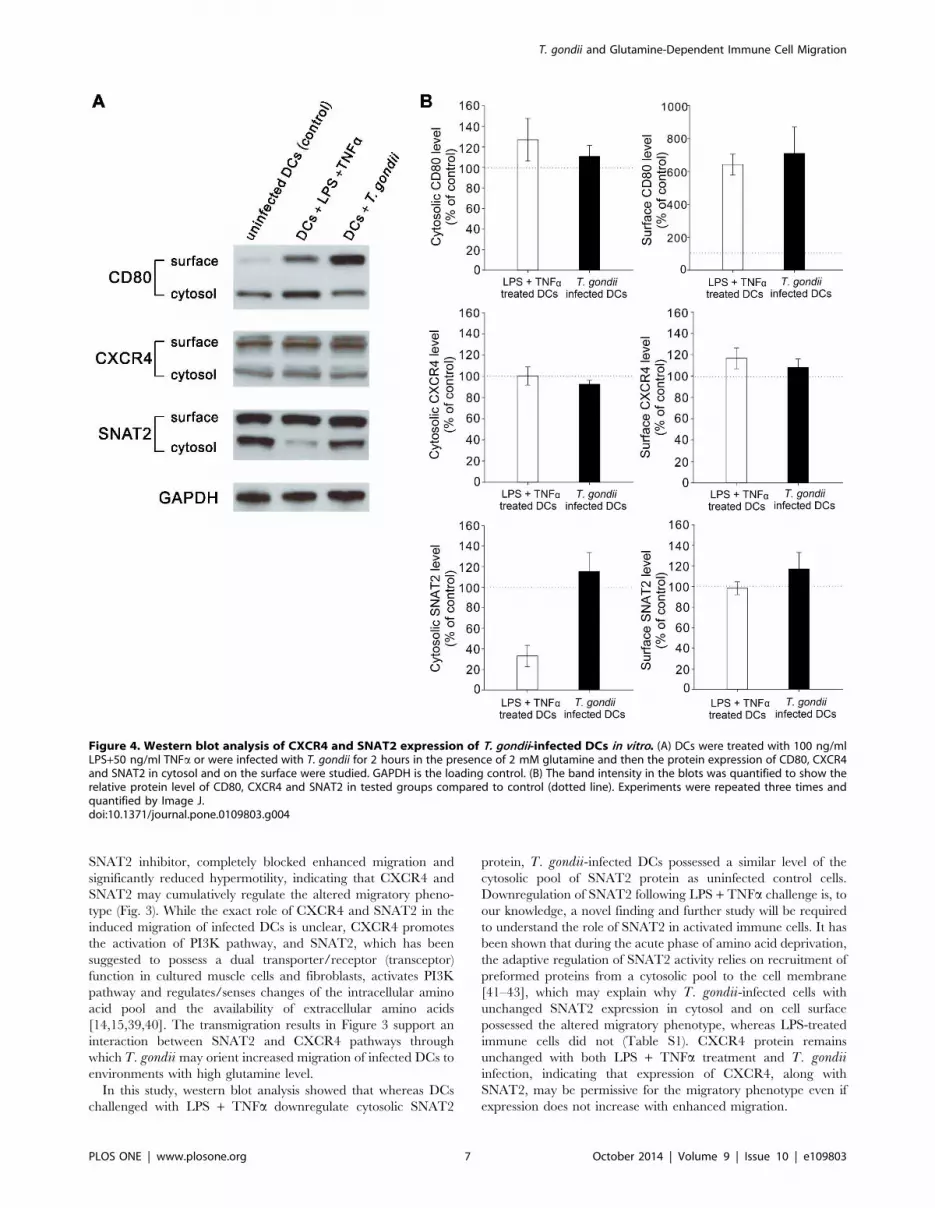
Figure 5. Schematic diagram of the hypothetical model. T. gondii increases the likelihood that infected immune cells will migrate toglutamine-rich environment by ‘‘turning on’’ the transceptor function of SNAT2 and modulating the CXCR4-PI3K-Rho kinase signaling pathwayswhich are known to regulate migration. Dotted lines denote the new possible interactions between T. gondii, SNAT2 and CXCR4.doi:10.1371/journal.pone.0109803.g005
T. gondii and Glutamine-Dependent Immune Cell Migration
PLOS ONE | www.plosone.org 8 October 2014 | Volume 9 | Issue 10 | e109803

parasites before 4% paraformaldehyde fixation. Coverslips were
then blocked for 30 minutes and incubated in Alexa Fluor 555
(1:1000) for 2 hours. A Zeiss confocal laser scanning microscope
(LSM) with a 406 objective was used to count the number of
parasites and total of 15 fields per coverslip were examined. The
ratio of intracellular tachyzoites to total tachyzoites in the
examined field on the coverslip is defined as infection efficiency.
T. gondii Replication Ability AssayHFF culture conditions were identical to the invasion assay.
HFFs were challenged with tachyzoites (MOI= 1) in complete
medium (which contains 2 mM L-glutamine) for 6 hours and then
washed with PBS to remove glutamine-rich medium and
extracellular tachyzoites before incubating in media with or
without 2 mM L-glutamine for another 16 hours. SAG1 antibody
was used to label extracellular parasites and stained with Alexa
Fluor 555 for 2 hours. The number of replication cycles
undergone by intracellular T. gondii was quantified for 15 fields
under a Zeiss confocal LSM with a 636 objective, and the mean
replication cycle was presented as the replication ability. No
parasites underwent more than 4 rounds of replication cycle in the
time period that we chose.
T. gondii Viability AssayFree tachyzoites were incubated in the medium containing 0 or
2 mM of L-glutamine or 2 mM of D-glutamine for 4 hours at
37uC in the incubator with 5% CO2, and then yellow MTT
(which is reduced to purple formazan by mitochondrial enzymes)
was added in living cells and the absorbance of colored solution
was quantified by a spectrophotometer.
Bone Marrow-Derived Dendritic Cell CultureThe bone barrow-derived DC culture was generated as
previously described [53], with slight modifications. Briefly, bone
marrow from 16 day-old Sprague-Dawley rat pups was isolated
and stored in liquid nitrogen until use. Upon thawing, cells were
washed with RPMI 1640 (Catalog number 21870, Life Technol-
ogy) and placed in T-75 flasks at a density of approximately 2.5-
3.56106 cells/ml in complete medium: RPMI 1640, 10% fetal
bovine serum, 2 mM L-glutamine, 1% nonessential amino acids,
1 mM sodium pyruvate, 50 units/ml penicillin +50 ug/ml strep-
tomycin (all from Life Technology) and the cytokines, interleukin-
4, granulocyte-macrophage colony stimulating factor (GM-CSF),
Flt-3 ligand (5 ng/ml per cytokine, R&D Systems). On the third
day, each flask was given 5 ml of fresh complete medium
containing GM-CSF only, and then three quarters of medium
containing non-adherent cells were replaced with fresh complete
medium with GM-CSF on day 5. Cells were used on day 7.
Flow CytometryDCs were infected with freshly egressed T. gondii (MOI=1) for
4 hours and then collected and washed in FACS buffer (1 mM
EDTA+1% FBS in PBS) on Day 7. An LSR II flow cytometer (BD
Biosciences) was used to investigate the infection frequency and
the results were analyzed with FlowJo Software (Tree Star, Inc.).
In Vitro Transmigration AssayCell migration was tested using Transwell assays (Costar 24-well
plate with inserts, 5 mm pore). Briefly, 1 million cells challenged
with freshly egressed T. gondii (MOI= 1) for 4 hours were
resuspended in culture medium consisting of RPMI 1640 with 1%
bovine serum albumin (BSA) and 2 mM L-glutamine in a 100-ml
volume. Another group of cells were infected with T. gondii thesame way for 2 hours following another 2 hours of inhibitor
treatments before being resuspended in culture medium as
described above. Infected and inhibitor-treated cells were then
counted by trypan blue staining to confirm the cell viability before
being transferred to the transwell system. Tested amino acids or
SDF-1a (final working concentration: 100 ng/ml) diluted in the
control medium (RPMI 1640+1% BSA) were added to the bottom
well in a 600-ml volume when the same amount of live cells in each
condition were added to the upper wells in a 100-ml volume. The
migration was assayed for 1.5 hours in a 37uC incubator with 5%
CO2 and then migrated cells were collected and counted by trypan
blue staining with a hemocytometer.
The tested amino acids were diluted in control medium at the
concentrations as following: 0.133 mM glycine, 0.0968 mM
histidine, 1.15 mM arginine, 0.136 mM glutamic acid, 0.15 mM
aspartic acid, 0.129 mM tyrosine, 0.0245 mM tryptophan,
0.0909 mM phenylalanine (Sigma). Because the formula of RPMI
1640 contains a mixture of amino acids with the concentrations
described above, adding another equal amount of amino acids in
the migration medium created 26 of tested amino acids, which
were comparable to 4 mM of glutamine (26Gln). Inhibitors were
used at following final concentrations: 40 mM MeAIB (Sigma),
5 mM MSO (Sigma), 50 mM LY294002 (Cell Signaling), 10 mM
Y27632 (Sigma), 50 mM AMD3100 (Abcam).
Cell Surface Protein Biotinylation and Western BlotSurface proteins of DCs were isolated by using the Pierce Cell
Surface Protein Isolation kit (Thermo Scientific) according to the
manufacturer’s protocol. In brief, cells grown in Falcon T-75 flasks
were washed with PBS, incubated with EZ-LINK Sulfo-NHS-
SSbiotin for 30 minutes at 4uC and then lysed with lysis buffer
containing protease inhibitor. The biotinylated surface proteins
were trapped within NeutrAvidin agarose gel when cytosol
proteins were eluted, and then surface proteins were eluted by
Bio-Rad sample buffer containing DTT.
Protein samples (25 mg) were separated on 10% SDS-PAGE gel
and then transferred to Bio-Rad PVDF membranes and
immunoblotted with antibodies against SNAT2 (1:500, Santa
Cruz biotechnology), CXCR4 (1:500, Abcam) and CD80
(1:10000, Abcam). Glyceraldehyde 3-phosphate dehydrogenase
(GAPDH) was used as a loading control (1:10000, Sigma). The
density of bands on 3 independent blots was quantified by Image J.
Statistical AnalysisStatistical analyses were performed using IBM SPSS statistics
software (version 20).
Supporting Information
Figure S1 Viability assay of T. gondii-infected HFFs in
vitro.
(DOCX)
Figure S2 Characterization of rat bone marrow-derived
DC cultures on Day 7.
(DOCX)
Figure S3 Viability assay of intracellular T. gondii in
HFFs following inhibitor treatments.
(DOCX)
T. gondii and Glutamine-Dependent Immune Cell Migration
PLOS ONE | www.plosone.org 9 October 2014 | Volume 9 | Issue 10 | e109803

Table S1 Transmigration assay in vitro: cell migrationof activated DCs to glutamine or other tested aminoacids.(DOCX)
Acknowledgments
We thank Anita Koshy and John Boothroyd for the strain of T. gondii andthe SAG1 antibody. Advice and feedback was provided in all phases of this
project by John Boothroyd and Michelle Cheng.
Author Contributions
Conceived and designed the experiments: IL. Performed the experiments:
IL AKE CY MGW VK ZDM NCM. Analyzed the data: IL AKE CY
MGW. Wrote the paper: IL AKE RMS.
References
1. MacRae JI, Dixon MW, Dearnley MK, Chua HH, Chambers JM, et al. (2013)
Mitochondrial metabolism of sexual and asexual blood stages of the malaria
parasite Plasmodium falciparum. BMC Biol 11: 67.
2. MacRae JI, Sheiner L, Nahid A, Tonkin C, Striepen B, et al. (2012)
Mitochondrial metabolism of glucose and glutamine is required for intracellular
growth of Toxoplasma gondii. Cell Host Microbe 12: 682–692.
3. Blume M, Rodriguez-Contreras D, Landfear S, Fleige T, Soldati-Favre D, et al.
(2009) Host-derived glucose and its transporter in the obligate intracellular
pathogen Toxoplasma gondii are dispensable by glutaminolysis. Proc Natl Acad
Sci U S A 106: 12998–13003.
4. Sheiner L, Vaidya AB, McFadden GI (2013) The metabolic roles of the
endosymbiotic organelles of Toxoplasma and Plasmodium spp. Curr Opin
Microbiol 16: 452–458.
5. Broer S, Brookes N (2001) Transfer of glutamine between astrocytes and
neurons. J Neurochem 77: 705–719.
6. Kalloniatis M, Tomisich G (1999) Amino acid neurochemistry of the vertebrate
retina. Prog Retin Eye Res 18: 811–866.
7. Tjader I, Berg A, Wernerman J (2007) Exogenous glutamine—compensating a
shortage? Crit Care Med 35: S553–S556.
8. Tanhoffer RA, Yamazaki RK, Nunes EA, Pchevozniki AI, Pchevozniki AM,
et al. (2007) Glutamine concentration and immune response of spinal cord-
injured rats. J Spinal Cord Med 30: 140–146.
9. Albrecht J, Sidoryk-Wegrzynowicz M, Zielinska M, Aschner M (2010) Roles of
glutamine in neurotransmission. Neuron Glia Biol 6: 263–276.
10. Lambert H, Hitziger N, Dellacasa I, Svensson M, Barragan A (2006) Induction
of dendritic cell migration upon Toxoplasma gondii infection potentiates
parasite dissemination. Cell Microbiol 8: 1611–1623.
11. Lambert H, Vutova PP, Adams WC, Lore K, Barragan A (2009) The
Toxoplasma gondii-shuttling function of dendritic cells is linked to the parasite
genotype. Infect Immun 77: 1679–1688.
12. Procko E, McColl SR (2005) Leukocytes on the move with phosphoinositide 3-
kinase and its downstream effectors. Bioessays 27: 153–163.
13. Cain RJ, Ridley AJ (2009) Phosphoinositide 3-kinases in cell migration. Biol Cell
101: 13–29.
14. Hyde R, Cwiklinski EL, MacAulay K, Taylor PM, Hundal HS (2007) Distinct
sensor pathways in the hierarchical control of SNAT2, a putative amino acid
transceptor, by amino acid availability. J Biol Chem 282: 19788–19798.
15. Evans K, Nasim Z, Brown J, Clapp E, Amin A, et al. (2008) Inhibition of
SNAT2 by metabolic acidosis enhances proteolysis in skeletal muscle. J Am Soc
Nephrol 19: 2119–2129.
16. Mackenzie B, Erickson JD (2004) Sodium-coupled neutral amino acid (System
N/A) transporters of the SLC38 gene family. Pflugers Arch 447: 784–795.
17. Li P, Yin YL, Li D, Kim SW, Wu G (2007) Amino acids and immune function.
Br J Nutr 98: 237–252.
18. Newsholme P (2001) Why is L-glutamine metabolism important to cells of the
immune system in health, postinjury, surgery or infection? J Nutr 131: 2515S–
2522S.
19. Saeij JP, Boyle JP, Boothroyd JC (2005) Differences among the three major
strains of Toxoplasma gondii and their specific interactions with the infected
host. Trends Parasitol 21: 476–481.
20. Darde ML (2004) Genetic analysis of the diversity in Toxoplasma gondii. Ann Ist
Super Sanita 40: 57–63.
21. Saeij JP, Boyle JP, Grigg ME, Arrizabalaga G, Boothroyd JC (2005)
Bioluminescence imaging of Toxoplasma gondii infection in living mice reveals
dramatic differences between strains. Infect Immun 73: 695–702.
22. Boothroyd JC, Grigg ME (2002) Population biology of Toxoplasma gondii and
its relevance to human infection: do different strains cause different disease?
Curr Opin Microbiol 5: 438–442.
23. Vyas A, Kim SK, Giacomini N, Boothroyd JC, Sapolsky RM (2007) Behavioral
changes induced by Toxoplasma infection of rodents are highly specific to
aversion of cat odors. Proc Natl Acad Sci U S A 104: 6442–6447.
24. Vyas A (2013) Parasite-augmented mate choice and reduction in innate fear in
rats infected by Toxoplasma gondii. J Exp Biol 216: 120–126.
25. Maroni BJ, Karapanos G, Mitch WE (1986) System A amino acid transport in
incubated muscle: effects of insulin and acute uremia. Am J Physiol 251: F74–
F80.
26. Rae C, Hare N, Bubb WA, McEwan SR, Broer A, et al. (2003) Inhibition of
glutamine transport depletes glutamate and GABA neurotransmitter pools:
further evidence for metabolic compartmentation. J Neurochem 85: 503–514.
27. Song C, Li G (2011) CXCR4 and matrix metalloproteinase-2 are involved in
mesenchymal stromal cell homing and engraftment to tumors. Cytotherapy 13:
549–561.
28. Lee JY, Buzney CD, Poznansky MC, Sackstein R (2009) Dynamic alterations in
chemokine gradients induce transendothelial shuttling of human T cells under
physiologic shear conditions. J Leukoc Biol 86: 1285–1294.
29. Badillo AT, Zhang L, Liechty KW (2008) Stromal progenitor cells promote
leukocyte migration through production of stromal-derived growth factor
1alpha: a potential mechanism for stromal progenitor cell-mediated enhance-
ment of cellular recruitment to wounds. J Pediatr Surg 43: 1128–1133.
30. Kraemer BF, Borst O, Gehring EM, Schoenberger T, Urban B, et al. (2010) PI3
kinase-dependent stimulation of platelet migration by stromal cell-derived factor
1 (SDF-1). J Mol Med (Berl) 88: 1277–1288.
31. Fuks JM, Arrighi RB, Weidner JM, Kumar MS, Jin Z, et al. (2012) GABAergic
signaling is linked to a hypermigratory phenotype in dendritic cells infected by
Toxoplasma gondii. PLoS Pathog 8: e1003051.
32. Lambert H, Barragan A (2010) Modelling parasite dissemination: host cell
subversion and immune evasion by Toxoplasma gondii. Cell Microbiol 12: 292–
300.
33. Gupta R, Chattopadhyay D (2009) Glutamate is the chemotaxis-inducing factor
in placental extracts. Amino Acids 37: 359–366.
34. Gupta R, Palchaudhuri S, Chattopadhyay D (2013) Glutamate induces
neutrophil cell migration by activating class I metabotropic glutamate receptors.
Amino Acids 44: 757–767.
35. Rot A, von Andrian UH (2004) Chemokines in innate and adaptive host defense:
basic chemokinese grammar for immune cells. Annu Rev Immunol 22: 891–928.
36. Tan W, Martin D, Gutkind JS (2006) The Galpha13-Rho signaling axis is
required for SDF-1-induced migration through CXCR4. J Biol Chem 281:
39542–39549.
37. Fricker SP, Anastassov V, Cox J, Darkes MC, Grujic O, et al. (2006)
Characterization of the molecular pharmacology of AMD3100: a specific
antagonist of the G-protein coupled chemokine receptor, CXCR4. Biochem
Pharmacol 72: 588–596.
38. Hatse S, Princen K, Bridger G, De CE, Schols D (2002) Chemokine receptor
inhibition by AMD3100 is strictly confined to CXCR4. FEBS Lett 527: 255–
262.
39. Evans K, Nasim Z, Brown J, Butler H, Kauser S, et al. (2007) Acidosis-sensing
glutamine pump SNAT2 determines amino acid levels and mammalian target of
rapamycin signalling to protein synthesis in L6 muscle cells. J Am Soc Nephrol
18: 1426–1436.
40. Gazzola RF, Sala R, Bussolati O, Visigalli R, Dall’Asta V, et al. (2001) The
adaptive regulation of amino acid transport system A is associated to changes in
ATA2 expression. FEBS Lett 490: 11–14.
41. Hyde R, Christie GR, Litherland GJ, Hajduch E, Taylor PM, et al. (2001)
Subcellular localization and adaptive up-regulation of the System A (SAT2)
amino acid transporter in skeletal-muscle cells and adipocytes. Biochem J 355:
563–568.
42. Jones HN, Ashworth CJ, Page KR, McArdle HJ (2006) Expression and adaptive
regulation of amino acid transport system A in a placental cell line under amino
acid restriction. Reproduction 131: 951–960.
43. Ling R, Bridges CC, Sugawara M, Fujita T, Leibach FH, et al. (2001)
Involvement of transporter recruitment as well as gene expression in the
substrate-induced adaptive regulation of amino acid transport system A.
Biochim Biophys Acta 1512: 15–21.
44. Pinilla J, Aledo JC, Cwiklinski E, Hyde R, Taylor PM, et al. (2011) SNAT2
transceptor signalling via mTOR: a role in cell growth and proliferation? Front
Biosci (Elite Ed) 3: 1289–1299.
45. Hay N, Sonenberg N (2004) Upstream and downstream of mTOR. Genes Dev
18: 1926–1945.
46. Tokunaga C, Yoshino K, Yonezawa K (2004) mTOR integrates amino acid-
and energy-sensing pathways. Biochem Biophys Res Commun 313: 443–446.
47. Hundal HS, Taylor PM (2009) Amino acid transceptors: gate keepers of nutrient
exchange and regulators of nutrient signaling. Am J Physiol Endocrinol Metab
296: E603–E613.
T. gondii and Glutamine-Dependent Immune Cell Migration
PLOS ONE | www.plosone.org 10 October 2014 | Volume 9 | Issue 10 | e109803

48. Boothroyd JC, Dubremetz JF (2008) Kiss and spit: the dual roles of Toxoplasmarhoptries. Nat Rev Microbiol 6: 79–88.
49. Bougdour A, Tardieux I, Hakimi MA (2013) Toxoplasma exports dense granuleproteins beyond the vacuole to the host cell nucleus and rewires the host genomeexpression. Cell Microbiol.
50. Persat F, Mercier C, Ficheux D, Colomb E, Trouillet S, et al. (2012) A syntheticpeptide derived from the parasite Toxoplasma gondii triggers human dendriticcells’ migration. J Leukoc Biol 92: 1241–1250.
51. Fleckenstein MC, Reese ML, Konen-Waisman S, Boothroyd JC, Howard JC,et al. (2012) A Toxoplasma gondii pseudokinase inhibits host IRG resistanceproteins. PLoS Biol 10: e1001358.
52. Hunter CA, Sibley LD (2012) Modulation of innate immunity by Toxoplasmagondii virulence effectors. Nat Rev Microbiol 10: 766–778.
53. Manley NC, Caso JR, Works MG, Cutler AB, Zemlyak I, et al. (2013)Derivation of injury-responsive dendritic cells for acute brain targeting andtherapeutic protein delivery in the stroke-injured rat. PLoS One 8: e61789.
T. gondii and Glutamine-Dependent Immune Cell Migration
PLOS ONE | www.plosone.org 11 October 2014 | Volume 9 | Issue 10 | e109803
Copyright © 2022 FDOKUMEN