Design and biophysical characterization of atrazine-sensing peptides mimicking the Chlamydomonas...
8
13108 Phys. Chem. Chem. Phys., 2013, 15, 13108--13115 This journal is c the Owner Societies 2013 Cite this: Phys. Chem. Chem. Phys., 2013, 15, 13108 Design and biophysical characterization of atrazine- sensing peptides mimicking the Chlamydomonas reinhardtii plastoquinone binding niche Viviana Scognamiglio, a Pasquale Stano, b Fabio Polticelli, bc Amina Antonacci, a Maya Dimova Lambreva, a Giorgio Pochetti, a Maria Teresa Giardi a and Giuseppina Rea* a The plastoquinone (Q B ) binding niche of the Photosystem II (PSII) D1 protein is the subject of intense research due to its capability to bind also anthropogenic pollutants. In this work, the Chlamydomonas reinhardtii D1 primary structure was used as a template to computationally design novel peptides enabling the binding of the herbicide atrazine. Three biomimetic molecules, containing the Q B -binding site in a loop shaped by two a-helices, were reconstituted by automated protein synthesis, and their structural and functional features deeply analysed by biophysical techniques. Standing out among the others, the biomimetic mutant peptide, D1pepMut, showed high ability to mimic the D1 protein in binding both Q B and atrazine. Circular dichroism spectra suggested a typical properly-folded a-helical structure, while isothermal titration calorimetry (ITC) provided a complete thermodynamic characterization of the molecular interaction. Atrazine binds to the D1pepMut with a high affinity (K d = 2.84 mM), and a favourable enthalpic contribution (DH = 11.9 kcal mol 1 ) driving the interaction. Fluorescence spectroscopy assays, in parallel to ITC data, provided hyperbolic titration curves indicating the occurrence of a single atrazine binding site. The binding resulted in structural stabilisation of the D1pepMut molecule, as suggested by atrazine-induced cooperative profiles for the fold–unfold transition. The interaction dynamics and the structural stability of the peptides in response to the ligand were particularly considered as mandatory parameters for biosensor/ biochip development. These studies paved the way to the set-up of an array of synthetic mutant peptides with a wide range of affinity towards different classes of target analytes, for the development of optical nanosensing platforms for herbicide detection. Highlights Novel biomimetic peptides of the photosynthetic plastoquinone binding niche were designed by exploring the D1 primary structure of Chlamydomonas reinhardtii. Circular dichroism, fluorescence spectroscopy and micro- calorimetry experiments addressed the biophysical and struc- tural properties of the novel produced bio-inspired molecules. The redefined more hydrophilic peptide sequence of D1pepMut adopted a properly-folded a-helical structure. D1pepMut had the ability to bind either plastoquinone or atrazine. Atrazine binding to D1pepMut enhances the peptide structural stability. Introduction The photosynthetic membranes host macromolecular assem- blies of enzymes and pigments enabling plants, cyanobacteria and algae to sustain life on Earth. 1 Acting as a light-driven water:plastoquinone oxidoreductase, the Photosystem II (PSII) entraps light energy by the chlorophyll–protein harvesting complexes and transfers it to the active reaction center domi- nated by the D1–D2 heterodimer and oxygen evolving complex. The absorbed excitons induce a charge separation in a special dimer of Chl a molecules generating a single electron transfer towards a pheophytin molecule, then to a plastoquinone mole- cule in D2, Q A , and finally, to a second plastoquinone in D1, Q B . The electron transfer chain rounds out beyond the PSII, leading a Institute of Crystallography, CNR, Via Salaria km 29.3, Monterotondo, 00015 Rome, Italy. E-mail: [email protected] b Department of Sciences, University Roma Tre, 00146 Rome, Italy c National Institute of Nuclear Physics, Roma Tre Section, 00146 Rome, Italy Received 8th May 2013, Accepted 2nd June 2013 DOI: 10.1039/c3cp51955d www.rsc.org/pccp PCCP PAPER
-
Upload
independent -
Category
Documents
-
view
4 -
download
0
Transcript of Design and biophysical characterization of atrazine-sensing peptides mimicking the Chlamydomonas...

13108 Phys. Chem. Chem. Phys., 2013, 15, 13108--13115 This journal is c the Owner Societies 2013
Cite this: Phys. Chem.Chem.Phys.,2013,15, 13108
Design and biophysical characterization of atrazine-sensing peptides mimicking the Chlamydomonasreinhardtii plastoquinone binding niche
Viviana Scognamiglio,a Pasquale Stano,b Fabio Polticelli,bc Amina Antonacci,a
Maya Dimova Lambreva,a Giorgio Pochetti,a Maria Teresa Giardia andGiuseppina Rea*a
The plastoquinone (QB) binding niche of the Photosystem II (PSII) D1 protein is the subject of intense
research due to its capability to bind also anthropogenic pollutants. In this work, the Chlamydomonas
reinhardtii D1 primary structure was used as a template to computationally design novel peptides enabling
the binding of the herbicide atrazine. Three biomimetic molecules, containing the QB-binding site in a loop
shaped by two a-helices, were reconstituted by automated protein synthesis, and their structural and
functional features deeply analysed by biophysical techniques. Standing out among the others, the
biomimetic mutant peptide, D1pepMut, showed high ability to mimic the D1 protein in binding both QB
and atrazine. Circular dichroism spectra suggested a typical properly-folded a-helical structure, while
isothermal titration calorimetry (ITC) provided a complete thermodynamic characterization of the molecular
interaction. Atrazine binds to the D1pepMut with a high affinity (Kd = 2.84 mM), and a favourable enthalpic
contribution (DH = �11.9 kcal mol�1) driving the interaction. Fluorescence spectroscopy assays, in parallel to
ITC data, provided hyperbolic titration curves indicating the occurrence of a single atrazine binding site. The
binding resulted in structural stabilisation of the D1pepMut molecule, as suggested by atrazine-induced
cooperative profiles for the fold–unfold transition. The interaction dynamics and the structural stability of
the peptides in response to the ligand were particularly considered as mandatory parameters for biosensor/
biochip development. These studies paved the way to the set-up of an array of synthetic mutant peptides
with a wide range of affinity towards different classes of target analytes, for the development of optical
nanosensing platforms for herbicide detection.
Highlights
� Novel biomimetic peptides of the photosynthetic plastoquinonebinding niche were designed by exploring the D1 primarystructure of Chlamydomonas reinhardtii.� Circular dichroism, fluorescence spectroscopy and micro-
calorimetry experiments addressed the biophysical and struc-tural properties of the novel produced bio-inspired molecules.� The redefined more hydrophilic peptide sequence of
D1pepMut adopted a properly-folded a-helical structure.� D1pepMut had the ability to bind either plastoquinone or
atrazine.
� Atrazine binding to D1pepMut enhances the peptidestructural stability.
Introduction
The photosynthetic membranes host macromolecular assem-blies of enzymes and pigments enabling plants, cyanobacteriaand algae to sustain life on Earth.1 Acting as a light-drivenwater:plastoquinone oxidoreductase, the Photosystem II (PSII)entraps light energy by the chlorophyll–protein harvestingcomplexes and transfers it to the active reaction center domi-nated by the D1–D2 heterodimer and oxygen evolving complex.The absorbed excitons induce a charge separation in a specialdimer of Chl a molecules generating a single electron transfertowards a pheophytin molecule, then to a plastoquinone mole-cule in D2, QA, and finally, to a second plastoquinone in D1, QB.The electron transfer chain rounds out beyond the PSII, leading
a Institute of Crystallography, CNR, Via Salaria km 29.3, Monterotondo,
00015 Rome, Italy. E-mail: [email protected] Department of Sciences, University Roma Tre, 00146 Rome, Italyc National Institute of Nuclear Physics, Roma Tre Section, 00146 Rome, Italy
Received 8th May 2013,Accepted 2nd June 2013
DOI: 10.1039/c3cp51955d
www.rsc.org/pccp
PCCP
PAPER

This journal is c the Owner Societies 2013 Phys. Chem. Chem. Phys., 2013, 15, 13108--13115 13109
to the final production of reducing equivalents and molecularoxygen.2,3
The highly homologous D1 and D2 proteins stand out at theheart of the PSII core complex interacting with all the redoxcofactors involved in the charge separation and electron trans-port across the membrane. Since its discovery, the chloroplasticpsbA gene-encoded D1 protein has received particular attentiondue to its involvement in the PSII assembly and repair cycle,4,5 andcapability to bind either plastoquinones (PQs) or xenobiotics.6 TheD1 primary structure is highly conserved and the protein foldsinto five transmembrane a-helices (named from A to E), withtheir N- and C-termini exposed to the stromal and lumenalsides, respectively. Besides, two short helices occur in thestromal loop, one connecting the membrane spanning helicesIII–IV (named CD helix), and the other in the lumenal loopconnecting helices IV–V (named DE helix). The helices IV and Vand their connecting loop integrating the small DE helix build upthe QB binding niche.7–9 An in silico model of the Chlamydomonasreinhardtii D1 protein three-dimensional structure identified asequence of approx. 70 amino acids, ranging from residue 211 toresidue 280, involved in the arrangement of the QB pocket, andpaved the way to the exploration of its physico-chemical propertiesby novel computational and engineering methodologies.8,10,11
In the agro-food field, the discovery of spontaneous D1mutations in several weed species survived to the extensive useof the PSII herbicide atrazine,12 laid a foundation for the rationaleuse of herbicides, and provided insights into the molecularmechanisms underlying the photosynthetic electron transfer.13,14
Atrazine competitively inhibits QB binding to the D1 proteinimpairing the natural flux of the electrons to the subsequentacceptors, and inducing an accumulation of the reduced form ofQA. Acting in a similar manner, other poisonous xenobiotics suchas triazinic and ureic herbicides affect the photosynthetic electrontransfer.15,16 This amazing feature of PSII to bind different classesof toxic compounds stimulated the design of analytical devicesexploiting the photosynthetic activity of whole cells, or their activesubcomponents, as bio-recognition elements.17,18 In this context,although the use of thylakoidal membranes or isolated proteincomplexes provides high sensitivity, their extraction, purificationor in vitro reconstitution procedures are often laborious, expensiveand in some cases not always reliable. On the other side, wholecell-based biosensors preserve photosystem functionality, butprovide a slow response and sometimes low sensitivity.19
The improved resolution of PSII atomic structure from thermo-philic cyanobacteria revealed molecular insights into the PSIIreaction centre empowering the exploitation of protein engineeringtools to fine tune its structural and functional properties.20–22
In the nanotechnology era, numerous research efforts havebeen focused on the design of novel biomimetic molecules ableto simulate biological processes, such as the binding of specificanalytes.23,24 These synthetic compounds include peptides,mini-proteins, imprinting polymers or aptamers, and usuallyderive from a size-minimization strategy, which also aims to finetune their sensitivity, selectivity, and robustness properties.25,26
The novel products can be exploited as simplified models todeepen the knowledge of the natural macromolecular functions
and as molecular tools finding applications in the developmentof analytical devices, such as biosensors and biochips.27–30
In this work, the C. reinhardtii D1 primary structure was used asa template to design new peptides enabling atrazine binding. Bycomputational modeling and automated protein synthesis, the D1plastoquinone–atrazine binding niche in native and mutated formswas reconstituted and the structural and functional features wereanalyzed in detail by circular dichroism, fluorescence spectroscopyand microcalorimetry. In particular, the interaction dynamics andthe structural stability of the peptides in response to the ligandwere considered being mandatory parameters in biosensors/biochips development.
ExperimentalAnalysis of D1 protein three-dimensional structure and designof the biomimetic peptides
Analysis of the C. reinhardtii D1 protein three-dimensionalstructure was carried out on a previously published molecularmodel of the C. reinhardtii reaction center6 built using thecrystal structure of the Thermosynechococcus elongatus reactioncenter as a template15 (PDB code 2AXT). Analysis of the modelevidenced that the QB binding niche is held in place by theinteraction between hydrophobic residues located on the twoadjacent D and E helices. In particular, a hydrophobic cluster,formed by residue Phe211 on helix D, and residues Phe274,Leu275, Trp278 and Pro279 on helix E, is located just beneaththe QB binding pocket. Thus, with the aim of preserving theseinteractions, a first protein minimization was carried out bydesigning a peptide encompassing the 211–280 region of the D1protein (named D1pep70). Characterization of the correspondingsynthetic peptide revealed its poor solubility (see the Results anddiscussion section) prompting the addition of two Lys residues atthe N-terminus and at the C-terminus of the peptide to increase itssolubility. However, notwithstanding the higher water solubility ofthis second peptide variant, named D1pepLys, CD spectroscopyanalysis indicated a low ordered structure content. A third peptidevariant was then designed by substituting six Phe residues with Tyr,four Ala residues with Ser (213, 233, 250, and 263), eliminatingresidues 276 to 280 and adding three Lys residues at both theN- and C-terminal ends of the peptide. This latter peptide variantwas named D1pepMut.
Biomimetic peptide synthesis
The peptides were produced by automated synthesis by GenScriptCorporation (120 Centennial Ave., Suite 105, Piscataway, NJ 08854,USA). The peptides were synthesized by liquid phase peptidesynthesis (LPPS) and solid phase peptide synthesis (SPPS) platforms(FlexPeptideTM), and purified to obtain samples with >90% ofpurity. The synthetic molecules were analyzed by MS/HPLC andsequence confirmation was performed.
Circular dichroism
Circular dichroism spectra were performed under a nitrogen flowon a J-600 Spectropolarimeter (Jasco, Tokyo, Japan) equippedwith the Neslab RTE-110 temperature controlled liquid system
Paper PCCP
13110 Phys. Chem. Chem. Phys., 2013, 15, 13108--13115 This journal is c the Owner Societies 2013
(Neslab Instruments, Portsmouth, NH). Peptide solutions ofabout 0.2 mg ml�1 in 1 mM NaPi buffer pH 7.0 were placed insealed cuvettes with a 0.05 cm path length (Hellma, Mulheim,Germany), and spectra were measured between 250 and 190 nmwith the following setup: bandwidth 1 nm, response 0.25 s, speed50 nm min�1, sensitivity 20 mdeg, and step resolution 1 nm.All spectra were averaged 16 times and smoothed using theSpectropolarimeter System Software, Version 1.00 (Jasco). Beforemeasurements, all samples were temperature equilibrated for5 min. During the measurement the photomultiplier voltagenever exceeded 600 V. The results were expressed in terms of themolar ellipticity, as previously reported.31
Fluorescence spectroscopy
Fluorescence spectra were recorded in 1 mM NaPi buffer, pH 7.0peptide solutions. Steady-state fluorescence measurements wereperformed on a F-8200 Spectrofluorometer (Jasco, Tokyo, Japan)with a cell temperature controlled sample holder, equipped withthe Neslab RTE-110 temperature controlled liquid system(Neslab Instruments, Portsmouth, NH). Peptide fluorescencewas excited at 280 nm, with an emission and excitation slitwidth of 5 nm. Before measurements, all samples were tempera-ture equilibrated for 5 min.
Quenching
Peptide fluorescence was quenched by acrylamide or sodiumiodide and observed at the fluorescence maximum. Steady statequenching was analyzed by the Stern–Volmer equation:
F0
F� 1 ¼ KSV½Q�
where F0 and F are the fluorescence intensities in the absenceand in the presence of the quencher, respectively, [Q] is theconcentration of the quencher, and KSV the apparent Stern–Volmer constant.32
Isothermal titration calorimetry
ITC experiments were performed at 25 1C using a MicroCalITC200 microcalorimeter (MicroCal Inc., Northampton, MA, USA).The peptide solution (20 mM, in buffer 1 mM PBS pH 7.0, 1%MeOH, 0.1% SDS) was placed in the sample cell, and the ligandsolution (200 mM, in the peptide buffer) was loaded into thesyringe injector. The titration involved 19 injections of 2 mL at180 s intervals. The syringe stirring speed was set to 1000 rpm.Reference titration of the ligand into buffer was used to correctfor heat of dilution. The thermodynamic data were processedusing the Origin 7.0 software provided by MicroCal. Fitting thebinding isotherm with the one-site binding model yieldedthe values of DH and of the association constant (Ka). Thesystem also gave information of the change in entropy (DS).
The inflection point in the isotherm gives the stoichiometryvalue n, indicating the ligand/peptide ratio of the binding. Tocorrect for any discrepancy in the baseline outlined by thesoftware, a manual adjustment was performed.
Results and discussion
A computational strategy has been adopted for designing andminimizing the QB binding domain of C. reinhardtii as a receptorfor atrazine binding. In particular, we explored by in silico/in vitrostudies, the structure and binding ability of synthetic peptidesthat could mimic the QB–herbicide binding niche of the PSIIreaction center D1 protein. The primary sequences of thebiomimetic peptides are reported in Table 1.
The in silico model of the whole D1 protein from C. reinhardtiiwas preliminary to the actual studies8 (Fig. 1A). Starting from thispoint, we have identified a protein sequence, possibly having theproper physico-chemical features to adopt a stable fold, andtherefore reproduce the QB–herbicide binding niche (highlightedin Fig. 1A). The protein domains were designed to consist of threealpha helices connected by a hydrophilic loop. Hence, weapproached the goal of producing synthetic biomimetic peptidesby a stepwise optimization of the peptide sequence followingexperimental analyses of the different peptide variants.
The first peptide, called D1pep70, is a 70 amino acids longpeptide corresponding to the native D1 sequence from residue 211to 280. The in silico model of D1pep70 consists of two a-helicesat the N-terminal and the C-terminal and a loop of 50 aminoacids, which corresponds to the QB binding site. The chemicallysynthesized D1pep70 appeared as a highly hydrophobic moleculesoluble only in buffers containing DMSO (20% v/v) and 0.1% (w/v)octyl-glucoside. Due to the presence of such a high concentration ofDMSO, any structural analysis of the molecule resulted to bedifficult and prone to artifacts (data not shown).
For this reason, a second peptide called D1pepLys wasdesigned by adding two Lys residues at the N- and C-terminalends of the D1pep70 peptide, in order to increase its solubilityin water. The addition of two terminal Lys residues had theadditional advantage of facilitating the immobilization of themacromolecule on different organic/inorganic supports by meansof covalent attachment.33 D1pepLys peptide showed a highersolubility in aqueous solvents when compared to D1pep70. Thisallowed a series of circular dichroism (CD) and fluorescencespectroscopy studies aimed at determining its structural stabilityand binding capability. However, CD measurements also revealedin this case a poorly structured fold, so we moved one step furtherand introduced additional changes in the peptide sequence.
The third peptide, called D1pepMut, was obtained byredefining the sequence in order to obtain a new moleculewhich could be more hydrophilic although maintaining its
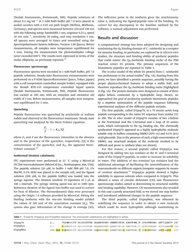
Table 1 D1pep70, D1pepLys and D1pepMut sequences. The aminoacidic substitutions are highlighted in yellow
D1pep70D1pepLys
D1pepMut
PCCP Paper
This journal is c the Owner Societies 2013 Phys. Chem. Chem. Phys., 2013, 15, 13108--13115 13111
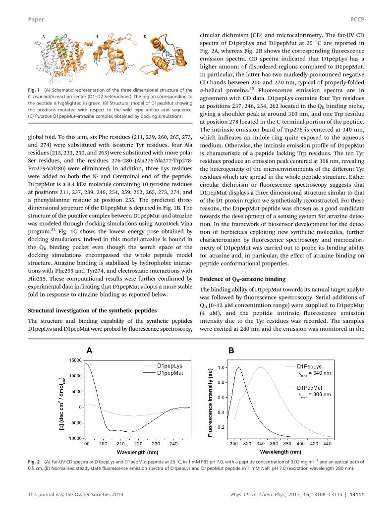
global fold. To this aim, six Phe residues (211, 239, 260, 265, 273,and 274) were substituted with isosteric Tyr residues, four Alaresidues (213, 233, 250, and 263) were substituted with more polarSer residues, and the residues 276–280 (Ala276-Ala277-Trp278-Pro279-Val280) were eliminated; in addition, three Lys residueswere added to both the N- and C-terminal end of the peptide.D1pepMut is a 8.4 kDa molecule containing 10 tyrosine residuesat positions 211, 237, 239, 246, 254, 259, 262, 265, 273, 274, anda phenylalanine residue at position 255. The predicted three-dimensional structure of the D1pepMut is depicted in Fig. 1B. Thestructure of the putative complex between D1pepMut and atrazinewas modeled through docking simulations using AutoDock Vinaprogram.34 Fig. 1C shows the lowest energy pose obtained bydocking simulations. Indeed in this model atrazine is bound inthe QB binding pocket even though the search space of thedocking simulations encompassed the whole peptide modelstructure. Atrazine binding is stabilized by hydrophobic interac-tions with Phe255 and Tyr274, and electrostatic interactions withHis215. These computational results were further confirmed byexperimental data indicating that D1pepMut adopts a more stablefold in response to atrazine binding as reported below.
Structural investigation of the synthetic peptides
The structure and binding capability of the synthetic peptidesD1pepLys and D1pepMut were probed by fluorescence spectroscopy,
circular dichroism (CD) and microcalorimetry. The far-UV CDspectra of D1pepLys and D1pepMut at 25 1C are reported inFig. 2A, whereas Fig. 2B shows the corresponding fluorescenceemission spectra. CD spectra indicated that D1pepLys has ahigher amount of disordered regions compared to D1pepMut.In particular, the latter has two markedly pronounced negativeCD bands between 200 and 220 nm, typical of properly-foldeda-helical proteins.35 Fluorescence emission spectra are inagreement with CD data. D1pepLys contains four Tyr residuesat positions 237, 246, 254, 262 located in the QB binding niche,giving a shoulder peak at around 310 nm, and one Trp residueat position 278 located in the C-terminal portion of the peptide.The intrinsic emission band of Trp278 is centered at 340 nm,which indicates an indole ring quite exposed to the aqueousmedium. Otherwise, the intrinsic emission profile of D1pepMutis characteristic of a peptide lacking Trp residues. The ten Tyrresidues produce an emission peak centered at 308 nm, revealingthe heterogeneity of the microenvironments of the different Tyrresidues which are spread in the whole peptide structure. Eithercircular dichroism or fluorescence spectroscopy suggests thatD1pepMut displays a three-dimensional structure similar to thatof the D1 protein region we synthetically reconstructed. For thesereasons, the D1pepMut peptide was chosen as a good candidatetowards the development of a sensing system for atrazine detec-tion. In the framework of biosensor development for the detec-tion of herbicides exploiting new synthetic molecules, furthercharacterization by fluorescence spectroscopy and microcalori-metry of D1pepMut was carried out to probe its binding abilityfor atrazine and, in particular, the effect of atrazine binding onpeptide conformational properties.
Evidence of QB–atrazine binding
The binding ability of D1pepMut towards its natural target analytewas followed by fluorescence spectroscopy. Serial additions ofQB (0–12 mM concentration range) were supplied to D1pepMut(4 mM), and the peptide intrinsic fluorescence emissionintensity due to the Tyr residues was recorded. The sampleswere excited at 280 nm and the emission was monitored in the
Fig. 1 (A) Schematic representation of the three dimensional structure of theC. reinhardtii reaction center (D1–D2 heterodimer). The region corresponding tothe peptide is highlighted in green. (B) Structural model of D1pepMut showingthe positions mutated with respect to the wild type amino acid sequence.(C) Putative D1pepMut–atrazine complex obtained by docking simulations.
Fig. 2 (A) Far-UV CD spectra of D1pepLys and D1pepMut peptide at 25 1C, in 1 mM PBS pH 7.0, with a peptide concentration of 0.02 mg ml�1 and an optical path of0.5 cm. (B) Normalised steady-state fluorescence emission spectra of D1pepLys and D1pepMut peptide in 1 mM NaPi pH 7.0 (excitation wavelength 280 nm).
Paper PCCP

13112 Phys. Chem. Chem. Phys., 2013, 15, 13108--13115 This journal is c the Owner Societies 2013
300 to 450 nm region. The tyrosine emission band decreases asthe quinone concentration increases. Corrections were madefor dilution of the sample and for a background signal frombuffer. The peptide showed high affinity towards QB as indi-cated by the fluorescence binding curve of D1pepMut in thepresence of increasing amounts of QB in Fig. 3A. To measureligand binding affinity towards herbicides, serial additions ofatrazine (0–12 mM concentration range) were supplied toD1pepMut (4 mM), with the same procedure described above.Fig. 3B shows the fluorescence binding curve of D1pepMut inthe presence of increasing amounts of atrazine.
Fluorescence characterization
In depth-fluorescence analyses were performed to shed light onthe effect of herbicide binding on the structural stability of thepeptide. Variations of the D1pepMut intrinsic fluorescenceinduced by physico-chemical denaturant agents, such as tem-perature and guanidinium chloride (GdnHCl), were determinedin the absence and in the presence of atrazine (Fig. 4). As shownin Fig. 4A, the fluorescence melting curve of D1pepMutdecreases almost linearly in the range 20–95 1C. This transitionexhibited very low cooperativity, suggesting that no major
structural change takes place in this temperature range. Inter-estingly, in the presence of atrazine the thermal denaturationappears to be cooperative, giving a sigmoidal profile thatfits with a two-state unfolding model. In particular, in thepresence of 100 mM atrazine, D1pepMut emission intensityexhibits a modest change up to about 50 1C followed by a steepdecrease between 50 and 70 1C, and again a lower slope athigher temperatures. By fitting this curve, a melting tempera-ture (TM) of 63.8 1C was obtained. This pattern indicates thatatrazine induces a conformational change in the D1pepMutstructure stabilizing it, so that the tyrosine residues thatare directly (or indirectly) affected by its presence display acooperative transition, as expected by a niche/site of afolded peptide. The chemical denaturation profiles displayeda similar pattern, as shown in Fig. 4B. The intrinsic D1pepMutfluorescence increases as the GdnHCl concentration is raisedfrom 0 to 8 M. The stabilization-by-substrate hypothesis,suggested by the thermal denaturation experiments, is inagreement with the chemical denaturation data presented inFig. 5B, where the peptide–atrazine complex appears to unfoldat substantially higher GdnHCl concentrations than the freepeptide.
Fig. 3 Fluorescence binding assay of the D1pepMut to (A) the natural ligand QB, and (B) the herbicide atrazine.
Fig. 4 Thermal and chemical stability of D1pepMut in the absence and in the presence of 100 mM atrazine. (A) Temperature dependence of the emission spectraobtained between 15 and 95 1C at 5 1C increments. (B) GdnHCl concentration dependence of the emission spectra in the range of GdnHCl concentration 0.0–8.0 M.The fluorescence intensities were measured at the emission maximum.
PCCP Paper
This journal is c the Owner Societies 2013 Phys. Chem. Chem. Phys., 2013, 15, 13108--13115 13113
Fluorescence quenching
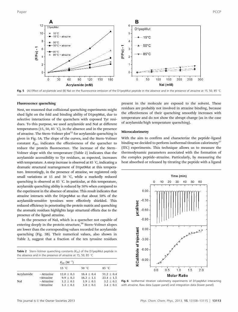
Next, we reasoned that collisional quenching experiments mightshed light on the fold and binding ability of D1pepMut, due toselective interactions of the quenchers with exposed Tyr resi-dues. To this purpose, we used acrylamide and NaI at differenttemperatures (15, 50, 85 1C), in the absence and in the presenceof atrazine. The Stern–Volmer plot32 for acrylamide quenching isgiven in Fig. 5A. The slope of the curves, and the Stern–Volmerconstant KSV, indicates the effectiveness of the quencher toreduce the protein fluorescence. The increase of the Stern–Volmer slope with the temperature (Table 2) indicates that theacrylamide accessibility to Tyr residues, as expected, increaseswith temperature. A steep increase is observed at 85 1C, indicating adramatic structural rearrangement of D1petMut at this tempera-ture. Interestingly, in the presence of atrazine, we registered onlysmall variations at 15 and 50 1C, while a markedly reducedquenching is observed at 85 1C. In particular, at this temperature,acrylamide quenching ability is reduced by 50% when compared tothe experiment in the absence of atrazine. This result indicates thatatrazine interacts with the D1pepMut so that about 50% of theacrylamide-sensitive tyrosines were effectively shielded. Thisreduced efficiency in penetrating the protein matrix and quenchingthe aromatic residues highlights large structural effects due to thepresence of the ligand atrazine.
In the presence of NaI, which is a quencher not capable ofentering deeply in the protein structure,36 Stern–Volmer slopesare lower than the corresponding values recorded for acrylamidequenching (Fig. 5B). Their numerical values, also shown inTable 2, suggest that a fraction of the ten tyrosine residues
present in the molecule are exposed to the solvent. Theseresidues are probably not involved in atrazine binding, becausethe effectiveness of their quenching smoothly increases withtemperature and do not show the abrupt change (as in the caseof acrylamide/high temperature quenching).
Microcalorimetry
With the aim to confirm and characterize the peptide–ligandbinding we decided to perform isothermal titration calorimetry37
(ITC) experiments. This technique allows us to measure thethermodynamic parameters associated with the formation ofthe complex peptide–atrazine. Particularly, by measuring theheat absorbed or released by titrating the peptide with a ligand
Fig. 5 (A) Effect of acrylamide and (B) NaI on the fluorescence emission of the D1pepMut peptide in the absence and in the presence of atrazine at 15, 50, 85 1C.
Table 2 Stern–Volmer quenching constants (KSV) of the D1pepMut peptide inthe absence and in the presence of atrazine at 15, 50, 85 1C
KSV (M�1)
15 1C 50 1C 85 1C
Acrylamide �Atrazine 12.0 � 0.3 16.4 � 0.4 51.2 � 0.4+Atrazine 9.9 � 0.3 16.3 � 1.1 23.4 � 1.5
NaI �Atrazine 1.2 � 0.1 1.9 � 0.1 3.2 � 0.1+Atrazine 1.3 � 0.1 1.8 � 0.1 3.4 � 0.1
Fig. 6 Isothermal titration calorimetry experiments of D1pepMut interactingwith atrazine. Raw data (upper panel) and integration data (lower panel).
Paper PCCP

13114 Phys. Chem. Chem. Phys., 2013, 15, 13108--13115 This journal is c the Owner Societies 2013
at constant temperature it is possible to obtain the stoichio-metry of the reaction, the binding enthalpy and the affinityconstant. Fig. 6 shows the calorimetric data (raw and integra-tion data) obtained in the titration of D1pepMut with atrazine.After subtraction of the heat of dilution from the raw data andlinear curve fitting under the ‘‘one binding site’’ model thethermodynamic parameters shown in the lower panel of Fig. 6were obtained. Atrazine binds to the peptide with an affinityconstant of 3.52 � 105 M�1 (Kd = 2.84 mM). The stoichiometricn value (n = 0.86) corresponds to a 1 : 1 binding mode. Despite avery unfavorable entropic term (�TDS = 4.3 kcal mol�1), thefavorable enthalpic contribution (DH = �11.9 kcal mol�1) leadsto a net negative free energy value of �7.6 kcal mol�1, suggest-ing an enthalpy driven reaction with productive interactionsbetween peptide and atrazine. The loss of entropy could beascribed to a more ordered and stable structure of the peptideupon ligand binding.
Conclusions
The increasing knowledge of biological natural molecules continu-ously challenges and stimulates the design of novel and artificialmaterials with similar biomimetic properties. The recent progress inmolecular engineering and nanotechnology witnesses the greatinterest to create novel molecular structures de novo or by rationaldesign. In particular, the development of biomimetic peptides andsmall proteins is an expanding area in this research field, which willpave the way to a vast area of future applications.38
In this paper we described the first attempt to create novelD1-inspired biomimetic molecules with high potential applica-tions in the realization of nanosensor arrays for the detection ofherbicides. The synthesized biomimetic molecules were deeplycharacterized and their structural and functional features studiedin response to atrazine binding. In this context, we reported thatatrazine binding induces extensive changes in the peptide mole-cules, resulting in a more stable structure as indicated by fluores-cence measurements. We have also documented that D1pepMutshows a high affinity towards atrazine, detecting the herbicide inthe micromolar range. Taken together, these results showed thatpeptides could represent an important alternative as recognitionmolecules, exhibiting high sensitivity towards target analytes,without the stability and reliability weaknesses often associatedwith entire native proteins. Future efforts will be dedicated tofurther improve the peptide binding affinity towards atrazine, aswell as to deliver a library of peptide variants with increasedselectivity and sensitivity headed for a wide range of herbicides.This achievement will pilot the design of a nanosensor array ofbiomimetic molecules with different specificities for differentchemical species, making these molecules a useful tool as probesin the development of last generation of biosensing systems.39
Acknowledgements
This work was supported by the FP7-SME-2008-1 SENSBIOSYNproject (ID: 232082, http://www.sensbiosyn.com), the Filas
MICROBIOSIS project, and the COST Action TD1102 PHOTO-TECH (http://www.phototech.eu).
Notes and references
1 J. Barber, Biochem. Soc. Trans., 2006, 34, 619.2 T. Wydrzynski and K. Satoh, in Advances in Photosynthesis
and Respiration, ed. Govindjee, Springer, 2005, p. 11.3 G. Renger and T. Renger, Photosynth. Res., 2008, 98, 53.4 J. Komenda, R. Sobotka and P. J. Nixon, Curr. Opin. Plant
Biol., 2012, 15, 245.5 P. J. Nixon, F. Michoux, J. Yu, M. Boehm and J. Komenda,
Ann. Bot., 2010, 106, 1.6 M. D. Lambreva, M. T. Giardi, I. Rambaldi, A. Antonacci,
S. Pastorelli, I. Bertalan, U. Johanningmeier and G. Rea,PLoS One, 2013, 8, e61851.
7 J. Xiong, S. Subramaniam and Govindjee, Protein Sci., 1996,5, 2054.
8 G. Rea, F. Polticelli, A. Antonacci, V. Scognamiglio,P. Katiyar, S. A. Kulkarni, U. Johanningmeier andM. T. Giardi, Protein Sci., 2009, 18, 2139.
9 Y. Umena, K. Kawakami, J.-R. Shen and N. Kamiya, Nature,2011, 473, 55.
10 G. Rea, F. Polticelli, A. Antonacci, M. Lambreva, S. Pastorelli,V. Scognamiglio, V. Zobnina and M. T. Giardi, in Herbicides,Theory and Applications, ed. S. Soloneski and M. L. Larramendy,2011, p. 5.
11 Z. Perrine and R. Sayre, Biochemistry, 2011, 50, 1454.12 J. Hirschberg, A. B. Yehuda, I. Pecker and N. Ohad, in Plant
Molecular Biology, ed. D. von Wettstein and N.-H Chua,Plenum Press, New York, 1987, p. 357.
13 A. Trebst, Z. Naturforsch., 1987, 42, 742.14 C. Sundby, W. S. Chow and J. M. Anderson, Plant Physiol.,
1993, 103, 105.15 S. B. Powles and Q. Yu, Annu. Rev. Plant Biol., 2010, 61, 317.16 R. Takahashi, K. Hasegawa, A. Takano and T. Noguchi,
Biochemistry, 2010, 49, 5445.17 M. Kato, T. Cardona, A. W. Rutherford and E. Reisner,
J. Am. Chem. Soc., 2012, 134, 8332.18 F. Milano, R. R. Tangorra, O. H. Omar, R. Ragni,
A. Operamolla, A. Agostiano, G. M. Farinola andM. Trotta, Angew. Chem., Int. Ed., 2012, 51, 11019.
19 V. Scognamiglio, G. Pezzotti, I. Pezzotti, J. Cano,K. Buonasera, D. Giannini and M. T. Giardi, Microchim.Acta, 2010, 170, 215.
20 B. Loll, J. Kern, W. Saenger, A. Zouni and J. Biesiadka,Nature, 2005, 438, 1040.
21 F. Muh, W. Saenger and A. Zouni, ChemPhysChem, 2010,11, 1160.
22 A. Guskov, J. Kern, A. Gabdulkhakov, M. Broser, A. Zouniand W. Saenger, Nat. Struct. Mol. Biol., 2009, 16, 334.
23 K. H. Smith, E. Tejeda-Montes, M. Poch and A. Mata, Chem.Soc. Rev., 2011, 40, 4563.
24 C. Tamerler and M. Sarikaya, Acta Biomater., 2007, 3, 289.25 S. Lagziel-Simis, N. Cohen-Hadar, H. Moscovich-Dagan,
Y. Wine and A. Freeman, Curr. Opin. Biotechnol., 2006, 17, 569.
PCCP Paper

This journal is c the Owner Societies 2013 Phys. Chem. Chem. Phys., 2013, 15, 13108--13115 13115
26 M. Sarikaya, C. Tamerler, A. K. Jen, K. Schulten andF. Baneyx, Nat. Mater., 2003, 2, 577.
27 S. Li, Y. Ge, S. A. Piletsky and A. P. F. Turner, Adv. Funct.Mater., 2011, 21, 3344.
28 Y. Bar-Cohen, Int. J. Aeronaut. Space Sci., 2012, 13, 1.29 R. K. Mishra, R. B. Dominguez, S. Bhand, R. Munoz and
J.-L. Marty, Biosens. Bioelectron., 2012, 32, 56.30 M. T. Giardi, V. Scognamiglio, G. Rea, G. Rodio, A. Antonacci,
M. Lambreva, G. Pezzotti and U. Johanningmeier, Biosens.Bioelectron., 2009, 25, 294.
31 P. Herman, J. Vecer, V. Scognamiglio, M. Staiano, M. Rossiand S. D’Auria, Biotechnol. Prog., 2004, 20, 1847.
32 S. D’Auria, M. Rossi, G. Barone, F. Catanzano, P. Del Vecchio,G. Graziano and R. Nucci, J. Biochem., 1996, 120, 292.
33 A. Kumar and S. Garg, in Biotechnology Applications,ed. C. S. K. Mishra, IK International, New Delhi publisher,2009, p. 39.
34 A. Trott and J. Olson, J. Comput. Chem., 2010, 31, 455.35 R. W. Woody, A. K. Bunker and G. D. Fasman, in Circular
Dichroism and the Conformational Analysis of Biomolecules,ed. G. D. Fasman, Plenum, New York, 1996, vol. 85, p. 391.
36 J. R. Lakowicz and G. Weber, Biochemistry, 1973, 12, 4171.37 S. Leavitt and E. Freire, Curr. Opin. Struct. Biol., 2001,
11, 560.38 A. Tiwari, A. K. Mishra, H. Kobayashi and A. P. Turner,
Intelligent nanomaterials, Wiley-Scrivener, 2012.39 Y. Cui, S. N. Kim, R. R. Naik and M. C. McAlpine, Acc. Chem.
Res., 2012, 45, 696.
Paper PCCP