DNA-Binding and -Bending Activities of SAP30L and SAP30 Are Mediated by a Zinc-Dependent Module and...
15
MOLECULAR AND CELLULAR BIOLOGY, Jan. 2009, p. 342–356 Vol. 29, No. 2 0270-7306/09/$08.000 doi:10.1128/MCB.01213-08 Copyright © 2009, American Society for Microbiology. All Rights Reserved. DNA-Binding and -Bending Activities of SAP30L and SAP30 Are Mediated by a Zinc-Dependent Module and Monophosphoinositides † Keijo M. Viiri, 1 Janne Ja ¨nis, 2 Trevor Siggers, 3 Taisto Y. K. Heinonen, 1 Jarkko Valjakka, 5 Martha L. Bulyk, 3,4,6 Markku Ma ¨ki, 1 and Olli Lohi 1 * Paediatric Research Centre, University of Tampere Medical School and Tampere University Hospital, 33520 Tampere, Finland 1 ; Department of Chemistry, University of Joensuu, FI-80101 Joensuu, Finland 2 ; Division of Genetics, Department of Medicine, Brigham & Women’s Hospital and Harvard Medical School, Boston, Massachusetts 02115 3 ; Department of Pathology, Brigham and Women’s Hospital and Harvard Medical School, Boston, Massachusetts 02115 4 ; Institute of Medical Technology and Tampere University Hospital, University of Tampere, FI-33014 Tampere, Finland 5 ; and Harvard/MIT Division of Health Sciences and Technology, Harvard Medical School, Boston, Massachusetts 02115 6 Received 1 August 2008/Returned for modification 17 September 2008/Accepted 6 November 2008 Deacetylation of histones is carried out by a corepressor complex in which Sin3A is an essential scaffold protein. Two proteins in this complex, the Sin3A-associated proteins SAP30L and SAP30, have previously been suggested to function as linker molecules between various corepressors. In this report, we demonstrate new functions for human SAP30L and SAP30 by showing that they can associate directly with core histones as well as naked DNA. A zinc-coordinating structure is necessary for DNA binding, one consequence of which is bending of the DNA. We provide evidence that a sequence motif previously shown to be a nuclear localization signal is also a phosphatidylinositol (PI)-binding element and that binding of specific nuclear monophosphoi- nositides regulates DNA binding and chromatin association of SAP30L. PI binding also decreases the repres- sion activity of SAP30L and affects its translocation from the nucleus to the cytoplasm. Our results suggest that SAP30L and SAP30 play active roles in recruitment of deacetylating enzymes to nucleosomes, and mediate key protein-protein and protein-DNA interactions involved in chromatin remodeling and transcription. A basic unit of chromatin is the nucleosome, in which 147 bp of DNA is wrapped around a histone octamer core composed of the four histones H2A, H2B, H3, and H4. The N-terminal “tail” domains of these histones project out of the nucleosome core and are the main sites of posttranslational modifications, such as acetylation, methylation, and phosphorylation. These covalent modifications have been proposed to play important roles in regulation of gene expression. According to the “his- tone code” hypothesis, the modifications function as “marks” which are recognized by various proteins required for the dy- namic alterations in chromatin structure that are needed to make a gene accessible to the components of the transcription machinery. The recruitment of histone deacetylases (HDACs) to chro- matin is a common mechanism of transcriptional repression (59). While certain repressors, such as Rb and YY1, are able to recruit HDACs directly (5, 58), others, such as Mad-Max and the nuclear hormone receptor, require association with core- pressors (1, 17, 18, 29, 42). Another example of the latter is the Sin3A corepressor complex, in which the Sin3A protein itself does not bind DNA or possess any enzymatic activity. Instead, it is composed of domains that mediate protein-protein inter- actions and thereby forms a platform for several enzymes (e.g., HDACs and methyltransferases), DNA-binding transcription factors (e.g., Mad family repressors, MeCP2, and Pf1), and other “bridging” proteins (e.g., SDS3) (52). The Sin3A-HDAC corepressor complex contains HDAC1 and HDAC2, the his- tone-binding proteins RbAp46 and RbAp48, Sin3A-associated protein 18 (SAP18), SAP30, and SDS3 (52). Mammalian HDAC1 and HDAC2 are almost identical, each containing an N-terminal catalytic domain, which removes acetyl moieties from the ε-amino groups of lysine residues, and a C-terminal tail. They are class I HDACs, which share se- quence similarity with the Rpd3 (reduced potassium depen- dency-3) protein in Saccharomyces cerevisiae (59). The core- pressor complex components RbAp46 and RbAp48 share 90% sequence identity and belong to the WD repeat family. These proteins have been shown to bind core histones H3 and H4 and are therefore thought to target HDAC-containing complexes to their histone substrates (56). Another member of the com- plex, SAP18, interacts directly with Sin3A and has multiple functions (60). In addition to regulating transcription, it also participates in mRNA processing (49). SDS3 has been sug- gested to play a role in stabilizing the Sin3A complex, and a yeast strain lacking SDS3 possesses only residual Sin3A-asso- ciated HDAC activity (31). SDS3 also participates in pericen- tric heterochromatin formation and chromosome segregation (9). Other components, such as SAP25, SAP130, and SAP180, have been reported to associate with the Sin3A-HDAC com- plex, but their roles in the complex have remained elusive (13, 51). SAP30 (Sin3A-associated protein 30) was originally identi- * Corresponding author. Mailing address: Paediatric Research Cen- tre, University of Tampere Medical School and Tampere University Hospital, 33520 Tampere, Finland. Phone: 358 3 35518405. Fax: 358 3 3551 8402. E-mail: olli.lohi@uta.fi. † Supplemental material for this article may be found at http://mcb .asm.org/. Published ahead of print on 17 November 2008. 342 on December 21, 2015 by guest http://mcb.asm.org/ Downloaded from
Transcript of DNA-Binding and -Bending Activities of SAP30L and SAP30 Are Mediated by a Zinc-Dependent Module and...

MOLECULAR AND CELLULAR BIOLOGY, Jan. 2009, p. 342–356 Vol. 29, No. 20270-7306/09/$08.00�0 doi:10.1128/MCB.01213-08Copyright © 2009, American Society for Microbiology. All Rights Reserved.
DNA-Binding and -Bending Activities of SAP30L and SAP30Are Mediated by a Zinc-Dependent Module
and Monophosphoinositides�†Keijo M. Viiri,1 Janne Janis,2 Trevor Siggers,3 Taisto Y. K. Heinonen,1 Jarkko Valjakka,5
Martha L. Bulyk,3,4,6 Markku Maki,1 and Olli Lohi1*Paediatric Research Centre, University of Tampere Medical School and Tampere University Hospital, 33520 Tampere, Finland1;
Department of Chemistry, University of Joensuu, FI-80101 Joensuu, Finland2; Division of Genetics, Department of Medicine,Brigham & Women’s Hospital and Harvard Medical School, Boston, Massachusetts 021153; Department of Pathology,
Brigham and Women’s Hospital and Harvard Medical School, Boston, Massachusetts 021154; Institute of Medical Technology andTampere University Hospital, University of Tampere, FI-33014 Tampere, Finland5; and Harvard/MIT Division of
Health Sciences and Technology, Harvard Medical School, Boston, Massachusetts 021156
Received 1 August 2008/Returned for modification 17 September 2008/Accepted 6 November 2008
Deacetylation of histones is carried out by a corepressor complex in which Sin3A is an essential scaffoldprotein. Two proteins in this complex, the Sin3A-associated proteins SAP30L and SAP30, have previously beensuggested to function as linker molecules between various corepressors. In this report, we demonstrate newfunctions for human SAP30L and SAP30 by showing that they can associate directly with core histones as wellas naked DNA. A zinc-coordinating structure is necessary for DNA binding, one consequence of which isbending of the DNA. We provide evidence that a sequence motif previously shown to be a nuclear localizationsignal is also a phosphatidylinositol (PI)-binding element and that binding of specific nuclear monophosphoi-nositides regulates DNA binding and chromatin association of SAP30L. PI binding also decreases the repres-sion activity of SAP30L and affects its translocation from the nucleus to the cytoplasm. Our results suggest thatSAP30L and SAP30 play active roles in recruitment of deacetylating enzymes to nucleosomes, and mediate keyprotein-protein and protein-DNA interactions involved in chromatin remodeling and transcription.
A basic unit of chromatin is the nucleosome, in which 147 bpof DNA is wrapped around a histone octamer core composedof the four histones H2A, H2B, H3, and H4. The N-terminal“tail” domains of these histones project out of the nucleosomecore and are the main sites of posttranslational modifications,such as acetylation, methylation, and phosphorylation. Thesecovalent modifications have been proposed to play importantroles in regulation of gene expression. According to the “his-tone code” hypothesis, the modifications function as “marks”which are recognized by various proteins required for the dy-namic alterations in chromatin structure that are needed tomake a gene accessible to the components of the transcriptionmachinery.
The recruitment of histone deacetylases (HDACs) to chro-matin is a common mechanism of transcriptional repression(59). While certain repressors, such as Rb and YY1, are able torecruit HDACs directly (5, 58), others, such as Mad-Max andthe nuclear hormone receptor, require association with core-pressors (1, 17, 18, 29, 42). Another example of the latter is theSin3A corepressor complex, in which the Sin3A protein itselfdoes not bind DNA or possess any enzymatic activity. Instead,it is composed of domains that mediate protein-protein inter-
actions and thereby forms a platform for several enzymes (e.g.,HDACs and methyltransferases), DNA-binding transcriptionfactors (e.g., Mad family repressors, MeCP2, and Pf1), andother “bridging” proteins (e.g., SDS3) (52). The Sin3A-HDACcorepressor complex contains HDAC1 and HDAC2, the his-tone-binding proteins RbAp46 and RbAp48, Sin3A-associatedprotein 18 (SAP18), SAP30, and SDS3 (52).
Mammalian HDAC1 and HDAC2 are almost identical, eachcontaining an N-terminal catalytic domain, which removesacetyl moieties from the ε-amino groups of lysine residues, anda C-terminal tail. They are class I HDACs, which share se-quence similarity with the Rpd3 (reduced potassium depen-dency-3) protein in Saccharomyces cerevisiae (59). The core-pressor complex components RbAp46 and RbAp48 share 90%sequence identity and belong to the WD repeat family. Theseproteins have been shown to bind core histones H3 and H4 andare therefore thought to target HDAC-containing complexesto their histone substrates (56). Another member of the com-plex, SAP18, interacts directly with Sin3A and has multiplefunctions (60). In addition to regulating transcription, it alsoparticipates in mRNA processing (49). SDS3 has been sug-gested to play a role in stabilizing the Sin3A complex, and ayeast strain lacking SDS3 possesses only residual Sin3A-asso-ciated HDAC activity (31). SDS3 also participates in pericen-tric heterochromatin formation and chromosome segregation(9). Other components, such as SAP25, SAP130, and SAP180,have been reported to associate with the Sin3A-HDAC com-plex, but their roles in the complex have remained elusive(13, 51).
SAP30 (Sin3A-associated protein 30) was originally identi-
* Corresponding author. Mailing address: Paediatric Research Cen-tre, University of Tampere Medical School and Tampere UniversityHospital, 33520 Tampere, Finland. Phone: 358 3 35518405. Fax: 358 33551 8402. E-mail: [email protected].
† Supplemental material for this article may be found at http://mcb.asm.org/.
� Published ahead of print on 17 November 2008.
342
on Decem
ber 21, 2015 by guesthttp://m
cb.asm.org/
Dow
nloaded from

fied as a conserved member of the Sin3A corepressor complex(28, 61). It was shown to be required for N-CoR-mediatedrepression by the antagonist-bound estrogen receptor and thePOU domain protein Pit-1 but not by the unliganded retinoicacid receptor or thyroid hormone receptor complexes (28). InSaccharomyces cerevisiae, SAP30 was demonstrated to be im-portant for cell growth and to affect gene expression in apromoter-dependent manner (61). A SAP30-deficient mutantstrain exhibited enhanced silencing of the ribosomal DNA,HMR, and telomeric loci, which suggested an antisilencingfunction for SAP30 (36, 54, 55). Meskauskas et al. observedthat mutations in Rpd3p, Sin3p, and Sap30p resulted in adefect in rRNA processing rather than ribosomal DNA tran-scription (41). Human SAP30, in addition to its associations inthe Sin3A-HDAC complex, has been reported to interact witha number of other proteins, such as retinoblastoma-bindingprotein 1 (RBP1) (30), the CBF1-interacting corepressor(CIR) (20), the YY1 transcription factor (21), and the inhibitorof growth 1b (ING1b) tumor suppressor protein (27, 53). Re-cently, SAP30-mediated transcriptional repression was shownto play a role in the transmission and propagation of certainviruses (26, 32). The above observations indicate that SAP30plays a vital role in transcriptional regulation, which can beeither negative or positive, depending on local factors, suchas chromatin state and the presence of various interactingpartners.
Human SAP30L (SAP30-like), thus named because it shares70% sequence identity with SAP30, is the “newest” member ofthe Sin3A corepressor complex. It was originally discovered asan expressed transcript in cultured T84 cells induced to differ-entiate in response to transforming growth factor � (35). Sincethen, SAP30L has been shown to associate with the Sin3A-HDAC complex and to induce transcriptional repression in aSin3A- and HDAC-dependent manner (57). Both SAP30 andSAP30L are able to localize to the nucleus or the nucleolus,and we have demonstrated that they can direct Sin3A to thenucleolus (57).
Previous work has led to the view that SAP30 and SAP30Lserve mainly a bridging role in various corepressor complexes.In this study, we set out to investigate the functions and thedomain structures of these proteins in more detail. We showthat both proteins directly bind and bend DNA and interactwith core histones 2A/2B. Interestingly, our results suggest thatboth DNA binding and chromatin association are regulated bynuclear monophosphorylated phosphoinositides (PIs).
MATERIALS AND METHODS
Antibodies, immunoblotting, and immunofluorescence. The primary antibod-ies used were those against histone 2B (sc-8650), c-myc (sc-40), Sin3A (sc-767and sc-5299), HDAC 1 (sc-7872), calregulin (sc-11398), and histone H1 (sc-8030)from Santa Cruz and glutathione S-transferase (GST) (27-4577-01) from GEHealthcare. Immunoblot analyses were done according to standard protocols,and anti-rabbit, anti-mouse, or anti-goat horseradish peroxidase-conjugated sec-ondary antibodies (p0217, p0260, or p0449, respectively) were obtained fromDako. Immunofluorescence was performed as described previously (57), andAlexa fluorophore-conjugated anti-mouse antibody (A11031) was used for de-tection of immunocomplexes.
Cloning and plasmid constructs. Full-length SAP30L and deletion mutantswere cloned into pcDNA 3.1-myc-his (Invitrogen), pGEX-4T1, and GAL4-DBD(Stratagene) vectors, and some of the constructs have already been described inreferences 25 and 57. The SAP30L-green fluorescent protein (GFP) andpcDNA3.1-Sin3A1-855 constructs are described in references 35 and 57, respec-
tively. The H2B-GFP construct was a generous gift from P. Peterson (Tartu,Estonia) (46). Point mutations were created using a QuikChange site-directedmutagenesis kit (Stratagene) according to the manufacturer’s instructions. Theluciferase reporter vector, under the control of 14D promoters harboring 5�Gal4 sites, was generously provided by D. Ayer (Salt Lake City, UT). The precisecoordinates of the constructs will be supplied on request. The integrity of theconstructs was confirmed by sequencing.
Cell culture, H2O2 treatment, and transfections. Human embryonic kidneyepithelial cells (HEK293T) were cultured in Dulbecco’s modified Eagle’s me-dium (Gibco) supplemented with penicillin and streptomycin, 5% fetal bovineserum, 1 mM sodium pyruvate, and 50 �g/ml uridine. HeLa cells were culturedin RPMI 1640 (Gibco) supplemented with penicillin and streptomycin, 10% fetalbovine serum, and L-glutamine. Cells were treated for 15 min with 0.5 mM H2O2,washed, and allowed to grow for 4 h before being harvested for analysis. DNAwas transfected with FuGENE 6 (for HEK293T cells) and FuGENE HD (forHeLa cells) reagents (Roche) according to the manufacturer’s protocol.
GST pulldown experiments. GST fusion proteins were produced in Escherichiacoli (BL-21 strain) and purified with glutathione-Sepharose 4B beads (Amer-sham Biosciences) according to the manufacturer’s instructions. The fusion pro-teins were eluted from the beads with reduced glutathione, if necessary. Thequantity and integrity of the GST fusion proteins were checked using Coomassie-stained sodium dodecyl sulfate-polyacrylamide gel electrophoresis (SDS-PAGE)gel.
GST pulldown experiments with nucleosomes and histones, with the Sin3a1-855 protein transcribed and translated in vitro, were carried out as describedpreviously (57), except that PI or PI 5-phosphate [PI(5)P] (Echelon, Inc.) wasadded to the reaction mixtures where indicated.
Electrophoretic mobility shift assays (EMSA) and a novel DNA ladder EMSA(L-EMSA). A 150-bp DNA probe comprising the mitochondrial tRNA-Leu(UUR) sequence was end labeled with [�-32P]ATP as described in reference22. The probe was incubated with 0.5 �g of a GST fusion protein on ice for 30min in a buffer containing 50 mM Tris-HCl, pH 7.5, 125 mM NaCl, 2.5 mMdithiothreitol, 0.5 mM EDTA, 1 mM MgCl2, and 4% glycerol. The reactionproducts were analyzed on a 6% nondenaturing polyacrylamide gel, dried, andautoradiographed.
Because of the non-sequence-specific nature of the binding by the SAP30Land SAP30 proteins, a faster and simpler assay, L-EMSA, was designed for theprotein-DNA interaction studies. Five micrograms of the fusion protein wasincubated with 0.25 �g of a 1-kb DNA ladder (GeneRuler; Fermentas) inphosphate-buffered saline for 10 min at room temperature. The reactions wererun on ethidium bromide-containing 1% agarose gel with standard DNA gelloading buffer. Prior to use, this method was validated by comparing the DNAband shifts in L-EMSA to shifts in conventional EMSA. The GST fusion proteinsused in Fig. 1A generated identical shifts in both assays (data not shown). Whereindicated, PI and PI(5)P were added after the protein-DNA complex formation.
PBM experiments and data analysis. Protein binding microarray (PBM) ex-periments and analyses were performed as described in reference 3 for fourdifferent GST-tagged protein constructs (full-length SAP30, full-length SAP30L,SAP30 residues 1 to 131 [SAP30 1-131], and SAP30L residues 1 to 92 [SAP30L1-92]) and a GST control (the protein concentration was �1uM). An Alexa488-conjugated anti-GST antibody was applied to the protein-bound microarrayto detect bound protein. The feature set of �44,000 oligonucleotides present onthe custom-designed Agilent microarrays followed the design described in ref-erence 3 but was incorporated on the Agilent 4x44K array platform, allowingfour independent PBM experiments to be performed simultaneously on the samemicroarray. To identify DNA-binding-site motifs, two approaches were used: (i)the approach described in reference 3, based on perturbations of the highest-ranked 8-bp sequence; and (ii) a de novo motif search of the sequences from the20, 30, and 50 brightest microarray probes, using the program MEME (2).
Mass spectrometry. Wild-type and mutant SAP30L 1-92 peptides were cleavedfrom GST by prothrombin, yielding SAP30L 1-94 peptides with two additionalamino acids from the GST vector. The samples were desalted on PD-10 columns(Amersham Biosciences, Uppsala, Sweden), and concentrations were estimatedfrom absorbance at 280 nm by using ε280 � 2,560 cm�1 M�1. Prior to measure-ments, the samples were further diluted with the appropriate solvents: CH3CN-H2O-acetic acid (49.5:49.5:1.0, vol/vol, pH 3.2) for denaturing solution condi-tions and 10 mM ammonium acetate buffer (pH 6.8) for nondenaturing solutionconditions. All experiments were performed with a 4.7-T hybrid quadrupoleFourier transform ion cyclotron resonance (Q-FT-ICR) instrument (APEX-Qe;Bruker Daltonics, Billerica, MA) interfaced with an external electrospray ion-ization (ESI) source (Apollo-II). The samples were infused directly at a flow rateof 1.5 �l min�1, with dry N2 serving as the drying (10 lb/in2, 200°C) and nebu-lizing gas. ESI-generated ions were externally accumulated in a hexapole ion trap
VOL. 29, 2009 SAP30L AND SAP30 BIND AND BEND DNA 343
on Decem
ber 21, 2015 by guesthttp://m
cb.asm.org/
Dow
nloaded from

for 0.5 to 1.0 s and transferred to an Infinity ICR cell for Sidekick trapping,conventional “RF-chirp” excitation, and broadband detection. A total of up to256 coadded (1-megaword) time domain transients were fast Fourier trans-formed prior to magnitude calculation and external frequency-to-m/z calibrationwith respect to the ions of an ES tuning mix (Agilent Technologies, Santa Clara,CA). All data were acquired and processed with the use of Bruker XMASS 7.0.8software.
DNA bending/ligation-mediated circularization assay. The ligation-mediatedcircularization assay was essentially performed as described previously (45). Seealso the legend to Fig. 3.
Protein-lipid blot assays. PI phosphate (PIP) strips and arrays were purchasedfrom Echelon Biosciences. Protein-lipid blot assays were performed by adding0.5 �g/ml of GST fusion proteins and were further processed as described in themanufacturer’s protocol. Each protein-lipid blot experiment was repeated atleast once.
Nucleosome preparations. Intact nucleosomes and tailless nucleosomes wereprepared as described previously (37). The presence of solubilized nucleosomeswas confirmed by DNA agarose gel electrophoresis and SDS-PAGE, followed byCoomassie staining (see Fig. S4A in the supplemental material). Calf thymushistones were purchased from Roche. For the GST fusion pulldown experiments,30 �g of nucleosomes, tailless nucleosomes, or histone proteins was used. For theinitial screening experiment (Fig. 4B), 100 �g of histones was used.
Interphase chromatin spreads, chromatin isolation, and subcellular fraction-ation. Chromatin spread preparation for the SAP30L-GFP-transfectedHEK293T cells was performed as described previously (38), with the followingmodifications. Nocodazole was not added, and the collected, phosphate-buff-ered-saline-washed, hypotonically swollen cells were dropped to a tilted micro-scope glass. The unfixed, dried drop was counterstained with DAPI (4,6-diamidino-2-phenylindole) and photographed under a confocal microscope.Chromatin isolation and subcellular fractionation were performed as describedpreviously (39).
Repression analysis. GAL4DBD-based repression analyses were performed asdescribed previously (57).
RESULTS
SAP30L and SAP30 bind DNA. It is generally believed thatspecific repressor proteins are needed to direct the Sin3A-HDAC complex to its target sequence, because Sin3A itselfdoes not bind DNA. Since SAP30L is able to repress transcrip-tion in a Sin3A- and HDAC-dependent manner (25, 57), weexamined if it could bind DNA directly. An EMSA was carriedout using a GST-SAP30L fusion protein incubated in the pres-ence of mitochondrial DNA. As shown in Fig. 1A, a markedshift was detected in the mobility of the DNA after incubationwith GST-SAP30L, indicating a direct interaction with DNA.A GST-SAP30 fusion protein also bound DNA, whereas GSTalone did not. SAP30L proteins with a C-terminal deletion(GST-SAP30L 1-120) or a mutated nucleolar localization sig-nal (NoLS; 8A mutant) were also able to interact with DNA.Deletion of 25 amino acid residues from the N terminus didnot affect DNA binding, whereas a larger deletion of 60 N-terminal residues (GST-SAP30L 61-183) abolished the shift.Similarly, a deletion mutant lacking 40 residues from the Nterminus failed to interact with DNA. These results show thatboth SAP30L and SAP30 are able to bind DNA and that theregion between residues 25 to 120 of SAP30L contains theDNA-binding determinants.
DNA binding by SAP30L and SAP30 is not sequence spe-cific. In order to investigate whether SAP30L and SAP30 cantarget Sin3A to specific DNA sequences, we took advantage of
FIG. 1. The N-terminal domains of SAP30L and SAP30 bind DNA in a sequence-independent manner. (A) A 32P-labeled DNA probe wasincubated with GST fusion proteins, and the DNA-protein complexes were analyzed by an EMSA. (B) Median fluorescence intensities of allmicroarray probe oligonucleotides containing a particular 8-mer sequence, from PBM experiments performed with GST, GST-SAP30L, GST-SAP30, and GST-Cbf1.
344 VIIRI ET AL. MOL. CELL. BIOL.
on Decem
ber 21, 2015 by guesthttp://m
cb.asm.org/
Dow
nloaded from

a recently developed method, the PBM (6a, 41a). GST-taggedproteins were applied to a microarray synthesized with double-stranded DNA oligonucleotides in order to assess the bindingpreference for all possible contiguous and gapped 8-bp DNA
sequence variants. Utilizing the universal microarray designand binding protocol recently described (3) (see Materials andMethods), we performed PBM experiments using four con-structs: full-length fusion proteins GST-SAP30L and GST-
FIG. 2. The N-terminal domains of SAP30L and SAP30 contain an evolutionarily conserved zinc-binding module which is needed for DNAbinding. (A) Clustal V alignment of the N-terminal amino acid sequences of SAP30L and SAP30 from various (selected) animals. Conservedcysteine (positions 29, 30, 38, and 74) and histidine (positions 70 and 77) residues in SAP30L are boxed, and the previously identified NLS is shadedin gray. Identical amino acids are marked by asterisks, and conservative substitutions are indicated by punctuation marks (colons and dots).(B) Charge-deconvoluted ESI Q-FT-ICR mass spectra of wild-type SAP30L in denaturing (upper) and nondenaturing (lower) solutions. The mostabundant isotopic masses for the detected peptide variants are indicated. The insets show an expanded view of the isotopic distributions for thevariant comprising residues 1 to 94 (this construct contains aa 1 to 92 of SAP30L and two amino acids derived from the thrombin cleavage siteof the vector). The theoretical isotopic distributions were calculated from the sequence-derived elemental compositions (Table 1). The small arrowindicates the most abundant isotopic peak. (C) The SAP30L C29S, C38S, C74S, and H77A mutant constructs are degraded within 16 h aftercleavage from GST, as analyzed by SDS-PAGE and Coomassie blue staining. wt, wild type. (D) L-EMSA with the GST-SAP30/SAP30L fusionproteins. (E) L-EMSA with the GST-SAP30L 1-92 fusion protein in the presence of 50 mM 1, 10-o-phenanthroline, a zinc-chelating agent.
VOL. 29, 2009 SAP30L AND SAP30 BIND AND BEND DNA 345
on Decem
ber 21, 2015 by guesthttp://m
cb.asm.org/
Dow
nloaded from

SAP30 (Fig. 1B) and N-terminally truncated fusion proteinsGST-SAP30L (amino acids [aa] 1 to 92) and SAP30 (aa 1 to131) (see Fig. S1 in the supplemental material). Using twodifferent approaches (see Materials and Methods), we wereunable to derive any binding motifs that would demonstratespecific binding behavior. We further examined the mediansignal intensity distribution of probe sequences containingeach 8-bp sequence variant and compared it to the distributionfrom a PBM experiment in which the sequence-specific tran-scription factor Cbf1 from S. cerevisiae was used as a positivecontrol (3). The Z-transformed distributions of results from allfour experiments with SAP30L were very similar to that for thenegative-control experiment (GST only), while the distributionof results from the Cbf1 control experiment showed a narrowerdistribution and a longer tail at high scores, indicative of spe-cifically bound probes (Fig. 1B). This comparison further sug-gested a lack of sequence-specific DNA binding of SAP30Land SAP30.
SAP30L and SAP30 contain an N-terminal zinc-coordinat-ing signature. A well-characterized DNA-binding element isthe zinc finger, which resembles a finger with a base of fourcysteine and/or histidine residues that coordinate a zinc ionthrough the thiol groups of cysteine residues and/or the im-idazole nitrogens of histidine residues (33, 48). In a stretch of49 residues (aa 29 to 77 in human SAP30L), we identified fourcysteine and two histidine residues, which suggested the pos-sibility of a zinc-coordinating motif. These residues are com-pletely conserved in a phylogenetic comparison of SAP30/SAP30L sequences from several species, including the fruit flyand the human (Fig. 2A). To investigate whether SAP30Lbinds zinc, we determined ESI Q-FT-ICR mass spectra for anN-terminal peptide of SAP30L (aa 1 to 92) and for mutants inwhich the putative zinc-coordinating cysteine residues werereplaced by serines (C29S, C30S, C38S, and C74S) and histi-dines by alanines (H70A and H77A), one at a time. Figure 2Bpresents the spectrum measured for SAP30L 1-94 (this peptidecontains two additional residues from the GST vector at its Nterminus) under denaturing conditions. To aid in the interpre-tation, the mass spectra were subjected to charge deconvolu-tion (i.e., conversion of m/z to Da). Four major peptide vari-ants, with relative abundance ratios of �4:31:7:58, weredetected, a result that is consistent with the SDS-PAGE anal-ysis (Fig. 2C). The mass of the second lightest variant(10,413.33 Da) agrees well with the mass calculated for theSAP30L 1-94 construct (Table 1). The species with the smallestmass (10,028.09 Da) is consistent with cleavage of three resi-dues from the C terminus and is presumed to comprise resi-dues 1 to 89 of SAP30L. However, two heavier variants(11,131.58 and 11,766.95 Da) could not be assigned to anypeptide sequence, even when all possible additional residuesfrom the expression vector were considered. All peptides ap-peared as apo peptides, i.e., no zinc binding was detectedunder denaturing conditions.
To detect zinc binding, the SAP30L 1-94 peptide was ana-lyzed under nondenaturing conditions (Fig. 2B). A 62.94-Daincrease in mass was detected for each of the four peptidevariants, consistent with the binding of one Zn2� cation (Table1). The binding of Zn2� (average mass � 65 Da) by zinc fingerdomains is always accompanied by the loss of two protons(deprotonation of two coordinating cysteines), which gives a
theoretical 62.92-Da increase in mass, as discussed in detailelsewhere (12). The precision of the mass measurements wasapproximately 0.01 Da, and such high accuracy provides anunequivocal identification of the incorporated Zn2� cation inSAP30L. No apo peptides or peptides with higher zinc-bindingstoichiometries were detected. Similar results were obtainedfor the SAP30L C30S and H70A mutants (see Fig. S2A in thesupplemental material). The calculated and determinedmasses for the peptides SAP30L 1-94, C30S, and H70A arelisted in Table 1. The SAP30L C29S, C38S, C74S, and H77Amutants had completely degraded into small peptide fragmentswhen expressed in E. coli (Fig. 2C; also see Fig. S2B in thesupplemental material) and when transiently transfected intomammalian cells (data not shown). Many of the peptide frag-ments that could be identified seemed to contain disulfidebridges. In summary, these results indicate that the C2CHmotif (C-X8-C-X35-C-X2-H) forms the zinc-binding module inSAP30L (see Fig. 7A) and that disruption of this moduledestabilizes the protein and leads to its rapid degradation.Similar instability has been reported to occur in the zinc-defi-cient mutant of the Spt10p zinc finger protein in yeast (40).
The C2CH module is needed for DNA binding. To furtherexplore the DNA-binding domains of SAP30L, we used a sim-plified EMSA which utilizes a commercial DNA ladder (L-EMSA). To validate the assay, we incubated the full-lengthGST-SAP30L and GST-SAP30 fusion proteins with the DNAladder and observed a marked shift in the mobility of theDNA. The GST-SAP30L 1-92 protein was able to bind DNAand therefore contains all determinants for interaction withDNA. Two polybasic regions (PBRs) were shown to be neces-sary for DNA binding (Fig. 2D). One of these is in the loop ofthe zinc-coordinating structure (aa 50 to 69), and the otherregion (aa 84 to 92) has previously been identified as a nuclearlocalization signal (NLS) (35). A hydrophobic region (aa 78 to84) between the zinc-coordinating structure and the NLS isalso required for DNA binding, since a construct containingboth the zinc-coordinating structure and the hydrophobic re-gion (aa 1 to 84) was able to bind DNA, whereas the zinc-coordinating structure alone (aa 1 to 77) was not. Similarly, aconstruct which includes both the NLS motif (aa 78 to 92) andthe hydrophobic region is able to bind DNA, but the NLSalone (aa 84 to 92) is not. The role of the NLS is furtherdemonstrated by the full-length GST-SAP30L-KAAAK con-
TABLE 1. Calculated and experimentally determined masses forwild-type SAP30L and the C30S and H70A mutants
Peptidea mexp(Da)b
mcalc(Da)c
mexp � mcalc(Da)
Elementalcompositiond
apo wild type 10,413.33 10,413.28 0.05 C447H726N140O137S5holo wild type 10,476.25 10,476.20 0.05 C447H724N140O137S5Zn1apo(C30S) 10,397.31 10,397.35 �0.04 C447H726N140O138S4holo(C30S) 10,460.24 10,460.22 0.02 C447H724N140O138S4Zn1apo(H70A) 10,347.26 10,347.30 �0.04 C444H724N138O137S5holo(H70A) 10,410.18 10,410.32 �0.14 C444H722N138O137S5Zn1
a The data are presented only for the peptide variants comprising residues1 to 94.
b Most abundant isotopic mass, experimentally determined.c Most abundant isotopic mass, calculated on the basis of the sequence-derived
elemental composition.d In the case of zinc binding, a loss of two protons was considered.
346 VIIRI ET AL. MOL. CELL. BIOL.
on Decem
ber 21, 2015 by guesthttp://m
cb.asm.org/
Dow
nloaded from
struct, which has markedly reduced affinity for DNA comparedto wild-type GST-SAP30L. The importance of the zinc-coor-dinating structure is best demonstrated in Fig. 1A, where itcan be seen that disruption of this structure (constructsGST-SAP30L 61-183 and 40-183) completely abolishesDNA binding.
The mapping experiments described above suggested thatthe zinc-coordinating structure is necessary for DNA binding.To explore whether DNA binding is dependent on zinc, theGST-SAP30L 1-92 fusion protein was incubated with 1,10-o-phenanthroline, a zinc-chelating agent, and the L-EMSA wasperformed. As shown in Fig. 2E, 1, 10-o-phenanthroline at a 50mM concentration abolished the DNA binding, as evidencedby a lack of shift in the mobility of DNA. Neither the GSTpeptide alone nor the methanol solvent elicited any mobilitychanges. Addition of different divalent cations showed thatonly Zn2� could partially restore the DNA-binding activity ofthe N-terminal domain of SAP30L (see Fig. S2C in the sup-plemental material). With DNA fragments less than 1,500 bpin length, we saw reproducible mobility shifts, which evidencedthe zinc dependence and specificity of DNA binding bySAP30L. We were unable to test the DNA-binding activity ofthe specific zinc-coordinating mutants described above, be-cause of extensive degradation of these proteins.
In summary, we have identified a putative zinc-bindingC2CH module in SAP30L and SAP30 and shown that DNAbinding is dependent on this module. This zinc-dependentstructure appears to be important for the stability of the entireprotein, and its disruption destabilizes the protein. Our resultsalso show that the loop region in the C2CH module is requiredfor DNA binding. Furthermore, the polybasic motif (NLS)
following the zinc-binding module and the intervening hydro-phobic pocket are also critical for DNA binding.
SAP30L has DNA-bending activity. The high-mobility-group(HMG) proteins provide a well-known example of a proteinfamily in which there can exist sequence-independent DNAbinding, accompanied by bending of the DNA (19). To test theability of SAP30L to mediate bending of double-strandedDNA, we examined its effect on the T4 DNA ligase-dependentcyclization of short DNA fragments. This ligation-mediatedcircularization assay works on the principle that any double-stranded DNA of less than 150 bp in length will not self-circularize in the presence of ligase, because of inherent limi-tations in the flexibility of DNA. Therefore, circularization isseen only in the presence of a DNA-bending protein. 32P-labeled PCR fragments (see Fig. S3 in the supplemental ma-terial) were incubated with the GST-SAP30L fusion protein orthe GST-only control as detailed in Materials and Methods. Asshown in Fig. 3A, 170-bp and 150-bp DNA fragments wereable to self-circularize, whereas shorter fragments were not. Inthe cases of shorter fragments (110 bp, 90 bp, and 70 bp),addition of GST-SAP30L resulted in the formation of circularmonomers, whereas GST alone did not form any circularmonomer molecules. The lack of monomer formation in thecase of 130-bp fragments suggests that SAP30L introducessuch a strong bend to the DNA that cohesive ends are unableto line up. Such a lack of monomer formation in the cases ofDNA molecules of certain lengths has also been reported forHMG proteins (45). The GST-SAP30L 1-92 region was able tobend DNA in a dose-dependent manner (Fig. 3B). It is rea-sonable to infer that SAP30 similarly bends DNA, given itsidentical zinc-binding module and DNA-binding properties.
FIG. 3. SAP30L bends DNA. (A) Probes of various lengths (170, 150, 130, 110, 90, and 70 bp), labeled internally with 32P and containing EcoRI“sticky ends,” were incubated with GST only (lanes 4, 9, 14, 19, 24, and 29) or with GST-SAP30L (lanes 5, 10, 15, 20, 25, and 30). The reactionmixtures for lanes 2 to 5, 7 to 10, 12 to 15, 17 to 20, 22 to 25, and 27 to 30 were incubated in the presence of T4 DNA ligase at 30°C for 20 min.The reaction mixtures for lanes 3 to 5, 8 to 10, 13 to 15, 18 to 20, 23 to 25, and 28 to 30 were subsequently treated with exonuclease (exo) III toremove any linear ligation products. The reaction products were electrophoresed on a 7% polyacrylamide gel, which was dried and subjected toautoradiography. Mono-, di-, and tricircular DNA ligation products are indicated by the numbered circles. nt, nucleotides or base pairs.(B) Increasing amounts of GST-SAP30L 1-92 were used in the ligation reactions, which were performed as described above.
VOL. 29, 2009 SAP30L AND SAP30 BIND AND BEND DNA 347
on Decem
ber 21, 2015 by guesthttp://m
cb.asm.org/
Dow
nloaded from
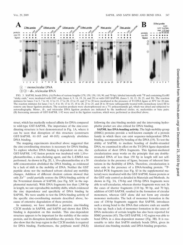
SAP30L and SAP30 bind core histones and nucleosomes.We noticed a similarity in the domain architecture of SAP30Land SAP30 to that of the HMG proteins, both having anN-terminal DNA-binding/bending domain followed by anacidic domain (Fig. 4A). As some reports indicate that theacidic region of HMG proteins mediates interactions with H1or core histones (4, 7), we set out to examine whether SAP30Lmay also associate with core histones and nucleosomes. To this
end, we used purified histones, isolated nucleosomes, and tryp-sin-cleaved nucleosomes (see Fig. S4A in the supplementalmaterial). In a pulldown experiment, GST-fused SAP30L wasable to associate with purified core histones 2A and 2B (Fig.4B). GST-SAP30L 1-92, which lacks the central acidic region,interacted with histone 2A/2B slightly less than full-lengthGST-SAP30L (Fig. 4B and C, lower panels). This comparisonis made difficult by the degradation of the full-length GST-
FIG. 4. SAP30L and SAP30 bind histones and nucleosomes. (A) Schematic representation of the domain architecture of the HMG proteins,SAP30L, and SAP30. The charge average of SAP30L is presented as a sliding window of 10 aa. Surface probability predictions were performedusing the approach of Emini (11), and values greater than 2 are shown as black lines. Zn, zinc-binding module; DNAbd & bending, DNA-bindingand -bending domain. (B) GST fusion protein pulldowns of calf thymus histones (Roche) were analyzed by SDS-PAGE and stained withCoomassie blue. The asterisks mark the GST fusion proteins, and the arrows indicate interaction with histones 2A/2B. (C) GST fusion proteinpulldowns of intact nucleosomes, of nucleosomes from which the tails had been removed with trypsin (see Fig. S4 in the supplemental material),and of calf thymus histones. WB, Western blot. (D) HEK293T cells were cotransfected with an H2B-GFP fusion protein and either wild-typeSAP30L or SAP30Ldel109-113 containing a myc-His tag. The cells were stained with the anti-myc antibody, and nuclei positive for both H2B andGFP were scored from 50 cells. The results are illustrated in the histogram. (E) GST fusion pulldowns of intact nucleosomes were analyzed usinga Western blot probed with the anti-H2B antibody.
348 VIIRI ET AL. MOL. CELL. BIOL.
on Decem
ber 21, 2015 by guesthttp://m
cb.asm.org/
Dow
nloaded from

SAP30L fusion protein and the possible loss of some of itsbinding determinants, which may lead to an underestimate ofthe difference. When the GST-SAP30L 1-120 construct, whichcontains the central acidic region, was used, histone bindingwas substantially increased (Fig. 4B and C, lower panels), sug-gesting that the central acidic region makes a significant con-tribution to histone binding. The interaction of SAP30L withhistones was confirmed for histone 2B by using a specific an-tibody (Fig. 4C), whereas in the case of histone 2A, the anti-body cross-reacted with SAP30L (data not shown), and thishampered the interpretation of the result. GST-SAP30 alsointeracted with histone 2B (data not shown).
Full-length GST-SAP30L and GST-SAP30L 1-92 were ableto interact with DNA-containing nucleosomes. Even thoughthe SAP30L 1-120 construct showed a high affinity for histoneswithout DNA, it did not interact with nucleosomes (Fig. 4C,upper). In the 1-120 construct, the acidic region is likely to beartificially exposed to acidic DNA, and consequently, its inter-action with nucleosomes is prevented. Interestingly, the inter-action of SAP30L with nucleosomes was not dependent on theprotruding N-terminal tails of histones, as demonstrated by alack of effect after trypsin cleavage of the tails (Fig. 4C, mid-dle). GST-SAP30 was similarly able to interact with nucleo-somes (Fig. 4E). Zhang et al. (61) have previously reportedthat SAP30 is unable to bind nucleosomes and core histones 3and 4. This is partially in line with our results in that wedetected only nonspecific interactions with histones 3 and 4, asthe GST moiety alone also bound them (Fig. 4B).
We have previously identified several mRNA isoforms ofSAP30L, including an isoform which lacks the entire exon 2and five residues of exon 3. Specific deletion of these fiveresidues (del109-113) markedly reduced the repression activityof SAP30L, whereas most of the HDAC activity was retained(25). Intriguingly, these residues reside in the central acidicdomain and include a single aspartate residue. As shown inFig. 4C, the del109-113 mutant was still able to interact withpurified histone 2B but not with purified nucleosomes contain-ing DNA, a result that may explain the reduced repressioncapability of this mutant. The lack of nucleosome binding bythe del109-113 mutant can be explained only by this mutant’sinability to interact with a histone-DNA complex, since it bindsnaked DNA with the same affinity as does the wild-type protein(see Fig. S4C in the supplemental material).
In confocal microscopy, colocalization of histone 2B andSAP30L was detected, with simultaneous relocalization of hi-stone 2B around the nucleolus in response to overexpression ofSAP30L (Fig. 4D). Coexpression with wild-type SAP30L, butnot with the SAP30Ldel109-113 mutant, increased the pe-rinucleolar localization of H2B from 10% to over 80% (Fig.4D). Intriguingly, overexpressed histone 2B was able to directNoLS-mutated SAP30L (8A) to the nucleolar region and thusovercome the lack of NoLS (data not shown). As a control,endogenous histone 1 showed no changes upon transient over-expression of SAP30L (data not shown).
The polybasic NLS binds monophosphoinositides. Pf1, arecently identified Sin3A-binding protein, has a PI-bindingPBR following the first PHD zinc finger (24). A similar orga-nization is found in ING2, in which the PHD zinc finger is alsofollowed by a PBR. We identified a similar modular organiza-tion in SAP30L, which also contains a zinc-binding element
followed by a PBR (85RNKRKRK91) (Fig. 5A). In SAP30L,the PBR motif has previously been shown to act as an NLS(35). To investigate the PI binding of SAP30L and SAP30, theGST fusion proteins were tested for binding to a variety ofimmobilized lipids, as depicted in Fig. 5B. Both GST-SAP30Land GST-SAP30 bound the monophosphorylated PIs PI(3)P,PI(4)P, and PI(5)P (Fig. 5C). No lipid binding was detected forGST alone. As a positive control, we used the PH domain ofphospholipase C-delta1, which interacted specifically withPI(4,5)P2 in this assay.
To quantify the relative affinities of SAP30L and SAP30 forvarious PIs, we used their fusion proteins to probe a lipid blotthat contained serial dilutions of eight different PIs (Fig. 5D).Full-length GST-SAP30L bound most tightly to PI(5)P, fol-lowed by PI(3)P and PI(4)P. The level of PI(5)P binding toGST-SAP30L was fourfold higher than that for PI(3)P andeightfold higher than that for PI(4)P. GST-SAP30 bound toimmobilized PIs in an identical manner, though with slightlylower affinities (Fig. 5D).
The determinants for PI binding were analyzed using trun-cated GST-SAP30L fusion proteins. Strikingly, deletion of 60residues from the N terminus (SAP30L 61-183) resulted incomplete loss of PI binding (Fig. 5E), which was rescued onlyby inclusion of the entire zinc-binding structure, indicating thatthe 25 residues in the N terminus are dispensable for thisinteraction. On the other hand, a construct containing the Nterminus and the intact zinc-coordinating structure (aa 1 to 77)was unable to bind PIs. Addition of the hydrophobic region (aa1 to 84) resulted in weak PI binding, whereas inclusion of thePBR motif following (aa 1 to 92) fully restored the interaction(this construct also exhibited some nonspecific binding, but thespecificity was restored in the 1-120 construct). The PBR motif(aa 84 to 92) by itself was not sufficient for PI binding, but aconstruct which includes both the PBR motif and the preced-ing hydrophobic region (aa 78 to 92) was sufficient for theinteraction. In the case of full-length SAP30L, mutating threebasic residues in the PBR motif to alanines markedly reducedits binding activity (KAAAK mutant). It is noteworthy thatdisruption of the loop in the zinc-coordinating structure bydeletion of residues 50 to 69 from otherwise intact SAP30Lcompletely abolished the PI interaction (Fig. 5E). Finally,swapping of basic residues in the PBR motif with the polybasicNoLS region (aa 1 to 84 plus the NoLS) changed the specificityof PI binding (Fig. 5E).
The above-mentioned results give rise to three conclusions.First, SAP30L and SAP30 interact specifically with monophos-phorylated PIs. Second, interaction of SAP30L with PIs ismediated by the PBR motif and supported by the precedinghydrophobic region and the zinc-coordinating structure. Third,the specificity of PI binding is partially determined by thecomposition of the basic sequence of the PBR. It is also note-worthy that the same region in SAP30L interacts with bothDNA and PIs, as summarized in Table 2.
Monophosphoinositides regulate chromatin association ofSAP30L. GFP-tagged SAP30L associated with chromatin invivo when hypotonically swollen HEK293 cells were splashedon a microscope slide and counterstained with DAPI (Fig. 6A).It should be noted that SAP30L is not a component of chro-matin in the same way as histones, since it is not present inmitotic chromosomes (data not shown). We next explored the
VOL. 29, 2009 SAP30L AND SAP30 BIND AND BEND DNA 349
on Decem
ber 21, 2015 by guesthttp://m
cb.asm.org/
Dow
nloaded from

FIG. 5. The zinc-binding structure and the PBR in SAP30L and SAP30 bind monophosphoinositides. (A) Manual alignment of the sequencesof the PBRs following the zinc-binding modules in SAP30L, SAP30, and Pf1. The last zinc-coordinating residue is boxed, and the basic residuesare indicated with bold letters. (B) Schematic diagram of a lipid blot membrane (PIP strip) containing 20-pmol spots from samples of the following:lysophosphatidic acid (LPA), lysophosphocholine (LPC), PI (PtdIns), PtdIns(3)P, PtdIns(4)P, PtdIns(5)P, phosphatidylethanolamine (PE),phosphatidylcholine (PC), sphingosine 1-phosphate (S1P), PtdIns(3,4)P2, PtdIns(3,5)P2, PtdIns(4,5P)2, PtdIns(3,4,5)P3, phosphatidic acid (PA),phosphatidylserine (PS), and blank. (C, D and E) The indicated GST fusion proteins (0.5 �g/ml) were incubated with PIP strips or with the PIParray as described in Materials and Methods. The lipids which bound most strongly are indicated. Arrows indicate the specificity differences fromthe 1-92 construct.
350 VIIRI ET AL. MOL. CELL. BIOL.
on Decem
ber 21, 2015 by guesthttp://m
cb.asm.org/
Dow
nloaded from
domains that regulate the chromatin association of SAP30L.As shown in Fig. 6B, KAAAK and del50-69 mutants ofSAP30L associated significantly less than wild-type SAP30Lwith the chromatin-enriched fraction, as assayed by subcellularfractionation. Also, an intact C-terminal domain was needed,presumably reflecting the importance of protein-protein inter-actions mediated by the C-terminal region.
As the KAAAK and del50-69 mutants are deficient in bothDNA/chromatin and PI binding (Fig. 2D and 5E), we asked ifthe association of SAP30L with chromatin is regulated by PIs,which could compete for the same binding sites and thus de-tach SAP30L from chromatin. Two lines of evidence indicatethat they do. First, the mobility shift generated by binding ofSAP30L to DNA in the L-EMSA was greatly diminished afteraddition of equivalent molar amounts of monophosphorylatedPIs but not other PIs (Fig. 6C). Second, H2O2 treatment, whichhas previously been shown to increase the amount of intranu-clear monophosphorylated PIs (23), led to a significant relo-calization of myc-tagged SAP30L as assayed by the chromatin-enriched fraction in HEK293 cells (Fig. 6D). In confocalmicroscopy of HeLa cells, 9% of nontreated and 41% of H2O2-treated cells expressed cytoplasmic GFP-SAP30L (Fig. 6E).The interaction of SAP30L with nucleosomes and Sin3A re-mained unchanged after addition of monophosphorylated PIs(Fig. 6F). These results suggest that intranuclear monophos-phorylated PIs associate with the PI-binding domain ofSAP30L and thereby regulate its association with chromatin.
PI binding decreases the repression activity of SAP30L.Finally, we tested if association of SAP30L with PIs influencedits repression activity by utilizing a Gal4 fusion system with aluciferase reporter vector as described previously (57). Asshown in Fig. 6G, reduced repression activity was observedboth in the PBR mutant (SAP30L KAAAK), which lacks DNAbinding and mimics PI binding, and after H2O2 treatment,which increases nuclear monophosphorylated PIs (23). Com-bined, these results suggest that association of SAP30L withchromatin is dependent on intact C-terminal and PI-/DNA-binding domains and that monophosphorylated PIs disrupt thisassociation, leading to decreased transcriptional repressionthrough SAP30L.
DISCUSSION
Although the Sin3A-HDAC corepressor complex has beenstudied extensively, the roles of the various members of thiscomplex are poorly understood. In this study, we have exploredthe functions of two members of this complex, the Sin3A-associated proteins SAP30L and SAP30, which share 70% se-quence identity. We have discovered three types of interactionsthat illuminate the functional roles of these proteins. First,both SAP30L and SAP30 interact directly with the core his-tones 2A/2B. Second, we demonstrate that both proteins haveintrinsic DNA-binding activity which is partly mediatedthrough a novel N-terminal zinc-containing structure consist-ing of a C2CH module and a coordinated zinc ion. Binding toDNA is sequence independent and induces strong bending ofthe DNA. Third, we have identified a PI-binding site, a basicdomain which binds monophosphorylated PIs specifically, ad-jacent to the zinc-binding element. Intriguingly, we have foundthat PI binding has a strong influence on the proteins’ affinityfor DNA in vitro, which leads us to suggest that the DNAbinding is actually regulated by PIs. An increase in the con-centration of PIs in the nucleus caused by hydrogen peroxideleads to reduced repression activity and cytoplasmic relocal-ization of SAP30L.
Previously, SAP30 has been assigned the role of a linkerprotein that mediates interactions of the Sin3-HDAC complexwith various transcriptional repressors (e.g., YY1) or corepres-sors (e.g., N-CoR, CIR, and RBP1). Specifically, the interac-tion of SAP30 with N-CoR was demonstrated to occur throughthe N terminus, whereas the C terminus bound mSin3a (28).Our results suggest a second function for the N terminus,which we found to bind DNA. Our results do not necessarilycontradict the previous reports, because one can imagine thatSAP30 can either bridge different multiprotein complexes oranchor a specific complex to nucleosomes, depending on thecircumstances. Functional diversity of this kind is not unprec-edented in Sin3A-associated proteins, since Fleischer et al.(13) have observed at least three separate Sin3A-containingcomplexes. Furthermore, the C terminus also seems to carrymultiple functions. Huang et al. (21) identified SAP30 as abinding partner for the transcription factor YY1 and showedthat it is able to enhance YY1-mediated repression in a dose-dependent manner. This interaction was mapped to the C-terminal region, i.e., the same region which also binds Sin3A,prompting the authors to suggest that the interactions ofSAP30 with YY1 and Sin3A are mutually exclusive. Huang etal. (21) suggested that HDAC activity could be brought to theYY1-SAP30 complex through a direct interaction of SAP30with HDAC1 (61). If SAP30L and SAP30 were to bind DNAand histones independently of Sin3A, it easy to envision thattheir N-terminal domains could participate in anchoring theYY1-SAP30-HDAC complex to chromatin to induce repres-sion of transcription.
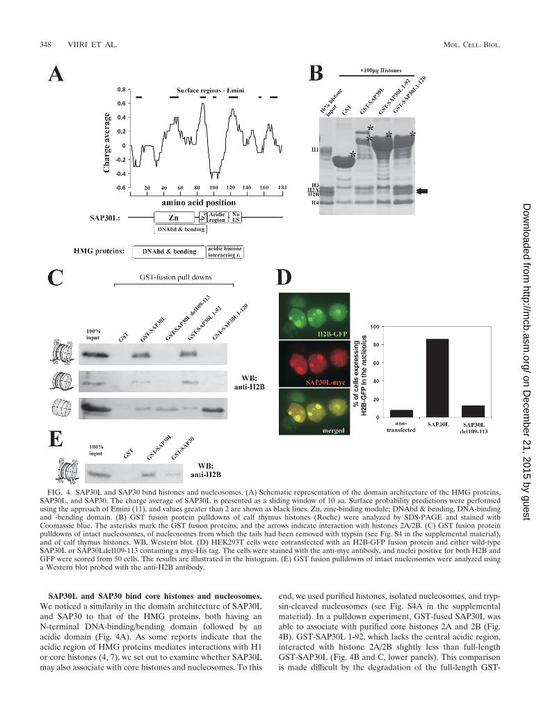
Sin3A by itself does not bind DNA or repress transcriptionbut instead mediates gene silencing through the enzymes thatit associates with (52). Targeting of the Sin3A complex iscarried out by DNA sequence-specific repressor proteins.Here, we show that SAP30L and SAP30 are able to bind DNAwithout any sequence specificity. This binding is dependent onan intact N terminus that contains a C2CH-type zinc module,
TABLE 2. Summary of mapping studies of DNA andPIP interactions
ConstructResulta for:
DNA interaction PIP interaction
Wild-type 1-183 ��� ���61-183 � �40-183 � NA35-183 NA �25-183 ��� ���1-77 � �1-84 �� ��1-92 ��� ���1-120 ��� ���84-92 � �78-92 ��� ���del50-69 � �87KAAAK91 � �
a NA, not available; �, no interaction; �, weak interaction; ��, moderateinteraction; ���, strong interaction.
VOL. 29, 2009 SAP30L AND SAP30 BIND AND BEND DNA 351
on Decem
ber 21, 2015 by guesthttp://m
cb.asm.org/
Dow
nloaded from
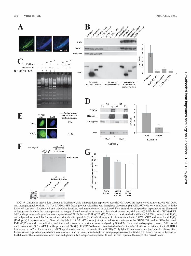
FIG. 6. Chromatin association, subcellular localization, and transcriptional repression activities of SAP30L are regulated by its interactions with DNAand monophosphoinositides. (A) The SAP30L-GFP fusion protein colocalizes with interphase chromatin. (B) HEK293T cells were transfected with theindicated constructs, fractionated into subcellular fractions, and immunoblotted as indicated. Data from three independent experiments are illustratedas histograms, in which the bars represent the ranges of band intensities as measured by a densitometer. wt, wild type. (C) L-EMSA with GST-SAP30L1-92 in the presence of equivalent molar quantities of PI (PtdIns) or PtdIns(5)P. (D) Cells were transfected with wild-type SAP30L, treated with H2O2,and subjected to subcellular fractionation as described for panel B. (E) Confocal images of cells transfected with SAP30L-GFP and treated with H2O2.(F) (Upper) In vitro-translated, 35S-methionine-labeled Sin3A1-855 was subjected to a pulldown experiment with GST-SAP30L and a GST-only control.PtdIns(5)P was added as indicated, and the results from the experiment were analyzed by SDS-PAGE and autoradiography. (Lower) Pulldown ofnucleosomes with GST-SAP30L in the presence of PIs. (G) HEK293T cells were cotransfected with a 5� Gal4-14D luciferase reporter vector, GAL4DBDfusions, and a LacZ vector, as indicated. At 24 h posttransfection, the cells were treated with 500 �M H2O2 for 15 min, washed, and lysed after 4 h of incubation.Luciferase and �-galactosidase activities were measured, and the histograms illustrate the average repressions of the GAL4DBD fusions relative to the level forGAL4 alone. The measurements were done in duplicate in two independent experiments, and the bars represent the ranges of observed values.
352 VIIRI ET AL. MOL. CELL. BIOL.
on Decem
ber 21, 2015 by guesthttp://m
cb.asm.org/
Dow
nloaded from
whose disruption abolishes the DNA binding. Zinc fingerswere originally identified as DNA-binding motifs, but they arenow known to bind RNA, protein, and lipid substrates as well(6, 14, 16, 24). A zinc finger consists of two antiparallel �strands and an helix, and the zinc ion is crucial for itsstability. Usually, a single zinc finger does not bind DNA withvery high affinity and can recognize only two or three basepairs, but when several, up to 60, zinc fingers are strung to-gether, the group binds more tightly and can recognize longerDNA sequences. In the cases of both SAP30L and SAP30, onlya single zinc-coordinating element was identified. Moreover,the stability of SAP30L was dependent on the zinc modulesince mutations in its zinc-coordinating residues led to rapiddegradation of the protein. We have previously observed thatN-terminally truncated SAP30L is poorly expressed in tran-sient transfections, but this could be overcome by usingMG132, a proteasome inhibitor, and now this can be explainedby the loss of the stabilizing zinc-dependent module in the Nterminus (57). Zinc-binding domains are usually relativelyshort, i.e., 20 to 30 residues, and the spacing of 35 residuesbetween the C2 and CH coordinating residues in SAP30L isunusually long. There is, however, a precedent for a largezinc-binding module, since THAP domains, which are con-served zinc-dependent modules capable of sequence-specificDNA binding, have a loop of 35 to 53 residues in the middle ofthe zinc-binding motif (8). The THAP domain, however, con-tains other conserved elements in addition to the C2CH mod-ule, making it distinct from the zinc-binding motif in SAP30L.
The sequence-independent nature of the DNA binding rulesout a sequence-specific targeting role for SAP30 and SAP30Land suggests a more general role in anchoring to nucleosomal/linker DNA. In addition, we demonstrated that this DNAbinding results in strong bending of the DNA. Classical exam-ples of proteins that bind and bend DNA in a sequence-inde-pendent manner are the HMG proteins (45), which interacttransiently with DNA. They are thought to antagonize histoneH1 binding by competing for the same chromatin sites. Gen-erally, they are thought to open up chromatin, although someHMG proteins may also compact chromatin (43). We findinteresting parallels between the HMG proteins and SAP30/SAP30L. Both are small and localized in the nucleus. Theirdomain structures are also similar, as both contain an N-ter-minal DNA-binding domain followed by an acidic region whichcontributes to histone interactions. This could imply functionalsimilarity as well, and it seems likely that SAP30/SAP30L haveroles in stabilizing the multiprotein complex on its target, in-creasing the availability of enzymatic targets to the complex, orpromoting the recruitment of interacting proteins, the cumu-lative effect being increased repression activity.
Perhaps one of the most intriguing features of SAP30L andSAP30 is the presence of a PI-binding site. PIs are known tofunction in nuclear signaling, and local changes in PI concen-trations are sensed by proteins with specific PI-binding do-mains, such as PH, ENTH, FYVE, and PHOX domains andlysine/arginine-rich patches (34, 44). A number of PI kinasesand phosphatases translocate to the nucleus upon activation,and many PI species have been shown to be intranuclear (10,15). Intervention of chromatin biology by signaling lipids is notunprecedented, since ATP-dependent chromatin-remodelingcomplexes, such as NURF, ISW2, INO80, and SWI/SNF, are
also modulated by specific inositol polyphosphates, the cleav-age products generated by PI-specific phospholipase C (50).Additionally, another SWI/SNF-like chromatin remodelingcomplex, BAF, is targeted to chromatin and the nuclear matrixspecifically by a PIP2-dependent mechanism upon lymphocyteactivation (62). Pf1, a recently identified nuclear binding part-ner for the corepressors mSin3A and TLE, has a PBR whichbinds specific monophosphoinositides (24). The Sin3A-bindingtumor suppressor ING2 binds PI(3), PI(4)P, and PI(5)P andshows PI(5)P-dependent association with chromatin and in-duction of p53-dependent apoptosis (16, 24). In response tocellular stress by UV irradiation or hydrogen peroxide, ING2associates with chromatin through a PI(5)P-mediated mecha-nism (23). Initially, the PHD domain of ING2 was reported tobe sufficient for PI binding, but later, the PBR motif wasdemonstrated to be both necessary and sufficient on its own(16, 24). Even though the PBRs of Pf1 and ING2 were deemedcritical for the binding activity and specificity, the precedingzinc-binding PHD domain contributed some specificity to theinteraction with PIs (24). SAP30 and SAP30L have a numberof similarities with the PI-binding proteins Pf1 and ING. First,the domain architecture of SAP30/SAP30L resembles that ofPf1, with a zinc-binding element followed by a basic PI-bindingmodule in both cases. Second, like SAP30/SAP30L, Pf1 andING2 are part of the Sin3A complex. Third, all three proteinsare nuclear and bind monophosphorylated PIs, albeit withdifferent preferences. Fourth, in the cases of SAP30/SAP30Land ING2, the subcellular localization and chromatin associa-tion are modified by PI binding. However, in the cases ofSAP30/SAP30L (but not in ING2), PI binding competes withDNA binding in vitro so that an increase in the concentrationof monophosphorylated PIs causes SAP30L to detach fromDNA. Furthermore, an increase in the concentration of nu-clear PIs elicited with hydrogen peroxide leads to reducedrepression activity and cytoplasmic relocalization of SAP30L.Although we note that these results are preliminary and mostlybased on in vitro experiments, they suggest the intriguing pos-sibility that changes in the concentration of nuclear monophos-phorylated PIs may regulate transcriptional repression throughSAP30/SAP30L in vivo. The site of PI binding was mapped toa region containing a motif previously shown to act as an NLS,and this motif is necessary for the PI-binding activity. However,the adjacent zinc-coordinating module also contributes to thisinteraction, a result that is in agreement with studies of otherproteins (47). The binding interface may reside on one side ofthe loop region of the zinc module, in a region which containsa stretch of basic residues. The specificity of PI binding is partlydetermined by the amino acid residue composition of the bind-ing motif, since replacing the NLS motif with another basicmotif (NoLS motif) in SAP30L led to changes in PI binding.Differences in binding specificity between different proteinsare also evident. SAP30/SAP30L prefer PI(5)P over PI(3)P/PI(4)P, whereas Pf1 prefers PI(3)P, with some binding activitytoward PI(3,5)P species (24).
We propose a model in which SAP30L/SAP30 are activelyinvolved in multiple protein-protein and protein-DNA inter-actions that modulate transcriptional repression. The domainstructures of SAP30L/30 and the proposed model are depictedin Fig. 7B and C, respectively. Briefly, we suggest that theDNA-binding activity plays a role in anchoring the Sin3A com-
VOL. 29, 2009 SAP30L AND SAP30 BIND AND BEND DNA 353
on Decem
ber 21, 2015 by guesthttp://m
cb.asm.org/
Dow
nloaded from
plex to nucleosomal and/or linker DNA in chromatin and thatthis binding is further strengthened by the interaction with corehistone 2A/2B dimers. One consequence of DNA binding isbending of the DNA, and we envision that this leads to en-hanced accessibility of nucleosomes and histone tails todeacetylating enzymes. Moreover, our results provide new ev-idence for the regulatory role played by nuclear PIs in tran-scriptional repression and relocalization of nuclear proteins.
ACKNOWLEDGMENTS
We thank Jorma Kulmala and Ritva Romppanen for technical as-sistance, Mike Berger for advice and assistance with PBM experimentsand data analysis, and Olli Silvennoinen for comments on the manu-script. We are grateful to P. Peterson (Tartu, Estonia) and D. Ayer(Salt Lake City, UT) for H2B-GFP and 14D promoter plasmids, re-spectively, and to Anne Hyvarinen (Tampere, Finland) for the EMSAprobe used in this work.
This work was supported by the Academy of Finland ResearchCouncil for Health (funding decision numbers 115260 and 201361) andfor Natural Sciences and Engineering (108533), the Foundation forPaediatric Research in Finland, the Competitive Research Funding ofthe Pirkanmaa Hospital District (EVO), the Nona and Kullervo VareFoundation, the Paivikki and Sakari Sohlberg Foundation, and grantR01 HG003985 from NIH/NHGRI to M.L.B. T.S. was supported inpart by a U.S. National Science Foundation Postdoctoral ResearchFellowship in Biological Informatics.
REFERENCES
1. Alland, L., R. Muhle, H. Hou, Jr., J. Potes, L. Chin, N. Schreiber-Agus, andR. A. DePinho. 1997. Role for N-CoR and histone deacetylase in Sin3-mediated transcriptional repression. Nature 387:49–55.
2. Bailey, T. L., N. Williams, C. Misleh, and W. W. Li. 2006. MEME: discov-ering and analyzing DNA and protein sequence motifs. Nucleic Acids Res.34:W369–W373.
3. Berger, M. F., A. A. Philippakis, A. M. Qureshi, F. S. He, P. W. Estep III, andM. L. Bulyk. 2006. Compact, universal DNA microarrays to comprehensivelydetermine transcription-factor binding site specificities. Nat. Biotechnol. 24:1429–1435.
4. Bernues, J., E. Espel, and E. Querol. 1986. Identification of the core-histone-binding domains of HMG1 and HMG2. Biochim. Biophys. Acta 866:242–251.
5. Brehm, A., E. A. Miska, D. J. McCance, J. L. Reid, A. J. Bannister, and T.Kouzarides. 1998. Retinoblastoma protein recruits histone deacetylase torepress transcription. Nature 391:597–601.
6. Brown, R. S. 2005. Zinc finger proteins: getting a grip on RNA. Curr. Opin.Struct. Biol. 15:94–98.
6a.Bulyk, M. L., X. H. Huang, Y. Choo, and G. M. Church. 2001. Exploring theDNA-binding specificities of zinc fingers with DNA microarrays. Proc. Natl.Acad. Sci. USA 98:7158–7163.
7. Carballo, M., P. Puigdomenech, and J. Palau. 1983. DNA and histone H1interact with different domains of HMG 1 and 2 proteins. EMBO J. 2:1759–1764.
8. Clouaire, T., M. Roussigne, V. Ecochard, C. Mathe, F. Amalric, and J. P.Girard. 2005. The THAP domain of THAP1 is a large C2CH module withzinc-dependent sequence-specific DNA-binding activity. Proc. Natl. Acad.Sci. USA 102:6907–6912.
9. David, G., G. M. Turner, Y. Yao, A. Protopopov, and R. A. DePinho. 2003.mSin3-associated protein, mSds3, is essential for pericentric heterochroma-tin formation and chromosome segregation in mammalian cells. Genes Dev.17:2396–2405.
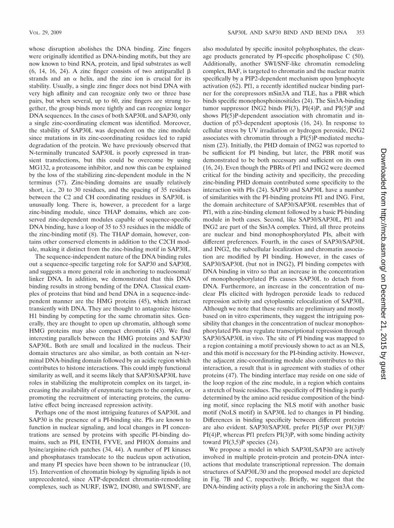
FIG. 7. Domain structure and a proposed mode of action for SAP30L and SAP30. (A) Schematic representation of the N-terminal zinc-coordinating motif of SAP30L. (B) Various domains of SAP30L identified in this and other studies (35, 57). Zn, zinc-coordinating motif; DNAbd,DNA-binding domain; PIPbd, PIP binding domain; protein bd, protein binding domain; acidic region, a central region contributing to histonebinding. (C) Proposed model. (Panel 1) When the histones are acetylated, the DNA is loosely packed and therefore accessible to RNA polymeraseII. (Panel 2) A sequence-specific transcriptional repressor (TF) recruits the Sin3A complex to its target promoter. SAP30 or SAP30L stabilizes thecomplex through interactions with DNA and histones 2A/2B. The interaction of SAP30/SAP30L with DNA induces bending of the DNA, as a resultof which the nucleosomes are more accessible to HDAC enzymes, and the repressome is fully formed. (Panel 3) Nuclear PIPs (PtdInsPs) interactwith the N-terminal domain of SAP30/SAP30L, displacing the DNA, which leads to relocalization of SAP30/SAP30L to the cytoplasm.
354 VIIRI ET AL. MOL. CELL. BIOL.
on Decem
ber 21, 2015 by guesthttp://m
cb.asm.org/
Dow
nloaded from
10. Deleris, P., D. Bacqueville, S. Gayral, L. Carrez, J. P. Salles, B. Perret, andM. Breton-Douillon. 2003. SHIP-2 and PTEN are expressed and active invascular smooth muscle cell nuclei, but only SHIP-2 is associated with nu-clear speckles. J. Biol. Chem. 278:38884–38891.
11. Emini, E. A., J. V. Hughes, D. S. Perlow, and J. Boger. 1985. Induction ofhepatitis A virus-neutralizing antibody by a virus-specific synthetic peptide.J. Virol. 55:836–839.
12. Fabris, D., Y. Hathout, and C. Fenselau. 1999. Investigation of zinc chelationin zinc-finger arrays by electrospray mass spectrometry. Inorg. Chem. 38:1322–1325.
13. Fleischer, T. C., U. J. Yun, and D. E. Ayer. 2003. Identification and charac-terization of three new components of the mSin3A corepressor complex.Mol. Cell. Biol. 23:3456–3467.
14. Gamsjaeger, R., C. K. Liew, F. E. Loughlin, M. Crossley, and J. P. Mackay.2007. Sticky fingers: zinc-fingers as protein-recognition motifs. Trends Bio-chem. Sci. 32:63–70.
15. Gozani, O., S. J. Field, C. G. Ferguson, M. Ewalt, C. Mahlke, L. C. Cantley,G. D. Prestwich, and J. Yuan. 2005. Modification of protein sub-nuclearlocalization by synthetic phosphoinositides: evidence for nuclear phospho-inositide signaling mechanisms. Adv. Enzyme Regul. 45:171–185.
16. Gozani, O., P. Karuman, D. R. Jones, D. Ivanov, J. Cha, A. A. Lugovskoy,C. L. Baird, H. Zhu, S. J. Field, S. L. Lessnick, J. Villasenor, B. Mehrotra,J. Chen, V. R. Rao, J. S. Brugge, C. G. Ferguson, B. Payrastre, D. G. Myszka,L. C. Cantley, G. Wagner, N. Divecha, G. D. Prestwich, and J. Yuan. 2003.The PHD finger of the chromatin-associated protein ING2 functions as anuclear phosphoinositide receptor. Cell 114:99–111.
17. Hassig, C. A., T. C. Fleischer, A. N. Billin, S. L. Schreiber, and D. E. Ayer.1997. Histone deacetylase activity is required for full transcriptional repres-sion by mSin3A. Cell 89:341–347.
18. Heinzel, T., R. M. Lavinsky, T. M. Mullen, M. Soderstrom, C. D. Laherty, J.Torchia, W. M. Yang, G. Brard, S. D. Ngo, J. R. Davie, E. Seto, R. N.Eisenman, D. W. Rose, C. K. Glass, and M. G. Rosenfeld. 1997. A complexcontaining N-CoR, mSin3 and histone deacetylase mediates transcriptionalrepression. Nature 387:43–48.
19. Hock, R., T. Furusawa, T. Ueda, and M. Bustin. 2007. HMG chromosomalproteins in development and disease. Trends Cell Biol. 17:72–79.
20. Hsieh, J. J., S. Zhou, L. Chen, D. B. Young, and S. D. Hayward. 1999. CIR,a corepressor linking the DNA binding factor CBF1 to the histone deacety-lase complex. Proc. Natl. Acad. Sci. USA 96:23–28.
21. Huang, N. E., C. H. Lin, Y. S. Lin, and W. C. Yu. 2003. Modulation of YY1activity by SAP30. Biochem. Biophys. Res. Commun. 306:267–275.
22. Hyvarinen, A. K., J. L. Pohjoismaki, A. Reyes, S. Wanrooij, T. Yasukawa,P. J. Karhunen, J. N. Spelbrink, I. J. Holt, and H. T. Jacobs. 2007. Themitochondrial transcription termination factor mTERF modulates replica-tion pausing in human mitochondrial DNA. Nucleic Acids Res. 35:6458–6474.
23. Jones, D. R., Y. Bultsma, W. J. Keune, J. R. Halstead, D. Elouarrat, S.Mohammed, A. J. Heck, C. S. D’Santos, and N. Divecha. 2006. NuclearPtdIns5P as a transducer of stress signaling: an in vivo role for PIP4Kbeta.Mol. Cell 23:685–695.
24. Kaadige, M. R., and D. E. Ayer. 2006. The polybasic region that follows theplant homeodomain zinc finger 1 of Pf1 is necessary and sufficient for specificphosphoinositide binding. J. Biol. Chem. 281:28831–28836.
25. Korkeamaki, H., K. Viiri, M. K. Kukkonen, M. Maki, and O. Lohi. 2008.Alternative mRNA splicing of SAP30L regulates its transcriptional repres-sion activity. FEBS Lett. 582:379–384.
26. Krithivas, A., D. B. Young, G. Liao, D. Greene, and S. D. Hayward. 2000.Human herpesvirus 8 LANA interacts with proteins of the mSin3 corepres-sor complex and negatively regulates Epstein-Barr virus gene expression indually infected PEL cells. J. Virol. 74:9637–9645.
27. Kuzmichev, A., Y. Zhang, H. Erdjument-Bromage, P. Tempst, and D. Rein-berg. 2002. Role of the Sin3-histone deacetylase complex in growth regula-tion by the candidate tumor suppressor p33ING1. Mol. Cell. Biol. 22:835–848.
28. Laherty, C. D., A. N. Billin, R. M. Lavinsky, G. S. Yochum, A. C. Bush, J. M.Sun, T. M. Mullen, J. R. Davie, D. W. Rose, C. K. Glass, M. G. Rosenfeld,D. E. Ayer, and R. N. Eisenman. 1998. SAP30, a component of the mSin3corepressor complex involved in N-CoR-mediated repression by specifictranscription factors. Mol. Cell 2:33–42.
29. Laherty, C. D., W. M. Yang, J. M. Sun, J. R. Davie, E. Seto, and R. N.Eisenman. 1997. Histone deacetylases associated with the mSin3 corepressormediate mad transcriptional repression. Cell 89:349–356.
30. Lai, A., B. K. Kennedy, D. A. Barbie, N. R. Bertos, X. J. Yang, M. C.Theberge, S. C. Tsai, E. Seto, Y. Zhang, A. Kuzmichev, W. S. Lane, D.Reinberg, E. Harlow, and P. E. Branton. 2001. RBP1 recruits the mSIN3-histone deacetylase complex to the pocket of retinoblastoma tumor suppres-sor family proteins found in limited discrete regions of the nucleus at growtharrest. Mol. Cell. Biol. 21:2918–2932.
31. Lechner, T., M. J. Carrozza, Y. Yu, P. A. Grant, A. Eberharter, D. Vannier,G. Brosch, D. J. Stillman, D. Shore, and J. L. Workman. 2000. Sds3 (sup-pressor of defective silencing 3) is an integral component of the yeastSin3[middle dot]Rpd3 histone deacetylase complex and is required for his-tone deacetylase activity. J. Biol. Chem. 275:40961–40966.
32. Le May, N., Z. Mansuroglu, P. Leger, T. Josse, G. Blot, A. Billecocq, R. Flick,Y. Jacob, E. Bonnefoy, and M. Bouloy. 2008. A SAP30 complex inhibitsIFN-beta expression in Rift Valley fever virus infected cells. PLoS Pathog.4:e13.
33. Lee, M. S., G. P. Gippert, K. V. Soman, D. A. Case, and P. E. Wright. 1989.Three-dimensional solution structure of a single zinc finger DNA-bindingdomain. Science 245:635–637.
34. Lemmon, M. A. 2003. Phosphoinositide recognition domains. Traffic 4:201–213.
35. Lindfors, K., K. M. Viiri, M. Niittynen, T. Y. Heinonen, M. Maki, and H.Kainulainen. 2003. TGF-beta induces the expression of SAP30L, a novelnuclear protein. BMC Genomics 4:53.
36. Loewith, R., J. S. Smith, M. Meijer, T. J. Williams, N. Bachman, J. D. Boeke,and D. Young. 2001. Pho23 is associated with the Rpd3 histone deacetylaseand is required for its normal function in regulation of gene expression andsilencing in Saccharomyces cerevisiae. J. Biol. Chem. 276:24068–24074.
37. Macfarlan, T., S. Kutney, B. Altman, R. Montross, J. Yu, and D. Chakra-varti. 2005. Human THAP7 is a chromatin-associated, histone tail-bindingprotein that represses transcription via recruitment of HDAC3 and nuclearhormone receptor corepressor. J. Biol. Chem. 280:7346–7358.
38. McGuinness, B. E., T. Hirota, N. R. Kudo, J. M. Peters, and K. Nasmyth.2005. Shugoshin prevents dissociation of cohesin from centromeres duringmitosis in vertebrate cells. PLoS Biol. 3:e86.
39. Mendez, J., and B. Stillman. 2000. Chromatin association of human originrecognition complex, Cdc6, and minichromosome maintenance proteins dur-ing the cell cycle: assembly of prereplication complexes in late mitosis. Mol.Cell. Biol. 20:8602–8612.
40. Mendiratta, G., P. R. Eriksson, C. H. Shen, and D. J. Clark. 2006. TheDNA-binding domain of the yeast Spt10p activator includes a zinc finger thatis homologous to foamy virus integrase. J. Biol. Chem. 281:7040–7048.
41. Meskauskas, A., J. L. Baxter, E. A. Carr, J. Yasenchak, J. E. Gallagher, S. J.Baserga, and J. D. Dinman. 2003. Delayed rRNA processing results insignificant ribosome biogenesis and functional defects. Mol. Cell. Biol. 23:1602–1613.
41a.Mukherjee, S., M. F. Berger, G. Jona, X. S. Wang, D. Muzzey, M. Snyder,R. A. Young, and M. L. Bulyk. 2004. Rapid analysis of the DNA bindingspecificities of transcription factors with DNA microarrays. Nat. Genet.36:1331–1339.
42. Nagy, L., H. Y. Kao, D. Chakravarti, R. J. Lin, C. A. Hassig, D. E. Ayer, S. L.Schreiber, and R. M. Evans. 1997. Nuclear receptor repression mediated bya complex containing SMRT, mSin3A, and histone deacetylase. Cell 89:373–380.
43. Narita, M., V. Krizhanovsky, S. Nunez, A. Chicas, S. A. Hearn, M. P. Myers,and S. W. Lowe. 2006. A novel role for high-mobility group a proteins incellular senescence and heterochromatin formation. Cell 126:503–514.
44. Overduin, M., M. L. Cheever, and T. G. Kutateladze. 2001. Signaling withphosphoinositides: better than binary. Mol. Interv. 1:150–159.
45. Paull, T. T., M. J. Haykinson, and R. C. Johnson. 1993. The nonspecificDNA-binding and -bending proteins HMG1 and HMG2 promote the as-sembly of complex nucleoprotein structures. Genes Dev. 7:1521–1534.
46. Pitkanen, J., A. Rebane, J. Rowell, A. Murumagi, P. Strobel, K. Moll, M.Saare, J. Heikkila, V. Doucas, A. Marx, and P. Peterson. 2005. Cooperativeactivation of transcription by autoimmune regulator AIRE and CBP. Bio-chem. Biophys. Res. Commun. 333:944–953.
47. Sankaran, V. G., D. E. Klein, M. M. Sachdeva, and M. A. Lemmon. 2001.High-affinity binding of a FYVE domain to phosphatidylinositol 3-phosphaterequires intact phospholipid but not FYVE domain oligomerization. Bio-chemistry 40:8581–8587.
48. Schwabe, J. W., and A. Klug. 1994. Zinc mining for protein domains. Nat.Struct. Biol. 1:345–349.
49. Schwerk, C., J. Prasad, K. Degenhardt, H. Erdjument-Bromage, E. White, P.Tempst, V. J. Kidd, J. L. Manley, J. M. Lahti, and D. Reinberg. 2003. ASAP,a novel protein complex involved in RNA processing and apoptosis. Mol.Cell. Biol. 23:2981–2990.
50. Shen, X., H. Xiao, R. Ranallo, W. H. Wu, and C. Wu. 2003. Modulation ofATP-dependent chromatin-remodeling complexes by inositol polyphos-phates. Science 299:112–114.
51. Shiio, Y., D. W. Rose, R. Aur, S. Donohoe, R. Aebersold, and R. N. Eisenman.2006. Identification and characterization of SAP25, a novel component ofthe mSin3 corepressor complex. Mol. Cell. Biol. 26:1386–1397.
52. Silverstein, R. A., and K. Ekwall. 2005. Sin3: a flexible regulator of globalgene expression and genome stability. Curr. Genet. 47:1–17.
53. Skowyra, D., M. Zeremski, N. Neznanov, M. Li, Y. Choi, M. Uesugi, C. A.Hauser, W. Gu, A. V. Gudkov, and J. Qin. 2001. Differential association ofproducts of alternative transcripts of the candidate tumor suppressor ING1with the mSin3/HDAC1 transcriptional corepressor complex. J. Biol. Chem.276:8734–8739.
54. Smith, J. S., E. Caputo, and J. D. Boeke. 1999. A genetic screen for ribo-somal DNA silencing defects identifies multiple DNA replication and chro-matin-modulating factors. Mol. Cell. Biol. 19:3184–3197.
55. Sun, Z. W., and M. Hampsey. 1999. A general requirement for the Sin3-
VOL. 29, 2009 SAP30L AND SAP30 BIND AND BEND DNA 355
on Decem
ber 21, 2015 by guesthttp://m
cb.asm.org/
Dow
nloaded from
Rpd3 histone deacetylase complex in regulating silencing in Saccharomycescerevisiae. Genetics 152:921–932.
56. Verreault, A., P. D. Kaufman, R. Kobayashi, and B. Stillman. 1998. Nucleo-somal DNA regulates the core-histone-binding subunit of the human Hat1acetyltransferase. Curr. Biol. 8:96–108.
57. Viiri, K. M., H. Korkeamaki, M. K. Kukkonen, L. K. Nieminen, K. Lindfors,P. Peterson, M. Maki, H. Kainulainen, and O. Lohi. 2006. SAP30L interactswith members of the Sin3A corepressor complex and targets Sin3A to thenucleolus. Nucleic Acids Res. 34:3288–3298.
58. Yang, W. M., C. Inouye, Y. Zeng, D. Bearss, and E. Seto. 1996. Transcrip-tional repression by YY1 is mediated by interaction with a mammalianhomolog of the yeast global regulator RPD3. Proc. Natl. Acad. Sci. USA93:12845–12850.
59. Yang, X. J., and E. Seto. 2008. The Rpd3/Hda1 family of lysine deacetylases:
from bacteria and yeast to mice and men. Nat. Rev. Mol. Cell Biol. 9:206–218.
60. Zhang, Y., R. Iratni, H. Erdjument-Bromage, P. Tempst, and D. Reinberg.1997. Histone deacetylases and SAP18, a novel polypeptide, are componentsof a human Sin3 complex. Cell 89:357–364.
61. Zhang, Y., Z. W. Sun, R. Iratni, H. Erdjument-Bromage, P. Tempst, M.Hampsey, and D. Reinberg. 1998. SAP30, a novel protein conserved betweenhuman and yeast, is a component of a histone deacetylase complex. Mol. Cell1:1021–1031.
62. Zhao, K., W. Wang, O. J. Rando, Y. Xue, K. Swiderek, A. Kuo, and G. R.Crabtree. 1998. Rapid and phosphoinositol-dependent binding of the SWI/SNF-like BAF complex to chromatin after T lymphocyte receptor signaling.Cell 95:625–636.
356 VIIRI ET AL. MOL. CELL. BIOL.
on Decem
ber 21, 2015 by guesthttp://m
cb.asm.org/
Dow
nloaded from