Cytotoxic and apoptotic potential of Myristica fragrans Houtt ...
bEditor-in-Chief
Luc Pieters, Antwerp, Belgium
Senior Editor
Adolf Nahrstedt, Münster, Germany
Review Editor
Matthias Hamburger, Basel, Switzerland
Editors
Rudolf Bauer, Graz, AustriaVeronika Butterweck, Muttenz,Switzerland
Thomas Efferth, Mainz, GermanyIrmgard Merfort, Freiburg, GermanyHermann Stuppner, Innsbruck, AustriaYang-Chang Wu, Taichung, Taiwan
Editorial Offices
Claudia Schärer, Basel, SwitzerlandTess De Bruyne, Antwerp, Belgium
Advisory Board
JohnT. Arnason, Ottawa, CanadaYoshinori Asakawa, Tokushima, JapanLars Bohlin, Uppsala, SwedenMark S. Butler, S. Lucia, AustraliaJoão Batista Calixto, Florianopolis, BrazilClaus Cornett, Copenhagen, DenmarkHartmut Derendorf, Gainesville, USAAlfonso Garcia-Piñeres, Frederick MD, USAJürg Gertsch, Zürich, SwitzerlandSimon Gibbons, London, UKDe-An Guo, Shanghai, ChinaAndreas Hensel, Münster, GermanyKurt Hostettmann, Geneva, SwitzerlandPeter J. Houghton, London, UKIkhlas Khan, Oxford MS, USAJinwoong Kim, Seoul, KoreaWolfgang Kreis, Erlangen, GermanyRoberto Maffei Facino, Milan, ItalyAndrew Marston, Bloemfontein,South Africa
Matthias Melzig, Berlin, GermanyEduardo Munoz, Cordoba, SpainNicolas Oberlies, Greensboro NC, USANigel B. Perry, Dunedin, New ZealandJoseph Pfeilschifter, Frankfurt, GermanyPeter Proksch, Düsseldorf, GermanyJose-Luis Rios, Valencia, SpainKurt Schmidt, Graz, AustriaThomas Schmidt, Münster, GermanyThomas Simmet, Ulm, GermanyLeandros Skaltsounis, Athens, GreeceHan-Dong Sun, Kunming, ChinaPing-Jyun Sung, Pingtung, TaiwanDeniz Tasdemir, London, UKArnold Vlietinck, Antwerp, BelgiumGünther Vollmer, Dresden, GermanyHeikki Vuorela, Helsinki, FinlandJean-Luc Wolfender, Geneva, SwitzerlandYang Ye, Shanghai, China
Publishers
Georg Thieme Verlag KGStuttgart · New YorkRüdigerstraße 14D-70469 StuttgartPostfach 30 11 20D-70451 Stuttgart
Thieme Publishers333 Seventh AvenueNew York, NY 10001, USAwww.thieme.com
Reprint
© Georg Thieme Verlag KGStuttgart ·New York
Reprint with the permissionof the publishers only
Planta MedicaJournal of Medicinal Plant and Natural Product Research
www.thieme.de/fz/plantamedica l www.thieme-connect.com/ejournals
bAbstract!
Sideritis scardica Griseb. (ironwort, mountaintea), an endemic plant of the Balkan Peninsula,has been used in traditional medicine in the treat-ment of gastrointestinal complaints, inflamma-tion, and rheumatic disorders. This study aimedto evaluate its gastroprotective and anti-inflam-matory activities. Besides, continuously increas-ing interest in assessing the role of the plant ac-tive constituents preventing the risk of cancerwas a reason to make a detailed examination ofthe investigated ethanol, diethyl ether, ethyl ace-tate, and n-butanol extracts regarding cytotoxic-ity. Oral administration of the investigated ex-tracts caused a dose-dependent anti-inflamma-tory effect in a model of carrageenan-induced ratpaw edema. Gastroprotective activity of the ex-tracts was investigated using an ethanol-inducedacute stress ulcer in rats. The cytotoxic activity ofplant extracts was assessed on PBMC, B16, andHL-60 cells and compared to the cytotoxicity ofphenolic compounds identified in extracts. Apo-ptotic and necrotic cell death were analyzed bydouble staining with fluoresceinisothiocyanate(FITC)-conjugated annexin V and PI. The devel-oped HPLC method enabled qualitative finger-
print analysis of phenolic compounds in the in-vestigated extracts. Compared to the effect of thepositive control, the anti-inflammatory drug in-domethacine (4mg/kg), which produced a 50%decrease in inflammation, diethyl ether and n-bu-tanol extracts exhibited about the same effect indoses of 200 and 100mg/kg (53.6 and 48.7%;48.4 and 49.9%, respectively). All investigated ex-tracts produced dose-dependent gastroprotectiveactivity with the efficacy comparable to that ofthe reference drug ranitidine. The diethyl etherextract showed significant dose-dependent cyto-toxicity on B16 cells and HL-60 cells, decreasingcell growth to 51.3% and 77.5% of control, respec-tively, when used at 100 µg/mL. It seems that phe-nolic compounds (apigenin, luteolin, and theircorresponding glycosides) are responsible for thediethyl ether extract cytotoxic effect. It also ap-pears that induction of oxidative stress might beinvolved in its cytotoxicity, since B16 and HL-60cells increased their ROS production in responseto treatment with diethyl ether extract. Neitherof the tested extracts nor any phenolic com-pounds showed significant cytotoxic effect to hu-man PBMC. These results demonstrated the po-tent anti-inflammatory and gastroprotective ac-tivities, as well as the promising cytotoxicity.
Anti-inflammatory, Gastroprotective, and CytotoxicEffects of Sideritis scardica Extracts
Authors Vanja M. Tadić1, Ivica Jeremic2,3, Silva Dobric4, Aleksandra Isakovic3, Ivanka Markovic3, Vladimir Trajkovic5,Dragica Bojovic6, Ivana Arsic1
Affiliations The affiliations are listed at the end of the article
Key wordsl" Sideritis scardica Griseb.l" Lamiaceael" anti‑inflammatoryl" gastroprotective activityl" cytotoxicityl" polyphenolsl" flavonoids
received October 10, 2011revised Dec. 12, 2011accepted Dec. 19, 2011
BibliographyDOI http://dx.doi.org/10.1055/s-0031-1298172Published online January 24,2012Planta Med © Georg ThiemeVerlag KG Stuttgart · New York ·ISSN 0032‑0943
CorrespondenceDr Vanja Tadić, Science AdvisorDepartment of PharmacyInstitute for Medicinal PlantResearch “Dr Josif Pančić”Tadeusa Koscuska 111000 BelgradeSerbiaPhone: + 381113031658Fax: + [email protected]
Original Papers
Thisis
aco
pyof
theau
thorʼs
person
alreprint
Thisis
aco
pyof
theau
thorʼs
person
alreprint
Introduction!
The promising new source of therapeutic agentsrefers to plant secondary metabolites, irregularlyoccurring compounds that characterize certainplants or plant groups. There is continuously in-creasing interest in assessing the role of the phe-nolic compounds which show antioxidative prop-erties and may act with beneficial health effects,reducing the risk of chronic diseases (inflamma-tion, cancer, osteporosis, and cardiovascular dis-eases). Among them, flavonoids, as a large groupof plant secondary metabolites, have been pro-
Tadić VM e
duced in the plant for the purpose of protectionfrom photosynthetic stress, reactive oxygen spe-cies (ROS), wounds, and herbivores. Studies of fla-vonoids have revealed the most compelling datafor cytotoxic activities in various types of cancers,and several flavonoids have been shown to inhibitcancer development while exhibiting antioxidantactivities in different animal models. Further-more, some studies suggest that the most promis-ing use of these compounds may be as an adju-vant to currently used therapies in antitumortreatment [1].
t al. Anti-inflammatory, Gastroprotective, and… Planta Med
Original Papers
Thisis
aco
pyof
theau
thorʼs
person
alreprint
Thisis
aco
pyof
theau
thorʼs
person
alreprint
bThe results of numerous preliminary investigations of plants be-longing to the genus Sideritis L. revealed a plant-derived source ofparticular pharmacological and nutritional interest. The genusSideritis L. (Lamiaceae) includes approximately 150 species of an-nual and perennial plants distributed mainly in the Mediterra-nean region. This genus is divided into two subgenera, Sideritisand Marrubiastrum, formed by the European and Macaronesianspecies, respectively. So far, different biological activities of Side-ritis species have been reported: anti-inflammatory, antiulcer,analgesic, antimicrobial and antifungal [2–6], immunomodulat-ing [7], macrophage NOS-2-expression inhibiting [8], and hypo-glycemic [5]. Recently, aldose reductase inhibiting activity [9],antiproliferative, anticholinesterase, and selective estrogen re-ceptor modulator-like effects have been reported [10–12]. Theprevious studies of Sideritis species reported the presence of fla-vonoid aglycones and glycosides, phenolic acids, di- and triterpe-noids, fatty acids, coumarines and iridoid glycosides [3,9,11,13–16], and essential oil as well [2]. Most of the studies on Sideritisspecies attributed the previously cited biological activitiesmainly to phenolic compounds [9,13]. Rios et al. [17] reportedthat flavonoids were reducing agents able to interact with freeradical species (of relevance to autoxidation mechanism) andcould prevent generation of inflammatory mediators.The genus Sideritis is represented in Serbia by one species only, S.montana L. [18], but because of its pro-oxidant properties thisplant has not been used in traditional medicine [19]. S. scardicaGriseb. (ironwort, mountain tea) is an endemic plant of the Bal-kan Peninsula belonging to the Empedoclea section. Aerial partsof mountain tea are traditionally known for their anti-inflamma-tory, antimicrobial, antibacterial, antirheumatic, and gastropro-tective properties. S. scardica is used as a loosening agent in bron-chitis and bronchial asthma, against common cold and lung em-physema. It has been imported in Serbia from the former Yugo-slav Republic of Macedonia and Albania and widely used in thetreatment of inflammation, gastrointestinal disorders, andcoughs, as well as an active constituent of dietary supplementsfor the prevention of anemia. In the literature, all previously citedbiological activities are mainly attributed to the phenolic contentof this plant [14].The present study aimed to investigate anti-inflammatory andgastroprotective activities of S. scardica extracts in order to ex-amine the above-stated folkloric utilizations and to establish thecorrelation between observed activities and phenolic constitu-ents of the extracts based on previous studies which have recog-nized flavonoids in S. scardica as potent biologically active sub-stances. Besides, in the present study we investigated the in vitrocytotoxic action of S. scardica extracts in order to establish theconnection of significant antitumor potential and the polyphenolcomponents present in the examined extracts. Qualitative andquantitative fingerprint analyses of polyphenolic compounds inthe investigated extracts were also conducted applying the HPLCmethod.
Materials and Methods!
GeneralSodium bicarbonate (analytical grade), DPPH (1,1′-diphenyl-2-picrylhydrazyl; analytical grade), indomethacin (purity ≥ 99.0%),carrageenan (EP grade), and trolox (purity ≥ 99.0%) were pur-chased from Sigma-Aldrich. Analytical grade reagents 2,6-di-tert-butyl-4-methylphenol (BHT, purity ≥ 99.8%), ether, petrol,
Tadić VM et al. Anti-inflammatory, Gastroprotective, and… Planta Med
dimethyl sulfoxide (DMSO), ethyl acetate, n-butanol (BuOH), ace-tone, and absolute ethanol (96%, v/v) were purchased fromMerck. Acetonitrile (MeCN), water, and methanol were of HPLCgrade and also from Merck. Reference HPLC standards p-couma-ric (purity ≥ 99.0%), protocatechuic (purity ≥ 99.0%), chlorogenic(purity ≥ 99.0%), vanillic (purity ≥ 95.0%), caffeic (purity≥ 90.0%), ferulic (purity ≥ 99.0%), and syringic (purity ≥ 95.0%)acids, luteolin-7-O-β-glucoside (purity ≥ 98.0%), apigenin-7-O-β-glucoside (purity ≥ 99.0%), luteolin (purity ≥ 99.0%), chrysoeriol(purity ≥ 99.0%), apigenin (purity ≥ 99.0%), hyperoside (purity≥ 99.0%), gallic acid (purity ≥ 99.0%), pyrogallol (purity ≥ 99.0%),and cisplatin [cis-diamineplatinum(II) dichloride, purity≥ 99.9%] were purchased from Sigma or from Extrasynthese.Their purity was declared as stated previously, based on theman-ufacturerʼs internal high-precision HPLC method. Ranitidine, pu-rity ≥ 95.0% (Ranisan ampoules), was purchased from Zdravlje-Actavis Company.
Plant material and the procedure for plant materialextractionThe wild growing species Sideritis scardica Griseb., Lamiaceae,was collected on Shara Mountain (at the hill foot of the Ljuboten,at ca. 1300m) during the time of flowering. Plantmaterial was airdried, packed in paper bags and kept in a dark and cool place untilanalysis. Plant material was verified, and the voucher specimenof the plant (SS/08) was deposited at the Herbarium of the Botan-ical Garden, Jevremovac, Belgrade, Serbia. The identification wasprovided by Prof. Dmitar Lakušić (Institute of Botany and Botani-cal Garden, Faculty of Biology, University of Belgrade). The shade-dried and powdered aerial parts of S. scardica (600 g) werecoarsely extracted using 70% (V/V) ethanol. The yield of the finalextract (crude extract, 1) in terms of starting crude material wasdetermined to be 16.7%. The crude ethanol extract (1) was redis-solved in distilled water, shaken vigorously and extracted with600mL of diethyl ether, 600mL ethyl acetate, and 600mL satu-rated n-butanol in a separating funnel, successively. The obtainedextracts were: diethylether extract, 2 (2.8 g); ethyl acetate ex-tract, 3 (1.3 g); and n-butanol extract, 4 (4.4 g). The yield of ex-traction for extracts 2, 3, and 4 was 16.7, 7.5, and 26.3% in crudeextracts, or 0.46, 0.21, and 0.73% of dry plant, respectively.
AnimalsAdult, male Wistar rats weighing 200–300 g were used for esti-mating mountain tea ethanol extract anti-inflammatory (carra-geenan-induced paw edema test) and gastroprotective activities(absolute ethanol-induced stress ulcer test). Experimental groupsconsisted of 6–10 animals each. The animals were deprived offood for 18–20 h before the beginning of experiments with freeaccess to tap water.This study was performed after approval from the local Institu-tional Animal Care and Use Committee and run in accordance tothe statements of the European Union regarding handling of ex-perimental animals (approval number 86/609/EEC, 31.01.2008).
Determination of total phenols contentThe total phenolic content was determined by the Folin-Ciocalteumethod [20]. One hundred microliters of the MeOH solution ofthe dry investigated extracts 1, 2, 3, and 4 (15.75, 31.5, and 63;27.75, 55.5, and 138.75; 7.28, 14.56, and 19.13; and 7.06, 14.13,and 28.25 µg/mL final quantity, respectively) were mixed with0.75mL of Folin-Ciocalteu reagent (previously diluted 10-foldwith distilled water) and allowed to stand at 22°C for 5min;
Original Papers
Thisis
aco
pyof
theau
thorʼs
person
alreprint
Thisis
aco
pyof
theau
thorʼs
person
alreprint
b0.75mL of sodium bicarbonate (60 g/L) solutionwas added to themixture. After 90min at 22°C, absorbance was measured at725 nm. Gallic acid (0–100mg/L) was used for calibration of astandard curve. The calibration curve showed the linear regres-sion at r > 0.99, and the results were expressed as milligrams ofgallic acid equivalents per gram of plant extracts dry weight (mgGAE/g DW). Triplicate measurements were taken, and data werepresented as mean ± standard deviation (SD).
Tannins contentThe percentage content of tannins was calculated using themethod described in the European Pharmacopoeia, Ph. Eur. 6.0[21]. Shortly, decoctions prepared from the investigated extractswere treated with phosphomolybdotungstic reagent in alkalinemedium after and without treatment with hide powder. The ab-sorbance was measured by UV‑VIS spectrophotometer HP 8453(Agilent Technologies) at λmax 760 nm. From the difference in ab-sorbance of total polyphenols and polyphenols not adsorbed byhide powder, the percentage content of tannins expressed as py-rogallol (%, w/w) was calculated from the expression:
62:5ðA1 � A2Þ �m2
A3 �m1
where m1 represents mass of the sample to be examined, ingrams; and m2 is mass of pyrogallol, in grams. The results repre-sent the mean ± SD of three determinations.
Total flavonoids contentThe percentage content of flavonoids was calculated using themethod described in the European Pharmacopoeia, Ph. Eur. 6.0[21]. Briefly, the sample was extracted with acetone/HCl underreflux condenser; the AlCl3 complex of the flavonoid fraction ex-tracted by ethyl acetate was measured by UV‑VIS spectropho-tometer HP 8453 at 425 nm. The content of flavonoids, expressedas hyperoside percentage, was presented as the mean ± standarddeviation of three determinations.
HPLC procedureChromatographic fingerprint of the extract and quantification ofidentified compounds were achieved by HPLC (Agilent Technolo-gies 1200). Detection was performed using diode array detector(DAD), and the chromatograms were recorded at λ = 260 nm (forprotocatechin and syringic acid), 280 nm (for chlorogenic, vanilic,p-coumaric, and caffeic acids), 325 nm (for ferulic acid), and360 nm (for flavonoids). HPLC separation of components wasachieved using a LiChrospher 100 RP 18e (5 µm), 250 × 4mm i.d.column, with a flow rate of 1mL/min and mobile phase, A[500mL of H2O plus 9.8mL of 85% H3PO4 (w/w)], B (MeCN), elu-tion, combination of gradient mode: 90–75% A, 0–25min; iso-cratic 75% A, 25–30min; 75–55% A, 30–46min. The sample wasprepared dissolving 118.6, 49.4, 9.4, and 53.0mg of the extracts1, 2, 3, and 4, respectively (obtained by the procedure previouslydescribed) in 10mL of MeOH, filtered through 0.2 µm PTFE filtersprior to HPLC analysis. The injected volume was 4 µL. Standardsolutions for the determination of flavonoids and polyphenolicacidwere prepared at a final concentration of 0.01mg/mL (proto-catechin, p-coumaric, vanilic, ferulic, and syringic acids, as well asluteolin and chrysoeriol), 0.05mg/mL (chlorogenic and caffeicacids, and apigenin), or 0.12mg/mL (apigenin-7-O-glycoside andluteolin-7-O-glycoside) in methanol. For the purpose of the phe-nolic compounds identification and determination in the investi-
gated extracts, three mixtures of the standards were preparedwith the already mentioned concentrations: mix 1 with caffeicacid, apigenin-7-O-glycoside, and apigenin; mix 2 with chloro-genic acid, luteolin-7-O-glycoside, luteolin, and chrysoeriol; mix3 contained the rest of the investigated phenolic compounds. Thevolume injected was 4 µL, the same as the investigated extract.The identification was carried out based on retention time andspectral matching. Once spectral matching succeeded, resultswere confirmed by spiking with respective standards to achievea complete identification by means of the so-called peak puritytest. Those peaks not fulfilling these requirements were notquantified. Quantification was performed by external calibrationwith standards.
Determination of the free radical scavenging activityThe DPPH scavenging assay was carried out according to the pro-cedure described by Blois, with some modifications [22]. Variousconcentrations of the samples (100 µL) were mixed with 900 µLof 0.04mg/mL methanolic solution of DPPH. UV spectra were re-corded on a UV‑VIS spectrophotometer HP 8453. Absorbance at517 nm was measured after 20min. The inhibition percentagewas calculated using the following equation:
I = [(Ac −As)/Ac] × 100
where I was the inhibition percentage, Ac was the absorbance ofthe negative control (contained 100 µL of MeOH instead of thesamples), and As was the absorbance of the samples. Syntheticantioxidants, trolox, and tert-butyl hydroxytoluene (BHT) wereused as positive controls. The inhibition percentage was plottedagainst concentration of the samples, and IC50 values, deter-mined by linear regression analysis, were presented as the mean± standard deviation of three determinations.
Carrageenan-induced rat paw edemaThe carrageenan-induced rat paw edema test was used as an ex-perimental model for screening the anti-inflammatory activityaccording to the modified method of Oyanagui and Sato [23].The extracts were administered p.o. in doses of 50, 100, and200mg/kg. Indomethacin, dissolved in DMSO, was used as a ref-erence in a dose of 4mg/kg p.o., whichwas a dose producing 50%reduction of rat paw edema. The control animals were givenDMSO in a dose of 1mL/kg p.o. Carrageenan-saline solution(0.5% in a volume of 0.1mL) was injected into the plantar surfaceof the right hind paw 1 h after the oral administration of the ex-tracts or indomethacin. A pure saline solution (0.9% NaCl, 0.1mL)was injected into the left hindpaw, which served as a control(non-inflamed paw). The animals were killed 3 h after the carra-geenan injection, and the pawswere cut off for weighing. The dif-ference in weight between the right and left paw, treated versusuntreated (control) rats, served as an indicator of the inflamma-tory response intensity (i.e., anti-inflammatory activity). The per-cent of anti-inflammatory effect was calculated from the expres-sion
Anti-inflammatory effect (%) ¼ �k��e�k � 100
where Δk represents the difference in the pawweight in the con-trol group; Δe is the difference in the paw weight in the treat-ment group.
Tadić VM et al. Anti-inflammatory, Gastroprotective, and… Planta Med
Original Papers
Thisis
aco
pyof
theau
thorʼs
person
alreprint
Thisis
aco
pyof
theau
thorʼs
person
alreprint
bAbsolute ethanol-induced stress ulcer in ratsTo study the gastroprotective activity of the investigated extracts,an experimental model of acute gastric mucosa damage inducedby absolute ethanol (1mL/rat p.o.) was used. The investigated ex-tracts, dissolved in DMSO, were administered p.o. in doses of 50–200mg/kg 60min prior to ethanol. Ranitidine given in doses of5–20mg/kg p.o. was used as a reference drug. The control ani-mals were given the vehicle in a dose of 1mL/kg p.o., also60min before ethanol. The animals were sacrificed 1 h after giv-ing ethanol, and their stomachs were removed and opened alongthe greater curvature. Lesions were examined under an illumi-natedmagnifier (3×). The intensity of gastric lesions was assessedaccording to a modified scoring system of Adami et al. [24].
Cell linesMouse melanoma cell line (B16) and the human promyelocyticleukemia cell line (HL-60) were obtained from the European Col-lection of Cell Cultures (ECACC) while peripheral blood mononu-clear cells (PBMC) were obtained from healthy blood donors afterwritten informal consent. This study was approved by the EthicalCommittee of the School of Medicine, University of Belgrade (ap-proval number 89/101/EC, 24.02.2011). All cell lines were main-tained at 37°C in a humidified atmosphere with 5% CO2. B16mouse melanoma cell lines were cultured in DMEM supple-mentedwith 5% fetal calf serum-FCSwhile the HL-60 human leu-kemia cell line and PBMC were cultivated in RPMI supplementedwith 10% FCS.The adherent cells were prepared for experiments using the con-ventional trypsinization procedure with trypsin/EDTA and incu-bated in 96-well flat-bottom plates (2 × 10⁴ cells/well) for viabil-ity and LDH analyses and in 6-well flat-bottom plates for flow cy-tometry analyses (3 × 10⁵ cells/well). Cells were rested for 24 hand then treated with plant extracts and different flavonoids:apigenin, luteolin, chlorogenic acid, apigenin 7-O-glucoside, lu-teolin 7-O-glucoside, chrysoeriol, and ferrulic acid. Suspensioncells were cultivated in 96-well flat-bottom plates (3.5 × 10⁴
cells/well) for assessing cell viability, and in 24-well flat-bottomplates for flow cytometry analyses (1.5 × 10⁵ cells/well). Cellswere rested for 2 h and then treated with plant extracts or flavo-noids. The extracts and flavonoids were dissolved in dimethylsulfoxide-DMSO and diluted in appropriate medium. Final con-centration of DMSO in the incubation mixture did not exceed0.1% and did not have any influence on cell viability. Cisplatin(25 µM) was used as the positive control in all methods exceptfor lactate dehydrogenase (LDH) release assay where Triton X-100 (3%) was used.
Determination of cell viabilityCell viability was assessed using acid-phosphatase method.Briefly, after treatment (24 h), adherent cells were washed twicewith phosphate buffered saline, and 100 µL of reaction mixture(0.1M acetate buffer pH 5.5, containing para-nitrophenyl phos-phate PNPP and 0.1% Triton-X) was added to each well. After 90minutes, the reaction was stopped by adding 50 µL of 0.1MNaOH. The absorbance of the developed yellow color, which wasdirectly proportional to the cells viability [25], was measured byan automated microplate reader at 405 nm. The results were pre-sented as percent of the control value (untreated cells), whichwas arbitrarily set to 100%. For determination of suspension cellviability, we used a modified acid-phosphatase method. Aftertreatment, 50 µL of reaction mixture (0.3M acetate bufferpH 5.5, containing PNPP and 0.2% Triton-X) was added to each
Tadić VM et al. Anti-inflammatory, Gastroprotective, and… Planta Med
well. After 60 minutes, the reaction was stopped by the additionof 50 µL of 0.3M NaOH into each well, and absorbance was readas described. At the same time blanks, containing cell culture me-dium (without cells), were prepared to achieve the correction forthe absorbance caused by medium color (at 405 nm).
Analysis of apoptosis and cell cycleApoptotic and necrotic cell death were analyzed by double stain-ing with fluoresceinisothiocyanate (FITC)-conjugated annexin Vand PI, in which annexin V bound to the apoptotic cells with ex-posed phosphatidylserine, while PI labeled the necrotic cells withmembrane damage. This staining was performed according tothe manufacturerʼs instructions (BD Pharmingen).The cell cycle was analyzed by measuring the amount of propi-diumiodide (PI)-labeled DNA in ethanol-fixed cells, exactly aspreviously described [26]. DNA fragmentation, as another markerof apoptosis, was determined during cell cycle analysis by count-ing the hypodiploid cells in the sub-G0/G1 cell cycle phase.The green (FL1) and red (FL2) fluorescences of annexin/PI-stained live cells and PI stained fixed cells were analyzed withFACSCalibur flow cytometer (BD).The number of viable (annexin−/PI−), apoptotic (annexin+/PI−),and necrotic (annexin+/PI+) cells as well as the proportion of cellsin different cell cycle phaseswere determinedwith Cell Quest Prosoftware (BD). Ten thousand cells (gated to exclude cell debris)were analyzed in each sample.
ROS measurementIntracellular production of ROS was determined by measuringthe intensity of green fluorescence emitted by redox-sensitivedye dihydrorhodamine 123 (DHR; Invitrogen), which was addedto cell cultures (2.5 µM) at the beginning of the treatment. At theend of incubation, cells were detached by trypsinization, washedin PBS, and the green fluorescence (FL1) of DHR-stained cells wasanalyzed using a FACSCalibur flow cytometer. The results are ex-pressed as mean intensity of DHR fluorescence.
Lactate dehydrogenase release assayThe release of the cytosolic enzyme lactate dehydrogenase (LDH)reflects a loss of membrane integrity in dying cells, and it was as-sessed by a colorimetric assay as previously described [27].Briefly, 100 µL of cell culture supernatant after treatment (cellsgrown in colorless medium) was mixed with 100 µL of solutioncontaining 54mM lactic acid, 0.28mM phenazine methosulfate,0.66mM p-iodonitrotetrazolium violet, and 1.3mM NAD+. Thepyruvate-mediated conversion of 2,4-dinitrophenylhydrazine in-to visible hydrazone precipitate was measured using an auto-mated microplate reader at 492 nm. The total loss of membraneintegrity resulting in complete loss of cell viability was deter-mined by lysing the cells with 3% Triton X-100 and using thissample as a positive control. The cytotoxicity in LDH release testwas calculated using the formula: (E‑C)/(T‑C) × 100, where E isthe experimental absorbance of cell cultures, C is the control ab-sorbance of cell culture medium, and T is the absorbance corre-sponding to the maximal (100%) LDH release of Triton X-100-lysed cells (positive control).
Statistical analysisThe statistical significance of the observed differences was ana-lyzed by the Mann-Whitney U- test and the Kruskal–Wallis test(in tests for anti-inflammatory and gastroprotective activities, re-
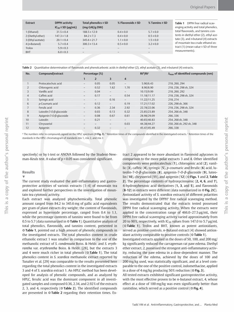
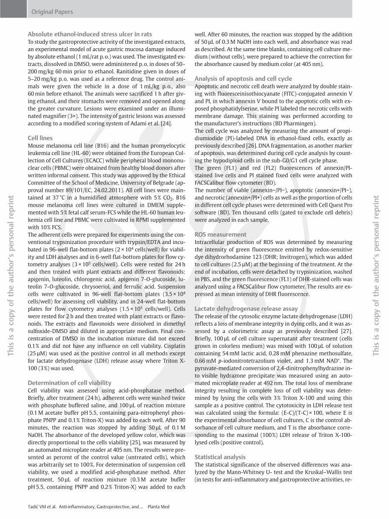
Table 2 Quantitative determination of flavonoids and phenolcarbonic acids in diethyl ether (2), ethyl acetate (3), and n-butanol (4) extracts.
No. Compound/extract Percentage (%) Rtb/Rtc λmax of identified compounds (nm)
1 2 3 4
1 Protocatechuic acid – 0.05 0.05 – 5.90/6.43 218, 260, 294
2 Chlorogenic acid – 0.52 1.62 1.70 8.90/8.90 218, 238, 298 sh, 324
3 Vanillic acid – 0.04 – – 10.15/9.99 218, 260, 292
4 Caffeic acid – 0.17 – 0.54 11.18/11.17 218, 238, 298 sh, 324
5 Syringic acid – – 0.16 – 11.22/11.25 218, 274
6 p-Coumaric acid – 0.12 – 0.19 17.21/17.02 226, 298 sh, 366
7 Ferulic acid – 0.36 2.34 2.92 22.78/22.06 218, 236, 298 sh, 324
8 Luteolin-7-O-β-glucoside – 0.03 0.13 0.32 23.85/23.89 254, 266 sh, 348
9 Apigenin-7-O-β-glucoside – 0.08 0.67 0.61 28.98/29.09 266, 336
10 Luteolin – 0.21 – – 40.65/40.83 254, 268 sh, 348
11 Chrysoeriol – – – 0.03 44.38/44.27 250, 266 sh, 292 sh, 348
12 Apigenin – 0.32 – – 45.47/45.49 266, 338
a The numbers refer to compounds signed on the HPLC spectrum (l" Fig. 3). b Retention times of the compounds identified in the investigated extracts. c Retention times of the
standards in the HPLC chromatogram of standards mix 1, mix 2, and mix 3
Table 1 DPPH free radical scav-enging activity and total phenolics,total flavonoids, and tannins con-tents in diethyl ether (2), ethyl ace-tate (3), and n-butanol (4) extractsof mountain tea crude ethanol ex-tract (1) (mean value ± SD of threemeasurements).
Extract DPPH activity
IC50 ± SD (µg/mL)
Total phenolics ± SD
(mg GAE/g DW)
% Flavonoids ± SD % Tannins ± SD
1 (Ethanol) 31.5 ± 0.4 188.5 ± 12.9 0.4 ± 0.0 5.7 ± 0.0
2 (Diethyl ether) 147.3 ± 1.8 84.2 ± 7.3 0.4 ± 0.0 0.5 ± 0.0
3 (Ethyl acetate) 20.1 ± 0.4 345.6 ± 21.7 1.1 ± 0.0 1.5 ± 0.0
4 (n-Butanol) 5.7 ± 0.4 300.3 ± 13.4 0.5 ± 0.0 3.2 ± 0.0
Trolox 5.9 ± 0.3 – – –
BHT 6.0 ± 0.3 – – –
Original Papers
orʼs
person
alreprint
Thisis
aco
pyof
theau
thorʼs
person
alreprint
spectively) or by t-test or ANOVA followed by the Student-New-man-Keuls test. A value of p < 0.05 was considered significant.
Thisis
aco
pyof
theau
th
bResults!The current study evaluated the anti-inflammatory and gastro-protective activities of various extracts (1–4) of mountain teaand explored further perspectives in the investigation of moun-tain tea cytotoxic activity.Each extract was analyzed phytochemically. Total phenolicamount ranged from 84.2 to 345.6mg of gallic acid equivalentsper gram of plant extracts dry weight; the content of flavonoids,expressed as hyperoside percentage, ranged from 0.4 to 1.1,while the percentage contents of tannins were found to be from0.5 to 5.7 (data summarized inl" Table 1). Quantitative analysis oftotal phenolics, flavonoids, and tannins content, presented inl" Table 1, pointed out a high amount of phenolic compounds inthe investigated extracts. The total phenolics content in crudeethanolic extract 1 was smaller in comparison to the one of themethanolic extract of S. condensata Boiss. & Heldr. and S. eryth-rantha var. erythrantha Boiss. & Heldr. [28], but the extracts 3and 4 were much richer in total phenols (l" Table 1). The totalphenolics content in S. scardica methanolic extract reported byTunalier et al. [29] was comparable to the results presented hereregarding the total phenolics content in the investigated extracts3 and 4 of S. scardica extract 1. An HPLC method has been devel-oped for analysis of phenolic compounds, and as analyzed byHPLC, ferulic acid was the dominant component in all investi-gated samples and composed 0.36, 2.34, and 2.92% of the extracts2, 3, and 4, respectively (l" Table 2). The identified compoundsare presented in l" Table 2 regarding their retention times. Ex-
tract 2 appeared to be more abundant in flavonoid aglycones incomparison to the more polar extracts 3 and 4. Other identifiedcomponents were protocatechuic (1), chlorogenic acid (2), vanil-lic (3), caffeic (4), syringic (5), p-coumaric and ferulic (6) acid, lu-teolin-7-O-β-glucoside (8), apigenin-7-O-β-glucoside (9), luteo-lin (10), chrysoeriol (11), and apigenin (12) (l" Figs. 1 and 2, Table2). The percentage contents of hydroxycinnamic (2, 4, 6, and 7),4-hydroxybenzoic acid derivatives (1, 3, and 5), and flavonoids(8-12) in extracts were different (data summarized in l" Fig. 2C).Antioxidant activity of S. scardica extracts of different polaritieswas investigated by the DPPH+ free radical scavenging method.The results demonstrated that the extracts tested possessedDPPH free radical scavenging activity. When the extracts wereapplied in the concentration range of 466.0–27.5 µg/mL, theirDPPH free radical scavenging activity varied approximately from20 to 90%, respectively, with IC50 values from 147.0 to 5.7 µg/mL(l" Table 1). Trolox and BHT, known as potent antioxidants,served as positive controls. n-Butanol extract (4) showed antiox-idant activity comparable to positive controls (l" Table 1).Investigated extracts applied in the doses of 50, 100, and 200mg/kg significantly reduced the carrageenan rat paw edema. Diethylether extract, 2, possessed the strongest anti-inflammatory activ-ity, reducing the paw edema in a dose-dependent manner. Thereduction of the edema, achieved by the doses of 100 and200mg/kg used, was statistically significant, and at a level com-parable to the one of the positive control, indomethacine, appliedin a dose of 4mg/kg producing 50% reduction (l" Fig. 3).All tested extracts exhibited significant gastroprotective activity,with the most effective proven to be n-butanol extract, 4, whoseeffect at a dose of 100mg/kg was even significantly better thanranitidine, which served as a positive control (l" Fig. 4).
Tadić VM et al. Anti-inflammatory, Gastroprotective, and… Planta Med
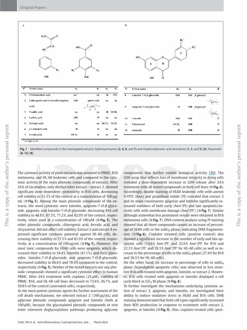
Fig. 1 Identified compounds in the investigated extracts: hydroxycinnamic (2, 4, 6, and 7) and 4-hydroxybenzoic acid derivatives (1, 3, and 5) (A); flavonoids(8–12) (B).
Original Papers
Thisis
aco
pyof
theau
thorʼs
person
alreprint
Thisis
aco
pyof
theau
thorʼs
person
alreprint
bThe cytotoxic activity of plant extracts was assessed in PBMC, B16melanoma, and HL-60 leukemic cells and compared to the cyto-toxic activity of the main phenolic compounds of extracts. After24 h of incubation, only diethyl ether extract – extract 2, showedsignificant dose-dependent cytotoxicity in B16 cells, decreasingcell viability to 51.3% of the control at a concentration of 100 µg/mL (l" Fig. 5). Among the main phenolic compounds of the ex-tracts, the most cytotoxic were luteolin, apigenin-7-O-β-glyco-side, apigenin, and luteolin-7-O-β-glycoside, decreasing B16 cellviability to 48.8%, 67.3%, 77.2%, and 82.0% of the control, respec-tively, when used at a concentration of 100 µM (l" Fig. 5). Theother phenolic compounds, chlorogenic acid, ferrulic acid, andchrysoeriol, did not affect cell viability. Extract 2 and extract 4 ex-pressed significant cytotoxic potential against HL-60 cells, de-creasing their viability to 77.5% and 81.9% of the control, respec-tively, at a concentration of 100 µg/mL (l" Fig. 5). However, themost toxic compounds for HL60 cells were apigenin, which de-creased their viability to 34.4%, luteolin (47.1%), and their glyco-sides, luteolin-7-O-β-glucoside and apigenin-7-O-β-glucoside,decreased viability to 66.6% and 78.4% compared to the control,respectively (l" Fig. 5). Neither of the tested extracts nor any phe-nolic compounds showed a significant cytotoxic effect to humanPBMC. After 24 h treatment with cisplatin (25 µM), viability ofPBMC, B16, and HL-60 cell lines decreased to 73.6%, 56.7%, and59.8% of the control (untreated cells), respectively.As the most potent cytotoxic agents for further assessment of thecell death mechanisms, we selected extract 2 (100 µg/mL) andaglycone phenolic compounds apigenin and luteolin (both at100 µM), because the glycosylated phenolic compounds in vivoenter extensive deglycosylation pathways producing aglycone
Tadić VM et al. Anti-inflammatory, Gastroprotective, and… Planta Med
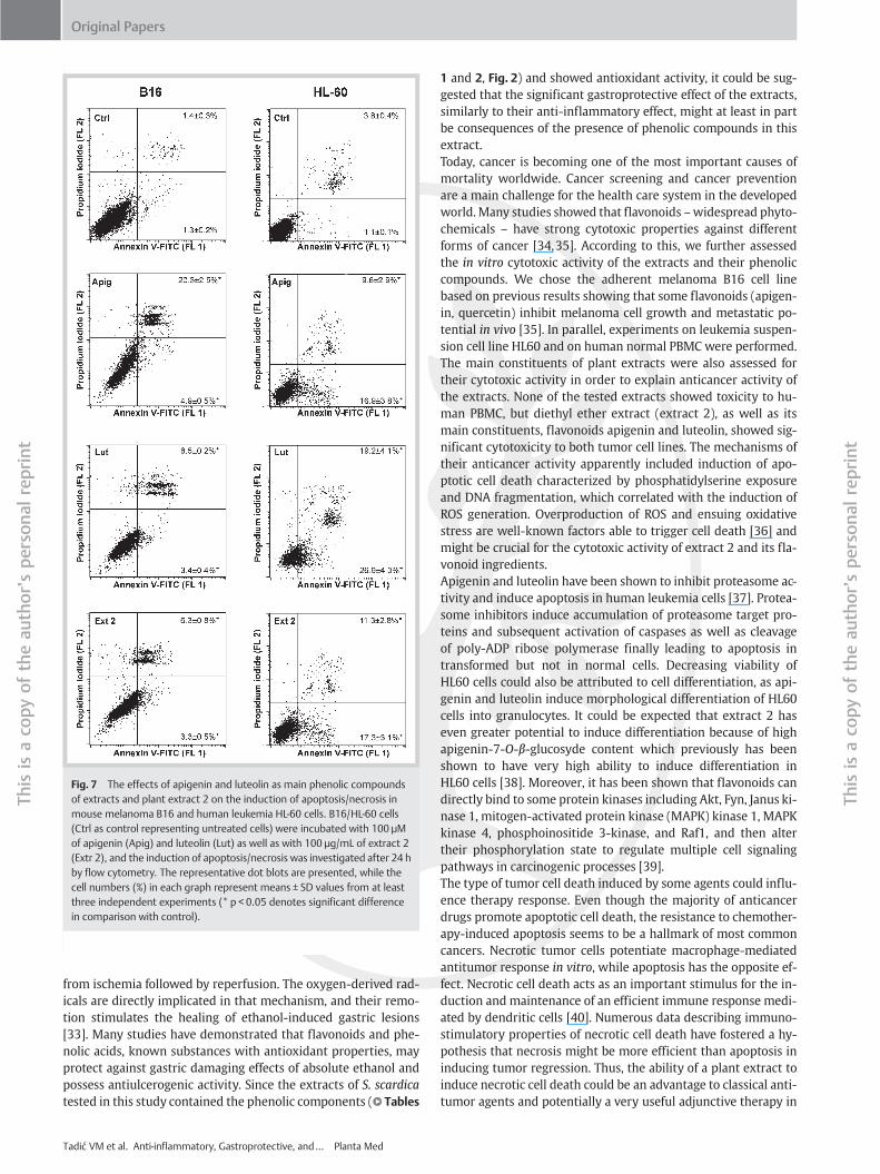
components that further exhibit biological activity [30]. TheLDH assay that reflects loss of membrane integrity in dying cellsrevealed a dose-dependent increase in LDH release after 24 htreatment with all tested compounds in both cell lines (l" Fig. 6).Accordingly, double staining of HL60 leukemic cells with anexinV-FITC (Ann) and propidium iodide (PI) revealed that extract 2and its main constituents apigenin and luteolin significantly in-creased numbers of both early (Ann+/PI) and late apoptotic/ne-crotic cells with membrane damage (Ann+/PI+) (l" Fig. 7). Similaralthough somewhat less prominent results were obtained in B16melanoma cells (l" Fig. 7). DNA content analysis using PI stainingshowed that all three compounds caused an increase of percent-age of HL60 cells in the subG0 phase, indicating DNA fragmenta-tion (l" Fig. 8). Cisplatin tretated cells (positive control) alsoshowed a significant increase in the number of early and late ap-optotic cells (14.6% Ann+/PI− and 22.6% Ann+/PI+ for B16 and22.3% Ann+/PI− and 28.3% Ann+/PI+ for HL-60 cells) as well as in-crease in the percentage of cells in the subG0 phase (27.4% for B16and 36.5% for HL-60 cells).On the other hand, no increase in percentage of cells in subG0
phase (hypodiploid-apoptotic cells) was observed in less sensi-tive B16 cells treatedwith apigenin, luteolin, or extract 2. Howev-er, B16 cells treated with apigenin or luteolin displayed a cellcycle block in S/G2M phase (l" Fig. 8).To further investigate the mechanisms underlying cytotoxic ac-tion of extract 2, apigenin, and luteolin, we investigated theirability to induce oxidative stress in HL60 and B16 cells. DHRstaining demonstrated that both cell types significantly increasedtheir ROS production in response to treatment with extract 2,apigenin, or luteolin (l" Fig. 9). Also, cisplatin-treated cells (posi-
bFig. 3 Effect of S. scardica extracts (ethanol, 1; di-ethyl ether, 2; ethyl acetate, 3; and n-butanol, 4)and reference substance (indomethacin – IND) oncarrageen-induced rat paw edema.
Fig. 2 HPLC chromatograms of the examined mountain tea extracts (dieth-yl ether, 2; ethyl acetate, 3; and n-butanol, 4) recorded at 360 and 280 nm,with the spectrum of identified compounds, compared to UV spectra of ref-erence standards and chemical structures of identified compounds (A).Numbers refer to the following: protocatechuic acid (1), chlorogenic acid (2),vanillic acid (3), caffeic acid (4), syringic acid (5), p-coumaric acid (6), ferulicacid (7), luteolin-7-O-β-glycoside (8), apigenin-7-O-β-glycoside (9), luteolin
(10), chrysoeriol (11), and apigenin (12). HPLC chromatograms of the etha-nol, diethyl ether, ethyl acetate, and n-butanol extracts (1, 2, 3, and 4, re-spectively) of mountain tea recorded at 360 nm mirrored to each other (B).The percentage content of hydroxycinnamic (2, 4, 6, and 7), 4-hydroxyben-zoic acid derivatives (1, 3, and 5) and flavonoids (8–12) in extracts (C).* Compounds present in all investigated extracts but not identified.
Original Papers
Thisis
aco
pyof
theau
thorʼs
person
alreprint
Thisis
aco
pyof
theau
thorʼs
person
alreprint
tive control) demonstrated the increase in ROS production of 1.8and a 2.1-fold increase for B16 and HL-60 cells, respectively, com-pared to untreated cells (negative control). However, the effectsof apigenin and extract 2 were more pronounced in B16 cells(l" Fig. 9). It therefore appears that induction of oxidative stressmight be involved in the cytotoxic activity of extract 2 and phe-nolic compounds.
Discussion!
The ethanol extract (1) of S. scardica, mountain tea, and diethylether (2), ethyl acetate (3), and n-butanol (4) extracts of the crudeethanol extract were evaluated for their traditionally known buthere for the first time confirmed anti-inflammatory and gastro-protective activities applying in vivo tests. Further, we investi-
Tadić VM et al. Anti-inflammatory, Gastroprotective, and… Planta Med
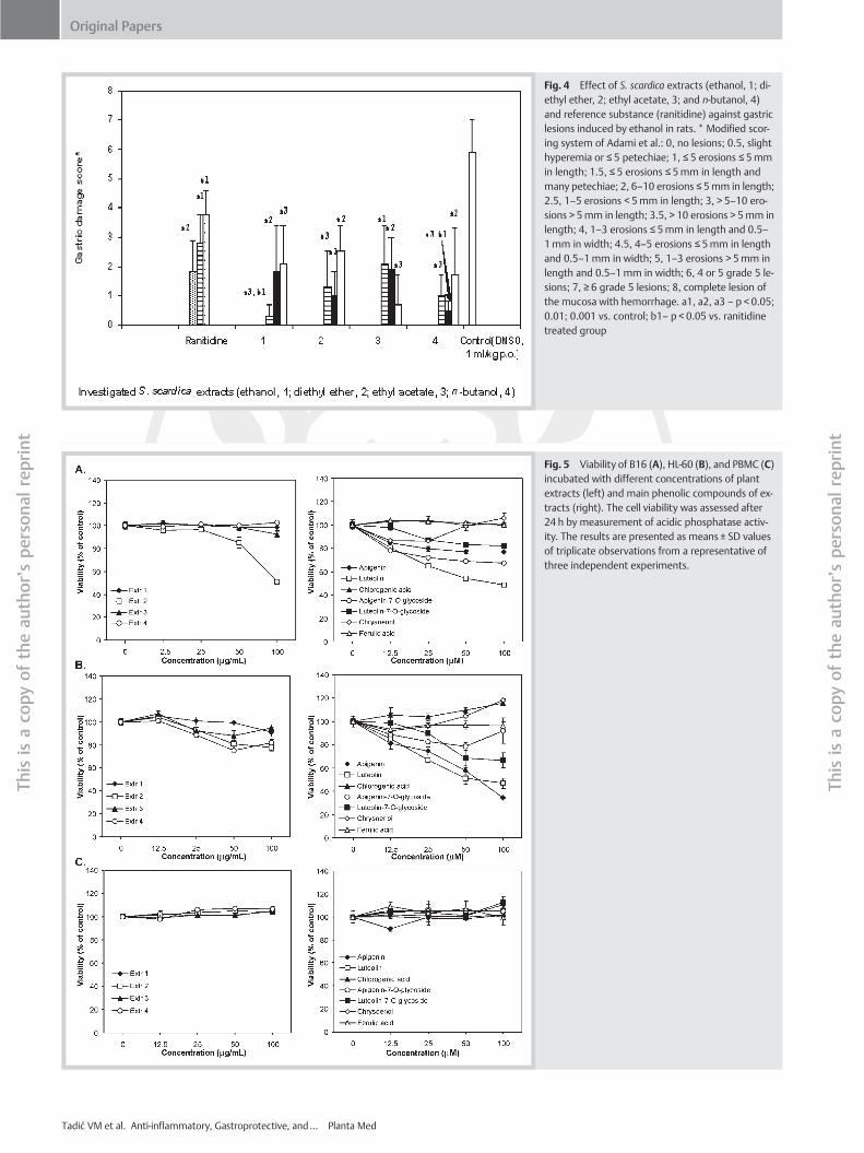
bFig. 4 Effect of S. scardica extracts (ethanol, 1; di-ethyl ether, 2; ethyl acetate, 3; and n-butanol, 4)and reference substance (ranitidine) against gastriclesions induced by ethanol in rats. * Modified scor-ing system of Adami et al.: 0, no lesions; 0.5, slighthyperemia or ≤ 5 petechiae; 1, ≤ 5 erosions ≤ 5mmin length; 1.5, ≤ 5 erosions ≤ 5mm in length andmany petechiae; 2, 6–10 erosions ≤ 5mm in length;2.5, 1–5 erosions < 5mm in length; 3, > 5–10 ero-sions > 5mm in length; 3.5, > 10 erosions > 5mm inlength; 4, 1–3 erosions ≤ 5mm in length and 0.5–1mm in width; 4.5, 4–5 erosions ≤ 5mm in lengthand 0.5–1mm in width; 5, 1–3 erosions > 5mm inlength and 0.5–1mm in width; 6, 4 or 5 grade 5 le-sions; 7, ≥ 6 grade 5 lesions; 8, complete lesion ofthe mucosa with hemorrhage. a1, a2, a3 – p < 0.05;0.01; 0.001 vs. control; b1– p < 0.05 vs. ranitidinetreated group
Fig. 5 Viability of B16 (A), HL-60 (B), and PBMC (C)incubated with different concentrations of plantextracts (left) and main phenolic compounds of ex-tracts (right). The cell viability was assessed after24 h by measurement of acidic phosphatase activ-ity. The results are presented as means ± SD valuesof triplicate observations from a representative ofthree independent experiments.
Tadić VM et al. Anti-inflammatory, Gastroprotective, and… Planta Med
Original Papers
Thisis
aco
pyof
theau
thorʼs
person
alreprint
Thisis
aco
pyof
theau
thorʼs
person
alreprint
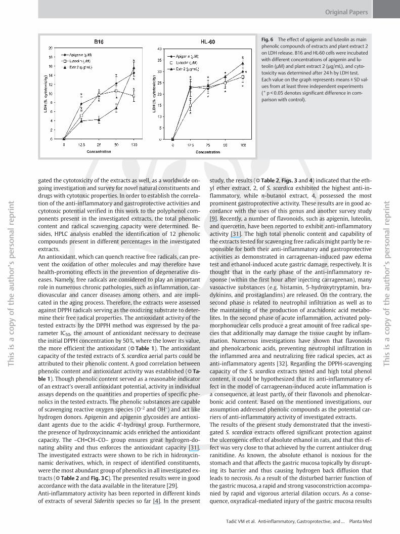
Fig. 6 The effect of apigenin and luteolin as mainphenolic compounds of extracts and plant extract 2on LDH release. B16 and HL-60 cells were incubatedwith different concentrations of apigenin and lu-teolin (µM) and plant extract 2 (µg/mL), and cyto-toxicity was determined after 24 h by LDH test.Each value on the graph represents means ± SD val-ues from at least three independent experiments(* p < 0.05 denotes significant difference in com-parison with control).
Original Papers
Thisis
aco
pyof
theau
thorʼs
person
alreprint
Thisis
aco
pyof
theau
thorʼs
person
alreprint
bgated the cytotoxicity of the extracts as well, as a worldwide on-going investigation and survey for novel natural constituents anddrugs with cytotoxic properties. In order to establish the correla-tion of the anti-inflammatory and gastroprotective activities andcytotoxic potential verified in this work to the polyphenol com-ponents present in the investigated extracts, the total phenoliccontent and radical scavenging capacity were determined. Be-sides, HPLC analysis enabled the identification of 12 phenoliccompounds present in different percentages in the investigatedextracts.An antioxidant, which can quench reactive free radicals, can pre-vent the oxidation of other molecules and may therefore havehealth-promoting effects in the prevention of degenerative dis-eases. Namely, free radicals are considered to play an importantrole in numerous chronic pathologies, such as inflammation, car-diovascular and cancer diseases among others, and are impli-cated in the aging process. Therefore, the extracts were assessedagainst DPPH radicals serving as the oxidizing substrate to deter-mine their free radical properties. The antioxidant activity of thetested extracts by the DPPH method was expressed by the pa-rameter IC50, the amount of antioxidant necessary to decreasethe initial DPPH concentration by 50%, where the lower its value,the more efficient the antioxidant (l" Table 1). The antioxidantcapacity of the tested extracts of S. scardica aerial parts could beattributed to their phenolic content. A good correlation betweenphenolic content and antioxidant activity was established (l" Ta-ble 1). Though phenolic content served as a reasonable indicatorof an extractʼs overall antioxidant potential, activity in individualassays depends on the quantities and properties of specific phe-nolics in the tested extracts. The phenolic substances are capableof scavenging reactive oxygen species (O−2 and OH−) and act likehydrogen donors. Apigenin and apigenin glycosides are antioxi-dant agents due to the acidic 4′-hydroxyl group. Furthermore,the presence of hydroxycinnamic acids enriched the antioxidantcapacity. The –CH=CH–CO– group ensures great hydrogen-do-nating ability and thus enforces the antioxidant capacity [31].The investigated extracts were shown to be rich in hidroxycin-namic derivatives, which, in respect of identified constituents,were themost abundant group of phenolics in all investigated ex-tracts (l" Table 2 and Fig. 3C). The presented results were in goodaccordance with the data available in the literature [29].Anti-inflammatory activity has been reported in different kindsof extracts of several Sideritis species so far [4]. In the present
study, the results (l" Table 2, Figs. 3 and 4) indicated that the eth-yl ether extract, 2, of S. scardica exhibited the highest anti-in-flammatory, while n-butanol extract, 4, possessed the mostprominent gastroprotective activity. These results are in good ac-cordance with the uses of this genus and another survey study[9]. Recently, a number of flavonoids, such as apigenin, luteolin,and quercetin, have been reported to exhibit anti-inflammatoryactivity [31]. The high total phenolic content and capability ofthe extracts tested for scavenging free radicals might partly be re-sponsible for both their anti-inflammatory and gastroprotectiveactivities as demonstrated in carrageenan-induced paw edematest and ethanol-induced acute gastric damage, respectively. It isthought that in the early phase of the anti-inflammatory re-sponse (within the first hour after injecting carrageenan), manyvasoactive substances (e.g. histamin, 5-hydroxytryptamin, bra-dykinins, and prostaglandins) are released. On the contrary, thesecond phase is related to neutrophil infiltration as well as tothe maintaining of the production of arachidonic acid metabo-lites. In the second phase of acute inflammation, activated poly-morphonuclear cells produce a great amount of free radical spe-cies that additionally may damage the tissue caught by inflam-mation. Numerous investigations have shown that flavonoidsand phenolcarbonic acids, preventing neutrophil infiltration inthe inflammed area and neutralizing free radical species, act asanti-inflammatory agents [32]. Regarding the DPPH-scavengingcapacity of the S. scardica extracts tested and high total phenolcontent, it could be hypothesized that its anti-inflammatory ef-fect in the model of carrageenan-induced acute inflammation isa consequence, at least partly, of their flavonols and phenolcar-bonic acid content. Based on the mentioned investigations, ourassumption addressed phenolic compounds as the potential car-riers of anti-inflammatory activity of investigated extracts.The results of the present study demonstrated that the investi-gated S. scardica extracts offered significant protection againstthe ulcerogenic effect of absolute ethanol in rats, and that this ef-fect was very close to that achieved by the current antiulcer drugranitidine. As known, the absolute ethanol is noxious for thestomach and that affects the gastric mucosa topically by disrupt-ing its barrier and thus causing hydrogen back diffusion thatleads to necrosis. As a result of the disturbed barrier function ofthe gastric mucosa, a rapid and strong vasoconstriction accompa-nied by rapid and vigorous arterial dilation occurs. As a conse-quence, oxyradical-mediated injury of the gastric mucosa results
Tadić VM et al. Anti-inflammatory, Gastroprotective, and… Planta Med
bFig. 7 The effects of apigenin and luteolin as main phenolic compoundsof extracts and plant extract 2 on the induction of apoptosis/necrosis inmouse melanoma B16 and human leukemia HL-60 cells. B16/HL-60 cells(Ctrl as control representing untreated cells) were incubated with 100 µMof apigenin (Apig) and luteolin (Lut) as well as with 100 µg/mL of extract 2(Extr 2), and the induction of apoptosis/necrosis was investigated after 24 hby flow cytometry. The representative dot blots are presented, while thecell numbers (%) in each graph represent means ± SD values from at leastthree independent experiments (* p < 0.05 denotes significant differencein comparison with control).
Original Papers
Thisis
aco
pyof
theau
thorʼs
person
alreprint
Thisis
aco
pyof
theau
thorʼs
person
alreprint
from ischemia followed by reperfusion. The oxygen-derived rad-icals are directly implicated in that mechanism, and their remo-tion stimulates the healing of ethanol-induced gastric lesions[33]. Many studies have demonstrated that flavonoids and phe-nolic acids, known substances with antioxidant properties, mayprotect against gastric damaging effects of absolute ethanol andpossess antiulcerogenic activity. Since the extracts of S. scardicatested in this study contained the phenolic components (l" Tables
Tadić VM et al. Anti-inflammatory, Gastroprotective, and… Planta Med
1 and 2, Fig. 2) and showed antioxidant activity, it could be sug-gested that the significant gastroprotective effect of the extracts,similarly to their anti-inflammatory effect, might at least in partbe consequences of the presence of phenolic compounds in thisextract.Today, cancer is becoming one of the most important causes ofmortality worldwide. Cancer screening and cancer preventionare a main challenge for the health care system in the developedworld. Many studies showed that flavonoids –widespread phyto-chemicals – have strong cytotoxic properties against differentforms of cancer [34,35]. According to this, we further assessedthe in vitro cytotoxic activity of the extracts and their phenoliccompounds. We chose the adherent melanoma B16 cell linebased on previous results showing that some flavonoids (apigen-in, quercetin) inhibit melanoma cell growth and metastatic po-tential in vivo [35]. In parallel, experiments on leukemia suspen-sion cell line HL60 and on human normal PBMC were performed.The main constituents of plant extracts were also assessed fortheir cytotoxic activity in order to explain anticancer activity ofthe extracts. None of the tested extracts showed toxicity to hu-man PBMC, but diethyl ether extract (extract 2), as well as itsmain constituents, flavonoids apigenin and luteolin, showed sig-nificant cytotoxicity to both tumor cell lines. The mechanisms oftheir anticancer activity apparently included induction of apo-ptotic cell death characterized by phosphatidylserine exposureand DNA fragmentation, which correlated with the induction ofROS generation. Overproduction of ROS and ensuing oxidativestress are well-known factors able to trigger cell death [36] andmight be crucial for the cytotoxic activity of extract 2 and its fla-vonoid ingredients.Apigenin and luteolin have been shown to inhibit proteasome ac-tivity and induce apoptosis in human leukemia cells [37]. Protea-some inhibitors induce accumulation of proteasome target pro-teins and subsequent activation of caspases as well as cleavageof poly-ADP ribose polymerase finally leading to apoptosis intransformed but not in normal cells. Decreasing viability ofHL60 cells could also be attributed to cell differentiation, as api-genin and luteolin induce morphological differentiation of HL60cells into granulocytes. It could be expected that extract 2 haseven greater potential to induce differentiation because of highapigenin-7-O-β-glucosyde content which previously has beenshown to have very high ability to induce differentiation inHL60 cells [38]. Moreover, it has been shown that flavonoids candirectly bind to some protein kinases including Akt, Fyn, Janus ki-nase 1, mitogen-activated protein kinase (MAPK) kinase 1, MAPKkinase 4, phosphoinositide 3-kinase, and Raf1, and then altertheir phosphorylation state to regulate multiple cell signalingpathways in carcinogenic processes [39].The type of tumor cell death induced by some agents could influ-ence therapy response. Even though the majority of anticancerdrugs promote apoptotic cell death, the resistance to chemother-apy-induced apoptosis seems to be a hallmark of most commoncancers. Necrotic tumor cells potentiate macrophage-mediatedantitumor response in vitro, while apoptosis has the opposite ef-fect. Necrotic cell death acts as an important stimulus for the in-duction and maintenance of an efficient immune response medi-ated by dendritic cells [40]. Numerous data describing immuno-stimulatory properties of necrotic cell death have fostered a hy-pothesis that necrosis might be more efficient than apoptosis ininducing tumor regression. Thus, the ability of a plant extract toinduce necrotic cell death could be an advantage to classical anti-tumor agents and potentially a very useful adjunctive therapy in
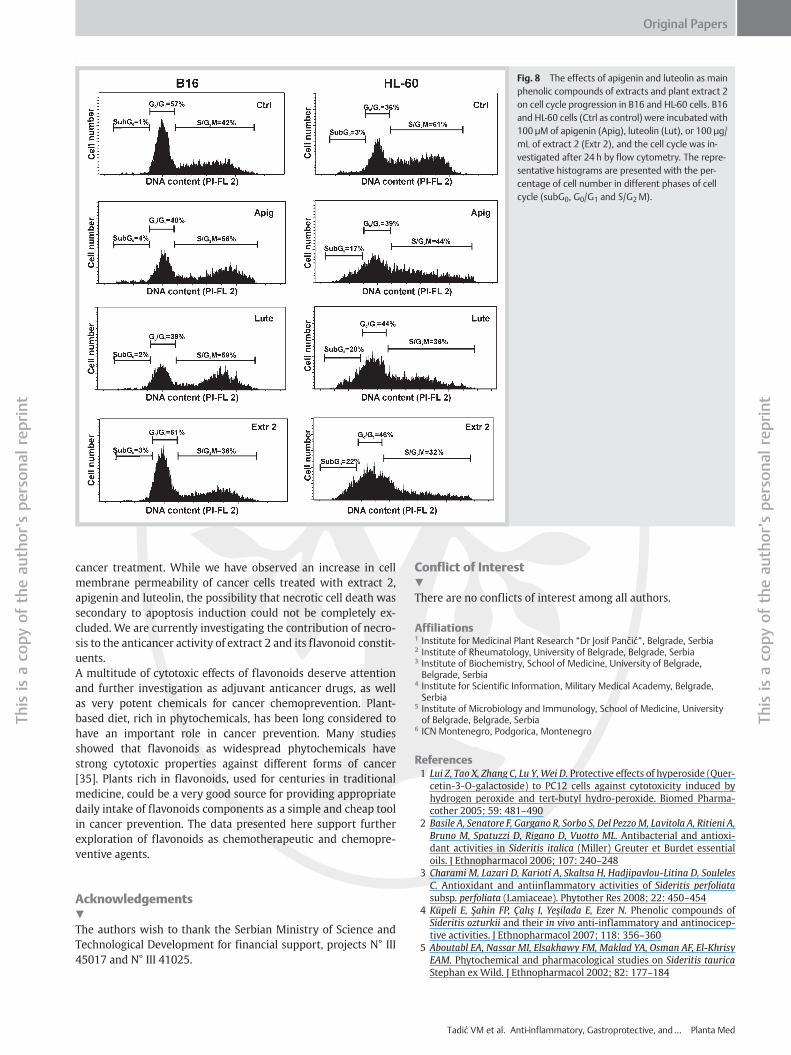
Fig. 8 The effects of apigenin and luteolin as mainphenolic compounds of extracts and plant extract 2on cell cycle progression in B16 and HL-60 cells. B16and HL-60 cells (Ctrl as control) were incubated with100 µM of apigenin (Apig), luteolin (Lut), or 100 µg/mL of extract 2 (Extr 2), and the cell cycle was in-vestigated after 24 h by flow cytometry. The repre-sentative histograms are presented with the per-centage of cell number in different phases of cellcycle (subG0, G0/G1 and S/G2M).
Original Papers
Thisis
aco
pyof
theau
thorʼs
person
alreprint
Thisis
aco
pyof
theau
thorʼs
person
alreprint
bcancer treatment. While we have observed an increase in cellmembrane permeability of cancer cells treated with extract 2,apigenin and luteolin, the possibility that necrotic cell death wassecondary to apoptosis induction could not be completely ex-cluded. We are currently investigating the contribution of necro-sis to the anticancer activity of extract 2 and its flavonoid constit-uents.A multitude of cytotoxic effects of flavonoids deserve attentionand further investigation as adjuvant anticancer drugs, as wellas very potent chemicals for cancer chemoprevention. Plant-based diet, rich in phytochemicals, has been long considered tohave an important role in cancer prevention. Many studiesshowed that flavonoids as widespread phytochemicals havestrong cytotoxic properties against different forms of cancer[35]. Plants rich in flavonoids, used for centuries in traditionalmedicine, could be a very good source for providing appropriatedaily intake of flavonoids components as a simple and cheap toolin cancer prevention. The data presented here support furtherexploration of flavonoids as chemotherapeutic and chemopre-ventive agents.
Acknowledgements!
The authors wish to thank the Serbian Ministry of Science andTechnological Development for financial support, projects N° III45017 and N° III 41025.
Conflict of Interest!
There are no conflicts of interest among all authors.
Affiliations1 Institute for Medicinal Plant Research “Dr Josif Pančić”, Belgrade, Serbia2 Institute of Rheumatology, University of Belgrade, Belgrade, Serbia3 Institute of Biochemistry, School of Medicine, University of Belgrade,Belgrade, Serbia
4 Institute for Scientific Information, Military Medical Academy, Belgrade,Serbia
5 Institute of Microbiology and Immunology, School of Medicine, Universityof Belgrade, Belgrade, Serbia
6 ICN Montenegro, Podgorica, Montenegro
References1 Lui Z, Tao X, Zhang C, Lu Y, Wei D. Protective effects of hyperoside (Quer-cetin-3-O-galactoside) to PC12 cells against cytotoxicity induced byhydrogen peroxide and tert-butyl hydro-peroxide. Biomed Pharma-cother 2005; 59: 481–490
2 Basile A, Senatore F, Gargano R, Sorbo S, Del PezzoM, Lavitola A, Ritieni A,Bruno M, Spatuzzi D, Rigano D, Vuotto ML. Antibacterial and antioxi-dant activities in Sideritis italica (Miller) Greuter et Burdet essentialoils. J Ethnopharmacol 2006; 107: 240–248
3 Charami M, Lazari D, Karioti A, Skaltsa H, Hadjipavlou-Litina D, SoulelesC. Antioxidant and antiinflammatory activities of Sideritis perfoliatasubsp. perfoliata (Lamiaceae). Phytother Res 2008; 22: 450–454
4 Küpeli E, Şahin FP, Çalış I, Yeşilada E, Ezer N. Phenolic compounds ofSideritis ozturkii and their in vivo anti-inflammatory and antinocicep-tive activities. J Ethnopharmacol 2007; 118: 356–360
5 Aboutabl EA, Nassar MI, Elsakhawy FM, Maklad YA, Osman AF, El-KhrisyEAM. Phytochemical and pharmacological studies on Sideritis tauricaStephan ex Wild. J Ethnopharmacol 2002; 82: 177–184
Tadić VM et al. Anti-inflammatory, Gastroprotective, and… Planta Med
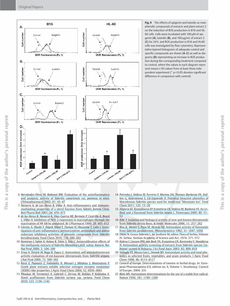
Fig. 9 The effects of apigenin and luteolin as mainphenolic compounds of extracts and plant extract 2on the induction of ROS production in B16 and HL-60 cells. Cells were incubated with 100 µM of api-genin (A), luteolin (B), and 100 µg/mL of extract 2(C) for 24 h, and ROS production in B16 and HL-60cells was investigated by flow cytometry. Represen-tative layered histograms of adequate control andspecific compounds are shown (A–C) as well as dia-grams (D) representing an increase in ROS produc-tion during the corresponding treatment comparedto control, where the values in each diagram repre-sent means ± SD values from at least three inde-pendent experiment (* p < 0.05 denotes significantdifference in comparison with control).
Original Papers
Thisis
aco
pyof
theau
thorʼs
person
alreprint
Thisis
aco
pyof
theau
thorʼs
person
alreprint
b6 Hernàndez-Pérez M, Rabanal RM. Evaluation of the antinflammatoryand analgesic activity of Sideritis canariensis var. pannosa in mice.J Ethnopharmacol 2002; 81: 43–47
7 Navarro A, de Las Heras B, Villar A. Anti-inflammatory and immuno-modulating properties of a sterol fraction from Sideitis foetens Clem.Biol Pharm Bull 2001; 24: 470–473
8 de las Heras B, Navarro A, Díaz-Guerra MJ, Bermejo P, Castrillo A, BoscáL, Villar A. Inhibition of NOS‑2 expression in macrophages through theinactivation of NF‑kB by andalusol. Br J Pharmacol 1999; 28: 605–612
9 Güvenç A, Okada Y, Küpeli Akkol E, Duman H, Okuyama T, Çalıs I. Inves-tigations of anti-inflammatory, antinociceptive, antioxidant and aldosereductase inhibitory activities of phenolic compounds from Sideritisbrevibracteata. Food Chem 2010; 118: 686–692
10 Demirtas I, Sahin A, Ayhan B, Tekin S, Telci I. Antiproliferative effects ofthe methanolic extracts of Sideritis libanotica Labill. subsp. linearis. RecNat Prod 2009; 3: 104–109
11 Ertaş A, Öztürk M, Boga B, Topçu G. Antioxidant and anticholinesteraseactivity evaluation of ent-kaurane diterpenoids from Sideritis arguta.J Nat Prod 2009; 72: 500–502
12 Kassi E, Papoutsi Z, Fokialakis N, Messari J, Mitakou S, Moutsatsou P.Greek plant extracts exhibit selective estrogen receptor modulator(SERM)-like properties. J Agric Food Chem 2004; 52: 6956–6961
13 Plioukas M, Termentzi A, Gabrieli C, Zervou M, Kefalas P, Kokkalou E.Novel acylflavones from Sideritis syriaca ssp. syriaca. Food Chem2010; 123: 1136–1141
Tadić VM et al. Anti-inflammatory, Gastroprotective, and… Planta Med
14 Petreska J, Stefova M, Ferreres F, Moreno DA, Thomas-Barberan FA, Stef-kov G, Kulevanova S, Gil-Izquiredo A. Potential bioactive phenolics ofMacedonian Sideritis species used for medicinal “Mountain tea”. FoodChem 2011; 125: 13–20
15 Alipieva KI, Kostadinova EP, Evstatieva LN, StefovaM, Bankova VS. An iri-doid and a flavonoid from Sideritis lanata L. Fitoterapia 2009; 80: 51–53
16 Kilic T. Isolation and biological activity of new and known diterpenoidsfrom Sideritis stricta Boiss. & Heldr. Molecules 2006; 11: 257–262
17 Rios JL, Manez S, Paya M, Alcaraz MJ. Antioxidant activity of flavonoidsfrom Sideritis javalabrensis. Phytochemistry 1992; 31: 1047–1050
18 DiklićN.Genus Sideritis L. In: JosifovićM, editor. Flora of Serbia, VolumeIV. Serbia: Serbian Academy of Science and Art; 1974: 371–372
19 Koleva I, Linssen JPH, van Beek TA, Evstatieva LN, Kortenska V, HandjievaN. Antioxidant activity screening of extracts from Sideritis species (La-biatae) grown in Bulgaria. J Sci Food Agric 2003; 83: 809–819
20 Velioglu YS, Mazza Gao L, Oomah BD. Antioxidant activity and total phe-nolics in selected fruits, vegetables, and grain products. J Agric FoodChem 1998; 46: 4113–4117
21 Council of Europe. Determinations of tannins in herbal drugs. In: Euro-pean Pharmacopoeia 6.0, edition no. 6, Volume 1. Strasbourg: Councilof Europe; 2004: 255
22 Blois MS. Antioxidant determinations by the use of a stable free radical.Nature 1958; 181: 1199–1200
Original Papers
nt
Thisis
aco
pyof
theau
thorʼs
person
alreprint
23 Oyanagui Y, Sato, S. Inhibition by nilvadipine of ischemic and carra-geenan paw oedema as well as of superoxide radical production fromneutrophils and xanthine oxidase. Drug Res 1991; 41: 469–474
24 Adami E, Marazzi-Uberti E, Turba C. Pharmacological research on gefar-nate, a new synthetic isoprenoid with an anti-ulcer action. Arch IntPharmacodyn Ther 1964; 58: 1780–1783
25 Touhey S, Heenan M. Cell culture. In: Davey J, Lord JM, editors. Essentialcell biology. Oxford: Oxford University Press; 2003
26 Mijatovic S, Maksimovic-Ivanic D, Radovic J, Miljkovic D, Harhaji L, Vuck-ovic O, Stosic-Grujicic S, Mostarica Stojkovic M, Trajkovic V. Anti-gliomaaction of aloe emodin: the role of ERK inhibition. Cell Mol Life Sci 2005;62: 589–598
27 Badovinac V, Trajkovic V, Mostarica-Stojkovic M. Nitric oxide promotesgrowth and major histocompatibility complex-unrestricted cytotoxic-ity of interleukin-2-activated rat lymphocytes. Scand J Immunol2000; 52: 62–70
28 Özkan G, Sagdiç O, Özcan M, Özçelik H, Ünver A. Antioxidant and anti-bacterial activities of Turkish endemic Sideritis extracts. Grasas Aceites2005; 56: 16–20
29 Tunalier Z, Kosar M, Ozturk N, Baser KHC, Duman H, Kirimer N. Antiox-idant properties and phenolic composition of Sideritis species. ChemNat Comp 2004; 40: 206–210
30 Spencer J, Mohsen M, Rice-Evans C. Cellular uptake and metabolism offlavonoids and their metabolites: implications for their bioactivity.Arch Biochem Biophys 2004; 423: 148–161
31 Rice-Evans CA, Miller NJ, Paganga G. Structure-antioxidant activity rela-tionships of flavonoids and phenolic acids. Free Radic Biol Med 1996;20: 933–956
b ri32 Maleki N, Garjani A, Nazemiyeh H, Nilfouroushan N, Eftekhar Sadat AT,Allameh Z, Hasannia N. Potent anti-inflammatory activities of hydroal-coholic extract from aerial parts of Stachys inflata in rats.J Ethnopharmacol 2001; 75: 213–218
33 Salim AS. Removing oxygen-derived free radicals stimulates healing ofethanol-induced erosive gastritis in the rat. Digestion 1990; 47: 24–28
34 Shukla S, Gupta S. Apigenin: a promising molecule for cancer preven-tion. Pharm Res 2010; 27: 962–978
35 Caltagirone S, Rossi C, Poggi A, Ranelletti FO, Natali PG, Brunetti M, AielloFB, Piantelli M. Flavonoids apigenin and quercetin inhibit melanomagrowth and metastatic potential. Int J Cancer 2000; 87: 595–600
36 Fiers W, Beyaert R, Declercq W, Vandenabeele P. More than one way todie: apoptosis, necrosis and reactive oxygen damage. Oncogene 1999;18: 7719–7730
37 Chen D, Chen MS, Cui QC, Yang H, Dou QP. Structure-proteasome-inhib-itory activity relationships of dietary flavonoids in human cancer cells.Front Biosci 2007; 12: 1935–1945
38 Abaza L, Talorete TP, Yamada P, Kurita Y, Zarrouk M, Isoda H. Inductionof growth inhibition and differentiation of human leukemia HL‑60cells by a Tunisian gerboui olive leaf extract. Biosci Biotechnol Biochem2007; 71: 1306–1312
39 Hou DX, KumamotoT. Flavonoids as protein kinase inhibitors for cancerchemoprevention: direct binding and molecular modeling. AntioxidRedox Signal 2010; 13: 691–719
40 Sauter B, Albert ML, Francisco L, Larsson M, Somersan S, Bhardwaj N.Consequences of cell death: exposure to necrotic tumor cells, but notprimary tissue cells or apoptotic cells, induces the maturation of im-munostimulatory dendritic cells. J Exp Med 2000; 191: 423–434
Tadić VM et al. Anti-inflammatory, Gastroprotective, and… Planta Med
Thisis
aco
pyof
theau
thorʼs
person
alrep
Copyright © 2022 FDOKUMEN