The signal pathways and treatment of cytokine storm in COVID ...
20
REVIEW ARTICLE OPEN The signal pathways and treatment of cytokine storm in COVID-19 Lan Yang 1,2 , Xueru Xie 1,2 , Zikun Tu 1,2 , Jinrong Fu 3 , Damo Xu 4,5 and Yufeng Zhou 1,2 The Coronavirus Disease 2019 (COVID-19) pandemic has become a global crisis and is more devastating than any other previous infectious disease. It has affected a significant proportion of the global population both physically and mentally, and destroyed businesses and societies. Current evidence suggested that immunopathology may be responsible for COVID-19 pathogenesis, including lymphopenia, neutrophilia, dysregulation of monocytes and macrophages, reduced or delayed type I interferon (IFN-I) response, antibody-dependent enhancement, and especially, cytokine storm (CS). The CS is characterized by hyperproduction of an array of pro-inflammatory cytokines and is closely associated with poor prognosis. These excessively secreted pro-inflammatory cytokines initiate different inflammatory signaling pathways via their receptors on immune and tissue cells, resulting in complicated medical symptoms including fever, capillary leak syndrome, disseminated intravascular coagulation, acute respiratory distress syndrome, and multiorgan failure, ultimately leading to death in the most severe cases. Therefore, it is clinically important to understand the initiation and signaling pathways of CS to develop more effective treatment strategies for COVID-19. Herein, we discuss the latest developments in the immunopathological characteristics of COVID-19 and focus on CS including the current research status of the different cytokines involved. We also discuss the induction, function, downstream signaling, and existing and potential interventions for targeting these cytokines or related signal pathways. We believe that a comprehensive understanding of CS in COVID-19 will help to develop better strategies to effectively control immunopathology in this disease and other infectious and inflammatory diseases. Signal Transduction and Targeted Therapy (2021)6:255 ; https://doi.org/10.1038/s41392-021-00679-0 INTRODUCTION Coronavirus Disease 2019 (COVID-19) caused by severe acute respiratory syndrome coronavirus 2 (SARS-CoV-2) rapidly spread worldwide and was declared a pandemic in early 2020. COVID-19 destroyed people’s mental and physical health and staggered global economic growth. As of May 18, 2021, 163 million infections, including 3.38 million deaths, have been recorded (source: World Health Organization). SARS-CoV-2 invades the host by virtue of angiotensin-converting enzyme 2 (ACE2) receptors broadly distributed on various tissues and immune cells. 1–5 The virus can cause a wide range of clinical manifesta- tions from mild forms such as fever, cough, and myalgia to moderate forms requiring hospitalization (pneumonia and localized inflammation) to severe/critical forms with fatal out- comes. 6,7 Severe or critical infection often manifests as pneu- monia, 8,9 disseminated intravascular coagulation (DIC), acute respiratory distress syndrome (ARDS), low blood pressure, and multiorgan failure (Fig. 1). 9–11 Several lines of evidence have shown that immunopathological damage may be responsible for the deterioration of COVID-19. Particularly, multiple studies have reported that highly elevated levels of pro-inflammatory cytokines are produced during the crosstalk between epithelial cells and immune cells in COVID-19, which has linked the cytokine storm (CS) with the severe complications and poor outcomes in this infection. 12–14 CS is a fast-developing, life-threatening, clinical condition in which the overproduction of inflammatory cytokines and excessive activation of immune cells lead to complicated medical syndromes from a persistent fever, nonspecific muscle pain, and hypotension, to capillary leak syndrome, DIC, ARDS, hemopha- gocytic lymphohistiocytosis (HLH), multiorgan failure, and death if treatment is not adequate. 15 Therefore, the timing of diagnosis and treatment of CS could be life-saving. The term CS was first used in 1993 in graft-versus-host disease, 16 and later, in many inflammatory diseases such as autoimmune conditions, organ transplantation, cancer chimeric antigen receptor (CAR) T cell therapy, and, most recently, in COVID-19. 17–23 However, the profile and causative effect of CS in different conditions can greatly vary. Thus far, precise diagnosis and treatment guidelines for CS in most of the conditions are lacking. Understanding the definite alterations and pathogenic roles of individual cytokines involved in the COVID-19-related CS (COVID-CS) is hence extremely important for the development of precise diagnosis and effective treatment. Although some aspects of this topic have been partly reviewed previously, a comprehensive view of COVID-CS to facilitate its Received: 21 March 2021 Revised: 22 May 2021 Accepted: 12 June 2021 1 Institute of Pediatrics, Children’s Hospital of Fudan University, National Children’s Medical Center, and the Shanghai Key Laboratory of Medical Epigenetics, International Co-laboratory of Medical Epigenetics and Metabolism, Ministry of Science and Technology, Institutes of Biomedical Sciences, Fudan University, Shanghai, China; 2 National Health Commission (NHC) Key Laboratory of Neonatal Diseases, Fudan University, Shanghai, China; 3 General Department, Children’s Hospital of Fudan University, Shanghai, China; 4 State Key Laboratory of Respiratory Disease for Allergy at Shenzhen University, Shenzhen Key Laboratory of Allergy and Immunology, Shenzhen University School of Medicine, Shenzhen, China and 5 Institute of Infection, Immunity and Inflammation, University of Glasgow, Glasgow, UK Correspondence: Damo Xu ([email protected]) or Yufeng Zhou ([email protected]) www.nature.com/sigtrans Signal Transduction and Targeted Therapy © The Author(s) 2021, corrected publication 2021 1234567890();,:
-
Upload
khangminh22 -
Category
Documents
-
view
6 -
download
0
Transcript of The signal pathways and treatment of cytokine storm in COVID ...
REVIEW ARTICLE OPEN
The signal pathways and treatment of cytokine storm inCOVID-19Lan Yang1,2, Xueru Xie1,2, Zikun Tu1,2, Jinrong Fu 3, Damo Xu4,5 and Yufeng Zhou 1,2
The Coronavirus Disease 2019 (COVID-19) pandemic has become a global crisis and is more devastating than any other previousinfectious disease. It has affected a significant proportion of the global population both physically and mentally, and destroyedbusinesses and societies. Current evidence suggested that immunopathology may be responsible for COVID-19 pathogenesis,including lymphopenia, neutrophilia, dysregulation of monocytes and macrophages, reduced or delayed type I interferon (IFN-I)response, antibody-dependent enhancement, and especially, cytokine storm (CS). The CS is characterized by hyperproduction of anarray of pro-inflammatory cytokines and is closely associated with poor prognosis. These excessively secreted pro-inflammatorycytokines initiate different inflammatory signaling pathways via their receptors on immune and tissue cells, resulting in complicatedmedical symptoms including fever, capillary leak syndrome, disseminated intravascular coagulation, acute respiratory distresssyndrome, and multiorgan failure, ultimately leading to death in the most severe cases. Therefore, it is clinically important tounderstand the initiation and signaling pathways of CS to develop more effective treatment strategies for COVID-19. Herein, wediscuss the latest developments in the immunopathological characteristics of COVID-19 and focus on CS including the currentresearch status of the different cytokines involved. We also discuss the induction, function, downstream signaling, and existing andpotential interventions for targeting these cytokines or related signal pathways. We believe that a comprehensive understanding ofCS in COVID-19 will help to develop better strategies to effectively control immunopathology in this disease and other infectiousand inflammatory diseases.
Signal Transduction and Targeted Therapy (2021) 6:255 ; https://doi.org/10.1038/s41392-021-00679-0
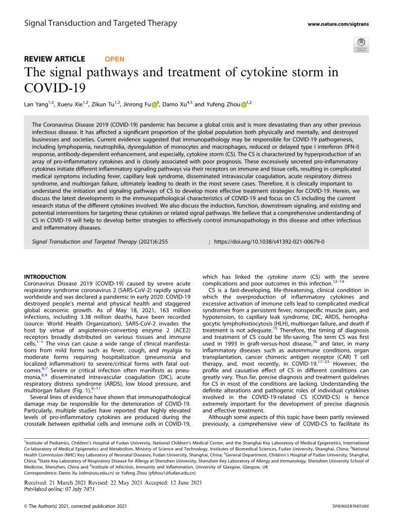
INTRODUCTIONCoronavirus Disease 2019 (COVID-19) caused by severe acuterespiratory syndrome coronavirus 2 (SARS-CoV-2) rapidly spreadworldwide and was declared a pandemic in early 2020. COVID-19destroyed people’s mental and physical health and staggeredglobal economic growth. As of May 18, 2021, 163 millioninfections, including 3.38 million deaths, have been recorded(source: World Health Organization). SARS-CoV-2 invades thehost by virtue of angiotensin-converting enzyme 2 (ACE2)receptors broadly distributed on various tissues and immunecells.1–5 The virus can cause a wide range of clinical manifesta-tions from mild forms such as fever, cough, and myalgia tomoderate forms requiring hospitalization (pneumonia andlocalized inflammation) to severe/critical forms with fatal out-comes.6,7 Severe or critical infection often manifests as pneu-monia,8,9 disseminated intravascular coagulation (DIC), acuterespiratory distress syndrome (ARDS), low blood pressure, andmultiorgan failure (Fig. 1).9–11
Several lines of evidence have shown that immunopathologicaldamage may be responsible for the deterioration of COVID-19.Particularly, multiple studies have reported that highly elevatedlevels of pro-inflammatory cytokines are produced during thecrosstalk between epithelial cells and immune cells in COVID-19,
which has linked the cytokine storm (CS) with the severecomplications and poor outcomes in this infection.12–14
CS is a fast-developing, life-threatening, clinical condition inwhich the overproduction of inflammatory cytokines andexcessive activation of immune cells lead to complicated medicalsyndromes from a persistent fever, nonspecific muscle pain, andhypotension, to capillary leak syndrome, DIC, ARDS, hemopha-gocytic lymphohistiocytosis (HLH), multiorgan failure, and death iftreatment is not adequate.15 Therefore, the timing of diagnosisand treatment of CS could be life-saving. The term CS was firstused in 1993 in graft-versus-host disease,16 and later, in manyinflammatory diseases such as autoimmune conditions, organtransplantation, cancer chimeric antigen receptor (CAR) T celltherapy, and, most recently, in COVID-19.17–23 However, theprofile and causative effect of CS in different conditions cangreatly vary. Thus far, precise diagnosis and treatment guidelinesfor CS in most of the conditions are lacking. Understanding thedefinite alterations and pathogenic roles of individual cytokinesinvolved in the COVID-19-related CS (COVID-CS) is henceextremely important for the development of precise diagnosisand effective treatment.Although some aspects of this topic have been partly reviewed
previously, a comprehensive view of COVID-CS to facilitate its
Received: 21 March 2021 Revised: 22 May 2021 Accepted: 12 June 2021
1Institute of Pediatrics, Children’s Hospital of Fudan University, National Children’s Medical Center, and the Shanghai Key Laboratory of Medical Epigenetics, InternationalCo-laboratory of Medical Epigenetics and Metabolism, Ministry of Science and Technology, Institutes of Biomedical Sciences, Fudan University, Shanghai, China; 2NationalHealth Commission (NHC) Key Laboratory of Neonatal Diseases, Fudan University, Shanghai, China; 3General Department, Children’s Hospital of Fudan University, Shanghai,China; 4State Key Laboratory of Respiratory Disease for Allergy at Shenzhen University, Shenzhen Key Laboratory of Allergy and Immunology, Shenzhen University School ofMedicine, Shenzhen, China and 5Institute of Infection, Immunity and Inflammation, University of Glasgow, Glasgow, UKCorrespondence: Damo Xu ([email protected]) or Yufeng Zhou ([email protected])
www.nature.com/sigtransSignal Transduction and Targeted Therapy
© The Author(s) 2021, corrected publication 2021
1234567890();,:
diagnosis and treatment is still lacking with unmet clinical needs.Herein, we provide an updated and full scenario of COVID-CS frombasic research to clinical diagnosis, treatment, and trials. Initially,we discuss the currently identified immunopathological featuresof COVID-19, especially the CS; its mechanism of action anddifferences with respect to CS in other disease conditions; andindividual cytokines involved in the COVID-CS including theirpathological role, downstream signaling, and existing interven-tions. In addition, the challenges and prospects in the diagnosisand treatment of COVID-CS are also discussed.
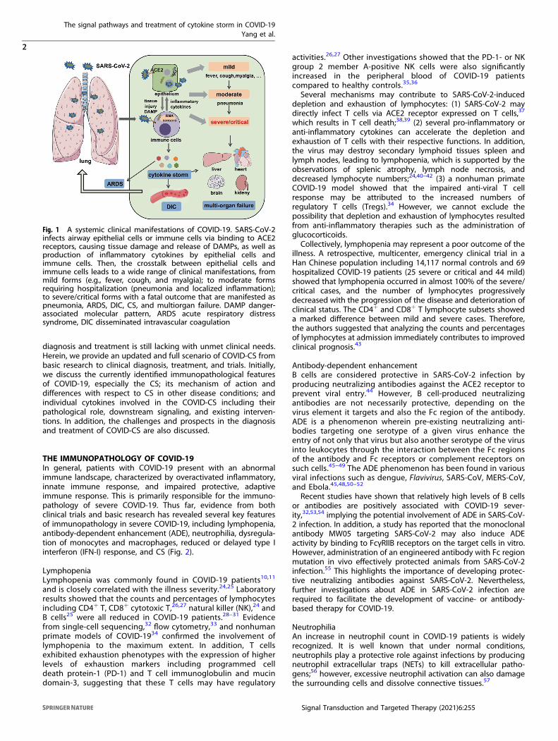
THE IMMUNOPATHOLOGY OF COVID-19In general, patients with COVID-19 present with an abnormalimmune landscape, characterized by overactivated inflammatory,innate immune response, and impaired protective, adaptiveimmune response. This is primarily responsible for the immuno-pathology of severe COVID-19. Thus far, evidence from bothclinical trials and basic research has revealed several key featuresof immunopathology in severe COVID-19, including lymphopenia,antibody-dependent enhancement (ADE), neutrophilia, dysregula-tion of monocytes and macrophages, reduced or delayed type Iinterferon (IFN-I) response, and CS (Fig. 2).
LymphopeniaLymphopenia was commonly found in COVID-19 patients10,11
and is closely correlated with the illness severity.24,25 Laboratoryresults showed that the counts and percentages of lymphocytesincluding CD4+ T, CD8+ cytotoxic T,26,27 natural killer (NK),24 andB cells25 were all reduced in COVID-19 patients.28–31 Evidencefrom single-cell sequencing,32 flow cytometry,33 and nonhumanprimate models of COVID-1934 confirmed the involvement oflymphopenia to the maximum extent. In addition, T cellsexhibited exhaustion phenotypes with the expression of higherlevels of exhaustion markers including programmed celldeath protein-1 (PD-1) and T cell immunoglobulin and mucindomain-3, suggesting that these T cells may have regulatory
activities.26,27 Other investigations showed that the PD-1- or NKgroup 2 member A-positive NK cells were also significantlyincreased in the peripheral blood of COVID-19 patientscompared to healthy controls.35,36
Several mechanisms may contribute to SARS-CoV-2-induceddepletion and exhaustion of lymphocytes: (1) SARS-CoV-2 maydirectly infect T cells via ACE2 receptor expressed on T cells,37
which results in T cell death;38,39 (2) several pro-inflammatory oranti-inflammatory cytokines can accelerate the depletion andexhaustion of T cells with their respective functions. In addition,the virus may destroy secondary lymphoid tissues spleen andlymph nodes, leading to lymphopenia, which is supported by theobservations of splenic atrophy, lymph node necrosis, anddecreased lymphocyte numbers;24,40–42 (3) a nonhuman primateCOVID-19 model showed that the impaired anti-viral T cellresponse may be attributed to the increased numbers ofregulatory T cells (Tregs).34 However, we cannot exclude thepossibility that depletion and exhaustion of lymphocytes resultedfrom anti-inflammatory therapies such as the administration ofglucocorticoids.Collectively, lymphopenia may represent a poor outcome of the
illness. A retrospective, multicenter, emergency clinical trial in aHan Chinese population including 14,117 normal controls and 69hospitalized COVID-19 patients (25 severe or critical and 44 mild)showed that lymphopenia occurred in almost 100% of the severe/critical cases, and the number of lymphocytes progressivelydecreased with the progression of the disease and deterioration ofclinical status. The CD4+ and CD8+ T lymphocyte subsets showeda marked difference between mild and severe cases. Therefore,the authors suggested that analyzing the counts and percentagesof lymphocytes at admission immediately contributes to improvedclinical prognosis.43
Antibody-dependent enhancementB cells are considered protective in SARS-CoV-2 infection byproducing neutralizing antibodies against the ACE2 receptor toprevent viral entry.44 However, B cell-produced neutralizingantibodies are not necessarily protective, depending on thevirus element it targets and also the Fc region of the antibody.ADE is a phenomenon wherein pre-existing neutralizing anti-bodies targeting one serotype of a given virus enhance theentry of not only that virus but also another serotype of the virusinto leukocytes through the interaction between the Fc regionsof the antibody and Fc receptors or complement receptors onsuch cells.45–49 The ADE phenomenon has been found in variousviral infections such as dengue, Flavivirus, SARS-CoV, MERS-CoV,and Ebola.45,48,50–52
Recent studies have shown that relatively high levels of B cellsor antibodies are positively associated with COVID-19 sever-ity,32,53,54 implying the potential involvement of ADE in SARS-CoV-2 infection. In addition, a study has reported that the monoclonalantibody MW05 targeting SARS-CoV-2 may also induce ADEactivity by binding to FcγRIIB receptors on the target cells in vitro.However, administration of an engineered antibody with Fc regionmutation in vivo effectively protected animals from SARS-CoV-2infection.55 This highlights the importance of developing protec-tive neutralizing antibodies against SARS-CoV-2. Nevertheless,further investigations about ADE in SARS-CoV-2 infection arerequired to facilitate the development of vaccine- or antibody-based therapy for COVID-19.
NeutrophiliaAn increase in neutrophil count in COVID-19 patients is widelyrecognized. It is well known that under normal conditions,neutrophils play a protective role against infections by producingneutrophil extracellular traps (NETs) to kill extracellular patho-gens;56 however, excessive neutrophil activation can also damagethe surrounding cells and dissolve connective tissues.57
Fig. 1 A systemic clinical manifestations of COVID-19. SARS-CoV-2infects airway epithelial cells or immune cells via binding to ACE2receptors, causing tissue damage and release of DAMPs, as well asproduction of inflammatory cytokines by epithelial cells andimmune cells. Then, the crosstalk between epithelial cells andimmune cells leads to a wide range of clinical manifestations, frommild forms (e.g., fever, cough, and myalgia); to moderate formsrequiring hospitalization (pneumonia and localized inflammation);to severe/critical forms with a fatal outcome that are manifested aspneumonia, ARDS, DIC, CS, and multiorgan failure. DAMP danger-associated molecular pattern, ARDS acute respiratory distresssyndrome, DIC disseminated intravascular coagulation
The signal pathways and treatment of cytokine storm in COVID-19Yang et al.
2
Signal Transduction and Targeted Therapy (2021) 6:255
An earlier clinical trial including 138 patients from Wuhan,China, showed that neutrophil counts were increased in non-survivors compared to survivors and continued to increase untildeath in the non-survivors.11 Another clinical study that integratedtranscriptomic, proteomic, and metabolomic platforms showedthat neutrophil counts were increased in patients with severe, butnot mild, COVID-19 as compared to healthy controls, andmolecules associated with NETs were significantly upregulatedin severe COVID-19 cases.58
The increased neutrophils manifested as both increasednumbers of mature and immature cells. In a clinical trial thatintegrated single-cell RNA-sequencing with single-cell proteomicsof blood and peripheral blood mononuclear cells (PBMCs),immature neutrophil precursors, and dysfunctional matureneutrophils expressing programmed death-ligand 1 appeared insevere COVID-19 cases.59 In addition, a single-cell sequencinganalysis by Wilk et al.32 and a flow cytometry analysis by Ronitet al.33 also identified the appearance of neutrophil progenitors atvarious developmental stages in PBMCs or bronchoalveolar lavagefluid (BALF) of COVID-19 patients with ARDS.Although the mechanism by which the virus promotes
neutrophil development in COVID-19 is still poorly understood,McElvaney et al.60 found that the levels of pyruvate kinase M2(PKM2), a regulator of glycolysis61 and coactivator of hypoxia-inducible factor-1α,62 as well as phosphorylated PKM2 werehigher in the neutrophils of COVID-19 patients in the ICU than inthose of non-ICU COVID-19 patients. This indicates that neutro-phils undergo immunometabolic reprogramming in severeCOVID-19 cases, which represents a potential intervention targetfor excessive neutrophil generation and activation in severe orcritical COVID-19.
Dysregulation of monocytes and macrophagesMonocytes and macrophages are the major innate immune cells ininfection and inflammation not just by virtue of their highernumbers but also by their functions. A single-cell RNA-sequencinganalysis showed that classic CD14+ monocytes were significantlyincreased, whereas nonclassical CD16+ monocytes and intermediateCD14+CD16+ monocytes were remarkably reduced in the blood ofCOVID-19 patients with severe symptoms. Classical monocytes candifferentiate into macrophages in tissue to initiate an inflammatoryresponse, whereas nonclassical monocytes were viewed as anti-inflammatory as they can maintain vascular homeostasis,63 whichmay explain the phenotypic shift of circulating monocytes fromCD16+ to CD14+. Analysis of the differentiation profiles of BALF andcirculating monocyte–macrophages from the same patient revealeda transition course of blood-toward-BALF. More importantly, multi-ple pro-inflammatory cytokines and chemokines were highlyexpressed by the BALF monocyte–macrophages, suggesting thatthe cells are inflamed.64 Another single-cell sequencing analysis ofperipheral blood samples also showed that CD16+ monocytes wereremarkably depleted in COVID-19 patients with ARDS, with aphenotypic shift from CD16+ to CD14+. However, significantupregulation of genes encoding pro-inflammatory cytokines orchemokines were not found in peripheral monocytes, indicating thatperipheral monocytes may not be responsible for the progression ofCS in COVID-19.32 Moreover, phenotyping leukocyte subpopulationsin BALF and blood of COVID-19 patients with ARDS showed that theexpression of activation markers such as CD16, CD64, CD69, andHLA-DR was higher in BALF macrophages than in peripheralmacrophages.33 Collectively, these existing studies were generallyconsistent and revealed the course of blood-toward-BALF transitionand the contribution of pulmonary monocyte–macrophages to CS
Fig. 2 The key immunopathology of severe COVID-19. The immunopathological manifestations of COVID-19 include lymphopenia,dysregulation of monocytes and macrophages, neutrophilia, ADE, reduced or delayed IFN-I response, and CS. Lymphopenia is commonlyobserved in severe COVID-19. In addition to decreased counts, lymphocytes often exhibit exhaustion phenotypes with the expression ofhigher levels of exhaustion markers PD-1, Tim-3, or NKG2A. Peripheral monocytes present a phenotype shift from CD16+ to CD14+, and BALFmacrophages are increased with a blood-to-BALF transition course. Neutrophil counts are increased with the presence of neutrophilprecursors in peripheral blood, especially in patients with severe COVID-19. The possible existence of ADE enhances the entry of SARS-CoV-2into cells through interaction between Fc regions and Fc receptors, leading to the aggravation of COVID-19. A CS is characterized by highlyelevated levels of pro-inflammatory mediators and is a particularly central feature for poor outcomes in patients with severe or criticalinfection. Reduced or delayed IFN-I response impedes viral clearance and induces paradoxical hyperinflammation, thus leading to thedeterioration of prognosis in COVID-19 patients. BALF bronchoalveolar lavage fluid, ADE antibody-dependent enhancement
The signal pathways and treatment of cytokine storm in COVID-19Yang et al.
3
Signal Transduction and Targeted Therapy (2021) 6:255
via the release of multiple pro-inflammatory cytokines andchemokines during severe COVID-19.Interestingly, a two-cohort study showed that activated HLA-
DRhighCD11chighCD14+ monocytes were increased in the PBMCs ofpatients with mild COVID-19, whereas dysfunctional HLA-DRlowCD163high (indicative of anti-inflammatory function) CD14+
monocytes were observed in severe COVID-19 cases.59 This meritsfurther investigation to understand the underlying mechanismand clinical significance.
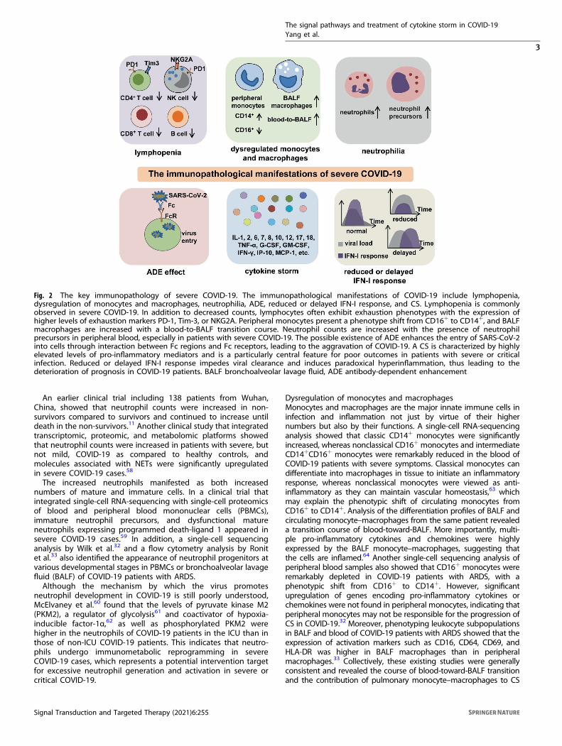
Reduced or delayed IFN-I responseThe IFN-I response is the first line of protective response andcritical to combat viral infections by promoting viral clearance andregulating innate and adaptive immune responses.65 Although thedetailed mechanism is still unknown when the infection occurs,the RNA of SARS-CoV-2 virus may be recognized by innateimmune cells via pattern recognition receptors (PRRs) includingtoll-like receptor (TLR); retinoic acid-inducible gene-I (RIG-I)-likereceptors (RLRs)/melanoma differentiation-associated gene 5(MDA5); and NOD-like receptors (NLRs).66 Subsequently, down-stream transcription factors including nuclear factor-κB (NF-κB),activator protein-1 (AP-1), and IFN regulatory factor 3/7 (IRF3/7)are activated to promote the transcription of pro-inflammatorycytokines and IFN-I. The IFN-I can activate the Janus kinase 1(JAK1)/tyrosine kinase 2–signal transducer and activator oftranscription 1/2 (STAT1/2) pathway, promoting the formation ofthe STAT1/2/IRF9 complex and initiating transcription of IFN-stimulated genes (ISGs) (Fig. 3).66,67
However, accumulating evidence has suggested that theprotective IFN-I response was remarkably reduced in severeCOVID-19 patients.26,68,69 At least two mechanisms have beenproposed to explain the deficient IFN-I response: (1) previous
studies have suggested that SARS-CoV employs various mechan-isms to inhibit IFN response, especially through the componentsof its structural proteins such as M protein,70 N protein,71 open-reading frame 3a (ORF3a) protein,72 and ORF6 protein.73
Considering that the structure of SARS-CoV-2 is similar to that ofSARS-CoV, it can be speculated that SARS-CoV-2 may exert similareffects on IFN response. For example, Yang et al.74 found that theNSP1 protein of SARS-CoV-2 can inhibit STAT1 phosphorylationand ISG transcription. (2) Decreased plasmacytoid dendritic cells(pDCs) may be partly responsible for the deficient IFN-I response.Sufficient evidence has suggested that pDC is a prominentproducer of IFN-I upon viral infection.75–77 However, existingstudies showed that counts of pDCs were decreased in the bloodof COVID-19 patients, especially in severe cases.26,32
Of note, although the levels of systemic IFN-I were low, localIFN-I and ISGs were noticeable in the BALF of some critically illpatients,78 which are related to the phenomenon of delayed IFN-Iresponse.69 Evidence has confirmed that a delayed IFN-I responsenot only impedes viral clearance but also induces paradoxicalhyperinflammation, thereby aggravating the immunopathologicalresponse.79,80 Collectively, these studies suggest that IFN-I-basedtherapy for COVID-19 should be applied as early as possible afterthe infection is confirmed.
Cytokine stormIn this section, we systemically review and discuss the character-istics, possible induction mechanism, pathogenesis, and diagnosisof CS in COVID-19.Sufficient evidence has revealed the components and character-
istics of CS in the patients with severe COVID-19, which arecomposed of an array of cytokines including interleukin-1 (IL-1), 2, 6,7, 8, 10, 12, 17, 18; tumor necrosis factor-α (TNF-α); IFN-γ; granulocyte
Fig. 3 The signaling pathways for the production and function of IFN-I after SARS-CoV-2 infection. After infection, the genomic ssRNAs andreplicative dsRNA intermediates of SARS-CoV-2 are recognized by endosomal toll-like receptors TLR3, 7, 8, and cytosolic RNA sensors, RIG-1/MDA5; next, downstream transcription factors including NF-κB and IRF3/7 are activated to induce the production of pro-inflammatorycytokines and IFN-I. IFN-I can activate the JAK1/TYK2–STAT1/2 pathway, promoting the formation of the STAT1/2/IRF9 complex and initiatingthe transcription of ISGs to produce anti-virus mediators, and it can also nonconventionally activate inflammatory pathways such as NF-κBand MAPK pathways to induce the expression of pro-inflammatory cytokines and paradoxical hyperinflammation in COVID-19
The signal pathways and treatment of cytokine storm in COVID-19Yang et al.
4
Signal Transduction and Targeted Therapy (2021) 6:255
colony-stimulating factor (G-CSF); granulocyte–macrophage colony-stimulating factor (GM-CSF); and monocyte chemoattractantprotein-1 (MCP-1).26,33,60,81–86 Reports of hemophagocytosis andclinical benefits from cytokine-targeted therapies in severe COVID-19 patients further confirmed the existence and pathogenesis ofCOVID-CS.87,88 Existing evidence has characterized and distinguishedCOVID-CS from CS in variable conditions such as HLH induced byspecific viral infections,89 macrophage activation syndrome (MAS)occurring after autoimmune disorders,90 and cytokine releasesyndrome (CRS) caused by CAR T cell therapy23 in several aspects.First, COVID-CS involves more inflammatory cytokines than other CSconditions, thereby providing an explanation for the aggressivenature of COVID-19. Second, lymphopenia, although relatively lessfrequent in other CS, was often observed in patients with COVID-CS,88 suggesting that COVID-CS may be mainly attributed to innate—rather than adaptive immune cells. Finally, compared withbacterial infection-induced CS (e.g., sepsis), the treatment ofCOVID-CS is more challenging, because blocking inflammatorycytokine function without effective anti-viral drug support mayexacerbate the infection.The initiation of COVID-CS induction during infection and the
predominant causative cytokine in COVID-19 immunopathologyremain largely unknown. Despite the lack of definite pathogen-associated molecule pattern (PAMP) of SARS-CoV-2, in analogywith SARS-CoV and MERS-CoV, it can be speculated that uponcellular entry of SARS-CoV-2 via its ACE2 receptor, viral genomicsingle-stranded RNA or other RNA compositions (double-strandedRNA) as PAMPs can be sensed by the related PRRs, including TLRsand RLRs in host cells. The downstream transcription factors IRF3/7 and NF-κB are activated following PAMP recognition to inducethe production of IFN-I and pro-inflammatory cytokines, respec-tively.91–93 However, as mentioned above, the protective IFN-Iresponse is quickly and selectively abrogated by SARS-CoV-2 viadifferent mechanisms. This is accompanied by an overwhelming
production of pro-inflammatory cytokines in the context ofCOVID-19, which not only impairs viral clearance but alsopromotes paradoxical hyperinflammation including CS. Therefore,from the immunology perspective, COVID-CS may be anunfortunate event whereby the intended host immune responsecombating the SARS-CoV-2 has lost control and transformed intoan inflammatory type.15
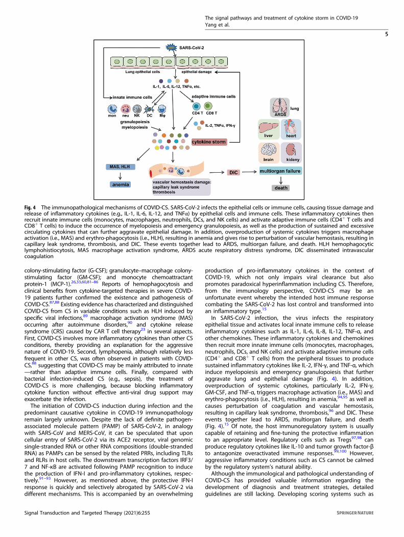
In SARS-CoV-2 infection, the virus infects the respiratoryepithelial tissue and activates local innate immune cells to releaseinflammatory cytokines such as IL-1, IL-6, IL-8, IL-12, TNF-α, andother chemokines. These inflammatory cytokines and chemokinesthen recruit more innate immune cells (monocytes, macrophages,neutrophils, DCs, and NK cells) and activate adaptive immune cells(CD4+ and CD8+ T cells) from the peripheral tissues to producesustained inflammatory cytokines like IL-2, IFN-γ, and TNF-α, whichinduce myelopoiesis and emergency granulopoiesis that furtheraggravate lung and epithelial damage (Fig. 4). In addition,overproduction of systemic cytokines, particularly IL-2, IFN-γ,GM-CSF, and TNF-α, triggers macrophage activation (i.e., MAS) anderythro-phagocytosis (i.e., HLH), resulting in anemia,94,95 as well ascauses perturbation of coagulation and vascular hemostasis,resulting in capillary leak syndrome, thrombosis,96 and DIC. Theseevents together lead to ARDS, multiorgan failure, and death(Fig. 4).15 Of note, the host immunoregulatory system is usuallycapable of retaining and fine-tuning the protective inflammationto an appropriate level. Regulatory cells such as Tregs97,98 canproduce regulatory cytokines like IL-10 and tumor growth factor-βto antagonize overactivated immune responses.99,100 However,aggressive inflammatory conditions such as CS cannot be calmedby the regulatory system’s natural ability.Although the immunological and pathological understanding of
COVID-CS has provided valuable information regarding thedevelopment of diagnosis and treatment strategies, detailedguidelines are still lacking. Developing scoring systems such as
Fig. 4 The immunopathological mechanisms of COVID-CS. SARS-CoV-2 infects the epithelial cells or immune cells, causing tissue damage andrelease of inflammatory cytokines (e.g., IL-1, IL-6, IL-12, and TNFα) by epithelial cells and immune cells. These inflammatory cytokines thenrecruit innate immune cells (monocytes, macrophages, neutrophils, DCs, and NK cells) and activate adaptive immune cells (CD4+ T cells andCD8+ T cells) to induce the occurrence of myelopoiesis and emergency granulopoiesis, as well as the production of sustained and excessivecirculating cytokines that can further aggravate epithelial damage. In addition, overproduction of systemic cytokines triggers macrophageactivation (i.e., MAS) and erythro-phagocytosis (i.e., HLH), resulting in anemia and gives rise to perturbation of vascular hemostasis, resulting incapillary leak syndrome, thrombosis, and DIC. These events together lead to ARDS, multiorgan failure, and death. HLH hemophagocyticlymphohistiocytosis, MAS macrophage activation syndrome, ARDS acute respiratory distress syndrome, DIC disseminated intravascularcoagulation
The signal pathways and treatment of cytokine storm in COVID-19Yang et al.
5
Signal Transduction and Targeted Therapy (2021) 6:255
HScore, MS score, HLH-2004, Penn grading scale, and theCommon Terminology Criteria for Adverse Events may bebeneficial to predict COVID-CS or related outcomes. Caricchioet al.101 proposed predictive criteria for COVID-CS diagnosis. Thesecriteria comprise three clusters: (1) albumin <2.87 mg/mL,lymphocytes <10.2%, neutrophil absolute count >11.4 × 10³/mL;(2) alanine aminotransferase >60 IU/L, aspartate aminotransferase>87 IU/L, D-dimer >4930 ng/mL, lactate dehydrogenase >416 U/L,troponin I >1.09 ng/mL; and (3) anion gap <6.8 mmol/L, chloride>106mmol/L, potassium >4.9 mmol/L, and blood urea nitrogen:creatinine ratio >29. In addition, ferritin >250 ng/mL andC-reactive protein (CRP) >4.6 mg/dL are added for the reassuranceof ongoing systemic inflammation. In another study, the authorsproposed that a diagnostic criterion including peripheral bloodoxygen saturation to the fraction of inspired oxygen (SpO2/FiO2),CRP, ferritin, cytokines/chemokines, and neutrophil/lymphocyteratio may have a strong diagnostic power for COVID-CS.102 Mehtaet al.13 proposed that prospective screening for hyperinflamma-tion using laboratory assays and the HScore should be performedin all severely ill COVID-19 patients to identify COVID-CS. Despitethe requirement for further validation, these criteria indeedprovide constructive suggestions for the development of officiallyrecognized guidelines for COVID-CS.COVID-CS is a complicated and dynamic inflammatory process
caused by a group of cytokines from initiation, immune cellhyperactivation, to organ dysfunction. The development ofprecise therapeutic intervention in appropriate time is requiredto effectively control COVID-CS. In principle, the treatmentstrategy is to control ongoing inflammatory response byspecifically or nonspecifically targeting inflammatory cytokinesor related signaling pathways and to resume the hostimmunoregulatory system. Herein, we discuss the role of thekey cytokines and associated signal pathways involved inCOVID-CS (Fig. 5).
IL-6/JAK/STAT signaling. A retrospective, multicenter studyincluding 150 patients from Wuhan, China showed significantlyelevated levels of IL-6 in severe COVID-19 cases.103 A study fromGermany showed that IL-6 >80 pg/mL in combination with CRP>97mg/L presented a relatively high specificity and sensitivity topredict respiratory failure.104 In addition, other studies have alsoshown the remarkably increased serum levels of IL-6 in patientswith severe COVID-19.68,105,106
IL-6, first produced by monocytes, macrophages, and DCs,serves as a prominent activator of the JAK/STAT3 pathway in thecontext of inflammation. Recent studies have determined that theIL-6–JAK–STAT3 axis is closely related to the severity of COVID-19,107,108 and the levels of phosphorylated STAT3 were higher indifferent subsets of leukocytes in COVID-19 patients than inhealthy controls.109 IL-6 deploys two signaling pathways—classiccis-signaling and trans-signaling—to trigger the activation ofdownstream JAK/STAT3 signaling through the membrane-bound(mIL-6R) and soluble form of IL-6R (sIL-6R), respectively.110 In cis-signaling, IL-6 binds to mIL-6Rs that are restrictedly expressed onimmune cells, forming an IL-6/IL-6R/gp130 complex to activatedownstream JAK/STAT3, Akt/mTOR, and MAPK signaling. Thisexerts pleiotropic effects on immune cells, which are manifestedas promoted differentiation of T-helper type 17 (Th17), CD8+ T,and B cells; increased migration of neutrophils; and reduceddevelopment of Tregs.111,112 These, in turn, induce increasedsecretion of IL-6 and aggravate inflammation. In trans-signaling,circulating IL-6 binds to sIL-6Rs to form a complex, then binds withthe gp130 dimer that is expressed on almost all cell types. Theresultant activation of the JAK–STAT3 signaling occurs in cells withabsent expression of mIL-6R, such as endothelial cells and vascularsmooth muscle cells (VSMCs). The overwhelming activation of theIL-6–IL-6R–JAK–STAT3 pathway triggers the secretion of variousmediators, such as IL-6 itself, IL-8, MCP-1, and vascular endothelialgrowth factor (VEGF), and reduces the expression of E-cadherin
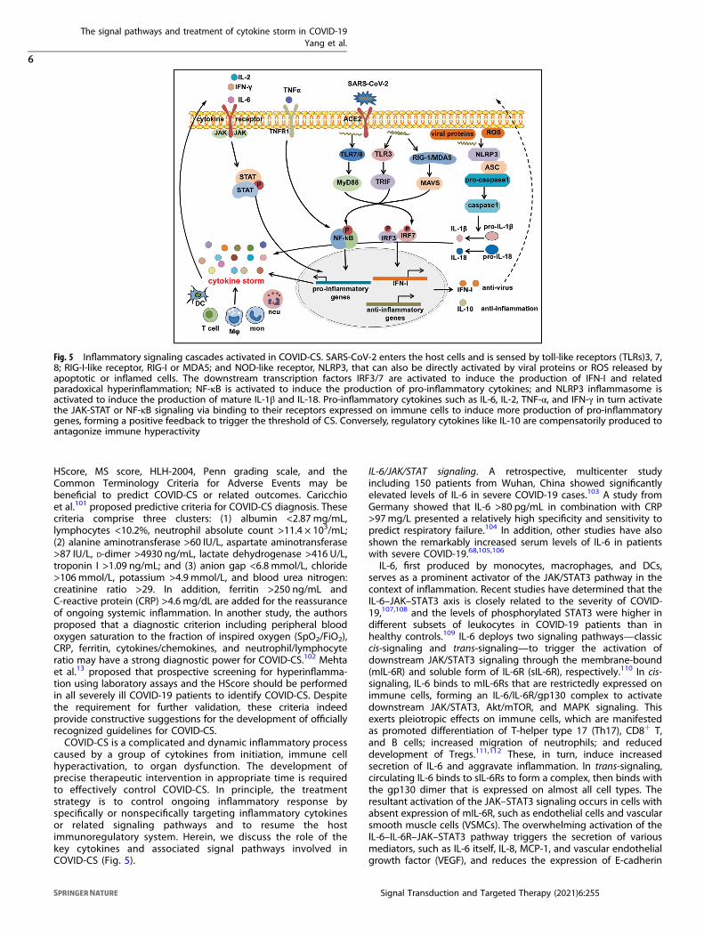
Fig. 5 Inflammatory signaling cascades activated in COVID-CS. SARS-CoV-2 enters the host cells and is sensed by toll-like receptors (TLRs)3, 7,8; RIG-I-like receptor, RIG-I or MDA5; and NOD-like receptor, NLRP3, that can also be directly activated by viral proteins or ROS released byapoptotic or inflamed cells. The downstream transcription factors IRF3/7 are activated to induce the production of IFN-I and relatedparadoxical hyperinflammation; NF-κB is activated to induce the production of pro-inflammatory cytokines; and NLRP3 inflammasome isactivated to induce the production of mature IL-1β and IL-18. Pro-inflammatory cytokines such as IL-6, IL-2, TNF-α, and IFN-γ in turn activatethe JAK-STAT or NF-κB signaling via binding to their receptors expressed on immune cells to induce more production of pro-inflammatorygenes, forming a positive feedback to trigger the threshold of CS. Conversely, regulatory cytokines like IL-10 are compensatorily produced toantagonize immune hyperactivity
The signal pathways and treatment of cytokine storm in COVID-19Yang et al.
6
Signal Transduction and Targeted Therapy (2021) 6:255
expressed on endothelial cells.113 Several studies have shown thatMCP-1 can facilitate the formation of atherogenesis,114 productionof adhesion molecules,115,116 and proliferation and migration ofVSMCs,117 which partly explains the occurrence of cardiovascularsymptoms observed in COVID-CS. In addition, increased VEGF anddecreased E-cadherin can lead to vascular permeability andleakage and accelerate the presence of hypotension andpulmonary dysfunction in COVID-CS.87 Moreover, IL-6 canpromote the secretion of various acute-phase proteins such asCRP, hepcidin, fibrinogen, thrombopoietin, complement C3, andferritin in hepatocytes.118,119 Collectively, IL-6 contributes to bothimmune cell hyperactivation and target organ dysfunction in CS.Evidence has suggested that the production of IL-6 can be
induced by angiotensin II in inflamed vessels. The underlyingmechanism is that angiotensin II binds to angiotensin II type 1(AT1) receptor and then activates JAK/STAT signaling to induce IL-6 production.120,121 Interestingly, existing studies have shown thatSARS-CoV may promote the expression of angiotensin II bydownregulating ACE2,122,123 which potentially leads to thepossibility that SARS-CoV-2 enhances IL-6 production via theangiotensin II/AT1 receptor/JAK/STAT axis, and the positive pro-inflammatory feedback of IL-6/JAK/STAT ultimately drives clinicalsignatures of COVID-19, especially COVID-CS.124
IFN-γ/JAK/STAT signaling. IFN-γ, mainly produced by macro-phages, T cells, and NK cells, participates in immunologicalprocesses such as inflammation. It is a dominating driver ofmacrophage, NK, and T cell activation, and exerts a predominanteffect on protective immunity against bacterial and viral infectionsthrough the activation of JAK1/JAK2 complex and downstreamSTAT1-IFN-γ-activated site (GAS) cascades.125–127
Evidence has shown that IFN-γ is highly involved in various CS-related disorders,128 as illustrated by its pathological role inprimary HLH, a syndrome of failure to eliminate pathogens owingto impaired NK cell activity. In such cases, despite excessive T cellactivation and large quantities of IFN-γ production, the increasedIFN-γ levels fail to combat pathogens and instead causeimmunopathology because of defective NK activity.129 WhetherIFN-γ plays a specific role in COVID-CS is still unknown; however,considering its role in promoting immune cell activation, we canspeculate that it contributes substantially to COVID-CS.Numerous studies have reported elevated levels of IFN-γ in
patients with COVID-19.9,130,131 Of note, one study showed thatIFN-γ produced by CD4+ T cells was decreased in patients withsevere disease compared to those with moderate disease, whichcould be explained by the reduced numbers and functionalexhaustion of T cells in severe COVID-19,132 as described– above.Therefore, this study suggested that elevated levels of IFN-γ inCOVID-19 are produced mainly by macrophages, not T cells.
TNFα/NF-κB signaling. TNFα is a well-known pro-inflammatorycytokine and is closely associated with many infectious, auto-immune diseases, and cancer, and is primarily produced bymonocytes, macrophages, and T cells.133,134 NF-κB plays anevolutionarily conserved role in the immune system,135,136 espe-cially in regulating the expression of various vital cytokines involvedin inflammation.137 TNFα, as an initial driver of NF-κB activation, canactivate the NF-κB signaling pathway to induce the expression ofseveral pro-inflammatory and antiapoptotic genes through itsreceptor TNFR1 and a series of intermediate adapters.138–140 In turn,NF-κB can induce TNFα expression in the context of inflammation,such as lipopolysaccharide (LPS) stimulation.141 Therefore, TNFα/NF-κB signaling may play pathological roles in the stage of initiationand immune cell hyperactivation in CS by inducing apoptosis ofepithelial cells to drive the epithelium–immune cell interplay andaugmenting systemic inflammation.Previous studies have found that excessive TNFα represented a
poor prognosis in SARS-CoV and MERS cases,142–145 and inhibition
of NF-κB improved pulmonary symptoms in SARS-CoV-infectedmice.146 However, the role of TNFα in COVID-19 is, so far, still notentirely clear. Recent studies have reported elevated serum levels ofTNFα in severe COVID-19 cases.9,13,24,81 In addition, a clinical trialfrom Wuhan, China, including 522 patients and 40 healthy controlsshowed that the concentration of TNFα was negatively correlatedwith T cell counts in COVID-19 patients.27 In contrast, a clinical trialfrom Chongqing, China, including 102 mild and 21 severe casesshowed that TNFα levels were within the normal values in almost allCOVID-19 patients (121/123).147 Thus, further research is urgentlyrequired to better understand the role of TNFα in COVID-CS.Although a recent report suggested that inhibition of the TNFα–NF-κB pathway may have protective effects in COVID-19,124 cautionshould be applied based on two aspects: (1) as mentioned above,the roles of TNFα in COVID-CS is still undefined; (2) blocking NF-κBnonspecifically may simultaneously impair its protective functionsin cellular homeostasis, as exemplified by a general suppression oninnate immunity.148
NLRP3/IL-1β signaling. IL-1β is perhaps the most well-studiedmember of the IL-1 family because of its prominent role inautoinflammatory diseases such as gout and chronic inflammatoryarthritis.149–151 IL-1β is mainly secreted by macrophages viaapoptosis and pyroptosis and exerts positive effects on themigration of immune cells to inflamed tissues; Th17 celldifferentiation; expression and release of various cytokines andadhesion factors; and NF-κB pathway activation to form a positivefeedback for its own production.152,153 Upstream, the NLRP3protein forms a complex with apoptosis-associated speck-likeprotein containing a caspase recruitment domain (ASC) andcysteinyl aspartate-specific proteinase-1 (caspase-1), termed theNLRP3 inflammasome, to cleave the inactive IL-1β precursor to themature form of IL-1β.154,155 Considering the positive roles of IL-1βin activating initiative and sustained inflammation,156 it waspostulated that NLRP3-IL-1β signaling might be involved inCOVID-CS.Multiple indicators from previous evidence have suggested that
IL-1β may contribute to CS in coronavirus infections.157–159 Zhanget al.160 reported the elevated levels of multiple cytokinesincluding IL-1β in COVID-19 cases with severe symptoms, whichwere also associated with SARS, hypercoagulation, and DIC.Consistently, Huang et al.9 also showed high serum concentrationsof IL-1β in COVID-19 patients. Moreover, a previous study showedthat NLRP3 can be directly activated by viral proteins of SARS-CoVsuch as ORF3a and ORF8b,161,162 which were also found on thegenome of SARS-CoV-2,163 suggesting a potentially similar effectof direct activation of the NLRP3 by SARS-CoV-2 protein. Thepotential roles of NLRP3 inflammasome in severe COVID-19 havebeen discussed in relevant reviews.164,165 Reactive oxygen species(ROS) was reportedly an initiator of NLRP3 activation.166–168 Thus,it was proposed that excessive production of ROS resulting frominflammation infiltration in severe COVID-19 may lead to NLRP3activation and IL-1β precursor cleavage, further aggravatinginflammation in COVID-CS.
IL-2/IL-2R/JAK/STAT5 signaling. IL-2 is mainly secreted by CD4+
T cells and plays crucial roles in the expansion and differentiationof CD4+ T, CD8+ T, NK, and other cells through the IL-2R–JAK–STAT5 signaling pathway.169,170 IL-2 can fine-tuneimmune responses and maintain self-tolerance,171 and itsdeficiency accounts for the occurrence of autoimmune dis-eases.172
Elevated levels of IL-2 have been reported in other types ofcoronavirus infections.173,174 Recent studies have also shown thatthe concentrations of IL-2 or IL-2R were elevated in COVID-19patients, especially in those with severe illness.9,13,24,132 However,a clinical trial including 54 COVID-19 patients from Beijing, China,reported a contradictory result. The authors found that compared
The signal pathways and treatment of cytokine storm in COVID-19Yang et al.
7
Signal Transduction and Targeted Therapy (2021) 6:255

with severe patients (n= 14), those with a critical illness (n= 6)had remarkably reduced plasma levels of IL-2. Accordingly, IL-2Rαlevels were significantly decreased in the PBMCs of patients with asevere and critical illness compared to common patients (n= 34)or healthy controls. Furthermore, the levels of JAK1 and STAT5were significantly lower in all three groups than in normalcontrols. Considering the supportive roles of IL-2 in the expansionand differentiation of T cells, the authors speculated that thepresence of lymphopenia especially in severe COVID-19 may beattributed to reduced levels of IL-2, IL-2R, JAK1, and STAT5.175 Inadditionally, CD4+ T cells are the primary source of IL-2, and thereduced numbers of lymphocytes during the stage of CS afterCOVID-19 infection can at least partly explain the reduced IL-2level and downregulation of IL-2 signaling. Future investigationsare required to confirm these findings in more patients.
IL-7/IL-7R signaling. IL-7/IL-7R signaling is essential for peripheralhomeostasis and the survival, differentiation, and maintenance ofT cells including CD4+ T, CD8+ T, naive T, and memory T cells.176–179 It is also indispensable for the development and maintenanceof innate lymphoid cells (ILCs), formation of lymphoid structures,and barrier defense.180
Recent reports have shown elevated levels of IL-7 in COVID-19patients, and these increases were related to disease sever-ity.9,13,147 However, the impact of enhanced levels of IL-7 inCOVID-19 is largely unknown. Considering the protective role ofIL-7, we can speculate that the increase of IL-7 may be a feedbackmechanism in response to the lymphopenia in patients withsevere/critical COVID-19.
IL-10 signaling. IL-10 is an important immunoregulatory cytokineproduced by a variety of immune cells including Th2 cells, Tregs,CD8+ T cells, B cells, DCs, macrophages, and NK cells, and signalsthrough the IL-10R/JAK/STAT3 pathway. IL-10 exerts anti-inflammatory functions by directly limiting the innate immune-related functions of macrophages and DCs in an autocrine andparacrine manner or indirectly via improving Treg development. Inaddition, IL-10 can activate mast cells and strengthen thefunctions of CD8+ T, B, and NK cells.181
A clinical trial including 102 COVID-19 patients and 45 controlsfrom Wuhan, China, showed that the serum IL-10 levels of patientswith a critical illness (n= 17) were significantly higher than thoseof patients with moderate (n= 42) and severe (n= 43) illness;further, the IL-10 levels were positively correlated with theconcentrations of serum CRP, indicating the potential of IL-10 asan indicator of disease severity.182 Huang et al.9 also reported thesignificantly high plasma levels of IL-10 in COVID-19 patientsadmitted to the ICU compared to those who were not. In addition,a follow-up clinical trial including 71 COVID-19 patients (53 mildand 18 severe) from Beijing, China and 18 controls showed thatthe production of IL-10 in the early stage was significantlycorrelated with disease severity.183
It can be speculated that the excessive production of IL-10 is anegative feedback mechanism to antagonize the hyperactivity ofthe immune system. However, when faced with an overwhelmingsecretion of inflammatory mediators and activation of pro-inflammatory cells in COVID-CS, the fine-tune function of IL-10 israther inadequate. Thus, administration of IL-10 has beenrecommended to treat ARDS in COVID-19.184 However, a recentreport showed that IL-10 may be detrimental in the initiationphase of SARS-CoV-2 infection by promoting T cell exhaustion.The authors proposed that blocking IL-10 with a neutralizingantibody in the initiation stage of SARS-CoV-2 infection may be apromising therapeutic approach.185
IL-12 signaling. IL-12, mainly produced by DCs, macrophages,and B lymphocytes,186 is a multifunctional immunoregulatoryfactor that can promote proliferation of Th1 and Th17 cells;
improve the cytotoxicity of NK cells; and induce expression of IFN-γ in Th1 cells, NK cells, DCs, and macrophages via a positivefeedback mechanism.187,188 Thus, IL-12 plays an aggressive role inCS by augmenting the activation of various immune cells.Existing studies have reported that viral infections induce the
production of IL-12 to defend against infections.189–191 Forinstance, during influenza virus infection, IL-12 is endogenouslyproduced to induce the secretion of IFN-γ from Th1 and NK cells,thereby inhibiting viral replication.190 A previous study reportedelevated plasma levels of IL-12 in patients infected by SARS-CoV.192 However, Huang et al.9 recently found that the plasmalevels of IL-12p70 showed no difference between COVID-19patients and healthy adults. Therefore, further research with large-sized samples is urgently required to determine the alterationsand functions of IL-12 in COVID-19, especially COVID-CS.
IL-17 signaling. IL-17 (primarily IL-17A) is produced by Th17,CD8+ T, and group 3 ILC (ILC3) and participates in many pro-inflammatory processes and autoimmune diseases.193–195 Target-ing IL-17 is now regarded as a common strategy to reduce theburden of several autoimmune diseases such as psoriasis andpsoriatic arthritis. Nevertheless, the functions of this inflammatorycytokine vary, from being protective against infections to havingdetrimental pro-inflammatory effects, depending on the tissuetype and location (gut, lung, or skin) where it is being expressedand its triggering factors.196
Increased levels of IL-17 were previously reported in SARS-CoV-or MERS-CoV-infected patients.197,198 In addition, IL-17 canaugment lung injury and decrease overall survival throughrecruitment of neutrophils; stimulate the expression of pro-inflammatory factors; and induce the expression of G-CSF toprevent apoptosis in both ARDS and an LPS-induced acute lunginjury model.199,200 Similarly, evidence has suggested that IL-17levels were elevated in COVID-19 patients, especially in those witha severe and critical illness.201 Asrani and Hassan156 showed thatIL-17 plays crucial roles in the stages of immune cell hyperactiva-tion and target organ dysfunction in COVID-CS by promoting therecruitment of neutrophils and producing symptoms such asfever, matrix damage, tissue remodeling, and inflammatoryinfiltration.Despite multiple evidence suggesting the potential of IL-17 as
an intervention target for COVID-CS, studies have shown that thelevels of IL-17 were within normal ranges in 102 and 21 patientswith mild and severe COVID-19, respectively.147 Therefore, moreclinical trials and fundamental research are required for furtherclarification.
GM-CSF signaling. GM-CSF is produced by endothelial, epithelial,hematopoietic, and other cell types.202 Under physiologicalconditions, low levels of GM-CSF can regulate the homeostasisof alveolar macrophages to maintain their antimicrobial func-tions.203,204 Under hyperinflammatory conditions such as CS, GM-CSF drives emergency myelopoiesis and recruits myeloid cells tothe inflammatory sites to perpetuate inflammatory reactions.205
Increased levels of GM-CSF have been observed in SARS,206
ARDS,207 and CRS.208 A recent study also reported elevated GM-CSF levels in both severe and mild COVID-19.9
Given the role of GM-CSF in maintaining antimicrobial functionsof alveolar macrophages, administration of GM-CSF to patientswith early-stage COVID-19 may strengthen the alveolar wall andenhance viral clearance.209 In contrast, the blockade of GM-CSFsignaling may achieve clinical benefits in COVID-CS.
CYTOKINE-BASED INTERVENTIONSThe current treatment for CS is mainly based on traditional anti-inflammatory drugs such as the administration of corticosteroids,chloroquine, and colchicines.210,211 Recently, biologics like
The signal pathways and treatment of cytokine storm in COVID-19Yang et al.
8
Signal Transduction and Targeted Therapy (2021) 6:255
recombinant cytokines; monoclonal antibodies against IL-6, IL-1β,TNF-α, and IFN-γ; and signaling pathway inhibitors are alsoavailable or in the pipeline for production. In this and the nextsection, we aim to discuss the key biologics that are currently orpotentially applied to treat CS (Fig. 6).
IFN-IAdministration of IFN-I has previously been investigated for thetreatment of SARS-CoV and MERS-CoV. Generally, it was found tobe relatively effective in vitro and in some animal models,212
although human studies were inconclusive.213
The research team at the University of Texas Medical Branch,USA, showed that low concentrations of IFN-α and IFN-βeffectively reduced virus titer and inhibited viral replication inVero cells in a dose-dependent manner, and they also found thatSARS-CoV-2 is more sensitive than SARS-CoV to IFN-I in vitro.214 Anuncontrolled, exploratory study from Tongji Medical College,Wuhan, China, showed that administration of IFN-α2b alone or incombination with arbidol remarkably accelerated virus clearanceas well as the recovery of IL-6 and CRP to normal levels in COVID-19 patients.215 A randomized, multicenter, prospective, phase 2clinical trial conducted at the University of Hong Kong showedthat compared with the control group (lopinavir/ritonavir), COVID-19 patients in the triple group (IFNβ-1b plus lopinavir/ritonavir+ribavirin) exhibited significantly alleviated symptoms; shortenedvirus shedding and hospital stay; and improved inflammatoryconditions (NCT04276688).216 A prospective study including 2944healthy medical staff in the epidemic areas from Taihe Hospital inShiyan City, Hubei, China, showed that the incidence rate ofCOVID-19 was zero in both the low-risk group (n= 2415) and thehigh-risk group (n= 529) after treatment with recombinant IFN-αnose drops for 30 days (NCT04320238),217 indicating thepreventive roles of recombinant IFN-α in COVID-19. A trialcomparing the clinical efficacy of subcutaneous IFN-β1a pluslopinavir/ritonavir with lopinavir/ritonavir alone; hydroxychloro-quine; and remdesivir in COVID-19 is still ongoing (NCT04315948).
Collectively, these studies suggested that IFN-I can inhibit SARS-CoV-2 infection and potential COVID-CS.Another type of interferon—IFN-III—may also have a clinical
benefit in COVID-19 treatment. Dinnon et al.218 confirmed theanti-viral effect of pegylated IFN-λ1a in both SARS-CoV-2-infectedhuman airway epithelial cells and mouse models. Some studieshave determined that MDA5 plays a leading role in activating IFN-I/III response to defend against SARS-CoV-2 in human epithelialcells,219–221 which may open a new avenue for enhancing IFNresponse in COVID-19. In addition, several clinical trials evaluatingthe clinical efficacy of IFN-λ are under investigation (NCT04343976,NCT04331899).
IL-7The supportive role of IL-7 in lymphocyte survival and expansionprovides clinical implications for the recovery of function oflymphocytes in severe/critical COVID-19.A case report from Hospices Civils de Lyon, France, showed that
a period of recombinant human IL-7 (rhIL-7) treatment remarkablyimproved the immune function in a 74-year-old ICU patient withsevere COVID-19, which manifested as elevated lymphocyte countand mHLA-DR expression. In this case, IL-7 was administered quitelate, on day 24 of admission. Thus, the authors prospectivelyproposed that earlier administration of IL-7 may have indicatedbetter clinical outcomes.222 In a case series from St. Luc UniversityHospital, Brussels, Belgium, the authors showed that patients inthe IL-7 treatment group seemed to have higher levels oflymphocyte counts than those in the control group, withoutaggravated inflammation and pulmonary injury.223 Unfortunately,the absence of detailed phenotypic or functional studies onlymphocytes weakens the reliability of these studies. A multi-center, double-blind clinical trial in a UK-based cohort is currentlyevaluating whether the administration of CYT107 (a commercialproduct of rhIL-7) can result in clinical improvement of patientswith severe COVID-19 through immune reconstitution(NCT04379076). Clark et al.224 proposed that administration of
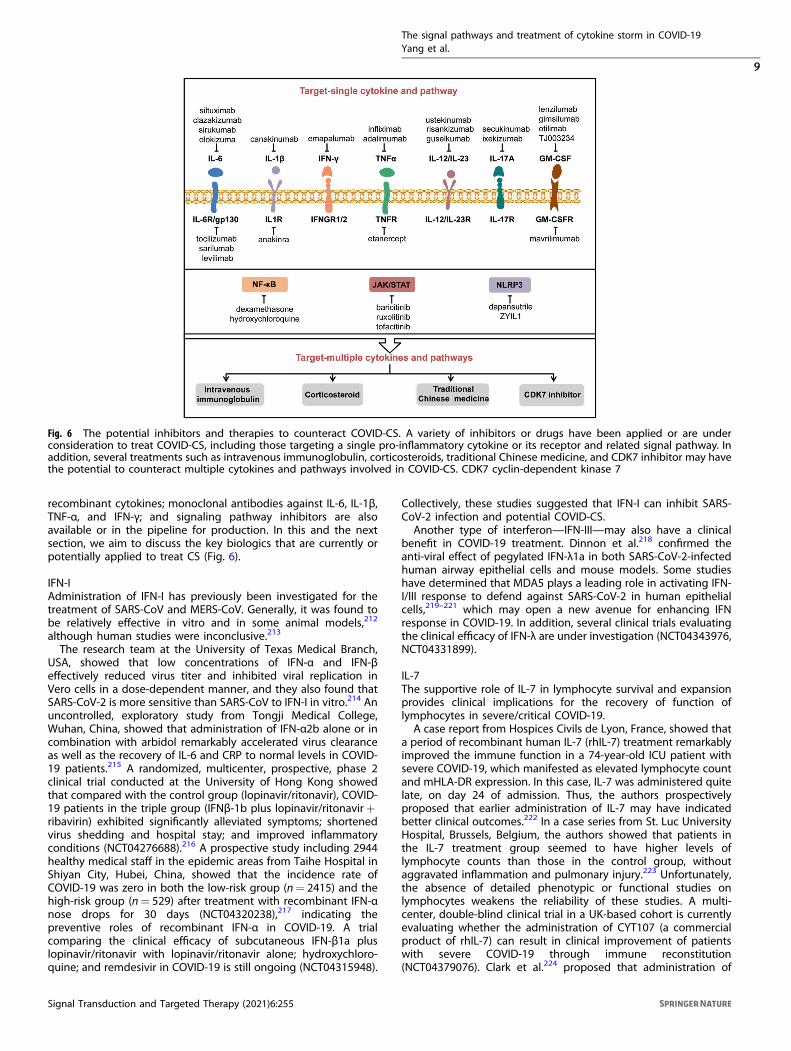
Fig. 6 The potential inhibitors and therapies to counteract COVID-CS. A variety of inhibitors or drugs have been applied or are underconsideration to treat COVID-CS, including those targeting a single pro-inflammatory cytokine or its receptor and related signal pathway. Inaddition, several treatments such as intravenous immunoglobulin, corticosteroids, traditional Chinese medicine, and CDK7 inhibitor may havethe potential to counteract multiple cytokines and pathways involved in COVID-CS. CDK7 cyclin-dependent kinase 7
The signal pathways and treatment of cytokine storm in COVID-19Yang et al.
9
Signal Transduction and Targeted Therapy (2021) 6:255
IL-7 in combination with dexamethasone may be an optimaltreatment for severe COVID-19. The underlying mechanism is thatdexamethasone enhances IL-7 activity by upregulating its receptorIL‐7Rα, which can also be used to explain the most effectiveproperties of dexamethasone in the more severe stage of thedisease.225,226
These studies indicate that appropriate administration of IL-7alone or in combination with other agents should be consideredas early as possible for critical COVID-19 patients with severelymphopenia.
BLOCKADE OF CYTOKINESBlockade of IL-6As mentioned above, IL-6 signaling is a leading inducer ofCOVID-CS. Currently, several drugs targeting IL-6 signaling suchas IL-6 inhibitors (siltuximab, clazakizumab, sirukumab, olokizu-mab) and IL-6R inhibitors (tocilizumab, sarilumab, levilimab) areavailable.227–229
A retrospective clinical trial from Anhui, China, including 21COVID-19 patients with severe or critical illness showed that fivedays of tocilizumab therapy immediately improved the clinicaloutcomes in most patients, manifested as decreased oxygenrequirements, serum CRP concentrations, and hospital stays, aswell as the rapid recovery of lymphocyte percentage.230 Anothermulticenter cohort study including 3924 COVID-19 patients inthe ICU across 68 hospitals in the United States showed that therisk of mortality was lower in 433 patients who receivedtocilizumab treatment immediately after ICU admission than inthose who did not receive early tocilizumab intervention.231 Inaddition, numerous reports evaluating the administration oftocilizumab for severe COVID-19 have also been pub-lished,106,232–247 and a total of 75 clinical trials are currentlyregistered in ClinicalTrials. gov.An open-label study from Italy including 56 patients with severe
COVID-19 showed that after 28 days of follow-up, sarilumabtreatment (n= 28) appeared to promote the recovery of patientswith mild lung consolidation (<17%) at baseline.248 Anotherretrospective, single-center, clinical trial from Italy including 15COVID-19 patients showed that sarilumab treatment improved theclinical symptoms and reduced serum CRP levels in most patients(n= 10).249 Currently, a total of 17 clinical trials on sarilumab areregistered in ClinicalTrials.gov for the treatment of COVID-19.A randomized, phase 3, clinical trial evaluating the administra-
tion of levilimab in patients with severe COVID-19 has beencompleted, but the results have not yet been published(NCT04397562).An observational, controlled cohort study from Italy including
30 patients requiring ventilator support showed that the 30-daymortality was significantly reduced in those receiving siltuximabplus optimal supportive care compared with the control patientswho received only optimal supportive care; ~50% of patientsreceiving siltuximab treatment were finally discharged(NCT04322188).250 Three other clinical trials evaluating the efficacyand safety of siltuximab in severe COVID-19 are underway(NCT04329650, NCT04330638, NCT04486521).In general, published clinical trials showed positive results with
respect to some common IL-6 or IL-6R antagonists for thetreatment of COVID-CS. Several clinical trials of other IL-6 or IL-6Rantagonists including clazakizumab (NCT04381052, NCT04494724,NCT04343989, NCT04363502, NCT04348500, NCT04659772);sirukumab (NCT04380961); and olokizumab (NCT04380519,NCT04452474) have been registered in ClinicalTrials.gov toinvestigate their therapeutic potential in COVID-CS.
Blockade of IL-1βConsidering the pathological role of IL-1β signaling in CS, severaldrugs targeting IL-1β signaling, including IL-1β antagonist
canakinumab and IL-1 receptor antagonist anakinra, may offerclinical benefits in the treatment of COVID-19, particularly in CS.A retrospective, three-center, clinical trial from France including 22
patients with severe/critical COVID-19 showed that after >8 days oftreatment, patients in the anakinra group (n= 12) presentedimproved clinical conditions as well as decreased mechanicalventilation requirements and serum CRP concentrations, as com-pared with the control group (n= 10) (INDS, MR4115050520).251 Aprospective, cohort study from the Netherlands showed that severalclinical parameters including temperature; white blood cell count;and levels of plasma ferritin, creatinine, procalcitonin, and bilirubinwere decreased after 28 days of anakinra treatment.252 Several otherstudies have also investigated the clinical effects of anakinra onCOVID-19.253–261 A total of 35 clinical trials are currently registered inClinicalTrials.gov.A retrospective study from Italy reported for the first time
that subcutaneous administration of 300 mg canakinumabrapidly reduced systemic inflammation and improved oxygena-tion in COVID-19 patients (n= 10) who presented hyperin-flammation but did not require mechanical ventilation.262
Another single-center, cohort study from Italy enrolled 34non-ICU patients with mild or severe COVID-19, with 17receiving standard treatment and 17 receiving 300 mg ofsubcutaneous canakinumab. The results showed that canaki-numab treatment significantly increased the PaO2/FiO2 ratioand reduced inflammatory indices.263 These two studiessuggest that canakinumab treatment may have therapeuticpotentials in non-ICU patients with mild or severe COVID-19.Several other reports have also evaluated the administration ofcanakinumab in COVID-19,264–266 and six clinical trials arecurrently registered in ClinicalTrials.gov (NCT04348448,NCT04476706, NCT04362813, NCT04365153, NCT04510493,NCT04278404).
Blockade of IFN-γEmapalumab, a monoclonal antibody targeting IFN-γ, has beenapproved to treat primary HLH, a condition with elevated serumlevels of IFN-γ.267 Considering the contribution of IFN-γ to CS asmentioned above, emapalumab may be effective in the treatmentof COVID-CS.A randomized, multicenter, clinical trial from Italy was registered
to investigate the efficacy of emapalumab treatment in combina-tion with anakinra to alleviate hyperinflammation and improverespiratory conditions (NCT04324021). Unfortunately, this trial isnow terminated and no further data are currently available.Therefore, other randomized, controlled clinical trials are urgentlyrequired to address these issues.
Blockade of TNFαRecent studies have provided the theoretical and practical basesto support TNFα blockade as a potential strategy for excessivecytokine release and hyperinflammation in COVID-19.268–271
Etanercept is a soluble TNFα receptor fusion protein that hasbeen used to treat toxic epidermal necrolysis (TEN), a condition ofsystemic hyperinflammation. Owing to the similarities in clinicalmanifestations and pathological characteristics between COVID-19and TEN, Chen et al.268 proposed the temporary use of etanerceptas a valuable approach to treat severe COVID-19. A case reportdescribed that a 60-year-old man who received subcutaneousetanercept treatment for spondyloarthritis prior to SARS-CoV-2infection presented no signs of respiratory failure and progressivedeterioration and showed rapid recovery from COVID-19.269
Infliximab is another clinically approved TNFα blocker. A total offour clinical trials of infliximab evaluating its therapeutic potentialin COVID-19 are currently underway (NCT04425538, NCT04734678,NCT04593940, NCT04344249). Adalimumab is a monoclonalantibody targeting TNFα and is currently undergoing evaluationin two clinical trials (ChiCTR2000030089, NCT04705844).
The signal pathways and treatment of cytokine storm in COVID-19Yang et al.
10
Signal Transduction and Targeted Therapy (2021) 6:255

Despite these promising results, we cannot exclude thepossibility of chance, and hence, randomized, controlled, pro-spective clinical trials with a large sample size are urgentlyrequired for further validation.
Blockade of IL-12/IL-23IL-12/23 inhibitors currently used in the clinic include risankizu-mab, guselkumab, tildrakizumab (targeting IL-23p19), and usteki-numab (targeting IL-12/IL-23p40) mainly for chronic inflammatoryand autoimmune diseases such as psoriasis and inflammatorybowel disease.272–276 A recent review proposed IL-12/IL-23 or IL-23inhibitors as potential interventional targets for the ongoingCOVID-19 pandemic.277
Several case reports have described the clinical efficacy of IL-12/IL-23 inhibitors including ustekinumab, guselkumab, risankizumab,and risankizumab in COVID-19 patients with psoriasis.278–282 Thereliability of case reports is relatively weak, which is whyrandomized, controlled, prospective clinical trials are so important.A multicenter, randomized, clinical trial is ongoing to evaluate
the efficacy of risankizumab alone or risankizumab in combinationwith remdesivir in COVID-19 (NCT04583956).
Blockade of IL-17ASeveral review articles have proposed targeting IL-17A signaling asan intervening measure for patients with COVID-CS.196,283–289
Case reports from Italy have shown that patients with a historyof psoriasis and previous treatment with IL-17A antagonistsincluding secukinumab and ixekizumab showed relatively mildCOVID-19 symptoms or were even asymptomatic.290–292 A retro-spective, observational, multicenter clinical trial from Italy contain-ing 5206 patients with psoriasis who have been prescribedmedications including IL-17 inhibitors showed that only fourpatients were hospitalized for COVID-19, and no deathsoccurred,293 indicating the protective role of IL-17A inhibitors inCOVID-CS and ARDS. However, using the general Italian popula-tion as the control group reduces the degree of standardizationand reliability of this trial. The absence of standard clinical orexperimental diagnosis for COVID-19 is another limitation. Inaddition, other prospective, randomized, clinical trials evaluatingthe administration of ixekizumab (NCT04724629) and secukinu-mab (NCT04403243) in COVID-19 are also underway. In addition,many researchers have proposed that simultaneously targeting IL-17A signaling and synergic IL-6 signaling may offer more clinicalbenefit for COVID-19 patients, particularly for those whoexperience CS.283,286
Blockade of GM-CSFSeveral studies have reported the protective roles of GM-CSF inthe early stage of infection,294–296 and numerous clinical trials ofhuman recombinant GM-CSF including sargramostim and mol-gramostim have been registered for the treatment of COVID-19;however, as mentioned above, GM-CSF indeed exerts a patholo-gical function in the phase of CS, implying that blocking GM-CSFsignaling may achieve clinical benefits in COVID-CS.Mavrilimumab is a monoclonal antibody against GM-CSF-Rα.297
A prospective cohort study from Italy including 39 patients withsevere COVID-19 showed that patients in the mavrilimumab group(n= 13) showed earlier improvement, lesser progression tomechanical ventilation, and faster fever resolution than those inthe control group (n= 26).298 In addition, several clinical trialsevaluating the administration of mavrilimumab in severe COVID-19 have been registered (NCT04447469, NCT04463004,NCT04492514, NCT04399980, NCT04397497).Lenzilumab is a recombinant monoclonal antibody against
human GM-CSF. A case–cohort study including 39 patients withsevere COVID-19 from the USA reported that compared to thecontrol group (n= 27), patients who received intravenoustreatment with lenzilumab (n= 12) exhibited significantly rapid
clinical improvement; reduced progression to ARDS; anddecreased inflammatory markers and inflammatory myeloidcells.299 In addition, several clinical trials for lenzilumab havebeen registered for the treatment of severe COVID-19(NCT04351152, NCT04583969, NCT04534725).These clinical trials showed that blockade of GM-CSF signaling
indeed improved clinical outcomes in patients with COVID-CS.Moreover, several other GM-CSF inhibitors such as gimsilumab,otilimab, and TJ003234 are undergoing clinical evaluation aspotential COVID-19 therapy (NCT04351243, NCT04376684,NCT04341116, respectively).
BLOCKADE OF SIGNALING PATHWAYSBlockade of JAK/STAT signalingThe JAK/STAT pathway lies downstream of various cytokinesinvolved in the CS. Thus, several studies have proposed that theJAK/STAT signaling inhibition may be a valuable preventive ortherapeutic option for COVID-CS.126,300–305 The clinical efficacy ofvarious JAK inhibitors (JAKinibs) such as tofacitinib targeting JAK1and JAK3306,307 as well as baricitinib and ruxolitinib, both targetingJAK1 and JAK2,308–310 are currently under investigation in clinicaltrials in the context of COVID-19.Hoang et al.311 found that baricitinib treatment significantly
improved the inflammatory condition in SARS-CoV-2-infectedrhesus macaque, as manifested by reduced inflammatory cellinfiltration and neutrophil recruitment; limited lung pathology;and suppressed expression of pro-inflammatory mediators in lungmacrophages. Baricitinib has also been evaluated in a series ofclinical trials. An observational, longitudinal trial including 76COVID-19 patients showed that compared with the control group(n= 56), patients in the baricitinib group (n= 20) presentedremarkably reduced serum levels of IL-6, IL-1β, and TNF-α;accelerated recovery of blood T and B cell counts; increasedproduction of antibodies against SARS-CoV-2; and progressivelyincreased PaO2/FiO2 ratio (NCT04438629).109 Another observa-tional cohort study from Spain showed that baricitinib improvedlung function in patients with moderate-to-severe COVID-19receiving corticosteroid treatment.312 Interestingly, existing stu-dies showed that besides the acknowledged inhibitory effects onCS, baricitinib can also dampen ACE2-mediated SARS-CoV-2endocytosis by inhibiting AP2-associated protein kinase 1 andcyclin G-associated kinase,313,314 which serves as another mechan-ism of its action in COVID-19. Moreover, several other reports havealso been published,313,315–322 and numerous clinical trials ofbaricitinib are registered at ClinicalTrials.gov to evaluate its clinicaleffects in severe COVID-19.Several studies have shown that ruxolitinib may also be
effective in the treatment of severe/critical COVID-19.323–326 In aprospective, multicenter, single-blind, phase 2 clinical trialincluding 41 COVID-19 patients from Wuhan, China, comparedwith patients in the placebo group (n= 21), those in theruxolitinib group (n= 20) exhibited remarkably reduced levels ofseven cytokines, as well as a faster rate of clinical improvementand lymphocyte-count recovery.325 In addition, several otherclinical trials for ruxolitinib evaluation and six clinical trials thatare evaluating the administration of tofacitinib in COVID-19patients have been registered at ClinicalTrial.gov (NCT04412252,NCT04415151, NCT04750317, NCT04469114, NCT04390061,NCT04332042).Despite these promising clinical data, JAKinibs should be used
with caution because of their side effects, based on the followingconsiderations: (1) nonselective inhibition of the JAK/STAT path-way increases the risk of secondary infection such as herpes zostervirus reactivation given its general inhibitory effects on multipleaspects of physiological actions, including the innate immunesystem.327,328 In addition, considering the general immunosup-pressive nature, some researchers are concerned that continuous
The signal pathways and treatment of cytokine storm in COVID-19Yang et al.
11
Signal Transduction and Targeted Therapy (2021) 6:255
treatment with JAKinibs for autoimmune diseases may increasethe risk of SARS-CoV-2 infections or contribute to poor outcomesin COVID-19. Hence, several retrospective clinical trials have beenconducted to address these concerns;329–334 (2) critical COVID-19is commonly accompanied by coagulopathy and thrombosis, andthe Food and Drug Administration has warned that administrationof some JAKinibs has increased the risk of thrombosis.335 Thus,developing JAKinibs with better specificity could be a futuredirection of research aimed to prevent/reduce CS and improve thesurvival of COVID-19 patients.
Blockade of NF-κB signalingThe overwhelming expression of multiple pro-inflammatoryproteins in COVID-CS has indicated the central roles of pro-inflammatory signaling pathways, and in particular, the NF-κBpathway. Immunomodulation of NF-κB activation has beenproven effective in SARS-CoV-infected cells or mice.146 Thus,recent reviews have proposed that the NF-κB pathway representsa potential therapeutic target for critical COVID-19 illness.336–338
An in vitro study showed that phillyrin (KD-1), a well-studiedanti-inflammatory and antioxidative agent, significantly reducedthe replication of SARS-CoV-2 and expression of pro-inflammatoryfactors in Huh-7 cells via inhibition of the NF-κB signalingpathway.339 Another study showed that a novel pyrazolederivative remarkably reduced the expression of IL-6, TNFα, andIL-1β in LPS-stimulated RAW267.4 cells by inhibiting NF-κBsignaling pathway activation.340 It has been suggested thatblocking phosphorylation of the inhibitor of NF-κB kinase subunitbeta, a primary downstream protein of NF-κB signaling, withpharmacological inhibitors may be an effective approach forCOVID-CS treatment.339 Moreover, Liu Shen capsules, a traditionalChinese medicine, were also reported to exert anti-viral and anti-inflammatory effects in SARS-CoV-2-infected Huh-7 and Vero E6cells, respectively, by suppressing the NF-κB signaling cascade.341
The effect of several anti-inflammatory or anti-viral drugs onCOVID-19 such as dexamethasone,342 hydroxychloroquine,343
macrolide antibiotics,344,345 and N-acetylcysteine346,347 are alsorelated to NF-κB cascade inhibition.Despite the existence of various nonselective agents for NF-κB
inhibition, developing selective NF-κB inhibitors and a series ofclinical trials are urgently required to further validate the clinicalbenefits.
Blockade of NLRP3 signalingSeveral studies have shown that NLRP3 inflammasome is apotential therapeutic target for COVID-CS.164,348,349
NLRP3 signaling inhibition may be a potential mechanism ofaction for several anti-inflammatory drugs effective in COVID-19,such as colchicine. Studies have shown that colchicine cannonselectively inhibit NLRP3 inflammation by inhibiting the activa-tion of P2X7 receptor or the interaction between NLRP3 protein andASC.350,351 In addition, chloroquine352 and curcumin353 are alsocapable of inhibiting NLRP3 signaling. Several investigational agentscapable of inhibiting NLRP3 activation, such as tranilast,354
dapansutrile (OLT1177, selective inhibitor),354 and thiazolo-alkenylsulfonylurea derivative 7,355 are also being considered for COVID-CStreatment as reviewed by Freeman and Swartz.164 In addition, somenonselective or selective agents against NLRP3 inflammasomeincluding melatonin (NCT04409522), OLT1177 (NCT04540120), açaipalm berry extract (Euterpe oleracea) (NCT04404218), and ZYIL1(selective inhibitor) (NCT04731324) are under investigation.
INTERVENTIONS TARGETING MULTIPLE CYTOKINES ANDPATHWAYSIntravenous immunoglobulin (IVIg) therapyIVIg is a natural immunoglobulin pool with a highly diverseantibody network and is administered to superimpose over a
patient’s imbalanced repertoire caused by infections.356 It hasbeen known for a while that a broad range of natural anti-cytokineautoantibodies such as those against IL-1, IL-6, and IFN-γ can bedetected in the IVIg of healthy individuals.357–360 Although howthe autoantibodies are induced is still poorly understood, it hasbeen demonstrated that many of the anti-cytokine autoantibodiesare neutralizing antibodies and may be responsible for the anti-inflammatory effect of IVIg in inflammatory and autoimmunedisorders.The potential efficacy of IVIg therapy was reported in SARS
and the 2009 H1N1 influenza pandemic.361,362 In a randomized,controlled trial including 84 COVID-19 patients, IVIg treatmentdid not demonstrate any therapeutic benefits in severe cases;however, a significant positive relationship between the numberof days from admission to IVIg treatment and the length ofhospitalization was observed,363 which indicated the potentialclinical benefit of IVIg administration during the early stage ofCOVID-19. In contrast, Suzuki et al.364 reported the potentialefficacy of IVIg administration along with mechanical ventilation,methylprednisolone, favipiravir, ivermectin, and tocilizumabtherapy in the late phase in an elderly patient with severeCOVID-19.In general, these studies were inconsistent with respect to the
timing of IVIg administration, i.e., early phase vs. late phase;therefore, a larger number of clinical trials are warranted.
Corticosteroid treatmentCorticosteroids are one of the most commonly used anti-inflammatory drugs in the treatment of many inflammatorydisorders. They exert immunoregulatory effects by inhibiting theexpression of multiple pro-inflammatory cytokines and activation ofvarious immune cells. At the beginning of 2020, corticosteroidtreatment in COVID-19 was either contraindicated or not recom-mended,365 because of the statement that no clinical data indicateda benefit from corticosteroid treatment. Moreover, it even increasedthe mortality and secondary infection rates in SARS-CoV and MERS-CoV.366 In March 2020, the RECOVERY trials, one of the largestrandomized, controlled trials for COVID-19 treatments including~15% of all hospitalized COVID-19 patients in the UK, were launched.The dexamethasone arm enrolled 2104 patients receiving a low-to-moderate dexamethasone dose of 6mg per day for 10 days and thecontrol arm comprised 4321 patients receiving standard care.Compared to the control group, dexamethasone treatment reducedthe 28-day mortality by one-third in mechanically ventilated patientsand by one-fifth in patients receiving oxygen only, but not in patientswith no need for ventilated support.367 Therefore, the UK govern-ment (https://www.gov.uk/government/news/world-first-coronavirus-treatment-approved-for-nhs-use-by-government/) and the NationalInstitutes of Health in the United States (https://www.covid19treatmentguidelines.nih.gov/dexamethasone/) have authorized the stan-dard use of dexamethasone in hospitalized COVID-19 patients whorequire oxygen. In addition, a meta-analysis of seven clinical trialsshowed that corticosteroid treatment was associated with a lower28-day all-cause mortality in critically ill COVID-19 patients.225
Collectively, the clinical benefit of glucocorticoids in COVID-19treatment is based on the selection of the correct dose, correctpatient, and appropriate timing. Several studies have shown thetemporal dynamics of viral shedding in SARS-CoV-2.368–370 Afterthe replicative peaks, immunopathological factors may play adominant role in the illness, while active viral replication may playa secondary one. Therefore, the viral load may serve as anindicator to determine the precise time of glucocorticoidtreatment. Moreover, in contrast to other agents, dexamethasoneis readily available worldwide at a low cost, which is beneficial indeveloping countries with limited access to health care. However,although corticosteroid improves clinical syndromes in critically illCOVID-19 patients, its impact on CS is still unclear and requiresfurther investigation.
The signal pathways and treatment of cytokine storm in COVID-19Yang et al.
12
Signal Transduction and Targeted Therapy (2021) 6:255
Traditional Chinese medicine (TCM) treatmentPrevious studies have reported the therapeutic effects of severalTCMs such as Lizhong Decoction,371 Liujunzi Decoction,372 andHuanglian Jiedu Decoction373 on inflammatory diseases. Recently,TCM treatment has also been shown effective for COVID-19 andcontributed substantially to control the pandemic in China. Forexample, a multicenter, randomized, controlled trial by NanshanZhong’ research team showed that Lianhuaqingwen capsule, arepurposed Chinese herb, can ameliorate clinical symptoms andshorten the recovery time in COVID-19 patients with no seriousadverse effects.374
Several TCMs may play an immunosuppressive role to treat CS viamultiple cytokines or pathways related to the CS. Yang et al.375
showed that Qingfei Paidu Decoction, one of the most well-knownanti-COVID-19 formulae, can defend against COVID-19 by regulatingmultiple CS-related signaling pathways such as the NF-κB and MAPKpathways and cytokines such as TNF-α, IL-1β, and IL-8. In addition,Dai et al.376 conducted a large-scale transcriptional study to evaluatethe effects of 578 herbs and all 338 reported anti-COVID-19 TCMformulae on CS-related signaling, by combining high-throughputsequencing-based screening assay with bioinformatics andcomputer-aided drug design. The results showed that some herbsmight inhibit the IL-6 pathway, some, the TNF-α pathway, and somedrugs such as Guizhi and Qingfei Paidu Decoction may inhibit bothpathways. Together, these studies provide scientific evidence for theadministration of TCMs in COVID-CS.
Blockade of cyclin-dependent kinase 7 (CDK7)It is challenging to block individual cytokines to achieve the desiredclinical benefit given the complicated crosstalk of cytokine signalingduring CS.377 Thus, developing anti-inflammatory strategies with awide-spectrum inhibitory effect is urgently required.CDK7 can regulate cell cycle and gene transcription.378 Previous
studies have shown that blockade of CDK7 manipulatedinflammation resolution by remodeling antitumor immunity378
and regulating cytokine secretion.379 A recent study by Weiet al.380 showed that small-molecule inhibitor, THZ1, mediated theblockade of CDK7 and thus mitigated hyperinflammatory statesand CRS caused by CAR T cell therapy. Mechanistically, when CDK7is blocked by THZ1, various pro-inflammatory genes, especiallySTAT1 and IL1 that are regulated by CDK7/RNA Pol II super-enhancers, are preferentially suppressed at the transcriptionallevel. This indicates that blockade of CDK7 may be a promisingstrategy to treat CRS. Considering similar cytokine profilesbetween COVID-CS and CRS, we can hypothesize that the strategymay also work in COVID-CS.
CONCLUSIONSClinical and basic research studies have identified and character-ized COVID-CS, which has greatly enhanced our understanding ofCS and related immunopathology in COVID-19. A full scenario ofCOVID-CS is now emerging, and it appears to be much larger inscale and contains more cytokines than the CS recognized in otherconditions, and is therefore also more damaging. Although it isstill unclear how the virus turns the protective cytokine profile intoan inflammatory CS, the cytokines appear to be producedpredominantly by innate cells because the lymphopenia wasfrequently reported in this condition. While the causative efficacyof individual cytokines on the development of certain immuno-pathogenic parameters in this condition is yet to be fullyunderstood; it is obvious that COVID-CS as a whole is closelyassociated with the major pathogenic changes of COVID-19. Totreat COVID-CS, several biologic interventions specifically target-ing inflammatory cytokines or related signaling pathways havebeen clinically evaluated with promising results and many othersare in the pipeline.
In principle, the treatment strategy should be to controlongoing inflammatory cytokine production or activity and resumethe host’s homeostasis. However, we still lack safe and effectivedrugs to control the CS, and clinically, the treatment of CS hasbeen proved difficult for several reasons: (1) many medical doctorsare not aware of the condition and hence, clinical diagnosis andtreatment guidelines are currently lacking; (2) it is a pharmaceu-tical challenge to simultaneously target multiple cytokines. It istherefore important to identify and target the key cytokinesupstream and the cytokine induction network, or directly targetthe predominant cytokine-producing cells such as monocytes andmacrophages. Alternatively, learning from the host immunoregu-latory system and identifying more effective and safer anti-inflammatory factor/cytokines with a wide-spectrum inhibitoryeffect may provide a better option for therapeutic intervention; IL-37 has been suggested for this purpose;381–384 (3) it is difficult tobalance CS and protective immunity in infectious diseases, as theappropriate level of inflammatory cytokines is protective againstinfections and inappropriately targeting inflammatory cytokinesmay lead to acquired immunodeficiency and subsequent infec-tions; (4) differences among individuals with respect to age,immune status, and other comorbidities may result in virtualdifferences in the component and scale of the CS and treatment.Therefore, precise treatment is required. Of note, ideally targetingpredominant cytokines or molecular pathways in a particular CScondition should be conducted first and in a timely manner.COVID-19 has taught us a critical lesson regarding how to deal
with natural pathogenic enemies. Knowledge and treatmentoptions developed from COVID-CS will be invaluable, not just forthis disease but also for other CS conditions.
ACKNOWLEDGEMENTSWe thank all medical, social, and scientific efforts to fight against the COVID-19pandemic. This work was supported by grants from the National Key R&D Program ofChina (2016YFC1305102 to Y.Z.); National Natural Science Foundation of China(81671561, 81974248 to Y.Z.); the International Joint Laboratory Program of NationalChildren’s Medical Center (EK1125180109 to Y.Z.); Program for Outstanding MedicalAcademic Leader (2019LJ19 to Y.Z.); Shanghai Municipal Planning Commission ofScience and Research Fund (201740065 to Y.Z.). Shanghai Committee of Science andTechnology (21140902400 to Y.Z.,21ZR1410000, 19ZR1406400 to J.F.); Versus ArthritisUK (21327 to D.X.); and Shenzhen Science and Technology Peacock Team Project(KQTD20170331145453160). Figure 1 and Figure 4 were produced with the assistanceof Servier Medical Art (https://smart.servier.com).
AUTHOR CONTRIBUTIONSL.Y., X.X., Z.T., and J.F. contributed to the collection of references and manuscriptpreparation. D.X. and Y.Z. contributed to the plan and modification of the manuscript.
ADDITIONAL INFORMATIONCompeting interests: The authors declare no competing interests.
REFERENCES1. Wan, Y. et al. Receptor recognition by the novel coronavirus from Wuhan: an
analysis based on decade-long structural studies of SARS coronavirus. J. Virol 94,e00127–20 (2020).
2. Hamming, I. et al. Tissue distribution of ACE2 protein, the functional receptor forSARS coronavirus. A first step in understanding SARS pathogenesis. J. Pathol.203, 631–637 (2004).
3. Crackower, M. A. et al. Angiotensin-converting enzyme 2 is an essential reg-ulator of heart function. Nature 417, 822–828 (2002).
4. Donoghue, M. et al. A novel angiotensin-converting enzyme-related carbox-ypeptidase (ACE2) converts angiotensin I to angiotensin 1-9. Circ. Res. 87, E1–E9(2000).
5. Tipnis, S. R. et al. A human homolog of angiotensin-converting enzyme. Cloningand functional expression as a captopril-insensitive carboxypeptidase. J. Biol.Chem. 275, 33238–33243 (2000).
The signal pathways and treatment of cytokine storm in COVID-19Yang et al.
13
Signal Transduction and Targeted Therapy (2021) 6:255
6. Siddiqi, H. K. & Mehra, M. R. COVID-19 illness in native and immunosuppressedstates: a clinical-therapeutic staging proposal. J. Heart Lung Transplant. 39,405–407 (2020).
7. Fu, L. et al. Clinical characteristics of coronavirus disease 2019 (COVID-19) inChina: a systematic review and meta-analysis. J. Infect. 80, 656–665 (2020).
8. Zhu, N. et al. A novel coronavirus from patients with pneumonia in China, 2019.N. Engl. J. Med. 382, 727–733 (2020).
9. Huang, C. et al. Clinical features of patients infected with 2019 novel coronavirusin Wuhan, China. Lancet 395, 497–506 (2020).
10. Chen, N. et al. Epidemiological and clinical characteristics of 99 cases of 2019novel coronavirus pneumonia in Wuhan, China: a descriptive study. Lancet 395,507–513 (2020).
11. Wang, D. et al. Clinical characteristics of 138 hospitalized patients with 2019novel coronavirus-infected pneumonia in Wuhan, China. JAMA 323, 1061–1069(2020).
12. Henderson, L. A. et al. On the alert for cytokine storm: immunopathology inCOVID-19. Arthritis Rheumatol. 72, 1059–1063 (2020).
13. Mehta, P. et al. COVID-19: consider cytokine storm syndromes and immuno-suppression. Lancet 395, 1033–1034 (2020).
14. Tufan, A., Avanoglu Guler, A. & Matucci-Cerinic, M. COVID-19, immune systemresponse, hyperinflammation and repurposing antirheumatic drugs. Turk. J.Med. Sci. 50, 620–632 (2020).
15. Mangalmurti, N. & Hunter, C. A. Cytokine storms: understanding COVID-19.Immunity 53, 19–25 (2020).
16. Ferrara, J. L., Abhyankar, S. & Gilliland, D. G. Cytokine storm of graft-versus-hostdisease: a critical effector role for interleukin-1. Transplant. Proc. 25, 1216–1217(1993).
17. Chousterman, B. G., Swirski, F. K. & Weber, G. F. Cytokine storm and sepsisdisease pathogenesis. Semin. Immunopathol. 39, 517–528 (2017).
18. Kumar, V. Toll-like receptors in sepsis-associated cytokine storm and theirendogenous negative regulators as future immunomodulatory targets. Int.Immunopharmacol. 89, 107087 (2020).
19. London, N. R. et al. Targeting Robo4-dependent Slit signaling to survive thecytokine storm in sepsis and influenza. Sci. Transl. Med. 2, 23ra19 (2010).
20. Imus, P. H. et al. Severe cytokine release syndrome after haploidentical per-ipheral blood stem cell transplantation. Biol. Blood Marrow Transplant. 25,2431–2437 (2019).
21. Wadia, P. P. & Tambur, A. R. Yin and yang of cytokine regulation in solid organgraft rejection and tolerance. Clin. Lab. Med. 28, 469–479, vii–viii (2008).
22. Davila, M. L. et al. Efficacy and toxicity management of 19-28z CAR T cell therapyin B cell acute lymphoblastic leukemia. Sci. Transl. Med. 6, 224ra225 (2014).
23. Grupp, S. A. et al. Chimeric antigen receptor-modified T cells for acute lymphoidleukemia. N. Engl. J. Med. 368, 1509–1518 (2013).
24. Qin, C. et al. Dysregulation of immune response in patients with coronavirus2019 (COVID-19) in Wuhan, China. Clin. Infect. Dis. 71, 762–768 (2020).
25. Wang, F. et al. Characteristics of peripheral lymphocyte subset alteration inCOVID-19 pneumonia. J. Infect. Dis. 221, 1762–1769 (2020).
26. Hadjadj, J. et al. Impaired type I interferon activity and inflammatory responsesin severe COVID-19 patients. Science 369, 718–724 (2020).
27. Diao, B. et al. Reduction and functional exhaustion of T cells in patients withcoronavirus disease 2019 (COVID-19). Front. Immunol. 11, 827 (2020).
28. Zhang, J. J. et al. Clinical characteristics of 140 patients infected with SARS-CoV-2in Wuhan, China. Allergy 75, 1730–1741 (2020).
29. Fang, L., Karakiulakis, G. & Roth, M. Are patients with hypertension and diabetesmellitus at increased risk for COVID-19 infection? Lancet Respir. Med. 8, e21(2020).
30. Wang, K. et al. CD147-spike protein is a novel route for SARS-CoV-2 infection tohost cells. Signal Transduct. Target Ther. 5, 283 (2020).
31. Yang, L. et al. COVID-19: immunopathogenesis and Immunotherapeutics. SignalTransduct. Target Ther. 5, 128 (2020).
32. Wilk, A. J. et al. A single-cell atlas of the peripheral immune response in patientswith severe COVID-19. Nat. Med. 26, 1070–1076 (2020).
33. Ronit, A. et al. Compartmental immunophenotyping in COVID-19 ARDS: a caseseries. J. Allergy Clin. Immunol. 147, 81–91 (2021).
34. Zheng, H. et al. Virulence and pathogenesis of SARS-CoV-2 infection in rhesusmacaques: a nonhuman primate model of COVID-19 progression. PLoS Pathog.16, e1008949 (2020).
35. Li, M. et al. Elevated exhaustion levels of NK and CD8(+) T cells as indicators forprogression and prognosis of COVID-19 disease. Front. Immunol. 11, 580237(2020).
36. Zheng, M. et al. Functional exhaustion of antiviral lymphocytes in COVID-19patients. Cell. Mol. Immunol. 17, 533–535 (2020).
37. Wang, X. et al. Retraction Note to: SARS-CoV-2 infects T lymphocytes through itsspike protein-mediated membrane fusion. Cell. Mol. Immunol. 17, 894 (2020).
38. Yue, Y. et al. SARS-coronavirus open reading frame-3a drives multimodalnecrotic cell death. Cell Death Dis. 9, 904 (2018).
39. Tan, Y. X. et al. Induction of apoptosis by the severe acute respiratory syndromecoronavirus 7a protein is dependent on its interaction with the Bcl-XL protein. J.Virol. 81, 6346–6355 (2007).
40. Tan, L. et al. Lymphopenia predicts disease severity of COVID-19: a descriptiveand predictive study. Signal Transduct. Target Ther. 5, 33 (2020).
41. Cao, X. COVID-19: immunopathology and its implications for therapy. Nat. Rev.Immunol. 20, 269–270 (2020).
42. Li, D. et al. Immune dysfunction leads to mortality and organ injury in patientswith COVID-19 in China: insights from ERS-COVID-19 study. Signal Transduct.Target Ther. 5, 62 (2020).
43. Xu, Y. et al. Clinical characteristics of SARS-CoV-2 pneumonia compared tocontrols in Chinese Han Population. Preprint at medRxiv https://doi.org/10.1101/2020.03.08.20031658 (2020).
44. Nielsen, S. C. A. et al. Human B cell clonal expansion and convergent antibodyresponses to SARS-CoV-2. Cell Host Microbe 28, 516–525 (2020). e515.
45. Halstead, S. B. & O’Rourke, E. J. Antibody-enhanced dengue virus infection inprimate leukocytes. Nature 265, 739–741 (1977).
46. Halstead, S. B. & O’Rourke, E. J. Dengue viruses and mononuclear phagocytes. I.Infection enhancement by non-neutralizing antibody. J. Exp. Med. 146, 201–217(1977).
47. Halstead, S. B., O’Rourke, E. J. & Allison, A. C. Dengue viruses and mononuclearphagocytes. II. Identity of blood and tissue leukocytes supporting in vitroinfection. J. Exp. Med. 146, 218–229 (1977).
48. Peiris, J. S. & Porterfield, J. S. Antibody-mediated enhancement of Flavivirusreplication in macrophage-like cell lines. Nature 282, 509–511 (1979).
49. Peiris, J. S., Gordon, S., Unkeless, J. C. & Porterfield, J. S. Monoclonal anti-Fcreceptor IgG blocks antibody enhancement of viral replication in macrophages.Nature 289, 189–191 (1981).
50. Yang, Z. Y. et al. Evasion of antibody neutralization in emerging severe acuterespiratory syndrome coronaviruses. Proc. Natl Acad. Sci. USA 102, 797–801(2005).
51. Wan, Y. et al. Molecular mechanism for antibody-dependent enhancement ofcoronavirus entry. J Virol. 94, e02015–19 (2020).
52. Kuzmina, N. A. et al. Antibody-dependent enhancement of ebola virus infectionby human antibodies isolated from survivors. Cell Rep. 24, 1802–1815.e1805(2018).
53. Zhao, J. et al. Antibody responses to SARS-CoV-2 in patients with novel cor-onavirus disease 2019. Clin. Infect. Dis. 71, 2027–2034 (2020).
54. Zhang, B. et al. Immune phenotyping based on the neutrophil-to-lymphocyteratio and IgG level predicts disease severity and outcome for patients withCOVID-19. Front. Mol. Biosci. 7, 157 (2020).
55. Wang, S. et al. Characterization of neutralizing antibody with prophylactic andtherapeutic efficacy against SARS-CoV-2 in rhesus monkeys. Nat. Commun. 11,5752 (2020).
56. Brinkmann, V. et al. Neutrophil extracellular traps kill bacteria. Science 303,1532–1535 (2004).
57. Weiss, S. J. Tissue destruction by neutrophils. N. Engl. J. Med. 320, 365–376(1989).
58. Chen, Y. M. et al. Blood molecular markers associated with COVID-19 immu-nopathology and multi-organ damage. EMBO J. 39, e105896 (2020).
59. Schulte-Schrepping, J. et al. Severe COVID-19 is marked by a dysregulatedmyeloid cell compartment. Cell 182, 1419–1440.e1423 (2020).
60. McElvaney, O. J. et al. Characterization of the inflammatory response to severeCOVID-19 illness. Am. J. Respir. Crit. Care Med. 202, 812–821 (2020).
61. Palsson-McDermott, E. M. et al. Pyruvate kinase M2 regulates Hif-1alpha activityand IL-1beta induction and is a critical determinant of the Warburg effect inLPS-activated macrophages. Cell Metab. 21, 347 (2015).
62. Luo, W. et al. Pyruvate kinase M2 is a PHD3-stimulated coactivator for hypoxia-inducible factor 1. Cell 145, 732–744 (2011).
63. Narasimhan, P. B., Marcovecchio, P., Hamers, A. A. J. & Hedrick, C. C. Nonclassicalmonocytes in health and disease. Annu. Rev. Immunol. 37, 439–456 (2019).
64. Xu, G. et al. The differential immune responses to COVID-19 in peripheral andlung revealed by single-cell RNA sequencing. Cell Discov. 6, 73 (2020).
65. Honda, K., Takaoka, A. & Taniguchi, T. Type I interferon [corrected] geneinduction by the interferon regulatory factor family of transcription factors.Immunity 25, 349–360 (2006).
66. Jensen, S. & Thomsen, A. R. Sensing of RNA viruses: a review of innate immunereceptors involved in recognizing RNA virus invasion. J. Virol. 86, 2900–2910(2012).
67. Park, A. & Iwasaki, A. Type I and Type III interferons - induction, signaling,evasion, and application to combat COVID-19. Cell Host Microbe 27, 870–878(2020).
The signal pathways and treatment of cytokine storm in COVID-19Yang et al.
14
Signal Transduction and Targeted Therapy (2021) 6:255
68. Blanco-Melo, D. et al. Imbalanced host response to SARS-CoV-2 drives devel-opment of COVID-19. Cell 181, 1036–1045.e1039 (2020).
69. Galani, I. E. et al. Untuned antiviral immunity in COVID-19 revealed by temporaltype I/III interferon patterns and flu comparison. Nat. Immunol. 22, 32–40 (2021).
70. Siu, K. L. et al. Severe acute respiratory syndrome coronavirus M protein inhibitstype I interferon production by impeding the formation of TRAF3.TANK.TBK1/IKKepsilon complex. J. Biol. Chem. 284, 16202–16209 (2009).
71. Hu, Y. et al. The severe acute respiratory syndrome coronavirus nucleocapsidinhibits Type I interferon production by interfering with TRIM25-mediated RIG-Iubiquitination. J. Virol. 91, e02143–16 (2017).
72. Minakshi, R. et al. The SARS coronavirus 3a protein causes endoplasmic reti-culum stress and induces ligand-independent downregulation of the type 1interferon receptor. PLoS ONE 4, e8342 (2009).
73. Kopecky-Bromberg, S. A. et al. Severe acute respiratory syndrome coronavirusopen reading frame (ORF) 3b, ORF 6, and nucleocapsid proteins function asinterferon antagonists. J. Virol. 81, 548–557 (2007).
74. Yang, D. et al. Attenuated interferon and proinflammatory response in SARS-CoV-2-infected human dendritic cells is associated with viral antagonism ofSTAT1 phosphorylation. J. Infect. Dis. 222, 734–745 (2020).
75. Webster, B., Assil, S. & Dreux, M. Cell-cell sensing of viral infection by plasma-cytoid dendritic cells. J. Virol. 90, 10050–10053 (2016).
76. Cervantes-Barragan, L. et al. Plasmacytoid dendritic cells control T-cell responseto chronic viral infection. Proc. Natl Acad. Sci. USA 109, 3012–3017 (2012).
77. Honda, K. et al. Spatiotemporal regulation of MyD88-IRF-7 signalling for robusttype-I interferon induction. Nature 434, 1035–1040 (2005).
78. Zhou, Z. et al. Heightened innate immune responses in the respiratory tract ofCOVID-19 patients. Cell Host Microbe 27, 883–890.e882 (2020).
79. Channappanavar, R. et al. Dysregulated Type I interferon and inflammatorymonocyte-macrophage responses cause lethal pneumonia in SARS-CoV-infected mice. Cell Host Microbe 19, 181–193 (2016).
80. Channappanavar, R. et al. IFN-I response timing relative to virus replicationdetermines MERS coronavirus infection outcomes. J. Clin. Invest. 129, 3625–3639(2019).
81. Lucas, C. et al. Longitudinal analyses reveal immunological misfiring in severeCOVID-19. Nature 584, 463–469 (2020).
82. Azkur, A. K. et al. Immune response to SARS-CoV-2 and mechanisms of immu-nopathological changes in COVID-19. Allergy 75, 1564–1581 (2020).
83. Debuc, B. & Smadja, D. M. Is COVID-19 a new hematologic disease? Stem CellRev. Rep. 17, 4–8 (2020).
84. Li, X. et al. Molecular immune pathogenesis and diagnosis of COVID-19. J.Pharm. Anal. 10, 102–108 (2020).
85. Nile, S. H. et al. COVID-19: pathogenesis, cytokine storm and therapeuticpotential of interferons. Cytokine Growth Factor Rev. 53, 66–70 (2020).
86. Wilson, M. P. & Jack, A. S. Coronavirus disease 2019 (COVID-19) in neurology andneurosurgery: a scoping review of the early literature. Clin. Neurol. Neurosurg.193, 105866 (2020).
87. Moore, J. B. & June, C. H. Cytokine release syndrome in severe COVID-19. Science368, 473–474 (2020).
88. Fajgenbaum, D. C. & June, C. H. Cytokine storm. N. Engl. J. Med. 383, 2255–2273(2020).
89. Esteban, Y. M., de Jong, J. L. O. & Tesher, M. S. An overview of hemophagocyticlymphohistiocytosis. Pediatr. Ann. 46, e309–e313 (2017).
90. Carter, S. J., Tattersall, R. S. & Ramanan, A. V. Macrophage activation syndrome inadults: recent advances in pathophysiology, diagnosis and treatment. Rheu-matology 58, 5–17 (2019).
91. Khanmohammadi, S. & Rezaei, N. Role of Toll-like receptors in the pathogenesisof COVID-19. J. Med. Virol. 93, 2735–2739 (2021).
92. Trasino, S. E. A role for retinoids in the treatment of COVID-19? Clin. Exp.Pharmacol. Physiol. 47, 1765–1767 (2020).
93. Onomoto, K., Onoguchi, K. & Yoneyama, M. Regulation of RIG-I-like receptor-mediated signaling: interaction between host and viral factors. Cell. Mol.Immunol. 18, 539–555 (2021).
94. Al-Samkari, H. & Berliner, N. Hemophagocytic lymphohistiocytosis. Annu. Rev.Pathol. 13, 27–49 (2018).
95. Suntharalingam, G. et al. Cytokine storm in a phase 1 trial of the anti-CD28monoclonal antibody TGN1412. N. Engl. J. Med. 355, 1018–1028 (2006).
96. Engelmann, B. & Massberg, S. Thrombosis as an intravascular effector of innateimmunity. Nat. Rev. Immunol. 13, 34–45 (2013).
97. Sakaguchi, S. Naturally arising CD4+ regulatory t cells for immunologic self-tolerance and negative control of immune responses. Annu. Rev. Immunol. 22,531–562 (2004).
98. Sakaguchi, S. et al. Foxp3+CD25+CD4+ natural regulatory T cells in dominantself-tolerance and autoimmune disease. Immunol. Rev. 212, 8–27 (2006).
99. de Waal Malefyt, R. et al. Interleukin 10 (IL-10) and viral IL-10 strongly reduceantigen-specific human T cell proliferation by diminishing the antigen-
presenting capacity of monocytes via downregulation of class II major histo-compatibility complex expression. J. Exp. Med. 174, 915–924 (1991).
100. Bogdan, C., Vodovotz, Y. & Nathan, C. Macrophage deactivation by interleukin10. J. Exp. Med. 174, 1549–1555 (1991).
101. Caricchio, R. et al. Preliminary predictive criteria for COVID-19 cytokine storm.Ann. Rheum. Dis. 80, 88–95 (2021).
102. Chen, L. Y. C. et al. Confronting the controversy: interleukin-6 and the COVID-19cytokine storm syndrome. Eur. Respir. J. 56, 2003006 (2020).
103. Ruan, Q. et al. Clinical predictors of mortality due to COVID-19 based on ananalysis of data of 150 patients from Wuhan, China. Intens. Care Med. 46,846–848 (2020).
104. Herold, T. et al. Elevated levels of IL-6 and CRP predict the need for mechanicalventilation in COVID-19. J. Allergy Clin. Immunol. 146, 128–136 (2020). e124.
105. Laing, A. G. et al. A dynamic COVID-19 immune signature includes associationswith poor prognosis. Nat. Med. 26, 1623–1635 (2020).
106. Price, C. C. et al. Tocilizumab treatment for cytokine release syndrome in hos-pitalized patients with coronavirus disease 2019: survival and clinical outcomes.Chest 158, 1397–1408 (2020).
107. Giamarellos-Bourboulis, E. J. et al. Complex immune dysregulation in COVID-19patients with severe respiratory failure. Cell Host Microbe 27, 992–1000.e1003(2020).
108. Hirano, T. & Murakami, M. COVID-19: a new virus, but a familiar receptor andcytokine release syndrome. Immunity 52, 731–733 (2020).
109. Bronte, V. et al. Baricitinib restrains the immune dysregulation in patients withsevere COVID-19. J. Clin. Invest. 130, 6409–6416 (2020).
110. Kang, S., Tanaka, T., Narazaki, M. & Kishimoto, T. Targeting interleukin-6 signalingin clinic. Immunity 50, 1007–1023 (2019).
111. Tanaka, T., Narazaki, M. & Kishimoto, T. IL-6 in inflammation, immunity, anddisease. Cold Spring Harb. Perspect. Biol. 6, a016295 (2014).
112. Ulich, T. R., del Castillo, J. & Guo, K. Z. In vivo hematologic effects of recombinantinterleukin-6 on hematopoiesis and circulating numbers of RBCs and WBCs.Blood 73, 108–110 (1989).
113. Tanaka, T., Narazaki, M. & Kishimoto, T. Immunotherapeutic implications of IL-6blockade for cytokine storm. Immunotherapy 8, 959–970 (2016).
114. Biswas, P. et al. Interleukin-6 induces monocyte chemotactic protein-1 in per-ipheral blood mononuclear cells and in the U937 cell line. Blood 91, 258–265(1998).
115. McLoughlin, R. M. et al. Differential regulation of neutrophil-activating chemo-kines by IL-6 and its soluble receptor isoforms. J. Immunol. 172, 5676–5683(2004).
116. van der Meer, I. M. et al. Inflammatory mediators and cell adhesion molecules asindicators of severity of atherosclerosis: the Rotterdam Study. Arterioscler.Thromb. Vasc. Biol. 22, 838–842 (2002).
117. Xiang, S. et al. Inhibitory effects of suppressor of cytokine signaling 3 oninflammatory cytokine expression and migration and proliferation of IL-6/IFN-gamma-induced vascular smooth muscle cells. J. Huazhong Univ. Sci. Technol.Med. Sci. 33, 615–622 (2013).
118. Schultz, D. R. & Arnold, P. I. Properties of four acute phase proteins: C-reactiveprotein, serum amyloid A protein, alpha 1-acid glycoprotein, and fibrinogen.Semin. Arthritis Rheum. 20, 129–147 (1990).
119. Alonzi, T. et al. Essential role of STAT3 in the control of the acute-phase responseas revealed by inducible gene inactivation [correction of activation] in the liver.Mol. Cell. Biol. 21, 1621–1632 (2001).
120. Schieffer, B. et al. Role of NAD(P)H oxidase in angiotensin II-induced JAK/STATsignaling and cytokine induction. Circ. Res. 87, 1195–1201 (2000).
121. Marrero, M. B. et al. Direct stimulation of Jak/STAT pathway by the angiotensin IIAT1 receptor. Nature 375, 247–250 (1995).
122. Kuba, K. et al. A crucial role of angiotensin converting enzyme 2 (ACE2) in SARScoronavirus-induced lung injury. Nat. Med. 11, 875–879 (2005).
123. Glowacka, I. et al. Differential downregulation of ACE2 by the spike proteins ofsevere acute respiratory syndrome coronavirus and human coronavirus NL63. J.Virol. 84, 1198–1205 (2010).
124. Catanzaro, M. et al. Immune response in COVID-19: addressing a pharmacolo-gical challenge by targeting pathways triggered by SARS-CoV-2. Signal Trans-duct. Target Ther. 5, 84 (2020).
125. Decker, T., Muller, M. & Stockinger, S. The yin and yang of type I interferonactivity in bacterial infection. Nat. Rev. Immunol. 5, 675–687 (2005).
126. Luo, W. et al. Targeting JAK-STAT signaling to control cytokine release syndromein COVID-19. Trends Pharm. Sci. 41, 531–543 (2020).
127. Farrar, M. A. & Schreiber, R. D. The molecular cell biology of interferon-gammaand its receptor. Annu. Rev. Immunol. 11, 571–611 (1993).
128. Tisoncik, J. R. et al. Into the eye of the cytokine storm. Microbiol. Mol. Biol. Rev.76, 16–32 (2012).
129. Bhattacharyya, M. & Ghosh, M. K. Hemophagoctic lymphohistiocytosis–recentconcept. J. Assoc. Physicians India 56, 453–457 (2008).
The signal pathways and treatment of cytokine storm in COVID-19Yang et al.
15
Signal Transduction and Targeted Therapy (2021) 6:255

130. Wang, W. et al. Definition and risks of cytokine release syndrome in 11 criticallyill COVID-19 patients with pneumonia: analysis of disease characteristics. J.Infect. Dis. 222, 1444–1451 (2020).
131. Chen, C., Zhang, X. R., Ju, Z. Y. & He, W. F. Advances in the research ofmechanism and related immunotherapy on the cytokine storm induced bycoronavirus disease 2019. Zhonghua Shao Shang Za Zhi 36, 471–475 (2020).
132. Chen, G. et al. Clinical and immunological features of severe and moderatecoronavirus disease 2019. J. Clin. Invest. 130, 2620–2629 (2020).
133. Pasquereau, S., Kumar, A. & Herbein, G. Targeting TNF and TNF receptor path-way in HIV-1 infection: from immune activation to viral reservoirs. Viruses 9, 64(2017).
134. Filik, H. & Avan, A. A. Electrochemical immunosensors for the detection ofcytokine tumor necrosis factor alpha: a review. Talanta 211, 120758 (2020).
135. Gilmore, T. D. & Wolenski, F. S. NF-kappaB: where did it come from and why?Immunol. Rev. 246, 14–35 (2012).
136. Hayden, M. S. & Ghosh, S. NF-kappaB in immunobiology. Cell Res. 21, 223–244(2011).
137. Acar, L., Atalan, N., Karagedik, E. H. & Ergen, A. Tumour necrosis factor-alpha andnuclear factor-kappa B gene variants in sepsis. Balk. Med J. 35, 30–35 (2018).
138. Aggarwal, B. B. Signalling pathways of the TNF superfamily: a double-edgedsword. Nat. Rev. Immunol. 3, 745–756 (2003).
139. Vanden Berghe, T. et al. Regulated necrosis: the expanding network of non-apoptotic cell death pathways. Nat. Rev. Mol. Cell Biol. 15, 135–147 (2014).
140. Dutta, J. et al. Current insights into the regulation of programmed cell death byNF-kappaB. Oncogene 25, 6800–6816 (2006).
141. Shakhov, A. N. et al. Kappa B-type enhancers are involved in lipopolysaccharide-mediated transcriptional activation of the tumor necrosis factor alpha gene inprimary macrophages. J. Exp. Med. 171, 35–47 (1990).
142. Cheung, C. Y. et al. Cytokine responses in severe acute respiratory syndromecoronavirus-infected macrophages in vitro: possible relevance to pathogenesis.J. Virol. 79, 7819–7826 (2005).
143. Law, H. K. et al. Chemokine up-regulation in SARS-coronavirus-infected,monocyte-derived human dendritic cells. Blood 106, 2366–2374 (2005).
144. Chu, H. et al. Middle East respiratory syndrome coronavirus efficiently infectshuman primary T lymphocytes and activates the extrinsic and intrinsic apop-tosis pathways. J. Infect. Dis. 213, 904–914 (2016).
145. Mahallawi, W. H. et al. MERS-CoV infection in humans is associated with a pro-inflammatory Th1 and Th17 cytokine profile. Cytokine 104, 8–13 (2018).
146. DeDiego, M. L. et al. Inhibition of NF-kappaB-mediated inflammation in severeacute respiratory syndrome coronavirus-infected mice increases survival. J. Virol.88, 913–924 (2014).
147. Wan, S. et al. Characteristics of lymphocyte subsets and cytokines in peripheralblood of 123 hospitalized patients with 2019 novel coronavirus pneumonia(NCP). Preprint at medRxiv https://doi.org/10.1101/2020.02.10.20021832 (2020).
148. Hiscott, J. et al. Manipulation of the nuclear factor-kappaB pathway and theinnate immune response by viruses. Oncogene 25, 6844–6867 (2006).
149. Martinon, F. et al. Gout-associated uric acid crystals activate the NALP3inflammasome. Nature 440, 237–241 (2006).
150. Martin, W. J., Walton, M. & Harper, J. Resident macrophages initiating anddriving inflammation in a monosodium urate monohydrate crystal-inducedmurine peritoneal model of acute gout. Arthritis Rheum. 60, 281–289 (2009).
151. Bucala, R., Ritchlin, C., Winchester, R. & Cerami, A. Constitutive production ofinflammatory and mitogenic cytokines by rheumatoid synovial fibroblasts. J.Exp. Med. 173, 569–574 (1991).
152. Mridha, A. R. et al. NLRP3 inflammasome blockade reduces liver inflammationand fibrosis in experimental NASH in mice. J. Hepatol. 66, 1037–1046 (2017).
153. Ather, J. L. et al. Serum amyloid A activates the NLRP3 inflammasome andpromotes Th17 allergic asthma in mice. J. Immunol. 187, 64–73 (2011).
154. Petrilli, V., Dostert, C., Muruve, D. A. & Tschopp, J. The inflammasome: a dangersensing complex triggering innate immunity. Curr. Opin. Immunol. 19, 615–622(2007).
155. Ting, J. P., Willingham, S. B. & Bergstralh, D. T. NLRs at the intersection of celldeath and immunity. Nat. Rev. Immunol. 8, 372–379 (2008).
156. Asrani, P. & Hassan, M. I. SARS-CoV-2 mediated lung inflammatory responses inhost: targeting the cytokine storm for therapeutic interventions. Mol. Cell. Bio-chem. 476, 675–687 (2021).
157. He, L. et al. Expression of elevated levels of pro-inflammatory cytokines in SARS-CoV-infected ACE2+ cells in SARS patients: relation to the acute lung injury andpathogenesis of SARS. J. Pathol. 210, 288–297 (2006).
158. Alosaimi, B. et al. MERS-CoV infection is associated with downregulation ofgenes encoding Th1 and Th2 cytokines/chemokines and elevated inflammatoryinnate immune response in the lower respiratory tract. Cytokine 126, 154895(2020).
159. Lau, S. K. P. et al. Delayed induction of proinflammatory cytokines and sup-pression of innate antiviral response by the novel Middle East respiratory
syndrome coronavirus: implications for pathogenesis and treatment. J. Gen.Virol. 94, 2679–2690 (2013).
160. Zhang, W. et al. The use of anti-inflammatory drugs in the treatment of peoplewith severe coronavirus disease 2019 (COVID-19): the perspectives of clinicalimmunologists from China. Clin. Immunol. 214, 108393 (2020).
161. Chen, I. Y., Moriyama, M., Chang, M. F. & Ichinohe, T. Severe acute respiratorysyndrome coronavirus viroporin 3a activates the NLRP3 Inflammasome. Front.Microbiol. 10, 50 (2019).
162. Shi, C. S., Nabar, N. R., Huang, N. N. & Kehrl, J. H. SARS-coronavirus open readingframe-8b triggers intracellular stress pathways and activates NLRP3 inflamma-somes. Cell Death Discov. 5, 101 (2019).
163. Mousavizadeh, L. & Ghasemi, S. Genotype and phenotype of COVID-19: theirroles in pathogenesis. J. Microbiol. Immunol. Infect. 54, 159-163 (2020).
164. Freeman, T. L. & Swartz, T. H. Targeting the NLRP3 inflammasome in severeCOVID-19. Front. Immunol. 11, 1518 (2020).
165. van den Berg, D. F. & Te Velde, A. A. Severe COVID-19: NLRP3 inflammasomedysregulated. Front. Immunol. 11, 1580 (2020).
166. Dostert, C. et al. Innate immune activation through Nalp3 inflammasome sen-sing of asbestos and silica. Science 320, 674–677 (2008).
167. Cassel, S. L. et al. The Nalp3 inflammasome is essential for the development ofsilicosis. Proc. Natl Acad. Sci. USA 105, 9035–9040 (2008).
168. Cruz, C. M. et al. ATP activates a reactive oxygen species-dependent oxidativestress response and secretion of proinflammatory cytokines in macrophages. J.Biol. Chem. 282, 2871–2879 (2007).
169. Rosenberg, S. A. IL-2: the first effective immunotherapy for human cancer. J.Immunol. 192, 5451–5458 (2014).
170. Fu, X. et al. Enhanced interaction between SEC2 mutant and TCR Vbeta inducesMHC II-independent activation of T cells via PKCtheta/NF-kappaB and IL-2R/STAT5 signaling pathways. J. Biol. Chem. 293, 19771–19784 (2018).
171. Yuan, X., Cheng, G. & Malek, T. R. The importance of regulatory T-cell hetero-geneity in maintaining self-tolerance. Immunol. Rev. 259, 103–114 (2014).
172. Abbas, A. K. et al. Revisiting IL-2: biology and therapeutic prospects. Sci.Immunol. 3, eaat1482 (2018).
173. Li, C. K. et al. T cell responses to whole SARS coronavirus in humans. J. Immunol.181, 5490–5500 (2008).
174. Channappanavar, R. et al. Virus-specific memory CD8 T cells provide substantialprotection from lethal severe acute respiratory syndrome coronavirus infection.J. Virol. 88, 11034–11044 (2014).
175. Shi, H. et al. The inhibition of IL-2/IL-2R gives rise to CD8(+) T cell and lym-phocyte decrease through JAK1-STAT5 in critical patients with COVID-19pneumonia. Cell Death Dis. 11, 429 (2020).
176. Schluns, K. S., Kieper, W. C., Jameson, S. C. & Lefrancois, L. Interleukin-7 mediatesthe homeostasis of naive and memory CD8 T cells in vivo. Nat. Immunol. 1,426–432 (2000).
177. Tan, J. T. et al. IL-7 is critical for homeostatic proliferation and survival of naiveT cells. Proc. Natl Acad. Sci. USA 98, 8732–8737 (2001).
178. Seddon, B., Tomlinson, P. & Zamoyska, R. Interleukin 7 and T cell receptor signalsregulate homeostasis of CD4 memory cells. Nat. Immunol. 4, 680–686 (2003).
179. Fry, T. J. & Mackall, C. L. The many faces of IL-7: from lymphopoiesis to peripheralT cell maintenance. J. Immunol. 174, 6571–6576 (2005).
180. Barata, J. T., Durum, S. K. & Seddon, B. Flip the coin: IL-7 and IL-7R in health anddisease. Nat. Immunol. 20, 1584–1593 (2019).
181. Saraiva, M. & O’Garra, A. The regulation of IL-10 production by immune cells.Nat. Rev. Immunol. 10, 170–181 (2010).
182. Han, H. et al. Profiling serum cytokines in COVID-19 patients reveals IL-6 and IL-10 are disease severity predictors. Emerg. Microbes Infect. 9, 1123–1130 (2020).
183. Zhao, Y. et al. Longitudinal COVID-19 profiling associates IL-1RA and IL-10 withdisease severity and RANTES with mild disease. JCI Insight 5, e139834 (2020).
184. Hu, W. Use interleukin-10 as the therapeutic agent for COVID-19. OSF Preprint athttps://doi.org/10.31219/osf.io/arfhb (2020).
185. Lu, L., Zhang, H., Dauphars, D. J. & He, Y. W. A potential role of interleukin 10 inCOVID-19 pathogenesis. Trends Immunol. 42, 3–5 (2021).
186. Ma, X. & Trinchieri, G. Regulation of interleukin-12 production in antigen-presenting cells. Adv. Immunol. 79, 55–92 (2001).
187. Vignali, D. A. & Kuchroo, V. K. IL-12 family cytokines: immunological playmakers.Nat. Immunol. 13, 722–728 (2012).
188. O’Shea, J. J. & Paul, W. E. Regulation of T(H)1 differentiation—controlling thecontrollers. Nat. Immunol. 3, 506–508 (2002).
189. Ozmen, L., Aguet, M., Trinchieri, G. & Garotta, G. The in vivo antiviral activity ofinterleukin-12 is mediated by gamma interferon. J. Virol. 69, 8147–8150 (1995).
190. Monteiro, J. M., Harvey, C. & Trinchieri, G. Role of interleukin-12 in primaryinfluenza virus infection. J. Virol. 72, 4825–4831 (1998).
191. Orange, J. S., Wolf, S. F. & Biron, C. A. Effects of IL-12 on the response andsusceptibility to experimental viral infections. J. Immunol. 152, 1253–1264(1994).
The signal pathways and treatment of cytokine storm in COVID-19Yang et al.
16
Signal Transduction and Targeted Therapy (2021) 6:255

192. Wong, C. K. et al. Plasma inflammatory cytokines and chemokines in severeacute respiratory syndrome. Clin. Exp. Immunol. 136, 95–103 (2004).
193. Cua, D. J. & Tato, C. M. Innate IL-17-producing cells: the sentinels of the immunesystem. Nat. Rev. Immunol. 10, 479–489 (2010).
194. Langrish, C. L. et al. IL-23 drives a pathogenic T cell population that inducesautoimmune inflammation. J. Exp. Med. 201, 233–240 (2005).
195. McInnes, I. B. et al. Secukinumab sustains improvement in signs and symptomsof psoriatic arthritis: 2 year results from the phase 3 FUTURE 2 study. Rheu-matology 56, 1993–2003 (2017).
196. Pacha, O., Sallman, M. A. & Evans, S. E. COVID-19: a case for inhibiting IL-17? Nat.Rev. Immunol. 20, 345–346 (2020).
197. Josset, L. et al. Cell host response to infection with novel human coronavirusEMC predicts potential antivirals and important differences with SARS cor-onavirus. mBio 4, e00165–00113 (2013).
198. Faure, E. et al. Distinct immune response in two MERS-CoV-infected patients:can we go from bench to bedside? PLoS ONE 9, e88716 (2014).
199. Muir, R. et al. Innate lymphoid cells are the predominant source of IL-17A duringthe early pathogenesis of acute respiratory distress syndrome. Am. J. Respir. Crit.Care Med. 193, 407–416 (2016).
200. Li, Q. et al. Blockade of interleukin-17 restrains the development of acute lunginjury. Scand. J. Immunol. 83, 203–211 (2016).
201. Liu, Y. et al. 2019-Novel coronavirus (2019-nCoV) infections trigger an exag-gerated cytokine response aggravating lung injury. ChinaXiv https://doi.org/10.12074/202002.00018 (2020).
202. Becher, B., Tugues, S. & Greter, M. GM-CSF: from growth factor to centralmediator of tissue inflammation. Immunity 45, 963–973 (2016).
203. Guilliams, M. et al. Alveolar macrophages develop from fetal monocytes thatdifferentiate into long-lived cells in the first week of life via GM-CSF. J. Exp. Med.210, 1977–1992 (2013).
204. Trapnell, B. C. et al. Pulmonary alveolar proteinosis. Nat. Rev. Dis. Prim. 5, 16(2019).
205. Hamilton, J. A. GM-CSF in inflammation. J. Exp. Med. 217, e20190945 (2020).206. Reghunathan, R. et al. Expression profile of immune response genes in patients
with Severe Acute Respiratory Syndrome. BMC Immunol. 6, 2 (2005).207. Matute-Bello, G. et al. Neutrophil apoptosis in the acute respiratory distress
syndrome. Am. J. Respir. Crit. Care Med. 156, 1969–1977 (1997).208. Ahmed, O. CAR-T-cell neurotoxicity: hope is on the horizon. Blood 133,
2114–2116 (2019).209. Lang, F. M. et al. GM-CSF-based treatments in COVID-19: reconciling opposing
therapeutic approaches. Nat. Rev. Immunol. 20, 507–514 (2020).210. Liu, Q., Zhou, Y. H. & Yang, Z. Q. The cytokine storm of severe influenza and
development of immunomodulatory therapy. Cell. Mol. Immunol. 13, 3–10(2016).
211. Lee, D. W. et al. Current concepts in the diagnosis and management of cytokinerelease syndrome. Blood 124, 188–195 (2014).
212. Chan, J. F. et al. Treatment with lopinavir/ritonavir or interferon-beta1bimproves outcome of MERS-CoV infection in a nonhuman primate model ofcommon marmoset. J. Infect. Dis. 212, 1904–1913 (2015).
213. Stockman, L. J., Bellamy, R. & Garner, P. SARS: systematic review of treatmenteffects. PLoS Med. 3, e343 (2006).
214. Mantlo, E. et al. Antiviral activities of type I interferons to SARS-CoV-2 infection.Antivir. Res. 179, 104811 (2020).
215. Zhou, Q. et al. Interferon-alpha2b treatment for COVID-19. Front. Immunol. 11,1061 (2020).
216. Hung, I. F. et al. Triple combination of interferon beta-1b, lopinavir-ritonavir, andribavirin in the treatment of patients admitted to hospital with COVID-19: anopen-label, randomised, phase 2 trial. Lancet 395, 1695–1704 (2020).
217. Meng, Z. et al. The effect of recombinant human interferon alpha nasal drops toprevent COVID-19 pneumonia for medical staff in an epidemic area. Curr TopMed Chem 21, 920–927 (2021).
218. Dinnon, K. H. 3rd et al. Publisher Correction: a mouse-adapted model of SARS-CoV-2 to test COVID-19 countermeasures. Nature 590, E22 (2021).
219. Yang, D., Geng, T., Harrison, A. G. & Wang, P. Differential roles of RIG-I-likereceptors in SARS-CoV-2 infection. Preprint at bioRxiv https://doi.org/10.1101/2021.02.10.430677 (2021).
220. Yin, X. et al. MDA5 governs the innate immune response to SARS-CoV-2 in lungepithelial cells. Cell Rep. 34, 108628 (2021).
221. Rebendenne, A. et al. SARS-CoV-2 triggers an MDA-5-dependent interferonresponse which is unable to control replication in lung epithelial cells. J. Virol.95, e02415–20 (2021).
222. Monneret, G. et al. Immune monitoring of interleukin-7 compassionate use in acritically ill COVID-19 patient. Cell. Mol. Immunol. 17, 1001–1003 (2020).
223. Laterre, P. F. et al. Association of interleukin 7 immunotherapy with lymphocytecounts among patients with severe coronavirus disease 2019 (COVID-19). JAMANetw. Open. 3, e2016485 (2020).
224. Clark, I. A. Background to new treatments for COVID-19, including its chronicity,through altering elements of the cytokine storm. Rev. Med. Virol. e2210 (2020).
225. WHOREAfC-TW Group. Association between administration of systemic corti-costeroids and mortality among critically ill patients with COVID-19: a meta-analysis. JAMA 324, 1330–1341 (2020).
226. Johnson, R. M. & Vinetz, J. M. Dexamethasone in the management of Covid -19.BMJ 370, m2648 (2020).
227. Lin, P. Targeting interleukin-6 for noninfectious uveitis. Clin. Ophthalmol. 9,1697–1702 (2015).
228. Karkhur, S. et al. Interleukin-6 inhibition in the management of non-infectiousuveitis and beyond. J. Ophthalmic Inflamm. Infect. 9, 17 (2019).
229. Kaplon, H. & Reichert, J. M. Antibodies to watch in 2021. mAbs 13, 1860476(2021).
230. Xu, X. et al. Effective treatment of severe COVID-19 patients with tocilizumab.Proc. Natl Acad. Sci. USA 117, 10970–10975 (2020).
231. Gupta, S. et al. Association between early treatment with tocilizumab andmortality among critically ill patients with COVID-19. JAMA Intern. Med. 181,41–51 (2021).
232. Veiga, V. C. et al. Effect of tocilizumab on clinical outcomes at 15 days in patientswith severe or critical coronavirus disease 2019: randomised controlled trial. BMJ372, n84 (2021).
233. Salama, C. et al. Tocilizumab in patients hospitalized with Covid-19 pneumonia.N. Engl. J. Med. 384, 20–30 (2021).
234. Galvan-Roman, J. M. et al. IL-6 serum levels predict severity and response totocilizumab in COVID-19: an observational study. J. Allergy Clin. Immunol. 147,72–80 (2021). e78.
235. Hermine, O. et al. Effect of tocilizumab vs usual care in adults hospitalized withCOVID-19 and moderate or severe pneumonia: a randomized clinical trial. JAMAIntern. Med. 181, 32–40 (2021).
236. Salvarani, C. et al. Effect of tocilizumab vs standard care on clinical worsening inpatients hospitalized with COVID-19 pneumonia: a randomized clinical trial.JAMA Intern. Med. 181, 24–31 (2021).
237. Stone, J. H. et al. Efficacy of tocilizumab in patients hospitalized with Covid-19.N. Engl. J. Med. 383, 2333–2344 (2020).
238. Ramiro, S. et al. Historically controlled comparison of glucocorticoids with orwithout tocilizumab versus supportive care only in patients with COVID-19-associated cytokine storm syndrome: results of the CHIC study. Ann. Rheum. Dis.79, 1143–1151 (2020).
239. Roumier, M. et al. Tocilizumab for severe worsening COVID-19 pneumonia: apropensity score analysis. J. Clin. Immunol. 41, 303–314 (2021).
240. Martinez-Sanz, J. et al. Effects of tocilizumab on mortality in hospitalizedpatients with COVID-19: a multicentre cohort study. Clin. Microbiol. Infect. 27,238–243 (2021).
241. Rodriguez-Bano, J. et al. Treatment with tocilizumab or corticosteroids forCOVID-19 patients with hyperinflammatory state: a multicentre cohort study(SAM-COVID-19). Clin. Microbiol. Infect. 27, 244–252 (2021).
242. Sanz Herrero, F. et al. Methylprednisolone added to tocilizumab reduces mor-tality in SARS-CoV-2 pneumonia: an observational study. J. Intern. Med. 289,259–263 (2021).
243. Strohbehn, G. W. et al. COVIDOSE: a phase II clinical trial of low-dose tocilizumabin the treatment of noncritical COVID-19 pneumonia. Clin. Pharmacol. Ther. 109,688–696 (2021).
244. Pomponio, G. et al. Tocilizumab in COVID-19 interstitial pneumonia. J. Intern.Med. 289, 738-746 (2021).
245. Perez-Saez, M. J. et al. Use of tocilizumab in kidney transplant recipients withCOVID-19. Am. J. Transplant. 20, 3182–3190 (2020).
246. Morillas, J. A. et al. Tocilizumab therapy in 5 solid and composite tissue trans-plant recipients with early ARDS due to SARS-CoV-2. Am. J. Transplant. 20,3191–3197 (2020).
247. Menzella, F. et al. Efficacy of tocilizumab in patients with COVID-19 ARDSundergoing noninvasive ventilation. Crit. Care 24, 589 (2020).
248. Della-Torre, E. et al. Interleukin-6 blockade with sarilumab in severe COVID-19pneumonia with systemic hyperinflammation: an open-label cohort study. Ann.Rheum. Dis. 79, 1277–1285 (2020).
249. Montesarchio, V. et al. Outcomes and biomarker analyses among patients withCOVID-19 treated with interleukin 6 (IL-6) receptor antagonist sarilumab at asingle institution in Italy. J. Immunother. Cancer 8, e001089 (2020).
250. Gritti, G. et al. IL-6 signalling pathway inactivation with siltuximab in patientswith COVID-19 respiratory failure: an observational cohort study. Preprint atmedRxiv https://doi.org/10.1101/2020.04.01.20048561 (2020).
251. Cauchois, R. et al. Early IL-1 receptor blockade in severe inflammatory respira-tory failure complicating COVID-19. Proc. Natl Acad. Sci. USA 117, 18951–18953(2020).
252. Kooistra, E. J. et al. Anakinra treatment in critically ill COVID-19 patients: aprospective cohort study. Crit. Care 24, 688 (2020).
The signal pathways and treatment of cytokine storm in COVID-19Yang et al.
17
Signal Transduction and Targeted Therapy (2021) 6:255

253. Dimopoulos, G. et al. Favorable Anakinra responses in severe Covid-19 patientswith secondary hemophagocytic lymphohistiocytosis. Cell Host Microbe 28,117–123.e111 (2020).
254. Huet, T. et al. Anakinra for severe forms of COVID-19: a cohort study. LancetRheumatol. 2, e393–e400 (2020).
255. Cavalli, G. et al. Interleukin-1 blockade with high-dose anakinra in patients withCOVID-19, acute respiratory distress syndrome, and hyperinflammation: a ret-rospective cohort study. Lancet Rheumatol. 2, e325–e331 (2020).
256. Balkhair, A. et al. Anakinra in hospitalized patients with severe COVID-19pneumonia requiring oxygen therapy: results of a prospective, open-label,interventional study. Int. J. Infect. Dis. 103, 288–296 (2021).
257. Bozzi, G. et al. Anakinra combined with methylprednisolone in patients withsevere COVID-19 pneumonia and hyperinflammation: an observational cohortstudy. J. Allergy Clin. Immunol. 147, 561–566.e564 (2021).
258. The CORIMUNO-19 Collaborative group. Effect of anakinra versus usual care inadults in hospital with COVID-19 and mild-to-moderate pneumonia (COR-IMUNO-ANA-1): a randomised controlled trial. Lancet Respir. Med. 9, 295-304(2021).
259. Iglesias-Julian, E. et al. High dose subcutaneous Anakinra to treat acuterespiratory distress syndrome secondary to cytokine storm syndrome amongseverely ill COVID-19 patients. J. Autoimmun. 115, 102537 (2020).
260. Navarro-Millan, I. et al. Use of Anakinra to prevent mechanical ventilation insevere COVID-19: a case series. Arthritis Rheumatol. 72, 1990–1997 (2020).
261. Kyriazopoulou, E. et al. An open label trial of anakinra to prevent respiratoryfailure in COVID-19. Elife 10, e66125 (2021).
262. Ucciferri, C. et al. Canakinumab in a subgroup of patients with COVID-19. LancetRheumatol. 2, e457–e458 (2020).
263. Katia, F. et al. Efficacy of canakinumab in mild or severe COVID-19 pneumonia.Immun. Inflamm. Dis 9, 399–405 (2021).
264. Sheng, C. C. et al. Canakinumab to reduce deterioration of cardiac andrespiratory function in SARS-CoV-2 associated myocardial injury with heigh-tened inflammation (canakinumab in Covid-19 cardiac injury: the three C study).Clin. Cardiol. 43, 1055–1063 (2020).
265. Generali, D. et al. Canakinumab as treatment for COVID-19-related pneumonia: aprospective case-control study. Int. J. Infect. Dis. 104, 433–440 (2020).
266. Landi, L. et al. Blockage of interleukin-1beta with canakinumab in patients withCovid-19. Sci. Rep. 10, 21775 (2020).
267. National Institute of Diabetes and Digestive and Kidney Disease. Emapalumab.LiverTox: Clinical and Research Information on Drug-Induced Liver Injury (NationalInstitute of Diabetes and Digestive and Kidney Diseases, 2012).
268. Chen, X. Y., Yan, B. X. & Man, X. Y. TNFalpha inhibitor may be effective for severeCOVID-19: learning from toxic epidermal necrolysis. Ther. Adv. Respir. Dis. 14,1753466620926800 (2020).
269. Duret, P. M. et al. Recovery from COVID-19 in a patient with spondyloarthritistreated with TNF-alpha inhibitor etanercept. Ann. Rheum. Dis. 79, 1251–1252(2020).
270. Kunisaki, R., Tsukiji, J. & Kudo, M. Potential inhibition of COVID-19-drivenpneumonia by immunosuppressive therapy and anti-TNFalpha antibodies: acase report. J. Crohns Colitis 14,1786-1787 (2020).
271. Feldmann, M. et al. Trials of anti-tumour necrosis factor therapy for COVID-19are urgently needed. Lancet 395, 1407–1409 (2020).
272. Bai, F. et al. Short-term efficacy and safety of IL-17, IL-12/23, and IL-23 inhibitorsbrodalumab, secukinumab, ixekizumab, ustekinumab, guselkumab, til-drakizumab, and risankizumab for the treatment of moderate to severe plaquepsoriasis: a systematic review and network meta-analysis of randomized con-trolled trials. J. Immunol. Res. 2019, 2546161 (2019).
273. Armstrong, A. W. & Read, C. Pathophysiology, clinical presentation, and treat-ment of psoriasis: a review. JAMA 323, 1945–1960 (2020).
274. Almradi, A. et al. Clinical trials of IL-12/IL-23 inhibitors in inflammatory boweldisease. BioDrugs 34, 713–721 (2020).
275. Feagan, B. G. et al. Ustekinumab as induction and maintenance therapy forCrohn’s disease. N. Engl. J. Med. 375, 1946–1960 (2016).
276. Moschen, A. R., Tilg, H. & Raine, T. IL-12, IL-23 and IL-17 in IBD: immunobiologyand therapeutic targeting. Nat. Rev. Gastroenterol. Hepatol. 16, 185–196 (2019).
277. Sadeghinia, A. & Daneshpazhooh, M. Immunosuppressive drugs for patientswith psoriasis during the COVID-19 pandemic era. A review. Dermatol. Ther. 34,e14498 (2021).
278. Ward, M. & Gooderham, M. Asymptomatic SARS-CoV2 infection in a patientreceiving risankizumab, an inhibitor of interleukin 23. JAAD Case Rep. 7, 60–61(2021).
279. Kiss, N. et al. Coronavirus disease 2019 in a psoriatic patient with concomitantchronic obstructive pulmonary disease under treatment with risankizumab.Dermatol. Ther. 33, e14186 (2020).
280. Messina, F., Pampaloni, F. & Piaserico, S. Comment on: recovery from COVID-19in a patient with spondyloarthritis treated with TNF-alpha inhibitor etanercept.
A report on a patient with COVID-19 with psoriatic arthritis receiving usteki-numab. Ann. Rheum. Dis. 80, E79–E79 (2020).
281. Messina, F. & Piaserico, S. SARS-CoV-2 infection in a psoriatic patient treatedwith IL-23 inhibitor. J. Eur. Acad. Dermatol. Venereol. 34, e254–e255 (2020).
282. Wang, C. J. & Truong, A. K. COVID-19 infection on IL-23 inhibition. Dermatol.Ther. 33, e13893 (2020).
283. Shibabaw, T. Inflammatory cytokine: IL-17A signaling pathway in patients pre-sent with COVID-19 and current treatment strategy. J. Inflamm. Res. 13, 673–680(2020).
284. Mendoza, V. M. M. Interleukin-17: a potential therapeutic target in COVID-19. J.Infect. 81, e136–e138 (2020).
285. Ayhan, E. et al. Potential role of anti-interleukin-17 in COVID-19 treatment.Dermatol. Ther. 33, e13715 (2020).
286. Casillo, G. M. et al. Could IL-17 represent a new therapeutic target for thetreatment and/or management of COVID-19-related respiratory syndrome?Pharm. Res. 156, 104791 (2020).
287. Piaserico, S., Meneguzzo, A. & Messina, F. REPLY TO: interleukin-17: a potentialtherapeutic target in COVID-19. J. Infect. 81, e37–e38 (2020).
288. Cafarotti, S. Severe acute respiratory syndrome-coronavirus-2 infection andpatients with lung cancer: the potential role of interleukin-17 target therapy. J.Thorac. Oncol. 15, e101–e103 (2020).
289. Wiche Salinas, T. R., Zheng, B., Routy, J. P. & Ancuta, P. Targeting the interleukin-17 pathway to prevent acute respiratory distress syndrome associated withSARS-CoV-2 infection. Respirology 25, 797–799 (2020).
290. Conti, A., Lasagni, C., Bigi, L. & Pellacani, G. Evolution of COVID-19 infection infour psoriatic patients treated with biological drugs. J. Eur. Acad. Dermatol.Venereol. 34, e360–e361 (2020).
291. Balestri, R., Rech, G. & Girardelli, C. R. SARS-CoV-2 infection in a psoriatic patienttreated with IL-17 inhibitor. J. Eur. Acad. Dermatol. Venereol. 34, e357–e358(2020).
292. Carugno, A. et al. Coronavirus disease 2019 (COVID-19) rash in a psoriatic patienttreated with Secukinumab: Is there a role for interleukin 17? Dermatol. Ther. 33,e14011 (2020).
293. Gisondi, P. et al. The impact of the COVID-19 pandemic on patients with chronicplaque psoriasis being treated with biological therapy: the Northern Italyexperience. Br. J. Dermatol. 183, 373–374 (2020).
294. Presneill, J. J. et al. A randomized phase II trial of granulocyte-macrophagecolony-stimulating factor therapy in severe sepsis with respiratory dysfunction.Am. J. Respir. Crit. Care Med. 166, 138–143 (2002).
295. Orozco, H. et al. Molgramostim (GM-CSF) associated with antibiotic treatment innontraumatic abdominal sepsis: a randomized, double-blind, placebo-controlled clinical trial. Arch. Surg. 141, 150–153 (2006); discussion 154.
296. Paine, R. 3rd et al. A randomized trial of recombinant human granulocyte-macrophage colony stimulating factor for patients with acute lung injury. Crit.Care Med. 40, 90–97 (2012).
297. Crotti, C. et al. Mavrilimumab: a unique insight and update on the current statusin the treatment of rheumatoid arthritis. Expert Opin. Investig. Drugs 28, 573–581(2019).
298. De Luca, G. et al. GM-CSF blockade with mavrilimumab in severe COVID-19pneumonia and systemic hyperinflammation: a single-centre, prospectivecohort study. Lancet Rheumatol. 2, e465–e473 (2020).
299. Temesgen, Z. et al. GM-CSF neutralization with lenzilumab in severe COVID-19pneumonia: a case-cohort study. Mayo Clin. Proc. 95, 2382–2394 (2020).
300. Zhang, X. et al. Baricitinib, a drug with potential effect to prevent SARS-COV-2from entering target cells and control cytokine storm induced by COVID-19. Int.Immunopharmacol. 86, 106749 (2020).
301. Yeleswaram, S. et al. Inhibition of cytokine signaling by ruxolitinib and impli-cations for COVID-19 treatment. Clin. Immunol. 218, 108517 (2020).
302. Seif, F. et al. JAK inhibition as a new treatment strategy for patients with COVID-19. Int. Arch. Allergy Immunol. 181, 467–475 (2020).
303. Pearce, L., Davidson, S. M. & Yellon, D. M. The cytokine storm of COVID-19: aspotlight on prevention and protection. Expert Opin. Ther. Targets 24, 723–730(2020).
304. Satarker, S. et al. JAK-STAT pathway inhibition and their implications in COVID-19 therapy. Postgrad. Med. 133, 489–507 (2020).
305. Calabrese, L. H., Lenfant, T. & Calabrese, C. Cytokine storm release syndrome andthe prospects for immunotherapy with COVID-19, part 4: the role of JAK inhi-bition: posted February 10, 2021. Clevel. Clin. J. Med. (2021).
306. Flanagan, M. E. et al. Discovery of CP-690,550: a potent and selective Januskinase (JAK) inhibitor for the treatment of autoimmune diseases and organtransplant rejection. J. Med. Chem. 53, 8468–8484 (2010).
307. Danese, S., Grisham, M., Hodge, J. & Telliez, J. B. JAK inhibition using tofaci-tinib for inflammatory bowel disease treatment: a hub for multiple inflam-matory cytokines. Am. J. Physiol. Gastrointest. Liver Physiol. 310, G155–G162(2016).
The signal pathways and treatment of cytokine storm in COVID-19Yang et al.
18
Signal Transduction and Targeted Therapy (2021) 6:255

308. Quintas-Cardama, A. et al. Preclinical characterization of the selective JAK1/2inhibitor INCB018424: therapeutic implications for the treatment of myelopro-liferative neoplasms. Blood 115, 3109–3117 (2010).
309. Verstovsek, S. et al. Safety and efficacy of INCB018424, a JAK1 and JAK2 inhi-bitor, in myelofibrosis. N. Engl. J. Med. 363, 1117–1127 (2010).
310. van Vollenhoven, R. F. Small molecular compounds in development for rheu-matoid arthritis. Curr. Opin. Rheumatol. 25, 391–397 (2013).
311. Hoang, T. N. et al. Baricitinib treatment resolves lower-airway macrophageinflammation and neutrophil recruitment in SARS-CoV-2-infected rhesusmacaques. Cell 184, 460–475.e421 (2021).
312. Rodriguez-Garcia, J. L. et al. Baricitinib improves respiratory function in patientstreated with corticosteroids for SARS-CoV-2 pneumonia: an observational cohortstudy. Rheumatology 60, 399–407 (2021).
313. Moreno-Gonzalez, G. et al. A phase I/II clinical trial to evaluate the efficacy ofbaricitinib to prevent respiratory insufficiency progression in onco-hematological patients affected with COVID19: a structured summary of astudy protocol for a randomised controlled trial. Trials 22, 116 (2021).
314. Stebbing, J. et al. Mechanism of baricitinib supports artificial intelligence-predicted testing in COVID-19 patients. EMBO Mol. Med. 12, e12697 (2020).
315. Cantini, F. et al. Baricitinib therapy in COVID-19: a pilot study on safety andclinical impact. J. Infect. 81, 318–356 (2020).
316. Kalil, A. C. et al. Baricitinib plus Remdesivir for hospitalized adults with Covid-19.N. Engl. J. Med. 384, 795–807 (2020).
317. Cantini, F. et al. Beneficial impact of Baricitinib in COVID-19 moderate pneu-monia; multicentre study. J. Infect. 81, 647–679 (2020).
318. Titanji, B. K. et al. Use of Baricitinib in patients with moderate and severe COVID-19. Clin. Infect. Dis. 72, 1247–1250 (2020).
319. Cingolani, A. et al. Baricitinib as rescue therapy in a patient with COVID-19 withno complete response to sarilumab. Infection 48, 767–771 (2020).
320. Hasan, M. J., Rabbani, R., Anam, A. M. & Huq, S. M. R. Additional baricitinibloading dose improves clinical outcome in COVID-19. Open Med. 16, 41–46(2021).
321. Sodani, P. et al. Successful recovery from COVID-19 pneumonia after receivingbaricitinib, tocilizumab, and remdesivir. a case report: review of treatments andclinical role of computed tomography analysis. Respir. Med. Case Rep. 31, 101115(2020).
322. Rosas, J. et al. Experience with the use of Baricitinib and Tocilizumab mono-therapy or combined, in patients with interstitial pneumonia secondary tocoronavirus COVID19: a real-world study. Reumatol. Clin. S1699-258X, 30271-0(2020).
323. Goker Bagca, B. & Biray Avci, C. The potential of JAK/STAT pathway inhibition byruxolitinib in the treatment of COVID-19. Cytokine Growth Factor Rev. 54, 51–62(2020).
324. Innes, A. J. et al. Ruxolitinib for tocilizumab-refractory severe COVID-19 infection.Br. J. Haematol. 190, e198–e200 (2020).
325. Cao, Y. et al. Ruxolitinib in treatment of severe coronavirus disease 2019 (COVID-19): a multicenter, single-blind, randomized controlled trial. J. Allergy Clin.Immunol. 146, 137–146.e133 (2020).
326. Caocci, G. & La Nasa, G. Could ruxolitinib be effective in patients with COVID-19infection at risk of acute respiratory distress syndrome (ARDS)? Ann. Hematol.99, 1675–1676 (2020).
327. Winthrop, K. L. et al. Herpes zoster infection in patients with ulcerative colitisreceiving Tofacitinib. Inflamm. Bowel Dis. 24, 2258–2265 (2018).
328. Colombel, J. F. Herpes zoster in patients receiving JAK inhibitors for ulcerativecolitis: mechanism, epidemiology, management, and prevention. Inflamm.Bowel Dis. 24, 2173–2182 (2018).
329. Askin, O., Ozkoca, D., Uzuncakmak, T. K. & Serdaroglu, S. Uzuncakmak, T. K. &Serdaroglu, S. Evaluation of the alopecia areata patients on tofacitinib treatmentduring the COVID-19 pandemic. Dermatol. Ther. 34, e14746 (2021).
330. Beyzarov, E. et al. Global Safety Database summary of COVID-19-related drugutilization-safety surveillance: a sponsor’s perspective. Drug Saf. 44, 95–105(2021).
331. Agrawal, M. et al. Characteristics and outcomes of IBD patients with COVID-19on Tofacitinib therapy in the SECURE-IBD tegistry. Inflamm. Bowel Dis. 27,585–589 (2020).
332. Favalli, E. G., Biggioggero, M., Maioli, G. & Caporali, R. Baricitinib for COVID-19: asuitable treatment? Lancet Infect. Dis. 20, 1012–1013 (2020).
333. Praveen, D., Puvvada, R. C. & M, V. A. Janus kinase inhibitor baricitinib is not anideal option for management of COVID-19. Int. J. Antimicrob. Agents 55, 105967(2020).
334. Picchianti Diamanti, A. et al. Cytokine release syndrome in COVID-19 patients, anew scenario for an old concern: the fragile balance between infections andautoimmunity. Int. J. Mol. Sci. 21, 3330 (2020).
335. US FDA. FDA Issues Tofacitinib Safety Alert. FDA Approves Boxed Warning AboutIncreased Risk of Blood Clots and Death with Higher Dose of Arthritis andUlcerative Colitis Medicine Tofacitinib (US FDA, 2019).
336. Kircheis, R. et al. NF-kappaB pathway as a potential target for treatment ofcritical stage COVID-19 patients. Front. Immunol. 11, 598444 (2020).
337. Kandasamy, M. NF-kappaB signalling as a pharmacological target in COVID-19:potential roles for IKKbeta inhibitors. Naunyn Schmiedebergs Arch. Pharmacol.394, 561–567 (2021).
338. Hariharan, A. et al. The role and therapeutic potential of NF-kappa-B pathway insevere COVID-19 patients. Inflammopharmacology 29, 91–100 (2021).
339. Ma, Q. et al. Phillyrin (KD-1) exerts anti-viral and anti-inflammatory activitiesagainst novel coronavirus (SARS-CoV-2) and human coronavirus 229E (HCoV-229E) by suppressing the nuclear factor kappa B (NF-kappaB) signaling pathway.Phytomedicine 78, 153296 (2020).
340. Masih, A. et al. Discovery of novel pyrazole derivatives as a potent anti-inflammatory agent in RAW264.7 cells via inhibition of NF-kB for possiblebenefit against SARS-CoV-2. J. Biochem. Mol. Toxicol. 35, e22656 (2020).
341. Ma, Q. et al. Liu Shen capsule shows antiviral and anti-inflammatory abilitiesagainst novel coronavirus SARS-CoV-2 via suppression of NF-kappaB signalingpathway. Pharm. Res. 158, 104850 (2020).
342. D’Acquisto, F., May, M. J. & Ghosh, S. Inhibition of nuclear factor kappa B (NF-B): an emerging theme in anti-inflammatory therapies. Mol. Interv. 2, 22–35(2002).
343. Liang, N. et al. Immunosuppressive effects of hydroxychloroquine and artemi-sinin combination therapy via the nuclear factor-kappaB signaling pathway inlupus nephritis mice. Exp. Ther. Med. 15, 2436–2442 (2018).
344. Cheung, P. S., Si, E. C. & Hosseini, K. Anti-inflammatory activity of azithromycin asmeasured by its NF-kappaB, inhibitory activity. Ocul. Immunol. Inflamm. 18,32–37 (2010).
345. Bleyzac, N., Goutelle, S., Bourguignon, L. & Tod, M. Azithromycin for COVID-19:more than just an antimicrobial? Clin. Drug Investig. 40, 683–686 (2020).
346. Assimakopoulos, S. F. & Marangos, M. N-acetyl-cysteine may prevent COVID-19-associated cytokine storm and acute respiratory distress syndrome. Med.Hypotheses 140, 109778 (2020).
347. Oka, S. et al. N-acetylcysteine suppresses TNF-induced NF-kappaB activationthrough inhibition of IkappaB kinases. FEBS Lett. 472, 196–202 (2000).
348. McCarty, M. F. et al. Nutraceutical strategies for suppressing NLRP3 inflamma-some activation: pertinence to the management of COVID-19 and beyond.Nutrients 13, 47 (2020).
349. Shah, A. Novel coronavirus-induced NLRP3 inflammasome activation: a poten-tial drug target in the treatment of COVID-19. Front. Immunol. 11, 1021 (2020).
350. Toldo, S. & Abbate, A. The NLRP3 inflammasome in acute myocardial infarction.Nat. Rev. Cardiol. 15, 203–214 (2018).
351. Marques-da-Silva, C. et al. Colchicine inhibits cationic dye uptake induced byATP in P2X2 and P2X7 receptor-expressing cells: implications for its therapeuticaction. Br. J. Pharmacol. 163, 912–926 (2011).
352. Chen, X. et al. The antimalarial chloroquine suppresses LPS-induced NLRP3inflammasome activation and confers protection against murine endotoxicshock. Mediat. Inflamm. 2017, 6543237 (2017).
353. Saeedi-Boroujeni, A., Mahmoudian-Sani, M. R., Bahadoram, M. & Alghasi, A.COVID-19: a case for inhibiting NLRP3 inflammasome, suppression of inflam-mation with curcumin? Basic Clin. Pharmacol. Toxicol. 128, 37–45 (2021).
354. Quagliariello, V. et al. SARS-CoV-2 infection: NLRP3 inflammasome as plausibletarget to prevent cardiopulmonary complications? Eur. Rev. Med. Pharmacol. Sci.24, 9169–9171 (2020).
355. Agarwal, S. et al. Identification of a novel orally bioavailable NLRP3 inflamma-some inhibitor. Bioorg. Med. Chem. Lett. 30, 127571 (2020).
356. Kohler, H. & Kaveri, S. How IvIg can mitigate Covid-19 disease: a symmetricalimmune network model. Monoclon. Antib. Immunodiagn. Immunother. 40, 17-20(2021).
357. Ross, C. et al. High avidity IFN-neutralizing antibodies in pharmaceutically pre-pared human IgG. J. Clin. Invest. 95, 1974–1978 (1995).
358. Mohan, P. V. et al. Can polyclonal intravenous immunoglobulin limit cytokinemediated cerebral damage and chronic lung disease in preterm infants? Arch.Dis. Child Fetal Neonatal Ed. 89, F5–F8 (2004).
359. Dalakas, M. C. Mechanism of action of intravenous immunoglobulin and ther-apeutic considerations in the treatment of autoimmune neurologic diseases.Neurology 51, S2–S8 (1998).
360. Watanabe, M. et al. High avidity cytokine autoantibodies in health and disease:pathogenesis and mechanisms. Cytokine Growth Factor Rev. 21, 263–273 (2010).
361. Hung, I. F. N. et al. Hyperimmune IV immunoglobulin treatment: a multicenterdouble-blind randomized controlled trial for patients with severe 2009 influenzaA(H1N1) infection. Chest 144, 464–473 (2013).
The signal pathways and treatment of cytokine storm in COVID-19Yang et al.
19
Signal Transduction and Targeted Therapy (2021) 6:255
362. Chong, P. Y. et al. Analysis of deaths during the severe acute respiratory syn-drome (SARS) epidemic in Singapore: challenges in determining a SARS diag-nosis. Arch. Pathol. Lab. Med. 128, 195–204 (2004).
363. Tabarsi, P. et al. Evaluating the effects of intravenous immunoglobulin (IVIg) onthe management of severe COVID-19 cases: a randomized controlled trial. Int.Immunopharmacol. 90, 107205 (2021).
364. Suzuki, Y. et al. Severe coronavirus disease 2019 that recovered from respiratoryfailure by treatment that included high-dose intravenous immunoglobulin.Intern. Med. 60, 457–461 (2021).
365. Dagens, A. et al. Scope, quality, and inclusivity of clinical guidelines producedearly in the covid-19 pandemic: rapid review. BMJ 369, m1936 (2020).
366. Russell, C. D., Millar, J. E. & Baillie, J. K. Clinical evidence does not supportcorticosteroid treatment for 2019-nCoV lung injury. Lancet 395, 473–475 (2020).
367. Group, R. C. et al. Dexamethasone in hospitalized patients with covid-19. N. Engl.J. Med. 384, 693–704 (2021).
368. He, X. et al. Temporal dynamics in viral shedding and transmissibility of COVID-19. Nat. Med. 26, 672–675 (2020).
369. Wolfel, R. et al. Virological assessment of hospitalized patients with COVID-2019.Nature 581, 465–469 (2020).
370. To, K. K. et al. Temporal profiles of viral load in posterior oropharyngeal salivasamples and serum antibody responses during infection by SARS-CoV-2: anobservational cohort study. Lancet Infect. Dis. 20, 565–574 (2020).
371. Shen, Y. et al. Protective effects of Lizhong decoction on ulcerative colitis inmice by suppressing inflammation and ameliorating gut barrier. J. Ethno-pharmacol. 259, 112919 (2020).
372. Chen, X. et al. Study on efficacy of liujunzi decoction combined with zuojin pillsin treating acute radioactive duodenitis and their mechanism. Zhongguo ZhongYao Za Zhi 39, 278–284 (2014).
373. Zhou, J. et al. Anti-inflammatory and regulatory effects of Huanglian JieduDecoction on lipid homeostasis and the TLR4/MyD88 signaling pathway in LPS-induced zebrafish. Front. Physiol. 10, 1241 (2019).
374. Hu, K. et al. Efficacy and safety of Lianhuaqingwen capsules, a repurposedChinese herb, in patients with coronavirus disease 2019: a multicenter, pro-spective, randomized controlled trial. Phytomedicine 85, 153242 (2021).
375. Yang, R. et al. Chemical composition and pharmacological mechanism ofQingfei Paidu Decoction and Ma Xing Shi Gan Decoction against coronavirusdisease 2019 (COVID-19): In silico and experimental study. Pharm. Res. 157,104820 (2020).
376. Dai, Y. et al. A large-scale transcriptional study reveals inhibition of COVID-19related cytokine storm by traditional Chinese medicines. Sci. Bull. 66, 884–888(2021).
377. Bonifant, C. L., Jackson, H. J., Brentjens, R. J. & Curran, K. J. Toxicity and man-agement in CAR T-cell therapy. Mol. Ther. Oncolytics 3, 16011 (2016).
378. Zhang, H. et al. CDK7 inhibition potentiates genome instability triggering anti-tumor immunity in small cell lung cancer. Cancer Cell 37, 37–54 (2020). e39.
379. Cartwright, J. A., Lucas, C. D. & Rossi, A. G. Inflammation resolution and theinduction of granulocyte apoptosis by cyclin-dependent kinase inhibitor drugs.Front. Pharmacol. 10, 55 (2019).
380. Wei, Y. et al. Targeting CDK7 suppresses super enhancer-linked inflammatorygenes and alleviates CAR T cell-induced cytokine release syndrome. Mol. Cancer20, 5 (2021).
381. Cavalli, G. & Dinarello, C. A. Suppression of inflammation and acquired immunityby IL-37. Immunol. Rev. 281, 179–190 (2018).
382. Dinarello, C. A. The IL-1 family of cytokines and receptors in rheumatic diseases.Nat. Rev. Rheumatol. 15, 612–632 (2019).
383. Nold, M. F. et al. IL-37 is a fundamental inhibitor of innate immunity. Nat.Immunol. 11, 1014–1022 (2010).
384. Banchereau, J., Pascual, V. & O’Garra, A. From IL-2 to IL-37: the expandingspectrum of anti-inflammatory cytokines. Nat. Immunol. 13, 925–931 (2012).
Open Access This article is licensed under a Creative CommonsAttribution 4.0 International License, which permits use, sharing,
adaptation, distribution and reproduction in anymedium or format, as long as you giveappropriate credit to the original author(s) and the source, provide a link to the CreativeCommons license, and indicate if changes were made. The images or other third partymaterial in this article are included in the article’s Creative Commons license, unlessindicated otherwise in a credit line to the material. If material is not included in thearticle’s Creative Commons license and your intended use is not permitted by statutoryregulation or exceeds the permitted use, you will need to obtain permission directlyfrom the copyright holder. To view a copy of this license, visit http://creativecommons.org/licenses/by/4.0/.
© The Author(s) 2021, corrected publication 2021
The signal pathways and treatment of cytokine storm in COVID-19Yang et al.
20
Signal Transduction and Targeted Therapy (2021) 6:255