Yeast Modulation of Human Dendritic Cell Cytokine Secretion: An In Vitro Study
14
Yeast Modulation of Human Dendritic Cell Cytokine Secretion: An In Vitro Study Ida M. Smith 1,2 , Jeffrey E. Christensen 1 , Nils Arneborg 2 , Lene Jespersen 2 * 1 Health & Nutrition Division Discovery, Chr. Hansen A/S, Hørsholm, Denmark, 2 Department of Food Science, University of Copenhagen, Frederiksberg, Denmark Abstract Probiotics are live microorganisms which when administered in adequate amounts confer a health benefit on the host. The concept of individual microorganisms influencing the makeup of T cell subsets via interactions with intestinal dendritic cells (DCs) appears to constitute the foundation for immunoregulatory effects of probiotics, and several studies have reported probiotic strains resulting in reduction of intestinal inflammation through modulation of DC function. Consequent to a focus on Saccharomyces boulardii as the fundamental probiotic yeast, very little is known about hundreds of non- Saccharomyces yeasts in terms of their interaction with the human gastrointestinal immune system. The aim of the present study was to evaluate 170 yeast strains representing 75 diverse species for modulation of inflammatory cytokine secretion by human DCs in vitro, as compared to cytokine responses induced by a S. boulardii reference strain with probiotic properties documented in clinical trials. Furthermore, we investigated whether cytokine inducing interactions between yeasts and human DCs are dependent upon yeast viability or rather a product of membrane interactions regardless of yeast metabolic function. We demonstrate high diversity in yeast induced cytokine profiles and employ multivariate data analysis to reveal distinct clustering of yeasts inducing similar cytokine profiles in DCs, highlighting clear species distinction within specific yeast genera. The observed differences in induced DC cytokine profiles add to the currently very limited knowledge of the cross-talk between yeasts and human immune cells and provide a foundation for selecting yeast strains for further characterization and development toward potentially novel yeast probiotics. Additionally, we present data to support a hypothesis that the interaction between yeasts and human DCs does not solely depend on yeast viability, a concept which may suggest a need for further classifications beyond the current definition of a probiotic. Citation: Smith IM, Christensen JE, Arneborg N, Jespersen L (2014) Yeast Modulation of Human Dendritic Cell Cytokine Secretion: An In Vitro Study. PLoS ONE 9(5): e96595. doi:10.1371/journal.pone.0096595 Editor: Junji Yodoi, Institute for Virus Research, Laboratory of Infection and Prevention, Japan Received August 29, 2013; Accepted April 10, 2014; Published May 9, 2014 Copyright: ß 2014 Smith et al. This is an open-access article distributed under the terms of the Creative Commons Attribution License, which permits unrestricted use, distribution, and reproduction in any medium, provided the original author and source are credited. Funding: The research leading to these results was funded by the EU’s Seventh Framework Programme (FP7) under grant agreement PITN-GA-2010-264717, in the form of a research fellowship for IMS. The funders had no role in study design, data collection and analysis, decision to publish, or preparation of the manuscript. Competing Interests: The authors have read the journal’s policy and have the following conflicts to declare. IMS and JEC are employees of Chr. Hansen A/S, a manufacturer of probiotic products, and the described work was carried out at Chr. Hansen A/S facilities in Hørsholm, Denmark. This does not alter the authors’ adherence to PLOS ONE policies on sharing data and materials. * E-mail: [email protected] Introduction The mucosal-associated lymphoid tissues lining the human gastrointestinal tract contain a network of immune cells with the important task of distinguishing potentially dangerous antigens from harmless substances. Dendritic cells (DCs) govern the balance between immunity and tolerance by sampling of intestinal contents and initiating appropriate immune responses to luminal antigens through pattern recognition receptor signaling, cytokine secretion, and their ability to migrate and present antigen to naı ¨ve T cells in draining lymph nodes [1,2]. At homeostasis, DCs in the intestinal mucosa are conditioned by commensal microorganisms to promote proliferation of Foxp3 + regulatory T cells (T regs ), strong producers of anti-inflammatory IL-10 contributing to intestinal tolerance [1,3,4]. During infection or active inflammation, pathogenic microorganisms bind to pattern recognition receptors expressed by DCs and activate signaling pathways involving MAP kinases and the nuclear transcription factor NFkB resulting in production and secretion of a wide range of chemokines and cytokines with distinct inflammatory effects. In this context, DC secretion of inflammatory cytokines such as TNFa and IL-1b is central for acute, innate inflammatory responses involving attraction of neutrophils and macrophages to the site of infection. In addition, DCs are central players in the regulation of adaptive immune responses. For example, DC secretion of IL-12 and IL-6 promotes the proliferation of Th1 and Th17 subpopulations, respectively, whereas DC modulation toward an IL-10 secreting phenotype contributes to induction of T reg responses promoting intestinal tolerance [5,6]. Furthermore, efficient antigen presenta- tion relies upon DC maturation, a process involving upregulation of co-stimulatory surface molecules as well as modulation of chemokine receptor expression. Probiotics are live microorganisms which when administered in adequate amounts confer a health benefit on the host [7]. Based on their role as key regulators of intestinal inflammation, DC involvement in probiotic functionality has been studied extensively [8–11]. The concept of individual commensal microorganisms influencing the makeup of intestinal T cell subsets via interactions with DCs appears to constitute the foundation for immunoregu- latory effects of probiotics [4], and several studies have reported probiotic strains resulting in reduction of intestinal inflammation through modulation of DC function [8,9,12–14]. Consequently, modulation of DC cytokine secretion and maturation by various PLOS ONE | www.plosone.org 1 May 2014 | Volume 9 | Issue 5 | e96595
Transcript of Yeast Modulation of Human Dendritic Cell Cytokine Secretion: An In Vitro Study
Yeast Modulation of Human Dendritic Cell CytokineSecretion: An In Vitro StudyIda M. Smith1,2, Jeffrey E. Christensen1, Nils Arneborg2, Lene Jespersen2*
1 Health & Nutrition Division Discovery, Chr. Hansen A/S, Hørsholm, Denmark, 2 Department of Food Science, University of Copenhagen, Frederiksberg, Denmark
Abstract
Probiotics are live microorganisms which when administered in adequate amounts confer a health benefit on the host. Theconcept of individual microorganisms influencing the makeup of T cell subsets via interactions with intestinal dendritic cells(DCs) appears to constitute the foundation for immunoregulatory effects of probiotics, and several studies have reportedprobiotic strains resulting in reduction of intestinal inflammation through modulation of DC function. Consequent to afocus on Saccharomyces boulardii as the fundamental probiotic yeast, very little is known about hundreds of non-Saccharomyces yeasts in terms of their interaction with the human gastrointestinal immune system. The aim of the presentstudy was to evaluate 170 yeast strains representing 75 diverse species for modulation of inflammatory cytokine secretionby human DCs in vitro, as compared to cytokine responses induced by a S. boulardii reference strain with probioticproperties documented in clinical trials. Furthermore, we investigated whether cytokine inducing interactions betweenyeasts and human DCs are dependent upon yeast viability or rather a product of membrane interactions regardless of yeastmetabolic function. We demonstrate high diversity in yeast induced cytokine profiles and employ multivariate data analysisto reveal distinct clustering of yeasts inducing similar cytokine profiles in DCs, highlighting clear species distinction withinspecific yeast genera. The observed differences in induced DC cytokine profiles add to the currently very limited knowledgeof the cross-talk between yeasts and human immune cells and provide a foundation for selecting yeast strains for furthercharacterization and development toward potentially novel yeast probiotics. Additionally, we present data to support ahypothesis that the interaction between yeasts and human DCs does not solely depend on yeast viability, a concept whichmay suggest a need for further classifications beyond the current definition of a probiotic.
Citation: Smith IM, Christensen JE, Arneborg N, Jespersen L (2014) Yeast Modulation of Human Dendritic Cell Cytokine Secretion: An In Vitro Study. PLoS ONE 9(5):e96595. doi:10.1371/journal.pone.0096595
Editor: Junji Yodoi, Institute for Virus Research, Laboratory of Infection and Prevention, Japan
Received August 29, 2013; Accepted April 10, 2014; Published May 9, 2014
Copyright: � 2014 Smith et al. This is an open-access article distributed under the terms of the Creative Commons Attribution License, which permitsunrestricted use, distribution, and reproduction in any medium, provided the original author and source are credited.
Funding: The research leading to these results was funded by the EU’s Seventh Framework Programme (FP7) under grant agreement PITN-GA-2010-264717, inthe form of a research fellowship for IMS. The funders had no role in study design, data collection and analysis, decision to publish, or preparation of themanuscript.
Competing Interests: The authors have read the journal’s policy and have the following conflicts to declare. IMS and JEC are employees of Chr. Hansen A/S, amanufacturer of probiotic products, and the described work was carried out at Chr. Hansen A/S facilities in Hørsholm, Denmark. This does not alter the authors’adherence to PLOS ONE policies on sharing data and materials.
* E-mail: [email protected]
Introduction
The mucosal-associated lymphoid tissues lining the human
gastrointestinal tract contain a network of immune cells with the
important task of distinguishing potentially dangerous antigens
from harmless substances. Dendritic cells (DCs) govern the
balance between immunity and tolerance by sampling of intestinal
contents and initiating appropriate immune responses to luminal
antigens through pattern recognition receptor signaling, cytokine
secretion, and their ability to migrate and present antigen to naıve
T cells in draining lymph nodes [1,2]. At homeostasis, DCs in the
intestinal mucosa are conditioned by commensal microorganisms
to promote proliferation of Foxp3+ regulatory T cells (Tregs), strong
producers of anti-inflammatory IL-10 contributing to intestinal
tolerance [1,3,4]. During infection or active inflammation,
pathogenic microorganisms bind to pattern recognition receptors
expressed by DCs and activate signaling pathways involving MAP
kinases and the nuclear transcription factor NFkB resulting in
production and secretion of a wide range of chemokines and
cytokines with distinct inflammatory effects. In this context, DC
secretion of inflammatory cytokines such as TNFa and IL-1b is
central for acute, innate inflammatory responses involving
attraction of neutrophils and macrophages to the site of infection.
In addition, DCs are central players in the regulation of adaptive
immune responses. For example, DC secretion of IL-12 and IL-6
promotes the proliferation of Th1 and Th17 subpopulations,
respectively, whereas DC modulation toward an IL-10 secreting
phenotype contributes to induction of Treg responses promoting
intestinal tolerance [5,6]. Furthermore, efficient antigen presenta-
tion relies upon DC maturation, a process involving upregulation
of co-stimulatory surface molecules as well as modulation of
chemokine receptor expression.
Probiotics are live microorganisms which when administered in
adequate amounts confer a health benefit on the host [7]. Based
on their role as key regulators of intestinal inflammation, DC
involvement in probiotic functionality has been studied extensively
[8–11]. The concept of individual commensal microorganisms
influencing the makeup of intestinal T cell subsets via interactions
with DCs appears to constitute the foundation for immunoregu-
latory effects of probiotics [4], and several studies have reported
probiotic strains resulting in reduction of intestinal inflammation
through modulation of DC function [8,9,12–14]. Consequently,
modulation of DC cytokine secretion and maturation by various
PLOS ONE | www.plosone.org 1 May 2014 | Volume 9 | Issue 5 | e96595
microorganisms has elucidated species and strain specific effects
that have guided the selection of novel probiotic strains for further
investigation [15–17].
Although the gut microbiota is dominated by bacteria [18],
communities of eukaryotic microorganisms are part of the human
microbiome [19–21]. In addition, eukaryotes such as food-related
yeasts have been utilized for the production of fermented food and
beverages and consumed by humans for centuries [22]. Thus,
much like for prokaryotes, interactions between eukaryotic
microorganisms and the intestinal immune system may influence
human health in various ways. While the majority of probiotic
microorganisms studied to date are lactic acid producing bacteria,
research in yeasts with potentially beneficial influences on human
health has mainly revolved around Saccharomyces boulardii [23,24], a
yeast taxonomically acknowledged as belonging to the S. cerevisiae
species [25,26] but in the following text referred to as S. boulardii.
S. boulardii has been included in numerous randomized
controlled trials and strong clinical evidence exists for the use of
S. boulardii for the prevention of antibiotic associated diarrhea,
Traveler’s diarrhea, and acute infectious diarrheas [27,28]. In
addition, S. boulardii has shown a positive impact on disease
outcome in clinical studies of inflammatory bowel diseases such as
Crohn’s disease and ulcerative colitis [27], indicating an ability of
S. boulardii to influence human immune responses underlying
intestinal inflammation. The molecular basis for the beneficial
effects of S. boulardii has been subject to extensive study, in vitro as
well as in animal models, and S. boulardii has been found to impact
inflammatory cytokine production by intestinal epithelial cells [29–
34], peripheral blood mononuclear cells (PBMCs) [24], and DCs
[23,35–37], reducing inflammatory scores in experimental colitis
models in rodents [24,30,32,33,38–40].
Consequent to the intense research focus on S. boulardii as the
fundamental probiotic yeast, very little is known about hundreds of
non-Saccharomyces yeasts in terms of their interaction with the
human gastrointestinal immune system. Other food-related yeast
species typically associated with dairy products such as kefir and
traditional cheeses include Kluyveromyces lactis, Kluyveromyces marx-
ianus, and Debaryomyces hansenii [41]. While isolates of all three
species have been evaluated for potential probiotic properties in in
vitro experimental conditions assessing acid and bile survival, and
adhesion to and modulation of cytokine secretion from intestinal
epithelial cells [41–46], studies of the interactions between these
yeasts and specialized immune cells have been far fewer [47,48].
The current definition of probiotics as ‘‘live microorganisms
which when administered in adequate amounts confer a health
benefit on the host’’ places importance on the viability of probiotic
microorganisms at the site of action, presumably the lower small
intestines. This has led to numerous studies focusing on the ability
of potentially probiotic microorganisms to survive the harsh
conditions of the human gastrointestinal tract, i.e. the acidic
environment in the gastric sac and the presence of bile salts in the
proximal small intestines [41,42,47]. However, while probiotic
effects caused by actively secreted molecules will depend on a
probiotic microorganism being alive [23,30,34,35], other probiotic
effects may depend solely on the interaction of microbial cell wall
molecules and surface receptors expressed by host cells without the
need for an active metabolic function of the probiotic. Indeed,
several studies have found heat killed, UV irradiated, and live
bacteria to display equal DC stimulatory patterns in vitro [6,10,49–
52]. Others have described the failure of nonviable Saccharomyces
yeasts to prevent pathogen induced cytokine and chemokine
expression in cultured epithelial cells [29,43], while a third study
reported that S. boulardii maintained an inhibitory effect on
Salmonella induced signaling pathways in epithelial cells even after
being subjected to a membrane disrupting glass bead treatment,
thus indicating the likely importance of yeast cell wall structures
for the observed inhibition [32].
Multiplexed immunoassays based on the principles of flow
cytometry allow for simultaneous determination of numerous
soluble proteins in very small sample volumes. The combination of
high throughput and impressive accuracy, sensitivity, and repro-
ducibility make these experimental techniques highly relevant for
screening purposes where rapid quantification of multiple com-
pounds is critical [53,54].
The aim of the present study was to evaluate a broad spectrum
of yeasts (170 strains representing 75 diverse yeast species were
included in the study) for modulation of inflammatory cytokine
secretion by human DCs, as compared to cytokine responses
induced by a S. boulardii (Ultra-Levure) reference strain with
probiotic properties documented in clinical trials [27]. To our
knowledge, this is the first large-scale study of highly diverse yeasts
in terms of their modulation of DC function, incorporating
secretion levels of several cytokines. Furthermore, we investigated
whether cytokine inducing interactions between yeasts and human
DCs are dependent upon yeast viability or rather a product of
membrane interactions regardless of yeast metabolic function.
Materials and Methods
Yeast strains and growth conditionsYeast strains included in this study were obtained from CBS
(www.cbs.knaw.nl). 170 strains were selected based on a desire to
include a broad range of yeast biodiversity (see complete list of
included strains in Table 1). Strains were cultured in YPD media
(0,5 % yeast extract, 1 % peptone, 1,1 % D-glucose) at 30uC under
aerobic conditions. Early stationary growth phase yeast cultures
were harvested by centrifugation, washed twice with DC media
(RPMI 1640 supplemented with 10 mM HEPES (Sigma-Aldrich,
Schnelldorf, Germany) and 50 mM 2-mercaptoethanol (Sigma-
Aldrich, Schnelldorf, Germany)), OD adjusted in DC media
containing 10 % glycerol, and cryopreserved at 280uC until time
of DC stimulation. Viability of frozen yeast cultures was verified by
staining with propidium iodide. For some experiments, yeast
strains were UV irradiated (70,000 mJ/cm2 for 5 min) or heat
treated (80uC at 650 rpm for 5 min) prior to cryopreservation at
280uC. Yeast cell viability after UV irradiation or heat treatment
was assessed by propidium iodide staining (UV ,40% intact cells;
heat ,20% intact cells), and the reproductive ability of UV
irradiated and heat treated yeasts was determined by colony
counts after 48 h incubation of YPD agar plates at 30uC.
Monocyte-derived DC generationImmature monocyte-derived DCs were generated in vitro by a 6
day procedure as described [50]. Human buffy coats from healthy
donors were supplied by Department of Clinical Immunology at
Copenhagen University Hospital, Copenhagen, Denmark. Use of
human samples with no identifying information was approved by
The National Committee on Health Research and the Danish
Society for Clinical Immunology, and all donors gave informed
written consent upon donation. Briefly, human peripheral blood
mononuclear cells were obtained from buffy coats by density
gradient centrifugation using Ficoll-Paque PLUS (GE Healthcare,
Freiburg, Germany). Monocytes were isolated by positive selection
for CD14 using magnetic-activated cell sorting with CD14
microbeads (Miltenyi Biotec, Bergisch Gladbach, Germany) and
cultured at a density of 26106 cells/mL in complete DC media
(RPMI 1640 supplemented with 10 mM HEPES (Sigma-Aldrich,
Schnelldorf, Germany), 50 mM 2-mercaptoethanol (Sigma-Al-
Yeast Modulation of Human DC Cytokine Secretion
PLOS ONE | www.plosone.org 2 May 2014 | Volume 9 | Issue 5 | e96595

Table 1. Yeast strains included in study.
Genus Species Strains
Ambrosiozyma monospora CBS2554
Barnettozyma pratensis CBS9053, CBS9055
Bensingtonia yamatoana CBS9336
Botryozyma mucatilis CBS9042, CBS9043
Brettanomyces custersianus CBS4806, CBS5207, CBS5208
naardenensis CBS7540
Candida amphixiae CBS9877
anneliseae CBS9837
atakaporum CBS9833
athensensis CBS9840, CBS9841
blattae CBS9871
bohiensis CBS9897
bolitotheri CBS9832
bombi CBS9017
buenavistaensis CBS9895
choctaworum CBS9831
chrysomelidarum CBS9904
elateridarum CBS9842
ghanaensis CBS8798
gigantensis CBS9896
litsaeae CBS8799
michaelii CBS9878
palmioleophila CBS8109
powellii CBS8795
taliae CBS9838
Citeromyces siamensis CBS9152, CBS9153
Cryptococcus laurentii var. laurentii CBS8796
podzolicus CBS9357, CBS9358
Cryptotrichosporon anacardii CBS9549, CBS9551
Debaryomyces fabryi CBS10579
hansenii CBS116, CBS767, CBS773, CBS1101, CBS1119, CBS1121, CBS1123, CBS1129, CBS1519,CBS1791, CBS1795, CBS1962, CBS2331, CBS2333, CBS4890, CBS5139, CBS5140, CBS6089,CBS6574, CBS7032, CBS7848, CBS8339, CBS9682, CBS9685, CBS9696
subglobosus CBS792, CBS1128
Dekkera anomala CBS77, CBS4212, CBS4711, CBS7250, CBS8138
bruxellensis CBS72, CBS75, CBS96, CBS2547, CBS4459, CBS4482, CBS4601, CBS4602, CBS6055
Geotrichum cucujoidarum CBS9893
Hanseniaspora lachancei CBS8818, CBS8819
opuntiae CBS8820, CBS9791
Kazachstania exigua CBS9330
Kluyveromyces lactis var. drosophilarum CBS9056
lactis var. lactis CBS9057, CBS9058, CBS9059, CBS9060
marxianus CBS1553
Kurtzmaniella cleridarum CBS8793
Lachancea fermentati CBS797
kluyveri CBS6545, CBS6546, CBS6547
thermotolerans CHCC5756
Lodderomyces elongisporus CBS7803
Metschnikowia arizonensis CBS9064
borealis CBS8431, CBS8432
Yeast Modulation of Human DC Cytokine Secretion
PLOS ONE | www.plosone.org 3 May 2014 | Volume 9 | Issue 5 | e96595

drich, Schnelldorf, Germany), 2 mM L-glutamine (Life Technol-
ogies Ltd, Paisley, UK), 10 % heat-inactivated fetal bovine serum
(Invitrogen, Paisley, UK), 100 U/mL penicillin (Biological Indus-
tries, Kibbutz Beit-Haemek, Israel), and 100 mg/mL streptomycin
(Biological Industries, Kibbutz Beit-Haemek, Israel)) containing
30 ng/mL human recombinant IL-4 and 20 ng/mL human
recombinant GM-CSF (both from Sigma-Aldrich, Saint Louis,
USA) at 37uC, 5 % CO2. Fresh complete DC media containing
full doses of IL-4 and GM-CSF was added after three days of
culture. At day 6, differentiation to immature DCs was verified by
surface marker expression analysis (CD11c .90% expression;
CD1a .75% expression).
DC stimulationImmature DCs were resuspended in fresh complete DC media
containing no antibiotics, seeded in 96-well plates at 16105 cells/
well, and allowed to acclimate at 37uC, 5 % CO2, for at least one
hour before stimulation. DC stimulation using thawed yeast strains
was performed at a yeast:DC ratio of 10:1, and stimulated DCs
were incubated for 20 h at 37uC, 5 % CO2, as time-course
experiments had shown a 20 h stimulation time to result in
quantifiable levels of all cytokines of interest. After 20 h
stimulation, DC supernatants were sterile filtered through a
0.2 mm AcroPrep Advance 96-well filter plate (Pall Corporation,
Ann Arbor, MI, USA) and stored at 280uC until time of cytokine
quantification.
DC staining for quantification of co-stimulatorymolecules and chemokine receptors
Immediately following 20 h stimulation time, DCs were
collected, centrifuged at 200x g for 5 min, and resuspended in
cold PBS containing 2 % BSA. Staining was performed using the
following monoclonal antibodies: FITC-conjugated anti-human
CD80 (clone L307.4), FITC-conjugated anti-human CD86 (clone
2331), APC-conjugated anti-human CCR6 (clone 11A9), FITC-
conjugated anti-human CCR7 (clone 150503), and appropriate
isotype controls (all from BD Biosciences, Erembodegem,
Belgium). DCs were incubated with mAb for 30 min on ice
protected from light, followed by repeated wash steps using 1 mL
cold PBS 2 % BSA. Finally, DCs were resuspended in PBS 2 %
BSA and kept on ice until flow cytometric analysis. Samples were
acquired on an LSRFortessa flow cytometer (BD Biosciences, San
Jose, CA, USA) using FACSDiva software (BD Biosciences, San
Jose, CA, USA).
Table 1. Cont.
Genus Species Strains
gruessii CBS9029, CBS9030
koreensis CBS9066
kunwiensis CBS9067, CBS9677, CBS9679, CBS9681
noctiluminum CBS9907
reukaufii CBS9018, CBS9019, CBS9020, CBS9021, CBS9022
Naumovozyma castelli CBS2248, CBS4310, CBS4906
dairensis CBS421
Ogataea dorogensis CBS9260, CBS9261
Pichia kluyveri CHCC11259
mandshurica CBS209
myanmarensis CBS9786
sporocuriosa CBS9200
Rhodosporidium diobovatum CBS9081, CBS9084
sphaerocarpum CBS9080
toruloides CBS14
Rhodotorula mucilaginosa var. mucilaginosa CBS9070, CBS9078, CBS9083
Saccharomyces arboricolus CBS10644
bayanus CBS381, CBS1641, CBS9787
boulardii CHCC11905, CHCC11906, 259*, 7103*, 7135*, 7136*, LSB*, Sb.A*, Sb.L*, Sb.P*
cariocanus CBS5313, CBS7994, CBS8841
cerevisiae CBS1646, CHCCJ4848, CBS6128, CHCC7036, CBS9564
kudriavzevii CBS8840
mikatae CBS8839, CBS10522, CBS10523
paradoxus CBS8442
pastorianus CBS1462, CBS1642
Zygosaccharomyces mellis CBS711, CBS738
rouxii CBS708, CBS733
Strain sources: CBS Centraalbureau voor Schimmelcultures, Utrecht, The NetherlandsCHCC Chr. Hansen Culture Collection, Hørsholm, Denmark* van der Aa Kuhle, A., Jespersen, L., 2003. Systematic and Applied Microbiology 26, 564–571.doi:10.1371/journal.pone.0096595.t001
Yeast Modulation of Human DC Cytokine Secretion
PLOS ONE | www.plosone.org 4 May 2014 | Volume 9 | Issue 5 | e96595

Cytokine quantificationSecreted levels of IL-12, TNF, IL-10, IL-6, and IL-1b were
quantified by the Human Inflammatory Cytokines cytometric
bead array (CBA) kit (BD Biosciences, Erembodegem, Belgium)
according to the manufacturer’s instructions. Briefly, fluorescent
beads coated with monoclonal capture antibodies were mixed with
PE conjugated detection antibodies and recombinant standards or
test samples and allowed to form sandwich complexes during 3 h
incubation protected from light. After repeated wash steps,
samples were acquired on an LSRFortessa flow cytometer (BD
Biosciences, San Jose, CA, USA) and data analysis was performed
using the FCAP Array 3 software (BD Biosciences, San Jose, CA,
USA). Detection limits for individual cytokines were as follows:
1.9 pg/mL IL-12, 3.7 pg/mL TNF, 3.3 pg/mL IL-10, 2.5 pg/
mL IL-6, and 7.2 pg/mL IL-1b.
Multivariate data analysis and statistical analysisMultivariate data analysis was performed using SIMCA-P+ 13
(Umetrics, Umea, Sweden). A PCA-class model was generated
based on data for induced levels of IL-12, IL-10, IL-6, TNFa, and
IL-1b by 12 biological replicates of the S. boulardii reference strain,
thereby centering the plot point of origin on the cytokine profile
induced by the S. boulardii reference strain. Induced cytokine data
(IL-12, IL-10, IL-6, TNFa, and IL-1b) for the 170 yeast strains
included in the screen constituted the prediction data set; i.e. the
distance of a given yeast strain from the plot point of origin
indicates how closely the induced cytokine profile resembles that of
the S. boulardii reference strain.
Statistical analysis (one-way ANOVA with Bonferroni’s multiple
comparison post test) was performed using GraphPad Prism 5
(GraphPad Software, La Jolla, USA).
Results
Yeasts induce highly diverse cytokine profiles in humanDCs
Given that modulation of DC cytokine secretion has been linked
to probiotic functionality related to intestinal inflammation, we
evaluated yeast modulation of DC secretion of five inflammation
related cytokines in vitro. We exposed human DCs to each yeast
strain in duplicate at a 10:1 yeast:DC ratio and quantified secreted
levels of IL-12, IL-10, IL-6, TNFa, and IL-1b after 20 h
stimulation. Stimulation time was selected based on time-course
experiments indicating 20 h as ideal for obtaining quantifiable
levels of cytokines produced at a slow rate (IL-12, IL-10, and IL-
1b) without reaching saturation conditions for rapidly secreted
cytokines (IL-6 and TNFa) (Fig S1). As a point of reference, we
included a S. boulardii strain with probiotic effects documented in
clinical trials [27]. As expected, S. boulardii engaged human
immune cells, as evidenced by induction of a robust response
across all five cytokines (Fig 1). In addition, S. boulardii induced
high levels of the co-stimulatory molecules CD80 and CD86,
indicative of strong activation of the immature DCs (Fig S2), and
affected DC chemokine receptor expression, as observed by down-
regulation of CCR6 and strong up-regulation of CCR7 (Fig S2),
indicating that S. boulardii activates immature DCs to a mature
phenotype primed for lymph node migration and efficient antigen
presentation.
For comparison of the yeast induced DC cytokine profiles,
multivariate data analysis was applied as a valuable tool for
visualizing and grouping yeast strains based on quantified levels of
all five cytokines. A PCA-class model was generated based on data
for induced levels of IL-12, IL-10, IL-6, TNFa, and IL-1b by 12
biological replicates of the S. boulardii reference strain, thereby
centering the PCA plot point of origin on the cytokine profile
induced by the reference strain (Fig 2). Induced cytokine data (IL-
12, IL-10, IL-6, TNFa, and IL-1b) for the 170 yeast strains
included in the study constituted the prediction data set; i.e. the
distance of a yeast strain from the plot point of origin indicates
how closely the induced cytokine profile resembles that of the S.
boulardii reference strain. Visualizing the obtained cytokine profiles
in this way revealed the interesting fact that induction of all five
cytokines was positively correlated in our study, as shown by the
loadings of individual cytokines in the Figure 2 insert.
As displayed in Figure 2, the yeasts included in this study
induced highly diverse cytokine profiles in human DCs. Not
surprisingly, the majority of Saccharomyces yeasts induced cytokine
profiles very similar to the S. boulardii reference strain, as indicated
by their location very close to the plot point of origin in Figure 2.
In addition, this overview plot shows Saccharomyces yeasts as strong
cytokine inducers, with very few non-Saccharomyces yeasts inducing
cytokine levels higher than the S. boulardii reference strain (i.e. not
many yeast strains present in the upper right quadrant of the plot).
For non-Saccharomyces yeasts, we observe a broad range of cytokine
inducing properties. For instance, a third of the included
Debaryomyces strains, several Dekkera strains, and all included
Zygosaccharomyces isolates dominate a distinct cluster of very low
cytokine inducing yeasts present at the bottom left corner of the
plot (Fig 2).
Distinct differences observed in cytokine inducingproperties of individual yeast genera
Next, we focused on the induced DC cytokine profiles of
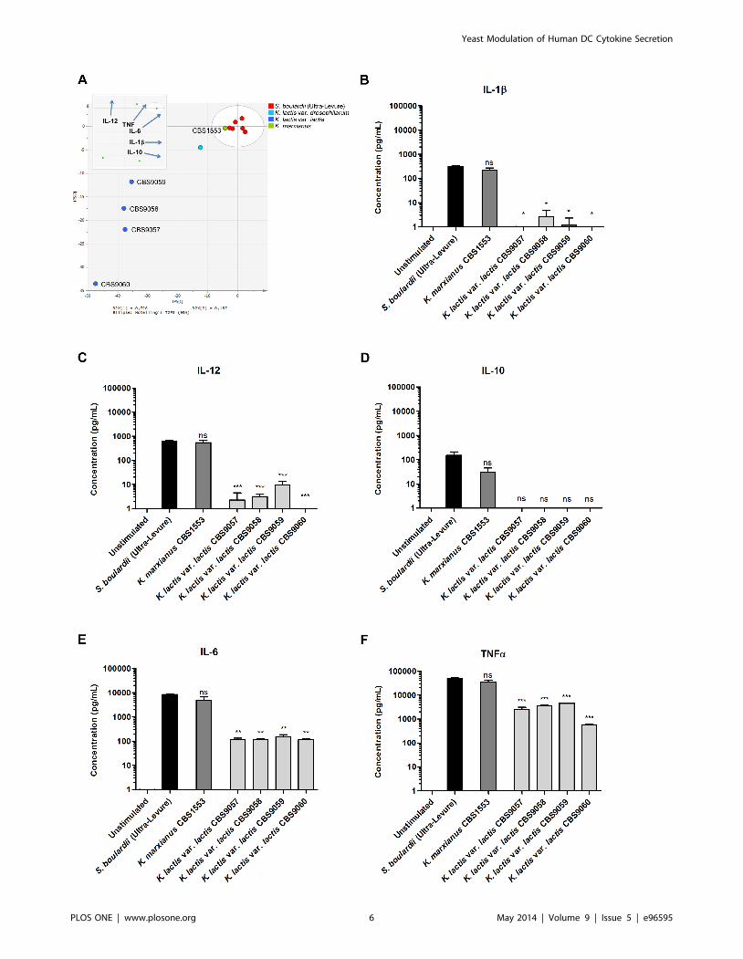
individual yeast genera. Six Kluyveromyces strains representing the
species K. marxianus, K. lactis var. lactis, and K. lactis var. drosophilarum
were included in our study, and multivariate data analysis of the
induced DC cytokine profiles revealed clear species distinctions in
immune stimulating capacities (Fig 1A). K. marxianus (CBS1553)
induced DC cytokine levels statistically indistinguishable (P.0.05)
from the profile induced by the S. boulardii reference strain for
every one of the quantified cytokines (Fig 1B-F). In contrast, the
four K. lactis var. lactis strains (CBS9057, CBS9058, CBS9059,
CBS9060) induced much lower levels of cytokines; in particular,
induced levels of IL-12, IL-10, and IL-1b were near or below the
detection limit of the assay. Strikingly, no significant differences
were observed between the DC cytokine profiles induced by the
four K. lactis var. lactis strains (Fig 1B-F, P.0.05 for all quantified
cytokines).
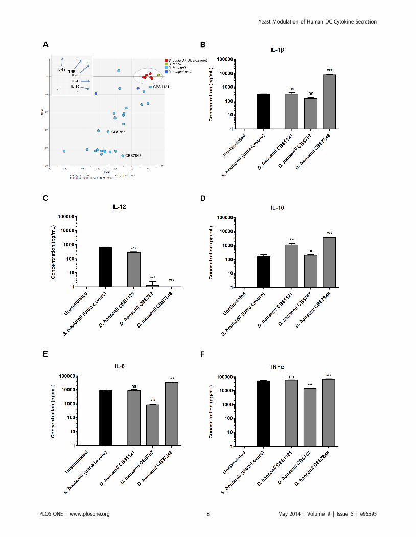
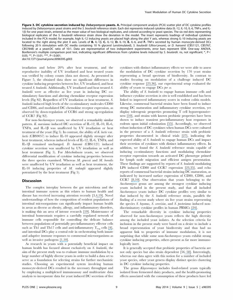
For Debaryomyces yeasts, 25 of the 28 strains included in our study
represented the species D. hansenii, and the induced DC cytokine
profiles revealed a remarkable diversity in immune stimulating
properties (Fig 3A). The strain CBS1121 induced DC cytokine
levels similar to the S. boulardii reference strain (Fig 3B-F), as
indicated by secreted levels of IL-1b, IL-6, and TNFa being
statistically indistinguishable from S. boulardii induced levels. The
D. hansenii type strain (CBS767) displayed much poorer cytokine
induction capabilities, as seen by significantly lower induction of
the pro-inflammatory cytokines IL-12, IL-6, and TNFa. Finally,
the D. hansenii strain CBS7848 induced a DC cytokine profile
characterized by levels of IL-1b, IL-10, IL-6, and TNFasignificantly higher than the S. boulardii reference strain, yet failed
to induce detectable levels of IL-12.
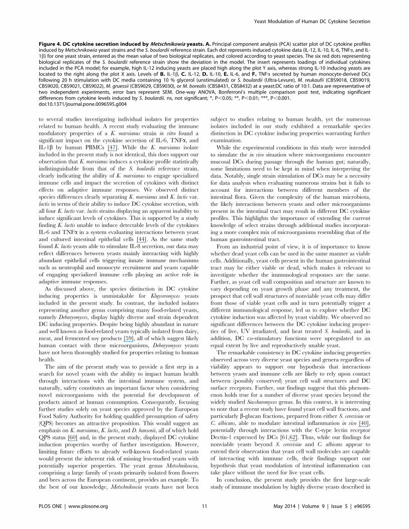
The yeast genus Metschnikowia represents a large family of yeasts
which, to the best of our knowledge, has not been explored for
properties relating to human health. The PCA plot in Figure 4A
displays the DC cytokine profiles induced by the 16 isolates
representing seven Metschnikowia species included in our study. The
plot reveals striking species distinctions separating species with
Yeast Modulation of Human DC Cytokine Secretion
PLOS ONE | www.plosone.org 5 May 2014 | Volume 9 | Issue 5 | e96595
Yeast Modulation of Human DC Cytokine Secretion
PLOS ONE | www.plosone.org 6 May 2014 | Volume 9 | Issue 5 | e96595

highly diverse cytokine inducing properties. While isolates of M.
reukaufii induced DC cytokine profiles very similar to the S. boulardii
reference strain across all five cytokines, M. gruessii isolates induced
robust levels of IL-1b, IL-10, IL-6, and TNFa, yet undetectable
levels of IL-12 (Fig 4B-F). In contrast, M. borealis displayed poor
cytokine inducing properties in general, as seen by an induced DC
cytokine profile characterized by significantly lower cytokine levels
compared to the S. boulardii reference strain.
Yeasts are capable of DC stimulation independently ofviability
Next, we investigated whether the observed interactions
between yeasts and DCs were dependent upon yeast viability.
We hypothesized that yeasts would be able to induce DC
activation regardless of metabolic activity and conducted exper-
iments to compare DC stimulation with live, UV irradiated, and
heat treated yeast. UV irradiation conditions were designed to
generate relatively intact yeast cells unable to reproduce, whereas
heat treatment was intended to cause severe yeast cell membrane
disruption. Propidium iodide staining confirmed a reduction in the
proportion of intact yeast cells to levels below 40% after UV
Figure 1. DC cytokine secretion induced by Kluyveromyces yeasts. A. Principal component analysis (PCA) scatter plot of DC cytokine profilesinduced by Kluyveromyces yeast strains and the S. boulardii reference strain. Each dot represents induced cytokine data (IL-12, IL-10, IL-6, TNFa, and IL-1b) for one yeast strain, entered as the mean value of two biological replicates, and colored according to yeast species. The six red dots representingbiological replicates of the S. boulardii reference strain show the deviation in the model. The insert represents loadings of individual cytokinesincluded in the PCA model; for example, high IL-12 inducing yeasts are placed high along the plot Y axis, whereas strong IL-10 inducing yeasts arelocated to the right along the plot X axis. Levels of B. IL-1b, C. IL-12, D. IL-10, E. IL-6, and F. TNFa secreted by human monocyte-derived DCsfollowing 20 h stimulation with DC media containing 10 % glycerol (unstimulated) or S. boulardii (Ultra-Levure), K. marxianus (CBS1553), or K. lactisvar. lactis (CBS9057, CBS9058, CBS9059, CBS9060) at a yeast:DC ratio of 10:1. Data are representative of two independent experiments, error barsrepresent SEM. One-way ANOVA, Bonferroni’s multiple comparison post test, indicating significant differences from cytokine levels induced by S.boulardii. ns, not significant; *, P,0.05; **, P,0.01; ***, P,0.001.doi:10.1371/journal.pone.0096595.g001
Figure 2. Yeast induced cytokine profiles in human DCs. Principal component analysis (PCA) scatter plot of induced cytokine data for 170yeast strains included in screen. Each dot represents induced cytokine data (IL-12, IL-10, IL-6, TNFa, and IL-1b) for one yeast strain, entered as themean value of two biological replicates, and colored according to yeast genera. The plot point of origin is centered on the cytokine profile induced bythe S. boulardii reference strain, and thus the distance of a given yeast strain from the plot point of origin indicates how closely the induced cytokineprofile resembles that of the S. boulardii reference strain. The insert represents loadings of individual cytokines included in the PCA model; forexample, high IL-12 inducing yeasts are placed high along the plot Y axis, whereas strong IL-10 inducing yeasts are located to the right along the plotX axis.doi:10.1371/journal.pone.0096595.g002
Yeast Modulation of Human DC Cytokine Secretion
PLOS ONE | www.plosone.org 7 May 2014 | Volume 9 | Issue 5 | e96595
Yeast Modulation of Human DC Cytokine Secretion
PLOS ONE | www.plosone.org 8 May 2014 | Volume 9 | Issue 5 | e96595
irradiation and below 20% after heat treatment, and the
reproductive inability of UV irradiated and heat treated yeasts
was verified by colony counts (data not shown). As presented in
Figure 5, the obtained data show no significant differences in
cytokine inducing properties between live, UV irradiated, and heat
treated S. boulardii. Additionally, UV irradiated and heat treated S.
boulardii were as effective as live yeast in inducing DC co-
stimulatory functions and altering chemokine receptor expression
levels (Fig S2). Regardless of UV irradiation or heat treatment, S.
boulardii induced high levels of the co-stimulatory molecules CD80
and CD86, and modulated DC chemokine receptor expression, as
observed by down-regulation of CCR6 and strong up-regulation
of CCR7 (Fig S2).
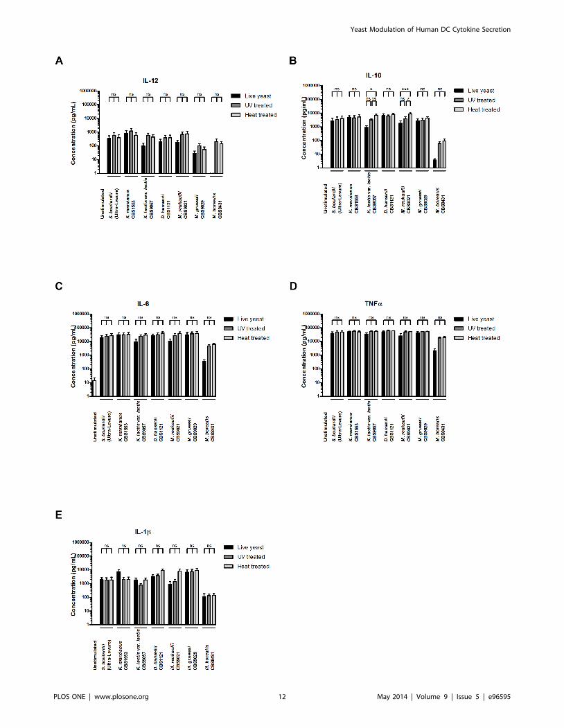
For non-Saccharomyces yeasts, we observed a remarkably similar
pattern. K. marxianus induced DC secretion of IL-12, IL-10, IL-6,
TNFa, and IL-1b was unaffected by UV irradiation or heat
treatment of the yeast (Fig 5). In contrast, the ability of K. lactis var.
lactis (CBS9057) to induce IL-10 appeared slightly stronger after
heat treatment, whereas induced levels of IL-12, IL-6, TNFa, and
IL-1b remained unchanged. D. hansenii (CBS1121) induced
cytokine secretion was unaffected by UV irradiation as well as
heat treatment (Fig 5). For Metschnikowia yeasts, we observed
differential modification of cytokine inducing properties between
the three species examined. Whereas M. gruessii and M. borealis
were unaffected by UV irradiation as well as heat treatment, the
IL-10 inducing properties of M. reukaufii appeared slightly
potentiated by the heat treatment (Fig 5).
Discussion
The complex interplay between the gut microbiota and the
intestinal immune system as this relates to human health and
disease has received increasing attention recently [3,4,19]. Novel
understandings of how the composition of resident populations of
intestinal microorganisms can significantly impact human health
in areas as diverse as obesity, allergy, and inflammatory disorders,
is making this an area of intense research [19]. Maintenance of
intestinal homeostasis requires a carefully regulated network of
immune cells responsible for controlling the delicate balance
between populations of potentially pro-inflammatory effector cells
such as Th1 and Th17 cells and anti-inflammatory Treg cells [4],
and intestinal DCs play a central role in orchestrating both innate
and adaptive immune responses to commensal microorganisms as
well as invasive pathogens [1,10].
As research in yeasts with a potentially beneficial impact on
human health has focused almost exclusively on S. boulardii, the
aim of the present study was to evaluate immune modulation by a
large number of highly diverse yeasts in order to build a data set to
serve as a foundation for selecting strains for further mechanistic
studies. Choosing an experimental system involving human
monocyte-derived DCs resulted in the necessary throughput and
by employing a multiplexed immunoassay and multivariate data
analysis to incorporate data for yeast induced DC secretion of five
cytokines with distinct inflammatory effects we were able to assess
the modulation of DC cytokine secretion by 170 yeast strains
representing a broad spectrum of biodiversity. In contrast to
studies focusing on modulation of a challenge induced DC
cytokine response [23,36], our experimental setup evaluated the
ability of yeasts to engage DCs per se.
The ability of S. boulardii to engage human immune cells and
influence cytokine secretion in vitro is well established and has been
linked to improved inflammatory scores in rodent colitis models.
Likewise, commensal bacterial strains have been found to induce
strong DC maturation and inflammatory cytokine secretion, yet
display tolerogenic properties promoting T cell hyporesponsive-
ness [10], and strains with known probiotic properties have been
shown to induce transient pro-inflammatory host responses in
rodents upon initial colonization [55]. Accordingly, we observed
robust induction of DC cytokines when human DCs were cultured
in the presence of a S. boulardii reference strain with probiotic
properties documented in clinical trials [27], indicating the
expected ability of S. boulardii to engage human DCs and impact
their secretion of cytokines with distinct inflammatory effects. In
addition, we found the S. boulardii reference strain capable of
inducing co-stimulatory functions and modulating chemokine
receptor expression towards an activated DC phenotype primed
for lymph node migration and efficient antigen presentation.
These findings are supported by reports of S. boulardii modulating
LPS induced CD80 and CCR7 expression [23,35], and echo
reports of commensal bacterial strains inducing DC maturation, as
indicated by increased surface expression of CD80, CD86, and
CCR7 [8,10]. Our observation that yeasts belonging to the
Saccharomyces genus are among the strongest cytokine inducing
yeasts included in the present study, and that all included
Saccharomyces yeasts induce DC cytokine profiles very similar to
that induced by the S. boulardii reference strain, parallels the
finding of a recent study where six live yeast strains representing
the species S. bayanus, S. cerevisiae, and S. pastorianus induced non-
discriminatory cytokine profiles in human PBMCs [24].
The remarkable diversity in cytokine inducing properties
observed for non-Saccharomyces yeasts reflects the high diversity
among the included yeast isolates. As the selection criteria for
inclusion in the present study were based on a desire to include a
broad representation of yeast biodiversity and thus had no
apparent link to properties of immune modulation, it is not
surprising that while some non-Saccharomyces yeasts exhibit strong
cytokine inducing properties, others present as far more immuno-
logically inert.
It is generally accepted that probiotic properties of bacteria are
not only species but also strain dependent [56–58]. Interestingly,
whereas our data agree with this notion for a number of included
yeast species, other yeast genera display distinct species clustering
in DC cytokine inducing properties.
The genus Kluyveromyces includes food-related yeasts typically
isolated from fermented dairy products, and the health-promoting
effects associated with the consumption of these products have led
Figure 3. DC cytokine secretion induced by Debaryomyces yeasts. A. Principal component analysis (PCA) scatter plot of DC cytokine profilesinduced by Debaryomyces yeast strains and the S. boulardii reference strain. Each dot represents induced cytokine data (IL-12, IL-10, IL-6, TNFa, and IL-1b) for one yeast strain, entered as the mean value of two biological replicates, and colored according to yeast species. The six red dots representingbiological replicates of the S. boulardii reference strain show the deviation in the model. The insert represents loadings of individual cytokinesincluded in the PCA model; for example, high IL-12 inducing yeasts are placed high along the plot Y axis, whereas strong IL-10 inducing yeasts arelocated to the right along the plot X axis. Levels of B. IL-1b, C. IL-12, D. IL-10, E. IL-6, and F. TNFa secreted by human monocyte-derived DCsfollowing 20 h stimulation with DC media containing 10 % glycerol (unstimulated), S. boulardii (Ultra-Levure), or D. hansenii (CBS1121, CBS767,CBS7848) at a yeast:DC ratio of 10:1. Data are representative of two independent experiments, error bars represent SEM. One-way ANOVA,Bonferroni’s multiple comparison post test, indicating significant differences from cytokine levels induced by S. boulardii. ns, not significant; *, P,
0.05; **, P,0.01; ***, P,0.001.doi:10.1371/journal.pone.0096595.g003
Yeast Modulation of Human DC Cytokine Secretion
PLOS ONE | www.plosone.org 9 May 2014 | Volume 9 | Issue 5 | e96595

Yeast Modulation of Human DC Cytokine Secretion
PLOS ONE | www.plosone.org 10 May 2014 | Volume 9 | Issue 5 | e96595
to several studies investigating individual isolates for properties
related to human health. A recent study evaluating the immune
modulatory properties of a K. marxianus strain in vitro found a
significant impact on the cytokine secretion of IL-6, TNFa, and
IL-1b by human PBMCs [47]. While the K. marxianus isolate
included in the present study is not identical, this does support our
observation that K. marxianus induces a cytokine profile statistically
indistinguishable from that of the S. boulardii reference strain,
clearly indicating the ability of K. marxianus to engage specialized
immune cells and impact the secretion of cytokines with distinct
effects on adaptive immune responses. We observed distinct
species differences clearly separating K. marxianus and K. lactis var.
lactis in terms of their ability to induce DC cytokine secretion, with
all four K. lactis var. lactis strains displaying an apparent inability to
induce significant levels of cytokines. This is supported by a study
finding K. lactis unable to induce detectable levels of the cytokines
IL-6 and TNFa in a system evaluating interactions between yeast
and cultured intestinal epithelial cells [44]. As the same study
found K. lactis yeasts able to stimulate IL-8 secretion, our data may
reflect differences between yeasts mainly interacting with highly
abundant epithelial cells triggering innate immune mechanisms
such as neutrophil and monocyte recruitment and yeasts capable
of engaging specialized immune cells playing an active role in
adaptive immune responses.
As discussed above, the species distinction in DC cytokine
inducing properties is unmistakable for Kluyveromyces yeasts
included in the present study. In contrast, the included isolates
representing another genus comprising many food-related yeasts,
namely Debaryomyces, display highly diverse and strain dependent
DC inducing properties. Despite being highly abundant in nature
and well known as food-related yeasts typically isolated from dairy,
meat, and fermented soy products [59], all of which suggest likely
human contact with these microorganisms, Debaryomyces yeasts
have not been thoroughly studied for properties relating to human
health.
The aim of the present study was to provide a first step in a
search for novel yeasts with the ability to impact human health
through interactions with the intestinal immune system, and
naturally, safety constitutes an important factor when considering
novel microorganisms with the potential for development of
products aimed at human consumption. Consequently, focusing
further studies solely on yeast species approved by the European
Food Safety Authority for holding qualified presumption of safety
(QPS) becomes an attractive proposition. This would suggest an
emphasis on K. marxianus, K. lactis, and D. hansenii, all of which hold
QPS status [60] and, in the present study, displayed DC cytokine
induction properties worthy of further investigation. However,
limiting future efforts to already well-known food-related yeasts
would present the inherent risk of missing less-studied yeasts with
potentially superior properties. The yeast genus Metschnikowia,
comprising a large family of yeasts primarily isolated from flowers
and bees across the European continent, provides an example. To
the best of our knowledge, Metschnikowia yeasts have not been
subject to studies relating to human health, yet the numerous
isolates included in our study exhibited a remarkable species
distinction in DC cytokine inducing properties warranting further
examination.
While the experimental conditions in this study were intended
to simulate the in vivo situation where microorganisms encounter
mucosal DCs during passage through the human gut; naturally,
some limitations need to be kept in mind when interpreting the
data. Notably, single strain stimulation of DCs may be a necessity
for data analysis when evaluating numerous strains but it fails to
account for interactions between different members of the
intestinal flora. Given the complexity of the human microbiota,
the likely interactions between yeasts and other microorganisms
present in the intestinal tract may result in different DC cytokine
profiles. This highlights the importance of extending the current
knowledge of select strains through additional studies incorporat-
ing a more complex mix of microorganisms resembling that of the
human gastrointestinal tract.
From an industrial point of view, it is of importance to know
whether dead yeast cells can be used in the same manner as viable
cells. Additionally, yeast cells present in the human gastrointestinal
tract may be either viable or dead, which makes it relevant to
investigate whether the immunological responses are the same.
Further, as yeast cell wall composition and structure are known to
vary depending on yeast growth phase and any treatment, the
prospect that cell wall structures of nonviable yeast cells may differ
from those of viable yeast cells and in turn potentially trigger a
different immunological response, led us to explore whether DC
cytokine induction was affected by yeast viability. We observed no
significant differences between the DC cytokine inducing proper-
ties of live, UV irradiated, and heat treated S. boulardii, and in
addition, DC co-stimulatory functions were upregulated to an
equal extent by live and reproductively unable yeast.
The remarkable consistency in DC cytokine inducing properties
observed across very diverse yeast species and genera regardless of
viability appears to support our hypothesis that interactions
between yeasts and immune cells are likely to rely upon contact
between (possibly conserved) yeast cell wall structures and DC
surface receptors. Further, our findings suggest that this phenom-
enon holds true for a number of diverse yeast species beyond the
widely studied Saccharomyces genus. In this context, it is interesting
to note that a recent study have found yeast cell wall fractions, and
particularly b-glucan fractions, prepared from either S. cerevisiae or
C. albicans, able to modulate intestinal inflammation in vivo [40],
potentially through interactions with the C-type lectin receptor
Dectin-1 expressed by DCs [61,62]. Thus, while our findings for
nonviable yeasts beyond S. cerevisiae and C. albicans appear to
extend their observation that yeast cell wall molecules are capable
of interacting with immune cells, their findings support our
hypothesis that yeast modulation of intestinal inflammation can
take place without the need for live yeast cells.
In conclusion, the present study provides the first large-scale
study of immune modulation by highly diverse yeasts described in
Figure 4. DC cytokine secretion induced by Metschnikowia yeasts. A. Principal component analysis (PCA) scatter plot of DC cytokine profilesinduced by Metschnikowia yeast strains and the S. boulardii reference strain. Each dot represents induced cytokine data (IL-12, IL-10, IL-6, TNFa, and IL-1b) for one yeast strain, entered as the mean value of two biological replicates, and colored according to yeast species. The six red dots representingbiological replicates of the S. boulardii reference strain show the deviation in the model. The insert represents loadings of individual cytokinesincluded in the PCA model; for example, high IL-12 inducing yeasts are placed high along the plot Y axis, whereas strong IL-10 inducing yeasts arelocated to the right along the plot X axis. Levels of B. IL-1b, C. IL-12, D. IL-10, E. IL-6, and F. TNFa secreted by human monocyte-derived DCsfollowing 20 h stimulation with DC media containing 10 % glycerol (unstimulated) or S. boulardii (Ultra-Levure), M. reukaufii (CBS9018, CBS9019,CBS9020, CBS9021, CBS9022), M. gruessii (CBS9029, CBS9030), or M. borealis (CBS8431, CBS8432) at a yeast:DC ratio of 10:1. Data are representative oftwo independent experiments, error bars represent SEM. One-way ANOVA, Bonferroni’s multiple comparison post test, indicating significantdifferences from cytokine levels induced by S. boulardii. ns, not significant; *, P,0.05; **, P,0.01; ***, P,0.001.doi:10.1371/journal.pone.0096595.g004
Yeast Modulation of Human DC Cytokine Secretion
PLOS ONE | www.plosone.org 11 May 2014 | Volume 9 | Issue 5 | e96595
Yeast Modulation of Human DC Cytokine Secretion
PLOS ONE | www.plosone.org 12 May 2014 | Volume 9 | Issue 5 | e96595

the scientific literature. Our data clearly demonstrate high
diversity in yeast induced cytokine secretion across a broad
spectrum of yeasts, and by employing multivariate data analysis we
reveal distinct clustering of yeasts inducing similar cytokine profiles
in DCs, highlighting clear species distinction within specific yeast
genera. The observed differences in induced DC cytokine profiles
may indicate distinct modes of interaction between yeasts and
human immune cells, and will aid in the selection of strains for
further characterization and development toward potentially novel
yeast probiotics. Additionally, we present data to support a
hypothesis that the interaction between yeasts and human DCs
does not solely depend on yeast viability, a concept which may
suggest a need for further classifications beyond the current
definition of a probiotic.
Supporting Information
Figure S1 Time-course of S. boulardii induced DCcytokine secretion supports a 20 h stimulation time.Levels of IL-12, IL-10, IL-1b, IL-6, and TNFa secreted by human
monocyte-derived DCs following incubation with S. boulardii
(Ultra-Levure) at a yeast:DC ratio of 10:1. Data are expressed as
mean6SEM (n = 2).
(TIF)
Figure S2 Modulation of DC co-stimulatory functionsand chemokine receptor expression occurs indepen-dently of yeast metabolic function. DC surface expression of
CD80, CD86, CCR6, and CCR7 following 20 h stimulation with
DC media containing 10% glycerol (unstimulated) or either live,
UV irradiated, or heat killed S. boulardii (Ultra-Levure) at a
yeast:DC ratio of 10:1. Data are expressed as mean6SEM (n = 4).
One-way ANOVA, Bonferroni’s multiple comparison post test,
indicating significant differences between cytokine levels induced
by live, UV treated, and heat killed yeast. ns, not significant; *, P,
0.05; **, P,0.01; ***, P,0.001.
(TIF)
Acknowledgments
We gratefully acknowledge Amparo Gamero Lluna for performing the
initial selection of strains included in this study, Stina Rikke Jensen for
valuable cell culture advice, Jeanne Olsen for excellent technical assistance,
and Jannik Vindeløv for guidance on multivariate data analysis.
Author Contributions
Conceived and designed the experiments: IMS JEC NA LJ. Performed the
experiments: IMS JEC. Analyzed the data: IMS JEC. Contributed
reagents/materials/analysis tools: IMS JEC. Wrote the paper: IMS.
References
1. Coombes JL, Powrie F (2008) Dendritic cells in intestinal immune regulation.
Nat Rev Immunol 8: 435–446.
2. Rescigno M, Urbano M, Valzasina B, Francolini M, Rotta G, et al. (2001)
Dendritic cells express tight junction proteins and penetrate gut epithelial
monolayers to sample bacteria. Nat Immunol 2: 361–367.
3. Maynard CL, Elson CO, Hatton RD, Weaver CT (2012) Reciprocal
interactions of the intestinal microbiota and immune system. Nature 489:
231–241.
4. Hooper LV, Littman DR, Macpherson AJ (2012) Interactions between the
microbiota and the immune system. Science 336: 1268–1273.
5. Manicassamy S, Ravindran R, Deng J, Oluoch H, Denning TL, et al. (2009)
Toll-like receptor 2-dependent induction of vitamin A-metabolizing enzymes in
dendritic cells promotes T regulatory responses and inhibits autoimmunity. Nat
Med 15: 401–409.
6. Donkor ON, Ravikumar M, Proudfoot O, Day SL, Apostolopoulos V, et al.
(2012) Cytokine profile and induction of T helper type 17 and regulatory T cells
by human peripheral mononuclear cells after microbial exposure. Clin Exp
Immunol 167: 282–295.
7. Joint FAO/WHO Working Group (2002) Guidelines for the evaluation of
probiotics in food.
8. Foligne B, Zoumpopoulou G, Dewulf J, Ben Younes A, Chareyre F, et al. (2007)
A key role of dendritic cells in probiotic functionality. PLoS One 2: e313.
9. Kwon HK, Lee CG, So JS, Chae CS, Hwang JS, et al. (2010) Generation of
regulatory dendritic cells and CD4+Foxp3+ T cells by probiotics administration
suppresses immune disorders. Proc Natl Acad Sci U S A 107: 2159–2164.
10. Baba N, Samson S, Bourdet-Sicard R, Rubio M, Sarfati M (2008) Commensal
bacteria trigger a full dendritic cell maturation program that promotes the
expansion of non-Tr1 suppressor T cells. J Leukoc Biol 84: 468–476.
11. Konieczna P, Groeger D, Ziegler M, Frei R, Ferstl R, et al. (2012)
Bifidobacterium infantis 35624 administration induces Foxp3 T regulatory cells
in human peripheral blood: Potential role for myeloid and plasmacytoid
dendritic cells. Gut 61: 354–366.
12. Jeon SG, Kayama H, Ueda Y, Takahashi T, Asahara T, et al. (2012) Probiotic
bifidobacterium breve induces IL-10-producing Tr1 cells in the colon. PLoS
Pathog 8: e1002714.
13. Di Giacinto C, Marinaro M, Sanchez M, Strober W, Boirivant M (2005)
Probiotics ameliorate recurrent Th1-mediated murine colitis by inducing IL-10
and IL-10-dependent TGF-beta-bearing regulatory cells. J Immunol 174: 3237–
3246.
14. Mann ER, Landy JL, Bernardo D, Peake ST, Hart AL, et al. (2013) Intestinal
dendritic cells: Their role in intestinal inflammation, manipulation by the gutmicrobiota and differences between mice and men. Immunol Lett.
15. Christensen HR, Frokiaer H, Pestka JJ (2002) Lactobacilli differentially
modulate expression of cytokines and maturation surface markers in murinedendritic cells. J Immunol 168: 171–178.
16. Weiss G, Christensen HR, Zeuthen LH, Vogensen FK, Jakobsen M, et al. (2011)Lactobacilli and bifidobacteria induce differential interferon-beta profiles in
dendritic cells. Cytokine 56: 520–530.
17. Plantinga TS, van Bergenhenegouwen J, Jacobs C, Joosten LA, van’t Land B,et al. (2012) Modulation of toll-like receptor ligands and candida albicans-
induced cytokine responses by specific probiotics. Cytokine 59: 159–165.
18. Qin J, Li R, Raes J, Arumugam M, Burgdorf KS, et al. (2010) A human gut
microbial gene catalogue established by metagenomic sequencing. Nature 464:
59–65.
19. Clemente JC, Ursell LK, Parfrey LW, Knight R (2012) The impact of the gut
microbiota on human health: An integrative view. Cell 148: 1258–1270.
20. Scanlan PD, Marchesi JR (2008) Micro-eukaryotic diversity of the human distal
gut microbiota: Qualitative assessment using culture-dependent and -indepen-
dent analysis of faeces. ISME J 2: 1183–1193.
21. Ghannoum MA, Jurevic RJ, Mukherjee PK, Cui F, Sikaroodi M, et al. (2010)
Characterization of the oral fungal microbiome (mycobiome) in healthyindividuals. PLoS Pathog 6: e1000713.
22. Hatoum R, Labrie S, Fliss I (2012) Antimicrobial and probiotic properties of
yeasts: From fundamental to novel applications. Front Microbiol 3: 421.
23. Thomas S, Przesdzing I, Metzke D, Schmitz J, Radbruch A, et al. (2009)
Saccharomyces boulardii inhibits lipopolysaccharide-induced activation ofhuman dendritic cells and T cell proliferation. Clin Exp Immunol 156: 78–87.
24. Foligne B, Dewulf J, Vandekerckove P, Pignede G, Pot B (2010) Probiotic yeasts:
Anti-inflammatory potential of various non-pathogenic strains in experimentalcolitis in mice. World J Gastroenterol 16: 2134–2145.
25. van der Aa Kuhle A, Jespersen L (2003) The taxonomic position ofsaccharomyces boulardii as evaluated by sequence analysis of the D1/D2
domain of 26S rDNA, the ITS1-5.8S rDNA-ITS2 region and the mitochondrial
cytochrome-c oxidase II gene. Syst Appl Microbiol 26: 564–571.
26. Vaughan-Martini A, Martini A (2011) SaccharomycesMeyen ex reess (1870). In:
Kurtzman CP, Fell JW, Boekhout T, editors. The Yeasts: A Taxonomic Study.London, UK: Elsevier. pp. 733.
27. McFarland LV (2010) Systematic review and meta-analysis of saccharomyces
boulardii in adult patients. World J Gastroenterol 16: 2202–2222.
Figure 5. Yeast induced DC cytokine secretion occurs independently of yeast metabolic function. Levels of A. IL-12, B. IL-10, C. IL-6, D.TNFa, and E. IL-1b secreted by human monocyte-derived DCs following 20 h stimulation with DC media containing 10 % glycerol (unstimulated) or S.boulardii (Ultra-Levure), K. marxianus (CBS1553), K. lactis var. lactis (CBS9057), D. hansenii (CBS1121), M. reukaufii (CBS9021), M. gruessii (CBS9029), or M.borealis (CBS8431). For each yeast strain, DC stimulation was performed with live yeast, UV irradiated yeast, and heat treated yeast at a yeast:DC ratioof 10:1. Data are expressed as mean6SEM (n = 4). One-way ANOVA, Bonferroni’s multiple comparison post test, indicating significant differencesbetween cytokine levels induced by live, UV irradiated, and heat treated yeast for each strain. ns, not significant; *, P,0.05; **, P,0.01; ***, P,0.001.doi:10.1371/journal.pone.0096595.g005
Yeast Modulation of Human DC Cytokine Secretion
PLOS ONE | www.plosone.org 13 May 2014 | Volume 9 | Issue 5 | e96595

28. Dinleyici EC, Eren M, Ozen M, Yargic ZA, Vandenplas Y (2012) Effectiveness
and safety of saccharomyces boulardii for acute infectious diarrhea. Expert OpinBiol Ther 12: 395–410.
29. Zanello G, Berri M, Dupont J, Sizaret PY, D’Inca R, et al. (2011)
Saccharomyces cerevisiae modulates immune gene expressions and inhibitsETEC-mediated ERK1/2 and p38 signaling pathways in intestinal epithelial
cells. PLoS One 6: e18573.30. Chen X, Kokkotou EG, Mustafa N, Bhaskar KR, Sougioultzis S, et al. (2006)
Saccharomyces boulardii inhibits ERK1/2 mitogen-activated protein kinase
activation both in vitro and in vivo and protects against clostridium difficile toxinA-induced enteritis. J Biol Chem 281: 24449–24454.
31. Dahan S, Dalmasso G, Imbert V, Peyron JF, Rampal P, et al. (2003)Saccharomyces boulardii interferes with enterohemorrhagic escherichia coli-
induced signaling pathways in T84 cells. Infect Immun 71: 766–773.32. Martins FS, Dalmasso G, Arantes RM, Doye A, Lemichez E, et al. (2010)
Interaction of saccharomyces boulardii with salmonella enterica serovar
typhimurium protects mice and modifies T84 cell response to the infection.PLoS One 5: e8925.
33. Lee SK, Kim YW, Chi SG, Joo YS, Kim HJ (2009) The effect of saccharomycesboulardii on human colon cells and inflammation in rats with trinitrobenzene
sulfonic acid-induced colitis. Dig Dis Sci 54: 255–263.
34. Sougioultzis S, Simeonidis S, Bhaskar KR, Chen X, Anton PM, et al. (2006)Saccharomyces boulardii produces a soluble anti-inflammatory factor that
inhibits NF-kappaB-mediated IL-8 gene expression. Biochem Biophys ResCommun 343: 69–76.
35. Thomas S, Metzke D, Schmitz J, Dorffel Y, Baumgart DC (2011) Anti-inflammatory effects of saccharomyces boulardii mediated by myeloid dendritic
cells from patients with crohn’s disease and ulcerative colitis. Am J Physiol
Gastrointest Liver Physiol.36. Gad M, Ravn P, Soborg DA, Lund-Jensen K, Ouwehand AC, et al. (2011)
Regulation of the IL-10/IL-12 axis in human dendritic cells with probioticbacteria. FEMS Immunol Med Microbiol 63: 93–107.
37. Pothoulakis C (2009) Review article: Anti-inflammatory mechanisms of action of
saccharomyces boulardii. Aliment Pharmacol Ther 30: 826–833.38. Martins FS, Nardi RM, Arantes RM, Rosa CA, Neves MJ, et al. (2005)
Screening of yeasts as probiotic based on capacities to colonize thegastrointestinal tract and to protect against enteropathogen challenge in mice.
J Gen Appl Microbiol 51: 83–92.39. Dalmasso G, Cottrez F, Imbert V, Lagadec P, Peyron JF, et al. (2006)
Saccharomyces boulardii inhibits inflammatory bowel disease by trapping T cells
in mesenteric lymph nodes. Gastroenterology 131: 1812–1825.40. Jawhara S, Habib K, Maggiotto F, Pignede G, Vandekerckove P, et al. (2012)
Modulation of intestinal inflammation by yeasts and cell wall extracts: Straindependence and unexpected anti-inflammatory role of glucan fractions. PLoS
One 7: e40648.
41. Kumura H, Tanoue Y, Tsukahara M, Tanaka T, Shimazaki K (2004) Screeningof dairy yeast strains for probiotic applications. J Dairy Sci 87: 4050–4056.
42. Pedersen LL, Owusu-Kwarteng J, Thorsen L, Jespersen L (2012) Biodiversityand probiotic potential of yeasts isolated from fura, a west african spontaneously
fermented cereal. Int J Food Microbiol 159: 144–151.43. Romanin D, Serradell M, Gonzalez Maciel D, Lausada N, Garrote GL, et al.
(2010) Down-regulation of intestinal epithelial innate response by probiotic
yeasts isolated from kefir. Int J Food Microbiol 140: 102–108.44. Saegusa S, Totsuka M, Kaminogawa S, Hosoi T (2007) Cytokine responses of
intestinal epithelial-like caco-2 cells to non-pathogenic and opportunisticpathogenic yeasts in the presence of butyric acid. Biosci Biotechnol Biochem
71: 2428–2434.
45. Reyes-Becerril M, Salinas I, Cuesta A, Meseguer J, Tovar-Ramirez D, et al.(2008) Oral delivery of live yeast debaryomyces hansenii modulates the main
innate immune parameters and the expression of immune-relevant genes in the
gilthead seabream (sparus aurata L.). Fish Shellfish Immunol 25: 731–739.
46. Macey BM, Coyne VE (2006) Colonization of the gastrointestinal tract of the
farmed south african abalone haliotis midae by the probionts vibrio midae SY9,
cryptococcus sp. SS1, and debaryomyces hansenii AY1. Mar Biotechnol (NY) 8:
246–259.
47. Maccaferri S, Klinder A, Brigidi P, Cavina P, Costabile A (2012) Potential
probiotic kluyveromyces marxianus B0399 modulates the immune response in
caco-2 cells and peripheral blood mononuclear cells and impacts the human gut
microbiota in an in vitro colonic model system. Appl Environ Microbiol 78:
956–964.
48. Kourelis A, Kotzamanidis C, Litopoulou-Tzanetaki E, Papaconstantinou J,
Tzanetakis N, et al. (2010) Immunostimulatory activity of potential probiotic
yeast strains in the dorsal air pouch system and the gut mucosa. J Appl Microbiol
109: 260–271.
49. Weiss G, Rasmussen S, Nielsen Fink L, Jarmer H, Nohr Nielsen B, et al. (2010)
Bifidobacterium bifidum actively changes the gene expression profile induced by
lactobacillus acidophilus in murine dendritic cells. PLoS One 5: e11065.
50. Zeuthen LH, Fink LN, Frokiaer H (2008) Toll-like receptor 2 and nucleotide-
binding oligomerization domain-2 play divergent roles in the recognition of gut-
derived lactobacilli and bifidobacteria in dendritic cells. Immunology 124: 489–
502.
51. Baba N, Samson S, Bourdet-Sicard R, Rubio M, Sarfati M (2009) Selected
commensal-related bacteria and toll-like receptor 3 agonist combinatorial codes
synergistically induce interleukin-12 production by dendritic cells to trigger a T
helper type 1 polarizing programme. Immunology 128: e523–31.
52. Zeuthen LH, Fink LN, Frokiaer H (2008) Epithelial cells prime the immune
response to an array of gut-derived commensals towards a tolerogenic phenotype
through distinct actions of thymic stromal lymphopoietin and transforming
growth factor-beta. Immunology 123: 197–208.
53. Carson RT, Vignali DA (1999) Simultaneous quantitation of 15 cytokines using
a multiplexed flow cytometric assay. J Immunol Methods 227: 41–52.
54. Vignali DA (2000) Multiplexed particle-based flow cytometric assays. J Immunol
Methods 243: 243–255.
55. Ruiz PA, Hoffmann M, Szcesny S, Blaut M, Haller D (2005) Innate mechanisms
for bifidobacterium lactis to activate transient pro-inflammatory host responses
in intestinal epithelial cells after the colonization of germ-free rats. Immunology
115: 441–450.
56. Wall R, Marques TM, O’Sullivan O, Ross RP, Shanahan F, et al. (2012)
Contrasting effects of bifidobacterium breve NCIMB 702258 and bifidobacter-
ium breve DPC 6330 on the composition of murine brain fatty acids and gut
microbiota. Am J Clin Nutr 95: 1278–1287.
57. Smelt MJ, de Haan BJ, Bron PA, van Swam I, Meijerink M, et al. (2012) L.
plantarum, L. salivarius, and L. lactis attenuate Th2 responses and increase treg
frequencies in healthy mice in a strain dependent manner. PLoS One 7: e47244.
58. Wells JM (2011) Immunomodulatory mechanisms of lactobacilli. Microb Cell
Fact 10 Suppl 1: S17-2859-10-S1-S17. Epub 2011 Aug 30.
59. Suzuki M, Prasad GS, Kurtzman CP (2011) DebaryomycesLodder & kreger-van rij
(1952). In: Kurtzman CP, Fell JW, Boekhout T, editors. The Yeasts: A
Taxonomic Study. London, UK: Elsevier. pp. 361.
60. EFSA Panel on Biological Hazards (BIOHAZ) (2010) Scientific opinion on the
maintenance of the list of QPS biological agents intentionally added to food and
feed (2010 update). EFSA Journal 8: 1944.
61. Iliev ID, Funari VA, Taylor KD, Nguyen Q, Reyes CN, et al. (2012)
Interactions between commensal fungi and the C-type lectin receptor dectin-1
influence colitis. Science 336: 1314–1317.
62. Brown GD (2006) Dectin-1: A signalling non-TLR pattern-recognition receptor.
Nat Rev Immunol 6: 33–43.
Yeast Modulation of Human DC Cytokine Secretion
PLOS ONE | www.plosone.org 14 May 2014 | Volume 9 | Issue 5 | e96595