Life-Long Shedding of Puumala Hantavirus in Wild Bank Voles (Myodes glareolus)

RESEARCH Open Access
Infection of human monocyte-derived dendriticcells by ANDES Hantavirus enhances pro-inflammatory state, the secretion of active MMP-9and indirectly enhances endothelial permeabilityDelphine Marsac1, Stephanie García1, Alexandra Fournet2, Adam Aguirre3, Karla Pino1, Marcela Ferres4,Alexis M Kalergis5, Marcelo Lopez-Lastra1 and Francisco Veas2*
Abstract
Background: Andes virus (ANDV), a rodent-borne Hantavirus, is the major etiological agent of Hantaviruscardiopulmonary syndrome (HCPS) in South America, which is mainly characterized by a vascular leakage with highrate of fatal outcomes for infected patients. Currently, neither specific therapy nor vaccines are available againstthis pathogen. ANDV infects both dendritic and epithelial cells, but in despite that the severity of the diseasedirectly correlates with the viral RNA load, considerable evidence suggests that immune mechanisms rather thandirect viral cytopathology are responsible for plasma leakage in HCPS. Here, we assessed the possible effect ofsoluble factors, induced in viral-activated DCs, on endothelial permeability. Activated immune cells, including DC,secrete gelatinolytic matrix metalloproteases (gMMP-2 and -9) that modulate the vascular permeability for theirtrafficking.
Methods: A clinical ANDES isolate was used to infect DC derived from primary PBMC. Maturation and pro-inflammatory phenotypes of ANDES-infected DC were assessed by studying the expression of receptors, cytokinesand active gMMP-9, as well as some of their functional status. The ANDES-infected DC supernatants were assessedfor their capacity to enhance a monolayer endothelial permeability using primary human vascular endothelial cells(HUVEC).
Results: Here, we show that in vitro primary DCs infected by a clinical isolate of ANDV shed virus RNA andproteins, suggesting a competent viral replication in these cells. Moreover, this infection induces an enhancedexpression of soluble pro-inflammatory factors, including TNF-a and the active gMMP-9, as well as a decreasedexpression of anti-inflammatory cytokines, such as IL-10 and TGF-b. These viral activated cells are less sensitive toapoptosis. Moreover, supernatants from ANDV-infected DCs were able to indirectly enhance the permeability of amonolayer of primary HUVEC.
Conclusions: Primary human DCs, that are primarily targeted by hantaviruses can productively be infected byANDV and subsequently induce direct effects favoring a proinflammatory phenotype of infected DCs. Finally, basedon our observations, we hypothesize that soluble factors secreted in ANDV-infected DC supernatants, importantlycontribute to the endothelial permeability enhancement that characterize the HCPS.
* Correspondence: [email protected] Montpellier 1, Comparative Molecular Immuno-Physiopathology Lab, Faculté de Pharmacie, 34093 Montpellier, FranceFull list of author information is available at the end of the article
Marsac et al. Virology Journal 2011, 8:223http://www.virologyj.com/content/8/1/223
© 2011 Marsac et al; licensee BioMed Central Ltd. This is an Open Access article distributed under the terms of the Creative CommonsAttribution License (http://creativecommons.org/licenses/by/2.0), which permits unrestricted use, distribution, and reproduction inany medium, provided the original work is properly cited.

BackgroundHantaviruses are rodent-born enveloped RNA-virusesbelonging to Bunyaviridae family. Two major severepathologies associated to Hantaviruses have beenreported: hemorrhagic fever with renal syndrome (HFRS)in the Eurasia and Hantavirus cardiopulmonary syn-drome (HCPS) in the Americas. HCPS is more frequentlyassociated (40%) to fatal outcomes than HFRS (<1%) [1].Andes Hantavirus (ANDV) is the major etiological agentof the HCPS in South America, syndrome characterizedby the presence of high amounts of pulmonary fluidsleading to an edema evolving to a cardiogenic shock thatsynergistically acts with hypovolemia due to capillaryleakage resulting in an abrupt cardiopulmonary collapse[2]. Although disease severity directly correlates with theviral RNA load [3], considerable evidence exists suggest-ing that immune mechanisms rather than direct viralcytopathology are indeed responsible for the massive vas-cular dysfunction and plasma leakage of HFRS and HCPS[4,5].The hemorrhagic viruses, including the members of the
Bunyaviridae as well as dengue viruses, target endothelialcells and immune cells, mainly monocyte-derived cellssuch as the professional antigen-presenting cells, Dendriticcells (DCs) [6-8]. DCs activation triggers their maturationand trans-endothelial migration occurring during woundhealing or inflammation. These processes require extracel-lular matrix remodeling and involve changes in endothelialpermeability regulated by the production of matrix metal-loproteases (gMMPs) or vascular endothelial growth factor(VEGF). However, in excess, these soluble factors can havedeleterious effects on endothelial cell integrity. Data fromdifferent reports show that endothelial cells infected bydengue virus trigger secretion of soluble factors such asVEGF and the decrease of VEGF-R2 receptor [9,10]. Wehave recently reported in vitro and in vivo showing thatsoluble factors secreted from DV-infected DCs enhanceendothelial permeability and down-regulate expression ofendothelial junction proteins, Pecam-1 and VE-cadherinin a gMMP-9-dependent manner [11]. More recently,complementary and convergent studies, to our own pre-vious data on dengue, have reported that Hantavirus-infected endothelial cells enhances the permeability viathe reduction of VE-cadherin expression due to its disso-ciation with VEGF-receptor2 (VEGF-R2) which, in turn,become associated with VEGF [12,13]. An accurate under-standing of Hantavirus pathogenesis is pivotal to design denovo therapeutic or vaccine approaches that are still lack-ing against this hemorrhagic viral infection. In this study,we show that ANDV-infected DC are quickly activatedand rapidly progress to an intermediate maturation andpro-inflammatory state that contributes to the increase of
soluble factors in their supernatant able to trigger theenhancement of endothelial permeability.
MethodsVirus and cellsThe primary isolate, ANDV strain CHI-7913 was propa-gated in the epithelial Vero-E6 cell line (ATCC CRL1586). Titrated supernatants of these cells were used toinfect, at a MOI of 1 for 2 h, human iDCs derived fromperipheral blood monocytes (PBMC), as previouslydescribed [14]. In these experiments, UV (l: 250 nm;15 min)-irradiated ANDV was used as the negative con-trol. Four days post-DC infection, ANDV N-protein wasdetected by indirect immunofluorescence (IFA) using awell characterized anti-ANDV N monoclonal antibody(MAb) [15]. Total RNA was extracted using the High Pureviral nucleic acid kit (Roche Molecular Biochemicals,Mannheim, Germany) following the manufacture’s proto-col and 1 μl of total RNA was amplified in a one step RT-PCR (SuperScript III One-Step RT-PCR with PlatinumTaq, Invitrogen) using primers that recognize the nucleo-capsid coding region (forward primer: 5’ ACA CGA ACAACA GCT CGT GAC ‘3 and reverse primer: 5’ AGG CTCAAG CCC TGT TGG ATC ‘3). To assess the viral infec-tivity, from ANDV-positive DCs, their supernatants wereused to infect Vero-E6 cells.
Phenotype profiling of ANDES-infected DCThe influence of ANDV (3 h post-viral) infection of iDCs(as described above) was assessed through the expressionof key cell surface proteins CD80, CD83, CD86 and HLA-DR, known to be associated with the mature DCs (mDCs)phenotype. Pro-inflammatory profile of ANDV-infectediDCs was assessed through the expression level of somepro and anti-inflammatory cytokines as well as gMMP. Asinfection of iDCs by other Hantaviruses induces the pro-inflammatory cytokines expression [7]. Lipopolysaccharide(LPS)-pulsed iDCs were used as a positive control for iDCsmaturation, while iDCs incubated with fresh culture med-ium (mock) were used as a negative control. Supernatantsof Mock-DCs, ANDV-infected DCs, and LPS-pulsed DCswere assessed for the production of the pro-inflammatorycytokine, TNF-a, (ELISA kit OptEIA™ Human TNFELISA Kit II; Pharmingen, Franklin Lakes, NJ).
Functional assessment of ANDV-infected DCThe effect of ANDV infection on the endocytic capacityof DCs was assessed by measuring the FITC-conjugatedDextran up-take at 37°C [16]. In this assay, 18 h LPS-pulsed iDCs were used as a control for cell maturationassessment, while iDCs, incubated at 4°C, were used asa control for the immature stage with lowest capacities
Marsac et al. Virology Journal 2011, 8:223http://www.virologyj.com/content/8/1/223
Page 2 of 9

of endocytosis. The apoptotic level of ANDV-infectedDCs was assessed by flow cytometry using the AnnexinV assay [17] using the same previous controls.
Gelatinolytic MMP-9 secretionIntra-cellular expression of gMMP-9 in ANDV-infectedDCs (3 h-post infection) was characterized by Westernblotting, using a mouse anti-gMMP-9 MAb (MAB13416;Chemicon International, Temecula, CA). The gelatinolyticactivity of gMMP-9 DC supernatants (SN) was assessed bygelatin zymography [18]. In these experiments fresh med-ium (SN-Mock) was used as a negative control for matura-tion, while medium containing LPS (1 μg/ml) (SN-LPS)was used as a positive control.
Endothelial monolayer permeability assessmentThe capacity to modify the vascular permeability ofANDV-infected DC supernatants was measured in vitro,using a monolayer of primary human umbilical vascularendothelial cells (HUVEC) [19] Briefly, HUVEC obtainedfrom pooled donors were plated onto collagen-coatedtranswell inserts (0.4 μm-pore, 6.5 mm-diameter Trans-well-COL; Costar) and once in confluence cells, wereexposed for 18 h to mock-SN, virus- free SN from ANDVinfected DCs, or to TNF-a (50 nM). Monolayer endothelialpermeability was assayed with the standard cell permeabil-ity assay according to the Chemicon ECM-640 procedure(Millipore), by adding FITC-conjugated dextran to theupper chamber of the transwell inserts and evaluating themigration of FITC-conjugated Dextran to the lower cham-ber by using a TECAN fluorometer at 490/530 nm. In thistranswell system, endothelial monolayer permeability isdirectly proportional to the flux of 70-kDa dextran passingthrough the HUVEC monolayer.
Statistical MethodsData sets were compared using Kruskal & Wallis analy-sis of variance was performed with the GraphPad Prism4.03 software. Two-sided p-values were considered.
Results and DiscussionPrimary isolate, ANDV strain CHI-7913, was shown to effi-ciently propagated in the epithelial Vero-E6 cell line(Figure 1A), as previously reported [20], and that theirtitrated supernatants efficiently targeted and infectedhuman iDCs (Figure 1A). At 4 days post-infection ANDVN-protein was detected by IFA using an anti-ANDV NMAb. Consistent with the cell immunofluorescence data,the presence of ANDV RNA in extensively washed infectedDCs was confirmed by an ANDV-specific RT-PCR [21](Figure 1B). The presence of both, viral antigens and RNA,in DCs does not necessarily imply viral replication, as theobservations can be explained by the uptake of exogenousviral particles by cells without productive infection. To
check for this option, infected DCs were extensivelywashed and fresh medium was added. Cell supernatantswere recovered, total RNA isolated as described above andsubjected to an ANDV-specific RT-PCR. The presence ofviral RNA in DC supernatants was confirmed (Figure 1C),suggesting shedding of de novo generated viral particles.To assess the viral infection capacity, supernatants fromANDV-positive DCs were used to infect Vero-E6 cells.Five days post-infection, the presence of ANDV in Vero-E6cells was confirmed by both IFA and RT-PCR as describedabove (data not shown). Together these observationsstrongly suggest that, consistent with other members of theHantavirus genus of the Bunyaviridae, ANDV is able toinfect and replicate in human DCs. Consequently, weexplored the possible effects of this viral infection on bothDC phenotypes and functionalities.The impact of DC infection by ADNV on cell viability,
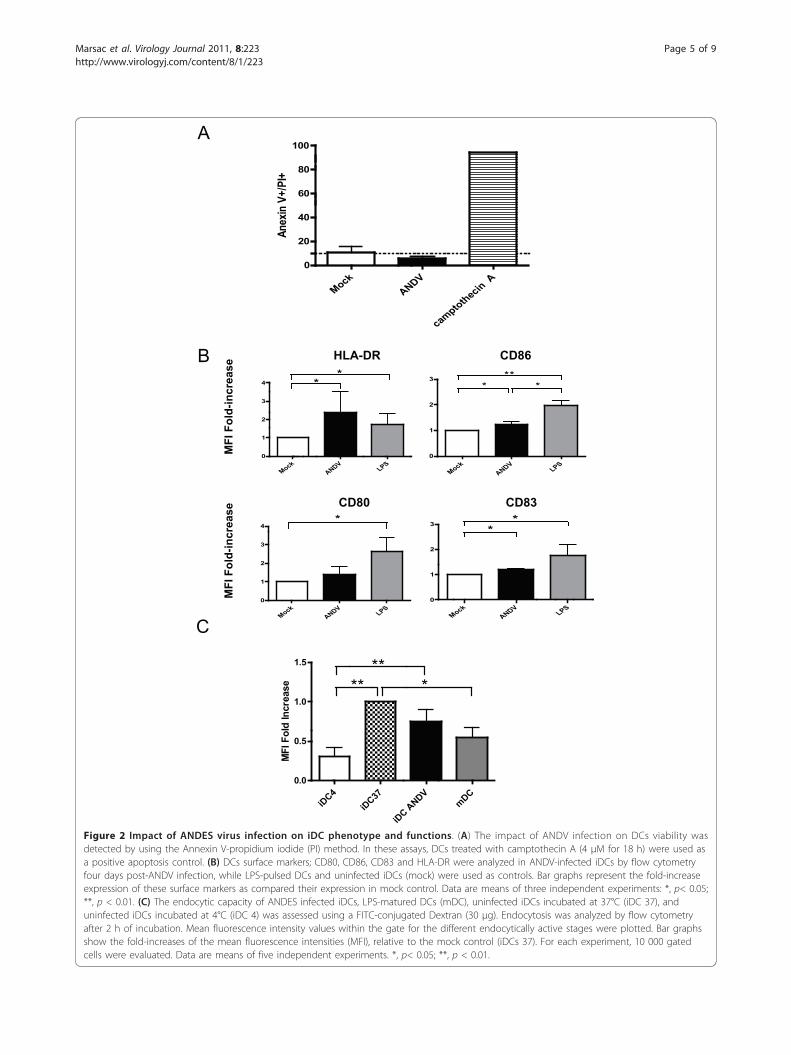
maturation and endocytosis was assessed. The apoptoticlevel, evaluated by flow cytometry using the Annexin Vassay [17], showed that infection of iDCs with ANDV doesnot affect cell viability (Figure 2A), observations in linewith what has been reported for other hantaviruses [6,22].In addition, ANDV infection did not induce any detectablecytopathic effect (data not shown). DCs play a pivotal roleas antigen-presenting cells in the antiviral immuneresponse. It is known that infection of iDCs by diverseviruses stimulates cell homing to inflammatory sites aswell as their maturation into antigen-presenting cells(APC), a process essential for the initiation and modula-tion of T cell-mediated immune responses. Hence, wenext evaluated whether ANDV infection of iDCs had adirect effect on the expression of key cell surface proteinsCD80, CD83, CD86 and HLA-DR, known to be associatedwith a mature DCs (mDCs) phenotype. Immature DCswere infected with ANDV as described above. ANDVinfection of iDCs induced a significant increase of HLA-DR, a marker protein that plays a pivotal role in guidingthe development and activation of CD4+ T helper cells.Markers CD83 and CD86 also increased, albeit to a muchlower extent than in LPS-pulsed iDCs conditions. Asexpected, expression of all surface markers increased inLPS-pulsed iDCs as compared to mock-DCs (Figure 2B).These data suggest that ANDV infection of human DCsinduced cell maturation, and that ANDV infection altersthe expression of HLA-DR on DCs. Constitutive expres-sion of HLA-DR molecules is associated to professionalantigens presenting cells (APCs) such as DCs, this basalexpression can be enhanced in an environment enrichedwith proinflammatory cytokines [7,23].Immature DCs efficiently capture and process antigen, a
characteristic cell function that decreases with maturation[7,8,24-27]. Therefore, we next sought to establish whetherthe partial DC maturation phenotype (Figure 2B) was cor-related with its decreased ability to capture antigens. To
Marsac et al. Virology Journal 2011, 8:223http://www.virologyj.com/content/8/1/223
Page 3 of 9

A
C
B300bp
200bp
Moc
k / V
ero-
E6AN
DV
/ DC
sM
ock
/ DC
sAN
DV
/ Ver
o-E6
Figure 1 Detection of ANDV infection of primary human dendritic cells. (A) Vero E6 epithelial cells and human immature DCs infected withADNV (strain CHI-7913). ANDV-nucleocapsid (N) protein detected by IFA by incubating ANDV-infected with a mouse anti-ANDV N MAb revealedby an FITC-conjugated anti-mouse IgG Ab (Green, right column) while the corresponding cell nuclei were stained with 4’,6-diamidino-2-phenylindole (DAPI, blue, left column). Negative control (mock) cells were incubated with supernatant from uninfected Vero-E6 cells. (B) TotalRNA was extracted from uninfected LPS-Pulsed DCs (lane 1), ANDV-infected LPS-pulsed DCs (lane 2), uninfected iDCs (lane 3), ANDV-infectediDCs (lane 4), ANDV-infected Vero-E6 cells (lane 5), and used as template in a RT-PCR reaction designed to specifically amplify the viral S RNA.This assay also included a negative RT-PCR control (lane 6). MW is a molecular weight marker (1 Kb, Fermentas, Burlington, Canada). (C) DCsgenerated from primary monocytes, recovered from four healthy donors, were incubated with ANDV (lanes 1, 4, 7, 10), UV-irradiated ANDV (lanes2, 5, 8, 11) or pulsed with LPS (lanes 3, 6, 9, 12). Total RNA was extracted from cell supernatants and used as a template in a RT-PCR reactiondesigned to specifically amplify the viral S RNA. MW is a molecular weight marker (100 pb, Fermentas).
Marsac et al. Virology Journal 2011, 8:223http://www.virologyj.com/content/8/1/223
Page 4 of 9
A
MockANDV
camptothecin A0
20
40
60
80
100
Anex
in V
+/PI
+
B
iDC4
iDC37
iDC A
NDVmDC
0.0
0.5
1.0
1.5
** ***
MFI
Fol
d In
crea
se
CMock
ANDVLPS
0
1
2
3
4*
MockANDV
LPS0
1
2
3 **
MockANDV
LPS0
1
2
3* *
**
MockANDV
LPS0
1
2
3
4 **
MFI F
old In
crea
se
CD83CD80
CD86HLA-DR
MFI
Fol
d-in
crea
se
MFI
Fol
d-in
crea
se
Figure 2 Impact of ANDES virus infection on iDC phenotype and functions. (A) The impact of ANDV infection on DCs viability wasdetected by using the Annexin V-propidium iodide (PI) method. In these assays, DCs treated with camptothecin A (4 μM for 18 h) were used asa positive apoptosis control. (B) DCs surface markers; CD80, CD86, CD83 and HLA-DR were analyzed in ANDV-infected iDCs by flow cytometryfour days post-ANDV infection, while LPS-pulsed DCs and uninfected iDCs (mock) were used as controls. Bar graphs represent the fold-increaseexpression of these surface markers as compared their expression in mock control. Data are means of three independent experiments: *, p< 0.05;**, p < 0.01. (C) The endocytic capacity of ANDES infected iDCs, LPS-matured DCs (mDC), uninfected iDCs incubated at 37°C (iDC 37), anduninfected iDCs incubated at 4°C (iDC 4) was assessed using a FITC-conjugated Dextran (30 μg). Endocytosis was analyzed by flow cytometryafter 2 h of incubation. Mean fluorescence intensity values within the gate for the different endocytically active stages were plotted. Bar graphsshow the fold-increases of the mean fluorescence intensities (MFI), relative to the mock control (iDCs 37). For each experiment, 10 000 gatedcells were evaluated. Data are means of five independent experiments. *, p< 0.05; **, p < 0.01.
Marsac et al. Virology Journal 2011, 8:223http://www.virologyj.com/content/8/1/223
Page 5 of 9

this purpose, we studied the effect of ANDV infection onthe endocytic capacity of DCs by measuring the up-take ofFITC-conjugated Dextran at 37°C [16]. Results indicatethat ANDV-infected DC exhibited a reduced endocyticcapacity as compared to uninfected iDCs (Figure 2C).However, in our conditions, infected DCs exhibited ahigher endocytic activity than the used dose of LPS topulse iDCs (positive control). Together, these observationssuggest that ANDV infection does indeed reduce antigencapture by DCs, supporting a role of ANDV infection incell maturation.The presence of high levels of pro-inflammatory cyto-
kines both in plasma and lungs have been reported aspathological markers associated with HCPS in humans.These excesses of pro-inflammatory cytokines are secretedin patients by both hantavirus-activated macrophages andspecific T cells [28]. Particularly, the levels of the pro-inflammatory cytokine TNF-a in HCPS patient sera are thedramatically elevated [29,30]. Infection of iDCs by otherHantaviruses induces the production of pro-inflammatorycytokines as well [7]. Therefore, we sought to evaluatewhether ANDV infection induces a similar DC phenotype.Supernatants of Mock-DCs, ANDV-infected DCs (3 hpost-viral infection), and LPS-pulsed DCs were assessed forthe production of the pro-inflammatory cytokine, TNF-a.In agreement with what has been described for other Han-taviruses, supernatants harvested from ANDV-infectedDCs display a significant increase in TNF-a as comparedwith supernatants from uninfected iDCs (Figure 3A).In a similar experimental setting, we also evaluated theexpression of IL-10 and TGF-b, two anti-inflammatorycytokines [31]. Interestingly, in supernatants harvestedfrom ANDV-infected DCs, IL-10 levels were weakly alteredas compared with uninfected DCs, while TGF-b levelsdecrease was more pronounced (Figure 3B and 3C). Thus,our observations suggest that ANDV-infected DCs exhibita pro-inflammatory cytokine profile.Upon antigen capture, DCs undergo a process of
maturation and a lymph nodes homing, where they acti-vate the adaptive immune system. The process of DCtrafficking is complex. This phenomenon requires theenhancement of the vascular permeability, which couldinvolve the expression of several components, such ascell to cell junction proteins including VE-cadherin,PECAM-1, occludin, claudins, as well as soluble factors,including, IL-1b, TNF-a, VEGF, and Kinins [32-34]. Thegelatinolytic matrix metalloproteases (gMMP)-2 and 9are reported as one of the major actors of this crucialphenomenon during fetal development and wound heal-ing [35]. Interestingly, factors such as the inflammatorycytokines IL-8, TNF-a and IL-1b as well as VEGF [36]are in the upstream of the gMMP expression pathway inhuman monocytes [37]. Additionally, even if iDC pro-duces gMMP, DC increase the MMP-9 production along
Mock
ANDVLPS
0.0
0.5
1.0
1.5* **
IL-1
0 Fo
ld In
crea
se
Mock
ANDVLP
S0.0
0.5
1.0
1.5
*
**
TGF-
Fol
d In
crea
se
B
C
Mock
ANDVLPS
0
50
100
150 *
*
TNF-
(pg/
ml)
A
Figure 3 Inflammatory state of ANDV-infected iDCs. The level ofTNF-a (A) IL-10 (B), and TGF-b (C) in supernatants of uninfectedDCs (Mock), ANDV-infected iDCs (MOI = 1) (ANDV), or LPS-pulsedDCs, was assessed by ELISA respectively at 3 h (A) and 48 h (B andC) post-ANDV infection. In (B) and (C), bar graphs depict the fold-increase of cytokine production relatively to mock. Data are meansof five to eight independent experiments *, p< 0.05; **, p < 0.01.
Marsac et al. Virology Journal 2011, 8:223http://www.virologyj.com/content/8/1/223
Page 6 of 9

DC maturation progression [38]. Interestingly, certainviruses can play opposite roles in DC maturation, forexample the human Cytomegalovirus (hCMV) inhibitscell maturation to escape the immune system (4),whereas some others, such as HIV-1 [18], Dengue virus[11], and West Nile virus [39], enhance DC maturation,the secretion of active gMMP-9, as well as plasma vascu-lar leakage. Thus, to extend our previous observation onthe fact that DCs have a proinflammatory profile that canbe compatible with the secretion of gMMP-9, we ana-lyzed the expression and activity of gMMP-9 in cells andits presence in DC supernatants (SN). Selection seemedadequate as the levels of IL-10, which has been reportedto inhibit MMP-9 induction, is weakly altered by ADNVinfection (Figure 3B) and DC maturation is known toincrease the level of active MMP-9 secretion. Intra-cellu-lar expression of gMMP-9 in ANDV-infected DCs (3 h-post infection) was characterized by Western blotting(Figure 4A), using a mouse an anti-gMMP-9 MAb. Datashow that gMMP-9 expression is indeed elevated inANDV-infected DCs as compared with mock iDCs(Figure 4A). Gelatinolytic MMP-9 is secreted as a proen-zyme, which remains inactive unless it is activated bythe removal of the propeptide domain by proteolyticenzymes. The gelatinolytic activity of gMMP-9 was there-fore assessed in cell supernatants (SN) by gelatin zymo-graphy [18]. Strikingly, ANDV-infected DC supernatantsexhibited an elevated gMMP-9 activity as compared withSN-mock (Figure 4B). Furthermore, gMMP-9 activitywas similar to that obtained by the LPS-induction of DCmaturation (SN-LPS; Figure 4B).ANDV belongs to the group of hemorrhagic fever
viruses known for inducing different forms of vascularleakage in different organs. For some of these hemorrhagicfever or encephalitic viruses the mechanism associated totheir capacity to enhance endothelial permeability in vitroand in vivo has been reported [11,18,39]. Upon viral infec-tion, the vascular permeability phenomenon is principallydue to viral-dependent dysfunctions of endothelial cellsjunctions that can be directly induced by the action of fac-tors from viral activated endothelial cells, such as VEGF orindirectly induced by factors from immune viral-activatedcells, via the MAP-kinase pathway [11,18]. For these rea-sons, we wonder if ANDV infection induced the produc-tion of soluble factors that could indirectly enhanceendothelial permeability. To evaluate this possibility, thecapacity of ANDV infected DCs-supernatants to modifythe permeability was measured through the amounts ofFITC-conjugated dextran that passed thru a monolayer ofprimary human umbilical vascular endothelial cells(HUVEC) seeded onto transwell plates that considered asvascular model [19]. Following the exposure of confluentHUVEC to SN from ANDV infected DCs, or to controls,the amounts of FITC-conjugated dextran detected in the
A
SN Mock
SN ANDV
SN LPS
0.0
0.5
1.0
1.5
2.0*
MM
P-9
gel
atin
ase
acti
vity
(fo
ld in
crea
se)
B
C
SN Moc
k
SN ANDV
TNF-
a0.0
0.5
1.0
1.5
2.0
**Fl
uore
scen
ce in
tens
ity(f
old
incr
ease
)
Figure 4 Secretion of active gMMP-9 and endothelialpermeability enhancement are triggered by ANDV infection ofiDCs. (A) The gMMP-9 expression in ANDV-infected iDCs wasassessed by Western blotting. Uninfected iDCs (Mock), ANDV-infected iDCs (ANDV) and LPS-treated iDCs (LPS) were collected 3 hpost-infection. Forty micrograms of total proteins from cell lysateswere separated in an 8% SDS-PAGE gel. Following an electricalprotein transfer onto a nitrocellulose membrane, an anti-gMMP-9antibody was used to probe the presence of gMMP-9 that wasrevealed using an HRP-conjugated anti-mouse IgG Ab. (B)Supernatants from ANDV-infected DCs or Mock cells collected 3 hpost-infection. Gelatinase activity was assayed by zymography.Gelatinolytic activity was quantified by gel densitometry using theImage J Software. Data are presented as the fold-increase of gMMP-9 activity in supernatants. Statistical significance (**, p < 0.01) wasdetermined from five independent experiments. (C) Enhancementof the endothelial cells permeability induced by supernatant fromANDV-infected iDCs. HUVEC confluent monolayers plated ontocollagen-coated transwell inserts were incubated with either ANDVsupernatant, mock control (uninfected DCs supernatant) or withTNF-a (50 ng/ml) as positive control. Following 18 h at 37°C in CO25% after addition, within the top chamber, of 500 μg/ml FITC-conjugated Dextran, paracellular permeability was measured byreading in the bottom chamber containing the infiltrated FITC-dextran at an excitation wavelength of 485 nm and an emission of530 nm. Data represent means of five independent experiments. **,p < 0.01.
Marsac et al. Virology Journal 2011, 8:223http://www.virologyj.com/content/8/1/223
Page 7 of 9

lower chamber of the transwell system (Figure 4C) suggestthat cell supernatant from ANDV-infected DCs containssoluble and active factors favoring the increase of theHUVEC monolayer permeability.Altogether, the in vitro data presented herein show
that ANDV infects and replicates in DCs, inducing apartial maturation and pro-inflammatory phenotype,while increasing the expression and secretion of activegMMP-9 as well as that supernatants from ANDV-infected DCs were able to enhance the permeability of amonolayer of primary human vascular endothelial cells.
ConclusionThe herein presented data and data recently published inthe literature are not only convergent but also comple-mentary, they allow to speculate that both indirect effectsof ANDV on DCs and direct effects of pathogenic hanta-viruses, such as ANDV, on endothelial cells generateconjugated inflammatory consequences that result in theincreased endothelial permeability associated to this virusinfection. A better understanding of major mechanismsinvolved in vascular pathogenesis observed in HCPS ispivotal to progress in the design of therapies that todayremain unavailable for this serious life-threateninghemorrhagic viral disease.
AcknowledgementsWe thank Dr. M. Rau (Oxford, UK) for critical reading and editing of themanuscript. We are grateful to Dr. Nicole Tischler, Fundación Ciencias para laVida, Santiago-Chile, for kindly providing the anti-N MAb used in this study.We thank colleagues from the Pontificia Universidad Católica de Chile: Prof.Roberto Ebenspenger and Lorena Rubio for kindly providing HUVECs;Connie Martinez for titrated viruses. The present study was supported byCONICYT through grant FONDECYT-POSTDOCTORADO-2008-3085029 to DM,FONDECYT N° 1100756, and PHS grant 2U01AI045452-11 to MLL, ProyectoInstituto Milenio P-07-088-F to AK and MLL, Chile. We also thank IRD andUniversity Montpellier 1, France, for supporting a part of this study.
Author details1Pontificia Universidad Católica de Chile, Millennium Institute onImmunology and Immunotherapy, Laboratorio de Virología Molecular,Facultad de Medicina, Santiago, Chile. 2UMR-MD3-University Montpellier 1,Comparative Molecular Immuno-Physiopathology Lab, Faculté de Pharmacie,34093 Montpellier, France. 3Universidad de Chile, Laboratorio deComunicaciones Celulares. Centro de Estudios Moleculares de la Célula,Facultad de Medicina, Santiago, Chile. 4Pontificia Universidad Católica deChile, Laboratorio de Infectologia, Centro de Investigaciones Medicas,Facultad de Medicina, Santiago, Chile. 5Pontificia Universidad Católica deChile, Millennium Institute on Immunology and Immunotherapy.Departamento de Genética Molecular y Microbiología, Facultad de CienciasBiológicas, Santiago, Chile.
Authors’ contributionsDM carried out the BSL3 experiments, hantavirus cultivation, and most ofthe experiments described in this paper, manuscript draft; SG participated toall of those experiments; AF zymograms and experimental permeabilitystudies set-up. AA performed the statistical analysis. KP RT-PCR; MF BSL-3facilities; AK DCs PBMC-derived and draft revision; MLL data analysis andmanuscript revision; FV designed the project, data interpretation, manuscriptrevisions, finalized and approved the manuscript. All authors read andapproved the final manuscript.
Authors’ informationsDM was a FONDECYT post-doctorate fellowship; SG is PhD student and KP isa technician at the MLL lab; AF is from the technical staff of the FV lab.
Competing interestsThe authors declare that they have no competing interests.
Received: 1 April 2011 Accepted: 13 May 2011 Published: 13 May 2011
References1. Hjertqvist M, Klein SL, Ahlm C, Klingstrom J: Mortality rate patterns for
hemorrhagic fever with renal syndrome caused by Puumala virus. EmergInfect Dis 16:1584-1586.
2. Nolte KB, Feddersen RM, Foucar K, Zaki SR, Koster FT, Madar D, Merlin TL,McFeeley PJ, Umland ET, Zumwalt RE: Hantavirus pulmonary syndrome inthe United States: a pathological description of a disease caused by anew agent. Hum Pathol 1995, 26:110-120.
3. Xiao R, Yang S, Koster F, Ye C, Stidley C, Hjelle B: Sin Nombre viral RNAload in patients with hantavirus cardiopulmonary syndrome. J Infect Dis2006, 194:1403-1409.
4. Maes P, Clement J, Gavrilovskaya I, Van Ranst M: Hantaviruses:immunology, treatment, and prevention. Viral Immunol 2004, 17:481-497.
5. Mertz GJ, Hjelle BL, Bryan RT: Hantavirus infection. Adv Intern Med 1997,42:369-421.
6. Pensiero MN, Sharefkin JB, Dieffenbach CW, Hay J: Hantaan virus infectionof human endothelial cells. J Virol 1992, 66:5929-5936.
7. Raftery MJ, Kraus AA, Ulrich R, Kruger DH, Schonrich G: Hantavirusinfection of dendritic cells. J Virol 2002, 76:10724-10733.
8. Zaki SR, Greer PW, Coffield LM, Goldsmith CS, Nolte KB, Foucar K,Feddersen RM, Zumwalt RE, Miller GL, Khan AS, et al: Hantaviruspulmonary syndrome. Pathogenesis of an emerging infectious disease.Am J Pathol 1995, 146:552-579.
9. Sathupan P, Khongphattanayothin A, Srisai J, Srikaew K, Poovorawan Y: Therole of vascular endothelial growth factor leading to vascular leakage inchildren with dengue virus infection. Ann Trop Paediatr 2007, 27:179-184.
10. Srikiatkhachorn A, Ajariyakhajorn C, Endy TP, Kalayanarooj S, Libraty DH,Green S, Ennis FA, Rothman AL: Virus-induced decline in soluble vascularendothelial growth receptor 2 is associated with plasma leakage indengue hemorrhagic Fever. J Virol 2007, 81:1592-1600.
11. Luplertlop N, Misse D, Bray D, Deleuze V, Gonzalez JP, Leardkamolkarn V,Yssel H, Veas F: Dengue-virus-infected dendritic cells trigger vascularleakage through metalloproteinase overproduction. EMBO Rep 2006,7:1176-1181.
12. Gorbunova E, Gavrilovskaya IN, Mackow ER: Pathogenic hantavirusesAndes virus and Hantaan virus induce adherens junction disassembly bydirecting vascular endothelial cadherin internalization in humanendothelial cells. J Virol 84:7405-7411.
13. Shrivastava-Ranjan P, Rollin PE, Spiropoulou CF: Andes virus disrupts theendothelial cell barrier by induction of vascular endothelial growthfactor and downregulation of VE-cadherin. J Virol 84:11227-11234.
14. Sallusto F, Lanzavecchia A: Efficient presentation of soluble antigen bycultured human dendritic cells is maintained by granulocyte/macrophage colony-stimulating factor plus interleukin 4 anddownregulated by tumor necrosis factor alpha. J Exp Med 1994,179:1109-1118.
15. Tischler ND, Rosemblatt M, Valenzuela PD: Characterization of cross-reactive and serotype-specific epitopes on the nucleocapsid proteins ofhantaviruses. Virus Res 2008, 135:1-9.
16. Lutz MB, Rovere P, Kleijmeer MJ, Rescigno M, Assmann CU, Oorschot VM,Geuze HJ, Trucy J, Demandolx D, Davoust J, Ricciardi-Castagnoli P:Intracellular routes and selective retention of antigens in mildly acidiccathepsin D/lysosome-associated membrane protein-1/MHC class II-positive vesicles in immature dendritic cells. J Immunol 1997,159:3707-3716.
17. Nicoletti I, Migliorati G, Pagliacci MC, Grignani F, Riccardi C: A rapid andsimple method for measuring thymocyte apoptosis by propidium iodidestaining and flow cytometry. J Immunol Methods 1991, 139:271-279.
18. Misse D, Esteve PO, Renneboog B, Vidal M, Cerutti M, St Pierre Y, Yssel H,Parmentier M, Veas F: HIV-1 glycoprotein 120 induces the MMP-9cytopathogenic factor production that is abolished by inhibition of the
Marsac et al. Virology Journal 2011, 8:223http://www.virologyj.com/content/8/1/223
Page 8 of 9

p38 mitogen-activated protein kinase signaling pathway. Blood 2001,98:541-547.
19. Jaffe EA, Nachman RL, Becker CG, Minick CR: Culture of human endothelialcells derived from umbilical veins. Identification by morphologic andimmunologic criteria. J Clin Invest 1973, 52:2745-2756.
20. Galeno H, Mora J, Villagra E, Fernandez J, Hernandez J, Mertz GJ, Ramirez E:First human isolate of Hantavirus (Andes virus) in the Americas. EmergInfect Dis 2002, 8:657-661.
21. Godoy P, Marsac D, Stefas E, Ferrer P, Tischler ND, Pino K, Ramdohr P, Vial P,Valenzuela PD, Ferres M, et al: Andes virus antigens are shed in urine ofpatients with acute hantavirus cardiopulmonary syndrome. J Virol 2009,83:5046-5055.
22. Yanagihara R, Silverman DJ: Experimental infection of human vascularendothelial cells by pathogenic and nonpathogenic hantaviruses. ArchVirol 1990, 111:281-286.
23. Holling TM, Schooten E, van Den Elsen PJ: Function and regulation ofMHC class II molecules in T-lymphocytes: of mice and men. HumImmunol 2004, 65:282-290.
24. Morelli AE, Thomson AW: Dendritic cells: regulators of alloimmunityand opportunities for tolerance induction. Immunol Rev 2003,196:125-146.
25. Gavrilovskaya IN, Peresleni T, Geimonen E, Mackow ER: Pathogenichantaviruses selectively inhibit beta3 integrin directed endothelial cellmigration. Arch Virol 2002, 147:1913-1931.
26. Hayasaka D, Maeda K, Ennis FA, Terajima M: Increased permeability ofhuman endothelial cell line EA.hy926 induced by hantavirus-specificcytotoxic T lymphocytes. Virus Res 2007, 123:120-127.
27. Temonen M, Vapalahti O, Holthofer H, Brummer-Korvenkontio M, Vaheri A,Lankinen H: Susceptibility of human cells to Puumala virus infection. JGen Virol 1993, 74(Pt 3):515-518.
28. Mori M, Rothman AL, Kurane I, Montoya JM, Nolte KB, Norman JE,Waite DC, Koster FT, Ennis FA: High levels of cytokine-producing cells inthe lung tissues of patients with fatal hantavirus pulmonary syndrome. JInfect Dis 1999, 179:295-302.
29. Borges AA, Campos GM, Moreli ML, Moro Souza RL, Saggioro FP,Figueiredo GG, Livonesi MC, Moraes Figueiredo LT: Role of mixed Th1 andTh2 serum cytokines on pathogenesis and prognosis of hantaviruspulmonary syndrome. Microbes Infect 2008, 10:1150-1157.
30. Linderholm M, Ahlm C, Settergren B, Waage A, Tarnvik A: Elevated plasmalevels of tumor necrosis factor (TNF)-alpha, soluble TNF receptors,interleukin (IL)-6, and IL-10 in patients with hemorrhagic fever withrenal syndrome. J Infect Dis 1996, 173:38-43.
31. Carvajal CA, Herrada AA, Castillo CR, Contreras FJ, Stehr CB, Mosso LM,Kalergis AM, Fardella CE: Primary aldosteronism can alter peripheral levelsof TGF-betaand TNF-alpha. J Endocrinol Invest 2009, 32:759-65.
32. Donmez G, Sullu Y, Baris S, Yildiz L, Aydin O, Karagoz F, Kandemir B:Vascular endothelial growth factor (VEGF), matrix metalloproteinase-9(MMP-9), and thrombospondin-1 (TSP-1) expression in urothelialcarcinomas. Pathol Res Pract 2009, 205:854-857.
33. Imamura T, Tanase S, Hayashi I, Potempa J, Kozik A, Travis J: Release of anew vascular permeability enhancing peptide from kininogens byhuman neutrophil elastase. Biochem Biophys Res Commun 2002,294:423-428.
34. Lehmann W, Edgar CM, Wang K, Cho TJ, Barnes GL, Kakar S, Graves DT,Rueger JM, Gerstenfeld LC, Einhorn TA: Tumor necrosis factor alpha (TNF-alpha) coordinately regulates the expression of specific matrixmetalloproteinases (MMPS) and angiogenic factors during fracturehealing. Bone 2005, 36:300-310.
35. Bartholome EJ, Van Aelst I, Koyen E, Kiss R, Willems F, Goldman M,Opdenakker G: Human monocyte-derived dendritic cells producebioactive gelatinase B: inhibition by IFN-beta. J Interferon Cytokine Res2001, 21:495-501.
36. Valable S, Montaner J, Bellail A, Berezowski V, Brillault J, Cecchelli R,Divoux D, Mackenzie ET, Bernaudin M, Roussel S, Petit E: VEGF-inducedBBB permeability is associated with an MMP-9 activity increase incerebral ischemia: both effects decreased by Ang-1. J Cereb Blood FlowMetab 2005, 25:1491-1504.
37. Saren P, Welgus HG, Kovanen PT: TNF-alpha and IL-1beta selectivelyinduce expression of 92-kDa gelatinase by human macrophages. JImmunol 1996, 157:4159-4165.
38. Osman M, Tortorella M, Londei M, Quaratino S: Expression of matrixmetalloproteinases and tissue inhibitors of metalloproteinases definethe migratory characteristics of human monocyte-derived dendritic cells.Immunology 2002, 105:73-82.
39. Verma S, Lo Y, Chapagain M, Lum S, Kumar M, Gurjav U, Luo H,Nakatsuka A, Nerurkar VR: West Nile virus infection modulates humanbrain microvascular endothelial cells tight junction proteins and celladhesion molecules: Transmigration across the in vitro blood-brainbarrier. Virology 2009, 385:425-433.
doi:10.1186/1743-422X-8-223Cite this article as: Marsac et al.: Infection of human monocyte-deriveddendritic cells by ANDES Hantavirus enhances pro-inflammatory state,the secretion of active MMP-9 and indirectly enhances endothelialpermeability. Virology Journal 2011 8:223.
Submit your next manuscript to BioMed Centraland take full advantage of:
• Convenient online submission
• Thorough peer review
• No space constraints or color figure charges
• Immediate publication on acceptance
• Inclusion in PubMed, CAS, Scopus and Google Scholar
• Research which is freely available for redistribution
Submit your manuscript at www.biomedcentral.com/submit
Marsac et al. Virology Journal 2011, 8:223http://www.virologyj.com/content/8/1/223
Page 9 of 9
Copyright © 2022 FDOKUMEN