Low-intensity pulsed ultrasound enhances the rate of lateral ...
31
Low-intensity pulsed ultrasound enhances the rate of lateral tooth movement and compensatory bone formation in rats. Chihiro Arai 1 , Nobuhiko Kawai 2 , Yoshiaki Nomura 3 , Atsushi Tsuge 1 , Yoshiki Nakamura 1 , Eiji Tanaka 2,4 1 Department of Orthodontics, Tsurumi University School of Dental Medicine, Yokohama, Japan 2 Department of Orthodontics and Dentofacial Orthopedics, Institute of Biomedical Sciences, Tokushima University Graduate School, Tokushima, Japan 3 Department of Translation Research, Tsurumi University School of Dental Medicine, Yokohama, Japan 4 Department of Orthodontics, Faculty of Dentistry, King Abdulaziz University, Jeddah, Saudi Arabia Author Contributions Conceived and designed the experiments: CA, YNa. Performed the experiments: CA, AT. Analyzed the data: CA, NK, AT, YNo. Contributed reagent/materials/analysis tools: CA, NK. Performed the statistical analysis:YNo, Senior advisor: YNa, ET. Wrote the paper: CA, YNo, YNa, ET. *Title Page © 2019. This manuscript version is made available under the CC-BY-NC-ND 4.0 license http://creativecommons.org/licenses/by-nc-nd/4.0/ The published version is available via https://doi.org/10.1016/j.ajodo.2019.01.027.
-
Upload
khangminh22 -
Category
Documents
-
view
0 -
download
0
Transcript of Low-intensity pulsed ultrasound enhances the rate of lateral ...

Low-intensity pulsed ultrasound enhances the rate of lateral tooth movement
and compensatory bone formation in rats.
Chihiro Arai1, Nobuhiko Kawai2, Yoshiaki Nomura3, Atsushi Tsuge1, Yoshiki Nakamura1,
Eiji Tanaka2,4
1Department of Orthodontics, Tsurumi University School of Dental Medicine, Yokohama,
Japan
2Department of Orthodontics and Dentofacial Orthopedics, Institute of Biomedical
Sciences, Tokushima University Graduate School, Tokushima, Japan
3Department of Translation Research, Tsurumi University School of Dental Medicine,
Yokohama, Japan
4Department of Orthodontics, Faculty of Dentistry, King Abdulaziz University, Jeddah,
Saudi Arabia
Author Contributions
Conceived and designed the experiments: CA, YNa. Performed the experiments: CA, AT.
Analyzed the data: CA, NK, AT, YNo. Contributed reagent/materials/analysis tools: CA, NK.
Performed the statistical analysis:YNo, Senior advisor: YNa, ET. Wrote the paper: CA, YNo,
YNa, ET.
*Title Page
© 2019. This manuscript version is made available under the CC-BY-NC-ND 4.0 license http://creativecommons.org/licenses/by-nc-nd/4.0/The published version is available via https://doi.org/10.1016/j.ajodo.2019.01.027.

Address for correspondences:
Chihiro Arai
Department of Orthodontics, Tsurumi University School of Dental Medicine
2-1-3 Tsurumi, Tsurumi-ku, Yokohama 230-8501, Japan
Tel: +81-45-581-1001, ext. 8438; Fax: +81-45-582-8688
E-mail: [email protected]
Highlights
・ LIPUS enhances osteoclastogenesis in the pressure zone of the alveolar bone
during lateral tooth movement.
・ LIPUS increases compensatory bone formation on the buccal surface of the alveolar
bone during lateral tooth movement.
・ LIPUS improves the rate of lateral tooth movement.
*Highlights (for review)
1
Low-intensity pulsed ultrasound enhances the rate of lateral tooth movement 1
and compensatory bone formation in rats. 2
Running title: LIPUS effect on lateral tooth movement 3
Keywords 4
LIPUS; Orthodontic tooth movement; Osteoclastgenesis; 3D micro CT; 5
Bone histomorhometry 6
Abstract 7
Introduction: Because mechanical stimulation of the periodontal ligament (PDL) by 8
low-intensity pulsed ultrasound (LIPUS) has been shown to increase the speed of bone 9
remodeling, the aim of this study was to examine the effects of LIPUS stimulation on the 10
rate of tooth movement and bone remodeling during lateral tooth movement. Methods: 11
Twelve-week-old Wistar rats were divided into two groups. The LIPUS group received 12
experimental tooth movement with LIPUS stimulation, and the tooth movement (TM) 13
group had experimental tooth movement without LIPUS. For the LIPUS and TM groups, 14
the upper right first molars were moved labially with fixed appliances. LIPUS exposure 15
was placed in the region corresponding to the right maxillary first molar. Three days 16
after tooth movement, tartrate-resistant acid phosphatase (TRAP) was examined. 17
Fourteen days after tooth movement, the intermolar width, bone mineral content (BMC), 18
*Manuscript (no author identifiers please)Click here to view linked References

2
and bone volume fraction (BV/TV) were examined by micro computed tomography 1
(micro-CT), and newly formed bone was measured histomorphometrically. Results: The 2
number of TRAP-positive cells at the compressed region was obviously greater in the 3
LIPUS group. The intermolar width was significantly greater in the LIPUS group than in 4
the TM group. The alveolar bone around the maxillary first molar showed no differences 5
in BMC or BV/TV between the LIPUS and TM groups. The LIPUS group exhibited a 6
significantly greater amount of newly formed alveolar bone than the TM group. 7
Conclusions: The present study provides evidence of the beneficial effects of LIPUS on 8
the lateral tooth movement. 9
INTRODUCTION 10
Tissue remodeling surrounding tooth roots is essential to the rate of orthodontic tooth 11
movement. Therefore, it is important to control the molecular mechanisms by which the 12
behaviors of cells in the alveolar bone and periodontal ligament (PDL) are regulated. 1-3 13
The duration of orthodontic treatment is the primary concern for most patients and 14
orthodontists. Unfortunately, long-term orthodontic treatment induces several 15
disadvantages, such as a higher predisposition to dental caries, gingival recession, and 16
root resorption. Consequently, much attention has been paid to find the possible 17
remedies that increase the rate of tooth movement with the fewest possible 18

3
disadvantages. 1
To date, several novel modalities have been reported to accelerate orthodontic tooth 2
movement.4 Surgical modalities including corticotomy, dentoalveolar distraction and 3
periodontal distraction is based on the principle that when the bone is irritated which 4
causes increased osteoclastogenesis, the tooth moves faster.5-7 Meanwhile, several 5
non-surgical modalities have also been reported, such as low-level laser therapy, 6
electromagnetic fields, and mechanical vibration.4 In addition, mechanical stimulation of 7
the PDL by low-intensity pulsed ultrasound (LIPUS) has been shown to increase the 8
speed of bone remodeling; therefore, LIPUS is also considered as a non-surgical 9
modality for accelerating tooth movement.8 LIPUS has been proven to act by inducing 10
osteoclastogenesis by stimulating the receptor activator of nuclear factor 11
kappa-B(RANK)/RANK ligand (RANKL) pathway and activating signaling molecules 12
such as MAPK.9 Furthermore, the use of LIPUS is safe, but the very limited 13
research-based evidence cannot support a solid conclusion that it accelerates 14
orthodontic tooth movement. 15
Xue et al. revealed that LIPUS might promote alveolar bone remodeling via increasing 16
the gene expression of the human growth factor/Runx2/bone morphogenetic protein 2 17
signaling pathway molecules, resulting in the rapid movement of teeth during 18

4
orthodontic treatment.10 Recent study also showed that LIPUS enhanced the amount of 1
tooth movement and the bone remodeling during orthodontic tooth movement in rat.11 2
However, these results were only found in a mesial orthodontic tooth movement model. 3
Although lateral tooth movement is frequently conducted in the clinical situation, the 4
effect of LIPUS on lateral orthodontic tooth movement has not been fully examined. 5
Thus, the aim of this study was to examine the effects of LIPUS stimulation on the rate 6
of tooth movement and bone remodeling during lateral tooth movement. 7
MATERIAL and METHODS 8
All of the procedures described in this study were performed in accordance with the 9
guidelines and regulations of the ///// University for Animal Research (29A038). A total of 10
twenty-six 12-week-old male Wistar rats, weighing 320-350 g, were randomly divided 11
into two groups. The LIPUS group received experimental tooth movement with LIPUS 12
stimulation, and the tooth movement (TM) group had experimental tooth movement 13
without LIPUS. Each animal was anesthetized with a mixture of three types of 14
anesthetic agents at a dose of 2.5 ml/kg body weight.12 The combination anesthetic was 15
prepared with 0.15 mg/kg medetomidine (Domitol®; Nippon Zenyaku Kogyo Co., Ltd., 16
Tokyo, Japan), 2 mg/kg midazolam (Dormicum®; Astellas Pharma Inc., Tokyo, Japan), 17
and 2.5 mg/kg butorphanol (Vetorphale®; Meiji Seika Pharma Co., Ltd.). For the LIPUS 18
5
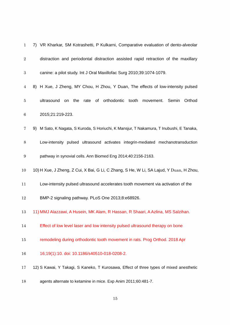
and TM groups, the upper right first molars were moved bucally with fixed appliances 1
(Fig 1, a-b). The initial force magnitude was approximately 10 g.13 2
An ultrasound exposure machine (Osteotron D2, ITO Co., Tokyo, Japan) was 3
employed in this study. This system was equipped with transducers with a circular 4
surface area of 9.6 cm2. The sound head of this device had an average beam 5
non-uniformity ratio (BNR) of 3.2-3.6:1 and an effective radiating area (ERA) of 90%. A 6
pulsed ultrasound signal was transmitted at a frequency of 1 MHz (the pulse repetition 7
frequency = 100 Hz), with an average spatial intensity of 30 mW/cm2 and a pulse of 1:4 8
(2 ms on and 8 ms off). The stimulation protocol, used in this study consisted of a 9
20-min LIPUS stimulation repeated every day. The rats were kept in an immovable 10
position under anesthesia, and the ultrasound transducer was placed in contact with 11
one side of the face, in the region corresponding to the right maxillary first molar. The fur 12
was shaved in the exposure region, and coupling gel was constantly in place in order to 13
optimize penetration of the ultrasound waves into the tissues. 14
Three days after tooth movement, 4% paraformaldehyde in 0.1M phosphate-buffered 15
saline (pH 7.4) was perfused for 15 min through the ascending aortae of ten rats from 16
the experimental and control groups, respectively. After fixation, the maxillae were 17
dissected and trimmed into small blocks containing the first molar, decalcified with 18

6
EDTA-Na (5.0%, pH 7.2, 4oC) solution containing 7.0% sucrose for 4 weeks, 1
dehydrated with a graded ethanol series, and embedded in paraffin. The serial sections 2
(7 μm) were cut perpendicular to the root axis. TRAP activity was examined in the 3
sections, using a TRAP staining kit (Wako, Tokyo, Japan). An area measuring 700 × 4
2400 μm2 was selected from the section for light microscopic examination (BZ-9000; 5
Keyence, Osaka, Japan) according to previous study (Fig 4, a and b).14 TRAP-positive 6
multinucleated osteoclasts on the pressure zone of the upper first molar was counted on 7
three sections for each specimen. The average of the three values was used in this 8
study. 9
Sixteen rats served to measure tooth movement and to analyze the bone properties 10
in micro-CT analysis. After 14 days of tooth movement, the micro-CT (The inspeXio 11
SMX-225CT, SHIMADZU Co., Kyoto, Japan) images were taken. The tube voltage was 12
set at 160kV and the current was constant at 70 μA. The resolution was set at 20 μm 13
per voxel and 1024 × 1024 pixels. On the three-dimensional (3D) models, the distance 14
between the distolingual cusps of the maxillary first molars was measured as the 15
intermolar width (Fig 2, a). The Region of interest (ROI) was alveolar bone proper of 16
the maxillary first molar (Fig 2, b-d). 17
Each ROI was measured with respect to bone mineral content (BMC; mg) and bone 18

7
volume fraction (BV/TV; %). Tissue volume (TV) was defined as the volume of tissue in 1
the enlarged ROI. Bone volume (BV) was excluded of the teeth (Fig 2, d). The 2
inter-molar width and bone parameters were measured by three-dimensional 3
image-analysis software (TRI/3D-BON; Ratoc System Engineering, Tokyo, Japan). 4
Sixteen rats were intraperitoneally injected with 0.1 mL of calcein (1.6 mg/kg) solution 5
one day before tooth movement and with xylenol orange (50 mg/kg) one day before the 6
end of tooth movement as fluorochrome labels. At day 15, the animals were sacrificed 7
under anesthesia with pentobarbital sodium at a fatal overdose of 50 mg/kg. After the 8
micro-CT images were taken, the maxillae were dissected, cut in half along the sagittal 9
plane, and immersed rapidly in liquid nitrogen. The frozen tissues were embedded with 10
optical cutting temperature compound (Miles Inc., Torrance, CA, USA). Frozen blocks 11
from the rats were frontally sectioned and 7-μm-thick serial sections were used for 12
histomorphometric analysis. The sections were prepared according to Kawamoto’s film 13
method.14 Newly formed bone was measured as the distance between the calcein and 14
xylenol orange lines under a fluorescence microscope (BZ-9000; Keyence, Osaka, 15
Japan), which were visible as green and red marks, respectively, at 50 μm, 150 μm and 16
300 μm from the alveolar crest, the average of these three morphometric values was 17
used for evaluation. 18

8
Statistical analysis 1
To determine the sample size of the experiments, power analysis was performed to 2
detect statistically significant differences in each experiment between lateral tooth 3
movement with or without LIPUS exposure during the experimental period was 4
determined (α = 0.05 and β = 0.80). The power analysis was performed S Plus ver. 5
6.0 ( NTT Data Tokyo, Japan). 6
Normality of variables were assessed by the Kolmogorov - Smirnov test separately by 7
the control and LIPUS groups. According to the normality, t tests or Mann-Whitney U 8
tests were used to detect the statistical significance between two groups. 9
For the assessment of reliability intermolar width measurements, samples were 10
measured by two examiners under masking experimental conditions. Measurements 11
were repeated after 2 weeks by the same examiner. A paired t test showed no 12
significant differences between the 2 repeated measurements (p=0.689). And intraclass 13
correlation coefficients evaluated by measured value (mean) was 0.967(95% CI: 14
0.912-0.989). P-value less than 0.05 was considered to be statistically significant. 15
Analysis except for the power analysis were carried out by SPSS Statistics ver. 25.0 16
(IBM, Tokyo, Japan). 17
RESULTS 18

9
During the tooth movement, no significant differences in body weight were found 1
between the two groups (Fig 3, a). Inter-molar width was significantly (p<0.05) greater 2
in the LIPUS group than in the TM group (Fig 3, b and Table). 3
The number of TRAP-positive cells at the compressed region was clearly greater in 4
the LIPUS group than in the control group (Fig 4, a and b). Furthermore, the number of 5
osteoclasts was significantly increased by the LIPUS exposure (Fig 4, c and Table). 6
Fluorescence microscopy revealed bone labelling in the section. Sharp, bright calcein 7
and xylenol orange labeling lines were observed in the alveolar bone on the periosteal 8
sides in each group. The width between these two lines represented the formation of 9
new bone during the experimental period. In the untreated control group without tooth 10
movement, two lines were observed in the alveolar crest area and lower part of the 11
alveolar bone (Fig 5, a). On the other hand, the TM group showed two parallel lines 12
located along the periosteal bone surface in the alveolar bone, representing 13
compensatory bone formation in response to the lateral tooth movement (Fig 5, b). The 14
LIPUS group also showed the same observations, but the width between the lines was 15
much greater than that in the TM group (Fig 5, c). The distance between the lines in the 16
LIPUS group was significantly wider than that in the TM group (Fig 5, d and Table). 17
The alveolar bone labeling on the periodontal side showed quite different 18

10
characteristics. Double calcein and xylenol orange labelling was scattered in the 1
alveolar bone on the periodontal side in the control group, but only a calcein labelling 2
was recognized in the TM and LIPUS groups. The lack of green labelling suggests that 3
the alveolar bone on the periodontal side was resorbed during the lateral tooth 4
movement. 5
The micro-CT images showed no differences in the vertical height of the alveolar 6
crest on the buccal side in the TM and LIPUS groups compared with the untreated 7
control group without tooth movement (Fig 6, a-c). In addition, the alveolar bone 8
surrounding the maxillary first molar was not significantly different in terms of the BMC 9
or BV/TV values between the LIPUS and TM groups (Fig 6, d-e). 10
DISCUSSION 11
To the best of knowledge, this is the first attempt to investigate the effects of LIPUS on 12
acceleration of tooth movement and compensatory bone formation during lateral tooth 13
movement. Our results clearly showed that the oseteoclastgenesis in the pressure zone 14
of the PDL was enhanced by LIPUS exposure (Fig 4, a-c). It is well known that RANKL 15
is an important factor in osteoclastgenesis16,17 and that it is expressed in the pressure 16
side of the PDL during orthodontic tooth movement.18 LIPUS induces 17
osteoclastogenesis by upregulation of the RANK/RANKL pathway and signaling 18

11
molecules such as MAPK.9 In addition, it has been shown that RANKL expression and 1
the number of osteoclasts were increased by LIPUS stimulation in the pressure side of 2
the PDL during orthodontic tooth movement in rats.10 Thus, LIPUS might enhance 3
induction of RANKL mediated osteoclastgenesis in the pressure side of the PDL during 4
lateral tooth movement, consequentrly accelerating tooth movement. 5
Shimpo et al. have reported that alveolar bone is reactive to orthodontic stimuli, which 6
induce periosteal bone formation in the palatal surface of the alveolar bone during 7
lateral tooth movement.19 The present results also demonstrated that compensatory 8
bone formation was specifically observed in the buccal surface of alveolar bone during 9
lateral tooth movement (Fig 5, b). However, the mechanism of this bone formation is 10
unclear. Nonetheless, it is known that the orthodontic force on a tooth is considered to 11
be a type of pathological stress loaded onto the PDL20-26, and that it induces further 12
bone remodeling.26 In this context, this compensatory bone formation on the buccal 13
surface may result from signals originating in the adjacent compressed periodontal 14
tissues. 15
Another interesting finding in the present study was that the amount of compensatory 16
bone formation was enhanced by LIPUS exposure (Fig 5, c). LIPUS has been reported 17
to promote osteogenesis, protein synthesis, calcium uptake, and DNA synthesis in 18

12
various cells.28 In addition, LIPUS stimulates ossification of the periosteal tissue.29 1
Therefore, LIPUS stimulation might promote not only osteoclastgenesis but also 2
ossification of the periosteum on the buccal side of alveolar bone, resulting in 3
enhancement of compensatory bone formation during lateral tooth movement. 4
Xue et al. have shown that although there was no difference in the tissue reactions 5
between with and without LIPUS stimulation in orthodontic tooth movement, the 6
changes observed in tissue upon LIPUS stimulation were more extensive, resulting in 7
the rapid movement of teeth during orthodontic treatment.10 The present results 8
revealed that although the micro-CT images of alveolar bone surrounding the molar root 9
showed no differences in BMC and BV/TV values between the LIPUS and TM groups 10
(Fig 6, d-e), the inter-molar width was significantly increased by LIPUS exposure (Fig 3, 11
b), resulting 12
in accelerated lateral tooth movement. These results were similar to those reported 13
previously. 14
Recent study showed that LIPUS not only accelerated orthodontic tooth movement but 15
also reduced the orthodontic induced inflammatory root resorption in rat and 30 in dog. 31 16
In this context, LIPUS has beneficial effects for orthodontic tooth movement. In the 17
future study, additional high-quality clinical research including tissue and cell biology is 18

13
required in order to estimate the efficacy of adjunctive interventions on orthodontic 1
lateral tooth movement and their potential clinical use. 2
CONCLUSION 3
The present study provides evidence of the beneficial effects of LIPUS on lateral tooth 4
movement. LIPUS accelerated not only tooth movement but also compensatory bone 5
formation. However, additional high-quality clinical research is required in order to 6
estimate the efficacy of adjunctive interventions on accelerating orthodontic tooth 7
movement and their potential clinical use. 8
Acknowledgments 9
This research was supported by Grants-in-Aid 26293436 (ET) for Science Research 10
from the Ministry of Education, Culture, Sports, Science and Technology, Japan. We are 11
grateful to Atsumi Ohta and Haruhisa Okada for providing the ultrasound devices and 12
technical support for the experiments. 13
14
15

14
References 1
1) V Krishnan, Z Davidovitch, Cellular, molecular, and tissuelevel reactions to orthodontic 2
force. Am J Orthod Dentofacial Orthop 2006;129(469):e1–e32. 3
2) GE Wise, GJ King, Mechanisms of tooth eruption and orthodontic tooth movement. J 4
Dent Res 2008;87:414–434. 5
3) M von Böhl, AM Kuijpers-Jagtman, Hyalinization during orthodontic tooth movement: a 6
systematic review on tissue reactions. Eur J Orthod 2009;31:30–36. 7
4) H Long, U Pyakurel, Y Wang, L Liao, L Zhou, W Lai, Interventions for accelerating 8
orthodontic tooth movement. A systematic review. Angle Orthod 2013;83:164-171. 9
5) WM Wilcko, MT Wilcko, KG Murphy, WJ Carroll, DJ Ferguson, DD Miley, JE 10
Bouquot, Full-thickness flap/subepithelial connective tissue grafting with 11
intramarrow penetrations: three case reports of lingual root coverage. Int J 12
Periodontics Restorative Dent 2005:325:561-569. 13
6) M Alikhani, M Raptis, B Zoldan, C Sangsuwon, YB Lee, B Alyami, C Corpodian, 14
LM Barrera, S Alansari, E Khoo, C Teixeira, Effect of micro-osteoperforations on the 15
rate of tooth movement. Am J Orthod Dentofacial Orthop 2013;144:639-648. 16
15
7) VR Kharkar, SM Kotrashetti, P Kulkarni, Comparative evaluation of dento-alveolar 1
distraction and periodontal distraction assisted rapid retraction of the maxillary 2
canine: a pilot study. Int J Oral Maxillofac Surg 2010;39:1074-1079. 3
8) H Xue, J Zheng, MY Chou, H Zhou, Y Duan, The effects of low-intensity pulsed 4
ultrasound on the rate of orthodontic tooth movement. Semin Orthod 5
2015;21:219-223. 6
9) M Sato, K Nagata, S Kuroda, S Horiuchi, K Mansjur, T Nakamura, T Inubushi, E Tanaka, 7
Low-intensity pulsed ultrasound activates integrin-mediated mechanotransduction 8
pathway in synovial cells. Ann Biomed Eng 2014;40:2156-2163. 9
10) H Xue, J Zheng, Z Cui, X Bai, G Li, C Zhang, S He, W Li, SA Lajud, Y Duan, H Zhou, 10
Low-intensity pulsed ultrasound accelerates tooth movement via activation of the 11
BMP-2 signaling pathway. PLoS One 2013;8:e68926. 12
11) MMJ Alazzawi, A Husein, MK Alam, R Hassan, R Shaari, A Azlina, MS Salzihan. 13
Effect of low level laser and low intensity pulsed ultrasound therapy on bone 14
remodeling during orthodontic tooth movement in rats. Prog Orthod. 2018 Apr 15
16;19(1):10. doi: 10.1186/s40510-018-0208-2. 16
12) S Kawai, Y Takagi, S Kaneko, T Kurosawa, Effect of three types of mixed anesthetic 17
agents alternate to ketamine in mice. Exp Anim 2011;60:481-7. 18

16
13) C Arai, Y Nomura, M Ishikawa, K Noda, JW Choi, Y Yashiro, N Hanada, Y Nakamura, 1
HSPA1A is upregulated in periodontal ligament at early stage of tooth movement in rats. 2
Histochem Cell Biol 2010;134:337-43. 3
14) K Noda, C Arai, Y Nakamura, Root resorption after experimental tooth movement using 4
superelastic forces in the rat. Eur J Orthod. 2010;32(6):681-7. 5
15) T Kawamoto, Use of a new adhesive film for the preparation of multi-purpose 6
fresh-frozen sections from hard tissues, whole-animals, insects and plants. Arch Histol 7
Cytol 2003;66:123–43. 8
16) T Suda, N Takahashi, N Udagawa, E Jimi, MT Gillespie, TJ Martin, Modulation of 9
osteoclast differentiation and function by the new members of the TNF receptor and 10
ligand families. Endocr. Rev. 1999;20:345–357. 11
17) H Yasuda, N Shima, N Nakagawa, K Yamaguchi, M Kinosaki, S Mochizuki, A 12
Tomoyasu, K Yano, M Goto, A Murakami, Osteoclast differentiation factor is a ligand 13
for osteoprotegerin/osteoclastogenesis-inhibitory factor and is identical to 14
TRANCE/RANKL. Proc. Natl. Acad. Sci. USA. 1998;95:3597–3602. 15
18) M Yamaguchi M, RANK/RANKL/OPG during orthodontic tooth movement. Orthod 16
Craniofac Res 2009;12(2):113-9. doi: 10.1111/j.1601-6343.2009.01444.x. 17
19) S Shimpo, Y Horiguchi, Y Nakamura, M Lee, T Oikawa, K Noda, Y Kuwahara, K 18

17
Kawasaki, Compensatory bone formation in young and old rats during tooth movement. 1
Eur J Orthod 2003;25(1):1-7. 2
20) LC Macapanpan, JP Weinmann, AG Broodie, Early tissue changes following tooth 3
movement in rats. Angle Orthod 1954;24:79-95. 4
21) K Reitan, Tissue behavior during orthodontic tooth movement. Am J Orthod 5
1960;46:881-900. doi:10.1016/0002-9416 (60) 90091-9 6
22) E Kvam, A study of the cell-free zone following experimental tooth movement in the rat. 7
Trans Eur Orthod Soc 1969:419-434. PMID: 5272793 8
23) M Azuma, Study on histologic changes of periodontal membrane incident to 9
experimental tooth movement. Bull Tokyo Med Dent Univ 1970;17:149-178. PMID: 10
4196072 11
24) P Rygh, Elimination of hyalinized periodontal tissues associated with orthodontic tooth 12
movement. Scand J Dent Res 1974;82:57-73. PMID: 4132927 13
25) V Vandevska-Radunovic, AB Kristansen, KJ Heyeraas, S Kvinnsland, Changes in blood 14
circulation in teeth and supporting tissues incident to experimental tooth movement. Eur 15
J Orthod. 1994;16:361-369. PMID: 7805809 16
26) Y Nakamura, T Tanaka, K Noda, S Shimpo, T Oikawa, A Hirashita, T Kawamoto, K 17
Kawasaki, Calcification of degenerating tissues in the periodontal ligament during tooth 18

18
movement. J Periodontal Res 2003;38:343-350. PMID: 12753374 1
27) Z Davidovitch, Tooth movement. Critical Reviews in Oral Biology and Medicine 2, 2
411-450. Nat Rev Immunol 1991;10(12):826-837. PMID: 1742417 3
28) K Naruse, A Miyauchi, M Itoman, Y Mikuni-Takagaki, Distinct anabolic response of 4
osteoblast to low-intensity pulsed ultrasound. J Bone Miner Res 2003;18:360 –369. 5
29) K Uchida, K Urabe, K Naruse, Y Mikuni-Takagaki, G Inoue, M Takaso, 5. 6
Accelerated Fracture Healing Targeting Periosteal Cells: Possibility of Combined 7
Therapy of Low-Intensity Pulsed Ultrasound (LIPUS), Bone Graft, and Growth 8
Factor (bFGF). J Orthop Trauma. 2016 ;30(8):S3. 9
30) T Inubushi T, E Tanaka, EB Rego, J Ohtani, A Kawazoe, K Tanne, M Miyauchi, T 10
Takata. Ultrasound stimulation attenuates resorption of tooth root induced by 11
experimental force application. Bone. 2013 Apr;53(2):497-506. doi: 12
10.1016/j.bone.2013.01.021. 13
31) S Al-Daghreer. Effect of low-intensity pulsed ultrasound on orthodontically induced 14
root resorption in beagle dogs. Ultrasound Med Biol. 2014 Jun;40(6):1187-96. doi: 15
10.1016/j.ultrasmedbio.2013.12.016. 16
17
18

19
1
Figure legends 2
Fig 1. (a) Illustration of the orthodontic appliance used in this study. The appliance 3
consisted of a mesh band, 0.019 × 0.025-inch stainless steel wire, and 4
0.010-inch stainless steel wire. The parts were assembled with silver solder. 5
(b) Image of the experimental tooth movement in a rat. The initial force 6
magnitude was 10 g, moving the maxillary right first molar (M1) in the buccal 7
direction (large arrow). 8
Fig 2. (a) Three-dimensional micro-CT image of the occlusal view of the maxilla. 9
The intermolar width was measured between the distolingual cusp of the 10
maxillary first molars (dashed line). R, right; B, buccal side; M, mesial side; L, 11
lingual side; D, distal side; M1, first molar; M2, second molar; M3, third molar; 12
bar = 3 mm. The white region shows the ROI of the maxillary first molar (a–c). 13
There are five roots in the rat maxillary first molar. m, mesial root; mb, 14
mesiobuccal root; ml, mesiolingual root; db, distobuccal root; dl, distolingual 15
root; bar = 1 mm. 16
Fig 3. (a) Changes in body weight in the TM and LIPUS groups during the 17
experiment. The body weights between the LIPUS group and the TM group 18

20
were not significantly different. (b) The intermolar width was significantly 1
higher in the LIPUS group than in theTM group. Each column and vertical bar 2
represent the mean ± standard deviation of eight preparations. *p < 0.05 by 3
the t test. 4
Fig 4. TRAP staining results of the upper first molar in the TM group (a) and the 5
LIPUS group (b). Representative photographs of both groups are shown. R, 6
root; Bo, bone; B, buccal side; arrow, direction of tooth movement; black 7
square, measurement area of 700 × 2400μm2; bar = 300μm. (c) The number 8
of osteoclasts on the alveolar bone surface adjacent to the root in the 9
measurement area. The LIPUS group had significantly more osteoclasts than 10
the TM group. Each column and vertical bar represent the mean ± standard 11
deviation of five preparations. *p < 0.05 by the Mann-Whitney U-test. 12
Fig 5. Images of the buccal site of the alveolar crest in the control without tooth 13
movement (a), TM (b), and LIPUS (c) groups. B, buccal; PDL, periodontal 14
ligament; arrow, calcein line; arrow head, xylenol orange line; bar = 200 μm. 15
(d) Comparisons of the amount of newly formed alveolar bone between the 16
TM and LIPUS groups. The LIPUS group had a significantly greater amount 17
of newly formed alveolar bone formation than the TM group. Each column 18

21
and vertical bar represent the mean ± standard deviation of eight 1
preparations. *p < 0.05 by the t test. 2
Fig 6. Three-dimensional micro-CT frontal images cut at the mesiolingual and 3
mesiobuccal root of the maxillary first molar of the control without tooth 4
movement (a), TM (b), and LIPUS (c) groups. B, buccal side; mb, 5
mesiobuccal root; ml, mesiolingual root; arrow, direction of orthodontic force; 6
bar = 1 mm. BMC (d) and BV/TV (e) of the alveolar bone proper of the M1 in 7
the TM and LIPUS groups. Each column and vertical bar represent the mean 8
± standard deviation of eight preparations. 9
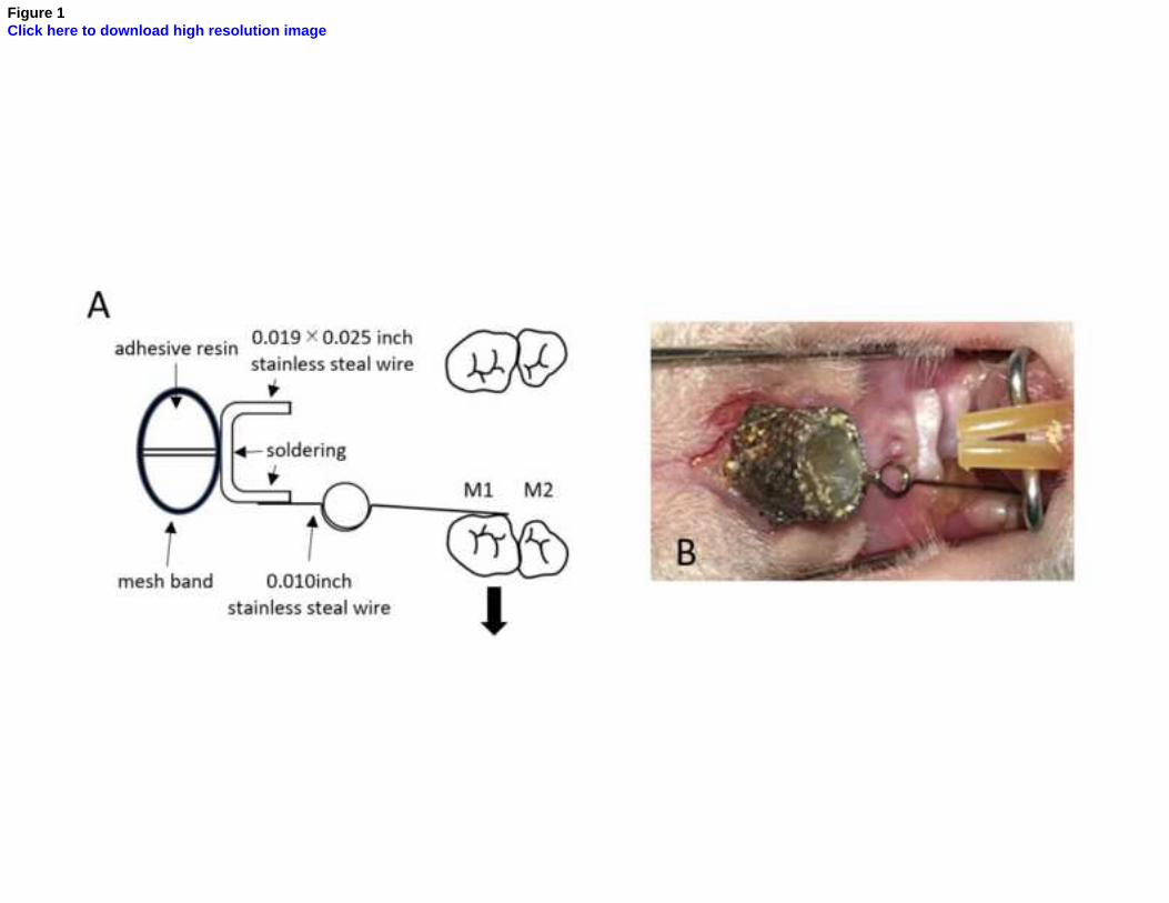
Figure 1Click here to download high resolution image

Figure 2Click here to download high resolution image
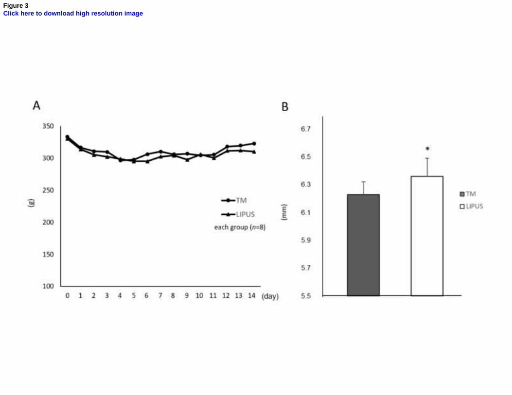
Figure 3Click here to download high resolution image

Figure 4Click here to download high resolution image

Figure 5Click here to download high resolution image

Figure 6Click here to download high resolution image

Table Descriptive statistics of the data.
Mean±S.D. ( 95% CI) Median(25th-75th percentile) Mean±S.D. ( 95% CI) Median(25th-75th percentile)
Intermolar width (mm) 6.23 ± 0.09 (6.16 - 6.31) 6.24 (6.14 - 6.25) 6.36 ± 0.13 (6.15 - 6.55) 6.36 ± (6.26-6.49)
Number of Osteoclasts 20.73 ± 6.23 (13.00 - 28.46) 21.50 (20.33 - 25.00) 59.03 ± 31.66 (19.72 - 98.35) 69.25 ± (29.67-85.67)
Alveolar Bone Formation(mm) 65.90 ±18.00 (49.26 - 82.55) 77.50 (47.17 - 81.33) 90.74 ± 25.55 (67.10 - 114.37) 85.83 ± (75.50-99.50)
BMC (mg) 6.26 ± 1.03 (5.40 - 7.12) 6.67 (5.31 - 7.02) 6.35 ± 0.94 (5.56 - 7.14) 6.07 ± (5.66-7.29)
Tooth Movement without LIPUS Tooth Movement with LIPUS
Table