Specific Ways Brain SPECT Imaging Enhances Clinical Psychiatric Practice
12
Journal of Psychoactive Drugs, 44 (2), 96–106, 2012 Copyright © Taylor & Francis Group, LLC ISSN: 0279-1072 print / 2159-9777 online DOI: 10.1080/02791072.2012.684615 Specific Ways Brain SPECT Imaging Enhances Clinical Psychiatric Practice Daniel G. Amen, M.D. a ; Diane Highum, M.D. b ; Robert Licata, M.D. b ; Joseph A. Annibali, M.D. b ; Lillian Somner, D.O. b ; H. Edmund Pigott, Ph.D. c ; Derek V. Taylor d ; Manuel Trujillo, M.D. e ; Andrew Newberg, M.D. f ; Theodore Henderson, M.D., Ph.D. g & Kristen Willeumier, Ph.D. h Abstract — Our objective was to ascertain in a prospective case series how often brain single photon emission computed tomography (SPECT) neuroimaging adds relevant information for diag- nosis and/or treatment beyond current standard assessment tools in complex psychiatric cases. Charts of 109 consecutively evaluated outpatients from four psychiatrics clinics that routinely utilize SPECT imaging for complex cases were analyzed in two stages. In stage one, psychiatrists reviewed detailed clinical histories, mental status exams, and the Structured Clinical Interview for DSM-IV, but not the results of SPECT studies, assigned a diagnosis based on DSM-IV criteria, and then developed a comprehensive treatment plan. In stage two, evaluators were given access to the SPECT studies for each patient. The addition of SPECT modified the diagnosis or treatment plan in 78.9% (n = 86; rated level 2 or 3 change) of cases. The most clinically significant changes were undetected brain trauma (22.9%), toxicity patterns (22.9%) and the need for a structural imaging study (9.2%). Specific functional abnormalities were seen as follows that potentially could impact treatment: temporal lobe dysfunction (66.1%) and prefrontal hypoperfusion (47.7%). SPECT has the potential to add clini- cally meaningful information to enhance patient care beyond current assessment tools in complex or treatment resistant cases. Keywords — brain SPECT, enhance diagnosis, modify treatment, psychiatric care, treatment resistant Psychiatry has been plagued since its inception with the dilemma of how to reliably diagnose and treat patients to optimize treatment outcome. Assessing the heterogeneity underlying each patient’s clinical presenta- tion is difficult using standard diagnostic methods. Current best practice procedures rely primarily on (1) the skill Dr. Daniel G. Amen, M.D., had full access to all the data in the study and takes responsibility for the integrity of the data and the accuracy of the data analysis. No author reports a conflict of interest or financial disclosure. a CEO and Medical Director, Amen Clinics, Inc., Newport Beach, CA. b Clinical Assessment, Amen Clinics, Inc, Newport Beach, CA. c Clinical Psychologist, Columbia, MD. d Research Associate, Amen Clinics, Inc., Newport Beach, CA. e Clinical Professor, Dept of Psychiatry, New York University, New York, NY. f Director of Research, Thomas Jefferson University, Philadelphia, PA. g CEO and President, The Synaptic Space, Centennial, CO. h Director of Research, Amen Clinics, Inc., Newport Beach, CA. Please address correspondence and reprint requests to Daniel G. Amen, M.D., Amen Clinics Inc., 4019 Westerly Place, Suite 100, Newport Beach, CA; phone: 949-266-3717, email: [email protected] of the psychiatric interviewer in obtaining all the rele- vant clinical information necessary to make criteria-based primary diagnoses and to accurately identify meaningful comorbidities; (2) the accuracy of the patient and his/her loved ones’ report of symptoms, life situation, and respon- siveness to prior treatments; (3) the completeness of the Journal of Psychoactive Drugs 96 Volume 44 (2), April – June 2012
-
Upload
independent -
Category
Documents
-
view
1 -
download
0
Transcript of Specific Ways Brain SPECT Imaging Enhances Clinical Psychiatric Practice
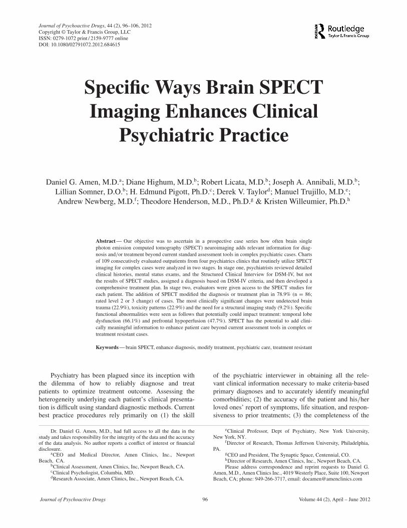
Journal of Psychoactive Drugs, 44 (2), 96–106, 2012Copyright © Taylor & Francis Group, LLCISSN: 0279-1072 print / 2159-9777 onlineDOI: 10.1080/02791072.2012.684615
Specific Ways Brain SPECTImaging Enhances Clinical
Psychiatric Practice
Daniel G. Amen, M.D.a; Diane Highum, M.D.b; Robert Licata, M.D.b; Joseph A. Annibali, M.D.b;Lillian Somner, D.O.b; H. Edmund Pigott, Ph.D.c; Derek V. Taylord; Manuel Trujillo, M.D.e;Andrew Newberg, M.D.f; Theodore Henderson, M.D., Ph.D.g & Kristen Willeumier, Ph.D.h
Abstract — Our objective was to ascertain in a prospective case series how often brain singlephoton emission computed tomography (SPECT) neuroimaging adds relevant information for diag-nosis and/or treatment beyond current standard assessment tools in complex psychiatric cases. Chartsof 109 consecutively evaluated outpatients from four psychiatrics clinics that routinely utilize SPECTimaging for complex cases were analyzed in two stages. In stage one, psychiatrists reviewed detailedclinical histories, mental status exams, and the Structured Clinical Interview for DSM-IV, but notthe results of SPECT studies, assigned a diagnosis based on DSM-IV criteria, and then developed acomprehensive treatment plan. In stage two, evaluators were given access to the SPECT studies foreach patient. The addition of SPECT modified the diagnosis or treatment plan in 78.9% (n = 86;rated level 2 or 3 change) of cases. The most clinically significant changes were undetected braintrauma (22.9%), toxicity patterns (22.9%) and the need for a structural imaging study (9.2%). Specificfunctional abnormalities were seen as follows that potentially could impact treatment: temporal lobedysfunction (66.1%) and prefrontal hypoperfusion (47.7%). SPECT has the potential to add clini-cally meaningful information to enhance patient care beyond current assessment tools in complex ortreatment resistant cases.
Keywords — brain SPECT, enhance diagnosis, modify treatment, psychiatric care, treatment resistant
Psychiatry has been plagued since its inception withthe dilemma of how to reliably diagnose and treatpatients to optimize treatment outcome. Assessing theheterogeneity underlying each patient’s clinical presenta-tion is difficult using standard diagnostic methods. Currentbest practice procedures rely primarily on (1) the skill
Dr. Daniel G. Amen, M.D., had full access to all the data in thestudy and takes responsibility for the integrity of the data and the accuracyof the data analysis. No author reports a conflict of interest or financialdisclosure.
aCEO and Medical Director, Amen Clinics, Inc., NewportBeach, CA.
bClinical Assessment, Amen Clinics, Inc, Newport Beach, CA.cClinical Psychologist, Columbia, MD.dResearch Associate, Amen Clinics, Inc., Newport Beach, CA.
eClinical Professor, Dept of Psychiatry, New York University,New York, NY.
fDirector of Research, Thomas Jefferson University, Philadelphia,PA.
gCEO and President, The Synaptic Space, Centennial, CO.hDirector of Research, Amen Clinics, Inc., Newport Beach, CA.Please address correspondence and reprint requests to Daniel G.
Amen, M.D., Amen Clinics Inc., 4019 Westerly Place, Suite 100, NewportBeach, CA; phone: 949-266-3717, email: [email protected]
of the psychiatric interviewer in obtaining all the rele-vant clinical information necessary to make criteria-basedprimary diagnoses and to accurately identify meaningfulcomorbidities; (2) the accuracy of the patient and his/herloved ones’ report of symptoms, life situation, and respon-siveness to prior treatments; (3) the completeness of the
Journal of Psychoactive Drugs 96 Volume 44 (2), April – June 2012

Amen et al. SPECT Enhances Clinical Psychiatric Practice
patient’s and his/her biological relatives’ medical his-tory; and (4) the use of psychometric measures to validatethe presence of specific symptoms and to quantify theirseverity.
Identifying additional procedures to properly diag-nose patients, and thereby better tailor their treatment,is critical to advancing psychiatric practice. While theDSM-IV is the current gold standard for psychiatric diag-nosis, its limitations are perhaps made most evident bythe fact that treatment effectiveness rates, after its intro-duction, have shown little improvement from what theywere in the 1970s despite forty years of randomizedcontrolled trials (RCT) resulting in more than 130 FDA-approved medications used in treating various DSM-defined disorders. This troubling fact is highlighted inthe 2006 American Journal of Psychiatry editorial by Dr.Raymond DePaulo, who in summarizing the results ofthe largest “effectiveness plus” studies ever conductedfor bipolar disorder (Systematic Treatment EnhancementProgram for Bipolar Disorder—STEP-BD/$28-million),major depression (Sequenced Treatment Alternatives toRelieve Depression—STAR∗D/$35-million), and antipsy-chotics (Clinical Antipsychotic Trials in InterventionEffectiveness—CATIE/$70-million) stated, “the threestudies taken together, however, underline the sugges-tion that modern pharmacological treatments may be nomore beneficial than older ones, despite their added cost”(DePaulo 2006). More recently in 2009, Dr. ThomasInsel, NIMH’s director, made observations similar to Dr.DePaulo’s, noting that despite their added costs, in everyNIMH-funded comparative effectiveness study the second-generation psychotropic drugs were found to be no betterthan the first-generation ones. He then went on to state,“The unfortunate reality is that current medications helptoo few people to get better and very few people to get well”(Insel 2009).
A key question is whether the limitations acknowl-edged by Drs. DePaulo, Insel and others (Angell 2011a,b) are related to the inadequacies of current treatmentsor to the limitations of the procedures used in diagnosticworkups. Currently, diagnostic procedures are often unableto predict clinical responses to specific treatments and/orcombinations of treatments, probably because they failto capture the underlying heterogeneity of each patient’sillness. Each of the NIMH-funded comparative effective-ness studies from the Multimodal Treatment study (MTACooperative Group 2004, MTA Cooperative Group 1999)for attention deficit hyperactivity disorder (ADHD) as wellas follow-up studies (Molina et al. 2009; Jensen et al. 2007)yielded disappointing results, far less robust than expectedby the researchers conducting the studies. Furthermore,many of these studies found that the gains that were madeduring acute-phase treatment too often disappeared duringfollow-up due to either loss of efficacy of study treatmentsand/or patient dropout. Finally, despite the millions of
dollars spent on these studies, in general they have pro-vided little new guidance to clinicians treating the mostcommon psychiatric disorders. Few, if any, of the findingsof these studies have become universally accepted prac-tice guidelines. Such meager results may be due to thesestudies’: (1) near universal failure to identify clinically-meaningful differences between compared treatments at theaggregate level, and more importantly, (2) their failure toidentify any patient-level predictors of response to specifictreatments.
Even more worrisome is the fact that such meagerclinical results were obtained while using evidence-basedtreatments (EBTs) that had been found most promising inprior randomized controlled trials (RCTs), and that theyused the current best practice standards in their diagnos-tic workups. The inevitable question is, what if the rootcause for the disappointing outcomes that were repeatedlyfound in these “effectiveness plus” studies of exemplaryand free care was the inability of these “best practice” diag-nostic procedures to fully account for the heterogeneity ofeach patients’ illness, and to provide enough informationto tailor treatment accordingly? In other words, what ifthese exemplary diagnostic workups were still missing keyneuropathological findings?
It is our contention that a primary problem with obtain-ing adequate treatment results is substantially related tothe field’s tenuous ability to adequately diagnose patients.While additional clinical measures may be useful, we haveargued (Amen et al. 2011) that the use of an objectivemeasure such as functional brain imaging could providean important source of diagnostic information that canlead a clinician to better diagnosis, and ultimately, tomore effective treatment planning. With this hypothesis inmind, the purpose of this study was to determine whetherthe addition of brain single photon emission computedtomography (SPECT) imaging to a comprehensive diag-nostic workup significantly changed the diagnosis and/ortreatment plan in four outpatient clinics that primarilytreat complex/treatment-resistant psychiatric patients. Thestudy was designed to build on the findings of Bloom andcolleagues (1996) and Borghesani and colleagues (2010)who reported that SPECT imaging added significant valuein the diagnostic workup and clinical management ofneurologically-involved patients.
Presently, there are a number of brain SPECT imag-ing patterns that have been shown to be associated withdifferent pathologies that are relevant to psychiatric prac-tice and helpful in suggesting different treatments (Amenet al. 2011). For example, diffuse decreased perfusions (or“scalloping” pattern) is often found to be due to diffuseencephalopathy, such as those caused by drug or alco-hol abuse or by exposure to environmental toxins (Amenet al. 2011; Kucuk et al. 2000). Hypoperfusion in theprefrontal pole, combined with decreased anterior temporalpole perfusion or with focal decreases, is often indicative
Journal of Psychoactive Drugs 97 Volume 44 (2), April – June 2012
Amen et al. SPECT Enhances Clinical Psychiatric Practice
of traumatic brain injury (Kant et al. 1997). In cases ofsuspected dementia, SPECT is useful in differentiatingbetween Alzheimer’s disease (AD), frontal lobe dementia(FLD), vascular dementia (VaD), Lewy body dementia(LBD), normal pressure hydrocephalus (NPH) and pseu-dodementia (PSD) (Ishii et al. 2009; Pimlott & Ebmeier2007; Bonte et al. 2006; Yoshikawa et al. 2003a, b; Devous2002; Jobst, Barnetson & Shepstone 1998; Starkstein et al.1996; Hanyu, Abe et al. 1993; Jagust, Budinger & Reed1987). SPECT perfusion imaging has been shown to bepredictive of a progression to dementia in patients withmild cognitive impairment, as well as predictive of thetype of dementia (Tranfaglia et al. 2009; Johnson et al.2007; Ishiwata et al. 2006; Huang et al. 2002; Kogure et al.2000; Wolfe et al. 1995). Many patients with OCD showincreased activity, manifested by increased perfusion andmetabolism, in the frontal cortex, anterior cingulate gyrus,caudate, putamen and thalamus (Van Laere et al. 2006;Carey et al. 2004; Diler, Kibar & Avci 2004; Saxena et al.2002; Rauch et al. 2001; Lucey et al. 1995; Hoehn-Saricet al. 1991; Machlin et al. 1991). Symptom provocation byexposure to relevant phobic stimuli (e.g., skin contact with“contaminated” objects in the case of patients with OCDwho have germ phobias) leads to increased activity in thesesame brain structures. During effective pharmacotherapy,perfusion and metabolism decreased toward normal inthese structures (Carey et al. 2004; Diler, Kibar & Avci2004; Saxena et al. 2002; Hoehn-Saric et al. 1991). Thehyperfrontal pattern can also be associated with psychi-atric disorders that share clinical features such as cognitiveinflexibility and persistent negative thoughts. Such pat-terns are present in disorders such as posttraumatic stressdisorder (PTSD), autism, and in some forms of anxietyand mood disorders (Hollander 1996). Hypofrontality, onthe other hand, is often associated with behavioral prob-lems, resulting from the exercise of poor judgment andfrom impulsivity. Its presence has been shown to predicta both a positive treatment response to stimulants (Amen,Hanks & Prunella 2008) as well as a negative response toserotonergic medication in depression (Brockmann et al.2009). Many patients with ADHD show decreased perfu-sion (activity) of the prefrontal cortex and middle temporalgyrus in a baseline scan compared to controls, whetherstudied with SPECT (Kim et al. 2002; Spalletta et al.2001; Amen & Carmichael 1997) or functional mag-netic resonance imaging (fMRI) (Rubia et al. 2011, Rubiaet al. 2005; Pliszka et al. 2006). A recent comprehen-sive review of the neuroimaging in ADHD assessed datafrom SPECT, PET and fMRI (Cherkasova & Hechtman2009). The authors reached the conclusion that all modal-ities showed similar findings and emphasized the centralrole of the frontostriatal circuit in a large proportion ofcases of ADHD. Temporal lobe abnormalities (hypo orhyperperfusion) are common in temporal lobe dysrhyth-mia or epilepsy (Thadani et al. 2004) and thus the clinical
problems associated with such findings may be more likelyto respond to anticonvulsant-based medications.
METHODS AND PROCEDURES
Subjects, Setting and Study DesignThe charts of 109 consecutively evaluated outpa-
tients were sequentially acquired from December 15th,2010 through January 5th, 2011 from four psychiatricclinics that utilize SPECT neuroimaging for complexcases. The clinics are located in four geographically dis-persed areas: Washington, DC area (Reston, VA), PacificNorthwest (Bellevue, WA), Northern California (Brisbane)and Southern California (Newport Beach). All patients inthis study gave consent for their clinical information to beused anonymously in research conducted by Amen Clinics.The patient’s refusal to participate in the research had noimpact on their care.
All clinical charts were divided into two files so thata two-stage analysis could be conducted. Included in thefirst chart was: (1) a detailed typewritten clinical historytaken by a trained medical historian that included history ofthe present illness, current and past psychiatric treatments(including medications and supplements), medical, family,school, sleep, occupational, military, drug and legal history;(2) a detailed mental status exam; (3) results from a com-puterized version of the Structured Clinical Interview forDSM-IV Axis I Disorders (SCID-IV; First et al.1997; and(4) Beck Depression Inventory (BDI; Beck et al. 1996).Clinical diagnoses and treatment recommendations werecollected via two forms included in the medical record.
The second chart contained the results of two brainSPECT studies of the same patient, including the imagesand report of the SPECT findings. One SPECT study wasperformed at baseline; the other while doing a concentra-tion task (Conner’s Continuous Performance Test II V.5(CCPT; Conners 2004).
Five board-certified psychiatrists with expertise inevaluating SPECT neuroimaging in clinical practice thensequentially evaluated the charts assigned to them, withno other knowledge of the patients. All psychiatrists hadbeen conducting diagnostic workups with SPECT imagingfor a minimum of two years prior to this study and theaverage number of new evaluations using SPECT, exceptfor the principal investigator (PI), was 649 (range 397 to1259). The PI has been using SPECT neuroimaging inclinical practice for more than 20 years. To ensure con-sistency among the evaluators, each psychiatrist underwenttwo weeks of training and inter-rater agreement trials withthe PI. Inter-rater reliability was not specifically tested inthe cases included in this study.
Each case was initially evaluated based on clinicalinformation alone (first chart). Afterwards, the cases werere-evaluated by the same psychiatrist with the inclusionof the SPECT imaging data (chart 2). Additions and/or
Journal of Psychoactive Drugs 98 Volume 44 (2), April – June 2012
Amen et al. SPECT Enhances Clinical Psychiatric Practice
changes to clinical management included: (1) orderingother studies (such as a CT or MRI); (2) addressing newlyfound medical morbidities (unexpected space occupyinglesions, unexpected trauma patterns, unexpected toxicitypatterns, unexpected signs of mild cognitive impairment ordementia, unexpected seizure activity, unexpected poten-tial vascular problems); and (3) discovering other targetfor treatments beyond those defined by the presence of anAxis I, Axis II, Axis III diagnosis, such as hyperfrontality,hypofrontality, cerebellar hypoperfusion, limbic systemhyperperfusion, temporal lobe abnormalities and othersthat were found through the use of SPECT scanning andhad not been found or even suspected by the state-of-the-art diagnostic evaluation described above. The expertclinicians then classified the changes in the diagnosis andtreatment recommendation which resulted from includingthe SPECT findings into the following level of changecategories: (1) no change; (2) mild change—when it wasconcluded that clinicians would likely have come to thesame conclusion through trial and error; (3) moderatechange—when it was estimated that the new findings couldmake a significant clinical impact; and (4) highly signif-icant change—when the new findings would completelychange the course of treatment.
Brain SPECT ImagingIn each SPECT brain imaging study an age- and
weight-appropriate dose of technetium Tc99m exametaz-ime (commercially available as Ceretec®) was adminis-tered intravenously. Photon emission was captured 30 min-utes after injection using a Picker (Phillips) Prism3000 triple-headed gamma camera with high resolution fanbeam collimators. Data were acquired in 128x128 matrices,yielding 120 images per scan with each image separated bythree degrees (6.6mm) spanning 360 degrees, and Changattenuation correction was performed using general linearmethods.
Baseline images were acquired in the following man-ner. All subjects sat upright in a quiet, dimly lit roomwith eyes open, with the bolus injected at ten minutes.The patient sat for an additional ten minutes post-injection.For the concentration scan, IV access was obtained andthen the patient performed a 15-minute computerized task,Connor’s Continuous Performance Test, while upright inthe same light as in the baseline condition. The injectionwas administered three minutes into performing the CCPT.
All images were processed using Odyssey softwareand all voxels were scaled to the brain maximum.Transaxial slices were oriented horizontal to the AC-PCline. Coronal, sagittal, and transaxial slice images (6.6mmapart, unsmoothed) were then rendered in the PickerOdyssey step-20 scale, where voxels are assigned a colorgradient based on their percentile of activity. Each colorstep represents a five-percentile difference in regional cere-bral blood flow (rCBF).
Clinical SPECT ReportUsing slices from all three orientations (coronal, sagit-
tal, and transaxial), 14 left and right regions of interest(ROIs) were visually inspected and rated by a cliniciantrained in neuroanatomy using the Mai Atlas of the HumanBrain (Mai, Assheuer & Paxinos 1997); these includedprefrontal poles, inferior orbits, lateral PFC, temporalpoles, lateral temporal lobes, medial temporal lobes, pari-etal lobes, occipital lobes, cerebellum, anterior cingulategyrus, anterior insular cortex, caudate, putamen, and tha-lamus. Raters had access to patients’ age, gender, medica-tions, and general complaints.
rCBF for each area above was visually rated on a Step-20 scale using the following formula: activity above the top95% was assigned a score of 4+; 91%-95% was scored 3+;86%-90% was scored 2+; 81%-85% was scored 1+; 61%-80% was scored 0; 56%-60% was scored –1; 51%-55% wasscored –2; 46%-50% was scored –3; and 41%-45% wasscored –4. In addition to the ratings for each area, each scanwas rendered in two 3-D images: a surface view, looking atthe top 45% of brain activity, which allows physicians toquickly visualize significant cortical hypoperfusion; and anactive view, where the most active 15% and 8% of the brainare rendered, allowing physicians to quickly visualize areasof hyperperfusion.
Statistical AnalysisAll statistics were performed using IBM SPSS
(PASW) version 18.
RESULTS
Subject CharacteristicsOf the 109 consecutive patients, 49 were female and
60 were male with an age range of 18 to 87 years(mean age 38.42, S.D. = 16.95). The average number ofdiagnoses upon entering the study was 4.2 per patient.The most common diagnostic categories included: anxiety(99/109; 90.8%), mood disorders (76/109; 69.7%), sub-stance abuse (70/109; 64.2%), ADHD (58/109; 53.2%),OCD (17/109; 15.5%), dementia of any type (11/109;10.0%), schizophrenia (4/109; 3.6%), epilepsy (2/109;1.8%), and head injury (39/109; 35.7%).
Impact of SPECT on Diagnoses and TreatmentTable 1 summarizes a sample of findings. Overall, in
52.3% of cases (n = 57), SPECT imaging changed theclinical diagnostic assessment in a highly substantial way(i.e., completely changed the course of treatment); 26.6%of cases (n = 29) were rated as a moderate change (newfindings that could make a substantial clinical impact),while 11% (n = 12) had a mild change (would likely getto the same conclusion with trial and error) and 10.1% ofcases (n = 11) had no change in the diagnosis and treat-ment plan with the addition of SPECT imaging. SPECT
Journal of Psychoactive Drugs 99 Volume 44 (2), April – June 2012

Amen et al. SPECT Enhances Clinical Psychiatric Practice
TABLE 1Summary of Primary Findings
SPECT Changed Clinical DiagnosticAssessment:
• Highly Significant (CompletelyChanged Course of Treatment)
52.3% (N = 57)
• Moderate (New Findings CouldMake Substantial Impact)
26.6% (N = 29)
• Mild (Likely Get to SameConclusion with Trial And Error)
11.0% (N = 12)
• None 10.1% (N = 11)
New Diagnostic Questions/Unexpected Issues
• Toxicity 22.9% (N = 25)• Brain Trauma 22.9% (N = 25)• Seizure Pattern 2.8% (N = 3)• Dementia Pattern 6.4% (N = 7)
Additional Targets of Treatment Seenon SPECT
• Temporal Lobe Dysfunction 66.1% (N = 72)• Hyperfrontailty 14.7% (N = 16)• Hypofrontality 47.7% (N = 52)• Cerebellar Hypoperfusion 43.1% (N = 47)
results added to Axis III diagnoses in 76.1% cases (n =83), with temporal lobe dysfunction (66.1%, n = 72) beingmost common, and hypofrontality of the prefrontal cortex(47.7%, n = 52) present in nearly half of cases. Cerebellardysfunction was identified in 43.1% of cases (n = 47),a pattern commonly observed with hypoperfusion of thefrontal lobes due to the high interconnectivity of theseneural networks (Glickstein 2006). Scalloping and diffusedecreased perfusion (toxicity) was observed in 22.9% ofcases (n = 25), suggestive of a diffuse encephalopathicprocess from drugs, alcohol, environmental toxins or otherdiffuse neuropathology, such as infections or metabolicissues (e.g., hypothyroidism). A traumatic brain injury pat-tern (TBI) was present in 22.9% of cases (n = 25), whilehyperfrontality, or increased perfusion of the prefrontalcortex, was observed in 14.7% of cases (n = 16).
One of the most clinically accepted uses of SPECTneuroimaging is in the evaluation of patients with suspecteddementia, and in this cohort, a previously unknown sugges-tion of mild cognitive impairment or early dementia patternwas observed in 6.4% of cases (n = 7). Due to the findingsfrom SPECT imaging, structural imaging (CT/MRI) wasrecommended in 9.2% of cases (n = 10). In four of thesecases SPECT imaging had revealed suspected ventricularenlargement.
The addition of SPECT imaging yielded 67% ofcases (n = 73) where at least one medication change wasrecommended. Of these, the suggestion was for additionalmedications in 93.2% of cases (68 of 73), while 31.5%
(23/73) had recommendations for medications reduction.Antiepileptic medications were recommended in 61.6%of cases (45/73), consistent with the high incidence oftemporal lobe abnormalities. Stimulant medications wererecommended in 37% of cases (27/73). Alternatively, in9.6% of the cases (7/73) a reduction in stimulant medica-tions was suggested. Antidepressant changes were advo-cated in 30.1% of cases with buproprion recommendedin 11% (8/73), a selective-serotonin reuptake inhibitor(SSRI) recommended in 9.6% (7/73) and a serotonin-norepinephrine reuptake inhibitor (SNRI) recommendedin 9.6% (7/73). The latter finding correlates with thepatients who showed hyperfrontality patterns on SPECT(14.7%, n = 16 out of 109), as this is likely to predict apositive treatment response to serotonergic medication indepression (Mayberg et al. 1997). Interestingly, these sameantidepressant medications were recommended to be dis-continued in 28.8% (21/73) of those cases where SPECTimaging suggested a medication change. Discontinuationof buproprion was recommended in 2.7% (2/73), of anSSRI in 16.4% (12/73), and SNRI in 9.6% (7/73). Thisreduction in the use of antidepressant medication canpossibly be attributed to those patients with hypofrontalitypatterns who were depressed and taking SSRIs.As hypofrontality has been associated with a negative treat-ment response to serotonergic medication in depression(Brockmann et al. 2009), such medications needed to bediscontinued in a sizable number of such depressed patientswith hypofrontal patterns revealed by SPECT imaging.
Patients who were rated as having “no change” (rat-ing = 0) between their initial evaluation and the addition ofSPECT imaging to their evaluation had no suggested medi-cation change. Medication changes were evenly distributedamong those who were rated as having a “mild change”(n = 6 no change; n = 6 medication change). Those whowere rated as having a “moderate change” were three timesas likely as others with this rating (compared to the entirecohort) to have a suggested change in their medication pro-file (n= 7 no change; n = 22 medication change). Thoserated as “highly significant change” were four times aslikely to have a suggested medication change (n = 12 nochange; n = 45 medication change). The higher rating scalescores correlated with a higher incidence of a medicationchange (χ2 = 28.6, P < 0.001). Figures 1 through 4 giveillustrative case examples.
Impact of SPECT in a Substance Abuse CohortFurthermore, we investigated the differences between
those who were clinically diagnosed as substance abusers(n = 33; 7 female, 26 male) within our patient popula-tion versus those who were not (n = 76) and found nosignificant level of change between the two groups withregards to adding a SPECT scan to the diagnostic workup.The diagnostic categorization of this cohort revealed themajority of subjects abused alcohol (n = 21) or cannabis
Journal of Psychoactive Drugs 100 Volume 44 (2), April – June 2012
Amen et al. SPECT Enhances Clinical Psychiatric Practice
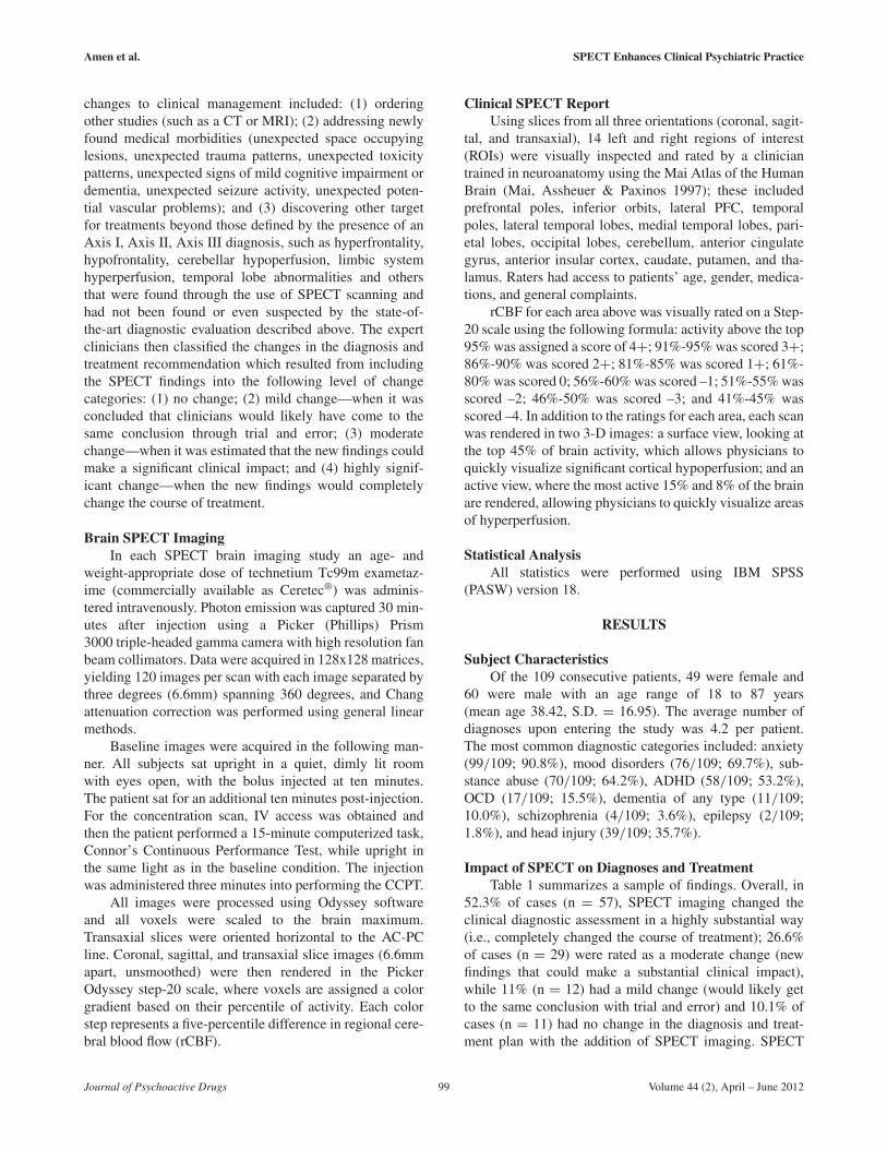
FIGURE 1Unexpected Toxicity (color figure available online)
Healthy Brain SPECTFull, even, symmetrical perfusion.
Threshold set at 55%ile.
Patient AOverall decreased perfusion
A, a 62-year-old female, was provisionally diagnosed with major depressive disorder, generalized anxiety disorder, and a panic disorder. Her SPECTresults revealed an unexpected diffuse encephalopathic pattern. The addition of SPECT resulted in a new diagnostic categorization to major depressiondue to a medical condition with a need for a more in-depth work up to better understand and treat other potential causes of the pattern.
(n = 15) followed by cocaine (n = 5), nicotine (n = 3),amphetamines (n = 1) and hallucinogens (n = 1). Overall,in 51.5% of these cases (n = 17), SPECT imaging changedthe clinical diagnostic assessment in a highly substantialway; 30.3% of cases (n = 10) were rated as a moderatechange, while 12.1% (n = 4) had a mild change and 6.1%of cases (n = 2) had no change in the diagnosis and treat-ment plan with the addition of SPECT imaging. As in thewhole cohort, SPECT results added to Axis III diagnosesin 75.8% cases (n = 25), with temporal lobe abnormalities(63.6%, n = 21), brain trauma (27.3%, n = 9) and toxicity(18.2%, n = 6) being present but unrecognized. These find-ings are noteworthy because when they are left untreatedthey can contribute to continued or accelerated substanceabuse. We did find that the substance abuse cohort had amean of 6.0 (± 1.84) diagnoses per patient versus 3.37(± 2.0) for the whole group, which was highly signifi-cant at p < 0.001. Taken together, these data suggest thatSPECT provides the same level of change as compared tothe original cohort, but the increased number of diagnosesper patient makes a compelling case for the need for theclinical guidance SPECT can contribute to this population.
DISCUSSION
The aim of this study was to quantify the value ofadding rest and concentration SPECT neuroimaging to the
diagnostic workup in clinics, which evaluates a high per-centage of treatment-resistant, complex psychiatric cases.Prospective analysis of 109 consecutive patients who pre-sented with an average of 4.2 Axis I diagnoses showedthat the addition of SPECT scans led to moderate orhighly substantial changes of diagnoses and/or treatmentin 78.9% of the cases. Two findings of particular clini-cal importance were that SPECT imaging detected highlevels of previously unrecognized brain trauma (22.9%)and overall decreased perfusion (22.9%), consistent with apreviously unknown diffuse encephalopathic process fromdrugs, alcohol, an environmental toxin or from insuffi-ciently addressed medical problems. These findings sup-port and corroborate the clinical wisdom of the followingquote from Harold Bursztajn, M.D., cofounder of thePsychiatry and Law Program at Harvard, who said “SPECTscans do not give you the answer, they teach you to ask bet-ter questions” (Bursztajn 2002). SPECT scan results cangive clues on how to investigate the clinical problems ofpatients suffering from complex and/or treatment resistantpsychiatric disorders. For example, when a brain injury ordiffuse encephalopathic pattern is seen but had not beenidentified by clinical history, it directs the clinician to con-duct a more focused inquiry about past brain injuries, toinvestigate if drugs or alcohol are an issue, and to ascertainwhether environmental toxins, or other potential medicalconditions, may be present but unrecognized. The line of
Journal of Psychoactive Drugs 101 Volume 44 (2), April – June 2012
Amen et al. SPECT Enhances Clinical Psychiatric Practice
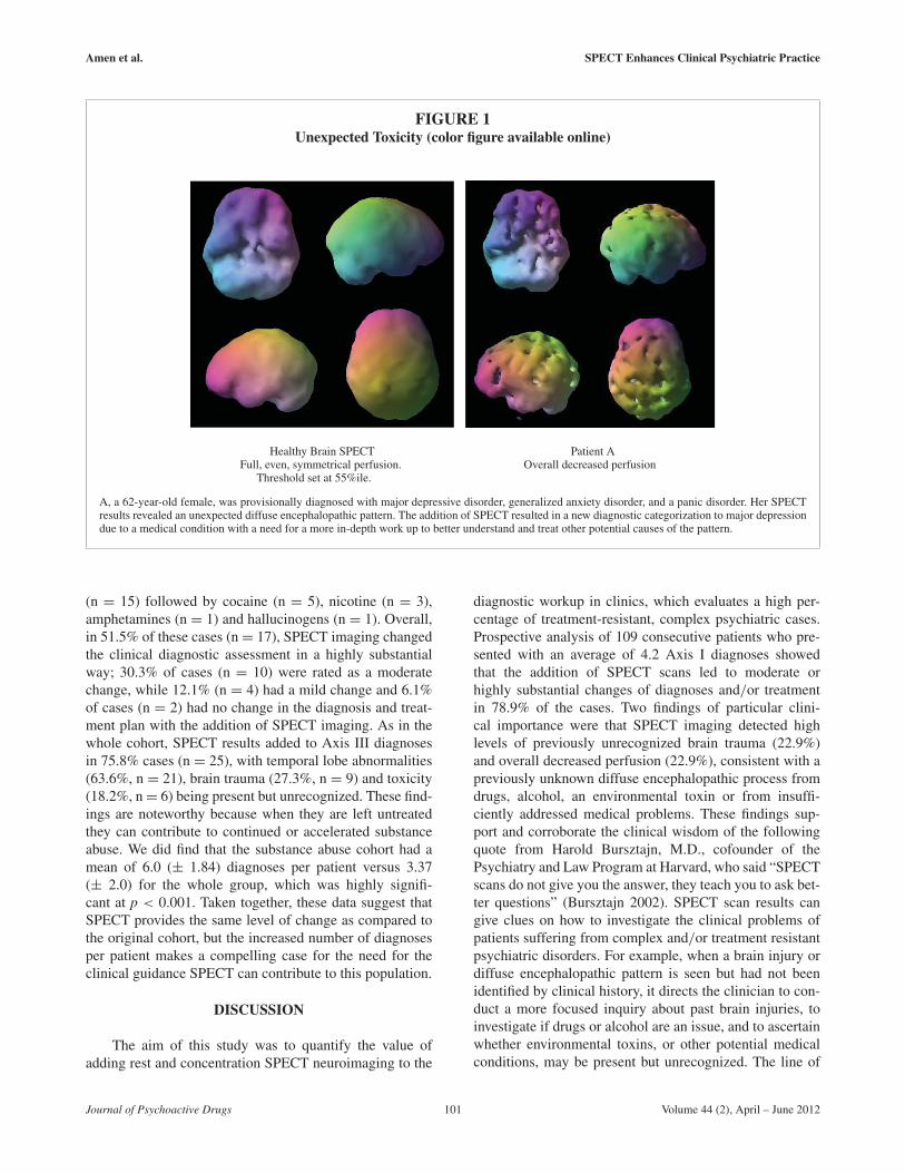
FIGURE 2Unexpected Brain Trauma (color figure
available online)
Patient B
B, a 48-year-old male, was diagnosed with panic disorderand severe insomnia of five months duration, made worse by ben-zodiazepines and sleep medication. The month before symptomsbegan, he had a “minor” fall from a mountain bike where he hithis head but had no loss of consciousness. SPECT scan revealedclear decreases in left prefrontal and temporal lobe region (arrows),consistent with prior brain trauma. This led to further questionsand ultimately to brain trauma rehabilitation strategies, such asneurofeedback and hyperbaric oxygen therapy.
inquiry and potential further work up can add valuableinformation.
In his 2005 keynote address to the AmericanPsychiatric Association (APA), Insel (2005) said:
The DSM-IV has 100% reliability and 0% validity . . .
We need to develop biomarkers, including brain imaging, todevelop the validity of these disorders . . . Trial-and-errordiagnosis will move to an era where we understand the under-lying biology of mental disorders . . . We are going to haveto use neuroimaging to begin to identify the systems pathol-ogy . . . to develop treatments that go after the core pathology,understood by imaging.
The same year Insel made similar observations inwriting:
Patterns of regional brain activity associated with normal andpathological mental experience can be visualized . . . and ulti-mately, biomarkers for mental disorders may not be proteins
or neurotransmitters but may emerge from neuroimaging(functional magnetic resonance imaging (fMRI), single photonemission computed tomography (SPECT), etc. Logically, ifthese are disorders of brain systems, then the visualization ofabnormal patterns of brain activity should detect the pathologyof these illnesses (Insel & Quirion 2005).
In 2009, the NIMH unveiled a new initiative, known asResearch Domain Criteria (RDoC; Insel 2010) to
. . . develop new ways of classifying mental disorders based ondimensions of observable behavior and neurobiological mea-sures . . . Increasing evidence suggests that abnormality in onedimension, such as impulsivity, frequently occurs in multiplediagnoses of mental disorders . . It will shift the way we doresearch and think about mental disorders.
It has become increasingly unlikely that DSM diag-noses represent distinct neurophysiological entities. Theywill likely prove to represent groups of neurophysiologicalprocesses. Individual symptoms likely represent abnormalneurophysiological processes that span across diagnoses(e.g., impulsivity is a key component of the diagnosis ofImpulse Control Disorder NOS, ADHD, bipolar mania,and certain personality disorders; but is also seen follow-ing frontal lobe injury, as in mild TBI, and in FrontalTemporal Dementia). So, it only follows that functionalneuroimaging will fail to reveal a singular neurophysiolog-ical process corresponding in a one-to-one fashion with aDSM diagnosis. Such correspondences do not exist. Thereis substantial evidence that there are multiple neurophysi-ological cases and imaging findings associated with condi-tions such as depression (Savitz & Drevets 2009), bipolardisorder (Pan et al. 2009), and ADHD (Cherkasova &Hechtman 2009). As this study suggests, SPECT imag-ing helps to provide clinical guidance in the managementof individual patients by adding critical new informationto better assess the heterogeneity underlying his or herparticular clinical presentation. Given the disappointingfindings in the various NIMH-funded “effectiveness plus”trials and, in particular, their failure to identify patient-level predictors of response to specific treatments despitetheir large Ns, one must wonder what undiagnosed neu-ropathology contributed to each individual patient’s failureto respond to best practices treatment. The current studyindicates that SPECT neuroimaging might have been help-ful in uncovering and in providing guidance for theirtreatment.
In the authors’ opinion, SPECT neuroimaging, as wellas any other neuroimaging or laboratory tool, should neverbe used in isolation, but always as an integrated part of athorough clinical evaluation. In this context, the additionof SPECT imaging is capable of revealing potential newtargets for treatment, as evidenced by the increased num-ber of Axis III diagnoses found in 76.1% (n = 83) of ourcases, although this finding may cause some to reconsidercurrent diagnostic nomenclature. We also found abnormal
Journal of Psychoactive Drugs 102 Volume 44 (2), April – June 2012
Amen et al. SPECT Enhances Clinical Psychiatric Practice
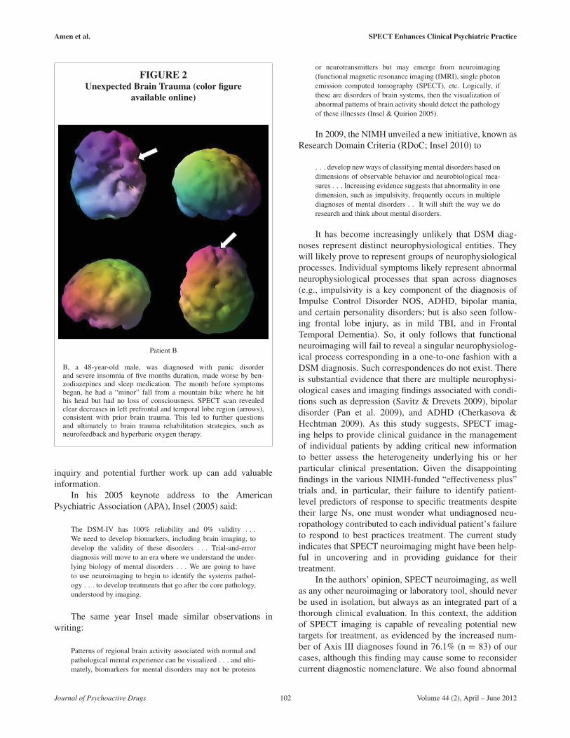
FIGURE 3Spect Helps Direct Medication Choice (color figure available online)
C’s surface SPECT scanNote deficit in cerebellum and occipital region
C’s active SPECT scanRed and white show most active areas of brain,
usually seen in cerebellum with HMPAO.Note marked hyperfrontality (increased perfusion in the
anterior cingulate and lateral prefrontal cortices).
C, a 19-year-old female diagnosed with cerebral palsy and Asperger’s syndrome, was evaluated for inattention, anxiety and social detachment afteranother student at school died suddenly. Her SPECT scan showed damage to her cerebellum and occipital lobes consistent with past oxygen deprivationand findings common in cerebral palsy and an autistic spectrum disorder. It also showed marked increased hyperfrontality, a pattern that suggests apositive response to a serotonergic intervention.
temporal lobe perfusion (a common finding in temporallobe epilepsy) to be present in 66.1% (n = 72) of cases. It iswell known that up to 50% or 60% of patients with chronicepilepsy have various mood disorders including depressionand anxiety (Beyenburg et al. 2005; Jones et al. 2005). Theaddition of anticonvulsant medications is a common strat-egy in complex or treatment resistant cases. Using SPECTimaging, the present study provides a possible explanationfor the success of this strategy. The finding of focal areasof increased or decreased temporal lobe perfusion couldoffer the clinician diagnostic clarification and buttress thedecision to use anticonvulsants.
Hyperfrontality has been associated with a positiveresponse to SSRIs in depressed and anxious patients, whilehypofrontality is associated with a negative response tosuch agents and has been reported in completed suicides(Willeumier, Taylor & Amen 2011). Little and colleagues(2005), on the other hand, found that lower prefrontalactivity prior to treatment was associated with a positiveresponse to buproprion and venlafaxine. These medica-tions enhance dopamine and norepinephrine neurotrans-mission, which, in the authors’ experience, tend to raiseprefrontal cortex activity, while SSRIs tend to downreg-ulate prefrontal cortex activity (Hoehn-Saric et al. 2001).
Even though there are reported common brain SPECTimaging patterns for many different psychiatric diagnosticcategories, such as OCD and ADHD, our extensive expe-rience tells us that the findings are variable in individualpatients, which is the reason why it is essential to knowthe individual imaging patterns in treatment resistant orcomplex cases to better personalize treatment.
The present study does not stand in isolation, butreinforces and extends previous studies. In 1996, Bloomand colleagues (1996) evaluated 94 consecutive patientsreferred for SPECT neuroimaging. These patients wereassigned to one of nine groups based on the clinical indi-cation for SPECT. All of the SPECT evaluations wereconducted with knowledge of the patient’s clinical history.Two weeks after scans, a standardized interview was con-ducted with both the interpreting nuclear physician and thereferring physician to determine if the addition of SPECTimaging had significantly altered patient management bychanging either the planned surgical or medication ther-apy. The key findings of Bloom and colleagues were thatSPECT neuroimaging: (1) significantly altered treatment in47% of all referred patients; (2) altered treatment in threeof six (50%) head trauma patients; and (3) altered treat-ment in 11 of the 18 (61%) dementia patients who were
Journal of Psychoactive Drugs 103 Volume 44 (2), April – June 2012
Amen et al. SPECT Enhances Clinical Psychiatric Practice
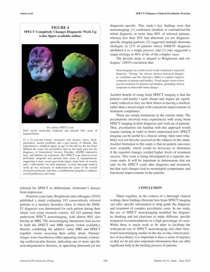
FIGURE 4SPECT Completely Changes Diagnostic Work Up
(color figure available online)
D’s surface SPECT scanNote severe prefrontal, temporal and parietal lobe areas ofhypoperfusion
D, a 51-year-old female, presented with anxiety, stress, disor-ganization, marital problems and a past history of bulimia. Sheexperienced a whiplash injury at age 15 but did not hit her head.Without the scans, the psychiatrist rater in the study gave her thediagnoses of Generalized Anxiety Disorder, AD/HD inattentivetype, and bulimia (in remission). Her SPECT scan showed severeprefrontal, temporal and parietal lobe areas of hypoperfusion,suggesting a more severe past brain injury, some form of toxicityand a vulnerability for early dementia. A more thorough medicalwork up was necessary to understand the cause of the overalldecreased perfusion, and then a rehabilitation program to enhanceoverall perfusion to her brain.
referred for SPECT to differentiate Alzheimer’s diseasefrom depression.
Fourteen years later, Borghesani and colleagues (2010)published a study evaluating 193 consecutively referredpatients to a memory disorders clinic in which the DSM-IV diagnosis was determined for each patient during theirinitial visit using research criteria. All 193 patients thenunderwent SPECT neuroimaging, with almost 80% alsohaving an MRI. The neuroimaging interpreters had accessto both the SPECT and MRI reports (when available),thereby combining the additive value MRI and SPECTtogether versus assessing their utility alone. Patients’images were classified as either appearing normal, evidenc-ing cardiovascular disease, indicating one or more specificneurodegenerative diseases, or appearing abnormal yet not
diagnostic-specific. This study’s key findings were thatneuroimaging: (1) confirmed, clarified, or contradicted theinitial diagnosis in more than 80% of referred patients,whereas less than 20% had abnormal yet not diagnosis-specific imaging patterns; (2) suggested multiple dementiaetiologies in 21% of patients whose DSM-IV diagnosisattributed it to a single process; and (3) only suggested asingle etiology in 46% of the of the complex cases.
The present study is aligned to Borghesani and col-leagues’ (2010) conclusion that:
Neuroimaging was useful even if it only confirmed a suspecteddiagnosis. “Seeing” the disease process increased diagnos-tic confidence and the clinician’s ability to explain cognitivesymptoms to patients and families. Visual images seem to havespecial resonance for patients and families, grounding clinicalsymptoms in observable brain changes.
Another benefit of using brain SPECT imaging is that thepatient’s and family’s guilt, shame and stigma are signifi-cantly reduced as they see their illness as having a medical,rather than a moral origin with concurrent improvements intreatment compliance.
There are certain limitations to the current study. Thepsychiatrists involved were experienced with using brainSPECT imaging in their diagnosis and work up of patients.Thus, psychiatrists not familiar with this approach wouldrequire training in order to better understand how SPECTimaging can be useful in a clinical setting. Inter-rater relia-bility was not directly assessed on the subjects in this study.Another limitation to this study is that no patient outcomeswere available, which would be necessary to determineif the reported changes yielded higher levels of treatmentsuccess. This issue is being investigated in a separate out-come study. It will be important to demonstrate that notonly do the SPECT scans alter diagnosis and treatment,but that such changes lead to meaningful symptomatic andfunctional improvements in the patients.
CONCLUSION
Taken together, in the context of a thorough clinicalworkup, these findings illustrate how brain SPECT imagingcan offer specific information to help guide the diagnosisand treatment of complex psychiatric cases. In our study,the use of SPECT neuroimaging modified the diagnos-tic thinking and led clinicians to make different, specifictreatment recommendations in a high percentage of cases.While there is much work to be done to facilitate thewidespread use of SPECT neuroimaging and other func-tional neuroimaging studies in the day-to-day clinical prac-tice of psychiatry, it is important to have a sense of urgencyso that we do not miss important information that can offersignificant help in the healing process of patients.
Journal of Psychoactive Drugs 104 Volume 44 (2), April – June 2012
Amen et al. SPECT Enhances Clinical Psychiatric Practice
REFERENCES
Amen, D.G. & Carmichael B.D. 1997. High-resolution brain SPECTimaging in ADHD. Annals of Clinical Psychiatry 9 (2): 81–86.
Amen, D. G.; Hanks, C. & Prunella, J. 2008. Predicting positive and neg-ative treatment responses to stimulants with brain SPECT imaging.Journal of Psychoactive Drugs 40 (2): 131–38.
Amen, D.G.; Trujillo, M.; Newberg, A.; Willeumier, K.; Tarzwell, R.;Wu, J.C. & Chaitin, B. 2011. Brain SPECT imaging in com-plex psychiatric cases: An evidence-based, underutilized tool. OpenNeuroimaging Journal 5: 40–48.
Angell, M. 2011a. The epidemic of mental illness: Why? New York Reviewof Books 58 (11). Available at http://www.nybooks.com/articles/archives/2011/jun/23/epidemic-mental-illness-why
Angell, M. 2011b. The illusions of psychiatry. New York Review of Books58 (12). Available at http://www.nybooks.com/articles/archives/2011/jul/14/illusions-of-psychiatry/
Beck, A.T.; Steer, R.A.; Ball, R. & Ranieri, W. 1996. Comparison ofBeck Depression Inventories -IA and -II in psychiatric outpatients.Journal of Personality Assessment 67 (3): 588–97.
Beyenburg, S.; Mitchell, A.J.; Schmidt, D.; Elger, C.E. & Reuber, M.2005. Anxiety in patients with epilepsy: Systematic review andsuggestions for clinical management. Epilepsy & Behavior 7 (2):161–71.
Bloom, M.; Jacobs, S.; Pile-Spellman, J.; Pozniakoff, A.; Mabutas, M.I.;Fawwaz, R.A. & Van Heertum, R.L. 1996. Cerebral SPECT imag-ing: Effect on clinical management. Journal of Nuclear Medicine 37(7): 1070–73.
Bonte, F.J.; Harris, T.S.; Hynan, L.S.; Bigio, E.H. & White, C.L. 3rd 2006.Tc-99m HMPAO SPECT in the differential diagnosis of the demen-tias with histopathologic confirmation. Clinical Nuclear Medicine31 (7): 376–78.
Borghesani, P.R.; DeMers, S.M.; Manchanda, V.; Pruthi, S.; Lewis,D.H. & Borson, S. 2010. Neuroimaging in the clinical diagnosis ofdementia: Observations from a memory disorders clinic. Journal ofthe American Geriatrics Society 58 (8): 1453–58.
Brockmann, H.; Zobel, A.; Joe, A.; Biermann, K.; Scheef, L.;Schuhmacher, A.; von Widdern, O.; Metten, M.; Biersack, H.J.;Maier, W. & Boecker, H. 2009. The value of HMPAO SPECT inpredicting treatment response to citalopram in patients with majordepression. Psychiatry Research 173 (2): 107–12.
Bursztajn, H.J. 2002. Personal communication.Carey, P.D.; Warwick, J.; Harvey, B.H.; Stein, D.J. & Seedat, S.
2004. Single photon emission computed tomography (SPECT) inobsessive-compulsive disorder before and after treatment with inos-itol. Metabolic Brain Disease 19 (1–2): 125–34.
Cherkasova, M.V. & Hechtman, L. 2009. Neuroimaging in attention-deficit hyperactivity disorder: Beyond the frontostriatal circuitry.Canadian Journal of Psychiatry 54 (10): 651–64.
Conners, K. 2004. Conners’ Continuous Performance Test II V.5. NorthTonawanda, NY: MHS.
DePaulo, J.R., Jr. 2006. Bipolar disorder treatment: An evidence-based reality check. American Journal of Psychiatry 163 (2):175–76.
Devous, M.D., Sr. 2002. Functional brain imaging in the dementias: Rolein early detection, differential diagnosis, and longitudinal studies.European Journal of Nuclear Medicine and Molecular Imaging 29(12): 1685–96.
Diler, R.S.; Kibar, M. & Avci, A. 2004. Pharmacotherapy and regionalcerebral blood flow in children with obsessive compulsive disorder.Yonsei Medical Journal 45 (1): 90–99.
First, M.B.; Gibbon M.; Spitzer, R.L.; Williams, J.B.W. & Benjamin L.S.1997. Structured Clinical Interview for DSM-IV Axis II PersonalityDisorders, (SCID-II). Washington, D.C.: American PsychiatricPress, Inc.
Glickstein, M. 2006. Thinking about the cerebellum. Brain 129 (Pt 2):288–90.
Hanyu, H.; Abe, S.; Arai, H.; Asano, T.; Iwamoto, T. & Takasaki, M.1993. Diagnostic accuracy of single photon emission computedtomography in Alzheimer’s disease. Gerontology 39 (5): 260–66.
Hoehn-Saric, R.; Schlaepfer, T.E.; Greenberg, B.D.; McLeod, D.R.;Pearlson, G.D. & Wong, S. H. 2001. Cerebral blood flow inobsessive-compulsive patients with major depression: Effect oftreatment with sertraline or desipramine on treatment responders andnon-responders. Psychiatry Research 108 (2): 89–100.
Hoehn-Saric, R.; Pearlson, G.D.; Harris, G.J.; Machlin, S.R. & Camargo,E.E. 1991. Effects of fluoxetine on regional cerebral blood flow inobsessive-compulsive patients. American Journal of Psychiatry 148(9): 1243–45.
Hollander, E. 1996. Obsessive-compulsive disorder-related disorders:The role of selective serotonergic reuptake inhibitors. InternationalClinical Psychopharmacology 11 (Suppl 5): 75–87.
Huang, C.; Wahlund, L.O.; Svensson, L.; Winblad, B. & Julin, P. 2002.Cingulate cortex hypoperfusion predicts Alzheimer’s disease in mildcognitive impairment. BMC Neurology 2 : 9.
Insel, T. R. 2009. Disruptive insights in psychiatry: Transforming aclinical discipline. Journal of Clinical Investigation 119 (4): 700–05.
Insel, T.R. 2005. Judd Marmor Award Lecture: American PsychiatricAssociation Annual Meeting, Atlanta, GA.
Insel, T. R. & Quirion, R. 2005. Psychiatry as a clinical neurosciencediscipline. Journal of the American Medical Association 294 (17):2221–24.
Insel, T.; Cuthbert, B.; Garvey, M.; Heinssen, R.; Pine, D.S.; Quinn, K.;Sanislow, C. & Wang, P. 2010. Research domain criteria (RDoC):Toward a new classification framework for research on mentaldisorders. American Journal of Psychiatry 167 (7): 748–51.
Ishii, K.; Kanda, T.; Uemura, T.; Miyamoto, N.; Yoshikawa, T.; Shimada,K.; Ohkawa, S. & Minoshima, S. 2009. Computer-assisted diag-nostic system for neurodegenerative dementia using brain SPECTand 3D-SSP. European Journal of Nuclear Medicine and MolecularImaging 36 (5): 831–40.
Ishiwata, A.; Sakayori, O.; Minoshima, S.; Mizumura, S.; Kitamura, S.& Katayama, Y. 2006. Preclinical evidence of Alzheimer changesin progressive mild cognitive impairment: A qualitative and quan-titative SPECT study. Acta Neurologica Scandinavica 114 (2):91–96.
Jagust, W.J.; Budinger, T.F. & Reed, B.R. 1987. The diagnosis of dementiawith single photon emission computed tomography. Archives ofNeurology 44 (3): 258–62.
Jensen, P.S.; Arnold, L.E.; Swanson, J.M.; Vitiello; B.; Abikoff; H.B.;Greenhill, L.L.; Hechtman, L.; Hinshaw, S.P.; Pelham, W.E.; Wells,K.C.; Conners, C.K.; Elliott, G.R.; Epstein, J.N.; Hoza, B.; March,J.S.; Molina, B.S.; Newcorn, J.H.; Severe, J.B.; Wigal, T.; Gibbons,R. D. & Hur, K. 2007. 3-year follow-up of the NIMH MTAstudy. Journal of the American Academy of Child and AdolescentPsychiatry 46 (8): 989–1002.
Jobst, K.A.; Barnetson, L.P. & Shepstone, B.J. 1998. Accurate predictionof histologically confirmed Alzheimer’s disease and the differ-ential diagnosis of dementia: The use of NINCDS-ADRDA andDSM-III-R criteria, SPECT, X-ray CT, and Apo E4 in medial tem-poral lobe dementias. Oxford Project to Investigate Memory andAging. International Psychogeriatrics 10 (3): 271–302.
Johnson, K.A.; Moran, E.K.; Becker, J.A.; Blacker, D.; Fischman, A. J. &Albert, M.S. 2007. Single photon emission computed tomogra-phy perfusion differences in mild cognitive impairment. Journal ofNeurology, Neurosurgery & Psychiatry 78 (3): 240–47.
Jones, J.E.; Hermann, B.P.; Barry, J.J.; Gilliam, F.; Kanner, A.M. &Meador, K.J. 2005. Clinical assessment of Axis I psychiatric
Journal of Psychoactive Drugs 105 Volume 44 (2), April – June 2012

Amen et al. SPECT Enhances Clinical Psychiatric Practice
morbidity in chronic epilepsy: A multicenter investigation. Journalof Neuropsychiatry and Clinical Neuroscience 17 (2): 172–79.
Kant, R.; Smith-Seemiller, L.; Isaac, G. & Duffy, J. 1997. Tc-HMPAOSPECT in persistent post-concussion syndrome after mild headinjury: Comparison with MRI/CT. Brain Injury 11 (2): 115–24.
Kim, B.N.; Lee, J.S.; Shin, M.S.; Cho, S.C. & Lee, D.S. 2002. Regionalcerebral perfusion abnormalities in attention deficit/hyperactivitydisorder. Statistical parametric mapping analysis. EuropeanArchives of Psychiatry and Clinical Neuroscience 252 (5):219–25.
Kogure, D.; Matsuda, H.; Ohnishi, T.; Asada, T.; Uno, M.; Kunihiro,T.; Nakano, S.; Takasaki, M. 2000. Longitudinal evaluation ofearly Alzheimer’s disease using brain perfusion SPECT. Journal ofNuclear Medicine 41 (7): 1155–62.
Kucuk, N.O.; Kilic, E.O.; Ibis, E.; Aysev, A.; Gencoglu, E.A.; Aras,G.; Soylu, A. & Erbay, G. 2000. Brain SPECT findings in long-term inhalant abuse. Nuclear Medicine Communications 21 (8):769–73.
Little, J.T.; Ketter, T.A.; Kimbrell, T.A.; Dunn, R.T.; Benson, B.E.;Willis, M.W.; Luckenbaugh, D.A. & Post, R.M. 2005. Bupropionand venlafaxine responders differ in pretreatment regional cerebralmetabolism in unipolar depression. Biological Psychiatry 57 (3):220–28.
Lucey, J.V.; Costa, D.C.; Blanes, T.; Busatto, G.F.; Pilowsky, L.S.;Takei, N.; Marks, I.M.; Ell, P.J. & Kerwin, R.W. 1995. Regionalcerebral blood flow in obsessive-compulsive disordered patientsat rest. Differential correlates with obsessive-compulsive andanxious-avoidant dimensions. British Journal of Psychiatry 167 (5):629–34.
Machlin, S.R.; Harris, G.J.; Pearlson, G.D.; Hoehn-Saric, R.; Jeffery, P. &Camargo, E.E. 1991. Elevated medial-frontal cerebral blood flow inobsessive-compulsive patients: A SPECT study. American Journalof Psychiatry 148 (9): 1240–42.
Mai, J.K.; Assheuer, J. & Paxinos, G. 1997. Atlas of the Human Brain.San Diego, London: Academic.
Mayberg, H.S.; Brannan, S.K.; Mahurin, R.K.; Jerabek, P.A.; Brickman,J.S.; Tekell, J.L.; Silva, J.A.; McGinnis, S.; Glass, T.G.; Martin,C.C. & Fox, P.T. 1997. Cingulate function in depression: A potentialpredictor of treatment response. Neuroreport 8 (4): 1057–61.
Molina, B.S.; Hinshaw, S.P.; Swanson, J.M.; Arnold, L.E.; Vitiello, B.;Jensen, P.S.; Epstein, J. N.; Hoza, B.; Hechtman, L.; Abikoff, H.B.;Elliott, G.R.; Greenhill, L.L.; Newcorn, J.H.; Wells, K.C.; Wigal, T.;Gibbons, R.D.; Hur, K. & Houck, P.R. 2009. The MTA at 8 years:Prospective follow-up of children treated for combined-type ADHDin a multisite study. Journal of the American Academy of Child andAdolescent Psychiatry 48 (5): 484–500.
MTA Cooperative Group. 2004. National Institute of Mental HealthMultimodal Treatment Study of ADHD follow-up: 24-month out-comes of treatment strategies for attention-deficit/hyperactivitydisorder. Pediatrics 113 (4): 754–61.
MTA Cooperative Group. 1999. A 14-month randomized clinical trialof treatment strategies for attention-deficit/hyperactivity disorder.Multimodal Treatment Study of Children with ADHD. Archives ofGeneral Psychiatry 56 (12): 1073–86.
Pan, L.; Keener, M.T.; Hassel, S. & Phillips, M.L. 2009. Functionalneuroimaging studies of bipolar disorder: Examining the wide clini-cal spectrum in the search for disease endophenotypes. InternationalReview of Psychiatry 21 (4): 368–79.
Pimlott, S.L. & Ebmeier, K.P. 2007. SPECT imaging in dementia. BritishJournal of Radiology 80 Spec No 2: S153–159.
Pliszka, S.R.; Glahn, D.C.; Semrud-Clikeman, M.; Franklin, C., Perez, R.,3rd; Xiong, J. & Liotti, M. 2006. Neuroimaging of inhibitory controlareas in children with attention deficit hyperactivity disorder whowere treatment naive or in long-term treatment. American Journalof Psychiatry 163 (6): 1052–60.
Rauch, S.L.; Dougherty, D.D.; Cosgrove, G.R.; Cassem, E.H.; Alpert,N.M.; Price, B.H.; Nierenberg, A.A.; Mayberg, H.S.; Baer, L.;Jenike, M.A. & Fischman, A.J. 2001. Cerebral metabolic corre-lates as potential predictors of response to anterior cingulotomyfor obsessive compulsive disorder. Biological Psychiatry 50 (9):659–67.
Rubia, K.; Cubillo, A.; Woolley, J.; Brammer, M. J. & Smith, A.2011. Disorder-specific dysfunctions in patients with attention-deficit/hyperactivity disorder compared to patients with obsessive-compulsive disorder during interference inhibition and attentionallocation. Human Brain Mapping 32 (4): 601–11.
Rubia, K.; Smith, A.B.; Brammer, M.J.; Toone, B. & Taylor, E. 2005.Abnormal brain activation during inhibition and error detection inmedication-naive adolescents with ADHD. American Journal ofPsychiatry 162 (6): 1067–75.
Savitz, J. & Drevets, W.C. 2009. Bipolar and major depressive disorder:Neuroimaging the developmental-degenerative divide. Neuroscience& Biobehavioral Reviews 33 (5): 699–771.
Saxena, S.; Brody, A.L.; Ho, M.L.; Alborzian, S.; Maidment, K.M.;Zohrabi, N.; Ho, M.K.; Huang, S.C.; Wu, H.M. & Baxter, L.R.,Jr. 2002. Differential cerebral metabolic changes with paroxetinetreatment of obsessive-compulsive disorder vs major depression.Archives of General Psychiatry 59 (3): 250–61.
Spalletta, G.; Pasini, A.; Pau, F.; Guido, G.; Menghini, L. & Caltagirone,C. 2001. Prefrontal blood flow dysregulation in drug naiveADHD children without structural abnormalities. Journal of NeuralTransmission 108 (10): 1203–16.
Starkstein, S.E.; Sabe, L.; Vazquez, S.; Teson, A.; Petracca, G.;Chemerinski, E.; DiLorenzo, G. & Leiguarda, R. 1996.Neuropsychological, psychiatric, and cerebral blood flow find-ings in vascular dementia and Alzheimer’s disease. Stroke 27 (3):408–14.
Thadani, V.M.; Siegel, A.; Lewis, P.; Siegel, A.M.; Jobst, B.C.; Gilbert,K.L.; Darcey, T.M.; Roberts, D.W. & Williamson, P.D. 2004.Validation of ictal single photon emission computed tomographywith depth encephalography and epilepsy surgery. NeurosurgicalReview 27 (1): 27–33.
Tranfaglia, C.; Palumbo, B.; Siepi, D.; Sinzinger, H.; Parnetti, L. 2009.Semi-quantitative analysis of perfusion of Brodmann areas in thedifferential diagnosis of cognitive impairment in Alzheimer’s dis-ease, fronto-temporal dementia and mild cognitive impairment.Hellenic Journal of Nuclear Medicine 12 (2): 110–14.
Van Laere, K.; Nuttin, B.; Gabriels, L.; Dupont, P.; Rasmussen, S.;Greenberg, B.D. & Cosyns, P. 2006. Metabolic imaging of anteriorcapsular stimulation in refractory obsessive-compulsive disorder: Akey role for the subgenual anterior cingulate and ventral striatum.Journal of Nuclear Medicine 47 (5): 740–47.
Willeumier, K.; Taylor, D.V. & Amen, D.G. 2011. Decreased cere-bral blood flow in the limbic and prefrontal cortex in acohort of completed suicides. Translational Psychiatry 1: e28doi:10.1038/tp.2011.28.
Wolfe, N.; Reed, B.R.; Eberling, J.L. & Jagust, W.J. 1995. Temporal lobeperfusion on single photon emission computed tomography predictsthe rate of cognitive decline in Alzheimer’s disease. Archives ofNeurology 52 (3): 257–62.
Yoshikawa, T.; Murase, K.; Oku, N.; Imaizumi, M.; Takasawa, M.; Rishu,P.; Kimura, Y.; Ikejiri, Y.; Kitagawa, K.; Hori, M. & Hatazawa, J.2003a. Heterogeneity of cerebral blood flow in Alzheimer diseaseand vascular dementia. AJNR American Journal of Neuroradiology24 (7): 1341–47.
Yoshikawa, T.; Murase, K.; Oku, N.; Kitagawa, K.; Imaizumi, M.;Takasawa, M.; Rishu, P.; Hashikawa, K.; Nishikawa, T.; Hori, M.;Matsumoto, M. 2003b. Quantification of the heterogeneity of cere-bral blood flow in vascular dementia. Journal of Neurology 250 (2):194–200.
Journal of Psychoactive Drugs 106 Volume 44 (2), April – June 2012

Copyright of Journal of Psychoactive Drugs is the property of Haight Ashbury Publications and its content may
not be copied or emailed to multiple sites or posted to a listserv without the copyright holder's express written
permission. However, users may print, download, or email articles for individual use.