Foundations of Psychiatric Sleep Medicine
435
-
Upload
khangminh22 -
Category
Documents
-
view
0 -
download
0
Transcript of Foundations of Psychiatric Sleep Medicine


This page intentionally left blank

Foundations of PsychiatricSleep Medicine

Foundations of PsychiatricSleep Medicine
Edited by
John W. Winkelman MD, PhDAssociate Professor of Psychiatry, Brigham and Women’s Hospital, Harvard Medical School, Boston, MA, USA
David T. Plante MDAssistant Professor of Psychiatry, University of Wisconsin School of Medicine and Public Health, Madison, WI, USA
CAMBR I DG E UN I V E R S I T Y P R E S S
Cambridge, New York, Melbourne, Madrid, Cape Town, Singapore,São Paulo, Delhi, Dubai, Tokyo, Mexico City
Cambridge University PressThe Edinburgh Building, Cambridge CB2 8RU, UK
Published in the United States of America byCambridge University Press, New York
www.cambridge.orgInformation on this title: www.cambridge.org/9780521515115
# Cambridge University Press 2010
This publication is in copyright. Subject to statutory exceptionand to the provisions of relevant collective licensing agreements,no reproduction of any part may take place withoutthe written permission of Cambridge University Press.
First published 2010
Printed in the United Kingdom at the University Press, Cambridge
A catalog record for this publication is available from the British Library
Library of Congress Cataloging-in-Publication Data
Foundations of psychiatric sleep medicine / editors, John W. Winkelman,David T. Plante.
p. ; cm.Includes bibliographical references and index.ISBN 978-0-521-51511-5 (Hardback)
1. Sleep disorders. 2. Sleep disorders–Psychological aspects.3. Psychiatry. 4. Mental illness–Complications. I. Winkelman, JohnW.II. Plante, David T. III. Title.[DNLM: 1. Sleep Disorders–diagnosis. 2. Sleep Disorders–
physiopathology. 3. Sleep Disorders–therapy. WM 188]RC547.F68 2010616.80498–dc22
2010028666
ISBN 978-0-521-51511-5 Hardback
Cambridge University Press has no responsibility for the persistence oraccuracy of URLs for external or third-party internet websites referred toin this publication, and does not guarantee that any content on suchwebsites is, or will remain, accurate or appropriate.
Every effort has been made in preparing this book to provide accurate andup-to-date information which is in accord with accepted standards andpractice at the time of publication. Although case histories are drawn fromactual cases, every effort has been made to disguise the identities of theindividuals involved. Nevertheless, the authors, editors and publishers canmake no warranties that the information contained herein is totally freefrom error, not least because clinical standards are constantly changingthrough research and regulation. The authors, editors and publisherstherefore disclaim all liability for direct or consequential damages resultingfrom the use of material contained in this book. Readers are stronglyadvised to pay careful attention to information provided by themanufacturerof any drugs or equipment that they plan to use.
Contents
List of contributors viiPreface xiEditors’ introduction xiii
Section I – Overview
1 Sleep medicine and psychiatry: historyand significance 1J. Allan Hobson
Section II – Normal Sleep
2 Neuroanatomy and neurobiology of sleepand wakefulness 13James T. McKenna, Ritchie E. Brown andRobert W. McCarley
3 Neurophysiology and neuroimagingof human sleep 36Fabio Ferrarelli and Ruth M. Benca
4 The function(s) of sleep 59Marcos G. Frank
5 Dreams 79Michael Schredl
Section III – Principles of Evaluationand Management
6 Taking a sleep history 95Karl Doghramji and Dani Choufani
Section IV – Primary Sleep Disorders inPsychiatric Contexts
7 Sleep-related breathing disorders 111In-Soo Lee and Joel E. Dimsdale
8 Sleep-related movement disorders 130Marta Novak, Andras Szentkiralyi andMagdolna Hornyak
9 Hypersomnias of central origin 146Chad C. Hagen and Jed E. Black
10 Parasomnias 160Thomas D. Hurwitz and Carlos H. Schenck
11 Circadian rhythm disorders 186Katy Borodkin and Yaron Dagan
Section V – Insomnia in PsychiatricContexts
12 Principles of insomnia 203Wendy M. Troxel and Daniel J. Buysse
13 Treatment of insomnia:pharmacotherapy 216Andrew D. Krystal
14 Cognitive behavioral therapy forinsomnia 233Philip R. Gehrman, Dieter Riemann,Donn Posner and Michael Perlis
Section VI – Sleep Disturbancein Psychiatric Illness
15 Depressive disorders 247Philip R. Gehrman, Michael E. Thase, DieterRiemann and Michael Perlis
16 Bipolar disorder 266David T. Plante and John W. Winkelman
v

17 Sleep in anxiety disorders 286Candice A. Alfano and Thomas A. Mellman
18 Psychotic disorders 298Allen C. Richert
19 Sleep in substance use disorders 314Deirdre A. Conroy, J. Todd Arnedt, and KirkJ. Brower
20 Sleep in dementias 330Aimee L. Pierce and Alon Y. Avidan
21 Sleep in attention-deficit/hyperactivitydisorder (ADHD) 343Samuele Cortese and Michel Lecendreux
22 Sleep in pediatric mood and anxietydisorders 358Valerie McLaughlin Crabtree and Anna Ivanenko
23 Sleep in developmental disorders 371Gregory Stores
Section VII – Future Directions
24 The future at the sleep–psychiatryinterface 387David T. Plante and John W. Winkelman
Index 398Color plate section between pp. 146 and 147.
Contents
vi
Contributors
Candice A. Alfano PhDAssistant Professor of Psychiatry and BehavioralSciences, Center for Neuroscience Research,The Children’s National Medical Center,The George WashingtonUniversity School of Medicine,Washington, DC, USA
J. Todd Arnedt PhDAssistant Professor,Departments of Psychiatry and Neurology,Director, Behavioral Sleep Medicine Program,University of Michigan Medical School,Ann Arbor, MI, USA
Alon Y. Avidan MD, MPHAssociate Director,UCLA Sleep Disorders Center; Director, UCLANeurology Clinic andNeurology Residency Program;Director, UCLA Department of Neurology,Los Angeles, CA, USA
Ruth M. Benca MD, PhDDirector, Center for Sleep Medicineand Sleep Research;Professor, Department of Psychiatry,University of Wisconsin–Madison,Madison, WI, USA
Jed E. Black MDDirector, Stanford Sleep Disorders Center;Associate Professor,Center for Sleep Medicine,Stanford University,Stanford, CA, USA
Katy Borodkin MADepartment of Psychology,Bar Ilan University,Ramat Gan, Israel
Kirk J. Brower MDExecutive Director,University of Michigan AddictionTreatment Services;Professor, Department of Psychiatry,University of Michigan Medical School,Ann Arbor, MI, USA
Ritchie E. Brown PhDDepartment of Psychiatry,VA Boston Healthcare System andHarvard Medical School,Brockton, MA, USA
Daniel J. Buysse MDDepartment of Psychiatry,University of Pittsburgh,Pittsburgh, PA, USA
Dani Choufani MDAssistant Professor of Medicine,Thomas Jefferson University,Philadelphia, PA, USA
Deirdre A. Conroy PhDClinical Assistant Professor,Department of Psychiatry;Clinical Director,Behavioral Sleep Medicine Program,University of Michigan Medical School,Ann Arbor, MI, USA
Samuele Cortese MD, PhDChild & Adolescent Psychopathology Unit,Robert Debré Hospital,Paris VII University,Paris, France;Child Neuropsychiatry Unit,G. B. Rossi Hospital,Department of Mother–Child and Biology–Genetics,Verona University, Verona, Italy
vii
Yaron DaganMedical Education Department,Sackler School of Medicine,Tel Aviv University,Tel Aviv, Israel;Institute for Sleep Medicine,Assuta Medical Center, Tel Aviv, Israel
Joel E. Dimsdale MDProfessor, Department of Psychiatry,School of Medicine,University of California, San Diego,La Jolla, CA, USA
Karl Doghramji MDProfessor of Psychiatry and Human Behavior;Associate Professor of Neurology;Medical Director,Jefferson Sleep Disorders Center; Program Director,Fellowship in Sleep Medicine, Thomas JeffersonUniversity,Philadelphia, PA, USA
Fabio Ferrarelli MD, PhDAssistant Researcher, Department of Psychiatry,University of Wisconsin–Madison,Madison, WI, USA
Marcos G. Frank PhDAssociate Professor of Neuroscience,Department of Neuroscience,School of Medicine, University of Pennsylvania,Philadelphia, PA, USA
Philip R. Gehrman PhD, CBSMBehavioral Sleep Medicine Program,Department of Psychiatry,University of Pennsylvania School of Medicine,Philadelphia, PA, USA
Chad C. Hagen MDAffiliate Faculty,Sleep Disorders Program,Department of Psychiatry,Oregon Health and Science University,Portland, OR, USA
J. Allan Hobson MDProfessor of Psychiatry,Harvard Medical School,Boston, MA, USA
Magdolna Hornyak MD, PhDAssociate Professor, Interdisciplinary Pain Centerand Department of Psychiatry and Psychotherapy,University Medical Center,Freiburg, Germany
Thomas D. Hurwitz MDDepartment of Psychiatry,Minneapolis VA Medical Center;Assistant Professor of Psychiatry,University of Minnesota Medical School,Minneapolis, MN, USA
Anna Ivanenko MD, PhDDepartment of Psychiatry and Behavioral Sciences,Feinberg School of Medicine,Northwestern University;Division of Child and Adolescent Psychiatry,Children’s Memorial Hospital,Chicago, IL, USA
Andrew D. Krystal MD, MSDirector, Insomnia and Sleep Research Program;Professor of Psychiatry and Behavioral Sciences,Duke University School of Medicine,Duke University Medical Center,Durham, NC, USA
Michel Lecendreux MDPediatric Sleep Disorders Center and ReferenceCenter for Pediatric Narcolepsy and RareHypersomnias, and Child andAdolescent Psychopathology Unit,Robert Debré Hospital,Paris VII University, Paris, France
In-Soo Lee MDPostdoctoral Fellow, Department of Psychiatry,University of California, San Diego,La Jolla, CA, USA
Robert W. McCarley MDDepartment of Psychiatry,VA Boston Healthcare System andHarvard Medical School,Brockton, MA, USA
James T. McKenna PhDDepartment of Psychiatry,VA Boston Healthcare System andHarvard Medical School,Brockton, MA, USA
List of contributors
viii

Valerie McLaughlin Crabtree PhDDepartment of Behavioral Medicine,St. Jude Children’s Research Hospital,Memphis, TN, USA
Thomas A. Mellman MDProfessor, Howard University Department ofPsychiatry and Behavioral Sciences,Howard University College of Medicine,Washington, DC, USA
Marta Novak MD, PhDHead, Sleep Medicine Group,Associate Professor of Psychiatry,Institute of Behavioral Sciences,Semmelweis University,Budapest, Hungary;Assistant Professor of Psychiatry,Department of Psychiatry,University Health Network,University of Toronto,Toronto, Canada
Michael Perlis PhDBehavioral Sleep Medicine Program,Department of Psychiatry,University of Pennsylvania School of Medicine,Philadelphia, PA, USA
Aimee L. Pierce MDFellow in Geriatric Neurology,University of California,Veterans Affairs San Diego Medical Center,San Diego, CA, USA
David T. Plante MDAssistant Professor of Psychiatry,University of Wisconsin School of Medicine andPublic Health, Madison, WI, USA
Donn Posner PhD, CBSMClinical Associate Professor of Psychiatry andHuman Behavior,The Warren Alpert Medical School ofBrown University,Providence, RI, USA
Allen C. Richert MDAssociate Professor and Vice Chair for Education,Sleep Medicine Division,Department of Psychiatry and Human Behavior,
University of Mississippi Medical Center,Jackson, MS, USA
Dieter Riemann PhDCenter for Sleep Research and Sleep Medicine,Department of Psychiatry and Psychotherapy,Freiburg University Medical Center,Freiburg, Germany
Carlos H. Schenck MDDepartment of Psychiatry,Minnesota Regional Sleep Disorders Center,Hennepin County Medical Center;Professor of Psychiatry,University of Minnesota Medical School,Minneapolis, MN, USA
Michael Schredl PhDResearcher, Sleep Laboratory,Central Institute of Mental Health,Mannheim, Germany
Gregory Stores MA, MD, DPM, FRCPsych, FRCPEmeritus Professor of DevelopmentalNeuropsychiatry, Department of Psychiatry,University of Oxford, Oxford, UK
Andras Szentkiralyi MDSleep Medicine Group, Institute ofBehavioral Sciences,Semmelweis University,Budapest, Hungary
Michael E. Thase MDMood and Anxiety Disorders Program,Department of Psychiatry,University of Pennsylvania School of Medicine,Philadelphia, PA, USA
Wendy M. Troxel PhDDepartment of Psychiatry,University of Pittsburgh,Pittsburgh, PA, USA
John W. Winkelman MD, PhDDivision of Sleep Medicine,Department of Medicine,Brigham and Women’s Hospital;Associate Professor of Psychiatry,Harvard Medical School,Boston, MA, USA
List of contributors
ix

Preface
Sleep complaints are extraordinarily common amongpatients with psychiatric illness. Determining all thecontributing causes of a sleep disturbance so thatappropriate management can be implemented is asignificant clinical challenge. Unfortunately, themajority of practicing clinicians are not givenadequate training regarding sleep and its disorders,and thus optimal diagnosis and treatment are toooften delayed or ignored.
Foundations of Psychiatric Sleep Medicine (FPSM)is a clinically accessible and academically rigorousprimer designed to bridge the gap between the bur-geoning field of sleep medicine and those who carefor patients with psychiatric illness. Composed of 24chapters divided into 7 sections, FPSM is a compre-hensive resource with contributions from leaders inboth fields detailing diagnostic and managementissues at the nexus of sleep medicine and psychiatry.The first section discusses the history of the twofields, demonstrating the mutual influences and par-allel development of the disciplines. The secondsection builds a foundation for the reader bydetailing molecular and physiological mechanismsunderlying sleep and wakefulness, methods used tocharacterize and stage sleep, as well as theoreticaldiscussions of dreams and the functions of sleep.
The third section outlines sleep history-taking, withparticular emphasis on the psychiatric patient. Thefourth section details primary sleep disorders includ-ing sleep disordered breathing, sleep-related move-ment disorders, narcolepsy, parasomnias, andcircadian rhythm disorders, with a particular focuson how these illnesses may present in psychiatriccontexts. The fifth section focuses on insomnia, themost common sleep complaint in psychiatric prac-tice, with separate chapters focusing on epidemiologyand the role of insomnia in psychiatric illness, aswell as pharmacological and psychotherapeutictreatments. The sixth section details the nature ofsleep disturbance across the psychiatric disease spec-trum including mood, anxiety, psychotic, substanceuse, and cognitive disorders. Also included are chap-ters devoted to disorders primarily occurring in chil-dren such as attention-deficit/hyperactivity disorder,pediatric mood and anxiety disorders, and develop-mental disorders. The book concludes with a discus-sion of future directions at the interface of sleepmedicine and psychiatry. FPSM is an invaluableresource for the practicing mental health clinician,and more broadly for any practitioner who managespatients with co-occurring sleep and psychiatriccomplaints.
xi

Editors’ introduction
It is clear to any clinician who cares for those withpsychiatric illness that disturbed sleep is a significantproblem and great source of distress for patients.Long regarded as an epiphenomenon of the mentalillness itself, it has previously been assumed that treat-ment of an underlying psychiatric disorder wouldresult in resolution of the sleep disturbance. This para-digm has been called into question in recent years bygrowing evidence that sleep disturbance itself may playa vital role in the presentation, management, andcourse of many psychiatric disorders.
Sleep and psychiatric illnesses are powerfullylinked in numerous ways. This is likely because, atboth the observable and neurobiological levels, whatand how we think and behave influences our sleep,and conversely, how we sleep influences our thinkingand behavior. The most prevalent sleep complaint,insomnia, is frequently co-morbid with mental illnessand is linked to a host of neuropsychiatric symptoms.In fact, insomnia may be present across the entirehistory of psychiatric disorders: as a risk factor forthe development of incident mood, anxiety, and sub-stance use disorders, as well as a symptom of activeillness, an iatrogenic response to psychotropic medi-cations, and risk factor for symptomatic relapse in anumber of psychiatric disorders.
The value to psychiatrists of understandingsleep medicine has become increasingly evidentover the past 20 years. The neuropsychiatricsequelae of primary sleep disorders such as sleep-related breathing disorders, sleep-related movementdisorders, and circadian rhythm sleep disorders areincreasingly recognized. In addition, psychotropicmedications prescribed to treat psychiatric illnessmay inadvertently induce or exacerbate primarysleep disorders such as REM behavior disorder,obstructive sleep apnea, and restless legs syndrome,leading to unsuccessful treatments, paradoxicalresponses, or unwanted/unintended side-effects.Furthermore, psychotropic medications prescribed
for the management of sleep-related symptomspresumed to be inherent to psychiatric illness, suchas stimulants used to treat hypersomnia in atypicaldepression, may mask a primary sleep disorder(e.g. obstructive sleep apnea or narcolepsy) thatmay be the true underlying cause of the sleep-related complaint.
Due to the interrelationship between sleep andpsychiatric illness, and psychiatrists’ expertise inpsychopharmacological and behavioral treatments,patients with sleep disturbance are often referred tomental health providers for evaluation and manage-ment. As a result, it has become increasingly import-ant for the practicing mental health clinician to have afirm grasp of diagnostic and therapeutic techniquesfor patients with sleep complaints. To do so requiresan understanding of the basic mechanisms of sleepand wakefulness, the pathophysiology of primarysleep disorders, the effects of psychiatric and psycho-logical treatments on sleep, and the nature of sleepdisturbance inherent to mental illness.
With the rise of sleep medicine as a specialty overthe last half-century, our scientific understanding ofsleep and its disorders has grown exponentially. Thehistory and development of psychiatry and sleepmedicine have been intimately intertwined, withpsychiatrists and psychologists contributing signifi-cantly to this burgeoning field. Unfortunately, despitethe contributions of these pioneering individuals,many of whom we are delighted to have as contribu-tors to this book, there continues to be a chasmbetween sleep medicine and psychiatry. Few trainees(at any level of training or in any specialty) receiveformal instruction on sleep and its disorders. Withoutsuch a knowledge base, patients with sleep-relatedcomplaints often do not receive optimal care becauseappropriate clinical questions are not asked, informeddifferential diagnoses are not developed, diagnostictests are not pursued, and treatment plans are notthoughtfully composed.
xiii
Thus, the primary impetus for the development ofFoundations of Psychiatric Sleep Medicine (FPSM) wasto bridge the gap between sleep medicine and psych-iatry, and to translate the findings from sleep andneuroscience into the clinical practice of caring forpatients with psychiatric illness. As such, our goal wasto develop FPSM to serve as a clinically useful, butacademically rigorous, primer on sleep medicine forthose who treat patients with mental illness. In add-ition, FPSM is a highly useful resource for sleep medi-cine clinicians with non-psychiatric backgrounds,who have not had formal training in managingpatients with mental illness, as sleep medicine special-ists are often asked to serve as consultants for patientswith psychiatric disorders. Thus, this text is not anattempt to emphasize divisions within sleep medicine,but rather to highlight advances in sleep science anddisorders to those in psychiatric and psychologicalprofessions. Furthermore, we hope it will simultan-eously foster understanding within the interdisciplin-ary community of sleep medicine regarding themanagement of sleep disturbance in psychiatricpopulations.
The overall design of FPSM is straightforwardand is comprised of seven sections. The first sectionreviews the history of sleep medicine and psychiatry,emphasizing the inter-related developments of bothfields over the last century. The second section (Chap-ters 2–5) focuses on normal sleep, from molecularmechanisms of the circadian clock to the neurophysi-ology of sleep, including sleep staging and neuroim-aging research. Other chapters discuss the theoreticalfunctions of sleep and our current scientific under-standing of dreams. The third section details clinicalsleep history-taking, with a particular emphasis onpsychiatric patients. Included are signs and symptomsthat suggest referral for diagnostic studies and/orconsultation with a sleep medicine specialist may beindicated. Section 4 of the book (Chapters 7–11)discusses primary sleep disorders including sleep dis-ordered breathing, sleep-related movement disorders,narcolepsy, parasomnias, and circadian rhythm dis-turbances, with a particular focus on how these
illnesses may present in psychiatric settings. This isparticularly relevant to the practicing mental healthclinician as many primary sleep disorders may pre-sent with neuropsychiatric symptoms, and severalpsychotropic medications can induce or exacerbateprimary sleep disorders. Section 5 focuses specificallyon insomnia, the most common sleep complaint inpsychiatric practice. The principles of insomnia inpsychiatric contexts, including the epidemiology androle of insomnia in the risk for development ofand relapse to psychiatric illness are discussed.Additionally, separate chapters address pharmaco-logical and psychotherapeutic treatments of insom-nia. Section 6 (Chapters 15–23) details the nature ofsleep disturbance in psychiatric illnesses across allmajor classes of psychiatric disorders including:mood, anxiety, psychotic, cognitive, and addictivedisorders. Additionally, chapters devoted to disordersprimarily occurring in childhood, including atten-tion-deficit/hyperactivity disorder, pediatric moodand anxiety disorders, and developmental disorders,are provided. The book concludes with a discussion ofthe future directions of discovery and treatment at thenexus of sleep medicine and psychiatry.
We would like to take this opportunity to thankthe authors who have so graciously contributed to thiswork, without whom such an endeavor would nothave been possible. We also thank our families, whosepatience and support have been invaluable through-out the process of developing this textbook. Finally wethank our teachers and mentors who have provided,and continue to provide, support for our growth anddevelopment. We hope that our efforts on thisvolume reflect their generosity and may spur theinterest of future trainees to pursue clinical workand research at the interface of sleep medicine andpsychiatry, in the hope of advancing both fields.
This material is based upon work supported bythe American Sleep Medicine Foundation. Any opin-ions, findings, and conclusions or recommendationsexpressed in this publication are those of the author(s),and do not necessarily reflect the views of theAmerican Sleep Medicine Foundation.
Editors’ introduction
xiv
Section IChapter
1Overview
Sleep medicine and psychiatry:history and significanceJ. Allan Hobson
IntroductionPsychiatry has not yet come fully to grips with sleepand dream research because it is not yet clear if orhow the new science can replace the existing theoret-ical structures of the field. In terms of psychology,many psychiatrists still prefer psychoanalysis (despiteits shortcomings) to the nascent cognitive psychiatrybecause psychoanalysis is more comprehensive andmore hopeful. Sleep medicine has attracted somepsychiatrists and provided the vast and reassuringdata base of scientific medicine, but even that pro-gressive move does not solve the mind–body problemthat underlies psychiatry.
Initially posited by René Descartes in the seven-teenth century, the notion of dualism of the mind andbrain has been a central theme as psychiatry hasdeveloped. For much of its history, psychiatry has beenpolarized around the mind–brain dilemma, with majorparadigm shifts pushing the field towards psychologicalversus biological trajectories. More recently, disen-chantment with psychoanalysis and the growth ofneurobiology and psychopharmacology have led manyto proclaim themselves biological psychiatrists, and forthem, sleep and dream research has provided someuseful support. This belief, however, does little to solvethe mind–brain problem that still fractures the field.
Sleep and dream research are truly foundational topsychiatry, and history reflects how psychiatry hasstruggled to come to terms with mind versus braindualism. In the future, psychiatry may be advanced bytaking advantage of the dramatic interaction betweenbrain physiology, as it changes in sleep, and mentalstate. In reconstructing our notions of dreaming as analtered state of consciousness, rather than an uncon-scious mental state, we may begin to integrate themind and brain in a more unified psychiatry.
Early historyIn the late nineteenth and early twentieth century,medically oriented psychiatrists in Europe like EmilKraepelin [1] and Eugen Bleuler [2] managed hugewarehouses of deranged human beings. Their impacton the field of psychiatry is now widely acknow-ledged: Kraepelin for differentiating dementia prae-cox from manic depression; Bleuler for recognizingthe former was not a true dementia, and subsequentlycoining the term “schizophrenia.” Both were neuro-logically oriented and were masters of description andclassification. Although they were hopeful the neuro-logical basis for the mental illnesses they observedcould be found, their work offered little insight intohow the brain might mediate the horrendous symp-toms of their patients. They ultimately had little tooffer their patients in the way of treatment; in theirtime, a mentally ill person was sick for life.
Enter Sigmund FreudStimulated by speculative dynamic neurologists likePierre Janet and Jean-Marie Charcot, Sigmund Freudcreated psychoanalysis in the same period Kraepelinand Bleuler labored. In so doing he turned away fromboth descriptive psychiatry and from brain science.He wanted to free his new theory of the mind from theshackles of medical science, especially neurology. Thisbold and rash revolt followed Freud’s failure to producea psychology based upon brain science of the time [3].
Of central importance to Freud’s theorywas his viewof dreaming as an unconsciousmental process bywhichdreamers could bowdlerize unacceptable unconsciouswishes that threatened to invade consciousness andawaken them [4]. Freud’s view was that dreaming wasa process akin to the neuroses, which bedeviled his
Foundations of Psychiatric SleepMedicine, ed. JohnW.Winkelman and David T. Plante. Published by Cambridge University Press.# Cambridge University Press 2010.
1

patients by day. He therefore resorted to dreaminterpretation using the patients’ free associations totheir dream material to reveal and relieve the noxiousunconscious force of both dreams and neurosis.
The power of this ingeniously simple but com-pletely speculative hypothesis grew to dominate Euro-pean psychiatry. Followers of Freud, including ErnestJones, spread psychoanalysis to America where itflourished to the point that by 1950, practically everydepartment chairman and every professor of psych-iatry in the United States was a psychoanalyst. Seriouscritics of psychoanalysis were few and far between.Those who were critical of psychoanalytic theory wereoften marginalized within the psychiatric community.
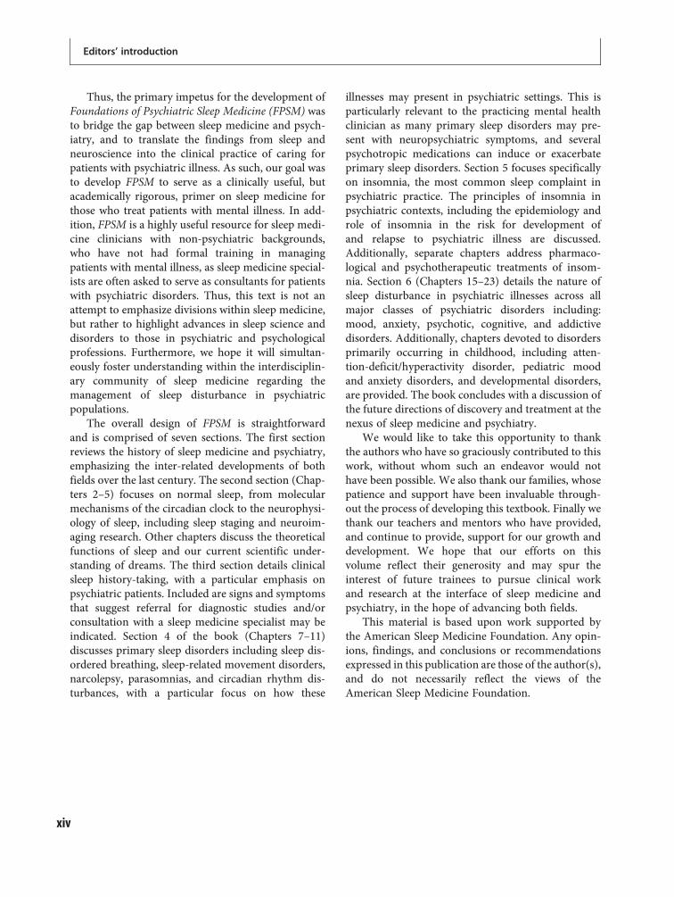
The discovery of REM sleepIn 1953, Eugene Aserinsky, then a graduate studentstudying physiology at the University of Chicago, wiredup his 9-year-old son, Armand, and other children toperform electroencephalogram (EEG) and electro-oculogram (EOG) recordings in his rebellious experi-ments on attention carried out with the encouragementof the neurophysiologist, Ralph Gerard. As is so oftenthe case, the monumental scientific discovery of REMsleep occurred in part by happenstance. When the childsubjects became bored by Aserinsky’s protocols, theyfell asleep and, because they were young, evinced rapideye movement (REM) periods shortly after sleep onsetduring which they dreamt. Aserinsky reported hisobservations to his supervisor, Nathaniel Kleitman,who suggested that they record the sleep of adults. Therest is history. All of Aserinsky and Kleitman’s adultsubjects showed EEG activation, REM periods, andwhen awakened reported long complex dreams, not atsleep onset but at 90–100 minute intervals throughoutthe night (Figure 1.1). Thus was modern sleep anddream science born [5].
Dream deprivationWilliam Dement, widely recognized as a pioneer inthe field of sleep medicine, and a graduate student andpsychiatrist working in Kleitman’s laboratory at thattime, was more interested in the dream story than hisphysiologist colleagues [6,7]. Dement was motivatedby the conviction that he could test Freud’s dreamtheory rigorously using the burgeoning knowledge ofsleep physiology and REM sleep. By preventing sub-jects from entering REM with enforced awakenings,he and the neurobiologically educated psychoanalyst,
Charles Fisher, theorized they could prevent dream-ing and thus cause psychological distress in theirsubjects [8]. Although their subjects did become psy-chologically distressed, they were perhaps moreimportantly increasingly difficult to awaken fromsleep. By the fifth night of the experiment, subjectsmade no less than 50 attempts to enter REM (uptenfold from five such attempts on the first night).This observation indicated that REM sleep, if notdreaming, was carefully conserved, and thereforemust be important.
In the years to follow, the National Institutes ofHealth (NIMH) funded a significant number of labora-tories focused on dream research. Subjects, usually stu-dents, were awakened during sleep so as to give reportsof antecedent mental activity that were then correlatedwith polysomnographic data. The results were incon-clusive and often contradictory. Controlled experimentsby Anthony Kales, another psychiatrist involved inthe early development of sleep research, revealed thatNREM sleep interruption was every bit as psychonox-ious as the enforced REM sleep awakenings [9], a find-ing that did not fit with the dominant psychoanalyticparadigm of the time. By 1975, when the NIMH ceasedfunding of “dream lab” research, the psychiatriccommunity had made relatively few inroads into thescientific status of Freud’s dream theory.
Neurobiological progressof sleep medicineDuring the same period in which dream laboratoryresearch was flourishing, remarkable progress wasbeing made using the (feline) animal model of sleepthat William Dement had also given the field [10].The prime mover of this initiative was Michel Jouvet,a French neurosurgeon working in Lyon, who hadspent a year at the University of California at LosAngeles (UCLA) founded by Horace Magoun, theco-discoverer with Giuseppe Moruzzi of the reticularactivating system [11]. Jouvet quickly localized theREM generator system to the pons [12] and suggestedthat the forebrain was activated, the eyes were causedto move, and spinal reflexes were inhibited [13]during REM sleep all from that central locale. Withthis discovery, dreaming could thus be redefined asthe inevitable subjective experience of a specificphysiological pattern of brain activation in sleep.As it turned out, all mammals shared this brainactivation process in sleep, casting doubt, albeit
Section I: Overview
2
indirectly, on Freud’s dream hypothesis. Under thisnew paradigm, REM sleep must be doing somethingbesides serving purely as a substrate for uncon-sciously driven dreaming.
How was REM sleep instantiated by the pons?Jouvet’s first guess, that it was enhanced cholinergic
neuromodulation, turned out to be correct [12]. Butwhen he read Kjell Fuxe and Anica Dahlstrom’sdescription [14] of the noradrenergic and seroton-ergic neuronal systems of the pontine brainstem, hewas sidetracked from his original (correct) hypoth-esis. Jouvet then suggested that norepinephrine drove
200
100
0
MICROVOLTS RAPID CONJUGATE EYEMOVEMENT
0 5 10
TIME IN SECONDS
15 20
(a)
BLOOD PRESSURE, RESPIRATION AND PULSE IN A 100-MINUTE SAMPLEOF UNINTERRUPTED SLEEP
5
1AIIIIIIIV
120
110S.B.P
RESP
PULSE
B.M
EEG
E.M
100
9010
529
25
204
0
TIME IN MINUTES200 210 220 230 240 250 260 270 280 290 300
V.P. 22Date : 2/21/62
(b)
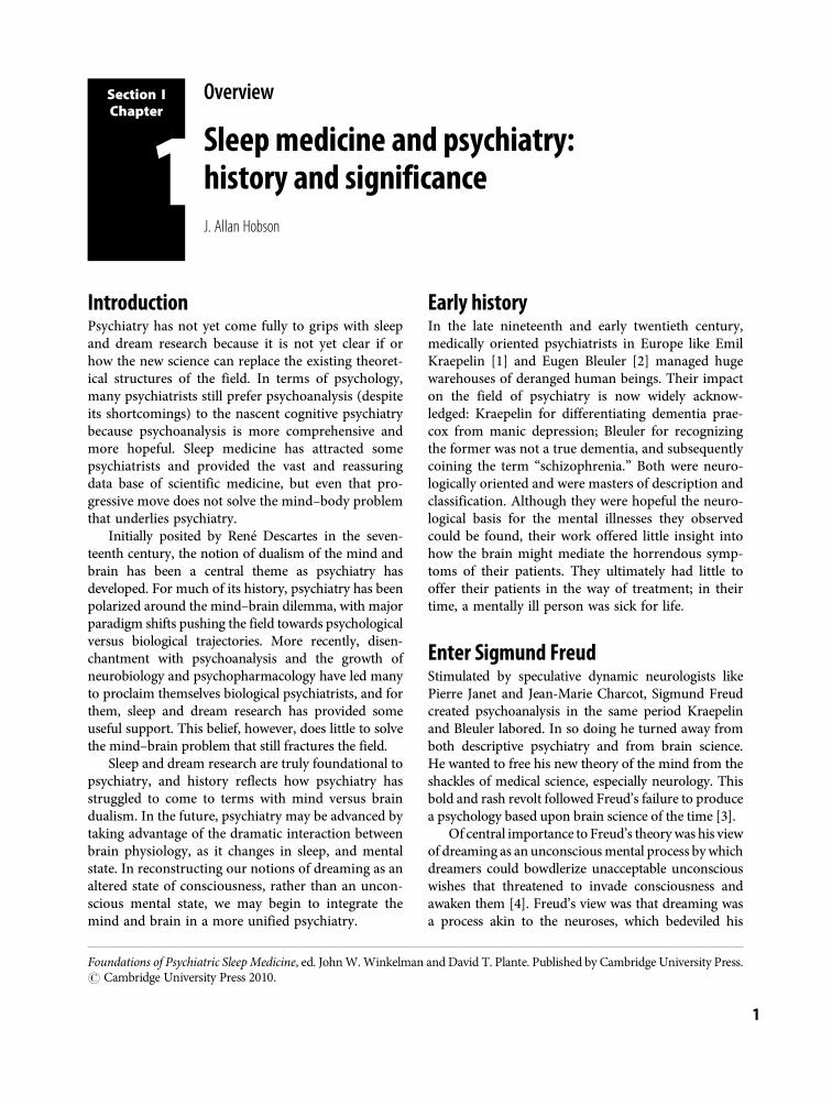
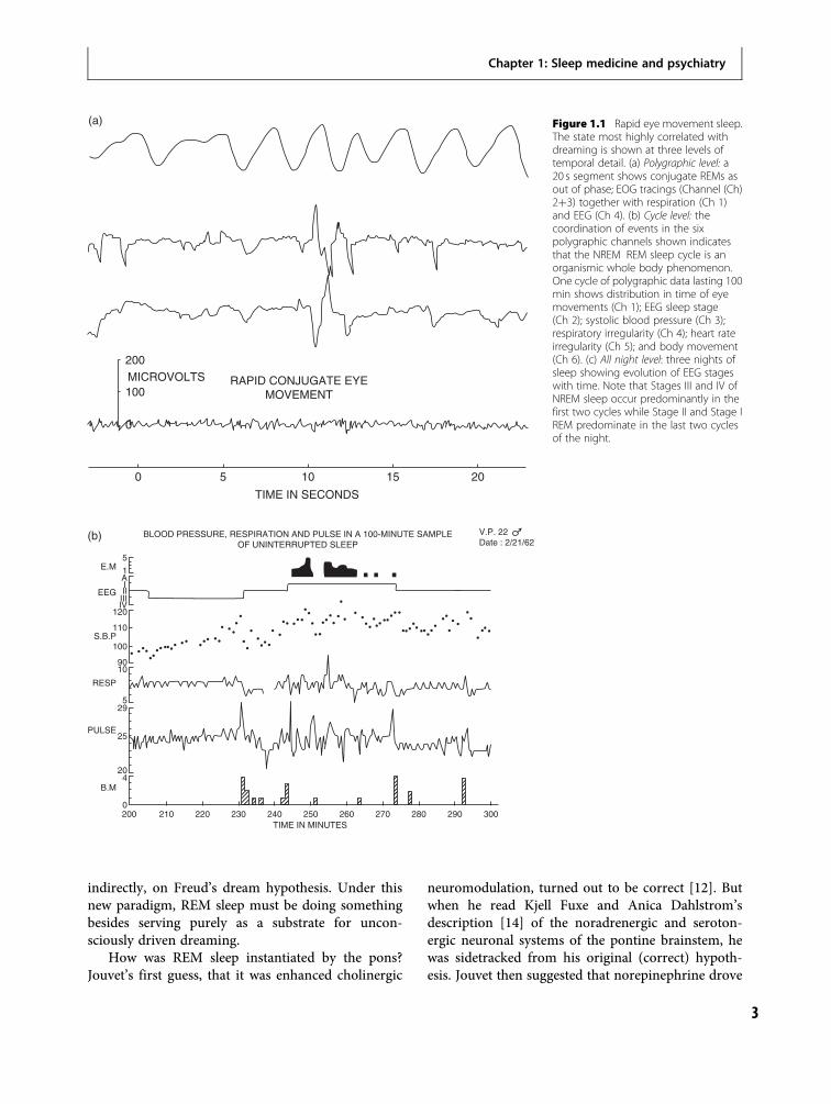
Figure 1.1 Rapid eye movement sleep.The state most highly correlated withdreaming is shown at three levels oftemporal detail. (a) Polygraphic level: a20 s segment shows conjugate REMs asout of phase; EOG tracings (Channel (Ch)2þ3) together with respiration (Ch 1)and EEG (Ch 4). (b) Cycle level: thecoordination of events in the sixpolygraphic channels shown indicatesthat the NREM REM sleep cycle is anorganismic whole body phenomenon.One cycle of polygraphic data lasting 100min shows distribution in time of eyemovements (Ch 1); EEG sleep stage(Ch 2); systolic blood pressure (Ch 3);respiratory irregularity (Ch 4); heart rateirregularity (Ch 5); and body movement(Ch 6). (c) All night level: three nights ofsleep showing evolution of EEG stageswith time. Note that Stages III and IV ofNREM sleep occur predominantly in thefirst two cycles while Stage II and Stage IREM predominate in the last two cyclesof the night.
3
Chapter 1: Sleep medicine and psychiatry

REM sleep and that serotonin drove NREM sleepwhile waking was the responsibility of dopamine[15]. By 1975, it was clear from single brainstem cellrecording experiments that both norepinephrine andserotonin cells enhanced waking rather than sleep.Instead of being excited in REM, the aminergicneurons needed to be inhibited for REM sleep tooccur. Acetylcholine-containing cells, on the otherhand, turned back on during REM, firing at levels ashigh as or higher than during waking. Subsequentexperiments utilizing cholinergic microstimulationdemonstrated that REM sleep could be induced bythese compounds. As was later discovered, dopamine-containing cells fired throughout the sleep–wake cycleeliminating any specific role for that neuromodulatorin state (i.e. wake, REM, or NREM) determination.However, this does not mean this neurotransmitterhas no role in sleep or dreaming, as it has beentheorized that dopamine – acting in the absence ofnoradrenergic and serotonergic influence – may con-tribute to the psychosis-like quality of dreaming.
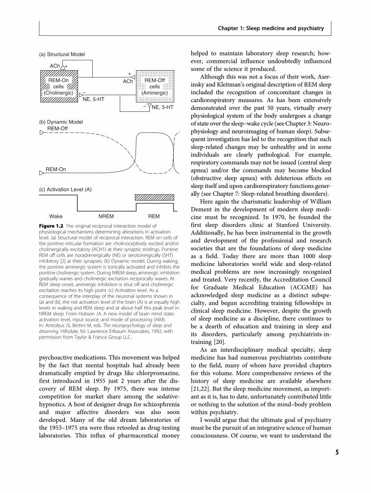
Robert McCarley (who at the time was my studentat Harvard) and I, having trained in Jouvet’s labora-tory in the 1960s, recorded from individual pontineneurons for almost 10 years before we realized thatthe brainstem had its own switching device forwaking and dreaming and that dreaming had a spe-cific neurophysiological basis. From these findings,
we constructed our “reciprocal interaction model”,that posited the level of activation (e.g. wake, NREM,or REM) was determined by the aforementionedinteraction of pontine cholinergic neurons withaminergic systems (Figure 1.2) [16,17]. In 1977, wepublished our corresponding “activation-synthesishypothesis” of the dream process in the AmericanJournal of Psychiatry [18], 1 month after issuing apaper that outlined the inaccuracies of Freud’s neuro-biological assumptions underlying psychoanalyticdream theory [19]. The activation-synthesis modelproposed that an automatically activated forebrainsynthesizes the dream by comparing information gen-erated in specific brainstem circuits with informationstored in memory. For the first time in 75 years, analternative to Freud’s theory of dreams became avail-able with this model. The ensuing negative responsefrom psychoanalytically oriented psychiatrists wasfervent at the time, and this issue is still hotly debated,reflecting how deep the roots of Freudian psychologyhad grown within psychiatry.
The rise of sleep medicineBy the mid-1970s, experimental dream laboratorieswere failing, but modern sleep medicine was burgeon-ing. There was an exponential increase in pharma-ceutical promotion of hypnotic sedatives and other
A
(c)
I
II
III
IV
A
I
II
III
IV
A
I
II
III
IV
HOURS
Figure 1.1 (cont.)
Section I: Overview
4
psychoactive medications. This movement was helpedby the fact that mental hospitals had already beendramatically emptied by drugs like chlorpromazine,first introduced in 1955 just 2 years after the dis-covery of REM sleep. By 1975, there was intensecompetition for market share among the sedative-hypnotics. A host of designer drugs for schizophreniaand major affective disorders was also soondeveloped. Many of the old dream laboratories ofthe 1953–1975 era were thus retooled as drug-testinglaboratories. This influx of pharmaceutical money
helped to maintain laboratory sleep research; how-ever, commercial influence undoubtedly influencedsome of the science it produced.
Although this was not a focus of their work, Aser-insky and Kleitman’s original description of REM sleepincluded the recognition of concomitant changes incardiorespiratory measures. As has been extensivelydemonstrated over the past 50 years, virtually everyphysiological system of the body undergoes a changeof state over the sleep–wake cycle (seeChapter 3:Neuro-physiology and neuroimaging of human sleep). Subse-quent investigation has led to the recognition that suchsleep-related changes may be unhealthy and in someindividuals are clearly pathological. For example,respiratory commands may not be issued (central sleepapnea) and/or the commands may become blocked(obstructive sleep apnea) with deleterious effects onsleep itself and upon cardiorespiratory functions gener-ally (see Chapter 7: Sleep-related breathing disorders).
Here again the charismatic leadership of WilliamDement in the development of modern sleep medi-cine must be recognized. In 1970, he founded thefirst sleep disorders clinic at Stanford University.Additionally, he has been instrumental in the growthand development of the professional and researchsocieties that are the foundations of sleep medicineas a field. Today there are more than 1000 sleepmedicine laboratories world wide and sleep-relatedmedical problems are now increasingly recognizedand treated. Very recently, the Accreditation Councilfor Graduate Medical Education (ACGME) hasacknowledged sleep medicine as a distinct subspe-cialty, and begun accrediting training fellowships inclinical sleep medicine. However, despite the growthof sleep medicine as a discipline, there continues tobe a dearth of education and training in sleep andits disorders, particularly among psychiatrists-in-training [20].
As an interdisciplinary medical specialty, sleepmedicine has had numerous psychiatrists contributeto the field, many of whom have provided chaptersfor this volume. More comprehensive reviews of thehistory of sleep medicine are available elsewhere[21,22]. But the sleep medicine movement, as import-ant as it is, has to date, unfortunately contributed littleor nothing to the solution of the mind–body problemwithin psychiatry.
I would argue that the ultimate goal of psychiatrymust be the pursuit of an integrative science of humanconsciousness. Of course, we want to understand the
(a) Structural Model
ACh
ACh
NE, 5-HT
(b) Dynamic ModelREM-Off
REM-On
(c) Activation Level (A)
Wake NREM REM
NE, 5-HT
+
+
–
–
REM-Oncells
(Cholinergic)
REM-Offcells
(Aminergic)
Figure 1.2 The original reciprocal interaction model ofphysiological mechanisms determining alterations in activationlevel. (a) Structural model of reciprocal interaction. REM on cells ofthe pontine reticular formation are cholinoceptively excited and/orcholinergically excitatory (ACH1) at their synaptic endings. PontineREM off cells are noradrenergically (NE) or serotonergically (5HT)inhibitory [2] at their synapses. (b) Dynamic model. During waking,the pontine aminergic system is tonically activated and inhibits thepontine cholinergic system. During NREM sleep, aminergic inhibitiongradually wanes and cholinergic excitation reciprocally waxes. AtREM sleep onset, aminergic inhibition is shut off and cholinergicexcitation reaches its high point. (c) Activation level. As aconsequence of the interplay of the neuronal systems shown in(a) and (b), the net activation level of the brain (A) is at equally highlevels in waking and REM sleep and at about half this peak level inNREM sleep. From Hobson JA. A new model of brain mind state:activation level, input source, and mode of processing (AIM).In: Antrobus JS, Bertini M, eds. The neuropsychology of sleep anddreaming. Hillsdale, NJ: Lawrence Erlbaum Associates; 1992; withpermission from Taylor & Francis Group LLC.
Chapter 1: Sleep medicine and psychiatry
5
biological mechanisms underlying behavioral neuro-science. Why does a person – or a snail – investigateor withdraw from an object and what molecular pro-cesses underlie these behaviors? But the field haslarger, more complex questions it must seek to answer.How can awareness arise from perception? How doeslearning become recollection? How does languagearise? Although these are daunting questions, I believethat sleep and dream research will allow some of thesequestions ultimately to be answered.
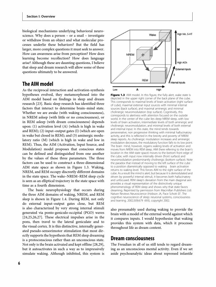
The AIMmodelAs the reciprocal interaction and activation-synthesishypotheses evolved, they metamorphosed into theAIM model based on findings in sleep and dreamresearch [23]. Basic sleep research has identified threefactors that interact to determine brain–mind state.Whether we are awake (with waking consciousness),in NREM asleep (with little or no consciousness), orin REM asleep (with dream consciousness) dependsupon: (1) activation level (A) (which is high in wakeand REM); (2) input–output gates (I) (which are openin wake but closed in REM); and (3) aminergic modu-latory ratio (M) (which is high in wake and low inREM). Thus, the AIM (Activation, Input Source, andModulation) model proposes that conscious statescan be defined and distinguished from one anotherby the values of these three parameters. The threefactors can be used to construct a three-dimensionalAIM state space as shown in Figure 1.3. Waking,NREM, and REM occupy discreetly different domainsin the state space. The wake–NREM–REM sleep cycleis seen as an elliptical trajectory in the state space withtime as a fourth dimension.
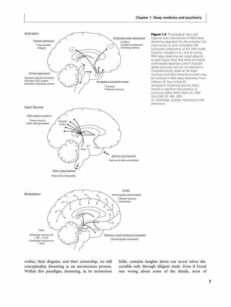
The basic neurophysiology that occurs duringthe three AIM domains of waking, NREM, and REMsleep is shown in Figure 1.4. During REM, not onlydo external input–output gates close, but REMis also characterized by very strong internal stimuligenerated via ponto-geniculo-occipital (PGO) waves[24,25,26,27]. These electrical impulses arise in thepons, then travel to the lateral geniculate and tothe visual cortex. It is this distinctive, internally gener-ated pseudo-sensorimotor stimulation that most dir-ectly supports the hypothesis that REM sleep dreamingis a protoconscious rather than an unconscious state.Not only is the brain activated and kept offline [28,29],but it autoactivates in such a way as to impressivelysimulate waking. Although inhibited, this system is
also presumably used during waking to provide thebrain with a model of the external world against whichit compares inputs. I would hypothesize that wakingprovides this system with data, which it processesthroughout life as dream content.
Dream consciousnessThe Freudian in all of us still tends to regard dream-ing as an unconscious mental activity. Even if we setaside psychoanalytic ideas about repressed infantile
Figure 1.3 AIM model. In this figure, the fully alert, wake state isdepicted in the upper right corner of the back plane of the cube.This corresponds to maximal levels of brain activation (right surfaceof cube), maximal external input sources with minimal internalsources (back surface), and maximal aminergic and minimalcholinergic neuromodulation (top surface). Cognitively, thiscorresponds to alertness with attention focused on the outsideworld. In the center of the cube lies deep NREM sleep, with lowlevels of brain activation, intermediate levels of both aminergic andcholinergic neuromodulation, and minimal levels of both externaland internal input. In this state, the mind tends towardsperseverative, non progressive thinking with minimal hallucinatoryactivity, and this is reflected in the brevity and poverty of NREMsleep reports. As cholinergic modulation increases and aminergicmodulation decreases, the modulatory function falls to its low point.The brain mind, however, regains waking levels of activation andmoves from NREM into REM sleep. AIM (here referring to the brain’slocation in the AIM state space) moves to the bottom front edge ofthe cube, with input now internally driven (front surface) andneuromodulation predominantly cholinergic (bottom surface). Notethe paradox that instead of moving to the left surface of the cubeto a position diametrically opposed to waking brain activationreturns to waking level. This forces AIM to the right surface of thecube. As a result the mind is alert, but because it is demodulated anddriven by powerful internal stimuli, it becomes both hallucinatoryand unfocused. REM sleep’s deviation from the main diagonal axisprovides a visual representation of the distinctively uniquephenomenology of REM sleep and shows why that state favorsdreaming. Reprinted by permission from Macmillan Publishers Ltd:Nature Reviews Neuroscience (Hobson JA, Pace Schott EF. Thecognitive neuroscience of sleep: neuronal systems, consciousnessand learning. 2002;3(9):679 693), copyright 2002.
Section I: Overview
6
wishes, their disguise, and their censorship, we stillconceptualize dreaming as an unconscious process.Within this paradigm, dreaming, in its mysterious
folds, contains insights about our secret selves dis-cernible only through diligent study. Even if Freudwas wrong about some of the details, most of
Activation
Input Source
Modulation
Parietal operculum
Visuospatialimagery
�
Prefrontal cortex deactivated
¯Volition¯Insight & judgement¯Working memory
Amygdala & paralimbic cortex
� Emotion� Remote memory
Pontine tegmentum
PGO system turned on
Fictive visual &motor data generated
Occiptalcortex
Geniculate
Sensory input blocked
Pons
Motor output blocked
Cortex
PPTLDTRN
IC
Aminergically demodulated
¯ Recent memory¯ Orientation
Thalamus, basal forebrain & Amygdala
Cholinergically modulated
Pons
Aminergic neurons off¯ NE ¯ 5-HT
Cholinergic neurons on¯ ACh
Real action impossible
Real world data unavailable
Activates reticular formationActivates PGO systemActivates cholinergic system
+
+ +
–
Figure 1.4 Physiological signs andregional brain mechanisms of REM sleepdreaming separated into the activation (A),input source (I), and modulation (M)functional components of the AIM model.Dynamic changes in A, I, and M duringREM sleep dreaming are noted adjacentto each figure. Note that these are highlyschematized depictions which illustrateglobal processes and do not attempt tocomprehensively detail all the brainstructures and their interactions which maybe involved in REM sleep dreaming. FromHobson JA, Pace Schott EF,Stickgold R. Dreaming and the brain:toward a cognitive neuroscience ofconscious states. Behav Brain Sci. 2000Dec;23(6):793 842, 2001;# Cambridge Journals, reproduced withpermission.
Chapter 1: Sleep medicine and psychiatry
7

psychiatry assumes he was right in this centralhypothesis that dreaming is an unconscious process.
I would argue that this dogma has outlived itsutility. If dreaming is not so much unconsciousmental activity in a Freudian sense but is, instead,an intensely conscious experience that is not remem-bered, our theoretical perspective changes dramatic-ally. Dream consciousness is vivid and distinctive, ofthat we can be sure, given our occasional recollectionof it. Interestingly, despite the nearly 2 hours of REMsleep that occurs in a healthy adult each night, ourrecall of dreams comes nowhere near this amount.I argue that this discrepancy is not due to repression,but rather to the fact that the majority of dreaming(whether it occurs in REM sleep, NREM sleep, or atsleep onset) is forgotten. But if dreaming isn’t disguis-ing repressed infantile wishes, then what is it doing?
My heretical answer to this question is thatdreaming is doing crucially important mental work.The mental work done by dream consciousness is farmore important to waking consciousness than mereprotection from unconscious infantile wishes asFreud suggested. I propose that dreaming is con-stantly serving consciousness in a variety of positiveand progressive ways.
Aserinsky and Kleitman revisited:REM sleep and immaturityThe reason that Aserinsky stumbled onto REM sleepin the first place is because he was studying childrenwho were bored and fell asleep and, fortunately,REM sleep occurs closer to sleep onset in the young.Not only does it occur at sleep onset but it occupies agreater proportion of a greater amount of sleep inthe younger the child. The newborn human infantspends 50% of its sleep time in REM. Since infantssleep 16 hours per day, they achieve nearly 8 hoursof REM sleep per day! With prematurity, these num-bers increase further, until at 30 weeks of gestation,the human fetus spends 24 hours/day in a brain-activated state that is something more like REM sleepthan wakefulness or NREM. Thus, I would arguethat REM sleep in infancy serves brain developmentin the specific enhancement of cognition andconsciousness.
This over-commitment to REM sleep by immatureanimals has not gone unnoticed. Howard Roffwarg,Joseph Muzio, and William Dement theorized in1966 that REM favored development of the visual
system [30]. Given the REM periods themselves, thePGO waves, and the intensely visual quality of dreams,I have always liked this idea and regret that it neverreceived its day in scientific court. But I suspect thedevelopmental hypothesis of Roffwarg and colleagueswas too modest. I would argue that it is not just vision,but consciousness itself that is the functional benefi-ciary of REM sleep.
Developmental considerationsBrain development proceeds, up to a point, undergenetic guidance. Chemical flavors are designatedand neuronal addresses are specified by the genome.When cholinergic and aminergic neurons meet in thepontine brainstem they interact automatically andspontaneously. When the cholinergic system is dom-inant as it is in REM, the brain is activated in onespecific mode and when the aminergic system isdominant as in waking, it is activated in anotherspecific mode. According to this theory, the aminer-gic system must develop later than the cholinergicsystem since waking consciousness follows dreamconsciousness by weeks or even months.
Although antecedence does not guarantee causal-ity, this temporal sequence means that REM sleepcould be a protoconscious state. What is meant bythe term protoconsciousness? First, it means that aprimitive sense of self could be instantiated. To para-phrase Descartes, my brain is activated, thereforeI am. When my self (or ego) is activated, I move.My self is therefore an agent. This is point two.According to Rodolfo Llinás, agent-initiated move-ment is instantiated early in development [31]. Notonly does the self-organized autoactive brain instanti-ate agent-initiated movement but it simulates boththe sensory and emotional concomitants of that activ-ity. These are points 3 and 4. REM sleep creates a selfthat acts, and feels, in a virtual world.
REM sleep and the binding problemOne of the most remarkable aspects of waking con-sciousness is its unity. Consciousness integrates a vastpanoply of information into what seems to us to be asimple and continuous flow of awareness. Strands ofdata from the outside world, from our bodies, and ourvery complex selves are woven together into a singlepiece. Our subjectivity may be conflicted but it isalways unified. Such an effect can only be achieved
Section I: Overview
8
by the binding of multiple neuronal representationsinto an integrated whole. Rather than assuming thatnumerous neural pathways pump all of this informa-tion into a single place in the brain, it has beenproposed that it is the synchrony of disparate brainparts that is the substrate of binding. Now we knowthat, in addition to synchrony, the brain utilizes modu-latory chemistry to achieve the widespread harmonynecessary to binding.
According to protoconsciousness theory, REMsleep serves binding automatically and spontaneously.No supervision is needed. And according to this newtheory, all mammals that have REM sleep have proto-consciousness. When they wake up, they have varyingdegrees of what Gerald Edelman calls primary con-sciousness [32]. According to the complexity of theirbrains, they may also achieve some degree of secondaryconsciousness. But, as far asweknow, only humans havedirected thought, propositional intent, and awareness-of-awareness. This sophisticated adaptation is presum-ably dependent on the acquisition of symbolic language.
The interaction of dreamingand waking consciousnessProtoconsciousness develops first, even before birth.There follows, especially in humans, a prolongedperiod in which waking consciousness develops. It isan explicit tenet of the protoconsciousness hypothesisthat dreaming and waking consciousness developtogether and that each of the two states enriches theother. Furthermore, it is proposed that the two statesmay interact negatively in the production of psychoticstates such as the organic mental syndrome, schizo-phrenia, and major affective disorder.
Since REM sleep brain activation precedes waking(and may occur before even birth), it follows thatwhile it may instantiate self, self-as-agent, movement,sensation, and emotion, it could not support dream-ing as we know it in adults. For adult dreaming tooccur, specific content information would need to begleaned in waking and cognitive capacity wouldneed to evolve, as it clearly does, accounting forthe fact that adult dreaming does not occur beforeages 6–8 [33]. Another empirical example of thisprinciple is the vision-free dreaming of the congeni-tally blind person. Vice versa, in order for a normalperson to see, in either waking or dreaming con-sciousness, the contentless formal frame supplied byREM sleep brain activation is essential.
The emerging picture is of a two-way street: REMsleep brain activation provides the formal substratefor waking consciousness and waking consciousnessprovides the perceptual building blocks for dreamconsciousness.
Summary and conclusionsPsychiatry was born when moral and medical forcescombined to separate the mentally ill from commoncriminals and other social undesirables. In thebeginning, the medical model prevailed but nobacteria, no viruses, and no malformations werefound in the brains of the vast majority of theseverely mentally ill.
Frustration with the organic orientation of thefield contributed to the uncritical acceptance of thepsychoanalytic psychology of Sigmund Freud whobased much of his speculation on the assumption thatdreaming was an unconscious mental process inimi-cal to waking consciousness. This point of viewgained ascendance in the first half of the twentiethcentury and continues to have support within psycho-analytic circles.
The discovery of REM sleep, and its associationwith dreaming by Aserinsky and Kleitman in 1953,was first greeted by many in the psychiatric commu-nity as an opportunity to confirm Freud. But as thesecond half of the twentieth century evolved, itbecome more and more clear that REM sleep likelyhas biological significance that transcends dreaming.During this same period, the field of sleep medicineexponentially developed, with many biologicallyoriented psychiatrists contributing significantly tothis nascent medical specialty.
Significant progress in basic sleep and dreamingresearch has yielded new insights previouslyunimaginable at the dawn of psychiatry. Now, in thetwenty-first century, psychiatric sleep medicine con-tinues to move forward with developments in sleepgenetics, bioenergetics, neurophysiology, and neuro-pharmacology. Within modern paradigms, it seemsmore likely that REM sleep (and dreaming) are thehandmaidens of waking consciousness, rather thanthe “royal road to the unconscious” as previouslyenvisioned by Freud. I suggest that REM sleep comesto support protoconsciousness (and dreams) in a waythat is specific and dynamic. It is possible that furtherconsideration of the connection between REM sleepdreaming and waking consciousness will yield the
Chapter 1: Sleep medicine and psychiatry
9
brain–mind integration necessary to a truly scientificpsychiatry. This theory is developed in more detail ina recent manuscript [34].
It is thus my deeply held belief that psychiatrymight well come of scientific age through an integra-tion of basic research of sleep, dreams, and con-sciousness. By this surprising means, it may bepossible for psychiatry not only to contribute to,but also to profit from, a specific model of brain–mind integration that could account for both normaland abnormal mental states.
References1. Kraeplin, E. Dementia praecox and paraphrenia.
Translated by R. Mary Barclay. Edited by George M.Robertson. Huntington, NY: R. E. Krieger Pub. Co;1971.
2. Bleuler E. [Dementia præcox, oder die Gruppe derSchizophrenien]. In: Aschaffenburg G, ed.Aschaffenburg, handbuch der psychiatrie. Leipzig:Franz Deuticke; 1911.
3. Freud S. The interpretation of dreams. In: Strachey J,ed. The standard edition of the complete psychologicalworks of Sigmund Freud, Vols. 4 and 5. London:Hogarth Press; 1953.
4. Freud S. Project for a scientific psychology. New York:Standard Edition; 1895.
5. Aserinsky E, Kleitman N. Regularly occurring periodsof ocular motility and concomitant phenomena duringsleep. Science. 1953;118:361 375.
6. Dement W, Kleitman N. The relation of eyemovements during sleep to dream activity: Anobjective method for the study of dreaming. J ExpPsychol. 1957;53:339 346.
7. Dement W, Kleitman N. Cyclic variations in EEGduring sleep and their relation to eye movements, bodymotility, and dreaming. Electroencephalogr ClinNeurophysiol. 1957;9(4):673 690.
8. Dement W. The effect of dream deprivation. Science.1960;131:1705 1707.
9. Kales A, Hoedemaker F, Jacobsen A, et al. Mentationduring sleep: REM and NREM recall reports. PerceptMot Skills. 1967;24:556 560.
10. Dement W. The occurrence of low voltage, fast,electroencephalogram patterns during behavioral sleepin the cat. Electroencephalogr Clin Neurophysiol. 1958;10(2):291 296.
11. Moruzzi M, Magoun HW. Brainstem reticularformation and activation of the EEG.Electroencephalogr Clin Neurophysiol. 1949;1:455 473.
12. Jouvet M. Research on the neural structures andresponsible mechanisms in different phases ofphysiological sleep. Arch Ital Biol. 1962;100:125 206.
13. Jouvet M, Michel F. Electromyographic correlationsof sleep in the chronic decorticate & mesencephaliccat. C R Seances Soc Biol Fil. 1959;153(3):422 425.
14. Dahlstrom A, Fuxe K. Evidence for the existence ofmonoamine containing neurons in the central nervoussystem. I. Demonstration in the cell bodies of brain stemneurons. Acta Physiologica Scandinavica. 1964;62:1 55.
15. Jouvet M. Biogenic amines and the states of sleep.Science. 1969;163(862):32 41.
16. Hobson JA, McCarley RW, Wyzinski PW. Sleep cycleoscillation: reciprocal discharge by two brain stemneuronal groups. Science. 1975;189:55 58.
17. McCarley RW, Hobson JA. Neuronal excitabilitymodulation over the sleep cycle: a structural andmathematical model. Science. 1975;189:58 60.
18. Hobson JA, McCarley RW. The brain as a dream stategenerator: an activation synthesishypothesisof thedreamprocess. Am J Psychiatry. 1977;134(12):1335 1348.
19. McCarley RW, Hobson JA. The neurobiologicalorigins of psychoanalytic dream theory.Am J Psychiatry. 1977;134:1211 1221.
20. Krahn LE, Hansen MR, Tinsley JA. Psychiatricresidents’ exposure to the field of sleep medicine:a survey of program directors. Acad Psychiatry.2002;26(4):253 256.
21. Shepard JW Jr, Buysse DJ, Chesson AL Jr, et al. Historyof the development of sleep medicine in the UnitedStates. J Clin Sleep Med. 2005;1(1):61 82.
22. Dement WC. History of sleep medicine. Sleep MedClinics 2008;3(2):147 156.
23. Hobson JA, Pace Schott EF, Stickgold R. Dreamingand the brain: toward a cognitive neuroscience ofconscious states. Behav Brain Sci. 2000;23(6):793 842.
24. Brooks DC, Bizzi E. Brain stem electrical activityduring deep sleep. Arch Ital Biol. 1963;101:648 665.
25. Bowker RM, Morrison AR. The startle reflex and PGOspikes. Brain Res. 1976;102(1):185 190.
26. Lydic R, McCarley RW, Hobson JA. Serotonin neuronsand sleep. II. Time course of dorsal raphé discharge,PGO waves, and behavioral states. Arch Ital Biol. 1987;126(1):1 28.
27. Nelson JP, McCarley RW, Hobson JA. REM sleep burstneurons, PGO waves, and eye movement information.J Neurophysiol. 1983;50(4):784 797.
28. Pompeiano O. The neurophysiological mechanismsof the postural and motor events duringdesynchronized sleep. Res Publ Assoc Res Nerv MentDis. 1967; 45:351 423.
Section I: Overview
10

29. Chase MH, Morales FR. Subthreshold excitatory activityand motorneuron discharge during REM periods ofactive sleep. Science. 1983;221(4616):1195 1198.
30. Roffwarg HP, Muzio JN, Dement WC. Ontogeneticdevelopment of the human sleep dream cycle. Science.1966;152(3722):604 619.
31. Llinás R. I of the vortex: from neurons to self.Cambridge, MA: MIT Press; 2001.
32. Edelman GM. Bright air, brilliant fire: on the matter ofthe mind. New York: Basic Books; 1992.
33. Foulkes D. Childrens’ dreaming and the developmentof consciousness. Cambridge: Harvard University Press;1999.
34. Hobson JA. REM sleep and dreaming: towards atheory of protoconsciousness. Nat Neurosci Rev. 2009;10: 803 814.
Chapter 1: Sleep medicine and psychiatry
11


Section IIChapter
2Normal Sleep
Neuroanatomy and neurobiology of sleepand wakefulnessJames T. McKenna, Ritchie E. Brown and Robert W. McCarley
IntroductionIn recent years, loss of sleep has increasingly beenrecognized as a major public health issue (for reviewand discussion, see http://healthysleep.med.harvard.edu/ and [1,2]). Sleep disturbance due to vocationaldemands, such as that experienced by shift workers,physicians, nurses, and emergency responders, maycontribute to decreased work performance, as well asincreased accident rates. Furthermore, sleep disorders,such as obstructive sleep apnea, insomnia, narcolepsy,and restless legs syndrome, affect millions of people.In addition, many psychiatric illnesses, such as depres-sion, anxiety, and post-traumatic stress disorder, areassociated with sleep disturbance.
Investigations conducted largely in the last centuryrevealed multiple brain systems responsible for thestates of wakefulness, rapid eyemovement (REM) sleep,and non-REM (NREM) sleep. Transitions betweenthese vigilance states involve neural systems of intercon-nected brain regions and neurotransmitters. In recentyears, knowledge of vigilance state regulation has pro-gressed considerably due to new techniques, includingpharmacological, lesioning, electrophysiological, and,most recently, molecular-level technologies. Thischapter will review both the neurobiological mechan-isms generating these different behavioral states, as wellas the factors (homeostatic and circadian) that influencetheir timing.
This chapter will begin with an overview of theneural systems involved in vigilance state regulation.This will include a description of relevant brainregions/nuclei, as well as the neurotransmittersinvolved. We will describe the flip-flop hypothesisof sleep/wake regulation, as well as the reciprocal
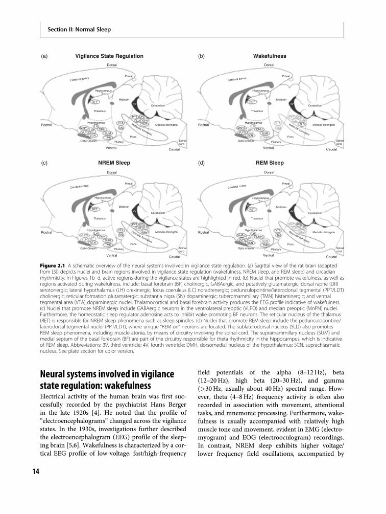
interaction model of REM sleep regulation. The two-process model of sleep regulation will then beexplained, describing the interaction of process S (thehomeostatic drive to sleep) and process C (the circa-dian drive to sleep). This discussion will include adescription of a homeostatic sleep regulator; namely,the purine adenosine. Process C will be described,including a description of the brain nuclei and neuralsystems involved in circadian rhythmicity, includingthe suprachiasmatic and dorsomedial hypothalamicnuclei. Furthermore, we will review the proposedmechanism by which circadian rhythmicity occursin the suprachiasmatic nucleus, involving a complexinteraction of genes.
To help orient the reader to the systems import-ant in maintaining each behavioral state, we firstpresent an anatomical schematic of their location(Figure 2.1a), followed by figures specifically indicat-ing the regions important in promoting wakefulness(Figure 2.1b), NREM sleep (Figure 2.1c), and REMsleep (Figure 2.1d). Figure legends for Figures 2.1b,2.1c, and 2.1d describe the transmitter(s) importantfor each state. Most of the investigations reviewed inthis chapter and, in turn, most of our knowledge ofsleep/wakefulness regulation is derived from animalmodels, usually rodent, feline, or canine. Many ofthe nuclei to be described are anatomically inter-connected, acting synergistically in vigilance stateregulation. Interestingly, the majority of studiesemploying lesioning of wakefulness- and sleep-relatednuclei have found only minor effects on the sleep–wake cycle, likely due to redundancy within theneuroanatomical circuitry involved in vigilance stateregulation.
Foundations of Psychiatric SleepMedicine, ed. JohnW.Winkelman andDavid T. Plante. Published by CambridgeUniversity Press.# Cambridge University Press 2010.
13
Neural systems involved in vigilancestate regulation: wakefulnessElectrical activity of the human brain was first suc-cessfully recorded by the psychiatrist Hans Bergerin the late 1920s [4]. He noted that the profile of“electroencephalograms” changed across the vigilancestates. In the 1930s, investigations further describedthe electroencephalogram (EEG) profile of the sleep-ing brain [5,6]. Wakefulness is characterized by a cor-tical EEG profile of low-voltage, fast/high-frequency
field potentials of the alpha (8–12Hz), beta(12–20Hz), high beta (20–30Hz), and gamma(>30Hz, usually about 40Hz) spectral range. How-ever, theta (4–8Hz) frequency activity is often alsorecorded in association with movement, attentionaltasks, and mnemonic processing. Furthermore, wake-fulness is usually accompanied with relatively highmuscle tone and movement, evident in EMG (electro-myogram) and EOG (electrooculogram) recordings.In contrast, NREM sleep exhibits higher voltage/lower frequency field oscillations, accompanied by
Dorsal
Vigilance State Regulation Wakefulness
NREM Sleep REM Sleep
Pineal
Hippocampus
3V
4V
4V
Pons
RETMidbrain
Cerebellum
Medulla oblongata
Spinalcord
RostralReticular formation
Thalamus
MnPnBF
SCN
Rostral
Caudal
VLPODMH
SUM
SN
VTA
PPT DR
SLDLDT LC
TMN
Pituitary
Ventral
Optic Chiasm
LHHypothalamus
Cerebral cortex
Dorsal
Pineal
Hippocampus
3V
4V
4V
Pons
RETMidbrain
Cerebellum
Medulla oblongata
Spinalcord
Reticular formation
Thalamus
MnPnBF
SCN
Caudal
VLPODMH
SUM
SN
VTA
PPT DR
SLDLDT LC
TMN
Pituitary
Ventral
Optic chiasm
LHHypothalamus
Cerebral cortex
Dorsal
Pineal
Hippocampus
3V
4V
4V
Pons
RETMidbrain
Cerebellum
Medulla oblongata
Spinalcord
RostralReticular formation
Thalamus
MnPNBF
SCN
Rostral
Caudal
VLPODMH
SUM
SN
VTA
PPT DR
SLDLDT LC
TMN
Pituitary
Ventral
Optic Chiasm
LHHypothalamus
Cerebral cortex
Dorsal
Pineal
Hippocampus
3V
4V
4V
Pons
RETMidbrain
Cerebellum
Medulla oblongata
Spinalcord
Reticular formation
Thalamus
MnPnBF
SCN
Caudal
VLPODMH
SUM
SN
VTA
PPT DR
SLDLDT LC
TMN
Pituitary
Ventral
Optic chiasm
LHHypothalamus
Cerebral cortex
(a) (b)
(c) (d)
Figure 2.1 A schematic overview of the neural systems involved in vigilance state regulation. (a) Sagittal view of the rat brain (adaptedfrom [3]) depicts nuclei and brain regions involved in vigilance state regulation (wakefulness, NREM sleep, and REM sleep) and circadianrhythmicity. In Figures 1b d, active regions during the vigilance states are highlighted in red. (b) Nuclei that promote wakefulness, as well asregions activated during wakefulness, include: basal forebrain (BF) cholinergic, GABAergic, and putatively glutamatergic; dorsal raphe (DR)serotonergic; lateral hypothalamus (LH) orexinergic; locus coeruleus (LC) noradrenergic; pedunculopontine/laterodorsal tegmental (PPT/LDT)cholinergic; reticular formation glutamatergic; substantia nigra (SN) dopaminergic; tuberomammillary (TMN) histaminergic; and ventraltegmental area (VTA) dopaminergic nuclei. Thalamocortical and basal forebrain activity produces the EEG profile indicative of wakefulness.(c) Nuclei that promote NREM sleep include GABAergic neurons in the ventrolateral preoptic (VLPO) and median preoptic (MnPN) nuclei.Furthermore, the homeostatic sleep regulator adenosine acts to inhibit wake promoting BF neurons. The reticular nucleus of the thalamus(RET) is responsible for NREM sleep phenomena such as sleep spindles. (d) Nuclei that promote REM sleep include the pedunculopontine/laterodorsal tegmental nuclei (PPT/LDT), where unique “REM on” neurons are located. The sublaterodorsal nucleus (SLD) also promotesREM sleep phenomena, including muscle atonia, by means of circuitry involving the spinal cord. The supramammillary nucleus (SUM) andmedial septum of the basal forebrain (BF) are part of the circuitry responsible for theta rhythmicity in the hippocampus, which is indicativeof REM sleep. Abbreviations: 3V, third ventricle; 4V, fourth ventricle; DMH, dorsomedial nucleus of the hypothalamus; SCN, suprachiasmaticnucleus. See plate section for color version.
Section II: Normal Sleep
14
little movement. During REM sleep, the cortical EEGprofile returns to low-voltage/high-frequency activity,but is accompanied by an overall lack of movementand postural tone. A more detailed review of the EEG/EMG profile of NREM and REM sleep is providedlater in this chapter.
Baron Constantin Von Economo studied theworldwide flu epidemic in the late 1920s, andobserved that in patients who developed encephalitislethargica, a neuropsychiatric disorder characterizedby either severe insomnia or hypersomnia, the anter-ior hypothalamus was damaged in the post-mortemtissue of patients that had suffered from insomnia;whereas damage posterior to the hypothalamus at thejunction of the forebrain and brainstem was identifiedin patients that exhibited excessive sleepiness [7].Therefore, he concluded that the brain’s sleep-inducing regions were located in the anterior hypo-thalamus, and wakefulness-promoting regions werelocated in the posterior hypothalamus. As describedbelow in the review of NREM sleep mechanisms ofthis chapter, Von Economo’s hypotheses were largelycorrect.
In the 1930s, Frederick Bremer performed seminalstudies of EEG activity in the cat [8,9]. In one prepar-ation, termed encephale isolé, a cut was made in thelower part of the medulla. In the other preparation,termed cerveau isolé, a cut was made at the junction ofthe midbrain and the brainstem. Interestingly, theencephale isolé preparation allowed a continual pres-ence of EEG activity reflecting the fluctuation betweenwake and sleep activity. In the second cerveau isolépreparation, though, the cat EEG profile remained ina constant sleep-like state. Therefore, it was concludedthat sleep involved a deafferentation of the cerebralcortex.
Following the seminal work of Bremer, Moruzziand Magoun reported that electrical stimulation ofthe brainstem reticular formation (RF) produced acortical EEG profile indicative of wakefulness [10].These findings lead to the concept of the reticularactivating system (RAS), comprised of the reticularformation and its connections, which generate wake-fulness (for review, see [11]). Subsequent investiga-tions have revealed that the RAS is composed of adorsal and ventral arousal pathway. The dorsalarousal pathway is composed of select nuclei in thebrainstem, particularly the pontine oralis and mesen-cephalic RF, that project to midline thalamic nuclei,which subsequently have widespread projections
throughout the neocortex. The ventral arousal path-way, initially identified by Von Economo, includesprojections from the RF to the basal forebrain andposterior hypothalamic regions, which then influencecortical EEG activation by means of projections to theneocortex that bypass the thalamus [11,12,13]. Initialprojections of both the dorsal and ventral branches ofthe ascending arousal systems are largely glutamater-gic, as are the related thalamic and a portion of thebasal forebrain projections to the cortex. Additionally,the ventral arousal pathway involves a number ofneurotransmitter systems besides glutamate, includ-ing acetylcholine, norepinephrine, serotonin, hista-mine, and orexin (also known as hypocretin), thatare involved in the generation and maintenance ofwakefulness.
AcetylcholineAcetylcholine not only plays a critical role in para-sympathetic and neuromuscular junctions [14], butalso in the control of vigilance states. Two brainregions rich in cholinergic neurons are of particularimportance in both the dorsal and ventral branches ofthe RAS. The first region is the basal forebrain (BF,Figure 2.1), including the medial septum/verticallimb of the diagonal band of Broca, the horizontallimb of the diagonal band, magnocellular preopticnucleus, substantia innominata, and nucleus basalis.A second region of interest is located in the midbraintegmentum, distributed across the laterodorsal teg-mental (LDT) and pedunculopontine tegmental(PPT) nuclei (see Figure 2.1).
Neuroanatomical studies have defined the BF,including cholinergic neurons, as a principal relayfor ascending activation of neocortical regions in theventral arousal pathway. Double-labeling experiments(employing immunohistochemistry techniques todetermine the cholinergic phenotypes of BF neurons,in combination with retrograde tract tracing tech-niques) have demonstrated BF cholinergic projectionsto widespread cortical regions [15,16]. This techniquehas also been used to describe the more specific pro-jections of the medial septum/vertical limb of thediagonal band of Broca to the hippocampus, includ-ing both GABAergic and cholinergic input(for review, see [17,18]). These septo-hippocampalprojections are part of the brain circuitry involvedin the generation of the EEG theta wave, recordedin the hippocampus during REM sleep, as well as
Chapter 2: Neuroanatomy and neurobiology
15
during movement and specific mnemonic/cognitivetasks in waking.
Immunohistochemical detection of the proteinc-Fos may be employed to indicate neuronal acti-vation, where c-Fos immunohistochemical stainingfollowing sacrifice of the animal reflects neuronalsubpopulation activation within the precedinghour [19,20]. These studies described a subpopulationof BF cholinergic neurons that express the c-Fos pro-tein during sleep deprivation [21,22,23], in contrast tolittle co-localization following sleep deprivationrecovery (when sleep rebound occurred) or naturalspontaneous sleep. Overall, c-Fos protein expressionin the cholinergic neurons of BF increased as thepercentage of time awake in the hour prior to sacrificeincreased. This observation is consistent with a rolefor BF cholinergic neurons in promoting corticalactivation. Although the documentation of c-Fosand other immediate early gene protein expressionis useful, it is not necessarily a faithful representationof neuronal discharge. For example, spinal alphamotor neurons do not express c-Fos with discharges,and c-Fos may reflect calcium influx even withoutdischarge. It is thus important to realize c-Fos evi-dence is provisional and needs to be confirmed byelectrophysiological recordings, as is discussed in arecent review [24].
Electrophysiological and pharmacological studiesconfirm the particular role of acetylcholine in corticalEEG activation [11,25]. Single unit recordings revealedthat neurons in the BF, LDT, and PPT regions weremore active during wakefulness, when compared toNREM sleep [25,26,27]. Subsequent investigationsdescribed BF neurons, determined to be cholinergic bymeans of post hoc juxtacellular labeling, which increasedfiring during cortical activation [28,29]. These choliner-gic neurons discharged in rhythmic bursts in associ-ation with wakefulness-related cortical EEG theta andgamma wave activity [30]. Also, inactivation of BF bylesioning of cell bodies, sparing fibers of passage,decreases wakefulness [11]. Although acetylcholineis a crucial neurotransmitter in the arousal mechanism,it is also important to recognize that BF GABAergicand glutamatergic neurons also play a role in corticalactivation [31,32,33].
Unlike other wakefulness-related neurotransmit-ter systems that are described in this chapter that aretypically decreased in both NREM and REM sleep,the highest levels of acetylcholine in the cortex andthalamus occur during both wakefulness and REM
sleep, implicating acetylcholine as an importantneurotransmitter in REM sleep regulation [34]. BFprojections provide the cholinergic innervation ofthe neocortex, while LDT/PPT projections providecholinergic innervation of thalamic regions involvedin the dorsal arousal pathway. Electrophysiologicalstudies demonstrated increased neuronal firingin BF and LDT/PPT during wakefulness and REMsleep [26,27,29]. The role of acetylcholine inREM sleep control will be further discussed in thefollowing sections.
SerotoninSeveral midbrain/brainstem regions rich in serotonin(5-HT), i.e. the dorsal, median, magnus, obscurus, andpallidus raphe nuclei [35,36], are involved in vigilancestate regulation. Serotonin plays a complex role in vigi-lance state regulation, due to its actions in differentbrain regions, as well as the complexity of the multipleserotonergic auto- and heteroreceptors distributedthroughout the arousal circuitry. Serotonergic neuronssend projections to many areas involved in vigilancestate regulation. These neurons are most active duringwakefulness, have considerably decreased firing duringNREM sleep, and cease activity during REM sleep[37,38]. Correspondingly, 5-HT levels in various brainregions, including the neocortex, significantly riseduring wakefulness, and decrease considerably duringNREM and REM sleep [39,40].
Recent neurochemical and electrophysiologicalinvestigations conclude that serotonin promoteswakefulness and inhibits REM sleep, complementingthe action of noradrenergic and histaminergicsystems. This contrasts with early studies which sug-gested a sleep-promoting role for serotonin, basedmainly on experiments which showed that depletionof serotonin by means of p-chlorophenylalanine(PCPA) led to insomnia [41,42]. Amounts of REMsleep increased following specific serotonergic inhib-ition by means of injection of 8-OH-DPAT into thedorsal raphe nucleus (acting on the 5-HT1A inhibitoryautoreceptor) [43,44]. Furthermore, infusion of8-OH-DPAT in the median raphe nucleus promotedhippocampal theta activity [45,46].
Recent studies in mice have reinforced thehypothesis that serotonin is involved in both depres-sion and sleep. In the serotonin transporter knock-out mouse, extracellular serotonin levels weresignificantly elevated, REM sleep was enhanced,
Section II: Normal Sleep
16

and these animals exhibited depression-like behavior[47,48,49,50]. The seemingly paradoxical increasein REM sleep in these knock-out mice is likely dueto compensatory developmental adaptations in theseanimals, such as downregulation of the 5-HT1A recep-tor (which normally inhibits REM sleep) on choliner-gic neurons in response to the enhanced serotonergictone. Blockade of the 5-HT1A receptor early in devel-opment reversed such phenomena in the knock-out mouse [51]. Furthermore, neonatal treatmentof normal (non-knock-out) female mice with theselective serotonin reuptake inhibitor, escitalopram,induced depression- and anxiety-like behavior, as wellas an increase in REM sleep [52].
Noradrenaline (NA)The locus coeruleus, located bilaterally in thedorsolateral pons, contains a large group of noradren-ergic neurons. Similar to serotonergic neurons,noradrenergic neurons of LC project to widespreadareas of the neuraxis, and are most active duringwakefulness, minimally active during NREM sleep,and cease activity during REM sleep [53,54,55].Non-specific lesions of the LC lead to a minimaldecrease in wakefulness behavior and attenuation ofthe cortical EEG indicative of wakefulness [56]. Thismay be due to the redundancy of systems involved invigilance state regulation, for the many wakefulness-related neurotransmitter systems described here actsimilarly on the brain nuclei comprising the dorsaland ventral arousal pathways, as well as in the cortex.Emotional states that involve an increase of NA activ-ity (e.g. stress) may lead to overexcitation of arousalregions, causing vigilance state dysregulation, such asthat seen in insomnia [57].
Interestingly, the actions of NA may be eitherexcitatory or inhibitory, depending on the type ofadrenoreceptor on the efferent neurons of interest.Overall, it appears that NA projections to nuclei ofarousal systems in the brainstem, BF, and spinal cordare excitatory, acting on alpha 1 receptors [58,59]. Incontrast, noradrenergic projections to sleep-relatedneurons, such as preoptic region neurons (to bereviewed later), as well as noradrenaline neuronsthemselves, and LDT/PPT neurons, are inhibitory,acting on alpha 2 receptors [60,61]. In particular,noradrenergic LC neurons may be involved in main-taining muscle tone during wakefulness (to bereviewed later).
DopamineIt is unclear what role, if any, dopamine (DA) plays invigilance state regulation. Substantia nigra and mid-brain dopaminergic neurons project to wakefulness-related brain regions, including BF and prefrontalcortex [62,63]. These neurons fire in bursts, correlatedto arousal and reward [64]. Single-unit recordingsin the substantia nigra and ventral tegmental area,though, were not able to determine firing rate fluctu-ation between wakefulness versus sleep states [65].Recently, another group of dopaminergic neurons inthe ventral periaqueductal gray (vlPAG) was described,where c-Fos protein expression correlatedmost stronglywith wakefulness [66]. These neurons were located nearthe dorsal raphe nucleus, interspersed with serotonergicwakefulness-active cells, and projected to many ofthe wakefulness-related nuclei. Further investigations,including electrophysiological profiling of theseneurons, will allow conclusive evidence of a role theseneurons may play in vigilance state regulation. Interest-ingly, drugs that block the reuptake of DA and NAand thus enhance dopaminergic and noradrenergicrelease, such as cocaine and amphetamine, are usuallyassociated with stimulatory effects. Additionally, DA-stimulating compounds are used to treat disorders of cen-tral hypersomnolence such as narcolepsy (see Chapter 9:Hypersomnias of central origin) [67,68].
HistamineOlder generation antihistamines, known to cross theblood/brain barrier, have sedating effects, acting asantagonists largely on the histamine 1 (H1) receptorof wakefulness-related nuclei [69]. Histaminergicneurons are located in the tuberomammillary nucleusof the posterior hypothalamus (TMN), and project to anumber of the arousal-related brain nuclei, as well asdirectly to the cortex [70,71]. The discharge profile ofthese neurons matches that of noradrenergic and sero-tonergic neurons, where most action potential firingoccurs during wakefulness, considerably less firing isobserved during NREM sleep, and firing ceases duringREM sleep [72,73,74]. Histamine release in the poster-ior hypothalamus is highest during wake, lower duringNREM sleep, and minimal during REM sleep [75],matching the discharge profile.
Non-specific lesioning of the TMN region, aswell as histaminergic-specific lesions and chemicalmanipulations, support the suggestion that this regionmay contain neurons that promote wakefulness [73,76].
Chapter 2: Neuroanatomy and neurobiology
17

An investigation by Siegel and colleagues recentlyrevealed a unique role of histaminergic neurons in themaintenance of wakefulness [74]. Cataplexy is a suddenloss of muscle tone in waking with preservation ofconscious awareness, and is a symptom of the sleepdisorder narcolepsy. During cataplectic attacks in thedog, NA neurons ceased firing, serotonergic neuronsreduced firing, but histaminergic neurons remainedactive. Thus, while NA and serotonergic neurons par-ticularly appear to maintain muscle tone during wake-fulness, histamine neurons may help to maintaincortical activity indicative of wakefulness. Mostrecently, reduced histamine levels have also beenreported in the cerebrospinal fluid of narcolepticpatients [77,78].
Orexin (hypocretin)Neurons containing the neuropeptide orexin (alsocalled hypocretin) are located largely in the periforni-cal region (near the fornix) of the lateral hypothal-amus. Recent investigations reveal a unique role oforexin neurons in vigilance state regulation, as well asin the sleep disorder narcolepsy, by means of theirextensive input to a number of sleep/wake-relatednuclei [79,80].
Narcolepsy is characterized by symptoms includ-ing excessive daytime sleepiness (hypersomnolence),hypnagogic hallucinations, cataplexy, sleep-onsetREM periods, and sleep paralysis (see Chapter 9:Hypersomnias of central origin). In 1999, two seminalinvestigations suggested that orexin plays a role inthis disease. A deficit of the orexin type II receptorwas discovered in a strain of dogs that exhibit narco-lepsy [81]. In another study, orexin knock-out mice(lacking the prepro-orexin gene, the precursor for theorexin protein) exhibited decreased wakefulness andbehavioral arrest during transitions from wakefulnessto sleep, similar to the narcoleptic symptoms of sleep-onset REM episodes and cataplexy [82]. Behavioralarrest was described as a sudden cessation of motoractivity, including a change in posture, which thenabruptly ended with resumption of motor activity.Follow-up orexin-specific lesioning and knock-downstudies described an increase in the number of tran-sitions between vigilance states, and abnormalexpression of REM-related characteristics duringwakefulness, similar to the symptomology of narco-lepsy [83,84,85,86]. An examination of post-mortemtissue of narcoleptic humans revealed an almost total
absence of orexin neurons [87,88]. Furthermore, lowcerebrospinal fluid orexin levels occur in many nar-coleptic patients, and prominently in cases with cata-plexy [89,90]. Therefore, orexin plays a central role inthe maintenance of wakefulness, and abnormalitiesmay lead to sleep-related pathology such as nar-colepsy. Of note, because there are no marketedmedications that directly affect orexigenic neuro-transmission, amphetamine-like stimulants and DAreuptake inhibitors have been prescribed to treatexcessive sleepiness in narcolepsy, but appear to havelittle effect on cataplexy, for which compounds thatinhibit adrenergic and serotonergic reuptake areeffective [68].
Direct application of wake-promoting agents intothe LH increases orexinergic neuronal activity[91].Similar to the majority of wakefulness-related nuclei,orexinergic cell firing largely occurs during wakeful-ness, decreases considerably during NREM sleep, andceases during REM sleep, except for some activityduring REM-related muscle twitches [92]. Microdia-lysis sampling in the lateral hypothalamus foundhighest orexin levels during wakefulness [93], andfollowing examination of c-Fos protein expression itwas concluded that orexinergic neurons were wake-fulness-active [94]. The unique role orexin may playin vigilance state regulation has been conceptualizedin a “flip-flop”model of wakefulness/sleep transitions,in which orexin serves to stabilize transitions betweensleep and wakefulness (see below).
Neural systems involved in vigilancestate regulation: NREM sleepNREM sleep may be classified into four stagesin humans according to criteria developed byRechtschaffen and Kales (R&K Criteria), or threestages (N1, N2, N3) according to recent criteria ofthe American Academy of Sleep Medicine (AASM).Stage 1 NREM sleep (N1) activity usually exhibits amixture of theta (4–8Hz) and alpha (8–12Hz) cor-tical EEG activity. During stage 2 NREM sleep (N2),sleep spindles and K-complexes become evident. Sleepspindles, usually of the frequency of 12–16Hz andlasting 0.5 to 1.5 seconds, are generated from theinterplay between thalamic and cortical regions, andare present throughout the deeper stages (2–4) ofsleep (for review, see [11]). K-complexes are brief,high-voltage (<100 µV) spikes that last approximately0.5 seconds, and are unique to stage 2 sleep. Stages 3
Section II: Normal Sleep
18
and 4 (together form N3) are considered “deeper”NREM sleep, and are characterized by large/high-amplitude, low-frequency delta range (1–4Hz) activ-ity; hence, it is also termed slow-wave sleep (SWS).NREM sleep states recorded in animals usually arenot subdivided into the stages employed in humansleep, although delta (deep) sleep is often differenti-ated from light sleep. Furthermore, neocortical slowoscillations (0.5–1Hz) are evident throughout NREMsleep, and play a role in facilitation of thalamic andintracortical synchronization (for review, see [11]).This slow oscillation may further act to synchronizeNREM delta activity with sleep spindles. NREM sleepis characterized by low muscle tone and moderate/lack of movement, evident in EMG and EOGrecording.
Sleep spindles during NREM sleepIt has been proposed that spindle activity is importantfor the disconnection of the cerebral cortex from theexternal environment during sleep. The bilateralreticular nucleus of the thalamus (RET) is the pace-maker of spindle oscillations, which are generated bymeans of RET GABAergic inhibitory projections tothalamocortical neurons that are responsible for cor-tical EEG activation [95,96]. Spindle oscillations maybe readily recorded in both thalamic and corticalregions during stages 2–4 of NREM sleep, and lesion-ing of the reticular nucleus-rostral thalamic nucleipathway attenuates spindle activity [97].
Cholinergic projections from the brainstem LDT/PPT excite thalamocortical relay neurons, and dir-ectly inhibit RET. These actions desynchronize thethalamic reticular network, and lead to arousal [11].Particularly, stimulation of PPT prevents spindleactivity in RET [98]. Basal forebrain projections mayplay a role in spindle blockage, by means of GABA-ergic and cholinergic projections to the rostral sub-regions of the RET [99], and chemical lesions of theBF have been shown to produce increased spindleactivity [100].
K-complexes during NREM sleepAnimal studies demonstrate that the K-complex,most often observed in human sleep during stage 2NREM sleep, represents a combination of the neocor-tical slow oscillations and sleep spindles. A cycle ofthe slow oscillation is followed by a brief spindlesequence in thalamocortical neurons, which is
transferred to the cortical EEG, and is seen as asurface-negative (upward) EEG transient, corres-ponding to the excitation in deeply lying neocorticalneurons, followed by a surface-positive (downward)component and a short sequence of spindle waves.For a more detailed review of K-complexes duringNREM sleep, the reader is referred to [11].
Delta frequency (1–4 Hz) EEG activityduring NREM sleepThe cellular basis of delta waves (0.5–4Hz) originat-ing in thalamocortical neurons is that a hyperpolar-ized membrane potential in cortically projectingthalamic neurons permits the occurrence of deltawaves in thalamocortical circuits. In both the thal-amus and cortex, a phasic input to the hyperpolarizedmembrane causes a calcium-mediated depolarization(low threshold spike, LTS) on which a burst ofsodium-mediated spikes occur (low threshold burst).The slow time course of delta waves is determined bythe time course of the LTS and the subsequent hyper-polarization to re-enable another LTS. Any factorspersistently depolarizing the membrane in either thal-amic or cortical neurons will block delta wavesthrough inactivation of the calcium channels mediat-ing the LTS, and these are the wake–active systemsillustrated in Fig. 2.1b. Cholinergic input from BF tothe cortex, and from LDT/PPT to the thalamus, isthought to be especially important in arresting deltawaves. Also, brainstem noradrenergic and serotoner-gic projections may disrupt delta activity in waking,although they are inactive during REM sleep. Thus,delta waves during sleep may be seen to representthalamocortical oscillations occurring in the absenceof activating inputs. For a more detailed review ofdelta frequency EEG activity during NREM sleep, thereader is referred to [11].
Neocortical slow (~0.5 Hz) EEG activity/oscillation during NREM sleepThe neocortical slow oscillation plays a role ingrouping delta and fast rhythms (gamma and fastbeta). This slow oscillation (~0.5Hz) was firstdescribed by Steriade and collaborators in anaesthe-tized animals (for review, see [11]), and has beenconfirmed in both naturally sleeping animals and inthe human EEG and magnetoencephalogram (MEG),which measures the magnetic field produced by
Chapter 2: Neuroanatomy and neurobiology
19
electrical brain activity. The cortical nature of theslow oscillation was demonstrated by its persistenceafter disconnecting input to the cortex, and isfound in cortical neurons of all layers. The slowoscillation consists of prolonged depolarizations,associated with brisk firing (~8–40Hz, “up states”,lasting 0.3–0.6 s), and prolonged hyperpolarizationsduring which neurons are silent (“down states”).The depolarizing phase consists of excitatory synapticinput (N-methyl-D-aspartic acid (NMDA) and non-NMDA mediated) and a voltage-dependent persistentsodium current, as well as fast inhibitory input (beta/gamma band) reflecting the action of synapticallycoupled GABAergic local-circuit cortical cells. Thehyperpolarizing phase is thought to result from theremoval of excitatory input (disfacilitation). Althoughthe literature consistently refers to it as an oscillation,its periodic occurrence has been difficult to demon-strate definitively, unlike, for example, sleep spindles.
GABAergic preoptic nucleiGamma-aminobutyric acid (GABA) is the mostprominent inhibitory neurotransmitter in the centralnervous system. A variety of anesthetics and hypnot-ics act on GABA receptors. For example, benzodi-azepines depress wakefulness/arousal circuitry bymeans of modulation of the GABA-A receptor.Although GABA is an important neurotransmitterfor sleep induction, some groups of GABA neuronsmay be involved in wakefulness as well as REM sleepcontrol (see below).
Early evidence from studies employing lesioningand electrophysiological recordings suggested that aproportion of neurons in the basal forebrainand neighboring preoptic regions act to promotesleep [12,101]. Recordings of preoptic neuronal activ-ity demonstrated increased firing of many neurons inthese areas during sleep, compared to activity duringwakefulness. Juxtacellular labeling further definedthese particular neurons as GABAergic[29]. c-Fosprotein expression studies described the location ofthese sleep-active populations as being largely in thepreoptic nuclei, and double-labeling of these neuronswith antibody for the GABA-synthesizing enzymeglutamic acid decarboxylase (GAD) led to the conclu-sion that the majority of sleep-active neuronswere GABAergic, located in the median preopticnucleus and the bilateral ventrolateral preoptic nuclei[102,103].
The ventrolateral preoptic nucleus (VLPO)receives input from a variety of different nuclei ofthe arousal circuitry, including serotonergic and nor-adrenergic input [104]. VLPO projections to a varietyof the wakefulness-related nuclei use the neurotrans-mitters galanin and GABA [105]. The core of VLPOcontains neurons that largely project to TMN, whilethe extended cluster surrounding region is more con-nected to other arousal-related regions, such as LCand the raphe nuclei [105]. Specific lesions located inthe central cluster of VLPO led largely to decreases ofNREM sleep, but lesions of the surrounding extendedregion of VLPO produced largely decreases ofREM sleep [106].
A subset of GABAergic neurons in the medianpreoptic nuclei (MnPN) are also active during NREMsleep, determined by electrophysiological studies aswell as c-Fos protein expression investigations (forreview, see [101]). Comparisons of MnPN versusVLPO activity demonstrated that MnPN cells firedimmediately preceding sleep states, and continued tofire during sleep [107]. In contrast, VLPO cell firingoccurred once NREM sleep began. Therefore, it wasconcluded that MnPN neurons may be more involvedin NREM sleep initiation and maintenance, whileVLPO neurons are more involved solely in NREMsleep maintenance.
Wakefulness–sleep transitions: the flip-flopswitch (Figure 2.2)As described above, the VLPO is reciprocally con-nected with a number of the arousal-related nuclei.It was therefore proposed that mutual inhibitionwould occur between the VLPO and arousal-relatednuclei [108]. For example, in vitro investigationsshowed that VLPO neurons are inhibited by 5-HTand NA [109]. Arousal systems therefore act to inhibitVLPO sleep-promoting activity and, as discussedabove, VLPO inhibits wakefulness-promotingsystems. Thus, these arousal and sleep-related nucleiare mutually inhibitory. This reciprocal relationshipis reminiscent of a “flip flop” switch, as described inelectrical engineering [110,111]. As depicted inFigure 2.2, distinct transitions occur between states,and intermediary states do not normally occur insuch a system. Since TMN neurons project toVLPO [104], it was assumed that histaminergic inputwould inhibit VLPO activity. In vitro, histamine hadno post-synaptic effect on VLPO activity [112].
Section II: Normal Sleep
20

However, other indirect inhibitory TMN mechanismsare possible, including presynaptic effects of histamineon inputs to VLPO. Also, TMN neurons containGAD65, a synthesizing enzyme for GABA, whichmay allow inhibition of VLPO neurons during wake.It was hypothesized that orexin plays a crucial role instabilizing the waking state of the switch, due to itswidespread projections to the arousal nuclei [110].Therefore, abnormalities in orexin systems, such asthat seen in narcolepsy, would result in more frequentand abnormal state transitions. Consistent with thismodel, orexin knock-out mice exhibited increasedtransitions between wakefulness and sleep states [85].An incompletely answered question in this model ishow the system exits states of wakefulness and sleep; aswitch remains flipped or flopped unless a third agent
causes a transition. The mechanism of this transition,the agent flipping the switch, is not clearly described, asreflected in the figure.
Neural systems involved in vigilancestate regulation: REM sleepAserinsky and Kleitman in the 1950s first docu-mented the rapid eye movements indicative ofREM sleep in humans, and coined the term “REMsleep” [113,114]. REM sleep mechanisms involvebrain regions and systems that differ from thoseinvolved with NREM sleep induction and mainten-ance. Jouvet and colleagues, employing transections,determined that regions of the brain rostral to themidbrain and caudal to the medulla were not neces-sary for the brainstem activity seen during REM sleep[115,116]. Lesion and transection studies in cats of thedorsolateral pons abolished the REM state. Asdescribed previously, this area includes the wakeful-ness-active noradrenergic, serotonergic and wakeful-ness/REM-active cholinergic neurons [117,118].
REM sleep is characterized by a return from thehigh-amplitude, slow-frequency cortical EEG profileevident during deep NREM sleep to fast-frequency,low-voltage EEG activity, such as that seen duringwakefulness. Hence, REM sleep is also referred toas “paradoxical sleep.” In contrast to wakefulness,EMG recordings during REM sleep exhibit a lackof postural muscle activity. In addition, EOGrecordings reveal the characteristic rapid eye move-ments of REM sleep. REM sleep exhibits furtherphenomena, including pontine-geniculo-occipital(PGO) waves, and theta rhythmicity evident in thehippocampal EEG.
Pontine-geniculo-occipital (PGO) wavespreceding and during REM sleepOne of the important advantages of animal work isthe ability to record intracranial EEG and neuronalactivity. Thus, investigators are able to detect a tran-sitional state that occurs just before the onset of REMsleep, often termed REM-T. The sleep researchermonitoring pontine reticular neuronal activity easilydetects the impending onset of REM by the flurry ofactivity on the oscilloscope or an audio monitor – allof this stigmatic subcortical activity occurring beforethe change of externally recorded polysomnographicsigns of REM. As depicted in Figure 2.3, REM-T is
LH
VLPO
Sleep
Serotonin (–)Noradrenaline (–)
TMN
DR
LC
Wake
Orexin (+)
GABA/Histamine (–)
LH
Sleep
VLPO
TMN
DR
LC
Wake
Galanin/GABA (–)
Gal
anin
/GAB
A (–
)
Galanin/GABA (–)
Galanin/GABA (–)
Figure 2.2 Graphic depiction of the flip flop switch model ofwakefulness and sleep systems. GABAergic/galaninergicventrolateral preoptic (VLPO) neurons, active during sleep, areconnected to wakefulness promoting brain nuclei, includingGABAergic/histaminergic neurons of the tuberomammillary nucleus(TMN), serotonergic neurons of the dorsal raphe nucleus (DR), andnoradrenergic neurons of the locus coeruleus (LC). Mutual inhibitionbetween these sleep related and arousal related neurons allowsdistinct transitions between states (Wake and Sleep). Orexinergicinput from the lateral hypothalamus (LH) to the wakefulness relatednuclei stabilizes the wakefulness state, and during sleep, VLPOinhibits orexinergic activity, as well as the other arousal mechanisms.
Chapter 2: Neuroanatomy and neurobiology
21

marked by the onset of ponto-geniculo-occipital(PGO) EEG waves, which are characteristic fieldpotentials in the pontine tegmentum (P), lateral gen-iculate nucleus (G), and the occipital cortex (O).These potentials first arise in the pons and propagatevia the lateral geniculate nucleus to the occipitalcortex, with PGO activity continuing in REM proper.The source of the pontine initiator of these PGOwaves was located in the dorsolateral pons, neighbor-ing the brachium conjunctivum and overlappingthe SubC/SLD region important for muscle atonia
[119,120,121]. These waves occur immediately pre-ceding REM sleep, and continue to occur duringthe REM sleep state [11,116,121]. Using positronemission tomography (PET), Maquet and colleaguesfound that in humans, the right geniculate body andoccipital cortex were more active during REM sleepwhen compared to the other vigilance states [122],supporting the possibility that PGO waves may bepresent in humans during REM sleep. Recentwork has reported PGO-like field potentials inhumans [123].
REM REM–W W
T – REM
0.5 SEC
Wm
TS – TS
(b)
−55−65
−45
−55−65 mV
−45
EMG
EEG
LGN
EOG
MPS S–T T T–REM
(a)
REM REM–W
100 μV
−65 mV−45
60 SEC
WWm
Figure 2.3 Changes in the membrane potential (MP) of a pontine tegmental neuron over the sleep wake cycle in the cat. These EEG andneuronal recordings illustrate the paucity of reticular activity during NREM sleep and the frenzy of activity during REM sleep. As well, thepremonitory transition period of the approach of REM sleep is not visible in the cortical EEG. The first trace of the top panel (a) is EMG fromthe deep nuchal muscles. The second trace is EEG from the frontal cortex. The third trace is LGN activity, showing PGO waves, which consist ofhigh amplitude pre REM waves (REM T), irregular, high frequency waves during REM sleep, and rather high amplitude waves near the endof REM sleep. The fourth trace is EOG from the lateral rectus extraocular muscles. The fifth trace is the ink writer membrane potential record ofthe pontine tegmental neuron, in which the many single spike like deflections on the trace are prominent EPSPs, or compounds of EPSPsand actual action potentials (the inkwriter sensitivity is not high enough to trace individual action potentials in the MP trace). In the bottompanel (b), oscilloscope photographs detail changes in the frequency of action potentials together with the MP level. The MP records in thebottom panel are eight photographs of the oscilloscope display of the tape recorded MP. The labels indicate the corresponding segmenton the inkwriter MP trace (arrows). Adapted from [121].
Section II: Normal Sleep
22

Hippocampal theta (4–8 Hz) rhythmicityduring REM sleepThe hippocampal theta rhythm is a sinusoidal waveof neuronal field activity in the frequency range of4–8Hz. Theta rhythmicity is evident throughout theREM state in all mammals, and is also present duringcertain specific wakefulness behaviors/conditions,including attentional processing, spatial navigation,and voluntary movement [124,125]. The origin ofhippocampal theta rhythm generation is thought tobe brainstem sites within the reticular formation,particularly the rostral pontis oralis, nucleus giganto-cellularis, and pontis caudalis. Such nuclei fire tonic-ally during states when hippocampal theta occurs.These RF nuclei project to the supramammillarynucleus, and here the tonic glutamatergic input fromRF is transformed into rhythmic firing. These nucleithen initiate and modulate hippocampal activity,either directly by means of projections to the hippo-campus, or indirectly through the medial septum/diagonal band, which in turn projects by means ofGABAergic and cholinergic projections to hippocam-pal regions.
Pontine cholinergic activation duringREM sleepPharmacological experiments have indicated a par-ticular role of cholinergic transmission in both wake-fulness and REM sleep promotion. The cholinergicneurons of the midbrain tegmentum, particularly ofLDT/PPT, are crucial for control of both wakefulnessand REM sleep. Non-specific lesioning of this region,by means of intracerebral kainic acid injections,attenuates REM sleep, and the amount of cholinergiccell loss correlates with the REM sleep loss [126]. Ofparticular interest, some neurons of LDT/PPT dis-charge selectively during wakefulness and/or REMsleep [26,27]. The LDT/PPT neurons that firepreferentially during REM sleep are often termed“REM-on” neurons. A separate subpopulation ofLDT/PPT cholinergic neurons is active during bothwakefulness and REM sleep and is referred to as“wake/REM-on” neurons. Systemic administrationof the acetylcholine-esterase (AChE) inhibitor physo-stigmine during NREM sleep induces REM sleep[127]. Direct injections of the cholinergic agonistcarbachol, muscarinic agonists, or AChE inhibitors
(physostigmine or neostigmine) into the RF, particu-larly pontine dorsolateral regions, induce a REM-likestate (for review, see [11]).
Noradrenergic and serotonergic inhibitionof REM-active neuronsNoradrenergic and serotonergic neurons have beendescribed as “wake-on/REM-off.” These neuronsdecrease activity as REM sleep approaches, and arelargely silent during REM sleep. LC noradrenergicand raphe serotonergic neurons counteract the“REM-on” neuronal activity of LDT/PPT neurons,by means of a substantial projection to LDT/PPT thatacts on inhibitory alpha 2 adrenergic and 5-HT1A
receptors, respectively (for review, see [25]). Asdepicted in Figure 2.4, Thakkar and colleagues dem-onstrated that “REM-on” neurons of LDT/PPT, butnot “wake/REM-on” neurons, were inhibited by8-OH-DPAT, a selective 5-HT1A agonist [128]. Sero-tonergic “REM-off” neurons act on LDT/PPTneurons, thus accounting for their REM-selective dis-charge (with the same effect postulated for LC nor-adrenergic neurons). Therefore, during REM sleep, asserotonergic raphe and noradrenergic LC neuronscease activity, LDT/PPT neurons are disinhibited.
Pontine sublaterodorsal nucleus activationduring REM sleepExcitatory cholinergic projections from LDT/PPT toregions of the pontine RF may be responsible forsome of the characteristic phenomena of REM sleep.One pontine RF region important for muscle atonia,just ventral to LC, was later defined as the perilocuscoeruleus alpha in the cat (also termed the subcoer-uleus (SubC) or sublaterodorsal nucleus (SLD) in therodent). Seminal investigations by Jouvet and col-leagues revealed that lesions of the perilocus coeruleusalpha in the cat produced suppression of muscle ato-nia during REM sleep [129]. Larger lesions of thisarea, including neighboring pontine regions con-nected to the superior colliculus and amygdala,caused “oneiric” (i.e. dream-like) behavior duringREM sleep, including locomotion, attack behavior,and head movement [130]. Recent models of REMsleep suggest that these muscle atonia-on neuronsare kept silent during wakefulness by means ofGABAergic inputs from neighboring RF regions.
Chapter 2: Neuroanatomy and neurobiology
23

These GABAergic neurons may be inhibited by cho-linergic input during REM sleep [131,132,133].
Magoun and Rhines discovered a zone in theventral medulla important for the muscle atonia seenduring REM sleep [134]. SubC sends descending pro-jections to glycinergic neurons in this area that, inturn, project to spinal cord motor neurons respon-sible for postural muscle tone [135]. Recent investi-gations, employing neuroanatomical and c-Fosexpression techniques, suggest that there may alsobe a glutamatergic direct projection from the SubCregion to spinal cord interneurons, bypassing themedial medulla region [66], which in turn inhibitthe motor neurons of the spinal cord via spinal glycin-ergic and GABAergic neurons.
The reciprocal interaction modelMcCarley and Hobson proposed the reciprocal inter-action model to explain the transitions into and out ofREM sleep states, described by the Lotka–Volterraequations, derived from population models of preda-tor/prey interaction [136]. The limit cycle model, anupdate of the reciprocal interaction model, incorpor-ated circadian and local GABAergic influences on REMsleep regulation [118,137].
As reviewed in Figure 2.5, select cholinergicneurons of LDT/PPT increase firing prior to andduring REM sleep. These cholinergic neurons specif-ically direct the firing of reticular formation neurons
(SubC, parabrachial nucleus, and PnO), whose glutam-atergic output is responsible for the activation ofthe various components of REM sleep describedabove, including muscle atonia, PGO wave gener-ation, and hippocampal theta activity. LDT/PPTneurons also project to GABAergic interneurons ofRF. Therefore, inhibition of these GABAergic RFneurons, which inhibit REM-on RF neurons, providespositive feedback, where glutamatergic RF neuronsare disinhibited as REM sleep begins. Cholinergicprojections to both DR and LC have been described,although in vitro evidence only supports a directexcitatory role of acetylcholine on LC neurons [138].The original model suggested that there was a veryslow modulation of LC firing by LDT/PPT due to aslowly acting intracellular cascade leading to signifi-cant excitation. However it now seems much morelikely that removal of a GABAergic input to LC/DRis important. We now postulate that, during REMsleep, GABAergic neurons, excited by LDT/PPT cho-linergic output, inhibit these serotonergic and nor-adrenergic REM-off neurons (evidence reviewed in[132]). In contrast, raphe serotonergic and locus coer-uleus noradrenergic neurons act as REM-off neurons,inhibiting the REM-on neurons of LDT/PPT duringwaking and NREM sleep. During wakefulness andNREM sleep, serotonergic and noradrenergic levelsbuild up, activating autoreceptors that inhibit thesesystems, allowing REM state expression.
REM-on neurons
aCSF
7
6
5
4
3
2
1
0
Behavioral state Behavioral state
AW QW NREM REM– REM+
8-OH-DPAT
Spi
kes/
sec
Wake/REM-on neurons4
3
2
1
0AW QW NREM REM– REM+
Spi
kes/
sec
Figure 2.4 Select LDT/PPT, presumptively cholinergic, neurons fire preferentially during REM sleep, termed “REM on” neurons. Left panel:“REM on” unit firing of LDT/PPT neurons was suppressed, comparing discharge during aCSF control perfusion (open circles) to activityfollowing 8 OH DPAT perfusion directly into LDT/PPT (filled circles). Right panel: “Wake/REM on” neurons were not affected by8 OH DPAT perfusion. Abbreviations: AW, active wake; QW, quiet wake; REM, REM sleep. Data expressed as mean �SEM. Adapted from [128].
Section II: Normal Sleep
24

The two-process model of sleepregulationProcess S: the homeostatic drive to sleepAs proposed by Borbély and colleagues, both a homeo-static drive (termed process S) and a circadian drive(termed process C) play a role in regulating sleep [139].Process S is an increase in the propensity to sleepproportional to the time spent awake. That is, as timeawake is extended, the drive to sleep increases. Duringslow-wave sleep, spectral activity in the delta range(1–4Hz) is predominant. It was proposed that thisdelta activity, particularly during NREM sleep, is anelectrophysiological correlate of the homeostatic sleepdrive. Following short-term sleep deprivation, an ele-vation of NREM delta activity, compared to controltimes of day, is noted in numerous studies. However,recent investigations have suggested that, followingmultiple days of sleep restriction, NREM delta powerdoes not directly reflect sleep loss [140,141].
We will review here the best characterized homeo-static sleep regulators. Such “homeostatic sleep factors”may be defined by two main criteria: (1) exogenousapplication of the substance should induce sleep; and(2) endogenous levels of the substance should correlatewith time spent awake (process S).
Sleep may serve the purpose of replenishment ofenergy stores/restoration of the brain [142,143].
Various somnogens have been described, and it hasbeen proposed that the endogenous neuromodulatoradenosine may be a homeostatic sleep factor[144,145,146]. Adenosine plays a central role in cellularmetabolism, as a product of adenosine triphosphate(ATP) use/degradation. Its potent inhibition of wake-fulness-active neurons in BF has led to the proposal thatadenosine may be a homeostatic cellular regulator ofenergy in the brain.
An early study showed that subcutaneous injectionof adenosine in the guinea pig resulted in sleep-likebehavior, although it is unlikely that this was a reactionto adenosine receptor activation in the brain [147].More recent pharmacological studies have describedthe sleep-inducing effects of adenosine, when exogen-ous adenosine or adenosine agonists were systemicallyor intracerebrally administered [144,145]. Conversely,antagonism of the adenosine receptor, by substancessuch as coffee (caffeine) and tea (theophylline),increases alertness.
Adenosine accumulates specifically in the basalforebrain and cortex of the rat and cat as wakefulnessis extended bymeans of sleep deprivation or disruption[21,148,149,150,151,152]. Inhibition of wakefulness-active, cortically projecting neurons in these regions(including those of cholinergic phenotype) by adeno-sine could contribute to the homeostatic sleep drive[25,144,145]. A diurnal rhythm of adenosine in the
REM-off –
–
–
+
+ +
+––
–
(GABAergic)
REM-offDR (serotonergic)LC (noradrenergic)
REM-on
Muscle atoniaSubC
PGO WavesSubC/PB
Hippocampal thetaPnO
LDT/PPT(cholinergic)
REM-on(GABAergic)
REM-onRF
(glutamatergic)
Figure 2.5 Graphic depiction of themodified reciprocal interaction model ofREM sleep control, originally proposed byMcCarley and Hobson [136]. REM offneurons inhibit the REM on neuronsduring waking and NREM sleep. DuringNREM sleep, REM off cell firingsignificantly decreases as a result of selfinhibition of the REM off neurons; thisresults in disinhibition of REM on neurons,and REM sleep then occurs. As REM sleepprogresses, REM on cells begin to excitethe REM off cells, which becomegradually more active and eventuallyterminate REM sleep. Cholinergic(REM on) LDT/PPT output excites localGABAergic interneurons that in turninhibit REM off neurons. Also, thischolinergic output acts to inhibit localGABAergic REM off interneurons, which inturn inhibit the glutamatergic REM onreticular formation neurons.
Chapter 2: Neuroanatomy and neurobiology
25
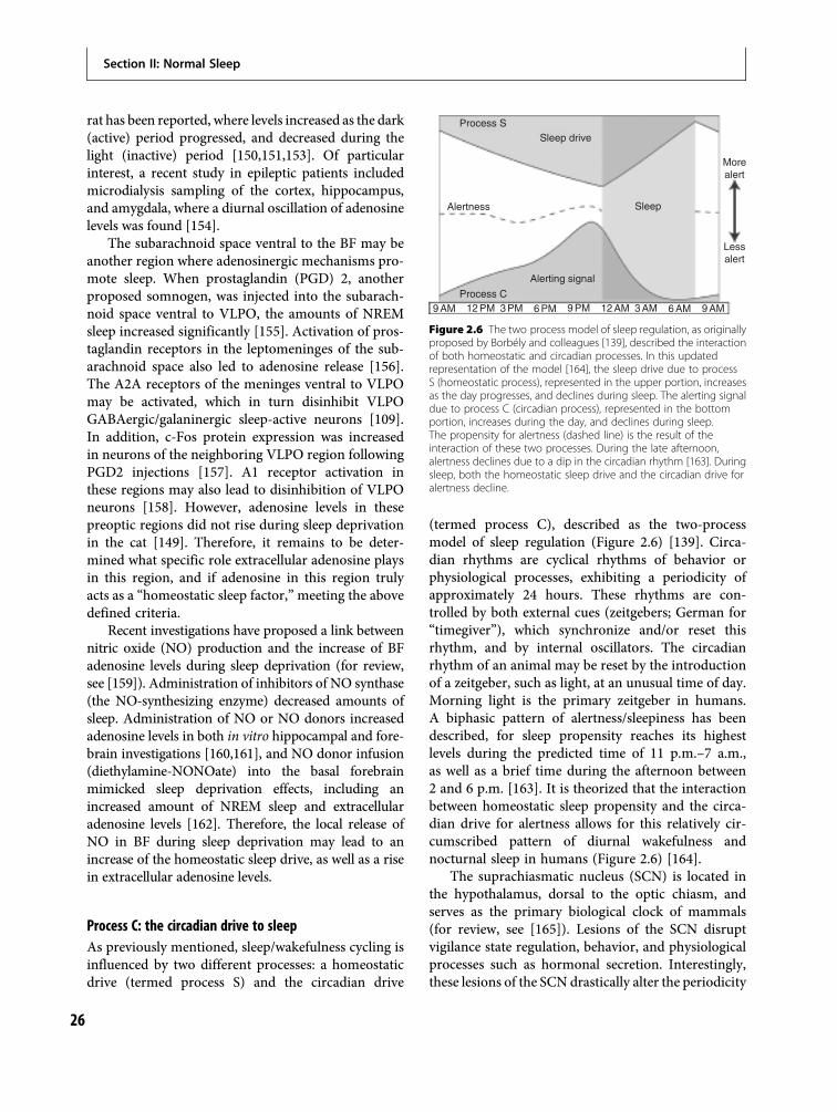
rat has been reported, where levels increased as the dark(active) period progressed, and decreased during thelight (inactive) period [150,151,153]. Of particularinterest, a recent study in epileptic patients includedmicrodialysis sampling of the cortex, hippocampus,and amygdala, where a diurnal oscillation of adenosinelevels was found [154].
The subarachnoid space ventral to the BF may beanother region where adenosinergic mechanisms pro-mote sleep. When prostaglandin (PGD) 2, anotherproposed somnogen, was injected into the subarach-noid space ventral to VLPO, the amounts of NREMsleep increased significantly [155]. Activation of pros-taglandin receptors in the leptomeninges of the sub-arachnoid space also led to adenosine release [156].The A2A receptors of the meninges ventral to VLPOmay be activated, which in turn disinhibit VLPOGABAergic/galaninergic sleep-active neurons [109].In addition, c-Fos protein expression was increasedin neurons of the neighboring VLPO region followingPGD2 injections [157]. A1 receptor activation inthese regions may also lead to disinhibition of VLPOneurons [158]. However, adenosine levels in thesepreoptic regions did not rise during sleep deprivationin the cat [149]. Therefore, it remains to be deter-mined what specific role extracellular adenosine playsin this region, and if adenosine in this region trulyacts as a “homeostatic sleep factor,”meeting the abovedefined criteria.
Recent investigations have proposed a link betweennitric oxide (NO) production and the increase of BFadenosine levels during sleep deprivation (for review,see [159]). Administration of inhibitors of NO synthase(the NO-synthesizing enzyme) decreased amounts ofsleep. Administration of NO or NO donors increasedadenosine levels in both in vitro hippocampal and fore-brain investigations [160,161], and NO donor infusion(diethylamine-NONOate) into the basal forebrainmimicked sleep deprivation effects, including anincreased amount of NREM sleep and extracellularadenosine levels [162]. Therefore, the local release ofNO in BF during sleep deprivation may lead to anincrease of the homeostatic sleep drive, as well as a risein extracellular adenosine levels.
Process C: the circadian drive to sleepAs previously mentioned, sleep/wakefulness cycling isinfluenced by two different processes: a homeostaticdrive (termed process S) and the circadian drive
(termed process C), described as the two-processmodel of sleep regulation (Figure 2.6) [139]. Circa-dian rhythms are cyclical rhythms of behavior orphysiological processes, exhibiting a periodicity ofapproximately 24 hours. These rhythms are con-trolled by both external cues (zeitgebers; German for“timegiver”), which synchronize and/or reset thisrhythm, and by internal oscillators. The circadianrhythm of an animal may be reset by the introductionof a zeitgeber, such as light, at an unusual time of day.Morning light is the primary zeitgeber in humans.A biphasic pattern of alertness/sleepiness has beendescribed, for sleep propensity reaches its highestlevels during the predicted time of 11 p.m.–7 a.m.,as well as a brief time during the afternoon between2 and 6 p.m. [163]. It is theorized that the interactionbetween homeostatic sleep propensity and the circa-dian drive for alertness allows for this relatively cir-cumscribed pattern of diurnal wakefulness andnocturnal sleep in humans (Figure 2.6) [164].
The suprachiasmatic nucleus (SCN) is located inthe hypothalamus, dorsal to the optic chiasm, andserves as the primary biological clock of mammals(for review, see [165]). Lesions of the SCN disruptvigilance state regulation, behavior, and physiologicalprocesses such as hormonal secretion. Interestingly,these lesions of the SCN drastically alter the periodicity
Process S
Process C9 AM 12 PM 3 PM 6 PM 9 PM 12 AM 3 AM 6 AM 9 AM
Sleep drive
Alertness Sleep
Morealert
Lessalert
Alerting signal
Figure 2.6 The two process model of sleep regulation, as originallyproposed by Borbély and colleagues [139], described the interactionof both homeostatic and circadian processes. In this updatedrepresentation of the model [164], the sleep drive due to processS (homeostatic process), represented in the upper portion, increasesas the day progresses, and declines during sleep. The alerting signaldue to process C (circadian process), represented in the bottomportion, increases during the day, and declines during sleep.The propensity for alertness (dashed line) is the result of theinteraction of these two processes. During the late afternoon,alertness declines due to a dip in the circadian rhythm [163]. Duringsleep, both the homeostatic sleep drive and the circadian drive foralertness decline.
Section II: Normal Sleep
26
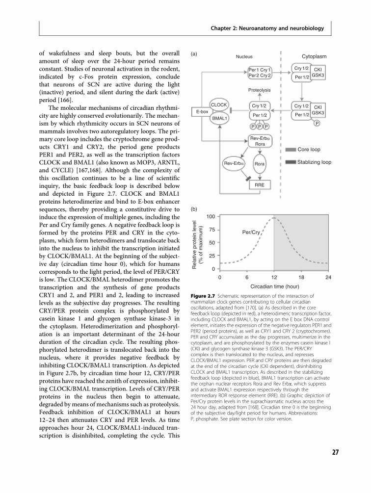
of wakefulness and sleep bouts, but the overallamount of sleep over the 24-hour period remainsconstant. Studies of neuronal activation in the rodent,indicated by c-Fos protein expression, concludethat neurons of SCN are active during the light(inactive) period, and silent during the dark (active)period [166].
The molecular mechanisms of circadian rhythmi-city are highly conserved evolutionarily. The mechan-ism by which rhythmicity occurs in SCN neurons ofmammals involves two autoregulatory loops. The pri-mary core loop includes the cryptochrome gene prod-ucts CRY1 and CRY2, the period gene productsPER1 and PER2, as well as the transcription factorsCLOCK and BMAL1 (also known as MOP3, ARNTL,and CYCLE) [167,168]. Although the complexity ofthis oscillation continues to be a line of scientificinquiry, the basic feedback loop is described belowand depicted in Figure 2.7. CLOCK and BMAL1proteins heterodimerize and bind to E-box enhancersequences, thereby providing a constitutive drive toinduce the expression of multiple genes, including thePer and Cry family genes. A negative feedback loop isformed by the proteins PER and CRY in the cyto-plasm, which form heterodimers and translocate backinto the nucleus to inhibit the transcription initiatedby CLOCK/BMAL1. At the beginning of the subject-ive day (circadian time hour 0), which for humanscorresponds to the light period, the level of PER/CRYis low. The CLOCK/BMAL heterodimer promotes thetranscription and the synthesis of gene productsCRY1 and 2, and PER1 and 2, leading to increasedlevels as the subjective day progresses. The resultingCRY/PER protein complex is phosphorylated bycasein kinase 1 and glycogen synthase kinase-3 inthe cytoplasm. Heterodimerization and phosphoryl-ation is an important determinant of the 24-hourduration of the circadian cycle. The resulting phos-phorylated heterodimer is translocated back into thenucleus, where it provides negative feedback byinhibiting CLOCK/BMAL1 transcription. As depictedin Figure 2.7b, by circadian time hour 12, CRY/PERproteins have reached the zenith of expression, inhibit-ing CLOCK/BMAL transcription. Levels of CRY/PERproteins in the nucleus then begin to attenuate,degraded bymeans of mechanisms such as proteolysis.Feedback inhibition of CLOCK/BMAL1 at hours12–24 then attenuates CRY and PER levels. As timeapproaches hour 24, CLOCK/BMAL1-induced tran-scription is disinhibited, completing the cycle. This
CytoplasmNucleus
100
75
50
25
0
Circadian time (hour)
Per/Cry
Rel
ativ
e pr
otei
n le
vel
(% o
f max
imum
)
0 6 12 18 24
Proteolysis
CLOCK
BMAL1
E-box
(a)
(b)
Cry 1/2
Per 1/2
P P
Rev-Erbα
Rev-Erbα
Rora
Rora
Core loop
Stablizing loop
RRE
PP
Cry 1/2
Per 1/2
Cry 1/2
Per 1/2
CKIGSK3
CKIGSK3
Per 1 Cry 1Per 2 Cry 2
Figure 2.7 Schematic representation of the interaction ofmammalian clock genes contributing to cellular circadianoscillations, adapted from [170]. (a) As described in the corefeedback loop (depicted in red), a heterodimeric transcription factor,including CLOCK and BMAL1, by acting on the E box DNA controlelement, initiates the expression of the negative regulators PER1 andPER2 (period proteins), as well as CRY1 and CRY 2 (cryptochromes).PER and CRY accumulate as the day progresses, multimerize in thecytoplasm, and are phosphorylated by the enzymes casein kinase I(CKI) and glycogen synthase kinase 3 (GSK3). The PER/CRYcomplex is then translocated to the nucleus, and repressesCLOCK/BMAL1 expression. PER and CRY proteins are then degradedat the end of the circadian cycle (CKI dependent), disinhibitingCLOCK and BMAL1 transcription. As described in the stabilizingfeedback loop (depicted in blue), BMAL1 transcription can activatethe orphan nuclear receptors Rora and Rev Erba, which suppressand activate BMAL1 expression respectively through theintermediary ROR response element (RRE). (b) Graphic depiction ofPer/Cry protein levels in the suprachiasmatic nucleus across the24 hour day, adapted from [168]. Circadian time 0 is the beginningof the subjective day/light period for humans. Abbreviations:P, phosphate. See plate section for color version.
Chapter 2: Neuroanatomy and neurobiology
27
feedback loop takes approximately 24 hours to com-plete, and is therefore responsible for the cycling of thecircadian clock [169].
In addition to this primary core loop, a stabilizingfeedback loop helps maintain 24-hour periodicity;this involves the orphan nuclear receptors Rev-Erbaand the retinoid-related orphan receptor alpha(Rora), which suppress and activate BMAL1 expres-sion respectively [169–171]. The CLOCK/BMAL1heterodimer interacts with the E-box response elem-ent of the promoter region of the Rev-Erba and Roragenes. Rev-Erba and Rora compete to bind to theretinoid-related orphan receptor response elements(RRE) in the BMAL1 promoter region. In thismanner the circadian oscillation of BMAL is regu-lated by these orphan nuclear receptors [170,171].
Investigations have begun to describe how thesegenetic transcriptional/translation interactions mayregulate circadian-related events including SCN neur-onal activity [167,168]. Clock controlled genes(CCGs), such as vasopressin prepropressophysin andalbumin D-element binding protein, are promoted byexpression of clock-related genes, such as CLOCKand BMAL. Electrical activity of the SCN is enhancedby increased expression of CCGs through receptor-mediated excitation, as well as autocrine and para-crine mechanisms. A number of investigations haveemployed knock-out technology to breed mice thatpossess abnormal expression of a variety of thesecircadian-related genes. For example, a mouse modelknock-out of the CLOCK gene lengthened the circa-dian rhythm by 4 hours. These animals, whendeprived of zeitgeber cues (free-running) did notexhibit circadian rhythmicity in a number of physio-logical processes [172]. Interestingly, some studieshave suggested mutations in various clock-relatedgenes may contribute to human circadian rhythmsleep disorders (CRSDs) such as advanced anddelayed sleep phase disorder (for review, see [173]).
Visual input reaches the SCN by means of theretinohypothalamic pathway, which is largely gluta-matergic. Freedman and colleagues did not observealtered circadian rhythms with targeted gene knock-out of either the rods or cones in the mouse [174].Removal of the eyes, though, did affect rhythmicity,and it was later determined that melanopsin, uniqueto the ganglion cells of the retina, was a photosensitivemolecule responsible for changes in excitabilityof ganglion cells in the retina [175]. Furthermore,anatomical studies have revealed a monosynaptic
connection between melanopsin-positive cells andthe SCN, suggesting melanopsin is the “circadianphotoreceptor” [176,177].
Besides circadian rhythms that occur over anapproximate 24-hour period, biological rhythms withother periodicities occur and are often related tointeractions with the circadian system and changingexternal time cues. Seasonal rhythms are regulated bymelatonin secretion by means of a well-investigatedanatomical circuit involving the SCN, paraventricularnucleus of the hypothalamus, and preganglionicneurons in the sympathetic nervous system [178].Postganglionic cells in turn project directly to thepineal gland, which is responsible for the secretionof melatonin. Because the amount of light (day) anddark (night) hours fluctuates across seasons, this maybe the mechanism by which a seasonal clock occurs.Although the effectiveness of melatonin for jet lag isdisputed, some people take melatonin to “reset” theirbiological clock, for travel across time zones [179].Melatonin is administered at the beginning of whatwould be the “new” night period, for melatonin levelsare highest at the beginning of the night.
Recent reports have proposed that anotherinternal clock may exist, which is entrainable basedon food availability. Food availability, only in theanimal’s inactive period, can shift rhythms, supersed-ing the photic zeitgeber cues. Such a shift in thisrhythm may be an adaptive response for survival ofthe organism. The dorsomedial nucleus of the hypo-thalamus (DMH) may correspond to this food-entrainable oscillator. Lesions of DMH block thefood-entrainability of the vigilance states [180]. Inter-estingly, in a mouse with knock-out of the BMALclock gene, viral vector injection of BMAL in theSCN restored light entrainment of the circadianrhythm [181]. When the BMAL viral vector wasinjected into DMH, animals were able to entrain tofood, but not light. Therefore, it was concluded that afood-entrainable clock is located in this region,although this is presently controversial [182]. As onemay predict, interplay between vigilance state andcircadian drive circuitry is due to interconnectionsbetween the two systems, however the nature of theseconnections is not fully understood at present. Itis notable that the DMH projects directly to theVLPO, as well as to the lateral hypothalamus, rich inwakefulness-active orexinergic neurons. Therefore,DMH may be a key player in the timing of circadianrhythmicity with the homeostatic drive. Future
Section II: Normal Sleep
28
investigations in this exciting avenue of sleep researchmay further elucidate the interaction of the SCN withvigilance state regulation systems.
ConclusionThe field of sleep medicine has seen a recent investiga-tional surge, due to the development of novel experi-mental techniques, as well as an increased publicawareness of sleep disorders and their implications.Only within the last century have investigationsattempted to explain the neural mechanisms respon-sible for vigilance state regulation. This chapter pro-vides a review of both the neurobiological mechanismsgenerating wakefulness, NREM sleep, and REM sleep,as well as the major homeostatic and circadian factorsthat influence the timing of these states. All human life,and indeed the life of most animals, is shaped byperiods of wakefulness and sleep, and thus knowledgeof the underlying mechanisms is of great biological,social, and medical significance. By furthering under-standing of the neural circuitry involved in vigilancestate regulation, pharmacological and behavioral treat-ments may then be developed to reverse the symp-tomology of a number of sleep-related disorders.
References1. Committee on Sleep Medicine and Research, Board on
Health Sciences Policy, Institute of Medicine of theNational Academies. Sleep disorders and sleepdeprivation: an unmet public health problem. Colten HR,Altevogt BM, eds. Washington, DC: NationalAcademies Press; 2006.
2. Mitler ME, Dement WC, Dinges DF. Sleep medicine,public policy, and public health. In Kryger MH, Roth T,Dement WC, eds. Principles and practice of sleepmedicine, 3rd ed. Philadelphia, PA: WB Saunders;2000:580 588.
3. Paxinos G, Watson C. The rat brain in stereotaxiccoordinates, 3rd ed. San Diego, CA: Academic PressLimited; 1997.
4. Berger H. [Ueber das Elektroenkelogramm desMenchen]. J Psychol Neurol. 1930;40:160 179.
5. Davis H, Davis PA, Loomis AL, Harvey EN, Hobart G.Changes in human brain potentials during the onset ofsleep. Science. 1937;86(2237):448 450.
6. Loomis AL, Harvey EN, Hobart G. Potential rhythms ofthe cerebral cortex during sleep. Science. 1935;81(2111):597 598.
7. Von Economo C. [Die Pathologie des Schlafes]. In: VonBethe A, Von Bergmann A, Embden G, Ellinger A, eds.
Handbuch des normalen und pathologischenphysiologie. Berlin: Springer; 1926:591 610.
8. Bremer F. [Cerveau isole et physiologie du sommeil].C R Soc Biol. 1935;118:1235 1241.
9. Bremer F. [Cerveau. Nouvelles recherches sur lemechanisme dur sommeil]. C R Soc Biol.1936;122:460 464.
10. Moruzzi G, Magoun HW. Brain stem reticularformation and activation of the EEG.Electroencephalogr Clin Neurophysiol. 1949;1(4):455 473.
11. Steriade M, McCarley RW. Brain control of wakefulnessand sleep, 2nd ed. New York, NY: Kluwer Academic/Plenum Publishers; 2005.
12. Jones BE. From waking to sleeping: neuronal andchemical substrates. Trends Pharmacol Sci. 2005;26(11):578 586.
13. Jones BE. Modulation of cortical activation andbehavioral arousal by cholinergic and orexinergicsystems. Ann N Y Acad Sci. 2008;1129:26 34.
14. Dale, H. Pharmacology and nerve endings. Proc R SocMed. 1935;28:319 332.
15. Gritti I, Mainville L, Mancia M, Jones BE. GABAergicand other noncholinergic basal forebrain neurons,together with cholinergic neurons, project to themesocortex and isocortex in the rat. J Comp Neurol.1997;383(2):163 177.
16. Henry P, Jones BE. Projections from basal forebrain toprefrontal cortex comprise cholinergic, GABAergicand glutamatergic inputs to pyramidal cells orinterneurons. Eur J Neurosci. 2008;27(3):654 670.
17. Smythe JW, Colom LV, Bland BH. The extrinsicmodulation of hippocampal theta depends on thecoactivation of cholinergic and GABA ergic medialseptal inputs. Neurosci Biobehav Rev.1992;16(3):289 308.
18. Vertes RP, Kocsis B. Brainstem diencephaloseptohippocampal systems controlling the thetarhythm of the hippocampus. Neuroscience. 1997;81(4):893 926.
19. Morgan JI, Curran T. Stimulus transcription couplingin the nervous system: involvement of the inducibleproto oncogenes fos and jun. Annu Rev Neurosci.1991;14:421 451.
20. Kovács KJ. c Fos as a transcription factor: a stressful(re)view from a functional map. Neurochem Int.1998;33(4):287 297.
21. Basheer R, Porkka Heiskanen T, Stenberg D, McCarleyRW. Adenosine and behavioral state control:adenosine increases c Fos protein and AP1 binding in
Chapter 2: Neuroanatomy and neurobiology
29
basal forebrain of rats. Brain Res Mol Brain Res.1999;73(1 2):1 10.
22. Greco MA, Lu J, Wagner D, Shiromani PJ. c Fosexpression in the cholinergic basal forebrain afterenforced wakefulness and recovery sleep. Neuroreport.2000;11(3):437 440.
23. Modirrousta M, Mainville L, Jones BE. Gabaergicneurons with alpha2 adrenergic receptors in basalforebrain and preoptic area express c Fos during sleep.Neuroscience. 2004;129(3):803 810.
24. Kovács KJ. Measurement of immediate early geneactivation c fos and beyond. J Neuroendocrinol.2008;20(6):665 672.
25. McCarley RW. Neurobiology of REM and NREMsleep. Sleep Med. 2007;8(4):302 330.
26. El Mansari M, Sakai K, Jouvet M. Unitarycharacteristics of presumptive cholinergic tegmentalneurons during the sleep waking cycle in freelymoving cats. Exp Brain Res. 1989;76(3):519 529.
27. Steriade M, Paré D, Datta S, Oakson G, Curró Dossi R.Different cellular types in mesopontine cholinergicnuclei related to ponto geniculo occipital waves.J Neurosci. 1990;10(8):2560 2579.
28. Manns ID, Alonso A, Jones BE. Discharge propertiesof juxtacellularly labeled and immunohistochemicallyidentified cholinergic basal forebrain neuronsrecorded in association with theelectroencephalogram in anesthetized rats. J Neurosci.2000;20(4):1505 1518.
29. Manns ID, Alonso A, Jones BE. Rhythmicallydischarging basal forebrain units comprise cholinergic,GABAergic, and putative glutamatergic cells.J Neurophysiol. 2003;89(2):1057 1066.
30. Lee MG, Hassani OK, Alonso A, Jones BE. Cholinergicbasal forebrain neurons burst with theta during wakingand paradoxical sleep. J Neurosci. 2005;25(17):4365 4369.
31. Blanco Centurion C, Xu M, Murillo Rodriguez E, et al.Adenosine and sleep homeostasis in the basalforebrain. J Neurosci. 2006;26(31):8092 8100.
32. Kaur S, Junek A, Black MA, Semba K. Effects ofibotenate and 192IgG saporin lesions of the nucleusbasalis magnocellularis/substantia innominata onspontaneous sleep and wake states and on recoverysleep after sleep deprivation in rats. J Neurosci.2008;28(2):491 504.
33. Kalinchuk AV, McCarley RW, Stenberg D, PorkkaHeiskanen T, Basheer R. The role of cholinergicbasal forebrain neurons in adenosine mediatedhomeostatic control of sleep: lessons from192 IgG saporin lesions. Neuroscience. 2008;157(1):238 253.
34. Steriade M. Acetylcholine systems and rhythmicactivities during the waking sleep cycle. Prog Brain Res.2004;145:179 196.
35. Dahlström A, Fuxe K. Localization of monoaminesin the lower brain stem. Experientia. 1964;20(7):398 399.
36. Jacobs BL, Azmitia EC. Structure and function of thebrain serotonin system. Physiol Rev. 1992;72(1):165 229.
37. McGinty DJ, Harper RM. Dorsal raphe neurons:depression of firing during sleep in cats. Brain Res.1976;101(3):569 575.
38. Trulson ME, Jacobs BL. Raphe unit activity in freelymoving cats: correlation with level of behavioralarousal. Brain Res. 1979;163(1):135 150.
39. Wilkinson LO, Auerbach SB, Jacobs BL. Extracellularserotonin levels change with behavioral state but notwith pyrogen induced hyperthermia. J Neurosci.1991;11(9):2732 2741.
40. Portas CM, McCarley RW. Behavioral state relatedchanges of extracellular serotonin concentration in thedorsal raphe nucleus: a microdialysis study in the freelymoving cat. Brain Res. 1994;648(2):306 312.
41. Mouret J, Froment JL, Bobillier P, Jouvet M.Neuropharmacologic and biochemical study ofinsomnia induced by P.chlorophenylalanine. J Physiol(Paris). 1967;59(4 Suppl):463 464.
42. Jouvet M. Sleep and serotonin: an unfinished story.Neuropsychopharmacology. 1999;21(2 Suppl):24S 27S.
43. Monti JM, Jantos H. Dose dependent effects of the5 HT1A receptor agonist 8 OH DPAT on sleep andwakefulness in the rat. J Sleep Res. 1992;1(3):169 175.
44. Portas CM, Thakkar M, Rainnie D, McCarley RW.Microdialysis perfusion of 8 hydroxy 2 (di npropylamino)tetralin (8 OH DPAT) in the dorsalraphe nucleus decreases serotonin release and increasesrapid eye movement sleep in the freely moving cat.J Neurosci. 1996;16(8):2820 2828.
45. Vertes RP, Kinney GG, Kocsis B, Fortin WJ.Pharmacological suppression of the median raphenucleus with serotonin1A agonists, 8 OH DPAT andbuspirone, produces hippocampal theta rhythm in therat. Neuroscience. 1994;60(2):441 451.
46. Kinney GG, Kocsis B, Vertes RP. Medial septal unitfiring characteristics following injections of 8 OHDPAT into the median raphe nucleus. Brain Res.1996;708(1 2):116 122.
47. Fabre V, Beaufour C, Evrard A, et al. Alteredexpression and functions of serotonin 5 HT1A and5 HT1B receptors in knock out mice lacking the5 HT transporter. Eur J Neurosci. 2000;12(7):2299 2310.
Section II: Normal Sleep
30
48. Li Q, Wichems C, Heils A, Lesch KP, Murphy DL.Reduction in the density and expression, but notG protein coupling, of serotonin receptors (5 HT1A)in 5 HT transporter knock out mice: gender and brainregion differences. J Neurosci. 2000;20(21):7888 7895.
49. Holmes A, Murphy DL, Crawley JN. Abnormalbehavioral phenotypes of serotonin transporterknockout mice: parallels with human anxiety anddepression. Biol Psychiatry. 2003;54(10):953 959.
50. Wisor JP, Wurts SW, Hall FS, et al. Altered rapid eyemovement sleep timing in serotonin transporterknockout mice. Neuroreport. 2003;14(2):233 238.
51. Alexandre C, Popa D, Fabre V, et al. Early life blockadeof 5 hydroxytryptamine 1A receptors normalizes sleepand depression like behavior in adult knock out micelacking the serotonin transporter. J Neurosci. 2006;26(20):5554 5564.
52. Popa D, Léna C, Alexandre C, Adrien J. Lastingsyndrome of depression produced by reduction inserotonin uptake during postnatal development:evidence from sleep, stress, and behavior. J Neurosci.2008;28(14):3546 3554.
53. Hobson JA, McCarley RW, Wyzinski PW. Sleep cycleoscillation: reciprocal discharge by two brainstemneuronal groups. Science. 1975;189(4196):55 58.
54. Aston Jones G, Bloom FE. Activity of norepinephrinecontaining locus coeruleus neurons in behaving ratsanticipates fluctuations in the sleep waking cycle.J Neurosci. 1981;1(8):876 886.
55. Rasmussen K, Morilak DA, Jacobs BL. Single unitactivity of locus coeruleus neurons in the freely movingcat. I. During naturalistic behaviors and in response tosimple and complex stimuli. Brain Res. 1986;371(2):324 334.
56. Jones BE. Paradoxical sleep and its chemical/structuralsubstrates in the brain. Neuroscience. 1991;40(3):637 656.
57. Saper CB, Cano G, Scammell TE. Homeostatic,circadian, and emotional regulation of sleep. J CompNeurol. 2005;493(1):92 98.
58. Vandermaelen CP, Aghajanian GK. Electrophysiologicaland pharmacological characterization of serotonergicdorsal raphe neurons recorded extracellularly andintracellularly in rat brain slices. Brain Res. 1983;289(1 2):109 119.
59. Brown RE, Sergeeva OA, Eriksson KS, Haas HL.Convergent excitation of dorsal raphe serotoninneurons by multiple arousal systems (orexin/hypocretin, histamine and noradrenaline). J Neurosci.2002;22(20):8850 8859.
60. Aghajanian GK, Vandermaelen CP. Alpha2 adrenoceptor mediated hyperpolarization of locus
coeruleus neurons: intracellular studies in vivo.Science. 1982;215(4538):1394 1396.
61. Williams JA, Reiner PB. Noradrenaline hyperpolarizesidentified rat mesopontine cholinergic neurons invitro. J Neurosci. 1993;13(9):3878 3883.
62. Swanson LW. The projections of the ventral tegmentalarea and adjacent regions: a combined fluorescentretrograde tracer and immunofluorescence study inthe rat. Brain Res Bull. 1982;9(1 6):321 353.
63. Gaykema RP, Zaborszky L. Direct catecholaminergiccholinergic interactions in the basal forebrain. II.Substantia nigra ventral tegmental area projections tocholinergic neurons. J Comp Neurol. 1996;374(4):555 577.
64. Horvitz JC. Mesolimbocortical and nigrostriataldopamine responses to salient non reward events.Neuroscience. 2000;96(4):651 656.
65. Miller JD, Farber J, Gatz P, Roffwarg H, German DC.Activity of mesencephalic dopamine andnon dopamine neurons across stages of sleep andwalking in the rat. Brain Res. 1983;273(1):133 141.
66. Lu J, Jhou TC, Saper CB. Identification ofwake active dopaminergic neurons in the ventralperiaqueductal gray matter. J Neurosci.2006;26(1):193 202.
67. Nishino S, Mignot E. Pharmacological aspects ofhuman and canine narcolepsy. Prog Neurobiol.1997;52(1):27 78.
68. Mignot E, Nishino S. Emerging therapies innarcolepsy cataplexy. Sleep. 2005;28(6):754 763.
69. Brown RE, Stevens DR, Haas HL. The physiologyof brain histamine. Prog Neurobiol.2001;63(6):637 672.
70. Watanabe T, Taguchi Y, Shiosaka S, et al. Distributionof the histaminergic neuron system in the centralnervous system of rats: a fluorescentimmunohistochemical analysis with histidinedecarboxylase as a marker. Brain Res. 1984;295(1):13 25.
71. Panula P, Yang HY, Costa E. Histamine containingneurons in the rat hypothalamus. Proc Natl Acad SciU S A. 1984;81(8):2572 2576.
72. Vanni Mercier G, Sakai K, Jouvet M. [Specific neuronsfor wakefulness in the posterior hypothalamus in thecat]. C R Acad Sci III. 1984;298(7):195 200.
73. Monti JM. Involvement of histamine in the control ofthe waking state. Life Sci. 1993;53(17):1331 1338.
74. John J, Wu MF, Boehmer LN, Siegel JM. Cataplexyactive neurons in the hypothalamus: implications forthe role of histamine in sleep and waking behavior.Neuron. 2004;42(4):619 634.
Chapter 2: Neuroanatomy and neurobiology
31
75. Strecker RE, Nalwalk J, Dauphin LJ, et al. Extracellularhistamine levels in the feline preoptic/anteriorhypothalamic area during natural sleep wakefulnessand prolonged wakefulness: an in vivo microdialysisstudy. Neuroscience. 2002;113(3):663 670.
76. Lin JS, Sakai K, Jouvet M. Evidence for histaminergicarousal mechanisms in the hypothalamus of cat.Neuropharmacology. 1988;27(2):111 122.
77. Kanbayashi T, Kodama T, Kondo H, et al. CSFhistamine contents in narcolepsy, idiopathichypersomnia and obstructive sleep apnea syndrome.Sleep 2009;32(2):181 187.
78. Nishino S, Sakurai E, Nevsimalova S, et al. DecreasedCSF histamine in narcolepsy with and without low CSFhypocretin 1 in comparison to healthy controls.Sleep 2009;32(2):175 180.
79. Peyron C, Tighe DK, van den Pol AN, et al. Neuronscontaining hypocretin (orexin) project to multipleneuronal systems. J Neurosci. 1998;18(23):9996 10015.
80. de Lecea L, Kilduff TS, Peyron C, et al. Thehypocretins: hypothalamus specific peptides withneuroexcitatory activity. Proc Natl Acad Sci U S A.1998;95(1):322 327.
81. Lin L, Faraco J, Li R, et al. The sleep disorder caninenarcolepsy is caused by a mutation in the hypocretin(orexin) receptor 2 gene. Cell. 1999;98(3):365 376.
82. Chemelli RM, Willie JT, Sinton CM, et al. Narcolepsyin orexin knockout mice: molecular genetics of sleepregulation. Cell. 1999;98(4):437 451.
83. Gerashchenko D, Kohls MD, Greco M, et al.Hypocretin 2 saporin lesions of the lateralhypothalamus produce narcoleptic like sleep behaviorin the rat. J Neurosci. 2001;21(18):7273 7283.
84. Gerashchenko D, Blanco Centurion C, Greco MA,Shiromani PJ. Effects of lateral hypothalamic lesionwith the neurotoxin hypocretin 2 saporin on sleep inLong Evans rats. Neuroscience. 2003;116(1):223 235.
85. Mochizuki T, Crocker A, McCormack S, et al.Behavioral state instability in orexin knock out mice.J Neurosci. 2004;24(28):6291 6300.
86. Chen L, Thakkar MM, Winston S, et al. REMsleep changes in rats induced by siRNA mediatedorexin knockdown. Eur J Neurosci. 2006;24(7):2039 2048.
87. Peyron C, Faraco J, Rogers W, et al. A mutation in acase of early onset narcolepsy and a generalizedabsence of hypocretin peptides in human narcolepticbrains. Nat Med. 2000;6(9):991 997.
88. Thannickal TC, Moore RY, Nienhuis R, et al. Reducednumber of hypocretin neurons in human narcolepsy.Neuron. 2000;27(3):469 474.
89. Nishino S, Ripley B, Overeem S, Lammers GJ,Mignot E. Hypocretin (orexin) deficiency in humannarcolepsy. Lancet. 2000;355(9197):39 40.
90. Mignot EM, Lammers GJ, Ripley B, et al. The role ofcerebrospinal fluid hypocretin measurement in thediagnosis of narcolepsy and other hypersomnias. ArchNeurol. 2002;59(10):1553 1562.
91. Brown RE. Involvement of hypocretins/orexins insleep disorders and narcolepsy. Drug News Perspect.2003;16(2):75 79.
92. Mileykovskiy BY, Kiyashchenko LI, Siegel JM.Behavioral correlates of activity in identifiedhypocretin/orexin neurons. Neuron. 2005;46(5):787 798.
93. Kiyashchenko LI, Mileykovskiy BY, Maidment N,et al. Release of hypocretin (orexin) duringwaking and sleep states. J Neurosci. 2002;22(13):5282 5286.
94. Estabrooke IV, McCarthy MT, Ko E, et al. Fosexpression in orexin neurons varies with behavioralstate. J Neurosci. 2001;21(5):1656 1662.
95. Morison RS, Bassett DL. Electrical activity of thethalamus and basal ganglia in decorticate cats.J Neurophysiol. 1945;8(5):309 314.
96. Steriade M, Deschenes M. The thalamus as a neuronaloscillator. Brain Res. 1984;320(1):1 63.
97. Steriade M, Domich L, Oakson G, Deschênes M. Thedeafferented reticular thalamic nucleus generatesspindle rhythmicity. J Neurophysiol. 1987;57(1):260 273.
98. Hu B, Steriade M, Deschênes M. The effects ofbrainstem peribrachial stimulation on perigeniculateneurons: the blockage of spindle waves. Neuroscience.1989;31(1):1 12.
99. Guillery RW, Harting JK. Structure and connectionsof the thalamic reticular nucleus: Advancing viewsover half a century. J Comp Neurol. 2003;463(4):360 371.
100. Buzsaki G, Bickford RG, Ponomareff G, et al.Nucleus basalis and thalamic control of neocorticalactivity in the freely moving rat. J Neurosci. 1988;8(11):4007 4026.
101. Szymusiak R, McGinty D. Hypothalamic regulationof sleep and arousal. Ann N Y Acad Sci.2008;1129:275 286.
102. Sherin JE, Shiromani PJ, McCarley RW, Saper CB.Activation of ventrolateral preoptic neurons duringsleep. Science. 1996;271(5246):216 219.
103. Gong H, McGinty D, Guzman Marin R, et al.Activation of c fos in GABAergic neurones in thepreoptic area during sleep and in response to sleepdeprivation. J Physiol. 2004;556(3):935 946.
Section II: Normal Sleep
32
104. Chou TC, Bjorkum AA, Gaus SE, et al. Afferents tothe ventrolateral preoptic nucleus. J Neurosci.2002;22(3):977 990.
105. Sherin JE, Elmquist JK, Torrealba F, Saper CB.Innervation of histaminergic tuberomammillaryneurons by GABAergic and galaninergic neurons inthe ventrolateral preoptic nucleus of the rat. J Neurosci.1998;18(12):4705 4721.
106. Lu J, Bjorkum AA, Xu M, et al. Selective activation ofthe extended ventrolateral preoptic nucleus duringrapid eye movement sleep. J Neurosci. 2002;22(11):4568 4576.
107. Suntsova N, Szymusiak R, AlamMN, Guzman Marin R,McGinty D. Sleep waking discharge patterns of medianpreoptic nucleus neurons in rats. J Physiol. 2002;543(2):665 677.
108. McGinty D, Szymusiak R. The sleep wake switch:A neuronal alarm clock. Nat Med. 2000;6(5):510 511.
109. Gallopin T, Luppi PH, Cauli B, et al. The endogenoussomnogen adenosine excites a subset of sleeppromoting neurons via A2A receptors in theventrolateral preoptic nucleus. Neuroscience. 2005;134(4):1377 1390.
110. Saper CB, Chou TC, Scammell TE. The sleep switch:hypothalamic control of sleep and wakefulness. TrendsNeurosci. 2001;24(12):726 731.
111. Lu J, Sherman D, Devor M, Saper CB. A putative flipflop switch for control of REM sleep. Nature. 2006;441(7093):589 594.
112. Gallopin T, Fort P, Eggermann E, et al. Identificationof sleep promoting neurons in vitro. Nature. 2000;404(6781):992 995.
113. Aserinsky E, Kleitman N. Regularly occurring periodsof eye motility, and concomitant phenomena, duringsleep. Science. 1953;118(3062):273 274.
114. Dement WC. Knocking on Kleitman’s door: theview from 50 years later. Sleep Med Rev. 2003;7(4):289 292.
115. Jouvet M. Paradoxical sleep a study of its nature andmechanisms. Prog Brain Res. 1965;18:20 62.
116. Jouvet M. Paradoxical sleep mechanisms. Sleep.1994;17(8 Suppl):S77 S83.
117. Jones BE. The role of noradrenergic locus coeruleusneurons and neighboring cholinergic neurons of thepontomesencephalic tegmentum in sleep wake states.Prog Brain Res. 1991;88:533 543.
118. McCarley RW. Mechanisms and models of REM sleepcontrol. Arch Ital Biol. 2004;142(4):429 467.
119. Sakai K, Petitjean F, Jouvet M. Effects of pontomesencephalic lesions and electrical stimulation upon
PGO waves and EMPs in unanesthetized cats.Electroencephalogr Clin Neurophysiol. 1976;41(1):49 63.
120. McCarley RW, Nelson JP, Hobson JA. Ponto geniculooccipital (PGO) burst neurons: correlative evidencefor neuronal generators of PGO waves. Science.1978;201(4352):269 272.
121. Ito K, YanagiharaM, ImonH,DauphinL,McCarleyRW.Intracellular recordings of pontine medialgigantocellular tegmental field neurons in the naturallysleeping cat: behavioral state related activity and somasize difference in order of recruitment. Neuroscience.2002;114(1):23 37.
122. Peigneux P, Laureys S, Fuchs S, et al. Generation ofrapid eye movements during paradoxical sleep inhumans. Neuroimage. 2001;14(3):701 708.
123. Lim AS, Lozano AM, Moro E, et al. Characterizationof REM sleep associated ponto geniculo occipitalwaves in the human pons. Sleep. 2007;30(7):823 827.
124. Bland BH. The power of theta: providing insightsinto the role of the hippocampal formation insensorimotor integration. Hippocampus. 2004;14(5):537 538.
125. Vertes RP, Hoover WB, Viana Di Prisco G. Thetarhythm of the hippocampus: subcortical control andfunctional significance. Behav Cogn Neurosci Rev.2004;3(3):173 200.
126. Webster HH, Jones BE. Neurotoxic lesions of thedorsolateral pontomesencephalic tegmentumcholinergic cell area in the cat. II. Effects upon sleepwaking states. Brain Res. 1988;458(2):285 302.
127. Sitaram N, Wyatt RJ, Dawson S, Gillin JC. REM sleepinduction by physostigmine infusion during sleep.Science. 1976;191(4233):1281 1283.
128. Thakkar MM, Strecker RE, McCarley RW. Behavioralstate control through differential serotonergicinhibition in the mesopontine cholinergic nuclei: asimultaneous unit recording and microdialysis study.J Neurosci. 1998;18(14):5490 5497.
129. Sastre JP, Jouvet M. Oneiric behavior in cats. PhysiolBehav. 1979;22(5):979 989.
130. Hendricks JC, Morrison AR, Mann GL. Differentbehaviors during paradoxical sleep without atoniadepend on pontine lesion site. Brain Res. 1982;239(1):81 105.
131. Xi MC, Morales FR, Chase MH. Interactions betweenGABAergic and cholinergic processes in the nucleuspontis oralis: neuronal mechanisms controlling active(rapid eye movement) sleep and wakefulness.J Neurosci. 2004;24(47):10670 10678.
132. Luppi PH, Gervasoni D, Verret L, et al.Paradoxical (REM) sleep genesis: the switch from
Chapter 2: Neuroanatomy and neurobiology
33
aminergic cholinergic to a GABAergic glutamatergichypothesis. J Physiol Paris. 2006;100(5 6):271 283.
133. Brown RE, McKenna JT, Winston S, et al.Characterization of GABAergic neurons in rapid eyemovement sleep controlling regions of the brainstemreticular formation in GAD67 green fluorescentprotein knock in mice. Eur J Neurosci. 2008;27(2):352 363.
134. Magoun HW, Rhines R. An inhibitory mechanism inthe bulbar reticular formation. J Neurophysiol.1946;9:165 171.
135. Chase MH, Morales FR. The atonia and myoclonia ofactive (REM) sleep. Annu Rev Psychol.1990;41:557 584.
136. McCarley RW, Hobson JA. Neuronal excitabilitymodulation over the sleep cycle: a structural andmathematical model. Science. 1975;189(4196):58 60.
137. McCarley RW, Massaquoi SG. A limit cyclemathematical model of the REM sleep oscillatorsystem. Am J Physiol. 1986;251(6 Pt 2):R1011 R1029.
138. Li X, Rainnie DG, McCarley RW, Greene RW.Presynaptic nicotinic receptors facilitatemonoaminergic transmission. J Neurosci. 1998;18(5):1904 1912.
139. Borbély AA, Achermann P. Sleep homeostasis andmodels of sleep regulation. J Biol Rhythms. 1999;14(6):557 568.
140. Van Dongen HP, Maislin G, Mullington JM, DingesDF. The cumulative cost of additional wakefulness:dose response effects on neurobehavioral functionsand sleep physiology from chronic sleep restrictionand total sleep deprivation. Sleep. 2003;26(2):117 126.
141. Kim Y, Laposky AD, Bergmann BM, Turek FW.Repeated sleep restriction in rats leads to homeostaticand allostatic responses during recovery sleep. ProcNatl Acad Sci U S A. 2007;104(25):10697 10702.
142. Benington JH, Heller HC. Restoration of brain energymetabolism as the function of sleep. Prog Neurobiol.1995;45(4):347 360.
143. Scharf MT, Naidoo N, Zimmerman JE, Pack AI.The energy hypothesis of sleep revisited. ProgNeurobiol. 2008;86(3):264 280.
144. Strecker RE, Morairty S, Thakkar MM, et al.Adenosinergic modulation of basal forebrain andpreoptic/anterior hypothalamic neuronal activity inthe control of behavioral state. Behav Brain Res.2000;115(2):183 204.
145. Basheer R, Strecker RE, Thakkar MM, McCarley RW.Adenosine and sleep wake regulation. Prog Neurobiol.2004;73(6):379 396.
146. Radulovacki M. Adenosine sleep theory: howI postulated it. Neurol Res. 2005;27(2):137 138.
147. Drury AN, Szent Györgyi A. The physiological activityof adenine compounds with especial reference totheir action upon the mammalian heart. J Physiol.1929;68(3):213 237.
148. Porkka Heiskanen T, Strecker RE, Thakkar M, et al.Adenosine: a mediator of the sleep inducing effectsof prolonged wakefulness. Science. 1997;276(5316):1265 1268.
149. Porkka Heiskanen T, Strecker RE, McCarley RW.Brain site specificity of extracellular adenosineconcentration changes during sleep deprivation andspontaneous sleep: an in vivo microdialysis study.Neuroscience. 2000;99(3):507 517.
150. McKenna JT, Dauphin LJ, Mulkern KJ, et al.Nocturnal elevation of extracellular adenosine in therat basal forebrain. Sleep Research Online. 2003;5:155 160.
151. Murillo Rodriguez E, Blanco Centurion C,Gerashchenko D, Salin Pascual RJ, Shiromani PJ.The diurnal rhythm of adenosine levels in the basalforebrain of young and old rats. Neuroscience.2004;123(2):361 370.
152. McKenna JT, Tartar JL, Ward CP, et al. Sleepfragmentation elevates behavioral, electrographic andneurochemical measures of sleepiness. Neuroscience.2007;146(4):1462 1473.
153. Chagoya de Sánchez V, Hernández Múñoz R, Suárez J,Vidrio S, Yáñez L, Díaz Múñoz M. Day nightvariations of adenosine and its metabolizing enzymesin the brain cortex of the rat possible physiologicalsignificance for the energetic homeostasis and thesleep wake cycle. Brain Res. 1993;612(1 2):115 121.
154. Zeitzer JM, Morales Villagran A, Maidment NT, et al.Extracellular adenosine in the human brain duringsleep and sleep deprivation: an in vivo microdialysisstudy. Sleep. 2006;29(4):455 461.
155. Satoh S, Matsumura H, Suzuki F, Hayaishi O.Promotion of sleep mediated by the A2a adenosinereceptor and possible involvement of this receptor inthe sleep induced by prostaglandin D2 in rats.Proc Natl Acad Sci U S A. 1996;93(12):5980 5984.
156. Hayaishi O. Molecular mechanisms of sleep wakeregulation: a role of prostaglandin D2. Philos TransR Soc Lond B Biol Sci. 2000;355(1394):275 280.
157. Scammell TE, Gerashchenko DY, Mochizuki T, et al.An adenosine A2a agonist increases sleep and inducesFos in ventrolateral preoptic neurons. Neuroscience.2001;107(4):653 663.
Section II: Normal Sleep
34

158. Morairty S, Rainnie D, McCarley R, Greene R.Disinhibition of ventrolateral preoptic area sleep activeneurons by adenosine: a new mechanism for sleeppromotion. Neuroscience. 2004;123(2):451 457.
159. Gautier Sauvigné S, Colas D, Parmantier P, et al. Nitricoxide and sleep. Sleep Med Rev. 2005;9(2):101 113.
160. Fallahi N, Broad RM, Jin S, Fredholm BB. Releaseof adenosine from rat hippocampal slices bynitric oxide donors. J Neurochem. 1996;67(1):186 193.
161. Rosenberg PA, Li Y, Le M, Zhang Y. Nitric oxidestimulated increase in extracellular adenosineaccumulation in rat forebrain neurons in culture isassociated with ATP hydrolysis and inhibition ofadenosine kinase activity. J Neurosci. 2000;20(16):6294 6301.
162. Kalinchuk AV, Lu Y, Stenberg D, Rosenberg PA,Porkka Heiskanen T. Nitric oxide production in thebasal forebrain is required for recovery sleep.J Neurochem. 2006;99(2):483 498.
163. Richardson GS, Carskadon MA, Orav EJ, Dement WC.Circadian variation of sleep tendency in elderly andyoung adult subjects. Sleep. 1982;5(Suppl 2):S82 S94.
164. Dijk DJ, Edgar DM. Circadian and homeostatic controlof wakefulness and sleep. In: Turek FW, Zee PC, eds.Regulation of sleep and circadian rhythms. New York:Marcel Dekker Inc.; 1999:111 147.
165. Harrington ME, Mistlberger RE. Anatomy andphysiology of the mammalian circadian system. In:Kryger MH, Roth T, Dement WC, eds. Principles andpractice of sleep medicine, 3rd ed. Philadelphia, PA: WBSaunders; 2000:334 345.
166. Morin LP. The circadian visual system. Brain Res BrainRes Rev. 1994;19(1):102 127.
167. King DP, Takahashi JS. Molecular genetics of circadianrhythms in mammals. Annu Rev Neurosci.2000;23:713 742.
168. Reppert SM, Weaver DR. Molecular analysis ofmammalian circadian rhythms. Annu Rev Physiol.2001;63:647 676.
169. Ko CH, Takahashi JS. Molecular components of themammalian circadian clock. Hum Mol Genet. 2006;15(2):R271 R277.
170. Gallego M, Virshup DM. Post translationalmodifications regulate the ticking of the circadianclock. Nat Rev Mol Cell Biol. 2007;8(2):139 148.
171. Jetten AM. Retinoid related orphan receptors (RORs):critical roles in development, immunity, circadianrhythm, and cellular metabolism. Nucl Recept Signal.2009;7:1 32.
172. Challet E, Takahashi JS, Turek FW. Nonphotic phaseshifting in clock mutant mice. Brain Res. 2000;859(2):398 403.
173. Sack RL, Auckley D, Auger RR, et al. Circadianrhythm sleep disorders: part II, advanced sleepphase disorder, delayed sleep phase disorder,free running disorder, and irregular sleep wakerhythm. An American Academy of SleepMedicine review. Sleep. 2007;30(11):1484 1501.
174. Freedman MS, Lucas RJ, Soni B, et al. Regulation ofmammalian circadian behavior by non rod, non cone,ocular photoreceptors. Science. 1999;284(5413):502 504.
175. Provencio I, Rodriguez IR, Jiang G, et al. A novelhuman opsin in the inner retina. J Neurosci. 2000;20(2):600 605.
176. Berson DM, Dunn FA, Takao M. Phototransductionby retinal ganglion cells that set the circadian clock.Science. 2002;295(5557):1070 1073.
177. Hattar S, Liao HW, Takao M, Berson DM,Yau KW. Melanopsin containing retinal ganglioncells: architecture, projections, and intrinsicphotosensitivity. Science. 2002;295(5557):1065 1070.
178. Buijs RM, Scheer FA, Kreier F, et al. Organization ofcircadian functions: interaction with the body. ProgBrain Res. 2006;153:341 360.
179. Deacon S, Arendt J. Adapting to phase shifts, II. Effectsof melatonin and conflicting light treatment. PhysiolBehav. 1996;59(4 5):675 682.
180. Gooley JJ, Schomer A, Saper CB. The dorsomedialhypothalamic nucleus is critical for the expression offood entrainable circadian rhythms. Nat Neurosci.2006;9(3):398 407.
181. Fuller PM, Lu J, Saper CB. Differential rescue of lightand food entrainable circadian rhythms. Science.2008;320(5879):1074 1077.
182. Mistlberger RE, Yamazaki S, Pendergast JS, et al.Comment on “Differential rescue of light and foodentrainable circadian rhythms.” Science. 2008;322(5902):675.
Chapter 2: Neuroanatomy and neurobiology
35
Chapter
3 Neurophysiology and neuroimagingof human sleepFabio Ferrarelli and Ruth M. Benca
Sleep is a behavior universally present in the animalkingdom, occurring in a range of species from dros-ophila to human beings [1]. Sleep occupies about one-third of the human life-span [2] and, although itsfunctions are still not understood, regular sleep isessential for survival. Aspects of normal and abnormalhuman sleep can be characterized by employing elec-trophysiological tools including electroencephalogram(EEG), electromyogram (EMG), and electrooculo-gram (EOG). Sleep can be subdivided into two types:rapid eye movement (REM) and non-rapid eye move-ment (NREM), with specific behavioral and neuro-physiological correlates [3,4]. Over the last fourdecades numerous studies have revealed the organiza-tion of REM and NREM sleep in healthy subjects andpatients with a broad range of neuropsychiatric dis-orders [5,6,7]. Additionally, the development of neuro-imaging techniques has allowed sleep researchers toexplore the neurobiological systems and mechanismsunderlying sleep organization [8], as well as the conse-quences of sleep disruption on cognitive functions [9].
This chapter reviews the characteristics of humansleep, with particular emphasis on REM/NREM sleepstages and their physiological correlates, and describesthe electrophysiological assessment of normal sleep andsleep disorders with polysomnography (PSG), as well asthe standardized clinical tests utilized to establish thedaytime consequences of sleep disorders: the multiplesleep latency test (MSLT) and themaintenance of wake-fulness test (MWT). The organization of sleep (sleeparchitecture) in young healthy adults, and the effects ofage, psychiatric disorders, and psychoactive medica-tions on sleep architecture are discussed. Finally, severalneuroimaging techniques are described, includingfunctional magnetic resonance imaging (f-MRI), posi-tron emission tomography (PET), and high-density
(hd)-EEG, which are increasingly utilized in sleepresearch to investigate the neural correlates of normalsleep and the effects of sleep deprivation on cognitivefunctions.
Characteristics of human sleepDefinition of sleepSleep is defined by a combination of behavioral andphysiological characteristics. Behaviorally, sleepinghumans tend to lie still in stereotypical postures. Thiscondition, which is characterized by a markedlyreduced awareness of the outside world, is different,however, from other unconscious states like coma oranesthesia because of its prompt reversibility [10].Using electrophysiological criteria, human sleep canbe divided into two types, REM and NREM sleep.During REM sleep, bursts of eye movement activityfrequently occur. REM sleep is also called paradoxicalsleep because of EEG activity similar to that observedduring wakefulness. On the other hand, duringNREM sleep, which is also described as orthodoxsleep, the EEG shows low-frequency activity.
Sleep stagesHuman sleep occurs in several cycles of NREM–REMsleep across the night, each approximately 90 to 110minutes long, which can be further classified intodifferent sleep stages [11]. Sleep stages are identifiedbased on three electrophysiological parameters: EEG,EOG, and EMG activity, the latter usually recordedfrom neck or chin muscles. For clinical and researchpurposes, sleep is usually scored in 30 second epochs.The criteria of sleep stage scoring, established byRechtschaffen and Kales (R&K) [12], have been
Foundations of Psychiatric Sleep Medicine, ed. JohnW.Winkelman and David T. Plante. Published by Cambridge University Press.# Cambridge University Press 2010.
36
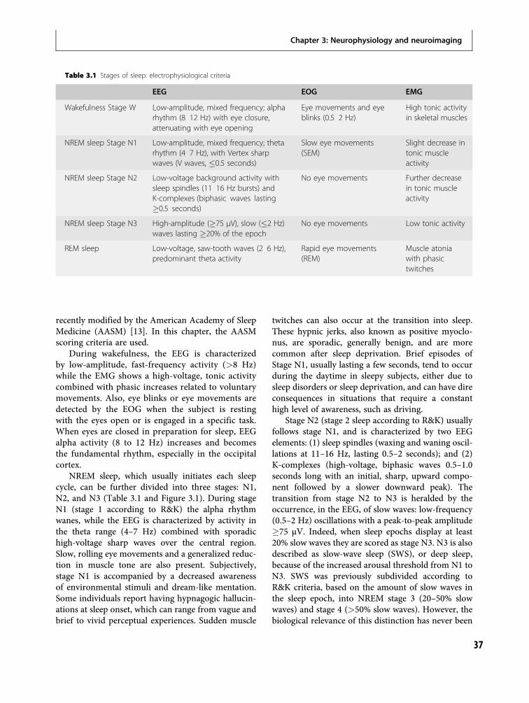
recently modified by the American Academy of SleepMedicine (AASM) [13]. In this chapter, the AASMscoring criteria are used.
During wakefulness, the EEG is characterizedby low-amplitude, fast-frequency activity (>8 Hz)while the EMG shows a high-voltage, tonic activitycombined with phasic increases related to voluntarymovements. Also, eye blinks or eye movements aredetected by the EOG when the subject is restingwith the eyes open or is engaged in a specific task.When eyes are closed in preparation for sleep, EEGalpha activity (8 to 12 Hz) increases and becomesthe fundamental rhythm, especially in the occipitalcortex.
NREM sleep, which usually initiates each sleepcycle, can be further divided into three stages: N1,N2, and N3 (Table 3.1 and Figure 3.1). During stageN1 (stage 1 according to R&K) the alpha rhythmwanes, while the EEG is characterized by activity inthe theta range (4–7 Hz) combined with sporadichigh-voltage sharp waves over the central region.Slow, rolling eye movements and a generalized reduc-tion in muscle tone are also present. Subjectively,stage N1 is accompanied by a decreased awarenessof environmental stimuli and dream-like mentation.Some individuals report having hypnagogic hallucin-ations at sleep onset, which can range from vague andbrief to vivid perceptual experiences. Sudden muscle
twitches can also occur at the transition into sleep.These hypnic jerks, also known as positive myoclo-nus, are sporadic, generally benign, and are morecommon after sleep deprivation. Brief episodes ofStage N1, usually lasting a few seconds, tend to occurduring the daytime in sleepy subjects, either due tosleep disorders or sleep deprivation, and can have direconsequences in situations that require a constanthigh level of awareness, such as driving.
Stage N2 (stage 2 sleep according to R&K) usuallyfollows stage N1, and is characterized by two EEGelements: (1) sleep spindles (waxing and waning oscil-lations at 11–16 Hz, lasting 0.5–2 seconds); and (2)K-complexes (high-voltage, biphasic waves 0.5–1.0seconds long with an initial, sharp, upward compo-nent followed by a slower downward peak). Thetransition from stage N2 to N3 is heralded by theoccurrence, in the EEG, of slow waves: low-frequency(0.5–2 Hz) oscillations with a peak-to-peak amplitude�75 µV. Indeed, when sleep epochs display at least20% slow waves they are scored as stage N3. N3 is alsodescribed as slow-wave sleep (SWS), or deep sleep,because of the increased arousal threshold from N1 toN3. SWS was previously subdivided according toR&K criteria, based on the amount of slow waves inthe sleep epoch, into NREM stage 3 (20–50% slowwaves) and stage 4 (>50% slow waves). However, thebiological relevance of this distinction has never been
Table 3.1 Stages of sleep: electrophysiological criteria
EEG EOG EMG
Wakefulness Stage W Low-amplitude, mixed frequency; alpharhythm (8 12 Hz) with eye closure,attenuating with eye opening
Eye movements and eyeblinks (0.5 2 Hz)
High tonic activityin skeletal muscles
NREM sleep Stage N1 Low-amplitude, mixed frequency; thetarhythm (4 7 Hz), with Vertex sharpwaves (V waves, �0.5 seconds)
Slow eye movements(SEM)
Slight decrease intonic muscleactivity
NREM sleep Stage N2 Low-voltage background activity withsleep spindles (11 16 Hz bursts) andK-complexes (biphasic waves lasting�0.5 seconds)
No eye movements Further decreasein tonic muscleactivity
NREM sleep Stage N3 High-amplitude (�75 µV), slow (�2 Hz)waves lasting �20% of the epoch
No eye movements Low tonic activity
REM sleep Low-voltage, saw-tooth waves (2 6 Hz),predominant theta activity
Rapid eye movements(REM)
Muscle atoniawith phasictwitches
Chapter 3: Neurophysiology and neuroimaging
37
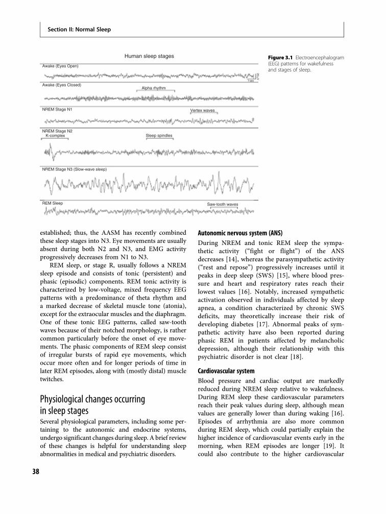
established; thus, the AASM has recently combinedthese sleep stages into N3. Eye movements are usuallyabsent during both N2 and N3, and EMG activityprogressively decreases from N1 to N3.
REM sleep, or stage R, usually follows a NREMsleep episode and consists of tonic (persistent) andphasic (episodic) components. REM tonic activity ischaracterized by low-voltage, mixed frequency EEGpatterns with a predominance of theta rhythm anda marked decrease of skeletal muscle tone (atonia),except for the extraocular muscles and the diaphragm.One of these tonic EEG patterns, called saw-toothwaves because of their notched morphology, is rathercommon particularly before the onset of eye move-ments. The phasic components of REM sleep consistof irregular bursts of rapid eye movements, whichoccur more often and for longer periods of time inlater REM episodes, along with (mostly distal) muscletwitches.
Physiological changes occurringin sleep stagesSeveral physiological parameters, including some per-taining to the autonomic and endocrine systems,undergo significant changes during sleep. A brief reviewof these changes is helpful for understanding sleepabnormalities in medical and psychiatric disorders.
Autonomic nervous system (ANS)During NREM and tonic REM sleep the sympa-thetic activity (“fight or flight”) of the ANSdecreases [14], whereas the parasympathetic activity(“rest and repose”) progressively increases until itpeaks in deep sleep (SWS) [15], where blood pres-sure and heart and respiratory rates reach theirlowest values [16]. Notably, increased sympatheticactivation observed in individuals affected by sleepapnea, a condition characterized by chronic SWSdeficits, may theoretically increase their risk ofdeveloping diabetes [17]. Abnormal peaks of sym-pathetic activity have also been reported duringphasic REM in patients affected by melancholicdepression, although their relationship with thispsychiatric disorder is not clear [18].
Cardiovascular systemBlood pressure and cardiac output are markedlyreduced during NREM sleep relative to wakefulness.During REM sleep these cardiovascular parametersreach their peak values during sleep, although meanvalues are generally lower than during waking [16].Episodes of arrhythmia are also more commonduring REM sleep, which could partially explain thehigher incidence of cardiovascular events early in themorning, when REM episodes are longer [19]. Itcould also contribute to the higher cardiovascular
Human sleep stages
Awake (Eyes Open)
Awake (Eyes Closed)Alpha rhythm
Vertex waves
1 sec
20μV
Sleep spindles
Saw-tooth waves
NREM Stage N1
NREM Stage N2K-complex
NREM Stage N3 (Slow-wave sleep)
REM Sleep
Figure 3.1 Electroencephalogram(EEG) patterns for wakefulnessand stages of sleep.
Section II: Normal Sleep
38
mortality seen in patients with major depression, whoshow increased REM sleep period duration andgreater phasic REM activity relative to healthy sub-jects, although this is theoretical [20].
Respiratory systemDuring sleep, air exchange decreases because of theloss of waking-related respiratory drive and a reduc-tion in respiratory rate and minute ventilation. At thesame time, the resistance in the upper airwaysincreases as a result of muscle relaxation [21].Reduced sensitivity of central chemoreceptors tohigher levels of CO2 or to lower levels of O2 is alsoobserved, especially during REM sleep [22]. Thesechanges, usually present in normal sleep, contributeto sleep disorders such as sleep apnea [23].
ThermoregulationDuring NREM sleep, homeostatic thermoregulationis preserved and is characterized, particularly in SWS,by a reduction in body temperature that follows adecrease in the hypothalamic temperature set point[24]. Conversely, during REM sleep the ability toregulate body temperature through sweating andshivering is markedly reduced [25]. Intriguingly,hypothermia and other abnormalities in thermoregu-lation are reported in psychiatric disorders, such asanorexia nervosa [26], which are also characterized bysleep abnormalities (i.e. SWS deficits) [27].
Neuroendocrine systemSeveral hormones are released during sleep. Growthhormone (GH) secretion occurs early in the night, andpeaks during SWS [28], while prolactin is usuallyreleased in the middle of the night [29]. Both of thesehormones have feedback effects on sleep: GH on SWS[30], prolactin on REM [31]. Adrenocorticotropichormone (ACTH) and cortisol, two hormones ofthe hypothalamic-pituitary-adrenal (HPA) axis, aredownregulated at sleep onset but increase and peaktoward the end of sleep, when they likely contribute tomorning arousal [32]. In contrast, the secretion ofthyroid-stimulating hormone (TSH) is maximal inthe evening before sleep onset, inhibited by sleep,and enhanced by sleep deprivation [33]. Melatonin,whose secretion by the pineal gland is permitted onlyat night by the circadian system, is also inhibited bylight and promoted by darkness, thus providing infor-mation to the brain regarding seasonal day length.Melatonin is often used as a sleep-promoting agent,
due to its ability to induce drowsiness and lower bodytemperature [34]. Altogether, while hormones such asprolactin and GH show a significant sleep-dependentsecretion, melatonin and cortisol levels are circadian-regulated and thus are relatively unaffected by sleep [35].
Chronic sleep disruption or sleep deprivation canhave dramatic effects on the endocrine system. Forexample, the secretion of GH and prolactin is markedlydecreased in sleep apnea patients [36]. Furthermore,there is increasing evidence that sleep loss is associatedwith decreased insulin sensitivity, which eventuallycould result in elevated risk for diabetes [37]. In add-ition, severe sleep restriction may result in a reductionof satiety hormones (e.g. leptin) as well as an increasein hunger-promoting hormones (e.g. ghrelin), whichcould lead to higher body mass index and obesity [38].
Reproductive systemDuring REM sleep, men commonly experience noc-turnal penile tumescence (NPT) [39]. The occurrenceof NPT, which is unrelated to the sexual content ofdreaming and persists into old age, can be usedto establish whether cases of impotence are biologicalor psychogenic in origin. However, testing for NPT isno longer commonly performed due to newer pharma-cological methods of evaluating impotence [40]. Inwomen, during REM sleep, a phasic increase in vaginalblood flow as well as clitoral engorgement and erectionoccur, phenomena which are also unrelated to thesexual content of dreaming activity [41].
Electrophysiological tools tomonitor sleepMonitoring normal human sleepEEG, since it was first utilized byHans Berger in 1929 todescribe brain rhythms (alpha, beta) generated duringwaking [42], has been extensively employed to investi-gate sleep activity. In combination with EMG (whichassesses muscle activity) and EOG (which measures eyemovement), EEG has played a critical role in character-izing the different stages of NREMas well as REM sleep.
EEG is recorded from electrodes placed on differ-ent scalp locations according to the 10–20 system[43]. This is a standardized electrode placementmethod developed to ensure high within- and across-subject reproducibility. Electrodes localized in dif-ferent scalp regions (frontal, central, and occipital)can best identify specific EEG patterns of waking
Chapter 3: Neurophysiology and neuroimaging
39

and sleep. For example, a frontal electrode canoptimally record K-complexes (NREM stage N2) andslow waves (NREM stage N3), whereas a central elec-trode is ideally located to detect sleep spindles (NREMstage N2). Conversely, an occipital electrode can bestrecord alpha rhythm, EEG oscillations at 8–12 Hzoccurring during resting wakefulness with the eyesclosed. The current availability of high-density(�60 channels) EEG (hd-EEG) systems has made itpossible to describe a highly detailed topography ofcharacteristic waking as well as sleep rhythms, such asslow waves and sleep spindles (Figure 3.2a). A recentstudy has also shown the potential of hd-EEG in iden-tifying local deficits in sleep rhythms in patients withschizophrenia (Figure 3.2b) [44].
EOG electrodes are placed in pairs (above and belowthe eye or to the left and right of it) in order to trackvertical and horizontal eye movements. EOG electrodescan detect the slow, rolling eye movement associatedwith NREM sleep stage N1 as well as the rapid eyemovements that occur during REM sleep. EMG record-ings are performed to monitor changes in tonic andphasic muscle activity during sleep. For example, EMGactivity is markedly decreased during REM sleep, whenskeletal muscles are virtually paralyzed (atonia). EMG isusually recorded from chin electrodes over the submen-talis muscle. Additional electrodes can be placed overthe anterior tibialis and intercostal muscles to monitorleg movements and respiratory efforts, respectively.
Monitoring abnormal sleep:polysomnographyPolysomnography (PSG) is an overnight procedurethat involves continuous recording of several physio-logical variables for the characterization of sleep for
the diagnosis of sleep disorders or for clinical research[45]. In addition to EEG, EOG, and EMG activity, thevariables monitored during clinical PSG include:(a) limb movements via EMG electrodes on armsand/or legs; (b) electrocardiogram (ECG) using chestleads; (c) respiratory effort with thoracic and abdom-inal piezoelectric belts or respiratory inductiveplethysmography (RIP) belts; (d) airflow at nose/mouth via heat-sensitive devices called thermistorsand/or nasal pressure transducers; and (e) oxygensaturation via pulse oximetry. PSG is commonly indi-cated for: (1) the diagnosis of sleep-related breathingdisorders (SRBDs); (2) titrating positive airway pres-sure (PAP) in patients with SRBDs; (3) cases of sus-pected narcolepsy; and (4) monitoring sleep behaviors(parasomnias) that are potentially harmful or areparticularly unusual for age of onset, duration, andfrequency of occurrence [46]. PSG is also employed inpatients with neuromuscular disorders reportingsleep complaints, in patients with parasomnias unre-sponsive to therapy, and in order to support diag-noses of seizure-related paroxysmal arousals orperiodic limb movement disorder [46]. Conversely,PSG is usually not indicated in cases of insomnia,circadian rhythm disorders, typical, uncomplicatedparasomnias, or to confirm sleep abnormalities (i.e.insomnia) in subjects affected by psychiatric dis-orders, such as depression or schizophrenia [46].
Clinical tests to establish daytimesleepinessDaytime sleepiness is characterized by difficulties inmaintaining wakefulness while performing daytimeactivities. It is a chronic problem for about 5% ofthe general population, and it is the most common
Slow waves60
(a) (b)
4020
0–20–40–60
6040200 0
–20–40–60
60
4020
–20–40–60
Sleep spindles Sleep spindles Sleep spindlesSleep spindles
Healthy controls Schizophrenics
Figure 3.2 (a) NREM sleep EEG traces and topographies of slow waves and sleep spindles in a healthy control. (b) Topographic analysisof sleep spindles revealed that schizophrenics had reduced spindle activity localized in a centroparietal area (adapted from [1]).See plate section for color version.
Section II: Normal Sleep
40
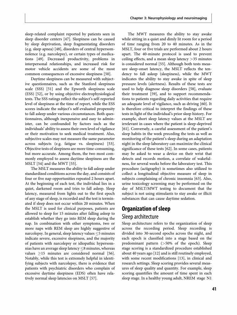
sleep-related complaint reported by patients seen insleep disorder centers [47]. Sleepiness can be causedby sleep deprivation, sleep fragmentating disorders(e.g. sleep apnea) [48], disorders of central hypersom-nolence (e.g. narcolepsy), or certain types of medica-tions [49]. Decreased productivity, problems ininterpersonal relationships, and increased risk formotor vehicle accidents are some of the mostcommon consequences of excessive sleepiness [50].
Daytime sleepiness can be measured with subject-ive questionnaires, such as the Stanford sleepinessscale (SSS) [51] and the Epworth sleepiness scale(ESS) [52], or by using objective electrophysiologicaltests. The SSS ratings reflect the subject’s self-reportedlevel of sleepiness at the time of report, while the ESSscores indicate the subject’s self-evaluated propensityto fall asleep under various circumstances. Both ques-tionnaires, although inexpensive and easy to admin-ister, can be confounded by factors such as theindividuals’ ability to assess their own level of vigilanceor their motivation to seek medical treatment. Also,subjective scales may not measure the same parameteracross subjects (e.g. fatigue vs. sleepiness) [53].Objective tests of sleepiness aremore time-consuming,but more accurate. Among them, the two most com-monly employed to assess daytime sleepiness are theMSLT [54] and the MWT [55].
The MSLT measures the ability to fall asleep understandardized conditions across the day, and consists offour or five nap opportunities repeated 2 hours apart.At the beginning of each test, the individual lies in aquiet, darkened room and tries to fall asleep. Sleeplatency, measured from lights out to the first epochof any stage of sleep, is recorded and the test is termin-ated if sleep does not occur within 20 minutes. Whenthe MSLT is used for clinical purposes, patients areallowed to sleep for 15 minutes after falling asleep toestablish whether they go into REM sleep during thenap. In combination with other symptoms, two ormore naps with REM sleep are highly suggestive ofnarcolepsy. In general, sleep latency values�5minutesindicate severe, excessive sleepiness, and the majorityof patients with narcolepsy or idiopathic hypersom-nias have an average sleep latency�8minutes, whereasvalues �15 minutes are considered normal [56].Notably, while this test is extremely helpful in identi-fying subjects with narcolepsy, there is evidence thatpatients with psychiatric disorders who complain ofexcessive daytime sleepiness (EDS) often have rela-tively normal sleep latencies on MSLT [57].
The MWT measures the ability to stay awakewhile sitting in a quiet and dimly lit room for a periodof time ranging from 20 to 40 minutes. As in theMSLT, four or five trials are performed about 2 hoursapart. The 40-minute protocol is used to preventceiling effects, and a mean sleep latency >35 minutesis considered normal [55]. Although both tests meas-ure sleep-onset latency, the MSLT reflects the ten-dency to fall asleep (sleepiness), while the MWTindicates the ability to stay awake in spite of sleeppressure levels (alertness). Results of these tests areused to help diagnose sleep disorders [58], evaluatetheir treatment [59], and to support recommenda-tions to patients regarding daily activities that requirean adequate level of vigilance, such as driving [60]. Itis therefore critical to interpret the findings of thesetests in light of the individual’s prior sleep history. Forexample, short sleep latency values at the MSLT areirrelevant in cases where the patient is sleep-deprived[61]. Conversely, a careful assessment of the patient’ssleep habits in the week preceding the tests as well asmonitoring of the patient’s sleep during an adaptationnight in the sleep laboratory can maximize the clinicalsignificance of these tests [62]. In some cases, patientsmay be asked to wear a device on their wrist thatdetects and records motion, a correlate of wakeful-ness, for several weeks before the laboratory test. Thisprocedure (actigraphy) is sometimes also utilized tocollect a longitudinal objective measure of sleep insubjects complaining of chronic insomnia [63]. Also,urine toxicology screening may be performed on theday of MSLT/MWT testing to document that thesubject is not using stimulants to stay awake or illicitsubstances that can cause daytime sedation.
Organization of sleepSleep architectureSleep architecture refers to the organization of sleepacross the recording period. Sleep recording isdivided into 30-second epochs across the night, andeach epoch is classified into a stage based on thepredominant pattern (>50% of the epoch). Sleepstage scoring is a standardized procedure establishedabout 40 years ago [12] and is still routinely employed,with some recent modifications [13], in clinical andresearch settings. Sleep scoring provides several meas-ures of sleep quality and quantity. For example, sleepscoring quantifies the amount of time spent in eachsleep stage. In a healthy young adult, NREM stage N1
Chapter 3: Neurophysiology and neuroimaging
41
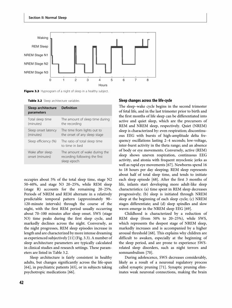
occupies about 5% of the total sleep time, stage N250–60%, and stage N3 20–25%, while REM sleep(stage R) accounts for the remaining 20–25%.Periods of NREM and REM alternate in a relativelypredictable temporal pattern (approximately 90–120-minute intervals) through the course of thenight, with the first REM period usually occurringabout 70–100 minutes after sleep onset. SWS (stageN3) time peaks during the first sleep cycle, andmarkedly declines across the night. Conversely, asthe night progresses, REM sleep episodes increase inlength and are characterized bymore intense dreamingas experienced subjectively [11] (Fig. 3.3). A number ofsleep architecture parameters are typically calculatedin clinical studies and research settings. These param-eters are listed in Table 3.2.
Sleep architecture is fairly consistent in healthyadults, but changes significantly across the life-span[64], in psychiatric patients [65], or in subjects takingpsychotropic medications [66].
Sleep changes across the life-cycleThe sleep–wake cycle begins in the second trimesterof fetal life, and in the last trimester prior to birth andthe first months of life sleep can be differentiated intoactive and quiet sleep, which are the precursors ofREM and NREM sleep, respectively. Quiet (NREM)sleep is characterized by: even respiration; discontinu-ous EEG with bursts of high-amplitude delta fre-quency oscillations lasting 2–4 seconds; low-voltage,inter-burst activity in the theta range; and an absenceof body or eye movements. Conversely, active (REM)sleep shows uneven respiration, continuous EEGactivity, and atonia with frequent myoclonic jerks aswell as rapid eye movements [67]. Newborns spend 16to 18 hours per day sleeping; REM sleep representsabout half of total sleep time, and tends to initiateeach sleep episode [68]. After the first 3 months oflife, infants start developing more adult-like sleepcharacteristics: (a) time spent in REM sleep decreasesprogressively; (b) sleep is initiated through NREMsleep at the beginning of each sleep cycle; (c) NREMstages differentiate; and (d) sleep spindles and slowwaves emerge in the NREM sleep EEG [69].
Childhood is characterized by a reduction ofREM sleep (from 50% to 20–25%), while SWS,which represents the deepest stage of NREM sleep,markedly increases and is accompanied by a higherarousal threshold [68]. This explains why children aredifficult to awaken, especially at the beginning ofthe sleep period, and are prone to experience SWS-related sleep disorders, such as night terrors andsomnambulism [70].
During adolescence, SWS decreases considerably,likely as a result of a neuronal regulatory processcalled synaptic pruning [71]. Synaptic pruning elim-inates weak neuronal connections, making the brain
Waking
REM Sleep
NREM Stage N1
NREM Stage N2
NREM Stage N3
Hours
0 1 2 3 4 5 6 7 8
Figure 3.3 Hypnogram of a night of sleep in a healthy subject.
Table 3.2 Sleep architecture variables
Sleep architectureparameters
Definition
Total sleep time(minutes)
The amount of sleep time duringthe recording
Sleep onset latency(minutes)
The time from lights out tothe onset of any sleep stage
Sleep efficiency (%) The ratio of total sleep timeto time in bed
Wake after sleeponset (minutes)
The amount of wake during therecording following the firstsleep epoch
Section II: Normal Sleep
42
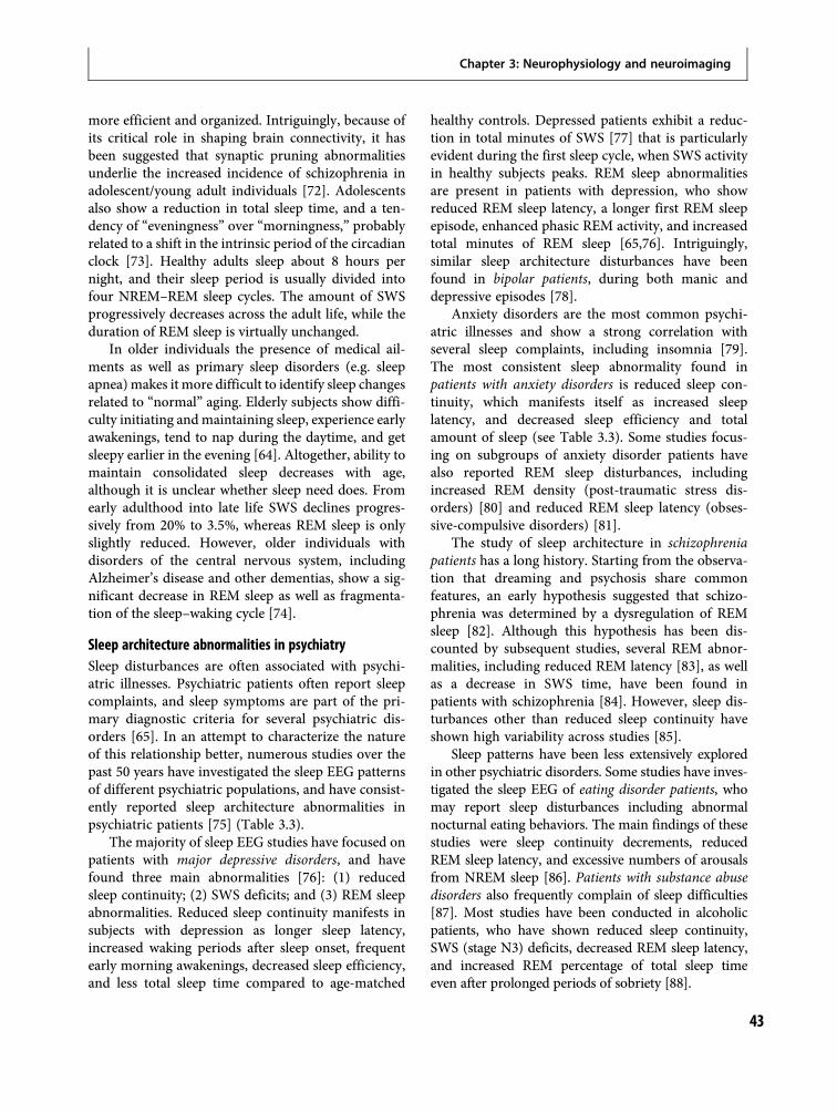
more efficient and organized. Intriguingly, because ofits critical role in shaping brain connectivity, it hasbeen suggested that synaptic pruning abnormalitiesunderlie the increased incidence of schizophrenia inadolescent/young adult individuals [72]. Adolescentsalso show a reduction in total sleep time, and a ten-dency of “eveningness” over “morningness,” probablyrelated to a shift in the intrinsic period of the circadianclock [73]. Healthy adults sleep about 8 hours pernight, and their sleep period is usually divided intofour NREM–REM sleep cycles. The amount of SWSprogressively decreases across the adult life, while theduration of REM sleep is virtually unchanged.
In older individuals the presence of medical ail-ments as well as primary sleep disorders (e.g. sleepapnea)makes it more difficult to identify sleep changesrelated to “normal” aging. Elderly subjects show diffi-culty initiating andmaintaining sleep, experience earlyawakenings, tend to nap during the daytime, and getsleepy earlier in the evening [64]. Altogether, ability tomaintain consolidated sleep decreases with age,although it is unclear whether sleep need does. Fromearly adulthood into late life SWS declines progres-sively from 20% to 3.5%, whereas REM sleep is onlyslightly reduced. However, older individuals withdisorders of the central nervous system, includingAlzheimer’s disease and other dementias, show a sig-nificant decrease in REM sleep as well as fragmenta-tion of the sleep–waking cycle [74].
Sleep architecture abnormalities in psychiatrySleep disturbances are often associated with psychi-atric illnesses. Psychiatric patients often report sleepcomplaints, and sleep symptoms are part of the pri-mary diagnostic criteria for several psychiatric dis-orders [65]. In an attempt to characterize the natureof this relationship better, numerous studies over thepast 50 years have investigated the sleep EEG patternsof different psychiatric populations, and have consist-ently reported sleep architecture abnormalities inpsychiatric patients [75] (Table 3.3).
The majority of sleep EEG studies have focused onpatients with major depressive disorders, and havefound three main abnormalities [76]: (1) reducedsleep continuity; (2) SWS deficits; and (3) REM sleepabnormalities. Reduced sleep continuity manifests insubjects with depression as longer sleep latency,increased waking periods after sleep onset, frequentearly morning awakenings, decreased sleep efficiency,and less total sleep time compared to age-matched
healthy controls. Depressed patients exhibit a reduc-tion in total minutes of SWS [77] that is particularlyevident during the first sleep cycle, when SWS activityin healthy subjects peaks. REM sleep abnormalitiesare present in patients with depression, who showreduced REM sleep latency, a longer first REM sleepepisode, enhanced phasic REM activity, and increasedtotal minutes of REM sleep [65,76]. Intriguingly,similar sleep architecture disturbances have beenfound in bipolar patients, during both manic anddepressive episodes [78].
Anxiety disorders are the most common psychi-atric illnesses and show a strong correlation withseveral sleep complaints, including insomnia [79].The most consistent sleep abnormality found inpatients with anxiety disorders is reduced sleep con-tinuity, which manifests itself as increased sleeplatency, and decreased sleep efficiency and totalamount of sleep (see Table 3.3). Some studies focus-ing on subgroups of anxiety disorder patients havealso reported REM sleep disturbances, includingincreased REM density (post-traumatic stress dis-orders) [80] and reduced REM sleep latency (obses-sive-compulsive disorders) [81].
The study of sleep architecture in schizophreniapatients has a long history. Starting from the observa-tion that dreaming and psychosis share commonfeatures, an early hypothesis suggested that schizo-phrenia was determined by a dysregulation of REMsleep [82]. Although this hypothesis has been dis-counted by subsequent studies, several REM abnor-malities, including reduced REM latency [83], as wellas a decrease in SWS time, have been found inpatients with schizophrenia [84]. However, sleep dis-turbances other than reduced sleep continuity haveshown high variability across studies [85].
Sleep patterns have been less extensively exploredin other psychiatric disorders. Some studies have inves-tigated the sleep EEG of eating disorder patients, whomay report sleep disturbances including abnormalnocturnal eating behaviors. The main findings of thesestudies were sleep continuity decrements, reducedREM sleep latency, and excessive numbers of arousalsfrom NREM sleep [86]. Patients with substance abusedisorders also frequently complain of sleep difficulties[87]. Most studies have been conducted in alcoholicpatients, who have shown reduced sleep continuity,SWS (stage N3) deficits, decreased REM sleep latency,and increased REM percentage of total sleep timeeven after prolonged periods of sobriety [88].
Chapter 3: Neurophysiology and neuroimaging
43
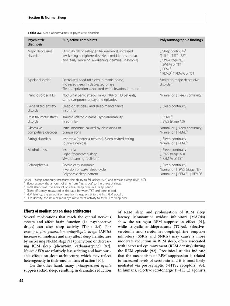
Effects of medications on sleep architectureSeveral medications that reach the central nervoussystem and affect brain function (i.e. psychoactivedrugs) can alter sleep activity (Table 3.4). Forexample, first-generation antiepileptic drugs (AEDs)increase somnolence and may affect sleep architectureby increasing NREM stage N1 (phenytoin) or decreas-ing REM sleep (phenytoin, carbamazepine) [89].Newer AEDs are relatively less sedating and have vari-able effects on sleep architecture, which may reflectheterogeneity in their mechanisms of action [90].
On the other hand, many antidepressant agentssuppress REM sleep, resulting in dramatic reduction
of REM sleep and prolongation of REM sleeplatency. Monoamine oxidase inhibitors (MAOIs)show the strongest REM suppression effect [91],while tricyclic antidepressants (TCAs), selective-serotonin and serotonin-norepinephrine reuptakeinhibitors (SSRIs and SNRIs) may cause a moremoderate reduction in REM sleep, often associatedwith increased eye movement (REM density) duringthe REM episode [92]. Preclinical studies indicatethat the mechanism of REM suppression is relatedto increased levels of serotonin and it is most likelymediated via post-synaptic 5-HT1A receptors [93].In humans, selective serotonergic (5-HT1A) agonists
Table 3.3 Sleep abnormalities in psychiatric disorders
Psychiatricdiagnosis
Subjective complaints Polysomnographic findings
Major depressivedisorder
Difficulty falling asleep (initial insomnia), increasedawakening at night/restless sleep (middle insomnia),and early morning awakening (terminal insomnia)
# Sleep continuity1(" SL2, # TST3, #SE4)# SWS (stage N3)# SWS % of TST# REML5
" REMD6 " REM % of TST
Bipolar disorder Decreased need for sleep in manic phase,increased sleep in depressed phaseSleep deprivation associated with elevation in mood
Similar to major depressivedisorder
Panic disorder (PD) Nocturnal panic attacks: in 40 70% of PD patients,same symptoms of daytime episodes
Normal or # sleep continuity1
Generalized anxietydisorder
Sleep-onset delay and sleep-maintenanceinsomnia
# Sleep continuity1
Post-traumatic stressdisorder
Trauma-related dreams. Hyperarousability(insomnia)
" REMD6
# SWS (stage N3)
Obsessive-compulsive disorder
Initial insomnia caused by obsessions orcompulsions
Normal or # sleep continuity1
Normal or # REML5
Eating disorders Insomnia (anorexia nervosa). Sleep-related eating(bulimia nervosa)
# Sleep continuity1
Normal or # REML5
Alcohol abuse InsomniaLight, fragmented sleepVivid dreaming (delirium)
# Sleep continuity1
# SWS (stage N3)" REM % of TST
Schizophrenia Severe early insomniaInversion of wake sleep cyclePolyphasic sleep pattern
# Sleep continuity1
Normal or # SWS (stage N3)Normal or # REML5, " REMD6
Notes: 1 Sleep continuity: measures the ability to fall asleep (SL2) and remain asleep (TST3, SE4).2 Sleep latency: the amount of time from “lights out” to the onset of sleep.3 Total sleep time: the amount of actual sleep time in a sleep period.4 Sleep efficiency: measured as the ratio between TST and time in bed.5 REM latency: the amount of time from sleep onset to the first REM epoch.6 REM density: the ratio of rapid eye movement activity to total REM sleep time.
Section II: Normal Sleep
44
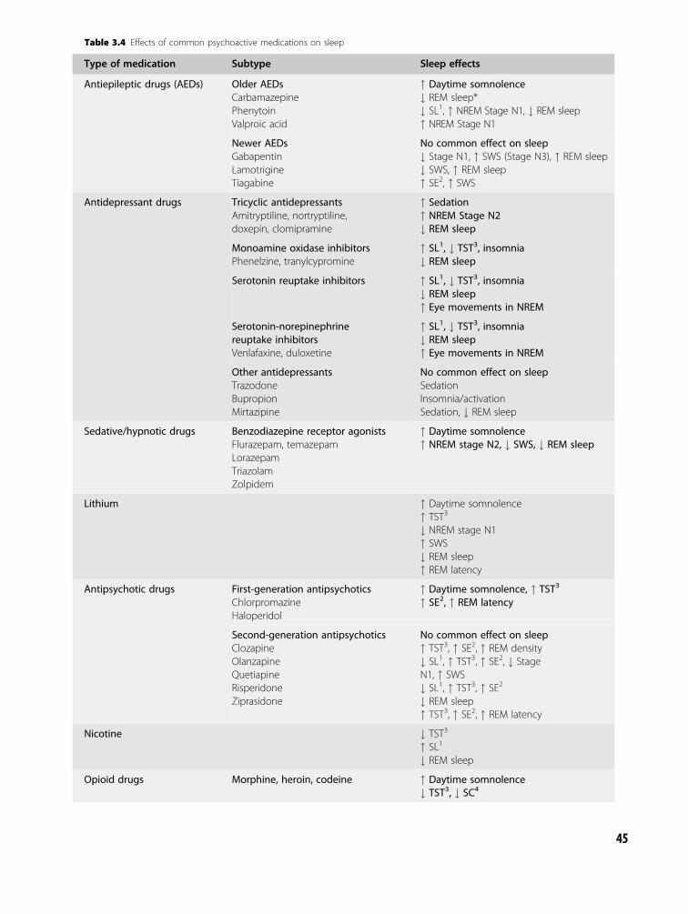
Table 3.4 Effects of common psychoactive medications on sleep
Type of medication Subtype Sleep effects
Antiepileptic drugs (AEDs) Older AEDsCarbamazepinePhenytoinValproic acid
" Daytime somnolence# REM sleep*# SL1, " NREM Stage N1, # REM sleep" NREM Stage N1
Newer AEDsGabapentinLamotrigineTiagabine
No common effect on sleep# Stage N1, " SWS (Stage N3), " REM sleep# SWS, " REM sleep" SE2, " SWS
Antidepressant drugs Tricyclic antidepressantsAmitryptiline, nortryptiline,doxepin, clomipramine
" Sedation" NREM Stage N2# REM sleep
Monoamine oxidase inhibitorsPhenelzine, tranylcypromine
" SL1, # TST3, insomnia# REM sleep
Serotonin reuptake inhibitors " SL1, # TST3, insomnia# REM sleep" Eye movements in NREM
Serotonin-norepinephrinereuptake inhibitorsVenlafaxine, duloxetine
" SL1, # TST3, insomnia# REM sleep" Eye movements in NREM
Other antidepressantsTrazodoneBupropionMirtazipine
No common effect on sleepSedationInsomnia/activationSedation, # REM sleep
Sedative/hypnotic drugs Benzodiazepine receptor agonistsFlurazepam, temazepamLorazepamTriazolamZolpidem
" Daytime somnolence" NREM stage N2, # SWS, # REM sleep
Lithium " Daytime somnolence" TST3
# NREM stage N1" SWS# REM sleep" REM latency
Antipsychotic drugs First-generation antipsychoticsChlorpromazineHaloperidol
" Daytime somnolence, " TST3
" SE2, " REM latency
Second-generation antipsychoticsClozapineOlanzapineQuetiapineRisperidoneZiprasidone
No common effect on sleep" TST3, " SE2, " REM density# SL1, " TST3, " SE2, # StageN1, " SWS# SL1, " TST3, " SE2
# REM sleep" TST3, " SE2, " REM latency
Nicotine # TST3
" SL1
# REM sleep
Opioid drugs Morphine, heroin, codeine " Daytime somnolence# TST3, # SC4
45
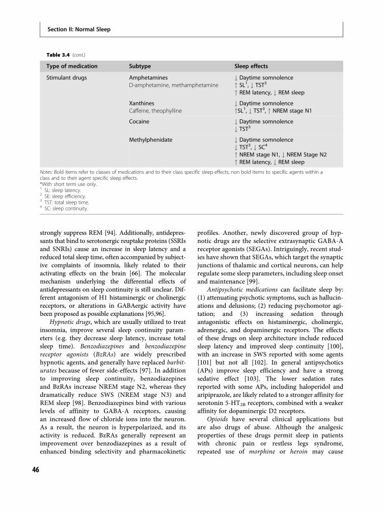
strongly suppress REM [94]. Additionally, antidepres-sants that bind to serotonergic reuptake proteins (SSRIsand SNRIs) cause an increase in sleep latency and areduced total sleep time, often accompanied by subject-ive complaints of insomnia, likely related to theiractivating effects on the brain [66]. The molecularmechanism underlying the differential effects ofantidepressants on sleep continuity is still unclear. Dif-ferent antagonism of H1 histaminergic or cholinergicreceptors, or alterations in GABAergic activity havebeen proposed as possible explanations [95,96].
Hypnotic drugs, which are usually utilized to treatinsomnia, improve several sleep continuity param-eters (e.g. they decrease sleep latency, increase totalsleep time). Benzodiazepines and benzodiazepinereceptor agonists (BzRAs) are widely prescribedhypnotic agents, and generally have replaced barbit-urates because of fewer side-effects [97]. In additionto improving sleep continuity, benzodiazepinesand BzRAs increase NREM stage N2, whereas theydramatically reduce SWS (NREM stage N3) andREM sleep [98]. Benzodiazepines bind with variouslevels of affinity to GABA-A receptors, causingan increased flow of chloride ions into the neuron.As a result, the neuron is hyperpolarized, and itsactivity is reduced. BzRAs generally represent animprovement over benzodiazepines as a result ofenhanced binding selectivity and pharmacokinetic
profiles. Another, newly discovered group of hyp-notic drugs are the selective extrasynaptic GABA-Areceptor agonists (SEGAs). Intriguingly, recent stud-ies have shown that SEGAs, which target the synapticjunctions of thalamic and cortical neurons, can helpregulate some sleep parameters, including sleep onsetand maintenance [99].
Antipsychotic medications can facilitate sleep by:(1) attenuating psychotic symptoms, such as hallucin-ations and delusions; (2) reducing psychomotor agi-tation; and (3) increasing sedation throughantagonistic effects on histaminergic, cholinergic,adrenergic, and dopaminergic receptors. The effectsof these drugs on sleep architecture include reducedsleep latency and improved sleep continuity [100],with an increase in SWS reported with some agents[101] but not all [102]. In general antipsychotics(APs) improve sleep efficiency and have a strongsedative effect [103]. The lower sedation ratesreported with some APs, including haloperidol andaripiprazole, are likely related to a stronger affinity forserotonin 5-HT2B receptors, combined with a weakeraffinity for dopaminergic D2 receptors.
Opioids have several clinical applications butare also drugs of abuse. Although the analgesicproperties of these drugs permit sleep in patientswith chronic pain or restless legs syndrome,repeated use of morphine or heroin may cause
Table 3.4 (cont.)
Type of medication Subtype Sleep effects
Stimulant drugs AmphetaminesD-amphetamine, methamphetamine
# Daytime somnolence" SL1, # TST3
" REM latency, # REM sleep
XanthinesCaffeine, theophylline
# Daytime somnolence"SL1, # TST3, " NREM stage N1
Cocaine # Daytime somnolence# TST3
Methylphenidate # Daytime somnolence# TST3, # SC4
" NREM stage N1, # NREM Stage N2" REM latency, # REM sleep
Notes: Bold items refer to classes of medications and to their class specific sleep effects; non bold items to specific agents within aclass and to their agent specific sleep effects.*With short term use only.1 SL: sleep latency.2 SE: sleep efficiency.3 TST: total sleep time.4 SC: sleep continuity.
Section II: Normal Sleep
46

sleep disturbances such as reduction in total sleepand sleep continuity [7]. Similar effects on sleepare caused by stimulants, which include caffeine,amphetamine, or related compounds (i.e. methyl-phenidate, cocaine). Stimulants also increase wake-fulness, prolong sleep latency, and decrease totalsleep time, SWS (NREM stage N3), and REM sleep[104–106]. For a more detailed review of the effectsof drugs of abuse/dependence on sleep, please referto Chapter 19: Sleep in substance use disorders.
Applications of neuroimagingtechniques in sleep medicineOver the last two decades, several neuroimagingtechniques, including positron emission tomog-raphy (PET), anatomical and functional magneticresonance imaging (MRI and fMRI), magnetic res-onance spectroscopy (MRS), single photon emissioncomputed tomography (SPECT), and high-densityelectroencephalography (hd-EEG), have been increas-ingly utilized in research and clinical settings(Table 3.5). For example, neuroimaging techniqueshave been employed in a range of experimental con-ditions to further our understanding of normal brainactivity during wakefulness. Additionally, given theirability to provide valuable information on brainactivity changes in different states of vigilance (e.g.sleep–wake cycle) [8] as well as in various patho-logical conditions (e.g. psychiatric and sleep dis-orders) [107,108], these techniques have been usedin sleep research to better characterize fundamentalaspects of normal sleep and of sleep disruption. Someof these aspects include: (a) the functional neuroanat-omy of sleep stages; (b) the relationship betweenwaking neuronal plastic changes (e.g. learning tasks,sensory deprivation) and sleep; and (c) the effects ofsleep deprivation on waking brain activities, especiallyon the neuronal circuits underlying key cognitivefunctions (e.g. memory, attention). The most relevantneuroimaging findings related to each of these aspectsare reviewed below.
Functional neuroanatomy of sleep stagesWhereas traditional EEG recordings have revealedbrain activity patterns characteristic of the sleep–wake cycle, neuroimaging studies employing PETand fMRI have significantly contributed to
characterize the functional neuroanatomy under-lying these EEG patterns. Specifically, these studieshave shown that both global and local brain activitychanges occur in sleep compared to waking and thatdifferent patterns of activity are present in REMand NREM sleep.
NREM sleep is characterized by a global reductionin brain metabolism and cerebral blood flow relative towaking, as well as a decrease in regional cerebral bloodflow (rCBF) in several subcortical (dorsal pons,cerebellum, midbrain, basal ganglia, basal forebrain,thalamus) [109,111] and cortical regions (anterior cin-gulate, inferior parietal, dorsolateral prefrontal cortex)[110,112–114] (Figure 3.4). A recent meta-analysis ofPET data has shown an inverse correlation betweenslow waves and the rCBF of all of these brain structures,with the exception of the thalamus [115]. Notably, thesefindings are consistent with evidence from electro-physiological studies showing that disruption of thal-amocortical connections leaves the slow oscillationintact, whereas damaging corticocortical connectionsdisrupts the synchronization of the slow oscillation[116,117].
Neuroimaging studies have also revealed thatdecreases in cortical rCBF during NREM sleep mainlyoccur in associative areas (dorsolateral prefrontal,orbitofrontal, and parietal cortices) and the precuneus[109,111,112], while primary cortices are the leastaffected [109] (Figure 3.4). The marked decrease ofrCBF in the associative areas likely reflects the highlevel of activity of these areas during wakefulness,while the rCBF reduction in the precuneus possiblyindicates the loss of wakefulness-promoting mechan-isms, as suggested by the deactivation observed in theprecuneus during states of decreased consciousness(vegetative state, anesthesia) [118].
A pivotal contribution to understanding the func-tional neuroanatomy of NREM sleep EEG rhythms,such as slow waves and sleep spindles, is expectedfrom a newly introduced neuroimaging technique,EEG/fMRI. By recording EEG with simultaneousfMRI, this technique provides a unique opportunityto combine high spatial (fMRI) and temporal (EEG)resolution. For example, a recent EEG/fMRI studyfound that the thalamus, in combination with limbicareas and superior temporal gyri, was significantlyactivated during sleep in healthy subjects when sleepspindles occurred [119]. While several electrophysio-logical studies in animals had previously revealed thatthalamic nuclei are critically involved in generating
Chapter 3: Neurophysiology and neuroimaging
47

sleep spindles [120], this study was the first toestablish a similar role for the thalamus in humans.This study also characterized the brain areas thatwere specifically activated during slow (11–13 Hz)and fast (13–15 Hz) spindles, the superior frontalgyrus and the sensorimotor/hippocampal regions
respectively, thus suggesting that these two spindletypes might have different functional roles duringNREM sleep [119].
In comparison to NREM sleep, REM sleep ischaracterized by an increase in neuronal activity,brain metabolism, and rCBF in the pons, midbrain,
Table 3.5 Neuroimaging techniques commonly utilized in sleep
Neuroimagingtechnique
Parametersmeasured
Clinical and research use Advantages Limitations
Magneticresonanceimaging (MRI)
Voxel-basedmorphometry(VBM)1
Examine gray/white matterchanges related to aging,neurodegenerative, or otherchronic diseases (dementia,schizophrenia) which affectbrain tissues
High spatial resolution,automatic procedurefaster than traditionalmorphometric analyses(ROI)2
Expensive, provideno functional data
Magneticresonancespectroscopy(MRS)
Metabolitelevels
Measure metabolite levels intumors or other disorders(OSAS3) where metabolitedeficits (i.e. choline) canoccur
Combine anatomical andfunctional data, confirminvolvement of certainmetabolites in specificbrain disorders
Expensive, lowerspatial resolutionthan MRI
Functionalmagneticresonanceimaging (fMRI)
Blood-oxygen-level-dependent(BOLD4) signal
Assess BOLD (O2 supply)changes in brain regionsinvolved in cognitive tasks oraffected by acute andchronic brain disorders
High spatial resolution,safe, non-invasive, allowsrepetitive measureswithin a session and overtime
Low temporalresolution (~4 sec),indirect measure ofneural activity
Single photonemissioncomputedtomography(SPECT)
Regionalcerebral bloodflow (or rCBF5,with 99mTc-HMPAO6)
Diagnosis of dementia, rCBFchanges in other chronicbrain disorders
Inexpensive relative toother imaging tools, goodspatial resolution
Poor temporalresolution (1 min),radiation exposure
Positronemissiontomography(PET)
rCBF5 (with 18[F]DG, H2
15O)7
receptorbinding(radioligands)8
Differentiate Alzheimer fromother types of dementia,assess receptor deficits inpsychiatric disorders
Less expensive than fMRI,good spatial resolution,imaging ofneuroreceptors’ activity
Low temporalresolution (40 sec),radiation exposure
High-density(hd)-EEG
EEG patternsduring wakingas well as sleep
Differentiate delirium fromcatatonia, identify seizures aswell as other waking andsleep rhythm abnormalities
Exquisite temporal andgood spatial resolution(relative to standard EEG),safe, inexpensive
Limited ability tolocalize corticalsources of activityand gather datafrom deep regions
Notes: 1 Identifies gray and white matters in all brain regions from high resolution MRI scans.2 ROI: region of interests manually drawn on MRI scannings.3 OSAS: obstructive sleep apnea syndrome.4 BOLD: Measures the level of blood oxygenated hemoglobin, which increases with higher neural activity.5 rCBF: the blood supplied to the different brain regions in a given time.6 Technetium hexamethylpropylene amine oxime radiotracer which accumulates in brain regions proportional to blood flow.7 (18Fluorodeoxyglucose, H2
15O) radiotracers which measure CBF to brain regions.8 These agents permit visualization of neuroreceptors in a plurality of neuropsychiatric illnesses.
Section II: Normal Sleep
48

basal ganglia, thalamus [109], amygdala, anterior cin-gulate, and temporal and orbito-medial prefrontalcortices [121] (Figure 3.4). Activation of the pons,the midbrain, and the thalamus during REM is con-sistent with electrophysiological evidence showingthat REM activity is initiated by pontine cholinergicnuclei [122,123], which then induce widespread cor-tical activation through a dorsal (thalamic) and aventral (midbrain) pathway [124,125]. Furthermore,electrophysiological studies in animals have shown an
involvement of neurons of the amygdala in generatingponto-geniculo-occipital (PGO) waves, key compo-nents of phasic REM sleep underlying rapid eyemovements [126,127]. In humans, dreaming activityoccurring during REM sleep is associated with anincreased activation in the temporal cortex, in aregion anatomically connected to the visual areas[113], as well as in the cingulate [113,128] andorbito-medial prefrontal cortices, both part of thelimbic system. This pattern of activation may account
From Waking to NREM Sleep
From NREM to REM Sleep
From REM Sleep to Waking
Figure 3.4 Brain maps showing changes in rCBF from waking to NREM (top), NREM to REM (middle), and REM to waking (bottom), measuredwith H2
15O PET. Top panel: brain regions with decreased rCBF during NREM sleep compared with presleep wakefulness. The reduction isexpressed in Z scores, ranging from 1 (light purple) to 4.5 (deep purple). Significant rCBF reductions were observed in the pons(A, arrowhead), midbrain (B, short arrow), basal ganglia (B, long arrow; C, medium arrowhead), thalamus (C, short arrow), caudal orbital cortex(B, small arrowhead), and cerebellum (A, arrow). Similar rCBF decreases were found in the anterior insula (B, medium arrowhead), anteriorcingulate (C and D, small arrowheads), as well as in the orbital (B, medium arrow), dorsolateral prefrontal (C, medium arrow; D, small arrow), andinferior parietal lobes (D, medium arrow). Middle panel: brain regions with increased rCBF during REM sleep compared with NREM sleep.Z scores values range from 1 (green) to 3.5 (red). Significant increases in rCBF during REM sleep were observed in the pons (A, arrowhead),midbrain (B, long arrow), basal ganglia (B, short arrow; C, small arrowhead), thalamus (C, medium arrowhead), and caudal orbital cortex(B, medium arrowhead). Increases were also found in anterior insula (B, small arrowhead), anterior cingulate (C, small arrow; D, mediumarrow), mesial temporal (parahippocampal) (B, medium arrow), fusiform inferotemporal (B, large arrowhead), lateral occipital (C, long arrow),auditory association (C, medium arrow), and medial prefrontal cortices (D, small arrow). Bottom panel: brain regions with rCBF increaseduring post sleep wakefulness compared to REM sleep. Z scores values are from 1 (green) to 3.5 (red). Significant increases in rCBFoccurred in the orbital (B, small arrow), dorsolateral prefrontal (C and D, small arrows), and inferior parietal lobes (D, medium arrow),as well as in the cerebellar hemispheres (A, small arrow). Reproduced, with permission, from [2]. See plate section for color version.
Chapter 3: Neurophysiology and neuroimaging
49
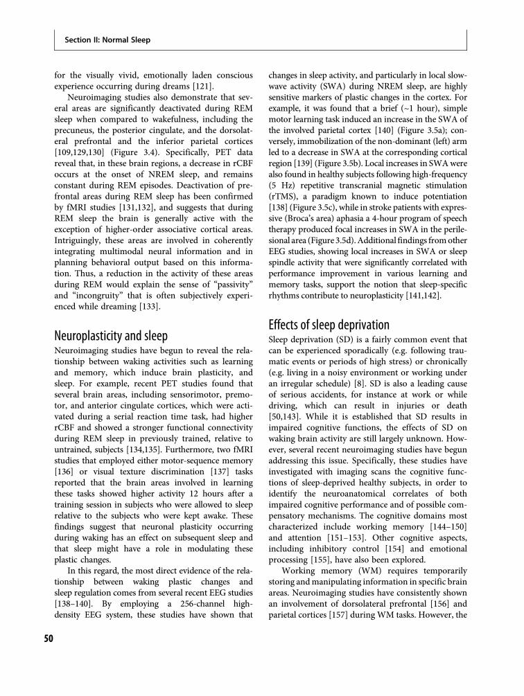
for the visually vivid, emotionally laden consciousexperience occurring during dreams [121].
Neuroimaging studies also demonstrate that sev-eral areas are significantly deactivated during REMsleep when compared to wakefulness, including theprecuneus, the posterior cingulate, and the dorsolat-eral prefrontal and the inferior parietal cortices[109,129,130] (Figure 3.4). Specifically, PET datareveal that, in these brain regions, a decrease in rCBFoccurs at the onset of NREM sleep, and remainsconstant during REM episodes. Deactivation of pre-frontal areas during REM sleep has been confirmedby fMRI studies [131,132], and suggests that duringREM sleep the brain is generally active with theexception of higher-order associative cortical areas.Intriguingly, these areas are involved in coherentlyintegrating multimodal neural information and inplanning behavioral output based on this informa-tion. Thus, a reduction in the activity of these areasduring REM would explain the sense of “passivity”and “incongruity” that is often subjectively experi-enced while dreaming [133].
Neuroplasticity and sleepNeuroimaging studies have begun to reveal the rela-tionship between waking activities such as learningand memory, which induce brain plasticity, andsleep. For example, recent PET studies found thatseveral brain areas, including sensorimotor, premo-tor, and anterior cingulate cortices, which were acti-vated during a serial reaction time task, had higherrCBF and showed a stronger functional connectivityduring REM sleep in previously trained, relative tountrained, subjects [134,135]. Furthermore, two fMRIstudies that employed either motor-sequence memory[136] or visual texture discrimination [137] tasksreported that the brain areas involved in learningthese tasks showed higher activity 12 hours after atraining session in subjects who were allowed to sleeprelative to the subjects who were kept awake. Thesefindings suggest that neuronal plasticity occurringduring waking has an effect on subsequent sleep andthat sleep might have a role in modulating theseplastic changes.
In this regard, the most direct evidence of the rela-tionship between waking plastic changes andsleep regulation comes from several recent EEG studies[138–140]. By employing a 256-channel high-density EEG system, these studies have shown that
changes in sleep activity, and particularly in local slow-wave activity (SWA) during NREM sleep, are highlysensitive markers of plastic changes in the cortex. Forexample, it was found that a brief (~1 hour), simplemotor learning task induced an increase in the SWA ofthe involved parietal cortex [140] (Figure 3.5a); con-versely, immobilization of the non-dominant (left) armled to a decrease in SWA at the corresponding corticalregion [139] (Figure 3.5b). Local increases in SWAwerealso found in healthy subjects following high-frequency(5 Hz) repetitive transcranial magnetic stimulation(rTMS), a paradigm known to induce potentiation[138] (Figure 3.5c), while in stroke patients with expres-sive (Broca’s area) aphasia a 4-hour program of speechtherapy produced focal increases in SWA in the perile-sional area (Figure 3.5d).Additional findings fromotherEEG studies, showing local increases in SWA or sleepspindle activity that were significantly correlated withperformance improvement in various learning andmemory tasks, support the notion that sleep-specificrhythms contribute to neuroplasticity [141,142].
Effects of sleep deprivationSleep deprivation (SD) is a fairly common event thatcan be experienced sporadically (e.g. following trau-matic events or periods of high stress) or chronically(e.g. living in a noisy environment or working underan irregular schedule) [8]. SD is also a leading causeof serious accidents, for instance at work or whiledriving, which can result in injuries or death[50,143]. While it is established that SD results inimpaired cognitive functions, the effects of SD onwaking brain activity are still largely unknown. How-ever, several recent neuroimaging studies have begunaddressing this issue. Specifically, these studies haveinvestigated with imaging scans the cognitive func-tions of sleep-deprived healthy subjects, in order toidentify the neuroanatomical correlates of bothimpaired cognitive performance and of possible com-pensatory mechanisms. The cognitive domains mostcharacterized include working memory [144–150]and attention [151–153]. Other cognitive aspects,including inhibitory control [154] and emotionalprocessing [155], have also been explored.
Working memory (WM) requires temporarilystoring andmanipulating information in specific brainareas. Neuroimaging studies have consistently shownan involvement of dorsolateral prefrontal [156] andparietal cortices [157] during WM tasks. However, the
Section II: Normal Sleep
50
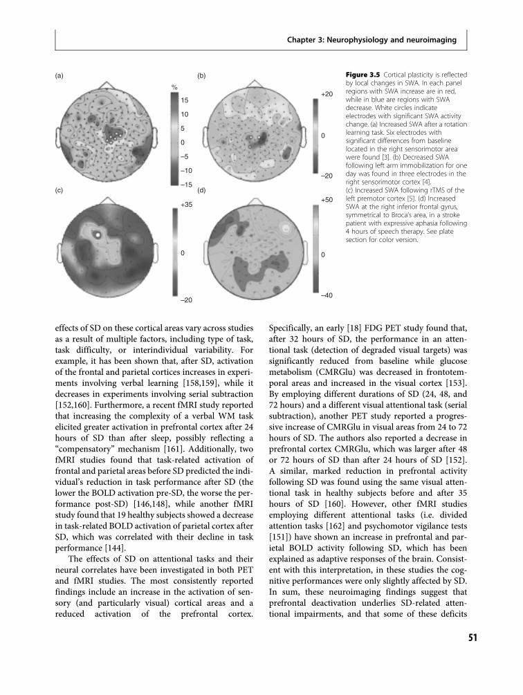
effects of SD on these cortical areas vary across studiesas a result of multiple factors, including type of task,task difficulty, or interindividual variability. Forexample, it has been shown that, after SD, activationof the frontal and parietal cortices increases in experi-ments involving verbal learning [158,159], while itdecreases in experiments involving serial subtraction[152,160]. Furthermore, a recent fMRI study reportedthat increasing the complexity of a verbal WM taskelicited greater activation in prefrontal cortex after 24hours of SD than after sleep, possibly reflecting a“compensatory” mechanism [161]. Additionally, twofMRI studies found that task-related activation offrontal and parietal areas before SD predicted the indi-vidual’s reduction in task performance after SD (thelower the BOLD activation pre-SD, the worse the per-formance post-SD) [146,148], while another fMRIstudy found that 19 healthy subjects showed a decreasein task-related BOLD activation of parietal cortex afterSD, which was correlated with their decline in taskperformance [144].
The effects of SD on attentional tasks and theirneural correlates have been investigated in both PETand fMRI studies. The most consistently reportedfindings include an increase in the activation of sen-sory (and particularly visual) cortical areas and areduced activation of the prefrontal cortex.
Specifically, an early [18] FDG PET study found that,after 32 hours of SD, the performance in an atten-tional task (detection of degraded visual targets) wassignificantly reduced from baseline while glucosemetabolism (CMRGlu) was decreased in frontotem-poral areas and increased in the visual cortex [153].By employing different durations of SD (24, 48, and72 hours) and a different visual attentional task (serialsubtraction), another PET study reported a progres-sive increase of CMRGlu in visual areas from 24 to 72hours of SD. The authors also reported a decrease inprefrontal cortex CMRGlu, which was larger after 48or 72 hours of SD than after 24 hours of SD [152].A similar, marked reduction in prefrontal activityfollowing SD was found using the same visual atten-tional task in healthy subjects before and after 35hours of SD [160]. However, other fMRI studiesemploying different attentional tasks (i.e. dividedattention tasks [162] and psychomotor vigilance tests[151]) have shown an increase in prefrontal and par-ietal BOLD activity following SD, which has beenexplained as adaptive responses of the brain. Consist-ent with this interpretation, in these studies the cog-nitive performances were only slightly affected by SD.In sum, these neuroimaging findings suggest thatprefrontal deactivation underlies SD-related atten-tional impairments, and that some of these deficits
(a) (b)
%
15
+35
0
–20
+50
0
–40
+20
0
–20
5
–5
–10
–15
0
10
(c) (d)
Figure 3.5 Cortical plasticity is reflectedby local changes in SWA. In each panelregions with SWA increase are in red,while in blue are regions with SWAdecrease. White circles indicateelectrodes with significant SWA activitychange. (a) Increased SWA after a rotationlearning task. Six electrodes withsignificant differences from baselinelocated in the right sensorimotor areawere found [3]. (b) Decreased SWAfollowing left arm immobilization for oneday was found in three electrodes in theright sensorimotor cortex [4].(c) Increased SWA following rTMS of theleft premotor cortex [5]. (d) IncreasedSWA at the right inferior frontal gyrus,symmetrical to Broca’s area, in a strokepatient with expressive aphasia following4 hours of speech therapy. See platesection for color version.
Chapter 3: Neurophysiology and neuroimaging
51
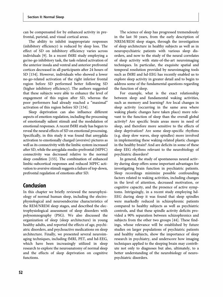
can be compensated for by enhanced activity in pre-frontal, parietal, and visual cortical areas.
The ability to inhibit inappropriate responses(inhibitory efficiency) is reduced by sleep loss. Theeffect of SD on inhibitory efficiency varies acrossindividuals [9]. In a recent fMRI study employing ago/no-go inhibitory task, the task-related activation ofthe anterior insula and ventral and anterior prefrontalcortices decreased in all participants after 24 hours ofSD [154]. However, individuals who showed a lowerno-go-related activation of the right inferior frontalregion before SD performed better following SD(higher inhibitory efficiency). The authors suggestedthat these subjects were able to enhance the level ofengagement of this region after SD, whereas thepoor performers had already reached a “maximal”activation of this region before SD [154].
Sleep deprivation can also influence differentaspects of emotion regulation, including the processingof emotionally salient stimuli and the modulation ofemotional responses. A recent fMRI study has begun toreveal the neural effects of SD on emotional processing.Specifically, in this study it was found that amygdalaactivation to emotionally charged (aversive) pictures aswell as its connectivity with the limbic system increasedafter SD, while the amygdala-medio-prefrontal (MPFC)connectivity was decreased relative to the normalsleep condition [155]. The combination of enhancedlimbic-subcortical responses and reduced MPFC acti-vation to aversive stimuli suggests a failure of top-down,prefrontal regulation of emotions after SD.
ConclusionIn this chapter we briefly reviewed the neurophysi-ology of normal human sleep, including the electro-physiological and neuroendocrine characteristics ofthe REM/NREM sleep stages, and described the elec-trophysiological assessment of sleep disorders withpolysomnography (PSG). We also discussed theorganization of sleep (sleep architecture) in younghealthy adults, and reported the effects of age, psychi-atric disorders, and psychoactive medications on sleeparchitecture. Finally, we presented several neuroim-aging techniques, including fMRI, PET, and hd-EEG,which have been increasingly utilized in sleepresearch to explore the neuroanatomy of normal sleepand the effects of sleep deprivation on cognitivefunctions.
The science of sleep has progressed tremendouslyin the last 50 years, from the early description ofNREM/REM sleep stages, through the investigationof sleep architecture in healthy subjects as well as inneuropsychiatric patients with various sleep dis-orders, and now to the study of the neural correlatesof sleep activity with state-of-the-art neuroimagingtechniques. In particular, the exquisite spatial andtemporal resolution provided by neuroimaging toolssuch as fMRI and hd-EEG has recently enabled us toexplore sleep activity in greater detail and to begin toaddress some of the fundamental questions regardingthe function of sleep.
For example, what is the exact relationshipbetween sleep and fundamental waking activities,such as memory and learning? Are local changes insleep activity (occurring in the same area wherewaking plastic changes have taken place) more rele-vant to the function of sleep than the overall globalactivity? Are specific brain areas more in need ofsleep, and therefore more sensitive to the effects ofsleep deprivation? Are some sleep-specific rhythms(e.g. sleep slow waves, sleep spindles) more involvedin implementing these waking-related plastic changesin the healthy brain? And are deficits in some of thesesleep EEG rhythms relevant to the neurobiology ofpsychiatric disorders?
In general, the study of spontaneous neural activ-ity during sleep offers some important advantages forinvestigating brain function in psychiatric patients.Sleep recordings minimize possible confoundingfactors related to waking activities, including changesin the level of attention, decreased motivation, orcognitive capacity, and the presence of active symp-toms. Intriguingly, in a recent study employing hd-EEG during sleep it was found that sleep spindleswere markedly reduced in schizophrenic patientscompared to healthy subjects as well as psychiatriccontrols, and that these spindle activity deficits pro-vided a 90% separation between schizophrenics andsubjects from the other two groups [44]. These find-ings, whose relevance will be established by futurestudies on larger populations of psychiatric patientsand healthy subjects, show the importance of sleepresearch in psychiatry, and underscore how imagingtechniques applied to the sleeping brain may contrib-ute not only to diagnoses but also, ultimately, to abetter understanding of the neurobiology of neuro-psychiatric disorders.
Section II: Normal Sleep
52
References1. Greenspan RJ, Tononi G, Cirelli C, Shaw PJ. Sleep and
the fruit fly. Trends Neurosci. 2001;24(3):142 145.
2. Banks S, Dinges DF. Behavioral and physiologicalconsequences of sleep restriction. J Clin Sleep Med.2007;3(5):519 528.
3. Aserinsky E. The discovery of REM sleep. J HistNeurosci. 1996;5(3):213 227.
4. Kryger MH, Roth T, Dement WC. Principles andpractices of sleep medicine, 3rd ed. Philadelphia: WBSaunders Co.; 2000.
5. Benca RM, Obermeyer WH, Thisted RA, Gillin JC.Meta analysis of sleep changes in psychiatric disorders.Sleep Res. 1991;20:169.
6. Markov D, Goldman M. Normal sleep and circadianrhythms: neurobiologic mechanisms underlying sleepand wakefulness. Psychiatr Clin North Am. 2006;29(4):841 853; abstract vii.
7. Wang D, Teichtahl H. Opioids, sleep architecture andsleep disordered breathing. Sleep Med Rev. 2007;11(1):35 46.
8. Dang Vu TT, Desseilles M, Petit D,et al. Neuroimagingin sleep medicine. Sleep Med. 2007;8(4):349 372.
9. Chee MW, Chuah LY. Functional neuroimaginginsights into how sleep and sleep deprivation affectmemory and cognition. Curr Opin Neurol. 2008;21(4):417 423.
10. Boveroux P, Bonhomme V, Boly M, et al. Brainfunction in physiologically, pharmacologically, andpathologically altered states of consciousness. IntAnesthesiol Clin. 2008;46(3):131 146.
11. Hirshkowitz M. Normal human sleep: an overview.Med Clin North Am. 2004;88(3):551 565, vii.
12. Rechtschaffen A, Kales A. A manual of standardizedterminology, techniques, and scoring system forsleep stages of human subjects. UCLA, Los Angeles:Brain Information Service/Brain ResearchInstitute; 1968.
13. Silber MH, Ancoli Israel S, Bonnet MH, et al. Thevisual scoring of sleep in adults. J Clin Sleep Med.2007;3(2):121 131.
14. Trinder J, Kleiman J, Carrington M, et al. Autonomicactivity during human sleep as a function of time andsleep stage. J Sleep Res. 2001;10(4):253 264.
15. Brandenberger G, Ehrhart J, Piquard F, Simon C.Inverse coupling between ultradian oscillations in deltawave activity and heart rate variability during sleep.Clin Neurophysiol. 2001;112(6):992 996.
16. Murali NS, Svatikova A, Somers VK. Cardiovascularphysiology and sleep. Front Biosci. 2003;8:s636 652.
17. Tasali E, Leproult R, Ehrmann DA, Van Cauter E.Slow wave sleep and the risk of type 2 diabetes inhumans. Proc Natl Acad Sci U S A. 2008;105(3):1044 1049.
18. Vgontzas AN, Bixler EO, Papanicolaou DA, et al.Rapid eye movement sleep correlates with theoverall activities of the hypothalamic pituitary adrenalaxis and sympathetic system in healthy humans.J Clin Endocrinol Metab. 1997;82(10):3278 3280.
19. Verrier RL, Muller JE, Hobson JA. Sleep, dreams, andsudden death: the case for sleep as an autonomic stresstest for the heart. Cardiovasc Res. 1996;31(2):181 211.
20. Bankier B, Littman AB. Psychiatric disordersand coronary heart disease in women a stillneglected topic: review of the literature from1971 to 2000. Psychother Psychosom. 2002;71(3):133 140.
21. Penzel T, Wessel N, Riedl M, et al. Cardiovascular andrespiratory dynamics during normal and pathologicalsleep. Chaos. 2007;17(1):015116.
22. Nattie E. CO2, brainstem chemoreceptors andbreathing. Prog Neurobiol. 1999;59(4):299 331.
23. Owens RL, Eckert DJ, Yeh SY, Malhotra A. Upperairway function in the pathogenesis of obstructivesleep apnea: a review of the current literature. CurrOpin Pulm Med. 2008;14(6):519 524.
24. Lack LC, Gradisar M, Van Someren EJ, Wright HR,Lushington K. The relationship between insomnia andbody temperatures. Sleep Med Rev. 2008;12(4):307 317.
25. Krauchi K. The human sleep wake cycle reconsideredfrom a thermoregulatory point of view. Physiol Behav.2007;90(2 3):236 245.
26. Gutierrez E, Vazquez R, Boakes RA. Activity basedanorexia: ambient temperature has been a neglectedfactor. Psychon Bull Rev. 2002;9(2):239 249.
27. Nobili L, Baglietto MG, Beelke M, et al. Impairment ofthe production of delta sleep in anorectic adolescents.Sleep. 2004;27(8):1553 1559.
28. Obal F Jr, Krueger JM. GHRH and sleep. Sleep MedRev. 2004;8(5):367 377.
29. Freeman ME, Kanyicska B, Lerant A, Nagy G.Prolactin: structure, function, and regulation ofsecretion. Physiol Rev. 2000;80(4):1523 1631.
30. Van Cauter E, Plat L, Copinschi G. Interrelationsbetween sleep and the somatotropic axis. Sleep. 1998;21(6):553 566.
31. Roky R, Obal F Jr, Valatx JL, et al. Prolactin and rapideye movement sleep regulation. Sleep. 1995;18(7):536 542.
Chapter 3: Neurophysiology and neuroimaging
53
32. Wagner U, Born J. Memory consolidation duringsleep: interactive effects of sleep stages and HPAregulation. Stress. 2008;11(1):28 41.
33. Luboshitzky R. Endocrine activity during sleep.J Pediatr Endocrinol Metab. 2000;13(1):13 20.
34. Krauchi K, Cajochen C, Pache M, Flammer J,Wirz Justice A. Thermoregulatory effects of melatoninin relation to sleepiness. Chronobiol Int. 2006;23(1 2):475 484.
35. Czeisler CA, Klerman EB. Circadian andsleep dependent regulation of hormone release inhumans. Recent Prog Horm Res. 1999;54:97 130;discussion 130 132.
36. Clark RW, Schmidt HS, Malarkey WB. Disorderedgrowth hormone and prolactin secretion inprimary disorders of sleep. Neurology. 1979;29(6):855 861.
37. Knutson KL, Van Cauter E. Associations between sleeploss and increased risk of obesity and diabetes. AnnN Y Acad Sci. 2008;1129:287 304.
38. Van Cauter E, Knutson KL. Sleep and the epidemic ofobesity in children and adults. Eur J Endocrinol.2008;159(Suppl 1):S59 66.
39. Schmidt MH, Schmidt HS. Sleep related erections:neural mechanisms and clinical significance. CurrNeurol Neurosci Rep. 2004;4(2):170 178.
40. Morales A, Condra M, Reid K. The role ofnocturnal penile tumescence monitoring in thediagnosis of impotence: a review. J Urol. 1990;143(3):441 446.
41. Abel GG, Murphy WD, Becker JV, Bitar A. Women’svaginal responses during REM Sleep. J Sex MaritalTher. 1979;5(1):5 14.
42. Berger H. [Uber das Elektrenkephalogramm desMenschen]. Archiv fur Psychiatrie undNervenkrankheiten. 1929;87:527 580.
43. Myslobodsky MS, Coppola R, Bar Ziv J,Weinberger DR. Adequacy of the International10 20 electrode system for computedneurophysiologic topography. J Clin Neurophysiol.1990;7(4):507 518.
44. Ferrarelli F, Huber R, Peterson MJ, et al. Reduced sleepspindle activity in schizophrenia patients. AmJ Psychiatry. 2007;164(3):483 492.
45. Littner M, Hirshkowitz M, Kramer M, et al.Practice parameters for using polysomnography toevaluate insomnia: an update. Sleep. 2003;26(6):754 760.
46. Kushida CA, Littner MR, Morgenthaler T, et al.Practice parameters for the indications forpolysomnography and related procedures: an updatefor 2005. Sleep. 2005;28(4):499 521.
47. Bixler EO, Kales A, Soldatos CR, Kales JD, Healey S.Prevalence of sleep disorders in the Los Angelesmetropolitan area. Am J Psychiatry. 1979;136(10):1257 1262.
48. Guilleminault C, Cummiskey J, Dement WC. Sleepapnea syndrome: recent advances. Adv Intern Med.1980;26:347 372.
49. Guilleminault C, Dement WC. 235 cases ofexcessive daytime sleepiness: diagnosis andtentative classification. J Neurol Sci. 1977;31(1):13 27.
50. Mitler MM, Carskadon MA, Czeisler CA, et al.Catastrophes, sleep, and public policy: consensusreport. Sleep. 1988;11(1):100 109.
51. Hoddes E, Zarcone V, Smythe H, Phillips R, DementWC. Quantification of sleepiness: a new approach.Psychophysiology. 1973;10(4):431 436.
52. Johns MW. A new method for measuring daytimesleepiness: the Epworth sleepiness scale. Sleep. 1991;14(6):540 545.
53. Benca R, Kwapil TR. Sleep disorders measures.In: Handbook of psychiatric measures. Washington,DC: American Psychiatric Association;2000:673 685.
54. Carskadon MA, Dement WC. The multiple sleeplatency test: what does it measure? Sleep. 1982;5(Suppl 2):S67 72.
55. Mitler MM, Gujavarty KS, Browman CP. Maintenanceof wakefulness test: a polysomnographic technique forevaluation treatment efficacy in patients with excessivesomnolence. Electroencephalogr Clin Neurophysiol.1982;53(6):658 661.
56. Carskadon MA, Dement WC, Mitler MM, et al.Guidelines for the multiple sleep latency test (MSLT): astandard measure of sleepiness. Sleep. 1986;9(4):519 524.
57. Billiard M, Partinen M, Roth T, Shapiro C. Sleep andpsychiatric disorders. J Psychosom Res. 1994;38(Suppl 1):1 2.
58. van den Hoed J, Kraemer H, Guilleminault C, et al.Disorders of excessive daytime somnolence:polygraphic and clinical data for 100 patients. Sleep.1981;4(1):23 37.
59. Guilleminault C, Dement WC. Sleep apneasyndromes and related sleep disorders. In:Williams RL, Karacan I, eds. Sleep disorders:diagnosis and treatment. New York: John Wiley &Sons; 1978:9 28.
60. Thorpy MJ. The clinical use of the Multiple SleepLatency Test. The Standards of Practice Committee ofthe American Sleep Disorders Association. Sleep.1992;15(3):268 276.
Section II: Normal Sleep
54

61. Rosenthal L, Roehrs TA, Rosen A, Roth T. Level ofsleepiness and total sleep time following various timein bed conditions. Sleep. 1993;16(3):226 232.
62. Arand D, Bonnet M, Hurwitz T, et al. The clinicaluse of the MSLT and MWT. Sleep. 2005;28(1):123 144.
63. Buysse DJ, Ancoli Israel S, Edinger JD, Lichstein KL,Morin CM. Recommendations for a standardresearch assessment of insomnia. Sleep. 2006;29(9):1155 1173.
64. Espiritu JR. Aging related sleep changes. Clin GeriatrMed. 2008;24(1):1 14, v.
65. Benca RM, Obermeyer WH, Thisted RA, Gillin JC.Sleep and psychiatric disorders: a meta analysis. ArchGen Psychiatry. 1992;49:651 668.
66. Obermeyer WH, Benca RM. Effects of drugs on sleep[Review]. Neurol Clin North Am. 1996;14(4):827 840.
67. Peirano P, Algarin C, Uauy R. Sleep wake states andtheir regulatory mechanisms throughout early humandevelopment. J Pediatr. 2003;143(4 Suppl):S70 79.
68. Davis KF, Parker KP, Montgomery GL. Sleep in infantsand young children: Part one: normal sleep. J PediatrHealth Care. 2004;18(2):65 71.
69. Grigg Damberger M, Gozal D, Marcus CL, et al.The visual scoring of sleep and arousal in infants andchildren. J Clin Sleep Med. 2007;3(2):201 240.
70. Davis KF, Parker KP, Montgomery GL. Sleep in infantsand young children: part two: common sleepproblems. J Pediatr Health Care. 2004;18(3):130 137.
71. Feinberg I, Higgins LM, Khaw WY, Campbell IG. Theadolescent decline of NREM delta, an indicator ofbrain maturation, is linked to age and sex but not topubertal stage. Am J Physiol Regul Integr Comp Physiol.2006;291(6):R1724 1729.
72. Feinberg I. Schizophrenia: caused by a fault inprogrammed synaptic elimination during adolescence?J Psychiatr Res. 1982;17(4):319 334.
73. Diaz Morales JF, Gutierrez Sorroche M. Morningnesseveningness in adolescents. Span J Psychol. 2008;11(1):201 206.
74. Bliwise DL. Sleep disorders in Alzheimer’s disease andother dementias. Clin Cornerstone. 2004;6(Suppl 1A):S16 28.
75. Krystal AD, Thakur M, Roth T. Sleep disturbance inpsychiatric disorders: effects on function and quality oflife in mood disorders, alcoholism, and schizophrenia.Ann Clin Psychiatry. 2008;20(1):39 46.
76. Kupfer DJ. Sleep research in depressive illness: clinicalimplications a tasting menu [Review]. BiolPsychiatry. 1995;38(6):391 403.
77. Kupfer DJ, Reynolds CF III, Ulrich RF, GrochocinskiVJ. Comparison of automated REM and slow wavesleep analysis in young and middle aged depressedsubjects. Biol Psychiatry. 1986;21:189 200.
78. Hudson JI, Lipinski JF, Keck PE, et al.Polysomnographic characteristics of young manicpatients: comparison with unipolar depressed patientsand normal control subjects. Arch Gen Psychiatry.1992;49:378 383.
79. Mellman TA. Sleep and anxiety disorders. PsychiatrClin North Am. 2006;29(4):1047 1058; abstract x.
80. Ross RJ, Ball WA, Dinges DF, et al. Rapid eyemovement sleep disturbance in posttraumatic stressdisorder. Biol Psychiatry. 1994;35:195 202.
81. Insel TR, Gillin JC, Moore A, et al. The sleep ofpatients with obsessive compulsive disorder. Arch GenPsychiatry. 1982;39:1372 1377.
82. Hendrick I. Dream resistance and schizophrenia. J AmPsychoanal Assoc. 1958;6(4):672 690.
83. Benson KL, Zarcone VP Jr. Low REM latency inschizophrenia. Sleep Res. 1985;14:124.
84. Hiatt JF, FloydTC,Katz PH, Feinberg I. Further evidenceof abnormal non rapid eye movement sleep inschizophrenia. Arch Gen Psychiatry. 1985;42:797 802.
85. Monti JM, Monti D. Sleep disturbance inschizophrenia. Int Rev Psychiatry. 2005;17(4):247 253.
86. Schenck CH, Hurwitz TD, Bundlie SR, MahowaldMW. Sleep related eating disorders: polysomnographiccorrelates of a heterogeneous syndrome distinctfrom daytime eating disorders. Sleep. 1991;14(5):419 431.
87. Brower KJ, Aldrich MS, Robinson EA, Zucker RA,Greden JF. Insomnia, self medication, and relapse toalcoholism. Am J Psychiatry. 2001;158(3):399 404.
88. Crum RM, Ford DE, Storr CL, Chan YF. Associationof sleep disturbance with chronicity and remission ofalcohol dependence: data from a population basedprospective study. Alcohol Clin Exp Res. 2004;28(10):1533 1540.
89. Placidi F, Scalise A, Marciani MG, et al. Effect ofantiepileptic drugs on sleep. Clin Neurophysiol.2000;111(Suppl 2):S115 119.
90. Bazil CW. Effects of antiepileptic drugs on sleepstructure: are all drugs equal? CNS Drugs. 2003;17(10):719 728.
91. Cohen RM, Pickar D, Garnett D, et al. REMsuppression induced by selective monoamine oxidaseinhibitors. Psychopharmacology. 1982;78:137 140.
92. Gervasoni D, Panconi E, Henninot V, et al. Effect ofchronic treatment with milnacipran on sleeparchitecture in rats compared with paroxetine and
Chapter 3: Neurophysiology and neuroimaging
55

imipramine. Pharmacol Biochem Behav. 2002;73(3):557 563.
93. Monaca C, Boutrel B, Hen R, Hamon M, Adrien J.5 HT 1A/1B receptor mediated effects of the selectiveserotonin reuptake inhibitor, citalopram, on sleep:studies in 5 HT 1A and 5 HT 1B knockout mice.Neuropsychopharmacology. 2003;28(5):850 856.
94. Gillin JC, Jernajczyk W, Valladares Neto DC, et al.Inhibition of REM sleep by ipsapirone, a 5HT1Aagonist, in normal volunteers. Psychopharmacology(Berl). 1994;116(4):433 436.
95. Haas H, Panula P. The role of histamine and thetuberomammillary nucleus in the nervous system. NatRev Neurosci. 2003;4(2):121 130.
96. Parent MB, Master S, Kashlub S, Baker GB. Effects ofthe antidepressant/antipanic drug phenelzine and itsputative metabolite phenylethylidenehydrazine onextracellular gamma aminobutyric acid levels in thestriatum. Biochem Pharmacol. 2002;63(1):57 64.
97. Linnoila M, Erwin CW, Logue PE. Efficacy and sideeffects of flurazepam and a combination ofamobarbital and secobarbital in insomniac patients.J Clin Pharmacol. 1980;20:117 123.
98. Benca RM. Diagnosis and treatment of chronicinsomnia: a review. Psychiatr Serv. 2005;56(3):332 343.
99. Ebert B, Wafford KA, Deacon S. Treating insomnia:current and investigational pharmacologicalapproaches. Pharmacol Ther. 2006;112(3):612 629.
100. Taylor SF, Tandon R, Shipley JE, Eiser AS. Effect ofneuroleptic treatment on polysomnographic measuresin schizophrenia. Biol Psychiatry. 1991;30(9):904 912.
101. Sharpley AL, Vassallo CM, Cowen PJ. Olanzapineincreases slow wave sleep: evidence for blockade ofcentral 5 HT(2C) receptors in vivo. Biol Psychiatry.2000;47(5):468 470.
102. Krystal AD, Goforth HW, Roth T. Effects ofantipsychotic medications on sleep in schizophrenia.Int Clin Psychopharmacol. 2008;23(3):150 160.
103. Benson KL. Sleep in schizophrenia: impairments,correlates, and treatment. Psychiatr Clin North Am.2006;29(4):1033 1045; abstract ix x.
104. Bonnet MH, Balkin TJ, Dinges DF, et al. The use ofstimulants to modify performance during sleep loss: areview by the Sleep Deprivation and Stimulant TaskForce of the American Academy of Sleep Medicine.Sleep. 2005;28(9):1163 1187.
105. Mitler MM, Hajdukovic R. Relative efficacy of drugsfor the treatment of sleepiness in narcolepsy. Sleep.1991;14(3):218 220.
106. Nicholson AN, Stone BM. Heterocyclic amphetaminederivatives and caffeine on sleep in man. Br J ClinPharmacol. 1980;9:195 203.
107. McGuire P, Howes OD, Stone J, Fusar Poli P.Functional neuroimaging in schizophrenia: diagnosisand drug discovery. Trends Pharmacol Sci. 2008;29(2):91 98.
108. Nofzinger EA. What can neuroimaging findings tell usabout sleep disorders? Sleep Med 2004;5(Suppl 1):S16 22.
109. Braun AR, Balkin TJ, Wesenten NJ, et al. Regionalcerebral blood flow throughout the sleep wake cycle.An H2(15)O PET study. Brain. 1997;120(Pt 7):1173 1197.
110. Kajimura N, Uchiyama M, Takayama Y, et al. Activityof midbrain reticular formation and neocortex duringthe progression of human non rapid eye movementsleep. J Neurosci. 1999;19(22):10065 10073.
111. Maquet P, Degueldre C, Delfiore G, et al. Functionalneuroanatomy of human slow wave sleep. J Neurosci.1997;17(8):2807 2812.
112. Andersson JL, Onoe H, Hetta J, et al. Brain networksaffected by synchronized sleep visualized by positronemission tomography. J Cereb Blood Flow Metab.1998;18(7):701 715.
113. Braun AR, Balkin TJ, Wesensten NJ, et al. Regionalcerebral blood flow throughout the sleep wake cycle.Brain. 1997;120:1173 1197.
114. Braun AR, Balkin TJ, Wesensten NJ, et al. Dissociatedpattern of activity in visual cortices and theirprojections during human rapid eye movement sleep.Science. 1998;279(5347):91 95.
115. Dang Vu TT, Desseilles M, Laureys S, et al. Cerebralcorrelates of delta waves during non REM sleeprevisited. Neuroimage. 2005;28(1):14 21.
116. Amzica F, Steriade M. Disconnection ofintracortical synaptic linkages disruptssynchronization of a slow oscillation. J Neurosci.1995;15(6):4658 4677.
117. Steriade M. The corticothalamic system in sleep. FrontBiosci. 2003;8:D878 899.
118. Cavanna AE. The precuneus and consciousness. CNSSpectr. 2007;12(7):545 552.
119. Schabus M, Dang Vu TT, Albouy G, et al.Hemodynamic cerebral correlates of sleep spindlesduring human non rapid eye movement sleep. ProcNatl Acad Sci U S A. 2007;104(32):13164 13169.
120. Steriade M, McCormick DA, Sejnowski TJ.Thalamocortical oscillations in the sleeping andaroused brain. Science. 1993;262(5134):679 685.
Section II: Normal Sleep
56

121. Maquet P, Peters J, Aerts J, et al. Functionalneuroanatomy of human rapid eye movement sleepand dreaming. Nature. 1996;383(6596):163 166.
122. Datta S. Neuronal activity in the peribrachial area:relationship to behavioral state control. NeurosciBiobehav Rev. 1995;19(1):67 84.
123. Marini G, Gritti I, Mancia M. Enhancement of tonicand phasic events of rapid eye movement sleepfollowing bilateral ibotenic acid injections intocentralis lateralis thalamic nucleus of cats.Neuroscience. 1992;48(4):877 888.
124. Jones BE. Basic mechanisms of sleep wake states. In:Kryger MH, Roth T, Dement WC, eds. Principles andpractice of sleep medicine, 3rd ed. Philadelphia: WBSaunders Co.; 2000:134 154.
125. Steriade M, Datta S, Pare D, Oakson G, Curro DossiRC. Neuronal activities in brain stem cholinergicnuclei related to tonic activation processes inthalamocortical systems. J Neurosci 1990;10:2541 2559.
126. Calvo JM, Badillo S, Morales Ramirez M, PalaciosSalas P. The role of the temporal lobe amygdala inponto geniculo occipital activity and sleeporganization in cats. Brain Res. 1987;403(1):22 30.
127. Calvo JM, Simon Arceo K, Fernandez Mas R.Prolonged enhancement of REM sleep produced bycarbachol microinjection into the amygdala.Neuroreport. 1996;7(2):577 580.
128. Nofzinger E, Mintun M, Wiseman M, Kupfer D,Moore R. Forebrain activation in REM sleep: an FDGPET study. Brain Res. 1997;770(1 2):192 201.
129. Maquet P, Peters JM, Aerts J, et al. Functionalneuroanatomy of human rapid eye movement sleepand dreaming. Nature. 1996;383:163 166.
130. Maquet P, Ruby P, Maudoux A, et al. Humancognition during REM sleep and the activity profilewithin frontal and parietal cortices: a reappraisal offunctional neuroimaging data. Prog Brain Res.2005;150:219 227.
131. Fuller PM, Saper CB, Lu J. The pontine REMswitch: past and present. J Physiol. 2007;584(Pt 3):735 741.
132. Lovblad KO, Thomas R, Jakob PM, et al. Silentfunctional magnetic resonance imaging demonstratesfocal activation in rapid eye movement sleep.Neurology. 1999;53(9):2193 2195.
133. Hobson JA, Pace Schott EF. The cognitiveneuroscience of sleep: neuronal systems, consciousnessand learning. Nat Rev Neurosci. 2002;3(9):679 693.
134. Maquet P. The role of sleep in learning and memory.Science. 2001;294(5544):1048 1052.
135. Maquet P, Laureys S, Peigneux P, et al. Experiencedependent changes in cerebral activation duringhuman REM sleep. Nat Neurosci. 2000;3(8):831 836.
136. Walker MP, Stickgold R, Alsop D, Gaab N, Schlaug G.Sleep dependent motor memory plasticity in thehuman brain. Neuroscience. 2005;133(4):911 917.
137. Walker MP, Stickgold R, Jolesz FA, Yoo SS. Thefunctional anatomy of sleep dependent visual skilllearning. Cereb Cortex. 2005;15(11):1666 1675.
138. Huber R, Esser SK, Ferrarelli F, et al. TMS inducedcortical potentiation during wakefulness locallyincreases slow wave activity during sleep. PLoS ONE.2007;2(3):e276.
139. Huber R, Ghilardi MF, Massimini M, et al. Armimmobilization causes cortical plastic changes andlocally decreases sleep slow wave activity. Nat Neurosci.2006;9(9):1169 1176.
140. Huber R, Ghilardi MF, Massimini M, Tononi G. Localsleep and learning. Nature. 2004;430(6995):78 81.
141. Walker MP, Stickgold R. Sleep, memory, and plasticity.Annu Rev Psychol. 2006;57:139 166.
142. Tononi G, Cirelli C. Some considerations on sleep andneural plasticity. Arch Ital Biol. 2001;139(3):221 241.
143. Horne JA, Reyner LA. Sleep related vehicle accidents.BMJ. 1995;310(6979):565 567.
144. Lim J, Choo WC, Chee MW. Reproducibility ofchanges in behaviour and fMRI activation associatedwith sleep deprivation in a working memory task.Sleep. 2007;30(1):61 70.
145. Chee MW, Chuah LY, Venkatraman V, et al.Functional imaging of working memory followingnormal sleep and after 24 and 35 h of sleep deprivation:Correlations of fronto parietal activation withperformance. Neuroimage. 2006;31(1):419 428.
146. Mu Q, Mishory A, Johnson KA, et al. Decreased brainactivation during a working memory task at restedbaseline is associated with vulnerability to sleepdeprivation. Sleep. 2005;28(4):433 446.
147. Choo WC, Lee WW, Venkatraman V, Sheu FS, CheeMW. Dissociation of cortical regions modulated byboth working memory load and sleep deprivation andby sleep deprivation alone. Neuroimage. 2005;25(2):579 587.
148. Caldwell JA, Mu Q, Smith JK, et al. Are individualdifferences in fatigue vulnerability related to baselinedifferences in cortical activation? Behav Neurosci.2005;119(3):694 707.
149. Habeck C, Rakitin BC, Moeller J, et al. An eventrelated fMRI study of the neurobehavioral impact ofsleep deprivation on performance of a delayed matchto sample task. Brain Res Cogn Brain Res. 2004;18(3):306 321.
Chapter 3: Neurophysiology and neuroimaging
57
150. Bell McGinty S, Habeck C, Hilton HJ, et al.Identification and differential vulnerability of a neuralnetwork in sleep deprivation. Cereb Cortex. 2004;14(5):496 502.
151. Drummond SP, Bischoff Grethe A, Dinges DF, et al.The neural basis of the psychomotor vigilance task.Sleep. 2005;28(9):1059 1068.
152. Thomas M, Sing H, Belenky G, et al. Neural basis ofalertness and cognitive performance impairmentsduring sleepiness. I. Effects of 24 h of sleep deprivationon waking human regional brain activity. J Sleep Res.2000;9(4):335 352.
153. Wu JC, Gillin JC, Buchsbaum MS, et al. The effectof sleep deprivation on cerebral glucose metabolicrate in normal humans assessed with positronemission tomography. Sleep. 1991;14(2):155 162.
154. Chuah YM, Venkatraman V, Dinges DF, Chee MW.The neural basis of interindividual variability ininhibitory efficiency after sleep deprivation. J Neurosci.2006;26(27):7156 7162.
155. Yoo SS, Gujar N, Hu P, Jolesz FA, Walker MP.The human emotional brain without sleep aprefrontal amygdala disconnect. Curr Biol. 2007;17(20):R877 878.
156. Curtis CE, D’Esposito M. Persistent activity in theprefrontal cortex during working memory. TrendsCogn Sci. 2003;7(9):415 423.
157. Pessoa L, Gutierrez E, Bandettini P, Ungerleider L.Neural correlates of visual working memory: fMRIamplitude predicts task performance. Neuron. 2002;35(5):975 987.
158. Drummond SP, Brown GG. The effects of total sleepdeprivation on cerebral responses to cognitiveperformance. Neuropsychopharmacology. 2001;25(5 Suppl):S68 73.
159. Drummond SP, Brown GG, Gillin JC, Stricker JL,Wong EC, Buxton RB. Altered brain responseto verbal learning following sleep deprivation[see comments]. Nature. 2000;403(6770):655 657.
160. Drummond SP, Brown GG, Stricker JL, Buxton RB,Wong EC, Gillin JC. Sleep deprivation inducedreduction in cortical functional response to serialsubtraction. Neuroreport. 1999;10(18):3745 3748.
161. Chee MW, Choo WC. Functional imaging of workingmemory after 24 hr of total sleep deprivation.J Neurosci. 2004;24(19):4560 4567.
162. Drummond SP,Gillin JC, BrownGG. Increased cerebralresponse during a divided attention task followingsleep deprivation. J Sleep Res. 2001;10(2):85 92.
Figure citations (see color plate section)1. Ferrarelli F, Huber R, Peterson MJ, et al. Reduced sleep
spindle activity in schizophrenia patients. AmJ Psychiatry. 2007;164(3):483 492.
2. Braun AR, Balkin TJ, Wesenten NJ, et al. Regionalcerebral blood flow throughout the sleep wake cycle:An H2(15)O PET study. Brain. 1997;120(Pt 7):1173 1197.
3. Huber R, Ghilardi MF, Massimini M, Tononi G.Local sleep and learning. Nature. 2004;430(6995):78 81.
4. Huber R, Ghilardi MF, Massimini M, et al. Armimmobilization causes cortical plastic changes andlocally decreases sleep slow wave activity. Nat Neurosci2006;9:1169 1176.
5. Huber R, Esser SK, Ferrarelli F, Massimini M, PetersonMJ, Tononi G. TMS induced cortical potentiationduring wakefulness locally increases slow waveactivity during sleep. PLoS ONE. 2007;2(3):e276.
Section II: Normal Sleep
58
Chapter
4 The function(s) of sleep
Marcos G. Frank
IntroductionIn the last few decades, scientists havemade extraordin-ary progress characterizing the neurobiology [1], regu-latory mechanisms [2], and genetic underpinnings ofmammalian sleep [3]. Yet despite these great advances,the most fundamental question about sleep is stillunanswered.Why animals sleep remains one ofNature’sgreatest unsolved mysteries. There are several reasonswhy this is more than just an embarrassing omission inthe field. Far from being a trivial behavior, sleep with afew possible exceptions [4] is abundant in the animalkingdom [5] and comprises about one-third of humanexistence [6]. When animals sleep, they do so at theexpense of clearly adaptive behaviors (eating, mating,defending territory) and put themselves at risk forpredation. In humans, sleep is characterized by pro-found changes in somatic and neural physiology andits disruption is associated with physical and cognitivedeficits and disease [7,8,9,10,11]. Therefore, our con-tinued ignorance about sleep function presents amajor impediment to our understanding of physiology,neurobiology, animal behavior, and sleep medicine.
In this chapter, I discuss the current status of thiscentral problem in the field of sleep biology. I beginby discussing key concepts and theoretical issues thatshould be kept in mind when evaluating theories ofsleep function. I then critique several categories ofsleep function proposed over the last 50 years. Aseach of these categories can form the basis of an entirereview, only selected studies are cited here. I concludewith a synthesis that winnows the thicket of compet-ing ideas to those most likely to be true.
Key concepts in sleep function(s)Over the years, sleep has been hypothesized to becritical for a dizzying array of somatic and neural
functions. A non-exhaustive list includes: anabolicprocesses, brain cooling, energy conservation, restor-ation of brain molecules, removal of neurotoxins, andhigher-order functions such as brain maturation,species-specific “programming,” and brain plasticity[12,13]. This complicated situation has led someresearchers to conclude that sleep has no primaryfunction and instead may serve different purposes indifferent animals and possibly at different stages oflife [4]. This is a valid position, but one that must becritically evaluated because it is also possible that thiscollection of hypotheses is merely an incompletedescription of a deeper, more basic process. Thereforeit is useful to consider why scientists have been unableto identify a core function of sleep, and what basicproperties of sleep a unifying theory of sleep functionmust explain.
The problem with evolutionSleep is reported in an impressively wide variety ofanimals, including terrestrial [14,15] and aquaticinvertebrates [16,17], birds [18], reptiles [19], placen-tal, marsupial, and monotrematous mammals [6,20],and even Caenorhabditis elegans [21]. While thisdiversity has yielded valuable clues about the originsof sleep behavior [22], it also paradoxically compli-cates the search for core sleep functions. This isbecause sleep in different animals is not the result ofa single evolutionary process, but the product of mil-lions of years of complex interactions between differ-ent evolutionary forces. For example, shared featuresof sleep across species (e.g. heightened arousalthresholds, homeostatic regulation [5]) may reflectcommon ancestral origins, or convergent evolution.More specialized features of sleep found only incertain animals (e.g. slow-wave EEG activity in birds
Foundations of Psychiatric SleepMedicine, ed. JohnW.Winkelman andDavid T. Plante. Published by CambridgeUniversity Press.# Cambridge University Press 2010.
59
and mammals [18]) may in turn reflect secondaryadaptations arising via divergent evolution or evolu-tionary spandrels.
These complex interactions have led to much con-fusion regarding sleep function. First, not all changesin the brain or the body during sleep may be criticallyimportant in a given species. Somemay reflect vestigialaspects of sleep that once were adaptive, but during thecourse of evolution have lost their importance, muchlike the appendix, or pilo-erection in humans. Anexample of such a vestigial sleep process may besleep-related changes in thermoregulation in humans[11]. A reduction in heat production (and associatedmetabolism) during sleep may have been an adaptive,energy-conservation strategy in our smaller mamma-lian ancestors (the so-called “energy conservation”theory of sleep) [23,24], but the energy savingsobtained in humans is minuscule [6] – and likely offsetby the increased brain metabolism of REM sleep [25].
Others may represent what Gould and Lewontintermed evolutionary spandrels, which are characteris-tics not directly selected for, but appear during evolu-tion as side-effects of adaptation [26]. For example, itis possible that EEG slow-wave activity is a “sleepspandrel” that arose as a by-product of the evolutionof mammalian neocortex and related structures inbirds [18], but is itself unrelated to sleep function.This may explain why hypnotic drugs that reduceslow-wave activity still produce restorative sleep [27]and do not impair hypothesized functions of sleep(i.e. brain plasticity [28]). Second, if not all observedsleep phenomena have important functions, and thisvaries across different species, cross-species correl-ations between sleep parameters and constitutionalvariables (e.g. brain mass, metabolism) may not berevealing with respect to core functions of sleep.Indeed, several studies employing this approach havereached strikingly different conclusions [6,23,29,30].Therefore, any unifying theory must reconcile thepresence of sleep in diverse species with the very realdifferences in sleep expression across the animal king-dom. It must also distinguish core functions of sleepfrom specialized adaptations present only in a givenspecies and vestigial, non-functional remnants due toa shared evolutionary path.
Ontogenetic considerationsA second important consideration is that sleep is notstatic across the life-span. For example, in mammals
sleep amounts, brain activity, and sleep regulationundergo dramatic transformations during develop-ment [31]. The most prominent changes are in theamounts and types of sleep, with overall sleepamounts (and REM sleep in particular) being muchhigher in early life than at any other point in the life-span [31]. The regulation of sleep is also quite differ-ent in developing mammals. Circadian rhythms insleep and wake are not observed at birth, even thoughthe states of REM and non-REM (NREM) sleep arepresent in many species [31]. The classic homeostaticincreases in EEG slow-wave activity and REM sleepamounts following sleep deprivation are also absent inneonatal/juvenile rats [32,33] and appear quite abruptlycoincident with changes in sleep-deprivation-inducedneurotrophins [34]. On the other hand, neonatal sleeptime is finely regulated [32], which suggests (seebelow) that it must serve some purpose for thedeveloping animal. Sleep in developing non-mamma-lian vertebrates and invertebrates has only been min-imally investigated, but similar changes in sleepamounts across the life-span may occur [32,33].A unifying theory of sleep function should thereforeaccount for these dramatic changes in sleep expres-sion. It should also address whether the presumedfunction is different in developing animals or pre-served in some fashion across the life-span.
Homeostatic regulationA critical criterion for sleep is that it be homeostat-ically regulated. This has been best demonstrated inmammals, where a sleep homeostat determines theamount and intensity of sleep based on prior sleep–wake history [35], though similar processes exist inmany animal species [5]. In mammals, this homeo-static mechanism can act globally and influence sleepbehavior and activity across the brain [36], and itappears that local homeostatic mechanisms canincrease sleep intensity within circuits previouslyactive during prior wakefulness [37]. The presenceof homeostasis is striking evidence of an importantfunction served by the regulated behavior. Thirst,hunger, and temperature regulation are but a fewsalient examples of behaviors and/or physiologicalprocesses that exhibit homeostatic regulation.
As discussed by Benington [38], the homeostaticregulation of sleep should also be closely associatedwith its function. According to this argument, for anysleep-dependent process there should be a feedback
Section II: Normal Sleep
60
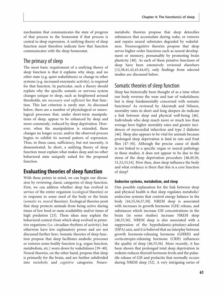
mechanism that communicates the state of progressof that process to the homeostat if that process iscentral to sleep expression. A unifying theory of sleepfunction must therefore indicate how that functioncommunicates with the sleep homeostat.
The primacy of sleepThe most basic requirement of a unifying theory ofsleep function is that it explains why sleep, and noother state (e.g. quiet wakefulness) or change in othersystems (e.g. increased enzymatic activity), is requiredfor that function. In particular, such a theory shouldexplain why the specific somatic or nervous systemchanges unique to sleep, such as heightened arousalthresholds, are necessary and sufficient for that func-tion. This last criterion is rarely met. As discussedbelow, there are a number of physiological or neuro-logical processes that, under short-term manipula-tions of sleep, appear to be enhanced by sleep andmore commonly, reduced by sleep deprivation. How-ever, when the manipulation is extended, thesechanges no longer occur, and/or the observed processbegins to exhibit its normal pattern of expression.Thus, in these cases, sufficiency, but not necessity, isdemonstrated. In short, a unifying theory of sleepfunction must explain what makes sleep and no otherbehavioral state uniquely suited for the proposedfunction.
Evaluating theories of sleep functionWith these points in mind, we can begin our discus-sion by reviewing classic categories of sleep function.First, we can address whether sleep has evolved inservice of the entire organism (ecological theories) orin response to some need of the body or the brain(somatic vs. neural theories). Ecological theories positthat sleep protects animals from being active duringtimes of low food or mate availability and/or times ofhigh predation [23]. These ideas may explain thebehavioral context from which sleep evolved in primi-tive organisms (i.e. circadian rhythms of activity), butotherwise have low explanatory power and are notdiscussed further here. Somatic theories of sleep func-tion propose that sleep facilitates anabolic processesor restores some bodily function (e.g. organ function,metabolism, etc.) worn down by wakefulness [39–40].Neural theories, on the other hand, propose that sleepis primarily for the brain, and are further subdividedinto metabolic and cognitive categories. Neuro-
metabolic theories propose that sleep detoxifiessubstances that accumulate during wake, or restoresand repairs neural substrates degraded by wakeful-ness. Neurocognitive theories propose that sleepserves higher-order functions such as neural develop-ment or memory, presumably by promoting brainplasticity [40]. As each of these putative functions ofsleep have been extensively reviewed elsewhere[12,38,41,42,43,44,45], only findings from selectedstudies are discussed below.
Somatic theories of sleep functionSleep has historically been thought of as a time whenour body reverses the wear and tear of wakefulness;but is sleep fundamentally concerned with somaticfunctions? As reviewed by Akerstedt and Nilsson,mortality rates in short and long sleepers do indicatea link between sleep and physical well-being [46].Individuals who sleep much more or much less thanaverage have higher mortality rates and greater inci-dences of myocardial infarction and type 2 diabetes[46]. Sleep also appears to be vital for animals becauseprolonged sleep deprivation is fatal in rats and fruitflies [47–50]. Although the precise cause of deathis not linked to a specific organ or neural pathologyin these studies, it does not appear to be due to thestress of the sleep deprivation procedure [48,49,50,51,52,53,54]. How then, does sleep influence the bodyand what evidence is there that this is a core functionof sleep?
Endocrine systems, metabolism, and sleepOne possible explanation for the link between sleepand physical health is that sleep regulates metabolic/endocrine systems that control energy balance in thebody [44,55,56,57,58]. NREM sleep is associatedwith increases in growth hormone (GH) release, andsubstances which increase GH concentrations in thebrain (in some studies) increase NREM sleep[46,55,56]. NREM sleep is also associated with asuppression of the hypothalamic-pituitary-adrenal(HPA) axis, and it is believed that an interplay betweengrowth hormone-releasing hormone (GHRH) andcorticotropin-releasing hormone (CRH) influencesthe quality of sleep [46,55,56]. More recently, it hasbeen shown that prolonged total sleep deprivation inrodents reduces thyroid hormone levels and the pulsa-tile release of GH and prolactin that normally occursduring NREM sleep [52]. A very intriguing series of
Chapter 4: The function(s) of sleep
61
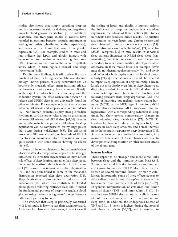
studies also shows that simply curtailing sleep inhumans increases the risk for diabetes, and negativelyimpacts blood glucose metabolism [8]. In addition,anatomical and transgenic studies in rodents haverevealed interactions between peptides important infeeding and satiety (ghrelin and leptin, respectively),and areas of the brain that control sleep/wakeexpression [58]. For example, studies in mice andrats indicate that leptin and ghrelin interact withhypocretin- and melanin-concentrating hormone(MCH)-containing neurons in the lateral hypothal-amus, which in turn regulate arousal and sleep(reviewed in [58]).
Despite these findings, it is still unclear if a corefunction of sleep is to regulate metabolic/endocrinebiology. Shorter periods of sleep deprivation (24–72hours) only modestly affect organ function, athleticperformance, and recovery from exercise [59–61].With respect to interactions between sleep and theendocrine system, the close correlation between GHrelease and NREM sleep is not universally found inother vertebrates. For example, only faint associationsbetween GH release and sleep are observed in rodents[62]. Non-human primates display strong circadianrhythms in corticosterone release, but no associationbetween GH release and NREM sleep [63,64]. Even inhumans the reduction in pulsatile GH release by sleepdeprivation can be compensated for by large pulsesthat occur during wakefulness [65]. The effects ofexogenous GH, somatostatin, or blockade of GHRHreceptors on mammalian sleep expression are alsoquite variable, with some studies showing no effects[66–69].
Some of the other changes in human metabolismobserved after sleep deprivation appear to be stronglyinfluenced by circadian mechanisms or may reflectside-effects of sleep deprivation rather than sleep per se.For example, cortisol release is under circadian con-trol [9], is known to increase after sleep deprivation[70], and has been linked to some of the metabolicdisturbances reported after sleep deprivation [71].Sleep deprivation is also known to reduce corticalmetabolism [72], which may contribute to elevatedblood glucose following restricted sleep [8]. If indeedthe fundamental purpose of sleep is to regulate bloodglucose, using the brain as a glucose “sponge” seems arather inelegant way to do it.
The evidence that sleep is principally concernedwith food intake is likewise less than straightforward.As is true for changes in hormones, it is not clear if
the cycling of leptin and ghrelin in humans reflectsthe influence of sleep, or independent circadianrhythms in the release of these peptides [8]. Studiesin rodents have produced mixed results. The putativeassociations between leptin and ghrelin release andsleep observed in humans do not occur in rats [73].Constitutive knock-out of leptin (ob/ob) [74] or leptin(db/db) receptors [75] in mice results in abnormalsleep patterns (increases in NREM sleep, sleep frag-mentation), but it is not clear if these changes aresecondary to other abnormalities, developmental orotherwise, in these mouse lines. For example, ob/obmice do not thermoregulate normally [74], and ob/oband db/dbmice both display abnormal levels of motoractivity [74,75]; either abnormality would be expectedto impact sleep expression, if only indirectly. Ghrelinknock-out mice display even fainter sleep phenotypes,displaying modest increases in NREM sleep timeversus wild-type mice both in the baseline andfollowing recovery from sleep deprivation [76]. Theeffects of knocking out melanin-concentrating hor-mone (MCH) or the MCH type 1 receptor (MCHR1) are also inconclusive. MCH knock-out mice arehyperactive (with a corresponding reduction in sleeptime), but show normal compensatory changes insleep following sleep deprivation [77]. MCH R1knock-outs, however, show no hyperactivity, anincrease in REM sleep amounts, and a small increasein the homeostatic response to sleep deprivation [78].As is true for other constitutive knock-out mice, it isunknown how many of these changes are due todevelopmental compensation or other indirect effectsof the absent gene.
Immune functionThere appear to be stronger and more direct linksbetween sleep and the immune system [45,56,57].Bacterial and viral infection in animals and humansare known to increase NREM sleep time via therelease of several immune factors (primarily cyto-kines). Importantly, some of these effects appear toreflect direct modulation of sleep/wake areas of thebrain rather than indirect effects of fever [45,56,57].Exogenous administration of cytokines like tumornecrosis factor (TNF) and interleukin 1B (IL-1B)also increase NREM sleep amounts, while antagon-ists of these cytokines or their receptors reducesleep time. In addition, the endogenous release ofTNF and IL-1B levels is highest during the normalrest phase in rodents [56,57], and in contrast to
Section II: Normal Sleep
62

molecular deletion of feeding/satiety peptides, knock-out of several immune factors leads to pronouncedalterations in sleep/wake expression and sleephomeostasis [79,80].
As is true for endocrine/metabolic and sleep inter-actions, a number of issues must be resolved beforeone can conclude that sleep’s primary function con-cerns immune biology. First, it has not been defini-tively shown that sleep is necessary for a healthyimmune system. Some studies indicate that shortperiods of sleep deprivation or sleep restrictiondecrease immune function as measured by changesin cytokines, lymphocytes, immune cell activity, andcontrol of microorganisms [81–85]. In other studies,however, sleep deprivation has negligible effects onimmunity, and with continued sleep deprivation,markers of normal immune function return to base-line and, in some cases, are heightened [56,86–89]. Inaddition, during prolonged sleep deprivation ratsgiven antibiotics still die even though their tissuesare free from infection; thus the cause of death cannotbe ascribed to compromised host defense [51]. Theknock-out mouse studies are intriguing, but onceagain, these are constitutive gene deletions, and theresulting phenotypes reflect a mixture of compensa-tory effects as well as absence of the targeted gene. Inthis regard, it is important to point out that somemice lacking immune peptides are underweight andshow disturbances in locomotor activity and abnor-malities in temperature regulation [79,80].
Neural theories of sleep functionA universal feature of sleep across the animal king-dom is that it involves both behavioral and neuralwithdrawal from the outside world. Behaviorally, thisis manifested by an animal constructing and/or secur-ing an isolated area that is safe from predators andreduces exogenous sensory input. Neurologically,sleep is associated with the activation of inhibitorymechanisms that reduce sensory transmissionthrough the thalamus, and during REM sleep, inhibitmotor output [7,90]. In addition, while the effects ofsleep loss on somatic function are variable, and pos-sibly restricted to certain mammalian species, theeffects on neural functioning appear more conserved[10,91–93]. Generally speaking, sleep loss results incognitive deficits in animals as diverse as the fruit-fly[93] to man [10]. These findings strongly suggest thatthe core function of sleep concerns the brain.
Detoxification theoriesAs originally proposed at the beginning of the twenti-eth century, the “hypnotoxin” theory of sleep functionposited that sleep removes a noxious by-product ofthe waking brain [94]. Modern versions of this theorypropose that sleep reverses or protects against neur-onal damage caused by prolonged glutamate releaseor oxidative processes that occur during wakefulness[95–97]. For example, it is hypothesized that uridineand glutathione accumulate in the brain as a functionof waking metabolism and induce sleep by enhancingGABAergic and reducing glutamatergic synaptictransmission, respectively. In addition, glutathionehas potent antioxidant properties and is hypothesizedto protect cells from oxidative damage [95]. While itis true that exogenous uridine and glutathione inducesleep, and both molecules accumulate in neuraltissues of sleep-deprived rats [95,98], the extent towhich waking or sleep influences oxidative processesin the brain is controversial. Prolonged REM sleepdeprivation in rodents selectively reduced glutathionelevels in the hypothalamus and thalamus, but othermarkers of oxidative stress were not altered [99,100].These findings were not replicated in a follow-upstudy from the same group that found signs of oxida-tive stress only in the hippocampus [101]. Slightlydifferent results were recently reported by a separategroup of investigators who found decreased gluta-thione after REM sleep deprivation in these structuresand also the hippocampus, but also signs of reducedoxidative stress in other brain structures, includingthe cortex [102]. The results of two studies of theeffects of extended total sleep deprivation using thesame technique are contradictory. Ramanathan et al.reported that 5–11 days of total sleep deprivationreduced levels of the free-radical scavenger super-oxide dismutase (SOD) in the hippocampus andbrainstem, while other markers of oxidative stress(mitochondrial metabolic activity, glutathione perox-idase) were unchanged [103]. Gopalakrishanan et al.,however, found no changes in SOD and little evidenceof oxidative stress in brains of rats totally sleepdeprived for 3–14 days. There was also no evidencethat sleep-deprived brains were less able to respond toan oxidative challenge [104].
There is no evidence that sleep loss producesneural damage as might be expected by uncheckedoxidative processes. Ninety-six hours of REM sleepdeprivation has no effect on neuronal apoptosis or
Chapter 4: The function(s) of sleep
63

necrotic markers [105] and total sleep deprivation(hours to days) has inconsistent neuronal effects[47,49]. Even in rats sleep-deprived to death, therewas no gross neuronal damage [51]. A more recentstudy using the same, extended sleep-deprivationtechniques in rats reported no changes in markers ofneural cell degeneration and no increases in apoptosisgene expression [47]. Eiland et al. found an increasein a more sensitive marker of cell degeneration in thesupraoptic nucleus (SON) after total sleep deprivationfor more than 45 hours. The significance of this latterfinding is unclear since no other signs of neuronaldamage were found in the SON [49].
Sleep and neuronal restoration: regenerationThe main evidence for neural regeneration duringsleep comes from studies of the effects of normal sleepand wake and sleep deprivation on the synthesis anddegradation of a variety of biomolecules (e.g. nucleo-tides, proteins, and mRNAs). NREM sleep has histor-ically been viewed as the “restorative” sleep state[106]. Though far from conclusive, there are anumber of findings which support this view. NREMsleep amounts are positively correlated with cerebralprotein synthesis in adult rats, monkeys, and theovine fetus [107–110]. Studies in rabbits show posi-tive correlations between RNA synthesis in purifiednuclear fractions of neocortical neurons and EEGsynchronization during sleep [111,112].
Molecular studies show that recovery sleep aftertotal sleep deprivation upregulates cortical andmedullary expression of genes that may play a rolein protein biogenesis in the endoplasmic reticulum(ER) [113]. Complementary results have been reportedafter 6 hours of total sleep deprivation in mice, whichinduces cellular events that decrease protein synthesis[114]. Other studies have found sleep-related increasesin several genes implicated in cholesterol synthesis,membrane trafficking, and vesicle maintenance andtransport [115–118]. Total sleep deprivation is alsoreported to reduce cell proliferation in the hippocam-pus [119–122]. This latter effect does not appear to besimply due to stress accompanying sleep deprivationbecause it persists even when stress hormones areclamped [123].
In contrast to NREM sleep, the evidence forneural regeneration in REM sleep is comparativelyweak. REM sleep deprivation also reduces hippocam-pal neurogenesis [124], but it has inconsistenteffects on protein synthesis, with some investigators
reporting no effects [125], and others showing reduc-tions, chiefly in non-cortical structures [126,127].REM sleep deprivation alters the expression of severalgenes associated with REM sleep mechanisms, butthere is little evidence that REM sleep enhances theexpression of genes other than those located in REMsleep circuits [128–130]. Although REM sleep isaccompanied by reduced monoaminergic activity[131], the significance of this interaction in terms ofneuroregeneration is unclear. For example, short-term REM sleep deprivation (96 hours) has beenshown to increase noradrenergic activity and down-regulate beta-adrenergic receptors [132,133], butextended total sleep deprivation or REM sleep depriv-ation minimally impact monoamine levels and recep-tor number [48,134–136], and only modestly affectneuronal morphology in cholinergic and noradrener-gic neurons [137].
Several studies indicate that REM sleep depriv-ation increases neuronal excitability, possibly byincreasing NA-K ATPase activity, which suggests thatREM sleep normally regulates this process [61,138–140]. These effects appear to be regionally dependentsince REM sleep deprivation does not increase neur-onal excitability in the hippocampus [140,141]. REMsleep deprivation has also been reported to alter neur-onal membrane fluidity [142]. Unfortunately, it isdifficult to isolate the effects of REM sleep loss fromstress and other side-effects of the REM sleep depriv-ation techniques (“flower-pot” or multiple-platform)in which the animal is kept on a small platformsurrounded by water so that it falls off during periodsof REM atonia, used in these latter studies. There arealso no studies that have conclusively demonstratedneuroregeneration of any kind during naturallyoccurring REM sleep.
Sleep and neuronal restoration: energy substratesSeveral studies have reported interesting relationshipsbetween sleep and brain energy [143–145]. Total sleepdeprivation, for example, has been shown to reducecortical metabolism [72,146] and alter lactate synthe-sis, which suggests that sleep might be important forcerebral energy supplies [147]. Sleep is also associatedwith increases in the expression of several genes import-ant for neuronal metabolism [116] and following sleepdeprivation, may promote the restoration of ATP in thehuman brain [148].
One of the more influential theories relatingbrain energy substrates and sleep was proposed by
Section II: Normal Sleep
64
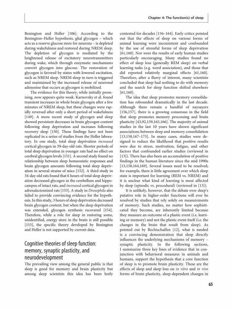
Benington and Heller [106]. According to theBenington–Heller hypothesis, glial glycogen – whichacts as a reserve glucose store for neurons – is depletedduring wakefulness and restored during NREM sleep.The depletion of glycogen is mediated by theheightened release of excitatory neurotransmittersduring wake, which through enzymatic mechanismsconvert glycogen into glucose. The restoration ofglycogen is favored by states with lowered excitation,such as NREM sleep. NREM sleep in turn is triggeredand maintained by the increased release of neuronaladenosine that occurs as glycogen is mobilized.
The evidence for this theory, while initially prom-ising, now appears quite weak. Karnovsky et al. foundtransient increases in whole brain glycogen after a fewminutes of NREM sleep, but these changes were rap-idly reversed after only a short period of wakefulness[149]. A more recent study of glycogen and sleepshowed persistent decreases in brain glycogen contentfollowing sleep deprivation and increases followingrecovery sleep [150]. These findings have not beenreplicated in a series of studies from the Heller labora-tory. In one study, total sleep deprivation increasedcortical glycogen in 59-day-old rats. Shorter periods oftotal sleep deprivation in younger rats had no effect oncerebral glycogen levels [151]. A second study found norelationship between sleep homeostatic responses andbrain glycogen amounts following total sleep depriv-ation in several strains of mice [152]. A third study in34-day-old rats found that 6 hours of total sleep depriv-ation decreased glycogen in the cerebellum and hippo-campus of intact rats, and increased cortical glycogen inadrenalectomized rats [153]. A study in Drosophila alsofailed to provide convincing evidence for the hypoth-esis. In this study, 3 hours of sleep deprivation decreasedbrain glycogen content, but when the sleep deprivationwas extended, glycogen synthesis recovered [154].Therefore, while a role for sleep in restoring some,unidentified, energy store in the brain is still possible[155], the specific theory developed by Beningtonand Heller is not supported by current data.
Cognitive theories of sleep function:memory, synaptic plasticity, andneurodevelopmentThe prevailing view among the general public is thatsleep is good for memory and brain plasticity butamong sleep scientists this idea has been hotly
contested for decades [156–164]. Early critics pointedout that the effects of sleep on various forms ofanimal learning were inconsistent and confoundedby the use of stressful forms of sleep deprivation[61,160]. Nor were the results of early human studiesparticularly encouraging. Many studies found noeffect of sleep loss (generally REM sleep) on verballearning tasks (e.g. word association), and those thatdid reported relatively marginal effects [61,160].Therefore, after a flurry of interest, many scientistsconcluded that sleep had nothing to do with memoryand the search for sleep function shifted elsewhere[61,160].
The idea that sleep promotes memory consolida-tion has rebounded dramatically in the last decade.Although there remain a handful of naysayers[156,157], there is a growing consensus in the fieldthat sleep promotes memory processing and brainplasticity [43,92,159,165,166]. The majority of animalstudies in the last 10 years have shown significantassociations between sleep and memory consolidation[13,158,167–173]. In many cases, studies were de-signed to reduce the likelihood that positive resultswere due to stress, motivation, fatigue, and otherfactors that confounded earlier studies (reviewed in[13]). There has also been an accumulation of positivefindings in the human literature since the mid 1990s[13,158,164,169]. Several issues need to be resolved;for example, there is little agreement over which sleepstate is important for learning (REM vs. NREM) andit is unclear what kind of learning is most affectedby sleep (episodic vs. procedural) (reviewed in [13]).
It is unlikely, however, that the debate over sleep’sputative role in higher-order functions will ever beresolved by studies that rely solely on measurementsof memory. Such studies, no matter how sophisti-cated they become, are inherently limited becausethey measure an outcome of a plastic event (i.e. learn-ing or memory) and not the plastic event itself (i.e. thechanges in the brain that result from sleep). Aspointed out by Rechtschaffen [12], what is neededis a convincing demonstration that sleep directlyinfluences the underlying mechanisms of memory –synaptic plasticity. In the following sections,I summarize three key lines of evidence that in con-junction with behavioral measures in animals andhumans, support the hypothesis that a core functionof sleep is to promote brain plasticity. These are theeffects of sleep and sleep loss on in vitro and in vivoforms of brain plasticity, sleep-dependent changes in
Chapter 4: The function(s) of sleep
65
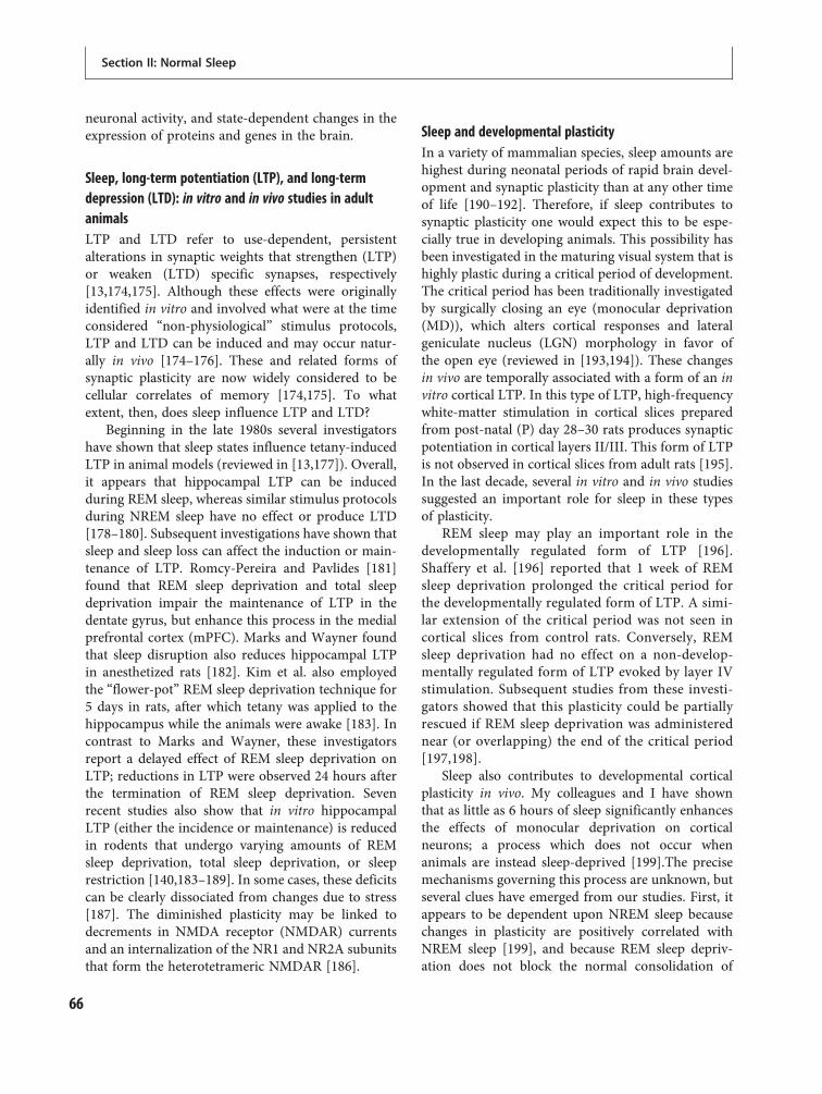
neuronal activity, and state-dependent changes in theexpression of proteins and genes in the brain.
Sleep, long-term potentiation (LTP), and long-termdepression (LTD): in vitro and in vivo studies in adultanimalsLTP and LTD refer to use-dependent, persistentalterations in synaptic weights that strengthen (LTP)or weaken (LTD) specific synapses, respectively[13,174,175]. Although these effects were originallyidentified in vitro and involved what were at the timeconsidered “non-physiological” stimulus protocols,LTP and LTD can be induced and may occur natur-ally in vivo [174–176]. These and related forms ofsynaptic plasticity are now widely considered to becellular correlates of memory [174,175]. To whatextent, then, does sleep influence LTP and LTD?
Beginning in the late 1980s several investigatorshave shown that sleep states influence tetany-inducedLTP in animal models (reviewed in [13,177]). Overall,it appears that hippocampal LTP can be inducedduring REM sleep, whereas similar stimulus protocolsduring NREM sleep have no effect or produce LTD[178–180]. Subsequent investigations have shown thatsleep and sleep loss can affect the induction or main-tenance of LTP. Romcy-Pereira and Pavlides [181]found that REM sleep deprivation and total sleepdeprivation impair the maintenance of LTP in thedentate gyrus, but enhance this process in the medialprefrontal cortex (mPFC). Marks and Wayner foundthat sleep disruption also reduces hippocampal LTPin anesthetized rats [182]. Kim et al. also employedthe “flower-pot” REM sleep deprivation technique for5 days in rats, after which tetany was applied to thehippocampus while the animals were awake [183]. Incontrast to Marks and Wayner, these investigatorsreport a delayed effect of REM sleep deprivation onLTP; reductions in LTP were observed 24 hours afterthe termination of REM sleep deprivation. Sevenrecent studies also show that in vitro hippocampalLTP (either the incidence or maintenance) is reducedin rodents that undergo varying amounts of REMsleep deprivation, total sleep deprivation, or sleeprestriction [140,183–189]. In some cases, these deficitscan be clearly dissociated from changes due to stress[187]. The diminished plasticity may be linked todecrements in NMDA receptor (NMDAR) currentsand an internalization of the NR1 and NR2A subunitsthat form the heterotetrameric NMDAR [186].
Sleep and developmental plasticityIn a variety of mammalian species, sleep amounts arehighest during neonatal periods of rapid brain devel-opment and synaptic plasticity than at any other timeof life [190–192]. Therefore, if sleep contributes tosynaptic plasticity one would expect this to be espe-cially true in developing animals. This possibility hasbeen investigated in the maturing visual system that ishighly plastic during a critical period of development.The critical period has been traditionally investigatedby surgically closing an eye (monocular deprivation(MD)), which alters cortical responses and lateralgeniculate nucleus (LGN) morphology in favor ofthe open eye (reviewed in [193,194]). These changesin vivo are temporally associated with a form of an invitro cortical LTP. In this type of LTP, high-frequencywhite-matter stimulation in cortical slices preparedfrom post-natal (P) day 28–30 rats produces synapticpotentiation in cortical layers II/III. This form of LTPis not observed in cortical slices from adult rats [195].In the last decade, several in vitro and in vivo studiessuggested an important role for sleep in these typesof plasticity.
REM sleep may play an important role in thedevelopmentally regulated form of LTP [196].Shaffery et al. [196] reported that 1 week of REMsleep deprivation prolonged the critical period forthe developmentally regulated form of LTP. A simi-lar extension of the critical period was not seen incortical slices from control rats. Conversely, REMsleep deprivation had no effect on a non-develop-mentally regulated form of LTP evoked by layer IVstimulation. Subsequent studies from these investi-gators showed that this plasticity could be partiallyrescued if REM sleep deprivation was administerednear (or overlapping) the end of the critical period[197,198].
Sleep also contributes to developmental corticalplasticity in vivo. My colleagues and I have shownthat as little as 6 hours of sleep significantly enhancesthe effects of monocular deprivation on corticalneurons; a process which does not occur whenanimals are instead sleep-deprived [199].The precisemechanisms governing this process are unknown, butseveral clues have emerged from our studies. First, itappears to be dependent upon NREM sleep becausechanges in plasticity are positively correlated withNREM sleep [199], and because REM sleep depriv-ation does not block the normal consolidation of
Section II: Normal Sleep
66
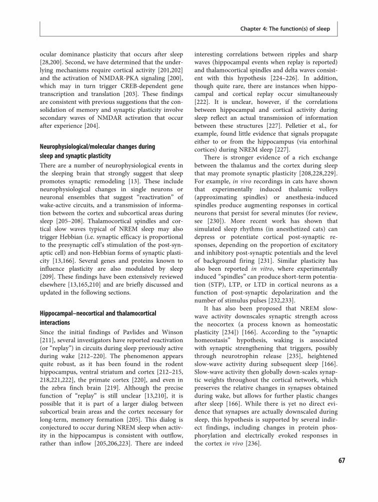
ocular dominance plasticity that occurs after sleep[28,200]. Second, we have determined that the under-lying mechanisms require cortical activity [201,202]and the activation of NMDAR-PKA signaling [200],which may in turn trigger CREB-dependent genetranscription and translation [203]. These findingsare consistent with previous suggestions that the con-solidation of memory and synaptic plasticity involvesecondary waves of NMDAR activation that occurafter experience [204].
Neurophysiological/molecular changes duringsleep and synaptic plasticityThere are a number of neurophysiological events inthe sleeping brain that strongly suggest that sleeppromotes synaptic remodeling [13]. These includeneurophysiological changes in single neurons orneuronal ensembles that suggest “reactivation” ofwake-active circuits, and a transmission of informa-tion between the cortex and subcortical areas duringsleep [205–208]. Thalamocortical spindles and cor-tical slow waves typical of NREM sleep may alsotrigger Hebbian (i.e. synaptic efficacy is proportionalto the presynaptic cell’s stimulation of the post-syn-aptic cell) and non-Hebbian forms of synaptic plasti-city [13,166]. Several genes and proteins known toinfluence plasticity are also modulated by sleep[209]. These findings have been extensively reviewedelsewhere [13,165,210] and are briefly discussed andupdated in the following sections.
Hippocampal–neocortical and thalamocorticalinteractionsSince the initial findings of Pavlides and Winson[211], several investigators have reported reactivation(or “replay”) in circuits during sleep previously activeduring wake [212–220]. The phenomenon appearsquite robust, as it has been found in the rodenthippocampus, ventral striatum and cortex [212–215,218,221,222], the primate cortex [220], and even inthe zebra finch brain [219]. Although the precisefunction of “replay” is still unclear [13,210], it ispossible that it is part of a larger dialog betweensubcortical brain areas and the cortex necessary forlong-term, memory formation [205]. This dialog isconjectured to occur during NREM sleep when activ-ity in the hippocampus is consistent with outflow,rather than inflow [205,206,223]. There are indeed
interesting correlations between ripples and sharpwaves (hippocampal events when replay is reported)and thalamocortical spindles and delta waves consist-ent with this hypothesis [224–226]. In addition,though quite rare, there are instances when hippo-campal and cortical replay occur simultaneously[222]. It is unclear, however, if the correlationsbetween hippocampal and cortical activity duringsleep reflect an actual transmission of informationbetween these structures [227]. Pelletier et al., forexample, found little evidence that signals propagateeither to or from the hippocampus (via entorhinalcortices) during NREM sleep [227].
There is stronger evidence of a rich exchangebetween the thalamus and the cortex during sleepthat may promote synaptic plasticity [208,228,229].For example, in vivo recordings in cats have shownthat experimentally induced thalamic volleys(approximating spindles) or anesthesia-inducedspindles produce augmenting responses in corticalneurons that persist for several minutes (for review,see [230]). More recent work has shown thatsimulated sleep rhythms (in anesthetized cats) candepress or potentiate cortical post-synaptic re-sponses, depending on the proportion of excitatoryand inhibitory post-synaptic potentials and the levelof background firing [231]. Similar plasticity hasalso been reported in vitro, where experimentallyinduced “spindles” can produce short-term potentia-tion (STP), LTP, or LTD in cortical neurons as afunction of post-synaptic depolarization and thenumber of stimulus pulses [232,233].
It has also been proposed that NREM slow-wave activity downscales synaptic strength acrossthe neocortex (a process known as homeostaticplasticity [234]) [166]. According to the “synaptichomeostasis” hypothesis, waking is associatedwith synaptic strengthening that triggers, possiblythrough neurotrophin release [235], heightenedslow-wave activity during subsequent sleep [166].Slow-wave activity then globally down-scales synap-tic weights throughout the cortical network, whichpreserves the relative changes in synapses obtainedduring wake, but allows for further plastic changesafter sleep [166]. While there is yet no direct evi-dence that synapses are actually downscaled duringsleep, this hypothesis is supported by several indir-ect findings, including changes in protein phos-phorylation and electrically evoked responses inthe cortex in vivo [236].
Chapter 4: The function(s) of sleep
67
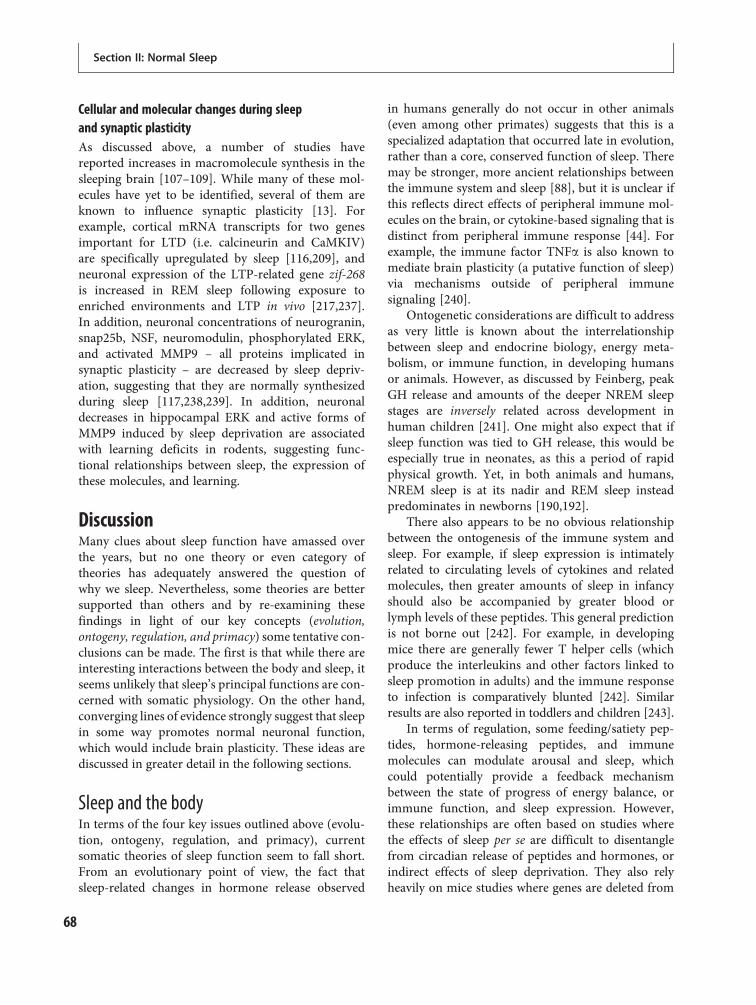
Cellular and molecular changes during sleepand synaptic plasticityAs discussed above, a number of studies havereported increases in macromolecule synthesis in thesleeping brain [107–109]. While many of these mol-ecules have yet to be identified, several of them areknown to influence synaptic plasticity [13]. Forexample, cortical mRNA transcripts for two genesimportant for LTD (i.e. calcineurin and CaMKIV)are specifically upregulated by sleep [116,209], andneuronal expression of the LTP-related gene zif-268is increased in REM sleep following exposure toenriched environments and LTP in vivo [217,237].In addition, neuronal concentrations of neurogranin,snap25b, NSF, neuromodulin, phosphorylated ERK,and activated MMP9 – all proteins implicated insynaptic plasticity – are decreased by sleep depriv-ation, suggesting that they are normally synthesizedduring sleep [117,238,239]. In addition, neuronaldecreases in hippocampal ERK and active forms ofMMP9 induced by sleep deprivation are associatedwith learning deficits in rodents, suggesting func-tional relationships between sleep, the expression ofthese molecules, and learning.
DiscussionMany clues about sleep function have amassed overthe years, but no one theory or even category oftheories has adequately answered the question ofwhy we sleep. Nevertheless, some theories are bettersupported than others and by re-examining thesefindings in light of our key concepts (evolution,ontogeny, regulation, and primacy) some tentative con-clusions can be made. The first is that while there areinteresting interactions between the body and sleep, itseems unlikely that sleep’s principal functions are con-cerned with somatic physiology. On the other hand,converging lines of evidence strongly suggest that sleepin some way promotes normal neuronal function,which would include brain plasticity. These ideas arediscussed in greater detail in the following sections.
Sleep and the bodyIn terms of the four key issues outlined above (evolu-tion, ontogeny, regulation, and primacy), currentsomatic theories of sleep function seem to fall short.From an evolutionary point of view, the fact thatsleep-related changes in hormone release observed
in humans generally do not occur in other animals(even among other primates) suggests that this is aspecialized adaptation that occurred late in evolution,rather than a core, conserved function of sleep. Theremay be stronger, more ancient relationships betweenthe immune system and sleep [88], but it is unclear ifthis reflects direct effects of peripheral immune mol-ecules on the brain, or cytokine-based signaling that isdistinct from peripheral immune response [44]. Forexample, the immune factor TNFa is also known tomediate brain plasticity (a putative function of sleep)via mechanisms outside of peripheral immunesignaling [240].
Ontogenetic considerations are difficult to addressas very little is known about the interrelationshipbetween sleep and endocrine biology, energy meta-bolism, or immune function, in developing humansor animals. However, as discussed by Feinberg, peakGH release and amounts of the deeper NREM sleepstages are inversely related across development inhuman children [241]. One might also expect that ifsleep function was tied to GH release, this would beespecially true in neonates, as this a period of rapidphysical growth. Yet, in both animals and humans,NREM sleep is at its nadir and REM sleep insteadpredominates in newborns [190,192].
There also appears to be no obvious relationshipbetween the ontogenesis of the immune system andsleep. For example, if sleep expression is intimatelyrelated to circulating levels of cytokines and relatedmolecules, then greater amounts of sleep in infancyshould also be accompanied by greater blood orlymph levels of these peptides. This general predictionis not borne out [242]. For example, in developingmice there are generally fewer T helper cells (whichproduce the interleukins and other factors linked tosleep promotion in adults) and the immune responseto infection is comparatively blunted [242]. Similarresults are also reported in toddlers and children [243].
In terms of regulation, some feeding/satiety pep-tides, hormone-releasing peptides, and immunemolecules can modulate arousal and sleep, whichcould potentially provide a feedback mechanismbetween the state of progress of energy balance, orimmune function, and sleep expression. However,these relationships are often based on studies wherethe effects of sleep per se are difficult to disentanglefrom circadian release of peptides and hormones, orindirect effects of sleep deprivation. They also relyheavily on mice studies where genes are deleted from
Section II: Normal Sleep
68
the embryonic period onward, leaving open thepossibility that some of the behavioral phenotypesreflect developmental compensation.
With respect to primacy, somatic theories seemwholly inadequate. If we accept that a core feature ofsleep is a diminution of consciousness, then whyshould this be required for the facilitation of endo-crine or immune function? These events could just aseasily occur during periods of quiet wake when theanimal is less subject to predation [60]. Moreover,while there is evidence in some species that sleep issufficient for hormone release or normal changes inimmune function, it is not yet clear if sleep is neces-sary for these processes. For example, as discussedearlier, GH release in humans is indeed blunted bysleep deprivation, but this can be compensated for bypulses in waking [65]. Likewise, while a single nightof sleep loss can negatively impact some immunemolecules [82,83], the clinical significance of thesechanges are unknown, nor has it been shown thatthese effects persist during longer sleep deprivations.
Sleep and the brainAs reviewed in the preceding sections, there areseveral categories of ideas that relate neural functionto sleep (metabolic: neurotoxin vs. regeneration,cognitive: brain plasticity and memory). Neurotoxintheories currently have weak experimental support. Incontrast, there is strong evidence in favor of one typeof neuroregeneration (macromolecule synthesis) andcognitive theories. For example, sleep is associatedwith cerebral protein synthesis and the upregulationof genes important for neuronal metabolism, mem-branes, and other structural components. There isalso behavioral, electrophysiological, cellular, andmolecular evidence to support the hypothesis thatsleep promotes higher order functions such asmemory consolidation and brain plasticity.
A parsimonious reconciliation of these findings isthat sleep is a brain state that promotes structuralrearrangements in neurons. This is an energetic pro-cess (hence the upregulation of genes and proteinsrelated to neuronal metabolism) that requires macro-molecule synthesis. It is also more likely to be inten-sified under conditions such as brain developmentand following events that trigger synaptic plasticity.One might also expect there to be a steady-state needfor such a brain state, as macromolecules in neurons(as well as neuronal processes such as dendritic
spines) are highly dynamic [244,245]. If indeed thisis the core function of sleep, it would account for thetwo strongest sets of findings we have concerningsleep and the brain, but how does it fare in light ofour four key concepts?
In terms of evolution, this theory may account forthe ubiquity of the central nervous system changesthat occur during sleep in the animal kingdom,and the fact that challenges to the brain that triggerstructural changes in synapses are associated withmore sleep, or more intense sleep in diverse species[13,21,166,246,247]. In terms of ontogeny, it mayexplain the abundance of sleep in early life, which is atime of massive synaptic rearrangement [13], and theabrupt appearance of adult forms of sleep homeostasisat ages when plasticity-related peptides (i.e. the neuro-trophin BDNF) concentrations sharply increase aftersleep deprivation [32,34]. It may also account for thedecline in sleep amounts and intensity in senescence[248] when brain plasticity reaches its nadir [249].
What is less clear is the relationship between thisprocess and sleep homeostasis (regulation) and whysleep would be the preferred state for this process tooccur (primacy). With respect to regulation, some pro-gress has been made in that plasticity-related molecules(e.g. neurotrophins, TNFa) can increase EEG correlatesof sleep intensity [235,250]. We have also shown thatastrocytes influence sleep homeostasis and, becausethey also modulate synaptic plasticity, they represent acellular link between sleep regulation and structuralchanges in circuits [251]. However, it is not clear ifthese relationships exist in animals that lack neocortex,or at earlier stages of life in animals that have neocortex.
In terms of primacy, it is hypothesized that theelectrical activity or the neurochemical/molecularmilieu of the sleeping brain is especially conducivefor structural changes in neurons [13,166]. Yet theprecise and complete set of cellular processes pro-moted by sleep (and not wake) in the brain has notbeen identified. It would appear, however, that insome cases they are necessary and sufficient formemory consolidation and brain plasticity becausein their absence, these processes do not occur and/orare attenuated [92,199,201].
Concluding remarksSleep scientists, for all their advances, remain in theawkward position of not knowing why we sleep. Ananalogous situation in the sciences is difficult to find.
Chapter 4: The function(s) of sleep
69
For example, eating, like sleep, is a complex, regulatedbehavior governed by specific brain regions, neuro-transmitters, and hormones. Eating, like sleep, isubiquitous in the animal kingdom and undergoesimportant transformations during ontogeny [252] –yet there is no confusion and debate over why animalseat. The evidence to date strongly suggests that sleepprimarily serves the brain rather than the body, andthe neural processes most impacted by sleep are cog-nitive. This does not, however, change the fact thatsleep deprivation or restriction negatively impactssomatic function – if perhaps only indirectly. Thechallenge now is to isolate further the effects of sleepversus other interlocking physiological systems on thebody and to determine precisely what sleep providesthe brain that wakefulness cannot.
In conclusion, scientists may eventually discoverthat there are deeper, more fundamental reasonswhy we sleep, but the idea that sleep promotes struc-tural changes in the brain provides a strong theoret-ical framework for understanding sleep function.Continued investigation along these lines is thereforeuseful because it will either elucidate exactly howsleep achieves this function or reveal a more elegant,unifying process.
References1. Jones BE. Basic mechanisms of sleep waking states. In:
Kryger MH, Roth T, Dement WC, eds. Principles andpractice of sleep medicine, 4th ed. Philadelphia: WBSaunders; 2005:136 153.
2. Dijk DJ, Lockley SW. Integration of human sleep wakeregulation and circadian rhythmicity. J Appl Physiol.2002;92(2):852 862.
3. Shaw PJ, Franken P. Perchance to dream: Solving themystery of sleep through genetic analysis. J Neurobiol.2003;54(1):179 202.
4. Siegel JM. Do all animals sleep? Trends Neurosci.2008;31(4):208 213.
5. Tobler I. Phylogeny of sleep regulation. In: Kryger M,Roth T, Dement WC, eds. Principles and practice ofsleep medicine, 4th ed. Philadelphia: W.B Saunders;2005:72 90.
6. Zepelin H. Mammalian sleep. In: Kryger M, Roth T,Dement WC, eds. Principles and practice of sleep medicine,4th ed. Philadelphia: WB Saunders; 2005:91 100.
7. Steriade M. Brain electrical activity and sensoryprocessing during waking and sleep states. In: KrygerMH, Roth T, Dement WC, eds. Principles and practice ofsleep medicine, Vol 4. Philadelphia: WB Saunders;2005:101 119.
8. Knutson K, Van Cauter E. Associations between sleeploss and increased risk of obesity and diabetes. Ann NYAcad Sci. 2008;1129:287 304.
9. Van Cauter E. Endocrine physiology. In: Kryger M,Roth T, Dement WC, eds. Principles and practice ofsleep medicine, 5th ed. Philadelphia: Elsevier;2005:266 282.
10. Banks S, Dinges DF. Behavioral and physiologicalconsequences of sleep restriction. J Clin Sleep Med.2007;3(5):519 528.
11. Glotzbach SF, Heller HC. Temperature regulation. In:Roth C, Kryger M, Dement WC, eds. Principles andpractice of sleep medicine, 3rd ed. Philadelphia: WBSaunders; 2000:289 304.
12. Rechtschaffen A. Current perspectives on the functionof sleep. Perspect Biol Med. 1998;41(3):359 390.
13. Benington JH, Frank MG. Cellular and molecularconnections between sleep and synaptic plasticity.Progr Neurobiol. 2003;69(2):77 101.
14. Shaw P. Awakening to the behavioral analysis of sleepin Drosophila. J Biol Rhythms. 2003;18(1):4 11.
15. Eban Rothschild AD, Bloch G. Differences inthe sleep architecture of forager and younghoneybees (Apis mellifera). J Exp Biol. 2008;211(15):2408 2416.
16. Brown ER, Piscopo S, De Stefano R, Giuditta A.Brain and behavioural evidence for rest activity cyclesin Octopus vulgaris. Behav Brain Res. 2006;172(2):355 359.
17. Ramon F, Hernandez Falcon J, Nguyen B, Bullock TH.Slow wave sleep in crayfish. PNAS. 2004;101(32):11857 11861.
18. Rattenborg NC, Martinez Gonzalez D, Lesku JA.Avian sleep homeostasis: Convergent evolutionof complex brains, cognition and sleep functionsin mammals and birds. Neurosci Biobehav Rev.2009;33(3):253 270.
19. Ayala Guerrero F, Mexicano G. Sleep and wakefulnessin the green iguanid lizard (Iguana iguana). CompBiochem Physiol A Mol Integr Physiol. 2008;151(3):305 312.
20. Siegel JM. Sleep phylogeny: Clues to the evolution andfunction of sleep. In: Luppi PH, ed. Sleep: circuitsand function. Boca Raton: CRC Press; 2005:163 176.
21. Raizen DM, Zimmerman JE, Maycock MH, et al.Lethargus is a Caenorhabditis elegans sleep like state.Nature. 2008;451(7178):569 572.
22. Allada R, Siegel JM. Unearthing the phylogenetic rootsof sleep. Curr Biol. 2008;18(15):R670 R679.
23. Siegel JM. Clues to the functions of mammalian sleep.Nature. 2005;437(7063):1264 1271.
Section II: Normal Sleep
70
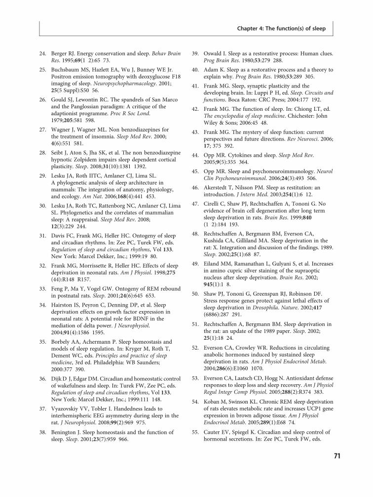
24. Berger RJ. Energy conservation and sleep. Behav BrainRes. 1995;69(1 2):65 73.
25. Buchsbaum MS, Hazlett EA, Wu J, Bunney WE Jr.Positron emission tomography with deoxyglucose F18imaging of sleep. Neuropsychopharmacology. 2001;25(5 Suppl):S50 56.
26. Gould SJ, Lewontin RC. The spandrels of San Marcoand the Panglossian paradigm: A critique of theadaptionist programme. Proc R Soc Lond.1979;205:581 598.
27. Wagner J, Wagner ML. Non benzodiazepines forthe treatment of insomnia. Sleep Med Rev. 2000;4(6):551 581.
28. Seibt J, Aton S, Jha SK, et al. The non benzodiazepinehypnotic Zolpidem impairs sleep dependent corticalplasticity. Sleep. 2008;31(10):1381 1392.
29. Lesku JA, Roth IITC, Amlaner CJ, Lima SL.A phylogenetic analysis of sleep architecture inmammals: The integration of anatomy, physiology,and ecology. Am Nat. 2006;168(4):441 453.
30. Lesku JA, Roth TC, Rattenborg NC, Amlaner CJ, LimaSL. Phylogenetics and the correlates of mammaliansleep: A reappraisal. Sleep Med Rev. 2008;12(3):229 244.
31. Davis FC, Frank MG, Heller HC. Ontogeny of sleepand circadian rhythms. In: Zee PC, Turek FW, eds.Regulation of sleep and circadian rhythms, Vol 133.New York: Marcel Dekker, Inc.; 1999:19 80.
32. Frank MG, Morrissette R, Heller HC. Effects of sleepdeprivation in neonatal rats. Am J Physiol. 1998;275(44):R148 R157.
33. Feng P, Ma Y, Vogel GW. Ontogeny of REM reboundin postnatal rats. Sleep. 2001;24(6):645 653.
34. Hairston IS, Peyron C, Denning DP, et al. Sleepdeprivation effects on growth factor expression inneonatal rats: A potential role for BDNF in themediation of delta power. J Neurophysiol.2004;91(4):1586 1595.
35. Borbely AA, Achermann P. Sleep homeostasis andmodels of sleep regulation. In: Kryger M, Roth T,Dement WC, eds. Principles and practice of sleepmedicine, 3rd ed. Philadelphia: WB Saunders;2000:377 390.
36. DijkD J, Edgar DM. Circadian and homeostatic controlof wakefulness and sleep. In: Turek FW, Zee PC, eds.Regulation of sleep and circadian rhythms, Vol 133.New York: Marcel Dekker, Inc.; 1999:111 148.
37. Vyazovskiy VV, Tobler I. Handedness leads tointerhemispheric EEG asymmetry during sleep in therat. J Neurophysiol. 2008;99(2):969 975.
38. Benington J. Sleep homeostasis and the function ofsleep. Sleep. 2001;23(7):959 966.
39. Oswald I. Sleep as a restorative process: Human clues.Prog Brain Res. 1980;53:279 288.
40. Adam K. Sleep as a restorative process and a theory toexplain why. Prog Brain Res. 1980;53:289 305.
41. Frank MG. Sleep, synaptic plasticity and thedeveloping brain. In: Luppi P H, ed. Sleep. Circuits andfunctions. Boca Raton: CRC Press; 2004:177 192.
42. Frank MG. The function of sleep. In: Chiong LT, ed.The encyclopedia of sleep medicine. Chichester: JohnWiley & Sons; 2006:45 48.
43. Frank MG. The mystery of sleep function: currentperspectives and future directions. Rev Neurosci. 2006;17; 375 392.
44. Opp MR. Cytokines and sleep. Sleep Med Rev.2005;9(5):355 364.
45. Opp MR. Sleep and psychoneuroimmunology. NeurolClin Psychoneuroimmunol. 2006;24(3):493 506.
46. Akerstedt T, Nilsson PM. Sleep as restitution: anintroduction. J Intern Med. 2003;254(1):6 12.
47. Cirelli C, Shaw PJ, Rechtschaffen A, Tononi G. Noevidence of brain cell degeneration after long termsleep deprivation in rats. Brain Res. 1999;840(1 2):184 193.
48. Rechtschaffen A, Bergmann BM, Everson CA,Kushida CA, Gilliland MA. Sleep deprivation in therat: X. Integration and discussion of the findings. 1989.Sleep. 2002;25(1):68 87.
49. Eiland MM, Ramanathan L, Gulyani S, et al. Increasesin amino cupric silver staining of the supraopticnucleus after sleep deprivation. Brain Res. 2002;945(1):1 8.
50. Shaw PJ, Tononi G, Greenspan RJ, Robinson DF.Stress response genes protect against lethal effects ofsleep deprivation in Drosophila. Nature. 2002;417(6886):287 291.
51. Rechtschaffen A, Bergmann BM. Sleep deprivation inthe rat: an update of the 1989 paper. Sleep. 2002;25(1):18 24.
52. Everson CA, Crowley WR. Reductions in circulatinganabolic hormones induced by sustained sleepdeprivation in rats. Am J Physiol Endocrinol Metab.2004;286(6):E1060 1070.
53. Everson CA, Laatsch CD, Hogg N. Antioxidant defenseresponses to sleep loss and sleep recovery. Am J PhysiolRegul Integr Comp Physiol. 2005;288(2):R374 383.
54. Koban M, Swinson KL. Chronic REM sleep deprivationof rats elevates metabolic rate and increases UCP1 geneexpression in brown adipose tissue. Am J PhysiolEndocrinol Metab. 2005;289(1):E68 74.
55. Cauter EV, Spiegel K. Circadian and sleep control ofhormonal secretions. In: Zee PC, Turek FW, eds.
Chapter 4: The function(s) of sleep
71

Regulation of sleep and circadian rhythms, Vol 133.New York: Marcel Dekker, Inc.; 1999:397 425.
56. Bryant PA, Trinder J, Curtis N. Sick and tired: Doessleep have a vital role in the immune system? Nat RevImmunol. 2004;4(6):457 467.
57. Krueger JM, Majde JA. Humoral links between sleepand the immune system: research issues. Ann NY AcadSci. 2003;992(1):9 20.
58. Adamantidis A, de Lecea L. Sleep and metabolism:shared circuits, new connections. Trends EndocrinolMetab. 2008;19(10):362 370.
59. Bonnet MH. Sleep deprivation. In: Kryger M, Roth T,Dement WC, eds. Principles and practice ofsleep medicine, 3rd ed. Philadelphia: WB Saunders;2000:53 71.
60. Horne JA. Sleep function, with particular referenceto sleep deprivation. Ann Clin Res. 1985;17(5):199 208.
61. Ellman SJ, Spielman AJ, Luck D, Steiner SS,Halperin R. REM deprivation: A Review. In: Ellman SJ,Antrobus JS, eds. The mind in sleep, 2nd ed. New York:John Wiley & Sons, Inc.; 1991:329 369.
62. Iyer KS, Marks GA, Kastin AJ, McCann SM. Evidencefor a role of delta sleep inducing peptide in slow wavesleep and sleep related growth hormone release in therat. PNAS. 1988;85(10):3653 3656.
63. Bunner DL, McNamee GA Jr, Dinterman RE,Wannemacher RW Jr. Lack of enhanced nocturnalgrowth hormone release in tethered cynomolgusmonkeys. Am J Physiol Regul Integr Comp Physiol.1982;243(3):R213 217.
64. Quabbe HJ, Gregor M, Bumke Vogt C, Eckhof A,Witt I. Twenty four hour pattern of growth hormonesecretion in the rhesus monkey: studies includingalterations of the sleep/wake and sleep stage cycles.Endocrinology. 1981;109(2):513 522.
65. Brandenberger G, Weibel L. The 24 h growthhormone rhythm in men: sleep and circadianinfluences questioned. J Sleep Res. 2004;13(3):251 255.
66. Van Cauter E, Latta F, Nedeltcheva A, et al. Reciprocalinteractions between the GH axis and sleep. GrowthHorm IGF Res. 2004;14(Suppl A):S10 17.
67. Jessup S, Malow B, Symons K, Barkan A. Blockade ofendogenous growth hormone releasing hormonereceptors dissociates nocturnal growth hormonesecretion and slow wave sleep. Eur J Endocrinol.2004;151(5):561 566.
68. Steiger A, Antonijevic IA, Bohlhalter S, et al. Effects ofhormones on sleep. Horm Res. 1998;49(3 4):125 130.
69. Kern W, Halder R, al Reda S, et al. Systemic growthhormone does not affect human sleep. J ClinEndocrinol Metab. 1993;76(6):1428 1432.
70. Tobler I, Murrison R, Ursin R, Ursin H, Borbely AA.The effect of sleep deprivation and recovery sleep onplasma corticosterone in the rat. Neurosci Lett.1983;35:297 300.
71. Mujica Parodi LR, Renelique R, Taylor MK. Higherbody fat percentage is associated with increasedcortisol reactivity and impaired cognitive resilience inresponse to acute emotional stress. Int J Obes.2009;33:157 165.
72. Wu JC, Gillin JC, Buchsbaum MS, et al. Frontal lobemetabolic decreases with sleep deprivation not totallyreversed by recovery sleep. Neuropsychopharmacology.2006;31(12):2783 2792.
73. Bodosi B, Gardi J, Hajdu I, et al. Rhythms of ghrelin,leptin, and sleep in rats: effects of the normal diurnalcycle, restricted feeding, and sleep deprivation. AmJ Physiol Regul Integr Comp Physiol. 2004;287(5):R1071 1079.
74. Laposky AD, Shelton J, Bass J, et al. Alteredsleep regulation in leptin deficient mice. AmJ Physiol Regul Integr Comp Physiol. 2006;290(4):R894 903.
75. Laposky AD, Bradley MA, Williams DL, Bass J, TurekFW. Sleep wake regulation is altered in leptin resistant(db/db) genetically obese and diabetic mice. AmJ Physiol Regul Integr Comp Physiol. 2008;295(6):R2059 R2066.
76. Szentirmai E, Kapas L, Sun Y, Smith RG, Krueger JM.Spontaneous sleep and homeostatic sleep regulation inghrelin knockout mice. Am J Physiol Regul Integr CompPhysiol. 2007;293(1):R510 517.
77. Willie JT, Sinton CM, Maratos Flier E, Yanagisawa M.Abnormal response of melanin concentratinghormone deficient mice to fasting: hyperactivity andrapid eye movement sleep suppression. Neuroscience.2008;156:819 829.
78. Adamantidis A, Salvert D, Goutagny R, et al. Sleeparchitecture of the melanin concentrating hormonereceptor1 knockout mice. Eur J Neurosci. 2008;27(7):1793 1800.
79. Baracchi F, Opp MR. Sleep wake behavior andresponses to sleep deprivation of mice lacking bothinterleukin 1[beta] receptor 1 and tumor necrosisfactor [alpha] receptor 1. Brain Behav Immun. 2008;22(6):982 993.
80. Jhaveri KA, Ramkumar V, Trammell RA, Toth LA.Spontaneous, homeostatic, and inflammationinduced sleep in NF kB p50 knockout mice. AmJ Physiol Regul Integr Comp Physiol. 2006;291(5):R1516 1526.
81. Everson CA, Toth LA. Systemic bacterial invasioninduced by sleep deprivation. Am J Physiol Regul IntegrComp Physiol. 2000;278(4):R905 916.
Section II: Normal Sleep
72

82. Benedict C, Dimitrov S, Marshall L, Born J.Sleep enhances serum interleukin 7 concentrationsin humans. Brain Behav Immun. 2007;21(8):1058 1062.
83. Dimitrov S, Lange T, Nohroudi K, Born J. Numberand function of circulating human antigen presentingcells regulated by sleep. Sleep. 2007;30(4):401 411.
84. Hu J, Chen Z, Gorczynski CP, et al. Sleep deprivedmice show altered cytokine production manifest byperturbations in serum IL 1ra, TNFa, and IL 6 levels.Brain Behav Immun. 2003;17(6):498 504.
85. Zager A, Andersen ML, Ruiz FS, Antunes IB, Tufik S.Effects of acute and chronic sleep loss on immunemodulation of rats. Am J Physiol Regul Integr CompPhysiol. 2007;293(1):R504 509.
86. Reneger KB, Floyd RA, Krueger JM. Effects of shortterm sleep deprivation on murine immunity toinfluenza virus in young adult and senescent mice.Sleep. 1998;21(3):241 248.
87. Reneger KB, Floyd R, Krueger JM. Effects of sleepdeprivation on serum influenza specific IgG. Sleep.1998;21(1):19 24.
88. Williams JA, Sathyanarayanan S, Hendricks JC,Sehgal A. Interactions between sleep and the immuneresponse in Drosophila: a role for the NFkappaB relish.Sleep. 2007;1(30):389 400.
89. Ricardo J, Cartner L, Oliver S, et al. No effect of a 30 hperiod of sleep deprivation on leukocyte trafficking,neutrophil degranulation and saliva IgA responsesto exercise. Eur J Appl Physiol. 2009;105:499 504.
90. Chase M, Morales FR. Control of motorneuronsduring sleep. In: Kryger M, Roth T, Dement WC, eds.Principles and practice of sleep medicine. Philadelphia:WB Saunders; 2000:155 168.
91. Smith C. Sleep states and learning: A review ofthe animal literature. Neurosci Biobehav Rev.1985;9:157 168.
92. Stickgold R. Sleep dependent memory consolidation.Nature. 2005;437(7063):1272 1278.
93. Seugnet L, Suzuki Y, Vine L, Gottschalk L, Shaw PJ.D1 receptor activation in the mushroom bodiesrescues sleep loss induced learning impairments inDrosophila. Curr Biol. 2008;18(15):1110 1117.
94. Ishimori K. True cause of sleep: a hypnogenicsubstance as evidenced in the brain of sleepdeprived animals. Tokyo Igakkai Zasshi. 1909;23:429 457.
95. Inoue S, Honda K, Komoda Y. Sleep as neuronaldetoxification and restitution. Behav Brain Res.1995;69:91 96.
96. Reimund E. The free radical theory of sleep. MedicalHypotheses. 1994;43:231 233.
97. Schulze G. Sleep protects excitatory cortical circuitsagainst oxidative damage. Medical Hypotheses.2004;63(2):203 207.
98. Honda K, Komoda Y, Inoue S. Oxidized glutathioneregulates physiological sleep in unrestrained rats. BrainRes. 1994;636:253 258.
99. D’Almeida V, Lobo LL, Hipolide DC, et al. Sleepdeprivation induces brain region specific decreases inglutathione levels. Neuroreport. 1998;9(12):2853 2856.
100. D’Almeida V, Hipolide DC, Azzalis LA, et al. Absenceof oxidative stress following paradoxical sleepdeprivation in rats. Neurosci Lett. 1997;235(1 2):25 28.
101. Silva RH, Abilio VC, Takatsu AL, et al. Role ofhippocampal oxidative stress in memory deficitsinduced by sleep deprivation in mice.Neuropharmacology. 2004;46(6):895 903.
102. Singh R, Kiloung J, Singh S, Sharma D. Effect ofparadoxical sleep deprivation on oxidative stressparameters in brain regions of adult and old rats.Biogerontology. 2008;9(3):153 162.
103. Ramanathan L, Gulyani S, Nienhuis R, Siegel JM. Sleepdeprivation decreases superoxide dismutase activity inrat hippocampus and brainstem. Neuroreport. 2002;13(11):1387 1390.
104. Gopalakrishnan A, Ji LL, Cirelli C. Sleep deprivationand cellular responses to oxidative stress. Sleep.2004;27(1):27 35.
105. Hipolide DC, D’Almeida V, Raymond R, Tufik S,Nobrega JN. Sleep deprivation does not affect indicesof necrosis or apoptosis in rat brain. Int J Neurosci.2002;112(2):155 166.
106. Benington J, Heller HC. Restoration of brain energymetabolism as the function of sleep. Progr Neurobiol.1995;45(4):347 360.
107. Czikk MJ, Sweeley JC, Homan JH, Milley JR,Richardson BS. Cerebral leucine uptake and proteinsynthesis in the near term ovine fetus: relation to fetalbehavioral state. Am J Physiol Regul Integr CompPhysiol. 2003;284(1):R200 207.
108. Ramm P, Smith CT. Rates of cerebral protein synthesisare linked to slow wave sleep in the rat. Physiol Behav.1990;48:749 753.
109. Nakanishi H, Sun Y, Nakamura RK, et al. Positivecorrelations between cerebral protein synthesis ratesand deep sleep in Macaca mulatta. Eur J Neurosci.1997;9:271 279.
110. Vazquez J, Hall SC, Witkowska HE, Greco MA. Rapidalterations in cortical protein profiles underliespontaneous sleep and wake bouts. J Cell Biochem.2008;105:1472 1484.
111. Giuditta A, Rutigliano B, Vitale Neugebauer A.Influence of synchronized sleep on the biosynthesis of
Chapter 4: The function(s) of sleep
73
RNA in two nuclear classes isolated from rabbitcerebral cortex. J Neurochem. 1980;35(6):1259 1266.
112. Giuditta A, Rutigliano B, Vitale Neugebauer A.Influence of synchronized sleep on the biosynthesis ofRNA in neuronal and mixed fractions isolatedfrom rabbit cerebral cortex. J Neurochem.1980;35(6):1267 1272.
113. Terao A, Steininger TL, Hyder K, et al. Differentialincrease in the expression of heat shock protein familymembers during sleep deprivation and during sleep.Neuroscience. 2003;116(1):187 200.
114. Naidoo N, Giang W, Galante RJ, Pack AI. Sleepdeprivation induces the unfolded protein responsein mouse cerebral cortex. J Neurochem.2005;92(5):1150 1157.
115. Taishi P, Sanchez C, Wang Y, et al. Conditionsthat affect sleep alter the expression of moleculesassociated with synaptic plasticity. Am J Physiol.2001;281:R839 R845.
116. Cirelli C, Gutierrez CM, Tononi G. Extensive anddivergent effects of sleep and wakefulness on braingene expression. Neuron. 2004;41(1):35 43.
117. Basheer R, Brown R, Ramesh V, Begum S, McCarleyRW. Sleep deprivation induced protein changes inbasal forebrain: Implications for synaptic plasticity.J Neurosci Res. 2005;82(5):650 658.
118. Mackiewicz M, Shockley KR, Romer MA, et al.Macromolecule biosynthesis: a key function of sleep.Physiol Genomics. 2007;31(3):441 457.
119. Guzman Marin R, Suntsova N, Stewart DR, et al. Sleepdeprivation reduces proliferation of cells in the dentategyrus of the hippocampus in rats. J Physiol (Lond ).2003;549(2):563 571.
120. Tung A, Takase L, Fornal C, Jacobs B. Effects of sleepdeprivation and recovery sleep upon cell proliferationin adult rat dentate gyrus. Neuroscience. 2005;134(3):721 723.
121. Guzman Marin R, Suntsova N, Methippara M, et al.Sleep deprivation suppresses neurogenesis in theadult hippocampus of rats. Eur J Neurosci.2005;22(8):2111 2116.
122. Hairston IS, Little MTM, Scanlon MD, et al. Sleeprestriction suppresses neurogenesis induced byhippocampus dependent learning. J Neurophysiol.2005;94(6):4224 4233.
123. Mueller AD, Pollock MS, Lieblich SE, et al. Sleepdeprivation can inhibit adult hippocampalneurogenesis independent of adrenal stress hormones.Am J Physiol Regul Integr Comp Physiol. 2008;294(5):R1693 1703.
124. Guzman Marin R, Suntsova N, Bashir T, et al. Rapideye movement sleep deprivation contributes to
reduction of neurogenesis in the hippocampal dentategyrus of the adult rat. Sleep. 2008;31(2):167 175.
125. Bobillier P, Sakai F, Seguin S, Jouvet M. Deprivationof paradoxical sleep and in vitro cerebral proteinsynthesis in the rat. Life Sciences. 1971;10(Part II):1349 1357.
126. Shapiro C, Girdwood P. Protein synthesis in ratbrain during sleep. Neuropharmacology. 1981;20:457 460.
127. Denin NN, Mallikov UM, Rubinskaia NL.Concentration of proteins and RNA in neurons andgliocytes of the rat locus coeruleus during natural sleepand REM sleep deprivation. Fiziol ZH SSSR ImI Sechnonovia. 1980;66(11):1626 1631.
128. Maloney KJ, Mainville L, Jones BE. c Fos expression indopaminergic and GABAergic neurons of the ventralmesencephalic tegmentum after paradoxical sleepdeprivation and recovery. Eur J Neurosci. 2002;15(4):774 778.
129. Toppila J, Stenberg D, Alanko L, et al. REM sleepdeprivation induces galanin gene expression in therat brain. Neurosci Lett. 1995;183(3):171 174.
130. Merchant Nancy H, Vazquez J, Aguilar Roblero R,Drucker Colin R. c fos proto oncogene changes inrelation to REM sleep duration. Brain Res. 1992;579(2):342 346.
131. Hobson JA. Neural control of sleep. In: Turek FW,Zee PC, eds. Regulation of sleep and circadianrhythms, Vol 133. New York: Marcel Dekker, Inc.;1999:81 110.
132. Andersen ML, Martins PJF, D’almeida V, Bignotto M,Tufik S. Endocrinological and catecholaminergicalterations during sleep deprivation and recovery inmale rats. J Sleep Res. 2005;14(1):83 90.
133. Pedrazzoli M, Benedito MAC. Rapid eye movementsleep deprivation induced down regulation of betaadrenergic receptors in the rat brainstem andhippocampus. Pharmacol Biochem Behav.2004;79(1):31 36.
134. Farooqui SM, Brock JW, Zhou J. Changes inmonoamines and their metabolite concentrations inREM sleep deprived rat forebrain nuclei. PharmacolBiochem Behav. 1996;54(2):385 391.
135. Hipolide DC, Tufik S, Raymond R, Nobrega JN.Heterogeneous effects of rapid eye movement sleepdeprivation on binding to [alpha] and [beta]adrenergic receptor subtypes in rat brain. Neuroscience.1998;86(3):977 987.
136. Porrka Heiskanen T, Smith SE, Taira T, et al.Noradrenergic activity in rat brain during rapid eyemovement sleep deprivation and rebound sleep. AmJ Physiol Regul Integr Comp Physiol. 1995;268(37):R1456 R1463.
Section II: Normal Sleep
74
137. Majumdar S, Mallick BN. Cytomorphometric changesin rat brain neurons after rapid eye movementsleep deprivation. Neuroscience. 2005;135(3):679 690.
138. Adya HVA, Mallick BN. Comparison of Na K ATPaseactivity in rat brain synaptosome under variousconditions. Neurochem Int. 1998;33:283 286.
139. Kaur S, Panchal M, Faisal M, et al. Long term blockingof GABA A receptor in locus coeruleus by bilateralmicroinfusion of picrotoxin reduced rapid eyemovement sleep and increased brain Na K ATPaseactivity in freely moving normally behaving rats. BehavBrain Res. 2004;151(1 2):185 190.
140. McDermott CM, LaHoste GJ, Chen C, et al.Sleep deprivation causes behavioral, synaptic,and membrane excitability alterations inhippocampal neurons. J Neurosci. 2003;23(29):9687 9695.
141. Gorter JA, Kamphuis W, Coenan AM. Paradoxicalsleep deprivation does not affect neuronal excitabilityin the rat hippocampus. Brain Res. 1989;476(1):16 20.
142. Mallick BN, Thakkar M, Gangabhagirathi R. Rapid eyemovement sleep deprivation decreases membranefluidity in the rat brain. Neurosci Res. 1995;22:117 122.
143. Karnovsky ML, Reich P. Biochemistry of sleep. AdvNeurochem. 1977;628(2):213 275.
144. Netchiporouk L, Shram N, Salvert D, Cespuglio R.Brain extracellular glucose assessed by voltammetrythroughout the rat sleep wake cycle. Eur J Neurosci.2001;13(7):1429 1434.
145. Shram N, Netchiporouk L, Cespuglio R. Lactate in thebrain of the freely moving rat: voltammetricmonitoring of the changes related to the sleep wakestates. Eur J Neurosci. 2002;16(3):461 466.
146. Nofzinger EA. Functional neuroimaging of sleep.Semin Neurol. 2005;25(1):9 18.
147. Urrila AS, Hakkarainen A, Heikkinen S, et al.Stimulus induced brain lactate: effects of aging andprolonged wakefulness. J Sleep Res. 2004;13(2):111 119.
148. Dorsey CM, Lukas SE, Moore CM, et al.Phosphorous31 magnetic resonance spectroscopy aftertotal sleep deprivation in healthy adult men. Sleep.2003;26(5):573 577.
149. Karnovsky ML, Reich P, Anchors JM, Burrows BL.Changes in brain glycogen during slow wave sleep inthe rat. J Neurochem. 1983;41:1498 1501.
150. Kong J, Shepel PN, Holden CP, et al. Brain glycogendecreases with increased periods of wakefulness:implications for homeostatic drive to sleep. J Neurosci.2002;22(13):5581 5587.
151. Gip P, Hagiwara G, Ruby NF, Heller HC. Sleepdeprivation decreases glycogen in the cerebellum but
not in the cortex of young rats. Am J Physiol RegulIntegr Comp Physiol. 2002;283(1):R54 59.
152. Franken P, Gip P, Hagiwara G, Ruby NF, Heller HC.Changes in brain glycogen after sleep deprivation varywith genotype. Am J Physiol Regul Integr Comp Physiol.2003;285(2):R413 419.
153. Gip P, Hagiwara G, Sapolsky RM, et al. Glucocorticoidsinfluence brain glycogen levels during sleepdeprivation. Am J Physiol Regul Integr Comp Physiol.2004;286(6):R1057 1062.
154. Zimmerman JE, Mackiewicz M, Galante RJ, et al.Glycogen in the brain of Drosophila melanogaster:diurnal rhythm and the effect of rest deprivation.J Neurochem. 2004;88(1):32 40.
155. Scharf MT, Naidoo N, Zimmerman JE, Pack AI. Theenergy hypothesis of sleep revisited. Progr Neurobiol.2008;86(3):264 280.
156. Siegel JM. The REM sleep memory consolidationhypothesis. Science. 2001;294(5544):1058 1063.
157. Vertes RP, Eastman KE. The case against memoryconsolidation in REM sleep. Behav Brain Sci.2000;23(6):867 876.
158. Stickgold R, Hobson JA, Fosse R, Fosse M. Sleep,learning and dreams: off line memory reprocessing.Science. 2001;294:1052 1057.
159. Maquet P. The role of sleep in learning and memory.Science. 2001;294:1048 1051.
160. Vertes RP. Memory consolidation in sleep: dream orreality. Neuron. 2004;44(1):135 148.
161. Vertes RP, Siegel JM. Time for the sleep community totake a critical look at the purported role of sleep inmemory processing. Sleep. 2005;28(10):1228 1229.
162. Walker MP, Stickgold R. Sleep dependent learning andmemory consolidation. Neuron. 2004;44(1):121 133.
163. Walker MP, Stickgold R, Alsop D, Gaab N, Schlaug G.Sleep dependent motor memory plasticity in thehuman brain. Neuroscience. 2005;133(4):911 917.
164. Stickgold R, Walker MP. Sleep and memory: theongoing debate. Sleep. 2005;28(10):1225 1227.
165. Frank MG, Benington J. The role of sleep in brainplasticity: dream or reality? The Neuroscientist.2006;12(6):477 488.
166. Tononi G, Cirelli C. Sleep function and synaptichomeostasis. Sleep Med Rev. 2006;10(1):49 62.
167. Smith C. Sleep states and memory processes. BehavBrain Res. 1995;69:137 145.
168. Datta S, Mavanji V, Ulloor J, Patterson EH. Activationof phasic pontine wave generator prevents rapid eyemovement sleep deprivation induced learningimpairment in the rat: A mechanism for sleepdependent plasticity. J. Neurosci. 2004;24(6):1416 1427.
Chapter 4: The function(s) of sleep
75
169. Rauchs G, Desgranges B, Foret J, Eustace F. Therelationships between memory systems and sleepstages. J Sleep Res. 2005;14:123 140.
170. Graves LA, Heller EA, Pack AI, Abel T. Sleepdeprivation selectively impairs memory consolidationfor contextual fear conditioning. Learn Mem.2003;10(3):168 176.
171. Ulloor J, Datta S. Spatio temporal activation of cyclicAMP response element binding protein, activityregulated cytoskeletal associated protein and brainderived nerve growth factor: a mechanism for pontinewave generator activation dependent two way activeavoidance memory processing in the rat. J Neurochem.2005;95(2):418 428.
172. Deregnaucourt S, Mitra PP, Feher O, Pytte C,Tchernichovski O. How sleep affects the developmentallearning of bird song. Nature. 2005;433(7027):710 716.
173. Datta S. Avoidance task training potentiates phasicpontine wave density in the rat: A mechanism forsleep dependent plasticity. J Neurosci. 2000;20(22):8607 8613.
174. Malenka RC, Bear MF. LTP and LTD: Anembarassment of riches. Neuron. 2004;44:5 21.
175. Bear MF, Malenka RC. Synaptic plasticity: LTP andLTD. Curr Opin Neurobiol. 1994;4:389 399.
176. Dan Y, Poo M M. Spike timing dependent plasticity ofneural circuits. Neuron. 2004;44:23 30.
177. Hennevin E, Huetz C, Edeline J M. Neuralrepresentations during sleep: From sensory processingto memory traces. Neurobiol Learn Mem. 2007;87(3):416 440.
178. Leonard BJ, McNaughton BL, Barnes CA. Suppressionof hippocampal synaptic plasticity during slow wavesleep. Brain Res. 1987;425:174 177.
179. Bramham CR, Srebro B. Synaptic plasticity in thehippocampus is modulated by behavioral state. BrainRes. 1989;493:74 86.
180. Bramham CR, Maho C, Laroche S. Suppression oflong term potentiation during alert wakefulness butnot during ‘enhanced’ REM sleep after avoidancelearning. Neuroscience. 1994;59:501 509.
181. Romcy Pereira R, Pavlides C. Distinct modulatoryeffects of sleep on the maintenance of hippocampaland medial prefrontal cortex LTP. Eur J Neurosci.2004;20(12):3453 3462.
182. Marks CA, Wayner MJ. Effects of sleep disruption onrat dentate granule cell LTP in vivo. Brain Res Bull.2005;66(2):114 119.
183. Kim E, Mahmoud GS, Grover LM. REM sleepdeprivation inhibits LTP in vivo in area CA1 of rathippocampus. Neurosci Lett. 2005;388(3):163 167.
184. Campbell IG, Guinan MJ, Horowitz JM. Sleepdeprivation impairs long term potentiation in therat hippocampal slices. J Neurophysiol.2002;88:1073 1076.
185. Davis CJ, Harding JW, Wright JW. REM sleepdeprivation induced deficits in the latency to peakinduction and maintenance of long term potentiationwithin the CA1 region of the hippocampus. Brain Res.2003;973(2):293 297.
186. McDermott CM, Hardy MN, Bazan NG, Magee JC.Sleep deprivation induced alterations in excitatorysynaptic transmission in the CA1 region of the rathippocampus. J Physiol (Lond ). 2006;570:553 565.
187. Kopp C, Longordo F, Nicholson JR, Luthi A.Insufficient sleep reversibly alters bidirectionalsynaptic plasticity and NMDA receptor function.J Neurosci. 2006;26(48):12456 12465.
188. Tartar JL, Ward CP, McKenna JT, et al. Hippocampalsynaptic plasticity and spatial learning are impaired ina rat model of sleep fragmentation. Eur J Neurosci.2006;23(10):2739 2748.
189. Ishikawa A, Kanayama Y, Matsumura H, et al.Selective rapid eye movement sleep deprivation impairsthe maintenance of long term potentiation in the rathippocampus. Eur J Neurosci. 2006;24(1):243 248.
190. Roffwarg HP, Muzio JN, Dement WC. Ontogeneticdevelopment of the human sleep dream cycle. Science.1966(152):604 619.
191. Frank MG, Heller HC. Development of REM andslow wave sleep in the rat. Am J Physiol. 1997;272:R1792 R1799.
192. Jouvet Mounier D, Astic L, Lacote D. Ontogenesis ofthe states of sleep in rat, cat and guinea pig during the firstpostnatal month. Dev Psychobiol. 1970;2(4):216 239.
193. Sengpiel F, Godecke I, Stawinski P, et al. Intrinsic andenvironmental factors in the development of functionalmaps in cat visual cortex. Neuropharmacology. 1998;37:607 621.
194. Singer W. Neuronal mechanisms in experiencedependent modification of visual cortex function.In: Cuenod M, Kreutzberg GW, Bloom FE, eds.Development and chemical sensitivity of neurons,Vol 31. Amsterdam: Elsevier/North HollandBiomedical Press; 1979:457 477.
195. Kirkwood A, Lee HK, Bear MF. Co regulation of longterm potentiation and experience dependent synapticplasticity in visual cortex. Nature. 1995;375:328 331.
196. Shaffery JP, Sinton CM, Bissette G, Roffwarg HP,Marks GA. Rapid eye movement sleep deprivationmodifies expression of long term potentiation in visualcortex of immature rats. Neuroscience. 2002;110(3):431 443.
Section II: Normal Sleep
76

197. Shaffery JP, Lopez J, Bissette G, Roffwarg HP. Rapideye movement sleep deprivation revives a form ofdevelopmentally regulated synaptic plasticity in thevisual cortex of post critical period rats. Neurosci Lett.2005;391(3):131 135.
198. Shaffery JP, Roffwarg HP. Rapid eye movement sleepdeprivation does not ‘rescue’ developmentallyregulated long term potentiation in visual cortex ofmature rats. Neurosci Lett. 2003;342(3):196 200.
199. Frank MG, Issa NP, Stryker MP. Sleep enhancesplasticity in the developing visual cortex. Neuron.2001;30(1):275 287.
200. Aton S, Seibt J, Dumoulin M, et al. Mechanisms ofsleep dependent consolidation of cortical plasticity.Neuron. 2009; 61(3):454 466.
201. Jha SK, Jones BE, Coleman T, et al. Sleep dependentplasticity requires cortical activity. J Neurosci.2005;25(40):9266 9274.
202. Frank MG, Jha SK, Coleman T. Blockade of postsynaptic activity in sleep inhibits developmentalplasticity in visual cortex. NeuroReport. 2006;17:1459 1463.
203. Dadvand L, Stryker MP, Frank MG. Sleep does notenhance the recovery of deprived eye responses indeveloping visual cortex. Neuroscience.2006;143(3):815 826.
204. Wang H, Hu Y, Tsien JZ. Molecular and systemsmechanisms of memory consolidation and storage.Progr Neurobiol. 2006;79(3):123 135.
205. Buzsaki G. The hippocampo neocortical dialogue.Cerebral Cortex. 1996;6:81 92.
206. Hasselmo ME. Neuromodulation: acetylcholineand memory consolidation. Trends Cog Sci. 1999;3(9):351 359.
207. Sejnowski TJ, Destexhe A. Why do we sleep? Brain Res.2000;886:208 223.
208. Timofeev I, Grenier F, Bazhenov M, et al. Short andmedium term plasticity associated with augmentingresponses in cortical slabs and spindles in intactcortex of cats in vivo. J Physiol (Lond ).2002;542(2):583 598.
209. Cirelli C. A molecular window on sleep: changesin gene expression between sleep and wakefulness.Neuroscientist. 2005;11(1):63 74.
210. Frank MG. Hippocampal dreams, cortical wishes:a closer look at neuronal replay and the hippocampalneocortical dialogue during sleep. Cell Sci Rev.2007;3(4):161 171.
211. Pavlides C, Winson J. Influences of hippocampal placecell firing in the awake state on the activity of thesecells during subsequent sleep. J Neurosci. 1989;9(8):2907 2918.
212. Wilson MA, McNaughton BL. Reactivation ofhippocampal ensemble memories during sleep. Science.1994;265:676 682.
213. Skaggs WE, McNaughton BL. Replay of neuronalfiring sequences in rat hippocampus during sleepfollowing spatial experience. Science. 1996;271:1870 1873.
214. Louie K, Wilson MA. Temporally structured replay ofawake hippocampal ensemble activity during rapideye movement sleep. Neuron. 2001;29:145 156.
215. Kudrimoti HS, Barnes CA, McNaughton BL.Reactivation of hippocampal cell assemblies: Effectsof behavioral state, experience and EEG dynamics.J Neurosci. 1999;19(10):4090 4101.
216. Nadasky Z, Hirase H, Czurko A, Csicsvari J, Buzsaki G.Replay and time compression of recurring spikesequences in the hippocampus. J Neurosci.1999;19(21):9497 9507.
217. Ribeiro S, Gervasoni D, Soares ES, et al. Long lastingnovelty induced neuronal reverberation duringslow wave sleep in multiple forebrain areas. PLoS Biol.2004;2(1):E24.
218. Lee AK, Wilson MA. Memory of sequential experiencein the hippocampus during slow wave sleep. Neuron.2002;36:1183 1194.
219. Dave AS, Margoliash D. Song replay during sleep andcomputational rules of sensorimotor vocal learning.Science. 2000;290:812 816.
220. Hoffman KL, McNaughton BL. Coordinatedreactivation of distributed memory traces in primatecortex. Science. 2002;297:2070 2073.
221. Pennartz CMA, Lee E, Verheul J, et al. The ventralstriatum in off line processing: ensemble reactivationduring sleep and modulation by hippocampal ripples.J Neurosci. 2004;24(29):6446 6456.
222. Ji D, Wilson MA. Coordinated memory replay in thevisual cortex and hippocampus during sleep. NatNeurosci. 2007;10(1):100 106.
223. Graves L, Pack A, Abel T. Sleep and memory:a molecular perspective. Trends Neurosci.2001;24(4):237 243.
224. Siapas AG, Wilson MA. Coordinated interactionsbetween hippocampal ripples and cortical spindlesduring slow wave sleep. Neuron. 1998;21:1123 1128.
225. Battaglia FP, Sutherland GR, McNaughton BL.Hippocampal sharp wave bursts coincide withneocortical “up state” transitions. Learn Mem.2004;11(6):697 704.
226. Sirota A, Csicsvari J, Buhl D, Buzsaki G.Communication between neocortex and hippocampusduring sleep in rodents. PNAS. 2003;100(4):2065 2069.
Chapter 4: The function(s) of sleep
77
227. Pelletier JG, Apergis J, Pare D. Low probabilitytransmission of neocortical and entorhinal impulsesthrough the perirhinal cortex. J Neurophysiol.2004;91(5):2079 2089.
228. Steriade M. Corticothalamic resonance, statesof vigilance and mentation. Neuroscience.2000;101(2):243 276.
229. Steriade M, Amzica F. Coalescence of sleep rhythmsand their chronology in corticothalamic networks.Sleep Res Online. 1998;1(1):1 10.
230. Steriade M, Timofeev I. Neuronal plasticity inthalamocortical networks during sleep and wakingoscillations. Neuron. 2003;37(4):563 576.
231. Crochet S, Fuentealba P, Cisse Y, Timofeev I,Steriade M. Synaptic plasticity in local cortical networkin vivo and its modulation by the level of neuronalactivity. Cereb Cortex. 2006;16:618 631.
232. Rosanova M, Ulrich D. Pattern specific associativelong term potentiation induced by a sleep spindlerelated spike train. J Neurosci. 2005;25(41):9398 9405.
233. Czarnecki A, Birtoli B, Ulrich D. Cellular mechanismsof burst firing mediated long term depression in ratneocortical pyramidal cells. J Physiol (Lond ).2007;578(2):471 479.
234. Turrigiano GG. Homeostatic plasticity in neuronalnetworks: the more things change, the more they staythe same. Trends Neurosci. 1999;22(5):221 227.
235. Faraguna U, Vyazovskiy VV, Nelson AB, Tononi G,Cirelli C. A causal role for brain derived neurotrophicfactor in the homeostatic regulation of sleep.J Neurosci. 2008;28(15):4088 4095.
236. Vyazovskiy VV, Cirelli C, Pfister Genskow M,Faraguna U, Tononi G. Molecular andelectrophysiological evidence for net synapticpotentiation in wake and depression in sleep. NatNeurosci. 2008;11(2):200 208.
237. Ribeiro S, Goyal V, Mello CV, Pavlides C. Brain geneexpression during REM sleep depends on prior wakingexperience. Learn Mem. 1999;6(5):500 508.
238. Neuner Jehle M, Rhyner TA, Borbely AA. Sleepdeprivation differentially affects the mRNA andprotein levels of neurogranin in rat brain. Brain Res.1995;685:143 153.
239. Guan Z, Peng X, Fang J. Sleep deprivation impairsspatial memory and decreases extracellular signal
regulated kinase phosphorylation in the hippocampus.Brain Res. 2004;1018(1):38 47.
240. Beattie EC, Stellwagen D, Morishita W, et al. Controlof synaptic strength by glial TNFalpha. Science.2002;295(5563):2282 2285.
241. Feinberg I. Slow wave sleep and release of growthhormone. JAMA. 2000;284(21):2717 2718.
242. Adkins B, Leclerc C, Marshall Clarke S. Neonataladaptive immunity comes of age. Nat Rev Immun.2004;4(7):553 564.
243. Yerkovich ST, Wikstrom ME, Suriyaarachchi D, et al.Postnatal development of monocyte cytokineresponses to bacterial lipopolysaccharide. Pediatr Res.2007;62(5):547 552.
244. McKinney RA. Physiological roles of spine motility:development, plasticity and disorders. Biochem SocTrans. 2005;33(Pt 6):1299 1302.
245. Esteban JA. Intracellular machinery for thetransport of AMPA receptors. Br J Pharmacol.2008;153(1):S35 S43.
246. von der Ohe CG, Garner CC, Darian Smith C, HellerHC. Synaptic protein dynamics in hibernation.J Neurosci. 2007;27(1):84 92.
247. Ganguly Fitzgerald I, Donlea J, Shaw PJ. Wakingexperience affects sleep need in drosophila. Science.2006;313(5794):1775 1781.
248. Espiritu JRD. Aging related sleep changes. Clin GeriatrMede Sleep Elderly Adults. 2008;24(1):1 14.
249. Mora F, Segovia G, del Arco A. Aging, plasticity andenvironmental enrichment: Structural changes andneurotransmitter dynamics in several areas of thebrain. Brain Res Rev Intercellr Commun Brain.2007;55(1):78 88.
250. Churchill L, Rector DM, Yasuda K, et al. Tumornecrosis factor [alpha]: Activity dependent expressionand promotion of cortical column sleep in rats.Neuroscience. 2008;156(1):71 80.
251. Hallasa M, Florian C, Fellin T, et al.Astrocytic modulation of sleep homeostasis andcognitive consequences of sleep loss. Neuron. 2009;61:213 219.
252. Broberger C. Brain regulation of food intake andappetite: molecules and networks. J Int Med.2005;258(4):301 327.
Section II: Normal Sleep
78
Chapter
5 Dreams
Michael Schredl
IntroductionMankind has been fascinated with dreaming sincethe dawn of history. The recorded dreams in the Gigla-mesh epic or the Old Testament (Joseph interpretingthe dreams of the Pharaoh) are examples of this fas-cination. The first comprehensive dream book wascompiled by Artemidorus of Daldis in the secondcentury AD. The “Oneirocritica” included interpret-ations of a large variety of dream elements like bodyparts and animals; this work served as basis for dreambooks that were popular in medieval times [1]. In thescientific realm, threemajor approaches to the study ofdreams can be differentiated: psychoanalytic, neuro-physiological, and psychological.
From the psychoanalytic perspective, describedinitially by Sigmund Freud in his fundamental book“Die Traumdeutung” (The Interpretation of Dreams)published in 1899, the dream is conceptualized asexpression of the person’s inner life, i.e. “the interpret-ation of dreams is the royal road to a knowledge of theunconscious activities of the mind” [2]. Although psy-choanalysis has brought together a large variety ofinteresting clinical material and may be helpful topatients on a case-by-case basis, psychoanalytic dreamtheories remain speculative and they are at least partlynot in concordance with recent research findings.
The discovery of REM sleep was the starting pointof the neurophysiological approach for studying thedream state. In 1953, Eugene Aserinsky and NathanielKleitman published their finding that in the course ofthe night the sleeper goes through different sleepstages, among others periods with rapid eye move-ments [3]. After awakenings from these sleep stages, avivid and pictorial dream was reported very often. Theinitial euphoria in hoping to find a direct access to thedream world subsided very quickly, because REM
sleep measured by EEG, EOG, and EMG recordingsand dreams (elicited by interviews) are two distinctrealms (physiological level vs. psychological level)which variably correspond (see Dream content andsleep physiology section) and might not share similarfunctions (see Functions of dreaming section).
The third approach is based on methods of aca-demic psychology. Although there had been severalpapers on dreams [4–6], the basic methodology wasprovided by Calvin S. Hall who published the book“The content analysis of dreams” with his coworkerRobert Van de Castle in 1966 [7]. Extensive researchcarried out since the 1940s led to a comprehensivecoding system for dream content analysis and to“norms” which were derived from 1000 dreams ofcollege students. Themajor advantage of this approachis that specific dream characteristics can be quantifiedin a reliable fashion so that hypotheses can be tested byusing common statistical methods. For example, Halland Van de Castle [7] have shown that men dreamsignificantly more often of physical aggression and sexthan women. Subsequent studies with differentsamples [8,9] were able to replicate this finding. Thepsychological approach tries to find answers to a var-iety of questions: What are dreams like? How aredreams related to waking-life experiences? And howdo dreams affect subsequent waking-life?
This chapter reviews the basic methodology andfindings of the psychologically oriented research andthe collaboration of neurophysiological and psycho-logical approaches which is necessary to investigatethe fascinating topic of possible interactions betweenphysiological processes and psychological experiencesduring REM sleep. In the end, the aim of this kind ofparadigm is to answer the question whether dreamingserves a function or several functions independently
Foundations of Psychiatric SleepMedicine, ed. JohnW.Winkelman andDavid T. Plante. Published by CambridgeUniversity Press.# Cambridge University Press 2010.
79
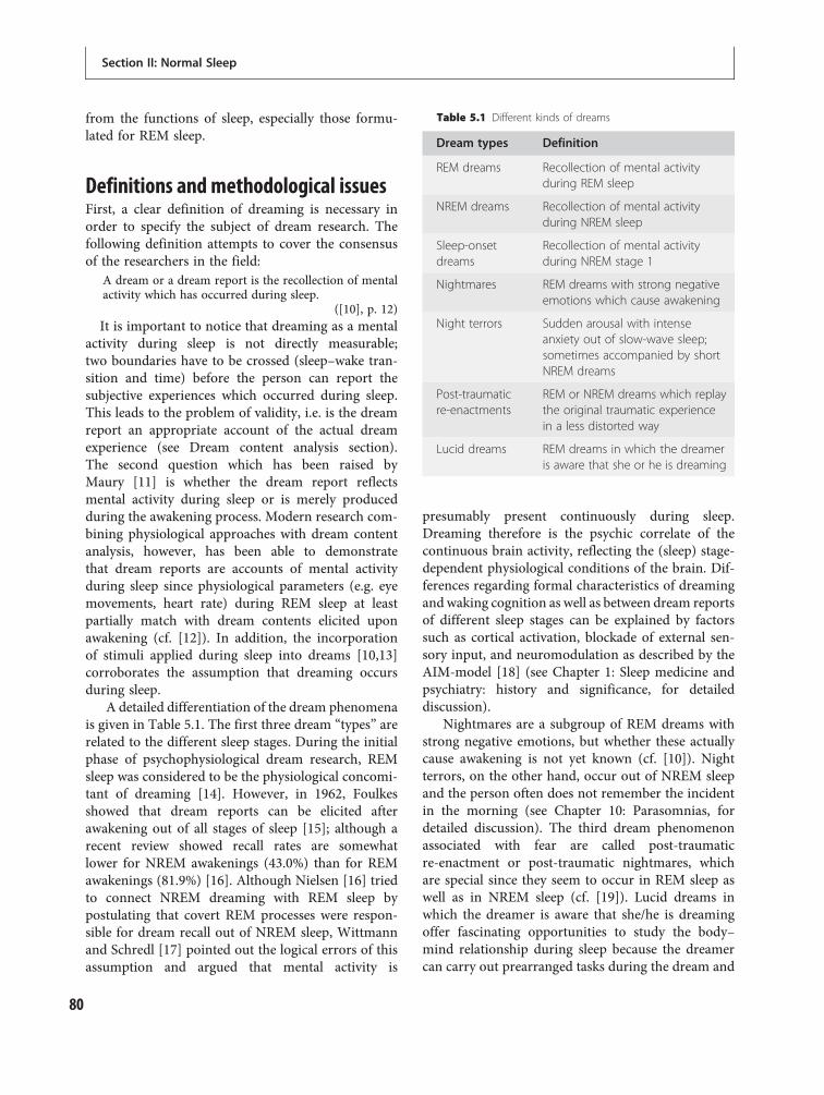
from the functions of sleep, especially those formu-lated for REM sleep.
Definitions and methodological issuesFirst, a clear definition of dreaming is necessary inorder to specify the subject of dream research. Thefollowing definition attempts to cover the consensusof the researchers in the field:
A dream or a dream report is the recollection of mentalactivity which has occurred during sleep.
([10], p. 12)It is important to notice that dreaming as a mental
activity during sleep is not directly measurable;two boundaries have to be crossed (sleep–wake tran-sition and time) before the person can report thesubjective experiences which occurred during sleep.This leads to the problem of validity, i.e. is the dreamreport an appropriate account of the actual dreamexperience (see Dream content analysis section).The second question which has been raised byMaury [11] is whether the dream report reflectsmental activity during sleep or is merely producedduring the awakening process. Modern research com-bining physiological approaches with dream contentanalysis, however, has been able to demonstratethat dream reports are accounts of mental activityduring sleep since physiological parameters (e.g. eyemovements, heart rate) during REM sleep at leastpartially match with dream contents elicited uponawakening (cf. [12]). In addition, the incorporationof stimuli applied during sleep into dreams [10,13]corroborates the assumption that dreaming occursduring sleep.
A detailed differentiation of the dream phenomenais given in Table 5.1. The first three dream “types” arerelated to the different sleep stages. During the initialphase of psychophysiological dream research, REMsleep was considered to be the physiological concomi-tant of dreaming [14]. However, in 1962, Foulkesshowed that dream reports can be elicited afterawakening out of all stages of sleep [15]; although arecent review showed recall rates are somewhatlower for NREM awakenings (43.0%) than for REMawakenings (81.9%) [16]. Although Nielsen [16] triedto connect NREM dreaming with REM sleep bypostulating that covert REM processes were respon-sible for dream recall out of NREM sleep, Wittmannand Schredl [17] pointed out the logical errors of thisassumption and argued that mental activity is
presumably present continuously during sleep.Dreaming therefore is the psychic correlate of thecontinuous brain activity, reflecting the (sleep) stage-dependent physiological conditions of the brain. Dif-ferences regarding formal characteristics of dreamingand waking cognition as well as between dream reportsof different sleep stages can be explained by factorssuch as cortical activation, blockade of external sen-sory input, and neuromodulation as described by theAIM-model [18] (see Chapter 1: Sleep medicine andpsychiatry: history and significance, for detaileddiscussion).
Nightmares are a subgroup of REM dreams withstrong negative emotions, but whether these actuallycause awakening is not yet known (cf. [10]). Nightterrors, on the other hand, occur out of NREM sleepand the person often does not remember the incidentin the morning (see Chapter 10: Parasomnias, fordetailed discussion). The third dream phenomenonassociated with fear are called post-traumaticre-enactment or post-traumatic nightmares, whichare special since they seem to occur in REM sleep aswell as in NREM sleep (cf. [19]). Lucid dreams inwhich the dreamer is aware that she/he is dreamingoffer fascinating opportunities to study the body–mind relationship during sleep because the dreamercan carry out prearranged tasks during the dream and
Table 5.1 Different kinds of dreams
Dream types Definition
REM dreams Recollection of mental activityduring REM sleep
NREM dreams Recollection of mental activityduring NREM sleep
Sleep-onsetdreams
Recollection of mental activityduring NREM stage 1
Nightmares REM dreams with strong negativeemotions which cause awakening
Night terrors Sudden arousal with intenseanxiety out of slow-wave sleep;sometimes accompanied by shortNREM dreams
Post-traumaticre-enactments
REM or NREM dreams which replaythe original traumatic experiencein a less distorted way
Lucid dreams REM dreams in which the dreameris aware that she or he is dreaming
Section II: Normal Sleep
80
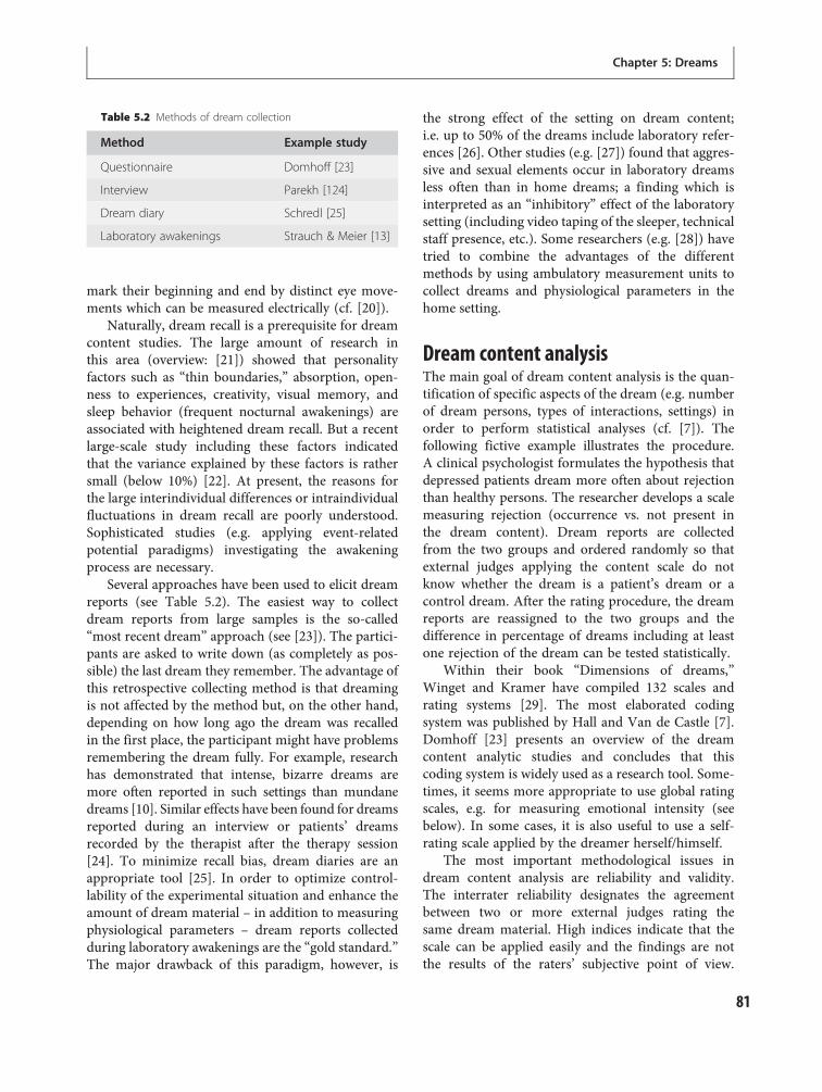
mark their beginning and end by distinct eye move-ments which can be measured electrically (cf. [20]).
Naturally, dream recall is a prerequisite for dreamcontent studies. The large amount of research inthis area (overview: [21]) showed that personalityfactors such as “thin boundaries,” absorption, open-ness to experiences, creativity, visual memory, andsleep behavior (frequent nocturnal awakenings) areassociated with heightened dream recall. But a recentlarge-scale study including these factors indicatedthat the variance explained by these factors is rathersmall (below 10%) [22]. At present, the reasons forthe large interindividual differences or intraindividualfluctuations in dream recall are poorly understood.Sophisticated studies (e.g. applying event-relatedpotential paradigms) investigating the awakeningprocess are necessary.
Several approaches have been used to elicit dreamreports (see Table 5.2). The easiest way to collectdream reports from large samples is the so-called“most recent dream” approach (see [23]). The partici-pants are asked to write down (as completely as pos-sible) the last dream they remember. The advantage ofthis retrospective collecting method is that dreamingis not affected by the method but, on the other hand,depending on how long ago the dream was recalledin the first place, the participant might have problemsremembering the dream fully. For example, researchhas demonstrated that intense, bizarre dreams aremore often reported in such settings than mundanedreams [10]. Similar effects have been found for dreamsreported during an interview or patients’ dreamsrecorded by the therapist after the therapy session[24]. To minimize recall bias, dream diaries are anappropriate tool [25]. In order to optimize control-lability of the experimental situation and enhance theamount of dream material – in addition to measuringphysiological parameters – dream reports collectedduring laboratory awakenings are the “gold standard.”The major drawback of this paradigm, however, is
the strong effect of the setting on dream content;i.e. up to 50% of the dreams include laboratory refer-ences [26]. Other studies (e.g. [27]) found that aggres-sive and sexual elements occur in laboratory dreamsless often than in home dreams; a finding which isinterpreted as an “inhibitory” effect of the laboratorysetting (including video taping of the sleeper, technicalstaff presence, etc.). Some researchers (e.g. [28]) havetried to combine the advantages of the differentmethods by using ambulatory measurement units tocollect dreams and physiological parameters in thehome setting.
Dream content analysisThe main goal of dream content analysis is the quan-tification of specific aspects of the dream (e.g. numberof dream persons, types of interactions, settings) inorder to perform statistical analyses (cf. [7]). Thefollowing fictive example illustrates the procedure.A clinical psychologist formulates the hypothesis thatdepressed patients dream more often about rejectionthan healthy persons. The researcher develops a scalemeasuring rejection (occurrence vs. not present inthe dream content). Dream reports are collectedfrom the two groups and ordered randomly so thatexternal judges applying the content scale do notknow whether the dream is a patient’s dream or acontrol dream. After the rating procedure, the dreamreports are reassigned to the two groups and thedifference in percentage of dreams including at leastone rejection of the dream can be tested statistically.
Within their book “Dimensions of dreams,”Winget and Kramer have compiled 132 scales andrating systems [29]. The most elaborated codingsystem was published by Hall and Van de Castle [7].Domhoff [23] presents an overview of the dreamcontent analytic studies and concludes that thiscoding system is widely used as a research tool. Some-times, it seems more appropriate to use global ratingscales, e.g. for measuring emotional intensity (seebelow). In some cases, it is also useful to use a self-rating scale applied by the dreamer herself/himself.
The most important methodological issues indream content analysis are reliability and validity.The interrater reliability designates the agreementbetween two or more external judges rating thesame dream material. High indices indicate that thescale can be applied easily and the findings are notthe results of the raters’ subjective point of view.
Table 5.2 Methods of dream collection
Method Example study
Questionnaire Domhoff [23]
Interview Parekh [124]
Dream diary Schredl [25]
Laboratory awakenings Strauch & Meier [13]
Chapter 5: Dreams
81
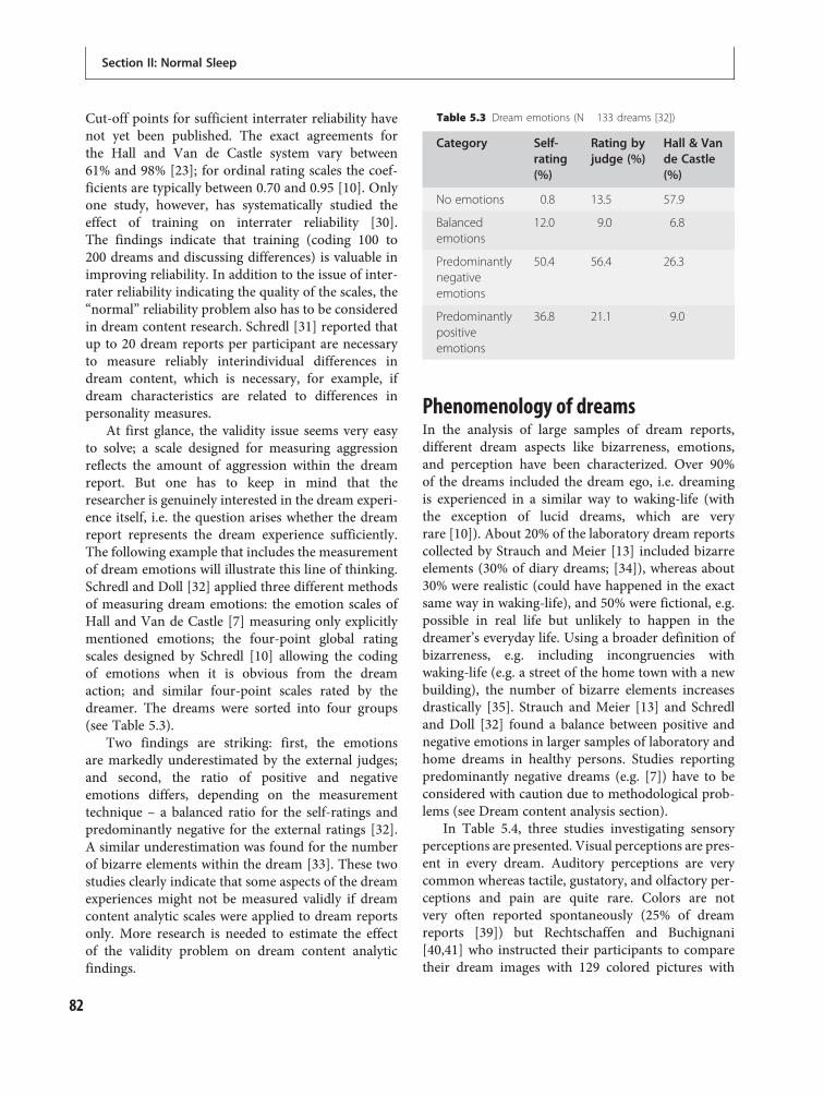
Cut-off points for sufficient interrater reliability havenot yet been published. The exact agreements forthe Hall and Van de Castle system vary between61% and 98% [23]; for ordinal rating scales the coef-ficients are typically between 0.70 and 0.95 [10]. Onlyone study, however, has systematically studied theeffect of training on interrater reliability [30].The findings indicate that training (coding 100 to200 dreams and discussing differences) is valuable inimproving reliability. In addition to the issue of inter-rater reliability indicating the quality of the scales, the“normal” reliability problem also has to be consideredin dream content research. Schredl [31] reported thatup to 20 dream reports per participant are necessaryto measure reliably interindividual differences indream content, which is necessary, for example, ifdream characteristics are related to differences inpersonality measures.
At first glance, the validity issue seems very easyto solve; a scale designed for measuring aggressionreflects the amount of aggression within the dreamreport. But one has to keep in mind that theresearcher is genuinely interested in the dream experi-ence itself, i.e. the question arises whether the dreamreport represents the dream experience sufficiently.The following example that includes the measurementof dream emotions will illustrate this line of thinking.Schredl and Doll [32] applied three different methodsof measuring dream emotions: the emotion scales ofHall and Van de Castle [7] measuring only explicitlymentioned emotions; the four-point global ratingscales designed by Schredl [10] allowing the codingof emotions when it is obvious from the dreamaction; and similar four-point scales rated by thedreamer. The dreams were sorted into four groups(see Table 5.3).
Two findings are striking: first, the emotionsare markedly underestimated by the external judges;and second, the ratio of positive and negativeemotions differs, depending on the measurementtechnique – a balanced ratio for the self-ratings andpredominantly negative for the external ratings [32].A similar underestimation was found for the numberof bizarre elements within the dream [33]. These twostudies clearly indicate that some aspects of the dreamexperiences might not be measured validly if dreamcontent analytic scales were applied to dream reportsonly. More research is needed to estimate the effectof the validity problem on dream content analyticfindings.
Phenomenology of dreamsIn the analysis of large samples of dream reports,different dream aspects like bizarreness, emotions,and perception have been characterized. Over 90%of the dreams included the dream ego, i.e. dreamingis experienced in a similar way to waking-life (withthe exception of lucid dreams, which are veryrare [10]). About 20% of the laboratory dream reportscollected by Strauch and Meier [13] included bizarreelements (30% of diary dreams; [34]), whereas about30% were realistic (could have happened in the exactsame way in waking-life), and 50% were fictional, e.g.possible in real life but unlikely to happen in thedreamer’s everyday life. Using a broader definition ofbizarreness, e.g. including incongruencies withwaking-life (e.g. a street of the home town with a newbuilding), the number of bizarre elements increasesdrastically [35]. Strauch and Meier [13] and Schredland Doll [32] found a balance between positive andnegative emotions in larger samples of laboratory andhome dreams in healthy persons. Studies reportingpredominantly negative dreams (e.g. [7]) have to beconsidered with caution due to methodological prob-lems (see Dream content analysis section).
In Table 5.4, three studies investigating sensoryperceptions are presented. Visual perceptions are pres-ent in every dream. Auditory perceptions are verycommon whereas tactile, gustatory, and olfactory per-ceptions and pain are quite rare. Colors are notvery often reported spontaneously (25% of dreamreports [39]) but Rechtschaffen and Buchignani[40,41] who instructed their participants to comparetheir dream images with 129 colored pictures with
Table 5.3 Dream emotions (N 133 dreams [32])
Category Self-rating(%)
Rating byjudge (%)
Hall & Vande Castle(%)
No emotions 0.8 13.5 57.9
Balancedemotions
12.0 9.0 6.8
Predominantlynegativeemotions
50.4 56.4 26.3
Predominantlypositiveemotions
36.8 21.1 9.0
Section II: Normal Sleep
82
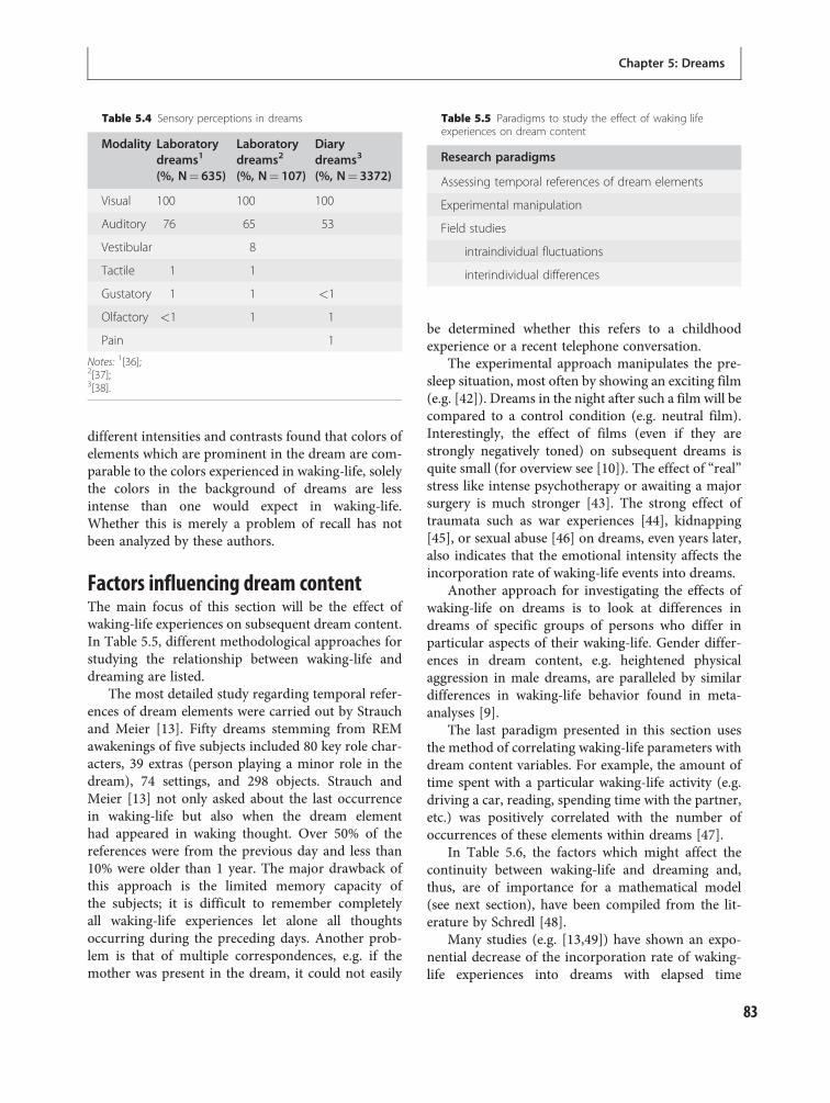
different intensities and contrasts found that colors ofelements which are prominent in the dream are com-parable to the colors experienced in waking-life, solelythe colors in the background of dreams are lessintense than one would expect in waking-life.Whether this is merely a problem of recall has notbeen analyzed by these authors.
Factors influencing dream contentThe main focus of this section will be the effect ofwaking-life experiences on subsequent dream content.In Table 5.5, different methodological approaches forstudying the relationship between waking-life anddreaming are listed.
The most detailed study regarding temporal refer-ences of dream elements were carried out by Strauchand Meier [13]. Fifty dreams stemming from REMawakenings of five subjects included 80 key role char-acters, 39 extras (person playing a minor role in thedream), 74 settings, and 298 objects. Strauch andMeier [13] not only asked about the last occurrencein waking-life but also when the dream elementhad appeared in waking thought. Over 50% of thereferences were from the previous day and less than10% were older than 1 year. The major drawback ofthis approach is the limited memory capacity ofthe subjects; it is difficult to remember completelyall waking-life experiences let alone all thoughtsoccurring during the preceding days. Another prob-lem is that of multiple correspondences, e.g. if themother was present in the dream, it could not easily
be determined whether this refers to a childhoodexperience or a recent telephone conversation.
The experimental approach manipulates the pre-sleep situation, most often by showing an exciting film(e.g. [42]). Dreams in the night after such a film will becompared to a control condition (e.g. neutral film).Interestingly, the effect of films (even if they arestrongly negatively toned) on subsequent dreams isquite small (for overview see [10]). The effect of “real”stress like intense psychotherapy or awaiting a majorsurgery is much stronger [43]. The strong effect oftraumata such as war experiences [44], kidnapping[45], or sexual abuse [46] on dreams, even years later,also indicates that the emotional intensity affects theincorporation rate of waking-life events into dreams.
Another approach for investigating the effects ofwaking-life on dreams is to look at differences indreams of specific groups of persons who differ inparticular aspects of their waking-life. Gender differ-ences in dream content, e.g. heightened physicalaggression in male dreams, are paralleled by similardifferences in waking-life behavior found in meta-analyses [9].
The last paradigm presented in this section usesthe method of correlating waking-life parameters withdream content variables. For example, the amount oftime spent with a particular waking-life activity (e.g.driving a car, reading, spending time with the partner,etc.) was positively correlated with the number ofoccurrences of these elements within dreams [47].
In Table 5.6, the factors which might affect thecontinuity between waking-life and dreaming and,thus, are of importance for a mathematical model(see next section), have been compiled from the lit-erature by Schredl [48].
Many studies (e.g. [13,49]) have shown an expo-nential decrease of the incorporation rate of waking-life experiences into dreams with elapsed time
Table 5.4 Sensory perceptions in dreams
Modality Laboratorydreams1
(%, N¼ 635)
Laboratorydreams2
(%, N¼ 107)
Diarydreams3
(%, N¼ 3372)
Visual 100 100 100
Auditory 76 65 53
Vestibular 8
Tactile 1 1
Gustatory 1 1 <1
Olfactory <1 1 1
Pain 1
Notes: 1[36];2[37];3[38].
Table 5.5 Paradigms to study the effect of waking lifeexperiences on dream content
Research paradigms
Assessing temporal references of dream elements
Experimental manipulation
Field studies
intraindividual fluctuations
interindividual differences
Chapter 5: Dreams
83
between experience and the subsequent dream. Also,the differences in effects of experimental stress and“real” stress and trauma research (see above) indicatethat emotional involvement affects the incorporationrate. Three studies [47,50,51] have shown that focusedthinking activity (reading, working with a computer)during dreams occurs less frequently than unfocusedactivities such as talking with friends, etc. Theseresults also indicate that the type of activity is ofimportance for the continuity between waking-lifeand dreaming. The time of the night or the timeinterval between sleep onset and dream onset hasaffected the incorporation rate of waking-life experi-ences in two studies [52,53]; dreams of the secondpart of the night comprise more elements of thedistant past while dreams of the first part of thenight incorporate mostly recent daytime experiences(cf. [54,55]). The last factor, which has been studiedrarely, is the interaction between personality traitsand incorporation of waking-life experiences. It seemsplausible that personality dimensions such as fielddependence (this concept describes how people areinfluenced by inner (field-independent) or environ-mental (field-dependent) cues in orienting themselves
in space and the extent to which they select infor-mation within the environment [56]) or thin bound-aries [57] moderate the magnitude of continuitybetween waking and dreaming.
The “continuity hypothesis”of dreamingMany researchers (e.g. [10,13,23]) are advocating theso-called “continuity hypothesis” of dreaming whichsimply states that dreams reflect waking-life experi-ences. However, for deriving specific hypotheses thecontinuity hypothesis in its general formulation is tooimprecise. In order to advance the research in this field,Schredl [48] postulated a mathematical model that isbased on the published findings and seems to be prom-ising for further empirical testing (see Figure 5.1). Themultiplying factor includes the effects of emotionalinvolvement (EI), type of waking-life experience(TYPE), and the interaction between personality traitsand incorporation rates (PERS). The relationshipsbetween these factors should be determined by futurestudies. The slope of the exponential function may bemoderated by the time interval between sleep onsetand dream onset (time of the night; TN).
Dreams and psychopathologyTwomotives have stimulated investigating the relation-ship between dreaming and mental disorders. First, thedream state itself was conceptualized by several theorists(e.g. [58]) as a mental disorder and, in reverse, hallucin-ations of schizophrenic patients have been thought ofas breakthroughs of dreams into the waking state(e.g. [59]). Second, many clinicians since Freud haveattempted to use dreams in the diagnosis and treatmentof their patients (e.g. [60]). The basic assumption forthis research is the so-called continuity hypothesis [48]
Table 5.6 Factors which affect the continuity betweenwaking life and dreaming
Factors
� Exponential decrease with time
� Emotional involvement
� Type of waking-life experience
� Personality traits
� Time of the night (time interval between sleep onsetand dream onset)
Incorporation rate = a (EI, TYPE, PERS) * e–b(TN)*t + Constant
a (EI, TYPE, PERS) multiplying factor which is a function of emotional involvement
(EI), type of the waking-life experience (TYPE), and the
interaction between experience and personality traits (PERS)
Slope of the exponential function which is itself a function of the
time interval between sleep onset and dream onset (TN)
Time interval between waking-life experience and occurrence of
the dream incorporation
b (TN)
t
Figure 5.1 Mathematical model for thecontinuity between waking life anddreaming.
Section II: Normal Sleep
84
which predicts that for patients withmental disorders, itshould thus be possible to detect the specific wakingsymptomatology within their dreams.
The literature reviews showed that the majority ofempirical studies support the continuity hypothesis[61–63]. On the one hand, it was found that hallucin-ations of schizophrenic patients are not dreamsexperienced during the waking state (e.g. [64]) andthat the concept of dreaming as a mental disorder isnot very helpful (cf. [65]). On the other hand, dreamsof schizophrenic patients are typical for this disorder,i.e. the dreams are more bizarre [65] and are charac-terized by aggression and negative emotions [61]. Fordepressive patients, Beck and Hurvich [66] and Beckand Ward [67] have found an increased amount of“masochistic” themes in their dreams. Subsequentstudies (overview: [68]) confirmed that dreams ofdepressive patients are more negatively toned andinclude unpleasant experiences more often (defin-itions of “masochistic” dream content according toBeck & Hurvich [66]) than healthy controls. Schredland Engelhardt [65] were able to demonstrate thatseverity of depressive symptomatology was directlycorrelated with the intensity of negative dream emo-tions, irrespective of the patients’ diagnoses, support-ing a dimensional and not categorical relationshipbetween waking-life symptoms and dream content.In addition, severely depressed patients dreamedmore often about aggression and death [65].
Regarding dream recall, many studies failed to showmarked differences among various diagnostic groups,such as schizophrenia, eating disorders, etc., and healthycontrols [61]. An exception is depression where patientshave a reduced dream recall frequency [69,70]; thereduction was again related to symptom severity [71].The explanations for the reduced dream recall indepressed patients, however, remain unclear. The ques-tion of whether the typical sleep architecture of depres-sive patients (decreased latency to REM sleep), cognitiveimpairment often found in severely disturbed patients,or intrinsic alterations related to depression is respon-sible has yet to be answered.
Dreams and sleep disordersThis section will focus on several sleep disorderswhich have been studied in relation to dreaming:insomnia, sleep apnea syndrome, narcolepsy, andrestless legs syndrome. For other diagnoses, like idio-pathic hypersomnia, or NREM parasomnias, such as
sleepwalking or night terrors, systematic dream con-tent analytic studies are lacking. However, extensivereviews are available for nightmares [72], REM sleepbehavior disorder (see Chapter 10: Parasomnias), anddreaming in post-traumatic stress disorder [19].
Schredl et al. [73] found an elevated dream recallfrequency in insomnia patients in contrast to healthycontrols; a finding which was no longer significantif number of nocturnal awakenings (self-reportmeasure) was statistically controlled. This parallelsthe correlation between nocturnal awakenings andincreased dream recall in healthy persons [22]. Per-centage of dream recall after REM awakenings carriedout in the laboratory did not differ between insomniapatients and controls [74]. Ermann [75] and Schredlet al. [73] found more negatively toned dreams inpatients with insomnia. In addition, occurrence of prob-lems within the dream was directly correlated with thenumber of waking-life problems the patients reported inthe questionnaire. Therefore, dreams might reflect thetopics which are at least partially responsible for thedevelopment andmaintenance of the primary insomnia.
In sleep apnea, the findings regarding the dreamrecall frequency are inconsistent (cf. [76]). In the nine-teenth century, nightmares were thought to be due todecreased flow of oxygen (e.g. due to pillow blocking ofthe mouth and nose; see [77]). However, parameterslike minimal oxygen saturation nadir or respiratorydisturbance index do not correlate with dreamrecall frequency [78]. Furthermore, a heightenednightmare frequency in sleep apnea patients has notbeen found [76]. Only very few dream reports includethe massive physiological apnea processes:
During the dream I felt tied up or chained. I saw thickropes around my arms and was not able to move.I experienced the fear of suffocation without being ableto cope with the situation. Powerlessness and alsoresignation came up.
(Patient with sleep apnea, male, 39 years, respiratorydisturbance index (RDI): 68.1 apneas per hour, maximaldrop of blood oxygen saturation: 43%).
([79], p. 295)
Overall, the low incidence of breathing-related dreamtopics might be explained by adaptation, i.e. theincrease of number and severity of sleep apneas overmonths and years might explain why this stimuli israrely incorporated into dreams whereas externalstimuli are at least sometimes incorporated intodreams [76]. It would be intriguing to conduct asystematic study where the sleeper wears a mask
Chapter 5: Dreams
85
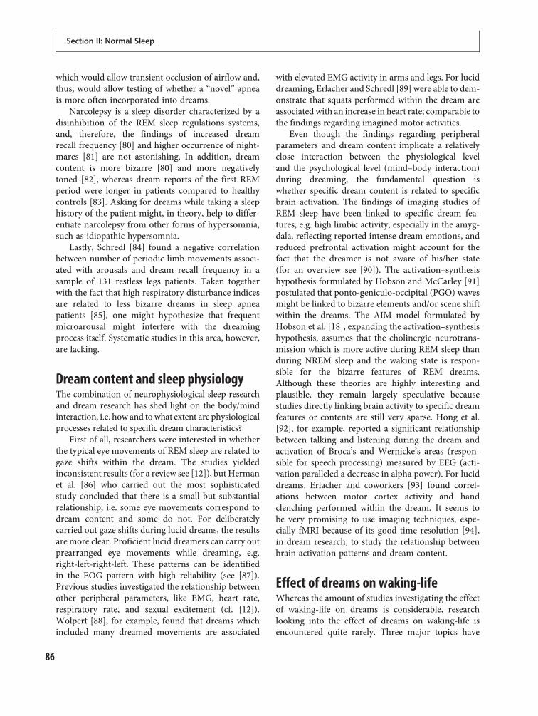
which would allow transient occlusion of airflow and,thus, would allow testing of whether a “novel” apneais more often incorporated into dreams.
Narcolepsy is a sleep disorder characterized by adisinhibition of the REM sleep regulations systems,and, therefore, the findings of increased dreamrecall frequency [80] and higher occurrence of night-mares [81] are not astonishing. In addition, dreamcontent is more bizarre [80] and more negativelytoned [82], whereas dream reports of the first REMperiod were longer in patients compared to healthycontrols [83]. Asking for dreams while taking a sleephistory of the patient might, in theory, help to differ-entiate narcolepsy from other forms of hypersomnia,such as idiopathic hypersomnia.
Lastly, Schredl [84] found a negative correlationbetween number of periodic limb movements associ-ated with arousals and dream recall frequency in asample of 131 restless legs patients. Taken togetherwith the fact that high respiratory disturbance indicesare related to less bizarre dreams in sleep apneapatients [85], one might hypothesize that frequentmicroarousal might interfere with the dreamingprocess itself. Systematic studies in this area, however,are lacking.
Dream content and sleep physiologyThe combination of neurophysiological sleep researchand dream research has shed light on the body/mindinteraction, i.e. how and towhat extent are physiologicalprocesses related to specific dream characteristics?
First of all, researchers were interested in whetherthe typical eye movements of REM sleep are related togaze shifts within the dream. The studies yieldedinconsistent results (for a review see [12]), but Hermanet al. [86] who carried out the most sophisticatedstudy concluded that there is a small but substantialrelationship, i.e. some eye movements correspond todream content and some do not. For deliberatelycarried out gaze shifts during lucid dreams, the resultsare more clear. Proficient lucid dreamers can carry outprearranged eye movements while dreaming, e.g.right-left-right-left. These patterns can be identifiedin the EOG pattern with high reliability (see [87]).Previous studies investigated the relationship betweenother peripheral parameters, like EMG, heart rate,respiratory rate, and sexual excitement (cf. [12]).Wolpert [88], for example, found that dreams whichincluded many dreamed movements are associated
with elevated EMG activity in arms and legs. For luciddreaming, Erlacher and Schredl [89] were able to dem-onstrate that squats performed within the dream areassociated with an increase in heart rate; comparable tothe findings regarding imagined motor activities.
Even though the findings regarding peripheralparameters and dream content implicate a relativelyclose interaction between the physiological leveland the psychological level (mind–body interaction)during dreaming, the fundamental question iswhether specific dream content is related to specificbrain activation. The findings of imaging studies ofREM sleep have been linked to specific dream fea-tures, e.g. high limbic activity, especially in the amyg-dala, reflecting reported intense dream emotions, andreduced prefrontal activation might account for thefact that the dreamer is not aware of his/her state(for an overview see [90]). The activation–synthesishypothesis formulated by Hobson and McCarley [91]postulated that ponto-geniculo-occipital (PGO) wavesmight be linked to bizarre elements and/or scene shiftwithin the dreams. The AIM model formulated byHobson et al. [18], expanding the activation–synthesishypothesis, assumes that the cholinergic neurotrans-mission which is more active during REM sleep thanduring NREM sleep and the waking state is respon-sible for the bizarre features of REM dreams.Although these theories are highly interesting andplausible, they remain largely speculative becausestudies directly linking brain activity to specific dreamfeatures or contents are still very sparse. Hong et al.[92], for example, reported a significant relationshipbetween talking and listening during the dream andactivation of Broca’s and Wernicke’s areas (respon-sible for speech processing) measured by EEG (acti-vation paralleled a decrease in alpha power). For luciddreams, Erlacher and coworkers [93] found correl-ations between motor cortex activity and handclenching performed within the dream. It seems tobe very promising to use imaging techniques, espe-cially fMRI because of its good time resolution [94],in dream research, to study the relationship betweenbrain activation patterns and dream content.
Effect of dreams on waking-lifeWhereas the amount of studies investigating the effectof waking-life on dreams is considerable, researchlooking into the effect of dreams on waking-life isencountered quite rarely. Three major topics have
Section II: Normal Sleep
86
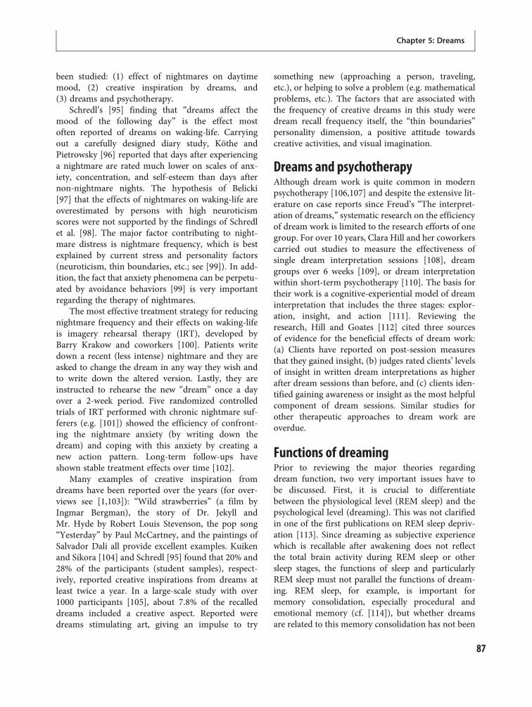
been studied: (1) effect of nightmares on daytimemood, (2) creative inspiration by dreams, and(3) dreams and psychotherapy.
Schredl’s [95] finding that “dreams affect themood of the following day” is the effect mostoften reported of dreams on waking-life. Carryingout a carefully designed diary study, Köthe andPietrowsky [96] reported that days after experiencinga nightmare are rated much lower on scales of anx-iety, concentration, and self-esteem than days afternon-nightmare nights. The hypothesis of Belicki[97] that the effects of nightmares on waking-life areoverestimated by persons with high neuroticismscores were not supported by the findings of Schredlet al. [98]. The major factor contributing to night-mare distress is nightmare frequency, which is bestexplained by current stress and personality factors(neuroticism, thin boundaries, etc.; see [99]). In add-ition, the fact that anxiety phenomena can be perpetu-ated by avoidance behaviors [99] is very importantregarding the therapy of nightmares.
The most effective treatment strategy for reducingnightmare frequency and their effects on waking-lifeis imagery rehearsal therapy (IRT), developed byBarry Krakow and coworkers [100]. Patients writedown a recent (less intense) nightmare and they areasked to change the dream in any way they wish andto write down the altered version. Lastly, they areinstructed to rehearse the new “dream” once a dayover a 2-week period. Five randomized controlledtrials of IRT performed with chronic nightmare suf-ferers (e.g. [101]) showed the efficiency of confront-ing the nightmare anxiety (by writing down thedream) and coping with this anxiety by creating anew action pattern. Long-term follow-ups haveshown stable treatment effects over time [102].
Many examples of creative inspiration fromdreams have been reported over the years (for over-views see [1,103]): “Wild strawberries” (a film byIngmar Bergman), the story of Dr. Jekyll andMr. Hyde by Robert Louis Stevenson, the pop song“Yesterday” by Paul McCartney, and the paintings ofSalvador Dali all provide excellent examples. Kuikenand Sikora [104] and Schredl [95] found that 20% and28% of the participants (student samples), respect-ively, reported creative inspirations from dreams atleast twice a year. In a large-scale study with over1000 participants [105], about 7.8% of the recalleddreams included a creative aspect. Reported weredreams stimulating art, giving an impulse to try
something new (approaching a person, traveling,etc.), or helping to solve a problem (e.g. mathematicalproblems, etc.). The factors that are associated withthe frequency of creative dreams in this study weredream recall frequency itself, the “thin boundaries”personality dimension, a positive attitude towardscreative activities, and visual imagination.
Dreams and psychotherapyAlthough dream work is quite common in modernpsychotherapy [106,107] and despite the extensive lit-erature on case reports since Freud’s “The interpret-ation of dreams,” systematic research on the efficiencyof dream work is limited to the research efforts of onegroup. For over 10 years, Clara Hill and her coworkerscarried out studies to measure the effectiveness ofsingle dream interpretation sessions [108], dreamgroups over 6 weeks [109], or dream interpretationwithin short-term psychotherapy [110]. The basis fortheir work is a cognitive-experiential model of dreaminterpretation that includes the three stages: explor-ation, insight, and action [111]. Reviewing theresearch, Hill and Goates [112] cited three sourcesof evidence for the beneficial effects of dream work:(a) Clients have reported on post-session measuresthat they gained insight, (b) judges rated clients’ levelsof insight in written dream interpretations as higherafter dream sessions than before, and (c) clients iden-tified gaining awareness or insight as the most helpfulcomponent of dream sessions. Similar studies forother therapeutic approaches to dream work areoverdue.
Functions of dreamingPrior to reviewing the major theories regardingdream function, two very important issues have tobe discussed. First, it is crucial to differentiatebetween the physiological level (REM sleep) and thepsychological level (dreaming). This was not clarifiedin one of the first publications on REM sleep depriv-ation [113]. Since dreaming as subjective experiencewhich is recallable after awakening does not reflectthe total brain activity during REM sleep or othersleep stages, the functions of sleep and particularlyREM sleep must not parallel the functions of dream-ing. REM sleep, for example, is important formemory consolidation, especially procedural andemotional memory (cf. [114]), but whether dreamsare related to this memory consolidation has not been
Chapter 5: Dreams
87
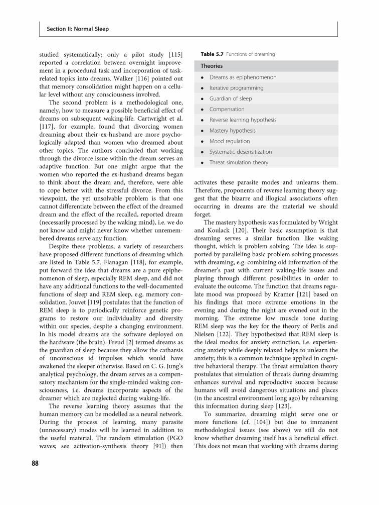
studied systematically; only a pilot study [115]reported a correlation between overnight improve-ment in a procedural task and incorporation of task-related topics into dreams. Walker [116] pointed outthat memory consolidation might happen on a cellu-lar level without any consciousness involved.
The second problem is a methodological one,namely, how to measure a possible beneficial effect ofdreams on subsequent waking-life. Cartwright et al.[117], for example, found that divorcing womendreaming about their ex-husband are more psycho-logically adapted than women who dreamed aboutother topics. The authors concluded that workingthrough the divorce issue within the dream serves anadaptive function. But one might argue that thewomen who reported the ex-husband dreams beganto think about the dream and, therefore, were ableto cope better with the stressful divorce. From thisviewpoint, the yet unsolvable problem is that onecannot differentiate between the effect of the dreameddream and the effect of the recalled, reported dream(necessarily processed by the waking mind), i.e. we donot know and might never know whether unremem-bered dreams serve any function.
Despite these problems, a variety of researchershave proposed different functions of dreaming whichare listed in Table 5.7. Flanagan [118], for example,put forward the idea that dreams are a pure epiphe-nomenon of sleep, especially REM sleep, and did nothave any additional functions to the well-documentedfunctions of sleep and REM sleep, e.g. memory con-solidation. Jouvet [119] postulates that the function ofREM sleep is to periodically reinforce genetic pro-grams to restore our individuality and diversitywithin our species, despite a changing environment.In his model dreams are the software deployed onthe hardware (the brain). Freud [2] termed dreams asthe guardian of sleep because they allow the catharsisof unconscious id impulses which would haveawakened the sleeper otherwise. Based on C. G. Jung’sanalytical psychology, the dream serves as a compen-satory mechanism for the single-minded waking con-sciousness, i.e. dreams incorporate aspects of thedreamer which are neglected during waking-life.
The reverse learning theory assumes that thehuman memory can be modelled as a neural network.During the process of learning, many parasite(unnecessary) modes will be learned in addition tothe useful material. The random stimulation (PGOwaves; see activation-synthesis theory [91]) then
activates these parasite modes and unlearns them.Therefore, proponents of reverse learning theory sug-gest that the bizarre and illogical associations oftenoccurring in dreams are the material we shouldforget.
The mastery hypothesis was formulated by Wrightand Koulack [120]. Their basic assumption is thatdreaming serves a similar function like wakingthought, which is problem solving. The idea is sup-ported by paralleling basic problem solving processeswith dreaming, e.g. combining old information of thedreamer’s past with current waking-life issues andplaying through different possibilities in order toevaluate the outcome. The function that dreams regu-late mood was proposed by Kramer [121] based onhis findings that more extreme emotions in theevening and during the night are evened out in themorning. The extreme low muscle tone duringREM sleep was the key for the theory of Perlis andNielsen [122]. They hypothesized that REM sleep isthe ideal modus for anxiety extinction, i.e. experien-cing anxiety while deeply relaxed helps to unlearn theanxiety; this is a common technique applied in cogni-tive behavioral therapy. The threat simulation theorypostulates that simulation of threats during dreamingenhances survival and reproductive success becausehumans will avoid dangerous situations and places(in the ancestral environment long ago) by rehearsingthis information during sleep [123].
To summarize, dreaming might serve one ormore functions (cf. [104]) but due to immanentmethodological issues (see above) we still do notknow whether dreaming itself has a beneficial effect.This does not mean that working with dreams during
Table 5.7 Functions of dreaming
Theories
� Dreams as epiphenomenon
� Iterative programming
� Guardian of sleep
� Compensation
� Reverse learning hypothesis
� Mastery hypothesis
� Mood regulation
� Systematic desensitization
� Threat simulation theory
Section II: Normal Sleep
88
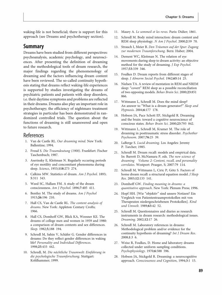
waking-life is not beneficial; there is support for thisapproach (see Dreams and psychotherapy section).
SummaryDreams have been studied from different perspectives:psychoanalysis, academic psychology, and neurosci-ences. After presenting the definition of dreamingand the methodological tools of dream research, themajor findings regarding the phenomenology ofdreaming and the factors influencing dream contenthave been reviewed. The so-called continuity hypoth-esis stating that dreams reflect waking-life experiencesis supported by studies investigating the dreams ofpsychiatric patients and patients with sleep disorders,i.e. their daytime symptoms and problems are reflectedin their dreams. Dreams also play an important role inpsychotherapy; the efficiency of nightmare treatmentstrategies in particular has been demonstrated in ran-domized controlled trials. The question about thefunctions of dreaming is still unanswered and opento future research.
References1. Van de Castle RL. Our dreaming mind. New York:
Ballentine; 1994.
2. Freud S. Die Traumdeutung (1900). Frankfurt: FischerTaschenbuch; 1987.
3. Aserinsky E, Kleitman N. Regularly occuring periodsof eye motility and concomitant phenomena duringsleep. Science, 1953;118:273 274.
4. Calkins MW. Statistics of dream. Am J Psychol. 1893;5:311 343.
5. Weed SC, Hallam FM. A study of the dreamconsciousness. Am J Psychol. 1896;7:405 411.
6. Bentley M. The study of dreams. Am J Psychol1915;26:196 210.
7. Hall CS, Van de Castle RL. The content analysis ofdreams. New York: Appleton Century Crofts;1966.
8. Hall CS, Domhoff GW, Blick KA, Weesner KE. Thedreams of college men and women in 1959 and 1980:a comparison of dream contents and sex differences.Sleep. 1982;5;188 194.
9. Schredl M, Sahin V, Schäfer G. Gender differences indreams: Do they reflect gender differences in wakinglife? Personality and Individual Differences.1998;25:433 442.
10. Schredl, M. Die nächtliche Traumwelt: Einführung indie psychologische Traumforschung. Stuttgart:Kohlhammer; 1999.
11. Maury A. Le sommeil et les reves. Paris: Didier; 1861.
12. Schredl M. Body mind interaction: dream content andREM sleep physiology. N Am J Psychol. 2000;2:59 70.
13. Strauch I, Meier B. Den Träumen auf der Spur: Zugangzur modernen Traumforschung. Bern: Huber; 2004.
14. Dement WC, Kleitman N. The relation of eyemovements during sleep to dream activity: an objectivemethod for the study of dreaming. J Exp Psychol.1957;53:339 346.
15. Foulkes D. Dream reports from different stages ofsleep. J Abnorm Social Psychol. 1962;65:14 25.
16. Nielsen TA. A review of mentation in REM and NREMsleep: “covert” REM sleep as a possible reconciliationof two opposing models. Behav Brain Sci. 2000;23:851866.
17. Wittmann L, Schredl M. Does the mind sleep?An answer to “What is a dream generator?” Sleep andHypnosis. 2004;6:177 178.
18. Hobson JA, Pace Schott EF, Stickgold R. Dreamingand the brain: toward a cognitive neuroscience ofconscious states. Behav Brain Sci. 2000;23:793 842.
19. Wittmann L, Schredl M, Kramer M. The role ofdreaming in posttraumatic stress disorder. PsychotherPsychosom. 2007;76:25 39.
20. LaBerge S. Lucid dreaming. Los Angeles: JeremyP. Tarcher; 1985.
21. Schredl M. Dream recall: models and empirical data.In: Barrett D, McNamara P, eds. The new science ofdreaming Volume 2: Content, recall, and personalitycorrelates. Westport: Praeger, S; 2007:79 114.
22. Schredl M, Wittmann L, Ciric P, Götz S. Factors ofhome dream recall: a structural equation model. J SleepRes. 2003;12:133 141.
23. Domhoff GW. Finding meaning in dreams: aquantitative approach. New York: Plenum Press; 1996.
24. Hopf HH. [Wie “objektiv” sind unsere Notizen? EinVergleich von Patiententraumprotokollen mit vonTherapeuten niedergeschriebenen Protokollen]. Kindund Umwelt. 1989;63:42 52.
25. Schredl M. Questionnaires and diaries as researchinstruments in dream research: methodological issues.Dreaming. 2002;12:17 26.
26. Schredl M. Laboratory references in dreams:Methodological problem and/or evidence for thecontinuity hypothesis of dreaming? Int J Dream Res.2008;1:3 6.
27. Weisz R, Foulkes, D. Home and laboratory dreamscollected under uniform sampling conditions.Psychophysiology. 1970;6:588 596.
28. Hobson JA, Stickgold R. Dreaming: a neurocognitiveapproach. Consciousness and Cognition, 1994;3:1 15.
Chapter 5: Dreams
89

29. Winget C, Kramer M. Dimensions of dreams.Gainesville: University of Florida Press; 1979.
30. Schredl M, Burchert N, Gabatin Y. The effect oftraining on interrater reliability in dream contentanalysis. Sleep and Hypnosis. 2004;6:139 144.
31. Schredl M. The stability and variability of dreamcontent. Percept Mot Skills. 1998;86:733 734.
32. Schredl M, Doll E. Emotions in diary dreams.Conscious Cogn. 1998;7:634 646.
33. Schredl M, Erlacher D. The problem of dream contentanalysis validity as shown by a bizarreness scale.Sleep and Hypnosis. 2003;5:129 135.
34. Schredl M, Schäfer G, Hofmann F, Jacob S. Dreamcontent and personality: thick vs. thin boundaries.Dreaming. 1999;9:257 263.
35. Hobson JA, Hoffman SA, Helfand R, Kostner D.Dream bizarreness and the activation synthesishypothesis. Hum Neurobiol. 1987;6:157 164.
36. Snyder F. The phenomenology of dreaming. In:Madow L, Snow LH, eds. The psychodynamicimplications of the physiological studies on dreams.Springfield: Charles C. Thomas; 1970:124 151.
37. McCarley RW, Hoffman E. REM sleep dreams and theactivation synthesis hypothesis. Am J Psychiatry.1981;138:904 912.
38. Zadra AL, Nielsen TA, Donderi DC. The prevalence ofauditory, olfactory, gustatory and pain experiences in3372 home dreams. Sleep Res. 1997;26:281.
39. Kahn E, Dement W, Fisher C, Barmack JE. Incidenceof color in immediately recalled dreams. Science.1962;137:1054 1055.
40. Rechtschaffen A, Buchignani C. Visual dimensionsand correlates of dream images. Sleep Res. 1983;12:189.
41. Rechtschaffen A, Buchignani C. The visual appearanceof dreams. In: Antrobus JS, Bertini M, eds. Theneuropsychology of sleep and dreaming. Hillsdale:Lawrence Erlbaum; 1992:143 155.
42. Foulkes D, Rechtschaffen A. Presleep determinantsof dream content. Percept Mot Skills. 1964;19:983 1005.
43. Breger L, Hunter I, Lane RW. The effect of stress ondreams. New York: International Universities Press;1971.
44. Schreuder BJ, Kleijn WC, Rooijmans HG. Nocturnalre experiencing more than forty years after wartrauma. J Trauma Stress. 2000;13:453 463.
45. Terr LC. Psychic trauma in children: Observationsfollowing the Chowchilla schoolbus kidnapping.Am J Psychiatry. 1981;138:14 19.
46. Krakow B, Tandberg D, Barey M, Scriggins L.Nightmares and sleep disturbance in sexually assaultedwomen. Dreaming. 1995;5:199 206.
47. Schredl M, Hofmann F. Continuity between wakingactivities and dream activities. Conscious Cogn.2003;12:298 308.
48. Schredl M. Continuity between waking and dreaming:A proposal for a mathematical model. Sleep andHypnosis. 2003;5:38 52.
49. Botman HI, Crovitz HF. Dream reports andautobiographical memory. Imagination, Cognition andPersonality. 1989 90;9:213 224.
50. Hartmann E. We do not dream of the 3 R’s:implications for the nature of dream mentation.Dreaming. 2000;10:103 110.
51. Schredl M. Continuity between waking life anddreaming: are all waking activities reflected equallyoften in dreams? Percept Mot Skills. 2000;90:844 846.
52. Verdone P. Temporal reference of manifest dreamcontent. Percept Mot Skills. 1965;20:1253 1268.
53. Roussy F, Raymond I, Gonthier I, Grenier J,De Koninck J. Temporal references in manifestdream content: confirmation of increased remotenessas the night progresses. Sleep Supplement. 1998;21:285.
54. Roffwarg HP, Herman JS, Bowe Anders C, Tauber ES.The effects of sustained alterations of waking visualinput on dream content. In: Arkin AM, Antrobus JS,Ellman SJ, eds. The mind in sleep: psychology andpsychophysiology. Hillsdale, New Jersey: LawrenceErlbaum; 1978:295 349.
55. Lauer C, Riemann D, Lund R, Berger M. ShortenedREM latency: a consequence of psychological strain?Psychophysiology. 1987;24:263 271.
56. Baekeland F, Resch R, Katz D. Presleep mentationand dream reports: I. Cognitive style, contiguity tosleep and time of the night. Arch Gen Psychiatry.1968;19:300 311.
57. Schredl M, Kleinferchner P, Gell T. Dreaming andpersonality: thick vs. thin boundaries. Dreaming.1996;6:219 223.
58. Hobson JA. Dreaming as delirium: a mental statusexamination of our nightly madness. Sem Neurol.1997;17:121 128.
59. Noble D. A study of dreams in schizophreniaand allied states. Am J Psychiatry. 1950;107:612 616.
60. Whitman RM, Kramer M, Ornstein PH, Baldridge BJ.The varying uses of the dream in clinical psychiatry.In: Madow L, Snow LH, eds. The psychodynamicimplications of the physiological studies on dreams.Springfield: Charles C. Thomas; 1970:24 46.
Section II: Normal Sleep
90
61. Kramer M, Roth T. Dreams in psychopathologicpatient groups. In: Williams RL, Karacan I, eds. Sleepdisorders: diagnosis and treatment. New York:John Wiley & Sons; 1978:323 349.
62. Mellen RR, Duffey TH, Craig SM. Manifest content inthe dreams of clinical populations. J Mental HealthCounseling. 1993;15:170 183.
63. Kramer M. Dreams and psychopathology. In:Kryger MH, Roth T, Dement WC, eds. Principlesand practice of sleep medicine. Philadelphia:WB Saunders; 2000:511 519.
64. Fischman LG. Dreams, hallucinogenic drug states andschizophrenia: a psychological and biologicalcomparison. Schizophr Bull. 1983;9:73 94.
65. Schredl M, Engelhardt H. Dreaming andpsychopathology: Dream recall and dream content ofpsychiatric inpatients. Sleep and Hypnosis.2001;3:44 54.
66. Beck AT, Hurvich MS. Psychological correlates ofdepression: I. Frequency of “masochistic” dreamcontent in a private practice sample. Psychosom Med.1959;1:50 55.
67. Beck AT, Ward CH. Dreams of depressed patients.Arch Gen Psychiatry. 1961;5:462 467.
68. Schredl M, Schnitzler M. [Träume und Depression:Eine Literaturübersicht]. Psycho. 1999;25:693 696.
69. Riemann D, Löw H, Schredl M, et al. Investigations ofmorning and laboratory dream recall and content indepressive patients during baseline conditions andunder antidepressive treatment with trimipramine.Psychiatric Journal of the University of Ottawa.1990;15:93 99.
70. Armitage R, Rochlen A, Fitch T, Trivedi M, Rush AJ.Dream recall and major depression: a preliminaryreport. Dreaming. 1995;5:189 198.
71. Schredl M. [Traumerinnerung bei depressivenPatienten]. Psychother Psychosom Med Psychol.1995;45:414 417.
72. Levin R, Nielson TA. Disturbed dreaming,posttraumatic stress disorder, and affect distress: areview and neurocognitive model. Psychol Bull.2007;133:482 528.
73. Schredl M, Schäfer G, Weber B, Heuser I. Dreamingand insomnia: Dream recall and dream content ofpatients with insomnia. J Sleep Res. 1998;7:191 198.
74. Ermann M, Peichl J, Pohl H, Schneider MM,Winkelmann Y. [Spontanerwachen und Träumebei Patienten mit psychovegetativen Schlafstörungen].Psychother Psychosom Med Psychol. 1993;43:333 340.
75. Ermann M. [Die Traumerinnerung bei Patienten mitpsychogenen Schlafstörungen: Empirische Befunde
und einige Folgerungen für das Verständnis desTräumens]. In: Leuschner W & Hau S, eds. Traumund Gedächtnis: Neue Ergebnisse aus psychologischer,psychoanalytischer und neurophysiologischerForschung. Münster: LIT Verlag; 1995:165 186.
76. Schredl M. Dreams in patients with sleep disorders.Sleep Med Rev. 2009;13:215 221.
77. Boerner, J. Das Alpdrücken: Seine Begründung undVerhütung. Würzburg: Carl Joseph Becker; 1855.
78. Schredl M, Schmitt J, Hein G, et al. Nightmares andoxygen desaturations: is sleep apnea related toheightened nightmare frequency? Sleep and Breathing.2006;10:203 209.
79. Schredl M. Träume und Schlafstörungen: EmpirischeStudie zur Traumerinnerungshäufigkeit und zumTrauminhalt von schlafgestörten PatientInnen.Marburg: Tectum; 1998.
80. Schredl M. Dream content in narcoleptic patients:preliminary findings. Dreaming. 1998;8:103 107.
81. Mayer G, Kesper K, Peter H, et al. [Untersuchung zurKomorbidität bei Narkolepsiepatienten]. Dtsch MedWochenschr. 2002;127:1942 1946.
82. Fosse R, Stickgold R, Hobson JA. Emotionalexperience during rapid eye movement sleep innarcolepsy. Sleep. 2002;25:724 732.
83. Cipolli C, Bellucci C, Mattarozzi K, et al. Story likeorganization of REM dreams in patients withnarcolepsy cataplexy. Brain Res Bull. 2008;77:206 213.
84. Schredl M. Dream recall frequency of patients withrestless legs syndrome. Eur J Neurol. 2001;8:185 189.
85. Schredl M, Kraft Schneider B, Kröger H, Heuser I.Dream content of patients with sleep apnea.Somnologie. 1999;3:319 323.
86. Herman JH, Erman M, Boys R, et al. Evidence fora directional correspondence between eye movementsand dream imagery in REM sleep. Sleep. 1984;7:52 63.
87. Erlacher D, Schredl M. Do REM (lucid) dreamed andexecuted actions share the same neural substrate? IntJ Dream Res. 2008;1:7 14.
88. Wolpert EA. Studies in psychophysiology of dreams:II. An electromyographic study of dreaming. Arch GenPsychiatry. 1960;2:231 241.
89. Erlacher D, Schredl M. Physiological response to“physical” activity during REM lucid dreaming.Dreaming. 2008;18:112 121.
90. Dang Vu TT, Desseilles M, Petit D, et al.Neuroimaging in sleep medicine. Sleep Med.2007;8:349 372.
Chapter 5: Dreams
91
91. Hobson JA, McCarley RW. The brain as a dreamstate generator: an activation synthesis hypothesisof the dream process. Am J Psychiatry. 1977;134:1335 1348.
92. Hong CC, Jin Y, Potkin S, et al. Language in dreamingand regional EEG alpha power. Sleep. 1996;19:232 235.
93. Erlacher D, Schredl M, LaBerge S. Motor areaactivation during dreamed hand clenching: a pilotstudy on EEG alpha band. Sleep and Hypnosis.2003;5:182 187.
94. Czisch M, Wehrle R, Kaufmann C, et al. FunctionalMRI during sleep: BOLD signal decreases and theirelectrophysiological correlates. Eur J Neurosci.2004;20:566 574.
95. Schredl M. The effect of dreams on waking life. Sleepand Hypnosis. 2000;2:120 124.
96. Köthe M, Pietrowsky R. Behavioral effects ofnightmares and their correlations to personalitypatterns. Dreaming. 2001;11:43 52.
97. Belicki K. Nightmare frequency versus nightmaredistress: Relation to psychopathology andcognitive style. J Abnorm Psychol. 1992;101:592 597.
98. Schredl M, Landgraf C, Zeiler O. Nightmarefrequency, nightmare distress and neuroticism. N AmJ Psychol. 2003;5:345 350.
99. Schredl M. Effects of state and trait factors onnightmare frequency. Eur Arch Psychiatry ClinNeurosci. 2003;253:241 247.
100. Krakow B, Zadra A. Clinical management of chronicnightmares: Imagery Rehearsal Therapy. Behav SleepMed. 2006;4:45 70.
101. Krakow B, Hollifield M, Johnston L, et al. Imageryrehearsal therapy for chronic nightmares in sexualassault survivors with posttraumatic stress disorder: arandomized controlled trial. JAMA.2001;286:537 545.
102. Krakow B, Kellner R, Neidhardt J, Pathak D,Lambert L. Imagery rehearsal treatment of chronicnightmares: with a thirty month follow up. J BehavTher Exp Psychiatry. 1993;24:325 330.
103. Barrett D. The committee of sleep: How artists,scientists, and athletes use dreams for creative problemsolving and how you can too. New York: Crown;2001.
104. Kuiken D, Sikora S. The impact of dreams on wakingthoughts and feelings. In: Moffitt A, Kramer M,Hoffmann R, eds. The functions of dreaming.Albany: State University of New YorkPress; 1993:419 476.
105. Schredl M, Erlacher D. Self reported effects of dreamson waking life creativity: An empirical study. J Psychol.2007;141:35 46.
106. Schredl M, Bohusch C, Mader A, Somesan A. The useof dreams in psycho therapy: a survey ofpsychotherapists in private practice. J Psychother: PractRes. 2000;9:81 87.
107. Crook RE, Hill CE. Working with dreams inpsychotherapy: the therapists’s perspective. Dreaming.2003;13:83 93.
108. Hill CE, Diemer R, Hess S, Hillyer A, Seeman R. Arethe effects of dream interpretation on session quality,insight and emotion due to the dream itself, toprojection or to the interpretation process? Dreaming.1993;3:269 280.
109. Falk DR, Hill CE. The effectiveness of dreaminterpretation groups for women undergoing a divorcetransition. Dreaming. 1995;5:29 42.
110. Diemer RA, Lobell LK, Vivino BL, Hill CE.Comparison of dream interpretation, eventinterpretation, and unstructured sessions in brieftherapy. J Consult Psychol. 1996;43:99 112.
111. Hill CE. Dream work in therapy: facilitatingexploration, insight, and action. Washington, DC:American Psychological Association; 2004.
112. Hill CE, Goates MK. Research on the Hill cognitiveexperiental dream model. In: Hill CE, ed. Dream workin therapy: facilitating exploration, insight, and action.Washington, DC: American Psychological Association;2004:245 288.
113. Dement WC. The effect of dream deprivation. Science.1960;131:1705 1707.
114. Wagner U, Born J. Memory consolidation duringsleep: interactive effects of sleep stages and HPAregulation. Stress. 2008;11:28 41.
115. De Koninck J, Prevost F, Lortie Lussier M. Verticalinversion of the visual field and REM sleep mentation.J Sleep Res. 1996;5:16 20.
116. Walker MP. A refined model of sleep and the timecourse of memory formation. Behav Brain Sci.2005;28:51 64.
117. Cartwright RD, Lloyd S, Knight S, Trenholm I.Broken dreams: a study of the effects of divorceand depression on dream content. Psychiatry.1984;47:251 259.
118. Flanagan O. Deconstructing dreams: The spandrels ofsleep. In: Hameroff SR, Kaszniak AW, Scott AC, eds.Toward a science of consciousness: The first Tucsondiscussions and debates Complex adaptive systems.Cambridge: MIT Press; 1996:67 88.
Section II: Normal Sleep
92

119. Jouvet M. Die Nachtseite des Bewußtseins Warum wirträumen. Reinbek: Rowohlt; 1994.
120. Wright J, Koulack D. Dreams and contemporarystress: a disruption avoidance adaptation model.Sleep. 1987;10:172 179.
121. Kramer M. The dream experience: asystematic exploration. New York: Routledge;2007.
122. Perlis ML, Nielsen TA. Mood regulation, dreamingand nightmares: evaluation of a desensitization
function for REM sleep. Dreaming. 1993;3:243 257.
123. Revonsuo A. The reinterpretation of dreams: anevolutionary hypothesis of the function of dreaming.Behav Brain Sci. 2000;23:877 901.
124. Parekh H. Träume der “Gesunden” Inhaltsanalysevon manifesten Traumtexten aus einer Zufallsstichprobeeiner Großstadtpopulation. UniversitätHeidelberg: Dissertation für Klinische MedizinMannheim; 1988.
Chapter 5: Dreams
93

Section IIIChapter
6Principles of Evaluation and Management
Taking a sleep history
Karl Doghramji and Dani Choufani
IntroductionThe history and examination are the cornerstones ofan evaluation of the patient who has sleep-relatedcomplaints. Various guidelines have been proposedfor history-taking in sleep disorders, yet differencesbetween these guidelines have not been systematicallyexplored. Additionally, proposed history-gatheringmethods do not address issues that are specific tothe evaluation of sleep disorders in the psychiatriccontext. In this chapter, we will present our recom-mendations regarding a systematic approach to thepsychiatric patient with sleep-related complaints. To alarge extent, these are consistent with recommenda-tions of major publications in the field [1,2,3,4].
The first step in gathering the history is identifica-tion of the chief complaint, which determines, in largepart, the specific questions and areas of exploration.The cardinal complaints indicating the presence of asleep disorder are insomnia, excessive daytime sleepi-ness, and parasomnias. Each of these complaints willbe discussed in the following sections with a focus onspecific components of the history that are pertinentto each of these problems. The reader will also bedirected to other chapters in this volume for furtherdetails regarding specific sleep disorders and/or sleepcomplaints in psychiatric disorders, as a detailedaccount of these is beyond the scope of this chapter.
Although a focused history is often of greatestclinical utility, there are components of a sleep historythat should be ascertained in all patients with anysleep-related complaint. These parameters encompasscore features of sleep–wake patterns and are listed inTable 6.1. We have found it helpful to organize clin-ical history-taking of the sleep complaint aroundthese key elements to construct a “typical” 24-hourpattern. For example, beginning with the time and
activities occurring before a patient prepares for bed,followed by bedtime, sleep latency, occurrencesduring the night, wake-time, time out of bed, daytimesymptoms, patterns, and activities (including naps),then concluding back at the time at which the inquirybegan. Once “typical” patterns are established, devi-ations from these patterns (e.g. sleeping in late on theweekends) can also be determined.
In addition to these principal elements of the sleephistory, after discussing history-taking of specific sleepcomplaints (insomnia, excessive sleepiness (ES), andparasomnias), we will detail other components of theevaluation that are crucial to the development of adifferential diagnosis of the sleep complaint. Theseinclude collecting collateral information, and ascer-tainment of other elements of the patient’s historyincluding: medical and psychiatric history, medica-tions, family history, and social/occupational history.We will also discuss how a focused physical exam maysuggest increased likelihood of specific sleep disorderssuch as obstructive sleep apnea syndrome (OSA).We will conclude by discussing diagnostic testing
Foundations of Psychiatric SleepMedicine, ed. JohnW.Winkelman andDavid T. Plante. Published by CambridgeUniversity Press.# Cambridge University Press 2010.
Table 6.1 Key sleep parameters that must be obtainedin all patients with sleep related complaints
Bedtime
Sleep latency (time to fall asleep after lights out)
Nocturnal awakenings; number and duration
Time of final morning awakening
Rising time (i.e. time out of bed)
Number, time, and duration of daytime naps
Daytime symptoms including levels of sleepiness andfatigue over the course of the day
95
modalities available to clinicians that can aid in deter-mining the final diagnosis.
Before we discuss specifics of sleep-related com-plaints, we feel it is important to acknowledge that, inthe office, it can seem a daunting task to collect all thepertinent information to make an accurate diagnosis.Most medical and mental health providers are notprovided with formal instruction on taking a sleephistory during their training, and thus may feeluncomfortable with doing so within the scope of theirpractice. It can be particularly challenging when evalu-ating sleep-related complaints in psychiatric patientsas the details and longitudinal course of the psychiatrichistory must be considered in concert with the sleephistory, and, at times, the details of the history may beunclear (e.g. if the patient has a cognitive or thoughtdisorder). However daunting it may be, it is importantto ask key questions about a sleep complaint and tobuild a differential diagnosis to guide appropriatetreatment, rather than treat symptoms without assess-ment. The concern is that a psychiatric patient maycomplain of insomnia and be prescribed a sedative-hypnotic, or ES and be prescribed a stimulant, withoutadequate consideration of the disorders that mayunderlie the complaint. In so doing, common sleepdisorders (e.g. restless legs syndrome (RLS), OSA, nar-colepsy, etc.) can be missed, and opportunities fornon-pharmacological interventions (e.g. use of cogni-tive behavioral techniques for insomnia, use of positiveairway pressure devices for sleep apnea, etc.) may beoverlooked. Therefore, one goal of this chapter is toprovide clinicians with tools to feel more confident inevaluating and treating sleep complaints themselves,and recognize symptoms and signs that may suggestreferral to a sleep medicine specialist is indicated.
Insomnia: history of the chief complaintInsomnia is a repeated difficulty with sleep initiation,duration, consolidation, or quality that occurs despiteadequate opportunity for sleep and results in someform of daytime impairment [5]. Stated in anotherway, an insomnia complaint can involve difficulty ininitiating sleep, multiple nocturnal awakenings, a dif-ficulty in reinitiating sleep, early morning awakening,or a combination of these. From the standpoint ofdiagnosis and treatment, it is important to identifythe portions of sleep that are affected.
The time of onset and duration of symptomsalso should be determined. Insomnias of long duration
are thought to have greater impact on daytime func-tioning. Although such beliefs seem to be clinicallysupported, they have yet to be confirmed by systematicstudies. Additionally, there is considerable variabilityin the definition of the time course for acute andlong-term insomnia, with minimum durations forchronic insomnia ranging from 30 days to as long as6 months [6].
The longitudinal pattern of insomnia is also animportant component of the history. Symptoms ofinsomnia typically change over time, as a complaintof initial insomnia, for example, can progress intoone of a difficulty in sleep maintenance. In one study,the nature of symptoms changed in over half ofinsomniacs over the course of 4 months [7]. Thetemporal relationship between insomnia and co-morbid illnesses can also indicate what factors mayhave caused insomnia, and provide a basis for treat-ment. It should be borne in mind, however, that thisrelationship can be complex, especially in the case ofpsychiatric illness. For example, the presence andpersistence of insomnia predicts the future onset ofnew psychiatric disorders, especially depression [8].Insomnia can temporally precede, follow, or occur inconcurrence with, the onset of a major depressiveepisode [9]. Insomnia can, therefore, be a harbingerof affective disease, and a residual symptom followingtreatment.
The severity of symptoms is also typically notstatic; bouts of insomnia tend to recur episodicallyover time, and insomniacs remain symptomatic tovariable degrees between episodes. The frequency ofnights affected per week or month during each epi-sode can be a useful indicator of severity. In a nationalsurvey [10], 36% of 1000 adults reported a currentsleep problem, of which 27% indicated that sleepproblems occurred occasionally and 9% reported dif-ficulty sleeping on a frequent basis; insomnia episodestypically lasted 4.7 days. Chronic insomniacs reportedthat sleep problems affected over half of the nights inan average month (16.4 days).
Precipitants of the insomnia complaint alsoshould be determined. Common precipitants includejob loss, engaging in shift work or travel across timezones, breaches in relationships or loss of relatives,onset of medical and psychiatric illness, and introduc-tion of new medications or changes in dosages andtimes of administration of existing medications,among others. Medications likely to precipitateinsomnia in psychiatric practice are antidepressants
Section III: Principles of Evaluation and Management
96
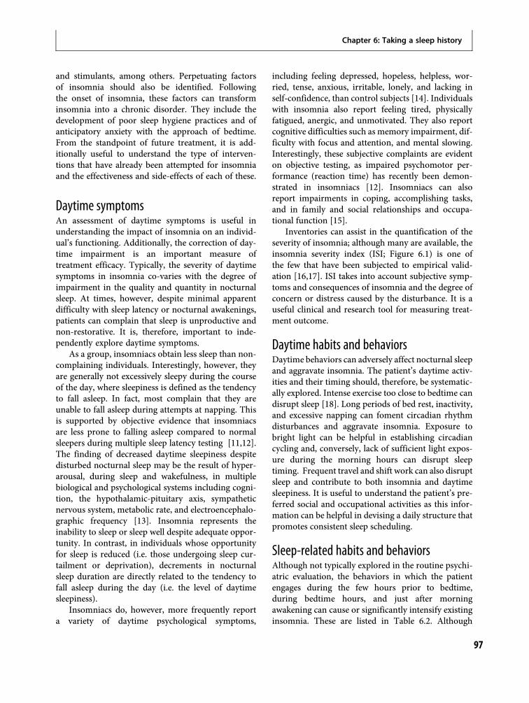
and stimulants, among others. Perpetuating factorsof insomnia should also be identified. Followingthe onset of insomnia, these factors can transforminsomnia into a chronic disorder. They include thedevelopment of poor sleep hygiene practices and ofanticipatory anxiety with the approach of bedtime.From the standpoint of future treatment, it is add-itionally useful to understand the type of interven-tions that have already been attempted for insomniaand the effectiveness and side-effects of each of these.
Daytime symptomsAn assessment of daytime symptoms is useful inunderstanding the impact of insomnia on an individ-ual’s functioning. Additionally, the correction of day-time impairment is an important measure oftreatment efficacy. Typically, the severity of daytimesymptoms in insomnia co-varies with the degree ofimpairment in the quality and quantity in nocturnalsleep. At times, however, despite minimal apparentdifficulty with sleep latency or nocturnal awakenings,patients can complain that sleep is unproductive andnon-restorative. It is, therefore, important to inde-pendently explore daytime symptoms.
As a group, insomniacs obtain less sleep than non-complaining individuals. Interestingly, however, theyare generally not excessively sleepy during the courseof the day, where sleepiness is defined as the tendencyto fall asleep. In fact, most complain that they areunable to fall asleep during attempts at napping. Thisis supported by objective evidence that insomniacsare less prone to falling asleep compared to normalsleepers during multiple sleep latency testing [11,12].The finding of decreased daytime sleepiness despitedisturbed nocturnal sleep may be the result of hyper-arousal, during sleep and wakefulness, in multiplebiological and psychological systems including cogni-tion, the hypothalamic-pituitary axis, sympatheticnervous system, metabolic rate, and electroencephalo-graphic frequency [13]. Insomnia represents theinability to sleep or sleep well despite adequate oppor-tunity. In contrast, in individuals whose opportunityfor sleep is reduced (i.e. those undergoing sleep cur-tailment or deprivation), decrements in nocturnalsleep duration are directly related to the tendency tofall asleep during the day (i.e. the level of daytimesleepiness).
Insomniacs do, however, more frequently reporta variety of daytime psychological symptoms,
including feeling depressed, hopeless, helpless, wor-ried, tense, anxious, irritable, lonely, and lacking inself-confidence, than control subjects [14]. Individualswith insomnia also report feeling tired, physicallyfatigued, anergic, and unmotivated. They also reportcognitive difficulties such as memory impairment, dif-ficulty with focus and attention, and mental slowing.Interestingly, these subjective complaints are evidenton objective testing, as impaired psychomotor per-formance (reaction time) has recently been demon-strated in insomniacs [12]. Insomniacs can alsoreport impairments in coping, accomplishing tasks,and in family and social relationships and occupa-tional function [15].
Inventories can assist in the quantification of theseverity of insomnia; although many are available, theinsomnia severity index (ISI; Figure 6.1) is one ofthe few that have been subjected to empirical valid-ation [16,17]. ISI takes into account subjective symp-toms and consequences of insomnia and the degree ofconcern or distress caused by the disturbance. It is auseful clinical and research tool for measuring treat-ment outcome.
Daytime habits and behaviorsDaytime behaviors can adversely affect nocturnal sleepand aggravate insomnia. The patient’s daytime activ-ities and their timing should, therefore, be systematic-ally explored. Intense exercise too close to bedtime candisrupt sleep [18]. Long periods of bed rest, inactivity,and excessive napping can foment circadian rhythmdisturbances and aggravate insomnia. Exposure tobright light can be helpful in establishing circadiancycling and, conversely, lack of sufficient light expos-ure during the morning hours can disrupt sleeptiming. Frequent travel and shift work can also disruptsleep and contribute to both insomnia and daytimesleepiness. It is useful to understand the patient’s pre-ferred social and occupational activities as this infor-mation can be helpful in devising a daily structure thatpromotes consistent sleep scheduling.
Sleep-related habits and behaviorsAlthough not typically explored in the routine psychi-atric evaluation, the behaviors in which the patientengages during the few hours prior to bedtime,during bedtime hours, and just after morningawakening can cause or significantly intensify existinginsomnia. These are listed in Table 6.2. Although
Chapter 6: Taking a sleep history
97
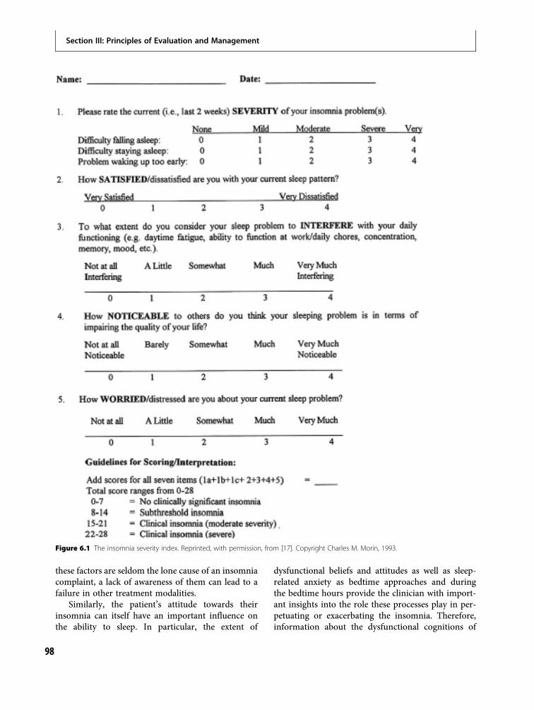
these factors are seldom the lone cause of an insomniacomplaint, a lack of awareness of them can lead to afailure in other treatment modalities.
Similarly, the patient’s attitude towards theirinsomnia can itself have an important influence onthe ability to sleep. In particular, the extent of
dysfunctional beliefs and attitudes as well as sleep-related anxiety as bedtime approaches and duringthe bedtime hours provide the clinician with import-ant insights into the role these processes play in per-petuating or exacerbating the insomnia. Therefore,information about the dysfunctional cognitions of
Figure 6.1 The insomnia severity index. Reprinted, with permission, from [17]. Copyright Charles M. Morin, 1993.
Section III: Principles of Evaluation and Management
98

the insomniac such as catastrophic attributions of theeffects of insomnia, the time of day that the insom-niac begins to worry about sleep, and the state ofmind of the insomniac during time awake duringthe night should all be solicited.
Sleep patternsAlthough the assessment of bedtime and sleeppatterns is important in the evaluation of anysleep-related complaint, it is a particularly essentialcomponent of the evaluation of insomnia (Table 6.1).Patients should be asked regarding these parameterson initial and follow-up visits. These patterns can alsobe assessed by utilizing patient-completed sleep logsor diaries that track sleep–wake patterns over time(Figure 6.2). Sleep logs and diaries may be moreuseful than subjective summaries since insomniacstend to underestimate total sleep time and overesti-mate sleep latency, possibly resulting from a prefer-ential recall bias for particularly bad nights of sleepthat do not reflect the longitudinal course of theircomplaints [19].
Ideally, individuals retire and emerge from bed atconsistent times day to day, including weekends. Thetime spent in bed between retiring and falling asleep
would preferably be less than 20 minutes, and indi-viduals would emerge from bed soon after awakeningin the morning. Additionally, awakenings in goodsleepers are typically limited to one or two, andtime spent in bed following awakenings kept to aminimum (i.e. less than 15 minutes). Of note,these variables may be affected by age [20]. Patientswith insomnia can have large discrepancies fromthese norms, and patterns may emerge that suggestspecific underlying causes of insomnia and/or helpguide treatment.
Characteristic disturbances in sleep–wake patternscan point to circadian rhythm disturbances such asdelayed sleep phase disorder, in which the time offalling asleep and the time of awakening are consist-ently delayed, and advanced sleep phase disorder, inwhich these times are consistently advanced, relativeto the desired night/day schedule (see Chapter 11:Circadian rhythm disorders). Multiple nocturnal awak-enings and prolonged sleep latencies are not specificfor any one disorder, yet keeping track of these par-ameters can help to quantify the severity of insomniaand determine the effectiveness of treatment meas-ures. Determining these patterns can also suggest theutility of certain behavioral interventions to improvesleep, such as restriction of overall time spent in bed.It is also useful to ascertain each of these parametersnot only for the “average” day, but also for a sequenceof days, as such a temporal record can demonstratevariability in sleep patterns over time which can, inturn, contribute to poor sleep. The assessment ofvariability between workdays/schooldays, weekends,and vacations can also be useful. Insomniacs charac-teristically do not maintain rigorous sleep/wakeschedules, and introducing such regularity into theirlives is one of the primary elements of sleep hygieneeducation and other cognitive behavioral techniques(see Chapter 14: Cognitive behavioral therapy forinsomnia).
Excessive sleepiness (ES): history of thechief complaintSleepiness, the tendency to fall asleep, is a normalphenomenon when it occurs at the desired time ofthe day. ES is the tendency to fall asleep at inappropri-ate times or settings [4]. ES should be distinguishedfrom fatigue, which is classically defined as the inabil-ity to sustain performance over time and is typicallyassociated with subjective reports of tiredness,
Table 6.2 Sleep related habits and behaviors that candisrupt sleep
Caffeine and alcohol prior to bedtime
Nicotine (both smoking and cessation)
Large meals or excessive fluid intake within 3 hours ofbedtime
Exercising within 3 hours of bedtime
Utilizing the bed for non-sleep activities (work,telephone, internet)
Staying in bed while awake for extended periods of time
Activating behaviors up to the point of bedtime
Excessive worrying at bedtime
Clock-watching prior to sleep onset or during nocturnalawakenings
Exposure to bright light prior to bedtime or duringawakenings
Keeping the bedroom too hot or too cold
Noise
Behaviors of a bed partner (e.g. snoring, leg movements)
Chapter 6: Taking a sleep history
99
weariness, exhaustion, and lack of energy [21]. Eventhough fatigued patients can perceive themselves asbeing excessively sleepy, in its pure form, fatigue doesnot result in an increased propensity to fall asleep.Fatigue is a symptom of a wide variety of medical,psychiatric, and neurological disorders and less spe-cific for sleep disorders than is ES. It has been mostwidely examined in the context of multiple sclerosis,autoimmune disorders, and psychiatric conditions(Figure 6.3). Also, a complaint of ES may co-occurwith that of hypersomnia, which is an abnormalincrease in time spent asleep or trying to sleep. In thissection, we will limit our discussion primarily to ES.
The time of onset, temporal pattern, duration, anddaily pattern of ES should be determined. ES onset inearly life is more consistent with narcolepsy, whereasonset in middle age is more consistent with OSA. ESthat is episodic over time is consistent with periods ofsleep restriction due to social and occupational needs.
However, ES that is constant and unremitting may bemore consistent with narcolepsy, OSA, or potentiallyaffective disorders. ES that is most prominent in themorning hours, especially when accompanied by aprolonged sleep latency and complaint of initiationinsomnia, can be due to delayed sleep phase syn-drome, whereas ES that is most prominent in the lateevening hours and associated with early morningawakening can be due to advanced sleep phase syn-drome. ES that is most prominent in the morninghours and gradually resolves as the day progresses isalso consistent with the carryover effects of a sedatingbedtime medication. Afternoon sleepiness is com-monly associated with disturbances of the qualityand quantity of nocturnal sleep, and the extent ofsleepiness in the afternoon is proportional to theextent of these disturbances.
The severity of ES should be quantified. Directquestioning regarding how sleepy an individual feels
Figure 6.2 Sleep log. Reproduced, with permission, from [5]. Copyright American Academy of Sleep Medicine, 2005.
Section III: Principles of Evaluation and Management
100

is generally not a highly useful measure of the degreeof sleepiness, since patients can over-report or under-report the sensation of sleepiness, compared to theirbehavioral tendency to nod off or fall asleep duringactivities of daily life. Individuals who are exposed tolong periods of sleep deprivation are especially proneto under-recognize the severity of their sleepiness,possibly owing to their becoming accustomed to thisstate. Therefore, behavioral assessments of ES may bemore useful. Behavioral indicators of ES includeyawning, ptosis, reduced activity, lapses in attention,and head nodding. The patient can also be questionedabout his/her tendency to fall asleep in situations ofeveryday life; milder levels of daytime sleepiness resultin falling asleep in passive situations such as whilereading, watching television, and attending meetings.In severe cases, individuals fall asleep while activelyengaged in complex tasks such as driving, speaking,writing, or even eating. Highly sleepy patients mayalso experience sleep attacks, episodes of sleep thatstrike without warning, whose occurrence mandatesrapid clinical intervention, in part due to significantlyincreased risk of motor vehicle accidents. However,all patients with a complaint of ES should be cau-tioned about the risks of drowsy driving.
A validated inventory for the quantification of thetendency to fall asleep is the Epworth sleepinessscale (ESS) [22]. It is widely utilized for the assessmentof the severity of ES and for the determination of treat-ment effects over time (Figure 6.4). The scores forindividual items are added, and the total score isreported. A score of 10 or above is considered to repre-sent an abnormally high level of daytime sleepiness.
Related symptomsPatients with ES may also complain of the manyconsequences of ES. In the evaluation of the sleepypatient, an exploration of these related complaintsmay be helpful in understanding the effects of ES onthe patient’s daily life. These consequences also repre-sent symptoms that should be followed clinically toensure that ES has been adequately managed.
Napping in patients with ES is common. If napsare reported, the possibility that they are related tonarcolepsy should be examined by determiningwhether they are refreshing, brief in duration, oraccompanied by dreams. Naps are also refreshing inindividuals who are sleep deprived. In contrast, theytend to not be refreshing in OSA and idiopathichypersomnia. The timing of naps should also bedetermined as this may alert the physician to thepossibility of circadian rhythm disorders.
Most of the sequelae of ES that have been exam-ined have fallen in the realm of psychomotor andattentional behaviors. These include slower responsetimes, cognitive slowing, performance errors, declinein both short-term recall and working memory,reduced learning of new tasks, increased responseperseveration on ineffective solutions, increased neg-lect of non-essential activities (loss of situationalawareness), increased compensatory effort to main-tain the same performance, and diminished insightinto subtle meanings [23]. ES can also contribute todepressed mood [24].
ES is associated with an increased risk for acci-dents at work and while driving [25] and has been
Statement*
1. My motivation is lower when I am fatigued. 2. Exercise brings on my fatigue. 3. I am easily fatigued. 4. Fatigue interferes with my physical functioning. 5. Fatigue causes frequent problems for me. 6. My fatigue prevents sustained physical functioning.7. Fatigue interferes with carrying out certain duties and responsibilities.8. Fatigue is among my three most disabling symptoms. 9. Fatigue interferes with my work, family, or social life.
*Patients are instructed to choose a number from 1 to 7 (1 = “strongly disagree” and7 = “strongly agree”) that indicates their degree of agreement with each statement.Reprinted with permission from Krupp et al. Arch Neurol. 1989; 46:1121–1123.Copyright 1989, American Medical Association. All rights reserved.
Figure 6.3 The fatigue severity scale.Copyright 1989, American MedicalAssociation.
Chapter 6: Taking a sleep history
101
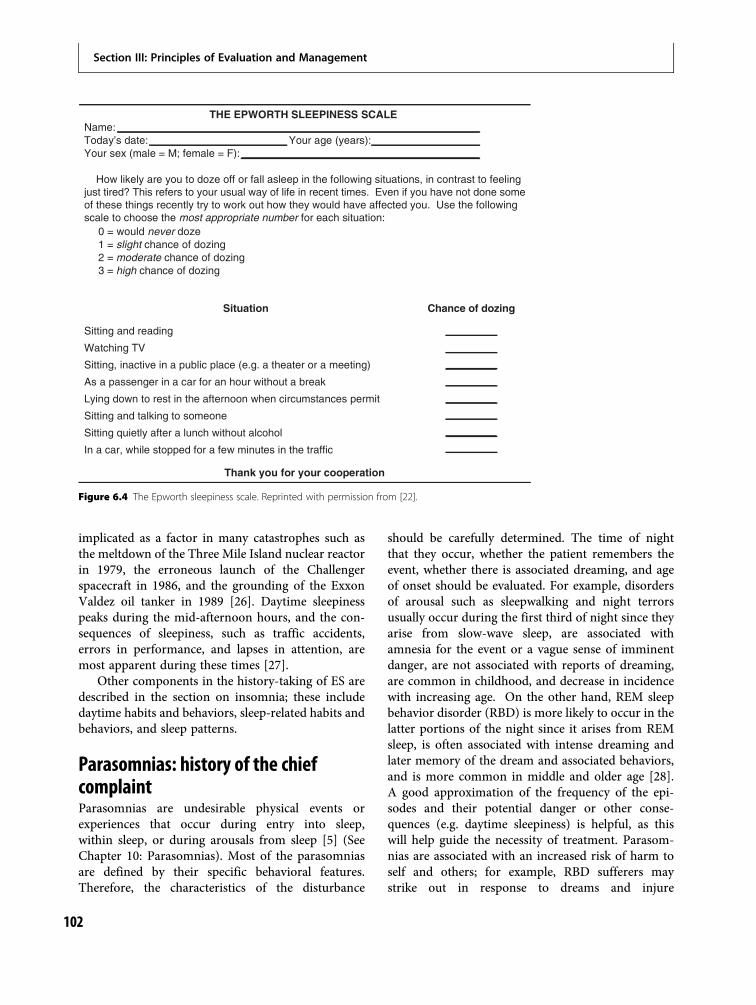
implicated as a factor in many catastrophes such asthe meltdown of the Three Mile Island nuclear reactorin 1979, the erroneous launch of the Challengerspacecraft in 1986, and the grounding of the ExxonValdez oil tanker in 1989 [26]. Daytime sleepinesspeaks during the mid-afternoon hours, and the con-sequences of sleepiness, such as traffic accidents,errors in performance, and lapses in attention, aremost apparent during these times [27].
Other components in the history-taking of ES aredescribed in the section on insomnia; these includedaytime habits and behaviors, sleep-related habits andbehaviors, and sleep patterns.
Parasomnias: history of the chiefcomplaintParasomnias are undesirable physical events orexperiences that occur during entry into sleep,within sleep, or during arousals from sleep [5] (SeeChapter 10: Parasomnias). Most of the parasomniasare defined by their specific behavioral features.Therefore, the characteristics of the disturbance
should be carefully determined. The time of nightthat they occur, whether the patient remembers theevent, whether there is associated dreaming, and ageof onset should be evaluated. For example, disordersof arousal such as sleepwalking and night terrorsusually occur during the first third of night since theyarise from slow-wave sleep, are associated withamnesia for the event or a vague sense of imminentdanger, are not associated with reports of dreaming,are common in childhood, and decrease in incidencewith increasing age. On the other hand, REM sleepbehavior disorder (RBD) is more likely to occur in thelatter portions of the night since it arises from REMsleep, is often associated with intense dreaming andlater memory of the dream and associated behaviors,and is more common in middle and older age [28].A good approximation of the frequency of the epi-sodes and their potential danger or other conse-quences (e.g. daytime sleepiness) is helpful, as thiswill help guide the necessity of treatment. Parasom-nias are associated with an increased risk of harm toself and others; for example, RBD sufferers maystrike out in response to dreams and injure
THE EPWORTH SLEEPINESS SCALE Name: Today’s date: Your age (years): Your sex (male = M; female = F):
How likely are you to doze off or fall asleep in the following situations, in contrast to feeling just tired? This refers to your usual way of life in recent times. Even if you have not done someof these things recently try to work out how they would have affected you. Use the followingscale to choose the most appropriate number for each situation:
0 = would never doze 1 = slight chance of dozing2 = moderate chance of dozing 3 = high chance of dozing
Situation Chance of dozing
Sitting and reading
Watching TV
Sitting, inactive in a public place (e.g. a theater or a meeting)
As a passenger in a car for an hour without a break
Lying down to rest in the afternoon when circumstances permit
Sitting and talking to someone
Sitting quietly after a lunch without alcohol
In a car, while stopped for a few minutes in the traffic
Thank you for your cooperation
Figure 6.4 The Epworth sleepiness scale. Reprinted with permission from [22].
Section III: Principles of Evaluation and Management
102

themselves or a bed partner. Therefore, an under-standing of the extent of such injurious behavior inthe past can be important in determining the urgencyof treatment and the implementation of safetyprecautions.
Collateral information from bedpartners and family membersMany of the symptoms and behaviors that are neces-sary for the completion of the history occur duringsleep, or during periods of extreme sleepiness, makingthe patient’s own account unreliable. Therefore, it ishelpful to obtain this information from bed partnersand family members. Examples of symptoms patientsmay underreport include:
1. Snoring2. Breathing pauses during sleep3. Unusual behaviors during sleep such as walking,
talking, thrashing, headbanging, body rocking,and limb movements
4. The tendency to fall asleep unintentionally duringthe day
5. The extent and frequency of naps6. Cognitive and behavioral disturbances associated
with insomnia and ES, such as diminished socialcontact, slow mannerisms, decrement in mood,lapses in memory, etc.
Past medical, psychiatric, and surgicalhistoryCo-morbid disorders should be reviewed, along withtheir dates of onset, types of treatment, and results oftreatment. Surgeries and hospitalizations should alsobe evaluated. Major medical disorders can affect sleepby virtue of their psychological impact, through painand discomfort, as well as direct effects on sleep andwakefulness. It is important to note that myriadpsychiatric illnesses are associated with sleep disturb-ance, and careful evaluation of the timing of thesleep complaint in relation to psychiatric symptomsmay be of high value in patients with psychiatricillness. For a review of psychiatric disorders and theireffects on sleep, please see Section VI of this book.
Additionally, the patient may have a history of aprimary sleep disorder, and review of prior documen-tation including results of polysomnographic studies
and other laboratory tests may be extremely helpful.For a review of primary sleep disorders, please seeSection IV of this volume.
MedicationsAhistory of current and priormedications is an integralpart of the history. The list should include not onlyprescribed medications, but over-the-counter agents,nutraceuticals, herbal substances, dietary supplements,and even foods. Their effects, side-effects, dosages orquantities, and timing of administration should berecorded. If the use of a medication correlates tempor-ally with the onset of the sleep complaint, a medication-induced sleep disorder should be suspected. Medica-tions can also have secondary effects by virtue of theirexacerbation of underlying conditions. For example,weight gain associated with medication use can lead tothe development of symptoms of OSA, such as ES,snoring, and breathing pauses during sleep. Allergiesto medications should also be recorded. The effectsof commonly used medications on sleep are reviewedin Chapter 3: Neurophysiology and neuroimaging ofhuman sleep.
SubstancesInsomnia, ES, and parasomnias can be related to sub-stance use. Chronic and excessive use of substancesmay lead to substance use disorders, which are charac-terized by either abuse or dependence. Stimulants suchas caffeine, amphetamines, and cocaine classically dis-rupt sleep. Sedatives such as opiates and analgesicscause ES. Alcohol, at low dosages, can help with sleepinitiation, yet chronic and excessive use can lead todisturbed sleep and the complaint of insomnia [29].The effects of substances on sleep are reviewed ingreater detail in Chapter 19: Sleep in substance usedisorders.
Family historyCertain sleep disorders, such as RLS, can have ahereditary component, in which 50% of primary caseshave a positive family history [30]. The incidence ofcertain parasomnias of arousal such as sleepwalkingand sleep terrors is ten times greater in first degreerelatives than in the general population [31]. OSAmay have a familial basis.
Chapter 6: Taking a sleep history
103
Social and occupational historySeveral social or occupational factors can contribute tosleep-related complaints, necessitating evaluation. Forexample, ES and insomnia are common in shiftworkers and individuals whose occupations requirefrequent travel across time zones. Exposure to indus-trial toxins and chemicals can also produce sleep/wakesymptoms. Job loss and retirement can result in theloss of regularity in daily schedule, which is importantin maintaining circadian rhythm consistency in someindividuals, leading to erratic sleep/wake hours and thecomplaints of insomnia and ES. Disruption in inter-personal relationships, family, job, and hobbies cancause anxiety and subsequent insomnia.
Differential diagnosisTable 6.3 outlines some of the more commonlyencountered sleep disorders that are related to insom-nia and ES. The differential diagnosis of the parasom-nias is discussed in Chapter 10: Parasomnias.
Several disorders may present with either insom-nia and/or ES, which may complicate the differentialdiagnosis. Thus, it is important for the practicingclinician to be familiar with some common symptoms
of sleep disorders, to aid in the evaluation and man-agement of sleep complaints.
Symptoms of specific disordersThe existence of certain symptoms in addition toinsomnia and/or ES can indicate the presence of spe-cific disorders as a cause of these complaints. Defin-ing symptoms for a selected list of sleep disorders arelisted in Table 6.4. Defining symptoms for the para-somnias is discussed in Chapter 10: Parasomnias.These symptoms should be systematically exploredto refine the diagnostic possibilities. Readers arereferred to Sections IV and VI of this book for a moredetailed discussion of each disorder and its hallmarksymptoms.
Physical examinationThe physical examination can contribute essentialinformation to the process of understanding the eti-ology of the sleep complaint. Vital signs should includethe measurement of neck circumference; a thick and/ormuscular neck, as well as a neck circumference of16 inches or greater in women and 17 inches or greaterin men, are associated with an increased risk for sleep-related breathing disorders [33]. Body habitus should beinspected; obesity with fat distribution around the neckor midriff suggests the diagnosis of OSA. Other con-tributors to sleep-related breathing disorders includenasal obstruction, mandibular hypoplasia, and retro-gnathia. Oropharyngeal abnormalities can also beinvolved, including enlarged tonsils and tongue, anelongated uvula and soft palate, diminished pharyngealpatency, and redundant pharyngeal mucosa. TheMallampati airway classification score should be deter-mined (Figure 6.5), which is useful in assessing the riskfor OSA. On average, for every 1-point increase in theMallampati score, the odds of having OSA increasesmore than two fold [34]. The chest examination shouldbe scrutinized for expiratory wheezes and kyphoscoli-osis, indicative of, among others, asthma and restrictivelung disease, respectively, which, in turn, can be associ-atedwith the complaint of insomnia. Signs of right heartfailure should be noted; heart failure can cause abnor-malities of breathing during sleep, which, in turn, can beassociated with the complaint of frequent nocturnalawakenings and unrefreshing sleep. A basic neuro-logical examination should be performed to rule outneurological disorders that may mimic certain sleepdisorders or which may co-exist with them. For
Table 6.3 Differential diagnosis of insomnia and ES
Disorder Insomnia ES
Inadequate sleep hygiene * *
Sleep restriction *
Adjustment sleep disorder *
Psychophysiological insomnia *
Obstructive sleep apnea syndrome * *
Central sleep apnea syndrome * *
Narcolepsy *
Periodic limb movement disorder * *
Idiopathic hypersomnia *
Restless legs syndrome * *
Drug-dependent and drug-inducedsleep disorders
* *
Circadian rhythm sleep disorders * *
Medical/psychiatric sleep disorders * *
Section III: Principles of Evaluation and Management
104

example, the presence of increased resting muscle tone,cogwheel rigidity, and tremor can indicate the presenceof Parkinson’s disease, which can share some of the
behavioral and sensory disturbances of REM behaviordisorder. Themental status examination should includean evaluation of affect, anxiety, psychomotor agitation
Table 6.4 Defining symptoms for selected disorders
Disorder Symptoms
Psychophysiological insomnia “Trying” to fall asleepDifficulty falling asleep at desired bedtimeFrequent nocturnal awakeningsAnxiety regarding sleeplessness
Restless legs syndrome1 Irresistible urge to move the extremitiesLimb paresthesiaeOnset of symptoms during periods of rest and in the evening or at bedtimeRelief of symptoms with movement
Periodic limb movement disorder Repetitive involuntary movements of the extremities during sleep or just prior tofalling asleep
Narcolepsy Excessive daytime sleepinessCataplexySleep paralysisHypnopompic and hypnagogic hallucinations
Obstructive sleep apneasyndrome
SnoringBreathing pauses during sleepChokingGaspingMorning dry mouth
Chronic obstructive pulmonarydisease
Dyspnea
Gastroesophageal reflux Epigastric pain or burningLaryngospasmAcid taste in mouthSudden nocturnal awakenings
Prostatic hypertrophy Frequent nocturia
Nocturnal seizures Thrashing in bedLoss of bladder or bowel control
Nocturnal panic attacks Sudden surges of anxietyTachycardiaDiaphoresisChokingLaryngospasm
Post-traumatic stress disorder Recurring, vivid dreams and nightmaresAnxiety and hypervigilance in sleep environment
Delayed sleep phase syndrome Inability to fall asleep at desired timeInability to awaken at desired time
Advanced sleep phase syndrome Inability to stay awake until the desired bedtimeInability to remain asleep until the desired awakening time
Note: 1 The URGE mnemonic has been proposed as a convenient way to recall most of the symptoms of RLS [32]. It includes the following:Urge to move; Rest induced; Gets better with activity; Evening and night accentuation.
Chapter 6: Taking a sleep history
105
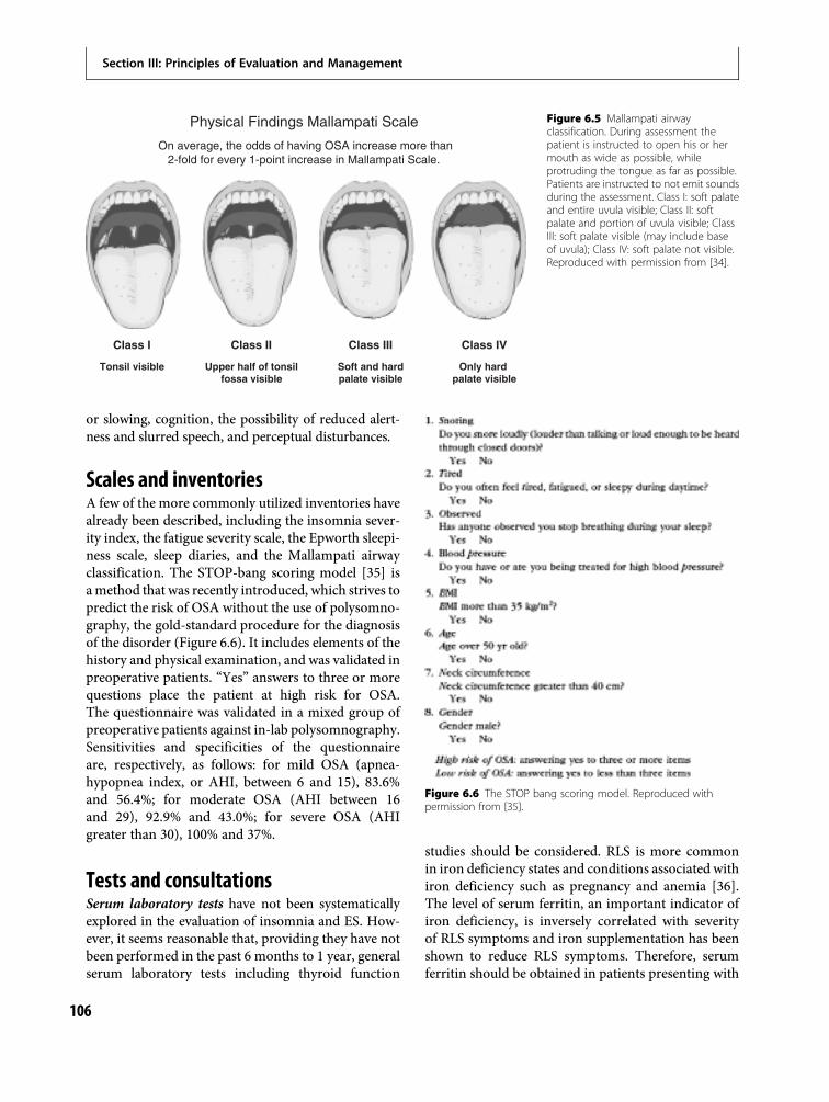
or slowing, cognition, the possibility of reduced alert-ness and slurred speech, and perceptual disturbances.
Scales and inventoriesA few of the more commonly utilized inventories havealready been described, including the insomnia sever-ity index, the fatigue severity scale, the Epworth sleepi-ness scale, sleep diaries, and the Mallampati airwayclassification. The STOP-bang scoring model [35] isamethod that was recently introduced, which strives topredict the risk of OSA without the use of polysomno-graphy, the gold-standard procedure for the diagnosisof the disorder (Figure 6.6). It includes elements of thehistory and physical examination, and was validated inpreoperative patients. “Yes” answers to three or morequestions place the patient at high risk for OSA.The questionnaire was validated in a mixed group ofpreoperative patients against in-lab polysomnography.Sensitivities and specificities of the questionnaireare, respectively, as follows: for mild OSA (apnea-hypopnea index, or AHI, between 6 and 15), 83.6%and 56.4%; for moderate OSA (AHI between 16and 29), 92.9% and 43.0%; for severe OSA (AHIgreater than 30), 100% and 37%.
Tests and consultationsSerum laboratory tests have not been systematicallyexplored in the evaluation of insomnia and ES. How-ever, it seems reasonable that, providing they have notbeen performed in the past 6 months to 1 year, generalserum laboratory tests including thyroid function
studies should be considered. RLS is more commonin iron deficiency states and conditions associated withiron deficiency such as pregnancy and anemia [36].The level of serum ferritin, an important indicator ofiron deficiency, is inversely correlated with severityof RLS symptoms and iron supplementation has beenshown to reduce RLS symptoms. Therefore, serumferritin should be obtained in patients presenting with
Physical Findings Mallampati Scale
On average, the odds of having OSA increase more than2-fold for every 1-point increase in Mallampati Scale.
Class I
Tonsil visible
Class II
Upper half of tonsil fossa visible
Class III
Soft and hard palate visible
Class IV
Only hard palate visible
Figure 6.5 Mallampati airwayclassification. During assessment thepatient is instructed to open his or hermouth as wide as possible, whileprotruding the tongue as far as possible.Patients are instructed to not emit soundsduring the assessment. Class I: soft palateand entire uvula visible; Class II: softpalate and portion of uvula visible; ClassIII: soft palate visible (may include baseof uvula); Class IV: soft palate not visible.Reproduced with permission from [34].
Figure 6.6 The STOP bang scoring model. Reproduced withpermission from [35].
Section III: Principles of Evaluation and Management
106

insomnia or ES and associated RLS symptoms, and alevel of 50 mg/L or less is considered significant.
Actigraphy utilizes small, wristwatch-like devicesto record movement. It assumes that lack of move-ment is equivalent to sleep, and is not, therefore,useful to measure exact sleep times. Although it isnot appropriate for the routine diagnosis of sleepdisorders, it can be useful as an adjunct to otherprocedures for the assessment of sleep–wake patternswhen such information is not reliably available byother means such as sleep logs. It can be appropriatefor the documentation of changes in sleep patternsover prolonged diagnosis and treatment periods; forthe assessment of whether an insomniac follows cer-tain sleep hygiene advice (e.g. to curtail time in bed orto regularize wake-up times) and of improvement insleep following behavioral treatment; and for theassessment of daytime and night-time sleep and day-time somnolence in ES disorders [37].
Polysomnography (PSG) is the technique of moni-toring multiple physiological measures during sleep,including brain waves, eye movements, heart rate,respirations, oxyhemoglobin saturation, and muscletone and activity. Video recording may be used toidentify abnormal movements during sleep. This testis typically performed in a sleep laboratory. Polysom-nography is appropriate to establish the diagnosiswhen the office-based evaluation raises the possibilityof a sleep-related breathing disorder (SRBD). It is alsoappropriate for a determination of the appropriatetreatment settings during continuous positive airwaypressure (CPAP) titration, and for the assessment ofSRDB treatment results. With multiple sleep latencytesting (see below), it is utilized for the diagnosis ofnarcolepsy. PSG is also utilized for the evaluation ofthe parasomnias which feature sleep-related behaviorsthat are violent or potentially injurious to the patientor others, to assist in the diagnosis of paroxysmalarousals or other sleep disruptions thought to beseizure related, and in a presumed parasomnia orsleep-related seizure disorder that does not respondto conventional therapy. It is also utilized for thediagnosis of periodic limb movement disorder [38].
Themultiple sleep latency test (MSLT) is a daytimepolysomnographic test whichmeasures the extent of ESby assessing the speed of onset of sleep in five daytimenap opportunities, beginning 1.5 to 3 hours afterawakening, and each separated by 2 hours. The patientlies down in a darkened room and is asked to not resistthe urge to fall asleep. The onset and stage of sleep
(including REM) are monitored. This test is used if thediagnosis of narcolepsy or idiopathic hypersomnia issuspected. A variation on theMSLT is themaintenanceof wakefulness test (MWT), in which the patient isasked to stay awake while sitting in a dimly lit bedroom.It is considered by many to be a more accurate measureof sleep tendency in situations of everyday living, whereindividuals are trying to stay awake, rather than fallasleep, during the day. It is utilized for the assessmentof individuals in whom the inability to remain awakeconstitutes a safety issue, and in patients with narco-lepsy or idiopathic hypersomnia to assess response totreatment with medications [39].
There are several situations in which sleep medi-cine consultations may be useful following the office-based evaluation. These include: when the diagnosis isin doubt, seeming adequate treatment of the pre-sumed disorder does not result in the alleviation ofsymptoms, or PSG/MSLT/MWT testing with follow-up treatment is desired.
Conclusions and recommendationsInsomnia, ES, and parasomnias are commonly encoun-tered in psychiatric practice. They are the hallmarksymptoms of a variety of sleep disorders, many ofwhich will be discussed in greater detail in the ensuingchapters. This chapter has focused on the critical firststeps in bridging symptom with disorder, namely thehistory and examination. These represent the corner-stones of the evaluation process. Below are some keyrecommendations that emerge from this chapter:
1. Develop a systematic process to address sleep-related complaints
2. Organize the clinical history around a typical24-hour sleep/wake pattern beginning withbedtime and ending back at the time at which theinquiry began and determine deviations fromnormal patterns
3. Obtain the nocturnal or diurnal pattern, onset andlongitudinal course, and severity of the chiefcomplaint
4. Understand the temporal relationship betweenthe chief complaint and potential precipitantssuch as psychosocial disruption, co-morbidillnesses, and prior treatments
5. Systematically explore sleep-related habitsand behaviors and maladaptive psychologicalreactions and cognitions that may foment andperpetuate sleep/wake disturbances
Chapter 6: Taking a sleep history
107
6. Appreciate the nature and extent ofconsequences of the chief complaint ondaytime functioning
7. Obtain collateral information from bed partners8. Ask for symptoms of specific sleep disorders9. Complete the essential elements of a generalmedical and psychiatric history gathering processand examination
10. Utilize inventories, tests, and consultations tocomplete the diagnostic picture
11. Arrive at a diagnostic formulation prior toresorting to treatment
References1. Sateia MJ, Doghramji K, Hauri PJ, Morin CM.
Evaluation of chronic insomnia. An American Academyof Sleep Medicine review. Sleep. 2000;23(2):243 308.
2. Morgenthaler TI, Lee Chiong T, Alessi C, et al. Practiceparameters for the clinical evaluation and treatmentof circadian rhythm sleep disorders. An AmericanAcademy of Sleep Medicine report. Sleep. 2007;30(11):1445 1459.
3. Schutte Rodin S, Broch L, Buysse D, Dorsey C,Sateia M. Clinical guideline for the evaluation andmanagement of chronic insomnia in adults. J Clin SleepMed. 2008;4(5):487 504.
4. Vaughn BV, O’Neill DF. Cardinal manifestations ofsleep disorders. In: Kryger M, Roth T, Dement WC, eds.Principles and practice of sleep medicine, 4th ed.Philadelphia: Elsevier; 2005:589.
5. American Academy of Sleep Medicine. Internationalclassification of sleep disorders, 2nd ed. Westchester, IL:American Academy of Sleep Medicine; 2005.
6. National Institutes of Health. National Institutes ofHealth state of the science conference statement onmanifestations and management of chronic insomnia inadults, june 13 15, 2005. Sleep. 2005;28(9):1049 1057.
7. Hohagen F, Rink K, Kappler C, et al. Prevalenceand treatment of insomnia in general practice:a longitudinal study. Eur Arch Psychiatry Clin Neurosci.1993;242(6):329 336.
8. Ford DE, Kamerow DB. Epidemiologic studyof sleep disturbances and psychiatric disorders.an opportunity for prevention? JAMA. 1989;262(11):1479 1484.
9. Ohayon MM, Roth T. Place of chronic insomnia in thecourse of depressive and anxiety disorders. J PsychiatrRes. 2003;37(1):9 15.
10. Ancoli Israel S, Roth T. Characteristics of insomnia inthe United States: Results of the 1991 National SleepFoundation survey. I. Sleep. 1999;22(Suppl 2):S347 353.
11. Stepanski E, Zorick F, Roehrs T, Young D, Roth T.Daytime alertness in patients with chronic insomniacompared with asymptomatic control subjects. Sleep.1988;11(1):54 60.
12. Edinger JD, Means MK, Carney CE, Krystal AD.Psychomotor performance deficits and their relation toprior nights’ sleep among individuals with primaryinsomnia. Sleep. 2008;31(5):599 607.
13. Buysse DJ, Germaine A, Moul D, Nofzinger EA.Insomnia. In: Buysse DJ, ed. Sleep disorders andpsychiatry. Arlington, VA: American PsychiatricPublishing, Inc.; 2005:31 84.
14. Kales JD, Kales A, Bixler EO, et al. Biopsychobehavioralcorrelates of insomnia, V: Clinical characteristicsand behavioral correlates. Am J Psychiatry. 1984;141(11):1371 1376.
15. Kuppermann M, Lubeck DP, Mazonson PD, et al.Sleep problems and their correlates in aworking population. J Gen Intern Med. 1995;10(1):25 32.
16. Bastien CH, Vallieres A, Morin CM. Validation ofthe insomnia severity index as an outcome measurefor insomnia research. Sleep Med. 2001;2(4):297 307.
17. Morin CM. Insomnia: psychological assessment andmanagement. New York: Guilford Press; 1993.
18. Stepanski EJ, Wyatt JK. Use of sleep hygiene in thetreatment of insomnia. Sleep Med Rev. 2003;7(3):215 225.
19. Carskadon MA, Dement WC, Mitler MM, et al. Selfreports versus sleep laboratory findings in 122drug free subjects with complaints of chronicinsomnia. Am J Psychiatry. 1976;133(12):1382 1388.
20. Cooke JR, Ancoli Israel S. Sleep and its disorders inolder adults. Psychiatr Clin North Am. 2006;29(4):1077 1093; abstract x xi.
21. Krupp LB, LaRocca NG, Muir Nash J, Steinberg AD.The fatigue severity scale. Application to patients withmultiple sclerosis and systemic lupus erythematosus.Arch Neurol. 1989;46(10):1121 1123.
22. Johns MW. A new method for measuring daytimesleepiness: The Epworth sleepiness scale. Sleep. 1991;14(6):540 545.
23. Wagner U, Gais S, Haider H, Verleger R, Born J.Sleep inspires insight. Nature. 2004;427(6972):352 355.
24. Blagrove M, Alexander C, Horne JA. The effects ofchronic sleep reduction on the performance of
Section III: Principles of Evaluation and Management
108
cognitive tasks sensitive to sleep deprivation.Appl Cogn Psychol. 1995;9:21 40.
25. Broughton R, Ghanem Q, Hishikawa Y, et al.Life effects of narcolepsy in 180 patients fromNorth America, Asia and Europe compared tomatched controls. Can J Neurol Sci. 1981;8(4):299 304.
26. National Commission on Sleep Disorders Research.Wake up America: A national sleep alert Report of theNational Commission on Sleep Disorders Research.Washington, DC: Department of Health and HumanServices, Superintendent of Documents, U.S.Government Printing Office; 1992;92.
27. Mitler MM, Carskadon MA, Czeisler CA, et al.Catastrophes, sleep, and public policy: Consensusreport. Sleep. 1988;11(1):100 109.
28. Mahowald MW, Schenck CH. REM sleep parasomnias.In: Kryger MH, Roth T, Dement WC, eds. Priniciplesand practice of sleep medicine, 4th ed. Philadelphia:Elsevier; 2005:897 916.
29. Roehrs T, Roth T. Sleep, sleepiness and alcohol use.Alcohol Res Health. 2001;25(2):101 109.
30. Montplaisir J, Allen R, Walters A, Ferini Strambi L.Restless legs syndrome and periodic limb movementsin sleep. In: Kryger M, Roth T, Dement W, eds. 4th ed.Philadelphia: Elsevier; 2005:839 852.
31. Kales A, Soldatos CR, Bixler EO, et al. Hereditaryfactors in sleepwalking and night terrors.Br J Psychiatry. 1980;137:111 118.
32. Hening W, Allen RP, Tenzer P, Winkelman JW.Restless legs syndrome: Demographics, presentation,and differential diagnosis. Geriatrics. 2007;62(9):26 29.
33. Millman RP, Carlisle CC, McGarvey ST, Eveloff SE,Levinson PD. Body fat distribution and sleep apneaseverity in women. Chest. 1995;107(2):362 366.
34. Nuckton TJ, Glidden DV, Browner WS, Claman DM.Physical examination: Mallampati score as anindependent predictor of obstructive sleep apnea.Sleep. 2006;29(7):903 908.
35. Chung F, Yegneswaran B, Liao P, et al. STOPquestionnaire: A tool to screen patients forobstructive sleep apnea. Anesthesiology. 2008;108(5):812 821.
36. Sun ER, Chen CA, Ho G, Earley CJ, Allen RP. Ironand the restless legs syndrome. Sleep. 1998;21(4):A371 377.
37. Sadeh A, Hauri PJ, Kripke DF, Lavie P. The role ofactigraphy in the evaluation of sleep disorders. Sleep.1995;18(4):288 302.
38. Kushida CA, Littner MR, Morgenthaler T, et al.Practice parameters for the indications forpolysomnography and related procedures: An updatefor 2005. Sleep. 2005;28(4):499 521.
39. Littner MR, Kushida C, Wise M, et al. Practiceparameters for clinical use of the multiple sleep latencytest and the maintenance of wakefulness test. Sleep.2005;28(1):113 121.
Chapter 6: Taking a sleep history
109


Section IVChapter
7Primary Sleep Disorders in Psychiatric Contexts
Sleep-related breathing disorders
In Soo Lee and Joel E. Dimsdale
IntroductionAlthough the historical description of obstructivesleep apnea goes back to at least the time of CharlesDickens’ Pickwick Papers, the first real case report waspublished roughly 50 years ago in the American Jour-nal of Medicine. It is interesting to read this rich casereport and to note the powerful confluence of psychi-atric and cardiopulmonary symptoms in sleep apnea.The case describes a sleepy, obese patient who was amiddle-aged business executive, troubled by anxietiesand simmering frustrations, who did not requestmedical attention, despite severe fatigue and func-tional limitations. He didn’t request evaluation, thatis, until a sentinel event occurred in his life. He was alife-long poker player and when he fell asleep afterhaving been dealt a “full house,” he decided some-thing needed to be done [1].
That setting of admixture of fatigue, forgetfulness,anxiety, and depressive symptoms is quite common inpatients with obstructive sleep apnea (OSA). This chap-ter reviews the association between sleep-relatedbreathing disorders (SRBDs) and cognitive functioning,depression, and anxiety. We also briefly review theeffects of psychotropicmedications thatmay be particu-larly important to consider in a setting of SRBD.
Sleep-related breathing disordersSleep-related breathing disorders are characterized bydisturbed respiration during sleep. These respiratoryevents may range from recurrent mild upper airwayconstriction (called upper airway resistance syndrome)to recurrent total obstruction. SRBDs are subdividedinto those with obstruction (obstructive sleep apnea)and those with diminished or absent respiratory effort(central sleep apnea). In addition, there may be a
mixture of these components (mixed type). Withinthese categories, there is a continuum in the degree ofobstruction and in the frequency of these obstructive orcentral events. A great deal of work has gone intodefining the epidemiology of these disturbances andtheir associated risks and pathophysiologies.Obstructive sleep apnea (OSA) is a chronic conditioncharacterized by repetitive upper airway obstructionduring sleep leading to apneic episodes, hypoxemia,and recurrent arousals from sleep [2]. It has been esti-mated that 2% to 4% of middle-aged men and 1% to 2%of middle-aged women suffer from OSA [3]. OSA isparticularly prevalent in later life [4], and in obesemen [5,6], although its existence in women [7], as wellas in lean individuals, is increasingly recognized [3].
Risk factors for OSA include obesity and cranio-facial or upper airway soft tissue abnormalities.Common craniofacial or upper airway soft tissueabnormalities that increase the risk of OSA includealterations in mandibular or maxillary size and pos-ition, narrowed nasal cavities, as well as tonsillarhypertrophy [8]. The Mallampati classification is auseful tool for assessing the probability of sleep dis-ordered breathing. The Mallampati classification isbased on the structures visualized with maximalmouth opening and tongue protrusion. The originalgoal of this classification was to assess whether theupper airway could be readily visualized during tra-cheal intubation [9] (see Figure 7.1). However, Liistroand colleagues showed that a high Mallampati score(stage 3 or 4) and nasal obstruction are associated riskfactors for OSA [10].
Obesity is the most important reversible riskfactor for OSA [11]. The prevalence of OSA amongmorbidly obese patients is 12- to 30-fold higher thanin the general population [12]. Other risk factors for
Foundations of Psychiatric SleepMedicine, ed. JohnW.Winkelman andDavid T. Plante. Published by CambridgeUniversity Press.# Cambridge University Press 2010.
111

OSA include genetic factors, smoking, menopause,and alcohol use [13].
The cardinal symptom of OSA is excessive daytimesleepiness, which appears to be related to recurrentarousals from sleep associated with obstructive events[14]. Other associated signs and symptoms includesnoring, unrefreshing sleep, nocturnal choking (ordreams about choking or drowning), witnessedapneas, nocturia, morning headaches, mild cognitiveimpairment, and reduced libido [2]. Diagnosis is con-firmed by assessment of sleep, typically via polysom-nography. While exact cut points are arbitrary, a meanof 5–15 obstructive events per hour of sleep representsmild dysfunction, 15–30 events per hour representsmoderate dysfunction, and >30 events per hourrepresents severe dysfunction [15]. Treatment optionsfor OSA include weight loss, including discontinu-ation of weight-increasing medications, positionaltherapy, continuous positive airway pressure (CPAP),and mandibular advancement devices or surgeryfor clearly identifiable causes of upper airway ob-struction [14]. The first line of therapy is CPAP,but its efficacy can be limited partially because adher-ence may be poor [5,16]. Weaver and colleagues
suggested “good” adherence should be defined as atleast 6 hours a night [17].
OSA is associated with considerable morbidityand mortality, particularly from hypertension, cardio-vascular disease, and insulin resistance [16]. Further-more, the excessive daytime sleepiness associated withOSA can result in decreased quality of life [18] andincreased risk for automobile accidents [19] or ser-ious industrial accidents [20].
Psychiatric presentations of OSAOSA patients complain of various neuropsychiatricsymptoms. Cognitive impairment [21] and affectivedisorders such as depression are frequently encoun-tered in OSA. In addition, a high prevalence of otherpsychiatric symptoms such as anxiety, somatization,ADHD-type, and obsessive-compulsive symptomshave been reported in these patients [22,23]. Clinicianscan also encounter nocturnal panic attacks, diverseparasomnias, delirium, psychosis, personality change,and violent outbursts in some OSA patients [24–30].Therefore, practicing psychiatrists and psychologistsmust consider OSA in the differential diagnosis ofa large number of psychiatric presentations.
Cognitive and performanceimpairments in OSAApproximately 80% of OSA patients complain of bothexcessive daytime sleepiness and cognitive impair-ments, and half report personality changes [31]. Inaddition to significant negative consequences on qual-ity of life and auto vehicle safety, the neuropsycho-logical effects of OSA can aggravate scholastic andoccupational achievement as well as contribute to rela-tionship problems.
Considerable research has examined neuro-psychological deficits associated with OSA. Beebeand colleagues conducted a meta-analysis of 25 recentstudies (involving >2000 patients) to examine pat-terns of neuropsychological deficits in OSA. Theyfound impairments in vigilance, executive function,and motor coordination but no effect of OSA ongeneral intelligence and verbal ability. The effects ofOSA on visual and motor skill and memory function-ing were inconsistent [32]. In a separate review, Aloiaand colleagues reported that 60% of reviewed studiesfound deficits in attention and vigilance, executivefunctioning, and memory impairment, and 80% of
Figure 7.1 The Mallampati classification. The class is determinedby looking at the oral cavity as the patient protrudes the tongue,and tongue size is described relative to oropharyngeal size. Thesubsequent classification is based on the pharyngeal structuresthat are visible. Class 1: full visibility of tonsils, uvula, and soft palate;Class 2: visibility of hard and soft palate, upper portion of tonsils,and uvula; Class 3: soft and hard palate and base of the uvula arevisible; Class 4: only hard palate visible. Modified after MallampatiSR, Gatt SP, Gugino LD, et al. A clinical sign to predict difficulttracheal intubation: a prospective study. Can Anaesth Soc J.1985;32:429 434. Reprinted from [167].
Section IV: Primary Sleep Disorders in Psychiatric Contexts
112
reviewed studies found visuoconstruction and psy-chomotor functioning impairments [33]. Below,we review selected studies to give a sense for the typesof neuropsychological probes that have been appliedin this area.
Sustained attention or vigilanceSustained attention is one of the most commonlyaffected cognitive problems for OSA patients [34–39].It is assessed by tests such as the psychomotor vigilancetask (PVT). OSA patients initially perform comparablyto normal controls during short-duration tasks, butperformance degrades with longer duration tasks,where one sees increased response time, lapses or failureto respond, and false responses [40,41]. Operating amotor vehicle requires sustained vigilance, and impair-ments in this cognitive function may contribute toelevated rate of car accidents. OSA patients with exces-sive daytime sleepiness are 6 to 10 times more likelyto have an accident than non-sleepy controls [42].The reaction time of patients with mild to moderatesleep disordered breathing is worse than that found inhealthy, non-sleepy subjects with blood alcohol levels of0.080 g/dL (the typical legal limit for intoxicated drivingin the USA) [43].
MemoryThere is less consistency in studies of short-term andlong-term memory functioning in OSA patients[32,34,44,45]. Bedard and colleagues described dimin-ished performance on short-term memory in patientswith moderate and severe sleep apnea, although onlythe severe group demonstrated evidence of impairmentin delayed recall. These memory disturbances wereassociated with a decrease in vigilance [34]. Othershave reported short- and long-term memory problems[38,46–49]. On the other hand, Greenberg and col-leagues found no differences between patients withOSA and controls on subscales of theWechslermemoryscale (in either immediate or delayed conditions) [37].
Differences in samples such as level of disease sever-ity, clinic versus population-based studies, or the use ofnormal controls versus norm-referenced comparisonsmay account for the inconsistencies. These inconsist-encies may also reflect impairment in organization andretrieval caused by different levels of executive functiondeficits in OSA patients [32].
Executive functionsExecutive function refers to a set of higher cognitiveabilities that control and regulate other basic abilitieslike attention, memory, and motor skills. Executivefunctions may involve the ability to engage in goal-directed behaviors and abstract thinking. Executivefunction impairments can be measured by assessingdiverse domains such as planning, sequencing, self-monitoring, set-shifting, verbal fluency, abstractreasoning, working memory, visual–spatial organiza-tion, and memory [50–52].
Many studies have examined disturbances inexecutive function in OSA, demonstrating impair-ment in several domains. For example, Bedard andcolleagues reported widespread deficits in variousexecutive functions (verbal fluency, planning, sequen-tial thinking, and constructional ability), with extentof impairment related to severity of the breathingabnormality [34,53].
Etiology and mechanism of cognitiveand performance impairmentsThe pathogenesis of cognitive deficits in OSA is con-troversial and most likely multifactorial. The twomost commonly implicated etiological mechanismsare repetitive sleep fragmentation and nocturnal hyp-oxemia. However, the evidence is as yet tenuouslinking either measure of OSA severity and any cog-nitive domain [33].
Researchers have assumed that neuropsychologicaltests reflect abnormalities in specific brain regions.The evidence of disturbance in executive functionshas led to the suggestion that OSA may be associatedwith frontal lobe dysfunction [34,36]. In this context,the prefrontal model has been proposed as a concep-tual framework for the relationship between sleepdisruption and nocturnal hypoxemia and primarilyfrontal deficits in OSA. This model hypothesizes thatOSA-related sleep disruption and intermittent hypox-emia as well as hypercarbia alter the brain’s restorativeprocesses, thereby inducing a variety of cellular andbiochemical stresses that disrupt functional homeo-stasis as well as neuronal and glial viability withinthe prefrontal cortex [54] (see Figure 7.2).
Animal studies demonstrate that intermittentexposure to lowO2 damages hippocampal CA1 regionsand frontal cortex. Such damage has been correlatedwith cognitive deficits [55], especially learning and
Chapter 7: Sleep-related breathing disorders
113
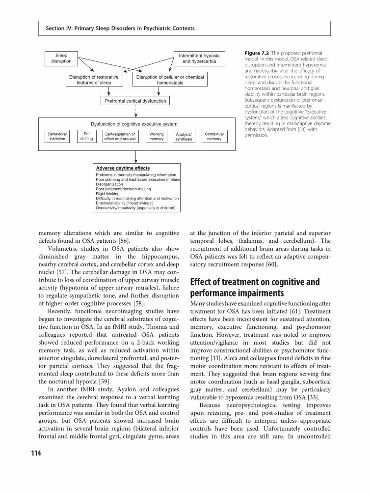
memory alterations which are similar to cognitivedefects found in OSA patients [56].
Volumetric studies in OSA patients also showdiminished gray matter in the hippocampus,nearby cerebral cortex, and cerebellar cortex and deepnuclei [57]. The cerebellar damage in OSA may con-tribute to loss of coordination of upper airway muscleactivity (hypotonia of upper airway muscles), failureto regulate sympathetic tone, and further disruptionof higher-order cognitive processes [58].
Recently, functional neuroimaging studies havebegun to investigate the cerebral substrates of cogni-tive function in OSA. In an fMRI study, Thomas andcolleagues reported that untreated OSA patientsshowed reduced performance on a 2-back workingmemory task, as well as reduced activation withinanterior cingulate, dorsolateral prefrontal, and poster-ior parietal cortices. They suggested that the frag-mented sleep contributed to these deficits more thanthe nocturnal hypoxia [59].
In another fMRI study, Ayalon and colleaguesexamined the cerebral response to a verbal learningtask in OSA patients. They found that verbal learningperformance was similar in both the OSA and controlgroups, but OSA patients showed increased brainactivation in several brain regions (bilateral inferiorfrontal and middle frontal gyri, cingulate gyrus, areas
at the junction of the inferior parietal and superiortemporal lobes, thalamus, and cerebellum). Therecruitment of additional brain areas during tasks inOSA patients was felt to reflect an adaptive compen-satory recruitment response [60].
Effect of treatment on cognitive andperformance impairmentsMany studies have examined cognitive functioning aftertreatment for OSA has been initiated [61]. Treatmenteffects have been inconsistent for sustained attention,memory, executive functioning, and psychomotorfunction. However, treatment was noted to improveattention/vigilance in most studies but did notimprove constructional abilities or psychomotor func-tioning [33]. Aloia and colleagues found deficits in finemotor coordination more resistant to effects of treat-ment. They suggested that brain regions serving finemotor coordination (such as basal ganglia, subcorticalgray matter, and cerebellum) may be particularlyvulnerable to hypoxemia resulting from OSA [33].
Because neuropsychological testing improvesupon retesting, pre- and post-studies of treatmenteffects are difficult to interpret unless appropriatecontrols have been used. Unfortunately controlledstudies in this area are still rare. In uncontrolled
Sleepdisruption
Disruption of restorativefeatures of sleep
Prefrontal cortical dysfunction
Adverse daytime effects
Dysfunction of cognitive executive system
Behavioralinhibition
Setshifting
Self-regulation ofaffect and arousal
Problems in mentally manipulating informationPoor planning and haphazard execution of plansDisorganizationPoor judgment/decision-makingRigid thinkingDifficulty in maintaining attention and motivationEmotional lability ('mood swings')Overactivity/impulsivity (especially in children)
Workingmemory
Contextualmemory
Analysis/synthesis
Disruption of cellular or chemicalhomeostasis
Intermittent hypoxiaand hypercarbia
Figure 7.2 The proposed prefrontalmodel. In this model, OSA related sleepdisruption and intermittent hypoxemiaand hypercarbia alter the efficacy ofrestorative processes occurring duringsleep, and disrupt the functionalhomeostasis and neuronal and glialviability within particular brain regions.Subsequent dysfunction of prefrontalcortical regions is manifested bydysfunction of the cognitive “executivesystem,” which alters cognitive abilities,thereby resulting in maladaptive daytimebehaviors. Adapted from [54], withpermission.
Section IV: Primary Sleep Disorders in Psychiatric Contexts
114
studies, CPAP treatment had a moderate to largeeffect on cognitive processing, memory, sustainedattention, and executive functions [38]. However,other studies show persistent cognitive deficits despitetreatment [62,63].
The majority of controlled clinical trials evaluatingthe efficacy of CPAP treatment have enrolled predom-inantly moderate to severe OSA patients. In mild OSApatients, the only improvement in quality of life after8 weeks of CPAP treatment was in vitality [64]. Somestudies suggest that beneficial CPAP treatment effectsfor cognition might be attributable to changes in theunderlying level of daytime sleepiness [64,65].
Bardwell and colleagues evaluated the effectivenessof 1-week CPAP treatment versus placebo-CPAP (i.e.CPAP administered at subtherapeutic pressure) on cog-nitive functioning in patients with OSA. AlthoughCPAP improved overall cognitive functioning, no bene-ficial effects in any specific domain were found. But,only 1 of the 22 neuropsychological test scores (DigitVigilance–Time, a measure of speed of informationprocessing, vigilance, or sustained attention and alert-ness) showed significant changes specific toCPAP treat-ment [66]. In their replication study with 2 weeks ofCPAP treatment, two-thirds of neuropsychological testscores improved with time regardless of treatment;however, once again only Digit Vigilance–Time showedsignificant improvement specific to CPAP treatment.They concluded that Digit Vigilance–Timemight be themost sensitive neuropsychological test for measuringthe effects of CPAP. Furthermore, 2 weeks of CPAPtreatmentmight be sufficient to improve speed of infor-mation processing, vigilance, as well as sustained atten-tion and alertness [67].
Evaluating the evidence for the efficacy of treatmenton the cognitive and performance impairments inOSA is challenging because of differences in length oftreatment, adherence to treatment, type of control, andplacebo employed. It is as yet unclear how long treat-ment should be provided in order to document efficacy.What is the time course of response? What cognitivefunctions respond first? Are there certain cognitivefunctions that improve only after weeks of treatment?Future research is required to clarify these issues.
Clinical implications of cognitiveimpairments in OSABeebe and colleagues recommended that sleep clin-icians should routinely assess cognitive symptoms of
OSA patients. If residual deficits are suspectedfollowing treatment, cognitive evaluations of vigi-lance, executive functioning, and motor functioningshould be undertaken. They also suggest interestinglythat undiagnosed sleep disordered breathing shouldbe suspected in individuals who display evidence ofpoor vigilance, or executive dysfunction [32].
Depression in OSADepression is the most commonly encountered affect-ive disorder associated with OSA [68]. The prevalenceof depression in OSA has ranged from 7% to 63% [69].Early investigations by Guilleminault and colleaguesreported that 24% of male patients with OSA hadpreviously seen a psychiatrist for anxiety and depres-sion [70]. A recent epidemiological study of 18 980subjects representative of the general population inthe UK, Germany, Italy, Portugal, and Spain foundthat 17.6% of subjects with a Diagnostic and StatisticalManual of Mental Disorders IV (DSM-IV) breathing-related sleep disorder diagnosis (by history, withoutsleep study) have major depressive disorder (MDD)[71]. Thus, some co-morbidity between SRBD anddepression has long been observed. The variations inthe prevalence of depression are thought to be affectedby sampling characteristics, mood assessmentmethods, and diagnostic difficulty due to an overlapof symptoms across depression and OSA.
A number of studies, using varied designs,report an association between OSA and depression,based on elevated co-morbidity of OSA and MDD[30,70,72–74], as well as increased levels of depressivesymptoms [75–80] that do not necessarily reflect amajor depressive episode [73]. Other studies observea correlation between apnea-hypopnea index (AHI;number of apneas and hypopneas per hour of sleep)and depression [81], with increased depressivesymptoms found in association with more severeSRBD [82], and improvement of depressive symp-toms after treatment with CPAP, uvulopalatopharyn-goplasty, or tracheostomy [74,76,77,79,83]. Peppardand colleagues found a dose–response associationbetween SRBD severity (AHI) and depression in theirpopulation-based, longitudinal investigation (seeFigure 7.3). Figure 7.3 compares the rates of depres-sion found amongst individuals with differing levelsof respiratory disturbance. Depression was defined asa score of 50 or higher on the Zung depression scalein individuals who were not receiving antidepressant
Chapter 7: Sleep-related breathing disorders
115
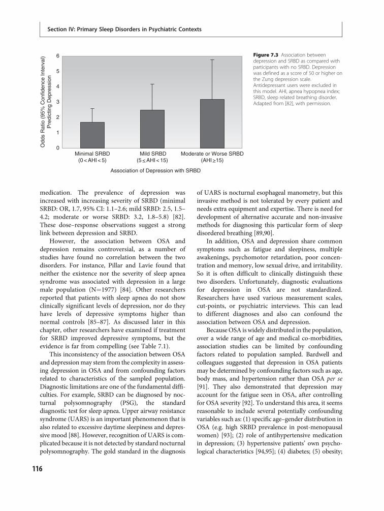
medication. The prevalence of depression wasincreased with increasing severity of SRBD (minimalSRBD: OR, 1.7, 95% CI: 1.1–2.6; mild SRBD: 2.5, 1.5–4.2; moderate or worse SRBD: 3.2, 1.8–5.8) [82].These dose–response observations suggest a stronglink between depression and SRBD.
However, the association between OSA anddepression remains controversial, as a number ofstudies have found no correlation between the twodisorders. For instance, Pillar and Lavie found thatneither the existence nor the severity of sleep apneasyndrome was associated with depression in a largemale population (N¼1977) [84]. Other researchersreported that patients with sleep apnea do not showclinically significant levels of depression, nor do theyhave levels of depressive symptoms higher thannormal controls [85–87]. As discussed later in thischapter, other researchers have examined if treatmentfor SRBD improved depressive symptoms, but theevidence is far from compelling (see Table 7.1).
This inconsistency of the association between OSAand depressionmay stem from the complexity in assess-ing depression in OSA and from confounding factorsrelated to characteristics of the sampled population.Diagnostic limitations are one of the fundamental diffi-culties. For example, SRBD can be diagnosed by noc-turnal polysomnography (PSG), the standarddiagnostic test for sleep apnea. Upper airway resistancesyndrome (UARS) is an important phenomenon that isalso related to excessive daytime sleepiness and depres-sive mood [88]. However, recognition of UARS is com-plicated because it is not detected by standard nocturnalpolysomnography. The gold standard in the diagnosis
of UARS is nocturnal esophageal manometry, but thisinvasive method is not tolerated by every patient andneeds extra equipment and expertise. There is need fordevelopment of alternative accurate and non-invasivemethods for diagnosing this particular form of sleepdisordered breathing [89,90].
In addition, OSA and depression share commonsymptoms such as fatigue and sleepiness, multipleawakenings, psychomotor retardation, poor concen-tration and memory, low sexual drive, and irritability.So it is often difficult to clinically distinguish thesetwo disorders. Unfortunately, diagnostic evaluationsfor depression in OSA are not standardized.Researchers have used various measurement scales,cut-points, or psychiatric interviews. This can leadto different diagnoses and also can confound theassociation between OSA and depression.
BecauseOSA iswidely distributed in the population,over a wide range of age and medical co-morbidities,association studies can be limited by confoundingfactors related to population sampled. Bardwell andcolleagues suggested that depression in OSA patientsmay be determined by confounding factors such as age,body mass, and hypertension rather than OSA per se[91]. They also demonstrated that depression mayaccount for the fatigue seen in OSA, after controllingfor OSA severity [92]. To understand this area, it seemsreasonable to include several potentially confoundingvariables such as: (1) specific age–gender distribution inOSA (e.g. high SRBD prevalence in post-menopausalwomen) [93]; (2) role of antihypertensive medicationin depression; (3) hypertensive patients’ own psycho-logical characteristics [94,95]; (4) diabetes; (5) obesity;
0
1
2
3
4
5
6
Minimal SRBD(0 < AHI < 5)
Moderate or Worse SRBD(AHI _>15)
Mild SRBD(5 <_ AHI < 15)
Odd
s R
atio
(95
% C
onfid
ence
Inte
rval
)P
redi
ctin
g D
epre
ssio
n
Association of Depression with SRBD
Figure 7.3 Association betweendepression and SRBD as compared withparticipants with no SRBD. Depressionwas defined as a score of 50 or higher onthe Zung depression scale.Antidepressant users were excluded inthis model. AHI, apnea hypopnea index;SRBD, sleep related breathing disorder.Adapted from [82], with permission.
Section IV: Primary Sleep Disorders in Psychiatric Contexts
116

Table 7.1 The effect of CPAP treatment on depression and anxiety in patients with obstructive sleep apnea
Reference(year)
Numberof subjects
Study design CPAP treatmenttime
Effect on mood
Borak et al.(1996) [22]
20 Prospective, uncontrolledstudy
3 months and12 months
No improvements in emotionalstatus after 3 and 12 months
Engleman et al.(1997) [168]
16 Prospective, placebo-controlled, randomized,crossover study
4 weeks Significant decline in HAD-depression
Engleman et al.(1998) [169]
23 Prospective, randomized,single blind, placebocontrolled, crossover study
4 weeks No changes in HAD
Jokic et al. (1998)[170]
10 Prospective, controlled,single blind crossoverstudy
4 weeks Use of CPAP was associated with adecrease in HAD-anxiety scores
Redline et al.(1998) [64]
97 Randomized, controlledstudy
8 weeks CPAP showed beneficial effect overmechanical nasal ventilator in POMSand PANAS scores
Engleman et al.(1999) [171]
34 Randomized, placebocontrolled, crossover study
4 weeks Compared with placebo, CPAPimproved depression score in HAD
Kingshott et al.(2000) [182]
62 Prospective, uncontrolledstudy
6 months Significant decline in HAD-depression and anxiety
Munoz et al.(2000) [114]
80 Prospective, controlledstudy
12 months CPAP failed to modify anxiety anddepression in Beck tests
Yamamoto et al.(2000) [172]
47 Prospective, uncontrolledstudy
2 years Significant decrease in SDS
Sanchez et al.(2001) [122]
51 Prospective, uncontrolledstudy
1 month and3 months
Significant decrease in BDI and STAI(depression and anxiety-trait) afterboth 1 month and 3 months
Barnes et al.(2002) [173]
28 Randomized, controlled,crossover study
8 weeks No benefit of CPAP on BDI andPOMS scores
Doherty et al.(2003) [174]
45 Prospective, uncontrolledstudy
8 weeks Significant decline in HAD-depression and anxiety
Means et al.(2003) [83]
39 Prospective, uncontrolledstudy
3 months Significant decrease in BDI
Svaldi et al.(2003) [175]
54 Prospective, uncontrolledstudy
6 9 weeks Significant decrease in BDI
Barnes et al.(2004) [176]
114 Randomized, controlled,crossover study
3 months Improvements of depression wereno better than placebo effect
Mackinger et al.(2004) [177]
54 Prospective, controlledstudy
2 months Significant decrease in BDI inpatients with previous depression
Goncalves et al.(2005) [178]
34 Prospective, controlledstudy
4 weeks Significant decrease in BDI
Schwartz et al.(2005) [179]
50 Prospective, uncontrolledstudy
4 6 weeks Significant decrease in BDI
Chapter 7: Sleep-related breathing disorders
117
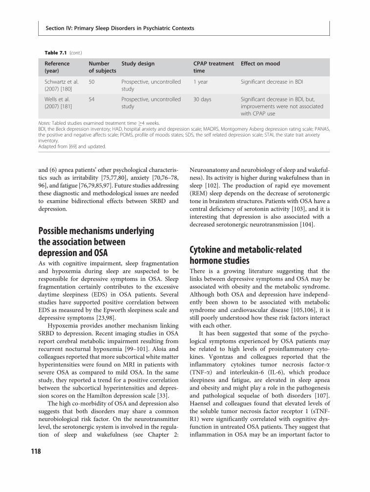
and (6) apnea patients’ other psychological characteris-tics such as irritability [75,77,80], anxiety [70,76–78,96], and fatigue [76,79,85,97]. Future studies addressingthese diagnostic and methodological issues are neededto examine bidirectional effects between SRBD anddepression.
Possible mechanisms underlyingthe association betweendepression and OSAAs with cognitive impairment, sleep fragmentationand hypoxemia during sleep are suspected to beresponsible for depressive symptoms in OSA. Sleepfragmentation certainly contributes to the excessivedaytime sleepiness (EDS) in OSA patients. Severalstudies have supported positive correlation betweenEDS as measured by the Epworth sleepiness scale anddepressive symptoms [23,98].
Hypoxemia provides another mechanism linkingSRBD to depression. Recent imaging studies in OSAreport cerebral metabolic impairment resulting fromrecurrent nocturnal hypoxemia [99–101]. Aloia andcolleagues reported that more subcortical white matterhyperintensities were found on MRI in patients withsevere OSA as compared to mild OSA. In the samestudy, they reported a trend for a positive correlationbetween the subcortical hyperintensities and depres-sion scores on the Hamilton depression scale [33].
The high co-morbidity of OSA and depression alsosuggests that both disorders may share a commonneurobiological risk factor. On the neurotransmitterlevel, the serotonergic system is involved in the regula-tion of sleep and wakefulness (see Chapter 2:
Neuroanatomy and neurobiology of sleep and wakeful-ness). Its activity is higher during wakefulness than insleep [102]. The production of rapid eye movement(REM) sleep depends on the decrease of serotonergictone in brainstem structures. Patients with OSA have acentral deficiency of serotonin activity [103], and it isinteresting that depression is also associated with adecreased serotonergic neurotransmission [104].
Cytokine and metabolic-relatedhormone studiesThere is a growing literature suggesting that thelinks between depressive symptoms and OSA may beassociated with obesity and the metabolic syndrome.Although both OSA and depression have independ-ently been shown to be associated with metabolicsyndrome and cardiovascular disease [105,106], it isstill poorly understood how these risk factors interactwith each other.
It has been suggested that some of the psycho-logical symptoms experienced by OSA patients maybe related to high levels of proinflammatory cyto-kines. Vgontzas and colleagues reported that theinflammatory cytokines tumor necrosis factor-a(TNF-a) and interleukin-6 (IL-6), which producesleepiness and fatigue, are elevated in sleep apneaand obesity and might play a role in the pathogenesisand pathological sequelae of both disorders [107].Haensel and colleagues found that elevated levels ofthe soluble tumor necrosis factor receptor 1 (sTNF-R1) were significantly correlated with cognitive dys-function in untreated OSA patients. They suggest thatinflammation in OSA may be an important factor to
Table 7.1 (cont.)
Reference(year)
Numberof subjects
Study design CPAP treatmenttime
Effect on mood
Schwartz et al.(2007) [180]
50 Prospective, uncontrolledstudy
1 year Significant decrease in BDI
Wells et al.(2007) [181]
54 Prospective, uncontrolledstudy
30 days Significant decrease in BDI, but,improvements were not associatedwith CPAP use
Notes: Tabled studies examined treatment time �4 weeks.BDI, the Beck depression inventory; HAD, hospital anxiety and depression scale; MADRS, Montgomery Asberg depression rating scale; PANAS,the positive and negative affects scale; POMS, profile of moods states; SDS, the self related depression scale; STAI, the state trait anxietyinventory.Adapted from [69] and updated.
Section IV: Primary Sleep Disorders in Psychiatric Contexts
118
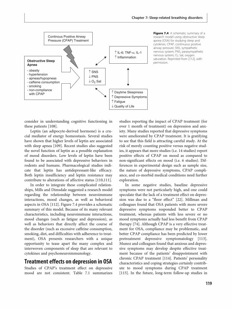
consider in understanding cognitive functioning inthese patients [108].
Leptin (an adipocyte-derived hormone) is a cru-cial mediator of energy homeostasis. Several studieshave shown that higher levels of leptin are associatedwith sleep apnea [109]. Recent studies also suggestedthe novel function of leptin as a possible explanationof mood disorders. Low levels of leptin have beenfound to be associated with depressive behaviors inrodents and humans. Pharmacological studies indi-cate that leptin has antidepressant-like efficacy.Both leptin insufficiency and leptin resistance maycontribute to alterations of affective status [110,111].
In order to integrate these complicated relation-ships, Mills and Dimsdale suggested a research modelregarding the relationship between neuroimmuneinteractions, mood changes, as well as behavioralaspects in OSA [112]. Figure 7.4 provides a schematicsummary of this model. Because of its many relevantcharacteristics, including neuroimmune interactions,mood changes (such as fatigue and depression), aswell as behaviors that directly affect the course ofthe disorder (such as excessive caffeine consumption,smoking, diet, and difficulties with adherence to treat-ment), OSA presents researchers with a uniqueopportunity to tease apart the many complex andinterwoven components of sleep that are relevant tocytokines and psychoneuroimmunology.
Treatment effects on depression in OSAStudies of CPAP’s treatment effect on depressivemood are not consistent. Table 7.1 summarizes
studies reporting the impact of CPAP treatment (forover 1 month of treatment) on depression and anx-iety. Many studies reported that depressive symptomswere ameliorated by CPAP treatment. It is gratifyingto see that this field is attracting careful study. At therisk of merely counting positive versus negative stud-ies, it appears that more studies (i.e. 14 studies) reportpositive effects of CPAP on mood as compared tonon-significant effects on mood (i.e. 6 studies). Dif-ferences in experimental design such as sample size,the nature of depressive symptoms, CPAP compli-ance, and co-morbid medical conditions need furtherexploration.
In some negative studies, baseline depressivesymptoms were not particularly high, and one couldspeculate that the lack of a treatment effect on depres-sion was due to a “floor effect” [22]. Millman andcolleagues found that OSA patients with more severedepressive symptoms responded better to CPAPtreatment, whereas patients with less severe or nomood symptoms actually had less benefit from CPAPtherapy [74]. Although CPAP is a very effective treat-ment for OSA, compliance may be problematic, andbetter CPAP compliance has been predicted by lowerpretreatment depressive symptomatology [113].Munoz and colleagues found that anxious and depres-sive symptoms may develop despite effective treat-ment because of the patients’ disappointment withchronic CPAP treatment [114]. Patients’ personalitycharacteristics and coping strategies certainly contrib-ute to mood symptoms during CPAP treatment[115]. In the future, long-term follow-up studies in
Continous Positive AirwayPressure (CPAP) Treatment
Obstructive SleepApnea
obesityhypertensionapneas/hypopneascaffeine consumptionsmokingnon-compliancewith CPAP
------
↑ IL-6; TNF-α; IL-1↑ Inflammation
↑ SNS↓ PNS↓ O2 Sat
↑ Daytime Sleepiness↑ Depressive Symptoms↑ Fatigue↓ Quality of Life
Figure 7.4 A schematic summary of aresearch model using obstructive sleepapnea (OSA) for studying sleep andcytokines. CPAP, continuous positiveairway pressure; SNS, sympatheticnervous system; PNS, parasympatheticnervous system; O2 Sat, oxygensaturation. Reprinted from [112], withpermission.
Chapter 7: Sleep-related breathing disorders
119
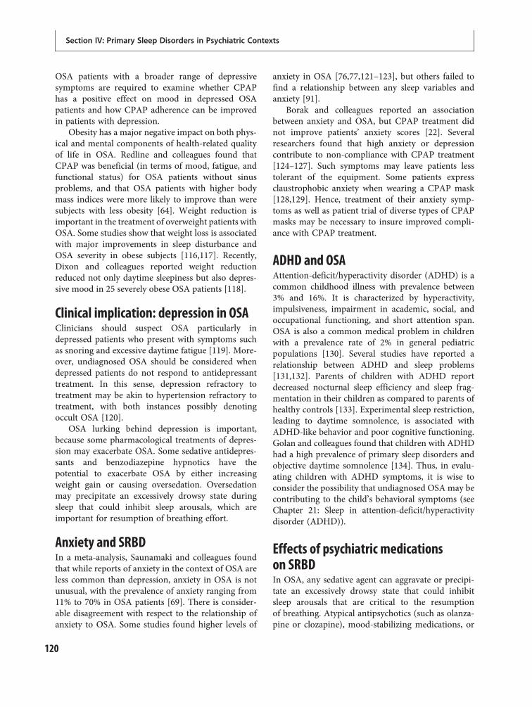
OSA patients with a broader range of depressivesymptoms are required to examine whether CPAPhas a positive effect on mood in depressed OSApatients and how CPAP adherence can be improvedin patients with depression.
Obesity has a major negative impact on both phys-ical and mental components of health-related qualityof life in OSA. Redline and colleagues found thatCPAP was beneficial (in terms of mood, fatigue, andfunctional status) for OSA patients without sinusproblems, and that OSA patients with higher bodymass indices were more likely to improve than weresubjects with less obesity [64]. Weight reduction isimportant in the treatment of overweight patients withOSA. Some studies show that weight loss is associatedwith major improvements in sleep disturbance andOSA severity in obese subjects [116,117]. Recently,Dixon and colleagues reported weight reductionreduced not only daytime sleepiness but also depres-sive mood in 25 severely obese OSA patients [118].
Clinical implication: depression in OSAClinicians should suspect OSA particularly indepressed patients who present with symptoms suchas snoring and excessive daytime fatigue [119]. More-over, undiagnosed OSA should be considered whendepressed patients do not respond to antidepressanttreatment. In this sense, depression refractory totreatment may be akin to hypertension refractory totreatment, with both instances possibly denotingoccult OSA [120].
OSA lurking behind depression is important,because some pharmacological treatments of depres-sion may exacerbate OSA. Some sedative antidepres-sants and benzodiazepine hypnotics have thepotential to exacerbate OSA by either increasingweight gain or causing oversedation. Oversedationmay precipitate an excessively drowsy state duringsleep that could inhibit sleep arousals, which areimportant for resumption of breathing effort.
Anxiety and SRBDIn a meta-analysis, Saunamaki and colleagues foundthat while reports of anxiety in the context of OSA areless common than depression, anxiety in OSA is notunusual, with the prevalence of anxiety ranging from11% to 70% in OSA patients [69]. There is consider-able disagreement with respect to the relationship ofanxiety to OSA. Some studies found higher levels of
anxiety in OSA [76,77,121–123], but others failed tofind a relationship between any sleep variables andanxiety [91].
Borak and colleagues reported an associationbetween anxiety and OSA, but CPAP treatment didnot improve patients’ anxiety scores [22]. Severalresearchers found that high anxiety or depressioncontribute to non-compliance with CPAP treatment[124–127]. Such symptoms may leave patients lesstolerant of the equipment. Some patients expressclaustrophobic anxiety when wearing a CPAP mask[128,129]. Hence, treatment of their anxiety symp-toms as well as patient trial of diverse types of CPAPmasks may be necessary to insure improved compli-ance with CPAP treatment.
ADHD and OSAAttention-deficit/hyperactivity disorder (ADHD) is acommon childhood illness with prevalence between3% and 16%. It is characterized by hyperactivity,impulsiveness, impairment in academic, social, andoccupational functioning, and short attention span.OSA is also a common medical problem in childrenwith a prevalence rate of 2% in general pediatricpopulations [130]. Several studies have reported arelationship between ADHD and sleep problems[131,132]. Parents of children with ADHD reportdecreased nocturnal sleep efficiency and sleep frag-mentation in their children as compared to parents ofhealthy controls [133]. Experimental sleep restriction,leading to daytime somnolence, is associated withADHD-like behavior and poor cognitive functioning.Golan and colleagues found that children with ADHDhad a high prevalence of primary sleep disorders andobjective daytime somnolence [134]. Thus, in evalu-ating children with ADHD symptoms, it is wise toconsider the possibility that undiagnosed OSA may becontributing to the child’s behavioral symptoms (seeChapter 21: Sleep in attention-deficit/hyperactivitydisorder (ADHD)).
Effects of psychiatric medicationson SRBDIn OSA, any sedative agent can aggravate or precipi-tate an excessively drowsy state that could inhibitsleep arousals that are critical to the resumptionof breathing. Atypical antipsychotics (such as olanza-pine or clozapine), mood-stabilizing medications, or
Section IV: Primary Sleep Disorders in Psychiatric Contexts
120

antiepileptic drugs (such as valproic acid and carba-mazepine), antidepressants (such as tricyclic anti-depressants), and benzodiazepine hypnotics have thepotential to exacerbate OSA by either increasingweight gain or causing oversedation.
AntipsychoticsPatients taking chronic antipsychotic medicationsmay gain 15–75 lb (6.8–33.8 kg) over the first 2 yearsof treatment, and 35–50% of such patients maybecome clinically obese [135]. Winkelman reportedthat obesity, male gender, and chronic neurolepticadministration are risk factors for OSA in psychiatricpatients. He also reported that OSA in schizophrenicpatients was severe, with a mean respiratory disturb-ance index (RDI) of 64.8 events per hour [136]. Sincepatients with schizophrenia are often on long-termneuroleptic treatment, they may have high rates ofOSA, aggravated by the weight gain produced by suchmedications.
Although OSA in schizophrenics might contributeto cardiovascular disease and increased mortality, thecondition is underdiagnosed, partially because of con-fusion between daytime sleepiness and negativesymptoms or medication side-effects.
AntidepressantsSeveral animal studies suggest that serotonin isimportant in the maintenance of upper airwaypatency. Serotonergic neurons (mediated predomin-antly via 5-HT2A and 5-HT2C receptor subtypes) exertan excitatory effect on upper airway dilator motorneurons [137,138]. When serotonin delivery isreduced to upper airway dilator motor neurons insleep, this contributes to reductions in dilator muscleactivity and upper airway obstruction.
Administration of paroxetine increased activity ofupper airway dilator muscle during wakefulness innormal subjects [139] and during non-REM sleepin patients with OSA [140], which suggested that theSSRI may be effective in treating OSA. However,in clinical studies, serotonergic drugs such as protrypti-line, fluoxetine, paroxetine, and mirtazapine have beentested as treatments forOSA,with limited improvementand significant side-effects [141,142]. Administration ofthe serotonin precursor, L-tryptophan, was reported tobe effective in decreasing obstructive apneas in non-REM sleep in an uncontrolled study of 12 patients withOSA [143]. However, L-tryptophan was previously
withdrawn from themarket as a result of reports linkingtryptophan use with eosinophilic myalgia syndromeand life-threatening pulmonary hypertension. The sero-tonin option for treatment of OSA remains an intri-guing, novel theoretical approach, but one that has notyet been proven effective.
SSRIs are widely used to treat depressive moodand anxiety in OSA patients. Moreover, SSRIs maybe at least partially (and theoretically) effective inOSA by virtue of their REM sleep suppression (asOSA tends to worsen during REM sleep when muscletone is lowest and the upper airway is most prone toobstruct) and/or augmentation of upper airway dila-tor muscle activity. While SSRIs are relatively toler-able in OSA, antidepressants with side-effects ofsedation or weight gain (such as antidepressants withH1 receptor blockade property) may exacerbate OSA.
Sedative hypnoticsExperts recommend caution in prescribing hypnoticsto patients with severe OSA, especially those with day-time hypoventilation or hypercapneic chronic obstruct-ive pulmonary disease (COPD), unless such patientsare simultaneously effectively treated with CPAP.Benzodiazepines can decrease respiratory effort, upperairway muscle tone, and blunt arousal responses tohypoxia/hypercapnia [144,145]. They may thus worsensleep apnea [146]. Long-acting benzodiazepines such asflurazepam [146] and nitrazepam [147] produce themost marked respiratory depression. Thus, it is recom-mended that benzodiazepine hypnotics be used cau-tiously in OSA patients with compromised respiratoryfunction [148]. If sedative hypnotic therapy is needed inpatients with OSA, it is very important for clinicians toensure CPAP adherence by periodic monitoring ofcompliance. Berry and colleagues reported that triazo-lam 0.25mg increases the arousal threshold to airwayocclusion, but this results in only modest prolongationof event duration and modestly increased desaturationin severe sleep apnea patients [149].
Non-benzodiazepine hypnotics (such as zolpidem,zopiclone, and zaleplon) generally produce fewer effectson respiratory depression in patients with OSA [150].Many previous studies demonstrated that zolpidemdoes not considerably affect respiratory parameters inpatients with COPD [151–154] or OSA [155,156].Zopiclone and eszopiclone also did not cause deterior-ation in respiratory parameters (SaO2, apnea index) inpatients with UARS [157,158] or with mild to moderate
Chapter 7: Sleep-related breathing disorders
121
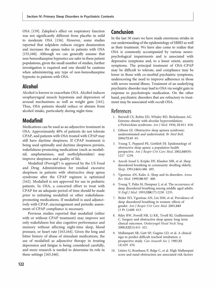
OSA [159]. Zaleplon’s effect on respiratory functionwas not significantly different from placebo in mildto moderate OSA [156]. However, some studiesreported that zolpidem reduces oxygen desaturationand increases the apnea index in patients with OSA[155,160]. Although we can generally assume thatnon-benzodiazepine hypnotics are safer in these patientpopulations, given the small number of studies, furtherinvestigation is required and one should be cautiouswhen administering any type of non-benzodiazepinehypnotic to patients with OSA.
AlcoholAlcohol is known to exacerbate OSA. Alcohol inducesoropharyngeal muscle hypotonia and depression ofarousal mechanisms as well as weight gain [161].Thus, OSA patients should reduce or abstain fromalcohol intake, particularly during night-time.
ModafinilMedications can be used as an adjunctive treatment inOSA. Approximately 40% of patients do not tolerateCPAP, and patients with OSA treated with CPAP maystill have daytime sleepiness. If CPAP treatment isbeing used optimally and daytime sleepiness persists,wakefulness-promoting medications (such as modafi-nil, amphetamines, and methylphenidate) mayimprove sleepiness and quality of life.
Modafinil (Provigil®) is approved by the US Foodand Drug Administration for residual excessivesleepiness in patients with obstructive sleep apneasyndrome after the CPAP regimen is optimized[162]. Modafinil is not approved for use in pediatricpatients. In OSA, a concerted effort to treat withCPAP for an adequate period of time should be madeprior to initiating modafinil or other wakefulness-promoting medications. If modafinil is used adjunct-ively with CPAP, encouragement and periodic assess-ment of CPAP compliance is necessary.
Previous studies reported that modafinil (eitherwith or without CPAP treatment) may improve notonly wakefulness but also cognitive performance andmemory without affecting night-time sleep, bloodpressure, or heart rate [163,164]. Given the long andbitter history of abuse of stimulant medications, theuse of modafinil as adjunctive therapy in treatingdepression and fatigue is being considered carefully,and more research is needed to determine its role inthese settings [165,166].
ConclusionIn the last 50 years we have made enormous strides inour understanding of the epidemiology of SRBD as wellas their treatment. We have also come to realize thatOSA is commonly accompanied by various neuro-psychological impairments and is associated withdepressive symptoms and, to a lesser extent, anxietysymptoms. The principal treatment of OSA–CPAPmay be difficult to tolerate, and compliance may belower in those with co-morbid psychiatric symptoms,underscoring the need to improve adherence in thosewith severe mental illness. Treatment of an underlyingpsychiatric disorder may lead to OSA via weight gain inresponse to psychotropic medication. On the otherhand, psychiatric disorders that are refractory to treat-ment may be associated with occult OSA.
References1. Burwell CS, Robin ED, Whaley RD, Bickelmann AG.
Extreme obesity with alveolar hypoventilation:a Pickwickian syndrome. Am J Med. 1956; 21:811 818.
2. Gibson GJ. Obstructive sleep apnoea syndrome:underestimated and undertreated. Br Med Bull.2004;72:49 65.
3. Young T, Peppard PE, Gottlieb DJ. Epidemiology ofobstructive sleep apnea: a population healthperspective. Am J Respir Crit Care Med. 2002;165(9):1217 1239.
4. Ancoli Israel S, Kripke DF, Klauber MR, et al. Sleepdisordered breathing in community dwelling elderly.Sleep. 1991;14(6):486 495.
5. Vgontzas AN, Kales A. Sleep and its disorders. AnnuRev Med. 1999;50:387 400.
6. Young T, Palta M, Dempsey J, et al. The occurrence ofsleep disordered breathing among middle aged adults.N Engl J Med. 1993;328(17):1230 1235.
7. Bixler EO, Vgontzas AN, Lin HM, et al. Prevalence ofsleep disordered breathing in women: effects ofgender. Am J Respir Crit Care Med. 2001;163(3 Pt 1):608 613.
8. Riley RW, Powell NB, Li KK, Troell RJ, GuilleminaultC. Surgery and obstructive sleep apnea: long termclinical outcomes. Otolaryngol Head Neck Surg.2000;122(3):415 421.
9. Mallampati SR, Gatt SP, Gugino LD, et al. A clinicalsign to predict difficult tracheal intubation: aprospective study. Can Anaesth Soc J. 1985;32(4):429 434.
10. Liistro G, Rombaux P, Belge C, et al. High Mallampatiscore and nasal obstruction are associated risk factors
Section IV: Primary Sleep Disorders in Psychiatric Contexts
122

for obstructive sleep apnoea. Eur Respir J. 2003;21(2):248 252.
11. Malhotra A, White DP. Obstructive sleep apnoea.Lancet. 2002;360(9328):237 245.
12. Peiser J, Lavie P, Ovnat A, Charuzi I. Sleep apneasyndrome in the morbidly obese as an indication forweight reduction surgery.Ann Surg. 1984;199(1):112 115.
13. Young T, Skatrud J, Peppard PE. Risk factors forobstructive sleep apnea in adults. JAMA. 2004;291(16):2013 2016.
14. Caples SM, Gami AS, Somers VK. Obstructive sleepapnea. Ann Intern Med. 2005;142(3):187 197.
15. American Academy of Sleep Medicine. Sleep relatedbreathing disorders in adults: recommendations forsyndrome definition and measurement techniques inclinical research: The Report of a Task Force. Sleep.1999;22(5):667 689.
16. Pack AI. Advances in sleep disordered breathing.Am J Respir Crit Care Med. 2006;173(1):7 15.
17. Weaver TE, Kribbs NB, Pack AI, et al. Night to nightvariability in CPAP use over the first three months oftreatment. Sleep. 1997;20(4):278 283.
18. Jenkinson C, Stradling J, Petersen S. Comparison ofthree measures of quality of life outcome in theevaluation of continuous positive airways pressuretherapy for sleep apnoea. J Sleep Res. 1997;6(3):199 204.
19. George CF, Nickerson PW, Hanly PJ, Millar TW,Kryger MH: Sleep apnoea patients have moreautomobile accidents. Lancet. 1987;2(8556):447.
20. Dinges DF. An overview of sleepiness and accidents.J Sleep Res. 1995;4(S2):4 14.
21. Kezirian EJ, Harrison SL, Ancoli Israel S, et al.Behavioral correlates of sleep disordered breathing inolder women. Sleep. 2007;30(9):1181 1188.
22. Borak J, Cieslicki JK, Koziej M, Matuszewski A,Zielinski J. Effects of CPAP treatment on psychologicalstatus in patients with severe obstructive sleep apnoea.J Sleep Res. 1996;5(2):123 127.
23. Yue W, Hao W, Liu P, et al. A case control study onpsychological symptoms in sleep apnea hypopneasyndrome. Can J Psychiatry. 2003;48(5):318 323.
24. Edlund MJ, McNamara ME, Millman RP. Sleep apneaandpanic attacks.Compr Psychiatry. 1991;32(2):130 132.
25. Enns MW, Stein M, Kryger M. Successful treatment ofcomorbid panic disorder and sleep apnea withcontinuous positive airway pressure. Psychosomatics.1995;36(6):585 586.
26. Whitney JF, Gannon DE. Obstructive sleep apneapresenting as acute delirium.AmJEmergMed. 1996;14(3):270 271.
27. Lee JW. Recurrent delirium associated with obstructivesleep apnea. Gen Hosp Psychiatry. 1998;20(2):120 122.
28. Berrettini WH. Paranoid psychosis and sleep apneasyndrome. Am J Psychiatry. 1980;137(4):493 494.
29. Lee S, Chiu HF, Chen CN. Psychosis in sleep apnoea.Aust N Z J Psychiatry. 1989;23(4):571 573.
30. Sharafkhaneh A, Giray N, Richardson P, Young T,Hirshkowitz M. Association of psychiatric disordersand sleep apnea in a large cohort. Sleep. 2005;28(11):1405 1411.
31. Guilleminault C, Hoed, JVD, Mitler M. Clinicaloverview of sleep apnea syndromes. New York: AllanR Liss; 1978.
32. Beebe DW, Groesz L, Wells C, Nichols A, McGee K.The neuropsychological effects of obstructive sleepapnea: a meta analysis of norm referenced and casecontrolled data. Sleep. 2003;26(3):298 307.
33. Aloia MS, Arnedt JT, Davis JD, Riggs RL, Byrd D.Neuropsychological sequelae of obstructive sleepapnea hypopnea syndrome: a critical review. J IntNeuropsychol Soc. 2004;10(5):772 785.
34. Bedard MA, Montplaisir J, Richer F, Rouleau I, Malo J.Obstructive sleep apnea syndrome: pathogenesis ofneuropsychological deficits. J Clin Exp Neuropsychol.1991;13(6):950 964.
35. Bedard MA, Montplaisir J, Malo J, Richer F, Rouleau I.Persistent neuropsychological deficits and vigilanceimpairment in sleep apnea syndrome after treatmentwith continuous positive airways pressure (CPAP).J Clin Exp Neuropsychol. 1993;15(2):330 341.
36. Feuerstein C, Naegele B, Pepin JL, Levy P. Frontallobe related cognitive functions in patients with sleepapnea syndrome before and after treatment. ActaNeurol Belg. 1997;97(2):96 107.
37. Greenberg GD, Watson RK, Deptula D.Neuropsychological dysfunction in sleep apnea. Sleep.1987;10(3):254 262.
38. Naegele B, Thouvard V, Pepin JL, et al. Deficits ofcognitive executive functions in patients with sleepapnea syndrome. Sleep. 1995;18(1):43 52.
39. Redline S, Tishler PV, Hans MG, et al. Racialdifferences in sleep disordered breathing in AfricanAmericans and Caucasians. Am J Respir Crit Care Med.1997;155(1):186 192.
40. Chugh D, Dinges D. Mechanisms of sleepiness.In: Pack A, ed. Sleep apnea: pathogenesis, diagnosis, andtreatment. New York: Marcel Dekker; 2002.
41. Weaver TE. Outcome measurement in sleep medicinepractice and research. Part 2: Assessment ofneurobehavioral performance and mood. Sleep MedRev. 2001;5(3):223 236.
Chapter 7: Sleep-related breathing disorders
123

42. George CF. Driving simulators in clinical practice.Sleep Med Rev. 2003;7(4):311 320.
43. Powell NB, Riley RW, Schechtman KB, et al.A comparative model: reaction time performance insleep disordered breathing versus alcohol impairedcontrols. Laryngoscope. 1999;109(10):1648 1654.
44. Rouleau I, Decary A, Chicoine AJ, Montplaisir J.Procedural skill learning in obstructive sleep apneasyndrome. Sleep. 2002;25(4):401 411.
45. Engleman HM, Kingshott RN, Martin SE, Douglas NJ.Cognitive function in the sleep apnea/hypopneasyndrome (SAHS). Sleep. 2000;23(Suppl 4):S102 108.
46. Findley LJ, Barth JT, Powers DC, et al. Cognitiveimpairment in patients with obstructive sleep apneaand associated hypoxemia. Chest. 1986;90(5):686 690.
47. Salorio CF, White DA, Piccirillo J, Duntley SP,Uhles ML. Learning, memory, and executive controlin individuals with obstructive sleep apnea syndrome.J Clin Exp Neuropsychol. 2002;24(1):93 100.
48. Roehrs T, Merrion M, Pedrosi B, et al.Neuropsychological function in obstructive sleepapnea syndrome (OSAS) compared to chronicobstructive pulmonary disease (COPD). Sleep. 1995;18(5):382 388.
49. Telakivi T, Kajaste S, Partinen M, et al. Cognitivefunction in middle aged snorers and controls: role ofexcessive daytime somnolence and sleep relatedhypoxic events. Sleep. 1988;11(5):454 462.
50. Lezak MD. Neuropsychological assessment. New York:Oxford University Press; 1995.
51. Eslinger PJ. Conceptualizing, describing, and measuringcomponents of executive function: a summary.Baltimore, MD: Paul H. Brookes Publishing; 1996.
52. Goldberg E. The executive brain: frontal lobes and thecivilized mind. Oxford: Oxford University Press; 2001.
53. Sateia MJ. Neuropsychological impairment and qualityof life in obstructive sleep apnea. Clin Chest Med.2003;24(2):249 259.
54. Beebe DW, Gozal D. Obstructive sleep apnea and theprefrontal cortex: towards a comprehensive modellinking nocturnal upper airway obstruction to daytimecognitive and behavioral deficits. J Sleep Res. 2002;11(1):1 16.
55. Row BW, Kheirandish L, Neville JJ, Gozal D. Impairedspatial learning and hyperactivity in developing ratsexposed to intermittent hypoxia. Pediatr Res. 2002;52(3):449 453.
56. Naegele B, Pepin JL, Levy P, et al. Cognitive executivedysfunction in patients with obstructive sleep apneasyndrome (OSAS) after CPAP treatment. Sleep.1998;21(4):392 397.
57. Macey PM, Henderson LA, Macey KE, et al.Brain morphology associated with obstructive sleepapnea. Am J Respir Crit Care Med. 2002;166(10):1382 1387.
58. Schmahmann JD. Rediscovery of an early concept.Int Rev Neurobiol. 1997;41:3 27.
59. Thomas RJ, Rosen BR, Stern CE, Weiss JW,Kwong KK. Functional imaging of working memoryin obstructive sleep disordered breathing. J ApplPhysiol. 2005;98(6):2226 2234.
60. Ayalon L, Ancoli Israel S, Klemfuss Z, Shalauta MD,Drummond SP. Increased brain activation duringverbal learning in obstructive sleep apnea. Neuroimage.2006;31(4):1817 1825.
61. Ancoli Israel S, Palmer BW, Cooke JR, et al.Effect of treating sleep disordered breathing oncognitive functioning in patients with Alzheimer’sdisease: a randomized controlled trial. J Am GeriatrSoc. 2008;56(11):2076 2081.
62. Valencia Flores M, Bliwise DL, Guilleminault C,Cilveti R, Clerk A. Cognitive function in patients withsleep apnea after acute nocturnal nasal continuouspositive airway pressure (CPAP) treatment: sleepinessand hypoxemia effects. J Clin Exp Neuropsychol.1996;18(2):197 210.
63. Kotterba S, Rasche K, Widdig W, et al.Neuropsychological investigations and event relatedpotentials in obstructive sleep apnea syndrome before andduring CPAP therapy. J Neurol Sci. 1998;159(1):45 50.
64. Redline S, Adams N, Strauss ME, et al. Improvementof mild sleep disordered breathing with CPAPcompared with conservative therapy. Am J Respir CritCare Med. 1998;157(3 Pt 1):858 865.
65. Barbe F, Mayoralas LR, Duran J, et al. Treatment withcontinuous positive airway pressure is not effective inpatients with sleep apnea but no daytime sleepiness: arandomized, controlled trial. Ann Intern Med.2001;134(11):1015 1023.
66. Bardwell WA, Ancoli Israel S, Berry CC, Dimsdale JE.Neuropsychological effects of one weekcontinuous positive airway pressure treatment inpatients with obstructive sleep apnea: a placebocontrolled study. Psychosom Med. 2001;63(4):579 584.
67. Lim W, Bardwell WA, Loredo JS, et al.Neuropsychological effects of 2 week continuouspositive airway pressure treatment and supplementaloxygen in patients with obstructive sleep apnea: arandomized placebo controlled study. J Clin Sleep Med.2007;3(4):380 386.
68. Andrews JG, Oei TP. The roles of depression andanxiety in the understanding and treatment ofObstructive Sleep Apnea Syndrome. Clin Psychol Rev.2004;24(8):1031 1049.
Section IV: Primary Sleep Disorders in Psychiatric Contexts
124
69. Saunamaki T, Jehkonen M. Depression and anxiety inobstructive sleep apnea syndrome: a review.Acta Neurol Scand. 2007;116(5):277 288.
70. Guilleminault C, Eldridge FL, Tilkian A, Simmons FB,Dement WC. Sleep apnea syndrome due to upperairway obstruction: a review of 25 cases. Arch InternMed. 1977;137(3):296 300.
71. Ohayon MM. The effects of breathing related sleepdisorders on mood disturbances in the generalpopulation. J Clin Psychiatry. 2003;64(10):1195 1200;quiz: 1274 1276.
72. Deldin PJ, Phillips LK, Thomas RJ. A preliminarystudy of sleep disordered breathing in majordepressive disorder. Sleep Med. 2006;7(2):131 139.
73. Reynolds CF 3rd, Kupfer DJ, McEachran AB, et al.Depressive psychopathology in male sleep apneics.J Clin Psychiatry. 1984;45(7):287 290.
74. Millman RP, Fogel BS, McNamara ME, Carlisle CC.Depression as a manifestation of obstructive sleepapnea: reversal with nasal continuous positiveairway pressure. J Clin Psychiatry. 1989;50(9):348 351.
75. Flemons WW, Tsai W. Quality of life consequences ofsleep disordered breathing. J Allergy Clin Immunol.1997;99(2):S750 756.
76. Engleman HM, Martin SE, Deary IJ, Douglas NJ. Effectof continuous positive airway pressure treatment ondaytime function in sleep apnoea/hypopnoeasyndrome. Lancet. 1994;343(8897):572 575.
77. Borak J, Cieslicki J, Szelenberger W, et al.Psychopathological characteristics of the consequencesof obstructive sleep apnea prior to and three monthsafter CPAP. Psychiatr Pol. 1994;28(3 Suppl):33 44.
78. Ramos Platon MJ, Espinar Sierra J. Changes inpsychopathological symptoms in sleep apnea patientsafter treatment with nasal continuous positive airwaypressure. Int J Neurosci. 1992;62(3 4):173 195.
79. Derderian SS, Bridenbaugh RH, Rajagopal KR.Neuropsychologic symptoms in obstructive sleepapnea improve after treatment with nasal continuouspositive airway pressure. Chest. 1988;94(5):1023 1027.
80. Kales A, Caldwell AB, Cadieux RJ, et al. Severeobstructive sleep apnea II: Associatedpsychopathology and psychosocial consequences.J Chronic Dis. 1985;38(5):427 434.
81. Pochat MD, Ferber C, Lemoine P. Depressivesymptomatology and sleep apnea syndrome.Encephale. 1993;19(6):601 607.
82. Peppard PE, Szklo Coxe M, Hla KM, Young T.Longitudinal association of sleep related breathingdisorder and depression. Arch Intern Med. 2006;166(16):1709 1715.
83. Means MK, Lichstein KL, Edinger JD, et al. Changes indepressive symptoms after continuous positiveairway pressure treatment for obstructive sleep apnea.Sleep Breath. 2003;7(1):31 42.
84. Pillar G, Lavie P. Psychiatric symptoms in sleep apneasyndrome: effects of gender and respiratorydisturbance index. Chest. 1998;114(3):697 703.
85. Gall R, Isaac L, Kryger M. Quality of life in mildobstructive sleep apnea. Sleep. 1993;16(8 Suppl):S59 61.
86. Lee S. Depression in sleep apnea: a different view.J Clin Psychiatry. 1990;51(7):309 310.
87. Flemons WW, Whitelaw WA, Brant R, Remmers JE.Likelihood ratios for a sleep apnea clinical predictionrule. Am J Respir Crit Care Med. 1994; 150(5 Pt 1):1279 1285.
88. Guilleminault C, Kirisoglu C, Poyares D, et al. Upperairway resistance syndrome: a long term outcomestudy. J Psychiatr Res. 2006; 40(3):273 279.
89. Kerl J, Köhler D, Schönhofer B. The application ofnasal and oronasal cannulas in detection of respiratorydisturbances during sleep: A review. SomnologieSchlafforschung und Schlafmedizin. 2002;6(4):169 172.
90. Loube DI, Andrada T, Howard RS. Accuracy ofrespiratory inductive plethysmography for thediagnosis of upper airway resistance syndrome. Chest.1999;115(5):1333 1337.
91. Bardwell WA, Berry CC, Ancoli Israel S, Dimsdale JE.Psychological correlates of sleep apnea. J PsychosomRes. 1999;47(6):583 596.
92. Bardwell WA, Moore P, Ancoli Israel S, Dimsdale JE.Fatigue in obstructive sleep apnea: driven by depressivesymptoms instead of apnea severity? Am J Psychiatry.2003;160(2):350 355.
93. Guilleminault C, Palombini L, Poyares D, ChowdhuriS. Chronic insomnia, postmenopausal women, andsleep disordered breathing: part 1. Frequency of sleepdisordered breathing in a cohort. J Psychosom Res.2002;53(1):611 615.
94. Diamond EL. The role of anger and hostility inessential hypertension and coronary heart disease.Psychol Bull. 1982;92(2):410 433.
95. Dimsdale JE, Pierce C, Schoenfeld D, Brown A,Zusman R, Graham R. Suppressed anger and bloodpressure: the effects of race, sex, social class, obesity,and age. Psychosom Med. 1986;48(6):430 436.
96. Cheshire K, Engleman H, Deary I, Shapiro C, DouglasNJ. Factors impairing daytime performance inpatients with sleep apnea/hypopnea syndrome.Arch Intern Med. 1992;152(3):538 541.
Chapter 7: Sleep-related breathing disorders
125
97. Brown LK. Sleep apnea syndromes: overview anddiagnostic approach.Mt Sinai JMed. 1994;61(2):99 112.
98. Sforza E, de Saint Hilaire Z, Pelissolo A, Rochat T,Ibanez V. Personality, anxiety and mood traits inpatients with sleep related breathing disorders: effectof reduced daytime alertness. Sleep Med. 2002;3(2):139 145.
99. Kamba M, Suto Y, Ohta Y, Inoue Y, Matsuda E.Cerebral metabolism in sleep apnea. Evaluation bymagnetic resonance spectroscopy. Am J Respir CritCare Med. 1997;156(1):296 298.
100. Kamba M, Inoue Y, Higami S, et al. Cerebral metabolicimpairment in patients with obstructive sleep apnoea:an independent association of obstructive sleep apnoeawith white matter change. J Neurol NeurosurgPsychiatry. 2001;71(3):334 339.
101. McGown AD, Makker H, Elwell C, et al. Measurementof changes in cytochrome oxidase redox state duringobstructive sleep apnea using near infraredspectroscopy. Sleep. 2003;26(6):710 716.
102. Portas CM, Bjorvatn B, Ursin R. Serotonin and thesleep/wake cycle: special emphasis on microdialysisstudies. Prog Neurobiol. 2000;60(1):13 35.
103. Hudgel DW, Gordon EA, Meltzer HY. Abnormalserotonergic stimulation of cortisol production inobstructive sleep apnea. Am J Respir Crit Care Med.1995;152(1):186 192.
104. Adrien J. Neurobiological bases for the relationbetween sleep and depression. Sleep Med Rev. 2002;6(5):341 351.
105. Gami AS, Somers VK. Obstructive sleep apnoea,metabolic syndrome, and cardiovascular outcomes.Eur Heart J. 2004;25(9):709 711.
106. Lett HS, Blumenthal JA, Babyak MA, et al. Depressionas a risk factor for coronary artery disease: evidence,mechanisms, and treatment. Psychosom Med. 2004;66(3):305 315.
107. Vgontzas AN, Papanicolaou DA, Bixler EO, et al.Elevation of plasma cytokines in disorders ofexcessive daytime sleepiness: role of sleep disturbanceand obesity. J Clin Endocrinol Metab. 1997;82(5):1313 1316.
108. Haensel A, Bardwell WA, Mills PJ, et al.Relationship between inflammation and cognitivefunction in obstructive sleep apnea. Sleep Breath.2009;13:35 41.
109. Vgontzas AN, Papanicolaou DA, Bixler EO, et al.Sleep apnea and daytime sleepiness and fatigue:relation to visceral obesity, insulin resistance, andhypercytokinemia. J Clin Endocrinol Metab. 2000;85(3):1151 1158.
110. Lu XY, Kim CS, Frazer A, Zhang W. Leptin: a potentialnovel antidepressant. Proc Natl Acad Sci U S A.2006;103(5):1593 1598.
111. Lu XY. The leptin hypothesis of depression: a potentiallink between mood disorders and obesity? Curr OpinPharmacol. 2007;7(6):648 652.
112. Mills PJ, Dimsdale JE. Sleep apnea: a model forstudying cytokines, sleep, and sleep disruption. BrainBehav Immun. 2004;18(4):298 303.
113. Edinger JD, Carwile S, Miller P, Hope V, Mayti C.Psychological status, syndromatic measures, andcompliance with nasal CPAP therapy for sleep apnea.Percept Mot Skills. 1994;78(3 Pt 2):1116 1118.
114. Munoz A, Mayoralas LR, Barbe F, Pericas J, AgustiAG. Long term effects of CPAP on daytimefunctioning in patients with sleep apnoea syndrome.Eur Respir J. 2000;15(4):676 681.
115. Bardwell WA, Ancoli Israel S, Dimsdale JE. Types ofcoping strategies are associated with increaseddepressive symptoms in patients with obstructive sleepapnea. Sleep 2001; 24(8):905 909.
116. KansanenM, Vanninen E, Tuunainen A, et al. The effectof a very low calorie diet induced weight loss on theseverity of obstructive sleep apnoea and autonomicnervous function in obese patients with obstructivesleep apnoea syndrome. Clin Physiol. 1998;18(4):377 385.
117. Pasquali R, Colella P, Cirignotta F, et al. Treatment ofobese patients with obstructive sleep apnea syndrome(OSAS): effect of weight loss and interference ofotorhinolaryngoiatric pathology. Int J Obes. 1990;14(3):207 217.
118. Dixon JB, Schachter LM, O’Brien PE.Polysomnography before and after weight loss in obesepatients with severe sleep apnea. Int J Obes (Lond ).2005;29(9):1048 1054.
119. Kaplan R. Obstructive sleep apnoea and depression:diagnostic and treatment implications. Aust N ZJ Psychiatry. 1992;26(4):586 591.
120. Farney RJ, Lugo A, Jensen RL, Walker JM, ClowardTV. Simultaneous use of antidepressant andantihypertensive medications increases likelihood ofdiagnosis of obstructive sleep apnea syndrome. Chest.2004;125(4):1279 1285.
121. Aikens JE, Mendelson WB, Baehr EK. Replicability ofpsychometric differences between obstructive sleepapnea, primary snoring, and periodic limbmovement disorder. Sleep and Hypnosis 1999; 4:212 216.
122. Sanchez AI, Buela Casal G, Bermudez MP, CasasMaldonado F. The effects of continuous positive airpressure treatment on anxiety and depression levels in
Section IV: Primary Sleep Disorders in Psychiatric Contexts
126

apnea patients. Psychiatry Clin Neurosci. 2001;55(6):641 646.
123. Kaplan R. Sleep fragmentation presenting with moodchanges. Aust N Z J Psychiatry. 1995;29(1):159.
124. Kjelsberg FN, Ruud EA, Stavem K. Predictors ofsymptoms of anxiety and depression in obstructivesleep apnea. Sleep Med. 2005;6(4):341 346.
125. Chasens ER, Pack AI, Maislin G, Dinges DF, WeaverTE. Claustrophobia and adherence to CPAP treatment.West J Nurs Res. 2005;27(3):307 321.
126. Brostrom A, Stromberg A, Martensson J, et al.Association of Type D personality to perceived sideeffects and adherence in CPAP treated patients withOSAS. J Sleep Res. 2007;16(4):439 447.
127. Ayalon L, Ancoli Israel S, Stepnowsky C, et al.Adherence in patients with Alzheimer’s diseaseand obstructive sleep apnea. Am J GeriatrPsychiatry 2006;14(2):176 180.
128. Grunstein RR. Sleep related breathing disorders. 5.Nasal continuous positive airway pressure treatmentfor obstructive sleep apnoea. Thorax. 1995;50(10):1106 1113.
129. Kribbs NB, Pack AI, Kline LR, et al. Effects ofone night without nasal CPAP treatment on sleepand sleepiness in patients with obstructive sleep apnea.Am Rev Respir Dis. 1993;147(5):1162 1168.
130. Rosen CL, Larkin EK, Kirchner HL, et al. Prevalenceand risk factors for sleep disordered breathing in 8 to11 year old children: association with race andprematurity. J Pediatr. 2003;142(4):383 389.
131. Picchietti DL, England SJ, Walters AS, Willis K,Verrico T. Periodic limb movement disorder andrestless legs syndrome in children with attentiondeficit hyperactivity disorder. J Child Neurol. 1998;13(12):588 594.
132. Corkum P, Tannock R, Moldofsky H. Sleepdisturbances in children with attention deficit/hyperactivity disorder. J Am Acad Child AdolescPsychiatry. 1998;37(6):637 646.
133. Marcotte AC, Thacher PV, Butters M, et al. Parentalreport of sleep problems in children with attentionaland learning disorders. J Dev Behav Pediatr. 1998;19(3):178 186.
134. Golan N, Pillar G. The relationship between attentiondeficit hyperactivity disorder and sleep alertnessproblems. Harefuah. 2004;143(9):676 680, 693.
135. Silverstone T, Smith G, Goodall E. Prevalence ofobesity in patients receiving depot antipsychotics.Br J Psychiatry. 1988;153:214 217.
136. Winkelman JW. Schizophrenia, obesity, and obstructivesleep apnea. J Clin Psychiatry. 2001;62(1):8 11.
137. Berger AJ, Bayliss DA, Viana F. Modulation ofneonatal rat hypoglossal motoneuron excitability byserotonin. Neurosci Lett. 1992;143(1 2):164 168.
138. Fenik P, Veasey SC. Pharmacological characterizationof serotonergic receptor activity in the hypoglossalnucleus. Am J Respir Crit Care Med. 2003;167(4):563 569.
139. Sunderram J, Parisi RA, Strobel RJ. Serotonergicstimulation of the genioglossus and the response tonasal continuous positive airway pressure. Am J RespirCrit Care Med. 2000;162(3 Pt 1):925 929.
140. Berry RB, Yamaura EM, Gill K, Reist C. Acute effectsof paroxetine on genioglossus activity in obstructivesleep apnea. Sleep. 1999; 22(8):1087 1092.
141. Veasey SC. Serotonin agonists and antagonists inobstructive sleep apnea: therapeutic potential.Am J Respir Med. 2003;2(1):21 29.
142. Marshall NS, Yee BJ, Desai AV, et al. Tworandomized placebo controlled trials to evaluate theefficacy and tolerability of mirtazapine for thetreatment of obstructive sleep apnea. Sleep. 2008;31(6):824 831.
143. Schmidt HS. L tryptophan in the treatment ofimpaired respiration in sleep. Bull Eur PhysiopatholRespir. 1983;19(6):625 629.
144. Cohn MA. Hypnotics and the control of breathing:a review. Br J Clin Pharmacol. 1983;16(Suppl 2):S245 S250.
145. Guilleminault C. Benzodiazepines, breathing, andsleep. Am J Med. 1990;88(3A):25S 28S.
146. Mendelson WB, Garnett D, Gillin JC. Flurazepaminduced sleep apnea syndrome in a patient withinsomnia and mild sleep related respiratory changes.J Nerv Ment Dis. 1981;169(4):261 264.
147. Model DG. Nitrazepam induced respiratory depressionin chronic obstructive lung disease. Br J Dis Chest.1973;67(2):128 130
148. Maczaj M. Pharmacological treatment of insomnia.Drugs. 1993;45(1):44 55.
149. Berry RB, Kouchi K, Bower J, Prosise G, Light RW.Triazolam in patients with obstructive sleep apnea. AmJ Respir Crit Care Med. 1995;151(2 Pt 1):450 454.
150. Wagner J, Wagner ML. Non benzodiazepines forthe treatment of insomnia. Sleep Med Rev. 2000;4(6):551 581.
151. Murciano D, Armengaud MH, Cramer PH, et al. Acuteeffects of zolpidem, triazolam and flunitrazepam onarterial blood gases and control of breathing in severeCOPD. Eur Respir J. 1993;6(5):625 629.
152. Murciano D, Aubier M, Palacios S, Pariente R.Comparison of zolpidem (Z), triazolam (T), and
Chapter 7: Sleep-related breathing disorders
127
flunitrazepam (F) effects on arterial blood gases andcontrol of breathing in patients with severe chronicobstructive pulmonary disease (COPD). Chest. 1990;97(3 Suppl):S51 S52.
153. Girault C, Muir JF, Mihaltan F, et al. Effects ofrepeated administration of zolpidem on sleep, diurnaland nocturnal respiratory function, vigilance, andphysical performance in patients with COPD. Chest.1996;110(5):1203 1211.
154. Steens RD, Pouliot Z, Millar TW, Kryger MH, GeorgeCF. Effects of zolpidem and triazolam on sleep andrespiration in mild to moderate chronic obstructivepulmonary disease. Sleep. 1993;16(4):318 326.
155. Cirignotta F, Mondini S, Zucconi M, et al. Zolpidempolysomnographic study of the effect of a newhypnotic drug in sleep apnea syndrome. PharmacolBiochem Behav. 1988;29(4):807 809.
156. George CF. Perspectives on the management ofinsomnia in patients with chronic respiratorydisorders. Sleep. 2000;23(Suppl 1):S31 35; discussionS36 38.
157. Lofaso F, Goldenberg F, Thebault C, Janus C, Harf A.Effect of zopiclone on sleep, night time ventilation,and daytime vigilance in upper airway resistancesyndrome. Eur Respir J. 1997;10(11):2573 2577.
158. Lettieri CJ, Quast TN, Eliasson AH, Andrada T.Eszopiclone improves overnight polysomnographyand continuous positive airway pressure titration:a prospective, randomized, placebo controlled trial.Sleep. 2008;31(9):1310 1316.
159. Rosenberg R, Roach JM, Scharf M, Amato DA. A pilotstudy evaluating acute use of eszopiclone in patientswith mild to moderate obstructive sleep apneasyndrome. Sleep Med. 2007;8(5):464 470.
160. Quera Salva MA, McCann C, Boudet J et al. Effects ofzolpidem on sleep architecture, night time ventilation,daytime vigilance and performance in heavy snorers.Br J Clin Pharmacol. 1994;37(6):539 543.
161. Issa FG, Sullivan CE. Alcohol, snoring and sleep apnea.J Neurol Neurosurg Psychiatry. 1982;45(4):353 359.
162. Morgenthaler TI, Kapen S, Lee Chiong T, et al.Practice parameters for the medical therapy ofobstructive sleep apnea. Sleep. 2006;29(8):1031 1035.
163. Kingshott RN, Vennelle M, Coleman EL, et al.Randomized, double blind, placebo controlledcrossover trial of modafinil in the treatment of residualexcessive daytime sleepiness in the sleep apnea/hypopnea syndrome. Am J Respir Crit Care Med.2001;163(4):918 923.
164. Dinges DF, Weaver TE. Effects of modafinil onsustained attention performance and quality of life inOSA patients with residual sleepiness while
being treated with nCPAP. Sleep Med. 2003;4(5):393 402.
165. DeBattista C, Doghramji K, Menza MA, RosenthalMH, Fieve RR. Adjunct modafinil for the short termtreatment of fatigue and sleepiness in patients withmajor depressive disorder: a preliminary double blind,placebo controlled study. J Clin Psychiatry. 2003;64(9):1057 1064.
166. Ninan PT, Hassman HA, Glass SJ, McManus FC.Adjunctive modafinil at initiation of treatment with aselective serotonin reuptake inhibitor enhances thedegree and onset of therapeutic effects in patients withmajor depressive disorder and fatigue. J ClinPsychiatry. 2004;65(3):414 420.
167. Panossian L, Avidan, AY. Movement disorders of sleepand sleep disordered breathing. US Neurol Dis.2007;2:63 68.
168. Engleman HM, Martin SE, Deary IJ, Douglas NJ. Effectof CPAP therapy on daytime function in patients withmild sleep apnoea/hypopnoea syndrome. Thorax.1997;52(2):114 119.
169. Engleman HM, Martin SE, Kingshott RN, et al.Randomised placebo controlled trial of daytimefunction after continuous positive airway pressure(CPAP) therapy for the sleep apnoea/hypopnoeasyndrome. Thorax. 1998;53(5):341 345.
170. Jokic R, Klimaszewski A, Sridhar G, Fitzpatrick MF.Continuous positive airway pressure requirementduring the first month of treatment in patients withsevere obstructive sleep apnea. Chest. 1998;114(4):1061 1069.
171. Engleman HM, Kingshott RN, Wraith PK, et al.Randomized placebo controlled crossover trial ofcontinuous positive airway pressure for mild sleepapnea/hypopnea syndrome. Am J Respir Crit Care Med.1999;159(2):461 467.
172. Yamamoto H, Akashiba T, Kosaka N, Ito D, Horie T.Long term effects of nasal continuous positive airwaypressure on daytime sleepiness, mood and trafficaccidents in patients with obstructive sleep apnoea.Respir Med. 2000;94(1):87 90.
173. Barnes M, Houston D, Worsnop CJ, et al.A randomized controlled trial of continuous positiveairway pressure in mild obstructive sleep apnea.Am J Respir Crit Care Med. 2002;165(6):773 780.
174. Doherty LS, Kiely JL, Lawless G, McNicholas WT.Impact of nasal continuous positive airway pressuretherapy on the quality of life of bed partners of patientswith obstructive sleep apnea syndrome. Chest.2003;124(6):2209 2214.
175. Svaldi JJ, Mackinger HF. Obstructive sleep apneasyndrome: autobiographical memory predicts the
Section IV: Primary Sleep Disorders in Psychiatric Contexts
128

course of depressive affect after nCPAP therapy.Scand J Psychol. 2003;44(1):31 37.
176. Barnes M, McEvoy RD, Banks S, et al. Efficacy ofpositive airway pressure and oral appliance in mild tomoderate obstructive sleep apnea. Am J Respir CritCare Med. 2004;170(6):656 664.
177. Mackinger HF, Svaldi JJ. Autobiographical memorypredicts cognitive but not somatic change in sleepapnea patients vulnerable for affective disorder. J AffectDisord. 2004;81(1):17 22.
178. Goncalves MA, Guilleminault C, Ramos E, Palha A,Paiva T. Erectile dysfunction, obstructive sleep apneasyndrome and nasal CPAP treatment. Sleep Med.2005;6(4):333 339.
179. Schwartz DJ, Kohler WC, Karatinos G. Symptoms ofdepression in individuals with obstructive sleepapnea may be amenable to treatment with continuous
positive airway pressure. Chest. 2005;128(3):1304 1349.
180. Schwartz DJ, Karatinos G. For individuals withobstructive sleep apnea, institution of CPAPtherapy is associated with an amelioration ofsymptoms of depression which is sustainedlong term. J Clin Sleep Med. 2007;3(6):631 635.
181. Wells RD, Freedland KE, Carney RM, Duntley SP,Stepanski EJ. Adherence, reports of benefits, anddepression among patients treated with continuouspositive airway pressure. Psychosom Med. 2007;69(5):449 454.
182. Kingshott RN, Vennelle M, Hoy CJ, et al. Predictors ofimprovements in daytime function outcomes withCPAP therapy. Am J Respir Crit Care Med. 2000;161(3 Pt 1):866 871.
Chapter 7: Sleep-related breathing disorders
129
Chapter
8 Sleep-related movement disorders
Marta Novak, Andras Szentkiralyi and Magdolna Hornyak
IntroductionRestless legs syndrome (RLS) and periodic limb move-ment disorder (PLMD) are prevalent disorders in thegeneral population. RLS is considered to be the mostcommon sleep-related movement disorder. Parasom-nias and sleep-related epileptic seizures also involvemovements during sleep, however are not consideredas sleep-related movement disorders per se and arediscussed in a separate chapter (see Chapter 10: Para-somnias). Both RLS and PLMD have a high impact onsleep quality and daily performance but are oftenunrecognized or misdiagnosed. In a study conductedfrom 2002 to 2003, general physicians correctly diag-nosed RLS in less than 10% of cases [1]. Despite grow-ing knowledge about the disorder, RLS is still oftenreferred to as “the most common disorder you havenever heard of.” Inappropriate treatments, such as theuse of neuroleptics or other dopamine antagonists, canaggravate RLS or PLMD symptoms. Therefore, accur-ate diagnosis and treatment of these disorders maysubstantially improve the quality of sleep and life inpatients who suffer from them. In this chapter, wesummarize recent knowledge regarding the diagnosis,epidemiology, pathophysiology, and treatment of RLSand PLMD, and their relevance in psychiatric clinicalpractice.
Diagnosis of restless legs syndromeand periodic limb movement disorderRLS is a clinical diagnosis and is based on the patient’sdescription. Diagnostic criteria and clinical character-istics of the disorder were outlined by the InternationalRestless Legs Syndrome Study Group in 1995 [2].Later, at a National Institutes of Health (NIH)
consensus meeting, diagnostic criteria were modifiedaccording to clinical experience and the available sci-entific research [3] (see also Table 8.1). RLS typicallypresents with an urge to move the extremities, mostlythe legs, caused or accompanied by unpleasant sen-sations (paresthesias) in the affected limbs. Manypatients state they have difficulty describing the sen-sations, except as uncomfortable and deep insidethe legs, while other patients report sensations like“tearing,” “burning,” “electric current,” or “pain.”Other required features of the disorder are the appear-ance of symptoms at rest and an increase in severity ofsymptoms in the evening or during the night com-pared to the daytime, though some patients may alsodevelop symptoms during the day as RLS progresses.These essential diagnostic criteria for RLS are easilyrecalled by the practicing clinician using the mne-monic U-R-G-E (Urge to move the legs; Rest-induced;Gets better with movement; Evening predominance).
Due to the nocturnal worsening of symptoms, RLShas its greatest impact on sleep. Sleep disturbance isthe most frequent reason that patients with RLS seekmedical help [3]. Patients with RLS may suffer fromseverely disrupted sleep, whichmostly presents as initialinsomnia. Polysomnography, although not routinelyindicated for the evaluation of RLS, reveals an ele-vated number of periodic leg movements during sleep(PLMS) in 80–90% of RLS cases [4]. PLMS are a sleep-related phenomenon that manifest with periodicepisodes of repetitive, stereotyped leg movements[5,6]. These periodic movements are characterizedby the extension of the big toe in combinationwith flexion of the ankle, knee, and sometimes the hip.Most RLS patients also will demonstrate periodicleg movements during wakefulness (PLMW) during
Foundations of Psychiatric SleepMedicine, ed. JohnW.Winkelman andDavid T. Plante. Published by CambridgeUniversity Press.# Cambridge University Press 2010.
130

polysomnographic recordings while awake [6]. PLMSare not specific to RLS and may frequently occur inother sleep disorders such as narcolepsy, obstructivesleep apnea syndrome, or REM sleep behavior disorder.PLMS were found also in various medical and neuro-logical disorders that do not primarily affect sleep [7].Anelevatednumber ofPLMSwith complaints of insom-nia (particularly restless or non-restorative sleep) and/or excessive daytime sleepiness, but without symptomsof RLS or related sleep disorders, has been defined bythe International Classification of Sleep Disorders(ICSD) as periodic limb movement disorder (PLMD)[8]. The frequency of PLMS needed to diagnose PLMDis somewhat controversial because of overlap betweensymptomatic and asymptomatic individuals; however,a cutoff PLMS index of 15 per hour in adults and 5 perhour in children is generally accepted [8]. Unlike thediagnosis of RLS (described below), the diagnosis ofPLMD requires polysomnographic confirmation andis based on the exclusion of other causes of sleepdisturbance [8].
The diagnosis of RLS is based on clinical history,and thus can be challenging in patients who havedifficulty giving a cogent description of symptoms.Even otherwise healthy patients often have difficultiesdescribing RLS sensations except to say that they areuncomfortable and/or deep inside their legs. Theverbal expression of bodily sensations may be particu-larly disturbed in patients with severe psychiatricsymptoms such as psychosis or cognitive impairment.However, the observation of patient behavior such asrubbing the legs or excessive motor activity may be aphysical expression of paresthesias and urge to move.Therefore, it has been proposed to apply the diagnos-tic criteria for RLS in a modified form in patientswith cognitive deterioration. The revised criteria [3]emphasize behavioral indicators and supportive fea-tures within the diagnostic framework (Table 8.2).
There are several conditions that mimic RLSwhich should be differentiated when possible.Painful states such as arthritis involving the lowerlimbs can be distinguished from RLS, as they are notcharacterized by a circadian rhythm with an increaseof severity in the evening or during the night.Symptoms of peripheral neuropathy are commonlyworse at night, however the accompanying pain typ-ically does not improve with movement. Unlike RLS,nocturnal leg cramps arise precipitously from sleep,involve sustained contraction of the affected muscle,and are not associated with an urge to move the
Table 8.1 Diagnostic criteria for RLS [3] (all four essential criteriaare necessary for the diagnosis; the supportive criteria canhelp resolve any diagnostic uncertainty)
Essential criteria
1. An urge to move the legs, usually accompanied orcaused by uncomfortable and unpleasant sensationsin the legs. (Sometimes the urge to move is presentwithout the uncomfortable sensations andsometimes the arms or other body parts are involvedin addition to the legs.)
2. The urge to move or unpleasant sensations begin orworsen during periods of rest or inactivity such aslying or sitting
3. The urge to move or unpleasant sensations arepartially or totally relieved by movement, such aswalking or stretching, at least as long as the activitycontinues
4. The urge to move or unpleasant sensations areworse in the evening or night than during the dayor only occur in the evening or night. (Whensymptoms are very severe, the worsening at nightmay not be noticeable but must have been previouslypresent.)
Supportive clinical features of RLS
1. Positive family history of RLS
2. Response to dopaminergic therapy
Nearly all people with RLS show at least aninitial positive therapeutic response to eitherL-dopa or a dopamine receptor agonist.This initial response is not, however, universallymaintained
3. Periodic limb movements (during wakefulnessor sleep)
Associated features of RLS
1. Natural clinical course
The clinical course of the disorder varies considerably
2. Sleep disturbance
Disturbed sleep is a common major morbidity for RLSand deserves special consideration in planningtreatment
3. Medical evaluation/physical examination
The physical examination is generally normal anddoes not contribute to the diagnosis except for thoseconditions that may be co-morbid or secondarycauses of RLS
Chapter 8: Sleep-related movement disorders
131
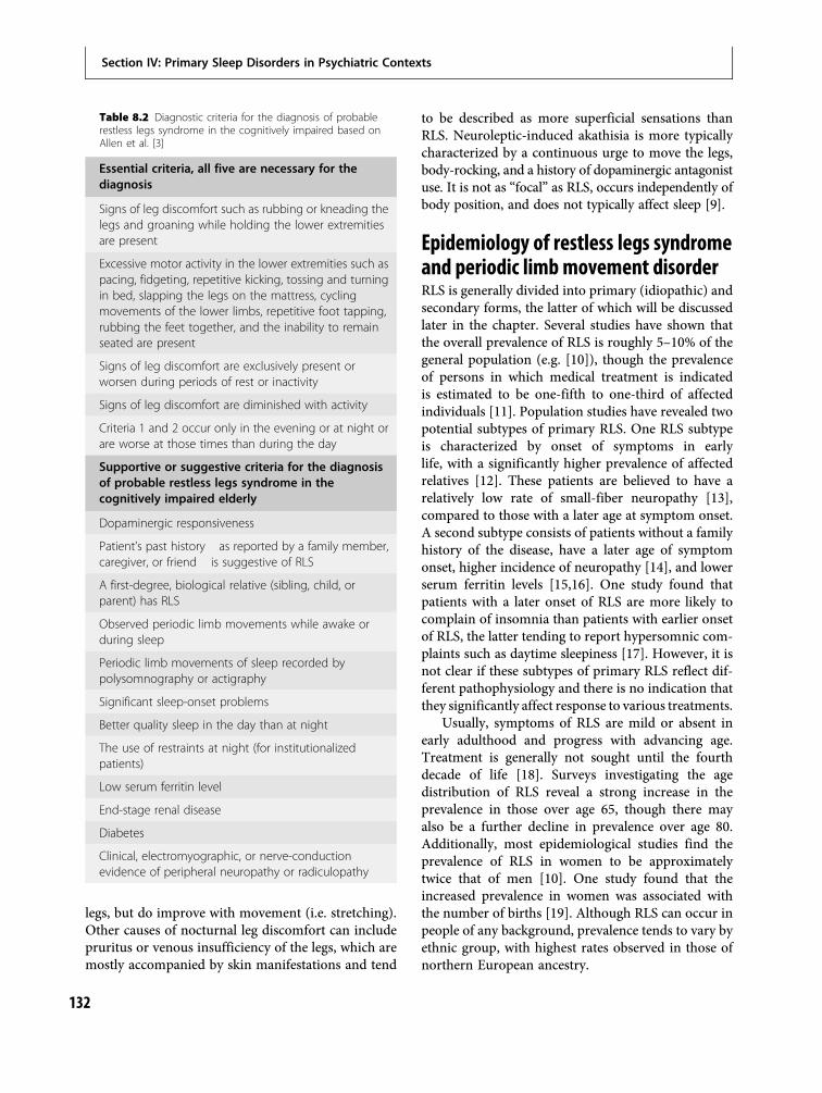
legs, but do improve with movement (i.e. stretching).Other causes of nocturnal leg discomfort can includepruritus or venous insufficiency of the legs, which aremostly accompanied by skin manifestations and tend
to be described as more superficial sensations thanRLS. Neuroleptic-induced akathisia is more typicallycharacterized by a continuous urge to move the legs,body-rocking, and a history of dopaminergic antagonistuse. It is not as “focal” as RLS, occurs independently ofbody position, and does not typically affect sleep [9].
Epidemiology of restless legs syndromeand periodic limb movement disorderRLS is generally divided into primary (idiopathic) andsecondary forms, the latter of which will be discussedlater in the chapter. Several studies have shown thatthe overall prevalence of RLS is roughly 5–10% of thegeneral population (e.g. [10]), though the prevalenceof persons in which medical treatment is indicatedis estimated to be one-fifth to one-third of affectedindividuals [11]. Population studies have revealed twopotential subtypes of primary RLS. One RLS subtypeis characterized by onset of symptoms in earlylife, with a significantly higher prevalence of affectedrelatives [12]. These patients are believed to have arelatively low rate of small-fiber neuropathy [13],compared to those with a later age at symptom onset.A second subtype consists of patients without a familyhistory of the disease, have a later age of symptomonset, higher incidence of neuropathy [14], and lowerserum ferritin levels [15,16]. One study found thatpatients with a later onset of RLS are more likely tocomplain of insomnia than patients with earlier onsetof RLS, the latter tending to report hypersomnic com-plaints such as daytime sleepiness [17]. However, it isnot clear if these subtypes of primary RLS reflect dif-ferent pathophysiology and there is no indication thatthey significantly affect response to various treatments.
Usually, symptoms of RLS are mild or absent inearly adulthood and progress with advancing age.Treatment is generally not sought until the fourthdecade of life [18]. Surveys investigating the agedistribution of RLS reveal a strong increase in theprevalence in those over age 65, though there mayalso be a further decline in prevalence over age 80.Additionally, most epidemiological studies find theprevalence of RLS in women to be approximatelytwice that of men [10]. One study found that theincreased prevalence in women was associated withthe number of births [19]. Although RLS can occur inpeople of any background, prevalence tends to vary byethnic group, with highest rates observed in those ofnorthern European ancestry.
Table 8.2 Diagnostic criteria for the diagnosis of probablerestless legs syndrome in the cognitively impaired based onAllen et al. [3]
Essential criteria, all five are necessary for thediagnosis
Signs of leg discomfort such as rubbing or kneading thelegs and groaning while holding the lower extremitiesare present
Excessive motor activity in the lower extremities such aspacing, fidgeting, repetitive kicking, tossing and turningin bed, slapping the legs on the mattress, cyclingmovements of the lower limbs, repetitive foot tapping,rubbing the feet together, and the inability to remainseated are present
Signs of leg discomfort are exclusively present orworsen during periods of rest or inactivity
Signs of leg discomfort are diminished with activity
Criteria 1 and 2 occur only in the evening or at night orare worse at those times than during the day
Supportive or suggestive criteria for the diagnosisof probable restless legs syndrome in thecognitively impaired elderly
Dopaminergic responsiveness
Patient’s past history as reported by a family member,caregiver, or friend is suggestive of RLS
A first-degree, biological relative (sibling, child, orparent) has RLS
Observed periodic limb movements while awake orduring sleep
Periodic limb movements of sleep recorded bypolysomnography or actigraphy
Significant sleep-onset problems
Better quality sleep in the day than at night
The use of restraints at night (for institutionalizedpatients)
Low serum ferritin level
End-stage renal disease
Diabetes
Clinical, electromyographic, or nerve-conductionevidence of peripheral neuropathy or radiculopathy
Section IV: Primary Sleep Disorders in Psychiatric Contexts
132
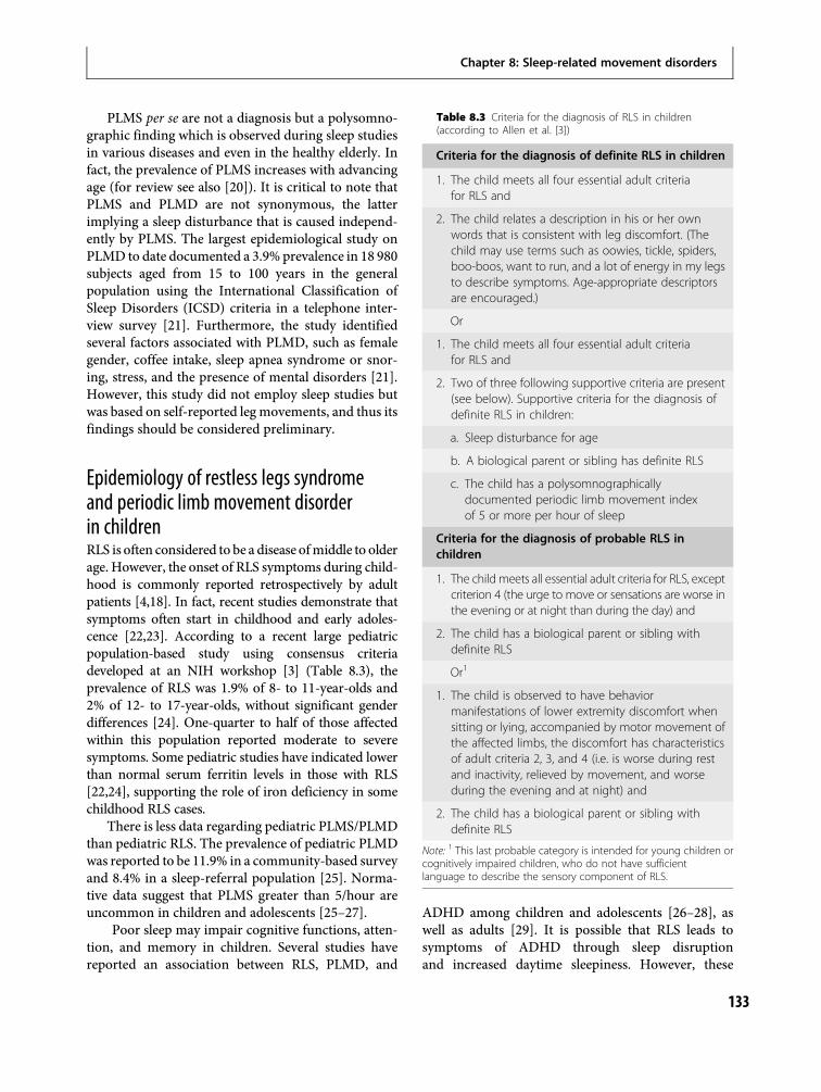
PLMS per se are not a diagnosis but a polysomno-graphic finding which is observed during sleep studiesin various diseases and even in the healthy elderly. Infact, the prevalence of PLMS increases with advancingage (for review see also [20]). It is critical to note thatPLMS and PLMD are not synonymous, the latterimplying a sleep disturbance that is caused independ-ently by PLMS. The largest epidemiological study onPLMD to date documented a 3.9% prevalence in 18 980subjects aged from 15 to 100 years in the generalpopulation using the International Classification ofSleep Disorders (ICSD) criteria in a telephone inter-view survey [21]. Furthermore, the study identifiedseveral factors associated with PLMD, such as femalegender, coffee intake, sleep apnea syndrome or snor-ing, stress, and the presence of mental disorders [21].However, this study did not employ sleep studies butwas based on self-reported leg movements, and thus itsfindings should be considered preliminary.
Epidemiology of restless legs syndromeand periodic limb movement disorderin childrenRLS is often considered to be a disease ofmiddle to olderage. However, the onset of RLS symptoms during child-hood is commonly reported retrospectively by adultpatients [4,18]. In fact, recent studies demonstrate thatsymptoms often start in childhood and early adoles-cence [22,23]. According to a recent large pediatricpopulation-based study using consensus criteriadeveloped at an NIH workshop [3] (Table 8.3), theprevalence of RLS was 1.9% of 8- to 11-year-olds and2% of 12- to 17-year-olds, without significant genderdifferences [24]. One-quarter to half of those affectedwithin this population reported moderate to severesymptoms. Some pediatric studies have indicated lowerthan normal serum ferritin levels in those with RLS[22,24], supporting the role of iron deficiency in somechildhood RLS cases.
There is less data regarding pediatric PLMS/PLMDthan pediatric RLS. The prevalence of pediatric PLMDwas reported to be 11.9% in a community-based surveyand 8.4% in a sleep-referral population [25]. Norma-tive data suggest that PLMS greater than 5/hour areuncommon in children and adolescents [25–27].
Poor sleep may impair cognitive functions, atten-tion, and memory in children. Several studies havereported an association between RLS, PLMD, and
ADHD among children and adolescents [26–28], aswell as adults [29]. It is possible that RLS leads tosymptoms of ADHD through sleep disruptionand increased daytime sleepiness. However, these
Table 8.3 Criteria for the diagnosis of RLS in children(according to Allen et al. [3])
Criteria for the diagnosis of definite RLS in children
1. The child meets all four essential adult criteriafor RLS and
2. The child relates a description in his or her ownwords that is consistent with leg discomfort. (Thechild may use terms such as oowies, tickle, spiders,boo-boos, want to run, and a lot of energy in my legsto describe symptoms. Age-appropriate descriptorsare encouraged.)
Or
1. The child meets all four essential adult criteriafor RLS and
2. Two of three following supportive criteria are present(see below). Supportive criteria for the diagnosis ofdefinite RLS in children:
a. Sleep disturbance for age
b. A biological parent or sibling has definite RLS
c. The child has a polysomnographicallydocumented periodic limb movement indexof 5 or more per hour of sleep
Criteria for the diagnosis of probable RLS inchildren
1. The child meets all essential adult criteria for RLS, exceptcriterion 4 (the urge to move or sensations are worse inthe evening or at night than during the day) and
2. The child has a biological parent or sibling withdefinite RLS
Or1
1. The child is observed to have behaviormanifestations of lower extremity discomfort whensitting or lying, accompanied by motor movement ofthe affected limbs, the discomfort has characteristicsof adult criteria 2, 3, and 4 (i.e. is worse during restand inactivity, relieved by movement, and worseduring the evening and at night) and
2. The child has a biological parent or sibling withdefinite RLS
Note: 1 This last probable category is intended for young children orcognitively impaired children, who do not have sufficientlanguage to describe the sensory component of RLS.
Chapter 8: Sleep-related movement disorders
133
disorders may share a common pathology of thecentral nervous system, like iron deficiency [30] ordopaminergic hypofunction. Sleep disruption associ-ated with RLS may also be related to the developmentof mood and anxiety disorders in childhood. A recentpediatric survey found a co-morbid anxiety and/ordepression in nearly 50% of RLS cases, which alwaysdeveloped following the historical onset of clinicalsleep disturbance [24].
Pathophysiology of idiopathic restlesslegs syndromeThe pathophysiology of RLS is still not completelyunderstood; however, in the last few years considerableprogress in a number of areas has been made.
GeneticsRoughly half of RLS patients have relatives who alsohave RLS. In comparison to non-familial cases, famil-ial RLS usually has a younger age at onset and has amore slowly progressive course [12]. The symptomseverity within an RLS family can be variable. Linkagestudies in RLS families have revealed eight loci ofinterest in RLS. Based on the assumption that thedisease follows a recessive mode of inheritance,linkage to chromosome 12q (RLS1) was identified ina French-Canadian family. Further studies showedlinkage to chromosome 14q (RLS2), 9p (RLS3), 20p(RLS4), and 2q (RLS5). Two loci were identified in asingle RLS family on chromosome 4q and 17p (forrecent overview see [31]). Two recent studies used alarge-scale genome-wide approach to identify singlenucleotide polymorphisms associated with RLS/PLMS [32,33]. In the study of Winkelmann et al.[33], conducted in German and Canadian popula-tions, three genomic regions were identified encodingthe genes MEIS1, BTBD9, and a third region contain-ing MAP2K5 and LBXCOR1. In the study of Stefans-son and colleagues [32], conducted in Icelandic andUS populations, an association was found with theidentical variant in BTBD9. Both studies used differ-ent methods of assessment of the phenotype.Although the mechanism through which these genesmay confer risk for RLS remains unknown, theserecent findings suggest RLS may be a developmentaldisorder of the central nervous system. In such aparadigm, RLS is a neurological disorder in whichthe disease manifests when a genetic “inborn error”
confers increased susceptibility to alterations ofvarious neural structures, resulting in the observedclinical symptoms and associated sleep disturbance.
Dopaminergic systemMultiple etiologies likely impact the dopamine systemin RLS. The primary evidence for this comes from useof dopaminergic agents over the past 20 years. MostRLS symptoms (and PLMS) respond to treatmentwith dopaminergic agents, and, conversely, the useof dopamine receptor antagonists exacerbates theRLS symptoms. However, a clear understanding ofany defect in the metabolism or signaling in thedopamine system remains elusive. Post-mortem andimaging studies have not provided clear evidence ofdysfunction in the dopamine system. The results ofSPECT and PET studies are inconclusive or conflictwith one another (for overview see also [34] and [35]).
IronConditions associated with reduced body iron contentsuch as late stage pregnancy, advanced renal failure,and blood loss from frequent blood donations appearto precipitate RLS [36,37]. However, only 20–25% ofpatients with iron deficiency exhibit symptoms ofRLS. Studies of cerebrospinal fluid, magnetic reson-ance imaging of the brain, and post-mortem tissue alldemonstrate CNS iron deficiency in RLS. Recenttherapeutic trials of oral and intravenously adminis-tered iron [38,39] have shown positive effects on thecourse of RLS in patients with iron deficiency orserum ferritin levels �45 mg/L. Contrasting findingswere reported by Earley and coworkers [40] in aplacebo-controlled study of high-dose intravenousiron sucrose in idiopathic RLS and normal iron status(serum ferritin �45 mg/L). Explanations for the dis-crepant results may include the type of iron formula-tion used, the dosing regimen, patient characteristics,or, importantly, the peripheral iron status with orwithout significant iron store depletion.
Neurophysiological testingRoutine clinical studies of electromyography and nerveconduction velocity and evoked potentials are typicallynormal in patients with RLS. Research evidence fromstudies seeking to identify the generator of the periodicmovements and sensations of RLS has been mixed.However, experimental studies repeatedly demonstrate
Section IV: Primary Sleep Disorders in Psychiatric Contexts
134
disinhibition of the descending cerebrospinal pathwaysin RLS. Similarly, pain thresholds, H-reflex testing, andquantitative sensory testing are abnormal in smallpopulations of patients with RLS (for overview seealso [34]). However, these findings are neither specificnor directive in the diagnostic workup.
Restless legs syndrome secondary to orassociated with another diseaseRLS may manifest as a secondary disorder due to anunderlying primary medical, neurological, or mentaldisease (Table 8.4). Anemia and iron deficiency haveparticular relevance, as conditions associated withreduced body iron content appear to precipitate RLS[3]. Furthermore, augmentation, defined as thereappearance and worsening of RLS complaints aftera prolonged period of dopamine-induced symptomcontrol, has been associated with low ferritin levels[41]. Prevalence of RLS is also increased in the lasttrimester of pregnancy, when iron deficiency iscommon. Folate deficiency and elevated estradiollevels may also contribute to the increased occurrenceof RLS in pregnant women [42]. End-stage renal dis-ease is known to be associated with RLS and mayoccur both in younger and older patients. RLS isespecially frequent among patients on dialysis treat-ment [43,44]. Restless legs syndrome usually improvesafter kidney transplantation; the prevalence of RLS isnot elevated in patients with functioning transplants[43]. Neuropathies and radiculopathies may oftenco-occur with RLS, and have been implicated as pos-sible underlying causes of RLS, though there is limiteddata to support this, and it does not appear that thereis a higher prevalence of RLS in those with neur-opathy [45]. Rheumatoid (but not osteo-) arthritishas an elevated prevalence of RLS, and a high preva-lence of RLS was reported in other rheumatologicalconditions such as fibromyalgia, Sjögren syndrome,and scleroderma [46], suggesting a possible inflam-matory cause of RLS in such individuals.
Further, possibly causally related conditions in-clude among others diabetes mellitus, liver disease,and malignancies. A recent study found RLS in 54out of 121 patients (45%) with type 2 diabetes [47].Patients with type 2 diabetes and co-morbid RLShad more co-morbidities, worse sleep quality, tookmore medications to help them sleep, and were moreimpaired in daytime functioning. As sleep disturbancemay affect long-term health outcomes, the evaluation of
this frequent co-morbidity is important to consider inthe management of diabetic patients. Chronic liver dis-ease may be a further risk factor for RLS: RLS hasbeen reported in up to 62% of patients in hepatologyclinics [48], and interferon-alpha treatment of hepatitismay lead to RLS in this population [49]. Leg restlessnessappears to be one of themost common reasons for sleepdisturbances inmalignant diseases. Davidson et al. [50].reported the symptom “restless legs” in 41% of a largepopulation of cancer patients. Iron deficiency may havebeen the underlying cause of this association.
An elevated prevalence of RLS was observed inpatients after organ transplantation: one-third of45 patients with heart transplantation had PLMSand 45% of these patients also had RLS [51], andnearly 50% of lung transplant recipients were affectedby RLS [52]. However, in a large study of kidney-transplanted patients the prevalence of RLS was 5%,which is a similar frequency to that found in thegeneral population [43].
RLS can also occur in several neurological dis-orders, including neurodegenerative disorders suchas Parkinson’s disease, multiple system atrophy, spi-nocerebellar ataxia (for overview see [53]), and scler-osis multiplex [54]. Spinal cord injuries [55,56] andischemic stroke may also be associated with RLS [57].Recently, a 15–30% prevalence of RLS was foundamong subjects with migraine [58,59]. Most of thestudies investigating co-morbid RLS did not controlfor underlying causes of RLS such as iron or folatedeficiency and the use of medications known toaggravate RLS. If treatment of the underlying disorderis not possible, RLS in secondary cases is treatedidentically to that in primary RLS.
Drug-induced RLS/PLMSDrug-induced RLS is a common, though often un-recognized, reason for sleep disturbance. This isparticularly important for the practicing psycho-pharmacologist, as many agents that cause RLS arepsychotropic drugs. Several antidepressants, anti-psychotics (including atypical antipsychotics like ris-peridone, olanzapine, and quetiapine), and lithium caninduce or exacerbate RLS and PLMS (for overviewsee [60,61]). Of note, leg restlessness is also a commoncomplaint among those undergoing opiate withdrawal[62]. Systematic or controlled studies evaluatingdrug-induced RLS still do not exist. Antidepressants,especially mirtazapine, as well as selective serotonin
Chapter 8: Sleep-related movement disorders
135
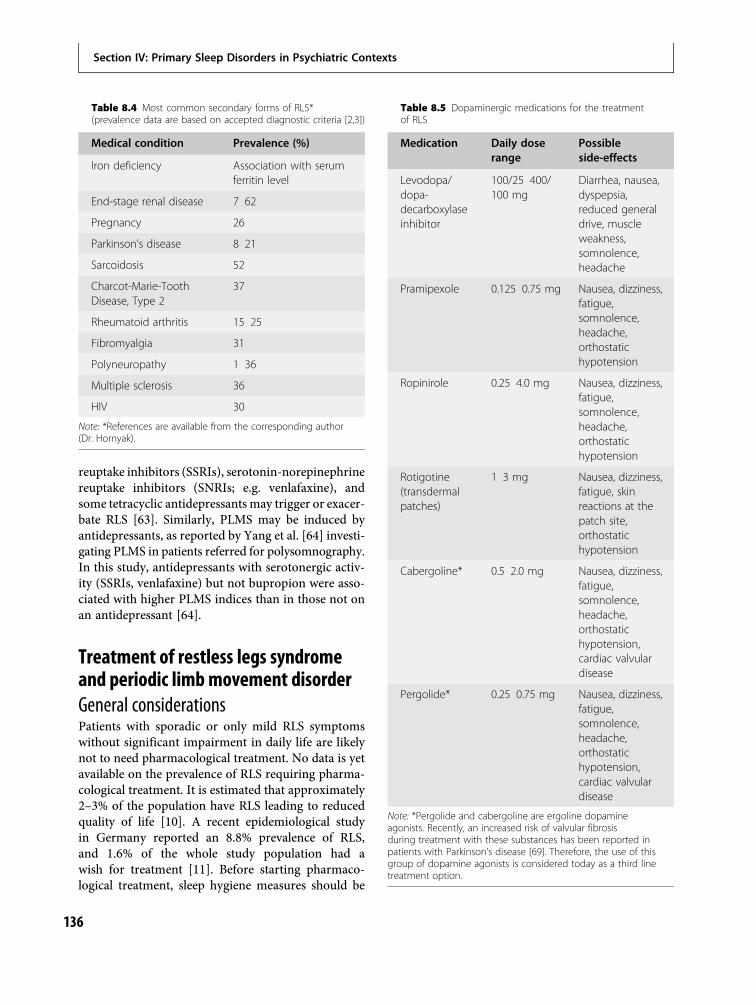
reuptake inhibitors (SSRIs), serotonin-norepinephrinereuptake inhibitors (SNRIs; e.g. venlafaxine), andsome tetracyclic antidepressants may trigger or exacer-bate RLS [63]. Similarly, PLMS may be induced byantidepressants, as reported by Yang et al. [64] investi-gating PLMS in patients referred for polysomnography.In this study, antidepressants with serotonergic activ-ity (SSRIs, venlafaxine) but not bupropion were asso-ciated with higher PLMS indices than in those not onan antidepressant [64].
Treatment of restless legs syndromeand periodic limb movement disorderGeneral considerationsPatients with sporadic or only mild RLS symptomswithout significant impairment in daily life are likelynot to need pharmacological treatment. No data is yetavailable on the prevalence of RLS requiring pharma-cological treatment. It is estimated that approximately2–3% of the population have RLS leading to reducedquality of life [10]. A recent epidemiological studyin Germany reported an 8.8% prevalence of RLS,and 1.6% of the whole study population had awish for treatment [11]. Before starting pharmaco-logical treatment, sleep hygiene measures should be
Table 8.5 Dopaminergic medications for the treatmentof RLS
Medication Daily doserange
Possibleside-effects
Levodopa/dopa-decarboxylaseinhibitor
100/25 400/100 mg
Diarrhea, nausea,dyspepsia,reduced generaldrive, muscleweakness,somnolence,headache
Pramipexole 0.125 0.75 mg Nausea, dizziness,fatigue,somnolence,headache,orthostatichypotension
Ropinirole 0.25 4.0 mg Nausea, dizziness,fatigue,somnolence,headache,orthostatichypotension
Rotigotine(transdermalpatches)
1 3 mg Nausea, dizziness,fatigue, skinreactions at thepatch site,orthostatichypotension
Cabergoline* 0.5 2.0 mg Nausea, dizziness,fatigue,somnolence,headache,orthostatichypotension,cardiac valvulardisease
Pergolide* 0.25 0.75 mg Nausea, dizziness,fatigue,somnolence,headache,orthostatichypotension,cardiac valvulardisease
Note: *Pergolide and cabergoline are ergoline dopamineagonists. Recently, an increased risk of valvular fibrosisduring treatment with these substances has been reported inpatients with Parkinson’s disease [69]. Therefore, the use of thisgroup of dopamine agonists is considered today as a third linetreatment option.
Table 8.4 Most common secondary forms of RLS*(prevalence data are based on accepted diagnostic criteria [2,3])
Medical condition Prevalence (%)
Iron deficiency Association with serumferritin level
End-stage renal disease 7 62
Pregnancy 26
Parkinson’s disease 8 21
Sarcoidosis 52
Charcot-Marie-ToothDisease, Type 2
37
Rheumatoid arthritis 15 25
Fibromyalgia 31
Polyneuropathy 1 36
Multiple sclerosis 36
HIV 30
Note: *References are available from the corresponding author(Dr. Hornyak).
Section IV: Primary Sleep Disorders in Psychiatric Contexts
136
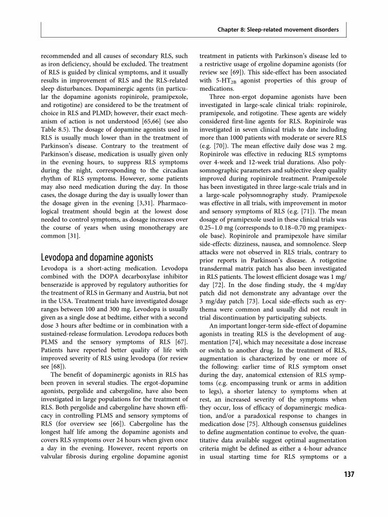
recommended and all causes of secondary RLS, suchas iron deficiency, should be excluded. The treatmentof RLS is guided by clinical symptoms, and it usuallyresults in improvement of RLS and the RLS-relatedsleep disturbances. Dopaminergic agents (in particu-lar the dopamine agonists ropinirole, pramipexole,and rotigotine) are considered to be the treatment ofchoice in RLS and PLMD; however, their exact mech-anism of action is not understood [65,66] (see alsoTable 8.5). The dosage of dopamine agonists used inRLS is usually much lower than in the treatment ofParkinson’s disease. Contrary to the treatment ofParkinson’s disease, medication is usually given onlyin the evening hours, to suppress RLS symptomsduring the night, corresponding to the circadianrhythm of RLS symptoms. However, some patientsmay also need medication during the day. In thosecases, the dosage during the day is usually lower thanthe dosage given in the evening [3,31]. Pharmaco-logical treatment should begin at the lowest doseneeded to control symptoms, as dosage increases overthe course of years when using monotherapy arecommon [31].
Levodopa and dopamine agonistsLevodopa is a short-acting medication. Levodopacombined with the DOPA decarboxylase inhibitorbenserazide is approved by regulatory authorities forthe treatment of RLS in Germany and Austria, but notin the USA. Treatment trials have investigated dosageranges between 100 and 300 mg. Levodopa is usuallygiven as a single dose at bedtime, either with a seconddose 3 hours after bedtime or in combination with asustained-release formulation. Levodopa reduces bothPLMS and the sensory symptoms of RLS [67].Patients have reported better quality of life withimproved severity of RLS using levodopa (for reviewsee [68]).
The benefit of dopaminergic agonists in RLS hasbeen proven in several studies. The ergot-dopamineagonists, pergolide and cabergoline, have also beeninvestigated in large populations for the treatment ofRLS. Both pergolide and cabergoline have shown effi-cacy in controlling PLMS and sensory symptoms ofRLS (for overview see [66]). Cabergoline has thelongest half life among the dopamine agonists andcovers RLS symptoms over 24 hours when given oncea day in the evening. However, recent reports onvalvular fibrosis during ergoline dopamine agonist
treatment in patients with Parkinson’s disease led toa restrictive usage of ergoline dopamine agonists (forreview see [69]). This side-effect has been associatedwith 5-HT2B agonist properties of this group ofmedications.
Three non-ergot dopamine agonists have beeninvestigated in large-scale clinical trials: ropinirole,pramipexole, and rotigotine. These agents are widelyconsidered first-line agents for RLS. Ropinirole wasinvestigated in seven clinical trials to date includingmore than 1000 patients with moderate or severe RLS(e.g. [70]). The mean effective daily dose was 2 mg.Ropinirole was effective in reducing RLS symptomsover 4-week and 12-week trial durations. Also poly-somnographic parameters and subjective sleep qualityimproved during ropinirole treatment. Pramipexolehas been investigated in three large-scale trials and ina large-scale polysomnography study. Pramipexolewas effective in all trials, with improvement in motorand sensory symptoms of RLS (e.g. [71]). The meandosage of pramipexole used in these clinical trials was0.25–1.0 mg (corresponds to 0.18–0.70 mg pramipex-ole base). Ropinirole and pramipexole have similarside-effects: dizziness, nausea, and somnolence. Sleepattacks were not observed in RLS trials, contrary toprior reports in Parkinson’s disease. A rotigotinetransdermal matrix patch has also been investigatedin RLS patients. The lowest efficient dosage was 1 mg/day [72]. In the dose finding study, the 4 mg/daypatch did not demonstrate any advantage over the3 mg/day patch [73]. Local side-effects such as ery-thema were common and usually did not result intrial discontinuation by participating subjects.
An important longer-term side-effect of dopamineagonists in treating RLS is the development of aug-mentation [74], which may necessitate a dose increaseor switch to another drug. In the treatment of RLS,augmentation is characterized by one or more ofthe following: earlier time of RLS symptom onsetduring the day, anatomical extension of RLS symp-toms (e.g. encompassing trunk or arms in additionto legs), a shorter latency to symptoms when atrest, an increased severity of the symptoms whenthey occur, loss of efficacy of dopaminergic medica-tion, and/or a paradoxical response to changes inmedication dose [75]. Although consensus guidelinesto define augmentation continue to evolve, the quan-titative data available suggest optimal augmentationcriteria might be defined as either a 4-hour advancein usual starting time for RLS symptoms or a
Chapter 8: Sleep-related movement disorders
137
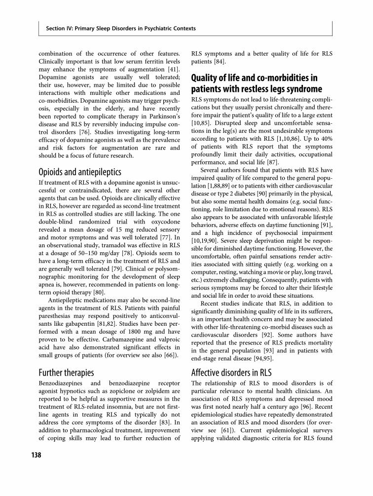
combination of the occurrence of other features.Clinically important is that low serum ferritin levelsmay enhance the symptoms of augmentation [41].Dopamine agonists are usually well tolerated;their use, however, may be limited due to possibleinteractions with multiple other medications andco-morbidities. Dopamine agonists may trigger psych-osis, especially in the elderly, and have recentlybeen reported to complicate therapy in Parkinson’sdisease and RLS by reversibly inducing impulse con-trol disorders [76]. Studies investigating long-termefficacy of dopamine agonists as well as the prevalenceand risk factors for augmentation are rare andshould be a focus of future research.
Opioids and antiepilepticsIf treatment of RLS with a dopamine agonist is unsuc-cessful or contraindicated, there are several otheragents that can be used. Opioids are clinically effectivein RLS, however are regarded as second-line treatmentin RLS as controlled studies are still lacking. The onedouble-blind randomized trial with oxycodonerevealed a mean dosage of 15 mg reduced sensoryand motor symptoms and was well tolerated [77]. Inan observational study, tramadol was effective in RLSat a dosage of 50–150 mg/day [78]. Opioids seem tohave a long-term efficacy in the treatment of RLS andare generally well tolerated [79]. Clinical or polysom-nographic monitoring for the development of sleepapnea is, however, recommended in patients on long-term opioid therapy [80].
Antiepileptic medications may also be second-lineagents in the treatment of RLS. Patients with painfulparesthesias may respond positively to anticonvul-sants like gabapentin [81,82]. Studies have been per-formed with a mean dosage of 1800 mg and haveproven to be effective. Carbamazepine and valproicacid have also demonstrated significant effects insmall groups of patients (for overview see also [66]).
Further therapiesBenzodiazepines and benzodiazepine receptoragonist hypnotics such as zopiclone or zolpidem arereported to be helpful as supportive measures in thetreatment of RLS-related insomnia, but are not first-line agents in treating RLS and typically do notaddress the core symptoms of the disorder [83]. Inaddition to pharmacological treatment, improvementof coping skills may lead to further reduction of
RLS symptoms and a better quality of life for RLSpatients [84].
Quality of life and co-morbidities inpatients with restless legs syndromeRLS symptoms do not lead to life-threatening compli-cations but they usually persist chronically and there-fore impair the patient’s quality of life to a large extent[10,85]. Disrupted sleep and uncomfortable sensa-tions in the leg(s) are the most undesirable symptomsaccording to patients with RLS [1,10,86]. Up to 40%of patients with RLS report that the symptomsprofoundly limit their daily activities, occupationalperformance, and social life [87].
Several authors found that patients with RLS haveimpaired quality of life compared to the general popu-lation [1,88,89] or to patients with either cardiovasculardisease or type 2 diabetes [90] primarily in the physical,but also some mental health domains (e.g. social func-tioning, role limitation due to emotional reasons). RLSalso appears to be associated with unfavorable lifestylebehaviors, adverse effects on daytime functioning [91],and a high incidence of psychosocial impairment[10,19,90]. Severe sleep deprivation might be respon-sible for diminished daytime functioning. However, theuncomfortable, often painful sensations render activ-ities associated with sitting quietly (e.g. working on acomputer, resting, watching amovie or play, long travel,etc.) extremely challenging. Consequently, patients withserious symptoms may be forced to alter their lifestyleand social life in order to avoid these situations.
Recent studies indicate that RLS, in addition tosignificantly diminishing quality of life in its sufferers,is an important health concern and may be associatedwith other life-threatening co-morbid diseases such ascardiovascular disorders [92]. Some authors havereported that the presence of RLS predicts mortalityin the general population [93] and in patients withend-stage renal disease [94,95].
Affective disorders in RLSThe relationship of RLS to mood disorders is ofparticular relevance to mental health clinicians. Anassociation of RLS symptoms and depressed moodwas first noted nearly half a century ago [96]. Recentepidemiological studies have repeatedly demonstratedan association of RLS and mood disorders (for over-view see [61]). Current epidemiological surveysapplying validated diagnostic criteria for RLS found
Section IV: Primary Sleep Disorders in Psychiatric Contexts
138
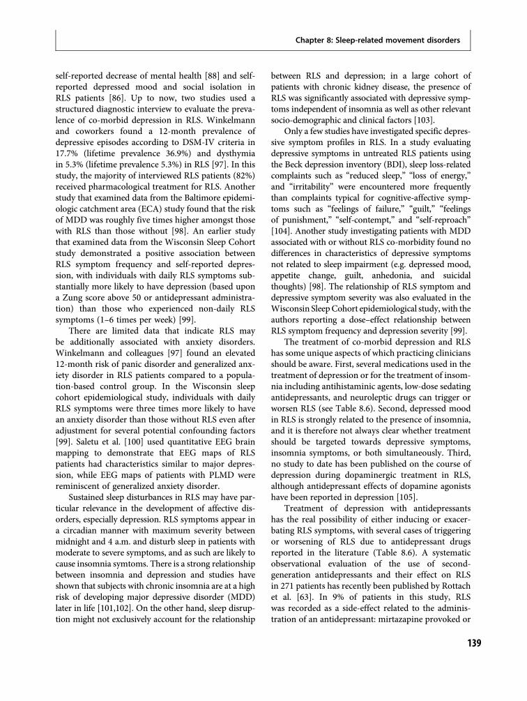
self-reported decrease of mental health [88] and self-reported depressed mood and social isolation inRLS patients [86]. Up to now, two studies used astructured diagnostic interview to evaluate the preva-lence of co-morbid depression in RLS. Winkelmannand coworkers found a 12-month prevalence ofdepressive episodes according to DSM-IV criteria in17.7% (lifetime prevalence 36.9%) and dysthymiain 5.3% (lifetime prevalence 5.3%) in RLS [97]. In thisstudy, the majority of interviewed RLS patients (82%)received pharmacological treatment for RLS. Anotherstudy that examined data from the Baltimore epidemi-ologic catchment area (ECA) study found that the riskof MDD was roughly five times higher amongst thosewith RLS than those without [98]. An earlier studythat examined data from the Wisconsin Sleep Cohortstudy demonstrated a positive association betweenRLS symptom frequency and self-reported depres-sion, with individuals with daily RLS symptoms sub-stantially more likely to have depression (based upona Zung score above 50 or antidepressant administra-tion) than those who experienced non-daily RLSsymptoms (1–6 times per week) [99].
There are limited data that indicate RLS maybe additionally associated with anxiety disorders.Winkelmann and colleagues [97] found an elevated12-month risk of panic disorder and generalized anx-iety disorder in RLS patients compared to a popula-tion-based control group. In the Wisconsin sleepcohort epidemiological study, individuals with dailyRLS symptoms were three times more likely to havean anxiety disorder than those without RLS even afteradjustment for several potential confounding factors[99]. Saletu et al. [100] used quantitative EEG brainmapping to demonstrate that EEG maps of RLSpatients had characteristics similar to major depres-sion, while EEG maps of patients with PLMD werereminiscent of generalized anxiety disorder.
Sustained sleep disturbances in RLS may have par-ticular relevance in the development of affective dis-orders, especially depression. RLS symptoms appear ina circadian manner with maximum severity betweenmidnight and 4 a.m. and disturb sleep in patients withmoderate to severe symptoms, and as such are likely tocause insomnia symtoms. There is a strong relationshipbetween insomnia and depression and studies haveshown that subjects with chronic insomnia are at a highrisk of developing major depressive disorder (MDD)later in life [101,102]. On the other hand, sleep disrup-tion might not exclusively account for the relationship
between RLS and depression; in a large cohort ofpatients with chronic kidney disease, the presence ofRLS was significantly associated with depressive symp-toms independent of insomnia as well as other relevantsocio-demographic and clinical factors [103].
Only a few studies have investigated specific depres-sive symptom profiles in RLS. In a study evaluatingdepressive symptoms in untreated RLS patients usingthe Beck depression inventory (BDI), sleep loss-relatedcomplaints such as “reduced sleep,” “loss of energy,”and “irritability” were encountered more frequentlythan complaints typical for cognitive-affective symp-toms such as “feelings of failure,” “guilt,” “feelingsof punishment,” “self-contempt,” and “self-reproach”[104]. Another study investigating patients with MDDassociated with or without RLS co-morbidity found nodifferences in characteristics of depressive symptomsnot related to sleep impairment (e.g. depressed mood,appetite change, guilt, anhedonia, and suicidalthoughts) [98]. The relationship of RLS symptom anddepressive symptom severity was also evaluated in theWisconsin SleepCohort epidemiological study, with theauthors reporting a dose–effect relationship betweenRLS symptom frequency and depression severity [99].
The treatment of co-morbid depression and RLShas some unique aspects of which practicing cliniciansshould be aware. First, several medications used in thetreatment of depression or for the treatment of insom-nia including antihistaminic agents, low-dose sedatingantidepressants, and neuroleptic drugs can trigger orworsen RLS (see Table 8.6). Second, depressed moodin RLS is strongly related to the presence of insomnia,and it is therefore not always clear whether treatmentshould be targeted towards depressive symptoms,insomnia symptoms, or both simultaneously. Third,no study to date has been published on the course ofdepression during dopaminergic treatment in RLS,although antidepressant effects of dopamine agonistshave been reported in depression [105].
Treatment of depression with antidepressantshas the real possibility of either inducing or exacer-bating RLS symptoms, with several cases of triggeringor worsening of RLS due to antidepressant drugsreported in the literature (Table 8.6). A systematicobservational evaluation of the use of second-generation antidepressants and their effect on RLSin 271 patients has recently been published by Rottachet al. [63]. In 9% of patients in this study, RLSwas recorded as a side-effect related to the adminis-tration of an antidepressant: mirtazapine provoked or
Chapter 8: Sleep-related movement disorders
139
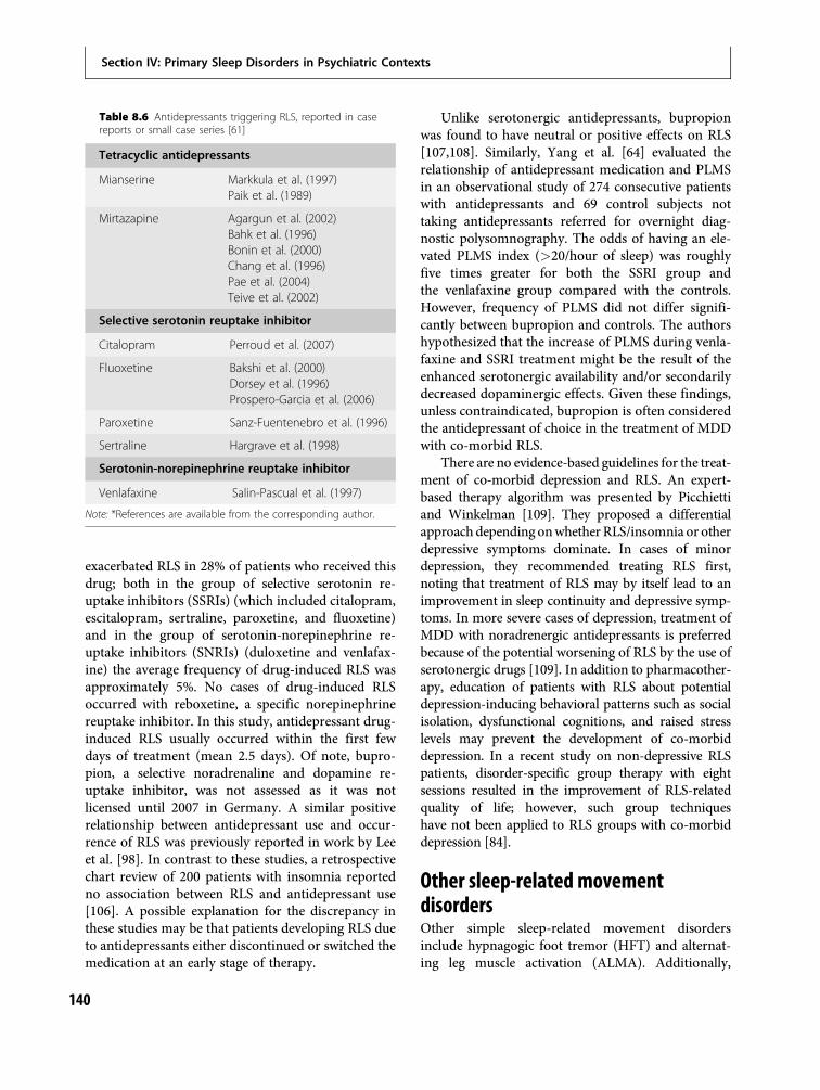
exacerbated RLS in 28% of patients who received thisdrug; both in the group of selective serotonin re-uptake inhibitors (SSRIs) (which included citalopram,escitalopram, sertraline, paroxetine, and fluoxetine)and in the group of serotonin-norepinephrine re-uptake inhibitors (SNRIs) (duloxetine and venlafax-ine) the average frequency of drug-induced RLS wasapproximately 5%. No cases of drug-induced RLSoccurred with reboxetine, a specific norepinephrinereuptake inhibitor. In this study, antidepressant drug-induced RLS usually occurred within the first fewdays of treatment (mean 2.5 days). Of note, bupro-pion, a selective noradrenaline and dopamine re-uptake inhibitor, was not assessed as it was notlicensed until 2007 in Germany. A similar positiverelationship between antidepressant use and occur-rence of RLS was previously reported in work by Leeet al. [98]. In contrast to these studies, a retrospectivechart review of 200 patients with insomnia reportedno association between RLS and antidepressant use[106]. A possible explanation for the discrepancy inthese studies may be that patients developing RLS dueto antidepressants either discontinued or switched themedication at an early stage of therapy.
Unlike serotonergic antidepressants, bupropionwas found to have neutral or positive effects on RLS[107,108]. Similarly, Yang et al. [64] evaluated therelationship of antidepressant medication and PLMSin an observational study of 274 consecutive patientswith antidepressants and 69 control subjects nottaking antidepressants referred for overnight diag-nostic polysomnography. The odds of having an ele-vated PLMS index (>20/hour of sleep) was roughlyfive times greater for both the SSRI group andthe venlafaxine group compared with the controls.However, frequency of PLMS did not differ signifi-cantly between bupropion and controls. The authorshypothesized that the increase of PLMS during venla-faxine and SSRI treatment might be the result of theenhanced serotonergic availability and/or secondarilydecreased dopaminergic effects. Given these findings,unless contraindicated, bupropion is often consideredthe antidepressant of choice in the treatment of MDDwith co-morbid RLS.
There are no evidence-based guidelines for the treat-ment of co-morbid depression and RLS. An expert-based therapy algorithm was presented by Picchiettiand Winkelman [109]. They proposed a differentialapproach depending onwhether RLS/insomnia or otherdepressive symptoms dominate. In cases of minordepression, they recommended treating RLS first,noting that treatment of RLS may by itself lead to animprovement in sleep continuity and depressive symp-toms. In more severe cases of depression, treatment ofMDD with noradrenergic antidepressants is preferredbecause of the potential worsening of RLS by the use ofserotonergic drugs [109]. In addition to pharmacother-apy, education of patients with RLS about potentialdepression-inducing behavioral patterns such as socialisolation, dysfunctional cognitions, and raised stresslevels may prevent the development of co-morbiddepression. In a recent study on non-depressive RLSpatients, disorder-specific group therapy with eightsessions resulted in the improvement of RLS-relatedquality of life; however, such group techniqueshave not been applied to RLS groups with co-morbiddepression [84].
Other sleep-related movementdisordersOther simple sleep-related movement disordersinclude hypnagogic foot tremor (HFT) and alternat-ing leg muscle activation (ALMA). Additionally,
Table 8.6 Antidepressants triggering RLS, reported in casereports or small case series [61]
Tetracyclic antidepressants
Mianserine Markkula et al. (1997)Paik et al. (1989)
Mirtazapine Agargun et al. (2002)Bahk et al. (1996)Bonin et al. (2000)Chang et al. (1996)Pae et al. (2004)Teive et al. (2002)
Selective serotonin reuptake inhibitor
Citalopram Perroud et al. (2007)
Fluoxetine Bakshi et al. (2000)Dorsey et al. (1996)Prospero-Garcia et al. (2006)
Paroxetine Sanz-Fuentenebro et al. (1996)
Sertraline Hargrave et al. (1998)
Serotonin-norepinephrine reuptake inhibitor
Venlafaxine Salin-Pascual et al. (1997)
Note: *References are available from the corresponding author.
Section IV: Primary Sleep Disorders in Psychiatric Contexts
140
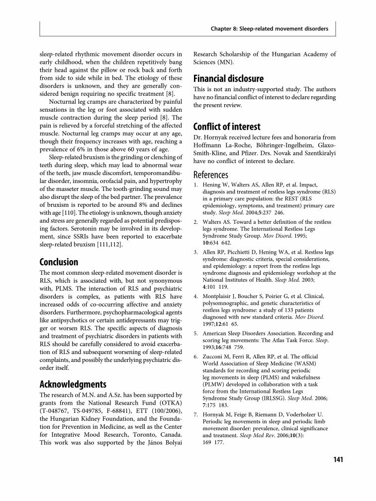
sleep-related rhythmic movement disorder occurs inearly childhood, when the children repetitively bangtheir head against the pillow or rock back and forthfrom side to side while in bed. The etiology of thesedisorders is unknown, and they are generally con-sidered benign requiring no specific treatment [8].
Nocturnal leg cramps are characterized by painfulsensations in the leg or foot associated with suddenmuscle contraction during the sleep period [8]. Thepain is relieved by a forceful stretching of the affectedmuscle. Nocturnal leg cramps may occur at any age,though their frequency increases with age, reaching aprevalence of 6% in those above 60 years of age.
Sleep-related bruxism is the grinding or clenching ofteeth during sleep, which may lead to abnormal wearof the teeth, jaw muscle discomfort, temporomandibu-lar disorder, insomnia, orofacial pain, and hypertrophyof the masseter muscle. The tooth-grinding sound mayalso disrupt the sleep of the bed partner. The prevalenceof bruxism is reported to be around 8% and declineswith age [110]. The etiology is unknown, though anxietyand stress are generally regarded as potential predispos-ing factors. Serotonin may be involved in its develop-ment, since SSRIs have been reported to exacerbatesleep-related bruxism [111,112].
ConclusionThe most common sleep-related movement disorder isRLS, which is associated with, but not synonymouswith, PLMS. The interaction of RLS and psychiatricdisorders is complex, as patients with RLS haveincreased odds of co-occurring affective and anxietydisorders. Furthermore, psychopharmacological agentslike antipsychotics or certain antidepressants may trig-ger or worsen RLS. The specific aspects of diagnosisand treatment of psychiatric disorders in patients withRLS should be carefully considered to avoid exacerba-tion of RLS and subsequent worsening of sleep-relatedcomplaints, and possibly the underlying psychiatric dis-order itself.
AcknowledgmentsThe research of M.N. and A.Sz. has been supported bygrants from the National Research Fund (OTKA)(T-048767, TS-049785, F-68841), ETT (100/2006),the Hungarian Kidney Foundation, and the Founda-tion for Prevention in Medicine, as well as the Centerfor Integrative Mood Research, Toronto, Canada.This work was also supported by the János Bolyai
Research Scholarship of the Hungarian Academy ofSciences (MN).
Financial disclosureThis is not an industry-supported study. The authorshave no financial conflict of interest to declare regardingthe present review.
Conflict of interestDr. Hornyak received lecture fees and honoraria fromHoffmann La-Roche, Böhringer-Ingelheim, Glaxo-Smith-Kline, and Pfizer. Drs. Novak and Szentkiralyihave no conflict of interest to declare.
References1. Hening W, Walters AS, Allen RP, et al. Impact,
diagnosis and treatment of restless legs syndrome (RLS)in a primary care population: the REST (RLSepidemiology, symptoms, and treatment) primary carestudy. Sleep Med. 2004;5:237 246.
2. Walters AS. Toward a better definition of the restlesslegs syndrome. The International Restless LegsSyndrome Study Group. Mov Disord. 1995;10:634 642.
3. Allen RP, Picchietti D, Hening WA, et al. Restless legssyndrome: diagnostic criteria, special considerations,and epidemiology: a report from the restless legssyndrome diagnosis and epidemiology workshop at theNational Institutes of Health. Sleep Med. 2003;4:101 119.
4. Montplaisir J, Boucher S, Poirier G, et al. Clinical,polysomnographic, and genetic characteristics ofrestless legs syndrome: a study of 133 patientsdiagnosed with new standard criteria. Mov Disord.1997;12:61 65.
5. American Sleep Disorders Association. Recording andscoring leg movements: The Atlas Task Force. Sleep.1993;16:748 759.
6. Zucconi M, Ferri R, Allen RP, et al. The officialWorld Association of Sleep Medicine (WASM)standards for recording and scoring periodicleg movements in sleep (PLMS) and wakefulness(PLMW) developed in collaboration with a taskforce from the International Restless LegsSyndrome Study Group (IRLSSG). Sleep Med. 2006;7:175 183.
7. Hornyak M, Feige B, Riemann D, Voderholzer U.Periodic leg movements in sleep and periodic limbmovement disorder: prevalence, clinical significanceand treatment. Sleep Med Rev. 2006;10(3):169 177.
Chapter 8: Sleep-related movement disorders
141

8. American Academy of Sleep Medicine. Internationalclassification of sleep disorders. 2nd Edition:Diagnostic and coding manual. Westchester, Illinois:American Academy of Sleep Medicine; 2005.
9. Walters AS, Hening W, Rubinstein M, Chokroverty S.A clinical and polysomnographic comparison ofneuroleptic induced akathisia and the idiopathicrestless legs syndrome. Sleep. 1991;14(4):339 345.
10. Allen RP, Walters AS, Montplaisir J, et al. Restless legssyndrome prevalence and impact: REST generalpopulation study. Arch Intern Med. 2005;165:1286 1292.
11. Happe S, Vennemann M, Evers S, Berger K. Treatmentwish of individuals with known and unknown restlesslegs syndrome in the community. J Neurol.2008;255:1365 1371.
12. Allen RP, Earley CJ. Defining the phenotype of therestless legs syndrome (RLS) using age of symptomonset. Sleep Med. 2000;1:11 19.
13. Polydefkis M, Allen RP, Hauer P, et al. Subclinicalsensory neuropathy in late onset restless legssyndrome. Neurology. 2000;55:1115 1121.
14. Ondo WG, Vuong KD, Wang Q. Restless legssyndrome in monozygotic twins: clinical correlates.Neurology. 2000;55:1404 1406.
15. Berger K, von Eckardstein A, Trenkwalder C, et al.Iron metabolism and the risk of restless legs syndromein an elderly general population the MEMO Study.J Neurol. 2002;249:1195 1199.
16. O’Keeffe ST. Secondary causes of restless legssyndrome in older people. Age Ageing. 2005;34:349 352.
17. Bassetti CL, Mauerhofer D, Gugger M, Mattis J, HessCW. Restless legs syndrome: a clinical study of 55patients. Eur Neurol. 2001;45:67 74.
18. Walters AS, Hickey K, Maltzman J, et al.A questionnaire study of 138 patients with restless legssyndrome: the ‘Night Walkers’ survey. Neurology.1996;46:92 95.
19. Berger K, Luedemann J, Trenkwalder C, John U,Kessler C. Sex and the risk of restless legs syndrome inthe general population. Arch Intern Med. 2004;164:196 202.
20. Hornyak M, Voderholzer U, Riemann D. Treatment ofdepression in patients with restless legs syndrome:what is evidence based? Sleep Med. 2006;7:301 302; author reply 303 304.
21. Ohayon MM, Roth T. Prevalence of restless legssyndrome and periodic limb movement disorder in thegeneral population. J Psychosom Res. 2002;53:547 554.
22. Kotagal S, Silber MH. Childhood onset restless legssyndrome. Ann Neurol. 2004:56:803 807.
23. Muhle H, Neumann A, Lohmann Hedrich K, et al.Childhood onset restless legs syndrome: clinical andgenetic features of 22 families. Mov Disord.2008;23:1113 1121; quiz 1203.
24. Picchietti DL, Stevens HE. Early manifestations ofrestless legs syndrome in childhood and adolescence.Sleep Med. 2008;9:770 781.
25. Crabtree VM, Ivanenko A, O’Brien LM, Gozal D.Periodic limb movement disorder of sleep in children.J Sleep Res. 2003;12:73 81.
26. Picchietti DL, England SJ, Walters AS, Willis KK,Verrico T. Periodic limb movement disorder andrestless legs syndrome in children with attentiondeficit hyperactivity disorder. J Child Neurol.1998;13:588 594.
27. Picchietti DL, Underwood DJ, Farris WA, et al.Further studies on periodic limb movement disorderand restless legs syndrome in children with attentiondeficit hyperactivity disorder. Mov Disord.1999;14:1000 1007.
28. Chervin RD, Archbold KH, Dillon JE, et al.Associations between symptoms of inattention,hyperactivity, restless legs, and periodic legmovements. Sleep. 2002;25:213 218.
29. Wagner ML, Walters AS, Fisher BC. Symptoms ofattention deficit/hyperactivity disorder in adults withrestless legs syndrome. Sleep. 2004;27:1499 1504.
30. Oner P, Dirik EB, Taner Y, Caykoylu A, Anlar O.Association between low serum ferritin and restlesslegs syndrome in patients with attention deficithyperactivity disorder. Tohoku J Exp Med.2007;213:269 276.
31. Trenkwalder C, Högl B, Winkelmann J. Recentadvances in the diagnosis, genetics and treatment ofrestless legs syndrome. J Neurol. 2009;256:539 553.
32. Stefansson H, Rye DB, Hicks A, et al. A genetic riskfactor for periodic limb movements in sleep. N EnglJ Med. 2007;357:639 647.
33. Winkelmann J, Schormair B, Lichtner P, et al.Genome wide association study of restless legssyndrome identifies common variants in threegenomic regions. Nat Genet. 2007;39: 1000 1006.
34. Paulus W, Dowling P, Rijsman R, Stiasny Kolster K,Trenkwalder C. Update of the pathophysiology of therestless legs syndrome. Mov Disord. 2007;22(Suppl 18):S431 S439.
35. Satija P, Ondo WG. Restless legs syndrome:pathophysiology, diagnosis and treatment. CNS Drugs.2008;22(6):497 518.
36. Bordelon YM, Smith M. Movement disorders inpregnancy. Semin Neurol. 2007;27(5):467 475.
Section IV: Primary Sleep Disorders in Psychiatric Contexts
142
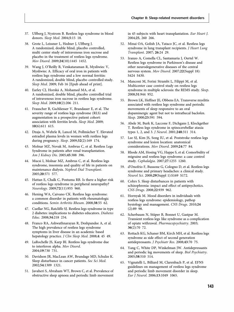
37. Ulfberg J, Nystrom B. Restless legs syndrome in blooddonors. Sleep Med. 2004;5:15 18.
38. Grote L, Leissner L, Hedner J, Ulfberg J.A randomized, double blind, placebo controlled,multi center study of intravenous iron sucrose andplacebo in the treatment of restless legs syndrome.Mov Disord. 2009;24(10):1445 1452.
39. Wang J, O’Reilly B, Venkataraman R, Mysliwiec V,Mysliwiec A. Efficacy of oral iron in patients withrestless legs syndrome and a low normal ferritin:A randomized, double blind, placebo controlled study.Sleep Med. 2009; Feb 16 [Epub ahead of print].
40. Earley CJ, Horská A, Mohamed MA, et al.A randomized, double blind, placebo controlled trialof intravenous iron sucrose in restless legs syndrome.Sleep Med. 2009;10(2):206 211.
41. Frauscher B, Gschliesser V, Brandauer E, et al. Theseverity range of restless legs syndrome (RLS) andaugmentation in a prospective patient cohort:association with ferritin levels. Sleep Med. 2009;10(6):611 615.
42. Dzaja A, Wehrle R, Lancel M, Pollmächer T. Elevatedestradiol plasma levels in women with restless legsduring pregnancy. Sleep. 2009;32(2):169 174.
43. Molnar MZ, Novak M, Ambrus C, et al. Restless LegsSyndrome in patients after renal transplantation.Am J Kidney Dis. 2005;45:388 396.
44. Mucsi I, Molnar MZ, Ambrus C, et al. Restless legssyndrome, insomnia and quality of life in patients onmaintenance dialysis. Nephrol Dial Transplant.2005;20:571 577.
45. Hattan E, Chalk C, Postuma RB. Is there a higher riskof restless legs syndrome in peripheral neuropathy?Neurology. 2009;72(11):955 960.
46. Hening WA, Caivano CK. Restless legs syndrome:a common disorder in patients with rheumatologicconditions. Semin Arthritis Rheum. 2008;38:55 62.
47. Cuellar NG, Ratcliffe SJ. Restless legs syndrome in type2 diabetes: implications to diabetes educators. DiabetesEduc. 2008;34:218 234.
48. Franco RA, Ashwathnarayan R, Deshpandee A, et al.The high prevalence of restless legs syndromesymptoms in liver disease in an academic basedhepatology practice. J Clin Sleep Med. 2008;4: 45 49.
49. LaRochelle JS, Karp BI. Restless legs syndrome dueto interferon alpha. Mov Disord.2004;19:730 731.
50. Davidson JR, MacLean AW, Brundage MD, Schulze K.Sleep disturbance in cancer patients. Soc Sci Med.2002;54:1309 1321.
51. Javaheri S, AbrahamWT, Brown C, et al. Prevalence ofobstructive sleep apnoea and periodic limb movement
in 45 subjects with heart transplantation. Eur Heart J.2004;25, 260 266.
52. Minai OA, Golish JA, Yataco JC, et al. Restless legssyndrome in lung transplant recipients. J Heart LungTransplant. 2007; 26:24 29.
53. Iranzo A, Comella CL, Santamaria J, Oertel W.Restless legs syndrome in Parkinson’s disease andother neurodegenerative diseases of the centralnervous system. Mov Disord. 2007;22(Suppl 18):S424 S430.
54. Manconi M, Ferini Strambi L, Filippi M, et al.Multicenter case control study on restless legssyndrome in multiple sclerosis: the REMS study. Sleep.2008;31:944 952.
55. Brown LK, Heffner JE, Obbens EA. Transverse myelitisassociated with restless legs syndrome and periodicmovements of sleep responsive to an oraldopaminergic agent but not to intrathecal baclofen.Sleep. 2000;23:591 594.
56. Abele M, Burk K, Laccone F, Dichgans J, KlockgetherT. Restless legs syndrome in spinocerebellar ataxiatypes 1, 2, and 3. J Neurol. 2001;248:311 314.
57. Lee SJ, Kim JS, Song IU, et al. Poststroke restless legssyndrome and lesion location: anatomicalconsiderations. Mov Disord. 2009;24:77 84.
58. Rhode AM, Hosing VG, Happe S, et al. Comorbidity ofmigraine and restless legs syndrome: a case controlstudy. Cephalalgia. 2007;27:1255 1260.
59. d’Onofrio F, Bussone G, Cologno D, et al. Restless legssyndrome and primary headaches: a clinical study.Neurol Sci. 2008;29(Suppl 1):S169 S172.
60. Cohrs S. Sleep disturbances in patients withschizophrenia: impact and effect of antipsychotics.CNS Drugs. 2008;22:939 962.
61. Hornyak M. Mood disorders in individuals withrestless legs syndrome: epidemiology, pathophysiology and management. CNS Drugs. 2010;24(2):89 98.
62. Scherbaum N, Stüper B, Bonnet U, Gastpar M.Transient restless legs like syndrome as a complicationof opiate withrawal. Pharmacopsychiatry. 2003;36(2):70 72.
63. Rottach KG, Schaner BM, Kirch MH, et al. Restless legssyndrome as side effect of second generationantidepressants. J Psychiatr Res. 2008;43:70 75.
64. Yang C, White DP, Winkelman JW. Antidepressantsand periodic leg movements of sleep. Biol Psychiatry.2005;58:510 514.
65. Vignatelli L, Billiard M, Clarenbach P, et al. EFNSguidelines on management of restless legs syndromeand periodic limb movement disorder in sleep.Eur J Neurol. 2006;13:1049 1065.
Chapter 8: Sleep-related movement disorders
143

66. Trenkwalder C, Hening WA, Montagna P, et al.Treatment of restless legs syndrome: an evidence basedreview and implications for clinical practice. MovDisord. 2008;23:2267 2302.
67. Trenkwalder C, Stiasny K, Pollmächer T, et al. L dopatherapy of uremic and idiopathic restless legssyndrome: a double blind, crossover trial. Sleep.1995;18(8):681 688.
68. Conti CF, de Oliveira MM, Andriolo RB, et al.Levodopa for idiopathic restless legs syndrome:evidence based review. Mov Disord. 2007;22(13):1943 1951.
69. Antonini A, Poewe W. Fibrotic heart valve reactions todopamine agonist treatment in Parkinson’s disease.Lancet Neurol. 2007;6(9):826 829.
70. Bogan RK, Fry JM, Schmidt MH, Carson SW, RitchieSY. Ropinirole in the treatment of patients with restlesslegs syndrome: a US based randomized, double blind,placebo controlled clinical trial. Mayo Clin Proc.2006;81:17 27.
71. Winkelman JW, Sethi KD, Kushida CA, et al. Efficacyand safety of pramipexole in restless legs syndrome.Neurology. 2006;67:1034 1039.
72. Trenkwalder C, Benes H, Poewe W, et al. Efficacy ofrotigotine for treatment of moderate to severe restlesslegs syndrome: a randomised, double blind, placebocontrolled trial. Lancet Neurol. 2008;7(7):595 604.
73. Stiasny Kolster K, Kohnen R, Schollmayer E, MöllerJC, Oertel WH. Rotigotine Sp 666 Study Group.Patch application of the dopamine agonist rotigotineto patients with moderate to advanced stages ofrestless legs syndrome: a double blind, placebocontrolled pilot study. Mov Disord. 2004;19(12):1432 1438.
74. Paulus W, Trenkwalder C. Less is more:pathophysiology of dopaminergic therapy relatedaugmentation in restless legs syndrome. Lancet Neurol.2006:5:878 886.
75. García Borreguero D, Allen RP, Kohnen R, et al.Diagnostic standards for dopaminergic augmentationof restless legs syndrome: report from a WorldAssociation of Sleep Medicine International RestlessLegs Syndrome Study Group consensus conference atthe Max Planck Institute. Sleep Med. 2007;8(5):520 530.
76. Tippmann Peikert M, Park JG, Boeve BF, Shepard JW,Silber MH. Pathologic gambling in patients withrestless legs syndrome treated with dopaminergicagonists. Neurology. 2007;68(4):301 303.
77. Walters AS, Wagner ML, Hening WA, et al. Successfultreatment of the idiopathic restless legs syndrome in arandomized double blind trial of oxycodone versusplacebo. Sleep. 1993;16(4):327 332.
78. Lauerma H, Markkula J. Treatment of restless legssyndrome with tramadol: an open study. J ClinPsychiatry. 1999;60(4):241 244.
79. Walters AS, Winkelman J, Trenkwalder C, et al.Long term follow up on restless legs syndromepatients treated with opioids. Mov Disord.2001;16:1105 1109.
80. Farney RJ, Walker JM, Cloward TV, Rhondeau S.Sleep disordered breathing associated with long termopioid therapy. Chest. 2003;123(2):632 639.
81. Happe S, Klosch G, Saletu B, Zeitlhofer J. Treatment ofidiopathic restless legs syndrome (RLS) withgabapentin. Neurology. 2001;57:1717 1719.
82. Garcia Borreguero D, Larrosa O, de la Llave Y, et al.Treatment of restless legs syndrome with gabapentin:a double blind, cross over study. Neurology.2002;59:1573 1579.
83. Bezerra M, Martinez J. Zolpidem in restless legssyndrome. Eur Neurol. 2002;48:180 181.
84. Hornyak M, Grossmann C, Kohnen R, et al. Cognitivebehavioural group therapy to improve patients’strategies for coping with restless legs syndrome:a proof of concept trial. J Neurol Neurosurg Psychiatry.2008;79:823 825.
85. Happe S, Reese JP, Stiasny Kolster K, et al. Assessinghealth related quality of life in patients with restlesslegs syndrome. Sleep Med. 2009;10(3):295 305.
86. Ulfberg J, Nystrom B, Carter N, Edling C. Prevalenceof restless legs syndrome among men aged 18 to 64years: an association with somatic disease andneuropsychiatric symptoms. Mov Disord.2001;16:1159 1163.
87. Garcia Borreguero D. Time to REST: epidemiologyand burden. Eur J Neurol. 2006;13(Suppl 3):15 20.
88. Phillips B, Young T, Finn L, et al. Epidemiology ofrestless legs symptoms in adults. Arch Intern Med.2000;160:2137 2141.
89. McCrink L, Allen RP, Wolowacz S, et al. Predictors ofhealth related quality of life in sufferers with restlesslegs syndrome: a multi national study. Sleep Med.2007;8:73 83.
90. Abetz L, Allen RP, Follet A, et al. Evaluating the qualityof life of patients with restless legs syndrome. ClinTher. 2004;26:925 935.
91. Phillips B, Hening W, Britz P, Mannino D. Prevalenceand correlates of restless legs syndrome: results fromthe 2005 National Sleep Foundation Poll. Chest.2006;129:76 80.
92. Winkelman JW, Shahar E, Sharief I, Gottlieb DJ.Association of restless legs syndrome andcardiovascular disease in the Sleep Heart Health Study.Neurology. 2008;70(1):35 42.
Section IV: Primary Sleep Disorders in Psychiatric Contexts
144
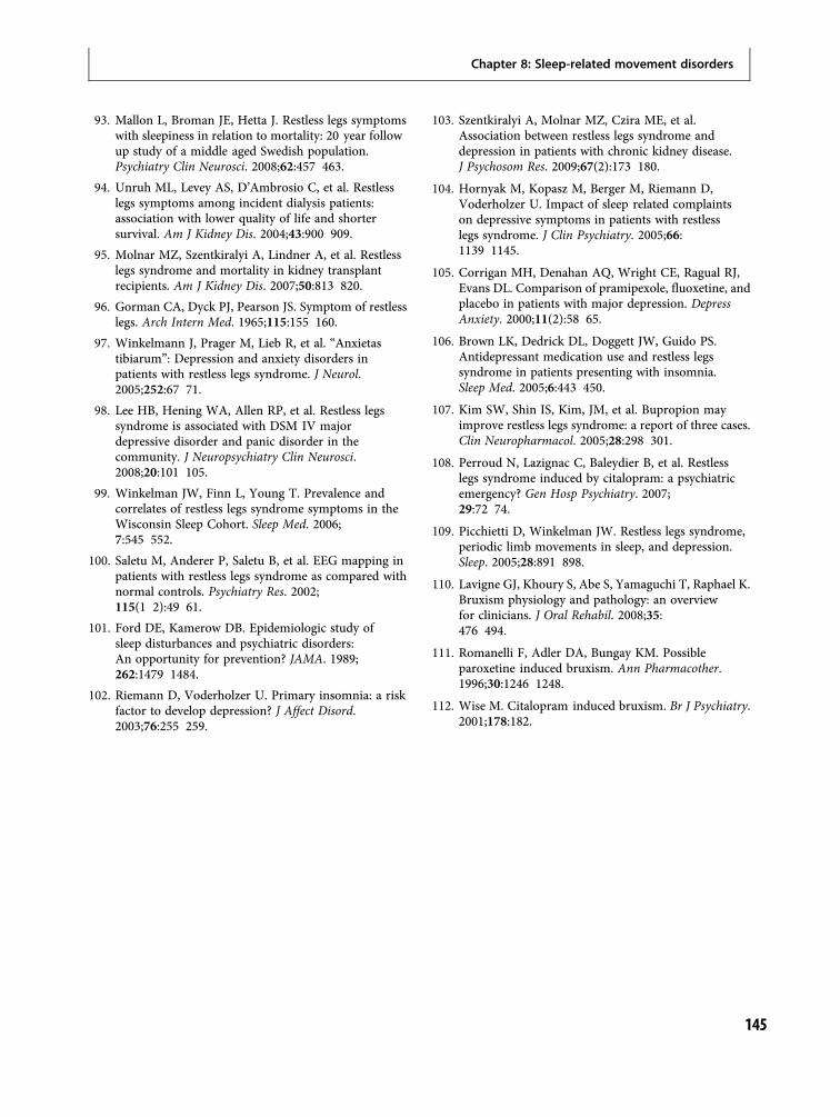
93. Mallon L, Broman JE, Hetta J. Restless legs symptomswith sleepiness in relation to mortality: 20 year followup study of a middle aged Swedish population.Psychiatry Clin Neurosci. 2008;62:457 463.
94. Unruh ML, Levey AS, D’Ambrosio C, et al. Restlesslegs symptoms among incident dialysis patients:association with lower quality of life and shortersurvival. Am J Kidney Dis. 2004;43:900 909.
95. Molnar MZ, Szentkiralyi A, Lindner A, et al. Restlesslegs syndrome and mortality in kidney transplantrecipients. Am J Kidney Dis. 2007;50:813 820.
96. Gorman CA, Dyck PJ, Pearson JS. Symptom of restlesslegs. Arch Intern Med. 1965;115:155 160.
97. Winkelmann J, Prager M, Lieb R, et al. “Anxietastibiarum”: Depression and anxiety disorders inpatients with restless legs syndrome. J Neurol.2005;252:67 71.
98. Lee HB, Hening WA, Allen RP, et al. Restless legssyndrome is associated with DSM IV majordepressive disorder and panic disorder in thecommunity. J Neuropsychiatry Clin Neurosci.2008;20:101 105.
99. Winkelman JW, Finn L, Young T. Prevalence andcorrelates of restless legs syndrome symptoms in theWisconsin Sleep Cohort. Sleep Med. 2006;7:545 552.
100. Saletu M, Anderer P, Saletu B, et al. EEG mapping inpatients with restless legs syndrome as compared withnormal controls. Psychiatry Res. 2002;115(1 2):49 61.
101. Ford DE, Kamerow DB. Epidemiologic study ofsleep disturbances and psychiatric disorders:An opportunity for prevention? JAMA. 1989;262:1479 1484.
102. Riemann D, Voderholzer U. Primary insomnia: a riskfactor to develop depression? J Affect Disord.2003;76:255 259.
103. Szentkiralyi A, Molnar MZ, Czira ME, et al.Association between restless legs syndrome anddepression in patients with chronic kidney disease.J Psychosom Res. 2009;67(2):173 180.
104. Hornyak M, Kopasz M, Berger M, Riemann D,Voderholzer U. Impact of sleep related complaintson depressive symptoms in patients with restlesslegs syndrome. J Clin Psychiatry. 2005;66:1139 1145.
105. Corrigan MH, Denahan AQ, Wright CE, Ragual RJ,Evans DL. Comparison of pramipexole, fluoxetine, andplacebo in patients with major depression. DepressAnxiety. 2000;11(2):58 65.
106. Brown LK, Dedrick DL, Doggett JW, Guido PS.Antidepressant medication use and restless legssyndrome in patients presenting with insomnia.Sleep Med. 2005;6:443 450.
107. Kim SW, Shin IS, Kim, JM, et al. Bupropion mayimprove restless legs syndrome: a report of three cases.Clin Neuropharmacol. 2005;28:298 301.
108. Perroud N, Lazignac C, Baleydier B, et al. Restlesslegs syndrome induced by citalopram: a psychiatricemergency? Gen Hosp Psychiatry. 2007;29:72 74.
109. Picchietti D, Winkelman JW. Restless legs syndrome,periodic limb movements in sleep, and depression.Sleep. 2005;28:891 898.
110. Lavigne GJ, Khoury S, Abe S, Yamaguchi T, Raphael K.Bruxism physiology and pathology: an overviewfor clinicians. J Oral Rehabil. 2008;35:476 494.
111. Romanelli F, Adler DA, Bungay KM. Possibleparoxetine induced bruxism. Ann Pharmacother.1996;30:1246 1248.
112. Wise M. Citalopram induced bruxism. Br J Psychiatry.2001;178:182.
Chapter 8: Sleep-related movement disorders
145
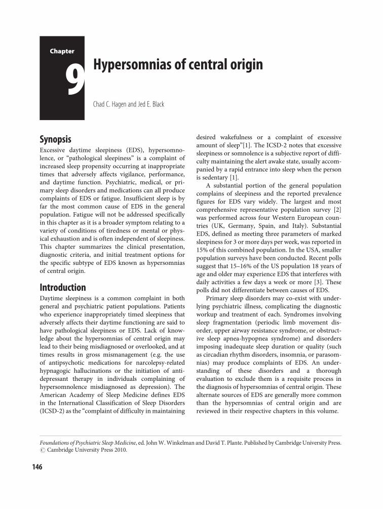
Chapter
9 Hypersomnias of central origin
Chad C. Hagen and Jed E. Black
SynopsisExcessive daytime sleepiness (EDS), hypersomno-lence, or “pathological sleepiness” is a complaint ofincreased sleep propensity occurring at inappropriatetimes that adversely affects vigilance, performance,and daytime function. Psychiatric, medical, or pri-mary sleep disorders and medications can all producecomplaints of EDS or fatigue. Insufficient sleep is byfar the most common cause of EDS in the generalpopulation. Fatigue will not be addressed specificallyin this chapter as it is a broader symptom relating to avariety of conditions of tiredness or mental or phys-ical exhaustion and is often independent of sleepiness.This chapter summarizes the clinical presentation,diagnostic criteria, and initial treatment options forthe specific subtype of EDS known as hypersomniasof central origin.
IntroductionDaytime sleepiness is a common complaint in bothgeneral and psychiatric patient populations. Patientswho experience inappropriately timed sleepiness thatadversely affects their daytime functioning are said tohave pathological sleepiness or EDS. Lack of know-ledge about the hypersomnias of central origin maylead to their being misdiagnosed or overlooked, and attimes results in gross mismanagement (e.g. the useof antipsychotic medications for narcolepsy-relatedhypnagogic hallucinations or the initiation of anti-depressant therapy in individuals complaining ofhypersomnolence misdiagnosed as depression). TheAmerican Academy of Sleep Medicine defines EDSin the International Classification of Sleep Disorders(ICSD-2) as the “complaint of difficulty in maintaining
desired wakefulness or a complaint of excessiveamount of sleep”[1]. The ICSD-2 notes that excessivesleepiness or somnolence is a subjective report of diffi-culty maintaining the alert awake state, usually accom-panied by a rapid entrance into sleep when the personis sedentary [1].
A substantial portion of the general populationcomplains of sleepiness and the reported prevalencefigures for EDS vary widely. The largest and mostcomprehensive representative population survey [2]was performed across four Western European coun-tries (UK, Germany, Spain, and Italy). SubstantialEDS, defined as meeting three parameters of markedsleepiness for 3 or more days per week, was reported in15% of this combined population. In the USA, smallerpopulation surveys have been conducted. Recent pollssuggest that 15–16% of the US population 18 years ofage and older may experience EDS that interferes withdaily activities a few days a week or more [3]. Thesepolls did not differentiate between causes of EDS.
Primary sleep disorders may co-exist with under-lying psychiatric illness, complicating the diagnosticworkup and treatment of each. Syndromes involvingsleep fragmentation (periodic limb movement dis-order, upper airway resistance syndrome, or obstruct-ive sleep apnea-hypopnea syndrome) and disordersimposing inadequate sleep duration or quality (suchas circadian rhythm disorders, insomnia, or parasom-nias) may produce complaints of EDS. An under-standing of these disorders and a thoroughevaluation to exclude them is a requisite process inthe diagnosis of hypersomnias of central origin. Thesealternate sources of EDS are generally more commonthan the hypersomnias of central origin and arereviewed in their respective chapters in this volume.
Foundations of Psychiatric SleepMedicine, ed. JohnW.Winkelman andDavid T. Plante. Published by CambridgeUniversity Press.# Cambridge University Press 2010.
146
Dorsal
Vigilance State Regulation Wakefulness
NREM Sleep REM Sleep
Pineal
Hippocampus
3V
4V
4V
Pons
RETMidbrain
Cerebellum
Medulla oblongata
Spinalcord
RostralRet cular formation
Thalamus
MnPnBF
SCN
Rostral
Caudal
VLPODMH
SUM
SN
VTA
PPT DR
SLDLDT LC
TMN
Pituitary
Ventral
Optic Chiasm
LHHypothalamus
Cerebral cortex
Dorsal
Pineal
Hippocampus
3V
4V
4V
Pons
RETMidbrain
Cerebellum
Medulla oblongata
Spinalcord
Reticular formation
Thalamus
MnPnBF
SCN
Caudal
VLPODMH
SUM
SN
VTA
PPT DR
SLDLDT LC
TMN
P tuitary
Ventral
Optic chiasm
LHHypothalamus
Cerebral cortex
Dorsal
Pineal
Hippocampus
3V
4V
4V
Pons
RETMidbrain
Cerebellum
Medulla oblongata
Spinalcord
RostralReticular formation
Thalamus
MnPNBF
SCN
Rostral
Caudal
VLPODMH
SUM
SN
VTA
PPT DR
SLDLDT LC
TMN
Pituitary
Ventral
Optic Chiasm
LHHypothalamus
Cerebral cortex
Dorsal
Pineal
Hippocampus
3V
4V
4V
Pons
RETMidbrain
Cerebellum
Medulla oblongata
Spinalcord
Reticular formation
Thalamus
MnPnBF
SCN
Caudal
VLPODMH
SUM
SN
VTA
PPT DR
SLDLDT LC
TMN
P tuitary
Ventral
Optic chiasm
LHHypothalamus
Cerebral cortex
(a)
(c) (d)
(b)
Figure 2.1 A schematic overview of the neural systems involved in vigilance state regulation. (a) Sagittal view of the rat brain (adapted from[3]) depicts nuclei and brain regions involved in vigilance state regulation (wakefulness, NREM sleep, and REM sleep) and circadian rhythmicity.In Figures 1b�d, active regions during the vigilance states are highlighted in red. (b) Nuclei that promote wakefulness, as well as regionsactivated during wakefulness, include: basal forebrain (BF) cholinergic, GABAergic, and putatively glutamatergic; dorsal raphe (DR) serotonergic;lateral hypothalamus (LH) orexinergic; locus coeruleus (LC) noradrenergic; pedunculopontine/laterodorsal tegmental (PPT/LDT) cholinergic;reticular formation glutamatergic; substantia nigra (SN) dopaminergic; tuberomammillary (TMN) histaminergic; and ventral tegmental area(VTA) dopaminergic nuclei. Thalamocortical and basal forebrain activity produces the EEG profile indicative of wakefulness. (c) Nuclei thatpromote NREM sleep include GABAergic neurons in the ventrolateral preoptic (VLPO) and median preoptic (MnPN) nuclei. Furthermore, thehomeostatic sleep regulator adenosine acts to inhibit wake promoting BF neurons. The reticular nucleus of the thalamus (RET) is responsiblefor NREM sleep phenomena such as sleep spindles. (d) Nuclei that promote REM sleep include the pedunculopontine/laterodorsal tegmentalnuclei (PPT/LDT), where unique “REM on” neurons are located. The sublaterodorsal nucleus (SLD) also promotes REM sleep phenomena,including muscle atonia, by means of circuitry involving the spinal cord. The supramammillary nucleus (SUM) and medial septum of the basalforebrain (BF) are part of the circuitry responsible for theta rhythmicity in the hippocampus, which is indicative of REM sleep. Abbreviations: 3V,third ventricle; 4V, fourth ventricle; DMH, dorsomedial nucleus of the hypothalamus; SCN, suprachiasmatic nucleus.
Slow waves60
(a) (b)
40
20
0
–20
–40
–60
60
40
20
0 0–20
–40
–60
60
40
20
–20
–40
–60
Sleep spindles Sleep spindles Sleep spindlesSleep spindles
Healthy controls Schizophrenics
Figure 3.2 (a) NREM sleep EEG traces and topographies of slow waves and sleep spindles in a healthy control. (b) Topographic analysisof sleep spindles revealed that schizophrenics had reduced spindle activity localized in a centroparietal area (adapted from [1]).
CytoplasmNucleus
100
75
50
25
0
Circadian time (hour)
Per/Cry
Rel
ativ
e po
rtei
n le
vel
(% o
f max
imum
)
0 6 12 18 24
Proteolysis
CLOCK
BMAL1
E-box
(a)
(b)
Cry 1/2
Per 1/2
P P
Rev-Erbα
Rev-Erbα
Rora
Rora
Core loop
Stablizing loop
RRE
PP
Cry 1/2
Per 1/2
Cry 1/2
Per 1/2
CKIGSK3
CKIGSK3
Per 1 Cry 1Per 2 Cry 2
Figure 2.7 Schematic representation of the interaction ofmammalian clock genes contributing to cellular circadianoscillations, adapted from [170]. (a) As described in the corefeedback loop (depicted in red), a heterodimeric transcription factor,including CLOCK and BMAL1, by acting on the E box DNA controlelement, initiates the expression of the negative regulators PER1 andPER2 (period proteins), as well as CRY1 and CRY 2 (cryptochromes).PER and CRY accumulate as the day progresses, multimerize in thecytoplasm, and are phosphorylated by the enzymes casein kinase I(CKI) and glycogen synthase kinase 3 (GSK3). The PER/CRY complexis then translocated to the nucleus, and represses CLOCK/BMAL1expression. PER and CRY proteins are then degraded at the end ofthe circadian cycle (CKI dependent), disinhibiting CLOCK and BMAL1transcription. As described in the stabilizing feedback loop (depictedin blue), BMAL1 transcription can activate the orphan nuclearreceptors Rora and Rev Erbα, which suppress and activate BMAL1expression respectively through the intermediary ROR responseelement (RRE). (b) Graphic depiction of Per/Cry protein levels in thesuprachiasmatic nucleus across the 24 hour day, adapted from [168].Circadian time 0 is the beginning of the subjective day/light periodfor humans. Abbreviations: P, phosphate.
Figure 10.3 Polysomnogram of72 year old man with REM sleepbehavior disorder. Although the rapideye movements are typical of REMsleep and the EEG is characteristicallydesynchronized, chin muscle atonia isincomplete during this burst of phasiceye movements, and there is prominentelectromyographic activation ofmuscles of upper and lower extremitiesmovements with correspondingmovement. Patient yelled out duringthis event and, retrospectively, recalleddreaming of fighting.

From Waking to NREM Sleep
From NREM to REM Sleep
From REM Sleep to Waking
Figure 3.4 Brain maps showing changes in rCBF from waking to NREM (top), NREM to REM (middle), and REM to waking (bottom), measuredwith H2
15O PET. Top panel: brain regions with decreased rCBF during NREM sleep compared with presleep wakefulness. The reduction isexpressed in Z scores, ranging from �1 (light purple) to �4.5 (deep purple). Significant rCBF reductions were observed in the pons(A, arrowhead), midbrain (B, short arrow), basal ganglia (B, long arrow; C, medium arrowhead), thalamus (C, short arrow), caudal orbital cortex(B, small arrowhead), and cerebellum (A, arrow). Similar rCBF decreases were found in the anterior insula (B, medium arrowhead), anteriorcingulate (C and D, small arrowheads), as well as in the orbital (B, medium arrow), dorsolateral prefrontal (C, medium arrow; D, small arrow), andinferior parietal lobes (D, medium arrow). Middle panel: brain regions with increased rCBF during REM sleep compared with NREM sleep.Z scores values range from 1 (green) to 3.5 (red). Significant increases in rCBF during REM sleep were observed in the pons (A, arrowhead),midbrain (B, long arrow), basal ganglia (B, short arrow; C, small arrowhead), thalamus (C, medium arrowhead), and caudal orbital cortex(B, medium arrowhead). Increases were also found in anterior insula (B, small arrowhead), anterior cingulate (C, small arrow; D, medium arrow),mesial temporal (parahippocampal) (B, medium arrow), fusiform inferotemporal (B, large arrowhead), lateral occipital (C, long arrow),auditory association (C, medium arrow), and medial prefrontal cortices (D, small arrow). Bottom panel: brain regions with rCBF increaseduring post sleep wakefulness compared to REM sleep. Z scores values are from 1 (green) to 3.5 (red). Significant increases in rCBF occurred inthe orbital (B, small arrow), dorsolateral prefrontal (C and D, small arrows), and inferior parietal lobes (D, medium arrow), as well as in thecerebellar hemispheres (A, small arrow). Reproduced, with permission, from [2].
(a) Figure 10.2 (a) Oximetry tracing froman obese 38 year old woman showingfrequent desaturations ofoxyhemoglobin, reaching a nadir of<60% during polysomnographicallydocumented non REM sleep, secondaryto obstructive sleep apnea. Apnea andhypopnea events occurred at a rate of80 per hour of sleep. Each desaturation isaccompanied by evidence of arousal fromsleep on the correspondingpolysomnogram. She had complained offrequent spells of SW, which includedeating foods from her cupboards andrefrigerator. She responded well to CPAPat 8 9 cmH2O pressure, with resolution ofobstructive sleep apnea and SW.
Figure 10.1 Nocturnal polysomnogram of 32 year old man with history of SW/ST demonstrating an abrupt spontaneous arousal fromnon REM stage N2 sleep as the record becomes suddenly obscured by muscular movement artifact. Tachycardia develops later duringthe arousal and the man subsequently left the bed and later reported dream recall of being in his son’s house in a living room filled with redfurniture and becoming convinced that “some disaster like a fire” was about to occur. He dreamed that he attempted to warn his wifewhen he, in fact, bolted from his bed and subsequently only slowly was convinced by the technologist that there was no real imminentdanger. There is no epileptiform activity in the EEG.
(a) (b)
%
15
+35
0
–20
+50
0
–40
+20
0
–20
5
–5
–10
–15
0
10
(c) (d)
Figure 3.5 Cortical plasticity is reflected by local changes in SWA. In each panel regions with SWA increase are in red, while in blue areregions with SWA decrease. White circles indicate electrodes with significant SWA activity change. (a) Increased SWA after a rotation learningtask. Six electrodes with significant differences from baseline located in the right sensorimotor area were found [3]. (b) Decreased SWAfollowing left arm immobilization for one day was found in three electrodes in the right sensorimotor cortex [4]. (c) Increased SWA followingrTMS of the left premotor cortex [5]. (d) Increased SWA at the right inferior frontal gyrus, symmetrical to Broca’s area, in a stroke patient withexpressive aphasia following 4 hours of speech therapy.
Evaluation of the patient with EDSA detailed history and physical examination are of keyimportance in evaluating the patient complaining ofEDS. Important points of the sleep history that theclinician should document include, but are not limitedto, total daily 24-hour sleep time, nightly and dailysleep and nap patterns, time to initial sleep onset, andnumber and duration of nocturnal awakenings. Asleep log in either narrative or graphical form may beused to characterize a patient’s sleep further.
When reviewing a patient’s sleep log in theworkup of EDS, special attention is paid to the quan-tity, consistency, and continuity of reported sleep.The consistency of sleep–wake times provides insightinto circadian and social pressures on the timing ofsleep and may help identify causes of insufficientsleep. If the patient is unable to give a reliable historyor nightly sleep times are in question, monitoring forseveral days with actigraphy (a wristwatch-like devicethat registers movement by the patient) may be usefulin evaluating patterns of waking and sleep.
Thorough screening for primary sleep disordersincludes assessment of the presence of snoring, wit-nessed apneas, symptoms of restless legs syndrome,periodic limb movements, and restless sleep or non-refreshing sleep. Medical conditions and alcohol ordrug use can be significant contributors to EDS; if anyof these are suspected, appropriate evaluation shouldensue. Any chronic sedating medications should alsobe noted.
If a thorough history and physical examinationraises suspicion of disorders such as periodic limbmovements, obstructive sleep apnea, nocturnal seizure,or parasomnia, then nocturnal polysomnography(PSG) – overnight sleep study – may be indicated toeither rule out sleep disturbances or to quantify theirseverity. This is typically related to sleep disorders thatproduce sleep fragmentation or sleep inefficiencyincluding sleep-related breathing disorders, periodiclimbmovement disorder, REM sleep behavior disorder,other sleep-related movement disorders, parasomnias,or nocturnal seizures.
Several questionnaires have been developed tohelp clinicians screen for and evaluate patients forsleepiness and its impact on daily living. These ques-tionnaires include, among others, the Epworth sleepi-ness scale (ESS) [4], the Stanford sleepiness scale [5],and the sleep–wake activity inventory [6]. The ESS isthe most commonly used questionnaire because of its
ease of use and its well-documented validity as aclinically relevant measure of sleepiness in conditionsof pathological EDS [7–9].
Although ESS scores are not necessarily represen-tative of true levels of sleepiness and the questionnaireis neither highly specific nor sensitive for the exist-ence of pathological sleepiness, the ESS serves as auseful screen for patients who are severely sleepy [10].In our experience it has also been a simple, quick, andlow-cost method for following patient response totreatment. Given its ease of use and the high preva-lence of sleepiness among the general population, werecommend administering the ESS in patients withcomplaints of excessive sleepiness [4].
The multiple sleep latency test (MSLT) provides amore objective quantification of sleepiness than ques-tionnaires by objectively measuring the propensity tofall asleep. Please see Chapter 3: Neurophysiology andneuroimaging of human sleep for a more detailedreview of this procedure. Essentially, this test meas-ures the capacity to fall asleep and propensity for bothearly sleep onset and early rapid eye movement sleep(REM) onset. The MSLT has specificity limitationsdue to the variety of conditions that can produce anabnormal result. Nevertheless, following a thoroughclinical evaluation, the MSLT is the primary tool forconfirming some of the hypersomnias [1,11]. TheMSLT consists of four or five 20-minute polysomno-graphically monitored daytime nap opportunitiesseparated by 2-hour intervals; for the nap opportun-ities, the patient is reclined in a sleep laboratory bed ina dark room with instructions to fall asleep. Theprimary assessments made by the MSLT are the rap-idity of sleep onset, which correlates to degree ofsleepiness, and the occurrence of REM sleep if sleepoccurs during the nap opportunity. REM sleep epi-sodes (a period of sleep during which dreams occur)at or close to sleep onset are known as sleep-onsetREM periods (SOREMPs). Sleep latencies in normaladults are often between 10 and 20 minutes; patho-logical sleepiness is manifested by a latency of lessthan 8 minutes [11,12]. The MSLT should be per-formed only after the patient has received an adequateamount of nocturnal sleep (approximately 8 hoursper night for the typical adult) for a period of at least2 weeks and immediately following a nocturnal poly-somnogram to exclude other causes of EDS due toeither sleep fragmentation or insufficient sleep. If thepolysomnogram is positive for other causes of EDS,these conditions must be adequately treated before an
Chapter 9: Hypersomnias of central origin
147
evaluation of EDS with MSLT is pursued. Practicingclinicians should also be aware that commonly pre-scribed antidepressants can have profound effects onthe MSLT by suppressing potential SOREMPs. There-fore, the decision regarding whether such medicationsshould be tapered prior to MSLT should be madecooperatively with the patient, and include the risksof potentially worsening the psychiatric illness versusa confounded MSLT.
The maintenance of wakefulness test (MWT) isanother diagnostic test in the sleep laboratory. Ratherthan evaluating the propensity to fall asleep (like theMSLT), the MWT assesses the capacity to maintainwakefulness while sedentary in a setting conducive tosleep during the patient’s regular waking hours. Thistest is not generally used in the evaluation of hyper-somnias of central origin, rather it is used for safetyassessment to evaluate the impact of treatment forsleep disorder-related EDS in specific occupationssuch as heavy equipment operators or airline pilots.
Behaviorally induced insufficient sleepInsufficient sleep is the most common cause of EDSin Western culture. Although the exact prevalence isunclear, in the 2002 Sleep in America Poll conductedby the National Sleep Foundation [3], 37% of adultsreported sleeping less than 7 hours a night on week-nights and 68% reported sleeping less than 8 hours.Of employed respondents to the 2008 Sleep in Amer-ica Poll, 44% reported sleeping less than 7 hours onwork nights [13]. Although sleep requirements varyamong individuals, these polls find that weeknightsleep is 2 hours less than that obtained on weekendsfor 18% of respondents. Of subjects describing thispattern across the week, 24% endorsed sleepinessimpairing their performance at least a few days perweek, and 40% with this pattern admitted drivingwhen drowsy at least once per month for the previousyear. This discrepancy between weeknight and week-end sleep times implies ongoing voluntary sleeprestriction during the week with attempted sleep com-pensation on the weekends.
The number of hours of experimental sleep loss innormal volunteers is directly proportional to thedegree of increased daytime sleepiness as assessed bythe MSLT [14]. The effects of sleep deprivation maybe cumulative [8], but this accumulated sleep debtmay be countered by extending sleep time over sev-eral days [15]. Insufficient sleep may be due to
voluntary lifestyle choices, job or school demands,shift work, or poor sleep hygiene.
As much as 16% of the US workforce is engaged inshift work [16]. Some research has suggested thatdespite subjectively experiencing adequate sleep, shiftworkers sleep approximately 5 to 7 hours less perweek when compared with diurnal workers [17].Studies have consistently shown that workers whoregularly work night shifts experience more disruptedsleep as well as sleepiness during waking hours, com-pared with day workers [18,19]. Moreover, the ten-dency to feel sleepy or to doze may be as great for aworker during the night shift as for a person withuntreated narcolepsy [20].
Although insufficient sleep might be expectedto lead to frank EDS, a constellation of other subject-ive complaints are more commonly seen, includingtiredness, lack of energy, or fatigue. Decrements inattention, learning capacity, short-term memory,and psychomotor performance, with or withoutEDS, also may be present. Moreover, irritability, poorimpulse control, or other forms of mood instabilitymay exist alone or in combination with these featuresin individuals with insufficient sleep. While it is obvi-ous that complete sleep deprivation compromisesperformance and well-being, until the last decade anerroneous belief has persisted that people adapt tochronic sleep loss without significant compromise.This is being challenged by a rapidly expanding lit-erature suggesting a wide range of adverse medical,cognitive, and socioeconomic effects of sleep loss anduntreated sleep disorders [21]. The effects of chronic-ally restricted sleep are often insidious and may beunrecognized by many patients. It has been demon-strated that reducing sleep duration to 6 hours pernight for 2 weeks produces detriments in vigilance,attention, and concentration that are equivalent to2 nights of complete sleep deprivation [22]. Interest-ingly, subjective ratings of sleepiness in this studyincreased acutely in response to sleep restriction, butminimal additional change was reported on subse-quent days, suggesting that subjects were unaware oftheir deteriorating neurobehavioral functioning, per-haps explaining why the impact of chronic sleeprestriction is misperceived as relatively benign.
While it may seem obvious, education and coun-seling regarding good sleep habits is the first-linetreatment for insufficient sleep (Box 9.1). Manypatients with behaviorally induced insufficient sleepare surprised to hear that they should be sleeping 7.5
Section IV: Primary Sleep Disorders in Psychiatric Contexts
148
to 8 hours per night or more. Many struggle withcompeting social obligations and may be under tre-mendous pressure to sleep an inadequate duration.Despite presenting with a complaint of excessivesleepiness, some may argue or resist recommenda-tions to increase sleep time and improve consistencyin the timing of sleep. Others need only to bereminded that sleep is essential to well-being and itneeds to be prioritized more highly among their otherobligations. Shift workers with unavoidable insuffi-cient sleep and concomitant EDS may benefit frommodafinil, which has been approved by the Food andDrug Administration (FDA) for treatment of EDS inshift work sleep disorder.
NarcolepsyNarcolepsy is the most clearly understood of the cen-tral hypersomnia syndromes. It is characterized byprofound EDS and is associated with cataplexy, sleepparalysis, hypnopompic or hypnagogic hallucinations,automatic behavior, and disrupted sleep. Narcolepsy israre in all populations with reported prevalencesranging from 0.02% to 0.18% with lower prevalencein Israel and higher reported prevalence in Japan [1].Symptoms usually arise in adolescence or young adult-hood and rarely as early as age 5, although symptomonset can be delayed until the 6th or 7th decade of life.
Narcolepsy results from dysfunction within thehypothalamic hypocretin (orexin) system in bothanimal models and humans. While mice and caninemodels of narcolepsy are associated with hypocretinligand knockout or receptor gene mutations [23,24],genetic abnormalities in the hypocretin system areextremely rare in humans. About 90% of patients withunequivocal narcolepsy with cataplexy have low orundetectable levels of hypocretin in their cerebrospinalfluid – a finding which is quite specific to narcolepsy[25,26]. Human leukocyte antigen (HLA) subtypeDQB1*0602 has a strong, but non-specific associationwith narcolepsy with cataplexy. Nearly all patientswith cataplexy have this subtype, while it is foundin 12–38% of the general population, across variousraces. These and other immunological associationsalong with the age of onset and finding of post-mortem destruction of hypothalamic hypocretin-producing cells have led to the theory that narcolepsymay arise from an autoimmune process.
The routine diagnosis of narcolepsy is based on aclinical history of excessive daytime sleepiness occur-ring almost daily for more than 3 months with aformal workup that excludes other potential etiologiesfor EDS and amean sleep latency of less than 8minuteswith two or more sleep onset REM periods on theMSLT. Narcolepsy is categorized as either with cata-plexy or without cataplexy. Cataplexy is defined as adiscrete episode of sudden and transient bilateralloss of voluntarymuscle tone triggered by emotion [1].
Low CSF hypocretin is far more specific than theMSLT for the diagnosis of narcolepsy [27]. HLA sub-typing is less invasive and more readily available, butgrossly non-specific given the prevalence of this sub-type in the general population. The clinical utility ofHLA DQB1*0602 subtyping is limited to the fact thatin the absence of cataplexy, a negative result verystrongly suggests normal hypocretin function andobviates the need for CSF hypocretin evaluation. Cur-rently these assays are not considered standard in thediagnosis of narcolepsy, but ongoing efforts are clari-fying their potential clinical utility.
Excessive sleepinessEDS in narcoleptic patients manifests as an increasedpropensity to fall asleep in relaxed or sedentary situ-ations or a struggle to avoid sleeping in thesesituations. These so-called “sleep attacks” are oftenmistakenly believed to be unique to narcolepsy, but
Box 9.1 Healthy sleep habits for people withbehaviorally induced insufficient sleep
1. Stay in bed for 8 hours per night2. Maintain a dark, quiet, comfortable sleep envir-
onment (without television or music duringsleep)
3. Go to bed and wake up at the same time 7 nightsper week
4. Optimize bright light exposure in first few hoursof the morning
5. Avoid bright light and stimulating activities(stressful activities, computer use, television,video games, etc.) in the few hours beforebedtime
6. Use the bedroom for sleep and sex only7. Obtain regular exercise, but complete it 3 hours
or more before bedtime8. Avoid caffeine for 5 hours before bedtime9. Avoid nicotine and alcohol for 3 hours before
bedtime
Chapter 9: Hypersomnias of central origin
149

actually represent sleepiness that is identical to that ofother sleep disorders such as untreated obstructivesleep apnea or sleep deprivation [17]. Epworth sleepi-ness scale scores of �15 are common in untreatedpatients [4,17,20]. The irresistible urge to sleep com-monly occurs in inappropriate or dangerous situ-ations and thus produces significant social andoccupational dysfunction. While the ICSD-2 specifiesan MSLT mean sleep latency of less than 8 minutes assupport for the diagnosis of narcolepsy, narcolepsypatients commonly evidence much shorter meansleep latencies of less than 5 minutes (Table 9.1).
CataplexyCataplexy is a partial or complete transient bilateralweakness in voluntary muscles in response to strongemotion. This often manifests as bilateral ptosis, headdrooping, slurred speech, or dropping things from thehand. It less commonly results in total body paralysisor collapse and injuries are rare as effort and timeusually permit successful redirection or protection ofoneself. Cataplexy may be associated with a variety ofemotions, but is most reliably associated with humor-ous thoughts, laughing, or joking [17]. Events lastfrom a few seconds up to 3 minutes but occasionallycontinue longer [28]. Although alert and orientedduring the event, if unable to move or speak duringan episode of cataplexy, some patients may ultimatelylapse into sleep given the narcoleptic’s propensity tosleep in sedentary situations. Unlike the other featuresof narcolepsy, cataplexy is very specific to narcolepsy.
The first experience of cataplexy is usually withina few months of the onset of excessive sleepiness, butthe onset of cataplexy can occur many years after theinitial onset of sleepiness [28]. Additionally, cataplexyseverity can increase over time.
Hypnagogic or hypnopompic hallucinationsThese phenomena may be visual, tactile, auditory, ormulti-sensory events, usually brief but occasionally con-tinuing for a few minutes, that occur at the transitionfrom wakefulness to sleep (hypnagogic) or from sleepto wakefulness (hypnopompic). Hallucinations maycontain elements of dream sleep and consciousnesscombined and are often bizarre or disturbing topatients. As with excessive sleepiness, these hallucin-atory experiences are not specific to narcolepsy, andcan be seen with other conditions of excessive sleepi-ness. Additionally, these phenomena are not necessarilypathological, are common in the general population,and may be reported more frequently in individualswith mood, anxiety, or psychotic illness [29].
Sleep paralysisSleep paralysis is the inability to move, lasting froma few seconds to minutes, during the transitionfrom sleep to wakefulness or from wakefulness tosleep. Episodes of sleep paralysis may alarm patients,particularly those who experience the sensation ofbeing unable to breathe. This occurs because acces-sory respiratory muscles may not be active duringthese episodes; however, diaphragmatic activity con-tinues and air exchange remains adequate. Sleep par-alysis, like excessive sleepiness and hypnagogic andhypnopompic hallucinations, is not specific to narco-lepsy and is often seen with various disorders thatcause excessive sleepiness (sleep apnea or chronicsleep deprivation). Additionally, sleep paralysis isnot necessarily indicative of pathology, but may bemore common in patients with mood and anxietydisorders [30].
Treatment of narcolepsyThe most significant impact on the treatment ofnarcolepsy in recent history has been the character-ization of the effect of sodium oxybate (the sodiumsalt of g-hydroxybutyrate) on cataplexy, daytimesleepiness, and nocturnal sleep fragmentation.Sodium oxybate’s sleep-promoting effect appears to
Table 9.1 Average multiple sleep latency test (MSLT) resultsby disorder
Mean SleepLatency(Minutes)
Number of napswith REM sleep
Narcolepsy 3.1 2 or more
Idiopathichypersomnia
6.2 <2
Obstructivesleep apnea
10.6 0
Normalcontrols
11* 0
Note: *30% of normal controls have a mean sleep latency less than8 minutes.Source: Adapted from ICSD 2 [1] and AASM MSLT [11] practiceguidelines.
Section IV: Primary Sleep Disorders in Psychiatric Contexts
150

be largely mediated via gamma-aminobutyric acid(GABA) receptor subtype B agonism, but the mech-anism by which it improves cataplexy and EDSis unknown. Although the use of low-dose sodiumoxybate in treating narcolepsy has been studied for35 years, it has only been in the past 10 years thatthorough investigations in the form of multi-centertrials have been conducted. These trials have exten-sively characterized the dose–response impact ofsodium oxybate on the enhancement of nocturnalsleep, improvement of cataplexy, and improvementof EDS [31,32]. These findings support the view thatsodium oxybate may be the optimal first-line agentfor the treatment of narcolepsy with cataplexy.
While sodium oxybate has demonstrated its effi-cacy in treating all symptoms of narcolepsy, alertingagents provide a critical adjunctive component in thetreatment of patients with narcolepsy, specifically forthe treatment of EDS. Sodium oxybate is a controlledsubstance and distribution is restricted to a singlecentral pharmacy. Appropriate patient selection andmonitoring are essential as side-effects include centralnervous system depression, and potential for diver-sion or abuse.
Although stimulants remain the first-line agentsfor the treatment of EDS in patients with narcolepsy,in our clinical practice they are now considereda useful addition to sodium oxybate in patientswho have narcolepsy with cataplexy. Common prac-tice is to combine two agents when one agent does notadequately ameliorate symptoms. The use of modafi-nil as an adjunct to sodium oxybate has been shownto provide significantly greater improvement in meas-ures of EDS symptoms than either agent alone [33].Some patients may wish to avoid medications andattempt to take extra naps during the day, but nappingalone is rarely successful in alleviating EDS enoughto function at or near normal capacity. While alertingagents may not eliminate daytime symptoms, theyhave been shown to produce substantial improvementin EDS associated with narcolepsy [34].
Modafinil is a novel alerting agent that promoteswakefulness primarily via its dopaminergic effect[35]. Similar to traditional stimulants, modafinilappears to function as a dopamine transporterinhibitor, but unlike the amphetamines, it has areduced receptor affinity and it does not inducedopamine release. This difference in activity mayaccount for the improved tolerability of modafinilover traditional stimulants as well as its diminished
potential for street use, abuse, and addiction by illicitusers [36].
In addition to modafinil, commonly used stimu-lants include methylphenidate, dextro-amphetamine,and methamphetamine. Side-effects are not uncom-mon with any stimulant or alerting agent. Agitation,anxiety, tremor, and palpitations are just a few ofthe commonly reported side-effects associated withtraditional stimulants. Some patients may report arebound hypersomnia as the dose wears off, and toler-ance may occur with time. Traditional stimulants arestill an important resource in the arsenal ofmedicationsfor the treatment of narcolepsy, but in our clinicalpractice they have become second-line agents behindsodium oxybate and/or modafinil for treatment of EDSassociated with narcolepsy (see Table 9.2).
While modafinil and traditional stimulants canameliorate EDS, these agents impart no beneficialimpact on cataplexy. However, drugs other thansodium oxybate can be used effectively to treat cata-plexy.While not rigorously studied in controlled trials,agents which exhibit affinity for CNS norepinephrinereceptors have been in common use for decadesas anticataplectic agents. Specifically, tricyclic anti-depressants, mixed serotonin-norepinephrine re-uptake inhibitors, and more selective serotoninreuptake inhibitors with parent compound or activemetabolite affinity for noradrenergic receptors (e.g.fluoxetine) can be effective in the treatment of cata-plexy. Frequently patients with narcolepsy experienceEDS many months in advance of the development ofcataplexy. At this early stage of disease developmentthe patientmay present to a physician with a complaintof “tiredness” coupled with diminished motivationand reduced energy and therefore be misdiagnosedwith an atypical form of depression. Treating such apatient with antidepressants for presumed depressionbears the risk of masking the later development ofcataplexy, due to the anticataplectic effect of the anti-depressant, and delaying (potentially substantially) theaccurate diagnosis of narcolepsy.
Idiopathic hypersomniaIdiopathic hypersomnia (previously labeled “idio-pathic CNS hypersomnia”) is a primary disorder ofEDS that has historically been given as a diagnosis ofexclusion to individuals who complain of EDS whenother disorders that cause hypersomnolence have notbeen found or clearly characterized. There are
Chapter 9: Hypersomnias of central origin
151
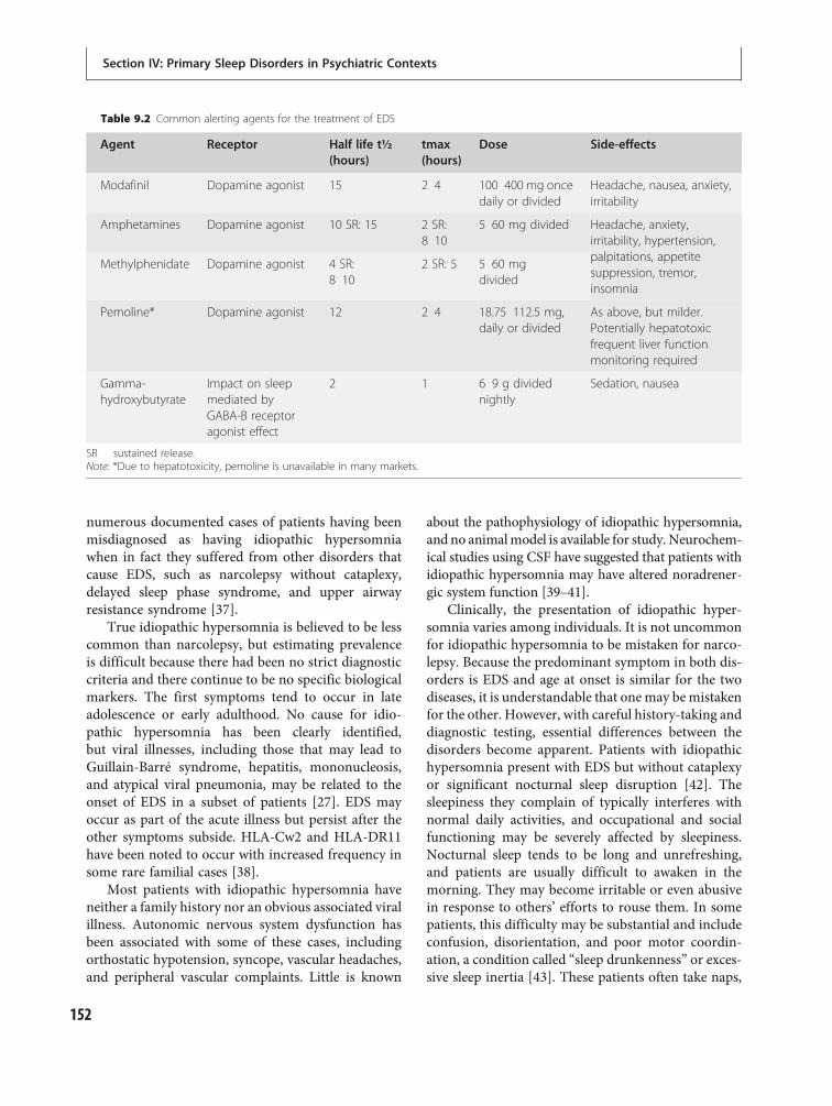
numerous documented cases of patients having beenmisdiagnosed as having idiopathic hypersomniawhen in fact they suffered from other disorders thatcause EDS, such as narcolepsy without cataplexy,delayed sleep phase syndrome, and upper airwayresistance syndrome [37].
True idiopathic hypersomnia is believed to be lesscommon than narcolepsy, but estimating prevalenceis difficult because there had been no strict diagnosticcriteria and there continue to be no specific biologicalmarkers. The first symptoms tend to occur in lateadolescence or early adulthood. No cause for idio-pathic hypersomnia has been clearly identified,but viral illnesses, including those that may lead toGuillain-Barré syndrome, hepatitis, mononucleosis,and atypical viral pneumonia, may be related to theonset of EDS in a subset of patients [27]. EDS mayoccur as part of the acute illness but persist after theother symptoms subside. HLA-Cw2 and HLA-DR11have been noted to occur with increased frequency insome rare familial cases [38].
Most patients with idiopathic hypersomnia haveneither a family history nor an obvious associated viralillness. Autonomic nervous system dysfunction hasbeen associated with some of these cases, includingorthostatic hypotension, syncope, vascular headaches,and peripheral vascular complaints. Little is known
about the pathophysiology of idiopathic hypersomnia,and no animalmodel is available for study. Neurochem-ical studies using CSF have suggested that patients withidiopathic hypersomnia may have altered noradrener-gic system function [39–41].
Clinically, the presentation of idiopathic hyper-somnia varies among individuals. It is not uncommonfor idiopathic hypersomnia to be mistaken for narco-lepsy. Because the predominant symptom in both dis-orders is EDS and age at onset is similar for the twodiseases, it is understandable that onemay bemistakenfor the other. However, with careful history-taking anddiagnostic testing, essential differences between thedisorders become apparent. Patients with idiopathichypersomnia present with EDS but without cataplexyor significant nocturnal sleep disruption [42]. Thesleepiness they complain of typically interferes withnormal daily activities, and occupational and socialfunctioning may be severely affected by sleepiness.Nocturnal sleep tends to be long and unrefreshing,and patients are usually difficult to awaken in themorning. They may become irritable or even abusivein response to others’ efforts to rouse them. In somepatients, this difficulty may be substantial and includeconfusion, disorientation, and poor motor coordin-ation, a condition called “sleep drunkenness” or exces-sive sleep inertia [43]. These patients often take naps,
Table 9.2 Common alerting agents for the treatment of EDS
Agent Receptor Half life t½(hours)
tmax(hours)
Dose Side-effects
Modafinil Dopamine agonist 15 2 4 100 400 mg oncedaily or divided
Headache, nausea, anxiety,irritability
Amphetamines Dopamine agonist 10 SR: 15 2 SR:8 10
5 60 mg divided Headache, anxiety,irritability, hypertension,palpitations, appetitesuppression, tremor,insomnia
Methylphenidate Dopamine agonist 4 SR:8 10
2 SR: 5 5 60 mgdivided
Pemoline* Dopamine agonist 12 2 4 18.75 112.5 mg,daily or divided
As above, but milder.Potentially hepatotoxicfrequent liver functionmonitoring required
Gamma-hydroxybutyrate
Impact on sleepmediated byGABA-B receptoragonist effect
2 1 6 9 g dividednightly
Sedation, nausea
SR sustained release.Note: *Due to hepatotoxicity, pemoline is unavailable in many markets.
Section IV: Primary Sleep Disorders in Psychiatric Contexts
152
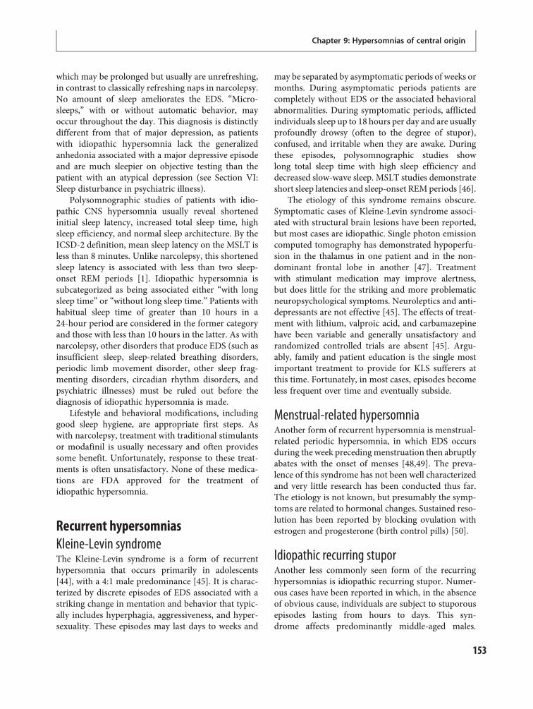
which may be prolonged but usually are unrefreshing,in contrast to classically refreshing naps in narcolepsy.No amount of sleep ameliorates the EDS. “Micro-sleeps,” with or without automatic behavior, mayoccur throughout the day. This diagnosis is distinctlydifferent from that of major depression, as patientswith idiopathic hypersomnia lack the generalizedanhedonia associated with a major depressive episodeand are much sleepier on objective testing than thepatient with an atypical depression (see Section VI:Sleep disturbance in psychiatric illness).
Polysomnographic studies of patients with idio-pathic CNS hypersomnia usually reveal shortenedinitial sleep latency, increased total sleep time, highsleep efficiency, and normal sleep architecture. By theICSD-2 definition, mean sleep latency on the MSLT isless than 8 minutes. Unlike narcolepsy, this shortenedsleep latency is associated with less than two sleep-onset REM periods [1]. Idiopathic hypersomnia issubcategorized as being associated either “with longsleep time” or “without long sleep time.” Patients withhabitual sleep time of greater than 10 hours in a24-hour period are considered in the former categoryand those with less than 10 hours in the latter. As withnarcolepsy, other disorders that produce EDS (such asinsufficient sleep, sleep-related breathing disorders,periodic limb movement disorder, other sleep frag-menting disorders, circadian rhythm disorders, andpsychiatric illnesses) must be ruled out before thediagnosis of idiopathic hypersomnia is made.
Lifestyle and behavioral modifications, includinggood sleep hygiene, are appropriate first steps. Aswith narcolepsy, treatment with traditional stimulantsor modafinil is usually necessary and often providessome benefit. Unfortunately, response to these treat-ments is often unsatisfactory. None of these medica-tions are FDA approved for the treatment ofidiopathic hypersomnia.
Recurrent hypersomniasKleine-Levin syndromeThe Kleine-Levin syndrome is a form of recurrenthypersomnia that occurs primarily in adolescents[44], with a 4:1 male predominance [45]. It is charac-terized by discrete episodes of EDS associated with astriking change in mentation and behavior that typic-ally includes hyperphagia, aggressiveness, and hyper-sexuality. These episodes may last days to weeks and
may be separated by asymptomatic periods of weeks ormonths. During asymptomatic periods patients arecompletely without EDS or the associated behavioralabnormalities. During symptomatic periods, afflictedindividuals sleep up to 18 hours per day and are usuallyprofoundly drowsy (often to the degree of stupor),confused, and irritable when they are awake. Duringthese episodes, polysomnographic studies showlong total sleep time with high sleep efficiency anddecreased slow-wave sleep. MSLT studies demonstrateshort sleep latencies and sleep-onset REMperiods [46].
The etiology of this syndrome remains obscure.Symptomatic cases of Kleine-Levin syndrome associ-ated with structural brain lesions have been reported,but most cases are idiopathic. Single photon emissioncomputed tomography has demonstrated hypoperfu-sion in the thalamus in one patient and in the non-dominant frontal lobe in another [47]. Treatmentwith stimulant medication may improve alertness,but does little for the striking and more problematicneuropsychological symptoms. Neuroleptics and anti-depressants are not effective [45]. The effects of treat-ment with lithium, valproic acid, and carbamazepinehave been variable and generally unsatisfactory andrandomized controlled trials are absent [45]. Argu-ably, family and patient education is the single mostimportant treatment to provide for KLS sufferers atthis time. Fortunately, in most cases, episodes becomeless frequent over time and eventually subside.
Menstrual-related hypersomniaAnother form of recurrent hypersomnia is menstrual-related periodic hypersomnia, in which EDS occursduring the week precedingmenstruation then abruptlyabates with the onset of menses [48,49]. The preva-lence of this syndrome has not been well characterizedand very little research has been conducted thus far.The etiology is not known, but presumably the symp-toms are related to hormonal changes. Sustained reso-lution has been reported by blocking ovulation withestrogen and progesterone (birth control pills) [50].
Idiopathic recurring stuporAnother less commonly seen form of the recurringhypersomnias is idiopathic recurring stupor. Numer-ous cases have been reported in which, in the absenceof obvious cause, individuals are subject to stuporousepisodes lasting from hours to days. This syn-drome affects predominantly middle-aged males.
Chapter 9: Hypersomnias of central origin
153
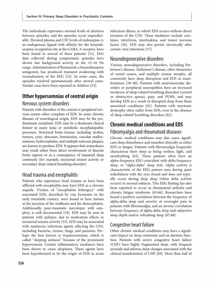
The individuals experience normal levels of alertnessbetween episodes, and the episodes occur unpredict-ably. Elevated plasma and CSF levels of endozepine-4,an endogenous ligand with affinity for the benzodi-azepine recognition site at the GABA-A receptor, havebeen found in several of these patients [51]. EEGdata collected during symptomatic episodes haveshown fast background activity in the 13–16 Hzrange. Administration of flumazenil, a benzodiazepineantagonist, has produced transient awakening withnormalization of the EEG [52]. In some cases, theepisodes resolved spontaneously after several years.Similar cases have been reported in children [53].
Other hypersomnias of central originNervous system disordersPatients with disorders of the central or peripheral ner-vous system often complain of EDS. In some chronicdiseases of neurological origin, EDS may be the pre-dominant complaint. EDS may be a dominant clinicalfeature in many toxic or metabolic encephalopathicprocesses. Structural brain lesions, including strokes,tumors, cysts, abscesses, hematomas, vascular malfor-mations, hydrocephalus, and multiple sclerosis plaques,are known to produce EDS. It appears that somnolencemay result either from direct involvement of discretebrain regions or as a consequence of impaired sleepcontinuity (for example, nocturnal seizure activity orsecondary sleep-related breathing disorder).
Head trauma and encephalitisPatients who experience head trauma or have beenafflicted with encephalitis may have EDS as a chronicsequela. Victims of “encephalitis lethargica” withassociated EDS, described by von Economo in theearly twentieth century, were found to have lesionsat the junction of the midbrain and the diencephalon.Additionally, post-traumatic narcolepsy with cata-plexy is well documented [54]. EDS may be seen inpatients with epilepsy, due to medication effects ornocturnal seizure activity [55]. EDS may be associatedwith numerous infectious agents affecting the CNS,including bacteria, viruses, fungi, and parasites. Per-haps the best known is trypanosomiasis, which iscalled “sleeping sickness” because of the prominenthypersomnia. Certain inflammatory mediators havebeen shown to cause sleepiness. These agents havebeen hypothesized to be the origin of EDS in acute
infectious illness, in which EDS occurs without directinvasion of the CNS. These mediators include cyto-kines, interferon, interleukins, and tumor necrosisfactor [56]. EDS may also persist chronically aftercertain viral infections [57].
Neurodegenerative disordersVarious neurodegenerative disorders, including Par-kinson’s disease, Alzheimer’s disease, other dementiasof varied causes, and multiple system atrophy, allcommonly have sleep disruption and EDS as mani-festations [58–60]. Patients with neuromuscular dis-orders or peripheral neuropathies have an increasedincidence of sleep-related breathing disorders (centralor obstructive apnea), pain, and PLMS, and maydevelop EDS as a result of disrupted sleep from theseassociated conditions [61]. Patients with myotonicdystrophy often suffer from EDS, even in the absenceof sleep-related breathing disorders [62].
Chronic medical conditions and EDSFibromyalgia and rheumatoid diseasesChronic medical conditions may also cause signifi-cant sleep disturbance and manifest clinically as eitherEDS or fatigue. Patients with fibromyalgia frequentlycharacterize their sleep as being restless, light, andunrefreshing [63]. These patients often have analpha-frequency EEG coincident with delta-frequencysleep, or “alpha-delta” sleep [64]. Alpha activity ischaracteristic of the EEG pattern seen during quietwakefulness with the eyes closed and does not typic-ally occur during deep sleep (when delta activityoccurs) in normal subjects. This EEG finding has alsobeen reported to occur in rheumatoid arthritis andchronic fatigue syndrome [65,66]. Researchers havefound a positive correlation between the frequency ofalpha-delta sleep and severity of overnight pain inpatients with fibromyalgia, and an inverse correlationbetween frequency of alpha-delta sleep and subjectivesleep depth and/or refreshing sleep [67,68].
Congestive heart failureOther chronic medical conditions may have a signifi-cant impact on sleep continuity and on daytime func-tion. Patients with severe congestive heart failure(CHF) have highly fragmented sleep, with frequentarousals and adverse sleep changes associated with theclinical manifestation of CHF [69]. More than half of
Section IV: Primary Sleep Disorders in Psychiatric Contexts
154
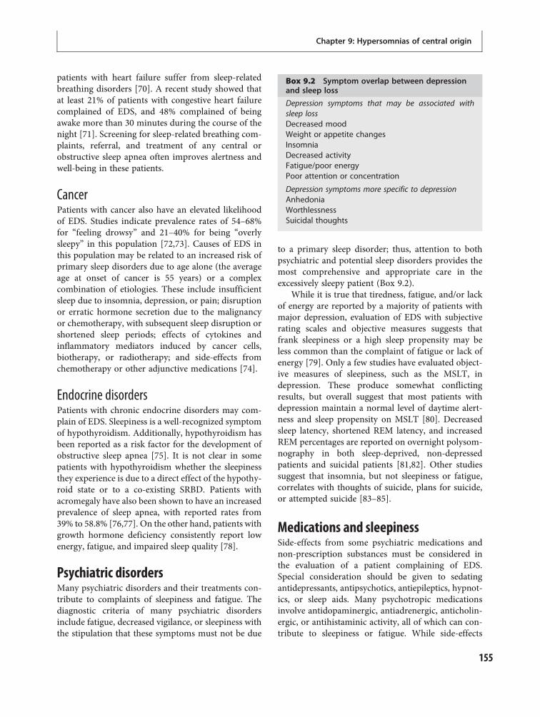
patients with heart failure suffer from sleep-relatedbreathing disorders [70]. A recent study showed thatat least 21% of patients with congestive heart failurecomplained of EDS, and 48% complained of beingawake more than 30 minutes during the course of thenight [71]. Screening for sleep-related breathing com-plaints, referral, and treatment of any central orobstructive sleep apnea often improves alertness andwell-being in these patients.
CancerPatients with cancer also have an elevated likelihoodof EDS. Studies indicate prevalence rates of 54–68%for “feeling drowsy” and 21–40% for being “overlysleepy” in this population [72,73]. Causes of EDS inthis population may be related to an increased risk ofprimary sleep disorders due to age alone (the averageage at onset of cancer is 55 years) or a complexcombination of etiologies. These include insufficientsleep due to insomnia, depression, or pain; disruptionor erratic hormone secretion due to the malignancyor chemotherapy, with subsequent sleep disruption orshortened sleep periods; effects of cytokines andinflammatory mediators induced by cancer cells,biotherapy, or radiotherapy; and side-effects fromchemotherapy or other adjunctive medications [74].
Endocrine disordersPatients with chronic endocrine disorders may com-plain of EDS. Sleepiness is a well-recognized symptomof hypothyroidism. Additionally, hypothyroidism hasbeen reported as a risk factor for the development ofobstructive sleep apnea [75]. It is not clear in somepatients with hypothyroidism whether the sleepinessthey experience is due to a direct effect of the hypothy-roid state or to a co-existing SRBD. Patients withacromegaly have also been shown to have an increasedprevalence of sleep apnea, with reported rates from39% to 58.8% [76,77]. On the other hand, patients withgrowth hormone deficiency consistently report lowenergy, fatigue, and impaired sleep quality [78].
Psychiatric disordersMany psychiatric disorders and their treatments con-tribute to complaints of sleepiness and fatigue. Thediagnostic criteria of many psychiatric disordersinclude fatigue, decreased vigilance, or sleepiness withthe stipulation that these symptoms must not be due
to a primary sleep disorder; thus, attention to bothpsychiatric and potential sleep disorders provides themost comprehensive and appropriate care in theexcessively sleepy patient (Box 9.2).
While it is true that tiredness, fatigue, and/or lackof energy are reported by a majority of patients withmajor depression, evaluation of EDS with subjectiverating scales and objective measures suggests thatfrank sleepiness or a high sleep propensity may beless common than the complaint of fatigue or lack ofenergy [79]. Only a few studies have evaluated object-ive measures of sleepiness, such as the MSLT, indepression. These produce somewhat conflictingresults, but overall suggest that most patients withdepression maintain a normal level of daytime alert-ness and sleep propensity on MSLT [80]. Decreasedsleep latency, shortened REM latency, and increasedREM percentages are reported on overnight polysom-nography in both sleep-deprived, non-depressedpatients and suicidal patients [81,82]. Other studiessuggest that insomnia, but not sleepiness or fatigue,correlates with thoughts of suicide, plans for suicide,or attempted suicide [83–85].
Medications and sleepinessSide-effects from some psychiatric medications andnon-prescription substances must be considered inthe evaluation of a patient complaining of EDS.Special consideration should be given to sedatingantidepressants, antipsychotics, antiepileptics, hypnot-ics, or sleep aids. Many psychotropic medicationsinvolve antidopaminergic, antiadrenergic, anticholin-ergic, or antihistaminic activity, all of which can con-tribute to sleepiness or fatigue. While side-effects
Box 9.2 Symptom overlap between depressionand sleep loss
Depression symptoms that may be associated withsleep lossDecreased moodWeight or appetite changesInsomniaDecreased activityFatigue/poor energyPoor attention or concentration
Depression symptoms more specific to depressionAnhedoniaWorthlessnessSuicidal thoughts
Chapter 9: Hypersomnias of central origin
155
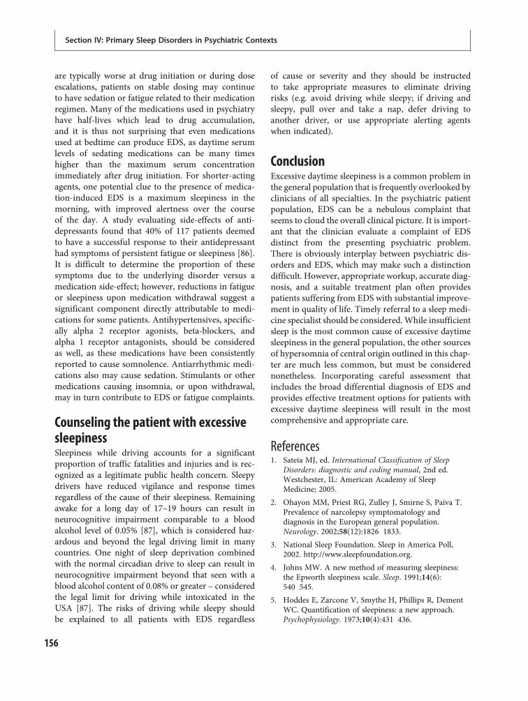
are typically worse at drug initiation or during doseescalations, patients on stable dosing may continueto have sedation or fatigue related to their medicationregimen. Many of the medications used in psychiatryhave half-lives which lead to drug accumulation,and it is thus not surprising that even medicationsused at bedtime can produce EDS, as daytime serumlevels of sedating medications can be many timeshigher than the maximum serum concentrationimmediately after drug initiation. For shorter-actingagents, one potential clue to the presence of medica-tion-induced EDS is a maximum sleepiness in themorning, with improved alertness over the courseof the day. A study evaluating side-effects of anti-depressants found that 40% of 117 patients deemedto have a successful response to their antidepressanthad symptoms of persistent fatigue or sleepiness [86].It is difficult to determine the proportion of thesesymptoms due to the underlying disorder versus amedication side-effect; however, reductions in fatigueor sleepiness upon medication withdrawal suggest asignificant component directly attributable to medi-cations for some patients. Antihypertensives, specific-ally alpha 2 receptor agonists, beta-blockers, andalpha 1 receptor antagonists, should be consideredas well, as these medications have been consistentlyreported to cause somnolence. Antiarrhythmic medi-cations also may cause sedation. Stimulants or othermedications causing insomnia, or upon withdrawal,may in turn contribute to EDS or fatigue complaints.
Counseling the patient with excessivesleepinessSleepiness while driving accounts for a significantproportion of traffic fatalities and injuries and is rec-ognized as a legitimate public health concern. Sleepydrivers have reduced vigilance and response timesregardless of the cause of their sleepiness. Remainingawake for a long day of 17–19 hours can result inneurocognitive impairment comparable to a bloodalcohol level of 0.05% [87], which is considered haz-ardous and beyond the legal driving limit in manycountries. One night of sleep deprivation combinedwith the normal circadian drive to sleep can result inneurocognitive impairment beyond that seen with ablood alcohol content of 0.08% or greater – consideredthe legal limit for driving while intoxicated in theUSA [87]. The risks of driving while sleepy shouldbe explained to all patients with EDS regardless
of cause or severity and they should be instructedto take appropriate measures to eliminate drivingrisks (e.g. avoid driving while sleepy; if driving andsleepy, pull over and take a nap, defer driving toanother driver, or use appropriate alerting agentswhen indicated).
ConclusionExcessive daytime sleepiness is a common problem inthe general population that is frequently overlooked byclinicians of all specialties. In the psychiatric patientpopulation, EDS can be a nebulous complaint thatseems to cloud the overall clinical picture. It is import-ant that the clinician evaluate a complaint of EDSdistinct from the presenting psychiatric problem.There is obviously interplay between psychiatric dis-orders and EDS, which may make such a distinctiondifficult. However, appropriate workup, accurate diag-nosis, and a suitable treatment plan often providespatients suffering from EDS with substantial improve-ment in quality of life. Timely referral to a sleep medi-cine specialist should be considered.While insufficientsleep is the most common cause of excessive daytimesleepiness in the general population, the other sourcesof hypersomnia of central origin outlined in this chap-ter are much less common, but must be considerednonetheless. Incorporating careful assessment thatincludes the broad differential diagnosis of EDS andprovides effective treatment options for patients withexcessive daytime sleepiness will result in the mostcomprehensive and appropriate care.
References1. Sateia MJ, ed. International Classification of Sleep
Disorders: diagnostic and coding manual, 2nd ed.Westchester, IL: American Academy of SleepMedicine; 2005.
2. Ohayon MM, Priest RG, Zulley J, Smirne S, Paiva T.Prevalence of narcolepsy symptomatology anddiagnosis in the European general population.Neurology. 2002;58(12):1826 1833.
3. National Sleep Foundation. Sleep in America Poll,2002. http://www.sleepfoundation.org.
4. Johns MW. A new method of measuring sleepiness:the Epworth sleepiness scale. Sleep. 1991;14(6):540 545.
5. Hoddes E, Zarcone V, Smythe H, Phillips R, DementWC. Quantification of sleepiness: a new approach.Psychophysiology. 1973;10(4):431 436.
Section IV: Primary Sleep Disorders in Psychiatric Contexts
156
6. Rosenthal L, Roehr TA, Roth T. The sleep wakeactivity inventory: a self report measure of daytimesleepiness. Biol Psychiatry. 1993;34(11):810 820.
7. Richardson G, Carskadon M, Flagg W, et al. Excessivedaytime sleepiness in man: multiple sleep latencymeasurements in narcoleptic and control subjects.Electroencepholgr Clin Neurophysiol. 1978;45(5):621 627.
8. Chua LWY, Yu NC, Golish JA, et al. Epworthsleepiness scale and the multiple sleep latency test:dilemma of the elusive link. Sleep. 1998;21(Suppl):184.
9. US Modafinil in Narcolepsy Multicenter Study Group.Randomized trial of modafinil for the treatment ofpathological somnolence in narcolepsy. Ann Neurol.1998;43(1):88 97.
10. Johns MW, Hocking B. Daytime sleepiness and sleephabits of Australian workers. Sleep. 1997;20(10):844 849.
11. Review by the MSLT and MWT Task Force of theStandards of Practice Committee of the AmericanAcademy of Sleep Medicine. Sleep. 2005;28(1):123 144.
12. CarskadonMA,DementWC.Cumulative effects of sleeprestriction on daytime sleepiness. Psychophysiology.1981;18(2):107 113.
13. Sleep in America Poll, 2008. http://www.sleepfoundation.org
14. Rosenthal L, Roehrs TA, Rosen A, Roth T. Level ofsleepiness and total sleep time following various timein bed conditions. Sleep. 1993;16(3):226 232.
15. Roehrs TA, Shore E, Papineau K, Rosenthal L, Roth T.A two week sleep extension in sleepy normals. Sleep.1996;19(7):576 582.
16. Beers T. Flexible schedules and shift work: replacingthe ‘9 to 5’ workday? Monthly Labor Rev. 2000:33 40.
17. Monk TH. Shift work. In: Kryger MH, Roth T, DementWC, eds. Priniciples and practices of sleep medicine,3rd ed. Philadelphia, PA: WB Saunders; 2000:602 603.
18. Akerstedt T. Sleepiness as a consequence of shift work.Sleep. 1988;11(1):17 34.
19. Budnick LD, Lerman SE, Baker TL, Jones H,Czeisler CA. Sleep and alertness in a 12 hour rotatingshift work environment. J Occup Med. 1994;36(12):1295 1300.
20. Czeisler CA, Walsh JK, Roth T, et al. Modafinil forexcessive sleepiness associated with shift work sleepdisorder. New Engl J Med. 2005;353(5):476 486.
21. Colton HR, Altevogt BM, eds. Sleep disorders and sleepdeprivation: an unmet public health problem.Committee on Sleep Medicine and Research. TheNational Academies Press; 2006:55 172.
22. Van Dongen HP, Maislin G, Mullington JM, DingesDF. The cumulative cost of additional wakefulness:dose response effects on neurobehavioral functions
and sleep physiology from chronic sleep restrictionand total sleep deprivation. Sleep. 2003;26(2):117 126.
23. Chemelli RM, Willie JT, Sinton CM, et al. Narcolepsyin orexin knockout mice: Molecular genetics of sleepregulation. Cell. 1999;98(4):437 451.
24. Lin L, Faraco J, Li R, et al. The sleep disorder caninenarcolepsy is caused by a mutation in the hypocretin(orexin) receptor 2 gene. Cell. 1999;98(3):365 376.
25. Mignot E, Nishino S. Emerging therapies innarcolepsy cataplexy. Sleep. 2005;28(6):754 763.
26. Nishino S, Ripley B, Overeem S, Lammers GJ, MignotE. Hypocretin (orexin) deficiency in humannarcolepsy. Lancet. 2000;355(9197):39 40.
27. Black JE, Brooks SN, Nishino S. Narcolepsy andsyndromes of primary excessive daytime somnolence.Sem Neurol. 2004;24(3):271 282.
28. Honda Y. Clinical features of narcolepsy: Japaneseexperiences. In: Honda Y, Juji T, eds. HLA innarcolepsy. Berlin: Springer Verlag; 1988:24 57.
29. Ohayon MM, Priest RG, Caulet M, Guilleminault C.Hypnogogic and hypnopompic hallucinations:pathological phenomena? Br J Psychiatry. 1996;169(4):459 467.
30. Ohayon MM, Zulley J, Guilleminault C, Smirne S.Prevalence and pathologic associations of sleepparalysis in the general population. Neurology. 1999;52(6):1194 1200.
31. Mamelak M, Black J, Monplaisir J, Ristanovic R.A pilot study on the effects of sodium oxybate on sleeparchitecture and daytime alertness in narcolepsy. Sleep.2004;27(7):1327 1334.
32. US Xyrem in Narcolepsy Multi center Study Group.A randomized, double blind, placebo controlledmulticenter trial comparing the effects of three doses oforally administered sodium oxybate with placebo forthe treatment of narcolepsy. Sleep. 2002;25(1):42 49.
33. Black J. Nightly administration of sodium oxybate iseffective for the treatment of excessive daytimesleepiness in narcolepsy. Sleep. 2005;28:A218(abstract).
34. Mitler MM, Aldrich MS, Koob GF, Zarcone VP.Narcolepsy and its treatment with stimulants: ASDAstandards of practice. Sleep. 1994;17(4):352 371.
35. Wisor JP, Ericksson KS. Dopaminergic adrenergicinteractions in the wake promoting mechanism ofmodafinil. Neuroscience. 2005;132(4):1027 1034.
36. US Modafinil in Narcolepsy Multicenter Study Group.Randomized trial of modafinil as a treatment for theexcessive daytime somnolence of narcolepsy.Neurology. 2000;54(5):1166 1175.
37. Guilleminault C, Stoohs R, Clerk A, Cetel M,Maistros P. A cause of excessive daytime sleepiness:
Chapter 9: Hypersomnias of central origin
157

The upper airway resistance syndrome. Chest.1993;104(3):781 787.
38. Montplaisir J, Poirier G. HLA in disorders of excessivesleepiness without cataplexy in Canada. In: Honda Y,Juji T, eds. HLA in narcolepsy. Berlin: Springer Verlag;1988:186.
39. Montplaisir J, De Champlain J, Young SN, et al.Narcolepsy and idiopathic hypersomnia: biogenicamines and related compounds in CSF. Neurology.1982;32(11):1299 1302.
40. Faull KF, Guilleminault C, Berger PA, Barchas JD.Cerebrospinal fluid monoamine metabolites innarcolepsy and hypersomnia. Ann Neurol. 1983;13(3):258 263.
41. Faull KF, Thiemann S, King RJ, Guilleminault C.Monoamine interactions in narcolepsy andhypersomnia: a preliminary report. Sleep. 1986;9(1 Pt 2):246 249.
42. Billiard M, Dauvillies Y. Idiopathic hypersomnia.Sleep Med Rev. 2001;5(5):351 360.
43. Roth B, Nevsimalova S, Rechtschaffen A. Hypersomniawith “sleep drunkenness.” Arch Gen Psychiatry.1972;26(5):456 462.
44. Critchley M. The syndrome of hypersomnia andperiodical megaphagia in the adult male (KleineLevin): what is its natural course? Rev Neurol. 1967;116(6):647 650.
45. Arnulf I, Zeitzer JM, File J, Farber N, Mignot E.Kleine Levin syndrome: a systematic review of 186cases in the literature. Brain. 128(12):2763 2776.
46. Rosenow F, Kotagal P, Cohen BH, Green C, Wyllie E.Multiple sleep latency test and polysomnography indiagnosing Kleine Levin syndrome and periodichypersomnia. J Clin Neurophysiol 2000;17(5):519 522.
47. Arias M, Crespo Iglesias JM, Perez J, et al. Klein Levinsyndrome: contribution of brain SPECT in diagnosis.Rev Neurol. 2003;35(6):531 533.
48. Billiard M, Guilleminault C, Dement WC.A menstruation linked periodic hypersomnia. KleineLevin syndrome or a new clinical entity? Neurology.1975;25(5):436 443.
49. Sachs C, Persson H, Hagenfeldt K. Menstruationassociated periodic hypersomnia: a case studywith successful treatment. Neurology. 1982;32(12):1376 1379.
50. Bamford CR. Menstrual associated sleep disorder:an unusual hypersomniac variant associated withboth menstruation and amenorrhea with a possiblelink to prolactin and metoclopramide. Sleep.1993;16(5):484 486.
51. Rothstein JD, Guidotti A, Tinuper P, et al. Endogenousbenzodiazepine receptor ligands in idiopathicrecurring stupor. Lancet. 1992;340(8826):1002 1004.
52. Lugaresi E, Montagna P, Tinuper P, et al. Endozepinestupor: recurring stupor linked to endozepine 4accumulation. Brain. 1998;121(Pt 1):127 133.
53. Soriani S, Carrozzi M, De Carlo L, et al. Endozepinestupor in children. Cephalalgia. 1997;17(6):658 661.
54. Francisco GE, Ivanhoe CB. Successful treatment ofpost traumatic narcolepsy with methylphenidate: acase report. Am J Phys Med Rehabil. 1996;75(1):63 65.
55. Manni R, Tantara A. Evaluation of sleepiness inepilepsy. Clin Neurophysiol. 2000;111(Suppl 2):S111 114.
56. Toth LA, Opp MR. Sleep and infection. In:Lee Chiong TL, Sateia MJ, Carskadon MA, eds.Sleep medicine. Philadelphia, PA: Hanley & Belfus;2002:77 83.
57. Guilleminault C, Mondini S. Mononucleosis andchronic daytime sleepiness: a long term follow upstudy. Arch Int Med. 1986;146(7):1333 1335.
58. Askenasy JJM. Sleep in parkinson’s disease. ActaNeurol Scand. 1993;87(3):167 170.
59. Chokroverty S. Sleep and degenerative neurologicdisorders. Neurol Clin. 1996;14(4):807 826.
60. Trenkwalder C. Sleep dysfunction in Parkinson’sdisease. Clin Neurosci. 1998;5(2):107 114.
61. George CFP. Neuromuscular disorders. In: KrygerMH, Roth T, Dement WC, eds. Principles and practiceof sleep medicine, 3rd ed. Philadelphia, PA: WBSaunders; 2000:1087 1092.
62. Gibbs JW, Ciafaloni E, Radtke RA. Excessive daytimesomnolence and increased rapid eye movementpressure in myotonic dystrophy. Sleep. 2002;25(6):662 665.
63. Campbell SM, Clark S, Tindall EA, Forehand ME,Bennett RM. Clinical characteristics of fibrositis, I:A “blinded” controlled study of symptoms and tenderpoints. Arthritis Rheum. 1983;26(7):817 824.
64. Hyyppa MT, Kronhom E. Nocturnal motor activity infibromyalgia patients with poor sleep quality.J Psychosom Res. 1995;39(1):85 91.
65. Moldofsky H, Saskin P, Lue FA. Sleep and symptomsin fibrositis syndrome after a febrile illness.J Rheumatol. 1988;15(11):1701 1704.
66. Moldofsky H, Lue FA, Smythe H. Alpha EEG sleep andmorning symptoms of rheumatoid arthritis.J Rheumatol. 1983;10(3):373 379.
67. Perlis ML, Giles DE, Bootzin RR, et al. Alpha sleepand information processing, perception of sleep, pain,
Section IV: Primary Sleep Disorders in Psychiatric Contexts
158

and arousability in fibromyalgia. Int J Neurosci.1997;89(3 4):265 280.
68. MoldolfskyH, Lue FA.The relationship of alpha delta EEGfrequencies to pain and mood in “fibrositis” patients withchlorpromazine and L tryptophan. ElectroencephalogrClin Neurophysiol. 1980;50(1 2):71 80.
69. Yamashiro Y, Kryger MH. Sleep in heart failure. Sleep.1993;16(6):513 523.
70. Javaheri S, Parker TJ, Liming JD, et al. Sleep apnea in81 ambulatory male patients with stable heart failure:types and their prevalences, consequences, andpresentations. Circulation. 1998;97(21):2154 2159.
71. Brostrom A, Stromber A, Dahlsrom U, Fridland B.Sleep difficulties, daytime sleepiness, and healthrelated quality of life in patients with chronic heartfailure. J Cardiovasc Nurs. 2004;19(4):234 242.
72. Davidson JR, MacLean AW, Brundage MD, Schulze K.Sleep disturbance in cancer patients. Social Sci. 2002;54(9):1309 1321.
73. Portenoy RK, Tahler HT, Kombilth AB, et al.Symptom prevalence, characteristics and distress in acancer population. Qual Life Res. 1994;3(3):183 189.
74. Vena C, Parker K, Cunningham M, Clark J,McMillan S. Sleep wake disturbances in people withcancer part I: an overview of sleep, sleep regulation,and effects of disease and treatment. Oncol NursForum. 2004;31(4):735 746.
75. Resta O, Paanacciulli N, Di Gioia G, et al. Highprevalence of previously unknown subclinicalhypothyroidism in obese patients referred to a sleepclinic for sleep disordered breathing. Nutr MetabCardiovasc Dis. 2004;14(5):248 253.
76. Blanco Perez JJ, Blanco Ramos MA, Zamarron Sanz C,et al. Acromegaly and sleep apnea. Arch Bronconeumol.2004;40(8):355 359.
77. Rosenow F, Reuter S, Deuss U, et al. Sleepapnoea in treated acromegaly: relative frequency andpredisposing factors. Clin Endocrinol (Oxf). 1996;45(5):563 569.
78. Bjork S, Jonsson B, Westphal O, Levin JE. Qualityof life of adults with growth hormone deficiency:a controlled study. Acta Paediatr Scand Suppl.1989;356:55 59.
79. Tylee A, Gastpar M, Lepine JP, Mendlewicz J.DEPRES II (Depression Research in EuropeanSociety II): a patient survey of the symptoms, disabilityand current management of depression in thecommunity. DEPRES Steering Committee.Int Clin Psychopharmacol. 1999;14(3):139 151.
80. Reynolds CF 3rd, Coble PA, Kupfer DJ, Holzer BC.Application of the multiple sleep latency test indisorders of excessive sleepiness. ElectroencephalogrClin Neurophysiol. 1982;53(4):443 452.
81. Liu X, Buysse DJ. Sleep and youth suicidal behavior:A neglected field. Curr Opin Psychiatry. 2006;19:288 293.
82. Fava M. Daytime sleepiness and insomnia ascorrelates of depression. J Clin Psychiatry. 2004;65:27 32.
83. Chellappa SL, Araujo JF. Sleep disorders and suicidalideation in patients with depressive disorder.Psychiatry Res. 2007;153(2):131 136.
84. McCall WV, Harding D, O’Donovan C. Correlates ofdepressive symptoms in patients with obstructive sleepapnea. J Clin Sleep Med. 2006;2:424 426.
85. Sjostrom N, Waern M, Hetta J. Nightmares and sleepdisturbances in relation to suicidality in suicideattempters. Sleep. 2007;30:91 95.
86. Fava M, Graves LM, Benazzi F, et al. A cross sectionalstudy of the prevalence of cognitive and physicalsymptoms during long term antidepressant treatment.J Clin Psychiatry. 2006;67:1754 1759.
87. Williamson AM, Feyer AM. Moderate sleepdeprivation produces impairments in cognitive andmotor perfromance equivalent to legally prescribedlevels of alcohol intoxication. Occup Environ Med.2000;57:649 655.
Chapter 9: Hypersomnias of central origin
159
Chapter
10 Parasomnias
Thomas D. Hurwitz and Carlos H. Schenck
IntroductionWith the evolution of sleep medicine, the unwantedand mysterious physiological and behavioral eventsthat have for centuries been associated with sleep anddreaming have been afforded their place within thecurrently utilized nosology of sleep disorders. Thebizarre and often dangerous parasomnias such assleepwalking and sleep terrors have been historicallyregarded as symptoms of psychiatric illness. Aristotleregarded dreaming as the sleep-related process that,when dissociated from the sleeping state into wakeful-ness, influenced the development of mental disease [1].The incubus, in modern times described as sleep par-alysis, was regarded as a demonic influence since thetime of Plato, and over time came to be associatedwith states of psychopathology [2]. Caelius Aurelianus,a Roman physician of the late third century AD,likened the incubus to pavor nocturnes (now callednight terrors) as “a fear of something obscure whichthe patient does not know . . . which resides in theimagination . . . there is also torpor of the body . . .oppression . . . sometimes inability to move . . . and tospeak . . . often confusion.” Though similar to epilepsy,it was thought to require little treatment other thanpeace and calm [3].
Later, during the nineteenth century, somnambu-lism was studied in fields as disparate as philosophyand literature. In medicine it was associated withmental disorders such as lethargy and catalepsy.Indeed, these three were known as “magnetic dis-eases” with some relationship to hysteria in the post-Mesmeric era when magnetic effects upon livingtissue were thought to influence disease. Indeed, hyp-nosis had been considered an artificially induced formof somnambulism. Sleepwalking, then, was longconsidered to be a dissociative disorder [4]. Other
psychiatric literature of the day addressed sleepwalk-ing as “. . . a transient paroxysm of madness,” [5]and as a manifestation of “some morbid conditionin the . . . cerebral organism” [6]. Freud, in The Inter-pretation of Dreams, did not address the issue of sleep“for that is essentially a problem of physiology,”though he definitely associated cognitive dreamexperience with the core of unconscious life and areflection of neurosis. He did not address motoricallyactive dreaming in this work [7].
In our day, sleep-related behaviors may still bethought to be associated with psychopathological con-ditions, but frequently a presumed association maylead to erroneous diagnosis and inappropriate treat-ment. Because the symptomatic expression of para-somnias may be misinterpreted as a manifestation ofa mental disorder, it behooves the modern psychiatricclinician to understand the differential diagnosisof these disorders so as to pursue timely and appro-priate evaluation and treatment. This group of sleepdisorders will be addressed from the perspective ofclinical psychiatry.
Parasomnias are undesirable physical, experien-tial, or behavioral phenomena that occur exclusivelyduring sleep or are exacerbated by the sleeping state.In the psychiatric nosology, DSM-IV-TR distin-guishes nightmare disorder (formerly known asdream anxiety disorder), sleep terror disorder, sleep-walking disorder, and parasomnias not otherwise spe-cified [8]. Unfortunately, this limited categorizationunderstates the richness of the various disorders rep-resenting, as a group, the unusual and frequentlybizarre manifestations of sleep–wake state misalign-ment. The International Classification of SleepDisorders distinguishes them as disorders of arousalor partial arousal from non-REM sleep, disorders
Foundations of Psychiatric SleepMedicine, ed. JohnW.Winkelman andDavid T. Plante. Published by CambridgeUniversity Press.# Cambridge University Press 2010.
160
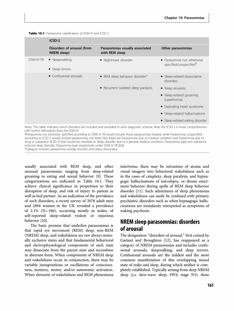
usually associated with REM sleep, and otherunusual parasomnias ranging from sleep-relatedgroaning to eating and sexual behavior [9]. Thesecategorizations are indicated in Table 10.1. Theyachieve clinical significance in proportion to theirdisruption of sleep, and risk of injury to patient aswell as bed partner. As an indication of the prevalenceof such disorders, a recent survey of 2078 adult menand 2894 women in the UK revealed a prevalenceof 2.1% (N¼106), occurring mostly in males, ofself-reported sleep-related violent or injuriousbehavior [10].
The basic premise that underlies parasomnias isthat rapid eye movement (REM) sleep, non-REM(NREM) sleep, and wakefulness are not always mutu-ally exclusive states and that fundamental behavioraland electrophysiological components of each statemay dissociate from the parent state and recombinein aberrant form. When components of NREM sleepand wakefulness occur in conjunction, there may bevariable juxtapositions or oscillations of conscious-ness, memory, motor, and/or autonomic activation.When elements of wakefulness and REM phenomena
intertwine, there may be intrusions of atonia andvisual imagery into behavioral wakefulness such asin the cases of cataplexy, sleep paralysis, and hypna-gogic hallucinations of narcolepsy, or dream enact-ment behavior during spells of REM sleep behaviordisorder [11]. Such admixtures of sleep phenomenaand wakefulness can easily be confused with primarypsychiatric disorders such as when hypnagogic hallu-cinations are mistakenly interpreted as symptoms ofwaking psychosis.
NREM sleep parasomnias: disordersof arousalThe designation “disorders of arousal,” first coined byGastaut and Broughton [12], has reappeared as acategory of NREM parasomnias and includes confu-sional arousals, sleepwalking, and sleep terrors.Confusional arousals are the mildest and the mostcommon manifestation of this overlapping mixedstate of wake and sleep, during which neither is com-pletely established. Typically arising from deep NREMsleep (i.e. slow-wave sleep; SWS; stage N3), these
Table 10.1 Parasomnia classification of DSM IV and ICSD 2
ICSD-2
Disorders of arousal (fromNREM sleep)
Parasomnias usually associatedwith REM sleep
Other parasomnias
DSM-IV-TR � Sleepwalking � Nightmare disorder � Parasomnia not otherwisespecified/unspecified{
� Sleep terrors
� Confusional arousals � REM sleep behavior disorder* � Sleep-related dissociativedisorders
� Recurrent isolated sleep paralysis � Sleep enuresis
� Sleep-related groaning(catathrenia)
� Exploding head syndrome
� Sleep-related hallucinations
� Sleep-related eating disorder
Notes: This table indicates which disorders are included and excluded in each diagnostic schema. Note the ICSD 2 is more comprehensivewith further delineation than the DSM IV.{Parasomnia not otherwise specified according to DSM IV TR would include those parasomnias shaded; while Parasomnia, unspecifiedaccording to ICSD 2 would include parasomnias not listed. Not listed are Parasomnias due to medical condition and Parasomnia due todrug or substance (ICSD 2) that would be classified as Sleep disorder due to a general medical condition, Parasomnia type and substanceinduced sleep disorder, Parasomnia type respectively under DSM IV TR [8,9].*Category includes parasomnia overlap disorder and status dissociatus.
Chapter 10: Parasomnias
161

arousals, or incomplete awakenings, have also beenknown as sleep drunkenness and sleep inertia [13,14].The affected individuals demonstrate variable degreesof confusion, disorientation, retrograde and antero-grade amnesia, as well as disturbed and occasionallyviolent behavior during periods of variable duration.Mild, brief episodes often occur without recognition,as the individual may not even rise out of bed. Twovariants in adolescents and adults, severe morningsleep inertia and sleep-related abnormal sexual behav-iors, are also recognized [9]. Confusional arousalsmay affect individuals of all ages, and may be precipi-tated by sleep deprivation, circadian rhythm alter-ation (jet lag, shift work, etc.), alcohol, medications,and by conditions of excessive somnolence. Bothsexes may be affected and there is a familial predis-position to deeper sleep. Epidemiological data suggestconfusional arousals may occur in as many as 4.2% ofthe adult population, though due to the inherentdifficulties in reporting these events, this may dramat-ically underestimate their prevalence [10].
Sleepwalking and sleep terrors: overviewand clinical descriptionSleepwalking (SW) represents motor activationduring an abnormal arousal, usually from deepNREM sleep. Common during childhood (1–17%),SW tends to diminish in frequency following adoles-cence but can persist into or even begin during adultlife (2.5%) [15,16]. Behaviors can vary from sitting upin the bed to full ambulation. Behaviors can be verycomplex and include walking, running, driving,eating, and violence. Mental activity is usually poorlyrecalled but may include dream-like visual imagery,which tends to be both less detailed and less bizarrethan traditional REM dream reports. Individualsare variably amnestic for the episodes. Typicallyoccurring during the early part of the sleep cycleand emerging from the “deeper” stage N3 of NREMsleep, they may technically derive from any NREMstage. Many individuals report that frequency andseverity of sleepwalking increase with stressful lifeexperiences. Cases have been associated with migraineand thyrotoxicosis [17,18]. There is often a familialpredisposition and it occurs in both sexes. Spellscan be precipitated in susceptible individuals byanything that stimulates arousals from sleep. Thesecan include exogenous stimuli such as noises, orendogenous factors such as obstructive sleep apnea
(OSA), periodic limb movements of sleep (PLMS),gastroesophageal reflux disorder, other physical ill-ness, or full bladder, without any specific psychologicalmeaning [9,11,19,20–22]. Nathaniel Kleitman, fatherof American sleep research, wrote: “all the charac-teristics of somnambulism underline the differencebetween wakefulness and consciousness.” [23] Thisidea is reinforced by a more recent report of SPECTimaging during a polygraphically documented SWepisode, demonstrating increased cerebral blood flow(CBF) in the anterior cerebellum (vermis) and poster-ior cingulate cortex when compared to quiet slow-wave sleep. There were also large areas of frontal andparietal cortical decrements of CBF when comparedwith normal awake subjects. As anticipated by Kleit-man, SW appears to represent a concurrence ofincreased motor activation and decreased executivefunction during incomplete, disordered arousalsfrom sleep [24].
Sleep terrors (ST) are spells of abrupt arousalswith profound autonomic activation including tachy-cardia, tachypnea, flushing, diaphoresis, andmydriasisthat may include sitting up in bed and may progress toSW. The two disorders, often occurring in combin-ation, represent a continuum of events with exclusivelymotor or autonomic activation as polar examples.Pure ST occur more frequently in young children,arising in approximately 3% of children and less than1% of adults. Arising typically during the first third ofthe night, ST are followed by return to sleep andamnesia for the event. Precipitants are not necessarilypresent, but may include fever, prior sleep deprivation,or central nervous system depressant drugs includingalcohol. Polysomnography (PSG) findings are essen-tially those described with SW below but with notableincreases in cardiac and respiratory rates [9].
Though often unnecessary in typical, benigncases of SW and ST, PSG is indicated when they havean adult onset (as this may indicate another under-lying precipitating sleep disorder), when there is riskof significant injury to oneself or another person,or when there is substantial disruption of one’s sleepor the sleep of the bed partner. Such studies shouldinclude an expanded “seizure” montage of EEG chan-nels, electromyographic (EMG) monitoring of fourlimbs to detect movement, and continuous audio-visual monitoring (that is time-synchronized to thePSG), in order to rule out seizure disorder, REM sleepbehavior disorder (RBD), OSA, PLMS, or other pri-mary sleep disorders [25,26]. Electrocardiogram
Section IV: Primary Sleep Disorders in Psychiatric Contexts
162
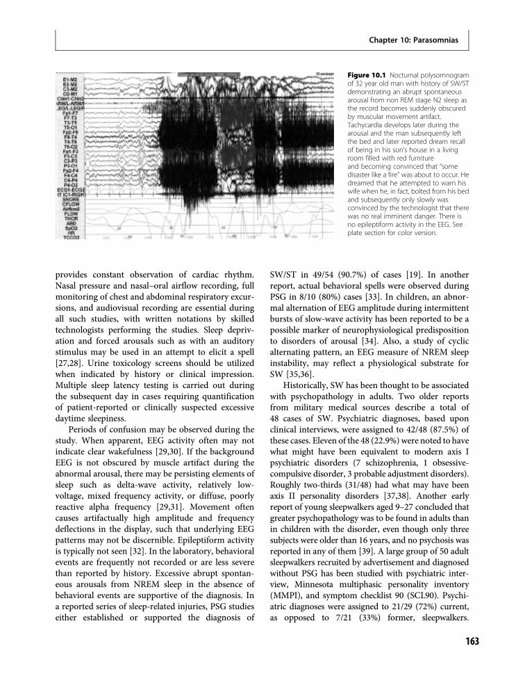
provides constant observation of cardiac rhythm.Nasal pressure and nasal–oral airflow recording, fullmonitoring of chest and abdominal respiratory excur-sions, and audiovisual recording are essential duringall such studies, with written notations by skilledtechnologists performing the studies. Sleep depriv-ation and forced arousals such as with an auditorystimulus may be used in an attempt to elicit a spell[27,28]. Urine toxicology screens should be utilizedwhen indicated by history or clinical impression.Multiple sleep latency testing is carried out duringthe subsequent day in cases requiring quantificationof patient-reported or clinically suspected excessivedaytime sleepiness.
Periods of confusion may be observed during thestudy. When apparent, EEG activity often may notindicate clear wakefulness [29,30]. If the backgroundEEG is not obscured by muscle artifact during theabnormal arousal, there may be persisting elements ofsleep such as delta-wave activity, relatively low-voltage, mixed frequency activity, or diffuse, poorlyreactive alpha frequency [29,31]. Movement oftencauses artifactually high amplitude and frequencydeflections in the display, such that underlying EEGpatterns may not be discernible. Epileptiform activityis typically not seen [32]. In the laboratory, behavioralevents are frequently not recorded or are less severethan reported by history. Excessive abrupt spontan-eous arousals from NREM sleep in the absence ofbehavioral events are supportive of the diagnosis. Ina reported series of sleep-related injuries, PSG studieseither established or supported the diagnosis of
SW/ST in 49/54 (90.7%) of cases [19]. In anotherreport, actual behavioral spells were observed duringPSG in 8/10 (80%) cases [33]. In children, an abnor-mal alternation of EEG amplitude during intermittentbursts of slow-wave activity has been reported to be apossible marker of neurophysiological predispositionto disorders of arousal [34]. Also, a study of cyclicalternating pattern, an EEG measure of NREM sleepinstability, may reflect a physiological substrate forSW [35,36].
Historically, SW has been thought to be associatedwith psychopathology in adults. Two older reportsfrom military medical sources describe a total of48 cases of SW. Psychiatric diagnoses, based uponclinical interviews, were assigned to 42/48 (87.5%) ofthese cases. Eleven of the 48 (22.9%) were noted to havewhat might have been equivalent to modern axis Ipsychiatric disorders (7 schizophrenia, 1 obsessive-compulsive disorder, 3 probable adjustment disorders).Roughly two-thirds (31/48) had what may have beenaxis II personality disorders [37,38]. Another earlyreport of young sleepwalkers aged 9–27 concluded thatgreater psychopathology was to be found in adults thanin children with the disorder, even though only threesubjects were older than 16 years, and no psychosis wasreported in any of them [39]. A large group of 50 adultsleepwalkers recruited by advertisement and diagnosedwithout PSG has been studied with psychiatric inter-view, Minnesota multiphasic personality inventory(MMPI), and symptom checklist 90 (SCL90). Psychi-atric diagnoses were assigned to 21/29 (72%) current,as opposed to 7/21 (33%) former, sleepwalkers.
Figure 10.1 Nocturnal polysomnogramof 32 year old man with history of SW/STdemonstrating an abrupt spontaneousarousal from non REM stage N2 sleep asthe record becomes suddenly obscuredby muscular movement artifact.Tachycardia develops later during thearousal and the man subsequently leftthe bed and later reported dream recallof being in his son’s house in a livingroom filled with red furnitureand becoming convinced that “somedisaster like a fire” was about to occur. Hedreamed that he attempted to warn hiswife when he, in fact, bolted from his bedand subsequently only slowly wasconvinced by the technologist that therewas no real imminent danger. There isno epileptiform activity in the EEG. Seeplate section for color version.
Chapter 10: Parasomnias
163
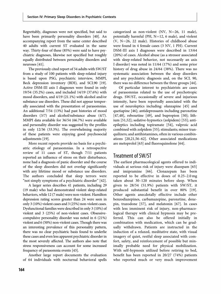
Regrettably, diagnoses were not specified, but said tohave been primarily personality disorders [40]. Anaccompanying report from the same center described40 adults with current ST evaluated in the sameway. Thirty-four of these (85%) were said to have psy-chiatric diagnoses, likewise not specified but roughlyequally distributed between personality disorders andneuroses [41].
The previously cited report of 54 adults with SW/STfrom a study of 100 patients with sleep-related injuryis based upon PSG, psychiatric interview, MMPI,Beck depression inventory (BDI), and SCL90 [19].Active DSM-III axis I diagnoses were found in only19/54 (35.2%) cases, and included 14/19 (37.6%) withmood disorders, and 4/19 (21.1%) with alcohol and/orsubstance use disorders. These did not appear tempor-ally associated with the presentation of parasomnias.An additional 7/54 (13%) had past histories of mooddisorders (3/7) and alcohol/substance abuse (4/7).MMPI data available for 36/54 (66.7%) were availableand personality disorder was suggested by the profilesin only 12/36 (33.3%). The overwhelming majorityof these patients were enjoying good psychosocialadjustments [19].
More recent reports provide no basis for a psychi-atric etiology of parasomnias. In a retrospectivereview of 11 cases of ST, though 7/11 patientsreported an influence of stress on their disturbance,none had a diagnosis of panic disorder and the courseof the sleep disorders did not overlap significantlywith any lifetime mood or substance use disorders.The authors concluded that sleep terrors were“not simply symptoms of a psychiatric disorder” [42].
A larger series describes 41 patients, including 29(19 male) who had demonstrated violent sleep-relatedbehaviors, while 12 (7male) were non-violent. Hamiltondepression rating scores greater than 24 were seen inonly 3 (10%) violent cases and 3 (25%) non-violent cases.Dysfunctional families were described in only 3 (10%) ofviolent and 3 (25%) of non-violent cases. Obsessive-compulsive personality disorder was noted in 6 (21%)violent and 6 (50%) non-violent cases. Though there wasan interesting prevalence of this personality pattern,there was no clear psychiatric basis found to underliethese cases and even less apparent psychiatric disorder inthe most severely affected. The authors also note thatstress responsiveness can account for some increasedfrequency of parasomnia events [43].
Another large report documents the evaluationof 64 individuals with nocturnal behavioral spells
categorized as non-violent (NV, N¼26, 11 male),potentially harmful (PH, N¼12, 6 male), and violent(V, N¼26, 22 male). Histories of childhood abusewere found in 4 female cases (3 NV, 1 PH). CurrentDSM-III axis I diagnoses were described in 13/64(20%) of cases. Alcohol abuse (as a stressor associatedwith sleep-related behavior, not necessarily an axisI disorder) was noted in 11/64 (17%) and some priorhistory of drug abuse in 24/64 (38%). There was nosystematic association between the sleep disordersand any psychiatric diagnosis and, on the SCL 90,there was no difference between the three groups [44].
Of particular interest to psychiatrists are casesof parasomnias related to the use of psychotropicdrugs. SW/ST, occasionally of severe and injuriousintensity, have been reportedly associated with theuse of neuroleptics including: olanzapine [45] andquetiapine [46]; antidepressants including paroxetine[47,48], reboxetine [49], and bupropion [50]; lith-ium [51,52]; sedative-hypnotics (zolpidem) [53]; anti-epileptics including topiramate [54], valproic acidcombined with zolpidem [55]; stimulants; minor tran-quilizers; and antihistamines, often in various combin-ations [20,21,56–62]. Other associated medicationsare metoprolol [63] and fluoroquinolone [64].
Treatment of SW/STThe earliest pharmacological agents offered to indi-viduals at serious risk of injury were diazepam [65]and imipramine [66]. Clonazepam has beenreported to be effective in doses of 0.25–2.0mgtaken about 30–120 minutes before sleep. Whengiven to 28/54 (51.9%) patients with SW/ST, itproduced substantial benefit in over 80% [19].Other agents anecdotally effective include otherbenzodiazepines, carbamazepine, paroxetine, doxe-pin, trazodone [57], and melatonin [67]. In caseswith less imminent risk of injury, non-pharmaco-logical therapy with clinical hypnosis may be pre-ferred. This can also be offered initially incombination with medication, which can be grad-ually withdrawn. Patients are instructed in theinduction of a relaxed, meditative state, with visualimagery of quiet, restful sleep associated with com-fort, safety, and reinforcement of possible but min-imally probable need for physical mobilization.With self-hypnosis utilized before retiring to bed,benefit has been reported in 20/27 (74%) patientswho reported much or very much improvement
Section IV: Primary Sleep Disorders in Psychiatric Contexts
164
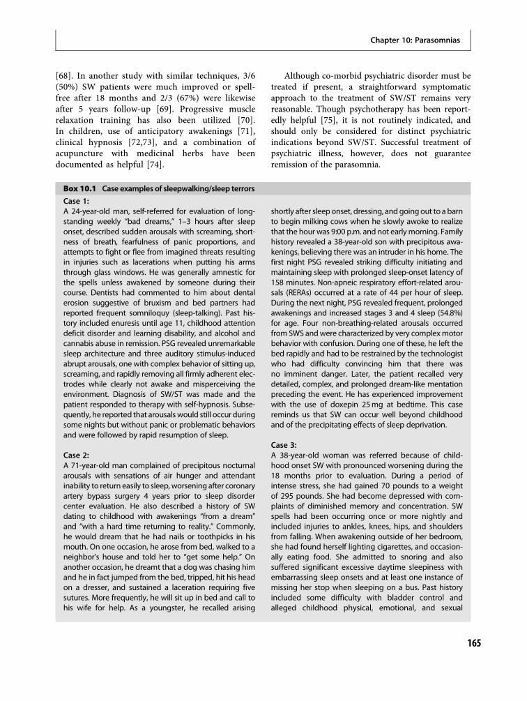
[68]. In another study with similar techniques, 3/6(50%) SW patients were much improved or spell-free after 18 months and 2/3 (67%) were likewiseafter 5 years follow-up [69]. Progressive musclerelaxation training has also been utilized [70].In children, use of anticipatory awakenings [71],clinical hypnosis [72,73], and a combination ofacupuncture with medicinal herbs have beendocumented as helpful [74].
Although co-morbid psychiatric disorder must betreated if present, a straightforward symptomaticapproach to the treatment of SW/ST remains veryreasonable. Though psychotherapy has been report-edly helpful [75], it is not routinely indicated, andshould only be considered for distinct psychiatricindications beyond SW/ST. Successful treatment ofpsychiatric illness, however, does not guaranteeremission of the parasomnia.
Box 10.1 Case examples of sleepwalking/sleep terrors
Case 1:A 24-year-old man, self-referred for evaluation of long-standing weekly “bad dreams,” 1–3 hours after sleeponset, described sudden arousals with screaming, short-ness of breath, fearfulness of panic proportions, andattempts to fight or flee from imagined threats resultingin injuries such as lacerations when putting his armsthrough glass windows. He was generally amnestic forthe spells unless awakened by someone during theircourse. Dentists had commented to him about dentalerosion suggestive of bruxism and bed partners hadreported frequent somniloquy (sleep-talking). Past his-tory included enuresis until age 11, childhood attentiondeficit disorder and learning disability, and alcohol andcannabis abuse in remission. PSG revealed unremarkablesleep architecture and three auditory stimulus-inducedabrupt arousals, one with complex behavior of sitting up,screaming, and rapidly removing all firmly adherent elec-trodes while clearly not awake and misperceiving theenvironment. Diagnosis of SW/ST was made and thepatient responded to therapy with self-hypnosis. Subse-quently, he reported that arousals would still occur duringsome nights but without panic or problematic behaviorsand were followed by rapid resumption of sleep.
Case 2:A 71-year-old man complained of precipitous nocturnalarousals with sensations of air hunger and attendantinability to return easily to sleep, worsening after coronaryartery bypass surgery 4 years prior to sleep disordercenter evaluation. He also described a history of SWdating to childhood with awakenings “from a dream”and “with a hard time returning to reality.” Commonly,he would dream that he had nails or toothpicks in hismouth. On one occasion, he arose from bed, walked to aneighbor’s house and told her to “get some help.” Onanother occasion, he dreamt that a dog was chasing himand he in fact jumped from the bed, tripped, hit his headon a dresser, and sustained a laceration requiring fivesutures. More frequently, he will sit up in bed and call tohis wife for help. As a youngster, he recalled arising
shortly after sleep onset, dressing, and going out to a barnto begin milking cows when he slowly awoke to realizethat the hour was 9:00 p.m. and not earlymorning. Familyhistory revealed a 38-year-old son with precipitous awa-kenings, believing there was an intruder in his home. Thefirst night PSG revealed striking difficulty initiating andmaintaining sleep with prolonged sleep-onset latency of158 minutes. Non-apneic respiratory effort-related arou-sals (RERAs) occurred at a rate of 44 per hour of sleep.During the next night, PSG revealed frequent, prolongedawakenings and increased stages 3 and 4 sleep (54.8%)for age. Four non-breathing-related arousals occurredfrom SWS andwere characterized by very complexmotorbehavior with confusion. During one of these, he left thebed rapidly and had to be restrained by the technologistwho had difficulty convincing him that there wasno imminent danger. Later, the patient recalled verydetailed, complex, and prolonged dream-like mentationpreceding the event. He has experienced improvementwith the use of doxepin 25mg at bedtime. This casereminds us that SW can occur well beyond childhoodand of the precipitating effects of sleep deprivation.
Case 3:A 38-year-old woman was referred because of child-hood onset SW with pronounced worsening during the18 months prior to evaluation. During a period ofintense stress, she had gained 70 pounds to a weightof 295 pounds. She had become depressed with com-plaints of diminished memory and concentration. SWspells had been occurring once or more nightly andincluded injuries to ankles, knees, hips, and shouldersfrom falling. When awakening outside of her bedroom,she had found herself lighting cigarettes, and occasion-ally eating food. She admitted to snoring and alsosuffered significant excessive daytime sleepiness withembarrassing sleep onsets and at least one instance ofmissing her stop when sleeping on a bus. Past historyincluded some difficulty with bladder control andalleged childhood physical, emotional, and sexual
Chapter 10: Parasomnias
165

Parasomnias usually associatedwith REM sleepREM sleep behavior disorder: overviewFirst predicted by animal experimentation in 1965and certainly having existed for centuries in humans,
REM sleep behavior disorder (RBD) was firstreported in 1985 after systematic evaluation of anelderly gentleman complaining of injurious dream-enactment behavior. He would fly out of bed duringdreams of playing football, and then collide withfurniture or a wall. Countless cases have now been
abuse at the hands of a violent, alcoholic father. PSGrevealed profound sleep fragmentation with OSA at arate of 72 apneas or hypopneas per hour of sleepand additional respiratory effort-related arousals at arate of 73 per hour. Oxyhemoglobin saturation reacheda low of 60% during the night (Figure 10.2a). Sheresponded well to nasal continuous positive airway
pressure (CPAP) therapy at a pressure of 12 cm ofwater with 28% supplemental oxygen (Figure 10.2b). Herworsening SW, obviously in response to sleep disorderedbreathing-induced arousals, was eliminated entirelyby effective CPAP in spite of continued severe financialstress.
(a)
(b)
Box 10.1 (cont.)
Figure 10.2 (a) Oximetry tracingfrom an obese 38 year old womanshowing frequent desaturations ofoxyhemoglobin, reaching a nadir of<60% during polysomnographicallydocumented non REM sleep,secondary to obstructive sleepapnea. Apnea and hypopnea eventsoccurred at a rate of 80 per hourof sleep. Each desaturation isaccompanied by evidence of arousalfrom sleep on the correspondingpolysomnogram. She hadcomplained of frequent spells of SW,which included eating foods fromher cupboards and refrigerator.She responded well to CPAP at8 9 cmH2O pressure, with resolutionof obstructive sleep apnea and SW.See plate section for color version.(b) Polysomnogram correspondingto 2 minutes of sleep duringbaseline recording of sleep in thecase of a 38 year old obese womanwith obstructive sleep apnea,demonstrated by reduction of oralnasal airflow in the presence ofrespiratory effort noted bydeflections representing chest andabdominal movements. The ensuingarousals from sleep represent eventsthat had historically been likely tohave precipitated confusionalarousals and sleepwalking.
Section IV: Primary Sleep Disorders in Psychiatric Contexts
166
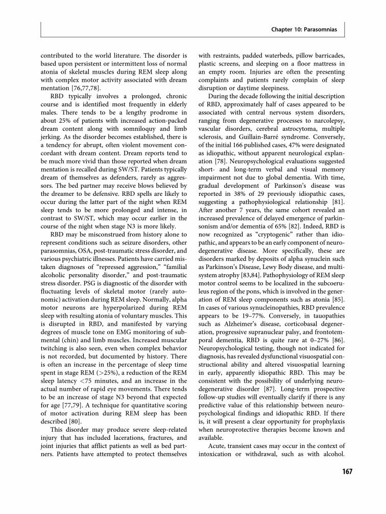
contributed to the world literature. The disorder isbased upon persistent or intermittent loss of normalatonia of skeletal muscles during REM sleep alongwith complex motor activity associated with dreammentation [76,77,78].
RBD typically involves a prolonged, chroniccourse and is identified most frequently in elderlymales. There tends to be a lengthy prodrome inabout 25% of patients with increased action-packeddream content along with somniloquy and limbjerking. As the disorder becomes established, there isa tendency for abrupt, often violent movement con-cordant with dream content. Dream reports tend tobe much more vivid than those reported when dreammentation is recalled during SW/ST. Patients typicallydream of themselves as defenders, rarely as aggres-sors. The bed partner may receive blows believed bythe dreamer to be defensive. RBD spells are likely tooccur during the latter part of the night when REMsleep tends to be more prolonged and intense, incontrast to SW/ST, which may occur earlier in thecourse of the night when stage N3 is more likely.
RBD may be misconstrued from history alone torepresent conditions such as seizure disorders, otherparasomnias, OSA, post-traumatic stress disorder, andvarious psychiatric illnesses. Patients have carried mis-taken diagnoses of “repressed aggression,” “familialalcoholic personality disorder,” and post-traumaticstress disorder. PSG is diagnostic of the disorder withfluctuating levels of skeletal motor (rarely auto-nomic) activation during REM sleep. Normally, alphamotor neurons are hyperpolarized during REMsleep with resulting atonia of voluntary muscles. Thisis disrupted in RBD, and manifested by varyingdegrees of muscle tone on EMG monitoring of sub-mental (chin) and limb muscles. Increased musculartwitching is also seen, even when complex behavioris not recorded, but documented by history. Thereis often an increase in the percentage of sleep timespent in stage REM (>25%), a reduction of the REMsleep latency <75 minutes, and an increase in theactual number of rapid eye movements. There tendsto be an increase of stage N3 beyond that expectedfor age [77,79]. A technique for quantitative scoringof motor activation during REM sleep has beendescribed [80].
This disorder may produce severe sleep-relatedinjury that has included lacerations, fractures, andjoint injuries that afflict patients as well as bed part-ners. Patients have attempted to protect themselves
with restraints, padded waterbeds, pillow barricades,plastic screens, and sleeping on a floor mattress inan empty room. Injuries are often the presentingcomplaints and patients rarely complain of sleepdisruption or daytime sleepiness.
During the decade following the initial descriptionof RBD, approximately half of cases appeared to beassociated with central nervous system disorders,ranging from degenerative processes to narcolepsy,vascular disorders, cerebral astrocytoma, multiplesclerosis, and Guillain-Barré syndrome. Conversely,of the initial 166 published cases, 47% were designatedas idiopathic, without apparent neurological explan-ation [78]. Neuropsychological evaluations suggestedshort- and long-term verbal and visual memoryimpairment not due to global dementia. With time,gradual development of Parkinson’s disease wasreported in 38% of 29 previously idiopathic cases,suggesting a pathophysiological relationship [81].After another 7 years, the same cohort revealed anincreased prevalence of delayed emergence of parkin-sonism and/or dementia of 65% [82]. Indeed, RBD isnow recognized as “cryptogenic” rather than idio-pathic, and appears to be an early component of neuro-degenerative disease. More specifically, these aredisorders marked by deposits of alpha synuclein suchas Parkinson’s Disease, Lewy Body disease, and multi-system atrophy [83,84]. Pathophysiology of REM sleepmotor control seems to be localized in the subcoeru-leus region of the pons, which is involved in the gener-ation of REM sleep components such as atonia [85].In cases of various synucleinopathies, RBD prevalenceappears to be 19–77%. Conversely, in tauopathiessuch as Alzheimer’s disease, corticobasal degener-ation, progressive supranuclear palsy, and frontotem-poral dementia, RBD is quite rare at 0–27% [86].Neuropsychological testing, though not indicated fordiagnosis, has revealed dysfunctional visuospatial con-structional ability and altered visuospatial learningin early, apparently idiopathic RBD. This may beconsistent with the possibility of underlying neuro-degenerative disorder [87]. Long-term prospectivefollow-up studies will eventually clarify if there is anypredictive value of this relationship between neuro-psychological findings and idiopathic RBD. If thereis, it will present a clear opportunity for prophylaxiswhen neuroprotective therapies become known andavailable.
Acute, transient cases may occur in the context ofintoxication or withdrawal, such as with alcohol.
Chapter 10: Parasomnias
167

Many psychotropic drugs can be associated withREM sleep without atonia or frank RBD. Theseinclude selective and non-selective serotonergic anti-depressants, monoamine oxidase inhibitors (MAOI),and withdrawal from TCA and MAOI. This drug-related RBD may be self-limiting, affect young per-sons, and include both sexes [56,58,60,77,86,88].It is not yet known if there is any particular pre-disposition to drug-induced REM without atoniaor RBD, or if these cases present similar probabil-ity of developing subsequent neurodegenerativedisorders.
About 20% of RBD cases include an activeaxis I psychiatric disorder while about 13% have ahistory of past axis I illness. Diagnoses have beenconfined to mood disorders (generally recurrentmajor depression) and alcohol/substance use disorders(largely in remission). In a report of 96 patients withRBD, 9 (9.4%) appeared to have had onset of the sleepdisorder temporally linked with a psychiatric illness.Five (4 male) of these 9 had RBD related to drug abuseincluding cessation of alcohol, cocaine, and amphet-amine. Mean age � SD was 44.0 (range 31–60), andRBD had been occurring for months to 17 years(unpublished data). A principal mechanism in thesecases may be the effect of pathological REM reboundafter withdrawal of REM suppressant drugs as waslikely the case in a 67-year-old woman with acute,transient RBD following rapid withdrawal of imipra-mine for treatment of major depression [89].
However, RBD may also arise as the result of anti-depressant treatment, as was the case for a 34-year-old man who developed RBD with typical dreamenactment during fluoxetine treatment of obsessive-compulsive disorder. [90] A psychiatric disorder rarelyassociated with RBD is adjustment disorder inresponse to severe psychosocial stress. A 69-year-oldman presented after the stress of divorce and a 61-year-old man had a similar course following an automobileaccident with a major risk of death but no actualinjury. In both cases, RBD persisted long after thestresses had subsided. Pathological dreaming in thesecases was typical of RBD, rather than involving obvi-ous post-traumatic imagery, and may representa manifestation of central nervous system (CNS)reorganization in response to psychological stress.It is possible that CNS plasticity in response to stressmay influence the possibility of a post-traumaticform of RBD, which has also been reported in twocases by other authors [19,89,91,92,93,94].
It must be underscored that parasomnias includ-ing RBD have been misconstrued as psychiatric dis-orders. Such patients may even find their way topsychiatric intensive care units as was the case for a62-year-old man hospitalized for recurrent majordepression with suicidality. Other patients and staffin spite of no wakeful manifestations of psychosis orthreatening behavior misattributed his dream enact-ment violence as a manifestation of a psychiatricdisorder [95].
Figure 10.3 Polysomnogram of a72 year old man with REM sleep behaviordisorder. Although the rapid eyemovements are typical of REM sleep andthe EEG is characteristicallydesynchronized, chin muscle atonia isincomplete during this burst of phasiceye movements, and there is prominentelectromyographic activation of musclesof upper and lower extremitiesmovements with correspondingmovement. Patient yelled out during thisevent and, retrospectively, recalleddreaming of fighting. See plate sectionfor color version.
Section IV: Primary Sleep Disorders in Psychiatric Contexts
168
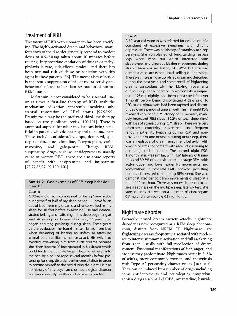
Treatment of RBDTreatment of RBD with clonazepam has been gratify-ing. The highly activated dream and behavioral mani-festations of the disorder generally respond to modestdoses of 0.5–2.0mg taken about 30 minutes beforeretiring. Inappropriate escalation of dosage or tachy-phylaxis is rare, side-effects modest, and there hasbeen minimal risk of abuse or addiction with thisagent in these patients [96]. The mechanism of actionis apparently suppression of phasic motor activity andbehavioral release rather than restoration of normalREM atonia.
Melatonin is now considered to be a second-line,or at times a first-line therapy of RBD, with themechanism of action apparently involving sub-stantial restoration of REM atonia [97,98,99].Pramipexole may be the preferred third-line therapybased on two published series [100,101]. There isanecdotal support for other medications being bene-ficial in patients who do not respond to clonazepam.These include carbidopa/levodopa, donepezil, que-tiapine, clozapine, clonidine, L-tryptophan, carba-mazepine, and gabapentin. Though REM-suppressing drugs such as antidepressants usuallycause or worsen RBD, there are also some reportsof benefit with desipramine and imipramine[77,79,86,97–99,100–102].
Nightmare disorderFormerly termed dream anxiety attacks, nightmaredisorder is now recognized as a REM sleep phenom-enon, distinct from NREM ST. Nightmares arefrightening dreams, frequently associated with moder-ate to intense autonomic activation and full awakeningfrom sleep, usually with full recollection of dreamcontent. Emotional manifestations of fear, anger, andsadness may predominate. Nightmares occur in 5–8%of adults, more commonly women, and individualswith “type A” personality characteristics [103–105].They can be induced by a number of drugs includingsome antidepressants and neuroleptics, antiparkin-sonian drugs such as L-DOPA, amantadine, lisuride,
Box 10.2 Case examples of REM sleep behaviordisorder
Case 1:A 72-year-old man complained of being “very activeduring the first half of my sleep period . . . I have fallenout of bed from my dreams and once walked in mysleep for 10 feet before awakening.” He had demon-strated jerking and twitching in his sleep beginning atleast 42 years prior to evaluation and, 37 years later,began shouting profanity during sleep. Three yearsbefore evaluation, he found himself falling from bedwhen dreaming of kicking an unfamiliar attackinganimal or unfamiliar human assailant. His wife hadavoided awakening him from such dreams becauseshe “then become[s] incorporated in his dream whichcould be dangerous.” He began sleeping tethered intothe bed by a belt or rope several months before pre-senting for sleep disorder center consultation in orderto confine himself to the bed during the night. He hadno history of any psychiatric or neurological disorderand was medically healthy and led a vigorous life.
Case 2:A 72-year-old woman was referred for evaluation of acomplaint of excessive sleepiness with chronicdepression. There was no history of cataplexy or sleepparalysis. She complained of longstanding restlesslegs when lying still which interfered withsleep onset and vigorous kicking movements duringsleep. There was no history of SW/ST but she haddemonstrated occasional loud yelling during sleep.There was increasing action-filled dreaming describedduring the past year, and some recall of frighteningdreams concordant with her kicking movementsduring sleep. These seemed to worsen when imipra-mine 125mg nightly had been prescribed for over1 month before being discontinued 4 days prior toPSG study. Alprazolam had been tapered and discon-tinued over a period of time aswell. The first night PSGrevealed very brief REM latency of 11 minutes, mark-edly increased REM sleep (52.2% of total sleep time)with loss of atonia during REM sleep. There were veryprominent extremity movements and frequentrandom extremity twitching during REM and non-REM sleep. On one occasion during REM sleep, therewas an episode of dream enactment behavior withwaving of arms concordant with recall of gesturing toher daughter in a dream. The second night PSG,1 month later, was similar, with REM latency of 8 min-utes and 39.6% of total sleep time in stage REM, withactive upper and lower extremity movements andvocalizations. Submental EMG showed persistingperiods of elevated tone during REM sleep. She alsodemonstrated periodic limb movements of sleep at arate of 19 per hour. There was no evidence of exces-sive sleepiness on the multiple sleep latency test. Shesubsequently did well on a regimen of clonazepam0.5mg and pramipexole 0.5mg nightly.
Chapter 10: Parasomnias
169

clonidine, methyldopa, prazosin, reserpine, lipophilicbeta blockers, ciprofloxacin, donepezil, and amiodar-one. Withdrawal from REM-suppressing agentssuch as tricyclic antidepressants, monoamine oxidaseinhibitors, clonidine, alcohol, and amphetamines maycause nightmares as a result of REM sleep rebound.Nightmares not uncommonly occur following trau-matic experiences, and occasional psychotic episodesmay be heralded by their occurrence [106].Non-psychotic individuals suffering frequent lifelongnightmares tend to be open, trusting, defenseless, vul-nerable, creative people described as having “thin”interpersonal, ego, and sleep–wake boundaries [107–109]. Schizotypal personality characteristics have beenassociated with nightmare distress in one report butindividuals with those traits also reported enjoyabledreaming. This implicates a more general notion ofimaginativeness influencing dream experience [110].
Reported treatments include a cognitive strategyknown as imagery rehearsal, which treats the disorderas a sleep disturbance rather than a manifestation ofspecific psychopathology. Patients are taught torestructure the dream scenario into a more acceptableexperience by rewriting the script as an exerciseduring wakefulness [111]. Instruction in lucid dream-ing has also been cited in a case report [112].Pharmacotherapy, based largely on experience withpost-traumatic stress disorder, includes reported bene-fit with cyproheptadine and prazosin [113,114] (seeChapter 17: Sleep in anxiety disorders).
Recurrent isolated sleep paralysisSleep paralysis is essentially the atonia of REM sleepthat has become dissociated and occurs at times otherthan the typical periods of REM sleep during thenight. It can either intrude into light NREM sleep atsleep onset or persist into awakening at the offsetof sleep. In either case, its occurrence during sleep–wake transitions is often experienced as discomfort-ing or frightening. It is classically found in associationwith the quintessential dissociation of REM sleepcomponents in narcolepsy, but it is not specific forthis disorder (see Chapter 9: Hypersomnias of centralorigin). In this case, it is similar to cataplexy, or theintrusion of atonia during a period of full wakeful-ness. Isolated sleep paralysis may occur with a lifetimeprevalence of 2.3–40%, depending on the countryand study population. Unless the history includeshypersomnia or cataplexy, there is no need for PSG.
Treatment is usually unnecessary unless there is sig-nificant sleep disruption or subjective distress. In thatcase, REM-suppressing agents such as fluoxetine orimipramine may be indicated [115].
The term nachtmahr originally referred to theperception of an oppressive presence of a spirit(mare), causing a feeling of suffocation and anawakening. As in the incubus or succubus attacks ofancient times and middle ages, the experience wasattributed to demonic influence. The conjunction ofvivid visual imagery and muscular paralysis suggeststhat this has always been associated with theREM sleep state, and when occurring closer to the timeof sleep onset, is a manifestation of abnormally timed,sleep-onset REM periods and can be described ashypnagogic hallucination with sleep paralysis. Thisexperience has been explained in various ways in dif-ferent cultures. In Newfoundland, tradition has heldthat these experiences represented supernatural attacksof “the Old Hag,” that could result in feeling tired or“haggard” (hag-ridden) the next day [116,117].
Other parasomniasSleep-related dissociative disordersDissociative disorders may also occur during the sleepperiod, typically in individuals suffering daytime syn-dromes such as dissociative identity disorder (DID),psychogenic amnesia, and dissociative disorder nototherwise specified (NOS). In a report of polysomno-graphic studies of 21 patients with dissociativedisorders, 6/21 (27.5%) demonstrated nocturnal dis-sociative episodes, with two of them showing a cleartransition to an alter personality [118]. The originalpublished report includes only one case of exclusivelysleep-related DID without daytime dissociation in a19-year-old male. This unusual case presented withnocturnal arousals and subsequent behavior consistentwith vague dream recall of being a large cat following afemale zookeeper in anticipation of feeding on rawmeat. During events, he would demonstrate greatstrength such as grasping furniture in his jaws andmoving a mattress gripped between his teeth. He hadwalked about with raw bacon in his mouth. Otherpatients with diurnal dissociative disorders have dem-onstrated nocturnal behaviors resembling SW/ST thatcan be quite prolonged, often with amnesia. Ifobserved in the sleep laboratory during PSG, thesecomplex, lengthy, and repetitive behaviors are seen to
Section IV: Primary Sleep Disorders in Psychiatric Contexts
170
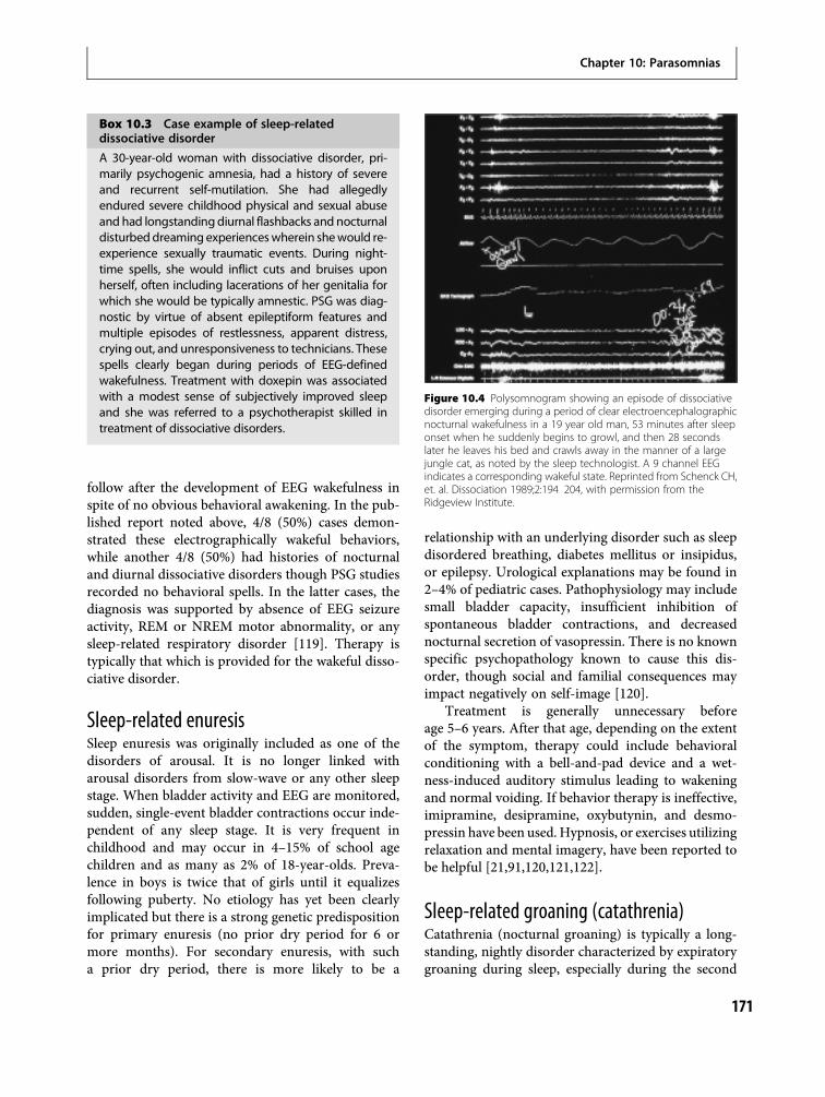
follow after the development of EEG wakefulness inspite of no obvious behavioral awakening. In the pub-lished report noted above, 4/8 (50%) cases demon-strated these electrographically wakeful behaviors,while another 4/8 (50%) had histories of nocturnaland diurnal dissociative disorders though PSG studiesrecorded no behavioral spells. In the latter cases, thediagnosis was supported by absence of EEG seizureactivity, REM or NREM motor abnormality, or anysleep-related respiratory disorder [119]. Therapy istypically that which is provided for the wakeful disso-ciative disorder.
Sleep-related enuresisSleep enuresis was originally included as one of thedisorders of arousal. It is no longer linked witharousal disorders from slow-wave or any other sleepstage. When bladder activity and EEG are monitored,sudden, single-event bladder contractions occur inde-pendent of any sleep stage. It is very frequent inchildhood and may occur in 4–15% of school agechildren and as many as 2% of 18-year-olds. Preva-lence in boys is twice that of girls until it equalizesfollowing puberty. No etiology has yet been clearlyimplicated but there is a strong genetic predispositionfor primary enuresis (no prior dry period for 6 ormore months). For secondary enuresis, with sucha prior dry period, there is more likely to be a
relationship with an underlying disorder such as sleepdisordered breathing, diabetes mellitus or insipidus,or epilepsy. Urological explanations may be found in2–4% of pediatric cases. Pathophysiology may includesmall bladder capacity, insufficient inhibition ofspontaneous bladder contractions, and decreasednocturnal secretion of vasopressin. There is no knownspecific psychopathology known to cause this dis-order, though social and familial consequences mayimpact negatively on self-image [120].
Treatment is generally unnecessary beforeage 5–6 years. After that age, depending on the extentof the symptom, therapy could include behavioralconditioning with a bell-and-pad device and a wet-ness-induced auditory stimulus leading to wakeningand normal voiding. If behavior therapy is ineffective,imipramine, desipramine, oxybutynin, and desmo-pressin have been used. Hypnosis, or exercises utilizingrelaxation and mental imagery, have been reported tobe helpful [21,91,120,121,122].
Sleep-related groaning (catathrenia)Catathrenia (nocturnal groaning) is typically a long-standing, nightly disorder characterized by expiratorygroaning during sleep, especially during the second
Box 10.3 Case example of sleep-relateddissociative disorder
A 30-year-old woman with dissociative disorder, pri-marily psychogenic amnesia, had a history of severeand recurrent self-mutilation. She had allegedlyendured severe childhood physical and sexual abuseand had longstanding diurnal flashbacks and nocturnaldisturbeddreaming experienceswherein shewould re-experience sexually traumatic events. During night-time spells, she would inflict cuts and bruises uponherself, often including lacerations of her genitalia forwhich she would be typically amnestic. PSG was diag-nostic by virtue of absent epileptiform features andmultiple episodes of restlessness, apparent distress,crying out, and unresponsiveness to technicians. Thesespells clearly began during periods of EEG-definedwakefulness. Treatment with doxepin was associatedwith a modest sense of subjectively improved sleepand she was referred to a psychotherapist skilled intreatment of dissociative disorders.
Figure 10.4 Polysomnogram showing an episode of dissociativedisorder emerging during a period of clear electroencephalographicnocturnal wakefulness in a 19 year old man, 53 minutes after sleeponset when he suddenly begins to growl, and then 28 secondslater he leaves his bed and crawls away in the manner of a largejungle cat, as noted by the sleep technologist. A 9 channel EEGindicates a corresponding wakeful state. Reprinted from Schenck CH,et. al. Dissociation 1989;2:194 204, with permission from theRidgeview Institute.
Chapter 10: Parasomnias
171
half of the night. Polysomnography documents recur-rent bradypneic episodes that occur mainly duringREM sleep and often appear in clusters, in which adeep inspiration is followed by prolonged expirationthat is accompanied by a monotonous vocalizationclosely resembling groaning. There is no observedrespiratory distress or anguished facial expression des-pite the presence of moaning.
The affected person is usually unaware of thegroaning and it is the bed partner or roommate whois disturbed by it, especially since the groaning is oftenloud and may resemble cracking, humming, orroaring sounds. The groaning is not associated withany body movements, sleepwalking, or dreaming. Theaffected person, who could be a young adult or olderadult, usually has no sleep-related complaint. Noassociation with respiratory disorders or with psycho-logical problems or psychiatric disorders has beenfound. General physical examinations and routinelaboratory testing, along with neurological and otor-hinolaryngological examinations, are unremarkable.The prevalence is unknown. However, it appears tobe quite rare. No predisposing factors have beenidentified and few of the reported patients have ahistory of any other parasomnia. The main compli-cation is interpersonal, as the bed partner or others inthe same household have their sleep disrupted, orbecome distressed from the groaning. Therapy canbe problematic, with nasal CPAP being effective insome patients [123,124].
Exploding head syndromeExploding head syndrome is characterized by thesudden sensation of a loud noise or sense of a violentexplosion in the head occurring as the affected person isfalling asleep or waking during the night. Descriptionsof these events also include a painless loud bang, anexplosion, or a clash of cymbals, but less alarmingsounds can be experienced. This can be very frightening,and many patients believe they are having a stroke. Thenumber of attacks can vary widely. It is important toemphasize that this is not a headache disorder. Theprevalence is unknown. The median age of onset is 58years, but onsets at all ages have been reported. Mostpatients cannot identify precipitating factors. Thecourse is benignwith no reported neurological sequelae.Symptoms often appear to remit spontaneously. Thecondition appears to be a sensory variant of the better-known transient (and benign) motor phenomenon of
“sleep starts” or hypnic jerks occurring at wake–sleeptransitions [125,126].
Sleep-related hallucinationsThe most common hallucinations emerging in rela-tion to sleep involve the hypnagogic (occurring in thetwilight of sleep onset) and hypnopompic (occurringon awakening in the morning) hallucinations that areoften associated with narcolepsy, although they canoccur as isolated phenomena. In addition, “complexnocturnal visual hallucinations” are another recentlyrecognized form of sleep-related hallucinations [127].Most patients with this condition are women (withouthistory of daytime hallucinations), who wake up sud-denly from sleep, usually without prior dream recall,and see vivid, realistic images of relatively immobile,usually silent, and sometimes distorted people oranimals that vanish when a light is turned on. Thepatient is clearly awake and sometimes responds byjumping out of bed in terror. Unlike hypnagogic andhypnopompic hallucinations, which are usually lessvivid, these hallucinations are often difficult to distin-guish from waking phenomena. They may be audi-tory, tactile, or kinetic and are often associated withsleep paralysis. In contrast, the visual hallucinationsof sleep-related complex partial epileptic seizures areusually brief, stereotyped, and fragmentary, althoughoccasionally more vivid visual imagery may be pre-sent. Sleep-related migraine with aura at times can beassociated with complex visual hallucinations, but aheadache should follow. Complex nocturnal visualhallucinations can represent a benign, idiopathic con-dition, or it can be associated with the use of beta-adrenergic receptor blocking medications, dementiawith Lewy bodies, visual loss (Charles Bonnet hallu-cinations), and other brain pathology (peduncularhallucinosis) [127].
Sleep-related eating disorderAmong the interesting behavioral manifestations ofdisorders of arousal is sleep-related eating disorder(SRED), often a relentless, chronic condition. It ischaracterized by consumption of high caloric foodsand sometimes bizarre substances. Foods high incarbohydrates and fats are typically eaten, and bin-ging is common. The majority of patients report thatbreads, pies, and dairy products such as ice cream,and also chocolate, other sweets, and peanut butter
Section IV: Primary Sleep Disorders in Psychiatric Contexts
172
are most commonly consumed. Interestingly, hungeris typically absent in SRED and the food choices areoften not consumed during the daytime.
Of 38 adults with complaints of sleep-relatedinvoluntary nocturnal eating in an early study,the problematic behaviors appeared to be related toSW in 26 (68.4%), restless legs syndrome/periodiclimb movement disorder (RLS/PLMD) in 5 (13.2%),triazolam abuse in 1 (2.6%), and obstructive sleepapnea syndrome in 4 (10.5%). Over half (57.9%)described nocturnal eating frequency as nightly, butsometimes more than once in a night. Eighteen of thepatients (47.4%) had evidence of DSM-III axisI psychiatric disorders and only 2 (5.3%) had daytimeeating disorders, which were in remission for 3–7years prior to sleep evaluation. Eating behaviorsincluded sloppy preparation or consumption, injuriesfrom cooking or eating, and weight gain. A similarearlier series of 23 consecutive cases included 11/23(48%) based upon SW. The majority of patientsdescribed a long history of involuntary nocturnaleating (mean duration 15.8 years) and nearly allreported eating on a nightly basis (1–6 times pernight). A lifetime diagnosis of daytime eating disorderwas noted in 8/23 (35%) of these individuals [128].In a prior study, Winkelman noted that sleep-relatedeating disorder was found in 8.7% of outpatient eatingdisorders, 16.7% of inpatient eating disorders, 1.0% ofobese patients, 3.4% of depressed outpatients, and4.6% of student controls. There was an associationwith depressed mood in those outpatients with bothdaytime and sleep-related eating disorders [129]. In amore recent study reporting 35 patients with noctur-nal eating, 25 ate more than once a night and 8 atemore than 5 times a night [130].
Food preparation and consumption has resultedin safety concerns and adverse health consequences.Hazardous activity has included: drinking excessivelyhot liquids, choking on thick foods, and lacerationsfrom careless food preparation. Furthermore, ined-ible and toxic substances have been consumed suchas frozen food, uncooked spaghetti, cat food, eggshells, coffee grounds, sunflower shells, buttered cig-arettes, glue, and cleaning solutions. It is importantfor psychiatrists to query patients receiving mono-amine oxidase inhibitor antidepressants and theirfamilies regarding nocturnal eating or SW/STbecause of the risk of possible hypertensive crisis ina patient unwittingly vulnerable to ingestion of pro-scribed foods.
Various medical and dental consequences canoccur from repeated nocturnal eating. Weight gainis commonly reported, and the increased BMI maythen precipitate, or aggravate, pre-existing diabetesmellitus (type I or II), hyperlipidemia, hypercholes-terolemia, hypertension, and OSA. Furthermore,patients with SRED are at risk of ingesting poisonoussubstances or food to which they are allergic. Patientswith SRED are at risk for poor dentition as the feed-ing behavior, usually high in carbohydrates, is nottypically followed by dental hygiene practices. Fur-thermore, many patients will sleep with an oral bolusof food which combined with the circadian declinein salivary flow promotes the development of caries.Finally, failure to exhibit control over nocturnaleating can lead to secondary depressive disordersrelated to excessive weight gain.
SRED has been associated with psychotropic medi-cations, in particular sedative-hypnotics. Sporadiccases of SRED have been reported with agents, suchas tricyclic antidepressants, anticholinergics, lithium,triazolam, olanzapine, and risperidone. More recentlySRED has been implicated with use of the benzodi-azepine receptor agonist zolpidem [62,131,132].Because of its association with sometimes complexautomatic behavior, the drug should be discontinued.
Various forms of psychotherapy, hypnother-apy, and behavior therapy have proven ineffective.Generally, therapy for these disorders includesvarious combinations of dopaminergics (carbidopa/levodopa, bromocriptine), codeine (usually combinedin an acetaminophen preparation), and clonazepamwhich were found associated with sustained pharma-cological control in 23/27 (85.2%) of patients forwhom outcome data were available [133,134]. Morerecently, the anticonvulsant topiramate has beenshown to be efficacious in two open-label series insuppressing nocturnal eating in roughly two-thirds ofpatients with either the sleepwalking or the idiopathicvariant of SRED [135,136].
The major differential diagnosis is night eatingsyndrome (NES), in which there is overeating in theevening, and often also during full nocturnal awaken-ings with subsequent recall the next day. NES isconsidered to be a sleep maintenance insomnia dis-order with eating (usually not binging) helpinginduce the resumption of sleep, whereas SRED is aparasomnia (often with binging) with reduced aware-ness and compromised subsequent recall. SRED isnot associated with overeating in the evening. There
Chapter 10: Parasomnias
173

are a number of overlapping and divergent featuresbetween NES and SRED, as recently reviewed [134].
Additional varietiesSexsomniaSexsomnia has been classified in the ICSD-2 within thegroup of parasomnias named “disorders of arousal(from NREM sleep),” with the designation of “sleep-related abnormal sexual behaviors” being a variant ofconfusional arousals (and SW). There is an establishedand growing literature on the latter phenomenon thathas also been named “sleepsex,” “sexsomnia,” and“atypical sexual behaviors in sleep.” A recent reviewhas provided the first classification of all reportedabnormal sexual behaviors associated with sleep dis-orders, including the sexual parasomnias. The major-ity of published cases involve young adult males with
rich histories of NREM sleep parasomnias, suchas sleepwalking and sleep terrors. Most patientsresponded to standard therapy for disorders of arousal(e.g. clonazepam at hs), or CPAP therapy of OSA[137]. This parasomnia can present serious forensic
Box 10.4 Case example of sleep-related eatingdisorder
A 19-year-old female presented 2 months before shewas due to leave home to attend university. Her chiefcomplaint was a 3-year history of increasingly fre-quent episodes of sleepwalking with eating. Theseepisodes usually began 1–2 hours after she fellasleep, and she could have up to 3 episodes on asingle night for 3–6 nights each week. She usuallyhad some vague recall of her eating during the night.The patient was concerned about what her newroommates at university would think about her onaccount of her peculiar and disruptive sleep behav-iors, and she was also concerned about her safety inwandering around a college dormitory or going out-side searching for food during the night. Also, shehad gained almost 15 pounds from the nocturnaleating, and was worried about further weight gainat university from her sleep-eating. She no longerhad a desire to eat breakfast, and many morningsshe awakened with food in her mouth and with herstomach feeling full. She worried about the possibil-ity of choking in her sleep from unswallowed food.She felt thoroughly disgusted about her loss of con-trol during sleep. She never, however, consideredpurging herself in any manner, day or night. Shehad an accurate appraisal of her body size and shape.
Treatment with topiramate, with an initial dose of25 mggradually increased to 100mg qhs, controllednearly all episodes of sleep-eating throughout thefirst half of the school year.
Box 10.5 Case example of sexsomnia
A 32-year-old married man, with a chief complaint of“fondling my wife during sleep,” presented with hiswife of 10 years who had urged him to seek help forhis sleep problem that had begun 4 years earlier,when he commenced to snore and also grope andfondle his wife sexually while being sound asleep. Hissnoring became progressively louder over time andhis wife reported that “he would keep trying to humpme while he was asleep.” She was “shocked” toobserve her husband engage in a full repertoire ofsexual behavior with her while he was clearly asleep, 4nights weekly over a period of years. His wife reportedthat he was somewhat insistent with his sleep sexinitiatives with her, but never aggressive or violent,and he always responded promptly to her setting oflimits. There had been no underlying marital prob-lems at the time of referral or in the past. The sleepsex eventually did influence a growing strain on theirmarriage. They endorsed having a normal sexual rela-tionship and could not identify any psychosocial trig-gering factor for the emergence of sleep sex 4 yearspreviously, such as disordered sexual function, sexualdeprivation, or other stress. There was no history ofprior sleep disorder, and specifically no childhood orsubsequent history of parasomnia. There was nohistory ofmedical, neurological, or psychiatric disorder,nor any loss of consciousness or seizure-like spells.There was no history of paraphilia, or criminal sexualmisconduct, andhedenied havingproblemswith com-pulsive masturbation, or excessive/inappropriatesexual fantasies or preoccupations. He denied any his-tory of having been sexually molested during child-hood or subsequently. There was no history ofalcohol or substance abuse, or excessive caffeine use;he smoked a pack of cigarettes daily.
PSG evaluation revealed moderately severeobstructive apnea, with an apnea-hypopnea index of19/hour, oxygen desaturation nadir of 78%, and aver-age duration of apnea, 22.7 seconds. Administration ofCPAP 10cmH2O pressure completely eliminated thesleep disordered breathing. At 3-month follow-up, thepatient’s wife reported that nightly CPAP therapy hadcontrolled his snoring and also completely eliminatedhis sexsomnia, which presumably had emergedduring ongoing OSA-induced confusional arousals.
174
Section IV: Primary Sleep Disorders in Psychiatric Contexts
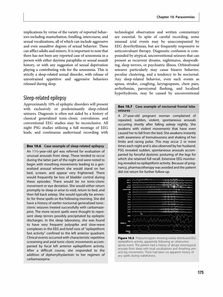
implications by virtue of the variety of reported behav-iors including masturbation, fondling, intercourse, andsexual vocalizations, all of which can include aggressiveand even assaultive degrees of sexual behavior. Thesecan afflict adults andminors. It is important to note thatthere has not been any reported case of sexsomnia in aperson with either daytime paraphilia or sexual assaulthistory, or with any suggestion of sexual deprivationplaying a contributing role for the sexsomnia. This isstrictly a sleep-related sexual disorder, with release ofunrestrained appetitive and aggressive behaviorsreleased during sleep.
Sleep-related epilepsyApproximately 10% of epileptic disorders will presentwith exclusively or predominantly sleep-relatedseizures. Diagnosis is often not aided by a history ofclassical generalized tonic-clonic convulsions andconventional EEG studies may be inconclusive. Allnight PSG studies utilizing a full montage of EEGleads, and continuous audiovisual recording with
technologist observation and written commentaryare essential. In spite of careful recording, someunusual ictal events may be unaccompanied byEEG dysrrhythmias, but are frequently responsive toanticonvulsant therapy. Diagnostic confusion is com-pounded by atypical, unconventional seizures that canpresent as recurrent dreams, nightmares, sleepwalk-ing, sleep terrors, or psychiatric illness. Orbitofrontalseizures particularly may cause bizarre behavior,peculiar clustering, and a tendency to be nocturnal.Any sleep-related behavior, even such events asapnea, stridor, coughing, laryngospasm, chest pain,arrhythmias, paroxysmal flushing, and localizedhyperhydrosis, may be caused by unconventional
Box 10.6 Case example of sleep-related epilepsy
An 11½-year-old girl was referred for evaluation ofunusual arousals from sleep. These tended to occurduring the latter part of the night and were noted tobegin with mouthing movements leading to a gen-eralized arousal wherein she would stand on herbed, scream, and appear very frightened. Therewould frequently be loss of bladder control duringthese episodes. There would be no tonic-clonicmovement or eye deviation. She would either returnpromptly to sleep or arise to void, return to bed, andthen fall back asleep. She would typically be amnes-tic for these spells on the following morning. She didhave a history of earlier nocturnal generalized tonic-clonic seizures treated successfully with carbamaze-pine. The more recent spells were thought to repre-sent sleep terrors possibly precipitated by epilepticdischarges. In the sleep laboratory, she was foundto have very frequent polyspike and slow-wavecomplexes in the EEG and brief runs of “epileptiformfast activity” confined to the left anterior quadrant.Clinical events occurred with characteristic repetitivescreaming and axial tonic-clonic movements accom-panied by focal left anterior epileptiform activity.After a difficult course, she responded to theaddition of diphenylhydantoin to her regimen ofcarbamazepine.
Box 10.7 Case example of nocturnal frontal lobeseizures
A 27-year-old pregnant woman complained ofrepeated, sudden, violent, spontaneous arousalsoccurring shortly after falling asleep nightly. Sheawakens with violent movements that have evencaused her to fall from the bed. She awakens instantlywith awareness of tremulousness and shaking of herlimbs and racing pulse. This may occur 2 or moretimes each night and is also observed by her husband.PSG revealed sudden, spontaneous arousals accom-panied by forceful dystonic posturing of the legs forwhich she retained full recall. Extensive EEG monitor-ing revealed no epileptiform activity. Because of preg-nancy, pharmacotherapy was avoided and the patientdid not return for further follow-up.
Figure 10.5 Polysomnogram showing widely distributed EEGepileptiform activity, apparently following an obstructiveapnea event. The patient had a history of abrupt stereotypicalarousals from sleep with loud vocalizations and thrashing armand leg movements. There had been no apparent history ofany spells during wakefulness.
Chapter 10: Parasomnias
175
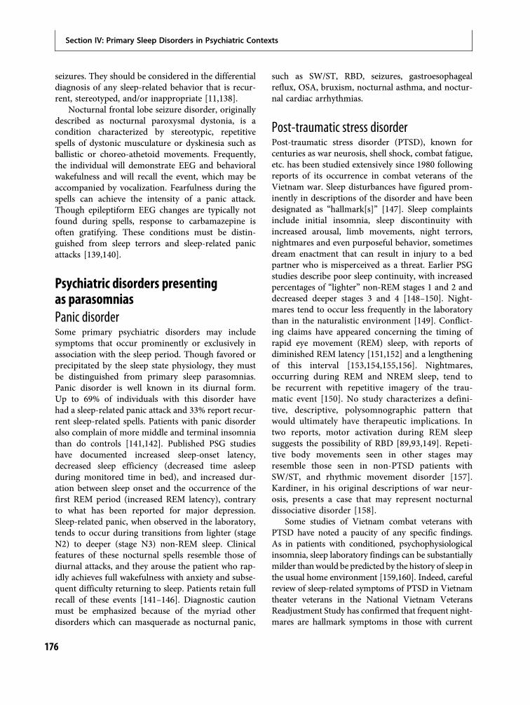
seizures. They should be considered in the differentialdiagnosis of any sleep-related behavior that is recur-rent, stereotyped, and/or inappropriate [11,138].
Nocturnal frontal lobe seizure disorder, originallydescribed as nocturnal paroxysmal dystonia, is acondition characterized by stereotypic, repetitivespells of dystonic musculature or dyskinesia such asballistic or choreo-athetoid movements. Frequently,the individual will demonstrate EEG and behavioralwakefulness and will recall the event, which may beaccompanied by vocalization. Fearfulness during thespells can achieve the intensity of a panic attack.Though epileptiform EEG changes are typically notfound during spells, response to carbamazepine isoften gratifying. These conditions must be distin-guished from sleep terrors and sleep-related panicattacks [139,140].
Psychiatric disorders presentingas parasomniasPanic disorderSome primary psychiatric disorders may includesymptoms that occur prominently or exclusively inassociation with the sleep period. Though favored orprecipitated by the sleep state physiology, they mustbe distinguished from primary sleep parasomnias.Panic disorder is well known in its diurnal form.Up to 69% of individuals with this disorder havehad a sleep-related panic attack and 33% report recur-rent sleep-related spells. Patients with panic disorderalso complain of more middle and terminal insomniathan do controls [141,142]. Published PSG studieshave documented increased sleep-onset latency,decreased sleep efficiency (decreased time asleepduring monitored time in bed), and increased dur-ation between sleep onset and the occurrence of thefirst REM period (increased REM latency), contraryto what has been reported for major depression.Sleep-related panic, when observed in the laboratory,tends to occur during transitions from lighter (stageN2) to deeper (stage N3) non-REM sleep. Clinicalfeatures of these nocturnal spells resemble those ofdiurnal attacks, and they arouse the patient who rap-idly achieves full wakefulness with anxiety and subse-quent difficulty returning to sleep. Patients retain fullrecall of these events [141–146]. Diagnostic cautionmust be emphasized because of the myriad otherdisorders which can masquerade as nocturnal panic,
such as SW/ST, RBD, seizures, gastroesophagealreflux, OSA, bruxism, nocturnal asthma, and noctur-nal cardiac arrhythmias.
Post-traumatic stress disorderPost-traumatic stress disorder (PTSD), known forcenturies as war neurosis, shell shock, combat fatigue,etc. has been studied extensively since 1980 followingreports of its occurrence in combat veterans of theVietnam war. Sleep disturbances have figured prom-inently in descriptions of the disorder and have beendesignated as “hallmark[s]” [147]. Sleep complaintsinclude initial insomnia, sleep discontinuity withincreased arousal, limb movements, night terrors,nightmares and even purposeful behavior, sometimesdream enactment that can result in injury to a bedpartner who is misperceived as a threat. Earlier PSGstudies describe poor sleep continuity, with increasedpercentages of “lighter” non-REM stages 1 and 2 anddecreased deeper stages 3 and 4 [148–150]. Night-mares tend to occur less frequently in the laboratorythan in the naturalistic environment [149]. Conflict-ing claims have appeared concerning the timing ofrapid eye movement (REM) sleep, with reports ofdiminished REM latency [151,152] and a lengtheningof this interval [153,154,155,156]. Nightmares,occurring during REM and NREM sleep, tend tobe recurrent with repetitive imagery of the trau-matic event [150]. No study characterizes a defini-tive, descriptive, polysomnographic pattern thatwould ultimately have therapeutic implications. Intwo reports, motor activation during REM sleepsuggests the possibility of RBD [89,93,149]. Repeti-tive body movements seen in other stages mayresemble those seen in non-PTSD patients withSW/ST, and rhythmic movement disorder [157].Kardiner, in his original descriptions of war neur-osis, presents a case that may represent nocturnaldissociative disorder [158].
Some studies of Vietnam combat veterans withPTSD have noted a paucity of any specific findings.As in patients with conditioned, psychophysiologicalinsomnia, sleep laboratory findings can be substantiallymilder thanwould be predicted by the history of sleep inthe usual home environment [159,160]. Indeed, carefulreview of sleep-related symptoms of PTSD in Vietnamtheater veterans in the National Vietnam VeteransReadjustment Study has confirmed that frequent night-mares are hallmark symptoms in those with current
Section IV: Primary Sleep Disorders in Psychiatric Contexts
176

PTSD and correlatemore with level of combat exposurethan insomnia symptoms [161]. It is very possible thatobjective studies of sleep in PTSD may underreportnightmare frequency and severity because moreseverely affected subjects may avoid participation [162].
A striking finding in the PTSD sleep literature isthe elevation of auditory arousal thresholds inaffected subjects during NREM as well as REM sleep[163–165]. Kramer interprets these findings as evi-dence that in chronic PTSD there is a heightenedresponsiveness to internal events while individualsare less arousable by external stimuli [166]. Increaseddepth of sleep may represent a chronic adaptation totrauma. Kaminer and Lavie describe the diminisheddream recall of their better-adjusted Holocaust sur-vivors compared with those less well adjusted [167].The common subjective sleep complaints of patientsmay reflect a breakdown of this adaptation but withenough resilience to allow intact sleep in a safe, neu-tral environment such as a sleep laboratory.
Treatment of PTSD has progressed in recentyears. There are a number of reports of improvementin PTSD nightmares with pharmacotherapy and somestudies suggest benefit from 5-HT2 receptor antagon-ists such as nefazodone. Effectiveness of clonidine,GABAergic drugs such as tiagabine, atypical neuro-leptics such as quetiapine, and anticonvulsants suchas topiramate have also been reported [168–173].Few placebo-controlled studies have been reported,and there is some evidence for utility of olanzapineas well as prazosin [174,175]. Prazosin is an alpha 1adrenoreceptor antagonist, which has been effectivein reducing frequency and intensity of nightmaresas well as non-nightmare distressed awakenings[114,176,177]. Non-pharmacological therapies haveincluded clinical hypnosis for relief of insomniaand dream rehearsal therapy for nightmares [111,178–180]. There is accumulating evidence of efficacyfor the technique known as eye movement desensi-tization and reprocessing therapy (EMDR), which hasbeen theorized to modulate the intensity of PTSDby shifting the memory processing of the brain intoa mode resembling REM sleep [181].
Factitious parasomniaRarely, an individual may present for evaluation witha factitious parasomnia. There is no reason to assumethat such a disorder could not be chosen for thispurpose. The forensic literature includes numerousexamples of sleep-related violence, some of whichresult in exculpation because of the presence of a sleepdisorder [182–184]. A particular form of factitiousdisorder is the Munchausen syndrome, which caneven occur by proxy, such as the bizarre case of amother who described obstructive sleep apnea in herchild. When studied in the sleep laboratory, the childwas observed on video recording to be suffocatedby the mother who then summoned the technologistfor assistance [185].
Malingering might be expected in situationsinvolving litigation, correctional institutions, mili-tary settings, and many others. This must be verycarefully considered as a diagnosis by exclusion,supported by observations, reliable reports, or otherfactors that would support the likelihood of malin-gering. Likewise, even though diagnostic findingsmay emerge during PSG, they do not necessarilyprove that alleged acts are attributable to the dis-order. The ideal role of the sleep specialist in thesecases is to educate the court about the complexity ofthese disorders and the likelihood of the diagnoses,rather than to adopt an adversarial position[186,187].
Box 10.8 Case example of PTSD
A 44-year-old veteran of combat during the VietnamWar suffered post-traumatic stress disorder (PTSD)beginning about 2 years after his tour of duty. Hehad symptoms satisfying DSM-IIIR criteria for it and
also described cycles of waxing and waning sleepdisturbance with a periodicity of around 2 weeks.Following 2 or 3 nights of terribly disturbed sleepwith traumatic nightmares, he has calmer, improvedsleep but with a crescendo once again to a new peakof disturbed sleep. Alcohol abuse had long been inremission. He had PSG study at a time when heexpected to be at a peak level of sleep disruption.During 3 nights, PSG revealed no significant abnor-mality in spite of increasing subjective distress thathe felt to be usually predictive of impaired sleep.He subsequently attributed his improvement in thelaboratory to sleeping in the supine position andfound that this position continued to be associatedwith improved sleep in his home until he beganreturning to the lateral position and again experi-enced poor sleep.
Chapter 10: Parasomnias
177

Table
10.2
Characterstcs
ofparasomnas
Non
-REM
parasom
nias
REM
parasom
nias
Other
parasom
nias
Parasomna
Con
fuson
aarou
sas
Seepw
akng
Seep
terrors
RBD
Seepparays
sNgh
tmare
dsorder
SRED
SRH*
SRDD
Stageof
arou
sa,
,V
,V
,V
REM
REM
REM
,,V
REM
NREM
orREM
Typca
tmeof
ngh
tAnytm
eFrst2ho
urs
Frst2
hours
Anytm
eAnytm
e(frst
2ho
urs)
Anytm
eAnytm
eOnset/offset
ofseep
Anytm
e
EEGdu
rngeven
tNA
Mxed
Mxed
REM
pattern
Wakepattern
NA
Mxed
Wakepattern
Wake
pattern
EMGactv
tydu
rngeven
t"
""
"#
NA
"NA
"Decreased
respon
svene
ssdu
rng
even
t
þþ
þþ
–þ
þ–
þ
Auton
omchype
ractvty
––
þþ
þ/�
þ–
þ/�
þ/�
Amne
sa
þþ
þ–(dream
reca
)–(experen
cereca
)�
(dream
reca
)þ/
�(parta
)�
þ
Con
fuson
postep
sode
þþ
þ–
––
þ�
þFam
yhstory
þþ
þ–
þ/�
–þ
þ/�
Unkno
wn
EEG,e
ectroe
ncep
haog
ram;EMG,e
ectrom
yogram
;NREM,non
-rap
deyemovem
ent;RBD,REM
behavor
dsorder;REM
,rap
deyemovem
ent;SRED
,seep-reated
eatn
gdsorder;SRH
,seep-
reated
hauc
nato
ns;SRD
D,s
eep-reated
dssoc
atve
dsorder.
Note:*SRH
forth
stabenc
udes
hypn
agog
c/po
mpcha
ucnato
ns.The
few
EEGstud
esof
compex
nocturna
hauc
nato
nssugg
estthey
canoccurfro
mNREM.
Reprod
uced
from
Pante
DT,W
nkeman
JW.Parasom
nas:Psych
atrc
Con
sde
rato
ns.Sleep
Med
Clin.2008;3(2):217–229,w
thpe
rmsson
from
Esever.
178

Evaluation and treatmentAlthough the most typical and benign parasomniasrequire no treatment, the anxiety and concern theyarouse can be calmed through education and reassur-ance. Any parasomnia that is significantly disruptiveto individual or family life warrants careful evaluationand may require consultation with an experiencedsleep medicine specialist. Any parasomnia that ispotentially or actually injurious to self or others mustbe evaluated thoroughly at an accredited sleep dis-orders center with staff skilled in such evaluation.Description of clinical history must be based uponas many sources of information as possible in viewof difficulties with subjective self-reports from individ-uals whose symptoms include varying degrees ofamnesia. Initial history must include age of onset,estimations of frequency of spells at various periodsin the course of the disorder, any changes over time,medical and psychiatric history, family history, generalsleep history with reference to sleep hygiene and obser-vations of respiration, sleep disruption and bodymovements during sleep, apparent precipitatingfactors including drug/alcohol abuse or dependence,and prior responses to treatment. Description of thespells must include reference to typical time of onset inthe sleep period, recent frequency, specific behaviorswith special regard for dangers posed to self or others,dream recall, and degree of amnesia for either dreamcontent or actual behavior. Psychiatric assessmentshould focus especially on the presence or absence ofmood disorders as well as drug and/or alcohol abuse ordependence. Psychiatric interview as well as screeninginstruments such as the MMPI, Beck depressioninventory, SCL90, dissociative experiences scale,and Zung or Beck self-rating anxiety inventories mayindicate areas deserving of further psychiatric assess-ment or therapy. A recent publication includes Table10.2, which summarizes the clinical and polysomno-graphic features that distinguish various parasomnias.
When behavioral spells are observed during PSGstudy, the diagnosis can be confirmed. In a report of100 consecutive adult patients with sleep-relatedinjury, diagnoses were established by PSG in 65%of cases (42.6% of SW/ST, 100% of RBD, 42.9% ofdissociative disorders, 100% of nocturnal seizures, and100% of PLMS). Diagnoses were strongly supported inanother 26% (48.1% of SW/ST), accounting foran overall positive yield of PSG studies of 91%. Con-firmatory findings include behaviors such as moaning,
somniloquy, yelling, limb movements, gesturingincluding violent movements, finger pointing, sittingup abruptly, looking around in a confused manner,apparently hallucinated behavior, and leaving the bedduring particular stages of sleep or arousals. Supportivefindings include excessive abrupt, spontaneous arousalsfrom stages of NREM sleep in cases of SW/ST [19,34].
While behavioral manifestations of many dis-orders can lead to diagnostic confusion, proper evalu-ation can lead to gratifying therapeutic results withreduction of the risk of sleep-related injury for suffer-ers and their bed partners. Seemingly obvious SW/STcan occasionally be found to arise from a seizuredisorder. Many parasomnias with bizarre, complexbehaviors can be misrepresented as psychiatric illness.Treatment of nocturnal dissociative disorder withclonazepam as if it were SW/ST could be unsuccessfulor even worsen the spells [119]. Treatment of RBDwith neuroleptic medication as if it represented noc-turnal agitation in a geriatric patient couldallow the untreated parasomnia to pose continuedrisk of serious injury to the patient and/or bed partnerand antidepressant therapy could precipitate orworsen RBD. Furthermore, the presence of RBD sig-nals the need for careful neurological assessmentand follow-up for detection of emergent neurodegen-erative disorders. Only careful clinical and polysom-nographic evaluations can definitively clarify thenature of the most difficult and dangerous of thesedisorders.
References1. Roccatagliata G. In: A history of ancient psychiatry.
Westport, Connecticut: Greenwood Press;1986:107 108.
2. Roccatagliata G. In: A history of ancient psychiatry.Westport, Connecticutt: Greenwood Press;1986:216.
3. Roccatagliata G. In: A history of ancient psychiatry.Westport, Connecticut: Greenwood Press; 1986:241.
4. Ellenberger H. The discovery of the unconscious: thehistory and evolution of dynamic psychiatry. New York:Basic Books; 1970.
5. Rush B. Of dreaming, incubus, or night mare, andsomnambulism. In: Medical inquiries and observationsupon the diseases of the mind. Philadelphia: Kimberand Richardson; 1812:300 305.
6. Ray I. Somnambulism (Chapter 19), Legalconsequences of somnambulism (Chapter 20), andSimulated somnambulism (Chapter 21). In: Treatise
Chapter 10: Parasomnias
179
on the medical jurisprudence of insanity. Boston:Charles C. Little and James Brown; 1838:386 400.
7. Freud S. The Interpretation of dreams. New York:Basic Books; 1953.
8. American Psychiatric Association. Diagnostic andstatistical manual of mental disorders, 4th ed, textrevision. Washington, DC: American PsychiatricAssociation; 2000.
9. American Academy of Sleep Medicine. Internationalclassification of sleep disorders, 2nd ed: Diagnostic andcoding manual. Westchester, IL: American Academyof Sleep Medicine; 2005.
10. Ohayon M, Guilleminault C, Priest R. Night terrors,sleepwalking, and confusional arousals in the generalpopulation: Their frequency and relationship to othersleep; and mental disorders. J Clin Psychiatry. 1999;60(4):268 276.
11. Mahowald M, Ettinger M. Things that go bump in thenight: the parasomnias revisited. J Clin Neurophysiol.1990;7(1):119 143.
12. Gastaut H, Broughton R. A clinical and polygraphicstudy of episodic phenomena during sleep.In: Wortis J, ed. Recent advances in biologicalpsychiatry. New York: Plenum Press; 1965:197 223.
13. Roth B, Nevsimalova S, Rechtschaffen A. Hypersomniawith sleep drunkenness. Arch Gen Psychiatry.1972;26:456 462.
14. Tassi P, Muzet A. Sleep inertia. Sleep Med Rev.2000;4:341 353.
15. Klackenberg G. Somnambulism in childhood:prevalence, course and behavioral correlations.Acta Paediatr Scand. 1982;71:494 499.
16. Bixler E, Kales A, Soldatos C, et al. Prevalence of sleepdisorders in the Los Angeles metropolitan area.Am J Psychiatry. 1979;136:1257 1262.
17. Casez O, Dananchet Y, Besson G. Migraine andsomnambulism. Neurology 2005;65(8):1334 1335.
18. Ajlouni K, Ahmad A, El Zahiri M, et al. Sleepwalkingassociated with hyperthyroidism. Endocr Pract.2005;11(4):294.
19. Schenck C, Milner D, Hurwitz T, Bundlie S,Mahowald M. A polysomnographic and clinicalreport on sleep related injury in 100 adult patients.Am J Psychiatry. 1989;146(9):1166 1173.
20. Guilleminault C, Sylvestri R. Disorders of arousal andepilepsy during sleep. In: Sterman M, Shouse M,Passouant P, eds. Sleep and epilepsy. New York:Academic Press; 1982:513 531.
21. Millman R, Kipp G, Carskadon M. Sleepwalkingprecipitated by treatment of sleep apnea with nasalCPAP. Chest. 1991;99:750 751.
22. Espa F, Dauvilliers Y, Ondze B, Billiard M, Besset A.Arousal reactions in sleepwalking and night terrors inadults: the role of respiratory events. Sleep. 2002;25(8):871 875.
23. Kleitman N. Sleep and wakefulness, revised and enlargededition. Chicago: University of Chicago Press; 1963.
24. Bassetti C, Vella S, Donati F, Wielepp P, Weder B.SPECT during sleepwalking. Lancet. 2000;356:484 485.
25. Chesson A, Ferber F, Fry J, et al. The indications forpolysomnography and related procedures. Sleep.1997;20(6):423 487.
26. Kushida C, Littner M, Morgenthaler T, et al. Practiceparameters for the indications for polysomnographyand related procedures: an update for 2005. Sleep.2005;28(4):499 521.
27. PilonM,Montplaisir J, Zadra A. Precipitating factors ofsomnambulism: impact of sleep deprivation and forcedarousals. Neurology. 2008;70(24):2284 2290.
28. Zadra A, Pilon M, Montplaisir J. Polysomnographicdiagnosis of sleepwalking: effects of sleep deprivation.Ann Neurol. 2008;63(4):513 519.
29. Schenck C, Pareja J, Patterson A, Mahowald M.Analysis of polysomnographic events surrounding 252slow wave sleep arousals in thirty eight adults withinjurious sleepwalking and sleep terrors. J ClinNeurophysiol. 1998;15(2):159 166.
30. Zadra A, Pilon M, Joncas S, Rompre S, Montplaisir J.Analysis of postarousal EEG activity duringsomnambulistic episodes. J Sleep Res. 2004;13(3):279 284.
31. Diagnostic Classification Steering Committee TM,Chairman. International classification of sleepdisorders: Diagnostic and coding manual. Rochester,Minnesota: American Sleep Disorders Association;1990.
32. Denesle R, Gosselin A, Zadra A, Nicolas A,Montplaisir J. Arousals from REM and non REMsleep in sleepwalkers and normal controls. Sleep.1998;21(3 Suppl):254.
33. Kavey N, Whyte J, Resor S, Gidro Frank S.Somnambulism in adults. Neurology. 1990;40:749 752.
34. Bruni O, Ferri R, Novelli L, et al. NREM sleepinstability in children with sleep terrors: the role ofslow wave activity interruptions. Clin Neurophysiol.2008;119(5):985 992.
35. Guilleminault C, Kirisoglu C, da Rosa A, Lopes C,Chan A. Sleepwalking, a disorder of NREM sleepinstability. Sleep Med. 2006;7(2):163 170.
36. Guilleminault C, Lee J, Chan A, et al. Non REM sleepinstability in recurrent sleepwalking in pre pubertalchildren. Sleep Med. 2005;6(6):515 521.
Section IV: Primary Sleep Disorders in Psychiatric Contexts
180
37. Pierce C, Lipcon H. Somnambulism: Psychiatricinterview studies. US Armed Forces Med J. 1956;7(8):1143 1153.
38. Sours J, Frumkin P, Indermill R. Somnambulism, itsclinical significance and dynamic meaning in lateadolescence and adulthood. Arch Gen Psychiatry.1963;9:400 413.
39. Kales A, Jacobson A, Paulson M, Kales J.Somnambulism: psychophysiological correlates,II. Psychiatric interviews, psychological testing, anddiscussion. Arch Gen Psychiatry. 1966;14:595 604.
40. Kales A, Soldatos C, Caldwell A, et al. Somnambulism,clinical characteristics and personality patterns.Arch Gen Psychiatry. 1980;37:1406 1410.
41. Kales J, Kales A, Soldatos C, et al. Night terrors, clinicalcharacteristics and personality patterns. Arch GenPsychiatry. 1980;37:1413 1417.
42. Llorente M, Currier M, Norman S, Mellman T. Nightterrors in adults: phenomenology and relationshipto psychopathology. J Clin Psychiatry. 1992;53(11):392 394.
43. Guilleminault C, Moscovitch A, Leger D. Forensicsleep medicine: nocturnal wandering and violence.Sleep. 1995;18(9):740 748.
44. Moldofsky H, Gilbert R, Lue F, MacLean A. Sleeprelated violence. Sleep. 1995;18(9):731 739.
45. Chiu Y, Chen C, Shen W. Somnambulism secondaryto olanzapine treatment in one patient with bipolardisorder. Prog Neuropsychopharmacol Biol Psychiatry.2008;15(2):581 582.
46. Hafeez A, Kalinowski C. Two cases of somnambulisminduced by quetiapine. Prim Care Companion J ClinPsychiatry. 2007;9(4):313.
47. Khawaja I, Hurwitz T, Schenck C. Violent parasomniaassociated with a serotonin reuptake inhibitor (SSRI):A case report. J Clin Psychiatry. 2008;69:1982 1983.
48. Kawashima T, Yamada S. Paroxtine inducedsomnambulism. J Clin Psychiatry. 2003;64(4):483.
49. Kunzel H, Schuld A, Pollmacher T. Sleepwalkingassociated with reboxetine in a young female patientwith major depression: a case report.Pharmacopsychiatry. 2004;37(6):307 308.
50. Khazaal Y, Krenz S, Zullino D. Bupropion inducedsomnambulism. Addict Biol. 2003;8:359 362.
51. Landry P, Warnes H, Nielsen T, Montplaisir J.Somnambulistic like behaviour in patients attendinga lithium clinic. Int Clin Psychopharmacol. 1999;14(3):173 175.
52. Landry P, Montplaisir J. Lithium inducedsomnambulism. Can J Psychiatry. 1998;43(9):957 958.
53. Tsai J, Yang P, Chung W, et al. Zolpidem inducedamnesia and somnambulism: Rare occurrences?Eur Neuropsychopharmacol. 24 Sept 2008; Epub aheadof print.
54. Varkey B, Varkey L. Topiramate inducedsomnambulism and automatic behavior. Indian J MedSci. 2003;57(11):508 510.
55. Sattar S, Ramaswamy S, Bhatia S, Petty F.Somnambulism due to probable interaction of valproicacid and zolpidem. Ann Pharmacother. 2003;37(10):1429 1433.
56. Mahowald M, Schenck C. REM sleep behaviordisorder. In: Thorpy M, ed. Handbook of sleepdisorders. New York: Marcel Dekker; 1990:567 593.
57. Mahowald M, Schenck C. NREM sleep parasomnias.Neurol Clin. 1996;14(4):675 696.
58. Charney D, Kales A, Soldatos C, Nelson J.Somnambulistic like episodes secondary to combinedlithium neuroleptic treatment. Br J Psychiatry.1979;135:418 424.
59. Hartmann E. Two case reports: night terrors withsleepwalking: a potentially lethal disorder. J Nerv MentDis. 1983;171:503 505.
60. Landry P, Warnes H, Osivka S, Montplaisir J.Somnambulistic like behaviors induced by lithiumtaken with or without other pharmacological agents.Sleep Res. 1997;26:407.
61. Mendelson W. Sleepwalking associated with zolpidem.J Clin Psychopharmacol. 1994;14(2):150.
62. Morgenthaler T, Silber M. Amnestic sleep relatedeating disorder associated with zolpidem. Sleep Med.2002;3(4):323 327.
63. Hensel J, Pillman F. Late life somnambulism aftertherapy with metoprolol. Clin Neuropsychopharmacol.2008;31(4):248 250.
64. von Vigier R, Vella S, Bianchetti M. Agitatedsleepwalking with fluoroquinolone therapy. PediatrInfect Dis J. 1999;18(5):484 485.
65. Glick B, Schulman D, Turecki S. Diazepam (Valium)treatment in childhood sleep disorders: A preliminaryinvestigation. Dis Nerv Syst. 1971;32(8):565 566.
66. Cooper A. Treatment of coexistent night terrors andsomnambulism in adults with imipramine anddiazepam. J Clin Psychiatry. 1987;48(5):209 210.
67. Jan J, Freeman R, Wasdell M, Bomben M. A child withsevere night terrors and sleep walking responds tomelatonin therapy. Dev Med Child Neurol.2004;46(11):789.
68. Hurwitz T, Mahowald M, Schenck C, Schluter J,Bundlie S. A retrospective outcome study and reviewof hypnosis as treatment of adults with sleepwalking
Chapter 10: Parasomnias
181

and sleep terror. J Nerv Ment Dis. 1991;179(4):228 233.
69. Hauri P. The treatment of parasomnias with hypnosis:a 5 year follow up study. J Clin Sleep Med. 2007;3(4):369 373.
70. Kellerman J. Behavioral treatment of night terrors in achild with acute leukemia. J Nerv Ment Dis.1979;167:182 185.
71. Tobin J. Treatment of somnambulism withanticipatory awakening. J Pediatr. 1993;122:426 427.
72. Koe G. Hypnotic treatment of sleep terror disorder:a case report. Am J Clin Hypn. 1989;32(1):36 40.
73. Kohen D, Mahowald M, Rosen G. Sleep terrordisorder in children: the role of self hypnosis inmanagement. Am J Clin Hypn. 1992;34(4):233 244.
74. Jiarong L. 10 cases of somnambulism treated withcombined acupuncture and medicinal herbs. J TraditChin Med. 1989;9:174 175.
75. Kales J, Cadieux R, Soldatos C, Kales A. Psychotherapywith night terror patients. Am J Psychother.1982;36:399 407.
76. Schenck C, Bundlie S, Ettinger M, Mahowald M.Chronic behavioral disorders of human REM sleep:a new category of parasomnia. Sleep. 1986;9:293 308.
77. Schenck C, Mahowald M. Polysomnographic,neurologic, psychiatric, and clinical outcome report on70 consecutive cases with REM sleep behavior disorder(RBD): sustained clonazepam efficacy in 89.5% of 57treated patients. Cleveland Clin J Med. 1990;57(Suppl):9 23.
78. Schenck C, Hurwitz T, Mahowald M. REM sleepbehaviour disorder: an update on a series of 96 patientsand a review of the world literature. J Sleep Res.1993;2:224 231.
79. Mahowald M, Schenck C. REM sleep behaviordisorder. In: Kryger M, Roth T, Dement W, eds.Principles and practice of sleep medicine. Philadelphia:WB Saunders; 1989:389 401.
80. Consens F, Chervin R, Koeppe R, et al. Validation of apolysomnographic score for REM sleep behaviordisorder. Sleep. 2005;28(8):993 997.
81. Schenck C, Bundlie S, Mahowald M. Delayedemergence of a parkinsonian disorder in 38% of29 older men initially diagnosed with idiopathic rapideye movement sleep behavior disorder. Neurology.1996;46:388 393.
82. Schenck C, Bundlie S, Mahowald M. REM behaviordisorder (RBD): delayed emergence of parkinsonismand/or dementia in 65% of older men initiallydiagnosed with idiopathic RBD, and analysis of the
minimum and maximum tonic and/or phasicelectromyographic abnormalities found during REMsleep. Sleep. 2003;26(Abstract Suppl):A316.
83. Fantini M, Ferini Strambi L, Montplaisir J. IdiopathicREM sleep behavior disorder: toward a betternosologic definition. Neurology. 2005;64:780 786.
84. Iranzo A, Molinuevo J, Santamaria J, et al. Rapid eyemovement sleep behaviour disorder as an early markerfor a neurodegenerative disorder: a descriptive study.Lancet Neurol. 2006;5(7):552 553.
85. Boeve B, Silber M, Saper C, et al. Pathophysiology ofREM sleep behaviour disorder and relevance toneurodegenerative disease. Brain. 2007;130(Pt 11):2770 2788.
86. Thomas A, Bonanni L, Onofrj M. SymptomaticREM sleep behavior disorder. Neurol Sci. 2007;28:S21 S36.
87. Ferini Strambi L, Di Gioia M, Castronovo V, et al.Neuropsychological assessment in idiopathic REMsleep behavior disorder (RBD): does the idiopathicform of RBD really exist? Neurology. 2004;62:41 45.
88. Winkelman J, James L. Serotonergic antidepressantsare associated with REM sleep without atonia. Sleep.2004;27:317 321.
89. Schenck C, Milner D, Hurwitz T, et al. Sleep relatedinjury in 85 adult patients: a polysomnographic study.Sleep Res. 1988;17:247.
90. Schenck C, Mahowald M, Kim S, O’Connor K,Hurwitz T. Prominent eye movements duringNREM sleep and REM sleep behavior disorderassociated with fluoxetine treatment of depressionand obsessive compulsive disorder. Sleep. 1992;15(3):226 235.
91. Schenck C, Hurwitz T, Mahowald M. REMsleep behavior disorder. Am J Psychiatry. 1988;145:652.
92. Kandel E. Cellular mechanisms of learning and thebiological basis of individuality. In: Kandel E,Schwartz J, Jessell T, eds. Principles of neural science,3rd ed. New York: Elsevier; 1991:1009 1031.
93. Hefez A, Metz L, Lavie P. Long term effects of extremesituational stress on sleep and dreaming.Am J Psychiatry. 1987;144(3):344 347.
94. Sugita Y, Taniguchi M, Terashima K, et al. A youngcase of idiopathic REM sleep behavior disorder (RBD)specifically induced by socially stressful conditions.Sleep Res. 1991;20A:394.
95. Schenck C, Mahowald M. Injurious sleep behaviordisorders (parasomnias) affecting patients onintensive care units. Intensive Care Med. 1991;17:219 224.
Section IV: Primary Sleep Disorders in Psychiatric Contexts
182
96. Schenck C, Mahowald M. Long term, nightlybenzodiazepine treatment of injurious parasomniasand other disorders of disrupted nocturnal sleep in170 adults. Am J Med. 1996;100:333 337.
97. Kunz D, Bes F. Melatonin as a therapy in REMsleep behavior disorder patients: an open labeledpilot study on the possible influence of melatonin onREM sleep regulation. Mov Disord. 1999;14(3):507 511.
98. Takeuchi N, Uchimura N, Hashizume Y, et al.Melatonin therapy for REM sleep behavior disorder.Psychiatry Clin Neurosci. 2001;55(3):267 269.
99. Boeve B, Silber M, Ferman T. Melatonin therapy fortreatment of REM sleep behavior disorder inneurologic disorders: results in 14 patients. Sleep Med.2003;4:281 284.
100. Fantini M, Gagnon J, Filipini D, Montplaisir J. Theeffects of pramipexole in REM sleep behavior disorder.Neurology. 2003;61(10):1418 1420.
101. Schmidt M, Koshal V, Schmidt H. Use of pramipexolein REM sleep behavior disorder: results from a caseseries. Sleep Med. 2006;7(5):418 423.
102. Schenck C, Mahowald M. REM sleep parasomnias.Neurol Clin. 1996;14(4):697 720.
103. Zadra A, Donderi D. Nightmares and bad dreams:their prevalence and relationship to well being. J AbnPsychol. 2000;109:273 281.
104. Ohayon M, Morselli P, Guilleminault C. Prevalence ofnightmares and their relationship to psychopathologyand daytime functioning in insomnia subjects. Sleep.1997;20:340 348.
105. Tan V, Hicks R. Type A B behavior and nightmaretypes among college students. Percept Mot Skills.1995;81(1):15 19.
106. Kitabayashi Y, Ueda H, Tsuchida H, et al. Doepezilinduced nightmares in mild cognitive impairment.Psychiatry Clin Neurosci. 2006;60(1):123 124.
107. Hartmann E. The nightmare: the psychology and biologyof terrifying dreams. New York: Basic Books; 1984.
108. Hartmann E. Normal and abnormal dreams. In:Kryger M, Roth T, Dement W, eds. Principles andpractice of sleep medicine. Philadelphia: WB Saunders;1989:191 195.
109. Dey S. Nightmare due to ciprofloxacin. Indian Ped.1995;32(8):918 920.
110. Claridge G, Clark K, Davis C. Nightmares, dreams,and schizotypy. Br J Clin Psychology. 1997;36(Pt3):377 86.
111. Krakow B, Zadra A. Clinical management of chronicnightmares: imagery rehearsal therapy. Behav SleepMed. 2006;4(1):45 70.
112. Abramovitch H. The nightmare of returning home:a case of acute onset nightmare disorder treatedby lucid dreaming. Israel J Psychiatry Rel Sci. 1995;32(2):140 145.
113. Kinzie J, Fredrickson R, Ben R, Fleck J, Karls W.Posttraumatic stress disorder among survivors ofCambodian concentration camps. Am J Psychiatry.1984;141(5):645 650.
114. Raskind M, Peskind E, Hoff D, et al. A parallel groupplacebo controlled study of prazosin for traumanightmares and sleep disturbance in combat veteranswith posttraumatic stress disorder. Biol Psychiatry.2007;61(8):928 934.
115. Koran L, Raghavan S. Fluoxetine for isolated sleepparalysis. Psychosomatics. 1993;34:184 187.
116. Hufford D. The terror that comes in the night.Philadelphia: University of Pennsylvania Press; 1982.
117. Hinton D, Hufford D, Kirmayer L. Culture and sleepparalysis. Transcult Psychiatry. 2005;42:5 10.
118. Agargun M, Kara Y, Ozer O, et al. Characteristics ofpatients with nocturnal dissociative disorders. Sleepand Hypnosis. 2001;3(4):131 134.
119. Schenck C, Milner D, Hurwitz T, Bundlie S,Mahowald M. Dissociative disorders presenting assomnambulism: polysomnographic, video and clinicaldocumentation (8 cases). Dissociation 1989;2(4):194 204.
120. Kotagal S. Parasomnias of childhood. Curr OpinPediatr. 2008;20(6):659 665.
121. Ferber R. Sleep associated enuresis in the child. In:Kryger M, Roth T, Dement W, eds. Principles andpractice of sleep medicine. Philadelphia: WB Saunders;1989:643 647.
122. Gardner G, Olness K. Hypnosis and hypnotherapywith children. New York: Grune and Stratton;1981:130 135.
123. Pevernagie D, Boon P, Mariman A, Verhaeghen D,Pauwels R. Vocalization during episodes of prolongedexpiration: a parasomnia related to REM sleep. SleepMed. 2001;2:19 30.
124. Vertrugno R, Provini F, Plazzi G, et al. Catathrenia(nocturnal groaning): a new type of parasomnia.Neurology. 2001;56:681 683.
125. Pearce J. Clinical features of the exploding headsyndrome. J Neurol Neurosurg Psychiatr.1989;52:907 910.
126. Sachs C, Svanborg E. The exploding head syndrome:polysomnographic recordings and therapeuticsuggestions. Sleep. 1991;14:263 266.
127. Silber M, Hansen M, Girish M. Complex nocturnalvisual hallucinations. Sleep Med. 2005;6:363 366.
Chapter 10: Parasomnias
183

128. Winkelman JW. Clinical and polysomnographicfeatures of sleep related eating disorder. J ClinPsychiatry. 1998;59(1):14 19.
129. Winkelman JW, Herzog DB, Fava M. The prevalenceof sleep related eating disorder in psychiatric andnon psychiatric populations. Psychol Med. 1999;29:1461 1466.
130. Vetrugno R, Manconi M, Ferini Strambi L, et al.Nocturnal eating: sleep related eating disorder or nighteating syndrome? A videopolysomnographic study.Sleep. 2006;29:949 954.
131. Schenck C, Connoy D, Castellanos M, et al. Zolpideminduced sleep related eating disorder (SRED) in19 patients. Sleep. 2005;28:A259.
132. Howell M, Schenck C, Crow S. A review of night timeeating disorders. Sleep Med Rev. 2008; doi:10.1016/j.smrv.2008.07.005.
133. Schenck C, Hurwitz T, Bundlie S, Mahowald M. Sleeprelated eating disorders: polysomnographic correlatesof a heterogeneous syndrome distinct from daytimeeating disorders. Sleep. 1991;14(5):419 431.
134. Schenck C, Hurwitz T, O’Connor K, Mahowald M.Additional categories of sleep related eating disordersand the current status of treatment. Sleep. 1993;16(5):457 466.
135. Schenck C. A study of circadian eating and sleepingpatterns in night eating syndrome (NES) points theway to future studies on NES and sleep related eatingdisorder. Sleep Med. 2006;7(8):653 656.
136. Winkelman J. Efficacy and tolerability of open labeltopiramate in the treatment of sleep related eatingdisorder: a retrospective case series. J Clin Psychiatry.2006;67:1729 1734.
137. Schenck C, Arnulf I, Mahowald M. Sleep and sex: whatcan go wrong? A review of the literature on sleeprelated disorders and abnormal sexual behaviors andexperiences. Sleep. 2007;30:683 702.
138. Pedley T, Guilleminault C. Episodic nocturnalwanderings responsive to anti convulsant drugtherapy. Ann Neurol. 1977;2:30 35.
139. Lugaresi E, Cirignotta F, Montagna P. Nocturnalparoxysmal dystonia. J Neurol Neurosurg Psychiatry.1986;49:375 380.
140. Tinuper P, Cerullo A, Cirignotta F, et al. Nocturnalparoxysmal dystonia with short lasting attacks: threecases with evidence for an epileptic frontal lobe originof seizures. Epilepsia. 1990;31:549.
141. Mellman T, Uhde T. Electroencephalographic sleep inpanic disorder, a focus on sleep related panic attacks.Arch Gen Psychiatry. 1989;46:178 184.
142. Mellman T, Uhde T. Sleep panic attacks: New clinicalfindings and theoretical implications. Am J Psychiatry.1989;146(9):1204 1207.
143. Lesser I, Poland R, Holcomb C, Rose D.Electroencephalographic study of nighttime panicattacks. J Nerv Ment Dis. 1985;173(12):744 746.
144. Lydiard R, Zealgerg J, Laraia M, et al.Electroencephalography during sleep of patients withpanic disorder. J Neuropsychiatry Clin Neurosci. 1989;1(Fall):372 376.
145. Hauri P, Friedman M, Ravaris C. Sleep in patients withspontaneous panic attacks. Sleep. 1989;12(4):323 337.
146. Mellman T, Uhde T. Sleep in panic and generalizedanxiety disorders. In: Ballenger J, ed. Neurobiologyof panic disorder. New York: Wiley Liss; 1990:365 376.
147. Ross R, Ball W, Sullivan K, Caroff S. Sleep disturbanceas the hallmark of posttraumatic stress disorder.Am J Psychiatry. 1989;146(6):697 707.
148. Friedman M. Post Vietnam syndrome: recognition andmanagement. Psychosomatics. 1981;22(11):931 943.
149. van der Kolk B, Blitz R, Burr W, Sherry S,Hartmann E. Nightmares and trauma: A comparisonof nightmares after combat with lifelong nightmaresin veterans. Am J Psychiatry. 1984;14(2):187 190.
150. Friedman M. Toward a rational pharmacotherapy forposttraumatic stress disorder: an interim report.Am J Psychiatry. 1988;145(3):281 285.
151. Kauffman C, Reist C, Djenderedjian A, Nelson J,Haier R. Biological markers of affective disorders andposttraumatic stress disorder: a pilot study withdesipramine. J Clin Psychiatry. 1987;48(9):366 367.
152. Greenberg R, Pearlman C, Gampel D. War neurosesand the adaptive function of REM sleep. Br J MedPsychol. 1972;45:27 33.
153. Kramer M, Kinney L, Scharf M. Sleep in delayed stressvictims. Sleep Res. 1982;11:113.
154. Kramer M, Schoen L, Kinney L. The dreamexperience in dream disturbed Vietnam veterans.In: Kolk BAVD, ed. Post traumatic stress disorder:psychological and biological sequelae. Washington, DC:American Psychiatric Press; 1984:82 95.
155. Schlossberg A, Benjamin M. Sleep patterns in threeacute combat fatigue cases. J Clin Psychiatry.1978;39:546 548.
156. Lavie P, Hefez A, Halperin G, Enoch D. Long termeffects of traumatic war related events on sleep.Am J Psychiatry. 1979;136(2):175 178.
157. Lavie P, Hertz G. Increased sleep motility andrespiration rates in combat neurotic patients.Biol Psychiatry. 1979;14(6):983 987.
158. Kardiner A. The traumatic neuroses of war.Washington, DC: Hoeber; 1941.
159. Hurwitz T, Mahowald M, Kuskowski M, Engdahl B.Polysomnographic sleep is not clinically impaired inVietnam combat veterans with chronic posttraumatic
Section IV: Primary Sleep Disorders in Psychiatric Contexts
184
stress disorder. Biol Psychiatry. 1998;44(10):1066 1073.
160. Breslau N, Roth T, Rosenthal L, Andreski P. Sleepdisturbance and psychiatric disorders: A longitudinalepidemiological study of young adults. Biol Psychiatry.1996;39:411 418.
161. Neylan T, Marmar C, Metzler T, et al. Sleepdisturbances in the Vietnam generation: findingsfrom a nationally representative sample of maleVietnam veterans. Am J Psychiatry. 1998;155(7):929 933.
162. Woodward S, Stegman W, Pavao J, et al. Self selectionbias in sleep and psychophysiological studies ofposttraumatic stress disorder. J Trauma Stress. 2007;20(4):619 623.
163. Dagan Y, Lavie P. Subjective and objectivecharacteristics of sleep and dreaming in war relatedPTSD patients: lack of relationships. Sleep Res.1991;20A:270.
164. Schoen L, Kramer M, Kinney L. Auditory thresholds inthe dream disturbed. Sleep Res. 1984;13:102.
165. Lavie P, Katz N, Pillar G, Zinger Y. Elevated awakingthresholds during sleep: characteristics of chronicwar related posttraumatic stress disorder patients.Biol Psychiatry. 1998;44:1060 1065.
166. Kramer M. Developing delayed, chronic PTSD: thecontribution of disturbances in sleep, dreams andresponsivity. In: International Society for TraumaticStress Studies. San Antonio; 1993.
167. Kaminer H, Lavie P. Sleep and dreaming in holocaustsurvivors, dramatic decrease in dream recall in welladjusted survivors. J Nerv Ment Dis. 1991;179(11):664 669.
168. Boehnlein J, Kinzie J. Pharmacologic reduction of CNSnoradrenergic activity in PTSD: the case for clonidineand prazosin. J Psychiatr Pract. 2007;13(2):72 78.
169. Kinzie J, Sack R, Riley C. The polysomnographiceffects of clonidine on sleep disorders in posttraumaticstress disorder: a pilot study with Cambodian patients.J Nerv Ment Dis. 1994;182(10):585 587.
170. Connor K, Davidson J, Meisler R, Zhang W,Abraham L. Tiagabine for posttraumatic stressdisorder: effects of open label and double blinddiscontinuation treatment. Psychopharmacology.2006;184(1):21 25.
171. Robert S, Hamner M, Kose S, et al. Quetiapineimproves sleep disturbances in combat veterans withPTSD: sleep data from a prospective, open label study.J Clin Psychopharm. 2005;25(4):387 388.
172. Neylan T, Lenoci M, Maglione M, et al. The effect ofnefazodone on subjective and objective sleep quality inposttraumatic stress disorder. J Clin Psychiatry.2003;64(4):445 450.
173. Berlant J. Topiramate in posttraumatic stress disorder:preliminary clinical observations. J Clin Psychiatry.2001;62(Suppl 17):60 63.
174. van Liempt S, Vermetten E, Geuze E, Westenberg H.Pharmacotherapy for disordered sleep in posttraumatic stress disorder: a systematic review. Int ClinPsychopharmacol. 2006;21(4):193 202.
175. Stein M, Kline N, Matloff J. Adjunctive olanzapine forSSRI resistant combat related PTSD: a double blindplacebo controlled study. Am J Psychiatry. 2002;159(10):1777 1779.
176. Thompson C, Taylor F, McFall M, Barnes R,Raskind E. Non nightmare distressed awakenings inveterans with posttraumatic stress disorder: responseto prazosin. J Trauma Stress. 2008;21(4):417 420.
177. Miller L. Prazosin for the treatment of posttraumaticstress disorder sleep disturbances. Pharmacotherapy.2008;28(5):656 666.
178. Abramowitz E, Barak Y, Ben Avi I, Knobler H.Hypnotherapy in the treatment of chronic combatrelated PTSD patients suffering from insomnia:a randomized, zolpidem controlled clinical trial.Int J Clin Exp Hypn. 2008;56(3):270 280.
179. Moore B, Krakow B. Imagery rehearsal therapy foracute posttraumatic nightmares among combatsoldiers in Iraq. Am J Psychiatry. 2007;164(4):683 684.
180. Germain A, Shear M, Hall M, Buysse D. Effects of abrief behavioral treatment for PTSD related sleepdisturbances: a pilot study. Behav Res Ther. 2007;45:3.
181. Stickgold R. EMDR: a putative neurobiologicalmechanism of action. J Clin Psychol. 2002;58(1):61 75.
182. Mahowald M, Bundlie S, Hurwitz T, Schenck C. Sleepviolence forensic science implications:polysomnographic and video documentation.J Forensic Sci. 1990;35(2):413 432.
183. Broughton R, Billings R, Cartwright R, et al.Homicidal somnambulism: a case report. Sleep.1994;17(3):253 264.
184. Broughton R, Shimizu T. Sleep related violence:A medical and forensic challenge. Sleep. 1995;18(9):727 730.
185. Mahowald MW, Schenck CH, Rosen GM,Hurwitz TD. The role of a sleep disorder centerin evaluating sleep violence. Arch Neurol.1992;49:604 607.
186. Bornemann M, Mahowald M, Schenck C.Parasomnias: clinical and forensic implications. Chest.2006;130(2):605 610.
187. Pressman M, Schenck C, Mahowald M, Bornemann M.Sleep science in the courtroom. J Forensic Leg Med.2007;14(2):108 111.
Chapter 10: Parasomnias
185
Chapter
11 Circadian rhythm disorders
Katy Borodkin and Yaron Dagan
OverviewHumans, like many other mammals, are awakethrough the daytime hours and sleep during the night.Sometimes, sleep and wake episodes are chronicallymisaligned with the 24-hour social and physicalenvironment. When this mismatch involves (1) com-plaints of insomnia or excessive daytime sleepiness orboth, and (2) impaired academic, occupational, social,or family functioning, a circadian rhythm sleep dis-order (CRSD) is established [1].
CRSDs can be classified as primary, secondary, orbehavioral according to their etiology. Primary CRSDsarise from an abnormality of the intrinsic biologicalclock and include four types. Delayed sleep phasetype (or delayed sleep phase disorder, DSPD) occurswhen habitual sleep–wake times are delayed usuallymore than 2 hours relative to conventional or sociallyacceptable times (see Figure 11.1). When forced tofollow an environmentally imposed schedule, thesepatients complain of difficulties falling asleep andwaking up in the morning, and feel sleepy during theday. Advanced sleep phase type (or advanced sleepphase disorder, ASPD) is characterized by habitualsleep onset and wake-up times that are several hoursearlier than desired or socially accepted (see Figure11.2). This pattern results in symptoms of compellingevening sleepiness, early sleep onset, and awakeningthat is earlier than desired.
The third type of primary CRSD is the non-entrained type (or non-24-hour sleep–wakesyndrome), in which the sleep–wake cycle is usuallylonger than a 24-hour period (see Figure 11.3). Sleepand wake episodes are progressively delayed each dayto later times, thus alternating between synchronyand complete asynchrony with the environmentalschedule. Lastly, the irregular sleep–wake type (or
irregular sleep–wake rhythm) is characterized bysleep and wake episodes that are temporally disorgan-ized and variable throughout the 24-hour period (seeFigure 11.4). These patients are likely to manifestinability to initiate and maintain sleep at night,frequent daytime napping, and excessive daytimesleepiness [1].
Secondary CRSDs are diagnosed if the CRSD isdue to an underlying medical or neurological condi-tion, among which are brain tumor [2,3], head trauma[4,5], dementia [6], and Parkinson’s disease [7]. TheCRSD is presumed to result from the effects of theunderlying condition on the circadian timing system.The sleep–wake disruption can be similar to any of thefour primary types of CRSD (DSPD, ASPD, non-24-hour sleep–wake syndrome, and irregular sleep–wakerhythm). Secondary CRSDs can also arise as a side-effect of a drug or substance, as will be described laterin this chapter.
Behavioral CRSDs are disorders that emerge whenthe activity of the normal intrinsic biological clock isdisrupted by external factors. One such CRSD is shiftwork sleep disorder, which occurs as a result ofworking hours scheduled during the habitual sleephours. The disorder is most commonly reported innight and early morning shift workers and mightinvolve complaints of daytime sleepiness, difficultyinitiating sleep and awakening, and insufficient sleep.The condition may last as long as the person con-tinues working shifts. In some cases, it may persistbeyond the duration of shift work.
Another type of behavioral CRSD is jet legsyndrome, in which there is a temporary incompati-bility between the intrinsic sleep–wake cycle and theenvironmental 24-hour cycle following a change intime zone. The disorder can arise following a
Foundations of Psychiatric Sleep Medicine, ed. JohnW.Winkelman and David T. Plante. Published by Cambridge University Press.# Cambridge University Press 2010.
186
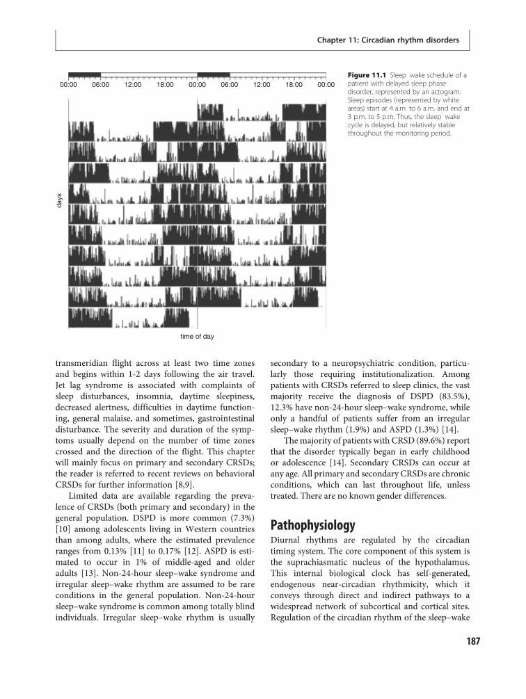
transmeridian flight across at least two time zonesand begins within 1-2 days following the air travel.Jet lag syndrome is associated with complaints ofsleep disturbances, insomnia, daytime sleepiness,decreased alertness, difficulties in daytime function-ing, general malaise, and sometimes, gastrointestinaldisturbance. The severity and duration of the symp-toms usually depend on the number of time zonescrossed and the direction of the flight. This chapterwill mainly focus on primary and secondary CRSDs;the reader is referred to recent reviews on behavioralCRSDs for further information [8,9].
Limited data are available regarding the preva-lence of CRSDs (both primary and secondary) in thegeneral population. DSPD is more common (7.3%)[10] among adolescents living in Western countriesthan among adults, where the estimated prevalenceranges from 0.13% [11] to 0.17% [12]. ASPD is esti-mated to occur in 1% of middle-aged and olderadults [13]. Non-24-hour sleep–wake syndrome andirregular sleep–wake rhythm are assumed to be rareconditions in the general population. Non-24-hoursleep–wake syndrome is common among totally blindindividuals. Irregular sleep–wake rhythm is usually
secondary to a neuropsychiatric condition, particu-larly those requiring institutionalization. Amongpatients with CRSDs referred to sleep clinics, the vastmajority receive the diagnosis of DSPD (83.5%),12.3% have non-24-hour sleep–wake syndrome, whileonly a handful of patients suffer from an irregularsleep–wake rhythm (1.9%) and ASPD (1.3%) [14].
The majority of patients with CRSD (89.6%) reportthat the disorder typically began in early childhoodor adolescence [14]. Secondary CRSDs can occur atany age. All primary and secondary CRSDs are chronicconditions, which can last throughout life, unlesstreated. There are no known gender differences.
PathophysiologyDiurnal rhythms are regulated by the circadiantiming system. The core component of this system isthe suprachiasmatic nucleus of the hypothalamus.This internal biological clock has self-generated,endogenous near-circadian rhythmicity, which itconveys through direct and indirect pathways to awidespread network of subcortical and cortical sites.Regulation of the circadian rhythm of the sleep–wake
00:00
days
time of day
06:00 12:00 18:00 00:00 06:00 12:00 18:00 00:00Figure 11.1 Sleep wake schedule of apatient with delayed sleep phasedisorder, represented by an actogram.Sleep episodes (represented by whiteareas) start at 4 a.m. to 6 a.m. and end at3 p.m. to 5 p.m. Thus, the sleep wakecycle is delayed, but relatively stablethroughout the monitoring period.
Chapter 11: Circadian rhythm disorders
187
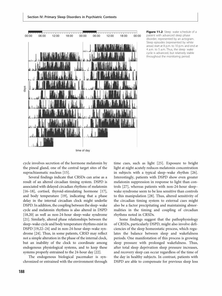
cycle involves secretion of the hormone melatonin bythe pineal gland, one of the central target sites of thesuprachiasmatic nucleus [15].
Several findings indicate that CRSDs can arise as aresult of an altered circadian timing system. DSPD isassociated with delayed circadian rhythms of melatonin[16–18], cortisol, thyroid-stimulating hormone [17],and body temperature [19], indicating that a phasedelay in the internal circadian clock might underlieDSPD. In addition, the coupling between the sleep–wakecycle and melatonin rhythms is also altered in DSPD[18,20] as well as non-24-hour sleep–wake syndrome[21]. Similarly, altered phase relationships between thesleep–wake cycle and body temperature rhythms exist inDSPD [19,22–24] and in non-24-hour sleep–wake syn-drome [24]. Thus, in some patients, CRSD may reflectnot a simple alteration in the phase of the internal clock,but an inability of the clock to coordinate amongendogenous physiological systems, and to keep thesesystems properly entrained to the 24-hour day [22].
The endogenous biological pacemaker is syn-chronized or entrained with the environment through
time cues, such as light [25]. Exposure to brightlight at night acutely reduces melatonin concentrationin subjects with a typical sleep–wake rhythm [26].Interestingly, patients with DSPD show even greatermelatonin suppression in response to light than con-trols [27], whereas patients with non-24-hour sleep–wake syndrome seem to be less sensitive than controlsto this manipulation [28]. Thus, altered sensitivity ofthe circadian timing system to external cues mightalso be a factor precipitating and maintaining abnor-malities in the timing and coupling of circadianrhythms noted in CRSDs.
Some findings suggest that the pathophysiologyof CRSDs, particularly DSPD, might also involve defi-ciencies of the sleep homeostatic process, which regu-lates the balance between sleep and wakefulnessperiods. One manifestation of this process is growingsleep pressure with prolonged wakefulness. Thus,after total sleep deprivation sleep pressure increases,and recovery sleep can occur regardless of the time ofthe day in healthy subjects. In contrast, patients withDSPD are able to compensate for previous sleep loss
00:00
days
time of day
06:00 12:00 18:00 00:00 06:00 12:00 18:00 00:00Figure 11.2 Sleep wake schedule of apatient with advanced sleep phasedisorder, represented by an actogram.Sleep episodes (represented by whiteareas) start at 8 p.m. to 10 p.m. and end at4 a.m. to 5 a.m. Thus, the sleep wakecycle is advanced, but relatively stablethroughout the monitoring period.
Section IV: Primary Sleep Disorders in Psychiatric Contexts
188

only during their circadian night [20,29]. Patientswith DSPD also show different temporal distributionof slow-wave sleep [22,24]. These alterations in sleeppropensity and sleep architecture, along with differ-ences in phase relationships of the sleep–wake cycleand physiological markers of the endogenous clock,can be interpreted as evidence of an imbalance betweencircadian and sleep homeostatic systems in the patho-physiology of DSPD. However, it is unclear if thesephysiological abnormalities described above are causedby the CRSDs, or an effect of the abnormal exposureto light which is characteristic of the syndromes.
Lastly, a genetic origin was demonstrated in someCRSDs. As many as 44% of patients with a CRSDhave other family members with similar sleep–wakepatterns as the patient [14]. In a pedigree of onefamily with DSPD, the trait was found to segregate
with either an autosomal dominant mode of heritancewith incomplete penetrance or a multifactorial modeof inheritance [30]. Structural polymorphisms onone of the haplotypes of the human period3 gene(hper3) were implicated as contributors to increasedsusceptibility to DSPD [31]. Several pedigrees offamilial ASPD were also reported, in which the ASPDtrait segregated as an autosomal dominant mode ofinheritance [32,33]. Although a mutation of humanperiod2 (hper2) gene was identified in a large familywith ASPD [34], other findings indicate genetic het-erogeneity in this disorder [35].
DiagnosisThe diagnosis of CRSDs can be established throughthree procedures: clinical interview, longitudinal
00:00
days
time of day
06:00 12:00 18:00 00:00 06:00 12:00 18:00 00:00Figure 11.3 Sleep wake schedule of apatient with non 24 hour sleep wakesyndrome, represented by an actogram.On the first 2 days of monitoring, sleepepisodes (represented by white areas)begin at around 1 a.m. During thefollowing 5 days, sleep onset is delayedeach day by an hour, thus by the end ofthe first week the patient falls asleep atapproximately 6 a.m. During the secondweek, sleep episodes are further andfurther delayed and so, by the end of themonitoring period, sleep episodes occurduring the day and early evening hours.Due to his longer than 24 hour sleepwake cycle, the patient sometimes sleepsat night and sometimes during the day.
Chapter 11: Circadian rhythm disorders
189
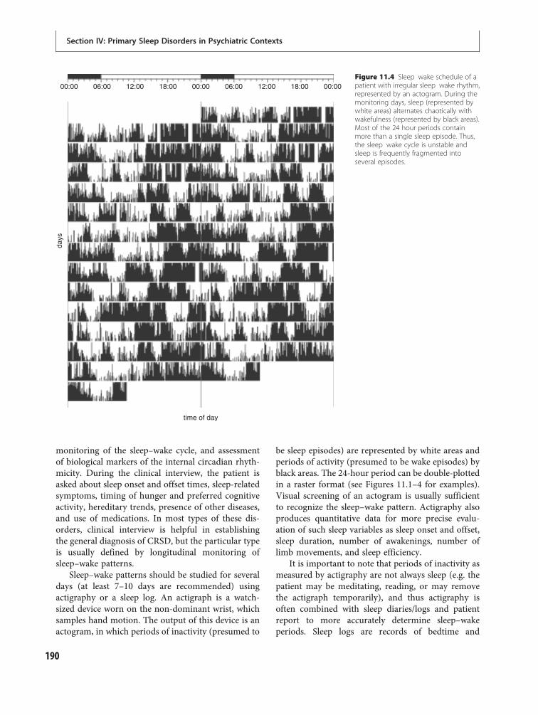
monitoring of the sleep–wake cycle, and assessmentof biological markers of the internal circadian rhyth-micity. During the clinical interview, the patient isasked about sleep onset and offset times, sleep-relatedsymptoms, timing of hunger and preferred cognitiveactivity, hereditary trends, presence of other diseases,and use of medications. In most types of these dis-orders, clinical interview is helpful in establishingthe general diagnosis of CRSD, but the particular typeis usually defined by longitudinal monitoring ofsleep–wake patterns.
Sleep–wake patterns should be studied for severaldays (at least 7–10 days are recommended) usingactigraphy or a sleep log. An actigraph is a watch-sized device worn on the non-dominant wrist, whichsamples hand motion. The output of this device is anactogram, in which periods of inactivity (presumed to
be sleep episodes) are represented by white areas andperiods of activity (presumed to be wake episodes) byblack areas. The 24-hour period can be double-plottedin a raster format (see Figures 11.1–4 for examples).Visual screening of an actogram is usually sufficientto recognize the sleep–wake pattern. Actigraphy alsoproduces quantitative data for more precise evalu-ation of such sleep variables as sleep onset and offset,sleep duration, number of awakenings, number oflimb movements, and sleep efficiency.
It is important to note that periods of inactivity asmeasured by actigraphy are not always sleep (e.g. thepatient may be meditating, reading, or may removethe actigraph temporarily), and thus actigraphy isoften combined with sleep diaries/logs and patientreport to more accurately determine sleep–wakeperiods. Sleep logs are records of bedtime and
00:00
days
time of day
06:00 12:00 18:00 00:00 06:00 12:00 18:00 00:00Figure 11.4 Sleep wake schedule of apatient with irregular sleep wake rhythm,represented by an actogram. During themonitoring days, sleep (represented bywhite areas) alternates chaotically withwakefulness (represented by black areas).Most of the 24 hour periods containmore than a single sleep episode. Thus,the sleep wake cycle is unstable andsleep is frequently fragmented intoseveral episodes.
Section IV: Primary Sleep Disorders in Psychiatric Contexts
190
awakening time made by the patient. If actigraphicdevices are unavailable, sleep logs alone can be areliable substitution for clinical purposes. In theevaluation of a potential CRSD, actigraphic/sleep logmonitoring should be carried out in unconstrainedconditions, such that the patient is free to choosebedtime and wake-up hours. This feature is highlyimportant in CRSD diagnosis, since a sleep–wakeschedule obtained under forced conditions can maskthe true pattern of the schedule, thus obscuring thediagnosis.
The third diagnostic procedure is assessment ofbiological markers of the internal biological clock, ascaptured by melatonin and body temperature circa-dian rhythms. This procedure is not mandatory,but can be helpful in complicated cases, especiallywhen non-24-hour sleep–wake syndrome or irregularsleep–wake rhythm is suspected. In clinical practice,salivary melatonin and oral temperature samplingscan bemade simultaneously every 2 hours for 36 hoursduring wakefulness. The acrophase (time of therhythm’s maximal values) of melatonin rhythm andthe nadir of temperature rhythm (time of the rhythm’sminimal values) are the most straightforward chrono-biological markers to establish the phase of therhythm. The period of each rhythm and phase rela-tionships between the rhythms are also useful in defin-ing the abnormality of the intrinsic biological clock.
Differential diagnosisPrimary CRSDs, especially DSPD and ASPD, shouldbe distinguished from “normal” sleep patterns. Someindividuals without sleep disorders might demon-strate a DSPD-like sleep–wake cycle. This might occurwhen sleep onset is delayed voluntarily, as a conse-quence of occupational, academic, and social activitiesin the late evening hours. These individuals differfrom patients with DSPD mainly in the flexibilityof their biological clock. Thus, an individual with anintact biological clock who is used to falling asleeplater than accepted is able to adjust his or her sleep–wake schedule to environmental changes (such astransition from weekend to weekday, vacations, newworking hours, etc.). Patients with DSPD seem to beunable to readily phase shift sleep episodes by motiv-ation or education alone. Similarly, early sleep onsetand awakening in the elderly should not be confusedwith ASPD. Whereas normal advancement in thephase of the sleep–wake cycle tends to occur in
the elderly, ASPD is a chronic condition that canappear as early as childhood, but typically begins inmiddle age.
CRSDs should also be differentiated from otherprimary sleep disorders, especially those presentingwith excessive daytime sleepiness (such as idiopathichypersomnia, sleep apnea, and narcolepsy). Patientswith CRSDs are not sleepy if allowed to follow theirintrinsic preferred sleep–wake cycle. Insomnia dis-orders can also be confused with DSPD, due to com-plaints of difficulties falling asleep that are present inboth disorders. Patients suffering from sleep-onsetinsomnia may feel sleepy at the desired bedtime,but have difficulty initiating sleep. By comparison,patients with DSPD who attempt to move their bed-time earlier (i.e. advance their sleep–wake cycle) willnot be sleepy and thus will be unable to fall asleep.Further, sleep-onset difficulties endure for as long asthe patient keeps his bedtime out of phase with hisendogenous sleep–wake rhythm. When allowed tomaintain the preferred schedule, sleep initiation isnormal, unlike insomnia patients who often havedifficulties initiating or maintaining sleep regardlessof the timing of sleep.
Primary CRSDs should be distinguished frombehavioral and secondary CRSDs. A thorough patientinterview and physical examination (when an under-lying medical condition is suspected) may help toestablish the etiology of the CRSD in such cases.
Lastly, the various types of primary CRSDs shouldbe differentiated from each other. Some individualswith non-24-hour sleep–wake syndrome might showDSPD-like symptoms, such as difficulty falling asleep,daytime sleepiness, and delayed sleep onset. To estab-lish the diagnosis, actigraphic/sleep log monitoring isoften required. In unconstrained conditions, rest–activity patterns of a patient with DSPD show relativestability in sleep onset and offset, whereas sleep epi-sodes of a patient with a non-24-hour sleep–wakesyndrome are progressively delayed to later hoursfrom day to day. If the actogram/sleep log is inconclu-sive, as might occur in atypical cases, assessment ofmelatonin/temperature circadian rhythm is helpful.In DSPD, both rhythms have a clear 24-hour period-icity, whereas in non-24-hour sleep–wake syndromethe period is usually longer than 24 hours. Further, ifboth melatonin and temperature rhythms are meas-ured simultaneously, their phases will be desynchron-ized with each other only in the case of non-24-hoursleep–wake syndrome.
Chapter 11: Circadian rhythm disorders
191
TreatmentBright light therapy and melatonin treatment, or acombination of these two, have proved to be the mosteffective treatment modalities for patients withCRSDs. These techniques aim to reset the sleep–wakecycle of patients to match the external 24-hourschedule.
Bright light is the most powerful time cue to theinternal circadian timing system. Light exposure atspecific times of the 24-hour period can result in aphase shift of the endogenous circadian rhythms ofvarious functions, such as melatonin secretion, bodytemperature, and sleep propensity [36–38]. In general,exposure to bright light in the morning induces aphase advance, whereas evening bright light exposureinduces phase delay. Bright light exposure as a therapycan be provided by means of artificial light devices.The patient is required to sit in front of a light-box at aprespecified time. In patients with DSPD, exposure to2000–2500 lux from 6 a.m. to 9 a.m. helps to advancethe sleep–wake rhythm, and avoidance of bright lightin the evening may be necessary to prevent the circa-dian rhythm from being further delayed [39]. Inpatients with ASPD, 2500 lux for 4 hours from8 p.m. to midnight, or 4000 lux for 2 or 3 hours from8 or 9 p.m. to 11 p.m. [40] is recommended to inducephase delay. Some evidence supports the effectivenessof light therapy in the treatment of non-24-hoursleep–wake syndrome [41], jet lag [42], and shift work[43]. Further recommendations on intensities andtime limits for bright light therapy in the treatmentof CRSDs have been provided by the American Acad-emy of Sleep Medicine [44].
Endogenous melatonin secreted by the pinealgland is another potent regulator of the sleep–wakecycle. It is thought that the night-time increase inmelatonin concentration reduces body temperature,which promotes the onset of sleep [45]. Pharmaco-logical preparations of melatonin can mimic theeffects of endogenous melatonin. These effects aretime dependent: phase advance is produced by mela-tonin administered in the evening, whereas melatoninadministration in the morning induces phase delays[46]. These effects are the opposite of those producedby light. For example, melatonin may be administeredin the evening (roughly 2 hours before the desiredsleep onset, rather than at bedtime) in DSPD to helpadvance the circadian rhythm. Of note, the use ofmorning melatonin to delay the circadian rhythm,
which would be theoretically useful in ASPD, can beproblematic as melatonin can also be sedating, thusdecreasing daytime alertness in some instances.Administration of melatonin might be a preferabletherapeutic strategy for many patients who findbright light therapy too demanding, leading todecreased compliance. The beneficial effects of mela-tonin 0.5–5 mg/day were demonstrated in severaltypes of CRSD [47–51], and in some instances, higherdoses may have no added effect (or even be lesseffective) in shifting the circadian rhythm comparedto lower doses. Importantly, treatment with mela-tonin not only synchronizes the sleep–wake cycle ofpatients with CRSD, but may also significantly improveseveral dimensions of their daytime functioning [52].Although the long-term effects of melatonin adminis-tration remain to be fully researched, the treatmentappears to be promising. Recent well-designed studiesindicate that even relatively large doses of melatonin(10 mg/day for a month) have no toxic effects [53].Further, a prolonged-release 2 mg melatonin prepar-ation (Circadin™) was approved by the EuropeanMedicines Agency for the short-term treatment of pri-mary insomnia in patients who are aged 55 or above.
Patients not responding to bright light or mela-tonin therapies may adopt a rehabilitative approach,according to which the patients would accept thattheir condition is permanent and consider changesin lifestyle that will be congruent with their sleep–wake cycle (e.g. working evening shifts in the case ofDSPD) [54].
Circadian rhythm sleep disordersin psychiatryGenerally, otherwise healthy patients with CRSDsseem to suffer from more emotional and cognitivedisturbances than healthy controls [55]. Psychiatricdisorders are also very common in these patients.For example, in a sample of 126 patients with CRSDsattending sleep clinics, 29% had a psychiatric diagno-sis [56]. In another study, 62% of sighted patients withnon-24-hour sleep–wake syndrome had a psychiatricco-morbidity [57]. While CRSDs can co-occur witha wide variety of mental disorders, the present chapterwill review the disorders that have been systematicallyexamined with regard to CRSDs, specifically mooddisorders, attention-deficit/hyperactivity disorder(ADHD), personality disorders, and obsessive-compulsive disorder (OCD).
Section IV: Primary Sleep Disorders in Psychiatric Contexts
192

The etiology of the relationship between CRSDs andpsychiatric disorders is a matter of debate. It has beensuggested that in patients who developed a CRSD afterthe onset of a psychiatric disorder, the sleep–wake alter-ation is an epiphenomenon that arises due to changesin behavioral patterns [57,58]. In other cases, theincompatibility of the sleep–wake cycle and the socialenvironment might be the primary factor predisposingpatients with CRSDs to psychiatric illness [59]. Anotherintriguing possibility is that the sleep–wake abnormalityand the psychiatric disorder share a common pathology[60], possibly in the circadian timing system.
Mood disordersDiurnal variation in mood, a common feature ofdepressive illness, has led many researchers to look forirregularities in circadian rhythms in these patients.Altered amplitude and phase of melatonin, cortisol,prolactin, and body temperature were found in patientswith major depressive disorder (MDD) [61,62], bipolardepression (BD) [63], and seasonal affective disorder(SAD) [64,65]. Recent studies have also demonstratedpolymorphisms of several clock genes (such as Period3,CLOCK, BMAL1, TIMELESS) in MDD [66], BD [67–70], and SAD [71] that may be associated with thedevelopment of the mood disorder and/or specificsleep–wake patterns during the course of the illness.
Sleep disturbance is one of the hallmarks of depres-sion. Complaints of insomnia (difficulty initiatingsleep, nocturnal arousals, and earlymorning wakening)are very common inMDD. Subjective reports of hyper-somnia tend to occur in BD during depressive episodes[72] and in SAD [73]. Sleep in MDD, as measured bypolysomnography, is characterized by longer sleeponset, more fragmented sleep, shorter total sleep time,reduced time spent in SWS, and several abnormalitiesin REM sleep (reviewed in [74]). Interestingly, noctur-nal sleep patterns in BD seem to be similar to those inMD, despite differences in the clinical manifestationsof these disorders [75]. SAD is also characterizedby sleep abnormalities, but these are different fromthose seen in MDD and BD. For example, somestudies reported longer (rather than shorter) total sleeptime [73], increased (rather than reduced) SWS dur-ation [76], and normal (rather than shortened) REMlatency [73] in SAD patients compared to healthycontrols.
Based on the aforementioned findings, severalresearchers hypothesized that depression is a
chronobiological disorder that arises as a result of amismatch between circadian rhythms [77–79].According to these approaches, insomnia symptomsare a manifestation of this mismatch. The pathologyof CRSDs is also linked to alterations in the circadiantiming system, and thus it is not surprising that thesedisorders are also highly co-morbid with mooddisorders. In a study of clinical profiles of DSPD in22 adolescents, 36% showed features of depressionand 27% received prior treatment for depression[80]. In 33 adult patients with DSPD, as many as76% were treated with antidepressants in the past orat the time of research, although such a high prevalencewas possibly a result of self-referral bias [81]. When alarge sample of DSPD patients was compared tohealthy controls, the results showed the same trends:DSPD was associated with greater current depressionratings, lifetime history of unipolar depression, depres-sion treatment, antidepressant use, and family historyof depression [60].
Depression also seems to be a frequent co-morbidity in non-24-hour sleep–wake syndrome.Among 57 sighted individuals with non-24-hoursleep–wake syndrome, major depression precededthe CRSD onset in 5.3% of the sample. Interestingly,34% developed major depression following theonset of non-24-hour sleep–wake syndrome. One-third of these reported that depression severity variedaccording to changes in sleep phase: it was aggravatedwhen sleep episodes occurred during the daytimeand slightly ameliorated when sleep episodes occurredduring the night-time [57].
Several mechanisms have been proposed to explainthe association between CRSDs and depression. Sincepatients with DSPD often have a family history ofdepression, the disorders might share genetic suscepti-bility factors [60]. Alternatively, a sleep–wake cyclethat is discrepant with the environment might lead tosocial isolation and reduced light exposure. Since bothfactors are considered synchronizers of the internalbiological clock, inappropriate exposure (both to lightand social cues) might also predispose to depression.Conversely, depression itself can cause social with-drawal and decrease in outdoor activities, exacerbatingor leading to a CRSD [57,60].
While these suggestions are plausible, they cannotfully account for CRSDs without depression. Futureresearch should further explore the conditions thatlead to co-occurrence of the disorders. Depressivedisorders are commonly recognized as heterogeneous.
Chapter 11: Circadian rhythm disorders
193
It is therefore possible that different subtypes can alsobe characterized by sleep disturbances with distinctetiologies, some of which may be related to circadianmechanisms. Such differentiation between subtypesmight be clinically important, since previous findingsindicate that the type of sleep disturbance can bepredictive of depression recurrence. For example,residual insomnia in patients with depression treatedwith antidepressants and psychotherapy confersgreater risk for subsequent depression [82]. On theother hand, melatonin administration in patientswith DSPD may successfully treat the sleep disorderand significantly reduce depressive symptoms [52,83].Thus, careful examination of the sleep disturbanceassociated with depression might improve our under-standing of the underlying pathology as well as thetreatment outcome.
Attention-deficit/hyperactivity disorderADHD is frequently associated with sleep disturb-ances. The characteristics and the implications ofsleep disorders such as sleep apnea and restless legssyndrome in ADHD received broad attention in theliterature (reviewed in [84,85]). Recently, a few stud-ies also provided a description of the characteristics ofCRSDs in ADHD patients. Van der Heijden andcolleagues examined a group of children with ADHDand sleep disturbance, which was defined by theauthors as idiopathic sleep-onset insomnia [86]. Com-pared to ADHD without insomnia, the ADHD withinsomnia group had later sleep onset and wake-uptime, and delayed melatonin rhythm. Importantly, nodifferences between the groups were found in meas-ures of sleep maintenance. This profile of sleep dis-turbance has led the authors to suggest that insomniain children with ADHD is, in fact, pediatric DSPD.
The prevalence of ADHD in pediatric patientswith DSPD seems to be high. In one study [87], asmany as 28 out of 62 (45%) children with suspectedDSPD were previously diagnosed with ADHD. Wefound a similar prevalence in our sample of childrenand adolescents diagnosed with DSPD: among 33patients, 15 (45%) had been previously diagnosedwith ADHD [88]. When adults with ADHD werestudied, 40.7% described themselves as evening types,as assessed by the morningness–eveningness ques-tionnaire [89]. Greater evening preference was associ-ated in this study with more subjective and objectivedeficits in functioning.
Treatment with melatonin generally improvessleep disturbances in children with ADHD and DSPD.Compared to placebo, a 1-month treatment with3-6 mg melatonin administered at 7 p.m. advancedsleep onset and melatonin rhythm, reduced sleeplatency, and increased total sleep time [87,90]. Long-term treatment with 3–5 mg melatonin taken 2 hoursbefore bedtime advanced sleep onset and total sleeptime in children and adolescents with DSPD and co-morbid ADHD [88]. It also reduced the frequency ofsleep-related complaints typical of DSPD: difficulty fall-ing asleep and waking up, as well as daytime sleepiness.
Some of the findings indicate that chronobiologic-al treatments might prove helpful not only for sleepdisturbances, but also for ADHD-related symptom-atology. Treatment with 5 mg melatonin at 7 p.m.improved health and functional status of childrenwith ADHD and suspected DSPD [87]. We also foundthat long-term treatment with 3–5 mg melatonintaken 2 hours before sleep reduced the frequency ofself-reported behavioral/social difficulties and ofabsences/late arrivals at school in children and ado-lescents with DSPD and ADHD [88]. However, inanother study, no effect of melatonin was found onproblem behavior, cognitive performance, or qualityof life [90]. Adults with ADHD treated with light(10 000 lux) administered for half an hour before8 a.m. for 3 weeks showed advance in circadian pref-erence, which also was the best predictor of improve-ment in the severity of ADHD symptoms [91].
The frequent co-occurrence of ADHD and DSPDin clinical samples could be related to selection bias,i.e. patients with ADHD exhibit more behavioralproblems and are more likely to receive professionalguidance. Other possibilities are that DSPD is second-ary to primary abnormalities in the central nervoussystem associated with ADHD or to treatment withstimulants. However, there is no evidence at presentto support such an etiology. Alternatively, DSPDmight be the underlying condition that contributesto hyperactive and inattentive behaviors. DSPD inschool-aged students involves chronic and substantialsleep loss [88]. Insufficient sleep and ADHD are bothassociated with impairment of executive functionsregulated by the prefrontal cortex [92]. Thus, chronicsleep loss during the school years might lead to and/orexacerbate ADHD symptomatology.
It was recently reported that one of the alleles ofthe CLOCK polymorphism is associated with adultADHD [93]. Thus, it is possible that co-occurrence
Section IV: Primary Sleep Disorders in Psychiatric Contexts
194
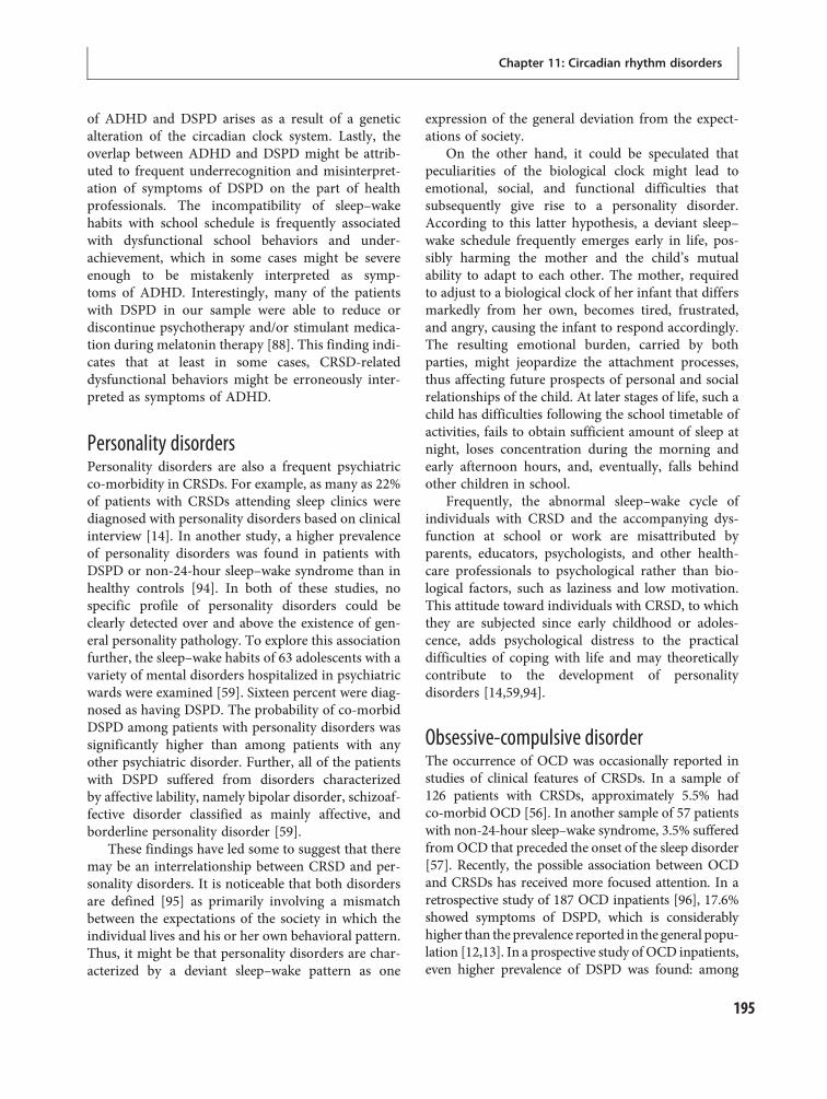
of ADHD and DSPD arises as a result of a geneticalteration of the circadian clock system. Lastly, theoverlap between ADHD and DSPD might be attrib-uted to frequent underrecognition and misinterpret-ation of symptoms of DSPD on the part of healthprofessionals. The incompatibility of sleep–wakehabits with school schedule is frequently associatedwith dysfunctional school behaviors and under-achievement, which in some cases might be severeenough to be mistakenly interpreted as symp-toms of ADHD. Interestingly, many of the patientswith DSPD in our sample were able to reduce ordiscontinue psychotherapy and/or stimulant medica-tion during melatonin therapy [88]. This finding indi-cates that at least in some cases, CRSD-relateddysfunctional behaviors might be erroneously inter-preted as symptoms of ADHD.
Personality disordersPersonality disorders are also a frequent psychiatricco-morbidity in CRSDs. For example, as many as 22%of patients with CRSDs attending sleep clinics werediagnosed with personality disorders based on clinicalinterview [14]. In another study, a higher prevalenceof personality disorders was found in patients withDSPD or non-24-hour sleep–wake syndrome than inhealthy controls [94]. In both of these studies, nospecific profile of personality disorders could beclearly detected over and above the existence of gen-eral personality pathology. To explore this associationfurther, the sleep–wake habits of 63 adolescents with avariety of mental disorders hospitalized in psychiatricwards were examined [59]. Sixteen percent were diag-nosed as having DSPD. The probability of co-morbidDSPD among patients with personality disorders wassignificantly higher than among patients with anyother psychiatric disorder. Further, all of the patientswith DSPD suffered from disorders characterizedby affective lability, namely bipolar disorder, schizoaf-fective disorder classified as mainly affective, andborderline personality disorder [59].
These findings have led some to suggest that theremay be an interrelationship between CRSD and per-sonality disorders. It is noticeable that both disordersare defined [95] as primarily involving a mismatchbetween the expectations of the society in which theindividual lives and his or her own behavioral pattern.Thus, it might be that personality disorders are char-acterized by a deviant sleep–wake pattern as one
expression of the general deviation from the expect-ations of society.
On the other hand, it could be speculated thatpeculiarities of the biological clock might lead toemotional, social, and functional difficulties thatsubsequently give rise to a personality disorder.According to this latter hypothesis, a deviant sleep–wake schedule frequently emerges early in life, pos-sibly harming the mother and the child’s mutualability to adapt to each other. The mother, requiredto adjust to a biological clock of her infant that differsmarkedly from her own, becomes tired, frustrated,and angry, causing the infant to respond accordingly.The resulting emotional burden, carried by bothparties, might jeopardize the attachment processes,thus affecting future prospects of personal and socialrelationships of the child. At later stages of life, such achild has difficulties following the school timetable ofactivities, fails to obtain sufficient amount of sleep atnight, loses concentration during the morning andearly afternoon hours, and, eventually, falls behindother children in school.
Frequently, the abnormal sleep–wake cycle ofindividuals with CRSD and the accompanying dys-function at school or work are misattributed byparents, educators, psychologists, and other health-care professionals to psychological rather than bio-logical factors, such as laziness and low motivation.This attitude toward individuals with CRSD, to whichthey are subjected since early childhood or adoles-cence, adds psychological distress to the practicaldifficulties of coping with life and may theoreticallycontribute to the development of personalitydisorders [14,59,94].
Obsessive-compulsive disorderThe occurrence of OCD was occasionally reported instudies of clinical features of CRSDs. In a sample of126 patients with CRSDs, approximately 5.5% hadco-morbid OCD [56]. In another sample of 57 patientswith non-24-hour sleep–wake syndrome, 3.5% sufferedfromOCD that preceded the onset of the sleep disorder[57]. Recently, the possible association between OCDand CRSDs has received more focused attention. In aretrospective study of 187 OCD inpatients [96], 17.6%showed symptoms of DSPD, which is considerablyhigher than the prevalence reported in the general popu-lation [12,13]. In a prospective study ofOCD inpatients,even higher prevalence of DSPD was found: among
Chapter 11: Circadian rhythm disorders
195

36 patients, 42% fulfilled criteria for DSPD [58]. In bothstudies, patients with DSPD had more severe OCD andexperienced higher levels of functional disability thanpatients with OCD and a normal sleep phase.
The authors found that the OCD groups with andwithout DSPD did not differ in the severity of depres-sive symptoms, the extent of rituals around bedtime,or in prescribed medications. Having alternativeexplanations ruled out, they suggested that a directlink between DSPD and OCD might exist. Further,the authors raised the possibility that DSPD was sec-ondary to OCD in their sample, since all patients inthe DSPD group reported that the delay in sleep phaseoccurred after the onset of OCD. Accordingly, thealteration of the sleep–wake cycle in OCD might haveoccurred as a result of common daily routines (suchas lengthy in-house rituals in the morning), socialisolation, and lack of regularity in meal times, all ofwhich can diminish the impact of external time cueson the internal biological clock. Other findings sug-gest that some of the physiological circadian rhythmsare altered in OCD [97,98], suggesting that sleep–wake schedule disorders in OCD might result froman interaction of an abnormal intrinsic biologicalclock and lack of appropriate external time signals.Clearly, more research is required to further explorethe nature of the association between OCD andCRSDs.
Psychoactive medicationSeveral cases of disrupted sleep–wake schedule as iat-rogenic effects of psychoactive drugs are documentedin the literature. Treatment with the typical neuroleptichaloperidol in a patientwith chronic schizophreniawasassociated with an irregular sleep–wake cycle. Switch-ing treatment to the atypical neuroleptic clozapineestablished a more organized and stable sleep–wakepattern and improved the clinical state of the patient[99]. In another study [100], patients receiving typicalneuroleptics (flupentixol or haloperidol) showed avariety of abnormalities in the daily rest–activityrhythm (e.g. delayed circadian phase syndrome, free-running sleep–wake syndrome, and irregular sleep–wake rhythm). In contrast, rest–activity cycles of thosepatients treated with the atypical neuroleptic clozapinewere highly organized and synchronized with theenvironmental schedule.
Similar effects were observed in a patient withearly-onset Alzheimer’s disease: when treated with
haloperidol, her rest–activity patterns became com-pletely arrhythmic, which was accompanied bymarked worsening of cognitive state. When haloperi-dol was replaced by clozapine, rapid normalization ofher sleep–wake cycle occurred and cognitive function-ing improved [101]. Similar effects of haloperidolwere described in a patient with Gilles de la Tourettesyndrome [102]. Prior to haloperidol treatment, thepatient reported having a normal sleep–wake sched-ule. Two years after commencing the treatment, hissleep–wake cycle became irregular. When therapywith haloperidol was changed to the atypical neuro-leptic risperidone, the timing and duration ofsleep episodes became more organized, although hissleep–wake schedule still remained somewhat disturbed.The patient’s sleep–wake circadian rhythm was fullyrecovered after addition of melatonin as a secondarytherapy.Thiswas accompaniedby improvement inqual-ity of life, social interactions, and employment status.
These findings suggest that typical neuroleptics likehaloperidol and flupentixol might alter the circadiansleep–wake rhythm, whereas atypical neuroleptics likeclozapine and risperidone tend to enhance the congru-ity of the individual’s sleep–wake cycle with the envir-onment. Since this effect was evident in severalmedicaldisorders, e.g. schizophrenia, Alzheimer’s disease, andTourette syndrome, it was argued that CRSDs mightbe a side-effect of typical neuroleptics, rather thanillness-related phenomena [99–102]. The exact mech-anisms through which typical and atypical neurolep-tics exert their differential effects on the sleep–wakecycle remain to be elucidated.
Other psychoactive drugs, such as selective sero-tonin reuptake inhibitors (SSRIs), can also trigger theemergence of CRSD as a side-effect. Treatment withfluvoxamine in patients with OCD was associatedwith DSPD [103]. It was postulated that DSPD inthese patients was secondary to fluvoxamine since: (1)all patients received no other medications except flu-voxamine prior to the onset of DSPD, (2) in all patients,DSPD first occurred following fluvoxamine initiation,(3) when fluvoxamine was withdrawn or the dose con-siderably reduced, the sleep–wake cycle returned tonormal, and (4) with re-exposure to fluvoxamine,DSPD recurred. Interestingly, emergence of DSPDmay be specific to fluvoxamine; treatment with twoother serotonin reuptake inhibitors (clomipramineand fluoxetine) was not associated with any adverseeffects on the sleep–wake cycle of these patients [103].The authors hypothesized that the alteration of the
Section IV: Primary Sleep Disorders in Psychiatric Contexts
196
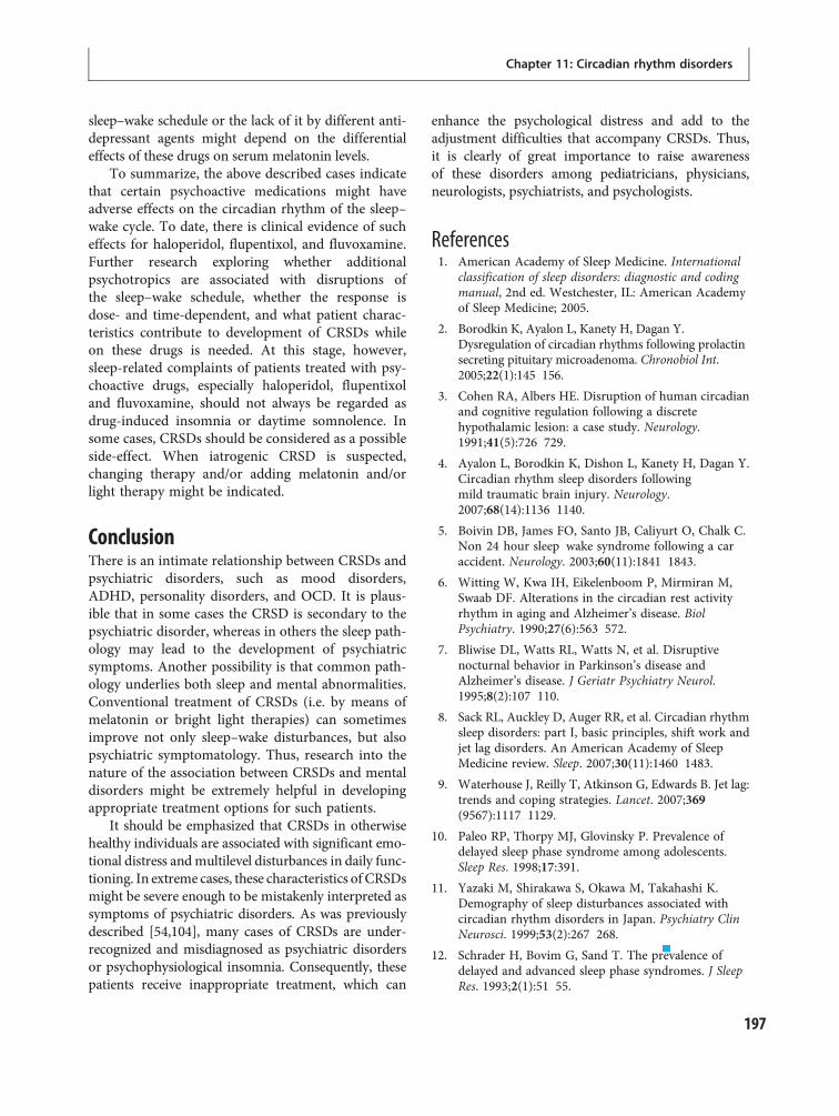
sleep–wake schedule or the lack of it by different anti-depressant agents might depend on the differentialeffects of these drugs on serum melatonin levels.
To summarize, the above described cases indicatethat certain psychoactive medications might haveadverse effects on the circadian rhythm of the sleep–wake cycle. To date, there is clinical evidence of sucheffects for haloperidol, flupentixol, and fluvoxamine.Further research exploring whether additionalpsychotropics are associated with disruptions ofthe sleep–wake schedule, whether the response isdose- and time-dependent, and what patient charac-teristics contribute to development of CRSDs whileon these drugs is needed. At this stage, however,sleep-related complaints of patients treated with psy-choactive drugs, especially haloperidol, flupentixoland fluvoxamine, should not always be regarded asdrug-induced insomnia or daytime somnolence. Insome cases, CRSDs should be considered as a possibleside-effect. When iatrogenic CRSD is suspected,changing therapy and/or adding melatonin and/orlight therapy might be indicated.
ConclusionThere is an intimate relationship between CRSDs andpsychiatric disorders, such as mood disorders,ADHD, personality disorders, and OCD. It is plaus-ible that in some cases the CRSD is secondary to thepsychiatric disorder, whereas in others the sleep path-ology may lead to the development of psychiatricsymptoms. Another possibility is that common path-ology underlies both sleep and mental abnormalities.Conventional treatment of CRSDs (i.e. by means ofmelatonin or bright light therapies) can sometimesimprove not only sleep–wake disturbances, but alsopsychiatric symptomatology. Thus, research into thenature of the association between CRSDs and mentaldisorders might be extremely helpful in developingappropriate treatment options for such patients.
It should be emphasized that CRSDs in otherwisehealthy individuals are associated with significant emo-tional distress andmultilevel disturbances in daily func-tioning. In extreme cases, these characteristics ofCRSDsmight be severe enough to be mistakenly interpreted assymptoms of psychiatric disorders. As was previouslydescribed [54,104], many cases of CRSDs are under-recognized and misdiagnosed as psychiatric disordersor psychophysiological insomnia. Consequently, thesepatients receive inappropriate treatment, which can
enhance the psychological distress and add to theadjustment difficulties that accompany CRSDs. Thus,it is clearly of great importance to raise awarenessof these disorders among pediatricians, physicians,neurologists, psychiatrists, and psychologists.
References1. American Academy of Sleep Medicine. International
classification of sleep disorders: diagnostic and codingmanual, 2nd ed. Westchester, IL: American Academyof Sleep Medicine; 2005.
2. Borodkin K, Ayalon L, Kanety H, Dagan Y.Dysregulation of circadian rhythms following prolactinsecreting pituitary microadenoma. Chronobiol Int.2005;22(1):145 156.
3. Cohen RA, Albers HE. Disruption of human circadianand cognitive regulation following a discretehypothalamic lesion: a case study. Neurology.1991;41(5):726 729.
4. Ayalon L, Borodkin K, Dishon L, Kanety H, Dagan Y.Circadian rhythm sleep disorders followingmild traumatic brain injury. Neurology.2007;68(14):1136 1140.
5. Boivin DB, James FO, Santo JB, Caliyurt O, Chalk C.Non 24 hour sleep wake syndrome following a caraccident. Neurology. 2003;60(11):1841 1843.
6. Witting W, Kwa IH, Eikelenboom P, Mirmiran M,Swaab DF. Alterations in the circadian rest activityrhythm in aging and Alzheimer’s disease. BiolPsychiatry. 1990;27(6):563 572.
7. Bliwise DL, Watts RL, Watts N, et al. Disruptivenocturnal behavior in Parkinson’s disease andAlzheimer’s disease. J Geriatr Psychiatry Neurol.1995;8(2):107 110.
8. Sack RL, Auckley D, Auger RR, et al. Circadian rhythmsleep disorders: part I, basic principles, shift work andjet lag disorders. An American Academy of SleepMedicine review. Sleep. 2007;30(11):1460 1483.
9. Waterhouse J, Reilly T, Atkinson G, Edwards B. Jet lag:trends and coping strategies. Lancet. 2007;369(9567):1117 1129.
10. Paleo RP, Thorpy MJ, Glovinsky P. Prevalence ofdelayed sleep phase syndrome among adolescents.Sleep Res. 1998;17:391.
11. Yazaki M, Shirakawa S, Okawa M, Takahashi K.Demography of sleep disturbances associated withcircadian rhythm disorders in Japan. Psychiatry ClinNeurosci. 1999;53(2):267 268.
12. Schrader H, Bovim G, Sand T. The prevalence ofdelayed and advanced sleep phase syndromes. J SleepRes. 1993;2(1):51 55.
Chapter 11: Circadian rhythm disorders
197
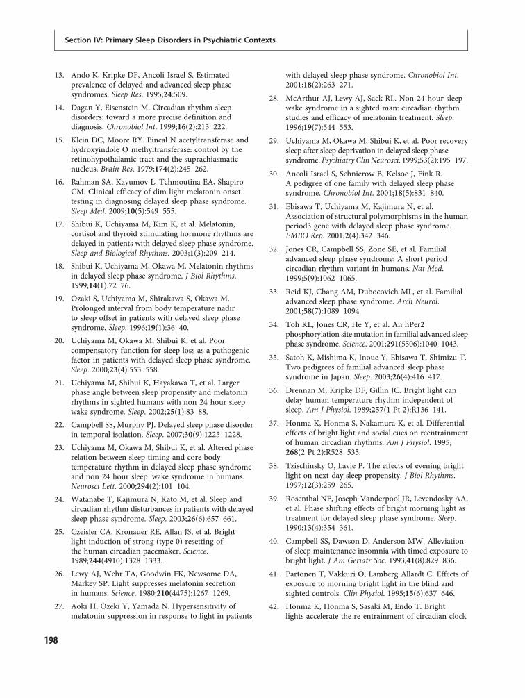
13. Ando K, Kripke DF, Ancoli Israel S. Estimatedprevalence of delayed and advanced sleep phasesyndromes. Sleep Res. 1995;24:509.
14. Dagan Y, Eisenstein M. Circadian rhythm sleepdisorders: toward a more precise definition anddiagnosis. Chronobiol Int. 1999;16(2):213 222.
15. Klein DC, Moore RY. Pineal N acetyltransferase andhydroxyindole O methyltransferase: control by theretinohypothalamic tract and the suprachiasmaticnucleus. Brain Res. 1979;174(2):245 262.
16. Rahman SA, Kayumov L, Tchmoutina EA, ShapiroCM. Clinical efficacy of dim light melatonin onsettesting in diagnosing delayed sleep phase syndrome.Sleep Med. 2009;10(5):549 555.
17. Shibui K, Uchiyama M, Kim K, et al. Melatonin,cortisol and thyroid stimulating hormone rhythms aredelayed in patients with delayed sleep phase syndrome.Sleep and Biological Rhythms. 2003;1(3):209 214.
18. Shibui K, Uchiyama M, Okawa M. Melatonin rhythmsin delayed sleep phase syndrome. J Biol Rhythms.1999;14(1):72 76.
19. Ozaki S, Uchiyama M, Shirakawa S, Okawa M.Prolonged interval from body temperature nadirto sleep offset in patients with delayed sleep phasesyndrome. Sleep. 1996;19(1):36 40.
20. Uchiyama M, Okawa M, Shibui K, et al. Poorcompensatory function for sleep loss as a pathogenicfactor in patients with delayed sleep phase syndrome.Sleep. 2000;23(4):553 558.
21. Uchiyama M, Shibui K, Hayakawa T, et al. Largerphase angle between sleep propensity and melatoninrhythms in sighted humans with non 24 hour sleepwake syndrome. Sleep. 2002;25(1):83 88.
22. Campbell SS, Murphy PJ. Delayed sleep phase disorderin temporal isolation. Sleep. 2007;30(9):1225 1228.
23. Uchiyama M, Okawa M, Shibui K, et al. Altered phaserelation between sleep timing and core bodytemperature rhythm in delayed sleep phase syndromeand non 24 hour sleep wake syndrome in humans.Neurosci Lett. 2000;294(2):101 104.
24. Watanabe T, Kajimura N, Kato M, et al. Sleep andcircadian rhythm disturbances in patients with delayedsleep phase syndrome. Sleep. 2003;26(6):657 661.
25. Czeisler CA, Kronauer RE, Allan JS, et al. Brightlight induction of strong (type 0) resetting ofthe human circadian pacemaker. Science.1989;244(4910):1328 1333.
26. Lewy AJ, Wehr TA, Goodwin FK, Newsome DA,Markey SP. Light suppresses melatonin secretionin humans. Science. 1980;210(4475):1267 1269.
27. Aoki H, Ozeki Y, Yamada N. Hypersensitivity ofmelatonin suppression in response to light in patients
with delayed sleep phase syndrome. Chronobiol Int.2001;18(2):263 271.
28. McArthur AJ, Lewy AJ, Sack RL. Non 24 hour sleepwake syndrome in a sighted man: circadian rhythmstudies and efficacy of melatonin treatment. Sleep.1996;19(7):544 553.
29. Uchiyama M, Okawa M, Shibui K, et al. Poor recoverysleep after sleep deprivation in delayed sleep phasesyndrome.Psychiatry ClinNeurosci. 1999;53(2):195 197.
30. Ancoli Israel S, Schnierow B, Kelsoe J, Fink R.A pedigree of one family with delayed sleep phasesyndrome. Chronobiol Int. 2001;18(5):831 840.
31. Ebisawa T, Uchiyama M, Kajimura N, et al.Association of structural polymorphisms in the humanperiod3 gene with delayed sleep phase syndrome.EMBO Rep. 2001;2(4):342 346.
32. Jones CR, Campbell SS, Zone SE, et al. Familialadvanced sleep phase syndrome: A short periodcircadian rhythm variant in humans. Nat Med.1999;5(9):1062 1065.
33. Reid KJ, Chang AM, Dubocovich ML, et al. Familialadvanced sleep phase syndrome. Arch Neurol.2001;58(7):1089 1094.
34. Toh KL, Jones CR, He Y, et al. An hPer2phosphorylation site mutation in familial advanced sleepphase syndrome. Science. 2001;291(5506):1040 1043.
35. Satoh K, Mishima K, Inoue Y, Ebisawa T, Shimizu T.Two pedigrees of familial advanced sleep phasesyndrome in Japan. Sleep. 2003;26(4):416 417.
36. Drennan M, Kripke DF, Gillin JC. Bright light candelay human temperature rhythm independent ofsleep. Am J Physiol. 1989;257(1 Pt 2):R136 141.
37. Honma K, Honma S, Nakamura K, et al. Differentialeffects of bright light and social cues on reentrainmentof human circadian rhythms. Am J Physiol. 1995;268(2 Pt 2):R528 535.
38. Tzischinsky O, Lavie P. The effects of evening brightlight on next day sleep propensity. J Biol Rhythms.1997;12(3):259 265.
39. Rosenthal NE, Joseph Vanderpool JR, Levendosky AA,et al. Phase shifting effects of bright morning light astreatment for delayed sleep phase syndrome. Sleep.1990;13(4):354 361.
40. Campbell SS, Dawson D, Anderson MW. Alleviationof sleep maintenance insomnia with timed exposure tobright light. J Am Geriatr Soc. 1993;41(8):829 836.
41. Partonen T, Vakkuri O, Lamberg Allardt C. Effects ofexposure to morning bright light in the blind andsighted controls. Clin Physiol. 1995;15(6):637 646.
42. Honma K, Honma S, Sasaki M, Endo T. Brightlights accelerate the re entrainment of circadian clock
Section IV: Primary Sleep Disorders in Psychiatric Contexts
198

to 8 hour phase advance shift of sleep wake schedule:1 Circadian rhythms in rectal temperature andplasma melatonin level. Jpn J Psychiatry Neurol.1991;45(1):153 154.
43. Czeisler CA, Johnson MP, Duffy JF, et al. Exposureto bright light and darkness to treat physiologicmaladaptation to night work. N Engl J Med.1990;322(18):1253 1259.
44. Chesson AL Jr, Anderson WM, Littner M, et al.Practice parameters for the nonpharmacologictreatment of chronic insomnia. An AmericanAcademy of Sleep Medicine report. Standards ofPractice Committee of the American Academy of SleepMedicine. Sleep. 1999;22(8):1128 1133.
45. Cagnacci A, Elliott JA, Yen SS. Melatonin: a majorregulator of the circadian rhythm of core temperaturein humans. J Clin Endocrinol Metab. 1992;75(2):447 452.
46. Lewy AJ, Ahmed S, Jackson JM, Sack RL. Melatoninshifts human circadian rhythms according to a phaseresponse curve. Chronobiol Int. 1992;9(5):380 392.
47. Dagan Y, Yovel I, Hallis D, Eisenstein M, Raichik I.Evaluating the role of melatonin in the long termtreatment of delayed sleep phase syndrome (DSPS).Chronobiol Int. 1998;15(2):181 190.
48. Dahlitz M, Alvarez B, Vignau J, et al. Delayed sleepphase syndrome response to melatonin. Lancet.1991;337(8750):1121 1124.
49. Nagtegaal JE, Kerkhof GA, Smits MG, Swart AC, VanDer Meer YG. Delayed sleep phase syndrome:A placebo controlled cross over study on the effects ofmelatonin administered five hours before theindividual dim light melatonin onset. J Sleep Res.1998;7(2):135 143.
50. Oldani A, Ferini Strambi L, Zucconi M, et al.Melatonin and delayed sleep phase syndrome:ambulatory polygraphic evaluation. Neuroreport.1994;6(1):132 134.
51. Sack RL, Brandes RW, Kendall AR, Lewy AJ.Entrainment of free running circadian rhythms bymelatonin in blind people. N Engl J Med. 2000;343(15):1070 1077.
52. Nagtegaal JE, Laurant MW, Kerkhof GA, et al. Effectsof melatonin on the quality of life in patients withdelayed sleep phase syndrome. J Psychosom Res.2000;48(1):45 50.
53. Seabra ML, Bignotto M, Pinto LR Jr, Tufik S.Randomized, double blind clinical trial, controlledwith placebo, of the toxicology of chronic melatonintreatment. J Pineal Res. 2000;29(4):193 200.
54. Dagan Y, Abadi J. Sleep wake schedule disorderdisability: a lifelong untreatable pathology of the
circadian time structure. Chronobiol Int. 2001;18(6):1019 1027.
55. Shirayama M, Shirayama Y, Iida H, et al. Thepsychological aspects of patients with delayed sleep phasesyndrome (DSPS). Sleep Med. 2003;4(5):427 433.
56. Yamadera W, Sasaki M, Itoh H, Ozone M, Ushijima S.Clinical features of circadian rhythm sleepdisorders in outpatients. Psychiatry Clin Neurosci.1998;52(3):311 316.
57. Hayakawa T, Uchiyama M, Kamei Y, et al. Clinicalanalyses of sighted patients with non 24 hour sleepwake syndrome: a study of 57 consecutively diagnosedcases. Sleep. 2005;28(8):945 952.
58. Turner J, Drummond LM, Mukhopadhyay S, et al.A prospective study of delayed sleep phasesyndrome in patients with severe resistant obsessivecompulsive disorder. World Psychiatry. 2007;6(2):108 111.
59. Dagan Y, Stein D, Steinbock M, Yovel I, Hallis D.Frequency of delayed sleep phase syndrome amonghospitalized adolescent psychiatric patients.J Psychosom Res. 1998;45(1 Spec No):15 20.
60. Kripke DF, Rex KM, Ancoli Israel S, et al. Delayedsleep phase cases and controls. J Circadian Rhythms.2008;6:6.
61. Koenigsberg HW, Teicher MH, Mitropoulou V, et al.24 h monitoring of plasma norepinephrine, MHPG,cortisol, growth hormone and prolactin in depression.J Psychiatr Res. 2004;38(5):503 511.
62. Szymanska A, Rabe Jablonska J, Karasek M. Diurnalprofile of melatonin concentrations in patients withmajor depression: relationship to the clinicalmanifestation and antidepressant treatment. NeuroEndocrinol Lett. 2001;22(3):192 198.
63. Nurnberger JI Jr, Adkins S, Lahiri DK, et al. Melatoninsuppression by light in euthymic bipolar andunipolar patients. Arch Gen Psychiatry. 2000;57(6):572 579.
64. Avery DH, Dahl K, Savage MV, et al. Circadiantemperature and cortisol rhythms during a constantroutine are phase delayed in hypersomnic winterdepression. Biol Psychiatry. 1997;41(11):1109 1123.
65. Wehr TA, Duncan WC Jr, Sher L, et al. A circadiansignal of change of season in patients with seasonalaffective disorder. Arch Gen Psychiatry. 2001;58(12):1108 1114.
66. Pirovano A, Lorenzi C, Serretti A, et al. Two new rarevariants in the circadian “clock” gene may influencesleep pattern. Genet Med. 2005;7(6):455 457.
67. Benedetti F, Dallaspezia S, Colombo C, et al. A lengthpolymorphism in the circadian clock gene Per3
Chapter 11: Circadian rhythm disorders
199
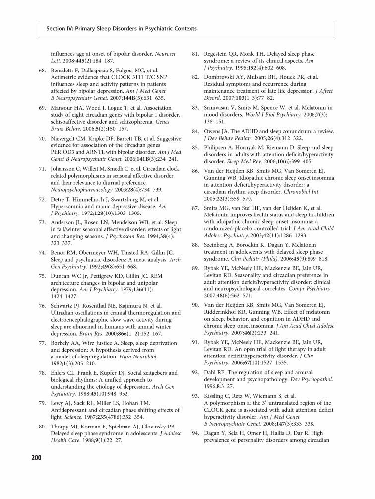
influences age at onset of bipolar disorder. NeurosciLett. 2008;445(2):184 187.
68. Benedetti F, Dallaspezia S, Fulgosi MC, et al.Actimetric evidence that CLOCK 3111 T/C SNPinfluences sleep and activity patterns in patientsaffected by bipolar depression. Am J Med GenetB Neuropsychiatr Genet. 2007;144B(5):631 635.
69. Mansour HA, Wood J, Logue T, et al. Associationstudy of eight circadian genes with bipolar I disorder,schizoaffective disorder and schizophrenia. GenesBrain Behav. 2006;5(2):150 157.
70. Nievergelt CM, Kripke DF, Barrett TB, et al. Suggestiveevidence for association of the circadian genesPERIOD3 and ARNTL with bipolar disorder. Am J MedGenet B Neuropsychiatr Genet. 2006;141B(3):234 241.
71. Johansson C,Willeit M, Smedh C, et al. Circadian clockrelated polymorphisms in seasonal affective disorderand their relevance to diurnal preference.Neuropsychopharmacology. 2003;28(4):734 739.
72. Detre T, Himmelhoch J, Swartzburg M, et al.Hypersomnia and manic depressive disease. AmJ Psychiatry. 1972;128(10):1303 1305.
73. Anderson JL, Rosen LN, Mendelson WB, et al. Sleepin fall/winter seasonal affective disorder: effects of lightand changing seasons. J Psychosom Res. 1994;38(4):323 337.
74. Benca RM, Obermeyer WH, Thisted RA, Gillin JC.Sleep and psychiatric disorders: A meta analysis. ArchGen Psychiatry. 1992;49(8):651 668.
75. Duncan WC Jr, Pettigrew KD, Gillin JC. REMarchitecture changes in bipolar and unipolardepression. Am J Psychiatry. 1979;136(11):1424 1427.
76. Schwartz PJ, Rosenthal NE, Kajimura N, et al.Ultradian oscillations in cranial thermoregulation andelectroencephalographic slow wave activity duringsleep are abnormal in humans with annual winterdepression. Brain Res. 2000;866(1 2):152 167.
77. Borbely AA, Wirz Justice A. Sleep, sleep deprivationand depression: A hypothesis derived froma model of sleep regulation. Hum Neurobiol.1982;1(3):205 210.
78. Ehlers CL, Frank E, Kupfer DJ. Social zeitgebers andbiological rhythms: A unified approach tounderstanding the etiology of depression. Arch GenPsychiatry. 1988;45(10):948 952.
79. Lewy AJ, Sack RL, Miller LS, Hoban TM.Antidepressant and circadian phase shifting effects oflight. Science. 1987;235(4786):352 354.
80. Thorpy MJ, Korman E, Spielman AJ, Glovinsky PB.Delayed sleep phase syndrome in adolescents. J AdolescHealth Care. 1988;9(1):22 27.
81. Regestein QR, Monk TH. Delayed sleep phasesyndrome: a review of its clinical aspects. AmJ Psychiatry. 1995;152(4):602 608.
82. Dombrovski AY, Mulsant BH, Houck PR, et al.Residual symptoms and recurrence duringmaintenance treatment of late life depression. J AffectDisord. 2007;103(1 3):77 82.
83. Srinivasan V, Smits M, Spence W, et al. Melatonin inmood disorders. World J Biol Psychiatry. 2006;7(3):138 151.
84. Owens JA. The ADHD and sleep conundrum: a review.J Dev Behav Pediatr. 2005;26(4):312 322.
85. Philipsen A, Hornyak M, Riemann D. Sleep and sleepdisorders in adults with attention deficit/hyperactivitydisorder. Sleep Med Rev. 2006;10(6):399 405.
86. Van der Heijden KB, Smits MG, Van Someren EJ,Gunning WB. Idiopathic chronic sleep onset insomniain attention deficit/hyperactivity disorder: acircadian rhythm sleep disorder. Chronobiol Int.2005;22(3):559 570.
87. Smits MG, van Stel HF, van der Heijden K, et al.Melatonin improves health status and sleep in childrenwith idiopathic chronic sleep onset insomnia: arandomized placebo controlled trial. J Am Acad ChildAdolesc Psychiatry. 2003;42(11):1286 1293.
88. Szeinberg A, Borodkin K, Dagan Y. Melatonintreatment in adolescents with delayed sleep phasesyndrome. Clin Pediatr (Phila). 2006;45(9):809 818.
89. Rybak YE, McNeely HE, Mackenzie BE, Jain UR,Levitan RD. Seasonality and circadian preference inadult attention deficit/hyperactivity disorder: clinicaland neuropsychological correlates. Compr Psychiatry.2007;48(6):562 571.
90. Van der Heijden KB, Smits MG, Van Someren EJ,Ridderinkhof KR, Gunning WB. Effect of melatoninon sleep, behavior, and cognition in ADHD andchronic sleep onset insomnia. J Am Acad Child AdolescPsychiatry. 2007;46(2):233 241.
91. Rybak YE, McNeely HE, Mackenzie BE, Jain UR,Levitan RD. An open trial of light therapy in adultattention deficit/hyperactivity disorder. J ClinPsychiatry. 2006;67(10):1527 1535.
92. Dahl RE. The regulation of sleep and arousal:development and psychopathology. Dev Psychopathol.1996;8:3 27.
93. Kissling C, Retz W, Wiemann S, et al.A polymorphism at the 30 untranslated region of theCLOCK gene is associated with adult attention deficithyperactivity disorder. Am J Med GenetB Neuropsychiatr Genet. 2008;147(3):333 338.
94. Dagan Y, Sela H, Omer H, Hallis D, Dar R. Highprevalence of personality disorders among circadian
Section IV: Primary Sleep Disorders in Psychiatric Contexts
200
rhythm sleep disorders (CRSD) patients. J PsychosomRes. 1996;41(4):357 363.
95. American Psychiatric Association. Diagnostic andstatisticalmanual ofmental disorders, 4th ed.Washington,DC: American Psychiatric Association; 1994.
96. Mukhopadhyay S, Fineberg NA, Drummond LM, et al.Delayed sleep phase in severe obsessive compulsivedisorder: a systematic case report survey. CNS Spectr.2008;13(5):406 413.
97. Millet B, Touitou Y, Poirier MF, et al. Plasmamelatonin and cortisol in patients with obsessivecompulsive disorder: relationship with axillarytemperature, physical activity, and clinical symptoms.Biol Psychiatry. 1998;44(9):874 881.
98. Monteleone P, Catapano F, Del Buono G, Maj M.Circadian rhythms of melatonin, cortisol andprolactin in patients with obsessive compulsivedisorder. Acta Psychiatr Scand. 1994;89(6):411 415.
99. Wirz Justice A, Cajochen C, Nussbaum P.A schizophrenic patient with an arrhythmiccircadian rest activity cycle. Psychiatry Res.1997;73(1 2):83 90.
100. Wirz Justice A, Haug HJ, Cajochen C. Disturbedcircadian rest activity cycles in schizophreniapatients: an effect of drugs? Schizophr Bull.2001;27(3):497 502.
101. Wirz Justice A, Werth E, Savaskan E, et al. Haloperidoldisrupts, clozapine reinstates the circadian restactivity cycle in a patient with early onset Alzheimerdisease. Alzheimer Dis Assoc Disord. 2000;14(4):212 215.
102. Ayalon L, Hermesh H, Dagan Y. Case study ofcircadian rhythm sleep disorder following haloperidoltreatment: reversal by risperidone and melatonin.Chronobiol Int. 2002;19(5):947 959.
103. Hermesh H, Lemberg H, Abadi J, Dagan Y.Circadian rhythm sleep disorders as a possibleside effect of fluvoxamine. CNS Spectr. 2001;6(6):511 513.
104. Dagan Y, Ayalon L. Case study: psychiatricmisdiagnosis of non 24 hours sleep wakeschedule disorder resolved by melatonin. J AmAcad Child Adolesc Psychiatry. 2005;44(12):1271 1275.
Chapter 11: Circadian rhythm disorders
201

Section VChapter
12Insomnia in Psychiatric Contexts
Principles of insomnia
Wendy M. Troxel and Daniel J. Buysse
OverviewInsomnia is the most common sleep disorder, withprevalence estimates ranging from 10% to 20% of thepopulation [1]. The term “insomnia” is used in thevernacular to refer to various complaints about poorsleep quality or an inability to fall asleep or stayasleep. The 1-year prevalence of insomnia symptomsin the general population ranges from 30% to 40%,and up to 66% in primary care and psychiatric set-tings [2]. Clinically, the insomnia disorder refers tocomplaints of an inability to fall asleep, stay asleep, ornon-restorative sleep, despite an adequate opportun-ity for sleep, combined with some form of daytimeimpairment (e.g. fatigue, occupational problems, rela-tional problems, etc.). One of the inherent challengesin diagnosing and treating insomnia is that there areno known physiological markers to indicate the pres-ence or absence of the phenotype. Rather, it is adisorder defined solely by the subjective complaintsof poor sleep and daytime impairment. As with allsubjective symptoms, these complaints may reflect ageneral negative affect bias [3] or a bias common toother psychiatric disorders (most notably, depressionand anxiety), rather than a distinct disorder, per se.Moreover, given the high prevalence of insomniasymptoms in the general population, the assessmentand diagnosis of the clinical disorder of insomniaposes a number of clinical challenges. Primarily forthese reasons, the field of insomnia research andtreatment has historically struggled to establish thedisorder’s legitimacy as a distinct clinical entity asso-ciated with significant morbidity, rather than beingmerely a symptom of another disorder or secondarydiagnosis. Over the past several decades, however,accumulating evidence has documented the highprevalence of insomnia in the population at large,
and in specific subpopulations, the significant psychi-atric and medical morbidity associated with insom-nia, and the efficacy of insomnia treatments forimproving sleep as well as overall well-being, and otherpsychiatric co-morbidities. Indeed, based on consist-encies in clinical features, course, and response totreatment, a 2005 National Institutes of Health Con-ference suggested that the term “co-morbid” insomniamay be more appropriate terminology for insomniaco-occurring with other psychiatric or medical diag-noses, rather than “secondary.” Such a change interminology reflects the growing recognition thatinsomnia is a significant public health concern in itsown right. To provide an overview of the currentstate-of-the-science in insomnia research and clinicalapplications, the current chapter will review topicspertaining to the assessment and diagnosis, preva-lence and course, epidemiology and risk factors, andconsequences of insomnia.
Assessment and diagnosisAn insomnia disorder is a syndrome consisting ofat least one insomnia complaint (difficulty fallingasleep, staying asleep, or non-restorative sleep) in anindividual with adequate opportunity/circumstancesfor sleep, combined with significant daytime impair-ment or distress. Common daytime impairmentsin insomnia patients include mood disturbances(irritability, mild dysphoria, low stress/frustrationtolerance), impaired cognitive function (problemswith memory, concentration, or difficulty completingcomplex, abstract, or creative tasks), daytime fatigue,or role impairments (difficulty at work or in familyroles) [4]. Interestingly, while fatigue is a verycommon daytime impairment in insomnia, physio-logical “sleepiness” is not [5], although patients may
Foundations of Psychiatric SleepMedicine, ed. JohnW.Winkelman andDavid T. Plante. Published by CambridgeUniversity Press.# Cambridge University Press 2010.
203
initially believe the terms are synonymous. To clarifywith patients, it is often helpful to describe “sleepi-ness” as the actual likelihood of falling asleep, whichcan be asked directly or subjectively measured withscales such as the Epworth sleepiness scale [6].In contrast, fatigue is the feeling of mental or physicalweariness or exhaustion. Indeed, once this termin-ology is clarified, patients will commonly make state-ments such as, “Yes, I feel fatigued all the time, butI could never fall asleep. That’s the problem.”
A key clinical feature of insomnia is that sleepcomplaints are present despite the fact that the indi-vidual has an adequate opportunity for sleep. Estab-lishing whether one has opportunity for sleep isparticularly relevant in today’s 24–7 society where itis common for people to regularly get less than 6hours of sleep, due to the demands of work, familylife, social roles, etc [7]. Moreover, in certain popula-tions such as the military, irregular sleep schedulesand sleep deprivation are common during deploy-ment [8]. Insomnia disorders are also highly preva-lent in returning military veterans [9]. However, it isimportant to distinguish the development of insom-nia, which is distinct from, though likely related to,the irregular sleep schedules and prolonged workhours, as well as other characteristics of deployment.
Current classification systemsThe importance of a consistent diagnosis and classifica-tion system cannot be overstated. For clinical purposes,such consistency facilitates effective communicationacross care providers and may also be critical forreimbursement for services provided. For researchpurposes, such consistency guides recruitment andtreatment response criteria, which will ultimatelyinform our understanding of the pathophysiologyand treatment of insomnia. The three primary classifi-cation schemes used to describe insomnia areincluded within the International Classification of SleepDisorders, 2nd Edition (ICSD-2 [10]), the Diagnosticand Statistical Manual of Mental Disorders, 4th Edition(DSM-IV [11]) and the World Health Organization’s(WHO) International Classification of Diseases (ICD-9CMand ICD-10 [12]). In addition, in 2004, the Ameri-can Academy of Sleep Medicine (AASM) publishedresearch diagnostic criteria for insomnia, to operation-ally define the disorder for insomnia research [13];however, the current chapter will focus on the primaryclassification systems for clinical practice.
Broadly speaking, the ICSD-2 provides the mostcomplete and specific system for defining the variousinsomnia disorders (i.e. specific subtypes); however, itis less widely used as compared to the DSM-IV or ICD.The ICSD-2 insomnia disorders are included as oneof six major categories of sleep disorders. Both theDSM-IV and ICD have fewer subclassifications andincludemore global diagnostic categories for insomniathan the ICSD-2. The DSM-IV has a specific categoryfor primary insomnia, defined as a disorder whichdoes not occur “exclusively during the course of Nar-colepsy, Breathing-Related Sleep Disorder, CircadianRhythm Disorder, or a parasomnia” or “anothermental disorder” (e.g. major depressive disorder, gen-eralized anxiety disorder, a delirium) or is due to asubstance or medication. In DSM-IV secondary diag-noses include co-morbid insomnia disorders, such asinsomnia related to another mental disorder or sub-stance-induced insomnia. The ICD system classifiessleep disorders according to presumptive etiology,organic versus non-organic.
Despite their differences, in general, each of theclassification systems describes three major categoriesof insomnia:
1. Insomnia secondary to or co-morbid with otherconditions. Consistent with the previouslydescribed DSM-IV criteria for secondarydiagnoses, this category refers to insomniasrelated to other medical or psychiatric disorders,or due to the direct physiological effects ofsubstance use or withdrawal. By far, this categoryis the single most common type of insomniadiagnosis seen in epidemiological studies and inclinical samples [14,15].
2. Insomnia as a symptom of other sleep-specificdisorders. This category includes insomniasymptoms that occur within restless legssyndrome, some cases of obstructive sleep apnea,and some cases of parasomnias.
3. Primary insomnias. This category refers todisorders in which insomnia complaints are theprimary symptoms, with no other disorder as aputative cause.
Differential diagnosisGiven that the underlying causes of insomnia areoften complex and multifactorial and that co-morbidinsomnias are the most prevalent type of insomniadisorder [15], carefully differentiating between
Section V: Insomnia in Psychiatric Contexts
204
insomnia symptoms and other co-morbid conditionsthat may contribute to or be exacerbated by theinsomnia symptoms, is critical for optimizing treat-ment efforts.
Psychiatric disordersRoughly half of individuals presenting with chronicinsomnia have a current or past psychiatric diagno-sis [14,15]. In addition, complaints of poor sleep or sleepdisturbances are included within the clinical profiles ofmany psychiatric disorders, including mood disorders,anxiety disorders, post-traumatic stress disorder, andsubstance use disorders. Given evidence that insomniasymptoms often appear as prodromal symptoms pre-ceding the onset of “full-blown” psychiatric disorders[2,16] and that insomnia symptoms often do not remiteven with successful treatment of the psychiatric dis-order [17], a thorough psychiatric history is necessaryfor the adequate assessment and treatment of insomnia.
Other sleep disordersOccult or untreated sleep disorders, such as periodicleg movement disorder, restless legs syndrome, andsleep-related breathing disorders can cause insomniasymptoms, including difficulty falling asleep, sleepfragmentation, and daytime impairments, such asfatigue. Polysomnography may be necessary for someinsomnia patients who have another suspected sleepdisorder. Insomnia complaints are also often presentwithin circadian rhythm disorders. Inclusion of sleepdiaries within the insomnia evaluation is particularlyuseful for evaluating the presence of a circadianrhythm disorder. Although they are significantly lesscommonly reported in conjunction with insomniacomplaints, parasomnias, which are characterized bya complaint of unusual behavior or events duringsleep that may lead to nocturnal arousals, should alsobe considered as part of the insomnia differentialdiagnosis.
Medical and neurological conditionsAnother critical element of the differential diagnosisis the evaluation of medical disorders (e.g. hyperthy-roidism, stroke, neuropathy) and use of medicationswhich may underlie insomnia complaints [18]. Such adetermination should be based on the patient’s med-ical history, available laboratory findings, and/orphysical examination.
The insomnia evaluation andassessment toolsPolysomnography, which is typically considered the“gold standard” in themeasurement of sleep and criticalfor the diagnosis of other sleep disorders, such as peri-odic leg movement disorder or obstructive sleep apnea,is generally “not recommended for the routine evalu-ation of insomnia” [5] (except as a rule-out for othersleep disorders). Rather, the assessment of insomniadepends on a thorough clinical history focusing onspecific sleep symptoms and associated daytime impair-ments, chronology of symptoms, including any identi-fiable or suspected precipitating events, exacerbatingand alleviating factors (e.g. seasonal effects, how insom-nia symptoms are affected by changes in usual bedroomenvironment), and response to previous treatments.Patient’s usual sleep and wake periods and the degreeof variability in these patterns, as well as behaviors(including napping), cognitions, and environmentalfactors related to bedtime routines are also criticalelements of the insomnia history. Symptoms of restlesslegs syndrome (RLS; e.g. restlessness or need to movelegs during or prior to falling asleep), symptoms ofobstructive sleep apnea (snoring, gasping, or breathingproblems, morning dry mouth), nightmares, and painor limitations to mobility or comfort during sleepshould also be assessed as a part of the differentialdiagnosis. Regularity of daytime routines, includingwork, social interactions, exercise routines, and meal-times should also be considered in the history.
Given that insomnia is often co-morbid withother psychiatric, sleep, and medical conditions, athorough insomnia assessment should include a com-prehensive sleep history, including common sleepdisturbances and sleep habits, and a review of thepatient’s psychiatric, medical, and social history.
Assessment toolsIn our laboratory and clinic, we routinely use anumber of self-report instruments, including a self-reported sleep history, the Epworth sleepiness scale [6],the Pittsburgh sleep quality index (PSQI [19]), andself-report measures of depression or anxiety symp-toms, to facilitate the assessment process. Moreover,in efforts to make the assessment as efficient as pos-sible, new patients are sent a package of these screeningmeasures and a sleep diary at least 2 weeks prior to theinitial evaluation.
Chapter 12: Principles of insomnia
205
Sleep–wake diariesSleep-wake diaries covering 1–2 weeks of the patient’susual sleep times and behaviors related to sleep arecritical tools in the assessment and treatment of insom-nia (Figure 12.1). Ask a patient to verbally report their“usual” sleep and wake time and you are likely to get areport of how he/she slept last night or his/her worstnight of sleep. Thus, a sleep diary is essential to avoidsuch primacy and recency biases, in order to receive amore valid representation of the patient’s typical sleeppatterns, as well as day-to-day variability in sleephours, and the specific sleep problems experienced bythe patient. Such an accurate representation is neces-sary not only for diagnosis, but also for treatmentpurposes, whether treatment is behavioral or pharma-cological. For instance, a central feature of virtually allbehavioral treatments is restricting time in bed tocoincide with actual time spent asleep. Thus, a carefulassessment of the patient’s usual sleep times is essen-tial. Similarly, knowledge of the patient’s usual bed-time routines and sleep problems is also important forappropriate choice of pharmacological treatment anddosing instructions.
ActigraphyActigraphy is useful as an objective measure of assess-ing rest–activity patterns. The actigraph itself is anon-invasive wrist-watch style device, typically wornon the individual’s non-dominant wrist for severaldays to weeks. Used in conjunction with a sleep diary,actigraphy can provide useful information regardingtemporal patterns, variability in rest–activity, andresponses to treatment. A number of studies haveshown moderate to strong correlations between ac-tigraphy patterns and sleep as monitored by polysom-nography (PSG) [20].
EpidemiologyThe experience of transient insomnia symptoms atsome point during the life course is a nearly universalexperience. The commonality of these symptomswithin the normative population has made the epi-demiology of insomnia complicated and prevalencerates have varied widely across sources. Epidemio-logical studies which generally rely on single orfew-item measures of sleep complaints, and rarelyinclude measures of associated daytime impairments,yield information about insomnia symptomatology.
These studies have led to somewhat alarmingprevalence rates, in the range of 30–40% in the gen-eral population and up to 66% in the primary careand psychiatric settings [1,21]. Symptom-based defin-itions, however, are only modestly associated withmore stringently defined criteria for the insomniadisorder [22]. Prevalence rates for insomnia as a spe-cific disorder have ranged from 4.4% to 11.7% [1].The disparity between prevalence rates for insomniasymptoms and those meeting DSM-IV diagnostic cri-teria for insomnia, highlights the fact that a signifi-cant proportion of the population is affected by sleepcomplaints, though the majority of those complaintsdo not meet clinically significant criteria.
Risk factorsA number of demographic, psychosocial, and behav-ioral risk factors have been identified as important riskfactors for chronic insomnia (persisting for 1month orlonger). Moreover, among the risk factors identified,they can be broadly categorized into those that predis-pose an individual to developing insomnia, those thatprecipitate the onset of the disorder, and those thatperpetuate the disorder. Among the predisposing orvulnerability factors, female sex has consistently beenshown to be a risk factor for insomnia [2,23]. The sexdisparity in insomnia rates may reflect women’sgreater tendency to report symptoms, more generally,or may reflect sex differences in traditional genderroles which are associated with sleep disturbances,such as parenting responsibilities and caregiving [24].
Older adults also have higher rates of insomniathan younger adults [25–27]; however, more recentstudies that carefully control for other co-morbiditiesshow weaker correlations between advancing age andinsomnia risk [28,29]. Thus, age, per se, is not a riskfactor for insomnia, but age-related deteriorations inmental and physical health status are associated withincreased risk of insomnia [28].
Marital status has also been identified as an import-ant risk factor for insomnia. Divorced/separated orwidowed individuals are at increased risk for insomniaas compared to their married counterparts [30]; how-ever, the magnitude of this effect is generally greater inwomen than in men [31]. A number of studies haveshown that low socioeconomic status and unemploy-ment are also risk factors for insomnia [32,33]. Finally,research on race/ethnicity and insomnia risk presents amore complicated picture, suggesting that the effects
Section V: Insomnia in Psychiatric Contexts
206
Figure 12.1 Example sleep diaries. Adapted from Buysse DJ. Introduction. In: Sleep disorders and psychiatry. Arlington, VA: AmericanPsychiatric Publishing, Inc: American Psychiatric Publishing Review of Psychiatry; 2005.
Chapter 12: Principles of insomnia
207
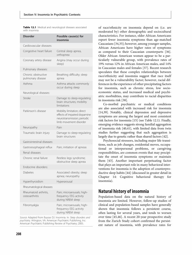
of race/ethnicity on insomnia depend on (i.e. aremoderated by) other demographic and socioculturalcharacteristics. For instance, older African Americansreport fewer insomnia symptoms than age-matchedCaucasians [34,35]; however, among younger samples,African Americans have higher rates of symptomsas compared to their Caucasian counterparts [36].Older African American women appear to be a par-ticularly vulnerable group, with prevalence rates of19% versus 12% in African American males, and 14%in Caucasian males and females [37]. Recent researchspeculates that these complex relationships betweenrace/ethnicity and insomnia suggest that race itselfmay not be a vulnerability factor; however, racial dif-ferences in the experience of other precipitating factorsfor insomnia, such as chronic stress, low socio-economic status, and increased medical and psychi-atric morbidities, may contribute to racial disparitiesin insomnia risk [38].
Co-morbid psychiatric or medical conditionsare also associated with increased risk for insomnia[14,39]. Notably, clinical depression and depressivesymptoms are among the largest and most consistentrisk factors for insomnia [15] (see Table 12.1). Finally,emerging evidence suggests strong familial aggregationof insomnia risk [40,41], with limited data from twinstudies further suggesting that such aggregation islargely due to genetic rather than shared factors [42].
Psychosocial stressors, including major life transi-tions, such as job changes, residential moves, occupa-tional or interpersonal problems, or caregivingresponsibilities, are common events that may precipi-tate the onset of insomnia symptoms or maintainthem [43]. Another important perpetuating factorthat plays an important role in many behavioral inter-ventions for insomnia is the adoption of counterpro-ductive sleep habits [44] (discussed in greater detail inChapter 14: Cognitive behavioral therapy forinsomnia).
Natural history of insomniaPopulation-based data on the natural history ofinsomnia are limited. However, follow-up studies ofclinical and population-based samples have generallyshown that insomnia follows a persistent course,often lasting for several years, and tends to worsenover time [45,46]. A recent 20-year prospective studyfrom the Zurich Study cohort confirmed the persist-ent nature of insomnia, with prevalence rates for
Table 12.1 Medical and neurological diseases associatedwith insomnia
Disorder Possible cause(s) forinsomnia
Cardiovascular diseases:
Congestive heart failure Central sleep apnea,orthopnea
Coronary artery disease Angina (may occur duringsleep)
Pulmonary diseases:
Chronic obstructivepulmonary disease
Breathing difficulty; sleepapnea
Asthma Asthma attacks commonlyoccur during sleep
Neurological diseases:
Stroke Damage to sleep-regulatingbrain structures; mobilitylimitations
Parkinson’s disease Mobility limitation; directeffects of impaired dopamineneurotransmission; periodiclimb movement disorder
Neuropathy Pain
Traumatic brain injury Damage to sleep-regulatingbrain structures
Gastrointestinal diseases:
Gastroesophageal reflux Pain; initiation of apneas
Renal diseases:
Chronic renal failure Restless legs syndrome;obstructive sleep apnea
Endocrine disorders:
Diabetes Associated obesity; sleepapnea; neuropathy
Hyperthyroidism Hyperarousal
Rheumatological diseases:
Rheumatoid arthritis,osteoarthritis
Pain; microarousals; high-frequency EEG activityduring NREM sleep
Fibromyalgia Pain; microarousals; high-frequency EEG activityduring NREM sleep
Source: Adapted from Buysse DJ. Insomnia. In: Sleep disorders andpsychiatry. Arlington, VA: American Psychiatric Publishing, Inc:American Psychiatric Publishing Review of Psychiatry; 2005.
Section V: Insomnia in Psychiatric Contexts
208
more chronic forms of insomnia (lasting 1 month orlonger) increasing gradually over time [47]. Similarly,results from a population-based study in Swedenconfirmed the high prevalence of insomnia and per-sistent course of the illness over a 1-year follow-upperiod [48].
Pathophysiology of insomniaThe physiological model of insomnia is based on thecentral assumption that insomnia represents a dis-order of increased physiological arousal. Arousal isbelieved to be a centrally mediated phenomenonrelated to the function of wake-promoting regions inthe ascending reticular activating system, hypothal-amus, and basal forebrain, which interact with sleep-promoting brain centers in the anterior hypothalamusand hypothalamus. Insomnia, according to this model,represents a disorder of increased arousal or “hyper-arousal,”whichmay be present tonically or in responseto specific cues, such as the sleep environment.Importantly, emerging evidence suggests that arousaland sleep are not mutually exclusive neurobehavioralstates, as was originally predicted by this model[49,50]. Rather, both states may co-occur in insomniaand it is precisely this simultaneous activation ofsleep and arousal states that may contribute to certainparadoxical findings in the insomnia literature (e.g.reports of daytime sleepiness and fatigue with aninability to nap). Indeed, a number of studies using avariety of techniques, including psychophysiologicaland metabolic, electrophysiological, neuroendocrine,and functional neuroanatomic methods, support thebasic contention that hyperarousal plays a role in thepathophysiology of insomnia.
Psychophysiological, metabolic, andelectrophysiological measures of arousalFindings from studies utilizing measures of heart rate,respiration, galvanic skin response, respiration rate,peripheral blood flow or vasoconstriction, and wholebody metabolic rate generally support the notion ofincreased physiological arousal in poor sleepers ascompared to good sleepers, with some evidence sug-gesting that such arousal is particularly evident atsleep onset [51–56]. For instance, Bonnet and Arand[57,58] showed that whole body metabolic rate, meas-ured by volume of oxygen consumption per unit oftime (VO2) is higher during sleep in patients with
insomnia compared to good sleepers. Althoughthere are important limitations in several of theseearlier studies, including the use of symptom-baseddefinitions of insomnia rather than clinically definedcriteria, and inclusion of a heterogeneous group ofinsomnia participants, including those with second-ary diagnoses, the consistency of the results suggeststhat individuals with insomnia do exhibit heightenedphysiological arousal.
Neuroendocrine measures of arousalActivation of the hypothalamic-pituitary-adrenal(HPA) axis is a critical component of the stressresponse system and is thought to play a role in thepurported hyperarousal in insomnia. Consistent withthis notion, several studies have shown that insomniapatients have elevated cortisol and adrenocortico-tropic hormone levels prior to and during sleep, withthe largest differences present in the first half of thenight as compared to good sleepers [59,60].
Functional neuroanatomic measuresof arousalSingle photon emission computed tomography(SPECT) and positron emission tomography (PET)are methods used to identify regional blood flow ormetabolic activity associated with specific tasks orstates. Only a handful of studies have utilized thesetechniques thus far in the context of insomnia. In thefirst PET study of primary insomnia, Nofzinger andcolleagues [50] found that patients had increasedglobal metabolic rates during both wakefulness andNREM sleep compared with good sleepers. Moreover,in insomnia patients, the usual sleep-related declinesin wake-promoting brain regions, including theascending reticular activating system, hypothalamus,and thalamus, were attenuated. However, duringwakefulness insomnia participants showed reduceddorsolateral prefrontal cortical activity. Together,these findings suggesting hyperarousal during NREMsleep and hypoarousal during wakefulness may cor-respond to insomnia patients’ sleep and daytimeimpairments. In patients with co-morbid insomniawith major depression, EEG beta activity was posi-tively associated with metabolic activity in the orbito-frontal cortex, and with complaints of poor sleepquality [61], further supporting the hyperarousalhypothesis. In contrast to these findings, a study using
Chapter 12: Principles of insomnia
209

Tc-HMPAO SPECT found that patients withinsomnia showed a consistent pattern of hypoperfu-sion (i.e. decreased blood flow) across eight a prioridefined brain regions, with the most prominent effectobserved in the basal ganglia [62]. Taken together,this limited literature suggests that there are indeedfunctional neuroanatomic changes during NREMsleep associated with insomnia; however, the precisenature of these changes and how they relate to insom-nia complaints requires further investigation [63,64].
Cognitive behavioral modelsAll cognitive behavioral models of insomnia share abasic framework outlining the relationships betweensleep-related thoughts, behaviors, and perceivedarousal, their antecedents, and consequences. However,the core dysfunction purported to underlie the clinicaldisorder of insomnia differs across these models.
Behavioral modelsSpielman and colleagues articulated a behavioralmodelin which insomnia develops as a result of the inter-action between predisposing or vulnerability factors(e.g. heightened physiological or cognitive arousal)and external precipitating factors (e.g. life stressors)[44]. Importantly, the factors that predispose or pre-cipitate the development of insomnia are distinct fromthe factors that perpetuate or maintain the disorder. Inparticular, maladaptive, compensatory sleep habits,such as spending more time in bed by going to bedearlier or having weekend “catch-up” sleep serve asperpetuating factors which maintain and reinforce theinsomnia even after the original precipitants recede.Thus, an individual may be prone to insomnia due toenduring, trait-level characteristics, and may experi-ence acute episodes of insomnia as a result of exposureto precipitating factors, such as life events. However, itis behavioral factors which are believed to underlie thechronic manifestations of the disorder.
Cognitive modelsIn contrast to Spielman’s focus on maladaptivebehaviors as the critical factor underlying chronicinsomnia, cognitive models highlight the role of cog-nitive factors, in particular, ruminative thoughts andworries, particularly around bedtime, as the centralcomponents underlying insomnia [65]. Repeatedpairings between these cognitions and temporal
(e.g. bedtime routines) and environmental (e.g. bed,bedroom) cues are purported to result in a condi-tioned arousal response particularly around bedtime.Consistent with the physiological models, the cogni-tive models contend that conditions of hyperarousalare antithetical to the sleep state. Thus, such condi-tioned cognitive arousal begets sleeplessness. Theresulting sleep disturbance and ruminations aboutone’s ability to sleep ultimately leads to daytime con-sequences such as mood disturbances and fatigue.Over time, these experiences can lead to dysfunctionalbeliefs and attitudes about one’s ability to sleep andthe consequences of sleep difficulties [66]. Ultimately,these dysfunctional beliefs lead to the development ofstrategies aimed at maximizing sleep (e.g. going tobed earlier, sleeping later) which further reinforcesleep disturbances and cognitive hyperarousal.
Harvey [67] reconceptualized the cognitive modelto emphasize the specific role of attentional biases andmonitoring of threat-related cues in the maintenanceof insomnia. According to this model, excessivenegative cognitive activity (e.g. worries, ruminativethoughts) leads to increased physiological hyper-arousal and distress, which in turn, leads to selectiveattention and monitoring towards internal and exter-nal cues indicative of sleeplessness. Such selectiveattention operates 24 hours a day, leading to bothcognitive distortions regarding sleep deficits and day-time consequences. These distorted beliefs regardingthe consequences of sleep loss and threats to sleep, inturn, lead to the development of “safety behaviors”during the daytime, such as avoidance of work orsocial activities, and at night-time, such as going tobed early or “catching up” on sleep after a poornight’s sleep. These safety behaviors are adopted inorder to avoid some feared outcome – in this case,sleeplessness. However, in actuality, these behaviorsfurther reinforce rumination and worry, physiologicalarousal, and perpetuate erroneous beliefs regardingsleep and daytime deficits seen in insomnia. In sup-port of Harvey’s model, evidence suggests thatpatients with insomnia do, in fact, selectively attendto sleep-related stimuli (e.g. a pillow), have exagger-ated (“catastrophic”) thoughts about sleep loss, andengage in daytime safety behaviors [68–70].
Neurocognitive modelPerlis and colleagues [71] proposed a neurocognitivemodel of insomnia which focuses on the central role
Section V: Insomnia in Psychiatric Contexts
210
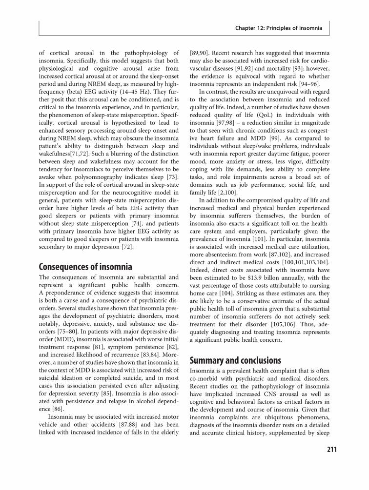
of cortical arousal in the pathophysiology ofinsomnia. Specifically, this model suggests that bothphysiological and cognitive arousal arise fromincreased cortical arousal at or around the sleep-onsetperiod and during NREM sleep, as measured by high-frequency (beta) EEG activity (14–45 Hz). They fur-ther posit that this arousal can be conditioned, and iscritical to the insomnia experience, and in particular,the phenomenon of sleep-state misperception. Specif-ically, cortical arousal is hypothesized to lead toenhanced sensory processing around sleep onset andduring NREM sleep, which may obscure the insomniapatient’s ability to distinguish between sleep andwakefulness[71,72]. Such a blurring of the distinctionbetween sleep and wakefulness may account for thetendency for insomniacs to perceive themselves to beawake when polysomnography indicates sleep [73].In support of the role of cortical arousal in sleep-statemisperception and for the neurocognitive model ingeneral, patients with sleep-state misperception dis-order have higher levels of beta EEG activity thangood sleepers or patients with primary insomniawithout sleep-state misperception [74], and patientswith primary insomnia have higher EEG activity ascompared to good sleepers or patients with insomniasecondary to major depression [72].
Consequences of insomniaThe consequences of insomnia are substantial andrepresent a significant public health concern.A preponderance of evidence suggests that insomniais both a cause and a consequence of psychiatric dis-orders. Several studies have shown that insomnia pres-ages the development of psychiatric disorders, mostnotably, depressive, anxiety, and substance use dis-orders [75–80]. In patients with major depressive dis-order (MDD), insomnia is associated with worse initialtreatment response [81], symptom persistence [82],and increased likelihood of recurrence [83,84]. More-over, a number of studies have shown that insomnia inthe context of MDD is associated with increased risk ofsuicidal ideation or completed suicide, and in mostcases this association persisted even after adjustingfor depression severity [85]. Insomnia is also associ-ated with persistence and relapse in alcohol depend-ence [86].
Insomnia may be associated with increased motorvehicle and other accidents [87,88] and has beenlinked with increased incidence of falls in the elderly
[89,90]. Recent research has suggested that insomniamay also be associated with increased risk for cardio-vascular diseases [91,92] and mortality [93]; however,the evidence is equivocal with regard to whetherinsomnia represents an independent risk [94–96].
In contrast, the results are unequivocal with regardto the association between insomnia and reducedquality of life. Indeed, a number of studies have shownreduced quality of life (QoL) in individuals withinsomnia [97,98] – a reduction similar in magnitudeto that seen with chronic conditions such as congest-ive heart failure and MDD [99]. As compared toindividuals without sleep/wake problems, individualswith insomnia report greater daytime fatigue, poorermood, more anxiety or stress, less vigor, difficultycoping with life demands, less ability to completetasks, and role impairments across a broad set ofdomains such as job performance, social life, andfamily life [2,100].
In addition to the compromised quality of life andincreased medical and physical burden experiencedby insomnia sufferers themselves, the burden ofinsomnia also exacts a significant toll on the health-care system and employers, particularly given theprevalence of insomnia [101]. In particular, insomniais associated with increased medical care utilization,more absenteeism from work [87,102], and increaseddirect and indirect medical costs [100,101,103,104].Indeed, direct costs associated with insomnia havebeen estimated to be $13.9 billon annually, with thevast percentage of those costs attributable to nursinghome care [104]. Striking as these estimates are, theyare likely to be a conservative estimate of the actualpublic health toll of insomnia given that a substantialnumber of insomnia sufferers do not actively seektreatment for their disorder [105,106]. Thus, ade-quately diagnosing and treating insomnia representsa significant public health concern.
Summary and conclusionsInsomnia is a prevalent health complaint that is oftenco-morbid with psychiatric and medical disorders.Recent studies on the pathophysiology of insomniahave implicated increased CNS arousal as well ascognitive and behavioral factors as critical factors inthe development and course of insomnia. Given thatinsomnia complaints are ubiquitous phenomena,diagnosis of the insomnia disorder rests on a detailedand accurate clinical history, supplemented by sleep
Chapter 12: Principles of insomnia
211
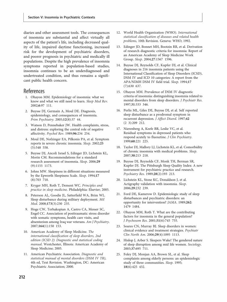
diaries and other assessment tools. The consequencesof insomnia are substantial and affect virtually allaspects of the patient’s life, including decreased qual-ity of life, impaired daytime functioning, increasedrisk for the development of psychiatric disorders,and poorer prognosis in psychiatric and medically illpopulations. Despite the high prevalence of insomniasymptoms reported in population-based studies,insomnia continues to be an underdiagnosed andundertreated condition, and thus remains a signifi-cant public health concern.
References1. Ohayon MM. Epidemiology of insomnia: what we
know and what we still need to learn. Sleep Med Rev.2002;6:97 111.
2. Buysse DJ, Germain A, Moul DE. Diagnosis,epidemiology, and consequences of insomnia.Prim Psychiatry. 2005;12(8):37 44.
3. Watson D, Pennebaker JW. Health complaints, stress,and distress: exploring the central role of negativeaffectivity. Psychol Rev. 1989;96:234 254.
4. Moul DE, Nofzinger EA, Pilkonis PA, et al. Symptomreports in severe chronic insomnia. Sleep. 2002;25(5):548 558.
5. Buysse DJ, Ancoli Israel S, Edinger JD, Lichstein KL,Morin CM. Recommendations for a standardresearch assessment of insomnia. Sleep. 2006;29(9):1155 1173.
6. Johns MW. Sleepiness in different situations measuredby the Epworth Sleepiness Scale. Sleep. 1994;17(8):703 710.
7. Kryger MH, Roth T, Dement WC. Principles andpractice in sleep medicine. Philadelphia: Elsevier; 2005.
8. Peterson AL, Goodie JL, Satterfield WA, Brim WL.Sleep disturbance during military deployment. MilMed. 2008;173(3):230 235.
9. Hoge CW, Terhakopian A, Castro CA, Messer SC,Engel CC. Association of posttraumatic stress disorderwith somatic symptoms, health care visits, andabsenteeism among Iraq war veterans. Am J Psychiatry.2007;164(1):150 153.
10. American Academy of Sleep Medicine. Theinternational classification of sleep disorders, 2ndedition (ICSD 2): Diagnostic and statistical codingmanual. Westchester, Illinois: American Academy ofSleep Medicine; 2005.
11. American Psychiatric Association. Diagnostic andstatistical manual of mental disorders (DSM IV TR),4th ed, Text Revision. Washington, DC: AmericanPsychiatric Association; 2000.
12. World Health Organization (WHO). Internationalstatistical classification of diseases and related healthproblems, 10th Revision. Geneva: WHO; 1992.
13. Edinger JD, Bonnet MH, Bootzin RR, et al. Derivationof research diagnostic criteria for insomnia: Report ofan American Academy of Sleep Medicine WorkGroup. Sleep. 2004;27:1567 1596.
14. Buysse DJ, Reynolds CF, Kupfer DJ, et al. Clinicaldiagnoses in 216 insomnia patients using theInternational Classification of Sleep Disorders (ICSD),DSM IV and ICD 10 categories: A report from theAPA/NIMH DSM IV field trial. Sleep. 1994;17(7):630 637.
15. Ohayon MM. Prevalence of DSM IV diagnosticcriteria of insomnia: distinguishing insomnia related tomental disorders from sleep disorders. J Psychiatr Res.1997;31:333 346.
16. Perlis ML, Giles DE, Buysse DJ, et al. Self reportedsleep disturbance as a prodromal symptom inrecurrent depression. J Affect Disord. 1997;42(2 3):209 212.
17. Nierenberg A, Keefe BR, Leslie VC, et al.Residual symptoms in depressed patients whorespond acutely to fluoxetine. J Clin Psychiatry.1999;60:221 225.
18. Taylor DJ, Mallory LJ, Lichstein KL, et al. Comorbidityof chronic insomnia with medical problems. Sleep.2007;30:213 218.
19. Buysse DJ, Reynolds CF, Monk TH, Berman SR,Kupfer DJ. The Pittsburgh Sleep Quality Index: A newinstrument for psychiatric practice and research.Psychiatry Res. 1989;28(2):193 213.
20. Lichstein KL, Stone KC, Donaldson J, et al.Actigraphy validation with insomnia. Sleep.2006;29:232 239.
21. Ford DE, Kamerow D. Epidemiologic study of sleepdisturbances and psychiatric disorders: anopportunity for intervention? JAMA. 1989;262:1479 1484.
22. Ohayon MM, Roth T. What are the contributingfactors for insomnia in the general population?J Psychosom Res. 2001;51(6):745 755.
23. Soares CN, Murray BJ. Sleep disorders in women:clinical evidence and treatment strategies. PsychiatrClin North Am. 2006;29(4):1095 1113.
24. Hislop J, Arber S. Sleepers Wake! The gendered natureof sleep disruption among mid life women. Sociology.2003;37:695 711.
25. Foley DJ, Monjan AA, Brown SL, et al. Sleepcomplaints among elderly persons: an epidemiologicstudy of three communities. Sleep. 1995;18(6):425 432.
Section V: Insomnia in Psychiatric Contexts
212
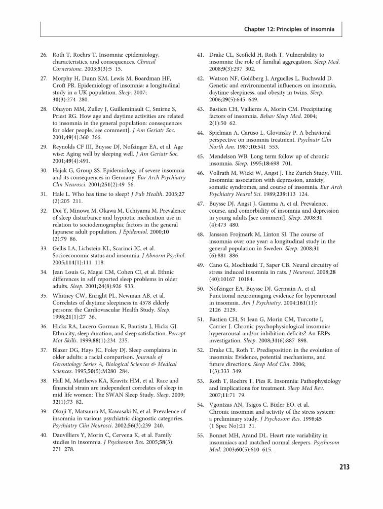
26. Roth T, Roehrs T. Insomnia: epidemiology,characteristics, and consequences. ClinicalCornerstone. 2003;5(3):5 15.
27. Morphy H, Dunn KM, Lewis M, Boardman HF,Croft PR. Epidemiology of insomnia: a longitudinalstudy in a UK population. Sleep. 2007;30(3):274 280.
28. Ohayon MM, Zulley J, Guilleminault C, Smirne S,Priest RG. How age and daytime activities are relatedto insomnia in the general population: consequencesfor older people.[see comment]. J Am Geriatr Soc.2001;49(4):360 366.
29. Reynolds CF III, Buysse DJ, Nofzinger EA, et al. Agewise: Aging well by sleeping well. J Am Geriatr Soc.2001;49(4):491.
30. Hajak G, Group SS. Epidemiology of severe insomniaand its consequences in Germany. Eur Arch PsychiatryClin Neurosci. 2001;251(2):49 56.
31. Hale L. Who has time to sleep? J Pub Health. 2005;27(2):205 211.
32. Doi Y, Minowa M, Okawa M, Uchiyama M. Prevalenceof sleep disturbance and hypnotic medication use inrelation to sociodemographic factors in the generalJapanese adult population. J Epidemiol. 2000;10(2):79 86.
33. Gellis LA, Lichstein KL, Scarinci IC, et al.Socioeconomic status and insomnia. J Abnorm Psychol.2005;114(1):111 118.
34. Jean Louis G, Magai CM, Cohen CI, et al. Ethnicdifferences in self reported sleep problems in olderadults. Sleep. 2001;24(8):926 933.
35. Whitney CW, Enright PL, Newman AB, et al.Correlates of daytime sleepiness in 4578 elderlypersons: the Cardiovascular Health Study. Sleep.1998;21(1):27 36.
36. Hicks RA, Lucero Gorman K, Bautista J, Hicks GJ.Ethnicity, sleep duration, and sleep satisfaction. PerceptMot Skills. 1999;88(1):234 235.
37. Blazer DG, Hays JC, Foley DJ. Sleep complaints inolder adults: a racial comparison. Journals ofGerontology Series A, Biological Sciences & MedicalSciences. 1995;50(5):M280 284.
38. Hall M, Matthews KA, Kravitz HM, et al. Race andfinancial strain are independent correlates of sleep inmid life women: The SWAN Sleep Study. Sleep. 2009;32(1):73 82.
39. Okuji Y, Matsuura M, Kawasaki N, et al. Prevalence ofinsomnia in various psychiatric diagnostic categories.Psychiatry Clin Neurosci. 2002;56(3):239 240.
40. Dauvilliers Y, Morin C, Cervena K, et al. Familystudies in insomnia. J Psychosom Res. 2005;58(3):271 278.
41. Drake CL, Scofield H, Roth T. Vulnerability toinsomnia: the role of familial aggregation. Sleep Med.2008;9(3):297 302.
42. Watson NF, Goldberg J, Arguelles L, Buchwald D.Genetic and environmental influences on insomnia,daytime sleepiness, and obesity in twins. Sleep.2006;29(5):645 649.
43. Bastien CH, Vallieres A, Morin CM. Precipitatingfactors of insomnia. Behav Sleep Med. 2004;2(1):50 62.
44. Spielman A, Caruso L, Glovinsky P. A behavioralperspective on insomnia treatment. Psychiatr ClinNorth Am. 1987;10:541 553.
45. Mendelson WB. Long term follow up of chronicinsomnia. Sleep. 1995;18:698 701.
46. Vollrath M, Wicki W, Angst J. The Zurich Study, VIII.Insomnia: association with depression, anxiety,somatic syndromes, and course of insomnia. Eur ArchPsychiatry Neurol Sci. 1989;239:113 124.
47. Buysse DJ, Angst J, Gamma A, et al. Prevalence,course, and comorbidity of insomnia and depressionin young adults.[see comment]. Sleep. 2008;31(4):473 480.
48. Jansson Frojmark M, Linton SJ. The course ofinsomnia over one year: a longitudinal study in thegeneral population in Sweden. Sleep. 2008;31(6):881 886.
49. Cano G, Mochizuki T, Saper CB. Neural circuitry ofstress induced insomnia in rats. J Neurosci. 2008;28(40):10167 10184.
50. Nofzinger EA, Buysse DJ, Germain A, et al.Functional neuroimaging evidence for hyperarousalin insomnia. Am J Psychiatry. 2004;161(11):2126 2129.
51. Bastien CH, St Jean G, Morin CM, Turcotte I,Carrier J. Chronic psychophysiological insomnia:hyperarousal and/or inhibition deficits? An ERPsinvestigation. Sleep. 2008;31(6):887 898.
52. Drake CL, Roth T. Predisposition in the evolution ofinsomnia: Evidence, potential mechanisms, andfuture directions. Sleep Med Clin. 2006;1(3):333 349.
53. Roth T, Roehrs T, Pies R. Insomnia: Pathophysiologyand implications for treatment. Sleep Med Rev.2007;11:71 79.
54. Vgontzas AN, Tsigos C, Bixler EO, et al.Chronic insomnia and activity of the stress system:a preliminary study. J Psychosom Res. 1998;45(1 Spec No):21 31.
55. Bonnet MH, Arand DL. Heart rate variability ininsomniacs and matched normal sleepers. PsychosomMed. 2003;60(5):610 615.
Chapter 12: Principles of insomnia
213
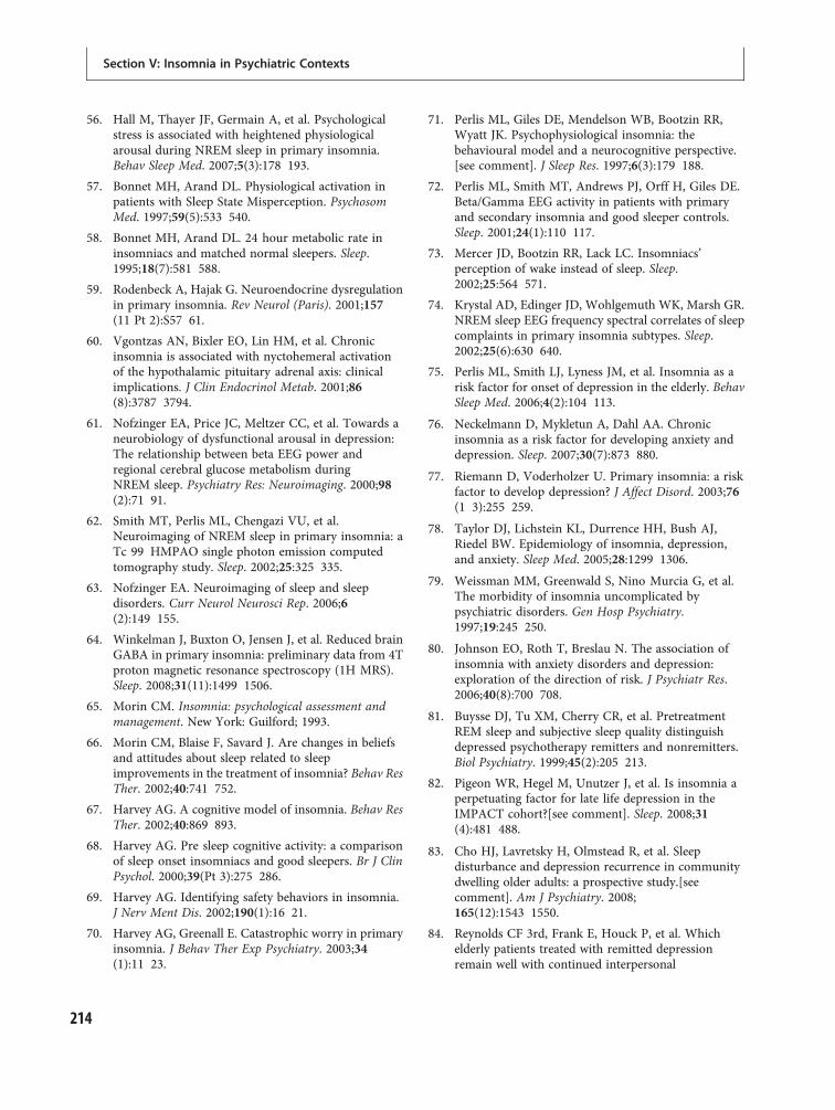
56. Hall M, Thayer JF, Germain A, et al. Psychologicalstress is associated with heightened physiologicalarousal during NREM sleep in primary insomnia.Behav Sleep Med. 2007;5(3):178 193.
57. Bonnet MH, Arand DL. Physiological activation inpatients with Sleep State Misperception. PsychosomMed. 1997;59(5):533 540.
58. Bonnet MH, Arand DL. 24 hour metabolic rate ininsomniacs and matched normal sleepers. Sleep.1995;18(7):581 588.
59. Rodenbeck A, Hajak G. Neuroendocrine dysregulationin primary insomnia. Rev Neurol (Paris). 2001;157(11 Pt 2):S57 61.
60. Vgontzas AN, Bixler EO, Lin HM, et al. Chronicinsomnia is associated with nyctohemeral activationof the hypothalamic pituitary adrenal axis: clinicalimplications. J Clin Endocrinol Metab. 2001;86(8):3787 3794.
61. Nofzinger EA, Price JC, Meltzer CC, et al. Towards aneurobiology of dysfunctional arousal in depression:The relationship between beta EEG power andregional cerebral glucose metabolism duringNREM sleep. Psychiatry Res: Neuroimaging. 2000;98(2):71 91.
62. Smith MT, Perlis ML, Chengazi VU, et al.Neuroimaging of NREM sleep in primary insomnia: aTc 99 HMPAO single photon emission computedtomography study. Sleep. 2002;25:325 335.
63. Nofzinger EA. Neuroimaging of sleep and sleepdisorders. Curr Neurol Neurosci Rep. 2006;6(2):149 155.
64. Winkelman J, Buxton O, Jensen J, et al. Reduced brainGABA in primary insomnia: preliminary data from 4Tproton magnetic resonance spectroscopy (1H MRS).Sleep. 2008;31(11):1499 1506.
65. Morin CM. Insomnia: psychological assessment andmanagement. New York: Guilford; 1993.
66. Morin CM, Blaise F, Savard J. Are changes in beliefsand attitudes about sleep related to sleepimprovements in the treatment of insomnia? Behav ResTher. 2002;40:741 752.
67. Harvey AG. A cognitive model of insomnia. Behav ResTher. 2002;40:869 893.
68. Harvey AG. Pre sleep cognitive activity: a comparisonof sleep onset insomniacs and good sleepers. Br J ClinPsychol. 2000;39(Pt 3):275 286.
69. Harvey AG. Identifying safety behaviors in insomnia.J Nerv Ment Dis. 2002;190(1):16 21.
70. Harvey AG, Greenall E. Catastrophic worry in primaryinsomnia. J Behav Ther Exp Psychiatry. 2003;34(1):11 23.
71. Perlis ML, Giles DE, Mendelson WB, Bootzin RR,Wyatt JK. Psychophysiological insomnia: thebehavioural model and a neurocognitive perspective.[see comment]. J Sleep Res. 1997;6(3):179 188.
72. Perlis ML, Smith MT, Andrews PJ, Orff H, Giles DE.Beta/Gamma EEG activity in patients with primaryand secondary insomnia and good sleeper controls.Sleep. 2001;24(1):110 117.
73. Mercer JD, Bootzin RR, Lack LC. Insomniacs’perception of wake instead of sleep. Sleep.2002;25:564 571.
74. Krystal AD, Edinger JD, Wohlgemuth WK, Marsh GR.NREM sleep EEG frequency spectral correlates of sleepcomplaints in primary insomnia subtypes. Sleep.2002;25(6):630 640.
75. Perlis ML, Smith LJ, Lyness JM, et al. Insomnia as arisk factor for onset of depression in the elderly. BehavSleep Med. 2006;4(2):104 113.
76. Neckelmann D, Mykletun A, Dahl AA. Chronicinsomnia as a risk factor for developing anxiety anddepression. Sleep. 2007;30(7):873 880.
77. Riemann D, Voderholzer U. Primary insomnia: a riskfactor to develop depression? J Affect Disord. 2003;76(1 3):255 259.
78. Taylor DJ, Lichstein KL, Durrence HH, Bush AJ,Riedel BW. Epidemiology of insomnia, depression,and anxiety. Sleep Med. 2005;28:1299 1306.
79. Weissman MM, Greenwald S, Nino Murcia G, et al.The morbidity of insomnia uncomplicated bypsychiatric disorders. Gen Hosp Psychiatry.1997;19:245 250.
80. Johnson EO, Roth T, Breslau N. The association ofinsomnia with anxiety disorders and depression:exploration of the direction of risk. J Psychiatr Res.2006;40(8):700 708.
81. Buysse DJ, Tu XM, Cherry CR, et al. PretreatmentREM sleep and subjective sleep quality distinguishdepressed psychotherapy remitters and nonremitters.Biol Psychiatry. 1999;45(2):205 213.
82. Pigeon WR, Hegel M, Unutzer J, et al. Is insomnia aperpetuating factor for late life depression in theIMPACT cohort?[see comment]. Sleep. 2008;31(4):481 488.
83. Cho HJ, Lavretsky H, Olmstead R, et al. Sleepdisturbance and depression recurrence in communitydwelling older adults: a prospective study.[seecomment]. Am J Psychiatry. 2008;165(12):1543 1550.
84. Reynolds CF 3rd, Frank E, Houck P, et al. Whichelderly patients treated with remitted depressionremain well with continued interpersonal
Section V: Insomnia in Psychiatric Contexts
214

psychotherapy after discontinuation of antidepressantmedication? Am J Psychiatry. 1997;154:958 962.
85. Liu X, Buysse DJ. Sleep and youth suicidal behavior:A neglected field. Curr Opin Psychiatry. 2006;19(3):288 293.
86. Brower KJ, Aldrich MS, Robinson EA, et al. Insomnia,self medication, and relapse to alcoholism. AmJ Psychiatry. 2001;158:399 404.
87. Leger D, Massuel MA, Metlaine A, Group SS.Professional correlates of insomnia.[see comment].Sleep. 2006;29(2):171 178.
88. Powell NB, Schectman KB, Riley RW, et al. Sleepydriving: accidents and injury. Otolaryngol Head NeckSurg. 2002;126(3):217 227.
89. Fitzpatrick P, Kirke PN, Daly L, et al. Predictors of firsthip fracture and mortality post fracture in olderwomen. Ir J Med Sci. 2001;170(1):49 53.
90. Brassington GS, King AC, Bliwise DL. Sleep problemsas a risk factor for falls in a sample of communitydwelling adults aged 64 99 years. J Am Geriatr Soc.2000;48(10):1234 1240.
91. Schwartz S, McDowell AW, Cole SR, et al. Insomniaand heart disease: a review of epidemiologic studies.J Psychosom Res. 1999;47(4):313 333.
92. Schwartz SW, Cornoni Huntley J, Cole SR, et al.Are sleep complaints an independent risk factor formyocardial infarction? Ann Epidemiol. 1998;8(6):384 392.
93. Pollak CP, Perlick D, Linsner JP, et al. Sleep problemsin the elderly as predictors of death and nursing homeplacement. J Comm Health. 1990;15:123 135.
94. Phillips B, Mannino DM. Do insomnia complaintscause hypertension or cardiovascular disease? J ClinSleep Med. 2007;3(5):489 494.
95. Althuis MD, Fredman L, Langenberg PW, Magaziner J.The relationship between insomnia and mortality
among community dwelling older women. J AmGeriatr Soc. 1998;46(10):1270 1273.
96. Kripke DF, Garfinkel L, Wingard DL, et al. Mortalityassociated with sleep duration and insomnia. Arch GenPsychiatry. 2002;59:131 136.
97. Byles JE, Mishra GD, Harris MA. The experience ofinsomnia among older women. Sleep. 2005;28(8):972 979.
98. Philip P, Leger D, Taillard J, et al. Insomniaccomplaints interfere with quality of life but not withabsenteeism: respective role of depressive and organiccomorbidity. Sleep Med. 2006;7(7):585 591.
99. Katz DA, McHorney CA. The relationship betweeninsomnia and health related quality of life in patientswith chronic illness. J Fam Pract. 2002;51(3):229 235.
100. Leger D, Guilleminault C, Bader G, Levy E, Paillard M.Medical and socio professional impact of insomnia.Sleep. 2002;25(6):625 629.
101. Ozminkowski RJ, Wang S, Walsh JK. The direct andindirect costs of untreated insomnia in adults in theUnited States. Sleep. 2007;30(3):263 273.
102. Godet Cayre V, Pelletier Fleury N, Le Vaillant M, et al.Insomnia and absenteeism at work. Who pays the cost?[see comment]. Sleep. 2006;29(2):179 184.
103. Metlaine A, Leger D, Choudat D. Socioeconomicimpact of insomnia in working populations. IndHealth. 2005;43(1):11 19.
104. Walsh JK. Clinical and socioeconomic correlates ofinsomnia. J Clin Psychiatry. 2004;65(Suppl 8):13 19.
105. Stinson K, Tang NK, Harvey AG. Barriers to treatmentseeking in primary insomnia in the United Kingdom:a cross sectional perspective. Sleep. 2006;29(12):1643 1646.
106. Leger D. Public health and insomnia: economicimpact. Sleep. 2000;23(Suppl 3):S69 76.
Chapter 12: Principles of insomnia
215
Chapter
13 Treatment of insomnia: pharmacotherapy
Andrew D. Krystal
IntroductionA large number of different types of medicationsare used to treat insomnia. This includes benzodi-azepines, non-benzodiazepines, melatonin receptoragonists, antidepressants, antipsychotics, anticonvul-sants, and antihistamines [1]. These agents exert thera-peutic effects on insomnia through a number ofdifferent pharmacological mechanisms. Some enhancethe sleep-promoting effects of GABA and melatoninsystems (benzodiazepines, non-benzodiazepines, mela-tonin agonists) while others have therapeutic effects onsleep disturbance by blocking the wake-promotingeffects of serotonin, norepinephrine, acetylcholine, andhistamine (antidepressants, antipsychotics, and antihis-tamines). The particular mechanism by which theseagents act affects the properties of their therapeuticeffects on insomnia. This mechanism, along with theother pharmacological effects, determines the side-effect profile of a given insomnia medication. Thischapter reviews the pharmacology of the therapeuticand adverse effects of these medications and identifiestheir distinguishing characteristics when used to treatinsomnia in clinical practice. The available data onthe treatment of insomnia in key patient subgroups isalso discussed. For each agent the available placebo-controlled trials are reviewed. These trials serve as thebasis for weighing the risks and benefits of each of theseagents when making treatment decisions [1].
Optimally, the decision of whether to administer atherapy for insomnia, which therapy to administer, andwhether to continue a therapy should be based on anempirically based weighing of the risks and benefits ofthe treatment options [1]. This should include the risksof not treating insomnia, which is reflected in the degreeof impairment in function and quality of life associatedwith the insomnia. There is evidence that insomnia
treatment decisions in clinical practice are not beingmade based on empirical evidence. Some of the mostfrequently administered insomnia agents have not beenestablished to have efficacy in the treatment of insomniaand their side-effect profile in insomnia therapyremains uncharacterized [2]. It is hoped that byreviewing the placebo-controlled trials that can serveas an empirical basis for clinical decision-making, thischapter will facilitate optimizing the treatment ofinsomnia among psychiatric practitioners.
Medications used to treat insomniaBenzodiazepinesThe benzodiazepines are a group of chemically relatedagents that have a therapeutic effect on insomniathrough enhancing the inhibitory effects of gamma-amino-butyric acid (GABA), the predominant inhibi-tory neurotransmitter in the brain and the mostimportant sleep-promoting system [1,3]. Benzodi-azepines have a binding site on one of the types ofGABA receptors, the GABA-A receptor, which is achannel comprised of five protein subunits that con-trols the flow of chloride ions across neuronal mem-branes [3]. When benzodiazepines bind to thisallosteric binding site, they cause a conformationalchange in the subunits which facilitates the inwardflow of chloride ions that occurs when GABA bindsto the receptor complex. This enhances GABA’sinhibitory effect because the extra inward negativeion flux results in added membrane hyperpolarization,which further biases the neuron away from exceedingthe threshold of depolarization needed to fire an actionpotential. This inhibition promotes sleep throughGABAergic projections to key wake-promoting regionsof the brain. These regions include the histaminergic
Foundations of Psychiatric Sleep Medicine, ed. JohnW.Winkelman and David T. Plante. Published by Cambridge University Press.# Cambridge University Press 2010.
216
tuberomammillary nucleus, the orexinergic/hypocret-inergic perifornical area, the serotonergic raphe nuclei,the cholinergic laterodorsal tegmentum/pedunculopon-tine tegmentum, thedopaminergic ventral periaqueduc-tal grey and the noradrenergic locus coeruleus (seeChapter 2: Neuroanatomy and neurobiology of sleepand wakefulness) [4].
GABA-A receptors are located in many regions ofthe brain other than those that affect sleep/wakefunction. As a result, benzodiazepines have clinicaleffects in addition to sleep enhancement. These mayinclude anxiolysis, anticonvulsant effects, myorelax-ation, memory impairment, motor impairment, andreinforcement. This accounts for the adverse effects ofthese agents which include sedation, cognitive impair-ment, motor impairment, and the potential for abuse(see Table 13.1). The associated anxiolytic and myore-laxant effects may be important assets when treatinginsomnia patients because insomnia frequently occursin conjunction with anxiety and/or pain [5].
Benzodiazepines most commonly used in thetreatment of insomnia include triazolam, flurazepam,temazepam, estazolam, quazepam, clonazepam, lor-azepam, and alprazolam. The properties of theseagents appear in Table 13.1. The primary featurewhich distinguishes these agents from each other istheir half life. Half life and dosage determine theduration of clinical effects. Longer half life and higherdosage both tend to lead to longer lasting effects. Theshortest half-life benzodiazepine commonly used inthe treatment of insomnia is triazolam. As a result, itis least likely to be associated with next-day effectswhen used in the recommended range of dosing. Incontrast, flurazepam, quazepam, and clonazepamhave half lives which exceed 24 hours and, as a result,are relatively likely to lead to next-day effects.
A number of the benzodiazepines have well-estab-lished efficacy in the treatment of insomnia. Placebo-controlled trials in insomnia patients aged 18–65 haveidentified a therapeutic effect on sleep onset and main-tenance for triazolam, temazepam, flurazepam, quaze-pam, and estazolam (see Table 13.1) [1]. Efficacy in thetreatment of insomnia in older adults has beenreported for triazolam and flurazepam in terms of bothsleep onset and maintenance, and a therapeutic effecton sleep maintenance has been reported for temaze-pam [1]. A few placebo-controlled studies have evalu-ated the use of benzodiazepines in the treatment ofinsomnia occurring with medical and psychiatric con-ditions (co-morbid insomnia). One study compared
the combination of clonazepam plus fluoxetine versusplacebo plus fluoxetine for the treatment of insomniaoccurring co-morbid with major depressive disorder[6]. Adjunctive clonazepam significantly improvedsleep compared with placebo and also led to a greaterantidepressant response. Another study compared theuse of triazolam versus placebo in treating insomniaassociated with rheumatoid arthritis [7]. Triazolamwas associated with significantly greater improvementin sleep and also led to less morning stiffness thanplacebo. These placebo-controlled trials in primaryand co-morbid insomnia represent the empirical basisfor making clinical treatment decisions with regard tothe benzodiazepines. The established efficacy of anumber of these agents in primary insomnia andthe two studies in co-morbid insomnias represent theexpected benefit that must be weighed against theknown risks. The most common risks are sedation,dizziness, memory side-effects, abuse, and ataxia [1].All of these risks increase in likelihood as the dosage isincreased. Adverse effects associated with impairmentin daytime function are also more likely to occur withlonger half-life agents [1].
Of the agents used to treat insomnia, benzodiaze-pines are most suited for use in the treatment ofindividuals with insomnia occurring with anxietyand/or pain given their profile of therapeutic effects.However, as is evident from the above review ofplacebo-controlled trials, there has been minimal sys-tematic research on the use of these agents for thetreatment of insomnia occurring in association withpain or anxiety. As a result, the actual risk–benefitprofile of their use in this setting remains unknown.The abuse potential of these agents suggests that theyshould be used with caution in those with a predis-position to substance abuse. In this regard it shouldbe noted that, among insomnia patients without ahistory of substance abuse, benzodiazepines are usedfor therapeutic purposes (in order to address theirsleep disturbance) and the risk of non-therapeuticuse appears to be very low [8].
Non-benzodiazepinesThe term “non-benzodiazepines” has been used torefer to a group of medications, zolpidem, zaleplon,and eszopiclone, which have a sleep-promoting effectthrough binding to the same site on the GABA-Areceptor complex as the benzodiazepines; however,they are chemically unrelated to the benzodiazepines.
Chapter 13: Treatment of insomnia: pharmacotherapy
217
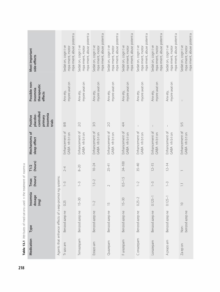
Table
13.1
Attrb
utes
ofmed
cato
nsused
nthetreatm
entof
nsom
na
Med
ication
Type
Insomnia
dosag
e(m
g)
Tmax
(hou
rs)
T1/2
(hou
rs)
Mecha
nism
sof
slee
peffect
Positive
placebo-
controlle
dprimary
insomnia
trials
Possible
non-
insomnia
therap
eutic
effects
Mostim
portant
side-effects
Age
ntsthat
enhanceeffectsof
seep-prom
otng
system
s:
Trazoam
Benzod
azep
ne0.25
1–3
2–4
Enhancem
entof
GABA
nhbto
n8/8
Anx
ety,
myoreaxaton
Sedato
n,cogn
tve
mpa
rmen
t,motor
mpa
rmen
t,abusepo
tent
a
Temazep
amBenzod
azep
ne15–30
1–3
8–20
Enhancem
entof
GABA
nhbto
n2/2
Anx
ety,
myoreaxaton
Sedato
n,cogn
tve
mpa
rmen
t,motor
mpa
rmen
t,abusepo
tent
a
Estazo
amBenzod
azep
ne1–2
1.5–2
10–24
Enhancem
entof
GABA
nhbto
n3/3
Anx
ety,
myoreaxaton
Sedato
n,cogn
tve
mpa
rmen
t,motor
mpa
rmen
t,abusepo
tent
a
Quazepam
Benzod
azep
ne15
225–41
Enhancem
entof
GABA
nhbto
n2/2
Anx
ety,
myoreaxaton
Sedato
n,cogn
tve
mpa
rmen
t,motor
mpa
rmen
t,abusepo
tent
a
Furazep
amBenzod
azep
ne15–30
0.5–1.5
24–100
Enhancem
entof
GABA
nhbto
n4/4
Anx
ety,
myoreaxaton
Sedato
n,cogn
tve
mpa
rmen
t,motor
mpa
rmen
t,abusepo
tent
a
Con
azep
amBenzod
azep
ne0.25–2
1–2
35–40
Enhancem
entof
GABA
nhbto
n–
Anx
ety,
myoreaxaton
Sedato
n,cogn
tve
mpa
rmen
t,motor
mpa
rmen
t,abusepo
tent
a
Lorazepam
Benzod
azep
ne0.125–1
1–3
12–15
Enhancem
entof
GABA
nhbto
n–
Anx
ety,
myoreaxaton
Sedato
n,cogn
tve
mpa
rmen
t,motor
mpa
rmen
t,abusepo
tent
a
Aprazoam
Benzod
azep
ne0.125–1
1–3
12–14
Enhancem
entof
GABA
nhbto
n–
Anx
ety,
myoreaxaton
Sedato
n,cogn
tve
mpa
rmen
t,motor
mpa
rmen
t,abusepo
tent
a
Zaep
onNon
-be
nzod
azep
ne10
1.1
1Enhancem
entof
GABA
nhbto
n5/5
–Sedato
n,cogn
tve
mpa
rmen
t,motor
mpa
rmen
t,abusepo
tent
a
218

Zopde
mNon
-be
nzod
azep
ne5–10
1.7–2.5
1.5–2.5
Enhancem
entof
GABA
nhbto
n8/8
–Sedato
n,cogn
tve
mpa
rmen
t,motor
mpa
rmen
t,abusepo
tent
a
Zopde
mCR
Non
-be
nzod
azep
ne6.25–12.5
1.7–2.5
1.5–2.5
Enhancem
entof
GABA
nhbto
n3/3
–Sedato
n,cogn
tve
mpa
rmen
t,motor
mpa
rmen
t,abusepo
tent
a
Eszopcon
eNon
-be
nzod
azep
ne1–3
1.3–1.6
5–7
Enhancem
entof
GABA
nhbto
n8/8
Anx
ety,
depresson
,pan
Sedato
n,cogn
tve
mpa
rmen
t,motor
mpa
rmen
t,abusepo
tent
a
Meaton
nHormon
e0.3–10
0.3–1
0.6–1
Meaton
nreceptor
agon
sm
13/14
Headache,sedato
n
Rameteon
Meaton
nreceptor
agon
st
80.7–
0.95
0.8–2
Meaton
nreceptor
agon
sm
4/4
Drowsne
ss,fatgu
e,dzzne
ss
Tagab
neAnt
epep
tcagen
t2–16
1–1.5
8GABA
reup
take
nhbto
n2/4
Sezures
Sedato
n,nausea,d
zzne
ss,
sezures
Age
ntsthat
bocktheeffectsof
wake-prom
otng
system
s:
Am
trptyne
Ant
depressant
10–100
2–5
10–100
Antagon
smof
NE,HA,A
ch–
Anx
ety,
depresson
,chroncpa
n
Sedato
n,dzzne
ss,w
egh
tga
n,OH,drymou
th,b
urred
vson
,con
stpato
n,ur
nary
retent
on
Doxep
nAnt
depressant
1–25
1.5–4
10–50
Antagon
smof
NE,HA,A
ch;n
owdo
sesHA
ony
5/5
Anx
ety,
depresson
,chroncpa
n
Sedato
n,dzzne
ss,w
egh
tga
n,OH,drymou
th,b
urred
vson
,con
stpato
n,ur
nary
retent
on
Trm
pram
neAnt
depressant
25–100
2–8
15–40
Antagon
smof
NE,HA,A
ch2/2
Anx
ety,
depresson
,chroncpa
n
Sedato
n,dzzne
ss,w
egh
tga
n,OH,drymou
th,b
urred
vson
,con
stpato
n,ur
nary
retent
on
Trazod
one
Ant
depressant
25–150
1–2
7–15
Antagon
smof
5HT 2,N
E,HA
0/1
Anx
ety,
depresson
Sedato
n,dzzne
ss,
headache
,dry
mou
th,
burredvson
,OH,p
rapsm
219
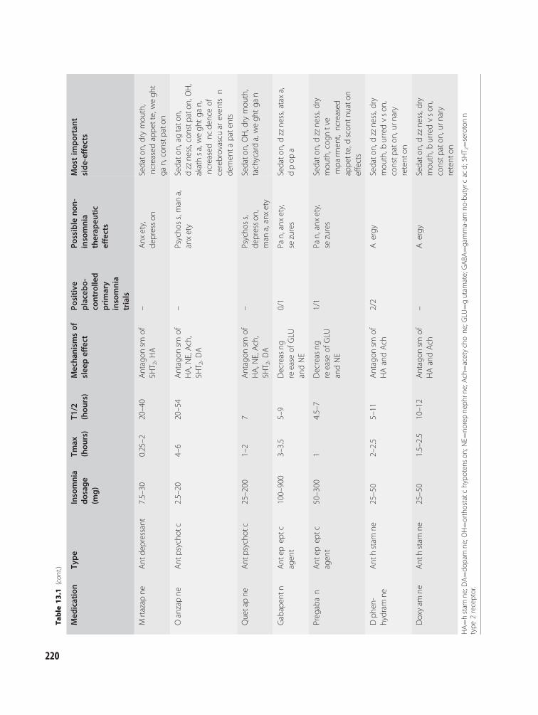
Table
13.1
(con
t.)
Med
ication
Type
Insomnia
dosag
e(m
g)
Tmax
(hou
rs)
T1/2
(hou
rs)
Mecha
nism
sof
slee
peffect
Positive
placebo-
controlle
dprimary
insomnia
trials
Possible
non-
insomnia
therap
eutic
effects
Mostim
portant
side-effects
Mrtazap
neAnt
depressant
7.5–30
0.25–2
20–40
Antagon
smof
5HT 2,H
A–
Anx
ety,
depresson
Sedato
n,drymou
th,
ncreased
appe
tte,wegh
tga
n,constp
aton
Oanzapne
Ant
psycho
tc2.5–20
4–6
20–54
Antagon
smof
HA,N
E,Ach,
5HT 2,D
A
–Psycho
ss,man
a,anxety
Sedato
n,ag
tato
n,dzzne
ss,con
stpato
n,OH,
akathsa,wegh
tga
n,ncreased
ncde
nceof
cerebrovascu
areven
tsn
demen
tapatents
Quetapne
Ant
psycho
tc25–200
1–2
7Antagon
smof
HA,N
E,Ach,
5HT 2,D
A
–Psycho
ss,
depresson
,man
a,anxety
Sedato
n,OH,d
rymou
th,
tachycarda,wegh
tga
n
Gabapen
tnAnt
epep
tcagen
t100–900
3–3.5
5–9
Decreas
ngreease
ofGLU
andNE
0/1
Pan,
anxety,
sezures
Sedato
n,dzzne
ss,ataxa,
dpop
a
Preg
aban
Ant
epep
tcagen
t50–300
14.5–7
Decreas
ngreease
ofGLU
andNE
1/1
Pan,
anxety,
sezures
Sedato
n,dzzne
ss,d
rymou
th,cog
ntve
mpa
rmen
t,ncreased
appe
tte,dscon
tnuato
neffects
Dph
en-
hydram
neAnt
hstam
ne25–50
2–2.5
5–11
Antagon
smof
HAandAch
2/2
Aergy
Sedato
n,dzzne
ss,d
rymou
th,b
urredvson
,constp
aton
,urn
ary
retent
on
Doxyam
neAnt
hstam
ne25–50
1.5–2.5
10–12
Antagon
smof
HAandAch
–Aergy
Sedato
n,dzzne
ss,d
rymou
th,b
urredvson
,constp
aton
,urn
ary
retent
on
HA¼h
stam
ne;D
A¼d
opam
ne;O
H¼o
rtho
statchypo
tens
on;N
E¼no
repne
phrn
e;Ach¼a
cetychone
;GLU
¼gutam
ate;GABA
¼gam
ma-am
no-butyrcac
d;5H
T 2¼s
eroton
ntype
2receptor.
220
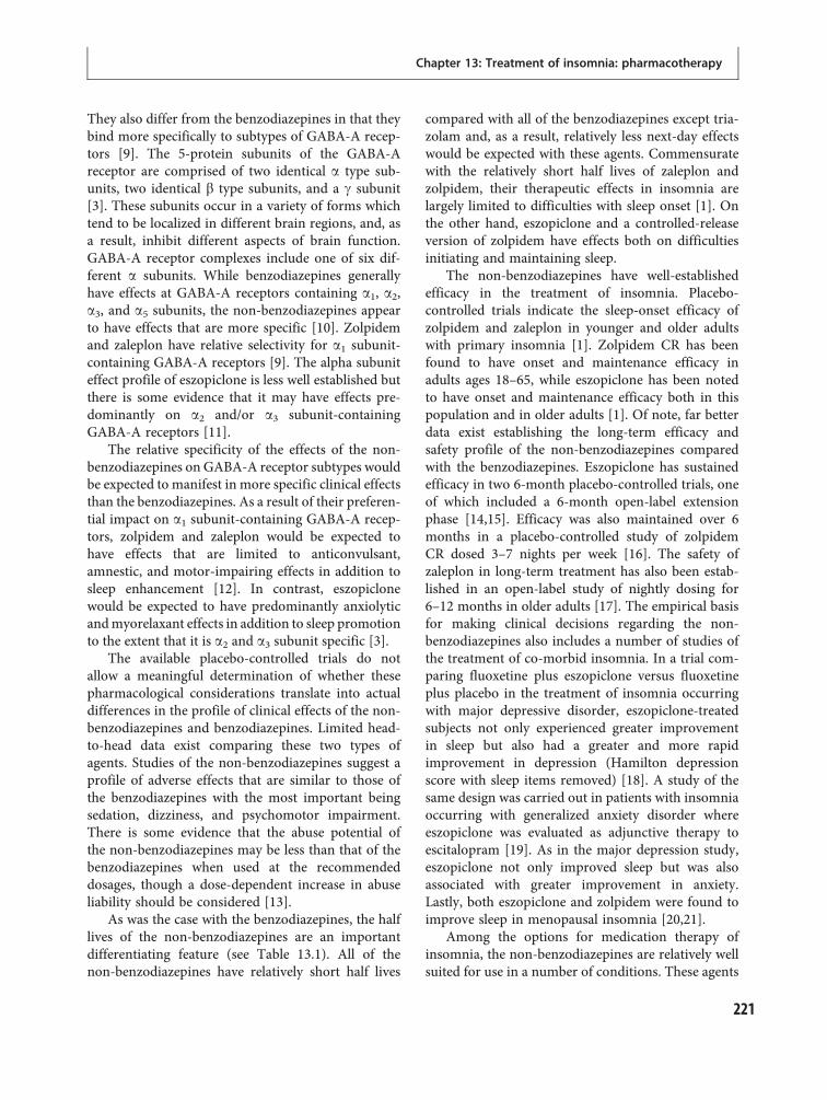
They also differ from the benzodiazepines in that theybind more specifically to subtypes of GABA-A recep-tors [9]. The 5-protein subunits of the GABA-Areceptor are comprised of two identical a type sub-units, two identical b type subunits, and a g subunit[3]. These subunits occur in a variety of forms whichtend to be localized in different brain regions, and, asa result, inhibit different aspects of brain function.GABA-A receptor complexes include one of six dif-ferent a subunits. While benzodiazepines generallyhave effects at GABA-A receptors containing a1, a2,a3, and a5 subunits, the non-benzodiazepines appearto have effects that are more specific [10]. Zolpidemand zaleplon have relative selectivity for a1 subunit-containing GABA-A receptors [9]. The alpha subuniteffect profile of eszopiclone is less well established butthere is some evidence that it may have effects pre-dominantly on a2 and/or a3 subunit-containingGABA-A receptors [11].
The relative specificity of the effects of the non-benzodiazepines on GABA-A receptor subtypes wouldbe expected to manifest in more specific clinical effectsthan the benzodiazepines. As a result of their preferen-tial impact on a1 subunit-containing GABA-A recep-tors, zolpidem and zaleplon would be expected tohave effects that are limited to anticonvulsant,amnestic, and motor-impairing effects in addition tosleep enhancement [12]. In contrast, eszopiclonewould be expected to have predominantly anxiolyticandmyorelaxant effects in addition to sleep promotionto the extent that it is a2 and a3 subunit specific [3].
The available placebo-controlled trials do notallow a meaningful determination of whether thesepharmacological considerations translate into actualdifferences in the profile of clinical effects of the non-benzodiazepines and benzodiazepines. Limited head-to-head data exist comparing these two types ofagents. Studies of the non-benzodiazepines suggest aprofile of adverse effects that are similar to those ofthe benzodiazepines with the most important beingsedation, dizziness, and psychomotor impairment.There is some evidence that the abuse potential ofthe non-benzodiazepines may be less than that of thebenzodiazepines when used at the recommendeddosages, though a dose-dependent increase in abuseliability should be considered [13].
As was the case with the benzodiazepines, the halflives of the non-benzodiazepines are an importantdifferentiating feature (see Table 13.1). All of thenon-benzodiazepines have relatively short half lives
compared with all of the benzodiazepines except tria-zolam and, as a result, relatively less next-day effectswould be expected with these agents. Commensuratewith the relatively short half lives of zaleplon andzolpidem, their therapeutic effects in insomnia arelargely limited to difficulties with sleep onset [1]. Onthe other hand, eszopiclone and a controlled-releaseversion of zolpidem have effects both on difficultiesinitiating and maintaining sleep.
The non-benzodiazepines have well-establishedefficacy in the treatment of insomnia. Placebo-controlled trials indicate the sleep-onset efficacy ofzolpidem and zaleplon in younger and older adultswith primary insomnia [1]. Zolpidem CR has beenfound to have onset and maintenance efficacy inadults ages 18–65, while eszopiclone has been notedto have onset and maintenance efficacy both in thispopulation and in older adults [1]. Of note, far betterdata exist establishing the long-term efficacy andsafety profile of the non-benzodiazepines comparedwith the benzodiazepines. Eszopiclone has sustainedefficacy in two 6-month placebo-controlled trials, oneof which included a 6-month open-label extensionphase [14,15]. Efficacy was also maintained over 6months in a placebo-controlled study of zolpidemCR dosed 3–7 nights per week [16]. The safety ofzaleplon in long-term treatment has also been estab-lished in an open-label study of nightly dosing for6–12 months in older adults [17]. The empirical basisfor making clinical decisions regarding the non-benzodiazepines also includes a number of studies ofthe treatment of co-morbid insomnia. In a trial com-paring fluoxetine plus eszopiclone versus fluoxetineplus placebo in the treatment of insomnia occurringwith major depressive disorder, eszopiclone-treatedsubjects not only experienced greater improvementin sleep but also had a greater and more rapidimprovement in depression (Hamilton depressionscore with sleep items removed) [18]. A study of thesame design was carried out in patients with insomniaoccurring with generalized anxiety disorder whereeszopiclone was evaluated as adjunctive therapy toescitalopram [19]. As in the major depression study,eszopiclone not only improved sleep but was alsoassociated with greater improvement in anxiety.Lastly, both eszopiclone and zolpidem were found toimprove sleep in menopausal insomnia [20,21].
Among the options for medication therapy ofinsomnia, the non-benzodiazepines are relatively wellsuited for use in a number of conditions. These agents
Chapter 13: Treatment of insomnia: pharmacotherapy
221

have a strong empirical base establishing their efficacyin insomnia therapy. Zaleplon and zolpidem areamong the shorter half-life agents available whichmake them well suited for the treatment of sleep-onset difficulties. Eszopiclone and zolpidem CR arethe only agents other than benzodiazepines that areapproved by the US Food and Drug Adminstration(FDA) for the treatment of difficulties with both sleeponset and maintenance. Because of their relativelylonger half lives, most of the benzodiazepines thatcan be used to treat sleep maintenance difficultiesare likely to be associated with more next-day effects.The non-benzodiazepines may be associated with lessabuse liability than the benzodiazepines; however,these agents should also be used with caution inabuse-prone individuals. Eszopiclone has better estab-lished long-term efficacy and safety with nightlydosing than any other agent, while the utility of zol-pidem CR for long-term intermittent dosing has alsobeen demonstrated. Lastly, controlled trials supportthe use of eszopiclone as adjunctive therapy forinsomnia occurring with major depressive disorderand generalized anxiety disorder while studies indi-cate the utility of eszopiclone and zolpidem in thetreatment of menopausal insomnia.
Melatonin receptor agonistsAvailable melatonin receptor agonists include mela-tonin and ramelteon. These agents are believed toexert their therapeutic effects on insomnia primarilythrough binding to MT1 and MT2 melatonin recep-tors. Melatonin is a hormone produced by the pinealgland that is obtainable as an “over-the-counter”medication. Ramelteon is an agent that is approvedby the US FDA for the treatment of insomnia.
Melatonin has circadian rhythm modifying effectsas well as a small but consistent effect on sleep-onsetlatency. The effect on sleep latency has been demon-strated in a wide range of patient populations includ-ing normal sleepers, children and adults withinsomnia, patients with Alzheimer’s disease, thosewith insomnia co-morbid with medical disorders,those with mood disorders, patients with schizophre-nia, children with attention-deficit disorder, and chil-dren with neurodevelopmental disorders [22–36]. Thegreat variation in the methods employed in these stud-ies limits their application to clinical practice [37,38].Most problematic in this regard is that they evaluateddosages of melatonin varying from 0.1 to 75 mg
(2–6 mg in most studies) which were administeredfrom 30 minutes to 3 hours before bedtime.
A factor complicating the determination of thetime of optimal dose and timing of dosing of mela-tonin is that, unlike benzodiazepines and non-benzo-diazepines, the sleep-enhancing effects of melatoninagonists are not tightly linked to the serum bloodlevel. A dose–response relationship does not existfor melatonin or ramelteon and a number of studiessuggest that the time lag from dosing of melatonin towhen its effects on sleep become evident depends onthe time of day of administration [39–41]. Despite thefact that the peak blood level of melatonin occursaround 30 minutes after ingestion, some studies haveidentified that the sleep effects may not occur until upto 3 hours after dosing.
Ramelteon also has a therapeutic effect on sleep-onset latency. Despite the lack of a clear relationshipbetween serum level and effects on sleep, the availablestudies have focused on a dosage of 8 mg administered30 minutes prior to lights out [1]. Studies in bothadults 18–65 years of age and older adults with insom-nia indicate a consistent therapeutic effect on sleep-onset latency which is more consistent when assessedwith measures derived from the overnight sleep study(polysomnogram) than with self-report measures.
Both melatonin and ramelteon have relativelyfavorable profiles of adverse effects. Headache is themost frequent side-effect of melatonin, and sedationand slowing of reaction time have been observed whenmelatonin has been taken during the day [39,42,43].There is some evidence that melatonin may affectfertility in both men and women [44–47]. As a result,it has been suggested that melatonin should not beused by individuals attempting to conceive. The mostcommon side-effects associated with ramelteon aresomnolence, dizziness, nausea, and fatigue [48,49].There is evidence that ramelteon can be safely used inthose with sleep apnea and mild to moderate chronicobstructive pulmonary disease [50,51]. Neither mela-tonin nor ramelteon appear to have significant abusepotential and would therefore be well suited for use inindividuals with insomnia who have a history of sub-stance abuse; however, neither agent has been studiedin a substance-abuse prone population.
AntidepressantsAntidepressants are frequently used to treat insomniain clinical practice, though relatively little data exist
Section V: Insomnia in Psychiatric Contexts
222
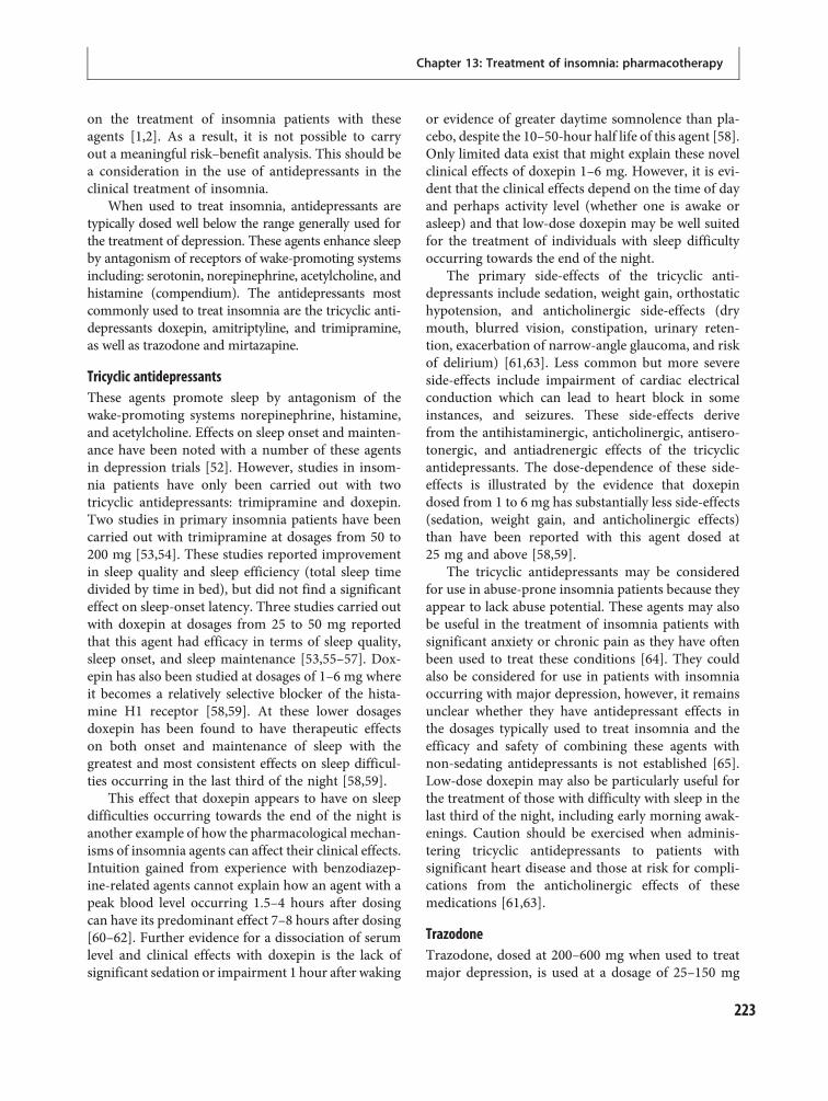
on the treatment of insomnia patients with theseagents [1,2]. As a result, it is not possible to carryout a meaningful risk–benefit analysis. This should bea consideration in the use of antidepressants in theclinical treatment of insomnia.
When used to treat insomnia, antidepressants aretypically dosed well below the range generally used forthe treatment of depression. These agents enhance sleepby antagonism of receptors of wake-promoting systemsincluding: serotonin, norepinephrine, acetylcholine, andhistamine (compendium). The antidepressants mostcommonly used to treat insomnia are the tricyclic anti-depressants doxepin, amitriptyline, and trimipramine,as well as trazodone and mirtazapine.
Tricyclic antidepressantsThese agents promote sleep by antagonism of thewake-promoting systems norepinephrine, histamine,and acetylcholine. Effects on sleep onset and mainten-ance have been noted with a number of these agentsin depression trials [52]. However, studies in insom-nia patients have only been carried out with twotricyclic antidepressants: trimipramine and doxepin.Two studies in primary insomnia patients have beencarried out with trimipramine at dosages from 50 to200 mg [53,54]. These studies reported improvementin sleep quality and sleep efficiency (total sleep timedivided by time in bed), but did not find a significanteffect on sleep-onset latency. Three studies carried outwith doxepin at dosages from 25 to 50 mg reportedthat this agent had efficacy in terms of sleep quality,sleep onset, and sleep maintenance [53,55–57]. Dox-epin has also been studied at dosages of 1–6 mg whereit becomes a relatively selective blocker of the hista-mine H1 receptor [58,59]. At these lower dosagesdoxepin has been found to have therapeutic effectson both onset and maintenance of sleep with thegreatest and most consistent effects on sleep difficul-ties occurring in the last third of the night [58,59].
This effect that doxepin appears to have on sleepdifficulties occurring towards the end of the night isanother example of how the pharmacological mechan-isms of insomnia agents can affect their clinical effects.Intuition gained from experience with benzodiazep-ine-related agents cannot explain how an agent with apeak blood level occurring 1.5–4 hours after dosingcan have its predominant effect 7–8 hours after dosing[60–62]. Further evidence for a dissociation of serumlevel and clinical effects with doxepin is the lack ofsignificant sedation or impairment 1 hour after waking
or evidence of greater daytime somnolence than pla-cebo, despite the 10–50-hour half life of this agent [58].Only limited data exist that might explain these novelclinical effects of doxepin 1–6 mg. However, it is evi-dent that the clinical effects depend on the time of dayand perhaps activity level (whether one is awake orasleep) and that low-dose doxepin may be well suitedfor the treatment of individuals with sleep difficultyoccurring towards the end of the night.
The primary side-effects of the tricyclic anti-depressants include sedation, weight gain, orthostatichypotension, and anticholinergic side-effects (drymouth, blurred vision, constipation, urinary reten-tion, exacerbation of narrow-angle glaucoma, and riskof delirium) [61,63]. Less common but more severeside-effects include impairment of cardiac electricalconduction which can lead to heart block in someinstances, and seizures. These side-effects derivefrom the antihistaminergic, anticholinergic, antisero-tonergic, and antiadrenergic effects of the tricyclicantidepressants. The dose-dependence of these side-effects is illustrated by the evidence that doxepindosed from 1 to 6 mg has substantially less side-effects(sedation, weight gain, and anticholinergic effects)than have been reported with this agent dosed at25 mg and above [58,59].
The tricyclic antidepressants may be consideredfor use in abuse-prone insomnia patients because theyappear to lack abuse potential. These agents may alsobe useful in the treatment of insomnia patients withsignificant anxiety or chronic pain as they have oftenbeen used to treat these conditions [64]. They couldalso be considered for use in patients with insomniaoccurring with major depression, however, it remainsunclear whether they have antidepressant effects inthe dosages typically used to treat insomnia and theefficacy and safety of combining these agents withnon-sedating antidepressants is not established [65].Low-dose doxepin may also be particularly useful forthe treatment of those with difficulty with sleep in thelast third of the night, including early morning awak-enings. Caution should be exercised when adminis-tering tricyclic antidepressants to patients withsignificant heart disease and those at risk for compli-cations from the anticholinergic effects of thesemedications [61,63].
TrazodoneTrazodone, dosed at 200–600 mg when used to treatmajor depression, is used at a dosage of 25–150 mg
Chapter 13: Treatment of insomnia: pharmacotherapy
223

“off-label” for treating insomnia [1]. In fact, it hasbeen among the most frequently administered treat-ments for insomnia over the last 20 years [2]. Trazo-done’s sleep-enhancing effects are believed to derivefrom antagonism of several wake-promoting systemsincluding serotonin (5HT2 receptors), norepineph-rine (a1 receptors), and histamine (H1 receptors).A factor which complicates the use of trazodone isthat it is metabolized into methyl-chlorophenylpipera-zine (mCPP) which has a wake-promoting effect andsomewhat unpredictably (because of the highly vari-able metabolism of trazodone and mCPP in the popu-lation) may undermine the therapeutic effects oftrazodone [66,67].
Despite the frequency with which trazodone isused in the treatment of insomnia, there is remark-ably scant evidence that it is safe and effective whenused for this purpose. Only one placebo-controlledstudy of the treatment of insomnia patients has beencarried out with trazodone [68]. In this 2-week study,patients treated with 50 mg had significantly betterself-reported sleep than placebo-treated subjects inthe first but not second week of treatment. Evidenceof the sleep-promoting effect of trazodone has beennoted in two small placebo-controlled studies, onecarried out in abstinent alcoholics and another inpatients with depression treated with either fluoxetineor bupropion [69,70].
The most common adverse effects seen with tra-zodone treatment are sedation, dizziness, headache,dry mouth, blurred vision, and orthostatic hypoten-sion [1,68]. A rare side-effect of trazodone in men ispriapism, a prolonged erection associated with pain,which may result in irreversible impotence [71].
Like the tricyclic antidepressants, trazodoneappears to be without significant abuse potential and,as a result, can be considered for use in abuse-proneinsomnia patients. The small placebo-controlled trialcarried out in abstinent alcoholics provides some sup-port for this use [69]. While trazodone probably lacksantidepressant efficacy in the dose range generallyused to treat insomnia, this has not been systematicallyevaluated. Preliminary evidence suggests that it can besafely used to treat insomnia in patients taking fluox-etine and bupropion but its use with other antidepres-sants remains unknown [70].
MirtazapineThe effects of mirtazapine are believed to derive fromantagonism of serotonergic (5HT2 and 5HT3),
adrenergic (a1 and a2), and histaminergic (H1)receptors [72]. All of these pharmacological effectsenhance sleep except for the antagonism of the a2receptor, which is a presynaptic receptor that inhibitsthe release of norepinephrine [73]. It is believed that,as a result of this a2 antagonism, the sleep-enhancingeffects of mirtazapine may diminish with increasingdosage. For this reason, dosages of 30 mg and lessare generally used to treat insomnia, while the rangeof antidepressant dosing is 7.5–45 mg. No placebo-controlled trials of the use of mirtazapine for thetreatment of insomnia patients have been carriedout. Data indicating that mirtazapine has sleep-enhancing effects derive from a double-blind studyevaluating mirtazapine versus fluoxetine carried outin depressed patients and open-label studies in agroup of healthy volunteers without sleep complaintsand a group of depressed patients [74–76]. The mostcommon side-effects associated with mirtazapine aresedation, increased appetite, weight gain, dry mouth,and constipation [73]. Like the other antidepressantsdiscussed, mirtazapine does not appear to be associ-ated with abuse potential and, therefore, could beconsidered for use in substance abuse-prone patientswith insomnia. Because the dosages used to treatinsomnia and depression with mirtazapine overlap,this agent can be considered for single-agent therapyin depressed patients with insomnia. Future studieswill be needed to determine the effectiveness of thisapproach compared to combining a non-sedatingantidepressant with an established insomnia therapy.
AntipsychoticsLike the antidepressants, antipsychotic agents aresometimes used “off-label” for the treatment ofinsomnia [2]. These agents enhance sleep throughantagonism of the following wake-promotingsystems: dopamine, histamine (H1 receptors), sero-tonin (5HT2 receptors), acetylcholine (muscarinicreceptors), and norepinephrine (a1 receptors) [1].When used to treat insomnia, antipsychotic agentsare typically administered at a lower dosage than usedfor the treatment of thought disorders and mooddisorders. The agents most commonly used to treatinsomnia, quetiapine and olanzapine, are dosed at25–250 mg and 2.5–20 mg, respectively [2,77].
No placebo-controlled trials of the treatment ofinsomnia patients have been carried out with any ofthe antipsychotic medications. This is a factor which
Section V: Insomnia in Psychiatric Contexts
224

limits the ability to make risk–benefit assessmentswith these agents. Quetiapine dosed at from 25 to75 mg has been found to have a sleep-enhancing effectin a population of primary insomnia patients and inhealthy volunteers in open-label studies [78]. Olanza-pine has been noted to enhance sleep in an open-labelstudy of healthy volunteers [77]. Both of these agentshave also been reported to enhance sleep in a numberof studies in patients with thought disorders andmood disorders [77,79,80]. The pharmacokinetics ofolanzapine (tmax of 4–6 hours) make it better suitedfor the treatment of sleep maintenance problems thansleep-onset problems, while quetiapine’s pharmaco-kinetics suggest that it has the potential to be helpfulfor both problems, staying asleep and falling asleep(tmax of 1–2 hours; half life of 7 hours) [1].
The most common side-effects of the antipsych-otic agents used to treat insomnia are: sedation, dizzi-ness, anticholinergic side-effects (dry mouth, blurredvision, constipation, urinary retention), and increasedappetite [77]. The frequency of these side-effectsvaries among the antipsychotic agents. Antipsychoticagents also have other potential side-effects. Amongthese are extrapyramidal side-effects that occur as aresult of dopamine antagonism (parkinsonism, acutedystonic reactions, akathisia, and tardive dyskinesia)[78]. These extrapyramidal side-effects appear tooccur rarely with the atypical antipsychotics as com-pared to typical antipsychotics. Olanzapine has beenassociated with an increased risk of insulin resistance,impaired cognition, and mortality in dementiapatients, and all antipsychotics should be used withcaution in older adults because of a potentialincreased risk of cardiac-related mortality [81].
Antipsychotic agents are most appropriate for thetreatment of patients who have insomnia occurring inthe setting of psychosis or a mood disorder. In add-ition to having FDA indications for the treatment ofpsychosis, a number of these agents are approved forthe treatment of mania and quetiapine is approved forthe treatment of bipolar depression. Antipsychoticagents can also be considered for use in substanceabuse-prone patients with insomnia as these agentsdo not have significant abuse potential.
AntihistaminesMany agents are significant antagonists of H1 hista-mine receptors and could therefore be referred to asantihistamines. This includes the antidepressants
doxepin and mirtazapine, which are among the morepotent and selective H1 antagonists, and the antipsych-otics quetiapine and olanzapine [1]. However, the term“antihistamine” is generally reserved for agents thatwere developed primarily for the treatment of allergies.Among these agents, diphenhydramine and doxyla-mine are used the most commonly for the treatmentof insomnia and are important constituents of manyinsomnia treatments available “over-the-counter.”These two medications have similar properties. Theusual dosage of both agents for the treatment of insom-nia is 25–50 mg and both have significant muscariniccholinergic antagonist effects.
Several placebo-controlled trials in insomniapatients have been carried out with diphenhydramine,though no such studies have evaluated the efficacyand safety of doxylamine. Diphenhydramine has beenobserved to improve sleep in trials in the followingpopulations: primary insomnia, psychiatric patients,primary care patients, and nursing home residents[82–86]. In these studies diphenhydramine appearedto have a stronger effect on maintenance than sleeponset. This is consistent with the findings observedwith low-dose doxepin, another potent H1 antagonist,suggesting that a greater effect on sleep maintenancemay be a characteristic of agents acting via blockingthe wake-promoting effects of histamine. One studywhere diphenhydramine was administered during theday found that the therapeutic effect was lost by the4th day of treatment [87]. Further studies will beneeded to determine if tolerance occurs to night-timedosing of this medication. The only study of doxyla-mine was a large double-blind trial carried out inpost-operative patients where a significant effect onsubjective measures of sleep was observed [88].
The predominant adverse effects associated withboth diphenhydramine and doxylamine are: anticholin-ergic effects (dry mouth, blurred vision, constipation,urinary retention, risk of delirium), sedation, dizzi-ness, and weight gain. Infrequent side-effects ofdiphenhydramine are agitation and insomnia whiledoxylamine has been associated with coma and rhab-domyolysis in case reports [89].
Neither of these medications has significant abusepotential and, therefore, may be considered for use inabuse-prone insomnia patients. Both of these agentsare particularly well suited for use in the treatment ofinsomnia occurring with allergies or significant nasal/sinus congestion. These agents should be avoided inthose at risk for complications due to anticholinergic
Chapter 13: Treatment of insomnia: pharmacotherapy
225
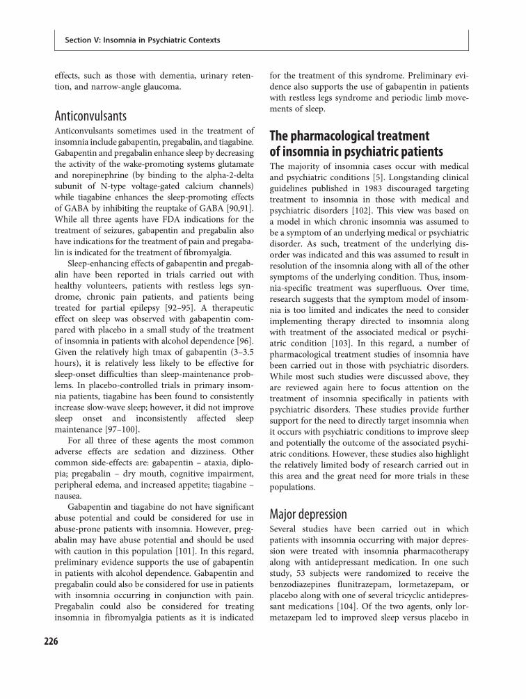
effects, such as those with dementia, urinary reten-tion, and narrow-angle glaucoma.
AnticonvulsantsAnticonvulsants sometimes used in the treatment ofinsomnia include gabapentin, pregabalin, and tiagabine.Gabapentin and pregabalin enhance sleep by decreasingthe activity of the wake-promoting systems glutamateand norepinephrine (by binding to the alpha-2-deltasubunit of N-type voltage-gated calcium channels)while tiagabine enhances the sleep-promoting effectsof GABA by inhibiting the reuptake of GABA [90,91].While all three agents have FDA indications for thetreatment of seizures, gabapentin and pregabalin alsohave indications for the treatment of pain and pregaba-lin is indicated for the treatment of fibromyalgia.
Sleep-enhancing effects of gabapentin and pregab-alin have been reported in trials carried out withhealthy volunteers, patients with restless legs syn-drome, chronic pain patients, and patients beingtreated for partial epilepsy [92–95]. A therapeuticeffect on sleep was observed with gabapentin com-pared with placebo in a small study of the treatmentof insomnia in patients with alcohol dependence [96].Given the relatively high tmax of gabapentin (3–3.5hours), it is relatively less likely to be effective forsleep-onset difficulties than sleep-maintenance prob-lems. In placebo-controlled trials in primary insom-nia patients, tiagabine has been found to consistentlyincrease slow-wave sleep; however, it did not improvesleep onset and inconsistently affected sleepmaintenance [97–100].
For all three of these agents the most commonadverse effects are sedation and dizziness. Othercommon side-effects are: gabapentin – ataxia, diplo-pia; pregabalin – dry mouth, cognitive impairment,peripheral edema, and increased appetite; tiagabine –nausea.
Gabapentin and tiagabine do not have significantabuse potential and could be considered for use inabuse-prone patients with insomnia. However, preg-abalin may have abuse potential and should be usedwith caution in this population [101]. In this regard,preliminary evidence supports the use of gabapentinin patients with alcohol dependence. Gabapentin andpregabalin could also be considered for use in patientswith insomnia occurring in conjunction with pain.Pregabalin could also be considered for treatinginsomnia in fibromyalgia patients as it is indicated
for the treatment of this syndrome. Preliminary evi-dence also supports the use of gabapentin in patientswith restless legs syndrome and periodic limb move-ments of sleep.
The pharmacological treatmentof insomnia in psychiatric patientsThe majority of insomnia cases occur with medicaland psychiatric conditions [5]. Longstanding clinicalguidelines published in 1983 discouraged targetingtreatment to insomnia in those with medical andpsychiatric disorders [102]. This view was based ona model in which chronic insomnia was assumed tobe a symptom of an underlying medical or psychiatricdisorder. As such, treatment of the underlying dis-order was indicated and this was assumed to result inresolution of the insomnia along with all of the othersymptoms of the underlying condition. Thus, insom-nia-specific treatment was superfluous. Over time,research suggests that the symptom model of insom-nia is too limited and indicates the need to considerimplementing therapy directed to insomnia alongwith treatment of the associated medical or psychi-atric condition [103]. In this regard, a number ofpharmacological treatment studies of insomnia havebeen carried out in those with psychiatric disorders.While most such studies were discussed above, theyare reviewed again here to focus attention on thetreatment of insomnia specifically in patients withpsychiatric disorders. These studies provide furthersupport for the need to directly target insomnia whenit occurs with psychiatric conditions to improve sleepand potentially the outcome of the associated psychi-atric conditions. However, these studies also highlightthe relatively limited body of research carried out inthis area and the great need for more trials in thesepopulations.
Major depressionSeveral studies have been carried out in whichpatients with insomnia occurring with major depres-sion were treated with insomnia pharmacotherapyalong with antidepressant medication. In one suchstudy, 53 subjects were randomized to receive thebenzodiazepines flunitrazepam, lormetazepam, orplacebo along with one of several tricyclic antidepres-sant medications [104]. Of the two agents, only lor-metazepam led to improved sleep versus placebo in
Section V: Insomnia in Psychiatric Contexts
226
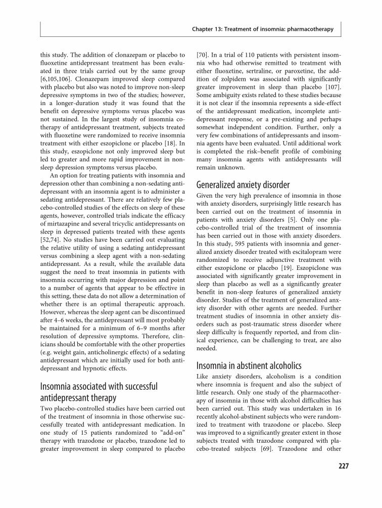
this study. The addition of clonazepam or placebo tofluoxetine antidepressant treatment has been evalu-ated in three trials carried out by the same group[6,105,106]. Clonazepam improved sleep comparedwith placebo but also was noted to improve non-sleepdepressive symptoms in two of the studies; however,in a longer-duration study it was found that thebenefit on depressive symptoms versus placebo wasnot sustained. In the largest study of insomnia co-therapy of antidepressant treatment, subjects treatedwith fluoxetine were randomized to receive insomniatreatment with either eszopiclone or placebo [18]. Inthis study, eszopiclone not only improved sleep butled to greater and more rapid improvement in non-sleep depression symptoms versus placebo.
An option for treating patients with insomnia anddepression other than combining a non-sedating anti-depressant with an insomnia agent is to administer asedating antidepressant. There are relatively few pla-cebo-controlled studies of the effects on sleep of theseagents, however, controlled trials indicate the efficacyof mirtazapine and several tricyclic antidepressants onsleep in depressed patients treated with these agents[52,74]. No studies have been carried out evaluatingthe relative utility of using a sedating antidepressantversus combining a sleep agent with a non-sedatingantidepressant. As a result, while the available datasuggest the need to treat insomnia in patients withinsomnia occurring with major depression and pointto a number of agents that appear to be effective inthis setting, these data do not allow a determination ofwhether there is an optimal therapeutic approach.However, whereas the sleep agent can be discontinuedafter 4–6 weeks, the antidepressant will most probablybe maintained for a minimum of 6–9 months afterresolution of depressive symptoms. Therefore, clin-icians should be comfortable with the other properties(e.g. weight gain, anticholinergic effects) of a sedatingantidepressant which are initially used for both anti-depressant and hypnotic effects.
Insomnia associated with successfulantidepressant therapyTwo placebo-controlled studies have been carried outof the treatment of insomnia in those otherwise suc-cessfully treated with antidepressant medication. Inone study of 15 patients randomized to “add-on”therapy with trazodone or placebo, trazodone led togreater improvement in sleep compared to placebo
[70]. In a trial of 110 patients with persistent insom-nia who had otherwise remitted to treatment witheither fluoxetine, sertraline, or paroxetine, the add-ition of zolpidem was associated with significantlygreater improvement in sleep than placebo [107].Some ambiguity exists related to these studies becauseit is not clear if the insomnia represents a side-effectof the antidepressant medication, incomplete anti-depressant response, or a pre-existing and perhapssomewhat independent condition. Further, only avery few combinations of antidepressants and insom-nia agents have been evaluated. Until additional workis completed the risk–benefit profile of combiningmany insomnia agents with antidepressants willremain unknown.
Generalized anxiety disorderGiven the very high prevalence of insomnia in thosewith anxiety disorders, surprisingly little research hasbeen carried out on the treatment of insomnia inpatients with anxiety disorders [5]. Only one pla-cebo-controlled trial of the treatment of insomniahas been carried out in those with anxiety disorders.In this study, 595 patients with insomnia and gener-alized anxiety disorder treated with escitalopram wererandomized to receive adjunctive treatment witheither eszopiclone or placebo [19]. Eszopiclone wasassociated with significantly greater improvement insleep than placebo as well as a significantly greaterbenefit in non-sleep features of generalized anxietydisorder. Studies of the treatment of generalized anx-iety disorder with other agents are needed. Furthertreatment studies of insomnia in other anxiety dis-orders such as post-traumatic stress disorder wheresleep difficulty is frequently reported, and from clin-ical experience, can be challenging to treat, are alsoneeded.
Insomnia in abstinent alcoholicsLike anxiety disorders, alcoholism is a conditionwhere insomnia is frequent and also the subject oflittle research. Only one study of the pharmacother-apy of insomnia in those with alcohol difficulties hasbeen carried out. This study was undertaken in 16recently alcohol-abstinent subjects who were random-ized to treatment with trazodone or placebo. Sleepwas improved to a significantly greater extent in thosesubjects treated with trazodone compared with pla-cebo-treated subjects [69]. Trazodone and other
Chapter 13: Treatment of insomnia: pharmacotherapy
227
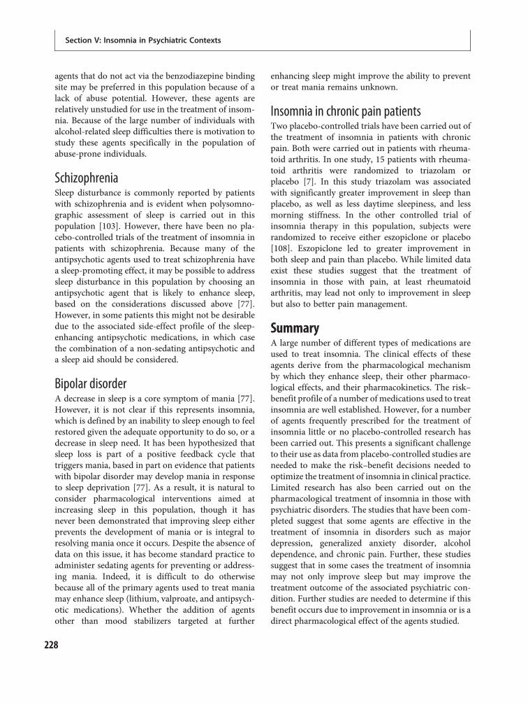
agents that do not act via the benzodiazepine bindingsite may be preferred in this population because of alack of abuse potential. However, these agents arerelatively unstudied for use in the treatment of insom-nia. Because of the large number of individuals withalcohol-related sleep difficulties there is motivation tostudy these agents specifically in the population ofabuse-prone individuals.
SchizophreniaSleep disturbance is commonly reported by patientswith schizophrenia and is evident when polysomno-graphic assessment of sleep is carried out in thispopulation [103]. However, there have been no pla-cebo-controlled trials of the treatment of insomnia inpatients with schizophrenia. Because many of theantipsychotic agents used to treat schizophrenia havea sleep-promoting effect, it may be possible to addresssleep disturbance in this population by choosing anantipsychotic agent that is likely to enhance sleep,based on the considerations discussed above [77].However, in some patients this might not be desirabledue to the associated side-effect profile of the sleep-enhancing antipsychotic medications, in which casethe combination of a non-sedating antipsychotic anda sleep aid should be considered.
Bipolar disorderA decrease in sleep is a core symptom of mania [77].However, it is not clear if this represents insomnia,which is defined by an inability to sleep enough to feelrestored given the adequate opportunity to do so, or adecrease in sleep need. It has been hypothesized thatsleep loss is part of a positive feedback cycle thattriggers mania, based in part on evidence that patientswith bipolar disorder may develop mania in responseto sleep deprivation [77]. As a result, it is natural toconsider pharmacological interventions aimed atincreasing sleep in this population, though it hasnever been demonstrated that improving sleep eitherprevents the development of mania or is integral toresolving mania once it occurs. Despite the absence ofdata on this issue, it has become standard practice toadminister sedating agents for preventing or address-ing mania. Indeed, it is difficult to do otherwisebecause all of the primary agents used to treat maniamay enhance sleep (lithium, valproate, and antipsych-otic medications). Whether the addition of agentsother than mood stabilizers targeted at further
enhancing sleep might improve the ability to preventor treat mania remains unknown.
Insomnia in chronic pain patientsTwo placebo-controlled trials have been carried out ofthe treatment of insomnia in patients with chronicpain. Both were carried out in patients with rheuma-toid arthritis. In one study, 15 patients with rheuma-toid arthritis were randomized to triazolam orplacebo [7]. In this study triazolam was associatedwith significantly greater improvement in sleep thanplacebo, as well as less daytime sleepiness, and lessmorning stiffness. In the other controlled trial ofinsomnia therapy in this population, subjects wererandomized to receive either eszopiclone or placebo[108]. Eszopiclone led to greater improvement inboth sleep and pain than placebo. While limited dataexist these studies suggest that the treatment ofinsomnia in those with pain, at least rheumatoidarthritis, may lead not only to improvement in sleepbut also to better pain management.
SummaryA large number of different types of medications areused to treat insomnia. The clinical effects of theseagents derive from the pharmacological mechanismby which they enhance sleep, their other pharmaco-logical effects, and their pharmacokinetics. The risk–benefit profile of a number ofmedications used to treatinsomnia are well established. However, for a numberof agents frequently prescribed for the treatment ofinsomnia little or no placebo-controlled research hasbeen carried out. This presents a significant challengeto their use as data from placebo-controlled studies areneeded to make the risk–benefit decisions needed tooptimize the treatment of insomnia in clinical practice.Limited research has also been carried out on thepharmacological treatment of insomnia in those withpsychiatric disorders. The studies that have been com-pleted suggest that some agents are effective in thetreatment of insomnia in disorders such as majordepression, generalized anxiety disorder, alcoholdependence, and chronic pain. Further, these studiessuggest that in some cases the treatment of insomniamay not only improve sleep but may improve thetreatment outcome of the associated psychiatric con-dition. Further studies are needed to determine if thisbenefit occurs due to improvement in insomnia or is adirect pharmacological effect of the agents studied.
Section V: Insomnia in Psychiatric Contexts
228

References1. Krystal A. A compendium of placebo controlled trials
of the risks/benefits of pharmacologic treatments forinsomnia: The empirical basis for clinical practice.Sleep Med Rev. 2009;13(4):265 274.
2. Walsh J K. Drugs used to treat insomnia in 2002:regulatory based rather than evidence based medicine.Sleep. 2004;27(8):14441 14442.
3. Sieghart W, Sperk G. Subunit composition,distribution and function of GABA(A) receptorsubtypes. Curr Top Med Chem. 2002;2:795 816.
4. Saper C, Scammell TE, Lu J. Hypothalamic regulationof sleep and circadian rhythms. Nature. 2005;437(7063):1257 1263.
5. Ford D, Kamerow D. Epidemiologic study of sleepdisturbances in psychiatric disorders. JAMA.1989;262:1479 1484.
6. Londborg P, Smith WT, Glaudin V, Painter J R. Shortterm cotherapy with clonazepam and fluoxetine:anxiety, sleep disturbance and core symptoms ofdepression. J Affect Disord. 2000;61:73 79.
7. Walsh J, Muehlbach MJ, Lauter SA, Hilliker NA,Schweitzer P K. Effects of triazolam on sleep, daytimesleepiness, and morning stiffness in patients withrheumatoid arthritis. J Rheumatol. 1996;23(2):245 252.
8. Roehrs T, Pedrosi B, Rosenthal L, et al. Hypnotic selfadministration and dose escalation.Psychopharmacology (Berl). 1996;127:150 154.
9. Sanna E, Busonero F, Talani G, et al. Comparison ofthe effects of zaleplon, zolpidem, and triazolam atvarious GABA(A) receptor subtypes. Eur J Pharmacol.2002;451(2):103 110.
10. Mohler H, Fritschy JM, Rudolph U. A newbenzodiazepine pharmacology. J Pharmacol Exp Ther.2002;300:2 8.
11. Jia F, Goldstein PA, Harrison N L. The modulation ofsynaptic GABA(A) receptors in the thalamus byeszopiclone and zolpidem. J Pharmacol Exp Ther.2009;328(3):1000 1006.
12. Crestani F, Assandri R, Tauber M, et al. Contributionof the alpha1 GABA(A) receptor subtype to thepharmacological actions of benzodiazepine site inverseagonists. Neuropharmacology. 2002;43:679 684.
13. Griffiths RR, Johnson MW. Relative abuse liability ofhypnotic drugs: a conceptual framework and algorithmfor differentiating among compounds. J Clin Psychol.2005;66(Suppl 9):31 41.
14. Krystal AD, Walsh JK, Laska E, et al. Sustained efficacyof eszopiclone over six months of nightly treatment:Results of a randomized, double blind, placebo
controlled study in adults with chronic insomnia.Sleep. 2003;26:793 799.
15. Walsh J, Krystal AD, Amato DA. Nightly treatment ofprimary insomnia with eszopiclone for six months:Effect on sleep, quality of life and work limitations.Sleep. 2007;30(8):959 968.
16. Krystal AD, Erman M, Zammit GK, et al. Long termefficacy and safety of zolpidem extended release12.5 mg, administered 3 to 7 nights per week for 24weeks, in patients with chronic primary insomnia: a6 month, randomized, double blind, placebocontrolled, parallel group, multicenter study. Sleep.2008;31(1):79 90.
17. Ancoli Israel S, Richardson GS, Mangano RM, et al.Long term use of sedative hypnotics in older patientswith insomnia. Sleep Med. 2005;6(2):107 113.
18. Fava M, McCall WV, Krystal A, et al. Eszopicloneco administered with fluoxetine in patients withinsomnia co existing with major depressive disorder.Biol Psychiatry. 2006;59:1052 1060.
19. Pollack M, Kinrys G, Krystal A, et al. Eszopicloneco administered with escitalopram in patients withinsomnia and comorbid generalized anxiety disorder.Arch Gen Psychiatry. 2008;65(5):551 562.
20. Soares C, Rubens R, Caron J, et al. Eszopiclonetreatment during menopausal transition: Sleep effects,impact on menopausal symptoms, and mood. Sleep.2006;29:A239.
21. Dorsey C, Lee KA, Scharf MB. Effect of zolpidem onsleep in women with perimenopausal andpostmenopausal insomnia: a 4 week, randomized,multicenter, double blind, placebo controlled study.Clin Ther. 2004;26(10):1578 1586.
22. Hughes R, Sack RL, Lewy AJ. The role of melatoninand circadian phase in age related sleep maintenanceinsomnia: assessment in a clinical trial of melatoninreplacement. Sleep. 1998;21:52 68.
23. Singer C, Tractenberg RE, Kaye J, et al. A multicenter,placebo controlled trial of melatonin for sleepdisturbance in Alzheimer’s disease. Sleep. 2003;26(7):893 901.
24. Smits M, Nagtegaal EE, van der Heijden J, CoenenAM, Kerkhof GA. Melatonin for chronic sleep onsetinsomnia in children: a randomized placebo controlledtrial. J Child Neurol. 2001;16(2):86 92.
25. Zhdanova I, Wurtman RJ, Morabito C, Piotrovska VR,Lynch HJ. Effects of low oral doses of melatonin, given2 4 hours before habitual bedtime, on sleep in normalyoung humans. Sleep. 1996;19(5):423 431.
26. Zhdanova I, Wurtman RJ, Wagstaff J. Effects of a lowdose of melatonin on sleep in children with Angelmansyndrome. J Pediatr Endocrinol. 1999;12(1):57 67.
Chapter 13: Treatment of insomnia: pharmacotherapy
229
27. Zhdanova IV, Wurtman RJ, Regan MM, et al.Melatonin treatment for age related insomnia. J ClinEndocrinol Metab. 2001;86:4727 4730.
28. Waldhauser F, Saletu B, Trinchard Lugan I. Sleeplaboratory investigations on hypnotic properties ofmelatonin. Psychopharmacology. 1990;100(2):222 226.
29. Dalton E, Rotondi D, Levitan RD, Kennedy SH, BrownGM. Use of slow release melatonin in treatmentresistant depression. J Psychiatr Neurosci. 2000;25(1):48 52.
30. Andrade C, Srihari BS, Reddy KP, Chandramma L.Melatonin in medically ill patients with insomnia: adouble blind, placebo controlled study. J Clin Psychol.2001;62(1):41 45.
31. SerfatyM,Kennell Webb S,Warner J, Blizard R,RavenP.Double blind randomised placebo controlled trial oflow dose melatonin for sleep disorders in dementia. IntJ Geriatr Psychiatry. 2002;17(12):1120 1127.
32. Suresh Kumar P, Andrade C, Bhakta SG, Singh NM.Melatonin in schizophrenic outpatients with insomnia:a double blind, placebo controlled study. J ClinPsychol. 2007;68(2):237 241.
33. Van der Heijden K, Smits MG, Van Someren EJ,Ridderinkhof KR, Gunning WB. Effect of melatoninon sleep, behavior, and cognition in ADHD andchronic sleep onset insomnia. J Am Acad Child AdolescPsychiatry. 2007;46(2):233 241.
34. Wasdell M, Jan JE, Bomben MM, et al. A randomized,placebo controlled trial of controlled release melatonintreatment of delayed sleep phase syndrome andimpaired sleep maintenance in children withneurodevelopmental disabilities. J Pineal Res. 2008;44(1):57 64.
35. Braam W, Didden R, Smits M, Curfs L. Melatonintreatment in individuals with intellectual disability andchronic insomnia: a randomized placebo controlledstudy. J Intellect Disabil Res. 2008;52(3):256 264.
36. Haimov I, Lavie P, Laudon M, et al. Melatoninreplacement therapy of elderly insomniacs. Sleep.1995;18(7):598 603.
37. Mendelson WB. Efficacy of melatonin as a hypnoticagent. J Biol Rhythms. 1997;12(6):651 656.
38. Sack R, Hughes RJ, Edgar DM, Lewy AJ. Sleeppromoting effects of melatonin: at what dose, inwhom, under what conditions, and by whatmechanisms? Sleep. 1997;20(10):908 915.
39. Krystal A. The possibility of preventing functionalimapairment due to sleep loss by pharmacologicallyenhancing sleep. Sleep. 2005;28:16 17.
40. Slotten H, Krekling S. Does melatonin have an effecton cognitive performance. Psychoneuroendocrinol.1996;21:673 680.
41. Hughes R, Badia P. Sleep promoting and hypothermiceffects of daytime melatonin administration inhumans. Sleep. 1997;20:124 131.
42. Graw P, Werth E, Krauchi K, et al. Early morningmelatonin administration impairs psychomotorvigilance. Behav Brain Res. 2001;121:167 172.
43. Dollins A, Zhdanova IV, Wurtman RJ, Lynch HJ,Deng MH. Effect of inducing nocturnal serummelatonin concentrations in daytime on sleep, mood,body temperature and performance. Proc Natl Acad SciUSA. 1994;91:1824 1828.
44. Lerchl A. Melatonin administration alters semenquality in normal men. J Androl. 2004;25(2):185 186.
45. Ianas O, Manda D, Câmpean D, Ionescu M, Soare G.Effects of melatonin and its relation to thehypothalamic hypophyseal gonadal axis. Adv Exp MedBiol. 1999;460:321 328.
46. Partonen T. Melatonin dependent infertility. MedHypotheses. 1999;52(3):269 270.
47. Pang SF, Li L, Ayre EA, et al. Neuroendocrinology ofmelatonin in reproduction: recent developments.J Chem Neuroanat. 1998;14(3 4):157 166.
48. Roth T, Seiden D, Sainati S, et al. Effects of ramelteonon patient reported sleep latency in older adults withchronic insomnia. Sleep Med. 2006;7(4):312 318.
49. Zammit G, Erman M, Wang Weigand S, et al.Evaluation of the efficacy and safety of ramelteon insubjects with chronic insomnia. J Clin Sleep Med.2007;3(5):495 504.
50. Kryger M, Roth T, Wang Weigand S, Zhang J. Theeffects of ramelteon on respiration during sleep insubjects with moderate to severe chronic obstructivepulmonary disease. Sleep Breath. 2009;13(1):79 84.
51. KrygerM,Wang Weigand S, Roth T. Safety of ramelteonin individuals with mild to moderate obstructive sleepapnea. Sleep Breath. 2007;11(3):159 164.
52. Dunleavy D, Brezinova V, Oswald I, MacLean AW,Tinker M. Changes during weeks in effects of tricyclicdrugs on the human sleep brain. Br J Psychiatry.1972;120:663 672.
53. Riemann D, Voderholzer U, Cohrs S, et al.Trimipramine in primary insomnia: results of apolysomnographic double blind controlled study.Pharmacopsychiatry. 2002;35(5):165 174.
54. Hohagen F, Montero RF, Weiss E. Treatment ofprimary insomnia with trimipramine: an alternative tobenzodiazepine hypnotics? Eur Arch Psychiatry ClinNeurosci. 1994;244(2):65 72.
55. Rodenbeck A, Cohrs S, Jordan W, Huether G, RutherE, Hajak G. The sleep improving effects of doxepin areparalleled by a normalized plasma cortisol secretion in
Section V: Insomnia in Psychiatric Contexts
230
primary insomnia. Psychopharmacology (Berl).2003;170:423 428.
56. Hajak G, Rodenbeck A, Adler L, et al. Nocturnalmelatonin secretion and sleep after doxepinadministration in chronic primary insomnia.Pharmacopsychiatry. 1996;29(5):187 192.
57. Hajak G, Rodenbeck A, Voderholzer U, et al. Doxepinin the treatment of primary insomnia: a placebocontrolled, double blind, polysomnographic study.J Clin Psychol. 2001;62(6):453 463.
58. Roth T, Rogowski R, Hull S, et al. Efficacy and safety ofdoxepin 1 mg, 3 mg, and 6 mg in adults with primaryinsomnia. Sleep. 2007;30(11):1555 1561.
59. Scharf M, Rogowski R, Hull S, et al. Efficacy and safety ofdoxepin 1 mg, 3 mg, and 6 mg in elderly patients withprimary insomnia: a randomized, double blind, placebocontrolled crossover study. J Clin Psychol.; in press.
60. Virtanen R, Scheinin M, Iisalo E. Single dosepharmacokinetics of doxepin in healthy volunteers.Acta Pharmacol Toxicol (Copenh). 1980;47(5):371 376.
61. Ziegler V, Biggs JT, Ardekani AB, Rosen SH.Contribution to the pharmacokinetics of amitriptyline.J Clin Pharmacol. 1978;18(10):462 467.
62. Rudorfer M, Potter W Z. Metabolism of tricyclicantidepressants. Cell Mol Neurobiol. 1999;19(3):373 409.
63. Richelson E, Nelson A. Antagonism by antidepressantsof neurotransmitter receptors of normal human brainin vitro. J Pharmacol Exp Ther. 1984;230(1):94 102.
64. Murphy D, Siever LJ, Insel TR. Therapeutic responsesto tricyclic antidepressants and related drugs in nonaffective disorder patient populations. Prog inNeuropsychopharm Biol Psychiatry. 1985;9(1):3 13.
65. Schweitzer I, Tuckwell V. Risk of adverse events withthe use of augmentation therapy for the treatment ofresistant depression. Drug Saf. 1998;19(6):455 464.
66. Caccia S, Ballabio M, Fanelli R, Guiso G, Zanini MG.Determination of plasma and brain concentrations oftrazodone and its metabolite, 1 mchlorophenylpiperazine, by gas liquidchromatography. J Chromatogr. 1981;5(210):311 318.
67. Greenblatt DJ, Friedman H, Burstein ES, et al.Trazodone kinetics: Effects of age, gender and obesity.Clin Pharmacol Ther. 1987;42:193 200.
68. Walsh JK, Erman M, Erwin CW, et al. Subjectivehypnotic efficacy of trazodone and zolpidem in DSMIII R primary insomnia. Hum Psychopharm.1998;13:191 198.
69. Le Bon O, Murphy JR, Staner L, et al. Double blind,placebo controlled study of the efficacy of trazodone inalcohol post withdrawal syndrome: polysomnographicand clinical evaluations. J Clin Psychopharmacol.2003;23(4):377 383.
70. Nierenberg AA, Adler LA, Peselow E, et al. Trazodonefor antidepressant associated insomnia. AmJ Psychiatry. 1994;151:1069 1072.
71. Warner M, Peabody CA, Whiteford HA, Hollister LE.Trazodone and priapism. J Clin Psychiatry. 1987;48(6):244 245.
72. De Boer T. The pharmacologic profile of mirtazapine.J Clin Psychol. 1996;57(Suppl 4):19 25.
73. Fawcett J, Barkin RL. Review of the results fromclinical studies on the efficacy, safety and tolerability ofmirtazapine for the treatment of patients with majordepression. J Affect Disord. 1998;51(3):267 285.
74. Winokur A, Sateia MJ, Hayes JB, et al. Acute effects ofmirtazapine on sleep continuity and sleep architecturein depressed patients: a pilot study. Biol Psychiatry.2000;48(1):75 78.
75. Winokur A DeMartinis NA, McNally DP, Gary EM,Cormier JL, Gary KA. Comparative effects ofmirtazapine and fluoxetine on sleep physiologymeasures in patients with major depression andinsomnia. J Clin Psychol. 2003;64(10):1224 1229.
76. Sørensen MJJ, Viby Mogensen J, Bettum V, DunbarGC, Steffensen K. A double blind group comparativestudy using the new anti depressant Org 3770, placeboand diazepam in patients with expected insomnia andanxiety before elective gynaecological surgery. ActaPsychiatr Scand. 1985;71(4):331 346.
77. Krystal A, Goforth HW, Roth T. Effects ofantipsychotic medications on sleep in schizophrenia.Int Clin Psychopharmacol. 2008;23(3):150 160.
78. Wiegand MH Landry F, Brückner T, Pohl C, VeselýZ, Jahn T. Quetiapine in primary insomnia: a pilotstudy. Psychopharmacology (Berl). 2008;196(2):337 338.
79. Moreno R, Hanna MM, Tavares SM, Wang YP.A double blind comparison of the effect of theantipsychotics haloperidol and olanzapine on sleep inmania. Braz J Med Biol Res. 2007;40(3):357 366.
80. Todder D, Caliskan S, Baune BT. Night locomotoractivity and quality of sleep in quetiapine treatedpatients with depression. J Clin Psychopharmacol.2006;26(6):638 642.
81. Kirshner H. Controversies in behavioral neurology: theuse of atypical antipsychotic drugs to treatneurobehavioral symptoms in dementia. Curr NeurolNeurosci Rep. 2008;8(6):471 474.
82. Kudo Y, Kurihara MC. Clinical evaluation ofdiphenhydramine hydrochloride for the treatment ofinsomnia in psychiatric patients: a double blind study.J Clin Pharmacol. 1990;30(11):1041 1048.
83. Rickels K, Morris RJ, Newman H, et al.Diphenhydramine in insomniac family practice
Chapter 13: Treatment of insomnia: pharmacotherapy
231

patients: a double blind study. J Clin Pharmacol.1983;23(5 6):234 242.
84. Morin C, Koetter U, Bastien C, Ware JC, Wooten V.Valerian hops combination and diphenhydramine fortreating insomnia: a randomized placebo controlledclinical trial. Sleep. 2005;28(11):1465 1471.
85. Meuleman J, Nelson RC, Clark RL. Evaluation oftemazepam and diphenhydramine as hypnotics in anursing home population. Drug Intell Clin Pharm.1987;21(9):716 720.
86. Glass JR SB, Herrmann N, Busto UE. Effects of 2 weektreatment with temazepam and diphenhydramine inelderly insomniacs: a randomized, placebo controlledtrial. J Clin Psychopharmacol. 2008;28(2):182 188.
87. Richardson G, Roehrs TA, Rosenthal L, Koshorek G,Roth T. Tolerance to daytime sedative effects of H1antihistamines. J Clin Psychopharmacol. 2002;22(5):511 515.
88. Smith G, Smith, PH. Effects of doxylamine andacetaminophen on postoperative sleep. Clin PharmacolTher. 1985;5:549 557.
89. Koppel C, Tenczer J, Ibe K. Poisoning with over thecounter doxylamine preparations: an evaluation of 109cases. Hum Toxicol. 1987;6(5):355 359.
90. Rose M, Kam C A. Gabapentin: pharmacology and itsuse in pain management. Anaesthesia. 2002;57:451 462.
91. Gajraj N. Pregabalin: its pharmacology and use in painmanagement. Anesth Analg. 2007;105(6):1805 1815.
92. Gilron I. Gabapentin and pregabalin for chronicneuropathic and early postsurgical pain: currentevidence and future directions. Curr Opin Anaesthesiol.2007;20(5):456 472.
93. Hindmarch I, Dawson J, Stanley N. A double blindstudy in healthy volunteers to assess the effects on sleepof pregabalin compared with alprazolam and placebo.Sleep. 2005;28(2):187 193.
94. de Has S, Otte A, de Weerd A, et al. Exploratorypolysomnographic evaluation of pregabalin on sleepdisturbance in patients with epilepsy. J Clin Sleep Med.2007;3(5):473 478.
95. Garcia Borreguero D, Larrosa O, de la Llave Y, et al.Treatment of restless legs syndrome with gabapentin: adouble blind, cross over study. Neurology. 2002;59(10):1573 1579.
96. Brower K, Myra Kim, H, Strobbe S, et al.A randomized double blind pilot trial of gabapentinversus placebo to treat alcohol dependence andcomorbid insomnia. Alcohol Clin Exp Res. 2008;32(8):1429 1438.
97. Walsh J, Zammit G, Schweitzer PK, Ondrasik J, RothT. Tiagabine enhances slow wave sleep and sleepmaintenance in primary insomnia. Sleep Med. 2006;7(2):155 161.
98. Roth T, Wright KP Jr, Walsh J. Effect of tiagabineon sleep in elderly subjects with primary insomnia: arandomized, double blind, placebo controlled study.Sleep. 2006;29(3):335 341.
99. Walsh J, Perlis M, Rosenthal M, et al. Tiagabineincreases slow wave sleep in a dose dependent fashionwithout affecting traditional efficacy measures inadults with primary insomnia. J Clin Sleep Med. 2006;2(1):35 41.
100. Walsh J, Randazzo AC, Frankowski S, et al. Doseresponse effects of tiagabine on the sleep of olderadults. Sleep. 2005;28(6):673 676.
101. Guay D. Pregabalin in neuropathic pain: a more“pharmaceutically elegant” gabapentin? Am J GeriatrPharmacother. 2005;3(4):274 287.
102. NIH. Consensus conference Drugs and insomnia:The use of medications to promote sleep. JAMA.1984;11(251):2410 2414.
103. Krystal A, Thakur M, Roth T. Sleep disturbance inpsychiatric disorders: effects on function and quality oflife in mood disorders, alcoholism, and schizophrenia.Ann Clin Psychol. 2008;20(1):39 46.
104. Nolen W,Haffmans PM, Bouvy PF, DuivenvoordenH J. Hypnotics as concurrent medication indepression: A placebo controlled, double blindcomparison of flunitrazepam and lormetazepam inpatients with major depression, treated with a (tri)cyclic antidepressant. J Affect Disord. 1993;28(3):179 188.
105. Smith W, Londborg PD, Glaudin V, et al. Short termaugmentation of fluoxetine with clonazepam in thetreatment of depression: a double blind study. AmJ Psychiatry. 1998;155(10):1339 1345.
106. Smith WT, Londborg PD, Glaudin V, Painter JR. Isextended clonazepam cotherapy of fluoxetine effectivefor outpatients with major depression? J Affect Disord.2002;70(3):251 259.
107. Asnis GM, Chakraburtty A, DuBoff EA, et al.Zolpidem for persistent insomnia in SSRI treateddepressed patients. J Clin Psychiatry. 1999;60:668 676.
108. Schnitzer T, Rubens R, Wessel T, et al. The effectof eszopiclone 3 mg compared with placebo inpatients with rheumatoid arthritis and co existinginsomnia. Prim Care Comp J Clin Psychiatry 2009;11(6):292 301.
Section V: Insomnia in Psychiatric Contexts
232
Chapter
14 Cognitive behavioral therapy for insomnia
Philip R. Gehrman, Dieter Riemann, Donn Posner, and Michael Perlis
IntroductionThe present chapter is divided into three sections. Thefirst section addresses the question “why treat insom-nia?” The second section provides a review of thevarious definitions of insomnia, with special referenceto not only type but the issue of severity as defined interms of symptom frequency, intensity, and chron-icity. This section also contains a review of the behav-ioral model of insomnia. The third section contains abrief review of “how” cognitive behavioral therapy forinsomnia (CBT-I) is conducted.
Why treat insomnia?Insomnia, when chronic, tends to be unremitting,disabling, costly, pervasive, and pernicious. Thesefactors, in combination with the existence of effectivetreatments, provide more than sufficient justificationfor the perspective that insomnia should be a primaryfocus for treatment, even when it occurs in the contextof psychiatric and/or medical disorders.
Insomnia is unremittingTo our knowledge, there are only a handful of investi-gations on the natural history of insomnia [1–4].In general, these studies find that chronic insomnia usu-ally does not spontaneously resolve [2,4] and the present-ing form of insomnia (i.e. initial, middle, or late) tends tobe unstable or variable over time.Mendelson [2] reportedthat subjects with difficulty sleeping at their initial assess-ment continued to report insomnia at two follow-upintervals (70% at 40 months and 88% at 64 months).
Insomnia is disablingTo date there are a number of investigations thatsuggest that individuals with chronic insomnia,
compared to those with no or occasional insomnia,have more difficulty with intellectual, social, and/orvocational functioning.
In terms of intellectual functioning, there are anumber of studies (cf. [5–7]) documenting thatpatients with chronic insomnia report impaired cog-nitive performance. In fact, this type of complaintconstitutes one of the defining attributes of insomniaas it is delineated in the International Classificationof Sleep Disorders, 2nd edition (ICSD-2). Thissaid, neuropsychological evaluations of patients withchronic insomnia have often failed to find evidence ofspecific cognitive deficits [8]. This discrepancybetween perceived and measured impairment maybe reflective of several things. First, individuals withinsomnia may have an attentional bias towards per-ceiving negative performance in the absence of actualdeficits [9,10]. Second, there may be a preoccupationwith poor performance irrespective of whether or notit occurs. Third, and finally, the perception of per-formance deficits may not be related to actual poorperformance, or altered self-monitoring, but rather tothe patient’s real appreciation of the fact that extraeffort is required to maintain normal or near normalperformance [8].
Patients with chronic insomnia reliably report diffi-culties with social functioning, including decreasedinterest in, facility with, and satisfaction from interper-sonal relationships and social interactions. For example,in patients being seen in a primary care practice,chronic insomnia was associated with decreased abilityto handle minor irritations, decreased ability to enjoyfamily/social life, and poorer interpersonal relation-ships with spouses [7].
With respect to vocational performance, severalstudies have found that sleep disturbance and/or
Foundations of Psychiatric SleepMedicine, ed. JohnW.Winkelman andDavid T. Plante. Published by CambridgeUniversity Press.# Cambridge University Press 2010.
233
chronic insomnia is associated with less job satisfac-tion, lower performance scores, less productivity, andhigher rates of absenteeism [11,12]. A more recentstudy by Leger and colleagues found higher rates ofabsenteeism compared to good sleepers (31% vs.19%), more errors at work in the previous month(15% vs. 6%), and more frequent self-reported poorwork efficiency in the past month (18% vs. 8%) [13].
Insomnia is costlyIn the USA alone, the direct and indirect costs attribut-able to insomnia exceed $100 billion annually [14].Direct costs, including physician visits, prescriptions,and procedures, equal or exceed $13 billion per annum[15]. These costs are, in part, related to the increasedtendency of patients with insomnia to use healthcareresources [13]. The estimated cost of physician visits isover $600 million per year and the cost of prescriptionmedications is estimated at over $800 million per year[15]. Indirect costs associated with motor vehicle andworkplace accidents, reduced productivity, and absen-teeism are thought to account for the majority of theeconomic consequences of insomnia with cost esti-mates between 77 and 92 billion dollars per annum.Viewed at the individual as opposed to the societal level,work byOzminkowski suggests that the per person costper annum direct healthcare costs are $1253 in youngeradults and $1143 in older adults [16].
Insomnia is pervasiveAs stated in the National Institutes of Health (NIH)State of the Science Statement on Manifestationsand Management of Chronic Insomnia in Adults“. . .chronic insomnia is known to be common. . .Population-based studies suggest that about 30 per-cent of the general population complains of sleepdisruption, while approximately 10 percent have asso-ciated symptoms of daytime functional impairmentconsistent with the diagnosis of insomnia” [17]. Thus,chronic insomnia affects a substantial proportion ofthe population.
Insomnia is perniciousWhile it may seem an overstatement to say “insomniais pernicious” there are a variety of studies suggestingthat chronic insomnia is a significant risk factorfor both new-onset and recurrent medical and psychi-atric illness.
With respect to medical disease, preliminarydata suggest that insomnia confers risk. Only a fewepidemiological studies have been conducted andeven fewer studies have assessed the association pro-spectively. This said, the data that exist suggest thatpatients with insomnia are more likely to suffer frompain conditions and gastrointestinal distress [12] andthat untreated insomnia puts sufferers at risk forhypertension [18,19] and heart disease [18,20]. It hasalso been suggested that insomnia may be a risk factorfor the development of diabetes in that patients withtype 2 diabetes have shown that poor sleep qualityis associated with poor glycemic regulation [21].Whether these associations are causal remains to bedetermined as does what factors specifically related toinsomnia confer and moderate/mediate risk.
With respect to psychiatric disease, there is apreponderance of data to suggest that insomnia con-fers increased risk for new-onset and recurrent illness,and that this is particularly true for depression/majordepressive disorder (MDD) [22]. There are now atleast 14 longitudinal studies showing that subjectswith chronic insomnia are between 2 and 6 timesmore likely to have a new-onset or recurrent episodesof depression (within 6 months to 3 years) as com-pared to subjects without chronic insomnia [23–36].In addition to the risk for new-onset and recurrentillness, there are two studies which suggest thatinsomnia is associated with the clinical course ofMDD such that insomnia acts as a prodromal symp-tom and risk factor for relapse [37,38].
Taken together, these data suggest that insomniamay serve as a predisposing, precipitating, and/orperpetuating factor for depression. This said, as withmedical disorders, it remains to be (1) shown thatthese associations are causal and (2) determined as towhat factors related to insomnia confer and mediaterisk. For additional information on the associationbetween insomnia and depression, the readeris referred to the chapter dedicated to this topic(Chapter 15: Depressive disorders).
Insomnia treatment is safe and effectiveThere are a number of meta-analyses that summarizethe literatures for both benzodiazepines (BZs)and benzodiazepine receptor agonists (BzRAs, i.e.zolpidem, eszopiclone, and zaleplon) and for cogni-tive behavioral therapy for insomnia (CBT-I) (e.g.[39–42]). There is also one comparative meta-analysis
Section V: Insomnia in Psychiatric Contexts
234

which evaluates the relative efficacy of benzodiazep-ines and BzRAs as compared to CBT-I [43]. The datafrom this last meta-analysis suggests, consistentwith the conclusions of the NIH State of theScience Conference [17], that: (1) BzRAs and CBT-Iare effective to treat insomnia in the short term, andCBT-I has more durable effects when active treatmentis discontinued; and (2) there is limited evidence thatBzRAs retain their efficacy during long-term treat-ment. As for safety, most agree that the first-linetreatment modalities have very benign safety profiles.To date, only a few studies have been conducted onthe relative safety of BZs and BzRAs and no studieshave been conducted comparing the efficacy andsafety of medications with an indication for insomniato either CBT-I or medications that are used off-label. Figure 14.1 (below) presents the data from thecomparative meta-analysis conducted by Smith andcolleagues. In this figure, CBT-I appears to be super-ior to pharmacotherapy for its effects on sleep latency,with the remaining effects being approximately com-parable. The authors suggest that sleep latencyeffect is an artifact of how the trials were conducted(prescribing the medications at time of bed asopposed to 30–60 minutes in advance of bed time)and conservatively conclude that the acute effects ofCBT-I and pharmacotherapy are likely to beequivalent.
Are these reasons enough to justifytreatment?The central proposition for this introduction is:Because chronic insomnia tends to be unremitting,disabling, costly, pervasive, and pernicious, andbecause there are effective treatments, this is more thansufficient justification for the perspective that insomniashould be a primary focus for treatment. While reason-able, this proposition doesn’t address the issue thatwould eliminate any and all doubt about the appro-priateness of aggressive treatment for insomnia; thepossibility that insomnia is a modifiable risk factorfor medical and psychiatric illness. To date, all thatcan be definitively said is that insomnia treatmentdiminishes the severity of insomnia and/or the psy-chological distress that accompanies this disorder.While a laudable end, there is very little informationavailable regarding the effects of treatment on day-time functioning. This is the case because treatmentoutcome has almost exclusively focused on thesleep continuity variables [44,45] and there is not yeta consensus regarding the criteria to determine whatconstitutes successful outcomes [46]. What is neededare clear demonstrations that insomnia treatment notonly reduces the incidence and severity of insomnia,but that such outcomes serve to improve daytimefunction/quality of life and/or reduce the incidenceand severity of other forms of medical and psychiatricsymptomatology and/or morbidity. To date there areonly a handful of such studies.
Within the daytime function arena there are threestudies which show that treatment positively impactsthis domain. Leger and colleagues have shown thatmedical treatment for insomnia results in significantimprovements with respect to quality of life [47].Krystal and colleagues have shown that pharmaco-logical treatment results in improved self-reportedhealth, mood, concentration/alertness, daytime func-tioning, and quality of life [45]. Walsh and colleaguesshow similar gains with pharmacotherapy on qualityof life measures and extend these findings toinclude positive outcomes with respect to work limi-tations [48].
Within the medical and psychiatric domain thereare two studies with medical outcomes and threeinvestigations with psychiatric outcomes. Withrespect to medical outcomes, Edinger and colleaguesfound that CBT-I leads to improvements in medicaland psychological functioning in patients with
EFFECT SIZE DIFFERENCES WITH ACUTE TX
1.6 *
*
1.4
1.2
1
0.8
0.6
0.4
0.2
0SL NUMA WASO TST
PT
CBT
Figure 14.1 Effect sizes of pharmacotherapy (PT) and cognitivebehavioral therapy for insomnia (CBT) in the acute treatment ofinsomnia on sleep latency (SL), number of awakenings (NUMA),wake after sleep onset (WASO), and total sleep time (TST).Based on data presented in Smith MT, Perlis ML, Park A, et al.Comparative meta analysis of pharmacotherapy and behaviortherapy for persistent insomnia. Am J Psychiatry. 2002;159(1):5 11.
Chapter 14: Cognitive behavioral therapy for insomnia
235
fibromyalgia [49]. Preliminary data from Pigeonet al. [50] show that CBT-I in chronic pain patientswith co-morbid insomnia leads to significant reduc-tions in insomnia, depression, and perceived pain(as measured by the pain disability index and themulti-dimensional pain inventory) as compared to patientswho received non-directive-supportive therapy or wererandomized to a wait list condition.
With respect to psychiatric outcomes, Fava andcolleagues [51] have shown that concomitant treat-ment for insomnia in the context of acute depression(fluoxetine þ/� eszopiclone) yields: (1) effects oninsomnia that are comparable with those seen inprimary insomnia; and (2) effects on the clinicalcourse of the co-morbid disorder. That is, a largerproportion of patients treated with dual therapyexperienced treatment response and/or remissionand did so at an accelerated rate. Similar preliminarydata with CBT-I were recently reported by Manberet al [52]. Finally, there is a study by Taylor andcolleagues [53] suggesting that CBT-I, when adminis-tered as a monotherapy, is associated with a signifi-cant decrease in depression severity in acutely illpatients with mild depression. Taken together thesedata provide the first examples that insomnia treat-ment may improve quality of life and have largereffects on medical and mental health.
Why treat insomnia? At present the answer issimply: because insomnia can be effectively treatedand treatment can be expected to reduce insomnia-related distress and suffering in the tens of millions ofpatients who live with this disorder. In the future, theanswer to this query is likely to be more compelling.If it can be shown that insomnia is a modifiablerisk factor for medical and/or psychiatric illnessand/or that targeted treatment for co-morbid insom-nia reliably reduces illness severity, promotes remis-sion, and/or reduces relapse then the answer tothe question will be: because treatment for insomniapromotes better medical and/or psychiatric health.In fact the question “why treat insomnia?” will, atthat time, be moot and the new question to answerwill be “when isn’t insomnia treatment indicated?”
What is insomnia?The DSM-IV differentiates between primary insom-nia, in which the sleep disorder occurs as an isolatedcondition, and secondary insomnia, in which itoccurs in the context of another disorder. It is
noteworthy that primary insomnia accounts foronly 10–20% of cases [54,55]. Most insomnias, there-fore, are related to medical conditions (e.g. chronicpain, cancer), psychiatric conditions (e.g. depression,post-traumatic stress disorder), substance abuse,and medication side-effects. As behavioral and cogni-tive models of insomnia predict, the precipitatingagents or factors leading to insomnia (be they a lifestressor like a divorce or a condition like depression),do not generally change the developmental course ofinsomnia [56].
It is important to distinguish primary insomniafrom secondary insomnia in the clinical evaluation ofthe patient. First, clinical experience suggests thatwhile medical and psychiatric disorders may precipi-tate insomnia, the sleep initiation and maintenanceproblems may often persist long after the parentconditions are stable or resolved. This is often thecase in psychiatric illness where insomnia persistsdespite improvement in the “primary” disorder. Thecommon strategy of treating what might be con-sidered primary (e.g. depression) and forgoinginsomnia interventions rests in the belief that thelatter is only a symptom. While this is certainlytrue for some individuals, such a strategy may notserve a sizable number of patients. In this reality, itmay be more accurate to consider the insomnia to be“co-morbid” rather than secondary.
A careful clinical history may indeed determinethat the inability to fall asleep, stay asleep, or thetendency to awaken early in the morning may berelated to a variety of factors including primary med-ical or psychiatric conditions, drug use or abuse, orother extrinsic or intrinsic sleep disorders. Typicalmedical exclusions for beginning with insomnia treat-ment include untreated or unstable gastrointestinaldisorders (e.g. gastroesophageal reflux disease),cardiopulmonary disorders (e.g. heart disease), andneuroendocrine disorders (e.g. estrogen deficiency).Typical psychiatric exclusions include untreated orunstable affective and/or anxiety disorders. Typicaldrug use or abuse exclusions include the use of medi-cations and/or “recreational” drugs that have insom-nia as a direct effect or as a withdrawal effect. Typicalsleep disorder exclusions include other intrinsic sleepdisorders (e.g. sleep apnea), circadian rhythm dis-orders (e.g. shift work sleep disorder), and extrinsicsleep disorders (e.g. inadequate sleep hygiene).
This said, it is important to understand that if theinsomnia has become co-morbid then treatment of
Section V: Insomnia in Psychiatric Contexts
236
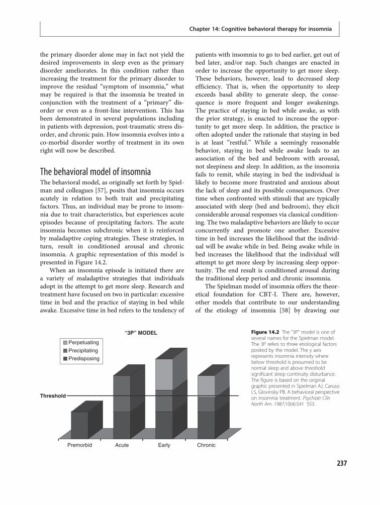
the primary disorder alone may in fact not yield thedesired improvements in sleep even as the primarydisorder ameliorates. In this condition rather thanincreasing the treatment for the primary disorder toimprove the residual “symptom of insomnia,” whatmay be required is that the insomnia be treated inconjunction with the treatment of a “primary” dis-order or even as a front-line intervention. This hasbeen demonstrated in several populations includingin patients with depression, post-traumatic stress dis-order, and chronic pain. How insomnia evolves into aco-morbid disorder worthy of treatment in its ownright will now be described.
The behavioral model of insomniaThe behavioral model, as originally set forth by Spiel-man and colleagues [57], posits that insomnia occursacutely in relation to both trait and precipitatingfactors. Thus, an individual may be prone to insom-nia due to trait characteristics, but experiences acuteepisodes because of precipitating factors. The acuteinsomnia becomes subchronic when it is reinforcedby maladaptive coping strategies. These strategies, inturn, result in conditioned arousal and chronicinsomnia. A graphic representation of this model ispresented in Figure 14.2.
When an insomnia episode is initiated there area variety of maladaptive strategies that individualsadopt in the attempt to get more sleep. Research andtreatment have focused on two in particular: excessivetime in bed and the practice of staying in bed whileawake. Excessive time in bed refers to the tendency of
patients with insomnia to go to bed earlier, get out ofbed later, and/or nap. Such changes are enacted inorder to increase the opportunity to get more sleep.These behaviors, however, lead to decreased sleepefficiency. That is, when the opportunity to sleepexceeds basal ability to generate sleep, the conse-quence is more frequent and longer awakenings.The practice of staying in bed while awake, as withthe prior strategy, is enacted to increase the oppor-tunity to get more sleep. In addition, the practice isoften adopted under the rationale that staying in bedis at least “restful.” While a seemingly reasonablebehavior, staying in bed while awake leads to anassociation of the bed and bedroom with arousal,not sleepiness and sleep. In addition, as the insomniafails to remit, while staying in bed the individual islikely to become more frustrated and anxious aboutthe lack of sleep and its possible consequences. Overtime when confronted with stimuli that are typicallyassociated with sleep (bed and bedroom), they elicitconsiderable arousal responses via classical condition-ing. The two maladaptive behaviors are likely to occurconcurrently and promote one another. Excessivetime in bed increases the likelihood that the individ-ual will be awake while in bed. Being awake while inbed increases the likelihood that the individual willattempt to get more sleep by increasing sleep oppor-tunity. The end result is conditioned arousal duringthe traditional sleep period and chronic insomnia.
The Spielman model of insomnia offers the theor-etical foundation for CBT-I. There are, however,other models that contribute to our understandingof the etiology of insomnia [58] by drawing our
“3P” MODEL
Perpetuating
Threshold
Premorbid Acute Early Chronic
Precipitating
Predisposing
Figure 14.2 The “3P” model is one ofseveral names for the Spielman model.The 3P refers to three etiological factorsposited by the model. The y axisrepresents insomnia intensity wherebelow threshold is presumed to benormal sleep and above thresholdsignificant sleep continuity disturbance.The figure is based on the originalgraphic presented in Spielman AJ, CarusoLS, Glovinsky PB. A behavioral perspectiveon insomnia treatment. Psychiatr ClinNorth Am. 1987;10(4):541 553.
Chapter 14: Cognitive behavioral therapy for insomnia
237
attention to how patients may engage in maladaptivesafety behaviors, harbor dysfunctional beliefs aboutsleep, engage in excessive rumination and catastro-phizing, as well as being cortically primed for presleeparousal and overt attention to stimuli that goodsleepers easily ignore [9,10,59,60]. All of these areantithetical to initiating and maintaining sleep andcan be the focus of treatment.
Cognitive behavioral treatmentof insomnia (CBT-I)In general there are three main types of therapy thatcan be employed, from a behavioral standpoint, todeal with chronic insomnia (in order of priority):stimulus control, sleep restriction, and sleep hygienetherapies. These therapies comprise the standardbehavioral therapy of insomnia. The session to sessionclinical regimen is described below. A description ofother therapies follows in the next section.
Stimulus control therapyStimulus control therapy is considered to be the first-line behavioral treatment for chronic primary insom-nia and therefore should be prioritized accordingly[61]. Stimulus control instructions limit the amountof time patients spend awake in bed or the bedroomand are designed to decondition presleep arousal.Typical instructions include: (1) keep a fixed wake-time 7 days per week, irrespective of how much sleepyou get during the night; (2) avoid any behavior in thebed or bedroom other than sleep or sexual activity; (3)sleep only in the bedroom; (4) leave the bedroom if notasleep after approximately 15 to 20 minutes; (5) returnonly when sleepy; and (6) if still unable to fall asleeprepeat steps 4 and 5 as often as is necessary. Thecombination of these instructions re-establishes thebed and bedroom as strong cues for sleep and entrainsthe circadian sleep–wake cycle to the desired phase. Itis important to note that such reconditioning will onlytake place over several days and, as such, overall sleepcontinuity is likely to be unchanged or worse in theearly phase of the treatment.
Sleep restrictionSleep restriction therapy requires patients to limit theamount of time they spend in bed to an amount equalto their average total sleep time. In order to accom-plish this, the clinician works with the patient to
(1) establish a fixed wake-time and (2) decrease sleepopportunity by prescribing a later bedtime. Initially,the therapy results in a reduction in total sleep time,but over the course of several days, results indecreased sleep latency, and decreased wake-timeafter sleep onset. As sleep efficiency increases, patientsare instructed to gradually increase the amount oftime they spend in bed. In practice, patients “rollback” their bedtime in 15-minute increments, givensleep diary data which shows that on the prior weekthe patient’s sleep was efficient (85% or more of thetime spent in bed was spent asleep). This therapy isthought to be effective for two reasons. First, itreverses the patients’ strategy of coping with theirinsomnia by extending sleep opportunity. The strat-egy of extending time in bed, while increasing theopportunity to get more sleep, produces a form ofsleep that is shallow and fragmented. Second, theinitial sleep loss that occurs with sleep restrictiontherapy is thought to increase the “pressure for sleep”which in turn produces quicker sleep latencies, lesswake after sleep onset, and more efficient sleep.It should be noted that the treatment has a paradox-ical aspect to it. Patients who report being unable tosleep are in essence being told: “sleep less.” Such aprescription needs to be delivered with care and com-pliance must be monitored. Sleep restriction is con-traindicated in patients with histories of mania orseizure disorder, because it may aggravate these con-ditions. Patients also need to be made aware of theincreased sleepiness that results from sleep restrictionand the need to take extra precautions when drivingor engaging in other behaviors which may be unsafedue to sleepiness.
Sleep hygieneThis requires that the clinician and patient review aset of instructions which are geared toward helpingthe patient maintain good sleep habits (seeTable 14.1). It should be noted that sleep hygieneinstructions are not helpful when provided as amonotherapy [62] and might be even less helpfulwhen provided as written instructions which are nottailored to the individual or are cast in absolute terms.With respect to the former, providing patients with a“handout” (especially without an explanation of theclinical science behind the issues) undermines patientcompliance. With respect to the latter, sleep hygieneinstructions have within them several absolute
Section V: Insomnia in Psychiatric Contexts
238
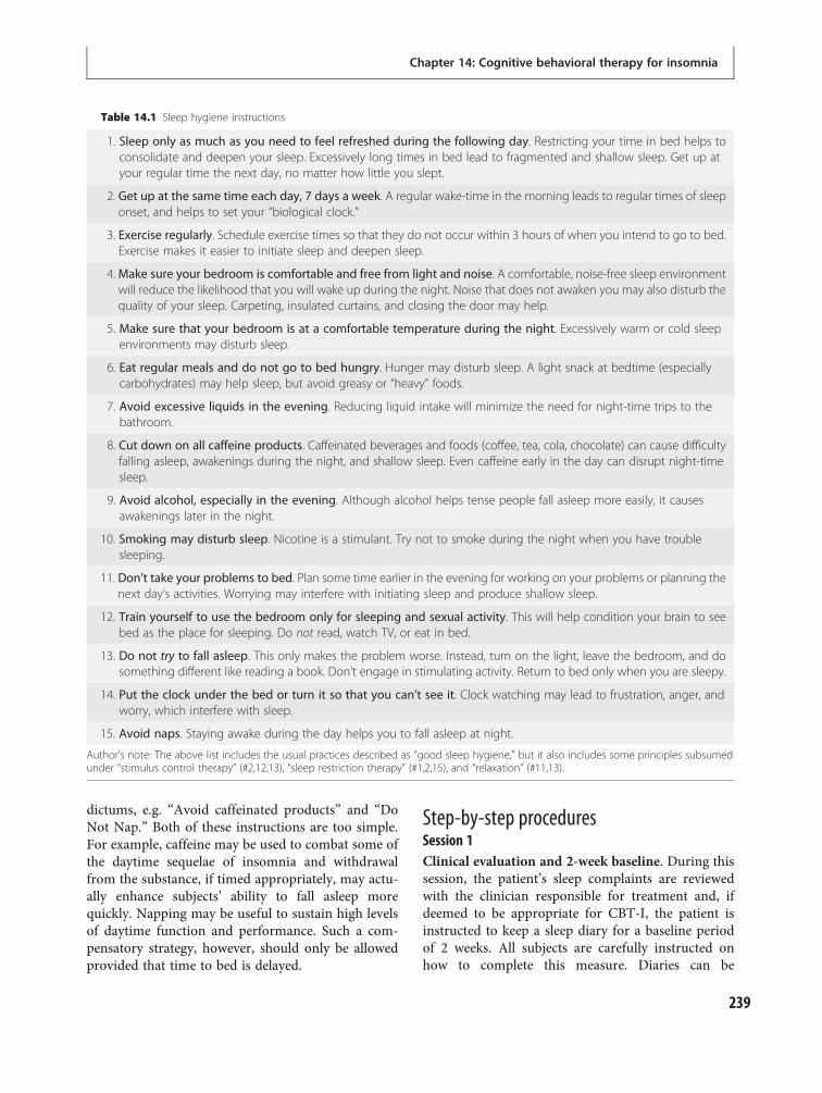
dictums, e.g. “Avoid caffeinated products” and “DoNot Nap.” Both of these instructions are too simple.For example, caffeine may be used to combat some ofthe daytime sequelae of insomnia and withdrawalfrom the substance, if timed appropriately, may actu-ally enhance subjects’ ability to fall asleep morequickly. Napping may be useful to sustain high levelsof daytime function and performance. Such a com-pensatory strategy, however, should only be allowedprovided that time to bed is delayed.
Step-by-step proceduresSession 1Clinical evaluation and 2-week baseline. During thissession, the patient’s sleep complaints are reviewedwith the clinician responsible for treatment and, ifdeemed to be appropriate for CBT-I, the patient isinstructed to keep a sleep diary for a baseline periodof 2 weeks. All subjects are carefully instructed onhow to complete this measure. Diaries can be
Table 14.1 Sleep hygiene instructions
1. Sleep only as much as you need to feel refreshed during the following day. Restricting your time in bed helps toconsolidate and deepen your sleep. Excessively long times in bed lead to fragmented and shallow sleep. Get up atyour regular time the next day, no matter how little you slept.
2. Get up at the same time each day, 7 days a week. A regular wake-time in the morning leads to regular times of sleeponset, and helps to set your “biological clock.”
3. Exercise regularly. Schedule exercise times so that they do not occur within 3 hours of when you intend to go to bed.Exercise makes it easier to initiate sleep and deepen sleep.
4.Make sure your bedroom is comfortable and free from light and noise. A comfortable, noise-free sleep environmentwill reduce the likelihood that you will wake up during the night. Noise that does not awaken you may also disturb thequality of your sleep. Carpeting, insulated curtains, and closing the door may help.
5. Make sure that your bedroom is at a comfortable temperature during the night. Excessively warm or cold sleepenvironments may disturb sleep.
6. Eat regular meals and do not go to bed hungry. Hunger may disturb sleep. A light snack at bedtime (especiallycarbohydrates) may help sleep, but avoid greasy or “heavy” foods.
7. Avoid excessive liquids in the evening. Reducing liquid intake will minimize the need for night-time trips to thebathroom.
8. Cut down on all caffeine products. Caffeinated beverages and foods (coffee, tea, cola, chocolate) can cause difficultyfalling asleep, awakenings during the night, and shallow sleep. Even caffeine early in the day can disrupt night-timesleep.
9. Avoid alcohol, especially in the evening. Although alcohol helps tense people fall asleep more easily, it causesawakenings later in the night.
10. Smoking may disturb sleep. Nicotine is a stimulant. Try not to smoke during the night when you have troublesleeping.
11. Don’t take your problems to bed. Plan some time earlier in the evening for working on your problems or planning thenext day’s activities. Worrying may interfere with initiating sleep and produce shallow sleep.
12. Train yourself to use the bedroom only for sleeping and sexual activity. This will help condition your brain to seebed as the place for sleeping. Do not read, watch TV, or eat in bed.
13. Do not try to fall asleep. This only makes the problem worse. Instead, turn on the light, leave the bedroom, and dosomething different like reading a book. Don’t engage in stimulating activity. Return to bed only when you are sleepy.
14. Put the clock under the bed or turn it so that you can’t see it. Clock watching may lead to frustration, anger, andworry, which interfere with sleep.
15. Avoid naps. Staying awake during the day helps you to fall asleep at night.
Author’s note: The above list includes the usual practices described as “good sleep hygiene,” but it also includes some principles subsumedunder “stimulus control therapy” (#2,12,13), “sleep restriction therapy” (#1,2,15), and “relaxation” (#11,13).
Chapter 14: Cognitive behavioral therapy for insomnia
239
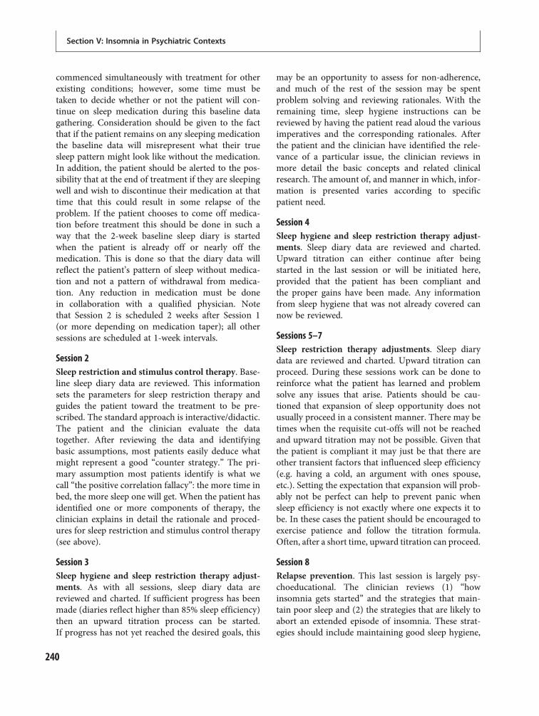
commenced simultaneously with treatment for otherexisting conditions; however, some time must betaken to decide whether or not the patient will con-tinue on sleep medication during this baseline datagathering. Consideration should be given to the factthat if the patient remains on any sleeping medicationthe baseline data will misrepresent what their truesleep pattern might look like without the medication.In addition, the patient should be alerted to the pos-sibility that at the end of treatment if they are sleepingwell and wish to discontinue their medication at thattime that this could result in some relapse of theproblem. If the patient chooses to come off medica-tion before treatment this should be done in such away that the 2-week baseline sleep diary is startedwhen the patient is already off or nearly off themedication. This is done so that the diary data willreflect the patient’s pattern of sleep without medica-tion and not a pattern of withdrawal from medica-tion. Any reduction in medication must be donein collaboration with a qualified physician. Notethat Session 2 is scheduled 2 weeks after Session 1(or more depending on medication taper); all othersessions are scheduled at 1-week intervals.
Session 2Sleep restriction and stimulus control therapy. Base-line sleep diary data are reviewed. This informationsets the parameters for sleep restriction therapy andguides the patient toward the treatment to be pre-scribed. The standard approach is interactive/didactic.The patient and the clinician evaluate the datatogether. After reviewing the data and identifyingbasic assumptions, most patients easily deduce whatmight represent a good “counter strategy.” The pri-mary assumption most patients identify is what wecall “the positive correlation fallacy”: the more time inbed, the more sleep one will get. When the patient hasidentified one or more components of therapy, theclinician explains in detail the rationale and proced-ures for sleep restriction and stimulus control therapy(see above).
Session 3Sleep hygiene and sleep restriction therapy adjust-ments. As with all sessions, sleep diary data arereviewed and charted. If sufficient progress has beenmade (diaries reflect higher than 85% sleep efficiency)then an upward titration process can be started.If progress has not yet reached the desired goals, this
may be an opportunity to assess for non-adherence,and much of the rest of the session may be spentproblem solving and reviewing rationales. With theremaining time, sleep hygiene instructions can bereviewed by having the patient read aloud the variousimperatives and the corresponding rationales. Afterthe patient and the clinician have identified the rele-vance of a particular issue, the clinician reviews inmore detail the basic concepts and related clinicalresearch. The amount of, and manner in which, infor-mation is presented varies according to specificpatient need.
Session 4Sleep hygiene and sleep restriction therapy adjust-ments. Sleep diary data are reviewed and charted.Upward titration can either continue after beingstarted in the last session or will be initiated here,provided that the patient has been compliant andthe proper gains have been made. Any informationfrom sleep hygiene that was not already covered cannow be reviewed.
Sessions 5–7Sleep restriction therapy adjustments. Sleep diarydata are reviewed and charted. Upward titration canproceed. During these sessions work can be done toreinforce what the patient has learned and problemsolve any issues that arise. Patients should be cau-tioned that expansion of sleep opportunity does notusually proceed in a consistent manner. There may betimes when the requisite cut-offs will not be reachedand upward titration may not be possible. Given thatthe patient is compliant it may just be that there areother transient factors that influenced sleep efficiency(e.g. having a cold, an argument with ones spouse,etc.). Setting the expectation that expansion will prob-ably not be perfect can help to prevent panic whensleep efficiency is not exactly where one expects it tobe. In these cases the patient should be encouraged toexercise patience and follow the titration formula.Often, after a short time, upward titration can proceed.
Session 8Relapse prevention. This last session is largely psy-choeducational. The clinician reviews (1) “howinsomnia gets started” and the strategies that main-tain poor sleep and (2) the strategies that are likely toabort an extended episode of insomnia. These strat-egies should include maintaining good sleep hygiene,
Section V: Insomnia in Psychiatric Contexts
240

always engaging in proper stimulus control, and usingsleep restriction as necessary when problems arise.
Adjunctive therapiesIn addition to these principal behavioral techniques,there are also several adjunctive therapies which mayprove helpful to the patient and can be included in thetreatment plan. These include cognitive therapy,relaxation training, and phototherapy. It may also beuseful to combine pharmacotherapy with CBT-I.
Cognitive therapyInterestingly, while therapy for insomnia is ubiqui-tously referred to as cognitive behavioral therapy forinsomnia (CBT-I) the “C” is often not a core com-ponent [41,42]. This said, it is by far the mostcommon adjuvant treatment. Several forms of cogni-tive therapy for insomnia have been developed andoften overlap. The forms of cognitive therapy utilizetechniques including psychoeducation [63], paradox-ical intention [64], cognitive restructuring [63], andtreatments that focus on safety behaviors [65] andattention bias [10]. While the approaches differ inprocedure, all are based on the observation thatpatients with insomnia have negative thoughts andbeliefs about their condition and its consequences.Helping patients to challenge the veracity and useful-ness of these beliefs is thought to decrease the anxietyand arousal associated with insomnia.
Relaxation trainingDifferent relaxation techniques target differentphysiological systems. Progressive muscle relaxationis used to diminish skeletal muscle tension. Diaphrag-matic breathing is used to make breathing slowerand shallower and resembles the form of breathing,interestingly, that naturally occurs at sleep onset.Autogenic training focuses on increasing peripheralblood flow. Biofeedback may similarly target specificsystems with a common goal of achieving a “relaxedstate.” To the list of relaxation techniques can beadded hypnosis and mindfulness-based stress reduc-tion. While the latter involves more than learning arelaxation technique, this is nonetheless its core prac-tice. Most practitioners select the optimal relaxationmethod based upon which technique is easiest for thepatient to learn, which is most consistent with howthe patient manifests arousal, and which technique is
not contraindicated by medical conditions (e.g. pro-gressive muscle relaxation might not be an idealchoice for patients with certain neuromusculardisorders).
PhototherapyThe sleep-promoting effects of bright light may occurvia several mechanisms including shifting the circa-dian system; enhancement of the amplitude of thecircadian pacemaker, promoting wakefulness duringthe day and sleep at night; or indirectly via its anti-depressant effects. In practice, bright light is used toshorten or lengthen the diurnal phase of the patient’sday. In the case where the patient’s insomnia has aphase delay component (i.e. the patient prefers to goto bed late and wake up late) waking early by alarmand being exposed to bright light in the morning for aperiod of time may enable them to “feel sleepy” at anearlier time in the evening. In the case where thepatient’s insomnia has a phase advance component(i.e. the patient prefers to go to bed early and wake upearly) bright light exposure in the evening may enablethem to stay awake later and wake up later. It isgenerally assumed that phototherapy has no signifi-cant side-effects, but this is not always the case. Maniamay be triggered by bright light in patients withbipolar mood disorder. Other side-effects includeinsomnia, hypomania, agitation, visual blurring, eyestrain, and headaches. Individuals with eye-relatedproblems or who are at risk for such conditions suchas diabetes should consult an eye care specialist priorto initiating light therapy.
Combining CBT-I with pharmacotherapyOne of the disadvantages of CBT-I, compared topharmacotherapy, is that the impact on sleep is grad-ual. Patients often may not experience any improve-ment until after 2–4 weeks of treatment. Althoughthis length of time is short relative to the typicalduration of insomnia experienced by most patientsprior to treatment, it can be a barrier to treatmentadherence. Faced with the daytime effects of sleeprestriction therapy, some patients abandon treatmentbefore experiencing benefits. An alternate strategy isto combine CBT-I with hypnotic medication in orderto capitalize on the rapid effects of pharmacotherapywhile waiting for CBT-I to “kick in.” Medications arethen gradually tapered in the second half of treatment.
Chapter 14: Cognitive behavioral therapy for insomnia
241

In perhaps the most experimentally rigorous test ofthis approach, Morin and colleagues randomlyassigned older adults to receive CBT-I alone, temaze-pam alone, combined CBT-I and temazepam, or pla-cebo [66]. At the post-treatment time point all threetreatment groups had improved sleep relative to pla-cebo, with a trend towards superior outcomes in thecombined group. By the last assessment at 24 monthspost-treatment, the temazepam only group hadreturned to baseline severity, whereas both CBT-Igroups maintained the treatment gains. Interestingly,the CBT-I only group had better outcomes than thecombined group. Other studies with different medi-cations have yielded similar outcomes, althoughsome found better outcomes in the combined treatmentgroup. It has been hypothesized that individuals whoreceive combined treatment may ascribe their treatmentgains to the results of the medication and failed todevelop self-efficacy, or confidence in their ability tosleep without medication. Once the medication waswithdrawn, they no longer felt capable of sleepingunaided, which likely would lead to feelings of anxietyabout sleep and subsequent difficulty sleeping [66]. Des-pite mixed findings thus far, there is still considerablepotential to develop treatment strategies that combineCBT-I and pharmacotherapy in an effective manner.
An alternate approach is to combine CBT-I withwake-promoting medication using a daytime dosingschedule. While seemingly counterintuitive, there is asolid rationale for this strategywhich spans three relatedpossibilities. First, since sleep restriction therapy is asso-ciated with increased daytime sleepiness that can lead totreatment drop-out, counteracting this side-effect mayenhance treatment adherence. Second, insomnia isoften associated with anxiety about the negative conse-quences of poor sleep. This anxiety, in turn, likelyexacerbates patients’ difficulties by augmenting existingarousal (somatic, cognitive, and/or cortical). Thus, if thedaytime consequences of insomnia could be attenuatedor eliminated, it stands to reason that this factor (orrelated factors like attention bias or the engagement ofsafety behaviors) would cease to be contributive. Third,and lastly, the use of wake-promoting agents such asmodafinil may enhance the patient’s ability to extendwakefulness. This will enable the patient to reverse thetendency to “extend sleep opportunity,” reduce themis-match between sleep ability and sleep opportunity, andwill prime the homeostatic drive for sleep viathe prolongation of wakefulness and/or increased acti-vation during wakefulness.
The first effort to evaluate the proposition thatwakefulness promotion may be useful for the man-agement of insomnia was conducted by Perlis andcolleagues [67]. In this study, the effects of modafinilwere assessed alone and in combination with CBT-I.The results from the investigation suggest that mod-afinil as a monotherapy did not produce significanteffects on sleep continuity; the linear trends, however,were in the predicted direction. Dual therapy also didnot result in improved efficacy but clearly resulted inan attenuation of sleepiness and enhanced compli-ance. The conceptual basis for this study (along withthe mean trends) lead to the hypothesis that an alter-ation to the treatment regimen (one which promoteda longer duration of action) might result in independ-ent effects for modafinil and additive effects whencombined with CBT-I. Future studies are needed toassess the potential of this novel treatment paradigm.
Techniques for dealing with clientnon-complianceCBT techniques for insomnia can be challenging forboth the client and the practitioner. This may beparticularly true early in the therapeutic process asthe client will be asked to accept a difficult regimen ofbehavioral change and concomitant sleep deprivation.Sleep loss alone may try the patience of a client andthis makes compliance with any therapeutic modalitya problematic issue. There are, however, some strat-egies (most based in clinical experience) which mayenhance the likelihood of a successful clinical out-come. Examples of these techniques include using:
1. Good salesmanship (a motivational approach totherapy) – There is no more important a methodthan the demonstration of a good knowledge baseregarding (in general) sleep medicine and (inspecific) the principles behind the behavioralinterventions. Patients will often have challengingquestions and concerns regarding the procedures.Simply providing a laundry list of techniques willnot likely lead to good adherence, especially assleep deprivation gets worse in the early stages.Clear and compelling explanations, provision ofrationale, and setting of expectancies will go a longway towards gaining patient trust and compliance.Sharing information about the clinical efficacyand effectiveness of treatment can also be apowerful aid in obtaining compliance.
Section V: Insomnia in Psychiatric Contexts
242
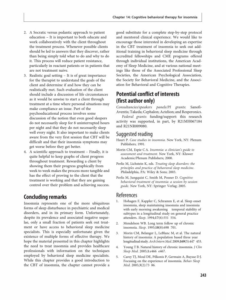
2. A Socratic versus pedantic approach to patienteducation – It is important to both educate andwork collaboratively with the client throughoutthe treatment process. Whenever possible clientsshould be led to answers that they discover, ratherthan being simply told what to do and why to doit. This process will reduce patient resistance,particularly in reactant patients or in patients thatare not treatment naive.
3. Realistic goal setting – It is of great importancefor the therapist to understand the goals of theclient and determine if and how they can berealistically met. Such evaluation of the clientshould include a discussion of life circumstancesas it would be unwise to start a client throughtreatment at a time where personal situations maymake compliance an issue. Part of thepsychoeducational process involves somediscussion of the notion that even good sleepersdo not necessarily sleep for 8 uninterrupted hoursper night and that they do not necessarily sleepwell every night. It also important to make clientsaware from the very first session that CBT will bedifficult and that their insomnia symptoms mayget worse before they get better.
4. A scientific approach to treatment – Finally, it isquite helpful to keep graphs of client progressthroughout treatment. Rewarding a client byshowing them their progress graphically fromweek to week makes the process more tangible andhas the effect of proving to the client that thetreatment is working and that they are gainingcontrol over their problem and achieving success.
Concluding remarksInsomnia represents one of the more ubiquitousforms of sleep disturbance in psychiatric and medicaldisorders, and in its primary form. Unfortunately,despite its prevalence and associated negative seque-lae, only a small fraction of patients seek out treat-ment or have access to behavioral sleep medicinespecialists. This is especially unfortunate given theexistence of multiple forms of effective therapy. Wehope the material presented in this chapter highlightsthe need to treat insomnia and provides healthcareprofessionals with information on the techniquesemployed by behavioral sleep medicine specialists.While this chapter provides a good introduction tothe CBT of insomnia, the chapter cannot provide a
good substitute for a complete step-by-step protocoland mentored clinical experience. We would like toencourage those interested in developing an expertisein the CBT treatment of insomnia to seek out add-itional training in behavioral sleep medicine throughaccredited fellowships and CME programs offeredthrough individual institutions, the American Acad-emy of Sleep Medicine, and at various national meet-ings like those of the Associated Professional SleepSocieties, the American Psychological Association,the Society for Behavioral Medicine, and the Associ-ation for Behavioral and Cognitive Therapies.
Potential conflict of interests(first author only)Consultancies/speakers panels/PI grants: Sanofi-Aventis; Takeda; Cephalon; Actelion; and Resperonics.
Federal grants: funding/support: this researchactivity was supported, in part, by R21MH067184and R21NR009080.
Suggested readingHauri P. Case studies in insomnia. New York, NY: Plenum
Publishers; 1991.
Morin CM, Espie CA. Insomnia: a clinician’s guide toassessment and treatment. New York, NY: KluwerAcademic/Plenum Publishers; 2000.
Perlis M, Lichstein K, eds. Treating sleep disorders: theprinciples and practice of behavioral sleep medicine.Philadelphia, PA: Wiley & Sons; 2003.
Perlis M, Jungquist C, Smith M, Posner D. Cognitivebehavioral treatment of insomnia: a session by sessionguide. New York, NY: Springer Verlag; 2005.
References1. Hohagen F, Kappler C, Schramm E, et al. Sleep onset
insomnia, sleep maintaining insomnia and insomniawith early morning awakening temporal stability ofsubtypes in a longitudinal study on general practiceattenders. Sleep. 1994;17(6):551 554.
2. Mendelson WB. Long term follow up of chronicinsomnia. Sleep. 1995;18(8):698 701.
3. Morin CM, Belanger L, LeBlanc M, et al. The naturalhistory of insomnia: A population based three yearlongitudinal study.Arch InternMed. 2009;169(5):447 453.
4. Young TB. Natural history of chronic insomnia. J ClinSleep Med. 2005;1:e466 e467.
5. Carey TJ, Moul DE, Pilkonis P, Germain A, Buysse D J.Focusing on the experience of insomnia. Behav SleepMed. 2005;3(2):73 86.
Chapter 14: Cognitive behavioral therapy for insomnia
243
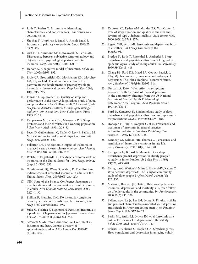
6. Roth T, Roehrs T. Insomnia: epidemiology,characteristics, and consequences. Clin Cornerstone.2003;5(3):5 15.
7. Shochat T, Umphress J, Israel A, Ancoli Israel S.Insomnia in primary care patients. Sleep. 1999;22:S359 365.
8. Orff HJ, Drummond SP, Nowakowski S, Perlis ML.Discrepancy between subjective symptomatology andobjective neuropsychological performance ininsomnia. Sleep. 2007;30(9):1205 1211.
9. Harvey A. A cognitive model of insomnia. Behav ResTher. 2002;40:869 893.
10. Espie CA, Broomfield NM, MacMahon KM, MacpheeLM, Taylor LM. The attention intention effortpathway in the development of psychophysiologicinsomnia: a theoretical review. Sleep Med Rev. 2006;10(4):215 245.
11. Johnson L, Spinweber CL. Quality of sleep andperformance in the navy: A longitudinal study of goodand poor sleepers. In: Guilleminault C, Lugaresi E, eds.Sleep/wake disorders: natural history, epidemiology,and long term evaluation. New York: Raven Press;1983:13 28.
12. Kupperman M, Lubeck DP, Mazonson PD. Sleepproblems and their correlates in a working population.J Gen Intern Med. 1995;10:25 32.
13. Leger D, Guilleminault C, Blader G, Levy E, Paillard M.Medical and socio professional impact of insomnia.Sleep. 2002;25:625 629.
14. Fullerton DS. The economic impact of insomnia inmanaged care: a clearer picture emerges. Am J ManagCare. 2006;12(8 Suppl):S246 252.
15. Walsh JK, Engelhardt CL. The direct economic costs ofinsomnia in the United States for 1995. Sleep. 1999;22(Suppl 2):S386 393.
16. Ozminkowski RJ, Wang S, Walsh J K. The direct andindirect costs of untreated insomnia in adults in theUnited States. Sleep. 2007;30(3):263 273.
17. NIH. State of the Science Conference Statement onmanifestations and management of chronic insomniain adults. NIH Consens State Sci Statements. 2005;22(2):1 30.
18. Phillips B, Mannino DM. Do insomnia complaintscause hypertension or cardiovascular disease? J ClinSleep Med. 2007;3(5):489 494.
19. Suka M, Yoshida K, Sugimori H. Persistent insomnia isa predictor of hypertension in Japanese male workers.J Occup Health. 2003;45(6):344 350.
20. Schwartz S, McDowell Anderson W, Cole SR, et al.Insomnia and heart disease: a review ofepidemiologic studies. J Psychosom Res. 1999;47(4):313 333.
21. Knutson KL, Ryden AM, Mander BA, Van Cauter E.Role of sleep duration and quality in the risk andseverity of type 2 diabetes mellitus. Arch Intern Med.2006;166(16):1768 1774.
22. Pigeon WR, Perlis ML. Insomnia and depression: birdsof a feather? Int J Sleep Disorders. 2007;1(3):82 91.
23. Breslau N, Roth T, Rosenthal L, Andreski P. Sleepdisturbance and psychiatric disorders: a longitudinalepidemiological study of young adults. Biol Psychiatry.1996;39(6):411 418.
24. Chang PP, Ford DE, Mead LA, Cooper Patrick L,Klag MJ. Insomnia in young men and subsequentdepression: The Johns Hopkins Precursors Study.Am J Epidemiol. 1997;146(2):105 114.
25. Dryman A, Eaton WW. Affective symptomsassociated with the onset of major depressionin the community: findings from the US NationalInstitute of Mental Health EpidemiologicCatchment Area Program. Acta Psychiatr Scand.1991;84(1):1 5.
26. Ford D, Kamerow D. Epidemiologic study of sleepdisturbance and psychiatric disorders: an opportunityfor prevention? JAMA. 1989;262:1479 1484.
27. Hohagen F, Rink K, Kappler C, et al. Prevalence andtreatment of insomnia in general practice:A longitudinal study. Eur Arch Psychiatry ClinNeurosci. 1993;242(6):329 336.
28. Kennedy GJ, Kelman HR, Thomas C. Persistence andremission of depressive symptoms in late life.Am J Psychiatry. 1991;148(2):174 178.
29. Livingston G, Blizard B, Mann A. Does sleepdisturbance predict depression in elderly people?A study in inner London. Br J Gen Pract. 1993;43(376):445 448.
30. LivingstonG,WatkinV,Milne B,ManelaMV,KatonaC.Who becomes depressed? The Islington communitystudy of older people. J Affect Disord. 2000;58(2):125 133.
31. Mallon L, Broman JE, Hetta J. Relationship betweeninsomnia, depression, and mortality: a 12 year followup of older adults in the community. Int Psychogeriatr.2000;12(3):295 306.
32. Paffenbarger RS Jr, Lee IM, Leung R. Physical activityand personal characteristics associated with depressionand suicide in American college men. Acta PsychiatrScand Suppl. 1994;377:16 22.
33. Perlis ML, Smith LJ, Lyness JM, et al. Insomnia as arisk factor for onset of depression in the elderly.Behav Sleep Med. 2006;4(2):104 113.
34. Roberts RE, Shema SJ, Kaplan GA, Strawbridge WJ.Sleep complaints and depression in an aging cohort:
Section V: Insomnia in Psychiatric Contexts
244
A prospective perspective. Am J Psychiatry. 2000;157(1):81 88.
35. Vollrath M, Wicki W, Angst J. The Zurich study. VIII.Insomnia: association with depression, anxiety,somatic syndromes, and course of insomnia. Eur ArchPsychiatry Neurol Sci. 1989;239(2):113 124.
36. Weissman M, Greenwald S, Nino Murcia G,Dement W. The morbidity of insomnia uncomplicatedby psychiatric disorders. Gen Hosp Psychiatry.1997;19:245 250.
37. Perlis ML, Giles DE, Buysse DJ, Tu X, Kupfer DJ. Selfreported sleep disturbance as a prodromal symptomin recurrent depression. J Affect Disord. 1997;42(2 3):209 212.
38. Pigeon WR, Hegel M, Unutzer J, et al. Is insomnia aperpetuating factor for late life depression in theIMPACT cohort? Sleep. 2008;31(4):481 488.
39. Nowell PD, Mazumdar S, Buysse DJ, et al.Benzodiazepines and zolpidem for chronic insomnia:a meta analysis of treatment efficacy. JAMA. 1997;278(24):2170 2177.
40. Glass J, Lanctot KL, Herrmann N, Sproule BA, BustoUE. Sedative hypnotics in older people with insomnia:meta analysis of risks and benefits. BMJ. 2005;331(7526):1169.
41. Morin CM, Culbert JP, Schwartz MS. Nonpharmacological interventions for insomnia: a metaanalysis of treatment efficacy. Am J Psychiatry.1994;151:1172 1180.
42. Murtagh DR, Greenwood KM. Identifying effectivepsychological treatments for insomnia: a metaanalysis. J Consult Clin Psychol. 1995;63:79 89.
43. Smith MT, Perlis ML, Park A, et al. Comparative metaanalysis of pharmacotherapy and behavior therapy forpersistent insomnia. Am J Psychiatry. 2002;159(1):5 11.
44. Aldrich MS. Automobile accidents in patients withsleep disorders. Sleep. 1989;12(6):487 494.
45. Krystal A. Treating the health, quality of life, andfunctional impairments in insomnia. J Clin Sleep Med.2007;3(1):63 72.
46. Morin CM. Measuring outcomes in randomizedclinical trials of insomnia treatments. Sleep Med Rev.2003;7(3):263 279.
47. Leger D, Quera Salva MA, Philip P. Health relatedquality of life in patients with insomnia treatedwith zopiclone. Pharmacoeconomics. 1996;10(Suppl 1):39 44.
48. Walsh JK, Krystal AD, Amato DA, et al. Nightlytreatment of primary insomnia with eszopiclone for sixmonths: effect on sleep, quality of life, and worklimitations. Sleep. 2007;30(8):959 968.
49. Edinger JD, Wohlgemuth WK, Krystal AD, Rice JR.Behavioral insomnia therapy for fibromyalgia patients:a randomized clinical trial. Arch Intern Med. 2005;165(21):2527 2535.
50. Pigeon WR, Jungquist CR, Matteson SR, Perlis ML.Pain, sleep and mood outcomes in chronicpain patients following cognitive behavioraltherapy for insomnia. Sleep. 2007;30:A255 A256.
51. Fava M, McCall WV, Krystal A, et al. Eszopicloneco administered with fluoxetine in patients withinsomnia coexisting with major depressive disorder.Biol Psychiatry. 2006;59(11):1052 1060.
52. Manber R, Edinger JD, San Pedro M. Combiningescitalopram oxalate (ESCIT) and individualcognitive behavioral therapy for insomnia (CBTI)to improve depression outcome. Sleep. 2007;30:A232.
53. Taylor DJ, Lichstein KL, Weinstock J, Sanford S,Temple JR. A pilot study of cognitive behavioraltherapy of insomnia in people with mild depression.Behav Ther. 2007;38(1):49 57.
54. Ohayon MM, Roth T. What are the contributingfactors for insomnia in the general population?J Psychosom Res. 2001;51(6):745 755.
55. Ohayon M. Epidemiology of insomnia: what we knowand what we still need to learn. Sleep Med Rev.2002;6:97 111.
56. American Academy of Sleep Medicine. Internationalclassification of sleep disorders, 2nd ed. Westchester, IL:American Academy of Sleep Medicine; 2005.
57. Spielman AJ, Caruso LS, Glovinsky PB. A behavioralperspective on insomnia treatment. Psychiatr ClinNorth Am. 1987;10(4):541 553.
58. Perlis ML, Smith MT, Pigeon WR. Etiology andpathophysiology of insomnia. In: Kryger M, Roth T,Dement WC, eds. Principles and practice of sleepmedicine, 4th ed. Philadelphia, PA: Elsevier Saunders;2005:714 725.
59. Morin C, Stone J, Trinkle D, Mercer J, Remsberg S.Dysfunctional beliefs and attitudes about sleep amongolder adults with and without insomnia complaints.Psychol Aging. 1993;8.
60. Perlis M, Giles D, Mendelson W, Bootzin R, Wyatt J.Psychophysiological insomnia: the behavioural modeland a neurocognitive perspective. J Sleep Res.1997;6:179 188.
61. Chesson AL Jr, Anderson WM, Littner M, et al.Practice parameters for the nonpharmacologictreatment of chronic insomnia. An AmericanAcademy of Sleep Medicine report. Standardsof Practice Committee of the American Academy
Chapter 14: Cognitive behavioral therapy for insomnia
245
of Sleep Medicine. Sleep. 1999;22(8):1128 1133.
62. Lacks P, Morin CM. Recent advances in the assessmentand treatment of insomnia. J Consult Clin Psychol.1992;60:586 594.
63. Morin C. Insomnia: psychological assessment andmanagement. New York: Guilford Press; 1996.
64. Shoham Salomon V, Rosenthal R. Paradoxicalinterventions: a meta analysis. J Consult Clin Psychol.1987;55(1):22 28.
65. Harvey AG. Identifying safety behaviors in insomnia.J Nerv Ment Dis. 2002;190(1):16 21.
66. Morin CM, Colecchi C, Stone J, Sood R, Brink D.Behavioral and pharmacological therapies for late lifeinsomnia: a randomized controlled trial. JAMA.1999;281:991 999.
67. Perlis ML, Smith MT, Orff H, et al. The effects ofmodafinil and cognitive behavior therapy on sleepcontinuity in patients with primary insomnia.Sleep. 2004;27(4):715 725.
Section V: Insomnia in Psychiatric Contexts
246

Section VIChapter
15Sleep Disturbance in Psychiatric Illness
Depressive disorders
Philip R. Gehrman, Michael E. Thase, Dieter Riemann and Michael Perlis
IntroductionDisturbed sleep is a central feature of depression. So itcomes as no surprise that there has been a fairamount of research on how it is that sleep is abnormalin the context of depression and how this aspect of thedisorder is related to the risk for, and clinical courseof, the illness.
In the present chapter the various manifestationsof disturbed sleep in depression are reviewed in termsof direct effects on sleep, sleep-related circadianabnormalities, and sleep-related neurobiological per-turbations. The clinical implications of these findingsare reviewed along with the major models put forth toexplain the relationship between sleep and depression.Lastly, an overview of strategies for the treatment ofsleep disturbance in depression is provided.
Sleep abnormalities in majordepressive disorderSleep may be construed in terms of sleep continuityE,sleep architecture, and sleep microarchitecture (EEGpower spectral profile), as well as various subtypes ofthese patterns. Each of these domains is reliablyaltered in significant proportions of people sufferingfrom major depressive disorder (MDD).
Sleep continuity disturbancesThe DSM-IV diagnostic criteria for MDD include sub-jective sleep disturbance in the form of either insomnia(i.e. difficulty initiating or maintaining sleep) or hyper-somnia [1]. Unfortunately, the definition of insomniainMDD is not uniform among clinicians or researchers.Insomnia has traditionally been clinically described interms of initial, middle, and late insomnia; in research,
including polysomnographic (PSG) assessments,insomnia is typically measured in terms of sleep latency(SL), wake after sleep onset (WASO), number of awak-enings (NWAK), sleep efficiency (SE), and total sleeptime (TST). Regardless of the classification schemaused, disturbed sleep quality is one of the most com-monly reported depressive symptoms, occurring in upto 90% of individuals seeking treatment (e.g. [2]). Asmaller subset of depressed individuals report increasedsleep or subjective hypersomnia, which is defined asprolonged sleep duration that extendsmore than 1 hourover habitual total sleep time. Rates of hypersomniavary more widely across studies and are somewhatpopulation-dependent, ranging from as low as 5% toas high as 36% of depressed persons. The prevalence ofhypersomnia is higher in studies of younger age groupsand tends to be highest in studies of individuals meetingcriteria for atypical depression [3,4].
Unlike the DSM-IV, the International Classifica-tion of Sleep Disorders, 2nd edition (ICSD-2) delineatesother forms of insomnia including: psychophysio-logical, paradoxical, and idiopathic insomnia [5].Psychophysiological insomnia includes problemswith sleep initiation and maintenance where thereis evidence of sleep difficulty and/or heightenedarousal in bed, which is thought to occur as a resultof psychological, behavioral, and conditioning factors.Paradoxical insomnia (formerly referred to as sleepstate misperception insomnia) manifests with a sub-jective complaint of sleep initiation and/or mainten-ance that is unusually severe and there is, by definition,a poor correspondence between subjective complaintand the severity of the complaint as measured by PSG.Currently, there is nowidely accepted theory regardingthe precipitation and/or perpetuation of this form ofinsomnia, although several have posited that sleep
Foundations of Psychiatric SleepMedicine, ed. JohnW.Winkelman andDavid T. Plante. Published by CambridgeUniversity Press.# Cambridge University Press 2010.
247

state “misperception” occurs in association with aheightened level of sensory and information process-ing at sleep onset and during NREM sleep and theattenuation of the normal mesograde amnesia ofsleep [6,7]. Idiopathic insomnia refers to sleep initi-ation and maintenance problems that have a child-hood onset. Little is known about the average severityof this type of insomnia, the extent to which sleep statemisperception occurs in these subjects, or this form ofthe disorder’s etiology and/or pathophysiology [8].
Interestingly, there has been no effort to classifysleep continuity disturbances associated with depres-sion in terms of the aforementioned ICSD-2 diagnostictypes. Instead, the focus has been on the insomnia“subtypes” or phenotypes (e.g. early, middle, and late).It is generally held that “late insomnia” is more charac-teristic of the severe forms of major depressive disorder(i.e. early morning awakenings are a cardinal signof endogenous depression ormelancholia, whereas “ini-tial insomnia” is equally characteristic of generalizedanxiety disorder). From our vantage point, there haveonly been a few studies that systematically assess thedifferential occurrence of the insomnia subtypes indepression (using contemporary classifications ofmoodand anxiety disorders) and the results from one ofthe largest such investigations suggests that all threesubtypes of insomnia occur with an equal prevalencein patients with MDD [9].
In terms of the severity of insomnia as measuredby self-report, there also are no large-scale studies thatprovide relevant descriptive data (i.e. mean, range,and standard deviation), especially in comparison tohealthy (“good sleeper”) or insomnia (without depres-sion) control groups. Thus, it cannot be said withconfidence that self-reported sleep continuity disturb-ances associated with depression are similar to, ordifferent from, those which are seen in individualswith primary insomnia. In the absence of definitivedata, the results from one very small-scale study sug-gests that sleep continuity disturbances are moresevere in primary insomnia than major depressivedisorder and that patients with primary insomnia alsoare more prone to sleep state misperception (seeTable 15.1) [10].
In terms of PSG-measured insomnia severity,there is (to our knowledge) only one contemporarystudy that provides descriptive data comparingsleep continuity in patients with depression to goodsleeper controls [11]. In this case, the MDD patients(N¼178) differed modestly from good sleeper
subjects (N¼108) on PSG-measured sleep continuity.Sleep latency was increased by ~5 minutes, wake aftersleep onset was increased by ~20 minutes, the fre-quency of awakenings was increased by ~6 episodesper night, early morning awakening was moved for-ward by ~15 minutes, total sleep time was reducedby ~35 minutes, and sleep efficiency was reduced by~8%. Interestingly, time in bed differed only minim-ally (~5 minutes). While these findings suggest thatthe insomnia in depression may not be driven by thesame perpetuating factors as primary insomnia (i.e.sleep extension [12]), it is possible that the similarityin time in bed is an artifact of the laboratory settinggiven that subjects in this study all were studied on afixed 11 p.m. to 7 a.m. schedule in the sleep labora-tory. In practice, depressed patients with insomniaoften report spending increased time in bed evenwhen they are not able to sleep.
Sleep architecture abnormalitiesA significant proportion of patients with major depres-sive disorder exhibit altered sleep architecture ascompared to good sleepers. These architectural abnor-malities include: (1) an altered distribution of slow-wavesleep (lack the traditional attenuation pattern across thenight) [13], (2) reduced slow-wave sleep (in minutesand/or percent) [9], (3) decreased latency to the firstepisode of REM sleep (i.e. reduced REM latency)[14,15], (4) a prolonged first REM period [14,15], (5)increased REM percent (if not REM time in minutes)[14,15], and (6) increased REM density (i.e. eye move-ments per minute of REM sleep) [14,15].
With the single exception of increased REMdensity,these effects are remarkably like what is seen with aging(i.e. in elderly subjects) [11] and this led Gillin andcolleagues [16], amongst others, to suggest that depres-sion may mimic the CNS aging process. Further, alter-ations of both NREM and REM sleep led to a vigorousongoing debate within the sleep community aboutwhether one set of the effects occurred actively whilethe other occurred only as a necessary consequence ofthe primary effects. One group speculated that the “pri-mary defect” existed within the NREM system and wasrelated to altered sleep homeostasis [17]. The othergroup argued that increased REM “pressure” waslargely responsible for the sleep alterations [18]. For atime, this debate centered on whether reduced REMlatency was a specific and sensitive biomarker for majordepressive disorder. This aspect of the debate lessened
Section VI: Sleep Disturbance in Psychiatric Illness
248
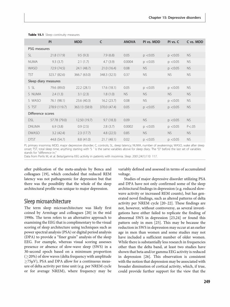
after publication of the meta-analysis by Benca andcolleagues [19], which concluded that reduced REMlatency was not pathognomic for depression but thatthere was the possibility that the whole of the sleeparchitectural profile was unique to major depression.
Sleep microarchitectureThe term sleep microarchitecture was likely firstcoined by Armitage and colleagues [20] in the mid1990s. The term refers to an alternative approach toexamining the EEG that is complimentary to the visualscoring of sleep architecture using techniques such aspower spectral analysis (PSA) or digital period analysis(DPA) to provide a “finer grain” analysis of the sleepEEG. For example, whereas visual scoring assessespresence or absence of slow-wave sleep (SWS) in a30-second epoch based on a minimum proportion(�20%) of slow waves (delta frequency with amplitude�75µV), PSA and DPA allow for a continuous meas-ure of delta activity per time unit (e.g. per NREM cycleor for average NREM), where frequency may be
variably defined and assessed in terms of accumulatedvoltage.
Studies of major depressive disorder utilizing PSAand DPA have not only confirmed some of the sleeparchitectural findings in depression (e.g. reduced slow-wave activity or increased REM counts), but has gen-erated novel findings, such as altered patterns of deltaactivity per NREM cycle [20–22]. These findings arenot, however, without controversy, as several investi-gations have either failed to replicate the finding ofabnormal SWS in depression [23,24] or found thispattern only in men [25]. This may be because thereduction in SWS in depressionmay occur at an earlierage in men than women and some studies may nothave included a sufficient number of older women.While there is substantially less research in frequenciesother than the delta band, at least two studies haveshown that beta and/or gamma EEG activity is reducedin depression [26]. This observation is consistentwith the notion that depressionmay be associated withbroader diminution of cortical activity, which, if true,could provide further support for the view that the
Table 15.1 Sleep continuity measures
PI MDD C ANOVA PI vs. MDD PI vs. C C vs. MDD
PSG measures
SL 21.8 (17.9) 9.5 (9.3) 7.9 (6.8) 0.05 p <0.05 p <0.05 NS
NUMA 9.3 (3.7) 2.1 (1.7) 4.7 (3.9) 0.0004 p <0.05 p <0.05 NS
WASO 72.9 (74.5) 24.1 (48.7) 21.0 (16.4) 0.08 NS p <0.05 NS
TST 323.7 (82.6) 366.7 (63.0) 348.3 (32.5) 0.37 NS NS NS
Sleep diary measures
S SL 79.6 (89.0) 22.2 (28.1) 17.6 (18.1) 0.05 p <0.05 p <0.05 NS
S NUMA 2.4 (1.3) 3.1 (2.3) 1.8 (1.0) NS NS NS NS
S WASO 76.1 (98.1) 23.6 (40.3) 16.2 (23.7) 0.08 NS p <0.05 NS
S TST 278.9 (119.7) 363.13 (58.9) 370.0 (47.4) 0.05 p <0.05 p <0.05 NS
Difference scores
DSL 57.78 (79.0) 12.50 (19.7) 9.7 (18.3) 0.09 NS p <0.05 NS
DNUMA 6.9 (3.8) 0.9 (2.5) 2.8 (3.7) 0.0002 p <0.05 p <0.05 P<.05
DWASO 3.2 (42.4) 2.3 (17.7) 4.8 (22.5) 0.85 NS NS NS
DTST 44.8 (54.7) 8.8 (41.0) 21.7 (48.1) 0.02 p <0.05 p <0.05 NS
PI, primary insomnia; MDD, major depressive disorder; C, controls; SL, sleep latency; NUMA, number of awakenings; WASO, wake after sleeponset; TST, total sleep time; anything starting with ‘S ’ is the same variables above for sleep diary. The “D” before the last set of variablesstands for “difference in.”Data from Perlis M, et al. Beta/gamma EEG activity in patients with insomnia. Sleep. 2001;24(1):110 117.
Chapter 15: Depressive disorders
249
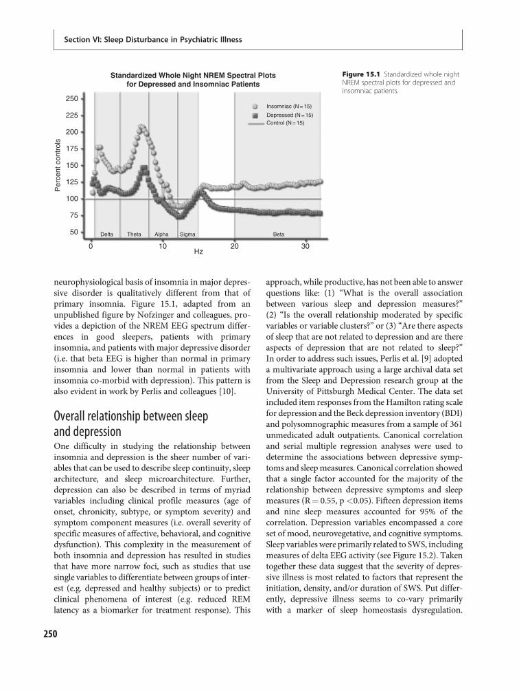
neurophysiological basis of insomnia in major depres-sive disorder is qualitatively different from that ofprimary insomnia. Figure 15.1, adapted from anunpublished figure by Nofzinger and colleagues, pro-vides a depiction of the NREM EEG spectrum differ-ences in good sleepers, patients with primaryinsomnia, and patients with major depressive disorder(i.e. that beta EEG is higher than normal in primaryinsomnia and lower than normal in patients withinsomnia co-morbid with depression). This pattern isalso evident in work by Perlis and colleagues [10].
Overall relationship between sleepand depressionOne difficulty in studying the relationship betweeninsomnia and depression is the sheer number of vari-ables that can be used to describe sleep continuity, sleeparchitecture, and sleep microarchitecture. Further,depression can also be described in terms of myriadvariables including clinical profile measures (age ofonset, chronicity, subtype, or symptom severity) andsymptom component measures (i.e. overall severity ofspecific measures of affective, behavioral, and cognitivedysfunction). This complexity in the measurement ofboth insomnia and depression has resulted in studiesthat have more narrow foci, such as studies that usesingle variables to differentiate between groups of inter-est (e.g. depressed and healthy subjects) or to predictclinical phenomena of interest (e.g. reduced REMlatency as a biomarker for treatment response). This
approach, while productive, has not been able to answerquestions like: (1) “What is the overall associationbetween various sleep and depression measures?”(2) “Is the overall relationship moderated by specificvariables or variable clusters?” or (3) “Are there aspectsof sleep that are not related to depression and are thereaspects of depression that are not related to sleep?”In order to address such issues, Perlis et al. [9] adopteda multivariate approach using a large archival data setfrom the Sleep and Depression research group at theUniversity of Pittsburgh Medical Center. The data setincluded item responses from the Hamilton rating scalefor depression and the Beck depression inventory (BDI)and polysomnographic measures from a sample of 361unmedicated adult outpatients. Canonical correlationand serial multiple regression analyses were used todetermine the associations between depressive symp-toms and sleepmeasures. Canonical correlation showedthat a single factor accounted for the majority of therelationship between depressive symptoms and sleepmeasures (R¼ 0.55, p <0.05). Fifteen depression itemsand nine sleep measures accounted for 95% of thecorrelation. Depression variables encompassed a coreset of mood, neurovegetative, and cognitive symptoms.Sleep variables were primarily related to SWS, includingmeasures of delta EEG activity (see Figure 15.2). Takentogether these data suggest that the severity of depres-sive illness is most related to factors that represent theinitiation, density, and/or duration of SWS. Put differ-ently, depressive illness seems to co-vary primarilywith a marker of sleep homeostasis dysregulation.
Standardized Whole Night NREM Spectral Plotsfor Depressed and Insomniac Patients
Insomniac (N = 15)
Depressed (N = 15)Control (N = 15)
Per
cent
con
trol
s
250
225
200
175
150
125
100
75
50
0
Delta Theta Alpha Sigma Beta
10 20Hz
30
Figure 15.1 Standardized whole nightNREM spectral plots for depressed andinsomniac patients.
Section VI: Sleep Disturbance in Psychiatric Illness
250

Interestingly, REM measures (including reduced REMlatency) accounted for very little variance in that ana-lysis. Parallel studies contrasting the sleep of depressedpatients before and after treatment, recovered patients,and good sleeper healthy controls would be helpful tountangle state from trait variables.
Sleep-related circadian rhythmabnormalitiesInvestigation of the role of circadian rhythms indepression has generally supported a phase advanceof the circadian rhythm. The reduction in REM latencyonce thought to be a cardinal sleep sign of depressionwas proposed to reflect an advance of the circadianrhythm of REM propensity [27]. Similarly, earlymorning awakenings could be the result of a phaseadvance such that the circadian alerting signalincreases at an earlier hour. A number of studies foundevidence of advances in the circadian rhythms ofbody temperature, cortisol, prolactin, norepinephrine,and melatonin, among others [28]. Interestingly,imposing a phase delay on non-depressed subjectscreates REM sleep changes similar to those observedin depression [29]. Further support for the role of aphase advance came from studies that found thatmany antidepressants delay the phase of circadianrhythms [27]. This would suggest that these
medications have circadian realignment as a mechan-ism of action. It should be noted, however, that manystudies have failed to find any evidence of circadianmisalignment in depression [30].
Souetre and colleagues [31] reported that it is theamplitude of circadian rhythms rather than the phasethat is altered in depression. In their study, patients withdepression had reduced amplitudes in body tempera-ture, plasma cortisol, norepinephrine (NE), thyroid-stimulating hormone (TSH), and melatonin rhythms,with lower amplitude associated with greater severity ofdepression. Whether it is the phase position, the ampli-tude, or some other features of circadian rhythms atplay, the data are mixed regarding the presence ofspecific abnormalities in depression. Further, it isunclear whether any abnormalities that do exist play acausative role or are merely epiphenomena.
Sleep-related neuroimaging andneuroendocrine abnormalitiesAn alternate approach to studying brain activityduring sleep is through the use of neuroimaging,which has produced data that help to clarify the earlierREM and NREM findings. Neuroimaging studiesduring sleep are largely limited to PET studies due tothe difficulty of sleeping during an fMRI (due tothe noise generated by magnetic resonance imaging).
Depressed mood (H)Weight loss (B)Loss of libido (H)Disturbed sleep (B)Psych anxiety (H)Self blame (B)Somaic anxiety (H)Initial insomnia (H)Work ability (B)
Delta counts-NREM2Delta counts-NREM3Stage 1%Whole night deltaDelta counts-NREM1REM counts-REM1REM efficiencySleep latencyDelta ratio
1.060.520.470.320.380.290.230.230.20
70% related to SWS
14% related to NREM
16% related to REM
HSRD & BDIVARIABLES
PSG
Canonical corrections between depressive and sleep variablesPerlis et al. Biol Psych, 1997
15 of 36 8 of 19
NOT REPRESENTED ARE FATIGUE,SUICIDE AND
PSYCHOMOTOR ABNORMALITIES
0.460.330.280.280.270.260.230.210.20
Figure 15.2 Canonical corrections between depressive and sleep variants. HSRD, Hamilton Rating Scale for Depression; (H), from theHRSD; (B), from the BDI. Reproduced from [9].
Chapter 15: Depressive disorders
251
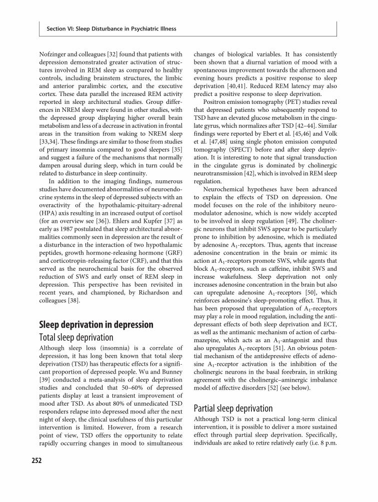
Nofzinger and colleagues [32] found that patients withdepression demonstrated greater activation of struc-tures involved in REM sleep as compared to healthycontrols, including brainstem structures, the limbicand anterior paralimbic cortex, and the executivecortex. These data parallel the increased REM activityreported in sleep architectural studies. Group differ-ences in NREM sleep were found in other studies, withthe depressed group displaying higher overall brainmetabolism and less of a decrease in activation in frontalareas in the transition from waking to NREM sleep[33,34]. These findings are similar to those from studiesof primary insomnia compared to good sleepers [35]and suggest a failure of the mechanisms that normallydampen arousal during sleep, which in turn could berelated to disturbance in sleep continuity.
In addition to the imaging findings, numerousstudies have documented abnormalities of neuroendo-crine systems in the sleep of depressed subjects with anoveractivity of the hypothalamic-pituitary-adrenal(HPA) axis resulting in an increased output of cortisol(for an overview see [36]). Ehlers and Kupfer [37] asearly as 1987 postulated that sleep architectural abnor-malities commonly seen in depression are the result ofa disturbance in the interaction of two hypothalamicpeptides, growth hormone-releasing hormone (GRF)and corticotropin-releasing factor (CRF), and that thisserved as the neurochemical basis for the observedreduction of SWS and early onset of REM sleep indepression. This perspective has been revisited inrecent years, and championed, by Richardson andcolleagues [38].
Sleep deprivation in depressionTotal sleep deprivationAlthough sleep loss (insomnia) is a correlate ofdepression, it has long been known that total sleepdeprivation (TSD) has therapeutic effects for a signifi-cant proportion of depressed people. Wu and Bunney[39] conducted a meta-analysis of sleep deprivationstudies and concluded that 50–60% of depressedpatients display at least a transient improvement ofmood after TSD. As about 80% of unmedicated TSDresponders relapse into depressed mood after the nextnight of sleep, the clinical usefulness of this particularintervention is limited. However, from a researchpoint of view, TSD offers the opportunity to relaterapidly occurring changes in mood to simultaneous
changes of biological variables. It has consistentlybeen shown that a diurnal variation of mood with aspontaneous improvement towards the afternoon andevening hours predicts a positive response to sleepdeprivation [40,41]. Reduced REM latency may alsopredict a positive response to sleep deprivation.
Positron emission tomography (PET) studies revealthat depressed patients who subsequently respond toTSD have an elevated glucose metabolism in the cingu-late gyrus, which normalizes after TSD [42–44]. Similarfindings were reported by Ebert et al. [45,46] and Volket al. [47,48] using single photon emission computedtomography (SPECT) before and after sleep depriv-ation. It is interesting to note that signal transductionin the cingulate gyrus is dominated by cholinergicneurotransmission [42], which is involved in REM sleepregulation.
Neurochemical hypotheses have been advancedto explain the effects of TSD on depression. Onemodel focuses on the role of the inhibitory neuro-modulator adenosine, which is now widely acceptedto be involved in sleep regulation [49]. The choliner-gic neurons that inhibit SWS appear to be particularlyprone to inhibition by adenosine, which is mediatedby adenosine A1-receptors. Thus, agents that increaseadenosine concentration in the brain or mimic itsaction at A1-receptors promote SWS, while agents thatblock A1-receptors, such as caffeine, inhibit SWS andincrease wakefulness. Sleep deprivation not onlyincreases adenosine concentration in the brain but alsocan upregulate adenosine A1-receptors [50], whichreinforces adenosine’s sleep-promoting effect. Thus, ithas been proposed that upregulation of A1-receptorsmay play a role in mood regulation, including the anti-depressant effects of both sleep deprivation and ECT,as well as the antimanic mechanism of action of carba-mazepine, which acts as an A1-antagonist and thusalso upregulates A1-receptors [51]. An obvious poten-tial mechanism of the antidepressive effects of adeno-sine A1-receptor activation is the inhibition of thecholinergic neurons in the basal forebrain, in strikingagreement with the cholinergic–aminergic imbalancemodel of affective disorders [52] (see below).
Partial sleep deprivationAlthough TSD is not a practical long-term clinicalintervention, it is possible to deliver a more sustainedeffect through partial sleep deprivation. Specifically,individuals are asked to retire relatively early (i.e. 8 p.m.
Section VI: Sleep Disturbance in Psychiatric Illness
252

or 10 p.m.) and permitted to sleep for an attenuatedamount of time (e.g. 2–4 hours) before beingawakened and asked to remain awake and active.Although the antidepressant effects of partial sleepdeprivation tend to be less pronounced than those ofTSD, they have been shown to meaningfully enhancethe effects of other treatments, such as antidepressants[53]. Unfortunately, even partial sleep deprivationinterventions are difficult to implement outside ofhospital settings.
Selective deprivation of REM sleepBased on the observation that all members of the tricyc-lic (TCA) and monoamine oxidase inhibitor (MAOI)classes of antidepressants significantly suppress REMsleep, the question arose whether this pharmacologicaleffectmay be a “conditio sine qua non” or even the basicunderlying mechanism of action of antidepressanttreatments. Vogel and colleagues [18] tested thishypothesis by depriving depressed individuals of REMsleepwithout pharmacological intervention by selective,PSG-monitored awakenings over a period of 3 weeks.This experimental therapy led to a 50% reduction inREM sleep and resulted in an antidepressant responsecomparable to that of the TCA imipramine. A controlgroup, which was awakened during NREM sleep, didnot show any clinical improvement.
Only one study [54] attempted to replicate the find-ings of Vogel and colleagues with a methodologicallyimproved design. Selective REM sleep deprivationwas compared to the same amount of well-balancedawakenings leading to NREM sleep deprivation indepressed inpatients. In accordance with the data ofVogel, REM deprivation was associated with a signifi-cant antidepressant effect. However, contrary to expect-ation, the group that was awakened from NREM sleepalso exhibited an antidepressant effect, which tended tobe larger in magnitude than observed with selectiveREM deprivation. In this study it was found thatboth experimental conditions increased the durationof NREM sleep cycles and it was therefore postulatedthat this might be the underlying mechanism of action.
Clinical correlates of disturbed sleepin depressionA number of investigators have examined the clinicalcorrelates of sleep disturbance in depression. The evi-dence overwhelmingly supports the view that
insomnia and sleep architecture perturbations are crit-ically related to disease state and long-term outcome.
Clinical relevance of sleep continuitydisturbanceDisturbances of sleep continuity have significant clin-ical implications that have been investigated across anumber of studies. These studies indicate that dis-turbed sleep continuity is a prodromal symptom aswell as a risk factor for new-onset illness and relapse.
Sleep continuity disturbance (insomnia) as a riskfactor for new-onset depression. A number of longitu-dinal studies with time frames ranging from 6 monthsto 3 years have found that insomnia increases the riskof both new-onset and recurrent depression [55–73].Studies with follow-up periods over decades have alsofound this relationship [56,63,64]. Averaged acrossstudies, there is an approximately 3.5-fold increasein the risk of depression for people with persistentinsomnia compared to those who do not complainof insomnia (see Figure 15.3). It is noteworthy thatthe measurement and definition of insomnia variedconsiderably across studies. The impact of age and sexinteractions on this relationship has been examined inseveral studies. In those studies that controlled forage, the relative risk of insomnia for depressive onsetwas consistently higher than in those studies thatdid not control for age. This suggests that insomniais not a uniform risk factor for all age cohorts; how-ever, studies have not specifically examined the agegroups for which risk is highest. In the studies thatevaluated sex differences [57,63], the associationbetween insomnia and depression was consistentlymore robust for women than men. Overall, these datastrongly support the hypothesis that insomnia is a riskfactor for depression.
Sleep continuity disturbance (insomnia) as a pro-dromal symptom of depression. In the natural history ofindividuals with recurrent illness, insomnia may be anearly indicator of an impending major depressive epi-sode. One retrospective study of depressed patientsfound that 40% reported a worsening of sleep priorto the onset of their first depressive episode [74]. Inthose subjects with recurrent depression, 56% reportedthat sleep disturbance preceded recurrence. In a pro-spective, 42-week study of remitted patients with ahistory of recurrent depression, weekly monitoring ofdepression symptoms was undertaken with the BDI,with increases in depressive symptoms resulting in a
Chapter 15: Depressive disorders
253
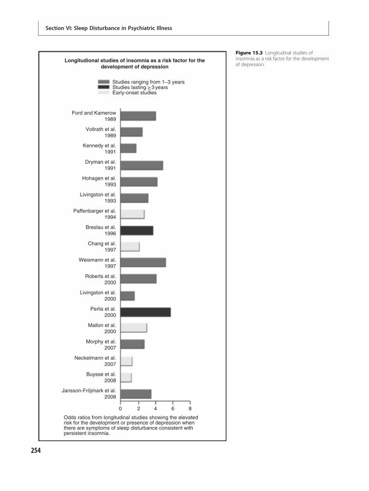
Longitudional studies of insomnia as a risk factor for thedevelopment of depression
Studies ranging from 1–3 years
Early-onset studies
Ford and Kamerow1989
Vollrath et al.1989
Kennedy et al.1991
Dryman et al.1991
Hohagen et al.1993
Livingston et al.1993
Paffenbarger et al.1994
Breslau et al.1996
Chang et al.1997
Weismann et al.1997
Roberts et al.2000
Livingston et al.2000
Perlis et al.2000
Mallon et al.2000
Morphy et al.2007
Neckelmann et al.2007
Buysse et al.2008
Jansson-Fröjmark et al.2008
0 2 4 6 8
Odds ratios from longitudinal studies showing the elevatedrisk for the development or presence of depression whenthere are symptoms of sleep disturbance consistent withpersistent insomnia.
Studies lasting >_ 3 years
Figure 15.3 Longitudinal studies ofinsomnia as a risk factor for the developmentof depression.
Section VI: Sleep Disturbance in Psychiatric Illness
254

formal evaluation for recurrence [75]. The subgroupthat experienced a syndromal recurrence of depressionwas compared to a subsamplematched on age, sex, andclinical history by examining the single sleep item onthe BDI (question 16) for the 5 weeks prior to the onsetof the recurrent episode in the index group and for atemporally equivalent period in the subsample that didnot suffer a recurrence. The time series data from thisperiod showed, in distinction to the non-recurrentgroup, those who suffered a recurrence exhibited aprogressively greater level of sleep disturbance thatreached highest severity at the week of recurrence(see Figure 15.4). These data strongly suggest thatinsomnia is a prodromal symptom of depression, andlend weight to the possibility that worsening insomniamay trigger or precipitate new depressive episodes.
Sleep continuity disturbance (insomnia) is a residualsymptom of depression and increases risk of depressiverelapse. Although insomnia often occurs in the contextof depression, remission of depression is not necessar-ily associated with improvement in sleep. Across stud-ies of both cognitive behavioral and pharmacologicaltreatment of depression, between 20% and 50% ofpatients continue to report insomnia despite clinicalimprovements in overall depression [76–79]. In onecohort study that assessed elderly patients with depres-sion at baseline and 6 months later, patients withpersistent insomnia were 10–12-fold less likely to haveachieved remission or an improvement of �50% indepressive symptoms compared with patients with noinsomnia [80]. Following resolution of a depressive
episode, the presence of residual insomnia symptomsincreases the risk of recurrence of depression[77,81,82] whereas those whose sleep improves post-treatment have reduced rates of recurrence [83]. Thesestudies demonstrate that insomnia is frequently aresidual symptom of depression that is associated withboth reduced likelihood of achieving remission andincreased risk of recurrence.
Clinical relevance of sleep architecturefindingsSimilar to sleep continuity disturbance, sleep archi-tecture abnormalities have important clinical impacton illness severity, response to treatment, and risk ofrelapse/recurrence.
Illness severity. The sleep architecture variablesthat have been most intensively studied with respectto illness severity are REM latency, REM density, andSWS time, with greater disturbances of sleep architec-ture typically associated with greater illness severity.REM latency has only been associated with illnessseverity to the extent that inpatients more reliablyshow this sleep abnormality compared to outpatients[84,85]. Most studies, however, show that REMlatency tends to be stable within individuals acrosstime (e.g. [14,86–89]). REM density tends to be cor-related with severity of depression and this measurevaries with clinical state [15,90–94]. Finally, there issome evidence that reduced SWS is associated withgreater illness severity [9,95]. Thus, there are
Sleep disturbance in patients with and without recurrence of major depression
1.5
1
0.5
0 WEEK-4
WEEK-2
WEEK-1
Figure 15.4 Score on the sleepdisturbance item from the BDI (y axis) andtime leading up to recurrence (z axis).The pattern on the right is for patientswho experienced a recurrence and whoexperienced an increase in sleepdisturbance in leading up to recurrence.The pattern on the left is a matchedgroup of patients who did not experiencerecurrence and who did not have anincrease in sleep disturbance.
Chapter 15: Depressive disorders
255
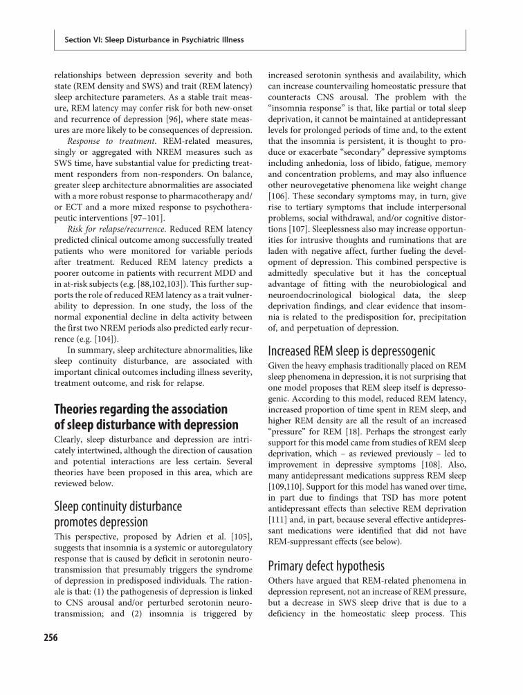
relationships between depression severity and bothstate (REM density and SWS) and trait (REM latency)sleep architecture parameters. As a stable trait meas-ure, REM latency may confer risk for both new-onsetand recurrence of depression [96], where state meas-ures are more likely to be consequences of depression.
Response to treatment. REM-related measures,singly or aggregated with NREM measures such asSWS time, have substantial value for predicting treat-ment responders from non-responders. On balance,greater sleep architecture abnormalities are associatedwith a more robust response to pharmacotherapy and/or ECT and a more mixed response to psychothera-peutic interventions [97–101].
Risk for relapse/recurrence. Reduced REM latencypredicted clinical outcome among successfully treatedpatients who were monitored for variable periodsafter treatment. Reduced REM latency predicts apoorer outcome in patients with recurrent MDD andin at-risk subjects (e.g. [88,102,103]). This further sup-ports the role of reduced REM latency as a trait vulner-ability to depression. In one study, the loss of thenormal exponential decline in delta activity betweenthe first two NREM periods also predicted early recur-rence (e.g. [104]).
In summary, sleep architecture abnormalities, likesleep continuity disturbance, are associated withimportant clinical outcomes including illness severity,treatment outcome, and risk for relapse.
Theories regarding the associationof sleep disturbance with depressionClearly, sleep disturbance and depression are intri-cately intertwined, although the direction of causationand potential interactions are less certain. Severaltheories have been proposed in this area, which arereviewed below.
Sleep continuity disturbancepromotes depressionThis perspective, proposed by Adrien et al. [105],suggests that insomnia is a systemic or autoregulatoryresponse that is caused by deficit in serotonin neuro-transmission that presumably triggers the syndromeof depression in predisposed individuals. The ration-ale is that: (1) the pathogenesis of depression is linkedto CNS arousal and/or perturbed serotonin neuro-transmission; and (2) insomnia is triggered by
increased serotonin synthesis and availability, whichcan increase countervailing homeostatic pressure thatcounteracts CNS arousal. The problem with the“insomnia response” is that, like partial or total sleepdeprivation, it cannot be maintained at antidepressantlevels for prolonged periods of time and, to the extentthat the insomnia is persistent, it is thought to pro-duce or exacerbate “secondary” depressive symptomsincluding anhedonia, loss of libido, fatigue, memoryand concentration problems, and may also influenceother neurovegetative phenomena like weight change[106]. These secondary symptoms may, in turn, giverise to tertiary symptoms that include interpersonalproblems, social withdrawal, and/or cognitive distor-tions [107]. Sleeplessness also may increase opportun-ities for intrusive thoughts and ruminations that areladen with negative affect, further fueling the devel-opment of depression. This combined perspective isadmittedly speculative but it has the conceptualadvantage of fitting with the neurobiological andneuroendocrinological biological data, the sleepdeprivation findings, and clear evidence that insom-nia is related to the predisposition for, precipitationof, and perpetuation of depression.
Increased REM sleep is depressogenicGiven the heavy emphasis traditionally placed on REMsleep phenomena in depression, it is not surprising thatone model proposes that REM sleep itself is depresso-genic. According to this model, reduced REM latency,increased proportion of time spent in REM sleep, andhigher REM density are all the result of an increased“pressure” for REM [18]. Perhaps the strongest earlysupport for this model came from studies of REM sleepdeprivation, which – as reviewed previously – led toimprovement in depressive symptoms [108]. Also,many antidepressant medications suppress REM sleep[109,110]. Support for this model has waned over time,in part due to findings that TSD has more potentantidepressant effects than selective REM deprivation[111] and, in part, because several effective antidepres-sant medications were identified that did not haveREM-suppressant effects (see below).
Primary defect hypothesisOthers have argued that REM-related phenomena indepression represent, not an increase of REM pressure,but a decrease in SWS sleep drive that is due to adeficiency in the homeostatic sleep process. This
Section VI: Sleep Disturbance in Psychiatric Illness
256
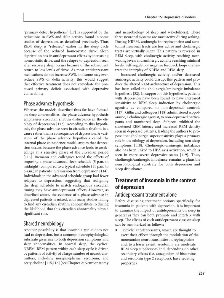
“primary defect hypothesis” [17] is supported by thereductions in SWS and delta activity found in somestudies of depression, as described previously. ThusREM sleep is “released” earlier in the sleep cyclebecause of the reduced homeostatic drive. Sleepdeprivation has its antidepressant effects by increasinghomeostatic drive, and the relapse to depression seenafter recovery sleep occurs because of the subsequentreturn to low levels of drive. As most antidepressantmedications do not increase SWS, and some may evenreduce SWS or delta activity, this would suggestthat effective treatment does not remediate the pro-posed primary deficit associated with depressivevulnerability.
Phase advance hypothesisWhereas the models described thus far have focusedon sleep abnormalities, the phase advance hypothesisemphasizes circadian rhythm disturbance in the eti-ology of depression [112]. According to this hypoth-esis, the phase advance seen in circadian rhythms is acause rather than a consequence of depression. A vari-ation of the phase advance hypothesis called theinternal phase coincidence model, argues that depres-sion occurs because the phase advance leads to awak-enings at a sensitive phase of the circadian period[113]. Riemann and colleagues tested the effects ofimposing a phase advanced sleep schedule (5 p.m. tomidnight) compared to a typical schedule (11 p.m. to6 a.m.) in patients in remission from depression [114].Individuals in the advanced schedule group had fewerrelapses to depression, suggesting that realigningthe sleep schedule to match endogenous circadiantiming may have antidepressant effects. However, asdescribed above, the evidence of a phase advance indepressed patients is mixed, with many studies failingto find any circadian rhythm abnormalities, reducingthe likelihood that this circadian abnormality plays asignificant role.
Shared neurobiologyAnother possibility is that insomnia per se does notlead to depression, but a common neurophysiologicalsubstrate gives rise to both depression symptoms andsleep abnormalities. In normal sleep, the cyclicalNREM–REM pattern within each sleep cycle is drivenby patterns of activity of a large number of neurotrans-mitters, including norepinephrine, serotonin, andacetylcholine [115,116] (see Chapter 2: Neuroanatomy
and neurobiology of sleep and wakefulness). Thesethree neuronal systems are most active during waking.During NREM, aminergic (norepinephrine and sero-tonin) neuronal tracts are less active and cholinergictracts are virtually silent. This pattern is reversed inREM sleep, with cholinergic activity reaching near-waking levels and aminergic activity reaching minimallevels. Self-regulatory negative feedback loops orches-trate the interplay of NREM and REM sleep.
Increased cholinergic activity and/or decreasedaminergic activity could disrupt this pattern and pro-duce the altered REM architecture of depression. Thishas been called the cholinergic/aminergic imbalancehypothesis [52]. In support of this hypothesis, patientswith depression have been found to have increasedsensitivity to REM sleep induction by cholinergicagonists as compared to non-depressed controls[117]. Gillin and colleagues [118] administered scopol-amine, a cholinergic agonist, to non-depressed partici-pants and monitored sleep. Subjects exhibited theshortened REM latency and increased REM densityseen in depressed patients, leading the authors to pro-pose that cholinergic supersensitivity plays a primaryrole in the etiology of depression, including non-sleepsymptoms [118]. Cholinergic–aminergic imbalancealso has been linked to HPA axis activation, which isseen in more severe depressive states [119]. Thus,cholinergic/aminergic imbalance remains a plausibleneurobiological substrate for both depression andsleep disturbance.
Treatment of insomnia in the contextof depressionAntidepressant treatment aloneBefore discussing treatment options specifically forinsomnia in patients with depression, it is importantto examine the impact of antidepressants on sleep ingeneral as they can both promote and interfere withsleep. The effects of each antidepressant class on sleepcan be summarized as follows:� Tricyclic antidepressants, which are thought to
exert their effects through the modulation of themonoamine neurotransmitter norepinephrineand, to a lesser extent, serotonin, are moderateREM sleep suppressors and, depending on othersecondary effects (i.e. antagonism of histamineand serotonin type 2 receptors), have sedatingproperties
Chapter 15: Depressive disorders
257
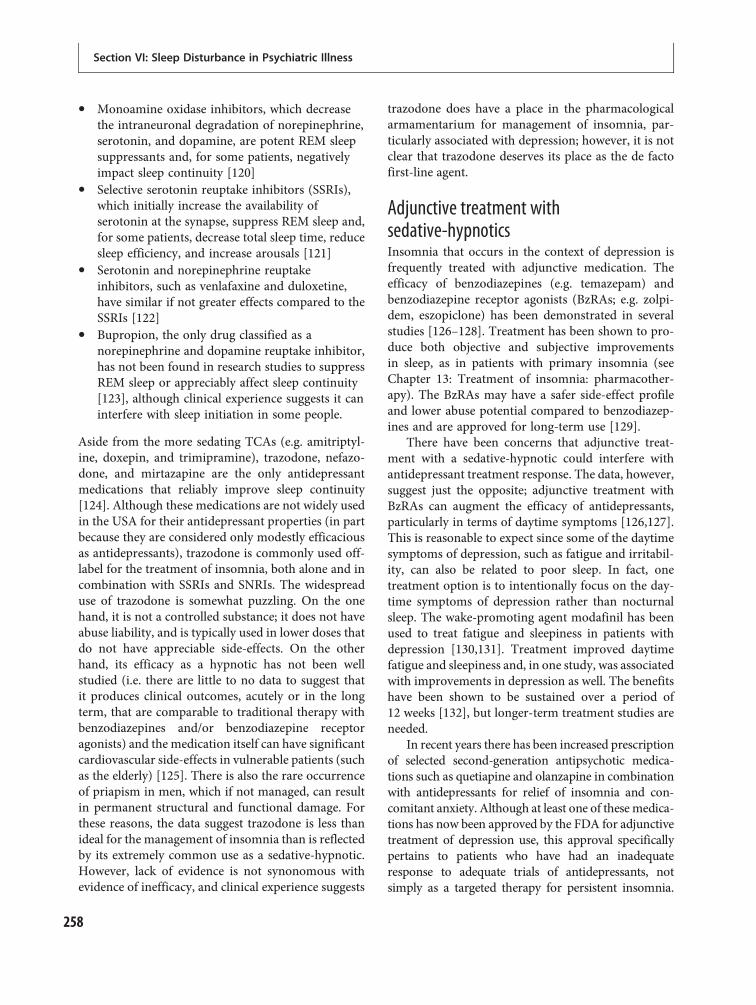
� Monoamine oxidase inhibitors, which decreasethe intraneuronal degradation of norepinephrine,serotonin, and dopamine, are potent REM sleepsuppressants and, for some patients, negativelyimpact sleep continuity [120]
� Selective serotonin reuptake inhibitors (SSRIs),which initially increase the availability ofserotonin at the synapse, suppress REM sleep and,for some patients, decrease total sleep time, reducesleep efficiency, and increase arousals [121]
� Serotonin and norepinephrine reuptakeinhibitors, such as venlafaxine and duloxetine,have similar if not greater effects compared to theSSRIs [122]
� Bupropion, the only drug classified as anorepinephrine and dopamine reuptake inhibitor,has not been found in research studies to suppressREM sleep or appreciably affect sleep continuity[123], although clinical experience suggests it caninterfere with sleep initiation in some people.
Aside from the more sedating TCAs (e.g. amitriptyl-ine, doxepin, and trimipramine), trazodone, nefazo-done, and mirtazapine are the only antidepressantmedications that reliably improve sleep continuity[124]. Although these medications are not widely usedin the USA for their antidepressant properties (in partbecause they are considered only modestly efficaciousas antidepressants), trazodone is commonly used off-label for the treatment of insomnia, both alone and incombination with SSRIs and SNRIs. The widespreaduse of trazodone is somewhat puzzling. On the onehand, it is not a controlled substance; it does not haveabuse liability, and is typically used in lower doses thatdo not have appreciable side-effects. On the otherhand, its efficacy as a hypnotic has not been wellstudied (i.e. there are little to no data to suggest thatit produces clinical outcomes, acutely or in the longterm, that are comparable to traditional therapy withbenzodiazepines and/or benzodiazepine receptoragonists) and the medication itself can have significantcardiovascular side-effects in vulnerable patients (suchas the elderly) [125]. There is also the rare occurrenceof priapism in men, which if not managed, can resultin permanent structural and functional damage. Forthese reasons, the data suggest trazodone is less thanideal for the management of insomnia than is reflectedby its extremely common use as a sedative-hypnotic.However, lack of evidence is not synonomous withevidence of inefficacy, and clinical experience suggests
trazodone does have a place in the pharmacologicalarmamentarium for management of insomnia, par-ticularly associated with depression; however, it is notclear that trazodone deserves its place as the de factofirst-line agent.
Adjunctive treatment withsedative-hypnoticsInsomnia that occurs in the context of depression isfrequently treated with adjunctive medication. Theefficacy of benzodiazepines (e.g. temazepam) andbenzodiazepine receptor agonists (BzRAs; e.g. zolpi-dem, eszopiclone) has been demonstrated in severalstudies [126–128]. Treatment has been shown to pro-duce both objective and subjective improvementsin sleep, as in patients with primary insomnia (seeChapter 13: Treatment of insomnia: pharmacother-apy). The BzRAs may have a safer side-effect profileand lower abuse potential compared to benzodiazep-ines and are approved for long-term use [129].
There have been concerns that adjunctive treat-ment with a sedative-hypnotic could interfere withantidepressant treatment response. The data, however,suggest just the opposite; adjunctive treatment withBzRAs can augment the efficacy of antidepressants,particularly in terms of daytime symptoms [126,127].This is reasonable to expect since some of the daytimesymptoms of depression, such as fatigue and irritabil-ity, can also be related to poor sleep. In fact, onetreatment option is to intentionally focus on the day-time symptoms of depression rather than nocturnalsleep. The wake-promoting agent modafinil has beenused to treat fatigue and sleepiness in patients withdepression [130,131]. Treatment improved daytimefatigue and sleepiness and, in one study, was associatedwith improvements in depression as well. The benefitshave been shown to be sustained over a period of12 weeks [132], but longer-term treatment studies areneeded.
In recent years there has been increased prescriptionof selected second-generation antipsychotic medica-tions such as quetiapine and olanzapine in combinationwith antidepressants for relief of insomnia and con-comitant anxiety. Although at least one of these medica-tions has now been approved by the FDA for adjunctivetreatment of depression use, this approval specificallypertains to patients who have had an inadequateresponse to adequate trials of antidepressants, notsimply as a targeted therapy for persistent insomnia.
Section VI: Sleep Disturbance in Psychiatric Illness
258
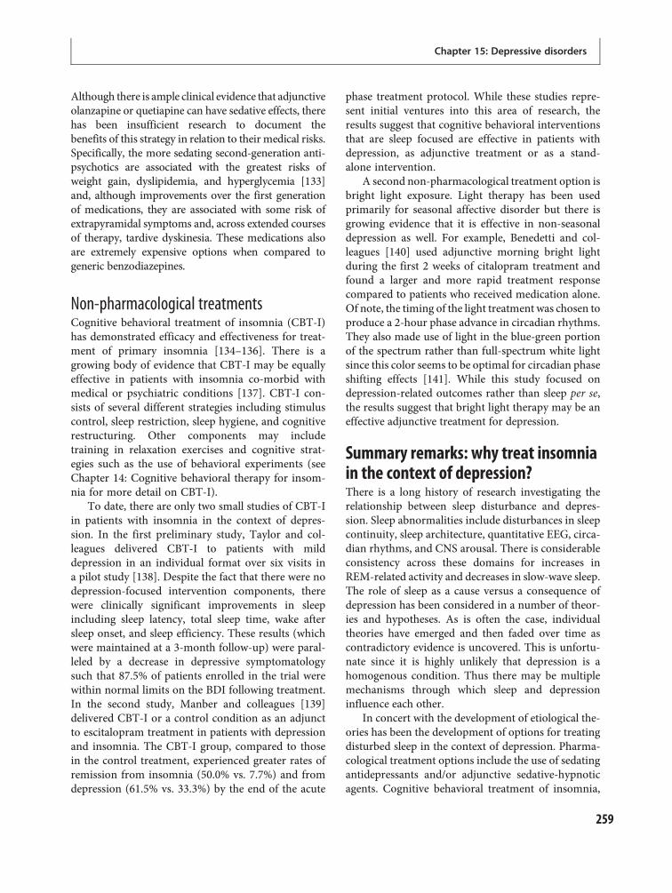
Although there is ample clinical evidence that adjunctiveolanzapine or quetiapine can have sedative effects, therehas been insufficient research to document thebenefits of this strategy in relation to their medical risks.Specifically, the more sedating second-generation anti-psychotics are associated with the greatest risks ofweight gain, dyslipidemia, and hyperglycemia [133]and, although improvements over the first generationof medications, they are associated with some risk ofextrapyramidal symptoms and, across extended coursesof therapy, tardive dyskinesia. These medications alsoare extremely expensive options when compared togeneric benzodiazepines.
Non-pharmacological treatmentsCognitive behavioral treatment of insomnia (CBT-I)has demonstrated efficacy and effectiveness for treat-ment of primary insomnia [134–136]. There is agrowing body of evidence that CBT-I may be equallyeffective in patients with insomnia co-morbid withmedical or psychiatric conditions [137]. CBT-I con-sists of several different strategies including stimuluscontrol, sleep restriction, sleep hygiene, and cognitiverestructuring. Other components may includetraining in relaxation exercises and cognitive strat-egies such as the use of behavioral experiments (seeChapter 14: Cognitive behavioral therapy for insom-nia for more detail on CBT-I).
To date, there are only two small studies of CBT-Iin patients with insomnia in the context of depres-sion. In the first preliminary study, Taylor and col-leagues delivered CBT-I to patients with milddepression in an individual format over six visits ina pilot study [138]. Despite the fact that there were nodepression-focused intervention components, therewere clinically significant improvements in sleepincluding sleep latency, total sleep time, wake aftersleep onset, and sleep efficiency. These results (whichwere maintained at a 3-month follow-up) were paral-leled by a decrease in depressive symptomatologysuch that 87.5% of patients enrolled in the trial werewithin normal limits on the BDI following treatment.In the second study, Manber and colleagues [139]delivered CBT-I or a control condition as an adjunctto escitalopram treatment in patients with depressionand insomnia. The CBT-I group, compared to thosein the control treatment, experienced greater rates ofremission from insomnia (50.0% vs. 7.7%) and fromdepression (61.5% vs. 33.3%) by the end of the acute
phase treatment protocol. While these studies repre-sent initial ventures into this area of research, theresults suggest that cognitive behavioral interventionsthat are sleep focused are effective in patients withdepression, as adjunctive treatment or as a stand-alone intervention.
A second non-pharmacological treatment option isbright light exposure. Light therapy has been usedprimarily for seasonal affective disorder but there isgrowing evidence that it is effective in non-seasonaldepression as well. For example, Benedetti and col-leagues [140] used adjunctive morning bright lightduring the first 2 weeks of citalopram treatment andfound a larger and more rapid treatment responsecompared to patients who received medication alone.Of note, the timing of the light treatment was chosen toproduce a 2-hour phase advance in circadian rhythms.They also made use of light in the blue-green portionof the spectrum rather than full-spectrum white lightsince this color seems to be optimal for circadian phaseshifting effects [141]. While this study focused ondepression-related outcomes rather than sleep per se,the results suggest that bright light therapy may be aneffective adjunctive treatment for depression.
Summary remarks: why treat insomniain the context of depression?There is a long history of research investigating therelationship between sleep disturbance and depres-sion. Sleep abnormalities include disturbances in sleepcontinuity, sleep architecture, quantitative EEG, circa-dian rhythms, and CNS arousal. There is considerableconsistency across these domains for increases inREM-related activity and decreases in slow-wave sleep.The role of sleep as a cause versus a consequence ofdepression has been considered in a number of theor-ies and hypotheses. As is often the case, individualtheories have emerged and then faded over time ascontradictory evidence is uncovered. This is unfortu-nate since it is highly unlikely that depression is ahomogenous condition. Thus there may be multiplemechanisms through which sleep and depressioninfluence each other.
In concert with the development of etiological the-ories has been the development of options for treatingdisturbed sleep in the context of depression. Pharma-cological treatment options include the use of sedatingantidepressants and/or adjunctive sedative-hypnoticagents. Cognitive behavioral treatment of insomnia,
Chapter 15: Depressive disorders
259

whose efficacy and effectiveness are well proven inprimary insomnia, may offer a non-pharmacologicaltreatment option as well. Regardless of treatmentoption, the current strategy is to provide insomniatreatment in patients with depression. While thisseems logical on the surface, management of sleepdisturbance may offer ameans for delaying or prevent-ing an episode of depression from occurring in the firstplace. The evidence that insomnia is a risk factor fornew-onset and recurrent depression suggests that suc-cessful treatment of insomnia in non-depressed indi-viduals has the potential for preventing futuredepressive episodes. This is a virtually unexploreddomain of clinical research that holds tremendouspotential for the future.
References1. American Psychiatric Association. Diagnostic and
statisticalmanual ofmental disorders, 4th ed.Washington,DC: American Psychiatric Association; 1994.
2. Tsuno N, Besset A, Ritchie K. Sleep and depression.J Clin Psychiatry. 2005;66(10):1254 1269.
3. Posternak MA, Zimmerman M. Symptoms of atypicaldepression. Psychiatry Res. 2001;104(2):175 181.
4. Roberts RE, Shema SJ, Kaplan GA, Strawbridge WJ.Sleep complaints and depression in an aging cohort:A prospective perspective. Am J Psychiatry. 2000;157(1):81 88.
5. American Academy of Sleep Medicine. Internationalclassification of sleep disorders, 2nd ed. Westchester,IL: American Academy of Sleep Medicine; 2005.
6. Mendelson W, Garnett D, Gillin J, Weingartner H. Theexperience of insomnia and daytime and nighttimefunctioning. Psychiatry Res. 1984;12:235 250.
7. Perlis M, Giles D, Mendelson W, Bootzin R, Wyatt J.Psychophysiological insomnia: The behavioural modeland a neurocognitive perspective. J Sleep Res.1997;6:179 188.
8. Hauri P, Olmstead E. Childhood onset insomnia.Sleep. 1980;3(1):59 65.
9. Perlis ML, Giles DE, Buysse DJ, et al. Which depressivesymptoms are related to which sleepelectroencephalographic variables? Biol Psychiatry.1997;42(10):904 913.
10. Perlis M, Smith M, Andrews P, Orff H, Giles D. Beta/gamma EEG activity in patients with primary andsecondary insomnia and good sleeper controls. Sleep.2001;24:110 117.
11. Riemann D, Hohagen F, Bahro M, Berger M. Sleep indepression: The influence of age, gender and
diagnostic subtype on baseline sleep and thecholinergic REM induction test with RS 86. Eur ArchPsychiatry Clin Neurosci. 1994;243(5):279 290.
12. Spielman A, Caruso L, Glovinsky P. A behavioralperspective on insomnia treatment. Psychiatr ClinNorth Am. 1987;10:541 553.
13. Kupfer DJ, Reynolds CF 3rd, Ulrich RF,Grochocinski VJ. Comparison of automated REM andslow wave sleep analysis in young and middle ageddepressed subjects. Biol Psychiatry. 1986;21(2):189 200.
14. Rush AJ, Erman MK, Giles DE, et al.Polysomnographic findings in recently drug free andclinically remitted depressed patients. Arch GenPsychiatry. 1986;43(9):878 884.
15. Kerkhofs M, Hoffmann G, De Martelaere V,Linkowski P, Mendlewicz J. Sleep EEG recordings indepressive disorders. J Affect Disord. 1985;9(1):47 53.
16. Gillin JC, Mendelson WB. Acetylcholine, sleep, anddepression. Human Neurobiol. 1982;1:211 219.
17. Borbely AA, Wirz Justice A. Sleep, sleep deprivationand depression: A hypothesis derived from a modelof sleep regulation.Hum Neurobiol. 1982;1(3):205 210.
18. Vogel GW, Vogel F, McAbee RS, Thurmond AJ.Improvement of depression by REM sleep deprivation.Arch Gen Psychiatry. 1980;37:247 253.
19. Benca RM, Obermeyer WH, Thisted RA, Gillin JC.Sleep and psychiatric disorders: A meta analysis. ArchGen Psychiatry. 1992;49:651 670.
20. Armitage R. Microarchitectural findings in sleep EEGin depression: Diagnostic implications. Biol Psychiatry.1995;37(2):72 84.
21. Borbely AA, Tobler I, Loepfe M, et al. All night spectralanalysis of the sleep EEG in untreated depressivesand normal controls. Psychiatry Res. 1984;12(1):27 33.
22. Kupfer DJ, Ulrich RF, Coble PA, et al. Application ofautomated REM and slow wave sleep analysis: II.testing the assumptions of the two process model ofsleep regulation in normal and depressed subjects.Psychiatry Res. 1984;13(4):335 343.
23. Mendelson WB, Sack DA, James SP, et al. Frequencyanalysis of the sleep EEG in depression. Psychiatry Res.1987;21:89 94.
24. Armitage R, Calhoun JS, Rush AJ, Roffwarg HP.Comparison of the delta EEG in the first and secondnon REM periods in depressed adults and normalcontrols. Psychiatry Res. 1992;41(1):65 72.
25. Hoffmann R, Hendrickse W, Rush AJ, Armitage R.Slow wave activity during non REM sleep in men withschizophrenia and major depressive disorders.Psychiatry Res. 2000;95(3):215 225.
Section VI: Sleep Disturbance in Psychiatric Illness
260

26. Perlis M, Merica H, Smith M, Giles D. Beta EEGactivity and insomnia. Sleep Med Rev. 2001;5:365 376.
27. Goodwin FK, Wirz Justice A, Wehr TA. Evidence thatthe pathophysiology of depression and the mechanismof action of antidepressant drugs both involvealterations in circadian rhythms. In: Costa E,Racagni G, eds. Typical and atypical antidepressants:clinical practice. New York: Raven Press; 1982:1 11.
28. Koenigsberg HW, Teicher MH, Mitropoulou V, et al.24 h monitoring of plasma norepinephrine, MHPG,cortisol, growth hormone and prolactin in depression.J Psychiatr Res. 2004;38(5):503 511.
29. David MM, MacLean AW, Knowles JB, Coulter ME.Rapid eye movement latency and mood following adelay of bedtime in healthy subjects: Do the effectsmimic changes in depressive illness? Acta PsychiatrScand. 1991;84(1):33 39.
30. Gillin JC, Borbely AA. Sleep: A neurobiologicalwindow on affective disorders. TINS. 1985:537 541.
31. Souetre E, Salvati E, Belugou JL, et al. Circadianrhythms in depression and recovery: Evidence forblunted amplitude as the main chronobiologicalabnormality. Psychiatry Res. 1989;28(3):263 278.
32. Nofzinger EA, Nichols TE, Meltzer CC, et al. Changesin forebrain function from waking to REM sleep indepression: Preliminary analyses of [18F]FDG PETstudies. Psychiatry Res. 1999;91(2):59 78.
33. Ho AP, Gillin JC, Buchsbaum MS, et al. Brain glucosemetabolism during non rapid eye movement sleep inmajor depression: A positron emission tomographystudy. Arch Gen Psychiatry. 1996;53(7):645 652.
34. Germain A, Nofzinger EA, Kupfer DJ, Buysse DJ.Neurobiology of non REM sleep in depression: Furtherevidence for hypofrontality and thalamic dysregulation.Am J Psychiatry. 2004;161(10):1856 1863.
35. Nofzinger EA, Buysse DJ, Germain A, et al. Functionalneuroimaging evidence for hyperarousal in insomnia.Am J Psychiatry. 2004;161(11):2126 2128.
36. Friess E, Wiedemann K, Steiger A, Holsboer F. Thehypothalamic pituitary adrenocortical system andsleep in man. Adv Neuroimmunol. 1995;5(2):111 125.
37. Ehlers C, Kupfer D. Hypothalamic peptide modulationof EEG sleep in depression: A further application of theS process hypothesis. Biol Psychiatry. 1987;22:513 517.
38. Richardson GS. Human physiological models ofinsomnia. Sleep Med. 2007;8(Suppl 4):S9 14.
39. Wu JC, Bunney WE. The biological basis of anantidepressant response to sleep deprivation andrelapse: Review and hypothesis. Am J Psychiatry.1990;147:14 21.
40. Reinink E, Bouhuys N, Wirz Justice A, van denHoofdakker R. Prediction of the antidepressant
response to total sleep deprivation by diurnal variationof mood. Psychiatry Res. 1990;32(2):113 124.
41. Van den Hoofdakker RH. Total sleep deprivation:Clinical and theoretical aspects. In: Honig A, ed.Depression. London: JohnWiley & Sons; 1997:563 589.
42. Wu JC, Gillin JC, Buchsbaum MS, et al. Effect of sleepdeprivation on brain metabolism of depressed patients.Am J Psychiatry. 1992;149(4):538 543.
43. Wu J, BuchsbaumMS, Gillin JC, et al. Prediction ofantidepressant effects of sleep deprivation by metabolicrates in the ventral anterior cingulate andmedial prefrontalcortex. Am J Psychiatry. 1999;156(8):1149 1158.
44. Smith GS, Reynolds CF 3rd, Pollock B, et al. Cerebralglucose metabolic response to combined total sleepdeprivation and antidepressant treatment in geriatricdepression. Am J Psychiatry. 1999;156(5):683 689.
45. Ebert D, Feistel H, Barocka A. Effects of sleepdeprivation on the limbic system and the frontal lobesin affective disorders: A study with tc 99m HMPAOSPECT. Psychiatry Res. 1991;40(4):247 251.
46. Ebert D, Feistel H, Barocka A, Kaschka W. Increasedlimbic blood flow and total sleep deprivation in majordepression with melancholia. Psychiatry Res. 1994;55(2):101 109.
47. Volk SA, Kaendler SH, Hertel A, et al. Can response topartial sleep deprivation in depressed patients bepredicted by regional changes of cerebral blood flow?Psychiatry Res. 1997;75(2):67 74.
48. Volk S, Kaendler SH, Weber R, et al. Evaluation of theeffects of total sleep deprivation on cerebral blood flowusing single photon emission computerizedtomography. Acta Psychiatr Scand. 1992;86(6):478 483.
49. Porkka Heiskanen T, Strecker RE, McCarley RW.Brain site specificity of extracellular adenosineconcentration changes during sleep deprivation andspontaneous sleep: An in vivo microdialysis study.Neuroscience. 2000;99(3):507 517.
50. Basheer R, Halldner L, Alanko L, et al. Oppositechanges in adenosine A1 and A2A receptor mRNA inthe rat following sleep deprivation. Neuroreport.2001;12(8):1577 1580.
51. Biber K, Fiebich BL, Gebicke Harter P, van Calker D.Carbamazepine induced upregulation of adenosineA1 receptors in astrocyte cultures affects coupling tothe phosphoinositol signaling pathway.Neuropsychopharmacology. 1999;20(3):271 278.
52. Janowsky DS, El Yousef MK, Davis JM. A cholinergicadrenergic hypothesis of mania and depression.Lancet. 1972;2:632 635.
53. Svestka J. Sleep deprivation therapy. Neuro EndocrinolLett. 2008;29(Suppl 1):65 92.
Chapter 15: Depressive disorders
261
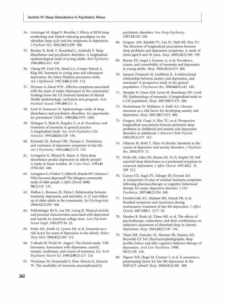
54. Grözinger M, Kögel P, Röschke J. Effects of REM sleepawakenings and related wakening paradigms on theultradian sleep cycle and the symptoms in depression.J Psychiatr Res. 2002;36(5):299 308.
55. Breslau N, Roth T, Rosenthal L, Andreski P. Sleepdisturbance and psychiatric disorders: A longitudinalepidemiological study of young adults. Biol Psychiatry.1996;39(6):411 418.
56. Chang PP, Ford DE, Mead LA, Cooper Patrick L,Klag MJ. Insomnia in young men and subsequentdepression: the Johns Hopkins precursors study.Am J Epidemiol. 1997;146(2):105 114.
57. Dryman A, Eaton WW. Affective symptoms associatedwith the onset of major depression in the community:Findings from the US National Institute of MentalHealth epidemiologic catchment area program. ActaPsychiatr Scand. 1991;84(1):1 5.
58. Ford D, Kamerow D. Epidemiologic study of sleepdisturbance and psychiatric disorders: An opportunityfor prevention? JAMA. 1989;262:1479 1484.
59. Hohagen F, Rink K, Kappler C, et al. Prevalence andtreatment of insomnia in general practice:A longitudinal study. Eur Arch Psychiatry ClinNeurosci. 1993;242(6):329 336.
60. Kennedy GJ, Kelman HR, Thomas C. Persistenceand remission of depressive symptoms in late life.Am J Psychiatry. 1991;148(2):174 178.
61. Livingston G, Blizard B, Mann A. Does sleepdisturbance predict depression in elderly people?A study in Inner London. Br J Gen Pract. 1993;43(376):445 448.
62. LivingstonG,WatkinV,Milne B,ManelaMV, Katona C.Who becomes depressed? The Islington communitystudy of older people. J Affect Disord. 2000;58(2):125 133.
63. Mallon L, Broman JE, Hetta J. Relationship betweeninsomnia, depression, and mortality: A 12 year followup of older adults in the community. Int Psychogeriatr.2000;12(3):295 306.
64. Paffenbarger RS Jr, Lee IM, Leung R. Physical activityand personal characteristics associated with depressionand suicide in American college men. Acta PsychiatrScand Suppl. 1994;377:16 22.
65. Perlis ML, Smith LJ, Lyness JM, et al. Insomnia as arisk factor for onset of depression in the elderly. BehavSleep Med. 2006;4(2):104 113.
66. Vollrath M, Wicki W, Angst J. The Zurich study. VIII.Insomnia: Association with depression, anxiety,somatic syndromes, and course of insomnia. Eur ArchPsychiatry Neurol Sci. 1989;239(2):113 124.
67. Weissman M, Greenwald S, Nino Murcia G, DementW. The morbidity of insomnia uncomplicated by
psychiatric disorders. Gen Hosp Psychiatry.1997;19:245 250.
68. Gregory AM, Rijsdijk FV, Lau JY, Dahl RE, Eley TC.The direction of longitudinal associations betweensleep problems and depression symptoms: A study oftwins aged 8 and 10 years. Sleep. 2009;32(2):189 199.
69. Buysse DJ, Angst J, Gamma A, et al. Prevalence,course, and comorbidity of insomnia and depressionin young adults. Sleep. 2008;31(4):473 480.
70. Jansson Frojmark M, Lindblom K. A bidirectionalrelationship between anxiety and depression, andinsomnia? A prospective study in the generalpopulation. J Psychosom Res. 2008;64(4):443 449.
71. Morphy H, Dunn KM, Lewis M, Boardman HF, CroftPR. Epidemiology of insomnia: A longitudinal study ina UK population. Sleep. 2007;30(3):274 280.
72. Neckelmann D, Mykletun A, Dahl AA. Chronicinsomnia as a risk factor for developing anxiety anddepression. Sleep. 2007;30(7):873 880.
73. Gregory AM, Caspi A, Eley TC, et al. Prospectivelongitudinal associations between persistent sleepproblems in childhood and anxiety and depressiondisorders in adulthood. J Abnorm Child Psychol.2005;33(2):157 163.
74. Ohayon M, Roth T. Place of chronic insomnia in thecourse of depressive and anxiety disorders. J PsychiatrRes. 2003;37:9 15.
75. Perlis ML, Giles DE, Buysse DJ, Tu X, Kupfer DJ. Selfreported sleep disturbance as a prodromal symptom inrecurrent depression. J Affect Disord. 1997;42(2 3):209 212.
76. Carney CE, Segal ZV, Edinger JD, Krystal AD.A comparison of rates of residual insomnia symptomsfollowing pharmacotherapy or cognitive behavioraltherapy for major depressive disorder. J ClinPsychiatry. 2007;68(2):254 260.
77. Dombrovski AY, Mulsant BH, Houck PR, et al.Residual symptoms and recurrence duringmaintenance treatment of late life depression. J AffectDisord. 2007;103(1 3):77 82.
78. Manber R, Rush AJ, Thase ME, et al. The effects ofpsychotherapy, nefazodone, and their combination onsubjective assessment of disturbed sleep in chronicdepression. Sleep. 2003;26(2):130 136.
79. Thase ME, Fasiczka AL, Berman SR, Simons AD,Reynolds CF 3rd. Electroencephalographic sleepprofiles before and after cognitive behavior therapy ofdepression. Arch Gen Psychiatry. 1998;55(2):138 144.
80. Pigeon WR, Hegel M, Unutzer J, et al. Is insomnia aperpetuating factor for late life depression in theIMPACT cohort? Sleep. 2008;31(4):481 488.
Section VI: Sleep Disturbance in Psychiatric Illness
262
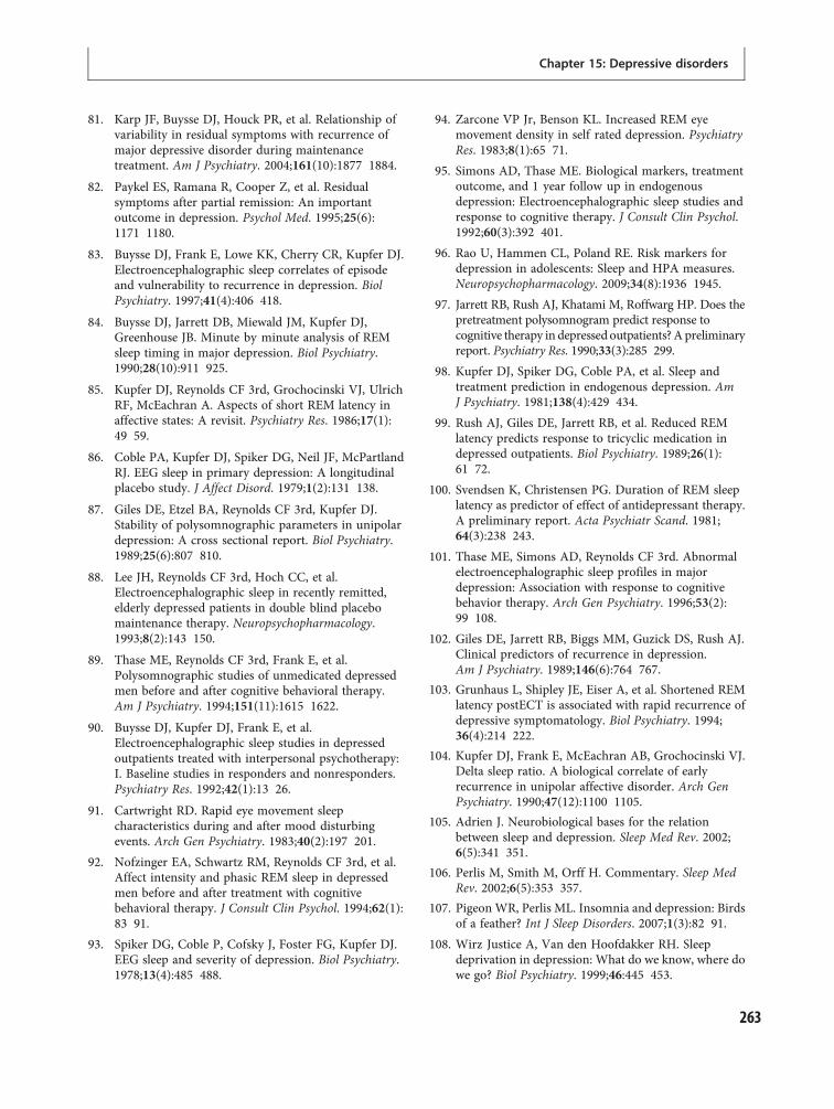
81. Karp JF, Buysse DJ, Houck PR, et al. Relationship ofvariability in residual symptoms with recurrence ofmajor depressive disorder during maintenancetreatment. Am J Psychiatry. 2004;161(10):1877 1884.
82. Paykel ES, Ramana R, Cooper Z, et al. Residualsymptoms after partial remission: An importantoutcome in depression. Psychol Med. 1995;25(6):1171 1180.
83. Buysse DJ, Frank E, Lowe KK, Cherry CR, Kupfer DJ.Electroencephalographic sleep correlates of episodeand vulnerability to recurrence in depression. BiolPsychiatry. 1997;41(4):406 418.
84. Buysse DJ, Jarrett DB, Miewald JM, Kupfer DJ,Greenhouse JB. Minute by minute analysis of REMsleep timing in major depression. Biol Psychiatry.1990;28(10):911 925.
85. Kupfer DJ, Reynolds CF 3rd, Grochocinski VJ, UlrichRF, McEachran A. Aspects of short REM latency inaffective states: A revisit. Psychiatry Res. 1986;17(1):49 59.
86. Coble PA, Kupfer DJ, Spiker DG, Neil JF, McPartlandRJ. EEG sleep in primary depression: A longitudinalplacebo study. J Affect Disord. 1979;1(2):131 138.
87. Giles DE, Etzel BA, Reynolds CF 3rd, Kupfer DJ.Stability of polysomnographic parameters in unipolardepression: A cross sectional report. Biol Psychiatry.1989;25(6):807 810.
88. Lee JH, Reynolds CF 3rd, Hoch CC, et al.Electroencephalographic sleep in recently remitted,elderly depressed patients in double blind placebomaintenance therapy. Neuropsychopharmacology.1993;8(2):143 150.
89. Thase ME, Reynolds CF 3rd, Frank E, et al.Polysomnographic studies of unmedicated depressedmen before and after cognitive behavioral therapy.Am J Psychiatry. 1994;151(11):1615 1622.
90. Buysse DJ, Kupfer DJ, Frank E, et al.Electroencephalographic sleep studies in depressedoutpatients treated with interpersonal psychotherapy:I. Baseline studies in responders and nonresponders.Psychiatry Res. 1992;42(1):13 26.
91. Cartwright RD. Rapid eye movement sleepcharacteristics during and after mood disturbingevents. Arch Gen Psychiatry. 1983;40(2):197 201.
92. Nofzinger EA, Schwartz RM, Reynolds CF 3rd, et al.Affect intensity and phasic REM sleep in depressedmen before and after treatment with cognitivebehavioral therapy. J Consult Clin Psychol. 1994;62(1):83 91.
93. Spiker DG, Coble P, Cofsky J, Foster FG, Kupfer DJ.EEG sleep and severity of depression. Biol Psychiatry.1978;13(4):485 488.
94. Zarcone VP Jr, Benson KL. Increased REM eyemovement density in self rated depression. PsychiatryRes. 1983;8(1):65 71.
95. Simons AD, Thase ME. Biological markers, treatmentoutcome, and 1 year follow up in endogenousdepression: Electroencephalographic sleep studies andresponse to cognitive therapy. J Consult Clin Psychol.1992;60(3):392 401.
96. Rao U, Hammen CL, Poland RE. Risk markers fordepression in adolescents: Sleep and HPA measures.Neuropsychopharmacology. 2009;34(8):1936 1945.
97. Jarrett RB, Rush AJ, Khatami M, Roffwarg HP. Does thepretreatment polysomnogram predict response tocognitive therapy in depressedoutpatients?A preliminaryreport. Psychiatry Res. 1990;33(3):285 299.
98. Kupfer DJ, Spiker DG, Coble PA, et al. Sleep andtreatment prediction in endogenous depression. AmJ Psychiatry. 1981;138(4):429 434.
99. Rush AJ, Giles DE, Jarrett RB, et al. Reduced REMlatency predicts response to tricyclic medication indepressed outpatients. Biol Psychiatry. 1989;26(1):61 72.
100. Svendsen K, Christensen PG. Duration of REM sleeplatency as predictor of effect of antidepressant therapy.A preliminary report. Acta Psychiatr Scand. 1981;64(3):238 243.
101. Thase ME, Simons AD, Reynolds CF 3rd. Abnormalelectroencephalographic sleep profiles in majordepression: Association with response to cognitivebehavior therapy. Arch Gen Psychiatry. 1996;53(2):99 108.
102. Giles DE, Jarrett RB, Biggs MM, Guzick DS, Rush AJ.Clinical predictors of recurrence in depression.Am J Psychiatry. 1989;146(6):764 767.
103. Grunhaus L, Shipley JE, Eiser A, et al. Shortened REMlatency postECT is associated with rapid recurrence ofdepressive symptomatology. Biol Psychiatry. 1994;36(4):214 222.
104. Kupfer DJ, Frank E, McEachran AB, Grochocinski VJ.Delta sleep ratio. A biological correlate of earlyrecurrence in unipolar affective disorder. Arch GenPsychiatry. 1990;47(12):1100 1105.
105. Adrien J. Neurobiological bases for the relationbetween sleep and depression. Sleep Med Rev. 2002;6(5):341 351.
106. Perlis M, Smith M, Orff H. Commentary. Sleep MedRev. 2002;6(5):353 357.
107. Pigeon WR, Perlis ML. Insomnia and depression: Birdsof a feather? Int J Sleep Disorders. 2007;1(3):82 91.
108. Wirz Justice A, Van den Hoofdakker RH. Sleepdeprivation in depression: What do we know, where dowe go? Biol Psychiatry. 1999;46:445 453.
Chapter 15: Depressive disorders
263

109. Vogel GW. Evidence for REM sleep deprivation as themechanism of action of antidepressant drugs. ProgNeuro Psychopharmacol Biol Psychiatry. 1983;7:343 349.
110. Vogel GW, Buffenstein A, Minter K, Hennessey A.Drug effects on REM sleep and on endogenousdepression. Neurosci Biobehav Rev. 1990;14:49 63.
111. Giedke H, Schwarzler F. Therapeutic use of sleepdeprivation in depression. Sleep Med Rev. 2002;6(5):361 377.
112. Wehr TA, Wirz Justice A, Goodwin FK, Duncan W,Gillin JC. Phase advance of the circadian sleep wakecycle as an antidepressant. Science. 1979;206:710 713.
113. Wehr TA, Wirz Justice A. Internal coincidence modelfor sleep deprivation and depression. In: Koella WP,ed. Sleep 1980. Basel: Karger; 1981:26 33.
114. Riemann D, Hohagen F, Konig A, et al. Advanced vs.normal sleep timing: Effects on depressed mood afterresponse to sleep deprivation in patients with a majordepressive disorder. J Affect Disord. 1996;37(2 3):121 128.
115. Hobson JA, McCarley RW, Wyzinski PW. Sleep cycleoscillation: Reciprocal discharge by two brainstemneuronal groups. Science. 1975;4196:55 58.
116. McCarley RW, Hobson JA. Neuronal excitabilitymodulation over the sleep cycle: A structural andmathematical model. Science. 1975;189:58 60.
117. Gillin JC, Sutton L, Ruiz C, et al. The cholinergic rapideye movement induction test with arecoline indepression. Arch Gen Psychiatry. 1991;48(3):264 270.
118. Gillin JC, Sitaram N, Duncan WC. Muscarinicsupersensitivity: A possible model for the sleepdisturbance of primary depression? Psychiatr Res.1979;1:17 22.
119. Risch SC, Janowsky DS, Mott MA, et al. Central andperipheral cholinesterase inhibition: Effects onanterior pituitary and sympathomimetic function.Psychoneuroendocrinology. 1986;11(2):221 230.
120. Mayers AG, Baldwin DS. Antidepressants and theireffect on sleep. Hum Psychopharmacol. 2005;20(8):533 559.
121. Jindal RD, Friedman ES, Berman SR, et al. Effects ofsertraline on sleep architecture in patients withdepression. J Clin Psychopharmacol. 2003;23(6):540 548.
122. Guelfi JD, Ansseau M, Timmerman L, Korsgaard S,Mirtazapine Venlafaxine Study Group. Mirtazapineversus venlafaxine in hospitalized severely depressedpatients with melancholic features. J ClinPsychopharmacol. 2001;21(4):425 431.
123. Nofzinger EA, Berman S, Fasiczka A, et al. Effects ofbupropion SR on anterior paralimbic function duringwaking and REM sleep in depression: Preliminary
findings using [18F] FDG PET. Psychiatry Res.2001;106(2):95 111.
124. Thase ME. Depression and sleep: Pathophysiologyand treatment. Dialogues Clin Neurosci. 2006;8(2):217 226.
125. James SP, Mendelson WB. The use of trazodone as ahypnotic: A critical review. J Clin Psychiatry. 2004;65(6):752 755.
126. Asnis GM, Chakraburtty A, DuBoff EA, et al. Zolpidemfor persistent insomnia in SSRI treated depressedpatients. J Clin Psychiatry. 1999;60(10):668 676.
127. Fava M, McCall WV, Krystal A, et al. Eszopicloneco administered with fluoxetine in patients withinsomnia coexisting with major depressive disorder.Biol Psychiatry. 2006;59(11):1052 1060.
128. Nowell PD, Buysse DJ. Treatment of insomnia inpatients with mood disorders. Depress Anxiety. 2001;14(1):7 18.
129. Walsh JK, Krystal AD, Amato DA, et al. Nightlytreatment of primary insomnia with eszopiclone for sixmonths: Effect on sleep, quality of life, and worklimitations. Sleep. 2007;30(8):959 968.
130. Fava M, Thase ME, DeBattista C, et al. Modafinilaugmentation of selective serotonin reuptake inhibitortherapy in MDD partial responders with persistentfatigue and sleepiness. Ann Clin Psychiatry. 2007;19(3):153 159.
131. DeBattista C, Doghramji K, Menza MA, RosenthalMH, Fieve RR, Modafinil in Depression Study Group.Adjunct modafinil for the short term treatment offatigue and sleepiness in patients with major depressivedisorder: A preliminary double blind, placebocontrolled study. J Clin Psychiatry. 2003;64(9):1057 1064.
132. Thase ME, Fava M, DeBattista C, Arora S, Hughes RJ.Modafinil augmentation of SSRI therapy in patientswith major depressive disorder and excessive sleepinessand fatigue: A 12 week, open label, extension study.CNS Spectr. 2006;11(2):93 102.
133. Newcomer JW. Metabolic considerations in the use ofantipsychotic medications: A review of recentevidence. J Clin Psychiatry. 2007;68(Suppl 1):20 27.
134. Murtagh DR, Greenwood KM. Identifying effectivepsychological treatments for insomnia: A metaanalysis. J Consult Clin Psychol. 1995;63:79 89.
135. Morin CM, Culbert JP, Schwartz MS. Nonpharmacological interventions for insomnia: A metaanalysis of treatment efficacy. Am J Psychiatry.1994;151:1172 1180.
136. Smith MT, Perlis ML, Park A, et al. Comparative metaanalysis of pharmacotherapy and behavior therapy
Section VI: Sleep Disturbance in Psychiatric Illness
264
for persistent insomnia. Am J Psychiatry. 2002;159(1):5 11.
137. Stepanski EJ, Rybarczyk B. Emerging research on thetreatment and etiology of secondary or comorbidinsomnia. Sleep Med Rev. 2006;10(1):7 18.
138. Taylor DJ, Lichstein KL, Weinstock J, Sanford S,Temple JR. A pilot study of cognitive behavioraltherapy of insomnia in people with mild depression.Behav Ther. 2007;38(1):49 57.
139. Manber R, Edinger JD, San Pedro M. Combiningescitalopram oxalate (ESCIT) and individual cognitivebehavioral therapy for insomnia (CBTI) to improvedepression outcome. Sleep. 2007;30:A232.
140. Benedetti F, Colombo C, Pontiggia A, et al. Morninglight treatment hastens the antidepressant effect of
citalopram: A placebo controlled trial. J ClinPsychiatry. 2003;64(6):648 653.
141. Horne JA, Donlon J, Arendt J. Green light attenuatesmelatonin output and sleepiness during sleepdeprivation. Sleep. 1991;14:233 240.
Endnote
1. Sleep continuity for the purpose of this chapter encompassesmultiple descriptive measures of sleep initiation and maintenance. While the term is not formally part of the sleep lexicon, ithas the heuristic value of being a global term whose meaningmay be contrasted with the class of variables that corresponds to“sleep architecture” and “sleep microarchitecture.”
Chapter 15: Depressive disorders
265
Chapter
16 Bipolar disorder
David T. Plante and John W. Winkelman
IntroductionSleep disturbance has long been recognized as anessential aspect of bipolar disorder. Emil Kraepelin,who first distinguished the fluctuating course ofmanic depression from the unremitting progressionof dementia praecox, noted nearly a century ago:
The attacks of manic depressive insanity are invariablyaccompanied by all kinds of bodily changes. By far themost striking are the disorders of sleep and generalnourishment. In mania sleep is in the more severe statesof excitement always considerably encroached upon;sometimes there is even almost complete sleeplessness,at most interrupted for a few hours, which may last forweeks, even months . . . In the states of depression inspite of great need for sleep, . . . the patients lie for hours,sleepless in bed, . . . although even in bed they find norefreshment [1].
Although Kraepelin’s observations have greatly influ-enced modern day nosological classifications of majormental illness, including sleep-related criteria formanic and depressive episodes as defined by the Diag-nostic and Statistical Manual, our understanding ofthe biology of sleep regulation and its relationship tobipolar disorder continues to advance. As detailed inprevious chapters (see Chapter 2: Neuroanatomy andneurobiology of sleep and wakefulness), currentmodels suggest that the interaction of a circadianclock and homeostatic sleep drive governs sleep onsetand maintenance. Thus, abnormalities in either thecircadian mechanism or sleep homeostat could theor-etically manifest as a disorder of the sleep–wake cycle[2,3]. However, the vast majority of experiments,especially those designed to probe sleep in psychiatricillness, are not designed to uncouple homeostatic andcircadian processes, and thus the mechanisms under-lying an observed disturbance of sleep and wakeful-ness are often not clear. This caveat is of particular
import when evaluating the literature regarding sleepin bipolar disorder, as disruptions in both sleep andcircadian rhythms have been described in thesepatients [4,5].
This chapter will detail sleep–wake disturbances inthe three primary states of bipolar disorder: mania,depression, and euthymia. Mixed states will not be aseparate focus of discussion due to a dearth of availableliterature. Additionally, this chapter will focus on bipo-lar disorder in adults; for discussion of sleep in pediatricbipolar disorder, the reader is directed to other chaptersin this volume (Chapter 22: Pediatric mood and anxietydisorders). In keeping with the aims of this volume tobe a rigorous, yet clinically useful, text, particular atten-tion will be paid to sleep in the course and treatmentof bipolar disorder, including pharmacological andbehavioral methods for maintaining adequate sleepquality and quantity in these patients.
Sleep in maniaThe relationship between sleep and the manic phase ofbipolar illness involves the following aspects:(1) decreased need for sleep is a marker of mania; (2)sleep deprivation can induce mania; (3) sleep durationmay predictmanic episodes; and (4) sleep timemay be amarker of response (and target of treatment) in mania.Each of these relationships will be addressed in whatfollows.
Decreased need for sleep as a markerof maniaDecreased need for sleep is one of seven characteristicsymptoms of the manic phase of bipolar disorder, andis of high diagnostic value since the ability to main-tain wakefulness and energy without sufficient sleep is
Foundations of Psychiatric SleepMedicine, ed. JohnW.Winkelman andDavid T. Plante. Published by CambridgeUniversity Press.# Cambridge University Press 2010.
266

not observed in many other psychiatric or medicaldisorders [6]. As evidence of its relatively high speci-ficity for bipolar disorder, decreased need for sleep,along with euphoria and grandiosity, described onehalf of all clinically validated bipolar I cases in theNational Co-morbidity Survey, and formed the onlymanic symptom profile that could be validly assessedwith the Composite International Diagnositc Inter-view (CIDI), a fully structured interview developedto generate diagnoses according to the definitions andcriteria of the DSM-III-R and ICD-10 [7].
Although the ability to maintain energy withoutsleep is characteristic of mania, it is noteworthy thatmanic patients still require sleep to sustain life. Priorto the advent of modern psychiatric treatments (e.g.neuroleptics and benzodiazepines), severe cases offlorid mania characterized by almost no sleep were attimes reported to result in death [8]. These observa-tions are similar to animal models of sleep deprivationin which animals predictably die from prolonged totalsleep deprivation, despite significantly increased foodintake [9].
That decreased sleep is also characteristic of maniais corroborated by objective (e.g. polysomnographicand actigraphic) measures. Although polysomnogr-aphy in manic patients is technically difficult, suchstudies of unmedicated manic patients demonstrateshortened total sleep time, increased time awakein bed, and shortened REM latency – similar to poly-somnographic parameters seen in depressed patients[10,11]. Also, longitudinal studies utilizing actigraphyto measure rest–activity patterns in patients hospital-ized with mania or mixed episodes demonstratedecreased total sleep time with increased nocturnalactivity compared to euthymic periods [12]. It isworth noting that sleep measures in mania maybe confounded by the effects of motor hyperactivitysince sleep architecture can be affected by increaseddaytime activity in normal subjects [13]. Additionally,manic patients demonstrate increased nocturnalcortisol and an advanced (i.e. earlier) nadir of cortisolsecretion compared to healthy subjects; however,similar alterations have been observed in sleep depriv-ation paradigms [14,15]. Thus, it is not clear ifpolysomnographic abnormalities seen in mania arecaused by the manic state per se, are secondaryto other associated abnormalities (e.g. increased phys-ical activity levels, changes in metabolic and endo-crine function, etc.), or are the sequelae of sleepdeprivation itself.
Sleep reduction as a cause of maniaPotential triggers for mania are vast and have beenreported to include drug use (licit and illicit), transmer-idinal travel, post-partum states, bereavement, etc., all ofwhich may be associated with sleep loss [16–22]. How-ever, in most such cases, it is unclear whether sleepless-ness was a cause or prodrome ofmania. Thus cause andeffect cannot be determined from case reports alone,especially if early manic symptoms may have spurredthe behavior (e.g. drug use, travel, etc.) which subse-quently produced sleep deprivation.
However, therapeutic studies of sleep deprivation inunipolar and bipolar depression do provide causal evi-dence for the propensity for sleep deprivation to inducemanic switching. Recent work in this area has beenperformed in well-characterized subjects; however, theolder literature (before definitions of rapid-cycling wereestablished) includes mixtures of patients with bothrapid and non-rapid cycling patterns, as well as thosewith unipolar depression. Further complicating inter-pretation of the older literature is that the majority ofearly sleep deprivation studies were not designed todetect mania, depressed unipolar and bipolar patientswere not distinguished from one another in reportedresults, and hypomania and mania were often reportedpost hoc [23,24]. A comprehensive review of the oldertherapeutic sleep deprivation literature found 29% ofbipolar depressed subjects and 25% of unspecifieddepressed subjects became hypomanic or manic afterone night of total sleep deprivation (TSD) [25]. How-ever, examination of sleep deprivation studies withexperimental design that was better suited for predict-ing the frequency of manic switching has estimateda risk of hypomania andmania of 12% and 7%, respect-ively, with TSD [26]. More recently, Colombo et al.(1999), reviewed data from 206 patients who receivedTSD (frequently with supplemental medicationsintended to extend the duration of antidepressantresponse) for bipolar depression, finding only 4.9%and 5.8% of such patients switched into mania or hypo-mania, respectively [27]. Interestingly, one-third of sub-jects who switched into mania had resolution ofsymptomswithin 3–5 dayswith nocturnal benzodiazep-ines; the remaining subjects required either mood sta-bilizers and/or antipsychotic medications [27]. Onenotable caveat when interpreting the sleep deprivationliterature is that all such studies have been designed totreat depression (unipolar and/or bipolar type), andthus may not accurately reflect the likelihood of sleep
Chapter 16: Bipolar disorder
267

deprivation to induce manic switching in euthymicbipolar patients.
As a synthesis of experimental and observationalstudies, Wehr and colleagues proposed the “finalcommon pathway” hypothesis, which posits sleepreduction as the fundamental proximal cause formanic switching (Figure 16.1) [28]. According tothis model, all triggers of mania including biologicalcauses (e.g. drugs, withdrawal), psychic effects (e.g.separation, bereavement), or direct disturbancesof sleep schedules (e.g. newborn infants, shift work,transmeridian travel) act through sleep reduction[28]. This theory proposes that sleep deprivation isboth a cause and consequence of mania, and thusmutually self-reinforcing sleep loss perpetuates themanic state. Prospective testing of the final commonpathway hypothesis is logistically difficult since sleepdeprivation is both a cause and prodromal symptomof mania. However, cases of bipolar inpatients switch-ing into mania after sleep deprivation from variedetiologies have been reported, supporting the finalcommon pathway hypothesis [29].
Of clinical import is the observation that primarysleep disorders, in particular obstructive sleep apnea,have been documented as causes of mania and/or
treatment resistance [30–32]. Thus, primary sleepdisorders may be an additional cause of functionalsleep deprivation leading to mania not originallyincluded in Wehr’s “final common pathway” model(Figure 16.1).
Sleep as a predictor of maniaIf sleep deprivation can trigger mania, then perhapssleep duration may anticipate and thus predict maniaover the course of the illness. Recent analysis of self-reported sleep duration of participants in the SystemicTreatment Enhancement Program for Bipolar Disorder(STEP-BD) trial demonstrates an association betweenreported short sleep duration (<6 hours) in the weekprior to enrollment and mood elevation, earlier age ofonset, and longer illness duration compared to normalor long sleepers [33]. However, despite its impressivesample size (greater than 2000 subjects), this study canonly suggest that current sleep duration is associatedwith mood and quality of life variables – inferencesregarding a causal relationship between sleep andmoodare not possible without longitudinal study.
There are unfortunately few longitudinal studiesthat examine the relationship between sleep and
Events with Somatic EffectsDrugs, withdrawal,
illness, etc.
Events Disrupting Sleep SchedulesNewborn, travel,
shift work,“all-nighter”,social activities, etc.
Emotional ReactionsExcitement, anxiety,
grief, etc.
Insomnia
Events with Psychic EffectsSeparation, loss, role change, etc..
Sleep Reduction
Mania
Sleep Deprivation
Primary Sleep DisordersOSA, etc.
Figure 16.1 Sleep Reduction as a “final common pathway of mania” revisited. From Plante and Winkelman, 2008 [190], originallyadapted from Wehr et al, 1987 [28]. Reprinted with permission from the American Journal of Psychiatry (Copyright 2008). AmericanPsychiatric Association.
Section VI: Sleep Disturbance in Psychiatric Illness
268

mood in bipolar patients. Wehr et al. (1982) followedinpatients with bipolar disorder (15 rapid-cycling and52 non-rapid-cycling), and found that the majorityexperienced one or more consecutive nights withoutsleep each time they switched from depression tomania [34]. Leibenluft et al. (1996) followed 11rapid-cycling bipolar subjects with sleep logs andmood ratings for 18 months, finding sleep durationpredicted the subsequent day’s mood, such thatincreased sleep was associated with decreased prob-ability of hypomania or mania the following day infive of eight subjects who had a sufficient number ofmood episodes for analysis [35]. Similarly, Bauer et al.(2008), building on previous pilot data, found asignificant negative cross-correlation between sleep(defined as sleep or bed rest) and mood on the dayof or before a mood change in 42% of a mixedpopulation of 101 bipolar I and II outpatients suchthat decreased sleep (particularly of <3 hours) wasmore predictive of hypomanic or manic symptoms[36]. Notably, subjects who demonstrated a signifi-cant cross-correlation tended to be more symptom-atic, with fewer euthymic days and more days withdepression or mania. Furthermore, sleep duration,rather than time of sleep onset or offset, seemed tobe most predictive of a mood change [37]. Perlmanet al. (2006) examined self-reported sleep duration atmonthly intervals in bipolar I subjects, finding sleepdeficit predicted depressive symptoms at 6-monthfollow-up, but was not predictive of manic epi-sodes [38]. Of note, manic patients were less likelyto complete sleep measures and were more likely todrop out, and examining monthly intervals of self-reported sleep duration may have been too infre-quent to detect a relationship between sleep andmania in this study [38].
There are also a few medication trials that exam-ine the association of sleep disturbance in manicrelapse. Klein et al. (1992), using actigraphy in asmall, non-randomized, double-blind crossover studyof lithium discontinuation, found relapsers did notdiffer from non-relapsers in estimated sleep efficiencyor sleep activity either pre- or post-lithium discon-tinuation; however, significant differences were foundin daytime motor activity [39]. In a post hoc analysisof Young Mania Rating Scale (YMRS) items in bipo-lar patients enrolled in a maintenance trial, Houstonet al. (2005) found increased motor activity andenergy was a strong marker of initial manic symp-toms, and that decreased need for sleep occurred in
25% and 10.5% of patients in the 2 weeks precedingmanic relapse in subjects maintained on olanzapineand lithium, respectively [40].
Although limited by recall bias, patient retrospect-ive reports also provide insight into the role of sleepin the genesis of mania. Jackson et al. (2003) foundsleep disturbance was the most commonly reportedprodromal symptom (77% of patients) prior to a manicepisode in a review of 11 studies [41]. Similarly, in aretrospective interview study of 127 women whodeveloped bipolar affective post-partum psychosis,48% of subjects reported not needing to sleep or beingunable to sleep as the first symptoms of their episode[42]. Such observations are important as teachingpatients to recognize early symptoms of a manicrelapse and to seek early treatment may increase timeto a manic episode, and improve occupational andsocial functioning [43].
Sleep as a marker of responseand a therapeutic target in maniaThere is some, albeit limited, evidence suggestingsleep duration may be a harbinger of response inmania. In a retrospective chart review, total sleep timeon the first night of hospitalization was a predictor ofrapid response (and discharge) for those admitted toan inpatient unit with mania [44]. Similarly, inpatientobservations of sleep duration have correlated withratings of cooperation and irritability among manicinpatients using the Nurses’ Observation Scale forInpatient Evaluation (NOSIE), but not using morestandardized measures such as the YMRS [45].
Although there is no causal evidence that increasedsleep improves the course of mania, clinicians havelong used agents that can increase sleep and/or sed-ation in manic patients to achieve therapeutic effect.Sedating agents including bromides, chloroform, alco-hol, and opium were utilized over a century ago tomanage agitated psychiatric states. More recently, thesedating medications most frequently used in acutemania are benzodiazepines and antipsychotics, andclinical experience suggests that sedation alone causedby these agents may be valuable in managing manicbehavior. Benzodiazepines (e.g. clonazepam and lor-azepam) are as effective as neuroleptics as adjunctivemedications when used with lithium in acute mania[46,47]. In fact, benzodiazepines are frequently used asrescue medications in clinical trials, and it is possiblethat a “placebo” response in some trials may be the
Chapter 16: Bipolar disorder
269
result of benzodiazepine use [48]. All atypical anti-psychotics demonstrate efficacy in the managementof acute mania, and it is noteworthy that these agentsare often sedating and several increase total sleep timein healthy subjects [49–51]. Unfortunately, it is notclear to what degree these agents exert their effectsvia sleep-related systems, or to what degree sedationalone either masks manic symptoms or reversesunderlying processes responsible for mania [24].
There is also evidence that improvement in sleepduration by manipulation of the circadian systemmay have some benefits in manic patients. In oneopen-label study, Bersani et al. (2000) found thatmelatonin (3mg at bedtime) improved subjectivesleep duration and manic symptoms in 11 outpatientswith mania whose insomnia was resistant to benzodi-azepines [52]. Since melatonin is a relatively poorhypnotic but is an important neurohormone in theregulation of circadian rhythms, this result may havebeen mediated through circadian processes ratherthan homeostatic or sedating mechanisms [53].
In addition to pharmacological management,behavioral interventions designed to improve or extendsleep have been used in the treatment of mania for over100 years [54]. During the nineteenth century, S. WeirMitchell advocated that “rest cure” (prolonged bed rest)be used for a variety of neuropsychiatric disorders [54].In modern times, similar techniques have shown prom-ise in treating patients with bipolar disorder.Wehr et al.(1998) used 14 hours of bed rest to stabilize a patientwith treatment refractory rapid-cycling bipolar dis-order. Interestingly, prolonged bed rest did not increasetotal sleep time, but rather decreased variability of sleepduration [55]. Similarly, 14 hours of enforced darknessin hospitalized manic patients significantly decreasedYMRS scores when treated within 2 weeks of onset ofthe manic episode, and patients treated with dark ther-apy alsowere discharged from the hospitalmore quicklyand required lower doses of antimanic agents than thosewith standard care [56]. In this study, nursing observa-tion of sleep duration suggested enforced darkness didincrease total sleep time; however, nursing observationof sleep duration can overestimate sleep duration[56,57]. Similar to the use of melatonin in mania, bedrest with enforced darkness may lead to improvementvia a circadian manipulation, since light is the primary“zeitgeber” (timegiver) of the circadian clock, andpatients may become better without clear improvementin sleep per se. Furthermore, more regulated light–darkcycles on inpatient units compared to outside the
hospital could theoretically be responsible, at least inpart, for the beneficial effects of the milieu on manicpatients.
Sleep in bipolar depressionAlthough less is known about sleep disturbance inbipolar as compared to unipolar depression, in thefollowing section we will highlight the phenomen-ology of sleep in bipolar depression and the potentialof sleep deprivation as a theraputic strategy in thedepressive phase of bipolar illness.
Objective and subjective sleep disturbancein bipolar depressionSleep disturbance, either insomnia or hypersomnia, isa core diagnostic feature of depressive episodes inunipolar and bipolar depression [6]. Polysomno-graphic studies of sleep in bipolar depression havegenerally found similar abnormalities in unipolarand bipolar depression, with a lack of consensusregarding potential differences among studies [58].However, the majority of studies did not carefullycontrol for potential confounding variables, andrecent data suggests that bipolar patients may havemore early morning awakenings and greater totalREM density than unipolar subjects when matchedfor age, gender, and severity of symptoms [59].
Some have argued that hypersomnia, rather thaninsomnia, either in isolation or as a component ofatypical depression is more indicative of bipolar thanunipolar depression [60,61]. Reported rates of hyper-somnia in studies of bipolar depression are highlyvariable, ranging from 17% to 78%, as are rates ofinsomnia, suggesting that neither can be relied uponsolely to discriminate between bipolar and unipolardepression [58,62]. However, recent studies demon-strate that patients with bipolar disorder are morelikely to report subjective hypersomnia during adepressive episode than unipolar subjects [63–65].Additionally, hypersomnia, as part of the constell-ation of atypical depression, has been correlated withincreased rates of family history of bipolar disorder,compared to depressed subjects without atypicalfeatures [66]. Thus the presence of subjective hyper-somnolence during a major depressive episodemay be suggestive of an underlying bipolar diathesis,but is certainly not specific and must be consideredin light of other current and past symptoms.
Section VI: Sleep Disturbance in Psychiatric Illness
270
It is important for practicing clinicians to notethat hypersomnolence that can occur in bipolardepression is different from that of narcolepsy andother disorders of central hypersomnolence (seeChapter 9: Hypersomnias of central origin). Whenevaluated using the multiple sleep latency test(MSLT), an objective measure of sleepiness, bipolardepressed subjects with subjective hypersomnia didnot exhibit objective evidence of excessive daytimesleepiness, suggesting that bipolar hypersomnolenceis more reflective of anergia/fatigue than true exces-sive sleepiness seen in some other primary sleep dis-orders [67]. This discrepancy between objective andsubjective measures of hypersomnolence is importantto consider when evaluating studies that examinerelationships between sleep duration and depressedmood in bipolar subjects since self-reported longsleep duration could be due to an increased sleeppropensity or to fatigue/anergia with increased timein bed without increased physiological sleep drive.
With this caveat in mind, there is some data tosuggest that the subjective report of long sleep dur-ation may correlate with depressed mood in bipolardisorder. Bauer et al. (2008) reported in their longitu-dinal sample of bipolar subjects that increased sleepfrom baseline (particularly >3 hours) was morepredictive of a mood change to depression [36].Additionally, self-reported average sleep duration thatwas either less than 6 hours or greater than 9 hoursper night in the week prior to enrollment was associ-ated with depressive symptoms, poorer life function-ing, and quality of life compared to normal sleepers inthe STEP-BD trial [33].
Sleep deprivation in bipolar depressionAs discussed previously, the use of sleep deprivationas a treatment for depression significantly advancedour understanding of the relationship between sleepdeprivation and mania. Although there is data sug-gesting that sleep deprivation may be an effectivetreatment for bipolar depression, the AmericanPsychiatric Association considers it a novel approach,and sleep deprivation as a somatic therapy is currentlynot widely used or studied [68]. This is likely due tolimited data compared to conventional treatments,concern about manic switching, logistical difficultiesof sleep deprivation in controlled settings, frequentreturn of depressive symptoms after recoverysleep, and the dominance of other areas (e.g.
pharmacotherapy, neurochemistry, genetics) in mooddisorders research [69]. Still, because sleep depriv-ation is the fastest known means of alleviating depres-sive symptoms, recent data suggest adjunctivetreatments may prolong its antidepressant response,and bipolar depression can be clinically difficult totreat and lead to significant morbidity, some havecalled for renewed interest and study of sleep depriv-ation as a somatic therapy [70].
Although previous reviews of the older thera-peutic sleep deprivation literature did not reveal dif-ferences in response to sleep deprivation amongbipolar and unipolar subjects, more recent studieshave suggested bipolar depressives may respond morerobustly than unipolar subjects [25]. Szuba et al.(1990), in a small prospective study of 37 subjects witheither unipolar, bipolar I, or bipolar II depression,found 8/9 (89%) bipolar I subjects responded to partialsleep deprivation (PSD), compared to 9/24 (38%) uni-polar subjects [71]. Also, in a prospective study of51 subjects using a repeated TSD paradigm, Barbiniet al. (1998) found that although all subjects hadimprovement in depressive symptoms, bipolar I, bipo-lar II, and first-episode unipolar subjects had signifi-cantly greater response to TSD when compared tounipolar subjects with a prior history of depressiveepisodes [72]. Interestingly, whether depressive symp-toms are amenable to sleep deprivation may alsodepend on the time course of the depressive episodeas a small case series found little response to sleepdeprivation early in the course of a depressive episodeamong rapid-cycling patients, but more significantresponses later in the course of the mood episode [73].
Numerous approaches to augment and extend theantidepressant response to sleep deprivation havebeen examined. Several reports demonstrate lithiummay improve response to sleep deprivation andsustain remission in unipolar and bipolar depressives[74–77]. Also, Smeraldi et al. (1999) demonstratedthat pindolol, a 5-HT1A-beta adrenoreceptor blockingagent, improved the response rates of bipolar depres-sives to TSD and that remission could be maintainedwith lithium salts alone in nearly two-thirds of cases[78]. Additionally, bright light in the morning is alsohelpful in sustaining antidepressant response to sleepdeprivation in bipolar subjects [79–81]. Also, phaseadvance (i.e. moving the sleep period several hoursearlier than usual) after sleep deprivation sustains theantidepressant effects of sleep deprivation in bothunipolar and bipolar subjects [82–85].
Chapter 16: Bipolar disorder
271

Increasingly, there is evidence that genetics, spe-cifically genes important in neurotransmission andthe molecular circadian mechanism, may play a rolein the antidepressant response to sleep deprivationin bipolar disorder. Homozygotes for the long variantof a polymorphism in the transcriptional controlregion upstreamof the coding sequence of the serotonintransporter 5-HTTLPR were more likely to respond tosleep deprivation than those who were heterozygotesor homozygotes for the short variant [86].Additionally, bipolar depressives who were homozyg-otes for the T variant on the 5-HT2A receptor genepolymorphism rs6313 had both perceived and observedbenefits, as well as a higher final response rate, from arepeated TSD paradigm, than carriers of the C allele[87]. More recently, subjects with bipolar disorderwho were homozygotes for a Val/Val variant in cat-echol-O-methyltransferase (COMT), which inactivatesnorepinephrine and dopamine, exhibited a decreasedantidepressant response to sleep deprivation followedby bright light when compared to heterozygotes (Val/Met) or homozygotes (Met/Met) [88]. Additionally, aglycogen synthase kinase 3-beta (GSK3-b) promotergene single nucleotide polymorphism (SNP) has anassociation with response to TSD in bipolar depression,which is noteworthy as GSK3-b is involved in the circa-dian molecular mechanism [89].
Sleep in euthymiaEuthymia, the absence of mood symptoms sufficientto warrant the diagnosis of a depressive, hypomanic,or manic episode, is the goal of treatment of bipolardisorder. However, most clinicians are well aware thatthis disease state is characterized by frequent subclin-ical symptomatology [90]. Although there is relativelylittle data about sleep in euthymic periods in bipolardisorder, it does appear that sleep disturbance is oneof the more common forms of interepisode problemsfor these patients.
There are a limited number of studies that haveutilized polysomnography in euthymic bipolar sub-jects. One study, using polysomnography overfive nights, found the only difference between the tenremitted bipolar patients and age-matched controlswas mildly elevated rates of arousals [91]. However,another study demonstrated increased REM density,percentage of REM sleep, and sensitivity to the REMlatency reducing effects of arecoline (an acetylcholineagonist) compared to normal controls [92].
A few more recent studies have used actigraphy tomonitor the sleep–wake patterns of remitted bipolarsubjects. Using sleep diaries and actigraphy, Millaret al. (2004) demonstrated that remitted bipolarI patients had greater sleep onset latency, increasedsleep duration, and more night-to-night variabilityof sleep patterns than healthy controls [93]. Joneset al. (2005) similarly compared the circadian activitypatterns of bipolar subjects to healthy controls, findinggreater variability of daytime activity patterns in bipolarsubjects, but no significant differences in sleep param-eters (e.g. sleep onset latency) between groups [94].However, since subjects were only asked to recordbedtime and getting up time, and the remaining sleepparameters were calculated using actigraphy, sleeplatency and wake time after sleep onset may have beenunderestimated if subjects were awake in bed with littlemovement [94]. Finally, Harvey et al. (2005) comparedeuthymic bipolar patients, patients with insomnia, andsubjects with good sleep, finding 70% of the euthymicbipolar patients exhibited a clinically significant sleepdisturbance [95]. Interestingly, when compared toother groups, euthymic bipolar patients had diminishedsleep efficiency, decreased daytime activity levels,increased anxiety and fear about poor sleep, and hadlevels of dysfunctional beliefs about sleep comparable tonon-bipolar patients with insomnia [95].
As previously mentioned, it has been hypothesizedthat alterations in the circadian systemmay be involvedin the pathophysiology of bipolar disorder. Of importis that sleep and/or circadian abnormalities are presentin periods of euthymia as well as duringmood episodes,suggesting that such abnormalities are trait markersof illness, rather than state dependent. Much of theolder literature described abnormalities includingblunting of the amplitude of the circadian rhythm, aswell as phase advanced or non-24-hour rhythms; how-ever, such conclusions were based on data from subjectswith active mood symptoms, making it difficult tointerpret whether such abnormalities are trait or statedependent [4,5]. More recently, two separate groupsdemonstrated that subjects with bipolar disorder aremore likely to have an evening chronotype as measuredby morningness/eveningness (M/E) scores, a stable,quantifiable self-report measure reflecting preferredcircadian phase [96–98]. However, it is notable thatdata from one group suggested that frequent moodswings and higher scores on the Beck depression inven-tory (BDI) increased the likelihood of lower (i.e. eveningpreference) M/E scores, and thus it is not clear to
Section VI: Sleep Disturbance in Psychiatric Illness
272
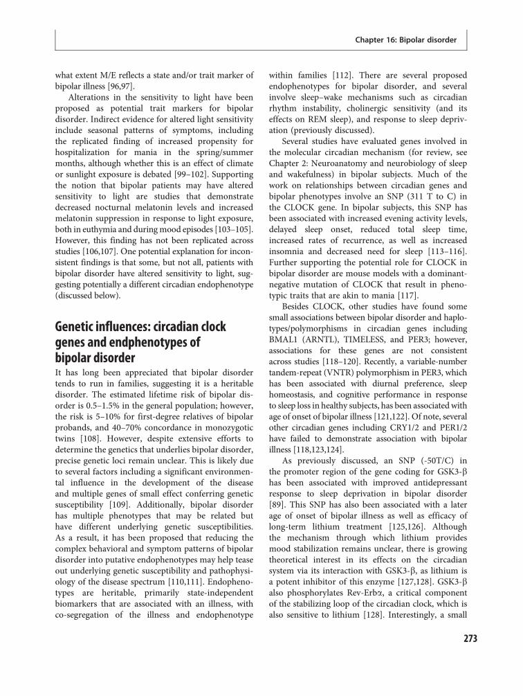
what extent M/E reflects a state and/or trait marker ofbipolar illness [96,97].
Alterations in the sensitivity to light have beenproposed as potential trait markers for bipolardisorder. Indirect evidence for altered light sensitivityinclude seasonal patterns of symptoms, includingthe replicated finding of increased propensity forhospitalization for mania in the spring/summermonths, although whether this is an effect of climateor sunlight exposure is debated [99–102]. Supportingthe notion that bipolar patients may have alteredsensitivity to light are studies that demonstratedecreased nocturnal melatonin levels and increasedmelatonin suppression in response to light exposure,both in euthymia and duringmood episodes [103–105].However, this finding has not been replicated acrossstudies [106,107]. One potential explanation for incon-sistent findings is that some, but not all, patients withbipolar disorder have altered sensitivity to light, sug-gesting potentially a different circadian endophenotype(discussed below).
Genetic influences: circadian clockgenes and endphenotypes ofbipolar disorderIt has long been appreciated that bipolar disordertends to run in families, suggesting it is a heritabledisorder. The estimated lifetime risk of bipolar dis-order is 0.5–1.5% in the general population; however,the risk is 5–10% for first-degree relatives of bipolarprobands, and 40–70% concordance in monozygotictwins [108]. However, despite extensive efforts todetermine the genetics that underlies bipolar disorder,precise genetic loci remain unclear. This is likely dueto several factors including a significant environmen-tal influence in the development of the diseaseand multiple genes of small effect conferring geneticsusceptibility [109]. Additionally, bipolar disorderhas multiple phenotypes that may be related buthave different underlying genetic susceptibilities.As a result, it has been proposed that reducing thecomplex behavioral and symptom patterns of bipolardisorder into putative endophenotypes may help teaseout underlying genetic susceptibility and pathophysi-ology of the disease spectrum [110,111]. Endopheno-types are heritable, primarily state-independentbiomarkers that are associated with an illness, withco-segregation of the illness and endophenotype
within families [112]. There are several proposedendophenotypes for bipolar disorder, and severalinvolve sleep–wake mechanisms such as circadianrhythm instability, cholinergic sensitivity (and itseffects on REM sleep), and response to sleep depriv-ation (previously discussed).
Several studies have evaluated genes involved inthe molecular circadian mechanism (for review, seeChapter 2: Neuroanatomy and neurobiology of sleepand wakefulness) in bipolar subjects. Much of thework on relationships between circadian genes andbipolar phenotypes involve an SNP (311 T to C) inthe CLOCK gene. In bipolar subjects, this SNP hasbeen associated with increased evening activity levels,delayed sleep onset, reduced total sleep time,increased rates of recurrence, as well as increasedinsomnia and decreased need for sleep [113–116].Further supporting the potential role for CLOCK inbipolar disorder are mouse models with a dominant-negative mutation of CLOCK that result in pheno-typic traits that are akin to mania [117].
Besides CLOCK, other studies have found somesmall associations between bipolar disorder and haplo-types/polymorphisms in circadian genes includingBMAL1 (ARNTL), TIMELESS, and PER3; however,associations for these genes are not consistentacross studies [118–120]. Recently, a variable-numbertandem-repeat (VNTR) polymorphism in PER3, whichhas been associated with diurnal preference, sleephomeostasis, and cognitive performance in responseto sleep loss in healthy subjects, has been associatedwithage of onset of bipolar illness [121,122]. Of note, severalother circadian genes including CRY1/2 and PER1/2have failed to demonstrate association with bipolarillness [118,123,124].
As previously discussed, an SNP (-50T/C) inthe promoter region of the gene coding for GSK3-bhas been associated with improved antidepressantresponse to sleep deprivation in bipolar disorder[89]. This SNP has also been associated with a laterage of onset of bipolar illness as well as efficacy oflong-term lithium treatment [125,126]. Althoughthe mechanism through which lithium providesmood stabilization remains unclear, there is growingtheoretical interest in its effects on the circadiansystem via its interaction with GSK3-b, as lithium isa potent inhibitor of this enzyme [127,128]. GSK3-balso phosphorylates Rev-Erba, a critical componentof the stabilizing loop of the circadian clock, which isalso sensitive to lithium [128]. Interestingly, a small
Chapter 16: Bipolar disorder
273
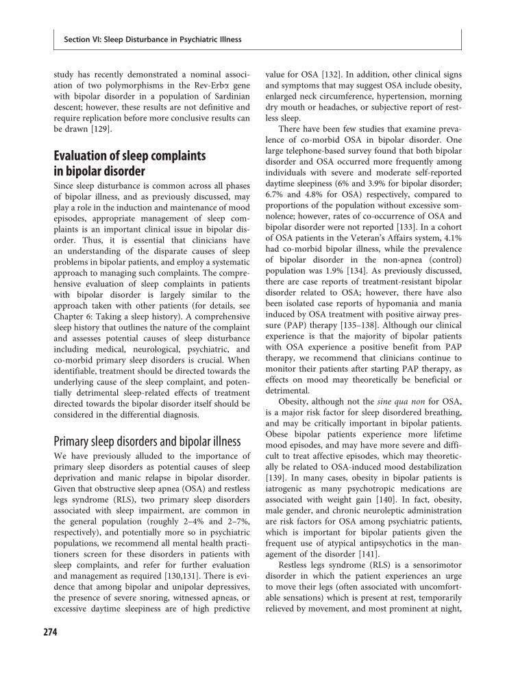
study has recently demonstrated a nominal associ-ation of two polymorphisms in the Rev-Erba genewith bipolar disorder in a population of Sardiniandescent; however, these results are not definitive andrequire replication before more conclusive results canbe drawn [129].
Evaluation of sleep complaintsin bipolar disorderSince sleep disturbance is common across all phasesof bipolar illness, and as previously discussed, mayplay a role in the induction and maintenance of moodepisodes, appropriate management of sleep com-plaints is an important clinical issue in bipolar dis-order. Thus, it is essential that clinicians havean understanding of the disparate causes of sleepproblems in bipolar patients, and employ a systematicapproach to managing such complaints. The compre-hensive evaluation of sleep complaints in patientswith bipolar disorder is largely similar to theapproach taken with other patients (for details, seeChapter 6: Taking a sleep history). A comprehensivesleep history that outlines the nature of the complaintand assesses potential causes of sleep disturbanceincluding medical, neurological, psychiatric, andco-morbid primary sleep disorders is crucial. Whenidentifiable, treatment should be directed towards theunderlying cause of the sleep complaint, and poten-tially detrimental sleep-related effects of treatmentdirected towards the bipolar disorder itself should beconsidered in the differential diagnosis.
Primary sleep disorders and bipolar illnessWe have previously alluded to the importance ofprimary sleep disorders as potential causes of sleepdeprivation and manic relapse in bipolar disorder.Given that obstructive sleep apnea (OSA) and restlesslegs syndrome (RLS), two primary sleep disordersassociated with sleep impairment, are common inthe general population (roughly 2–4% and 2–7%,respectively), and potentially more so in psychiatricpopulations, we recommend all mental health practi-tioners screen for these disorders in patients withsleep complaints, and refer for further evaluationand management as required [130,131]. There is evi-dence that among bipolar and unipolar depressives,the presence of severe snoring, witnessed apneas, orexcessive daytime sleepiness are of high predictive
value for OSA [132]. In addition, other clinical signsand symptoms that may suggest OSA include obesity,enlarged neck circumference, hypertension, morningdry mouth or headaches, or subjective report of rest-less sleep.
There have been few studies that examine preva-lence of co-morbid OSA in bipolar disorder. Onelarge telephone-based survey found that both bipolardisorder and OSA occurred more frequently amongindividuals with severe and moderate self-reporteddaytime sleepiness (6% and 3.9% for bipolar disorder;6.7% and 4.8% for OSA) respectively, compared toproportions of the population without excessive som-nolence; however, rates of co-occurrence of OSA andbipolar disorder were not reported [133]. In a cohortof OSA patients in the Veteran’s Affairs system, 4.1%had co-morbid bipolar illness, while the prevalenceof bipolar disorder in the non-apnea (control)population was 1.9% [134]. As previously discussed,there are case reports of treatment-resistant bipolardisorder related to OSA; however, there have alsobeen isolated case reports of hypomania and maniainduced by OSA treatment with positive airway pres-sure (PAP) therapy [135–138]. Although our clinicalexperience is that the majority of bipolar patientswith OSA experience a positive benefit from PAPtherapy, we recommend that clinicians continue tomonitor their patients after starting PAP therapy, aseffects on mood may theoretically be beneficial ordetrimental.
Obesity, although not the sine qua non for OSA,is a major risk factor for sleep disordered breathing,and may be critically important in bipolar patients.Obese bipolar patients experience more lifetimemood episodes, and may have more severe and diffi-cult to treat affective episodes, which may theoretic-ally be related to OSA-induced mood destabilization[139]. In many cases, obesity in bipolar patients isiatrogenic as many psychotropic medications areassociated with weight gain [140]. In fact, obesity,male gender, and chronic neuroleptic administrationare risk factors for OSA among psychiatric patients,which is important for bipolar patients given thefrequent use of atypical antipsychotics in the man-agement of the disorder [141].
Restless legs syndrome (RLS) is a sensorimotordisorder in which the patient experiences an urgeto move their legs (often associated with uncomfort-able sensations) which is present at rest, temporarilyrelieved by movement, and most prominent at night,
Section VI: Sleep Disturbance in Psychiatric Illness
274
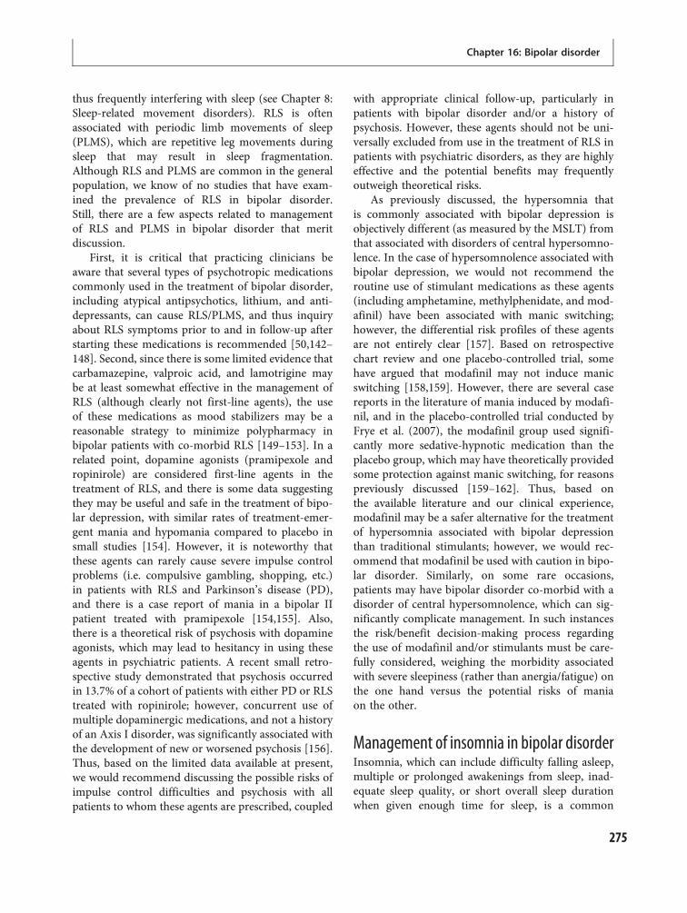
thus frequently interfering with sleep (see Chapter 8:Sleep-related movement disorders). RLS is oftenassociated with periodic limb movements of sleep(PLMS), which are repetitive leg movements duringsleep that may result in sleep fragmentation.Although RLS and PLMS are common in the generalpopulation, we know of no studies that have exam-ined the prevalence of RLS in bipolar disorder.Still, there are a few aspects related to managementof RLS and PLMS in bipolar disorder that meritdiscussion.
First, it is critical that practicing clinicians beaware that several types of psychotropic medicationscommonly used in the treatment of bipolar disorder,including atypical antipsychotics, lithium, and anti-depressants, can cause RLS/PLMS, and thus inquiryabout RLS symptoms prior to and in follow-up afterstarting these medications is recommended [50,142–148]. Second, since there is some limited evidence thatcarbamazepine, valproic acid, and lamotrigine maybe at least somewhat effective in the management ofRLS (although clearly not first-line agents), the useof these medications as mood stabilizers may be areasonable strategy to minimize polypharmacy inbipolar patients with co-morbid RLS [149–153]. In arelated point, dopamine agonists (pramipexole andropinirole) are considered first-line agents in thetreatment of RLS, and there is some data suggestingthey may be useful and safe in the treatment of bipo-lar depression, with similar rates of treatment-emer-gent mania and hypomania compared to placebo insmall studies [154]. However, it is noteworthy thatthese agents can rarely cause severe impulse controlproblems (i.e. compulsive gambling, shopping, etc.)in patients with RLS and Parkinson’s disease (PD),and there is a case report of mania in a bipolar IIpatient treated with pramipexole [154,155]. Also,there is a theoretical risk of psychosis with dopamineagonists, which may lead to hesitancy in using theseagents in psychiatric patients. A recent small retro-spective study demonstrated that psychosis occurredin 13.7% of a cohort of patients with either PD or RLStreated with ropinirole; however, concurrent use ofmultiple dopaminergic medications, and not a historyof an Axis I disorder, was significantly associated withthe development of new or worsened psychosis [156].Thus, based on the limited data available at present,we would recommend discussing the possible risks ofimpulse control difficulties and psychosis with allpatients to whom these agents are prescribed, coupled
with appropriate clinical follow-up, particularly inpatients with bipolar disorder and/or a history ofpsychosis. However, these agents should not be uni-versally excluded from use in the treatment of RLS inpatients with psychiatric disorders, as they are highlyeffective and the potential benefits may frequentlyoutweigh theoretical risks.
As previously discussed, the hypersomnia thatis commonly associated with bipolar depression isobjectively different (as measured by the MSLT) fromthat associated with disorders of central hypersomno-lence. In the case of hypersomnolence associated withbipolar depression, we would not recommend theroutine use of stimulant medications as these agents(including amphetamine, methylphenidate, and mod-afinil) have been associated with manic switching;however, the differential risk profiles of these agentsare not entirely clear [157]. Based on retrospectivechart review and one placebo-controlled trial, somehave argued that modafinil may not induce manicswitching [158,159]. However, there are several casereports in the literature of mania induced by modafi-nil, and in the placebo-controlled trial conducted byFrye et al. (2007), the modafinil group used signifi-cantly more sedative-hypnotic medication than theplacebo group, which may have theoretically providedsome protection against manic switching, for reasonspreviously discussed [159–162]. Thus, based onthe available literature and our clinical experience,modafinil may be a safer alternative for the treatmentof hypersomnia associated with bipolar depressionthan traditional stimulants; however, we would rec-ommend that modafinil be used with caution in bipo-lar disorder. Similarly, on some rare occasions,patients may have bipolar disorder co-morbid with adisorder of central hypersomnolence, which can sig-nificantly complicate management. In such instancesthe risk/benefit decision-making process regardingthe use of modafinil and/or stimulants must be care-fully considered, weighing the morbidity associatedwith severe sleepiness (rather than anergia/fatigue) onthe one hand versus the potential risks of maniaon the other.
Management of insomnia in bipolar disorderInsomnia, which can include difficulty falling asleep,multiple or prolonged awakenings from sleep, inad-equate sleep quality, or short overall sleep durationwhen given enough time for sleep, is a common
Chapter 16: Bipolar disorder
275

occurrence in bipolar disorder, and as previously dis-cussed, may play a role in the disease across all phasesof illness. In the absence of formal guidelines, potentialtreatments for bipolar insomnia include psychothera-peutic and pharmacological strategies that are similarto those used for primary insomnia and insomniarelated to other psychiatric disorders, with somenotable exceptions.
Psychotherapy for bipolar insomniaCognitive behavioral therapy for insomnia (CBT-I) iswell established as the most efficacious psychothera-putic treatment for primary insomnia (see Chapter 14:Cognitive behavioral therapy for insomnia). Unfortu-nately, there are currently no studies of CBT-I inbipolar insomnia, although the majority of CBT-Itechniques could likely be employed without negativesequelae in bipolar patients. The exception is sleeprestriction therapy, where time in bed is limited to thenumber of hours the patient believes they are sleep-ing, which would theoretically increase the likelihoodof manic switching or reinforce hypo/manic states[163]. Unfortunately, sleep restriction is one of themore efficacious CBT-I techniques, potentiallylimiting the overall value of psychotheraputic strat-egies for managing bipolar insomnia [164]. CBT-I inbipolar patients may also be complicated by the factthat some bipolar patients (particularly rapid-cycling)often complain of difficulty arising in the morningand may have mild hypomanic symptoms intensifyover the course of the day, potentially disruptingtheir ability to sleep at night or adhere to prescribedCBT-I-based interventions [165,166].
Although it is not possible to determine the extentthat sleep-related components of common psycho-therapeutic strategies to manage bipolar disordercontribute to the efficacy of the entire therapy, it isnotable that many have components designed toimprove or monitor sleep. Psychoeducational strat-egies that emphasize the identification of prodromalsymptoms (including sleep disturbance) reduce thefrequency of relapses to depressive, manic, hypo-manic, and mixed episodes [167,168]. Interpersonaland social rhythm therapy (IPSRT), which is based onmanagement of life stressors that disrupt patterns(including the sleep–wake cycle), can improve moodstability, prolong maintenance, and decrease affectiverelapse [169–170]. Similarly, cognitive behavioraltreatments for bipolar disorder frequently stressmaintenance of sleep–wake patterns through
cognitive behavioral approaches, and are an effectivemodality in bipolar disorder [171,172].
Pharmacotherapy for bipolar insomniaClinicians have a vast array of pharmacotherapiesavailable to treat bipolar insomnia. These include:benzodiazepines and benzodiazepine receptor agon-ists (BzRAs), sedating antidepressants, sedating anti-psychotics, antiepileptics, and melatonin receptoragonists. Although no medication is approved by theFDA specifically for management of bipolar insom-nia, the practicing clinician should be aware of thepotential benefits and pitfalls of these classes of medi-cations in the context of bipolar disorder.
Benzodiazepines are a vast improvement over thebarbiturates (that have a comparatively small thera-peutic index), which had previously been the primaryagents available as a treatment of insomnia. Benzodi-azepines are highly effective in the treatment ofinsomnia and offer a wide range of half lives amongthe various compounds in this class. Although clinicalexperience suggests benzodiazepines are efficacious inthe management of bipolar insomnia, there are nostudies that demonstrate benzodiazepines specificallyimprove sleep or mood stability in bipolar patients.However, in both an uncontrolled retrospective chartreview and prospective open trial (at the same insti-tution), clonazepam was effective as a replacement forneuroleptics (as an adjunct to lithium) in the main-tenance treatment of bipolar disorder, though thisapproach was not successful in two other studies[173–176].
Concerns regarding abuse, tolerance, withdrawal,daytime sedation, and motor/cognitive impairmentlimit the use of benzodiazepines for the treatment ofinsomnia. As a result, non-benzodiazepine benzodi-azepine receptor agonists (BzRAs; zolpidem, zaleplon,eszopiclone), which are more selective for GABA-Areceptors containing a-1 subunits than traditionalbenzodiazepines, are often the agents of choice.Marketed BzRAs have short to intermediate half lives,which reduce the likelihood of daytime carryover andresultant side-effects compared to agents with longerhalf lives. Although BzRAs also have potential fortolerance and withdrawal, there is data that suggestsnon-nightly use of BzRAs over 2–3 months is notassociated with such problems [177,178]. Further-more, newer agents have been studied for up to 6months without evidence of tolerance or reboundinsomnia on discontinuation [179,180]. Although
Section VI: Sleep Disturbance in Psychiatric Illness
276
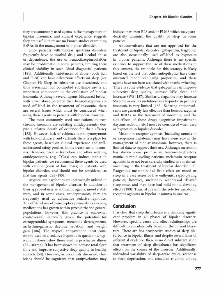
they are commonly used agents in the management ofbipolar insomnia, and clinical experience suggeststhey are useful, there are no known studies examiningBzRAs in the management of bipolar disorder.
Since patients with bipolar spectrum disordersfrequently have co-morbid drug and alcohol abuseor dependence, the use of benzodiazepines/BzRAsmay be problematic in some patients, limiting theirclinical viability in such “dual diagnosis” patients[181]. Additionally, substances of abuse (both licitand illicit) can have deleterious effects on sleep (seeChapter 19: Sleep in substance use disorders), andthus assessment for co-morbid substance use is animportant component in the evaluation of bipolarinsomnia. Although several agents (discussed below)with lower abuse potential than benzodiazepines areused off-label in the treatment of insomnia, thereare several issues which must be considered whenusing these agents in patients with bipolar disorder.
The most commonly used medications to treatchronic insomnia are sedating antidepressants, des-pite a relative dearth of evidence for their efficacy[182]. However, lack of evidence is not synonymouswith lack of efficacy, and certainly there is a place forthese agents, based on clinical experience and well-understood safety profiles, in the treatment of insom-nia. However, because trazodone and other sedatingantidepressants, (e.g. TCAs) can induce mania inbipolar patients, we recommend these agents be usedwith caution (even at low doses) in patients withbipolar disorder, and should not be considered asfirst-line agents [183–185].
Atypical antipsychotics are increasingly utilized inthe management of bipolar disorder. In addition totheir approved uses as antimanic agents, mood stabil-izers, and in some cases, antidepressants, they arefrequently used as adjunctive sedative-hypnotics.The off-label use of neuroleptics primarily as sleepingmedications has grown within psychiatric and generalpopulations; however, this practice is somewhatcontroversial, especially given the potential forextrapyramidal symptoms, metabolic derangements,arrhythmogenesis, daytime sedation, and weightgain [186]. The atypical antipsychotic most com-monly used as a sedative-hypnotic is quetiapine, typ-ically in doses below those used in psychiatric illness(25–100mg). It has been shown to increase total sleeptime and improve subjective sleep quality in healthysubjects [50]. However, as previously discussed, clin-icians should be cognizant that antipsychotics may
induce or worsen RLS and/or PLMS which may para-doxically diminish the quality of sleep in somepatients.
Anticonvulsants that are not approved for thetreatment of bipolar disorder (gabapentin, tiagabine)are also occasionally used off-label as hypnoticsin bipolar patients. Although there is no specificevidence to support the use of these medications inthis context, the rationale for this strategy is likelybased on the fact that other antiepileptics have dem-onstrated mood stabilizing properties, and theseagents have not been associated with manic switching.There is some evidence that gabapentin can improvesubjective sleep quality, increase REM sleep, andincrease SWS [187]. Similarly, tiagabine may increaseSWS; however, its usefulness as a hypnotic in primaryinsomnia is very limited [188]. Sedating anticonvul-sants are generally less effective than benzodiazepinesand BzRAs in the treatment of insomnia, and theside-effects of these drugs (cognitive impairment,daytime sedation, etc.) must be considered when usedas hypnotics in bipolar disorder.
Melatonin receptor agonists (including ramelteonor exogenous melatonin) may have some role in themanagement of bipolar insomnia; however, there islimited data to support their use. Although melatoninhas shown some promise in treatment-refractorymania in rapid-cycling patients, melatonin receptoragonists have not been carefully studied as a mainten-ance drug in the treatment of bipolar disorder [52].Exogenous melatonin had little effect on mood orsleep in a case series of five euthymic, rapid-cyclingpatients; however, melatonin withdrawal delayedsleep onset and may have had mild mood-elevatingeffects [189]. Thus, at present, the role for melatoninreceptor agonists in bipolar insomnia is unclear.
ConclusionIt is clear that sleep disturbance is a clinically signifi-cant problem in all phases of bipolar disorder.However, specific cause and effect relationships aredifficult to elucidate fully based on the current litera-ture. There are few prospective studies of sleep dis-turbance in bipolar illness, and despite several lines ofinferential evidence, there is no direct substantiationthat treatment of sleep disturbance has significanteffects on the course of the disorder. Additionally,individual variability of sleep–wake cycles, responseto sleep deprivation, and circadian rhythms among
Chapter 16: Bipolar disorder
277
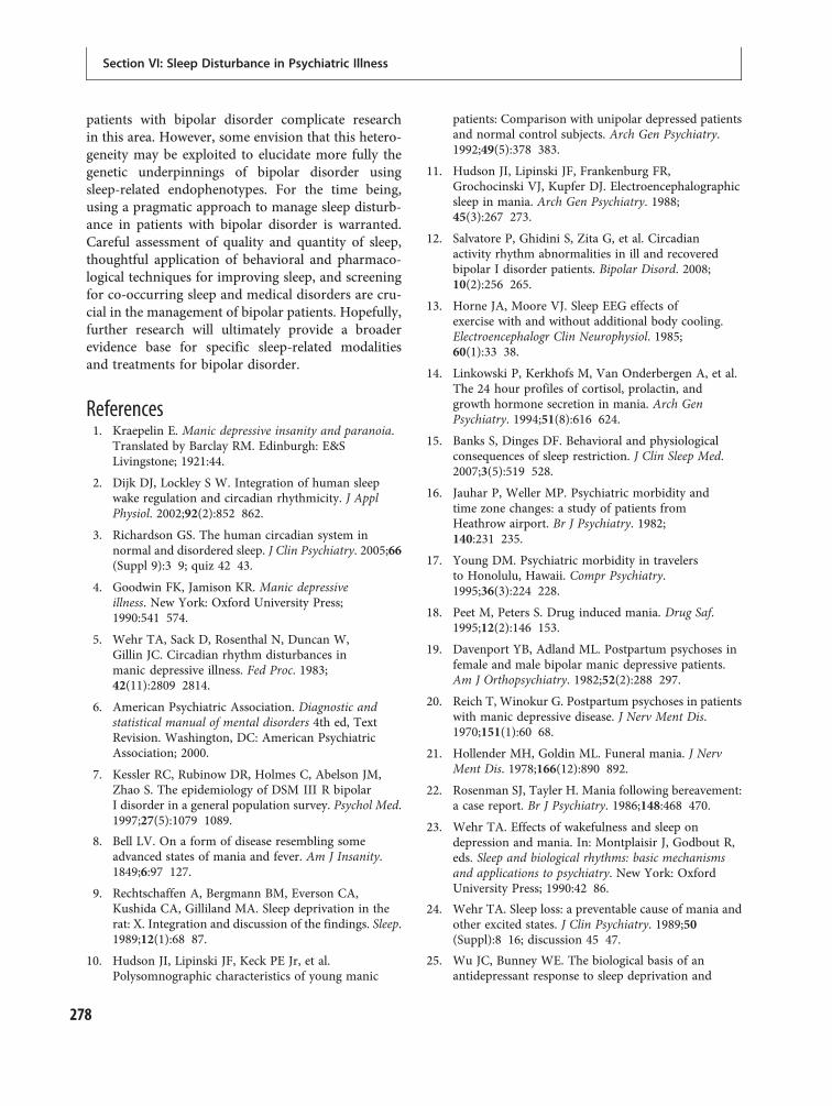
patients with bipolar disorder complicate researchin this area. However, some envision that this hetero-geneity may be exploited to elucidate more fully thegenetic underpinnings of bipolar disorder usingsleep-related endophenotypes. For the time being,using a pragmatic approach to manage sleep disturb-ance in patients with bipolar disorder is warranted.Careful assessment of quality and quantity of sleep,thoughtful application of behavioral and pharmaco-logical techniques for improving sleep, and screeningfor co-occurring sleep and medical disorders are cru-cial in the management of bipolar patients. Hopefully,further research will ultimately provide a broaderevidence base for specific sleep-related modalitiesand treatments for bipolar disorder.
References1. Kraepelin E. Manic depressive insanity and paranoia.
Translated by Barclay RM. Edinburgh: E&SLivingstone; 1921:44.
2. Dijk DJ, Lockley S W. Integration of human sleepwake regulation and circadian rhythmicity. J ApplPhysiol. 2002;92(2):852 862.
3. Richardson GS. The human circadian system innormal and disordered sleep. J Clin Psychiatry. 2005;66(Suppl 9):3 9; quiz 42 43.
4. Goodwin FK, Jamison KR. Manic depressiveillness. New York: Oxford University Press;1990:541 574.
5. Wehr TA, Sack D, Rosenthal N, Duncan W,Gillin JC. Circadian rhythm disturbances inmanic depressive illness. Fed Proc. 1983;42(11):2809 2814.
6. American Psychiatric Association. Diagnostic andstatistical manual of mental disorders 4th ed, TextRevision. Washington, DC: American PsychiatricAssociation; 2000.
7. Kessler RC, Rubinow DR, Holmes C, Abelson JM,Zhao S. The epidemiology of DSM III R bipolarI disorder in a general population survey. Psychol Med.1997;27(5):1079 1089.
8. Bell LV. On a form of disease resembling someadvanced states of mania and fever. Am J Insanity.1849;6:97 127.
9. Rechtschaffen A, Bergmann BM, Everson CA,Kushida CA, Gilliland MA. Sleep deprivation in therat: X. Integration and discussion of the findings. Sleep.1989;12(1):68 87.
10. Hudson JI, Lipinski JF, Keck PE Jr, et al.Polysomnographic characteristics of young manic
patients: Comparison with unipolar depressed patientsand normal control subjects. Arch Gen Psychiatry.1992;49(5):378 383.
11. Hudson JI, Lipinski JF, Frankenburg FR,Grochocinski VJ, Kupfer DJ. Electroencephalographicsleep in mania. Arch Gen Psychiatry. 1988;45(3):267 273.
12. Salvatore P, Ghidini S, Zita G, et al. Circadianactivity rhythm abnormalities in ill and recoveredbipolar I disorder patients. Bipolar Disord. 2008;10(2):256 265.
13. Horne JA, Moore VJ. Sleep EEG effects ofexercise with and without additional body cooling.Electroencephalogr Clin Neurophysiol. 1985;60(1):33 38.
14. Linkowski P, Kerkhofs M, Van Onderbergen A, et al.The 24 hour profiles of cortisol, prolactin, andgrowth hormone secretion in mania. Arch GenPsychiatry. 1994;51(8):616 624.
15. Banks S, Dinges DF. Behavioral and physiologicalconsequences of sleep restriction. J Clin Sleep Med.2007;3(5):519 528.
16. Jauhar P, Weller MP. Psychiatric morbidity andtime zone changes: a study of patients fromHeathrow airport. Br J Psychiatry. 1982;140:231 235.
17. Young DM. Psychiatric morbidity in travelersto Honolulu, Hawaii. Compr Psychiatry.1995;36(3):224 228.
18. Peet M, Peters S. Drug induced mania. Drug Saf.1995;12(2):146 153.
19. Davenport YB, Adland ML. Postpartum psychoses infemale and male bipolar manic depressive patients.Am J Orthopsychiatry. 1982;52(2):288 297.
20. Reich T, Winokur G. Postpartum psychoses in patientswith manic depressive disease. J Nerv Ment Dis.1970;151(1):60 68.
21. Hollender MH, Goldin ML. Funeral mania. J NervMent Dis. 1978;166(12):890 892.
22. Rosenman SJ, Tayler H. Mania following bereavement:a case report. Br J Psychiatry. 1986;148:468 470.
23. Wehr TA. Effects of wakefulness and sleep ondepression and mania. In: Montplaisir J, Godbout R,eds. Sleep and biological rhythms: basic mechanismsand applications to psychiatry. New York: OxfordUniversity Press; 1990:42 86.
24. Wehr TA. Sleep loss: a preventable cause of mania andother excited states. J Clin Psychiatry. 1989;50(Suppl):8 16; discussion 45 47.
25. Wu JC, Bunney WE. The biological basis of anantidepressant response to sleep deprivation and
Section VI: Sleep Disturbance in Psychiatric Illness
278
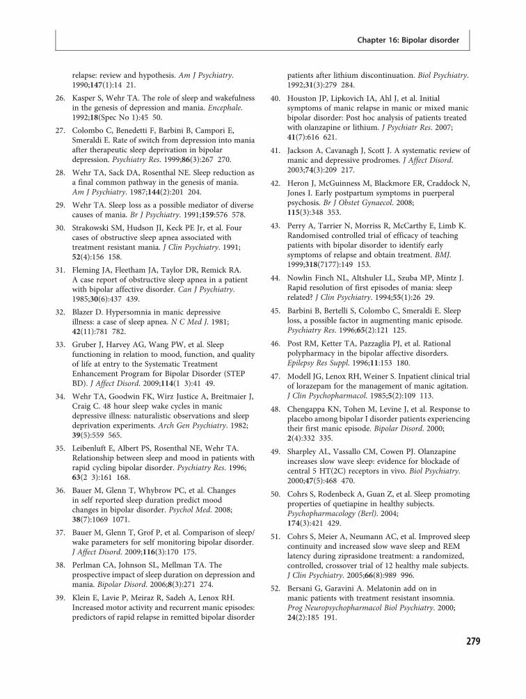
relapse: review and hypothesis. Am J Psychiatry.1990;147(1):14 21.
26. Kasper S, Wehr TA. The role of sleep and wakefulnessin the genesis of depression and mania. Encephale.1992;18(Spec No 1):45 50.
27. Colombo C, Benedetti F, Barbini B, Campori E,Smeraldi E. Rate of switch from depression into maniaafter therapeutic sleep deprivation in bipolardepression. Psychiatry Res. 1999;86(3):267 270.
28. Wehr TA, Sack DA, Rosenthal NE. Sleep reduction asa final common pathway in the genesis of mania.Am J Psychiatry. 1987;144(2):201 204.
29. Wehr TA. Sleep loss as a possible mediator of diversecauses of mania. Br J Psychiatry. 1991;159:576 578.
30. Strakowski SM, Hudson JI, Keck PE Jr, et al. Fourcases of obstructive sleep apnea associated withtreatment resistant mania. J Clin Psychiatry. 1991;52(4):156 158.
31. Fleming JA, Fleetham JA, Taylor DR, Remick RA.A case report of obstructive sleep apnea in a patientwith bipolar affective disorder. Can J Psychiatry.1985;30(6):437 439.
32. Blazer D. Hypersomnia in manic depressiveillness: a case of sleep apnea. N C Med J. 1981;42(11):781 782.
33. Gruber J, Harvey AG, Wang PW, et al. Sleepfunctioning in relation to mood, function, and qualityof life at entry to the Systematic TreatmentEnhancement Program for Bipolar Disorder (STEPBD). J Affect Disord. 2009;114(1 3):41 49.
34. Wehr TA, Goodwin FK, Wirz Justice A, Breitmaier J,Craig C. 48 hour sleep wake cycles in manicdepressive illness: naturalistic observations and sleepdeprivation experiments. Arch Gen Psychiatry. 1982;39(5):559 565.
35. Leibenluft E, Albert PS, Rosenthal NE, Wehr TA.Relationship between sleep and mood in patients withrapid cycling bipolar disorder. Psychiatry Res. 1996;63(2 3):161 168.
36. Bauer M, Glenn T, Whybrow PC, et al. Changesin self reported sleep duration predict moodchanges in bipolar disorder. Psychol Med. 2008;38(7):1069 1071.
37. Bauer M, Glenn T, Grof P, et al. Comparison of sleep/wake parameters for self monitoring bipolar disorder.J Affect Disord. 2009;116(3):170 175.
38. Perlman CA, Johnson SL, Mellman TA. Theprospective impact of sleep duration on depression andmania. Bipolar Disord. 2006;8(3):271 274.
39. Klein E, Lavie P, Meiraz R, Sadeh A, Lenox RH.Increased motor activity and recurrent manic episodes:predictors of rapid relapse in remitted bipolar disorder
patients after lithium discontinuation. Biol Psychiatry.1992;31(3):279 284.
40. Houston JP, Lipkovich IA, Ahl J, et al. Initialsymptoms of manic relapse in manic or mixed manicbipolar disorder: Post hoc analysis of patients treatedwith olanzapine or lithium. J Psychiatr Res. 2007;41(7):616 621.
41. Jackson A, Cavanagh J, Scott J. A systematic review ofmanic and depressive prodromes. J Affect Disord.2003;74(3):209 217.
42. Heron J, McGuinness M, Blackmore ER, Craddock N,Jones I. Early postpartum symptoms in puerperalpsychosis. Br J Obstet Gynaecol. 2008;115(3):348 353.
43. Perry A, Tarrier N, Morriss R, McCarthy E, Limb K.Randomised controlled trial of efficacy of teachingpatients with bipolar disorder to identify earlysymptoms of relapse and obtain treatment. BMJ.1999;318(7177):149 153.
44. Nowlin Finch NL, Altshuler LL, Szuba MP, Mintz J.Rapid resolution of first episodes of mania: sleeprelated? J Clin Psychiatry. 1994;55(1):26 29.
45. Barbini B, Bertelli S, Colombo C, Smeraldi E. Sleeploss, a possible factor in augmenting manic episode.Psychiatry Res. 1996;65(2):121 125.
46. Post RM, Ketter TA, Pazzaglia PJ, et al. Rationalpolypharmacy in the bipolar affective disorders.Epilepsy Res Suppl. 1996;11:153 180.
47. Modell JG, Lenox RH, Weiner S. Inpatient clinical trialof lorazepam for the management of manic agitation.J Clin Psychopharmacol. 1985;5(2):109 113.
48. Chengappa KN, Tohen M, Levine J, et al. Response toplacebo among bipolar I disorder patients experiencingtheir first manic episode. Bipolar Disord. 2000;2(4):332 335.
49. Sharpley AL, Vassallo CM, Cowen PJ. Olanzapineincreases slow wave sleep: evidence for blockade ofcentral 5 HT(2C) receptors in vivo. Biol Psychiatry.2000;47(5):468 470.
50. Cohrs S, Rodenbeck A, Guan Z, et al. Sleep promotingproperties of quetiapine in healthy subjects.Psychopharmacology (Berl). 2004;174(3):421 429.
51. Cohrs S, Meier A, Neumann AC, et al. Improved sleepcontinuity and increased slow wave sleep and REMlatency during ziprasidone treatment: a randomized,controlled, crossover trial of 12 healthy male subjects.J Clin Psychiatry. 2005;66(8):989 996.
52. Bersani G, Garavini A. Melatonin add on inmanic patients with treatment resistant insomnia.Prog Neuropsychopharmacol Biol Psychiatry. 2000;24(2):185 191.
Chapter 16: Bipolar disorder
279
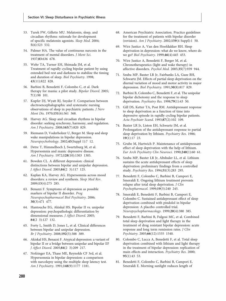
53. Turek FW, Gillette MU. Melatonin, sleep, andcircadian rhythms: rationale for developmentof specific melatonin agonists. Sleep Med. 2004;5(6):523 532.
54. Palmer HA. The value of continuous narcosis in thetreatment of mental disorders. J Ment Sci.1937;83:636 678.
55. Wehr TA, Turner EH, Shimada JM, et al.Treatment of rapidly cycling bipolar patient by usingextended bed rest and darkness to stabilize the timingand duration of sleep. Biol Psychiatry. 1998;43(11):822 828.
56. Barbini B, Benedetti F, Colombo C, et al. Darktherapy for mania: a pilot study. Bipolar Disord. 2005;7(1):98 101.
57. Kupfer DJ, Wyatt RJ, Snyder F. Comparison betweenelectroencephalographic and systematic nursingobservations of sleep in psychiatric patients. J NervMent Dis. 1970;151(6):361 368.
58. Harvey AG. Sleep and circadian rhythms in bipolardisorder: seeking synchrony, harmony, and regulation.Am J Psychiatry. 2008;165(7):820 829.
59. Riemann D, Voderholzer U, Berger M. Sleep and sleepwake manipulations in bipolar depression.Neuropsychobiology. 2002;45(Suppl 1):7 12.
60. Detre T, Himmelhoch J, Swartzburg M, et al.Hypersomnia and manic depressive disease.Am J Psychiatry. 1972;128(10):1303 1305.
61. Bowden CL. A different depression: clinicaldistinctions between bipolar and unipolar depression.J Affect Disord. 2005;84(2 3):117 125.
62. Kaplan KA, Harvey AG. Hypersomnia across mooddisorders: a review and synthesis. Sleep Med Rev.2009;13(4):275 285.
63. Benazzi F. Symptoms of depression as possiblemarkers of bipolar II disorder. ProgNeuropsychopharmacol Biol Psychiatry. 2006;30(3):471 477.
64. Hantouche EG, Akiskal HS. Bipolar II vs. unipolardepression: psychopathologic differentiation bydimensional measures. J Affect Disord. 2005;84(2 3):127 132.
65. Forty L, Smith D, Jones L, et al. Clinical differencesbetween bipolar and unipolar depression.Br J Psychiatry. 2008;192(5):388 389.
66. Akiskal HS, Benazzi F. Atypical depression: a variant ofbipolar II or a bridge between unipolar and bipolar II?J Affect Disord. 2005;84(2 3):209 217.
67. Nofzinger EA, Thase ME, Reynolds CF 3rd, et al.Hypersomnia in bipolar depression: a comparisonwith narcolepsy using the multiple sleep latency test.Am J Psychiatry. 1991;148(9):1177 1181.
68. American Psychiatric Association. Practice guidelinesfor the treatment of patients with bipolar disorder(revision). Am J Psychiatry. 2002;159(4 Suppl):1 50.
69. Wirz Justice A, Van den Hoofdakker RH. Sleepdeprivation in depression: what do we know, where dowe go? Biol Psychiatry. 1999;46(4):445 453.
70. Wirz Justice A, Benedetti F, Berger M, et al.Chronotherapeutics (light and wake therapy) inaffective disorders. Psychol Med. 2005;35(7):939 944.
71. Szuba MP, Baxter LR Jr, Fairbanks LA, Guze BH,Schwartz JM. Effects of partial sleep deprivation on thediurnal variation of mood and motor activity in majordepression. Biol Psychiatry. 1991;30(8):817 829.
72. Barbini B, Colombo C, Benedetti F, et al. The unipolarbipolar dichotomy and the response to sleepdeprivation. Psychiatry Res. 1998;79(1):43 50.
73. Gill DS, Ketter TA, Post RM. Antidepressant responseto sleep deprivation as a function of time intodepressive episode in rapidly cycling bipolar patients.Acta Psychiatr Scand. 1993;87(2):102 109.
74. Baxter LR Jr, Liston EH, Schwartz JM, et al.Prolongation of the antidepressant response to partialsleep deprivation by lithium. Psychiatry Res. 1986;19(1):17 23.
75. Grube M, Hartwich P. Maintenance of antidepressanteffect of sleep deprivation with the help of lithium.Eur Arch Psychiatry Clin Neurosci. 1990;240(1):60 61.
76. Szuba MP, Baxter LR Jr, Altshuler LL, et al. Lithiumsustains the acute antidepressant effects of sleepdeprivation: preliminary findings from a controlledstudy. Psychiatry Res. 1994;51(3):283 295.
77. Benedetti F, Colombo C, Barbini B, Campori E,Smeraldi E. Ongoing lithium treatment preventsrelapse after total sleep deprivation. J ClinPsychopharmacol. 1999;19(3):240 245.
78. Smeraldi E, Benedetti F, Barbini B, Campori E,Colombo C. Sustained antidepressant effect of sleepdeprivation combined with pindolol in bipolardepression: A placebo controlled trial.Neuropsychopharmacology. 1999;20(4):380 385.
79. Benedetti F, Barbini B, Fulgosi MC, et al. Combinedtotal sleep deprivation and light therapy in thetreatment of drug resistant bipolar depression: acuteresponse and long term remission rates. J ClinPsychiatry. 2005;66(12):1535 1540.
80. Colombo C, Lucca A, Benedetti F, et al. Total sleepdeprivation combined with lithium and light therapyin the treatment of bipolar depression: replication ofmain effects and interaction. Psychiatry Res. 2000;95(1):43 53.
81. Benedetti F, Colombo C, Barbini B, Campori E,Smeraldi E. Morning sunlight reduces length of
Section VI: Sleep Disturbance in Psychiatric Illness
280

hospitalization in bipolar depression. J Affect Disord.2001;62(3):221 223.
82. Berger M, Vollmann J, Hohagen F, et al. Sleepdeprivation combined with consecutive sleep phaseadvance as a fast acting therapy in depression: an openpilot trial in medicated and unmedicated patients.Am J Psychiatry. 1997;154(6):870 872.
83. Riemann D, Konig A, Hohagen F, et al. How topreserve the antidepressive effect of sleep deprivation:A comparison of sleep phase advance and sleep phasedelay. Eur Arch Psychiatry Clin Neurosci. 1999;249(5):231 237.
84. Neumeister A, Goessler R, Lucht M, et al. Bright lighttherapy stabilizes the antidepressant effect of partialsleep deprivation. Biol Psychiatry. 1996;39(1):16 21.
85. Benedetti F, Barbini B, Campori E, et al. Sleep phaseadvance and lithium to sustain the antidepressanteffect of total sleep deprivation in bipolar depression:new findings supporting the internal coincidencemodel? J Psychiatr Res. 2001;35(6):323 329.
86. Benedetti F, Serretti A, Colombo C, et al. Influence of afunctional polymorphism within the promoter of theserotonin transporter gene on the effects of total sleepdeprivation in bipolar depression. Am J Psychiatry.1999;156(9):1450 1452.
87. Benedetti F, Barbini B, Bernasconi A, et al. Serotonin5 HT2A receptor gene variants influenceantidepressant response to repeated total sleepdeprivation in bipolar depression. ProgNeuropsychopharmacol Biol Psychiatry. 2008;32(8):1863 1866.
88. Benedetti F, Barbini B, Bernasconi A, et al. Acuteantidepressant response to sleep deprivation combinedwith light therapy is influenced by the catechol Omethyltransferase Val(108/158)Met polymorphism.J Affect Disord. 2010;121(1 2):68 72.
89. Benedetti F, Serretti A, Colombo C, et al. A glycogensynthase kinase 3 beta promoter gene singlenucleotide polymorphism is associated with ageat onset and response to total sleep deprivationin bipolar depression. Neurosci Lett. 2004;368(2):123 126.
90. Sachs GS. Unmet clinical needs in bipolar disorder.J Clin Psychopharmacol. 2003;23(3 Suppl 1):S2 8.
91. Knowles JB, Cairns J, MacLean AW, et al. The sleep ofremitted bipolar depressives: comparison with sex andage matched controls. Can J Psychiatry. 1986;31(4):295 298.
92. Sitaram N, Nurnberger JI Jr, Gershon ES, Gillin JC.Cholinergic regulation of mood and REM sleep:potential model and marker of vulnerabilityto affective disorder. Am J Psychiatry. 1982;139(5):571 576.
93. Millar A, Espie CA, Scott J. The sleep ofremitted bipolar outpatients: a controlled naturalisticstudy using actigraphy. J Affect Disord. 2004;80(2 3):145 153.
94. Jones SH, Hare DJ, Evershed K. Actigraphicassessment of circadian activity and sleep patterns inbipolar disorder. Bipolar Disord. 2005;7(2):176 186.
95. Harvey AG, Schmidt DA, Scarna A, Semler CN,Goodwin GM. Sleep related functioning in euthymicpatients with bipolar disorder, patients with insomnia,and subjects without sleep problems. Am J Psychiatry.2005;162(1):50 57.
96. Mansour HA, Wood J, Chowdari KV, et al. Circadianphase variation in bipolar I disorder. Chronobiol Int.2005;22(3):571 584.
97. Wood J, Birmaher B, Axelson D, et al. Replicabledifferences in preferred circadian phase betweenbipolar disorder patients and control individuals.Psychiatry Res. 2009;166(2 3):201 209.
98. Ahn YM, Chang J, Joo YH, et al. Chronotypedistribution in bipolar I disorder and schizophreniain a Korean sample. Bipolar Disord. 2008;10(2):271 275.
99. Carney PA, Fitzgerald CT, Monaghan CE. Influence ofclimate on the prevalence of mania. Br J Psychiatry.1988;152:820 823.
100. Mulder RT, Cosgriff JP, Smith AM, Joyce PR.Seasonality of mania in New Zealand. Aust N ZJ Psychiatry. 1990;24(2):187 190.
101. Sayer HK, Marshall S, Mellsop GW. Mania andseasonality in the southern hemisphere. J Affect Disord.1991;23(3):151 156.
102. Lee HC, Tsai SY, Lin HC. Seasonal variations inbipolar disorder admissions and the association withclimate: a population based study. J Affect Disord.2007;97(1 3):61 69.
103. Lewy AJ, Nurnberger JI Jr, Wehr TA, et al.Supersensitivity to light: possible trait marker formanic depressive illness. Am J Psychiatry. 1985;142(6):725 727.
104. Lewy AJ, Wehr TA, Goodwin FK, Newsome DA,Rosenthal NE. Manic depressive patients may besupersensitive to light. Lancet. 1981;1(8216):383 384.
105. Kennedy SH, Kutcher SP, Ralevski E, Brown GM.Nocturnal melatonin and 24 hour6 sulphatoxymelatonin levels in various phasesof bipolar affective disorder. Psychiatry Res. 1996;63(2 3):219 222.
106. Whalley LJ, Perini T, Shering A, Bennie J. Melatoninresponse to bright light in recovered, drug free, bipolarpatients. Psychiatry Res. 1991;38(1):13 19.
Chapter 16: Bipolar disorder
281
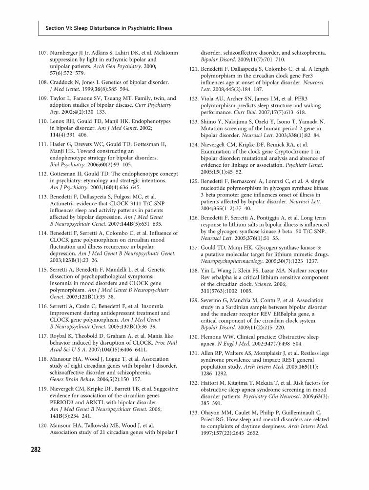
107. Nurnberger JI Jr, Adkins S, Lahiri DK, et al. Melatoninsuppression by light in euthymic bipolar andunipolar patients. Arch Gen Psychiatry. 2000;57(6):572 579.
108. Craddock N, Jones I. Genetics of bipolar disorder.J Med Genet. 1999;36(8):585 594.
109. Taylor L, Faraone SV, Tsuang MT. Family, twin, andadoption studies of bipolar disease. Curr PsychiatryRep. 2002;4(2):130 133.
110. Lenox RH, Gould TD, Manji HK. Endophenotypesin bipolar disorder. Am J Med Genet. 2002;114(4):391 406.
111. Hasler G, Drevets WC, Gould TD, Gottesman II,Manji HK. Toward constructing anendophenotype strategy for bipolar disorders.Biol Psychiatry. 2006;60(2):93 105.
112. Gottesman II, Gould TD. The endophenotype conceptin psychiatry: etymology and strategic intentions.Am J Psychiatry. 2003;160(4):636 645.
113. Benedetti F, Dallaspezia S, Fulgosi MC, et al.Actimetric evidence that CLOCK 3111 T/C SNPinfluences sleep and activity patterns in patientsaffected by bipolar depression. Am J Med GenetB Neuropsychiatr Genet. 2007;144B(5):631 635.
114. Benedetti F, Serretti A, Colombo C, et al. Influence ofCLOCK gene polymorphism on circadian moodfluctuation and illness recurrence in bipolardepression. Am J Med Genet B Neuropsychiatr Genet.2003;123B(1):23 26.
115. Serretti A, Benedetti F, Mandelli L, et al. Geneticdissection of psychopathological symptoms:insomnia in mood disorders and CLOCK genepolymorphism. Am J Med Genet B NeuropsychiatrGenet. 2003;121B(1):35 38.
116. Serretti A, Cusin C, Benedetti F, et al. Insomniaimprovement during antidepressant treatment andCLOCK gene polymorphism. Am J Med GenetB Neuropsychiatr Genet. 2005;137B(1):36 39.
117. Roybal K, Theobold D, Graham A, et al. Mania likebehavior induced by disruption of CLOCK. Proc NatlAcad Sci U S A. 2007;104(15):6406 6411.
118. Mansour HA, Wood J, Logue T, et al. Associationstudy of eight circadian genes with bipolar I disorder,schizoaffective disorder and schizophrenia.Genes Brain Behav. 2006;5(2):150 157.
119. Nievergelt CM, Kripke DF, Barrett TB, et al. Suggestiveevidence for association of the circadian genesPERIOD3 and ARNTL with bipolar disorder.Am J Med Genet B Neuropsychiatr Genet. 2006;141B(3):234 241.
120. Mansour HA, Talkowski ME, Wood J, et al.Association study of 21 circadian genes with bipolar I
disorder, schizoaffective disorder, and schizophrenia.Bipolar Disord. 2009;11(7):701 710.
121. Benedetti F, Dallaspezia S, Colombo C, et al. A lengthpolymorphism in the circadian clock gene Per3influences age at onset of bipolar disorder. NeurosciLett. 2008;445(2):184 187.
122. Viola AU, Archer SN, James LM, et al. PER3polymorphism predicts sleep structure and wakingperformance. Curr Biol. 2007;17(7):613 618.
123. Shiino Y, Nakajima S, Ozeki Y, Isono T, Yamada N.Mutation screening of the human period 2 gene inbipolar disorder. Neurosci Lett. 2003;338(1):82 84.
124. Nievergelt CM, Kripke DF, Remick RA, et al.Examination of the clock gene Cryptochrome 1 inbipolar disorder: mutational analysis and absence ofevidence for linkage or association. Psychiatr Genet.2005;15(1):45 52.
125. Benedetti F, Bernasconi A, Lorenzi C, et al. A singlenucleotide polymorphism in glycogen synthase kinase3 beta promoter gene influences onset of illness inpatients affected by bipolar disorder. Neurosci Lett.2004;355(1 2):37 40.
126. Benedetti F, Serretti A, Pontiggia A, et al. Long termresponse to lithium salts in bipolar illness is influencedby the glycogen synthase kinase 3 beta 50 T/C SNP.Neurosci Lett. 2005;376(1):51 55.
127. Gould TD, Manji HK. Glycogen synthase kinase 3:a putative molecular target for lithium mimetic drugs.Neuropsychopharmacology. 2005;30(7):1223 1237.
128. Yin L, Wang J, Klein PS, Lazar MA. Nuclear receptorRev erbalpha is a critical lithium sensitive componentof the circadian clock. Science. 2006;311(5763):1002 1005.
129. Severino G, Manchia M, Contu P, et al. Associationstudy in a Sardinian sample between bipolar disorderand the nuclear receptor REV ERBalpha gene, acritical component of the circadian clock system.Bipolar Disord. 2009;11(2):215 220.
130. Flemons WW. Clinical practice: Obstructive sleepapnea. N Engl J Med. 2002;347(7):498 504.
131. Allen RP, Walters AS, Montplaisir J, et al. Restless legssyndrome prevalence and impact: REST generalpopulation study. Arch Intern Med. 2005;165(11):1286 1292.
132. Hattori M, Kitajima T, Mekata T, et al. Risk factors forobstructive sleep apnea syndrome screening in mooddisorder patients. Psychiatry Clin Neurosci. 2009;63(3):385 391.
133. Ohayon MM, Caulet M, Philip P, Guilleminault C,Priest RG. How sleep and mental disorders are relatedto complaints of daytime sleepiness. Arch Intern Med.1997;157(22):2645 2652.
Section VI: Sleep Disturbance in Psychiatric Illness
282
134. Sharafkhaneh A, Giray N, Richardson P, Young T,Hirshkowitz M. Association of psychiatric disordersand sleep apnea in a large cohort. Sleep. 2005;28(11):1405 1411.
135. Berge D, Salgado P, Rodriguez A, Bulbena A. Onset ofmania after CPAP in a man with obstructive sleepapnea. Psychosomatics. 2008;49(5):447 449.
136. Trakada G, Steiropoulos P, Bouros D. Continuouspositive airways pressure treatment in a patient withsleep apnea hypopnea syndrome and coexistingbipolar disorder. Psychopharmacol Bull. 2008;41(2):89 92.
137. Lahera Forteza G, Gonzalez Aguado F. Psychoticmania after introduction of continuous positiveairway pressure (CPAP) in the treatment ofobstructive sleep apnoea. Actas Esp Psiquiatr. 2007;35(6):406 407.
138. Hilleret H, Jeunet E, Osiek C, Mohr S, Blois R,Bertschy G. Mania resulting from continuous positiveairways pressure in a depressed man with sleepapnea syndrome. Neuropsychobiology. 2001;43(3):221 224.
139. Fagiolini A, Kupfer DJ, Houck PR, Novick DM,Frank E. Obesity as a correlate of outcome in patientswith bipolar I disorder. Am J Psychiatry. 2003;160(1):112 117.
140. Devlin MJ, Yanovski SZ, Wilson GT. Obesity:what mental health professionals need to know.Am J Psychiatry. 2000;157(6):854 866.
141. Winkelman JW. Schizophrenia, obesity, andobstructive sleep apnea. J Clin Psychiatry. 2001;62(1):8 11.
142. Pinninti NR, Mago R, Townsend J, Doghramji K.Periodic restless legs syndrome associated withquetiapine use: a case report. J Clin Psychopharmacol.2005;25(6):617 618.
143. Kraus T, Schuld A, Pollmacher T. Periodic legmovements in sleep and restless legs syndromeprobably caused by olanzapine. J ClinPsychopharmacol. 1999;19(5):478 479.
144. Wetter TC, Brunner J, Bronisch T. Restless legssyndrome probably induced by risperidone treatment.Pharmacopsychiatry. 2002;35(3):109 111.
145. Duggal HS, Mendhekar DN. Clozapine associatedrestless legs syndrome. J Clin Psychopharmacol.2007;27(1):89 90.
146. Heiman EM, Christie M. Lithium aggravatednocturnal myoclonus and restless legs syndrome.Am J Psychiatry. 1986;143(9):1191 1192.
147. Yang C, White DP, Winkelman JW. Antidepressantsand periodic leg movements of sleep. Biol Psychiatry.2005;58(6):510 514.
148. Rottach KG, Schaner BM, Kirch MH, et al. Restless legssyndrome as side effect of second generationantidepressants. J Psychiatr Res. 2008;43(1):70 75.
149. Telstad W, Sorensen O, Larsen S, et al. Treatment ofthe restless legs syndrome with carbamazepine: adouble blind study. Br Med J (Clin Res Ed). 1984;288(6415):444 446.
150. Lundvall O, Abom PE, Holm R. Carbamazepine inrestless legs: A controlled pilot study. Eur J ClinPharmacol. 1983;25(3):323 324.
151. Zucconi M, Coccagna G, Petronelli R, et al. Nocturnalmyoclonus in restless legs syndrome: effect ofcarbamazepine treatment. Funct Neurol. 1989;4(3):263 271.
152. Eisensehr I, Ehrenberg BL, Rogge Solti S, Noachtar S.Treatment of idiopathic restless legs syndrome (RLS)with slow release valproic acid compared with slowrelease levodopa/benserazid. J Neurol. 2004;251(5):579 583.
153. Youssef EA, Wagner ML, Martinez JO, Hening W.Pilot trial of lamotrigine in the restless legs syndrome.Sleep Med. 2005;6(1):89.
154. Aiken CB. Pramipexole in psychiatry: a systematicreview of the literature. J Clin Psychiatry. 2007;68(8):1230 1236.
155. Sharma V, Smith A. A case of mania following the useof pramipexole. Am J Psychiatry. 2007;164(2):351.
156. Stoner SC, Dahmen MM, Makos M, et al.An exploratory retrospective evaluation of ropiniroleassociated psychotic symptoms in an outpatientpopulation treated for restless legs syndrome orParkinson’s disease. Ann Pharmacother. 2009;43(9):1426 1432.
157. Wingo AP, Ghaemi SN. Frequency of stimulanttreatment and of stimulant associated mania/hypomania in bipolar disorder patients.Psychopharmacol Bull. 2008;41(4):37 47.
158. Nasr S, Wendt B, Steiner K. Absence of mood switchwith and tolerance to modafinil: A replication studyfrom a large private practice. J Affect Disord. 2006;95(1 3):111 114.
159. Frye MA, Grunze H, Suppes T, et al. A placebocontrolled evaluation of adjunctive modafinil in thetreatment of bipolar depression. Am J Psychiatry.2007;164(8):1242 1249.
160. Ranjan S, Chandra PS. Modafinil induced irritabilityand aggression? A report of 2 bipolar patients. J ClinPsychopharmacol. 2005;25(6):628 629.
161. Wolf J, Fiedler U, Anghelescu I, Schwertfeger N. Manicswitch in a patient with treatment resistant bipolardepression treated with modafinil. J Clin Psychiatry.2006;67(11):1817.
Chapter 16: Bipolar disorder
283
162. Fountoulakis KN, Siamouli M, Panagiotidis P, et al.Ultra short manic like episodes after antidepressantaugmentation with modafinil. ProgNeuropsychopharmacol Biol Psychiatry. 2008;32(3):891 892.
163. Smith MT, Huang MI, Manber R. Cognitivebehavior therapy for chronic insomniaoccurring within the context of medical andpsychiatric disorders. Clin Psychol Rev. 2005;25(5):559 592.
164. Morgenthaler T, Kramer M, Alessi C, et al. Practiceparameters for the psychological and behavioraltreatment of insomnia: an update. An AmericanAcademy of Sleep Medicine report. Sleep. 2006;29(11):1415 1419.
165. Ashman SB, Monk TH, Kupfer DJ, et al. Relationshipbetween social rhythms and mood in patientswith rapid cycling bipolar disorder. Psychiatry Res.1999;86(1):1 8.
166. Feldman Naim S, Turner EH, Leibenluft E. Diurnalvariation in the direction of mood switches in patientswith rapid cycling bipolar disorder. J Clin Psychiatry.1997;58(2):79 84.
167. Colom F, Vieta E, Martinez Aran A, et al.A randomized trial on the efficacy ofgroup psychoeducation in the prophylaxisof recurrences in bipolar patients whosedisease is in remission. Arch Gen Psychiatry. 2003;60(4):402 407.
168. Colom F, Lam D. Psychoeducation: improvingoutcomes in bipolar disorder. Eur Psychiatry. 2005;20(5 6):359 364.
169. Frank E, Swartz HA, Kupfer D J. Interpersonaland social rhythm therapy: managing the chaosof bipolar disorder. Biol Psychiatry. 2000;48(6):593 604.
170. Frank E, Kupfer DJ, Thase ME, et al. Two yearoutcomes for interpersonal and social rhythm therapyin individuals with bipolar I disorder. Arch GenPsychiatry. 2005;62(9):996 1004.
171. Otto MW, Reilly Harrington N, Sachs GS.Psychoeducational and cognitive behavioral strategiesin the management of bipolar disorder. J Affect Disord.2003;73(1 2):171 181.
172. Lam DH, Watkins ER, Hayward P, et al.A randomized controlled study of cognitive therapy forrelapse prevention for bipolar affective disorder:outcome of the first year. Arch Gen Psychiatry. 2003;60(2):145 152.
173. Sachs GS, Rosenbaum JF, Jones L. Adjunctiveclonazepam for maintenance treatment of bipolaraffective disorder. J Clin Psychopharmacol. 1990;10(1):42 47.
174. Sachs GS, Weilburg JB, Rosenbaum JF. Clonazepamvs. neuroleptics as adjuncts to lithiummaintenance. Psychopharmacol Bull. 1990;26(1):137 143.
175. Aronson TA, Shukla S, Hirschowitz J. Clonazepamtreatment of five lithium refractory patientswith bipolar disorder. Am J Psychiatry. 1989;146(1):77 80.
176. Winkler D, Willeit M, Wolf R, et al. Clonazepam in thelong term treatment of patients with unipolardepression, bipolar and schizoaffective disorder.Eur Neuropsychopharmacol. 2003;13(2):129 134.
177. Walsh JK, Roth T, Randazzo A, et al. Eight weeks ofnon nightly use of zolpidem for primary insomnia.Sleep. 2000;23(8):1087 1096.
178. Perlis ML, McCall WV, Krystal AD, Walsh JK. Longterm, non nightly administration of zolpidem in thetreatment of patients with primary insomnia. J ClinPsychiatry. 2004;65(8):1128 1137.
179. Krystal AD, Walsh JK, Laska E, et al. Sustained efficacyof eszopiclone over 6 months of nightly treatment:results of a randomized, double blind, placebocontrolled study in adults with chronic insomnia.Sleep. 2003;26(7):793 799.
180. Erman M, Krystal A, Zammit G, Soubrane C, Roth T.Zolpidem extended release 12.5mg, taken for 24 weeks“as needed” up to 7 nights/week, improves subjectivemeasures of therapeutic global impression, sleep onset,and sleep maintenance in patients with chronicinsomnia. Int J Neuropsychopharmacol. 2006;9(Suppl 1):S256.
181. Vornik LA, Brown ES. Management of comorbidbipolar disorder and substance abuse. J Clin Psychiatry.2006;67(Suppl 7):24 30.
182. NIH. NIH State of the Science conference statementon manifestations and management of chronicinsomnia in adults. NIH Consensus and State of theScience Statements. 2005;22(2):1 30. http://consensus.nih.gov/2005/2005InsomniaSOS026PDF.pdf. Accessed30 October, 2009.
183. Peet M. Induction of mania with selective serotoninre uptake inhibitors and tricyclic antidepressants.Br J Psychiatry. 1994;164(4):549 550.
184. Jabeen S, Fisher CJ. Trazodone induced transienthypomanic symptoms and their management.Br J Psychiatry. 1991;158:275 278.
185. Terao T. Comparison of manic switch onset duringfluoxetine and trazodone treatment. Biol Psychiatry.1993;33(6):477 478.
186. Doghramji PP. Trends in the pharmacologicmanagement of insomnia. J Clin Psychiatry. 2006;67(Suppl 13):5 8.
Section VI: Sleep Disturbance in Psychiatric Illness
284

187. Foldvary Schaefer N, De Leon Sanchez I, Karafa M,et al. Gabapentin increases slow wave sleepin normal adults. Epilepsia. 2002;43(12):1493 1497.
188. Roth T, Wright KP Jr, Walsh J. Effect of tiagabine onsleep in elderly subjects with primary insomnia:a randomized, double blind, placebo controlled study.Sleep. 2006;29(3):335 341.
189. Leibenluft E, Feldman Naim S, Turner EH, Wehr TA,Rosenthal NE. Effects of exogenous melatoninadministration and withdrawal in five patients withrapid cycling bipolar disorder. J Clin Psychiatry.1997;58(9):383 388.
190. Plante DT, Winkelman JW. Sleep disturbance inbipolar disorder: therapeutic implications.Am J Psychiatry. 2008;165(7):830 843.
Chapter 16: Bipolar disorder
285

Chapter
17 Sleep in anxiety disorders
Candice A. Alfano and Thomas A. Mellman
IntroductionSleep disturbances including insomnia are frequentlyassociated with and can comprise core features ofanxiety disorders. The presence of anxiety poses riskfor the later development of insomnia, with approxi-mately 40% of adults reporting problems with insom-nia following the onset of an anxiety disorder [1].Similarly, sleep loss can exacerbate daytime symptomsof anxiety and persisting insomnia is a risk factor forthe subsequent onset of anxiety disorders [2–6]. Thisbidirectional relationship implies a shared underlyingpathophysiology and suggests the possibility of a pro-gressive worsening of both disorders and associatedimpairments over time. The links between anxiety andsleep disturbances may therefore be relevant to under-standing mechanisms and dysfunctions of arousalregulation that underlie both types of problems.
The senior author of this chapter (Dr. Mellman)has provided or contributed several relatively recentoverviews of sleep aspects of anxiety disorders inadults [7,8]. Building on this work, emerging dataindicate that not unlike adults, sleep problems duringchildhood are likely to co-occur with anxiety dis-orders. Since many adult sleep disorders originateduring and persist from childhood [9], collectively,there is a clear need for a developmental frameworkin attempting to understand the interactions and idealmanagement of these problems at different stagesacross the life-span. Thus, in line with the clinicalemphasis of the current volume, this chapter is dis-tinguished by its inclusion and discussion of sleepaspects of anxiety disorders in children, made possibleby the contribution of Dr. Alfano, a pediatric psych-ologist whose work focuses on sleep and anxiety inyouth. Through this collaborative writing we hope toprovide a more comprehensive overview and heighten
awareness of the potential linkages between the regu-lation of sleep, arousal, and emotion in anxious adultsas well as children.
While all types of anxiety disorders may co-occurwith some formof sleep disruption, only post-traumaticstress disorder (PTSD), generalized anxiety disorder(GAD), and separation anxiety disorder (SAD) featuresleep disturbance among their diagnostic criteria inDSM-IV. Criteria for PTSD include nightmares withtrauma-related content and difficulty initiating andmaintaining sleep, which is a common definition ofinsomnia. Insomnia is also a symptom criterion andcommon feature of GAD. Although not a formalDSM-IV criterion, panic disorder (PD) has also beenassociated with complaints of difficulty initiating andmaintaining sleep. Panic attacks that arise from sleepare not an uncommon occurrence with PD. All of theanxiety disorders that are diagnosed in adults can alsobe diagnosed in children, though PD prior to adoles-cence is rare. SAD is usually confined to childhood butmay be a progenitor of adult panic disorder [10]. SADcriteria include both difficulty or refusal to sleep inde-pendently and separation-themed nightmares and, inmany cases, sleep difficulties represent one of the moreprominent features of the disorder. Lastly, data alsoare beginning to emerge suggesting that youth withobsessive-compulsive disorder (OCD) commonly expe-rience sleep disruption, including difficulty falling asleep.
In adults and children there is considerable overlapbetween interventions that target insomnia and othersleep disturbances and those that are utilized intreating anxiety disorders. Overlapping approachesinclude medications, as well as behavioral modifica-tion and cognitive restructuring strategies. In children,attention to environmental and familial factors thatserve to maintain both anxiety and sleep problems is
Foundations of Psychiatric SleepMedicine, ed. JohnW.Winkelman andDavid T. Plante. Published by CambridgeUniversity Press.# Cambridge University Press 2010.
286

often necessary. For example, elimination of parentalreinforcement of children’s avoidant night-timebehaviors that delay and/or interfere with sleep onsetmay be critical for treatment success [11,12]. Overall,however, optimal sequencing and/or integration oftreatments targeting anxiety disorders and sleepproblems are not well investigated. It is importantfor future research to address this deficiency. Mean-while clinician’s consideration of both sleep and anx-iety symptoms in formulating their treatments iscritical toward mitigating the chronicity, elevatedrelapse risk, and impairments associated with theirco-occurrence.
Polysomnography (PSG) can provide objectiveindices of sleep initiation and maintenance, as well asinformation regarding sleep architecture. This tooland other laboratory methods applied to sleep canoffer unique perspectives on the function of the neuro-biological systems involved in arousal, sleep, and anx-iety phenomena. However, the practicing clinicianshould be aware that PSG is not routinely indicatedfor the evaluation of an anxiety disorder unless aprimary sleep disorder (e.g. obstructive sleep apnea)is suspected. In the following sections, clinical issuesand sleep laboratory information (when available)regarding sleep aspects of specific anxiety disordersin adults and children are discussed, followed by adiscussion of treatment issues that interface sleep andanxiety disorders.
Sleep in specific and social phobiasIntense fear and behavioral avoidance of certain thingsor situations are hallmark features of phobic disorders.Specific phobias often originate during childhood andfear and avoidance in general are not uncommonduring childhood. For example, transient night-timefears such as the dark, monsters, or burglars are con-sidered to be developmentally appropriate and occurin approximately 70% of young children [13,14].Although most will overcome or outgrow these fears,a proportion of children experience persistent orsevere enough fears that interfere with sleep and familyfunctioning on an ongoing basis. In such cases, per-sistent night-time fears may be indicative of an under-lying anxiety disorder. Muris et al. [14] found severenight-time fears to be related to one or more DSManxiety disorders in more than 10% of children aged4–12 years. Specific phobias, SAD, and GAD were themost common anxiety diagnoses. There is also some
evidence for associations between phobic and sleepdisorders during adolescence. In one study specificallyexamining parasomnias, including sleep terrors andsleepwalking in adolescents, increased rates ofco-morbidity with specific phobias and other anxietydisorders were reported [15]. Investigations relatingsleep to specific phobias in adults are more limited. Inone investigation by Clark et al. [16] sleep architecturewas similar in depressed persons with and withoutphobias.
Similar to specific phobias, social phobia (SOC) canbe an outgrowth of childhood shyness. In fact, modalage of onset occurs during adolescence [17]. The corefeature of SOC includes a persistent fear of one or moresocial or performance situations in which a person mayencounter unfamiliar people or possible scrutiny fromothers. Since these situations occur during interactionswith the environment during wakefulness, sleep dis-turbances are not typically regarded as central toor commonly associated with this disorder. Nonethe-less, persons with SOC may experience anticipatoryanxiety prior to an event which then disrupts sleep.This may be particularly prominent when, for example,a SOC child begins a new school year or an adult withSOC must give a speech at work.
In a study among SOC adults, subjective report ofpoorer sleep quality, longer sleep latency,more frequentsleep disturbances, and increased daytime dysfunctionwere found compared to controls [18]. However,another study utilizing PSG recordings among SOCadults reported normal findings of sleep [19]. In a clinicsample of youthwith SOC, SAD, andGAD,Alfano et al.[20] found that children with a diagnosis of SOC hadsignificantly fewer parent-reported sleep problems(including insomnia, difficulty sleeping alone or awayfrom home, and nightmares) than youth with SADor GAD.
Sleep in obsessive-compulsive disorderObsessive-compulsive disorder (OCD) features recur-ring intrusive thoughts (obsessions) that seem exces-sive or irrational to the individual, and behavioralrituals (compulsions) that are also excessive and usu-ally related to obsessive thoughts. Although onset ofOCD may occur anytime from preschool age intoadulthood, at least one-third of adults report an onsetof symptoms prior to adolescence [21]. The disorderis generally characterized by a chronic course thatcauses significant distress and interference with daily
Chapter 17: Sleep in anxiety disorders
287

life. Although sleep disturbances are not core syndro-mal manifestations or prominent associated features,it is not uncommon for individuals with OCD to get“stuck” on certain words, images, or thoughts or toengage in elaborate rituals at night which can inter-fere with sleep.
An early polysomnographic study reportedimpaired sleep maintenance and a reduced latency torapid eye movement (REM) sleep in adults with OCD[22]. However, two other studies utilizing PSG failedto replicate these results, reporting the sleep patternsof adults withOCD as essentially normal [23,24].Morerecently, Kluge and colleagues [25] compared 10 in-patients with OCD without co-morbid depression and10 healthy matched controls on PSG variables. Sleepdid not differ across the groups with the exception ofreduced stage 4 sleep in OCD patients. Also, similarto previous findings [22], dramatically reduced sleep-onset REM latencies (<10minutes) were found amongpatients with more severe forms of OCD.
Rapoport et al. [26] compared the sleep character-istics of a small sample of adolescents with OCD(aged 13–17 years) to healthy controls based on onenight of PSG. Significantly reduced sleep efficiencyand increased sleep latency were found among theOCD group. Adolescents with OCD required twiceas long as controls to initiate sleep. Storch and col-leagues [27] examined the presence of parent- andchild-reported sleep problems among a larger sample(N¼66) of youth with OCD, aged 8–17 years. Morethan 90% of the sample experienced at least one typeof sleep problem; trouble sleeping and daytime tired-ness were present in more than 50% of the sample.Sleep problems were more common among youngerchildren and females and associated with a moresevere form of illness. One limitation of this study,however, was that the majority of youth were takingselective serotonin reuptake inhibitors at the time ofassessment, which may have affected sleep. Overall,available findings suggest that sleep disruption ismore likely to co-occur with more severe forms ofOCD and is more likely during childhood.
Sleep in generalized anxiety disorderThe core feature of generalized anxiety disorder(GAD) is persistent, uncontrollable worry. A uniquepattern of distressing somatic symptoms also differ-entiates GAD from other anxiety disorders in adults
as well as children. DSM-IV requires the presence of(at least) three out of six symptoms in addition toworry, one of which is difficulty initiating or main-taining sleep or restless/unsatisfying sleep, for a diag-nosis. Only one additional symptom is required inchildren. Two of the other symptoms, fatigue and irrit-ability, are common consequences of sleep loss. In add-ition, the principal attribute of GAD, excessive worryor apprehensive expectation, is frequently implicatedin the genesis andmaintenance of insomnia. Thus, thereis a high degree of overlap between the two disorders.
Ohayon et al. [4] found that the co-morbidity ofGAD with insomnia was greater than for all of theother psychiatric disorders surveyed. Numerous stud-ies utilizing objective sleep recordings corroboratethese reports. Consistent findings of sleep disruptionin GAD across PSG studies include reduced total sleeptime, increased wake time after sleep onset, reducedsleep efficiency, decreased stage 4 sleep, and increasedstage 2 latency [28–32]. Increases in stage 1 sleep andshorter REM latency also have been reported, albeitless consistently [31,32]. Findings for number of noc-turnal awakenings have been somewhat inconsistent,with both significant increases [29,31] and decreases[30] reported. Overall, however, the data suggest sleepdisruption in GAD to be a problem of impairedsleep initiation as well as maintenance.
Although no study has used PSG to specificallyexamine the sleep of youth with GAD, findings froma study by Forbes and colleagues [33] where 80%(N¼19) of anxious children had a GAD diagnosissuggest that increased arousals, decreased slow-wavesleep, and greater sleep onset latency are characteris-tics of this population. Alfano et al. [34] comparedrates of parent-reported sleep problems among chil-dren with GAD and children with other anxiety dis-orders (aged 8–16 years) and reported that 94%compared to 74% had at least one sleep-related prob-lem, respectively. Nightmares, trouble sleeping, anddaytime tiredness were most commonly reported.Using a larger sample (N¼128) Alfano et al. [20]compared the prevalence of several sleep problemsamong anxious youth with and without a diagnosisof GAD. Children with GAD experienced significantlymore sleep problems than children with SOC but notSAD. Similar to previous findings, nightmares, insom-nia, and daytime tiredness were most common.
Consistent with their high degree of overlap andco-morbidity, there is also substantial overlap in
Section VI: Sleep Disturbance in Psychiatric Illness
288

treatment approaches for GAD and insomnia. Inter-vention strategies include the use of medications thattarget benzodiazepine receptors as well as psycho-therapeutic interventions that target worry and cog-nitive arousal. Application of these approaches isdiscussed further in the final section of this chapter.
Sleep in panic disorderThe core feature of panic disorder (PD) is recurrentand unexpected periods of extreme fear that occur ina sudden, crescendo-like manner. Fear is accompaniedby intense physical symptoms that may include chestpain, heart palpitations, shortness of breath, dizziness,or abdominal distress. Chronic anxiety related toanticipation of subsequent attacks and phobic avoid-ance (agoraphobia) are also common features. PDtypically has its onset in early adulthood but can occurearlier or later in life. Perhaps as a consequence ofits relative infrequency, research examining sleep inyouth with PD is unavailable.
Panic attacks can emerge from sleep. Sleep panicattacks have been suggested to result in a conditionedfear and apprehension of sleep, resulting in secondaryinsomnia [35]. Surveys document that insomniais more frequent in patients with PD than in controlpopulations [36,37]. Most [38–41] but not all [42,43]published studies of patients with PD that have utilizedobjective methods of sleep recordings have foundevidence of impaired sleep initiation andmaintenance.One of these studies found increased motor activityduring sleep [43]. While there appears to be an associ-ation between excess body movement during sleepand PD, two studies have found that, paradoxically,overall movement during sleep is reduced on nightswhen patients experience sleep panic attacks [38,44].Survey data have noted associations between sleepcomplaints and co-morbid depression in persons withPD [42]. However, it is unclear whether sleep disturb-ances may be attributable to the presence of a depres-sive illness, which commonly co-exists with panic, orwhether depression may be more likely to co-occurwith a more severe variant of PD. As these studies arecross-sectional, and since insomnia is a risk factor forthe subsequent onset of depression [2,3], depressionalso may be more likely to evolve as a co-morbidcondition when PD features disturbed sleep.
Sleep panic attacks are not uncommon in personswith PD. In a study that prospectively monitored panic
attacks, 18% occurred during sleep hours [45]. Insurveys and clinical evaluations, 33–71% of PD popula-tions reported having experienced sleep panic attacks[36,46–48]. As many as a third of patients will experi-ence sleep panic as, ormore frequently than, wake panicattacks [35,36]. It is not known how commonly patientsexperience sleep panic exclusively; however, in theseauthors’ experience patients who only panic duringsleep are rare. These episodes have been described asbeing awakened with a “jolt” and feature apprehensionand somatic symptoms similar to panic attacks thatare triggered during wake states. Studies that have cap-tured sleep panic attacks during PSG recordings findthat the episodes were preceded by either stage 2 or stage3 non-REMsleep [38,49].Mellman andUhde [38]morespecifically noted that sleep panic attacks originatedduring the transition from stage 2 into early slow-wavesleep, which is a period of diminishing arousal. Slow-wave sleep is also a state where cognitive activity is atits nadir [50]. The phenomenon of sleep panic indicatesthat in addition to panic attacks evolving from statesof heightening arousal and apprehension, panic can alsooccur during states of diminishing arousal.
Studies have compared PD patients who experi-ence sleep panic attacks with those who onlyexperience panic attacks from wake states. Associationsbetween sleep panic and early age of illness onset, highersymptom load, depression, and suicidal ideation suggesta relationship with a more severe variant of the illness[46,51]. Patients with sleep panic have also been notedto experience anxiety from relaxation and hypnosis, andto have less agoraphobic avoidance and fewer cata-strophic cognitions compared with PD patients whodo not experience sleep panic [36,47,52,53]. Thus,having sleep panic appears to mark a propensity forattacks triggered by lower levels of arousal and forattacks to occur relatively independently of situationaland cognitive variables. The associationwithmarkers ofseverity may reflect a stronger disease diathesis.
Sleep panic attacks can present in a manner thatmimicsmedical conditions. Holter cardiac monitoringor overnight PSG recordings may be indicated whenassociated symptomatology suggests cardiac arrhyth-mias or sleep-related breathing disorders. Patienteducation and informed reassurance are helpful topatients with these frequently upsetting occurrences.Sleep panic attacks have been anecdotally observed torespond to antidepressant types of antipanic medica-tions but not necessarily benzodiazepines [35].
Chapter 17: Sleep in anxiety disorders
289
Sleep in separation anxiety disorderAnxiety related to separating from parents or care-takers is a normal aspect of child development that formost children will subside by age 3. By comparison,separation anxiety disorder (SAD) includes excessive,age-inappropriate fear about being apart from familymembers, especially parents, and leads to problematicbehavioral avoidance. Children with SAD frequentlyrefuse to attend school, have difficulty making andmaintaining friends, are unwilling to do even basictasks independently, and develop other anxiety orphobic disorders. Not surprisingly, a significant pro-portion of children with SAD experience difficultysleeping independently and experience recurrentnightmares. Although earlier case reports sometimesreferred to these problems as a “sleep phobia”(e.g. [54]), closer examination of clinical descriptionsand case studies generally reveal a more pervasiveproblem with separation anxiety.
Despite its high prevalence, surprisingly few studieshave examined sleep disruption in children with SAD.Alfano et al. [20] compared the prevalence of sleepproblems in anxious youth with and without an SADdiagnosis. Children with SAD experienced significantlymore sleep problems than children with SOC but notGAD. Insomnia, refusal to sleep alone, and nightmaresweremost commonly reported by parents. In a study byVerduin and Kendall [55] where the occurrence ofspecific co-morbid diagnoses was examined amongchildren with primary SAD, GAD, and SOC, enuresisand sleep terror disorder were significantly morecommon among youth with SAD. It is important tonote, however, that children with SAD in both studieswere younger on average than anxious youth in com-parison groups and the ability to disentangle disorder-specific sleep problems from age-related differences islimited.
Sleep in post-traumatic stress disorderPost-traumatic stress disorder is an anxiety disorderthat develops in children and adults who are exposed toseverely threatening events. While PTSD is common,it develops in a minority of individuals who areexposed to trauma. Factors that influence the develop-ment of PTSD include severity of the trauma, personalrisk factors, and initial reactions to the traumatic event[56]. Sleep disturbances including trauma-relatednightmares and difficulty initiating and maintainingsleep represent DSM-IV criteria and prominent
symptoms of PTSD [57]. Nightmares and insomniaappear to be common also in the early aftermath oftrauma, especially among those who develop PTSD[58–61]. Furthermore, sleep disruption leads to fatigueand irritability, which are daytime symptoms of thedisorder. Sleep disruption also may interfere withhealthy emotional regulation and thereby contributeto the early development of the disorder.
Sleep laboratory study results, most of which havefeatured male war veterans during the chronic phaseof the disorder, have been mixed in terms of findingobjective indices of impaired sleep initiation andmaintenance [62]. A recent meta-analysis of 20 PSGstudies indicates that patients with PTSD have lightersleep (more stage 1, less stage 3) compared to controls[63]. It is possible that the perceived safety of the sleeplaboratory environment has a salutary effect on sleepdisturbance in research participants with PTSD. Tworecent studies that utilized home monitoring methodsfound increased sleep onset latency and reduced sleepefficiency, supporting this hypothesis [64,65].
There is also some controversy as to whetherPTSD is associated with sleep-related breathingabnormalities. Krakow et al. [66] found evidence forsleep disordered breathing in all but 4 of 44 treat-ment-seeking patients who were referred for PSGassessment. However, no increase in sleep disorderedbreathing in association with PTSD was found in astudy of community-recruited participants [67].
There has been relatively consistent evidence forabnormalities related to REM sleep in PTSD. Theaforementioned meta-analysis noted that increasedREM density and increased phasic motor activityduring REM have been found in combat veteranswith PTSD [68]. Nightmares and other symptomaticawakenings disproportionately arise from REMsleep [69,70]. Breslau et al. reported more frequenttransitions from REM sleep to stage 1 or wake ina community sample of subjects with either lifetime(i.e. remitted) or current PTSD compared withtrauma-exposed and trauma-unexposed controls[67]. A recent clinical study from Japan noted “REMinterruption” in association with PTSD [71]. In aunique study that applied PSG within a month oftrauma exposure, Mellman et al. [72] found that thepatients who later developed PTSD had shorter con-tinuous periods of REM sleep prior to stage shifts orarousals. Thus, there is converging evidence for dis-ruptions of REM sleep continuity (symptomatic awak-enings, increased awakening/arousals, and motor
Section VI: Sleep Disturbance in Psychiatric Illness
290
activity) and increased REM activation with PTSD.Mellman and Pigeon [73] hypothesized that the dis-ruption to REM sleep continuity in PTSD interfereswith the normal and adaptive memory processingfunctions of REM.
Although well documented in adults, research hasonly begun to examine associations between child-hood trauma exposure and sleep. Two studies haveused actigraphy to examine the sleep patterns ofabused children. Glod et al. [74] reported that abusedchildren demonstrate significantly longer sleep onsetlatencies than controls and depressed youth, andpoorer sleep efficiencies than controls. No differencesin sleep parameters were found among abused chil-dren based on the presence/absence of a PTSD diag-nosis, suggesting that the effects of early abuse onsleep occur irrespective of the development of theclinical disorder. Glodd and colleagues also reportedpoorer sleep efficiencies (measured using portableambulatory monitoring) among children with a his-tory of physical compared to sexual abuse; and thisfinding has been replicated by another study [75].Lastly, children with a PTSD diagnosis withoutco-morbid depression exhibited significantly greaternocturnal activity, longer sleep onset latencies, andreduced sleep efficiencies compared to childrenwith PTSD and depression. Although more researchis needed, the possibility exists that a co-morbiddepressive disorder may be protective of sleep byreducing hypervigilance and noradrenergic overacti-vation associated with PTSD [76].
The effects of early trauma on sleep extend intoadulthood. Bader et al. [77] examined associationsbetween childhood trauma and sleep in 39 adults withinsomnia based on 1week of actigraphy. After control-ling for demographic characteristics, current levels ofstress, and depression, childhood trauma was thestrongest predictor of sleep onset latency, sleep effi-ciency, and nocturnal activity in adulthood. Noll et al.[78] examined the presence of sleep disturbances in78 abused females 10 years after disclosure of sexualabuse occurring between the ages of 6 and 16 years.Compared to controls, sexually abused femalesreported significantly more sleep disturbances. Abusewas a unique predictor of sleep problems even aftercontrolling for depressive symptoms. Curiously, par-ticipants experiencing relatively less severe forms ofsexual abuse (e.g. abuse by a single perpetrator, min-imal physical violence, shorter durations of abuse,etc.) displayed the highest levels of adult sleep
disturbances. The authors propose that because theseparticipants may have initially presented with onlymild symptoms, appropriate treatment may not havebeen received. Despite striking concurrent and longi-tudinal associations, relationships between earlytrauma and adult sleep disturbances remain poorlyunderstood.
Treatment of sleep problemsassociated with anxietySleep disturbances including insomnia have beenfound to be a prospective risk factor for psychiatricdisorders including depression and anxiety [2,3,5].Therefore, in addition to alleviating distress associatedwith insomnia, amelioration of sleep disturbancescould possibly serve to prevent relapse and exacer-bation of symptoms. In contrast to melancholic sub-types of depression where mood can paradoxicallyimprove, anxiety disorders do not benefit and canworsen from sleep deprivation [79–81]. These datasuggest that intervention approaches need to betailored to individual patients.
Behavioral treatmentsBehavioral therapies for anxiety disorders and sleepdisturbances overlap to a great extent. Similar to anx-iety, cognitive behavioral treatments (CBT) developedfor insomnia have well-established efficacy (seeChapter 14: Cognitive behavioral therapy for insom-nia) [82]. A key intervention component includesinstruction on sleep hygiene such as maintainingconsistent bedtimes and wake times, avoiding mal-adaptive use of substances, and not spending excessivetime awake in bed. Additional components includerelaxation techniques and identifying and challengingdysfunctional beliefs that perpetuate insomnia (e.g.“If I only get five hours sleep I will not be able tofunction tomorrow”). Management of anxiousarousal, reducing avoidance behaviors. and cognitiverestructuring are also key components of CBT foranxiety disorders [83]. It would seem then that behav-ioral interventions designed for insomnia and anxietycan be synergistically applied.
One study documents improvement in insomniasymptoms in association with CBT for GAD [84]. Incontrast, DeViva et al. [85] identified a group ofpatients with significant residual insomnia who hadotherwise benefited from CBT for PTSD. They further
Chapter 17: Sleep in anxiety disorders
291

describe a series of cases where residual insomnia wasreduced by a subsequently administered CBT programfocused specifically on sleep. These studies notwith-standing, development and evaluation of sequentialor integrated treatments for insomnia and anxiety dis-orders is limited in adults as well as children.
A series of controlled trials indicate imageryrehearsal therapy (IRT) to be effective in reducingnightmares and insomnia symptoms as well as improv-ing overall symptomatology in PTSD [86–89]. IRT iscomprised of two main components: (1) educatingpatients about the impact of nightmares on sleep andhow nightmares promote and condition insomnia;and (2) linking daytime imagery and dreams byselecting a specific nightmare, modifying its content(in either an individual or group setting) and rehears-ing the new dream. The individual’s perceived masteryover negative dream elements appears to be a key factorin the effectiveness of IRT [90].
Since little is known about the safety, tolerability,and efficacy of pharmacological sleep agents in youth,pediatric treatment guidelines designate behavioralinterventions as treatments of choice for sleep prob-lems co-occuring with anxiety and other psychiatricdisorders [91]. Nonetheless, controlled behavioraltreatment trials are lacking. A few case reports andcontrolled studies have reported significant reduc-tions in sleep difficulties among night-time fearfulchildren based on the use of behavioral interventions[92,93], but the generalizability of these data to youthwith anxiety disorders is unclear.
Studies examining the impact of behavioral treat-ments for childhood anxiety on co-occurring sleepproblems are equally limited. Storch et al. [27] foundthat 14 weeks of CBT resulted in an overall decreasein parent-reported sleep problems, including night-mares and trouble sleeping, among 41 youth withprimary OCD. A reduction in sleep problems wasnot observed based on child report, however. Thesedata underscore the need to gather information frommultiple informants when assessing children’s sleepand highlight a need to understand better the imme-diate and long-term effectiveness of CBT for co-morbid sleep disturbance in anxious youth.
Pharmacological treatmentsVarious benzodiazepine receptor agonist medicationsare approved and marketed for hypnotic indicationsand/or treatment of anxiety disorders, particularly
GAD. Therefore if one is prescribing benzodiazep-ines, the use of the same agent for “24-hour coverage”with possible use of a higher dose at night would seema reasonable strategy. In support of this, one studyfound that agents marketed and approved as hyp-notics had benefits for daytime anxiety when usedfor the treatment of insomnia associated with GAD[94]. The novel agents pregabalin and tiagabine,which, like benzodiazepines, influence GABAergicneurotransmission, have been reported to improveinsomnia symptoms associated with GAD. However,neither drug has FDA approval for GAD or insomnia[95,96].
The newer antidepressant medications, particu-larly those in the selective serotonin reuptake inhibitor(SSRI) and selective serotonin and norepinephrineinhibitor (SNRI) categories, are effective for a rangeof anxiety disorders. They also have advantages withrespect to tolerance and dependence concerns relativeto benzodiazepines and are now considered to be first-line treatments for most anxiety disorders. Effects onsleep vary among agents and among individuals andsome have been noted to stimulate insomnia [97].Among the novel antidepressants, mirtazapine, whichis neither an SSRI nor an SNRI, tends to have sedating/sleep effects. Mirtazapine was recently reported to bebeneficial for GAD patients in a preliminary open-label trial [98], although it is not FDA approved forthis indication. A study utilizing the SSRI citalopramfor late-life anxiety disorders indicated improved sleepwith treatment in this subpopulation [99].
Studies examining pharmacological treatments(SSRIs) for childhood anxiety have generally failed toexamine potential changes in sleep. In one study thatdid examine sleep-related outcomes, Alfano et al. [20]found that fluvoxamine produced significantly greaterreductions in parent- and clinician-reported sleepproblems compared to placebo in youth with SAD,SOC, or GAD after 8 weeks of treatment. Specifically,symptoms of insomnia and reluctance/refusal to sleepalone were significantly reduced. However, improve-ments in nightmares were not observed and long-termsleep-related outcomes were not examined.
Recently, augmentation of antidepressant therapywith the hypnotic medication eszopiclone (3 mg atbedtime) was shown to result in significant improve-ments in sleep, daytime functioning, and anxiety in alarge sample of adults with GAD [100]. With regardto sleep characteristics specifically, adults treatedwith both escitalopram and eszopiclone evidenced
Section VI: Sleep Disturbance in Psychiatric Illness
292

significant reductions in sleep onset latency andincreases in total sleep time (based on electronic diar-ies) compared to those treated with escitalopramand placebo across the 12-week treatment period.Co-therapy with escitalopram and eszopiclone alsoresulted in a greater magnitude and acceleration ofanxiolytic effects, demonstrated from as early as weekone of therapy. Although follow-up studies usingobjective measures of sleep are needed, these findingssuggest that simultaneously targeting anxiety and sleepmay result in greater treatment gains among adultswith GAD.
Presently, SSRIs, specifically sertraline and parox-etine, are the only agents approved by the FDA for thetreatment of PTSD. Benefits of these treatments tendto be modest and do not typically include reductionsin sleep disturbance. Therefore, adjunctive interven-tions are often utilized to target sleep. There is nowevidence from a small crossover study and a modest-sized placebo-controlled parallel groups designed trialsupporting the efficacy of prazosin for nightmaresand insomnia in combat-related PTSD [101,102].Dosing was titrated gradually but aggressively withfinal doses averaging 9.5 and 13 mg at bedtime inthe two studies, respectively. Prazosin is an oldermedication that is traditionally prescribed for treatinghypertension and prostate outlet obstruction. Itsmechanism of action is blockade of alpha-adrenergicreceptors. Thus, its benefit may relate to the hypothe-sized role of noradrenergic signaling thought to dis-rupt REM sleep. A small controlled trial also reportedsleep-related benefits from the second-generationantipsychotic medication (SGA) olanzapine, whichwas prescribed at a mean dose of 15 mg per day[103]. While to date there are no published controlleddata, the SGA quetiapine appears to often be pre-scribed for sleep disturbances in PTSD. In choosingspecific medications clinicians need to consider thespectrum of side-effects, most notably the metabolicrisks associated with SGAs, as well as the fact thatneither prazosin, olanzapine, nor quetiapine are FDA-approved for PTSD.
SummarySleep disturbances are commonly associated with anx-iety disorders in adults as well as children, particularlyGAD, SAD, and PTSD, and cause significant impair-ments in overall functioning. Evidence of a reciprocalrelationship between anxiety and sleep is compelling
such that sleep loss may contribute to the onset andrelapse of these conditions. The underlying patho-physiology of these problems likely involves sharedmechanisms including dysregulation of arousal,inability to inhibit negative cognition, and specificneurophysiological changes. In anxious youth, in par-ticular, environmental contingencies, including inad-vertent reinforcement of bedtime avoidance, canexacerbate both problems. Collectively, data suggestthat effective treatment requires attention to bothunique and common factors. Established treatmentsfor anxiety disorders and insomnia overlap to a largeextent, yet optimal sequencing and integration of theseapproaches remains underinvestigated and generallyunknown.
References1. Ohayon MM, Roth T. Place of chronic insomnia in the
course of depressive and anxiety disorders. J PsychiatrRes. 2003;37:9 15.
2. Ford D, Kamerow D. Epidemiologic study of sleepdisturbances and psychiatric disorders: An opportunityfor prevention? JAMA. 1989;262:1479 1484.
3. Breslau N, Roth T, Rosenthal L, Andreski P. Sleepdisturbance and psychiatric disorders: a longitudinalepidemiological study of young adults. Biol Psychiatry.1996;39(6):411 418.
4. Ohayon M, Caulet M, Lemoine P. Comorbidity ofmental and insomnia disorders in the generalpopulation. Compr Psychiatry. 1998;39:185 197.
5. Gregory AM, Caspi A, Eley TC, et al. Prospectivelongitudinal associations between persistent sleepproblems in childhood and anxiety and depressiondisorders in adulthood. J Abnorm Child Psychol.2005;33:157 163.
6. Ong HS, Wickramaratne P, Min T, Weissman MM.Early childhood sleep and eating problems aspredictors of adolescent and adult mood and anxietydisorders. J Affect Disord. 2006;96:1 8.
7. Mellman TA. Sleep and anxiety disorders. PsychiatricClin North Am. 2006; 29:1047 1058.
8. Mellman TA. Sleep and anxiety disorders. Sleep MedClin. 2008;3:261 268.
9. Philip P, Guilleminault C. Adult psychophysiologicinsomnia and positive history of childhood insomnia.Sleep. 1996;19:S16 22.
10. Lewinsohn PM, Holm Denoma JM, Small JW, SeeleyJR, Joiner TE Jr. Separation anxiety disorder inchildhood as a risk factor for future mental illness.J Am Acad Child Adol Psychiatry. 2008;47(5):548 555.
Chapter 17: Sleep in anxiety disorders
293
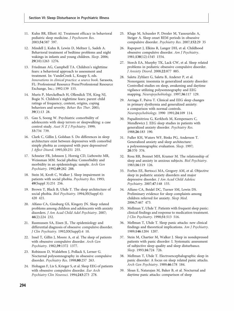
11. Kuhn BR, Elliott AJ. Treatment efficacy in behavioralpediatric sleep medicine. J Psychosom Res.2003;54:587 597.
12. Mindell J, Kuhn B, Lewin D, Meltzer L, Sadeh A.Behavioral treatment of bedtime problems and nightwakings in infants and young children. Sleep. 2006;29(10):1263 1276.
13. Friedman AG, Campbell TA. Children’s nighttimefears: a behavioral approach to assessment andtreatment. In: VandeCreek L, Knapp S, eds.Innovations in clinical practice: a source book. Sarasota,FL: Professional Resource Press/Professional ResourceExchange, Inc.; 1992:139 155.
14. Muris P, Merckelbach H, Ollendick TH, King NJ,Bogie N. Children’s nighttime fears: parent childratings of frequency, content, origins, copingbehaviors and severity. Behav Res Ther. 2001;39(1):13 28.
15. Gau S, Soong W. Psychiatric comorbidity ofadolescents with sleep terrors or sleepwalking: a casecontrol study. Aust N Z J Psychiatry. 1999;33:734 739.
16. Clark C, Gillin J, Golshan S. Do differences in sleeparchitecture exist between depressives with comorbidsimple phobia as compared with pure depressives?J Affect Disord. 1995;33:251 255.
17. Schneier FR, Johnson J, Hornig CD, Liebowitz MR,Weissman MM. Social phobia: Comorbidity andmorbidity in an epidemiologic sample. Arch GenPsychiatry. 1992;49:282 288.
18. Stein M, Kroft C, Walker J. Sleep impairment inpatients with social phobia. Psychiatry Res. 1993;49(Suppl 3):251 256.
19. Brown T, Black B, Uhde T. The sleep architecture ofsocial phobia. Biol Psychiatry. 1994;35(Suppl 6):420 421.
20. Alfano CA, Ginsburg GS, Kingery JN. Sleep relatedproblems among children and adolescents with anxietydisorders. J Am Acad Child Adol Psychiatry. 2007;46(2):224 232.
21. Rasmussen SA, Eisen JL. The epidemiology anddifferential diagnosis of obsessive compulsive disorder.J Clin Psychiatry. 1992;53(Suppl):4 10.
22. Insel T, Gillin J, Moore A, et al. The sleep of patientswith obsessive compulsive disorder. Arch GenPsychiatry. 1982;39:1372 1377.
23. Robinson D, Walsleben J, Pollack S, Lerner G.Nocturnal polysomnography in obsessive compulsivedisorder. Psychiatry Res. 1998;80:257 263.
24. Hohagan F, Lis S, Krieger S, et al. Sleep EEG of patientswith obsessive compulsive disorder. Eur ArchPsychiatry Clin Neurosci. 1994;243:273 278.
25. Kluge M, Schussler P, Dresler M, Yassouridis A,Steiger A. Sleep onset REM periods in obsessivecompulsive disorder. Psychiatry Res. 2007;152:29 35
26. Rapoport J, Elkins R, Langer DH, et al. Childhoodobsessive compulsive disorder. Am J Psychiatry.1981;138(12):1545 1554.
27. Storch EA, Murphy TK, Lack CW, et al. Sleep relatedproblems in pediatric obsessive compulsive disorder.J Anxiety Disord. 2008;22:877 885.
28. Saletu Zyhlarz G, Saletu B, Anderer P, et al.Nonorganic insomnia in generalized anxiety disorder:Controlled studies on sleep, awakening and daytimevigilance utilizing polysomnography and EEGmapping. Neuropsychobiology. 1997;36:117 129.
29. Arriaga F, Paiva T. Clinical and EEG sleep changesin primary dysthymia and generalized anxiety:a comparison with normal controls.Neuropsychobiology. 1990 1991;24:109 114.
30. Papadimitriou G, Kerkhofs M, Kempenaers C,Mendlewicz J. EEG sleep studies in patients withgeneralized anxiety disorder. Psychiatry Res.1988;26:183 190.
31. Fuller KH, Waters WF, Binks PG, Anderson T.Generalized anxiety and sleep architecture:a polysomnographic evaluation. Sleep. 1997;20:370 376.
32. Rosa RR, Bonnet MH, Kramer M. The relationship ofsleep and anxiety in anxious subjects. Biol Psychiatry.1983;16:119 126.
33. Forbes EE, Bertocci MA, Gregory AM, et al. Objectivesleep in pediatric anxiety disorders and majordepressive disorder. J Am Acad Child AdolescPsychiatry. 2007;47:148 155.
34. Alfano CA, Beidel DC, Turner SM, Lewin DS.Preliminary evidence for sleep complaints amongchildren referred for anxiety. Sleep Med.2006;7:467 473.
35. Mellman T, Uhde T. Patients with frequent sleep panic:clinical findings and response to medication treatment.J Clin Psychiatry. 1990;51:513 516.
36. Mellman T, Uhde T. Sleep panic attacks: new clinicalfindings and theoretical implications. Am J Psychiatry.1989;146:1204 1207.
37. Stein M, Chartier M, Walker J. Sleep in nondepressedpatients with panic disorder: I. Systematic assessmentof subjective sleep quality and sleep disturbance.Sleep. 1993;16:724 726.
38. Mellman T, Uhde T. Electroencephalographic sleep inpanic disorder: A focus on sleep related panic attacks.Arch Gen Psychiatry. 1989;46:178 184.
39. Sloan E, Natarajan M, Baker B, et al. Nocturnal anddaytime panic attacks: comparison of sleep
Section VI: Sleep Disturbance in Psychiatric Illness
294
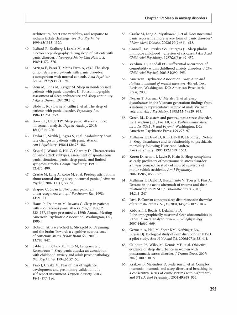
architecture, heart rate variablity, and response tosodium lactate challenge. Soc Biol Psychiatry.1999;45:1313 1320.
40. Lydiard R, Zealberg J, Laraia M, et al.Electroencephalography during sleep of patients withpanic disorder. J Neuropsychiatry Clin Neurosci.1989;1:372 376.
41. Arriaga F, Paiva T, Matos Pires A, et al. The sleepof non depressed patients with panic disorder:a comparison with normal controls. Acta PsychiatrScand. 1996;93:191 194.
42. Stein M, Enns M, Kryger M. Sleep in nondepressedpatients with panic disorder. II. Polysomnographicassessment of sleep architecture and sleep continuity.J Affect Disord. 1993;28:1 6.
43. Uhde T, Roy Byrne P, Gillin J, et al. The sleep ofpatients with panic disorder. Psychiatry Res.1984;12:251 259.
44. Brown T, Uhde TW. Sleep panic attacks: a micromovement analysis. Depress Anxiety. 2003;18(4):214 220.
45. Taylor C, Skeikh J, Agras S, et al. Ambulatory heartrate changes in patients with panic attacks.Am J Psychiatry. 1986;143:478 482.
46. Krystal J, Woods S, Hill C, Charney D. Characteristicsof panic attack subtypes: assessment of spontaneouspanic, situational panic, sleep panic, and limitedsymptom attacks. Compr Psychiatry. 1991;32:474 480.
47. Craske M, Lang A, Rowe M, et al. Presleep attributionsabout arousal during sleep: nocturnal panic. J AbnormPsychol. 2002;111(1):53 62.
48. Shapiro C, Sloan E. Nocturnal panic: anunderrecognized entity. J Psychosom Res. 1998;44:21 23.
49. Hauri P, Freidman M, Ravaris C. Sleep in patientswith spontaneous panic attacks. Sleep. 1989;12:323 337. [Paper presented at 139th Annual MeetingAmerican Psychiatric Association, Washington, DC,1986.]
50. Hobson JA, Pace Schott E, Stickgold R. Dreamingand the brain: Towards a cognitive neuroscienceof conscious states. Behav Brain Sci. 2000;23:793 842.
51. Labbate L, Pollack M, Otto M, Langenauer S,Rosenbaum J. Sleep panic attacks: an associationwith childhood anxiety and adult psychopathology.Biol Psychiatry. 1994;36:57 60.
52. Tsao J, Craske M. Fear of loss of vigilance:development and preliminary validation of aself report instrument. Depress Anxiety. 2003;18(4):177 186.
53. Craske M, Lang A, Mystkowski J, et al. Does nocturnalpanic represent a more severe form of panic disorder?J Nerv Ment Disease. 2002;190(9):611 618.
54. Connell HM, Persley GV, Sturgess JL. Sleep phobiain middle childhood a review of six cases. J Am AcadChild Adol Psychiatry. 1987;26(3):449 452.
55. Verduin TL, Kendall PC. Differential occurrence ofcomorbidity within childhood anxiety disorders. J ClinChild Adol Psychol. 2003;32:290 295.
56. American Psychiatric Association. Diagnostic andstatistical manual of mental disorders, 4th ed. TextRevision. Washington, DC: American PsychiatricPress; 2000.
57. Neylan T, Marmar C, Metzler T, et al. Sleepdisturbances in the Vietnam generation: findings froma nationally representative sample of male Vietnamveterans. Am J Psychiatry. 1998;155(7):929 933.
58. Green BL. Disasters and posttraumatic stress disorder.In: Davidson JRT, Foa EB, eds. Posttraumatic stressdisorder DSM IV and beyond. Washington, DC:American Psychiatric Press; 1993:75 97.
59. Mellman T, David D, Kulick Bell R, Hebding J, NolanB. Sleep disturbance and its relationship to psychiatricmorbidity following Hurricane Andrew.Am J Psychiatry. 1995;152:1659 1663.
60. Koren D, Arnon I, Lavie P, Klein E. Sleep complaintsas early predictors of posttraumatic stress disorder:a 1 year prospective study of injured survivors ofmotor vehicle accidents. Am J Psychiatry.2002;159(5):855 857.
61. Mellman T, David D, Bustamante V, Torres J, Fins A.Dreams in the acute aftermath of trauma and theirrelationship to PTSD. J Traumatic Stress. 2001;14:241 247.
62. Lavie P. Current concepts: sleep disturbances in the wakeof traumatic events. NEJM. 2001;345(25):1825 1832.
63. Kobayshi I, Boarts J, Delahanty D.Polysomnographically measured sleep abnormalities inPTSD: A meta analytic review. Psychophysiology.2007;44:660 669.
64. Germain A, Hall M, Shear KM, Nofzinger EA,Buysse DJ. Ecological study of sleep disruption in PTSD:a pilot study. Ann N Y Acad Sci. 2006;1071:438 441.
65. Calhoun PS, Wiley M, Dennis MF, et al. Objectiveevidence of sleep disturbance in women withposttraumatic stress disorder. J Traum Stress. 2007;20(6):1009 1018.
66. Krakow B, Melendrez D, Pederson B, et al. Complexinsomnia: insomnia and sleep disordered breathing ina consecutive series of crime victims with nightmaresand PTSD. Biol Psychiatry. 2001;49:948 953.
Chapter 17: Sleep in anxiety disorders
295
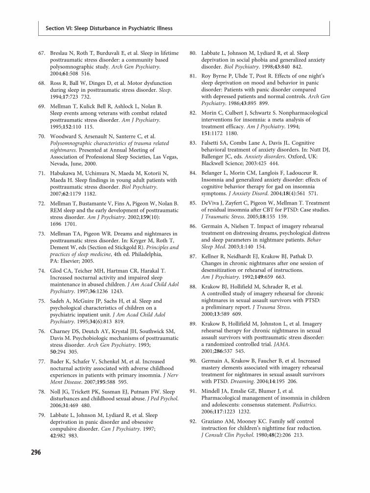
67. Breslau N, Roth T, Burduvali E, et al. Sleep in lifetimeposttraumatic stress disorder: a community basedpolysomnographic study. Arch Gen Psychiatry.2004;61:508 516.
68. Ross R, Ball W, Dinges D, et al. Motor dysfunctionduring sleep in posttraumatic stress disorder. Sleep.1994;17:723 732.
69. Mellman T, Kulick Bell R, Ashlock L, Nolan B.Sleep events among veterans with combat relatedposttraumatic stress disorder. Am J Psychiatry.1995;152:110 115.
70. Woodward S, Arsenault N, Santerre C, et al.Polysomnographic characteristics of trauma relatednightmares. Presented at Annual Meeting ofAssociation of Professional Sleep Societies, Las Vegas,Nevada, June, 2000.
71. Habukawa M, Uchimura N, Maeda M, Kotorii N,Maeda H. Sleep findings in young adult patients withposttraumatic stress disorder. Biol Psychiatry.2007;62:1179 1182.
72. Mellman T, Bustamante V, Fins A, Pigeon W, Nolan B.REM sleep and the early development of posttraumaticstress disorder. Am J Psychiatry. 2002;159(10):1696 1701.
73. Mellman TA, Pigeon WR. Dreams and nightmares inposttraumatic stress disorder. In: Kryger M, Roth T,Dement W, eds (Section ed Stickgold R). Principles andpractices of sleep medicine, 4th ed. Philadelphia,PA: Elsevier; 2005.
74. Glod CA, Teicher MH, Hartman CR, Harakal T.Increased nocturnal activity and impaired sleepmaintenance in abused children. J Am Acad Child AdolPsychiatry. 1997;36:1236 1243.
75. Sadeh A, McGuire JP, Sachs H, et al. Sleep andpsychological characteristics of children on apsychiatric inpatient unit. J Am Acad Child AdolPsychiatry. 1995;34(6):813 819.
76. Charney DS, Deutch AY, Krystal JH, Southwick SM,Davis M. Psychobiologic mechanisms of posttraumaticstress disorder. Arch Gen Psychiatry. 1993;50:294 305.
77. Bader K, Schafer V, Schenkel M, et al. Increasednocturnal activity associated with adverse childhoodexperiences in patients with primary insomnia. J NervMent Disease. 2007;195:588 595.
78. Noll JG, Trickett PK, Susman EJ, Putnam FW. Sleepdisturbances and childhood sexual abuse. J Ped Psychol.2006;31:469 480.
79. Labbate L, Johnson M, Lydiard R, et al. Sleepdeprivation in panic disorder and obsessivecompulsive disorder. Can J Psychiatry. 1997;42:982 983.
80. Labbate L, Johnson M, Lydiard R, et al. Sleepdeprivation in social phobia and generalized anxietydisorder. Biol Psychiatry. 1998;43:840 842.
81. Roy Byrne P, Uhde T, Post R. Effects of one night’ssleep deprivation on mood and behavior in panicdisorder: Patients with panic disorder comparedwith depressed patients and normal controls. Arch GenPsychiatry. 1986;43:895 899.
82. Morin C, Culbert J, Schwartz S. Nonpharmacologicalinterventions for insomnia: a meta analysis oftreatment efficacy. Am J Psychiatry. 1994;151:1172 1180.
83. Falsetti SA, Combs Lane A, Davis JL. Cognitivebehavioral treatment of anxiety disorders. In: Nutt DJ,Ballenger JC, eds. Anxiety disorders. Oxford, UK:Blackwell Science; 2003:425 444.
84. Belanger L, Morin CM, Langlois F, Ladouceur R.Insomnia and generalized anxiety disorder: effects ofcognitive behavior therapy for gad on insomniasymptoms. J Anxiety Disord. 2004;18(4):561 571.
85. DeViva J, Zayfert C, Pigeon W, Mellman T. Treatmentof residual insomnia after CBT for PTSD: Case studies.J Traumatic Stress. 2005;18:155 159.
86. Germain A, Nielsen T. Impact of imagery rehearsaltreatment on distressing dreams, psychological distressand sleep parameters in nightmare patients. BehavSleep Med. 2003;1:140 154.
87. Kellner R, Neidhardt EJ, Krakow BJ, Pathak D.Changes in chronic nightmares after one session ofdesensitization or rehearsal of instructions.Am J Psychiatry. 1992;149:659 663.
88. Krakow BJ, Hollifield M, Schrader R, et al.A controlled study of imagery rehearsal for chronicnightmares in sexual assault survivors with PTSD:a preliminary report. J Trauma Stress.2000;13:589 609.
89. Krakow B, Hollifield M, Johnston L, et al. Imageryrehearsal therapy for chronic nightmares in sexualassault survivors with posttraumatic stress disorder:a randomized controlled trial. JAMA.2001;286:537 545.
90. Germain A, Krakow B, Faucher B, et al. Increasedmastery elements associated with imagery rehearsaltreatment for nightmares in sexual assault survivorswith PTSD. Dreaming. 2004;14:195 206.
91. Mindell JA, Emslie GE, Blumer J, et al.Pharmacological management of insomnia in childrenand adolescents: consensus statement. Pediatrics.2006;117:1223 1232.
92. Graziano AM, Mooney KC. Family self controlinstruction for children’s nighttime fear reduction.J Consult Clin Psychol. 1980;48(2):206 213.
Section VI: Sleep Disturbance in Psychiatric Illness
296
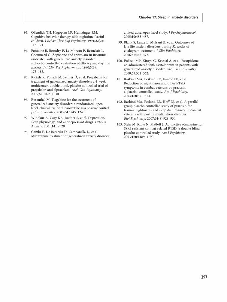
93. Ollendick TH, Hagopian LP, Huntzinger RM.Cognitive behavior therapy with nighttime fearfulchildren. J Behav Ther Exp Psychiatry. 1991;22(2):113 121.
94. Fontaine R, Beaudry P, Le Morvan P, Beauclair L,Chouinard G. Zopiclone and triazolam in insomniaassociated with generalized anxiety disorder:a placebo controlled evaluation of efficacy and daytimeanxiety. Int Clin Psychopharmacol. 1990;5(3):173 183.
95. Rickels K, Pollack M, Feltner D, et al. Pregabalin fortreatment of generalized anxiety disorder: a 4 week,multicenter, double blind, placebo controlled trial ofpregabalin and alprazolam. Arch Gen Psychiatry.2005;62:1022 1030.
96. Rosenthal M. Tiagabine for the treatment ofgeneralized anxiety disorder: a randomized, openlabel, clinical trial with paroxetine as a positive control.J Clin Psychiatry. 2003;64:1245 1249.
97. Winokur A, Gary KA, Rodner S, et al. Depression,sleep physiology, and antidepressant drugs. DepressAnxiety. 2001;14:19 28.
98. Gambi F, De Berardis D, Campanella D, et al.Mirtazapine treatment of generalized anxiety disorder:
a fixed dose, open label study. J Psychopharmacol.2005;19:483 487.
99. Blank S, Lenze E, Mulsant B, et al. Outcomes oflate life anxiety disorders during 32 weeks ofcitalopram treatment. J Clin Psychiatry.2006;67:468 472.
100. Pollack MP, Kinrys G, Krystal A, et al. Eszopicloneco administered with escitalopram in patients withgeneralized anxiety disorder. Arch Gen Psychiatry.2008;65:551 562.
101. Raskind MA, Peskind ER, Kanter ED, et al.Reduction of nightmares and other PTSDsymptoms in combat veterans by prazosin:a placebo controlled study. Am J Psychiatry.2003;160:371 373.
102. Raskind MA, Peskind ER, Hoff DJ, et al. A parallelgroup placebo controlled study of prazosin fortrauma nightmares and sleep disturbances in combatveterans with posttraumatic stress disorder.Biol Psychiatry. 2007;61(8):928 934.
103. Stein M, Kline N, Matloff J. Adjunctive olanzapine forSSRI resistant combat related PTSD: a double blind,placebo controlled study. Am J Psychiatry.2003;160:1189 1190.
Chapter 17: Sleep in anxiety disorders
297

Chapter
18 Psychotic disorders
Allen C. Richert
IntroductionSchizophrenia, the archetypal psychotic disorder, is achronic, currently incurable, prevalent, and devastat-ing syndrome. While sleep disturbances are import-ant aspects of psychotic illness, they are not primarysymptoms of schizophrenia or any other psychoticdisorder. Even so, sleep disturbances are frequentlycomponents of, influence quality of life in, and mayexacerbate psychotic illnesses. A better understandingof sleep abnormalities associated with psychotic dis-orders may improve our understanding of the patho-physiology of these disorders.
For several reasons, succinctly summarizing theliterature on sleep abnormalities associated withpsychotic disorders is difficult. First, the DSM-IV-TR[1] category of psychotic disorders contains severaldisorders that are not necessarily syndromically oreven pathophysiologically homogeneous. The dis-orders include schizophrenia, schizoaffective disorder,brief psychotic disorder, schizophreniform disorder,delusional disorder, shared psychotic disorder, andpsychotic disorder not otherwise specified (NOS).Second, considerably more sleep research has focusedon schizophrenia than any of the other psychotic dis-orders. Third, while some psychotic disorders (i.e.schizoaffective disorder and schizophreniform dis-order) incorporate similar symptoms as schizophre-nia, other psychotic disorders (i.e. delusional disorder,shared psychotic disorder, and brief psychotic dis-order) differ from schizophrenia sufficiently to raisedoubt that they spring from similar neurobiologicalabnormalities and therefore that their associated sleepabnormalities would be similar. Based upon thereasoning described above, this chapter will focus pri-marily on schizophrenia, and discuss other psychoticdisorders when feasible.
It is important to additionally recognize thatschizophrenia itself is a heterogeneous syndrome.The DSM-IV-TR acknowledges heterogeneity withinschizophrenia by recognizing subtypes of schizophre-nia, which include paranoid, disorganized, catatonic,undifferentiated, and residual types [1]. Because thesevariations within the diagnostic category of schizo-phrenia almost certainly arise as a result of variationsin the neurological abnormalities underlying each typeof schizophrenia, it would be short-sighted to assumethat different types of schizophrenia would generatethe same changes in sleep. It is with an appreciationof the above difficulties that the author endeavors tosynthesize a coherent, clinically relevant, and patho-physiologically illuminating summary of the sleepabnormalities associated with psychotic disorders.
Subjective sleep disturbancein psychotic illnessClinical lore and the author’s experience suggest thatsleep complaints frequently accompany schizophreniaand other psychotic disorders, just as they do manyother psychiatric disorders. However, in contrast tothe amount of research examining polysomnographicfeatures of sleep in psychotic disorders (i.e. mostlyschizophrenia), the literature examining subjectivesleep complaints in psychotic disorders is small. Thefield lacks large-scale epidemiological studies address-ing frequency, prevalence, and types of sleep complaintsassociated with psychotic disorders. But, a modestnumber of relatively small studies directly address ordescribe, as a secondary measure, sleep complaints inschizophrenia, schizophreniform disorder, schizoaffect-ive disorder, and delusional disorder. Essentially nosuch studies exist for shared psychotic disorder. Theauthor’s experience as a psychiatrist who practices
Foundations of Psychiatric SleepMedicine, ed. JohnW.Winkelman andDavid T. Plante. Published by CambridgeUniversity Press.# Cambridge University Press 2010.
298
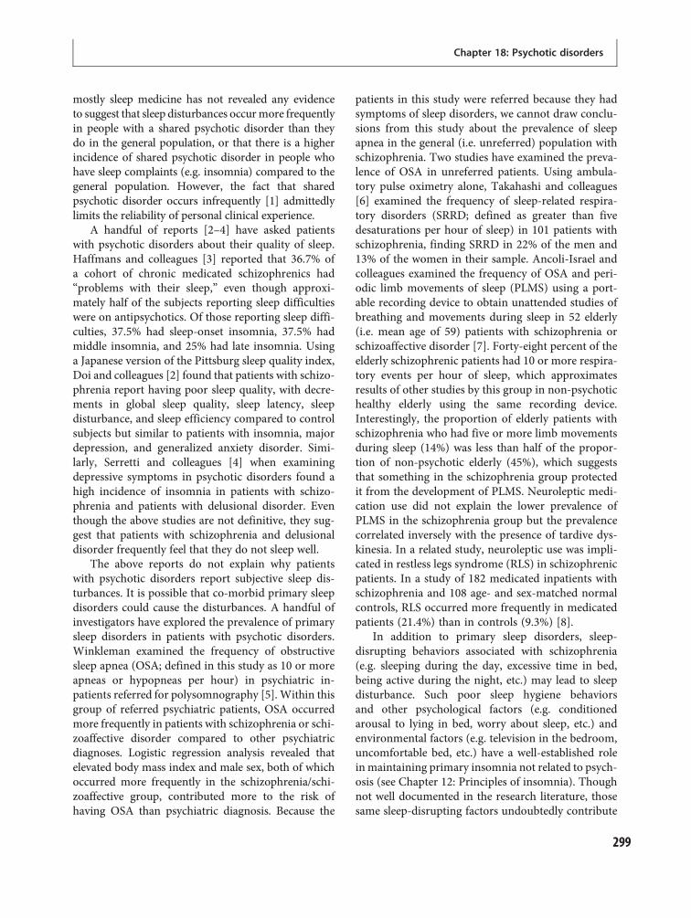
mostly sleep medicine has not revealed any evidenceto suggest that sleep disturbances occurmore frequentlyin people with a shared psychotic disorder than theydo in the general population, or that there is a higherincidence of shared psychotic disorder in people whohave sleep complaints (e.g. insomnia) compared to thegeneral population. However, the fact that sharedpsychotic disorder occurs infrequently [1] admittedlylimits the reliability of personal clinical experience.
A handful of reports [2–4] have asked patientswith psychotic disorders about their quality of sleep.Haffmans and colleagues [3] reported that 36.7% ofa cohort of chronic medicated schizophrenics had“problems with their sleep,” even though approxi-mately half of the subjects reporting sleep difficultieswere on antipsychotics. Of those reporting sleep diffi-culties, 37.5% had sleep-onset insomnia, 37.5% hadmiddle insomnia, and 25% had late insomnia. Usinga Japanese version of the Pittsburg sleep quality index,Doi and colleagues [2] found that patients with schizo-phrenia report having poor sleep quality, with decre-ments in global sleep quality, sleep latency, sleepdisturbance, and sleep efficiency compared to controlsubjects but similar to patients with insomnia, majordepression, and generalized anxiety disorder. Simi-larly, Serretti and colleagues [4] when examiningdepressive symptoms in psychotic disorders found ahigh incidence of insomnia in patients with schizo-phrenia and patients with delusional disorder. Eventhough the above studies are not definitive, they sug-gest that patients with schizophrenia and delusionaldisorder frequently feel that they do not sleep well.
The above reports do not explain why patientswith psychotic disorders report subjective sleep dis-turbances. It is possible that co-morbid primary sleepdisorders could cause the disturbances. A handful ofinvestigators have explored the prevalence of primarysleep disorders in patients with psychotic disorders.Winkleman examined the frequency of obstructivesleep apnea (OSA; defined in this study as 10 or moreapneas or hypopneas per hour) in psychiatric in-patients referred for polysomnography [5]. Within thisgroup of referred psychiatric patients, OSA occurredmore frequently in patients with schizophrenia or schi-zoaffective disorder compared to other psychiatricdiagnoses. Logistic regression analysis revealed thatelevated body mass index and male sex, both of whichoccurred more frequently in the schizophrenia/schi-zoaffective group, contributed more to the risk ofhaving OSA than psychiatric diagnosis. Because the
patients in this study were referred because they hadsymptoms of sleep disorders, we cannot draw conclu-sions from this study about the prevalence of sleepapnea in the general (i.e. unreferred) population withschizophrenia. Two studies have examined the preva-lence of OSA in unreferred patients. Using ambula-tory pulse oximetry alone, Takahashi and colleagues[6] examined the frequency of sleep-related respira-tory disorders (SRRD; defined as greater than fivedesaturations per hour of sleep) in 101 patients withschizophrenia, finding SRRD in 22% of the men and13% of the women in their sample. Ancoli-Israel andcolleagues examined the frequency of OSA and peri-odic limb movements of sleep (PLMS) using a port-able recording device to obtain unattended studies ofbreathing and movements during sleep in 52 elderly(i.e. mean age of 59) patients with schizophrenia orschizoaffective disorder [7]. Forty-eight percent of theelderly schizophrenic patients had 10 or more respira-tory events per hour of sleep, which approximatesresults of other studies by this group in non-psychotichealthy elderly using the same recording device.Interestingly, the proportion of elderly patients withschizophrenia who had five or more limb movementsduring sleep (14%) was less than half of the propor-tion of non-psychotic elderly (45%), which suggeststhat something in the schizophrenia group protectedit from the development of PLMS. Neuroleptic medi-cation use did not explain the lower prevalence ofPLMS in the schizophrenia group but the prevalencecorrelated inversely with the presence of tardive dys-kinesia. In a related study, neuroleptic use was impli-cated in restless legs syndrome (RLS) in schizophrenicpatients. In a study of 182 medicated inpatients withschizophrenia and 108 age- and sex-matched normalcontrols, RLS occurred more frequently in medicatedpatients (21.4%) than in controls (9.3%) [8].
In addition to primary sleep disorders, sleep-disrupting behaviors associated with schizophrenia(e.g. sleeping during the day, excessive time in bed,being active during the night, etc.) may lead to sleepdisturbance. Such poor sleep hygiene behaviorsand other psychological factors (e.g. conditionedarousal to lying in bed, worry about sleep, etc.) andenvironmental factors (e.g. television in the bedroom,uncomfortable bed, etc.) have a well-established rolein maintaining primary insomnia not related to psych-osis (see Chapter 12: Principles of insomnia). Thoughnot well documented in the research literature, thosesame sleep-disrupting factors undoubtedly contribute
Chapter 18: Psychotic disorders
299
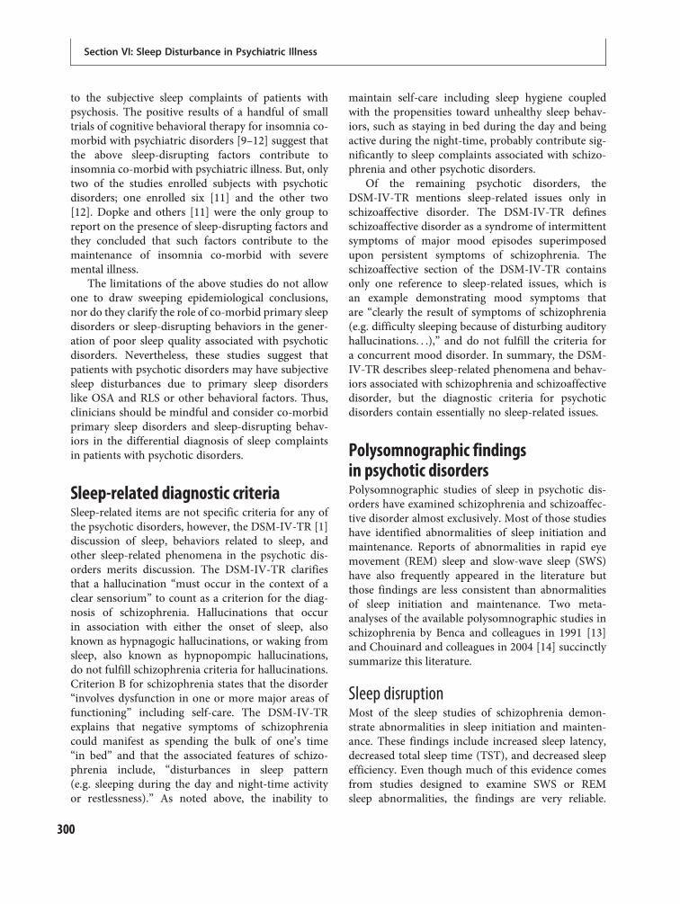
to the subjective sleep complaints of patients withpsychosis. The positive results of a handful of smalltrials of cognitive behavioral therapy for insomnia co-morbid with psychiatric disorders [9–12] suggest thatthe above sleep-disrupting factors contribute toinsomnia co-morbid with psychiatric illness. But, onlytwo of the studies enrolled subjects with psychoticdisorders; one enrolled six [11] and the other two[12]. Dopke and others [11] were the only group toreport on the presence of sleep-disrupting factors andthey concluded that such factors contribute to themaintenance of insomnia co-morbid with severemental illness.
The limitations of the above studies do not allowone to draw sweeping epidemiological conclusions,nor do they clarify the role of co-morbid primary sleepdisorders or sleep-disrupting behaviors in the gener-ation of poor sleep quality associated with psychoticdisorders. Nevertheless, these studies suggest thatpatients with psychotic disorders may have subjectivesleep disturbances due to primary sleep disorderslike OSA and RLS or other behavioral factors. Thus,clinicians should be mindful and consider co-morbidprimary sleep disorders and sleep-disrupting behav-iors in the differential diagnosis of sleep complaintsin patients with psychotic disorders.
Sleep-related diagnostic criteriaSleep-related items are not specific criteria for any ofthe psychotic disorders, however, the DSM-IV-TR [1]discussion of sleep, behaviors related to sleep, andother sleep-related phenomena in the psychotic dis-orders merits discussion. The DSM-IV-TR clarifiesthat a hallucination “must occur in the context of aclear sensorium” to count as a criterion for the diag-nosis of schizophrenia. Hallucinations that occurin association with either the onset of sleep, alsoknown as hypnagogic hallucinations, or waking fromsleep, also known as hypnopompic hallucinations,do not fulfill schizophrenia criteria for hallucinations.Criterion B for schizophrenia states that the disorder“involves dysfunction in one or more major areas offunctioning” including self-care. The DSM-IV-TRexplains that negative symptoms of schizophreniacould manifest as spending the bulk of one’s time“in bed” and that the associated features of schizo-phrenia include, “disturbances in sleep pattern(e.g. sleeping during the day and night-time activityor restlessness).” As noted above, the inability to
maintain self-care including sleep hygiene coupledwith the propensities toward unhealthy sleep behav-iors, such as staying in bed during the day and beingactive during the night-time, probably contribute sig-nificantly to sleep complaints associated with schizo-phrenia and other psychotic disorders.
Of the remaining psychotic disorders, theDSM-IV-TR mentions sleep-related issues only inschizoaffective disorder. The DSM-IV-TR definesschizoaffective disorder as a syndrome of intermittentsymptoms of major mood episodes superimposedupon persistent symptoms of schizophrenia. Theschizoaffective section of the DSM-IV-TR containsonly one reference to sleep-related issues, which isan example demonstrating mood symptoms thatare “clearly the result of symptoms of schizophrenia(e.g. difficulty sleeping because of disturbing auditoryhallucinations. . .),” and do not fulfill the criteria fora concurrent mood disorder. In summary, the DSM-IV-TR describes sleep-related phenomena and behav-iors associated with schizophrenia and schizoaffectivedisorder, but the diagnostic criteria for psychoticdisorders contain essentially no sleep-related issues.
Polysomnographic findingsin psychotic disordersPolysomnographic studies of sleep in psychotic dis-orders have examined schizophrenia and schizoaffec-tive disorder almost exclusively. Most of those studieshave identified abnormalities of sleep initiation andmaintenance. Reports of abnormalities in rapid eyemovement (REM) sleep and slow-wave sleep (SWS)have also frequently appeared in the literature butthose findings are less consistent than abnormalitiesof sleep initiation and maintenance. Two meta-analyses of the available polysomnographic studies inschizophrenia by Benca and colleagues in 1991 [13]and Chouinard and colleagues in 2004 [14] succinctlysummarize this literature.
Sleep disruptionMost of the sleep studies of schizophrenia demon-strate abnormalities in sleep initiation and mainten-ance. These findings include increased sleep latency,decreased total sleep time (TST), and decreased sleepefficiency. Even though much of this evidence comesfrom studies designed to examine SWS or REMsleep abnormalities, the findings are very reliable.
Section VI: Sleep Disturbance in Psychiatric Illness
300
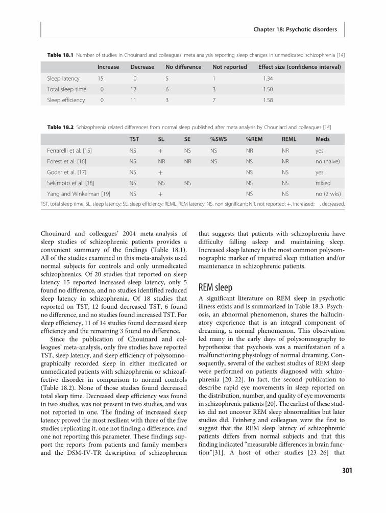
Chouinard and colleagues’ 2004 meta-analysis ofsleep studies of schizophrenic patients provides aconvenient summary of the findings (Table 18.1).All of the studies examined in this meta-analysis usednormal subjects for controls and only unmedicatedschizophrenics. Of 20 studies that reported on sleeplatency 15 reported increased sleep latency, only 5found no difference, and no studies identified reducedsleep latency in schizophrenia. Of 18 studies thatreported on TST, 12 found decreased TST, 6 foundno difference, and no studies found increased TST. Forsleep efficiency, 11 of 14 studies found decreased sleepefficiency and the remaining 3 found no difference.
Since the publication of Chouinard and col-leagues’ meta-analysis, only five studies have reportedTST, sleep latency, and sleep efficiency of polysomno-graphically recorded sleep in either medicated orunmedicated patients with schizophrenia or schizoaf-fective disorder in comparison to normal controls(Table 18.2). None of those studies found decreasedtotal sleep time. Decreased sleep efficiency was foundin two studies, was not present in two studies, and wasnot reported in one. The finding of increased sleeplatency proved the most resilient with three of the fivestudies replicating it, one not finding a difference, andone not reporting this parameter. These findings sup-port the reports from patients and family membersand the DSM-IV-TR description of schizophrenia
that suggests that patients with schizophrenia havedifficulty falling asleep and maintaining sleep.Increased sleep latency is the most common polysom-nographic marker of impaired sleep initiation and/ormaintenance in schizophrenic patients.
REM sleepA significant literature on REM sleep in psychoticillness exists and is summarized in Table 18.3. Psych-osis, an abnormal phenomenon, shares the hallucin-atory experience that is an integral component ofdreaming, a normal phenomenon. This observationled many in the early days of polysomnography tohypothesize that psychosis was a manifestation of amalfunctioning physiology of normal dreaming. Con-sequently, several of the earliest studies of REM sleepwere performed on patients diagnosed with schizo-phrenia [20–22]. In fact, the second publication todescribe rapid eye movements in sleep reported onthe distribution, number, and quality of eye movementsin schizophrenic patients [20]. The earliest of these stud-ies did not uncover REM sleep abnormalities but laterstudies did. Feinberg and colleagues were the first tosuggest that the REM sleep latency of schizophrenicpatients differs from normal subjects and that thisfinding indicated “measurable differences in brain func-tion”[31]. A host of other studies [23–26] that
Table 18.1 Number of studies in Chouinard and colleagues’ meta analysis reporting sleep changes in unmedicated schizophrenia [14]
Increase Decrease No difference Not reported Effect size (confidence interval)
Sleep latency 15 0 5 1 1.34
Total sleep time 0 12 6 3 1.50
Sleep efficiency 0 11 3 7 1.58
Table 18.2 Schizophrenia related differences from normal sleep published after meta analysis by Chouniard and colleagues [14]
TST SL SE %SWS %REM REML Meds
Ferrarelli et al. [15] NS þ NS NS NR NR yes
Forest et al. [16] NS NR NR NS NS NR no (naive)
Goder et al. [17] NS þ NS NS yes
Sekimoto et al. [18] NS NS NS NS NS mixed
Yang and Winkelman [19] NS þ NS NS no (2 wks)
TST, total sleep time; SL, sleep latency; SE, sleep efficiency; REML, REM latency; NS, non significant; NR, not reported;þ, increased; , decreased.
Chapter 18: Psychotic disorders
301
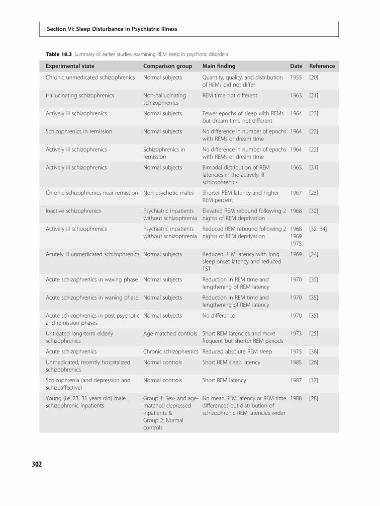
Table 18.3 Summary of earlier studies examining REM sleep in psychotic disorders
Experimental state Comparison group Main finding Date Reference
Chronic unmedicated schizophrenics Normal subjects Quantity, quality, and distributionof REMs did not differ
1955 [20]
Hallucinating schizophrenics Non-hallucinatingschizophrenics
REM time not different 1963 [21]
Actively ill schizophrenics Normal subjects Fewer epochs of sleep with REMsbut dream time not different
1964 [22]
Schizophrenics in remission Normal subjects No difference in number of epochswith REMs or dream time
1964 [22]
Actively ill schizophrenics Schizophrenics inremission
No difference in number of epochswith REMs or dream time
1964 [22]
Actively ill schizophrenics Normal subjects Bimodal distribution of REMlatencies in the actively illschizophrenics
1965 [31]
Chronic schizophrenics near remission Non-psychotic males Shorter REM latency and higherREM percent
1967 [23]
Inactive schizophrenics Psychiatric inpatientswithout schizophrenia
Elevated REM rebound following 2nights of REM deprivation
1968 [32]
Actively ill schizophrenics Psychiatric inpatientswithout schizophrenia
Reduced REM rebound following 2nights of REM deprivation
196819691975
[32 34]
Acutely ill unmedicated schizophrenics Normal subjects Reduced REM latency with longsleep onset latency and reducedTST
1969 [24]
Acute schizophrenics in waxing phase Normal subjects Reduction in REM time andlengthening of REM latency
1970 [35]
Acute schizophrenics in waning phase Normal subjects Reduction in REM time andlengthening of REM latency
1970 [35]
Acute schizophrenics in post-psychoticand remission phases
Normal subjects No difference 1970 [35]
Untreated long-term elderlyschizophrenics
Age-matched controls Short REM latencies and morefrequent but shorter REM periods
1973 [25]
Acute schizophrenics Chronic schizophrenics Reduced absolute REM sleep 1975 [36]
Unmedicated, recently hospitalizedschizophrenics
Normal controls Short REM sleep latency 1985 [26]
Schizophrenia (and depression andschizoaffective)
Normal controls Short REM latency 1987 [37]
Young (i.e. 23 31 years old) maleschizophrenic inpatients
Group 1: Sex- and age-matched depressedinpatients &Group 2: Normalcontrols
No mean REM latency or REM timedifferences but distribution ofschizophrenic REM latencies wider
1988 [28]
Section VI: Sleep Disturbance in Psychiatric Illness
302
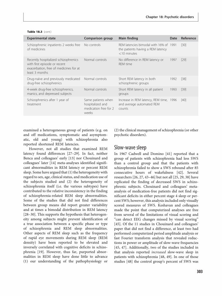
examined a heterogeneous group of patients (e.g. onand off medications, symptomatic and asymptom-atic, old and young) with schizophrenia alsoreported shortened REM latencies.
However, not all studies that examined REMlatency found differences [27–29]. In fact, neitherBenca and colleagues’ early [13] nor Chouinard andcolleagues’ later [14] meta-analyses identified signifi-cant abnormalities in REM latency or percent REMsleep. Some have argued that (1) the heterogeneity withregard to sex, age, clinical status, andmedication use ofthe subjects studied and (2) the heterogeneity ofschizophrenia itself (i.e. the various subtypes) havecontributed to the relative inconsistency in the findingof schizophrenia-related REM sleep abnormalities.Some of the studies that did not find differencesbetween group means did report greater variabilityand at times a bimodal distribution in REM latency[28–30]. This supports the hypothesis that heterogen-eity among subjects might prevent identification ofa true association between a specific phase or typeof schizophrenia and REM sleep abnormalities.Other aspects of REM sleep such as the frequencyof rapid eye movements during REM sleep (REMdensity) have been reported to be elevated andinversely correlated with cognitive deficits in schizo-phrenia [19]. However, thus far, purported abnor-malities in REM sleep have done little to advance(1) our understanding of the pathophysiology or
(2) the clinical management of schizophrenia (or otherpsychotic disorders).
Slow-wave sleepIn 1967 Cadwell and Domino [41] reported that agroup of patients with schizophrenia had less SWSthan a control group and that the patients withschizophrenia failed to show a SWS rebound after 85consecutive hours of wakefulness [42]. Severalresearchers [26, 27, 43–46] but not all [25, 29, 38] havereplicated the finding of decreased SWS in schizo-phrenic subjects. Chouinard and colleagues’ meta-analysis of medication-free patients did not find sig-nificant deficits in either percent stage 4 sleep or per-cent SWS; however, this analysis included only visuallyscored measures of SWS. Keshavan and colleaguesmade the point that computerized analyses are freefrom several of the limitations of visual scoring and“can detect EEG changes missed by visual scoring”[45]. Of the 11 studies in Chouinard and colleagues’paper that did not find a difference, at least two hadperformed computerized period amplitude analysis orfast Fourier transform analysis that revealed reduc-tions in power or amplitude of slow-wave frequencies[45, 47]. Additionally, two of the studies included inthat analysis reported increased slow-wave sleep inpatients with schizophrenia [48, 49]. In one of thosestudies [48] the control group’s percent of SWS was
Table 18.3 (cont.)
Experimental state Comparison group Main finding Date Reference
Schizophrenic inpatients 2 weeks freeof medicines
No controls REM latencies bimodal with 16% ofthe patients having a REM latency<10 minutes
1991 [30]
Recently hospitalized schizophrenicswith first episode or recentexacerbation, free of medicines for atleast 3 months
Normal controls No difference in REM latency orREM time
1997 [29]
Drug-naïve and previously medicateddrug-free schizophrenics
Normal controls Short REM latency in bothschizophrenic groups
1992 [38]
4-week drug-free schizophrenics,manics, and depressed subjects
Normal controls Short REM latency in all patientgroups
1993 [39]
Schizophrenics after 1 year oftreatment
Same patients whenhospitalized andmedication free for 2weeks
Increase in REM latency, REM time,and average automated REMcounts
1996 [40]
Chapter 18: Psychotic disorders
303
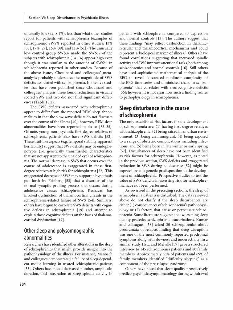
unusually low (i.e. 8.1%), less than what other studiesreport for patients with schizophrenia (examples ofschizophrenic SWS% reported in other studies: 13%[50], 17% [27], 16% [39], and 11% [51]). The unusuallylow control group SWS% made the SWS% of thesubjects with schizophrenia (14.1%) appear high eventhough it was similar to the amount of SWS% inschizophrenia reported in other studies. Because ofthe above issues, Chouinard and colleagues’ meta-analysis probably understates the magnitude of SWSdeficits associated with schizophrenia. In the five stud-ies that have been published since Chouinard andcolleagues’ analysis, three found reductions in visuallyscored SWS and two did not find significant differ-ences (Table 18.2).
The SWS deficits associated with schizophreniaappear to differ from the reported REM sleep abnor-malities in that the slow-wave deficits do not fluctuateover the course of the illness [40]; however, REM sleepabnormalities have been reported to do so [35–35].Of note, young non-psychotic first-degree relatives ofschizophrenia patients also have SWS deficits [52].These trait-like aspects (e.g. temporal stability, apparentheritability) suggest that SWS deficits may be endophe-notypes (i.e. genetically transmissible characteristicsthat are not apparent to the unaided eye) of schizophre-nia. The normal decrease in SWS that occurs over thecourse of adolescence is exaggerated in these first-degree relatives at high risk for schizophrenia [52]. Thisexaggerated decrease of SWS may support a hypothesisput forth by Feinberg [53] that a disorder of thenormal synaptic pruning process that occurs duringadolescence causes schizophrenia. Keshavan hasinvoked dysfunction of thalamocortical circuits in theschizophrenia-related failure of SWS [54]. Similarly,others have begun to correlate SWS deficits with cogni-tive deficits in schizophrenia [19] and attempt toexplain those cognitive deficits on the basis of thalamo-cortical dysfunction [17].
Other sleep and polysomnographicabnormalitiesResearchers have identified other alterations in the sleepof schizophrenics that might provide insight into thepathophysiology of the illness. For instance, Manoachand colleagues demonstrated a failure of sleep-depend-ent motor learning in treated schizophrenic patients[55]. Others have noted decreased number, amplitude,duration, and integration of sleep spindle activity in
patients with schizophrenia compared to depressionand normal controls [15]. The authors suggest thatthese findings “may reflect dysfunction in thalamic-reticular and thalamocortical mechanisms and couldrepresent a biological marker of illness.” Others havefound correlations suggesting that increased spindleactivity and SWS improve attentional tasks, both amongschizophrenics and normal controls [16]. Still othershave used sophisticated mathematical analysis of theEEG to reveal “decreased nonlinear complexity ofthe EEG time series and diminished chaos in schizo-phrenia” that correlates with neurocognitive deficits[56]; however, it is not clear how such a finding relatesto pathophysiology in schizophrenia.
Sleep disturbance in the courseof schizophreniaThe only established risk factors for the developmentof schizophrenia are: (1) having first-degree relativeswith schizophrenia, (2) being raised in an urban envir-onment, (3) being an immigrant, (4) being exposedto a range of obstetric complications including infec-tions, and (5) being born in late winter or early spring[57]. Disturbances of sleep have not been identifiedas risk factors for schizophrenia. However, as notedin the previous section, SWS deficits and exaggeratedreduction in SWS during adolescence [52] might beexpressions of a genetic predisposition to the develop-ment of schizophrenia. Prospective studies to test thevalue of SWS deficits in assessing risk for schizophre-nia have not been performed.
As reviewed in the preceding sections, the sleep ofschizophrenia patients is disturbed. The data reviewedabove do not clarify if the sleep disturbances areeither (1) consequences of schizophrenia’s pathophysi-ology or (2) factors that cause or perpetuate schizo-phrenia. Some literature suggests that worsening sleepquality precedes schizophrenic exacerbations. Kumarand colleagues [58] asked 30 schizophrenics aboutprodramata of relapse, finding that sleep disruptionwas one of the most commonly reported prodromalsymptoms along with slowness and underactivity. In asimilar study Herz and Melville [59] gave a structuredinterview to 145 schizophrenia patients and 80 familymembers. Approximately 65% of patients and 69% offamily members identified “difficulty sleeping” as acomponent of the pre-relapse syndrome.
Others have noted that sleep quality prospectivelypredicts psychotic symptomatology during withdrawal
Section VI: Sleep Disturbance in Psychiatric Illness
304

from psychotropic medicines. Chemerinski and col-leagues observed that baseline total insomnia and ter-minal insomnia scores on the Hamilton depressionrating scale respectively predicted increased psychoticsymptomatology and disorganization during a 3-weekmedication-washout period [60]. Similarly, Mattaiand colleagues found a positive correlation betweensleep disturbance and symptom severity during amedication-washout period in childhood-onset schizo-phrenia [61]. Despite the above reports, Krystal andcolleagues, who recently reviewed the effects of sleepdisturbance on psychiatric illness, found “scantresearch on the effects of sleep disorders and the man-agement of schizophrenia and that more researchneeds to be done” [64].
Interactions of sleep andpsychotic illnessSleep symptoms affect quality of lifeThe literature on the effects of primary sleep disorderson psychotic illnesses is small. However, primary sleepdisorders (e.g. OSA and RLS) exacerbate the symp-toms (e.g. cognitive deficits and daytime sleepiness)in psychotic disorders. More notable is the fact thatpoor subjective sleep quality, as measured by the Pitts-burgh sleep quality index, is associated with a lowerquality of life for patients with schizophrenia [62, 63].The strong correlation between quality of sleep andquality of life in schizophrenia patients underscoresthe significance of Krystal and colleagues’ assessment[64] that there is a relative paucity of research designedto improve sleep quality in schizophrenia, and theimportance of further work in this area.
Clinical correlates of sleep deficitsSeveral researchers have investigated the clinical cor-relates of sleep disturbances associated with schizo-phrenia. Most of the studies contain small numbersof subjects and only some have been replicated.The following is a brief synopsis of the findings fromthese studies. Investigators have correlated sleep onsetlatency with the thinking disturbance subscale of thebrief psychiatric rating scale (BPRS) [65]. Short REMlatency has been associated with negative symptomseverity [38, 66], REM latency has been inverselycorrelated with positive symptoms [51], and REMdensity and REM time have been negatively correlatedwith total BPRS, medication status [38], and poor
1-year outcomes in female patients [67]. REM densityhas also been both positively correlated with ratings ofhallucinatory behavior [31] and inversely correlatedwith positive symptoms [19], as well as inversely cor-related with cognitive symptoms, emotional discom-fort symptoms, and total score on the positive andnegative syndrome scale (PANSS) [19]. Within thesame study, minutes of REM sleep and REM densityin the first REM period of the night inversely correl-ated with neuropsychological performance, whileREM minutes occurring after the first REM periodcorrelated positively with neuropsychological per-formance. In a small study that did not correct formultiple statistical tests [42], REM activity and REMtime both in the whole night data and in the firstREM period correlated with suicidal behavior [68].SWS has been inversely correlated with cognitivesymptoms [19], negative outcomes at 1 and 2 years[69], and ventricular size [70]. While each of the abovereports suggest that the sleep disturbance associatedwith schizophrenia and related disorders might pro-vide insight into the pathophysiology of the illness or amarker for improved treatment approaches, none havedone so yet.
Explanation of REM abnormalitiesUnderstanding the biological mechanisms underlyingthe sleep disturbances associated with schizophreniacould provide further insight into the pathophysiologyof the illness. Of the schizophrenia-related sleep dis-turbances, investigators have studied the mechanismunderlying REM abnormalities the most. Tandon andcolleagues demonstrated that an anticholinergic agentlengthens REM latency less in unmedicated schizo-phrenics than in normal controls [71]. They inter-preted the muted response as evidence of increasedcholinergic activity in the patients with schizophrenia.REM latency has also been demonstrated to inverselycorrelate with plasma acetylcholinesterase levels [72],that is higher acetylcholinesterase plasma levels (whichsuggests higher than normal levels of acetlycholinergicactivity) are associated with shorter REM latency.Administration of a muscarinic agent also shortensREM latency in schizophrenia patients implying thatmuscarinic receptor supersensitivity might underlieshort REM latency associated with schizophrenia[49]. A different line of research has also implicatednicotinic cholinergic receptors in the pathophysiologyof schizophrenia. Patients with schizophrenia fail to
Chapter 18: Psychotic disorders
305

suppress the P50 EEG potential in response to thesecond tone in a repeated paired auditory stimuli para-digm. It has been demonstrated that non-REM sleep, astate of relatively low central cholinergic tone, reversesthe failed P50 suppression but only briefly [73], whileREM sleep, a state of relatively high cholinergic tone,does not normalize the P50 suppression [74]. Interest-ingly, this normalization of P50 gating modulatedby sleep can be reversed by exogenous exposure tonicotine [75].
Sleep and cognitive symptomsCognitive deficits are a core feature of schizophrenia,and recently researchers have begun to connect thecharacteristic sleep disturbances of schizophrenia withmemory deficits. For instance, a group of patientswith schizophrenia failed to show improvement on anewly learned finger-tapping motor-sequence taskafter a night of sleep, suggesting a failure of sleep-dependent procedural learning [55]. Goder and col-leagues implicated the deficient SWS of schizophreniapatients with visuospatial memory impairments [17].However, it is notable that impaired memory associ-ated with decreased SWS is not specific to schizophre-nia [76]. The same caveat is applicable to theconnection of alteration of REM sleep measures andcognitive performance in schizophrenia, since alteredREM sleep can occur in patients with depression, bor-derline personality disorder, and normal subjects [77].
Circadian rhythms and schizophreniaSchizophrenics have been described as preferring tosleep during the day and stay active during the night[1]. Such behavior suggests that abnormalities in cir-cadian rhythms might contribute to or arise from thepathophysiology underlying schizophrenia. A smallstudy of two schizophrenia patients in an isolationunit suggested that they had an abnormally short (i.e.23.7-hour) circadian period [78]. There is also a casereport of a schizophrenia patient with an arrhythmiccircadian rest-activity cycle while on haloperidol des-pite normal-amplitude but slightly phase-advancedmelatonin and body temperature cycles [79]. Despitethe apparent altered activity rhythms of some schizo-phrenia patients, their circadian rhythms of hor-mone secretion appear normal except for anexaggerated secretion of prolactin with sleep onset[80]. Older schizophrenia patients may have lessrobust circadian rhythms of activity associated with
age, that may also be affected by medications, life-style factors, and behavioral factors [81, 82]. Othershave suggested that schizophrenics seem to be pro-verbial night owls and the severity of their nocturnalpreference correlates with their quality of sleep, cogni-tions, and symptoms [83]. Unfortunately, the limitedinformation on circadian rhythms in schizophrenia isnot consistent. Nonetheless, the use of actigraphyto evaluate circadian rest–activity phases in schizo-phrenics has helped to facilitate psychosocial therapies(day structure) in one case report and thus couldtheoretically be applied to other patients [84].
Evaluation and treatment of co-morbidsleep disorders and psychotic illnessObstructive sleep apneaThe available literature on the treatment of OSA inpatients with psychotic disorders consists of fourcase reports of patients with either schizophrenia orschizoaffective disorder and sleep-related breathing dis-orders treated with continuous positive airway pressure(CPAP) therapy. Karanti and colleagues reported a caseof a patient who had been diagnosed with hebephrenicschizophrenia and whose symptoms resolved com-pletely upon treating the patient’s obesity hypoventila-tion syndrome with CPAP [85]. However, it should benoted that resolution of psychotic symptoms as a resultof treating a primary sleep disorder is highly unusual.Boufidis and colleagues [86] published a case reportof a patient with schizophrenia and OSAwhose positivesymptoms and daytime sleepiness improved after6 months of CPAP therapy; however, his cognitivedeficits remained unchanged. This result ismore similarto this author’s clinical experience. Similarly, Dennisand Crisham [87] described the case of a patient withchronic schizoaffective disorder and treatment-resistantviolent behavior whose aggressive behavior (but not allpsychotic symptoms) dramatically decreasedwith treat-ment of previously unrecognized severe OSA. One add-itional case report described a 52-year-old patient withresidual schizophrenia for whom CPAP therapyappeared to exacerbate psychotic symptoms [88]. Sucha negative response to CPAP therapy seems as unlikelyas the complete resolution of symptoms described pre-viously [85]. It seems likely, as Ramos and Espinar havesuggested [89], that psychosocial deficits accompanyuntreated OSA and improve with treatment of the sleepdisorder in non-psychotic patients. It is therefore
Section VI: Sleep Disturbance in Psychiatric Illness
306
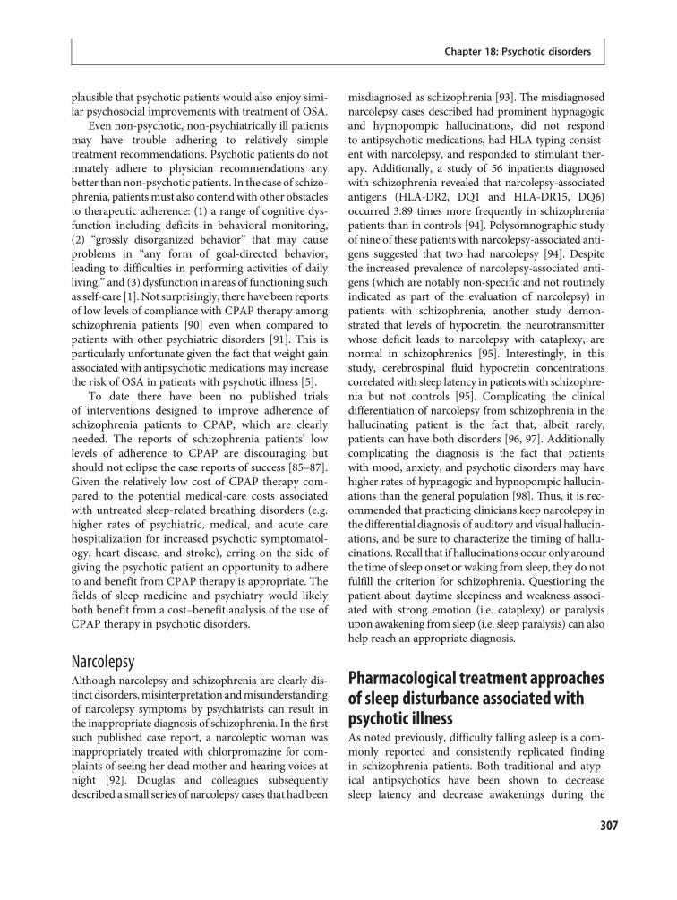
plausible that psychotic patients would also enjoy simi-lar psychosocial improvements with treatment of OSA.
Even non-psychotic, non-psychiatrically ill patientsmay have trouble adhering to relatively simpletreatment recommendations. Psychotic patients do notinnately adhere to physician recommendations anybetter than non-psychotic patients. In the case of schizo-phrenia, patientsmust also contendwith other obstaclesto therapeutic adherence: (1) a range of cognitive dys-function including deficits in behavioral monitoring,(2) “grossly disorganized behavior” that may causeproblems in “any form of goal-directed behavior,leading to difficulties in performing activities of dailyliving,” and (3) dysfunction in areas of functioning suchas self-care [1]. Not surprisingly, there have been reportsof low levels of compliance with CPAP therapy amongschizophrenia patients [90] even when compared topatients with other psychiatric disorders [91]. This isparticularly unfortunate given the fact that weight gainassociated with antipsychotic medications may increasethe risk of OSA in patients with psychotic illness [5].
To date there have been no published trialsof interventions designed to improve adherence ofschizophrenia patients to CPAP, which are clearlyneeded. The reports of schizophrenia patients’ lowlevels of adherence to CPAP are discouraging butshould not eclipse the case reports of success [85–87].Given the relatively low cost of CPAP therapy com-pared to the potential medical-care costs associatedwith untreated sleep-related breathing disorders (e.g.higher rates of psychiatric, medical, and acute carehospitalization for increased psychotic symptomatol-ogy, heart disease, and stroke), erring on the side ofgiving the psychotic patient an opportunity to adhereto and benefit from CPAP therapy is appropriate. Thefields of sleep medicine and psychiatry would likelyboth benefit from a cost–benefit analysis of the use ofCPAP therapy in psychotic disorders.
NarcolepsyAlthough narcolepsy and schizophrenia are clearly dis-tinct disorders,misinterpretation andmisunderstandingof narcolepsy symptoms by psychiatrists can result inthe inappropriate diagnosis of schizophrenia. In the firstsuch published case report, a narcoleptic woman wasinappropriately treated with chlorpromazine for com-plaints of seeing her dead mother and hearing voices atnight [92]. Douglas and colleagues subsequentlydescribed a small series of narcolepsy cases that had been
misdiagnosed as schizophrenia [93]. The misdiagnosednarcolepsy cases described had prominent hypnagogicand hypnopompic hallucinations, did not respondto antipsychotic medications, had HLA typing consist-ent with narcolepsy, and responded to stimulant ther-apy. Additionally, a study of 56 inpatients diagnosedwith schizophrenia revealed that narcolepsy-associatedantigens (HLA-DR2, DQ1 and HLA-DR15, DQ6)occurred 3.89 times more frequently in schizophreniapatients than in controls [94]. Polysomnographic studyof nine of these patients with narcolepsy-associated anti-gens suggested that two had narcolepsy [94]. Despitethe increased prevalence of narcolepsy-associated anti-gens (which are notably non-specific and not routinelyindicated as part of the evaluation of narcolepsy) inpatients with schizophrenia, another study demon-strated that levels of hypocretin, the neurotransmitterwhose deficit leads to narcolepsy with cataplexy, arenormal in schizophrenics [95]. Interestingly, in thisstudy, cerebrospinal fluid hypocretin concentrationscorrelatedwith sleep latency in patients with schizophre-nia but not controls [95]. Complicating the clinicaldifferentiation of narcolepsy from schizophrenia in thehallucinating patient is the fact that, albeit rarely,patients can have both disorders [96, 97]. Additionallycomplicating the diagnosis is the fact that patientswith mood, anxiety, and psychotic disorders may havehigher rates of hypnagogic and hypnopompic hallucin-ations than the general population [98]. Thus, it is rec-ommended that practicing clinicians keep narcolepsy inthe differential diagnosis of auditory and visual hallucin-ations, and be sure to characterize the timing of hallu-cinations. Recall that if hallucinations occur only aroundthe time of sleep onset or waking from sleep, they do notfulfill the criterion for schizophrenia. Questioning thepatient about daytime sleepiness and weakness associ-ated with strong emotion (i.e. cataplexy) or paralysisupon awakening from sleep (i.e. sleep paralysis) can alsohelp reach an appropriate diagnosis.
Pharmacological treatment approachesof sleep disturbance associated withpsychotic illnessAs noted previously, difficulty falling asleep is a com-monly reported and consistently replicated findingin schizophrenia patients. Both traditional and atyp-ical antipsychotics have been shown to decreasesleep latency and decrease awakenings during the
Chapter 18: Psychotic disorders
307
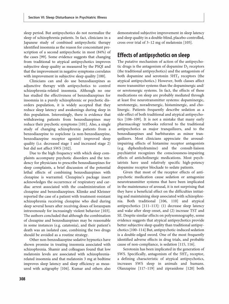
sleep period. But antipsychotics do not normalize thesleep of schizophrenia patients. In fact, clinicians in aJapanese study of combined antipsychotic therapyidentified insomnia as the reason for concomitant pre-scription of a second antipsychotic in most (84%) ofthe cases [99]. Some evidence suggests that changingfrom traditional to atypical antipsychotics improvessubjective sleep quality as measured by the PSQI andthat the improvement in negative symptoms correlateswith improvement in subjective sleep quality [100].
Clinicians can and do use benzodiazepines asadjunctive therapy with antipsychotics to controlschizophrenia-related insomnia. Although no onehas studied the effectiveness of benzodiazepines forinsomnia in a purely schizophrenic or psychotic dis-orders population, it is widely accepted that theyreduce sleep latency and awakenings during sleep inthis population. Interestingly, there is evidence thatwithdrawing patients from benzodiazepines mayreduce their psychotic symptoms [101]. Also, a singlestudy of changing schizophrenia patients from abenzodiazepine to zopiclone (a non-benzodiazepine,benzodiazepine receptor agonist) improved sleepquality (i.e. decreased stage 1 and increased stage 2)but did not affect SWS [102].
Due to the high frequency with which sleep com-plaints accompany psychotic disorders and the ten-dency for physicians to prescribe benzodiazepines forsleep complaints, a brief discussion of the potentiallethal effects of combining benzodiazepines withclozapine is warranted. Clozapine’s package insertacknowledges the occurrence of respiratory and car-diac arrest associated with the coadministration ofclozapine and benzodiazepines. Klimke and Kleisnerreported the case of a patient with treatment-resistantschizophrenia receiving clozapine who died duringsleep several hours after receiving doses of lorazepamintravenously for increasingly violent behavior [103].The authors concluded that although the combinationof clozapine and benzodiazepines may be reasonablein some instances (e.g. catatonia), and their patient’sdeath was an isolated case, combining the two drugsshould be avoided as a routine strategy.
Other non-benzodiazepine sedative hypnotics haveshown promise in treating insomnia associated withschizophrenia. Shamir and colleagues found that lowmelatonin levels are associated with schizophrenia-related insomnia and that melatonin 3 mg at bedtimeimproves sleep latency and sleep efficiency as meas-ured with actigraphy [104]. Kumar and others also
demonstrated subjective improvement in sleep latencyand sleep quality in a double-blind, placebo-controlled,cross over trial of 3–12 mg of melatonin [105].
Effects of antipsychotics on sleepThe putative mechanism of action of the antipsycho-tic drugs is the antagonism of dopamine D2 receptors(the traditional antipsychotics) and the antagonism ofboth dopamine and serotonin 5HT2 receptors (theatypical antipsychotics.) However, both classes affectmore transmitter systems than the dopaminergic and/or serotonergic systems. In fact, the effects of thesemedications on sleep are probably mediated throughat least five neurotransmitter systems: dopaminergic,serotonergic, noradrenergic, histaminergic, and cho-linergic. Patients frequently describe sedation as aside-effect of both traditional and atypical antipsycho-tics [106–109]. It is not a mistake that many earlypharmacology textbooks referred to the traditionalantipsychotics as major tranquilizers, and to thebenzodiazepines and barbiturates as minor tran-quilizers. Most clinicians appreciate the arousal-impairing effects of histamine receptor antagonists(e.g. diphenhydramine) and the consult-liaisonpsychiatrist recognizes the consciousness-impairingeffects of anticholinergic medications. Most psych-iatrists have used relatively specific high-potencydopamine receptor blockade to sedate patients.
Given that most of the receptor effects of anti-psychotic medication cause sedation or antagonizeneurotransmitter systems that have been implicatedin the maintenance of arousal, it is not surprising thatthey have a beneficial effect on the difficulties initiat-ing and maintaining sleep associated with schizophre-nia. Both traditional [106, 110] and atypicalantipsychotics [111–113]: (1) decrease sleep latencyand wake after sleep onset, and (2) increase TST andSE. Despite similar effects on polysomnography, someevidence suggests that atypical antipsychotics providebetter subjective sleep quality than traditional antipsy-chotics [100–114] But, antipsychotic-induced sedationis a double-edged sword. One of the most frequentlyidentified adverse effects in drug trials, and probablecause of non-compliance, is sedation [115, 116].
Serotonin has been implicated in the generation ofSWS. Specifically, antagonism of the 5HT2 receptor,a defining characteristic of atypical antipsychotics,increases SWS sleep in animals and humans.Olanzapine [117–119] and ziprasidone [120] both
Section VI: Sleep Disturbance in Psychiatric Illness
308
increase SWS in human studies. Recall that SWSdeficits have been correlated with negative symptoms(as has short REM sleep latency) and implicated incognitive deficits associated with schizophrenia.These associations suggest that a better understandingof the 5HT2 receptor’s role in the generation of SWSmight produce insight into the pathophysiology ofschizophrenia.
SummaryThe majority of the literature regarding the interfaceof sleep with psychotic disorders comes mostly fromdated polysomnographic studies of patients with vari-ous types of schizophrenia in various phases of theillness and states of treatment. Little attention hasbeen paid to other psychotic disorders, the psychoticpatient’s subjective experience of sleep, or how clin-icians can help improve sleep disturbances. The smallamount of available data regarding sleep complaintsamongst patients with psychosis (mostly schizophre-nia) reveals subjective difficulty initiating and main-taining sleep even when on stable antipsychoticmedications. The most consistent findings in poly-somnographic studies (i.e. increased sleep latency,decreased total sleep time, and decreased sleep effi-ciency) confirm the subjective reports of difficultyinitiating and maintaining sleep. Clinicians shouldscreen the patient with sleep complaints for primarysleep disorders like obstructive sleep apnea and rest-less legs syndrome, but there is no convincing evi-dence that any primary sleep disorder occurs morefrequently in schizophrenia than the general popula-tion. The reported changes in various parameters ofREM sleep and SWS associated with schizophreniaare interesting but inconsistent and clinically unhelp-ful. Both traditional and atypical antipsychotic medi-cations reduce sleep latency and increase total sleeptime but their effects on other sleep parameters likeREM latency, REM%, SWS%, etc. vary even withingroups (i.e. traditional and atypical). The tendencyof schizophrenic patients to engage in poor sleephygiene behaviors including substance abuse and thecontribution of poor sleep hygiene to their sleep dis-turbance deserves closer examination. The clinicianshould carefully evaluate sleep hygiene behaviors ofall patients who complain of insomnia and especiallythose with schizophrenia, who may have particulardifficulty maintaining proper sleep hygiene. Com-pared to the addition of medication, behavioral
modifications to improve sleep hygiene carry less riskof side-effects and cost less but offer an as yetunmeasured propensity to improve sleep quality.Cognitive behavioral therapy incorporates promotionof healthy sleep hygiene behaviors and other tech-niques such as sleep restriction, stimulus control,education, and cognitive restructuring. Because cog-nitive behavioral therapy has proved beneficial in thetreatment of insomnia co-morbid with other psychi-atric and medical disorders, the clinician should con-sider using it to treat the sleep disturbances associatedwith psychosis. As there currently is a paucity ofexperimental data to guide clinical decision-making,clinicians who manage sleep disturbance in patientswith psychotic illness should promote healthy sleephygiene behaviors as a mainstay of treatment, care-fully consider the potential risks and benefits ofpharmacological treatments, and closely follow thescientific literature for rigorous clinical trials of ther-apy (e.g. CBT, pharmacotherapy, etc.) for insomnia inpsychotic disorders.
References1. American Psychiatric Association. Diagnostic and
statistical manual of mental disorders, 4th ed, TextRevision. Washington, DC: American PsychiatricAssociation; 2000.
2. Doi Y, Minowa M, Uchiyama M, et al. Psychometricassessment of subjective sleep quality using the Japaneseversion of the Pittsburgh Sleep Quality Index (PSQI J)in psychiatric disordered and control subjects.Psychiatry Res. 2000;97:165 172.
3. Haffmans PM, Hoencamp E, Knegtering HJ, vanHeycop ten Ham BF. Sleep disturbance inschizophrenia. Br J Psychiatry. 1994;165:697 698.
4. Serretti A, Mandelli L, Lattuada E, Smeraldi E.Depressive syndrome in major psychoses: a study on1351 subjects. Psychiatry Res. 2004;127:85 99.
5. Winkelman JW. Schizophrenia, obesity, and obstructivesleep apnea. J Clin Psychiatry. 2001;62:8 11.
6. Takahashi KI, Shimizu T, Sugita T, et al. Prevalenceof sleep related respiratory disorders in 101schizophrenic inpatients. Psychiatry Clin Neurosci.1998;52:229 231.
7. Ancoli Israel S, Martin J, Jones DW, et al. Sleepdisordered breathing and periodic limb movements insleep in older patients with schizophrenia. BiolPsychiatry. 1999;45:1426 1432.
8. Kang SG, Lee HJ, Jung SW, et al. Characteristicsand clinical correlates of restless legs syndrome in
Chapter 18: Psychotic disorders
309

schizophrenia. Prog Neuropsychopharmacol BiolPsychiatry. 2007;31:1078 1083.
9. Edinger JD, Olsen MK, Stechuchak KM, et al.Cognitive behavioral therapy for patients with primaryinsomnia or insomnia associated predominantly withmixed psychiatric disorders: a randomized clinicaltrial. Sleep. 2009;32:499 510.
10. Perlis ML, Sharpe MC, Smith MT, Greenblatt DW,Giles DE. Behavioral treatment of insomnia:Treatment outcome and the relevance of medicaland psychiatric morbidity. J Behav Med. 2001;24:281 296.
11. Dopke CA, Lehner RK, Wells AM. Cognitivebehavioral group therapy for insomnia in individualswith serious mental illnesses: a preliminary evaluation.Psychiatr Rehabil J. 2004;27:235 242.
12. Dashevsky BA, Kramer MF. Behavioral treatment ofchronic insomnia in psychiatrically ill patients. J ClinPsychiatry. 1998;59:693 699.
13. Benca RM, Obermeyer WH, Thisted RA, Gillin JC.Sleep and psychiatric disorders: A meta analysis.Arch Gen Psychiatry. 1992;49:651 668.
14. Chouinard S, Poulin J, Stip E, Godbout R. Sleep inuntreated patients with schizophrenia: a meta analysis.Schizophr Bull. 2004;30:957 967.
15. Ferrarelli F, Huber R, Peterson MJ, et al. Reducedsleep spindle activity in schizophrenia patients.Am J Psychiatry. 2007;164:483 492.
16. Forest G, Poulin J, Daoust AM, et al. Attention andnon REM sleep in neuroleptic naive persons withschizophrenia and control participants. Psychiatry Res.2007;149:33 40.
17. Goder R, Boigs M, Braun S, et al. Impairment ofvisuospatial memory is associated with decreased slowwave sleep in schizophrenia. J Psychiatr Res.2004;38:591 599.
18. Sekimoto M, Kato M, Watanabe T, Kajimura N,Takahashi K. Reduced frontal asymmetry of deltawaves during all night sleep in schizophrenia.Schizophr Bull. 2007;33:1307 1311.
19. Yang C, Winkelman JW. Clinical significance ofsleep EEG abnormalities in chronic schizophrenia.Schizophr Res. 2006;82:251 260.
20. Dement W. Dream recall and eye movements duringsleep in schizophrenics and normals. J Nerv Ment Dis.1955;122:263 269.
21. Koresko RL, Snyder F, Feinberg I. ‘Dream Time’ inhallucinating and nonhallucinating schizophrenicpatients. Nature. 1963;199:1118 1119.
22. Feinberg I, Koresko RL, Gottlieb F, Wender PH. Sleepelectroencephalographic and eye movement patterns
in schizophrenic patients. Compr Psychiatry.1964;34:44 53.
23. Gulevich GD, Dement WC, Zarcone VP. All nightsleep recordings of chronic schizophrenics inremission. Compr Psychiatry. 1967;8:141 149.
24. Stern M, Fram DH, Wyatt R, Grinsponn L, Tursky B.All night sleep studies of acute schizophrenics. ArchGen Psychiatry. 1969;20:470 477.
25. Jus K, Bouchard M, Jus AK, Villeneuve A, Lachance R.Sleep EEG studies in untreated, long termschizophrenic patients. Arch Gen Psychiatry.1973;29:386 390.
26. Hiatt JF, Floyd TC, Katz PH, Feinberg I. Furtherevidence of abnormal non rapid eye movementsleep in schizophrenia. Arch Gen Psychiatry.1985;42:797 802.
27. Ganguli R, Reynolds CF 3rd, Kupfer DJ.Electroencephalographic sleep in young, nevermedicated schizophrenics: A comparison withdelusional and nondelusional depressives and withhealthy controls. Arch Gen Psychiatry. 1987;44:36 44.
28. Kempenaers C, Kerkhofs M, Linkowski P,Mendlewicz J. Sleep EEG variables in youngschizophrenic and depressive patients. Biol Psychiatry.1988;24:833 838.
29. Lauer CJ, Schreiber W, Pollmacher T, Holsboer F, KriegJC. Sleep in schizophrenia: a polysomnographic studyon drug naive patients. Neuropsychopharmacology.1997;16:51 60.
30. Taylor SF, Tandon R, Shipley JE, Eiser AS, Goodson J.Sleep onset REM periods in schizophrenic patients.Biol Psychiatry. 1991;30:205 209.
31. Feinberg I, Koresko RL, Gottlieb F. Furtherobservations on electrophysiological sleep patterns inschizophrenia. Compr Psychiatry. 1965;6:21 24.
32. Zarcone V, Gulevich G, Pivik T, Dement W. PartialREM phase deprivation and schizophrenia. Arch GenPsychiatry. 1968;18:194 202.
33. Zarcone V, Gulevich G, Pivik T, Azumi K, Dement W.REM deprivation and schizophrenia. Biol Psychiatry.1969;1:179 184.
34. Zarcone V, Azumi K, Dement W, Gulevich G,Kraemer H, Pivik T. REM phase deprivation andschizophrenia II. Arch Gen Psychiatry. 1975;32:1431 1436.
35. Kupfer DJ, Wyatt RJ, Scott J, Snyder F. Sleepdisturbance in acute schizophrenic patients.Am J Psychiatry. 1970;126:1213 1223.
36. Reich L, Weiss BL, Coble P, McPartland R, Kupfer DJ.Sleep disturbance in schizophrenia: A revisit. Arch GenPsychiatry. 1975;32:51 55.
Section VI: Sleep Disturbance in Psychiatric Illness
310
37. Zarcone VP Jr, Benson KL, Berger PA. Abnormalrapid eye movement latencies in schizophrenia.Arch Gen Psychiatry. 1987;44:45 48.
38. Tandon R, Shipley JE, Taylor S, et al.Electroencephalographic sleep abnormalities inschizophrenia: Relationship to positive/negativesymptoms and prior neuroleptic treatment. Arch GenPsychiatry. 1992;49:185 194.
39. Hudson JI, Lipinski JF, Keck PE Jr, et al.Polysomnographic characteristics of schizophreniain comparison with mania and depression.Biol Psychiatry. 1993;34:191 193.
40. Keshavan MS, Reynolds CF 3rd, Miewald JM,Montrose DM. A longitudinal study of EEG sleep inschizophrenia. Psychiatry Res. 1996;59:203 211.
41. Caldwell DF, Domino EF. Electroencephalographicand eye movement patterns during sleep in chronicschizophrenic patients. ElectroencephalogrClin Neurophysiol. 1967;22:414 420.
42. Taylor SF, Goldman RS, Tandon R, Shipley JE.Neuropsychological function and REM sleep inschizophrenic patients. Biol Psychiatry. 1992;32:529 538.
43. Feinberg I, Braun M, Koresko RL, Gottlieb F. Stage 4sleep in schizophrenia. Arch Gen Psychiatry.1969;21:262 266.
44. Kajimura N, Kato M, Okuma T, et al. A quantitativesleep EEG study on the effects of benzodiazepine andzopiclone in schizophrenic patients. Schizophr Res.1995;15:303 312.
45. Keshavan MS, Reynolds CF 3rd, Miewald MJ, et al.Delta sleep deficits in schizophrenia: evidence fromautomated analyses of sleep data. Arch Gen Psychiatry.1998;55:443 448.
46. Benson KL, Zarcone VP Jr. Testing the REM sleepphasic event intrusion hypothesis of schizophrenia.Psychiatry Res. 1985;15:163 173.
47. Hoffmann R, Hendrickse W, Rush AJ, Armitage R.Slow wave activity during non REM sleep in men withschizophrenia and major depressive disorders.Psychiatry Res. 2000;95:215 225.
48. Nishino S, Mignot E, Benson KL, Zarcone VP Jr.Cerebrospinal fluid prostaglandins and corticotropinreleasing factor in schizophrenics and controls:relationship to sleep architecture. Psychiatry Res.1998;78:141 150.
49. Riemann D, Hohagen F, Krieger S, et al. CholinergicREM induction test: muscarinic supersensitivityunderlies polysomnographic findings in bothdepression and schizophrenia. J Psychiatr Res.1994;28:195 210.
50. Benson KL, Faull KF, Zarcone VP Jr. Evidence for therole of serotonin in the regulation of slow wave sleep inschizophrenia. Sleep. 1991;14:133 139.
51. Poulin J, Daoust AM, Forest G, Stip E, Godbout R.Sleep architecture and its clinical correlates in firstepisode and neuroleptic naive patients withschizophrenia. Schizophr Res. 2003;62:147 153.
52. Keshavan MS, Diwadkar VA, Montrose DM,Stanley JA, Pettegrew JW. Premorbid characterizationin schizophrenia: the Pittsburgh High Risk Study.World Psychiatry. 2004;3:163 168.
53. Feinberg I. Schizophrenia: caused by a fault inprogrammed synaptic elimination during adolescence?J Psychiatr Res. 1982 1983;17:319 334.
54. Keshavan MS, Miewald J, Haas G, et al. Slow wavesleep and symptomatology in schizophrenia andrelated psychotic disorders. J Psychiatr Res.1995;29:303 314.
55. Manoach DS, Cain MS, Vangel MG, et al. A failureof sleep dependent procedural learning in chronic,medicated schizophrenia. Biol Psychiatry. 2004;56:951 956.
56. Keshavan MS, Cashmere JD, Miewald J, Yeragani VK.Decreased nonlinear complexity and chaos duringsleep in first episode schizophrenia: a preliminaryreport. Schizophr Res. 2004;71:263 272.
57. van Os J, Allardyce J. The clinical epidemiology ofschizophrenia. In: Sadock BJ, Sadock VA, Ruiz P, eds.Kaplan and Sadock’s comprehensive textbook ofpsychiatry. Philadelphia, PA: Lippincott Williams &Wilkins; 2009:1476 1487.
58. Kumar S, Thara R, Rajkumar S. Coping withsymptoms of relapse in schizophrenia. Eur ArchPsychiatry Neurol Sci. 1989;239:213 215.
59. Herz MI, Melville C. Relapse in schizophrenia.Am J Psychiatry. 1980;137:801 805.
60. Chemerinski E, Ho BC, Flaum M, et al. Insomnia as apredictor for symptom worsening followingantipsychotic withdrawal in schizophrenia.Compr Psychiatry. 2002;43:393 396.
61. Mattai AA, Tossell J, Greenstein DK, et al. Sleepdisturbances in childhood onset schizophrenia.Schizophr Res. 2006;86:123 129.
62. Ritsner M, Kurs R, Ponizovsky A, Hadjez J.Perceived quality of life in schizophrenia:relationships to sleep quality. Qual Life Res.2004;13:783 791.
63. Hofstetter JR, Lysaker PH, Mayeda AR. Quality ofsleep in patients with schizophrenia is associatedwith quality of life and coping. BMC Psychiatry.2005;5:13.
Chapter 18: Psychotic disorders
311

64. Krystal AD, Thakur M, Roth T. Sleep disturbance inpsychiatric disorders: effects on function and qualityof life in mood disorders, alcoholism, andschizophrenia. Ann Clin Psychiatry. 2008;20:39 46.
65. Zarcone VP, Benson KL. BPRS symptom factors andsleep variables in schizophrenia. Psychiatry Res.1997;66:111 120.
66. Tandon R, Shipley JE, Eiser AS, Greden JF. Associationbetween abnormal REM sleep and negativesymptoms in schizophrenia. Psychiatry Res.1989;27:359 361.
67. Goldman M, Tandon R, DeQuardo JR, et al. Biologicalpredictors of 1 year outcome in schizophrenia in malesand females. Schizophr Res. 1996;21:65 73.
68. Keshavan MS, Reynolds CF, Montrose D, et al. Sleepand suicidality in psychotic patients. Acta PsychiatrScand. 1994;89:122 125.
69. Keshaven MS, Reynolds CF 3rd, Miewald J,Montrose D. Slow wave sleep deficits and outcome inschizophrenia and schizoaffective disorder.Acta Psychiatr Scand. 1995;91:289 292.
70. Benson KL, Sullivan EV, Lim KO, et al. Slow wavesleep and computed tomographic measures of brainmorphology in schizophrenia. Psychiatry Res.1996;60:125 134.
71. Tandon R, Shipley JE, Greden JF, et al. Muscariniccholinergic hyperactivity in schizophrenia:Relationship to positive and negative symptoms.Schizophr Res. 1991;4:23 30.
72. Keshavan MS, Mahadik SP, Reynolds CF 3rd, et al.Plasma cholinesterase isozymes and REM latency inschizophrenia. Psychiatry Res. 1992;43:23 29.
73. Griffith JM, Waldo M, Adler LE, Freedman R.Normalization of auditory sensory gating inschizophrenic patients after a brief period for sleep.Psychiatry Res. 1993;49:29 39.
74. Griffith JM, Freedman R. Normalization of theauditory P50 gating deficit of schizophrenic patientsafter non REM but not REM sleep. Psychiatry Res.1995;56:271 278.
75. Griffith JM, O’Neill JE, Petty F, et al. Nicotinic receptordesensitization and sensory gating deficits inschizophrenia. Biol Psychiatry. 1998;44:98 106.
76. Bodizs R, Lazar AS. Schizophrenia, slow wave sleepand visuospatial memory: sleep dependentconsolidation or trait like correlation? J Psychiatr Res.2006;40:89 90.
77. Cazzullo CL, Fornari MG, Maffei C, et al. Sleep,psychophysiological functioning and learningprocesses in schizophrenia. Act Nerv Super. 1977;19(Suppl 2):409 417.
78. Mills JN, Morgan R, Minors DS, Waterhouse JM Thefree running circadian rhythms of two schizophrenics.Chronobiologia. 1977;4:353 360.
79. Wirz Justice A, Cajochen C, Nussbaum P.A schizophrenic patient with an arrhythmic circadianrest activity cycle. Psychiatry Res. 1997;73:83 90.
80. Van Cauter E, Linkowski P, Kerkhofs M, et al.Circadian and sleep related endocrine rhythms inschizophrenia. Arch Gen Psychiatry. 1991;48:348 356.
81. Martin J, Jeste DV, Caliguiri MP, et al. Actigraphicestimates of circadian rhythms and sleep/wake in olderschizophrenia patients. Schizophr Res. 2001;47:77 86.
82. Martin JL, Jeste DV, Ancoli Israel S. Olderschizophrenia patients have more disrupted sleep andcircadian rhythms than age matched comparisonsubjects. J Psychiatr Res. 2005;39:251 259.
83. Hofstetter JR, Mayeda AR, Happel CG, Lysaker PH.Sleep and daily activity preferences in schizophrenia:associations with neurocognition and symptoms.J Nerv Ment Dis. 2003;191:408 410.
84. Haug HJ, Wirz Justice A, Rossler W. Actigraphy tomeasure day structure as a therapeutic variable in thetreatment of schizophrenic patients. Acta PsychiatrScand Suppl. 2000;407:91 95.
85. Karanti A, Landen M. Treatment refractory psychosisremitted upon treatment with continuous positiveairway pressure: a case report. Psychopharmacol Bull.2007;40:113 117.
86. Boufidis S, Kosmidis MH, Bozikas VP, et al. Treatmentoutcome of obstructive sleep apnea syndrome in apatient with schizophrenia: case report. Int J PsychiatryMed. 2003;33:305 310.
87. Dennis JL, Crisham KP. Chronic assaultive behaviorimproved with sleep apnea treatment. J Clin Psychiatry.2001;62:571 572.
88. Chiner E, Arriero JM, Signes Costa J, Marco J. Acutepsychosis after CPAP treatment in a schizophrenicpatient with sleep apnoea hypopnoea syndrome.Eur Respir J. 2001;17:313 315.
89. Ramos Platon MJ, Espinar Sierra J. Changes inpsychopathological symptoms in sleep apnea patientsafter treatment with nasal continuous positive airwaypressure. Int J Neurosci. 1992;62:173 195.
90. DiPhillipo M, Fry J. Is schizophrenia a contraindicationfor nasal CPAP treatment? Sleep Res. 1990;19:212.
91. Cunningham S, Ellsworth S, Winkleman JW, et al.Compliance with nasal continuous positive airwaypressure in psychiatric patients with obstructive sleepapnea. Sleep Res. 1994;23:192.
92. Shapiro B, Spitz H. Problems in the differentialdiagnosis of narcolepsy versus schizophrenia.Am J Psychiatry. 1976;133:1321 1323.
Section VI: Sleep Disturbance in Psychiatric Illness
312
93. Douglass AB, Hays P, Pazderka F, Russell JM. Floridrefractory schizophrenias that turn out to be treatablevariants of HLA associated narcolepsy. J Nerv MentDis. 1991;179:12 17.
94. Douglass AB, Shipley JE, Haines RF, et al.Schizophrenia, narcolepsy, and HLA DR15, DQ6.Biol Psychiatry. 1993;34:773 780.
95. Nishino S, Ripley B, Mignot E, Benson KL, ZarconeVP. CSF hypocretin 1 levels in schizophrenics andcontrols: relationship to sleep architecture. PsychiatryRes. 2002;110:1 7.
96. Cadieux R, Kales J, Kales A, Biever J, Mann LD.Pharmacologic and psychotherapeutic issues incoexistent paranoid schizophrenia and narcolepsy:case report. J Clin Psychiatry. 1985;46(5):191 193.
97. Roy A. Psychiatric aspects of narcolepsy. Br JPsychiatry. 1976;128:562 565.
98. Ohayon MM, Priest RG, Caulet M, Guilleminault C.Hypnagogic and hypnopompic hallucinations:pathological phenomena? Br J Psychiatry.1996;169:459 467.
99. Huang SS, Liao YC, Hsieh YY, et al. Combinationantipsychotic therapy in psychiatric outpatient clinicsin Taiwan. Compr Psychiatry. 2006;47:421 425.
100. Yamashita H, Mori K, Nagao M, et al. Effects ofchanging from typical to atypical antipsychotic drugson subjective sleep quality in patients withschizophrenia in a Japanese population. J ClinPsychiatry. 2004;65:1525 1530.
101. Jus K, Jus A, Villeneuve A, Pires P, Fontaine P. Theutilization of hypnotics in chronic schizophrenics:some critical remarks. Biol Psychiatry.1979;14:955 960.
102. Kajimura N, Kato M, Okuma T, Onuma T. Effects ofzopiclone on sleep and symptoms in schizophrenia:comparison with benzodiazepine hypnotics. ProgNeuropsychopharmacol Biol Psychiatry. 1994;18:477 490.
103. Klimke A, Klieser E. Sudden death after intravenousapplication of lorazepam in a patient treated withclozapine. Am J Psychiatry. 1994;151:780.
104. Shamir E, Laudon M, Barak Y, et al. Melatoninimproves sleep quality of patients with chronicschizophrenia. J Clin Psychiatry. 2000;61:373 377.
105. Suresh Kumar PN, Andrade C, Bhakta SG, Singh NM.Melatonin in schizophrenic outpatients with insomnia:a double blind, placebo controlled study. J ClinPsychiatry. 2007;68:237 241.
106. Taylor SF, Tandon R, Shipley JE, Eiser AS. Effect ofneuroleptic treatment on polysomnographic measuresin schizophrenia. Biol Psychiatry. 1991;30:904 912.
107. Beasley CM Jr, Tollefson GD, Tran PV. Safety ofolanzapine. J Clin Psychiatry. 1997;58 (Suppl 10):13 17.
108. Angermeyer MC, Loffler W, Muller P, Schulze B,Priebe S. Patients’ and relatives’ assessment ofclozapine treatment. Psychol Med. 2001;31:509 517.
109. Gao K, Ganocy SJ, Gajwani P, et al. A review ofsensitivity and tolerability of antipsychotics in patientswith bipolar disorder or schizophrenia: focus onsomnolence. J Clin Psychiatry. 2008;69:302 309.
110. Neylan TC, van Kammen DP, Kelley ME, Peters JL.Sleep in schizophrenic patients on and off haloperidoltherapy: Clinically stable vs relapsed patients. Arch GenPsychiatry. 1992;49:643 649.
111. Salin Pascual RJ, Herrera Estrella M, Galicia Polo L,Laurrabaquio MR. Olanzapine acute administration inschizophrenic patients increases delta sleep and sleepefficiency. Biol Psychiatry. 1999;46:141 143.
112. Lee JH, Woo JI, Meltzer HY. Effects of clozapine onsleep measures and sleep associated changes in growthhormone and cortisol in patients with schizophrenia.Psychiatry Res. 2001;103:157 166.
113. Cohrs S, Rodenbeck A, Guan Z, et al. Sleep promotingproperties of quetiapine in healthy subjects.Psychopharmacology. 2004;174:421 429.
114. Dursun SM, Patel JK, Burke JG, Reveley MA. Effects oftypical antipsychotic drugs and risperidone on thequality of sleep in patients with schizophrenia: a pilotstudy. J Psychiatry Neurosci. 1999;24:333 337.
115. McEvoy JP, Lieberman JA, Perkins DO, et al. Efficacyand tolerability of olanzapine, quetiapine, andrisperidone in the treatment of early psychosis:a randomized, double blind 52 week comparison.Am J Psychiatry. 2007;164:1050 1060.
116. Twaites BR, Wilton LV, Shakir SA. The safety ofquetiapine: results of a post marketing surveillancestudy on 1728 patients in England. J Psychopharmacol.2007;21:392 399.
117. Lindberg N, Virkkunen M, Tani P, et al. Effect of asingle dose of olanzapine on sleep in healthy femalesand males. Int Clin Psychopharmacol. 2002;17:177 184.
118. Muller MJ, Rossbach W, Mann K, et al. Subchroniceffects of olanzapine on sleep EEG in schizophrenicpatients with predominantly negative symptoms.Pharmacopsychiatry. 2004;37:157 162.
119. Goder R, Fritzer G, Gottwald B, et al. Effects ofolanzapine on slow wave sleep, sleep spindles andsleep related memory consolidation in schizophrenia.Pharmacopsychiatry. 2008;41(3):92 99.
120. Cohrs S, Meier A, Neumann AC, et al. Improved sleepcontinuity and increased slow wave sleep and REMlatency during ziprasidone treatment: a randomized,controlled, crossover trial of 12 healthy male subjects.J Clin Psychiatry. 2005;66(8):989 996.
Chapter 18: Psychotic disorders
313

Chapter
19 Sleep in substance use disorders
Deirdre A. Conroy, J. Todd Arnedt, and Kirk J. Brower
IntroductionA survey conducted by theWorld Health Organizationresearch consortium found that the USA had amongthe highest lifetime rates of tobacco and alcohol useand led the world in the proportion of citizensreportingmarijuana or cocaine use at least once duringtheir lifetime. Individuals using, abusing, or dependenton illicit drugs cost the USA half a trillion dollars peryear and approximately 570 000 die each year due tosubstance-related deaths (NIDA, 2008).
Our current understanding of how substancesaffect sleep is influenced largely by factors related tothe pharmacology of the substance, the population inwhich it is being studied, and the various methodolo-gies used to measure sleep. Rarely are drugs studiedacross all populations and measures. Sleep complaintsin the substance-using population are extremelycommon, but whether or not the sleep complaintsare a consequence of substance use per se or a pre-existing complaint is a topic of increasing interest. Forexample, it is known that up to 91% of patients with ahistory of alcoholism have sleep complaints [1], butalso that a pre-existing history of insomnia canincrease the incidence of substance use disorders 3.5years after the insomnia diagnosis has been made [2].
The diagnostic criteria for substance-induced sleepdisorders are outlined in both the Diagnostic and Stat-istical Manual (DSM-IV) and the International Classi-fication of Sleep Disorders, 2nd edition (ICSD-2).In DSM-IV, a substance-induced sleep disorder canbe classified as insomnia, hypersomnia, parasomnia,or mixed with further specifications of onset duringintoxication or onset during withdrawal. The ICSD-2contains additional categories not found in DSM-IV,such as circadian rhythm sleep disorder and centralsleep apnea due to a drug or substance. This
chapter will explore the effects of both licit and illicitsubstances of abuse on sleep (see Table 19.1) and thereciprocal relationships among them. A greater under-standing of this vicious cycle may help to reducerelapse rates by making informed treatment decisionsof residual sleep disturbances during recovery fromsubstance use. Early detection of sleep disturbance inchildren at high risk for developing a substance usedisorder may help in the effort to prevent the onset ofsubstance use disorders.
CNS sedating substancesAlcoholInsomnia is an extremely common sleep complaint inpatients who are actively drinking alcohol as well asin patients who have recently stopped drinking. TheDSM-IV lists insomnia among its list of symptomsthat can occur within several hours to a few days aftercessation of alcohol use. Across seven studies of 1577alcohol-dependent patients undergoing treatment, theaverage rate of self-reported insomnia was 58%(range ¼ 36–91%) [1,3], substantially higher thanrates of insomnia among the general population.One of the contributing factors to difficulty fallingasleep in alcohol-dependent patients may be anunderlying delay in the circadian sleep phase. Onestudy revealed a delay of 1.5 hours in the onset ofnocturnal melatonin secretion in alcohol-dependentpatients compared to controls [4]. Another studysuggested that alcohol-dependent patients may haveless homeostatic drive (measured by EEG delta powerafter a night of partial sleep deprivation) for sleepthan healthy controls [5].
Studies using polysomnography (PSG) have shownthat when alcohol is consumed close to bedtime, it
Foundations of Psychiatric SleepMedicine, ed. JohnW.Winkelman andDavid T. Plante. Published by CambridgeUniversity Press.# Cambridge University Press 2010.
314
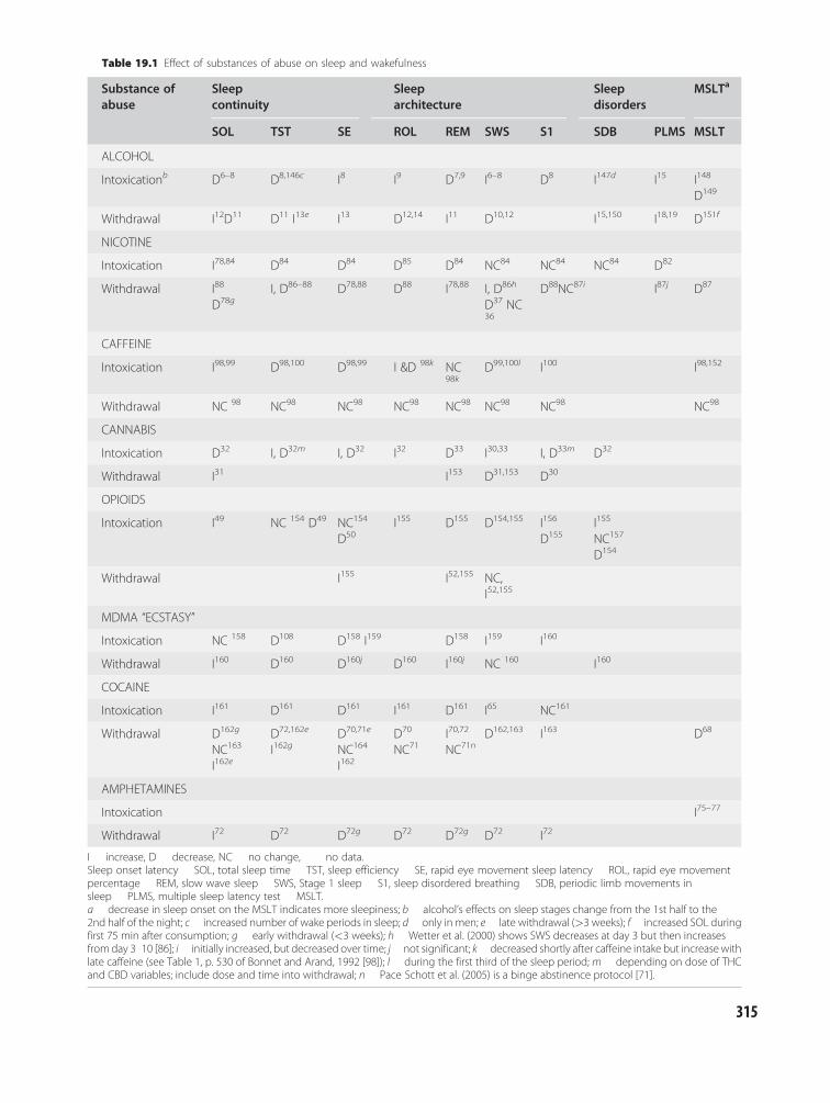
Table 19.1 Effect of substances of abuse on sleep and wakefulness
Substance ofabuse
Sleepcontinuity
Sleeparchitecture
Sleepdisorders
MSLTa
SOL TST SE ROL REM SWS S1 SDB PLMS MSLT
ALCOHOL
Intoxicationb D6–8 D8,146c I8 I9 D7,9 I6–8 D8 I147d I15 I148
D149
Withdrawal I12D11 D11 I13e I13 D12,14 I11 D10,12 I15,150 I18,19 D151f
NICOTINE
Intoxication I78,84 D84 D84 D85 D84 NC84 NC84 NC84 D82
Withdrawal I88
D78gI, D86–88 D78,88 D88 I78,88 I, D86h
D37 NC36
D88NC87i I87j D87
CAFFEINE
Intoxication I98,99 D98,100 D98,99 I &D 98k NC98k
D99,100l I100 I98,152
Withdrawal NC 98 NC98 NC98 NC98 NC98 NC98 NC98 NC98
CANNABIS
Intoxication D32 I, D32m I, D32 I32 D33 I30,33 I, D33m D32
Withdrawal I31 I153 D31,153 D30
OPIOIDS
Intoxication I49 NC 154 D49 NC154
D50I155 D155 D154,155 I156
D155I155
NC157
D154
Withdrawal I155 I52,155 NC,I52,155
MDMA “ECSTASY”
Intoxication NC 158 D108 D158 I159 D158 I159 I160
Withdrawal I160 D160 D160j D160 I160j NC 160 I160
COCAINE
Intoxication I161 D161 D161 I161 D161 I65 NC161
Withdrawal D162g
NC163
I162e
D72,162e
I162gD70,71e
NC164
I162
D70
NC71I70,72
NC71nD162,163 I163 D68
AMPHETAMINES
Intoxication I75–77
Withdrawal I72 D72 D72g D72 D72g D72 I72
I increase, D decrease, NC no change, no data.Sleep onset latency SOL, total sleep time TST, sleep efficiency SE, rapid eye movement sleep latency ROL, rapid eye movementpercentage REM, slow wave sleep SWS, Stage 1 sleep S1, sleep disordered breathing SDB, periodic limb movements insleep PLMS, multiple sleep latency test MSLT.a decrease in sleep onset on the MSLT indicates more sleepiness; b alcohol’s effects on sleep stages change from the 1st half to the2nd half of the night; c increased number of wake periods in sleep; d only in men; e late withdrawal (>3 weeks); f increased SOL duringfirst 75 min after consumption; g early withdrawal (<3 weeks); h Wetter et al. (2000) shows SWS decreases at day 3 but then increasesfrom day 3 10 [86]; i initially increased, but decreased over time; j not significant; k decreased shortly after caffeine intake but increasewithlate caffeine (see Table 1, p. 530 of Bonnet and Arand, 1992 [98]); l during the first third of the sleep period; m depending on dose of THCand CBD variables; include dose and time into withdrawal; n Pace Schott et al. (2005) is a binge abstinence protocol [71].
315

shortens sleep onset latency (SOL) [6–8], prolongsrapid eye movement onset latency (ROL) [9], andincreases slow-wave sleep (SWS) in the first half ofthe night [6,7,10]. Sleep quality worsens in the secondhalf of the night: stage 1 (S1) sleep, wakefulness, andthe percentage of rapid eye movement (REM) sleepincrease [6], and SWS decreases [6,7,10]. SOL, totalsleep time (TST), and sleep efficiency (SE) have beenshown to improve in those who remained abstinentduring the first year, but arousals from sleep and stagechanges remained elevated. REM sleep was disrupted(increased REM%, decreased REM sleep latency) evenafter 2 years of continued abstinence [11]. PSG find-ings have also been associated with rates of relapse.For example, abbreviated REM latency, less stage 4sleep [12], increased REM density [13], and increasesin REM% [14] have been shown to predict relapse.
Sleep-disordered breathing (SDB) has been shownto occurmore frequently in alcohol-dependent patients,particularly in older males [15]. Moreover, results froman epidemiological investigation of the natural historyof SDB show that men have a 25% greater chanceof having SDB (defined as apnea-hypopnea index(AHI) �5) with every drink per day[16].
Movement disorders associated with sleep disrup-tion, like restless legs syndrome (RLS) [17] and peri-odic limb movements in sleep, have been found inindividuals who consume �2 drinks/day. Aldrichet al. (1993) found that twice as many women whoreported high alcohol use had significant periodiclimb movements of sleep (PLMS) (defined as >20per hour), and were significantly more likely to bediagnosed with RLS, compared to women reportingnormal levels of alcohol consumption [18].Recovering alcohol-dependent patients were alsoshown to have significantly more periodic limb move-ments associated with arousals from sleep thancontrols, and the PLMS arousal index (number ofPLMS per hour of sleep) was significantly elevatedin relapsers compared to controls after 6 monthsof abstinence [19]. In fact, a discriminant functionanalysis based on PLMS arousal index correctly pre-dicted the status of 55% of the overall sample, includ-ing 80% of abstainers and 44% of relapsers after6 months of abstinence [19].
Sleep problems may predispose an individual todeveloping an alcohol use disorder or return to drink-ing alcohol once abstinent [20–22]. The MichiganLongitudinal Study, which has been following a cohortof children at high risk for alcoholism, found that
mother-reported sleep difficulties in 3–5-year-old boyspredicted earlier age of onset of drinking during earlyadolescence even after controlling for anxiety/depres-sion, attention problems, and family history [23]. Theonset of alcohol use is a robust indicator of problemswith alcohol use in late adolescence, and it also is astrong predictor of an alcohol use disorder in adult-hood. More recent work with a larger sample of thesame cohort, which also included girls, replicated themale findings and also showed independent relation-ships of the early sleep difficulty to adolescent internal-izing (e.g. anxiety/depression) and externalizing (e.g.impulsivity/acting out) problems [24]. This work sug-gests that sleep difficulties early in life may be a predis-posing factor for later alcohol and behavioralproblems.
In an epidemiological study of more than 10 000adults, the incidence of alcohol abuse or dependenceduring a 1-year follow-up period was twice as likely inindividuals with persistent complaints of insomniacompared to those without insomnia complaints, aftercontrolling for baseline psychiatric disorders [25].Given the sedating properties of alcohol, adults withsleep problems may be more likely to turn to alcoholas a way to self-medicate. In a forced choice paradigmacross four nights in the sleep laboratory, when giventhe choice of color-coded cups prior to bedtime, insom-nia subjects chose the cups containing alcohol on 67%of the nights versus the non-insomniacs who chose thecups with alcohol on only 22% of the nights [26].
In summary, both objective and subjective sleepproblems may play a role in the development andcourse of alcohol use disorders. Insomnia complaintsin alcohol-dependent patients are common and earlytreatment of insomnia, with pharmacological orbehavioral treatments, may reduce the rate of relapse.Some evidence suggests that SDB, sleep-related move-ment disorders, and altered circadian rhythms may bemore frequent in this patient population. Manage-ment of sleep problems in children may theoreticallyprevent the early onset of alcohol use during earlyadolescence, although data is currently insufficient tosupport this contention.
CannabisMost sleep complaints related to cannabis (mari-juana) are documented to occur during withdrawal,and the DSM-IV includes insomnia as one of thediagnostic symptoms of cannabis withdrawal.
Section VI: Sleep Disturbance in Psychiatric Illness
316

A study of 1735 frequent users of cannabis (>21 occa-sions in a single year), found that 235 (13.5%) reporteddifficulty sleeping during withdrawal [27]. Anotherstudy reported that 33% of cannabis users had diffi-culty sleeping during withdrawal [28]. The NationalEpidemiologic Survey on Alcohol and Related Condi-tions in the general US population revealed that of2613 frequent cannabis users 24.5% of the samplereported subjective hypersomnia and 6.1% reportedinsomnia during cannabis withdrawal [29].
Cannabis taken at bedtime has been shown toincrease (N¼7) [30], decrease (N¼2) [31], and haveno effect (N¼8) [32] on SWS and to decrease REMsleep [33]. Doses of 10, 20, and 30mg of tetrahydro-cannabinol (THC) prior to sleep have decreasedSOL after subjects reported achieving a “high” sub-jectively [34]. Studies are more conclusive withrespect to objective sleep disruption during cannabiswithdrawal. Difficulty falling asleep and decreasedSWS percentage have been documented by PSGduring the first two nights of withdrawal [31], butno studies using PSG after prolonged abstinence havebeen conducted. Bolla et al. (2008) examined sleep ofheavy cannabis users (104 þ/� 51 joints/week) in thethree nights prior to and after cannabis discontinu-ation. By the second night of withdrawal, cannabisusers demonstrated lower sleep efficiency (SE),shorter total sleep time (TST), longer SOL, andshorter ROL compared to controls [35], supportingstudies using subjective measures. Daytime conse-quences of cannabis use prior to bedtime are reportedonly following higher doses of THC (15mg THC plus15mg cannabidiol) and include increased sleepiness,mood changes, and impaired memory [32].
Emerging research suggests that early sleep prob-lems may be a pathway to cannabis use during theteenage years and into early adulthood. Wong et al.(2004) found that children with “trouble sleeping” orbeing “overtired” were almost three times more likelyto use cannabis between the ages of 12 and 14 yearsold [23]. In teenagers entering treatment centers, can-nabis is one of the most commonly reported illicitdrugs. Because of teenagers’ physiological tendency toexhibit a delayed circadian sleep phase, they are atgreater risk for reporting difficulty falling asleep andmorning tiredness. Therefore, the use of cannabisto self-medicate sleep problems has been exploredin adolescents [36,37]. In a cross-sectional analysisof data from the National Longitudinal Study of Ado-lescent Health, Roane and Taylor (2008) found that
adolescents with insomnia were 1.32 to 2.05 timesmore likely than adolescents without insomnia touse cannabis [36].
These studies suggest that sleep disturbances evenat a young age may be a risk factor to future cannabisuse. Addressing sleep problems in children and teen-agers may reduce the risk of drug use. Incorporatingtherapies to improve sleep into a continued care model,i.e. outpatient treatment services offered to teens afterresidential substance abuse treatment, may help toreduce risk of future substance use in adolescents [38].
OpioidsSubjective reports of opioids on sleep can vary. Manypatients with pain report longer TST with opioids,presumably because of fewer pain-related arousals andawakenings [39]. Similarly, chronic pain patients withosteoarthritis reported sleeping better when their painwas relieved with tramadol [40]. On the other hand,Vella-Brincat et al. (2007) found an association betweenopioid use and reports of sleep disturbance, dreams,and nightmares in palliative care patients [41].Subjective sleep quality is also impaired among treatedopioid-dependent patients. Sleep assessments from 225methadone-maintenance treatment (MMT) patients,for example, showed that 84% reported clinically sig-nificant sleep disturbances as defined by a score on thePittsburgh Sleep Quality Index (PSQI) of >5 (meanscore (SD) ¼ 10.64 (4.9)) [42]. A separate study alsofound elevated PSQI scores in a group of 101 MMTpatients (mean (SD) 9.4 (4.8)) which did not correlatewith duration of MMT, gender, age, or recent abuse ofopiates, cannabis, or cocaine [43]. Insomnia can bepresent during opioid withdrawal and is one of thesymptoms in the DSM-IV diagnostic criteria for opioidwithdrawal. Heroin-dependent patients detoxified withmethadone reported even greater sleep problems thanthose maintained on methadone during the firstmonths of heroin abstinence [44]. However, in anuncontrolled study, 57% of patients detoxified with acombination of buprenorphine and clonidine reportedimproved sleep [45].
Sleep laboratory studies suggest that opioids mayaffect sleep differently depending on whether or notthe user is in pain and if the user is dependent onopioids. Opioids have also been associated with centralsleep apnea in a dose-dependent fashion [46], occur-ring in about 24–30% of chronic users [47], a muchhigher rate than in the general population [48]. The
Chapter 19: Sleep in substance use disorders
317

latter study of consecutive patients from a pain clinicalso found at least one type of sleep apnea (central,obstructive, mixed) in 85% of patients [48]. PSG find-ings in 10 MMT patients (dose range 50–120mg/day)showed more central sleep apnea, more frequentawakenings [49], lower sleep efficiency, lower SWS,more stage 2 sleep, and less REM sleep than normalcontrols [50].
Sleep complaints often occur in association withpsychiatric illness or chronic pain, which can in turnbe a precipitating factor to opioid abuse or depend-ence. Peles et al. (2006) found that patients with ahigh score on the PSQI had higher rates of chronicpain, benzodiazepine abuse, psychiatric disorders,and had a longer duration of opiate abuse prior toentering treatment for opioid dependence. Moreover,patients who described themselves as “poor sleepers”used higher doses of methadone during their MMTcompared to “normal sleepers” [43].
Few studies have evaluated sleep across prolongedperiods of abstinence from opioids. Two early studieson male prisoners examined sleep across protractedmethadone abstinence. Martin et al. (1973) foundincreased REM sleep and delta sleep after 10 weeks ofmethadone withdrawal [51]. Kay (1975) foundincreased TST, decreased wake time, decreased REMsleep, and increased SWS across the 22-week studyperiod of methadone abstinence [52]. In contrastto studies with alcohol-dependent patients, sleepcomplaints in opioid-dependent patients predicteddecreased relapse rates 1 month after detoxificationin one recent study, although other related factors(e.g. increased attention by clinicians to subjects whohad sleep disruption after detoxification) might haveled to a spurious association [53]. Certainly, such find-ings require replication before concluding sleep dis-ruption is protective of relapse in opioid dependence.
In summary, opioids can become deleterious tosleep with abuse or dependence. Early studies onprolonged methadone abstinence suggest that sleepmay improve in MMT patients; however, studieslonger than 22 weeks of abstinence are lacking. Add-itional risk factors such as benzodiazepine abuse,chronic pain, or psychiatric disorders may be precipi-tating or perpetuating factors.
Benzodiazepine receptor agonists (BzRAs)Since approximately the 1990s, newer BzRAs (eszopi-clone, zolpidem, zaleplon, and zolpidem CR) have
been replacing benzodiazepines as the first-linepharmacological therapy for transient insomnia.Fewer prescriptions for the longer acting benzodi-azepines (BZDs) (e.g. temazepam, flurazepam) arebeing written because the BzRAs carry fewer side-effects such as next day sedation, tolerance, andrebound insomnia. Two reviews have explored theabuse and dependence on BzRAs [54,55]. In a reviewof the literature between 1991 and 2002, nearly allreports of abuse and dependence on zolpidem andzopiclone (zopiclone is not sold in the USA) werereported in former drug or alcohol abusers or inpatients with co-morbid psychiatric disorders.Another large review of the literature up to 2004focused on the abuse liability of hypnotic drugs [54].The authors generated a “likelihood of abuse score”for 19 hypnotics based on a cumulative score reflect-ing animal drug self-administration, human liking/reinforcement, and actual abuse. According to sum-mary data of the BzRAs commonly used in the USA(e.g. zaleplon, zolpidem, and eszopiclone), the lowestlikelihood of abuse score was found in zolpidem, forwhich abuse liability was rated as being similar tooxazepam and quazepam [54]. Zaleplon and eszopi-clone had slightly higher likelihoods of abuse withabuse liability in the moderate range, more closelysimilar to temazepam and triazolam. Hypnoticsnot mediated through GABA receptor sites, liketrazodone, ramelteon, and diphenhydramine, werereported to have the lowest likelihood of abuse.
Reports of abuse and dependence on BzRAsare relatively rare. In a 2003 review, for example,36 cases for zolpidem and 22 cases for zopiclone hadbeen reported in the medical literature [55]. Case stud-ies have reported on patients without a substance usedisorder history or co-morbid psychiatric illnesses.There have been three case studies on zolpidemdependence (10–15mg) reporting anterogradeamnesia with compulsive repetitive behaviors [56].Another case study of zolpidem abuse reported a27-year-old man who was taking zolpidem up to800mg. Upon withdrawal, he experienced extensivecraving, withdrawal seizures, and life-threateningbehavior [57]. Another study reported using diazepamto assist zolpidem detoxification [58]. Overdoses ofeszopiclone have recently been reported [59]. In onestudy reported from the Texas Poison Center Networkbetween 2005 and 2006, eszopiclone overdosesbetween 2 and 210mg (in the absence of coingestants)were associated withmostly minor tomoderate effects,
Section VI: Sleep Disturbance in Psychiatric Illness
318
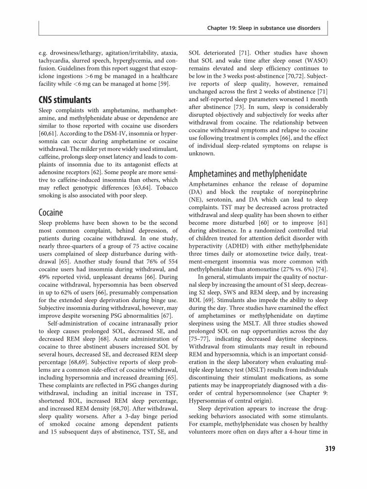
e.g. drowsiness/lethargy, agitation/irritability, ataxia,tachycardia, slurred speech, hyperglycemia, and con-fusion. Guidelines from this report suggest that eszop-iclone ingestions >6mg be managed in a healthcarefacility while <6mg can be managed at home [59].
CNS stimulantsSleep complaints with amphetamine, methamphet-amine, and methylphenidate abuse or dependence aresimilar to those reported with cocaine use disorders[60,61]. According to the DSM-IV, insomnia or hyper-somnia can occur during amphetamine or cocainewithdrawal. Themilder yetmorewidely used stimulant,caffeine, prolongs sleep onset latency and leads to com-plaints of insomnia due to its antagonist effects atadenosine receptors [62]. Some people are more sensi-tive to caffeine-induced insomnia than others, whichmay reflect genotypic differences [63,64]. Tobaccosmoking is also associated with poor sleep.
CocaineSleep problems have been shown to be the secondmost common complaint, behind depression, ofpatients during cocaine withdrawal. In one study,nearly three-quarters of a group of 75 active cocaineusers complained of sleep disturbance during with-drawal [65]. Another study found that 76% of 554cocaine users had insomnia during withdrawal, and49% reported vivid, unpleasant dreams [66]. Duringcocaine withdrawal, hypersomnia has been observedin up to 62% of users [66], presumably compensationfor the extended sleep deprivation during binge use.Subjective insomnia during withdrawal, however, mayimprove despite worsening PSG abnormalities [67].
Self-administration of cocaine intranasally priorto sleep causes prolonged SOL, decreased SE, anddecreased REM sleep [68]. Acute administration ofcocaine to three abstinent abusers increased SOL byseveral hours, decreased SE, and decreased REM sleeppercentage [68,69]. Subjective reports of sleep prob-lems are a common side-effect of cocaine withdrawal,including hypersomnia and increased dreaming [65].These complaints are reflected in PSG changes duringwithdrawal, including an initial increase in TST,shortened ROL, increased REM sleep percentage,and increased REM density [68,70]. After withdrawal,sleep quality worsens. After a 3-day binge periodof smoked cocaine among dependent patientsand 15 subsequent days of abstinence, TST, SE, and
SOL deteriorated [71]. Other studies have shownthat SOL and wake time after sleep onset (WASO)remains elevated and sleep efficiency continues tobe low in the 3 weeks post-abstinence [70,72]. Subject-ive reports of sleep quality, however, remainedunchanged across the first 2 weeks of abstinence [71]and self-reported sleep parameters worsened 1 monthafter abstinence [73]. In sum, sleep is considerablydisrupted objectively and subjectively for weeks afterwithdrawal from cocaine. The relationship betweencocaine withdrawal symptoms and relapse to cocaineuse following treatment is complex [66], and the effectof individual sleep-related symptoms on relapse isunknown.
Amphetamines and methylphenidateAmphetamines enhance the release of dopamine(DA) and block the reuptake of norepinephrine(NE), serotonin, and DA which can lead to sleepcomplaints. TST may be decreased across protractedwithdrawal and sleep quality has been shown to eitherbecome more disturbed [60] or to improve [61]during abstinence. In a randomized controlled trialof children treated for attention deficit disorder withhyperactivity (ADHD) with either methylphenidatethree times daily or atomoxetine twice daily, treat-ment-emergent insomnia was more common withmethylphenidate than atomoxetine (27% vs. 6%) [74].
In general, stimulants impair the quality of noctur-nal sleep by increasing the amount of S1 sleep, decreas-ing S2 sleep, SWS and REM sleep, and by increasingROL [69]. Stimulants also impede the ability to sleepduring the day. Three studies have examined the effectof amphetamines or methylphenidate on daytimesleepiness using the MSLT. All three studies showedprolonged SOL on nap opportunities across the day[75–77], indicating decreased daytime sleepiness.Withdrawal from stimulants may result in reboundREM and hypersomnia, which is an important consid-eration in the sleep laboratory when evaluating mul-tiple sleep latency test (MSLT) results from individualsdiscontinuing their stimulant medications, as somepatients may be inappropriately diagnosed with a dis-order of central hypersomnolence (see Chapter 9:Hypersomnias of central origin).
Sleep deprivation appears to increase the drug-seeking behaviors associated with some stimulants.For example, methylphenidate was chosen by healthyvolunteers more often on days after a 4-hour time in
Chapter 19: Sleep in substance use disorders
319
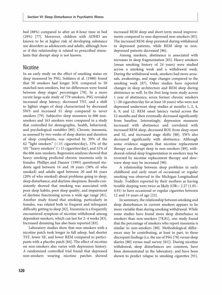
bed (88%) compared to after an 8-hour time in bed(29%) [77]. Moreover, children with ADHD areknown to be at higher risk of developing substanceuse disorders as adolescents and adults, although howor if this relationship is related to prescribed stimu-lants that disrupt sleep is not known.
NicotineIn an early study on the effect of smoking status onsleep measured by PSG, Soldatos et al. (1980) foundthat 50 smokers had longer SOL compared to 50matched non-smokers, but no differences were foundbetween sleep stages’ percentages [78]. In a morerecent large-scale study of 779 smokers, PSG revealedincreased sleep latency, decreased TST, and a shiftto lighter stages of sleep characterized by decreasedSWS and increased S1 sleep compared to neversmokers [79]. Subjective sleep measures in 606 non-smokers and 163 smokers were compared in a studythat controlled for demographic, health, behavioral,and psychological variables [80]. Chronic insomnia,as assessed by two weeks of sleep diaries and durationof sleep complaints, was reported by 39% of the62 “light smokers” (<15 cigarettes/day), 33% of the101 “heavy smokers” (>15 cigarettes/day), and 31% ofthe 606 non-smokers. Regression analyses showed thatheavy smoking predicted chronic insomnia only infemales. Phillips and Danner (1995) questioned stu-dents aged between 14 and 18 years (38% of whosmoked) and adults aged between 20 and 84 years(20% of who smoked) about problems going to sleep,sleep disturbance, and daytime sleepiness. Results con-sistently showed that smoking was associated withpoor sleep habits, poor sleep quality, and impairmentof daytime functioning across a wide age range [81].Another study found that smoking, particularly infemales, was related both to frequent and infrequentdifficulty getting to sleep [82]. Insomnia is a frequentlyencountered symptom of nicotine withdrawal amongdependent smokers, which can last for 2–4 weeks [83].Increased dreaming has also been reported [83].
Laboratory studies show that non-smokers with anicotine patch took longer to fall asleep, had shorterTST, lower SE, and lower REM compared to partici-pants with a placebo patch [84]. The effect of nicotineon non-smokers also varies with depression history.A randomized controlled trial found that depressednon-smokers wearing nicotine patches showed
increased REM sleep and short-term mood improve-ments compared to non-depressed non-smokers [85].The increased REM sleep persisted during withdrawalin depressed patients, while REM sleep in non-depressed patients decreased [86].
Among smokers, abstinence is associated withincreases in sleep fragmentation [83]. Heavy smokers(mean smoking history of 24 years) were studiedacross a smoking week and a withdrawal week.During the withdrawal week, smokers had more arou-sals, awakenings, and stage changes compared to thesmoking week [87]. Other studies have reportedchanges in sleep architecture and REM sleep duringabstinence as well. In the first long-term study across1 year of abstinence, seven former chronic smokers(>20 cigarettes/day for at least 10 years) who were notdepressed underwent sleep studies at months 1, 2, 4,6, 9, and 12. REM onset latency varied across the12 months and then eventually decreased significantlyfrom baseline. Interestingly, depression measuresincreased with abstinence and correlated withincreased REM sleep, decreased ROL from sleep onsetand S2, and increased stage shifts [88]. SWS alsodecreased significantly with abstinence. Althoughsome evidence suggests that nicotine replacementtherapy can disrupt sleep in non-smokers [89], with-drawal-related sleep fragmentation in smokers may bereversed by nicotine replacement therapy and slow-wave sleep may be increased [90].
A relationship between sleep problems in earlychildhood and early onset of occasional or regularsmoking was observed in the Michigan LongitudinalStudy. Toddlers reported by their mothers as havingtrouble sleeping were twice as likely (OR¼ 2.27 (1.05–4.91) to have occasional or regular cigarettes between12 and 14 years of age [23].
In summary, the relationship between smoking andsleep disturbances in current smokers appears to bemore variable than during smoking withdrawal. Whilesome studies have found more sleep disturbance insmokers than non-smokers [78,81], one study foundthat the percentage of smokers who report insomnia issimilar to non-smokers [80]. Methodological differ-ences may be contributing, at least in part, to thesediscrepant findings (i.e. the use of PSG [78] versus sleepdiaries [80] versus mail survey [81]). During nicotinewithdrawal, sleep disturbances are common, havebeen demonstrated in the laboratory, and have beenshown to predict relapse to smoking cigarettes [91].
Section VI: Sleep Disturbance in Psychiatric Illness
320
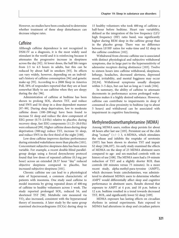
However, no studies have been conducted to determinewhether treatment of these sleep disturbances candecrease relapse rates.
CaffeineAlthough caffeine dependence is not recognized inDSM-IV as a diagnosis, it is the most widely usedstimulant in the world and it is reinforcing because itattenuates the progressive increase in sleepinessacross the day [92]. At lower doses, the half life rangesfrom 2.5 to 4.5 hours in adults, but this can bereduced by about half in smokers [93]. Its effectscan vary widely, however, depending on an individ-ual’s history of caffeine consumption [94] and geneticmake-up [95]. According to a 2008 Sleep in AmericaPoll, 58% of responders reported that they are at leastsomewhat likely to use caffeine when they are sleepyduring the day [96].
Administration of caffeine at bedtime has beenshown to prolong SOL, shorten TST, and reducetotal SWS and S4 sleep in a dose-dependent manner[97–99]. During sleep deprivation, low to moderatecaffeine doses (100–200mg) have been found toincrease S1 sleep and reduce the slow component ofEEG power (0.75–2.0Hz) relative to placebo; duringrecovery sleep, fast EEG components (11.25–20.0Hz)were enhanced [99]. Higher caffeine doses during sleepdeprivation (300mg) reduce TST, increase S1 sleep,and reduce SWS in the first third of the night [100].
Low-dose caffeine improves daytime performanceduring extended wakefulness more than placebo [101].Concomitant subjective sleepiness data has been morevariable. For example, a recent double-blind parallel-group design using a forced desynchrony protocolfound that low doses of repeated caffeine (0.3mg perhour) across an extended 28.57 hour “day” reducedobjective sleepiness compared to placebo, butincreased subjective sleepiness [101].
Chronic caffeine use can lead to a physiologicalstate of hyperarousal, a common characteristic ofpatients with insomnia. One study used caffeine tomodel insomnia by giving repeated doses of 400mgof caffeine to healthy volunteers across 1 week. Thestudy reported prolonged SOL, reduced S4, andshortened TST [98]. Metabolic rate measured viaVO2 also increased, consistent with the hyperarousaltheory of insomnia. A later study by the same groupexamined electrocardiograms (ECGs) during sleep in
15 healthy volunteers who took 400mg of caffeine ahalf-hour before bedtime. Heart rate variability,defined as the integration of the low frequency (LF)/high frequency (HF) ratio band, was significantlyhigher during REM sleep in the caffeine group thanin the placebo group. There was no differencebetween LF/HF ratios for wake-time and S2 sleep inthe caffeine condition [102].
Withdrawal from chronic caffeine use is associatedwith distinct physiological and subjective withdrawalsymptoms, due in large part to the hypersensitivity ofadenosine receptors during abstinence [103]. Twelveto sixteen hours into caffeine withdrawal, sleepiness,lethargy, headaches, decreased alertness, depressedmood, irritability, and mental fogginess may occur[92,94]. Withdrawal symptoms usually dissipatewithin 3–5 days, but can last as long as 1 week [104].
In summary, the ability of caffeine to attenuatedecrements in performance across prolonged wake-fulness makes it a highly desired substance. However,caffeine can contribute to impairments in sleep ifconsumed in close proximity to bedtime (up to about5 hours) and withdrawal can be associated withimpairment in cognitive functioning.
Methylenedioxmethamphetamine (MDMA)Among MDMA users, restless sleep persists for about48 hours after last use [105]. Persistent use of the clubdrug “ecstasy” (þ/�) 3, 4-MDMA, which stimulatesthe release and inhibits the reuptake of serotonin(5HT) has been shown to shorten TST and impairS2 sleep [106,107]. An early study examined the effectsof MDMA on the sleep of 23 MDMA abstinent userscompared to age- and sex-matched controls with nohistory of use [108]. TheMDMAusers had a 19-minutereduction of TST and a slightly shorter ROL thancontrols (60 minutes versus 75 minutes). In a morerecent study, alpha-methyl-para-tyrosine (AMPT),which decreases brain catecholamines, was adminis-tered to abstinent MDMA users to determine whetherAMPT would differentially affect sleep and cognitiveperformance in abstinent users. Results showed thatexposure to AMPT at 4 p.m. and 10 p.m. before a12 a.m. bedtime resulted in a trend towards decreasedTST, ROL, and significantly lower S2 sleep [109].
MDMA exposure has lasting effects on circadianrhythms in animal experiments. Rats exposed toMDMA had alterations in sleep and circadian pattern
Chapter 19: Sleep in substance use disorders
321

of activity (wheel running) for up to 5 days afterdosage. In addition, SWS was still altered after1 month [110]. In vitro administration of MDMA torat brain slices impaired the resetting ability of cells inthe suprachiasmatic nucleus (SCN) to a serotoninreceptor agonist 20 weeks after initial exposure[111]. In sum, use of MDMA can decrease the dur-ation and quality of the sleep period.
Treatment of insomnia in patientswith substance use disordersThis topic, which has only recently become a subjectof developing research, has been reviewed elsewhere,including recommended assessment and treatmentguidelines [21,112,113]. The best-studied treatmentsto date are targeted towards patients with alcoholdependence and include both behavioral and pharma-cological therapies.
Benzodiazepines and benzodiazepinereceptor agonists (BzRAs)Benzodiazepines and other benzodiazepine receptoragonists (e.g. zolpidem, zaleplon) are generally safeand efficacious sedative-hypnotics and often themedications of choice for treating transient insomniain patients without a history of substance use dis-orders [114]. They also may have beneficial effectson sleep during subacute alcohol withdrawal [115].However, in patients with a history of substance abuseor dependence there is an increased risk for sedative-hypnotic abuse [116–118], although some contro-versy endures and not all authors agree [119]. Mostaddiction treatment specialists recommend againstthe use of BzRAs in alcoholic patients as a first-lineagent (except for benzodiazepines during acute alco-hol withdrawal), because of their abuse potential,withdrawal effects, rebound insomnia, and potentialfor overdose when mixed with alcohol [120–123].Whenever possible, medications with low abuse andoverdose potential should be used in patients withsubstance use disorders [124]. If BzRAs are used, thena number of precautions are recommended, such aswritten informed consent about their abuse potentialand permission to conduct urine drug screens as acondition for refills, limited prescriptions in terms ofrefills, more frequent clinic visits, pill counts, andpossibly supervised administration when a supportperson is available.
AnticonvulsantsSelected anticonvulsants are commonly used in alco-holics for their sedative properties, in part becausethey do not lower the seizure threshold, an importantissue given the risk of seizures in alcoholic-dependentpatients. Carbamazepine, for example, reduced sub-jectively reported sleep problems more thanlorazepam during treatment for alcohol withdrawal[125]. Gabapentin has a potentially desirable hypnoticprofile for this population because it has minimalknown abuse potential, is not metabolized by the liver,does not interfere with the metabolism of other medi-cations, and does not require blood monitoring fortoxicity. The effects of gabapentin on sleep have beenstudied both during acute withdrawal and after severalweeks of abstinence. When used to treat alcohol with-drawal, gabapentin more effectively reduced sleep dis-turbance than lorazepam among patients with ahistory of multiple previous withdrawals [126], poten-tially due to its antikindling properties [127]. In anopen-label pilot study of post-withdrawal insomnia,alcohol-dependent patients who were treated withgabapentin (mean dose (range) ¼ 953 (200–1500)mg qhs), reported significantly improved sleep qualityover a 4–6 week period [128]. When gabapentin(mean dose (range) ¼ 888 (300–1800) mg qhs) wascompared to trazodone (mean dose (range) ¼ 105(50–300) mg qhs) in another open pilot trial inpatients at least 4 weeks post-withdrawal from alcohol,both medications were associated with significantlyimproved scores on the Sleep Problems Question-naire, but the gabapentin-treated group was less likelyto feel tired upon awakening [129]. In a randomizeddouble-blind trial of gabapentin in recently abstinentalcoholic patients with insomnia, the gabapentingroup (mean nightly dose range: 1218–1500 mg) hada delayed onset to heavy drinking across 6 weeks [130],although its effects on sleep could not be distinguishedfrom placebo. In summary, gabapentin has shownsome promise as an effective medication to improvesleep in alcohol-dependent patients and may delay thereturn to drinking.
The anticonvulsant tiagabine has been compared tolorazepam for its PSG effects in abstinent cocaine-dependent subjects [131]. Although lorazepam im-proved measures of sleep continuity, it did so at theexpense of promoting lighter stages of sleep, whereastiagabine was superior to lorazepam in increasing SWS.Moreover, lorazepam caused significantly more
Section VI: Sleep Disturbance in Psychiatric Illness
322
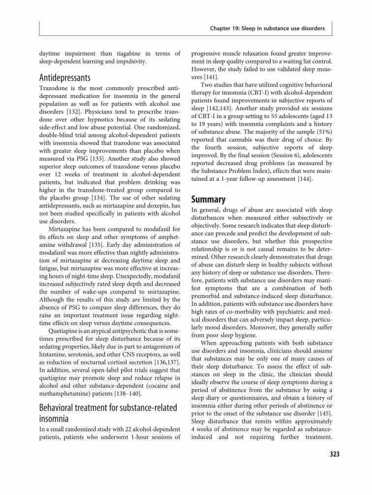
daytime impairment than tiagabine in terms ofsleep-dependent learning and impulsivity.
AntidepressantsTrazodone is the most commonly prescribed anti-depressant medication for insomnia in the generalpopulation as well as for patients with alcohol usedisorders [132]. Physicians tend to prescribe trazo-done over other hypnotics because of its sedatingside-effect and low abuse potential. One randomized,double-blind trial among alcohol-dependent patientswith insomnia showed that trazodone was associatedwith greater sleep improvements than placebo whenmeasured via PSG [133]. Another study also showedsuperior sleep outcomes of trazodone versus placeboover 12 weeks of treatment in alcohol-dependentpatients, but indicated that problem drinking washigher in the trazodone-treated group compared tothe placebo group [134]. The use of other sedatingantidepressants, such as mirtazapine and doxepin, hasnot been studied specifically in patients with alcoholuse disorders.
Mirtazapine has been compared to modafanil forits effects on sleep and other symptoms of amphet-amine withdrawal [135]. Early day administration ofmodafanil was more effective than nightly administra-tion of mirtazapine at decreasing daytime sleep andfatigue, but mirtazapine was more effective at increas-ing hours of night-time sleep. Unexpectedly, modafanilincreased subjectively rated sleep depth and decreasedthe number of wake-ups compared to mirtazapine.Although the results of this study are limited by theabsence of PSG to compare sleep differences, they doraise an important treatment issue regarding night-time effects on sleep versus daytime consequences.
Quetiapine is an atypical antipsychotic that is some-times prescribed for sleep disturbance because of itssedating properties, likely due in part to antagonism ofhistamine, serotonin, and other CNS receptors, as wellas reduction of nocturnal cortisol secretion [136,137].In addition, several open-label pilot trials suggest thatquetiapine may promote sleep and reduce relapse inalcohol and other substance-dependent (cocaine andmethamphetamine) patients [138–140].
Behavioral treatment for substance-relatedinsomniaIn a small randomized study with 22 alcohol-dependentpatients, patients who underwent 1-hour sessions of
progressive muscle relaxation found greater improve-ment in sleep quality compared to a waiting list control.However, the study failed to use validated sleep meas-ures [141].
Two studies that have utilized cognitive behavioraltherapy for insomnia (CBT-I) with alcohol-dependentpatients found improvements in subjective reports ofsleep [142,143]. Another study provided six sessionsof CBT-I in a group setting to 55 adolescents (aged 13to 19 years) with insomnia complaints and a historyof substance abuse. The majority of the sample (51%)reported that cannabis was their drug of choice. Bythe fourth session, subjective reports of sleepimproved. By the final session (Session 6), adolescentsreported decreased drug problems (as measured bythe Substance Problem Index), effects that were main-tained at a 1-year follow-up assessment [144].
SummaryIn general, drugs of abuse are associated with sleepdisturbances when measured either subjectively orobjectively. Some research indicates that sleep disturb-ance can precede and predict the development of sub-stance use disorders, but whether this prospectiverelationship is or is not causal remains to be deter-mined. Other research clearly demonstrates that drugsof abuse can disturb sleep in healthy subjects withoutany history of sleep or substance use disorders. There-fore, patients with substance use disorders may mani-fest symptoms that are a combination of bothpremorbid and substance-induced sleep disturbance.In addition, patients with substance use disorders havehigh rates of co-morbidity with psychiatric and med-ical disorders that can adversely impact sleep, particu-larly mood disorders. Moreover, they generally sufferfrom poor sleep hygiene.
When approaching patients with both substanceuse disorders and insomnia, clinicians should assumethat substances may be only one of many causes oftheir sleep disturbance. To assess the effect of sub-stances on sleep in the clinic, the clinician shouldideally observe the course of sleep symptoms during aperiod of abstinence from the substance by using asleep diary or questionnaires, and obtain a history ofinsomnia either during other periods of abstinence orprior to the onset of the substance use disorder [145].Sleep disturbance that remits within approximately4 weeks of abstinence may be regarded as substance-induced and not requiring further treatment.
Chapter 19: Sleep in substance use disorders
323

Persistent sleep disturbance with ongoing abstinencesuggests either substance-induced damage to sleep-regulation systems and/or other causes of sleep dys-function that require assessment and treatment,including primary sleep disorders.
Treatment of insomnia in substance-abusingpatients may both improve quality of life and daytimefunctioning as well as prevent relapse to substanceuse. Evidence-based research to guide treatment,however, is only emerging. Although benzodiazepinereceptor agonists are safe and effective for treatinginsomnia in most patients, they are contraindicated inpatients with substance use disorders, who are at riskof abusing alcohol and sedative-hypnotics. Con-versely, some effective and commonly used treat-ments for substance use disorders (e.g. nicotinereplacement therapy and methadone maintenance)may potentially disturb sleep. Therefore, furtherresearch examining the effect of medications on bothsleep and relapse to substance use is needed. At pres-ent, sedating antidepressants and anticonvulsantsshow promise for some patients, but cannot be rou-tinely recommended for all patients with both insom-nia and substance use disorders. Behavioral therapyfor insomnia in patients with substance use disordersalso shows promise and should be considered.
References1. Cohn T, Foster J, Peters T. Sequential studies of sleep
disturbance and quality of life in abstaining alcoholics.Addict Biol. 2003;8:455 462.
2. Breslau N, Roth T, Rosenthal L, Andreski P. Sleepdisturbance and psychiatric disorders: a longitudinalepidemiological study of young adults. Biol Psychiatry.1996;39:411 418.
3. Brower KJ, Aldrich M, Robinson EAR, Zucker RA,Greden JF. Insomnia, self medication, and relapse toalcoholism. Am J Psychiatry. 2001;158:399 404.
4. Kuhlwein E, Hauger R, Iriwn M. Abnormal nocturnalmelatonin secretion and disordered sleep in abstinentalcoholics. Soc Biol Psychiatry. 2003;54:1437 1443.
5. Irwin M, Gillin J, Dang J, et al. Sleep deprivation as aprobe of homeostatic sleep regulation in primaryalcoholics. Biol Psychiatry. 2002;51(8):632 641.
6. MacLean A, Cairns J. Dose response effects of ethanolon the sleep of young men. J Stud Alcohol. 1982;43(5):434 444.
7. Williams D, MacLean A, Cairns J. Dose response effectsof ethanol on the sleep of young women. J Stud Alcohol.1983;44(3):515 523.
8. Feige B, Gann H, Brueck R, et al. Effects of alcohol onpolysomnographically recorded sleep in healthysubjects. Alcohol Clinl Exp Res. 2006;30(9):1527 1537.
9. Miyata S, Noda A, Atarashi M, et al. REM sleepis impaired by a small amount of alcohol in youngwomen sensitive to alcohol. Intern Med. 2004;43:679 684.
10. Allen R, Wagman A, Funderburk F. Slow wave sleepchanges: alcohol tolerance and treatment implications.Adv Exp Med Biol. 1977;85A:629 640.
11. Drummond S, Gillin J, Smith T, DeModena A. Thesleep of abstinent pure primary alcoholic patients:natural course and relationship to relapse. Alcohol ClinExp Res. 1998;22:1796 1802.
12. Brower KJ, Aldrich MS, Hall JM. Polysomnographicand subjective sleep predictors of alcoholic relapse.Alcohol Clin Exp Res. 1998;22(8):1864 1871.
13. Clark C, Gillin J, Golshan S, et al. Increased REM sleepdensity at admission predicts relapse by threemonths in primary alcoholics with a lifetime diagnosisof secondary depression. Biol Psychiatry. 1998;43:601 607.
14. Gillin J, Smith T, Irwin M, et al. Increased pressure forrapid eye movement sleep at time of hospitaladmission predicts relapse in nondepressed patientswith primary alcoholism at 3 month follow up.Arch Gen Psychiatry. 1994;51:189 197.
15. Aldrich MS, Brower KJ, Hall JM. Sleep disorderedbreathing in alcoholics. Alcohol Clin Exp Res.1999;23:134 140.
16. Peppard P, Austin D, Brown R. Association of alcoholconsumption and sleep disordered breathing in menand women. J Clin Sleep Med. 2007;3(3):265 270.
17. Edinger J, Fins A, Sullivan R, et al. Sleep in thelaboratory and sleep at home: Comparisons of olderinsomniacs and normal sleepers. Sleep. 1997;20(12):1119 1126.
18. Aldrich M, Shipley J. Alcohol use and periodic limbmovements of sleep. Alcohol Clin Exp Res. 1993;17(1):192 196.
19. Gann H, Feige B, van Calker D, Voderholzer U,Riemann D. Periodic limb movements during sleep inalcohol dependent patients. Eur Arch PsychiatryNeurosci. 2002;252:124 129.
20. Blumenthal S, Fine T. Sleep abnormalities associatedwith mental and addictive disorders: implications forresearch and clinical practice. J Prac Psychiatry BehavHealth. 1996;2:67 79.
21. Brower KJ. Alcohol’s effects on sleep in alcoholics.Alcohol Res Health. 2001;25:110 125.
22. Brower KJ. Insomnia, alcoholism and relapse. SleepMed Rev. 2003;7(6):523 539.
Section VI: Sleep Disturbance in Psychiatric Illness
324
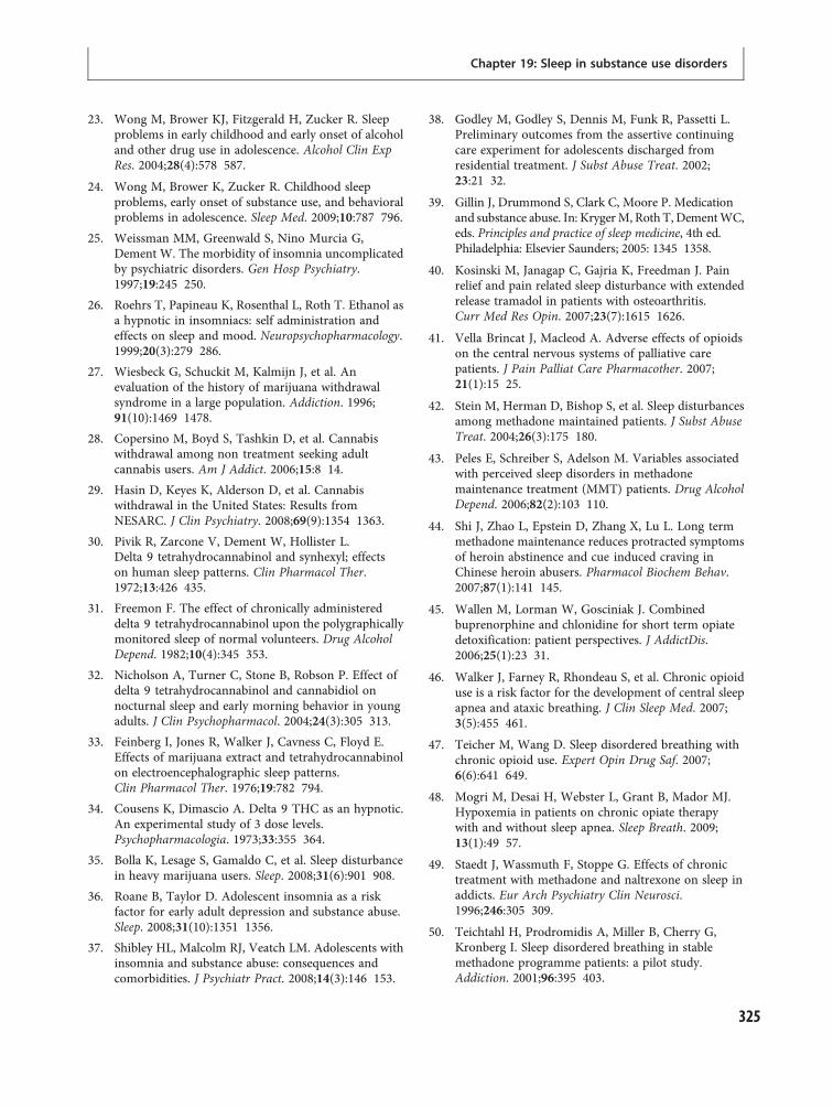
23. Wong M, Brower KJ, Fitzgerald H, Zucker R. Sleepproblems in early childhood and early onset of alcoholand other drug use in adolescence. Alcohol Clin ExpRes. 2004;28(4):578 587.
24. Wong M, Brower K, Zucker R. Childhood sleepproblems, early onset of substance use, and behavioralproblems in adolescence. Sleep Med. 2009;10:787 796.
25. Weissman MM, Greenwald S, Nino Murcia G,Dement W. The morbidity of insomnia uncomplicatedby psychiatric disorders. Gen Hosp Psychiatry.1997;19:245 250.
26. Roehrs T, Papineau K, Rosenthal L, Roth T. Ethanol asa hypnotic in insomniacs: self administration andeffects on sleep and mood. Neuropsychopharmacology.1999;20(3):279 286.
27. Wiesbeck G, Schuckit M, Kalmijn J, et al. Anevaluation of the history of marijuana withdrawalsyndrome in a large population. Addiction. 1996;91(10):1469 1478.
28. Copersino M, Boyd S, Tashkin D, et al. Cannabiswithdrawal among non treatment seeking adultcannabis users. Am J Addict. 2006;15:8 14.
29. Hasin D, Keyes K, Alderson D, et al. Cannabiswithdrawal in the United States: Results fromNESARC. J Clin Psychiatry. 2008;69(9):1354 1363.
30. Pivik R, Zarcone V, Dement W, Hollister L.Delta 9 tetrahydrocannabinol and synhexyl; effectson human sleep patterns. Clin Pharmacol Ther.1972;13:426 435.
31. Freemon F. The effect of chronically administereddelta 9 tetrahydrocannabinol upon the polygraphicallymonitored sleep of normal volunteers. Drug AlcoholDepend. 1982;10(4):345 353.
32. Nicholson A, Turner C, Stone B, Robson P. Effect ofdelta 9 tetrahydrocannabinol and cannabidiol onnocturnal sleep and early morning behavior in youngadults. J Clin Psychopharmacol. 2004;24(3):305 313.
33. Feinberg I, Jones R, Walker J, Cavness C, Floyd E.Effects of marijuana extract and tetrahydrocannabinolon electroencephalographic sleep patterns.Clin Pharmacol Ther. 1976;19:782 794.
34. Cousens K, Dimascio A. Delta 9 THC as an hypnotic.An experimental study of 3 dose levels.Psychopharmacologia. 1973;33:355 364.
35. Bolla K, Lesage S, Gamaldo C, et al. Sleep disturbancein heavy marijuana users. Sleep. 2008;31(6):901 908.
36. Roane B, Taylor D. Adolescent insomnia as a riskfactor for early adult depression and substance abuse.Sleep. 2008;31(10):1351 1356.
37. Shibley HL, Malcolm RJ, Veatch LM. Adolescents withinsomnia and substance abuse: consequences andcomorbidities. J Psychiatr Pract. 2008;14(3):146 153.
38. Godley M, Godley S, Dennis M, Funk R, Passetti L.Preliminary outcomes from the assertive continuingcare experiment for adolescents discharged fromresidential treatment. J Subst Abuse Treat. 2002;23:21 32.
39. Gillin J, Drummond S, Clark C, Moore P. Medicationand substance abuse. In: KrygerM, Roth T, DementWC,eds. Principles and practice of sleep medicine, 4th ed.Philadelphia: Elsevier Saunders; 2005: 1345 1358.
40. Kosinski M, Janagap C, Gajria K, Freedman J. Painrelief and pain related sleep disturbance with extendedrelease tramadol in patients with osteoarthritis.Curr Med Res Opin. 2007;23(7):1615 1626.
41. Vella Brincat J, Macleod A. Adverse effects of opioidson the central nervous systems of palliative carepatients. J Pain Palliat Care Pharmacother. 2007;21(1):15 25.
42. Stein M, Herman D, Bishop S, et al. Sleep disturbancesamong methadone maintained patients. J Subst AbuseTreat. 2004;26(3):175 180.
43. Peles E, Schreiber S, Adelson M. Variables associatedwith perceived sleep disorders in methadonemaintenance treatment (MMT) patients. Drug AlcoholDepend. 2006;82(2):103 110.
44. Shi J, Zhao L, Epstein D, Zhang X, Lu L. Long termmethadone maintenance reduces protracted symptomsof heroin abstinence and cue induced craving inChinese heroin abusers. Pharmacol Biochem Behav.2007;87(1):141 145.
45. Wallen M, Lorman W, Gosciniak J. Combinedbuprenorphine and chlonidine for short term opiatedetoxification: patient perspectives. J AddictDis.2006;25(1):23 31.
46. Walker J, Farney R, Rhondeau S, et al. Chronic opioiduse is a risk factor for the development of central sleepapnea and ataxic breathing. J Clin Sleep Med. 2007;3(5):455 461.
47. Teicher M, Wang D. Sleep disordered breathing withchronic opioid use. Expert Opin Drug Saf. 2007;6(6):641 649.
48. Mogri M, Desai H, Webster L, Grant B, Mador MJ.Hypoxemia in patients on chronic opiate therapywith and without sleep apnea. Sleep Breath. 2009;13(1):49 57.
49. Staedt J, Wassmuth F, Stoppe G. Effects of chronictreatment with methadone and naltrexone on sleep inaddicts. Eur Arch Psychiatry Clin Neurosci.1996;246:305 309.
50. Teichtahl H, Prodromidis A, Miller B, Cherry G,Kronberg I. Sleep disordered breathing in stablemethadone programme patients: a pilot study.Addiction. 2001;96:395 403.
Chapter 19: Sleep in substance use disorders
325

51. Martin W, Jasinki D, Haertzen C, et al. Methadone:a reevaluation.ArchGen Psychiatry. 1973;28(2):286 295.
52. Kay D. Human sleep and EEG through a cycle ofmethadone dependence. Electroencephalogr ClinNeurophysiol. 1975;38(1):35 43.
53. Dijkstra B, De Jong C, Krabbe P, van der Staak C.Prediction of abstinence in opioid dependent patients.J Addict Med. 2008;2(4):194 201.
54. Griffiths R, Johnson M. Relative abuse liability ofhypnotic drugs: A conceptual framework andalgorithm for differentiating among compounds. J ClinPsychiatry. 2005;66(Suppl 9):31 41.
55. Hajak G, Muller W, Wittchen H, Pittrow D, Kirch W.Abuse and dependence potential for the nonbenzodiazepine hypnotics zolpidem and zopiclone:a review of case reports and epidemiological data.Addiction. 2003;98:1371 1378.
56. Tsai M, Tsai Y, Huang Y. Compulsive activity andanterograde amnesia after zolpidem use. Clin Toxicol.2007;45:179 181.
57. Svitek J, Heberlein A, Bleich S, et al. Extensivecraving in high dose zolpidem dependency.Prog Neuropsychopharmacol Biol Psychiatry. 2008;32:591 592.
58. Rappa L, Larose Pierre M, Payne D, et al.Detoxification from high dose zolpidem usingdiazepam. Ann Pharmacother. 2004;38(4):590 594.
59. Forrester M. Eszopiclone ingestions reported to Texaspoison control centers, 2005 2006. Hum Exp Toxicol.2007;26:795 800.
60. Gossop M, Bradley B, Brewis R. Amphetaminewithdrawal and sleep disturbance. Drug AlcoholDepend. 1982;10:177 183.
61. McGregor C, Srisurapanont M, Jittiwutikarn J, et al.The nature, time course and severity ofmethamphetamine withdrawal. Addiction.2005;100:1320 1329.
62. Paterson L, Wilson S, Nutt D, Hutson P, Ivarsson M.Translational, caffeine induced model of onsetinsomnia in rats and healthy volunteers.Psychopharmacology. 2007;191(4):943 950.
63. Retey J, Adam M, Khatami R, et al. A genetic variationin the adenosine A2A receptor gene (ADORA2A)contributes to individual sensitivity to caffeine effectson sleep. Clin Pharmacol Ther. 2007;81(5):692 698.
64. Luciano M, Zhu G, Kirk K, et al. “No thanks, it keepsme awake”: the genetics of coffee attributed sleepdisturbance. Sleep. 2007;30:1378 1386.
65. Brower K, Maddahian E, Blow F, Beresford T.A comparison of self reported symptoms andDSM III R criteria for cocaine withdrawal. Am J DrugAlcohol Abuse. 1988;14(3):347 356.
66. Sofuoglu M, Dudish Poulsen S, Poling J, Mooney M,Katsukami D. The effect of individual cocainewithdrawal symptoms on outcome in cocaine users.Addict Behav. 2005;30:1125 1134.
67. Schierenbeck T, Riemann D, Berger M, Hornyak M.Effect of illicit recreational drugs upon sleep:Cocaine, ecstasy, and marijuana. Sleep Med Rev.2008;12(5):381 389.
68. Johanson C, Roehrs T, Schuh K, Warbasse L.The effects of cocaine on mood and sleep incocaine dependent males. Exp Clin Psychopharmacol.1999;7(4):338 346.
69. Foral PA, Malesker MA, Hopkins H. Medicationeffects on sleep. In: Bowman TJ, ed. Review of sleepmedicine. Burlington, MA: Butterworth Heinemann;2003: 94 99.
70. Kowatch R, Schnoll S, Knisely J, Green D, Elswick R.Electroencephalographic sleep and mood duringcocaine withdrawal. J Addict Disorders. 1992;11:21 45.
71. Pace Schott E, Stickgold R, Mazur A, et al. Sleepquality deteriorates over a binge abstinence cycle inchronic smoked cocaine users. Psychopharmacology.2005;179:873 883.
72. Thompson P, Gillin J, Golshan S, Irwin M. Polygraphicsleep measures differentiate alcoholics and stimulantabusers during short term abstinence. Biol Psychiatry.1995;38:831 836.
73. Weddington W, Brown B, Haertzen C, et al. Changesin mood, craving, and sleep during short termabstinence reported by male cocaine addicts:A controlled, residential study. Arch Gen Psychiatry.1990;47:861 868.
74. Sangal R, Owens J, Allen A, et al. Effects ofatomoxetine and methylphenidate on sleep in childrenwith ADHD. Sleep. 2006;29(12):1573 1585.
75. Bishop C, Roehrs T, Rosenthal L, Roth T. Alertingeffects of methylphenidate under basal and sleepdeprived conditions. Exp Clin Psychopharmacol. 1997;5(4):344 352.
76. Mitler M, Hajdukovic R, Erman M. Treatment ofnarcolepsy with methamphetamine. Sleep. 1993;16(4):306 317.
77. Roehrs T, Papineau K, Rosenthal L, Roth T. Sleepinessand the reinforcing and subjective effects ofmethylphenidate. Exp Clin Psychopharmacol. 1999;7(2):145 150.
78. Soldatos C, Kales J, Scharf M, Bixler E, Kales A.Cigarette smoking associated with sleep difficulties.Science. 1980;207:551 552.
79. Zhang L, Samet J, Caffo B, Punjabi N. Cigarettesmoking and nocturnal sleep architecture.Am J Epidemiol. 2006;164(6):529 537.
Section VI: Sleep Disturbance in Psychiatric Illness
326
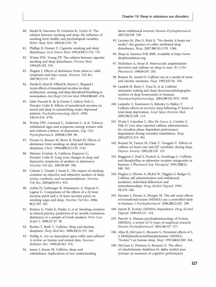
80. Riedel B, Durrence H, Lichstein K, Taylor D. Therelation between smoking and sleep: the influence ofsmoking level, health, and psychological variables.Behav Sleep Med. 2004;2(1):63 78.
81. Phillips B, Danner F. Cigarette smoking and sleepdisturbance Arch Intern Med. 1995;155(7):734 737.
82. Wetter DW, Young TB. The relation between cigarettesmoking and sleep disturbance. Prevent Med.1994;23:328 334.
83. Hughes J. Effects of abstinence from tobacco: validsymptoms and time course. Nicotine Tob Res.2007;9(3):315 327.
84. Davila D, Hurt R, Offord K, Harris C, Shepard J.Acute effects of transdermal nicotine on sleeparchitecture, snoring, and sleep disordered breathing innonsmokers.AmJRespCritCareMed. 1994;150:469 474.
85. Salin Pascual R, de la Fuente J, Galicia Polo L,Drucker Colin R. Effects of transdermal nicotine onmood and sleep in nonsmoking major depressedpatients. Psychopharmacology (Berl). 1995;121(4):476 479x.
86. Wetter DW, Carmack C, Anderson C, et al. Tobaccowithdrawal signs and symptoms among women withand without a history of depression. Exp. ClinPsychopharmacol. 2000;8(1):88 96.
87. Prosise G, Bonnet M, Berry R, Dickel M. Effects ofabstinence from smoking on sleep and daytimesleepiness. Chest. 1994;105(4):1136 1141.
88. Moreno Coutino A, Calderon Ezquerro C,Drucker Colin R. Long term changes in sleep anddepressive symptoms of smokers in abstinence.Nicotine Tob Res. 2007;9:389 396.
89. Colrain I, Trinder J, Swan G. The impact of smokingcessation on objective and subjective markers of sleep:review, synthesis, and recommendations. NicotineTob Res. 2004;6(6):913 925.
90. Aubin H, Luthringer R, Demazieres A, Dupont C,Lagrue G. Comparison of the effects of a 24 hournicotine patch and a 16 hour nicotine patch onsmoking urges and sleep. Nicotine Tob Res. 2006;8(2):193 201.
91. Boutou A, Tsiata E, Pataka A, et al. Smoking cessationin clinical practice: predictors of six month continuousabstinence in a sample of Greek smokers. Prim CareRespir J. 2008;17:32 38.
92. Roehrs T, Roth T. Caffeine: Sleep and daytimesleepiness. Sleep Med Rev. 2008;12(2):153 162.
93. Nehlig A. Are we dependent upon coffee and caffeine?A review on human and animal data. NeurosciBiobehav Rev. 1999;23:563 576.
94. James J, Keane M. Caffeine, sleep, andwakefulness: implications of new understanding
about withdrawal reversal. Human Psychopharmacol.2007;22:549 558.
95. Luciano M, Zhu G, Kirk K. “No thanks, it keeps meawake”: the genetics of coffee attributed sleepdisturbance. Sleep. 2007;30(10):1378 1386.
96. Sleep in America Poll 2008. Available at http://www.sleepfoundation.org.
97. Nicholson A, Stone B. Heterocyclic amphetaminederivative and caffeine on sleep in man. Br J ClinPharmacol. 1980;9:195 203.
98. Bonnet M, Arand D. Caffeine use as a model of acuteand chronic insomnia. Sleep. 1992;15:526 536.
99. Landolt H, Retey J, Tonz K, et al. Caffeineattenuates waking and sleep electroencephalographicmarkers of sleep homeostasis in humans.Neuropsychopharmacology. 2004;29(10):1933 1939.
100. LaJambe C, Kamimori G, Belenky G, Balkin T.Caffeine effects on recovery sleep following 27 hours oftotal sleep deprivation. Aviat Space Environ Med.2005;76(2):108 113.
101. Wyatt J, Cajochen C, Ritz De Cecco A, Czeisler C,Dijk D. Low dose repeated caffeine administrationfor circadian phase dependent performancedegradation during extended wakefulness. Sleep.2004;27(3):374 381.
102. Bonnet M, Tancer M, Uhde T, Yeragani V. Effects ofcaffeine on heart rate and QT variability during sleep.Depress Anxiety. 2005;22:150 155.
103. Biaggioni I, Paul S, Puckett A, Arzubiaga C. Caffeineand theophylline as adenosine receptor antagonists inhumans. J Pharmacol Exp Therapeut. 1991;258(2):588 593.
104. Hughes J, Oliveto A, Bickel W, Higgins S, Badger G.Caffeine self administration and withdrawal:incidence, individual differences andinterrelationships. Drug Alcohol Depend. 1993;32:239 246.
105. Huxster J, Pirona A, Morgan M. The sub acute effectsof recreational ecstasy (MDMA) use: a controlled studyin humans. J Psychopharmacol. 2006;20(2):281 290.
106. Jansen K. Ecstasy (MDMA) dependence. Drug AlcoholDepend. 1999;53:121 124.
107. Parrott A. Human psychopharmacology of Ecstasy(MDMA): a review of 15 years of empirical research.Human Psychopharmacol. 2001;16:557 577.
108. Allen R, McCann U, Ricaurte G. Persistent effects of 3,4 Methylenedioxymethamphetamine (MDMA,“Ecstasy”) on human sleep. Sleep. 1993;16(6):560 564.
109. McCann U, Peterson S, Ricaurte G. The effectof catecholamine depletion by alpha methyl paratyrosine on measures of cognitive performance
Chapter 19: Sleep in substance use disorders
327
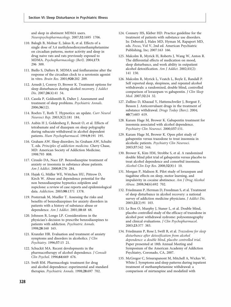
and sleep in abstinent MDMA users.Neuropsychopharmacology. 2007;32:1695 1706.
110. Balogh B, Molnar E, Jakus R, et al. Effects of asingle dose of 3,4 methylenedioxymethamphetamineon circadian patterns, motor activity and sleep indrug naive rats and rats previously exposed toMDMA. Psychopharmacology (Berl). 2004;173:296 309.
111. Biello S, Dafters R. MDMA and fenfluramine alter theresponse of the circadian clock to a serotonin agonistin vitro. Brain Res. 2001;920:202 209.
112. Arnedt J, Conroy D, Brower K. Treatment options forsleep disturbances during alcohol recovery. J AddictDis. 2007;26(4):41 54.
113. Casola P, Goldsmith R, Dalter J. Assessment andtreatment of sleep problems. Psychiatric Annals.2006;36(12).
114. Roehrs T, Roth T. Hypnotics: an update. Curr NeurolNeurosci Rep. 2003;3(2):181 184.
115. Aubin H J, Goldenberg F, Benoit O, et al. Effects oftetrabamate and of diazepam on sleep polygraphyduring subacute withdrawal in alcohol dependentpatients. Hum Psychopharmacol. 1994;9:191 195.
116. Graham AW. Sleep disorders. In: Graham AW, SchultzT, eds. Principles of addiction medicine. Chevy Chase,MD: American Society of Addiction Medicine;1998:793 808.
117. Ciraulo DA, Nace EP. Benzodiazepine treatment ofanxiety or insomnia in substance abuse patients.Am J Addict. 2000;9:276 284.
118. Hajak G, Müller WE, Wittchen HU, Pittrow D,Kirch W. Abuse and dependence potential for thenon benzodiazepine hypnotics zolpidem andzopiclone: a review of case reports and epidemiologicaldata. Addiction. 2003;98:1371 1378.
119. Posternak M, Mueller T. Assessing the risks andbenefits of benzodiazepines for anxiety disorders inpatients with a history of substance abuse ordependence. Am J Addict. 2001;10:48 68.
120. Johnson B, Longo LP. Considerations in thephysician’s decision to prescribe benzodiazepines topatients with addiction. Psychiatric Annals.1998;28:160 165.
121. Kranzler HR. Evaluation and treatment of anxietysymptoms and disorders in alcoholics. J ClinPsychiatry. 1996;57:15 21.
122. Schuckit MA. Recent developments in thepharmacotherapy of alcohol dependence. J ConsultClin Psychol. 1996;64:669 676.
123. Swift RM. Pharmacologic treatment for drugand alcohol dependence: experimental and standardtherapies. Psychiatric Annals. 1998;28:697 702.
124. Connery HS, Kleber HD. Practice guideline for thetreatment of patients with substance use disorders.In: Deborah J, Hales MD, Hyman M, Rapaport MD,eds. Focus, Vol V, 2nd ed. American PsychiatricPublishing, Inc; 2007:163 166.
125. Malcolm R, Myrick H, Roberts J, Wang W, Anton R.The differential effects of medication on mood,sleep disturbance, and work ability in outpatientalcohol detoxification. Am J Addict. 2002;11(2):141 150.
126. Malcolm R, Myrick L, Veatch L, Boyle E, Randall P.Self reported sleep, sleepiness, and repeated alcoholwithdrawals: a randomized, double blind, controlledcomparison of lorazepam vs gabapentin. J Clin SleepMed. 2007;32:24 32.
127. Zullino D, Khazaal Y, Hattenschwiler J, Borgeat F,Besson J. Anticonvulsant drugs in the treatment ofsubstance withdrawal. Drugs Today (Barc). 2004;40(7):603 619.
128. Karam Hage M, Brower K. Gabapentin treatment forinsomnia associated with alcohol dependence.Psychiatry Clin Neurosci. 2000;157:151.
129. Karam Hage M, Brower K. Open pilot study ofgabapentin versus trazodone to treat insomnia inalcoholic patients. Psychiatry Clin Neurosci.2003;57:542 544.
130. Brower K, Kim HM, Strobbe S, et al. A randomizeddouble blind pilot trial of gabapentin versus placebo totreat alcohol dependence and comorbid insomnia.Alcohol Clin Exp Res. 2008;32(8):1 10.
131. Morgan P, Malison R. Pilot study of lorazepam andtiagabine effects on sleep, motor learning, andimpulsivity in cocaine abstinence. Am J Drug AlcoholAbuse. 2008;34(6):692 702.
132. Friedmann P, Herman D, Freedman S, et al. Treatmentof sleep disturbance in alcohol recovery: a nationalsurvey of addiction medicine physicians. J Addict Dis.2003;22(2):91 103.
133. Le Bon O, Murphy J, Staner L, et al. Double blind,placebo controlled study of the efficacy of trazodone inalcohol post withdrawal sydrome: polysomnographyand clinical evaluations. J Clin Psychopharmacol.2003;23:377 383.
134. Friedmann P, Rose J, Swift R, et al. Trazodone for sleepdisturbance after detoxification from alcoholdependence: a double blind, placebo controlled trial.Paper presented at 18th Annual Meeting andSymposium of the American Academy of AddictionPsychiatry, Coronado, CA, 2007.
135. McGregor C, Srisurapanont M, Mitchell A, Wickes W,White J. Symptoms and sleep patterns during inpatienttreatment of methamphetamine withdrawal: acomparison of mirtazapine and modafinil with
Section VI: Sleep Disturbance in Psychiatric Illness
328
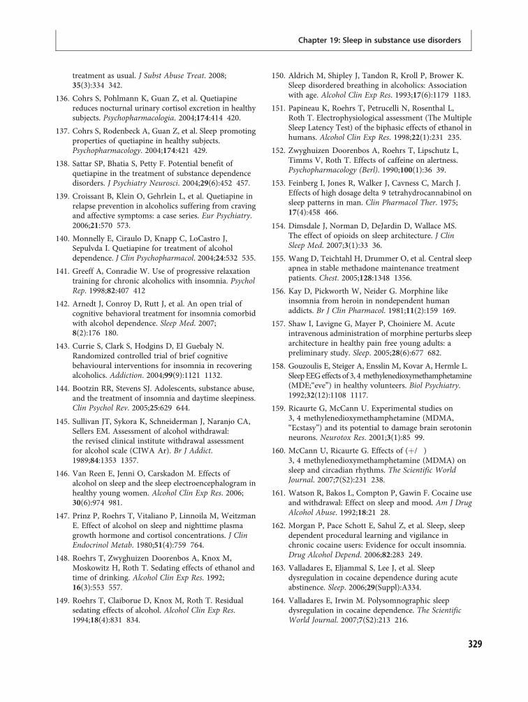
treatment as usual. J Subst Abuse Treat. 2008;35(3):334 342.
136. Cohrs S, Pohlmann K, Guan Z, et al. Quetiapinereduces nocturnal urinary cortisol excretion in healthysubjects. Psychopharmacologia. 2004;174:414 420.
137. Cohrs S, Rodenbeck A, Guan Z, et al. Sleep promotingproperties of quetiapine in healthy subjects.Psychopharmacology. 2004;174:421 429.
138. Sattar SP, Bhatia S, Petty F. Potential benefit ofquetiapine in the treatment of substance dependencedisorders. J Psychiatry Neurosci. 2004;29(6):452 457.
139. Croissant B, Klein O, Gehrlein L, et al. Quetiapine inrelapse prevention in alcoholics suffering from cravingand affective symptoms: a case series. Eur Psychiatry.2006;21:570 573.
140. Monnelly E, Ciraulo D, Knapp C, LoCastro J,Sepulvda I. Quetiapine for treatment of alcoholdependence. J Clin Psychopharmacol. 2004;24:532 535.
141. Greeff A, Conradie W. Use of progressive relaxationtraining for chronic alcoholics with insomnia. PsycholRep. 1998;82:407 412
142. Arnedt J, Conroy D, Rutt J, et al. An open trial ofcognitive behavioral treatment for insomnia comorbidwith alcohol dependence. Sleep Med. 2007;8(2):176 180.
143. Currie S, Clark S, Hodgins D, El Guebaly N.Randomized controlled trial of brief cognitivebehavioural interventions for insomnia in recoveringalcoholics. Addiction. 2004;99(9):1121 1132.
144. Bootzin RR, Stevens SJ. Adolescents, substance abuse,and the treatment of insomnia and daytime sleepiness.Clin Psychol Rev. 2005;25:629 644.
145. Sullivan JT, Sykora K, Schneiderman J, Naranjo CA,Sellers EM. Assessment of alcohol withdrawal:the revised clinical institute withdrawal assessmentfor alcohol scale (CIWA Ar). Br J Addict.1989;84:1353 1357.
146. Van Reen E, Jenni O, Carskadon M. Effects ofalcohol on sleep and the sleep electroencephalogram inhealthy young women. Alcohol Clin Exp Res. 2006;30(6):974 981.
147. Prinz P, Roehrs T, Vitaliano P, Linnoila M, WeitzmanE. Effect of alcohol on sleep and nighttime plasmagrowth hormone and cortisol concentrations. J ClinEndocrinol Metab. 1980;51(4):759 764.
148. Roehrs T, Zwyghuizen Doorenbos A, Knox M,Moskowitz H, Roth T. Sedating effects of ethanol andtime of drinking. Alcohol Clin Exp Res. 1992;16(3):553 557.
149. Roehrs T, Claiborue D, Knox M, Roth T. Residualsedating effects of alcohol. Alcohol Clin Exp Res.1994;18(4):831 834.
150. Aldrich M, Shipley J, Tandon R, Kroll P, Brower K.Sleep disordered breathing in alcoholics: Associationwith age. Alcohol Clin Exp Res. 1993;17(6):1179 1183.
151. Papineau K, Roehrs T, Petrucelli N, Rosenthal L,Roth T. Electrophysiological assessment (The MultipleSleep Latency Test) of the biphasic effects of ethanol inhumans. Alcohol Clin Exp Res. 1998;22(1):231 235.
152. Zwyghuizen Doorenbos A, Roehrs T, Lipschutz L,Timms V, Roth T. Effects of caffeine on alertness.Psychopharmacology (Berl). 1990;100(1):36 39.
153. Feinberg I, Jones R, Walker J, Cavness C, March J.Effects of high dosage delta 9 tetrahydrocannabinol onsleep patterns in man. Clin Pharmacol Ther. 1975;17(4):458 466.
154. Dimsdale J, Norman D, DeJardin D, Wallace MS.The effect of opioids on sleep architecture. J ClinSleep Med. 2007;3(1):33 36.
155. Wang D, Teichtahl H, Drummer O, et al. Central sleepapnea in stable methadone maintenance treatmentpatients. Chest. 2005;128:1348 1356.
156. Kay D, Pickworth W, Neider G. Morphine likeinsomnia from heroin in nondependent humanaddicts. Br J Clin Pharmacol. 1981;11(2):159 169.
157. Shaw I, Lavigne G, Mayer P, Choiniere M. Acuteintravenous administration of morphine perturbs sleeparchitecture in healthy pain free young adults: apreliminary study. Sleep. 2005;28(6):677 682.
158. Gouzoulis E, Steiger A, Ensslin M, Kovar A, Hermle L.SleepEEGeffects of 3, 4 methylenedioxymethamphetamine(MDE;“eve”) in healthy volunteers. Biol Psychiatry.1992;32(12):1108 1117.
159. Ricaurte G, McCann U. Experimental studies on3, 4 methylenedioxymethamphetamine (MDMA,“Ecstasy”) and its potential to damage brain serotoninneurons. Neurotox Res. 2001;3(1):85 99.
160. McCann U, Ricaurte G. Effects of (þ/ )3, 4 methylenedioxymethamphetamine (MDMA) onsleep and circadian rhythms. The Scientific WorldJournal. 2007;7(S2):231 238.
161. Watson R, Bakos L, Compton P, Gawin F. Cocaine useand withdrawal: Effect on sleep and mood. Am J DrugAlcohol Abuse. 1992;18:21 28.
162. Morgan P, Pace Schott E, Sahul Z, et al. Sleep, sleepdependent procedural learning and vigilance inchronic cocaine users: Evidence for occult insomnia.Drug Alcohol Depend. 2006;82:283 249.
163. Valladares E, Eljammal S, Lee J, et al. Sleepdysregulation in cocaine dependence during acuteabstinence. Sleep. 2006;29(Suppl):A334.
164. Valladares E, Irwin M. Polysomnographic sleepdysregulation in cocaine dependence. The ScientificWorld Journal. 2007;7(S2):213 216.
Chapter 19: Sleep in substance use disorders
329
Chapter
20 Sleep in dementias
Aimee L. Pierce and Alon Y. Avidan
IntroductionSleep disorders, such as insomnia, circadian rhythmdisturbances, obstructive sleep apnea, and rapid eyemovement (REM) sleep behavior disorder (RBD)are common in healthy older adults and even moreso in older patients with dementia. The presenta-tion, pathophysiology, diagnosis, and treatment ofsleep disorders in dementia will be discussed,including the emerging concept of RBD as a pre-clinical marker for a class of neurodegenerativediseases, the “synucleinopathies.” Sleep disordersmay worsen cognitive and behavioral symptoms indementia, and are a source of stress for caregivers,thus recognition and adequate treatment areimportant.
Changes in sleep associatedwith agingThe quest for a good nights’ sleep is truly life-long,with up to 50% of older Americans sufferingfrom chronic sleep complaints [1], including diffi-culty with sleep initiation, decreased total sleeptime, increased night-time awakenings, increaseddaytime napping, and increased early morningawakenings. Many members of the general public,and even physicians, suffer from the misconcep-tion that older adults “require” less sleep thanthe middle-aged, and sleep complaints may beunasked, unmentioned, or minimized in geriatricpatients. However, to do so misses a prime oppor-tunity to ameliorate both quality of health andquality of life.
Several qualitative and quantitative changes inthe polysomnogram occur with aging. One notes areduction in or absence of slow-wave sleep (stages 3
and 4 of non-REM), a decrease in REM sleep inproportion to total sleep time, and reduced sleepefficiency [2].
Sleep complaints in the elderly may be due to aprimary sleep disorder or secondary to another med-ical condition. Both primary and secondary causes ofsleep disorders are more common in older adults.The causes of secondary sleep disorders in olderadults are vast and include medical, psychiatric, andpsychosocial difficulties, as well as polypharmacy(Figure 20.1). Furthermore, the increased prevalenceof primary sleep disorders in older patients mayreflect age- or disease-related neurodegeneration ofbrain structures important for maintenance of thecircadian rhythm.
Insomnia is a very common problem in the olderpopulation; a large epidemiological study by Foleyet al. in patients 65 years and older found that 28%suffered from chronic insomnia and few patients weresatisfied with their sleep [3].
Older adults are also more prone to circadianrhythm disturbances, wherein they may experienceabnormal and irregular sleep–wake cycles. Thisoccurs when there is desynchrony between the activityof the brain’s pacemaker, the suprachiasmatic nucleus(SCN) of the anterior hypothalamus, and thedemands of the environment. Both pathophysiologic-al and environmental hypotheses have been pro-posed to account for circadian rhythm disturbancesin older adults. For example, the SCN deteriorateswith age, and its rhythms become progressively dis-rupted or weaker [4]. Furthermore, there is noted tobe a predictable reduction in nocturnal melatoninsecretion with aging [5]. Aside from, or synergisticwith, these neurohormonal changes, older adults havedecreased external cues to reset their circadian
Foundations of Psychiatric Sleep Medicine, ed. JohnW.Winkelman and David T. Plante. Published by Cambridge University Press.# Cambridge University Press 2010.
330
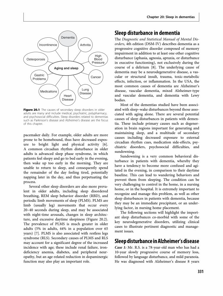
pacemaker daily. For example, older adults are moreprone to be homebound, thus have decreased expos-ure to bright light and physical activity [6].A common circadian rhythm disturbance in olderadults is advanced sleep phase syndrome, in whichpatients feel sleepy and go to bed early in the evening,then wake up too early in the morning. They areunable to return to sleep, and consequently spendthe remainder of the day feeling tired, potentiallynapping later in the day, and thus perpetuating theprocess.
Several other sleep disorders are also more preva-lent in older adults, including sleep disorderedbreathing, REM sleep behavior disorder (RBD), andperiodic limb movements of sleep (PLMS). PLMS arelimb (usually leg) movements that occur every20–40 seconds during sleep, and may be associatedwith night-time arousals, changes in sleep architec-ture, and excessive daytime sleepiness (Figure 20.2).The prevalence of PLMS is much greater in olderadults (5% in adults, 44% in a population over 65years) [7]. PLMS is also associated with restless legssyndrome (RLS). Secondary causes of PLMS and RLSmay account for a significant degree of the increasedincidence with age; these include renal failure, iron-deficiency anemia, diabetes, and peripheral neur-opathy, but an age-related reduction in dopaminergicfunction may also play an important role.
Sleep disturbance in dementiaThe Diagnostic and Statistical Manual of Mental Dis-orders, 4th edition (DSM-IV) describes dementia as aprogressive cognitive disorder composed of memoryimpairment in addition to at least one other cognitivedisturbance (aphasia, agnosia, apraxia, or disturbancein executive functioning), not exclusively during thecourse of a delirium [8]. The underlying cause ofdementia may be a neurodegenerative disease, a vas-cular or structural insult, trauma, toxic-metaboliceffects, infection, or inflammation. In the USA, themost common causes of dementia are Alzheimer’sdisease, vascular dementia, mixed Alzheimer-typeand vascular dementia, and dementia with Lewybodies.
Most of the dementias studied have been associ-ated with sleep–wake disturbances beyond those asso-ciated with aging alone. There are several potentialcauses of sleep disturbances in patients with demen-tia. These include primary causes such as degener-ation in brain regions important for generating andmaintaining sleep, and a multitude of secondarycauses including decreased exposure to externalcircadian rhythm cues, medication side-effects, psy-chiatric disorders, psychosocial difficulties, andsundowning.
Sundowning is a very common behavioral dis-turbance in patients with dementia, whereby theyhave a tendency to become more confused and agi-tated in the evening, in comparison to their daytimebaseline. This can lead to wandering behaviors andprevent them from sleeping. The condition can bevery challenging to control in the home, in a nursinghome, or in the hospital. It is extremely important torecognize and manage this problem, as well as othersleep disturbances in patients with dementia, becausethey may be an immediate precipitant, or an under-lying factor, in nursing home placement.
The following sections will highlight the import-ant sleep disturbances co-morbid with some of thekey neurodegenerative disorders, utilizing clinicalcases to illustrate pertinent diagnostic and manage-ment issues.
Sleepdisturbance inAlzheimer’sdiseaseCase 1: Mr. R.S. is a 78-year-old man who has had a10-year slowly progressive course of memory loss,followed by language disturbance, and mild paranoia.He was diagnosed with Alzheimer’s disease 8 years
Dementia
Arthritis
HighBlood
Pressure
Cancer
LungDisease
HeartDisease
Medications
GastricProblems
Osteoporosis
Incontinence
Aging and sleep
Figure 20.1 The causes of secondary sleep disorders in olderadults are many and include medical, psychiatric, polypharmacy,and psychosocial difficulties. Sleep disorders related to dementiassuch as Parkinson’s disease and Alzheimer’s disease are the focusof this chapter.
Chapter 20: Sleep in dementias
331

earlier, and is currently prescribed donepezil. He isstill living in his home with his wife; however, forthe past 6 months he has become agitated and con-fused in the evening: he has been unable to find hisway around his house to the restroom (althoughduring the day he can do so), and has also accusedhis wife of being an intruder when she tries to com-fort him in the evening. He stays up past midnightmost nights, and then wakes up at 6 a.m. andspends most of his waking hours sitting in a chairlanguidly, drifting between sleep and wakefulness.What is the underlying sleep disturbance, what arethe contributing factors, and what are the treatmentoptions?
Alzheimer’s disease (AD) is the most commoncause of dementia in the USA: there are currentlyestimated to be 4.5 million people with AD in theUSA [9]. The incidence of AD increases dramaticallywith age: about 5% of people between the ages of 65and 74 years have the disease; however, nearly 50% ofpeople over the age of 85 years have the disease [10].AD typically presents with deficits in short-termmemory, but as the disease progresses, deficits inlanguage and executive function become more prom-inent. Patients with AD also may suffer from psychi-atric symptoms such as depression, apathy, agitation,and psychosis. The majority of cases are sporadic,however 2–5% of cases are inherited in an autosomaldominant fashion, due to mutations in genes encodingthe amyloid precursor protein, presenilin 1, and
presenilin 2 [10]. In sporadic cases, presence ofthe E4 allele of the apolipoprotein gene confers athreefold increased risk of AD. The pathologicalfindings associated with AD include amyloidplaques, neurofibrillary tangles, and cerebral atro-phy. AD is a progressive disease, and there is nocure as of yet.
The most common sleep disturbances in AD arerelated to circadian rhythm abnormalities, commonlypresenting as fragmentation of the sleep–wake cycle(night-time wakefulness and frequent daytime naps)or sundowning. Polysomnographic changes inpatients with AD include increased time awake inbed, frequent awakenings, and less time in bothslow-wave sleep and REM sleep [11]. Furthermore,circadian rhythm disruptions worsen with diseaseseverity. In addition to sleep–wake fragmentation,patients with AD have disruptions of other circadianrhythms. For example, body temperature, cortisolrelease, and melatonin rhythms are less robust, withlower amplitude or less coordination, in patients withAD [12].
The mechanism of these abnormalities may beprimary neurodegeneration in the suprachiasmaticnucleus (SCN). In fact, amyloid plaques have beendiscovered in the SCN in pathological examination ofbrains of patients with AD [13]. Furthermore, there isreduced number and function of neurons in the SCNin both aging and AD [14] and reduced melatoninproduction by the pineal gland in AD [15]. Secondary
Figure 20.2 A 1 minute sleep epochfrom a diagnostic polysomnogram of a69 year old man with Parkinson’s diseasewho experiences difficulties initiating andmaintaining sleep, excessive daytimesleepiness, and uncomfortable sensationin his legs associated with an irresistibleurge to move his legs. His wife describesnight time kicking and jerkingmovements which disrupt his sleep.Illustrated in this figure is a succession ofperiodic limb movements occurring inthe left anterior tibialis muscles. Channelsare as follows: electrooculogram (left:LOC A2, right: ROC A1), chin EMG (chinchin), EEG (left central (C3 A2), rightcentral (C4 A1), left occipital (O1 A2), rightoccipital (O2 A1)), electrocardiogram(ECG), limb EMG (left leg (LAT), right leg(RAT)), patient position, snoring (SNORE),nasal oral airflow (N/O), respiratory effort(thoracic (THOR), abdominal (ABD)), nasalpressure (NPRE), and oxygen saturation(SpO2) and plethysmography channel.
Section VI: Sleep Disturbance in Psychiatric Illness
332
causes of circadian rhythm disturbances in patientswith AD include reduced exposure to ambient lightand diminished daytime activity.
The majority of patients with AD are prescribedan acetylcholinesterase inhibitor, memantine, or thecombination, as symptomatic treatment for dementia.These medications, particularly the acetylcholinester-ase inhibitors, may also have beneficial effects uponsleep. Donepezil has been shown to increase REMsleep in both young adults and patients with AD[16,17]. In patients with AD, the degree of increasein REM sleep seems to correlate with the degree ofcognitive improvement on donepezil [18]. The otheracetylcholinesterase inhibitors (galantamine and riv-astigmine) are not as well studied, although smallstudies suggest they have similar effects [19,20]. Datain laboratory animals indicated that while galanta-mine had no significant influence on the sleep pat-tern, donepezil and memantine showed significantalterations in sleep architecture consisting of increasesin sleep latency and total waking time and a decreasein total non-REM sleep time. In addition these datashow that memantine decreased total REM sleep time[21]. The effects of memantine on sleep architecturein humans have not been studied to date.
Treatments for AD circadian rhythm abnormal-ities include standard sleep hygiene practices andpharmacological treatments for insomnia and agita-tion. As for most aspects of their life, patients withAD respond well to routines. It is useful to keep aregular bedtime and awakening time. In addition, it isimportant to reduce environmental noise and light inthe bedroom and minimize night-time awakeningsfor administration of medications, vital signs collec-tion, or bedchecks (this is mainly an issue in a hos-pital or nursing home setting).
To date, none of the FDA-approved medicationsfor insomnia have undergone large-scale controlledtrials in patients with AD; however, standard and newmedications for insomnia are used widely. Whentreating sleep disturbances in AD, two principlesshould guide use of these medications. First, anti-psychotics should be used sparingly and with full atten-tion to risks and benefits of use, especially in light ofthe FDA black box warning: “both conventional andatypical antipsychotics are associated with anincreased risk of mortality in elderly patients treatedfor dementia-related psychosis.” [22] Furthermore,although they have not been tested for improvingsleep and sundowning in patients with dementia, in
a randomized double-blind, placebo-controlled trial,olanzipine, quetiapine, and risperidone did notimprove psychosis, aggression, or agitation in patientswith AD, and all were associated with increased rate ofadverse events [23]. As for sleep-related adverse events,patients on antipsychotics experienced a higher inci-dence of sedation (15–24%) than patients on placebo(5%). However, in all groups, 5% of patients experi-enced sleep disturbance as an adverse event.
Second, in treating sleep disturbances in AD,pertinent mechanisms of action and side-effects of hyp-notics must be attended to, including anticholinergicproperties of tricyclic antidepressants which mayworsen confusion in patients with dementia, and thefact that many hypnotics can cause daytime sedationwhichmay further compromise cognitive performance.
Bright light therapy and melatonin therapy areappealing potential treatments for circadian rhythmdisturbances in patients with AD, because they theor-etically may target the underlying environmental andpathophysiological causes of circadian rhythm dis-turbances in this population.
The results of several studies of bright light ther-apy (open-label and small controlled trials) havevaried. Some have shown that light exposure (of vary-ing times and intensities) benefits rest–activityrhythms in patients with AD, but others have not[24,25]. The difference in results is not unexpected,given the different patient populations, study loca-tion, light regimen, and outcome measures.
Abnormalities in melatonin receptor expressionand beneficial effects of melatonin in in vitro androdent models of Alzheimer’s disease have beendescribed. Levels of melatonin receptor subtypes aredecreased in the retina, pineal gland, and cortex ofhumans with AD [26–28]. In vitro, melatonin hasbeen demonstrated to reduce hyperphosphorylationof tau [29] and reduce amyloid pathology in a trans-genic mouse model of AD [30].
While theoretically plausible, melatonin’s role inAD as either a sleep aid or a cognitive enhancer isunclear. Although an open-label study and one smalltrial suggested that melatonin may improve sleepmeasures in patients with AD [31,32], the largestrandomized controlled trial to date, performed bythe Alzheimer’s Disease Cooperative Study, wasnegative [33]. In this study a total of 157 patients whoslept less than 7 hours per night and had frequentnocturnal awakenings were assigned to one of threegroups: placebo, 2.5 mg slow-release melatonin, or
Chapter 20: Sleep in dementias
333
10 mgmelatonin. Patients were observed for 2 monthswith wrist actigraphy. There were no statistically sig-nificant differences in nocturnal total sleep time, sleepefficiency, wake-time after sleep onset, and day�nightsleep ratio. The only randomized controlled trial formelatonin as a cognitive enhancer contained just 20patients; however, those receiving active drug (3 mg)had small, statistically significant improvements intheir scores on the Alzheimer’s disease assessmentscale (ADAS), both in cognitive and non-cognitivedomains [32].
The second sleep disturbance that is common inAD is obstructive sleep apnea (OSA). It has beenhypothesized that there may be cell loss in the brain-stem respiratory center in AD that predisposes thesepatients to sleep apnea. Conversely, nightly hypox-emia of sleep apnea could worsen the neuronal degen-eration of AD [34]. Another relationship between thetwo conditions was illuminated by recent results thatshowed that the APOE4 allele, the only known geneticrisk factor in sporadic AD, was also associated with anincreased risk of OSA [35]. This result was not foundin earlier studies, however the population backgroundand ages differed [36,37]. Despite this genetic result,at a population level there does not appear to be anincreased prevalence of OSA in patients with ADcompared to age-matched controls, and the high co-morbidity may be due to an increasing prevalence ofboth disorders with age, rather than any underlyingmechanistic interdependence. However, cliniciansmust still be sensitive to the problems of diagnosisand management of both diseases in the same patient.For example, OSA has been associated with cognitiveimpairment on neuropsychological testing in bothnormal adults and patients with AD. Among patientswith dementia, those with severe sleep apnea havemore cognitive impairment than those with mild orno apnea [38]. Those areas of cognition most affectedby OSA include attention, initiation, perseveration,conceptualization, and memory. In both normaladults and patients with AD, treatment of OSA withCPAP has been shown to ameliorate some of thesecognitive changes [39, 40].
Treatment of sleep apnea in patients with demen-tia may be challenging, because patients may findtheir CPAP mask to be uncomfortable and incompre-hensible, and there may be nightly battles over its use.If CPAP is worsening night-time agitation and pre-venting sleep, this may outweigh any benefit in day-time cognition, and use of CPAP may need to be
modified. For example, if the patient suffers frommild supine-related OSA, the use of positional ther-apy (by avoiding sleep in the supine position) may bea reasonable alternative.
The chronic mental and physical health burdensupon caregivers of patients with dementia are becom-ing more evident to the medical profession. Theseinclude elevations in stress hormones and inflamma-tory markers, depression, and sleep disturbance [41].Sleep disturbances in caregivers of patients withdementia are multifactorial and include insomniadue to depression, anxiety, appropriate worry, or cir-cadian rhythm disturbance due to ongoing caregivingduties during the night. Sleep disturbance in patientswith dementia is a strong predictor of subsequentsleep disturbance in caregivers and may predict insti-tutionalization. Thus, strategies such as night-timehired caregivers and temporary respite care may allowthe patient to remain in their home longer.
Case 1, discussion: This patient has advanced ADand is now manifesting circadian rhythm disturb-ances including an altered sleep–wake cycle and sun-downing. Aside from potential degeneration of theSCN and reduced production of melatonin by thepineal gland, a low level of physical activity andexposure to ambient light may worsen these symp-toms. Possible treatments include daytime bright lighttherapy, increase in daily outdoor physical activity(supervised, during daylight hours), strict regulationof bedtime and awakening time, and consideration ofa night-time caregiver (to reduce burden on his wife.)If these conservative measures do not resolve thesituation, some consideration may be given to mela-tonin therapy, control of evening agitation and delu-sions with an atypical antipsychotic (with attention tocurrent black box warnings in the elderly), or a lowdose of a hypnotic such as zolpidem.
Sleep disturbance in vascular dementiaVascular dementia is the second most common formof dementia in the USA. Vascular pathology fre-quently overlaps with AD pathology, which is knownas dementia of mixed vascular and Alzheimer’s path-ology. Because of this overlap, exact estimates ofprevalence are difficult, however vascular dementialikely accounts for between 10% and 33% of cases ofdementia [10]. The pathology of vascular dementia isheterogeneous and may include a single or multiplelarge cortical strokes, multiple lacunar infarcts or
Section VI: Sleep Disturbance in Psychiatric Illness
334

confluent white matter disease, or a single, strategic-ally placed stroke (such as in the angular gyrus).Clinical criteria include cognitive decline in memoryand at least one other domain of cognition, interfer-ing with activities of daily living, and a time coursethat is usually stepwise or abrupt. Symptoms aretypically those of subcortical frontal dementia, suchas apathy, impaired executive function, psychomotorslowing, and impaired gait. Alternatively, symptomsmay be attributable to the location of the stroke, suchas a receptive aphasia resulting from a left temporalstroke. The risk factors for vascular dementia are thesame as those for stroke, and include hypertension,diabetes, hyperlipidemia, and cigarette smoking.Treatment focuses on prevention and managementof risk factors, but both cholinesterase inhibitorsand memantine have been used with some benefit.
There has been very little published research onsleep disturbance in vascular dementia, though asmall actigraphic study found that patients withmulti-infarct dementia (a subtype of vascular demen-tia) had disrupted sleep–wake cycles compared toboth controls and ambulatory patients with AD [42].
More striking are the interactions between sleepand stroke, which are beginning to be elucidated. Itremains to be determined whether sleep disorderedbreathing is an independent risk factor for stroke.However, the blood pressure-lowering effects ofCPAP may lead to a reduction in risk of stroke ofup to 20% in patients with OSA [43]. Sleep disorderspost-stroke are common and can worsen outcomes; arecent study found that OSA was a risk factor fordeath after stroke [44]. Other post-stroke sleep dis-orders include central sleep apnea and failure of auto-matic breathing (Ondine’s curse), Cheyne-Stokesbreathing, and REM sleep behavior disorder associatedwith pontine tegmental infarct. Treatment of sleepdisordered breathing has been shown to improve sub-jective well-being andmood in stroke patients [45, 46].Finally, it is important to remember that the conse-quences of stroke such as depression, immobilization,and pain may also negatively impact sleep.
Sleep disturbance in dementia withLewy bodiesDementia with Lewy bodies (DLB) is the third mostcommon cause of dementia in the USA. The cardinalclinical features include a fluctuating course, visualhallucinations, and parkinsonism. The pathological
diagnosis requires cortical Lewy bodies, which areintracellular synuclein inclusions. DLB exists on aspectrum with both Parkinson’s disease and AD. Ina patient with parkinsonism and dementia, the clinicaldiagnosis depends upon which symptom developedfirst (DLB requires dementia at onset; in contrast,patients with idiopathic Parkinson’s disease (PD) usu-ally do not develop dementia until late in their disease).Ultimately, both patients with DLB and Parkinson’sdisease dementia will develop substantia nigra andcortical Lewy bodies. In the case of AD, there is a “Lewybody variant” with slight clinical differences, butmost often diagnosed post-mortem, with the classicAlzheimer’s pathology of plaques and tangles co-exist-ent with cortical Lewy bodies. Current treatment forDLB includes: cholinesterase inhibitors and meman-tine, dopamine or dopamine agonists for severe par-kinsonism (with attention to anyworseningpsychosis),and avoidance of neuroleptics when possible (patientswith DLB tend to be very sensitive to these medicationsand can develop severe parkinsonism.)
In the 1990s, physicians began to report a highincidence of REM sleep behavior disorder (RBD) intheir patients with DLB [47] and other “synucleino-pathies” – diseases associated with either mutations inor abnormal accumulations of the protein synuclein.These synucleinopathies include DLB, Parkinson’sdisease, and multiple system atrophy. Increasingawareness of RBD and its association with synuclei-nopathies has led the DLB consortium to includeRBD as a “suggestive feature” for the diagnosis [48].
RBD is characterized by absence of normal muscleatonia during REM sleep (Figure 20.3). Thus, patientsenact their dreams, resulting in vigorous, complicated,and sometimes dangerous motor activity during sleep.Both the patient themselves and the bed partner are atrisk for injury during violent or thrashing dreams.RBD is idiopathic in 40% of cases, and associated witha neurodegenerative disease in 60% of cases [49]. Thereis a much higher incidence of RBD in men, though thereason for this is unknown. There may be an under-lying pathophysiological difference between the sexes,or RBD may be under diagnosed in women, as theymay have less violent or active dream content, andmaybe less likely to have a bed partner (because of womens’longer life-span.)
The diagnosis of RBD is suggested by historyand confirmed by overnight polysomnography withmultiple limb electromyography and continuous videomonitoring, which demonstrates electromyographic
Chapter 20: Sleep in dementias
335
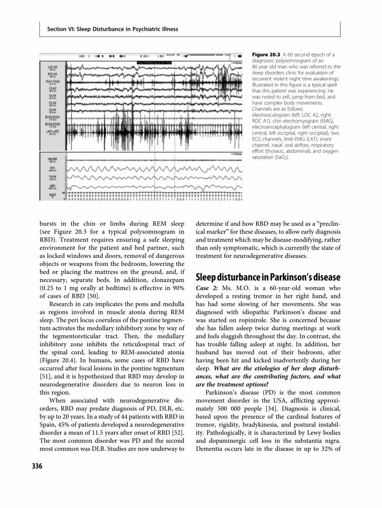
bursts in the chin or limbs during REM sleep(see Figure 20.3 for a typical polysomnogram inRBD). Treatment requires ensuring a safe sleepingenvironment for the patient and bed partner, suchas locked windows and doors, removal of dangerousobjects or weapons from the bedroom, lowering thebed or placing the mattress on the ground, and, ifnecessary, separate beds. In addition, clonazepam(0.25 to 1 mg orally at bedtime) is effective in 90%of cases of RBD [50].
Research in cats implicates the pons and medullaas regions involved in muscle atonia during REMsleep. The peri locus coeruleus of the pontine tegmen-tum activates the medullary inhibitory zone by way ofthe tegmentoreticular tract. Then, the medullaryinhibitory zone inhibits the reticulospinal tract ofthe spinal cord, leading to REM-associated atonia(Figure 20.4). In humans, some cases of RBD haveoccurred after focal lesions in the pontine tegmentum[51], and it is hypothesized that RBD may develop inneurodegenerative disorders due to neuron loss inthis region.
When associated with neurodegenerative dis-orders, RBD may predate diagnosis of PD, DLB, etc.by up to 20 years. In a study of 44 patients with RBD inSpain, 45% of patients developed a neurodegenerativedisorder a mean of 11.5 years after onset of RBD [52].The most common disorder was PD and the secondmost common was DLB. Studies are now underway to
determine if and how RBD may be used as a “preclin-ical marker” for these diseases, to allow early diagnosisand treatment whichmay be disease-modifying, ratherthan only symptomatic, which is currently the state oftreatment for neurodegenerative diseases.
Sleepdisturbance inParkinson’sdiseaseCase 2: Ms. M.O. is a 60-year-old woman whodeveloped a resting tremor in her right hand, andhas had some slowing of her movements. She wasdiagnosed with idiopathic Parkinson’s disease andwas started on ropinirole. She is concerned becauseshe has fallen asleep twice during meetings at workand feels sluggish throughout the day. In contrast, shehas trouble falling asleep at night. In addition, herhusband has moved out of their bedroom, afterhaving been hit and kicked inadvertently during hersleep. What are the etiologies of her sleep disturb-ances, what are the contributing factors, and whatare the treatment options?
Parkinson’s disease (PD) is the most commonmovement disorder in the USA, afflicting approxi-mately 500 000 people [34]. Diagnosis is clinical,based upon the presence of the cardinal features oftremor, rigidity, bradykinesia, and postural instabil-ity. Pathologically, it is characterized by Lewy bodiesand dopaminergic cell loss in the substantia nigra.Dementia occurs late in the disease in up to 32% of
Figure 20.3 A 60 second epoch of adiagnostic polysomnogram of an80 year old man who was referred to thesleep disorders clinic for evaluation ofrecurrent violent night time awakenings.Illustrated in this figure is a typical spellthat this patient was experiencing. Hewas noted to yell, jump from bed, andhave complex body movements.Channels are as follows:electrooculogram (left: LOC A2, right:ROC A1), chin electromyogram (EMG),electroencephalogram (left central, rightcentral, left occipital, right occipital), twoECG channels, limb EMG (LAT), snorechannel, nasal oral airflow, respiratoryeffort (thoracic, abdominal), and oxygensaturation (SaO2).
Section VI: Sleep Disturbance in Psychiatric Illness
336

patients, at which time it is called Parkinson’s diseasedementia (PDD) [53].
Sleep disturbances are myriad and very commonin PD, and tend to occur early in the course of thedisorder. Nearly two-thirds of patients with PDreported a sleep disorder in a community-basedsurvey of 245 patients [54]. The most commonlyreported problems include fragmented sleep, frequentarousals, and trouble going back to sleep. The poly-somnogram in PD reveals a reduction in sleep effi-ciency; increased wake-time after sleep onset;increased sleep fragmentation; reduction of slow-wave sleep and REM sleep; disruption of non-REM-to-REM cyclicity; loss of muscle atonia (suggestive ofREM sleep behavior disorder); and increased electro-myographic activity due to tremor or periodic legmovements of sleep (PLMS) [34].
Pathological daytime sleepiness including sleepattacks is a common problem in PD. A commoncause is the use of dopamine agonists such as prami-pexole and ropinirole. Another cause may be sleepdebt related to sleep fragmentation and/or circadianrhythm disturbances which can result in difficultieswith sleep initiation and maintenance, as well asspontaneous daytime dozing.
A second common sleep disorder in PD is sleepfragmentation related to motor abnormalities. These
include nocturnal immobility, rest tremor, eyeblinking, dyskinesias, periodic limb movements ofsleep (PLMS), restless legs syndrome, and fragmen-tary myoclonus [34]. Many of these motor phenom-ena can be treated by adjusting the evening dose oflevodopa/carbidopa, depending upon the symptom.For example, if the patient is waking up because theycannot roll over in bed or because of tremor, theymay benefit from a bedtime dose of immediate-release or long-acting levodopa/carbidopa, dependingupon the time the symptoms occur. If they are wakingup due to dyskinesias, the evening dose of levodopa/carbidopa should be reduced.
Patients with PD are also prone to sleep dis-ordered breathing including central sleep apnea,OSA, and alveolar hypoventilation. They may alsohave a restrictive pulmonary defect due to abnormalmuscle tone or respiratory incoordination [55].
RBD is frequently encountered in PD, as in othersynucleinopathies. Just as in DLB, interest is developingin attempting to use RBD as a potential preclinicalmarker for PD. RBD may begin before or after PD isdiagnosed. In a study of 80 patients with idiopathic PD,over 8 years, 27 patients were diagnosed with RBD [56].
Patients with PD may suffer from hypnagogichallucinations and vivid dreams, noted particularlyin the middle-to-late stages of illness. These
Stimulation
Inhibition
RBD
Lack of pontine-mediatedmedullar inhibition ofspinal motor neurons
Lack of medullary-mediatedspinal motor neuron inhibition
Lack of REM Atonia
Muscle
REM-associated Atonia
Spinal Motor Neuron
Spinal CordVentrolateral Reticulospinal Tract
Medullary CentersMagnocellularis Neurons
Lateral Tegmentoreticular T tract
Pedunculopontine CentersPerilocus Ceruleus
Figure 20.4 Muscle atonia during REMsleep results from pontine mediated perilocus ceruleus inhibition of motor activity.This pontine activity exerts an excitatoryinfluence on medullary centers(magnocellularis neurons) via the lateraltegmentoreticular tract. These neuronalgroups, in turn, hyperpolarize the spinalmotor neuron post synaptic membranesvia the ventrolateral reticulospinal tract. InREM sleep behavior disorder (RBD), thebrainstem mechanisms generatingmuscle atonia become disrupted.Understanding of the pathophysiology ofRBD in humans is based on the cat model.In the cat model, bilateral pontine lesionsresult in a persistent absence of REMatonia associated with prominent motoractivity during REM sleep, similar to thatobserved in RBD in humans. Thepathophysiology of the idiopathic form ofRBD in humans is still not very wellunderstood but may be related toreduction of striatal presynaptic dopaminetransporters. Modified from Avidan AY.Sleep disorders in the elderly. Primary Care:Clinics in Office Practice on Sleep Disorders.2005;32(2):536 587.
Chapter 20: Sleep in dementias
337

symptoms can disturb sleep and may be worsened byor secondary to medications for PD.
When evaluating sleep disorders in a patient withPD, it is very important to review the quality andtiming of symptoms as well as the medication list.Many medications used to treat PD have sleep-relatedside-effects including: daytime sleep attacks (e.g.dopamine agonists), hypnagogic hallucinations/vividdreams (e.g. dopamine agonists, levodopa/carbidopa,MAO-B inhibitors), and insomnia (e.g. dopamineagonists, MAO-B inhibitors, amantadine). If thesesymptoms are occurring, one option is to give medica-tions earlier in the day or reduce the dose. Conversely,if the patient is having motor symptoms relatedto inadequate dopamine repletion keeping themawake, such as tremor or stiffness, a bedtime dose ofimmediate-release or long-acting levodopa/carbidopais appropriate. Evaluation should also include atten-tion to co-existing psychiatric symptoms, such as anx-iety and depression, which are both common in PDand can alter sleep. If RBD, PLMS, or sleep-disorderedbreathing are suspected, evaluation should include anovernight polysomnogram. Treatment for these dis-orders in PD is similar to treatment in other patients.Finally, attention to the quality of sleep in caregivers ofpatients with PD is also warranted, as the decision tohospitalize or institutionalize patients is frequently dueto nightly sleep disturbance of the caregiver.
Case 2, discussion:This patient likely has REM sleepbehavior disorder associated with Parkinson’s disease;however, periodic limb movements of sleep is anotherpossibility. Diagnosis may be confirmed with overnightpolysomnogram. Treatment should attend to ensuringthe safety of the patient and her husband in theirbedroom. Treatment may also include clonazepam(0.25–1 mg) before bedtime if RBD is diagnosed, but,given the existing daytime sleepiness, a shorter actingbenzodiazepine (e.g. lorazepam) would be a reasonablealternative. In addition, she is describing daytime som-nolence and sleep attacks, which may be side-effects ofdopamine agonists such as ropinirole. Hypersomniamay also be due to sleep-disordered breathing, or maybe related to a sleep deficit from insomnia. A nocturnalpolysomnogram would be helpful for diagnosis of sleepapnea. In any case, with this level of daytime somno-lence, she should be advised not to drive, and ropiniroleshould be tapered or removed, if another treatable causeof somnolence is not found. Finally, the cause of herinitiation insomnia should be further investigated: arestiffness and pain preventing her from falling asleep?
This may be improved by a later dose of ropinirole orlevodopa/carbidopa. Does she have symptoms of rest-less legs syndrome? Is she anxious or depressed withruminative thoughts keeping her awake? If no othercause is found, again consider tapering, removing, orgiving the dose of ropinirole earlier in the day.
Sleep disturbance in other Parkinson’splus syndromesParkinson’s plus syndromes are neurodegenerativediseases which cause parkinsonism “plus” symptomssuch as autonomic dysfunction, ataxia, eye movementabnormalities, apraxia, early falls, or dementia. Theyare usually divided into categories based upon thepathogenic protein: “synucleinopathies” such as mul-tiple system atrophy and DLB, and “tauopathies” suchas progressive supranuclear palsy and corticobasaldegeneration. Using this schema, AD and some formsof frontotemporal dementia may also be classified astauopathies; please see Figure 20.5 for a taxonomicvisualization of the main synucleinopathies andtauopathies.
Multiple system atrophy (MSA) is a synucleino-pathy that requires at least two of the following: auto-nomic dysfunction, parkinsonism, or cerebellar ataxia.Depending upon the initial andmost prominent systeminvolved, it is designated as MSA-A (autonomic, for-merly Shy-Drager syndrome), MSA-P (parkinson,formerly striatonigral degeneration), or MSA-C (cere-bellar, formerly olivopontocerebellar atrophy.)
As in other synucleinopathies (such as PD andDLB), REM sleep behavior disorder is frequently seenin up to 90% of patients with MSA and may pre- orpost-date diagnosis of MSA [34]. In addition, patientswith MSA are prone to a unique and serious form ofsleep disordered breathing: inspiratory stridor associ-ated with upper airway obstruction. This is audiblydistinct from typical OSA, in that the stridor is highpitched and whining rather than low pitched andsnoring. This stridor may be due to vocal cordabductor paralysis and may lead to sudden deathduring sleep. In order to make the diagnosis, poly-somnogram and larygoscopy during sleep must beperformed. Although CPAP may be useful in somepatients, tracheostomy is the most reliable treatmentfor respiratory disturbances caused by vocal cordabductor paralysis.
Progressive supranuclear palsy (PSP) is a tau-opathy with clinical features of vertical supranuclear
Section VI: Sleep Disturbance in Psychiatric Illness
338
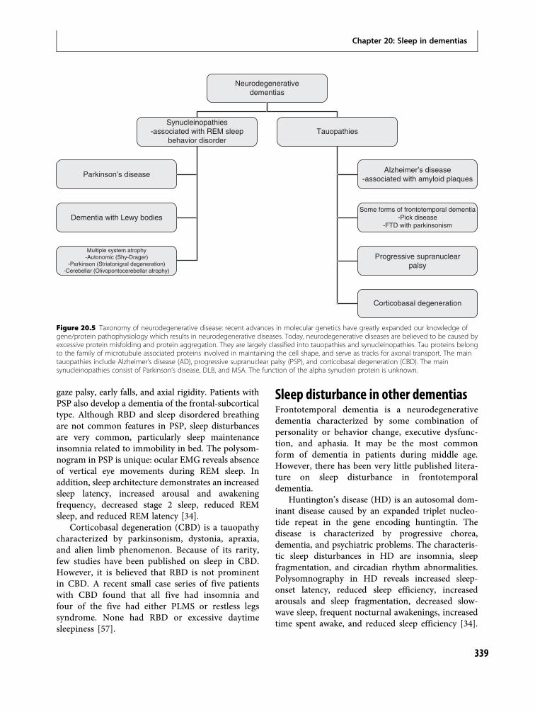
gaze palsy, early falls, and axial rigidity. Patients withPSP also develop a dementia of the frontal-subcorticaltype. Although RBD and sleep disordered breathingare not common features in PSP, sleep disturbancesare very common, particularly sleep maintenanceinsomnia related to immobility in bed. The polysom-nogram in PSP is unique: ocular EMG reveals absenceof vertical eye movements during REM sleep. Inaddition, sleep architecture demonstrates an increasedsleep latency, increased arousal and awakeningfrequency, decreased stage 2 sleep, reduced REMsleep, and reduced REM latency [34].
Corticobasal degeneration (CBD) is a tauopathycharacterized by parkinsonism, dystonia, apraxia,and alien limb phenomenon. Because of its rarity,few studies have been published on sleep in CBD.However, it is believed that RBD is not prominentin CBD. A recent small case series of five patientswith CBD found that all five had insomnia andfour of the five had either PLMS or restless legssyndrome. None had RBD or excessive daytimesleepiness [57].
Sleep disturbance in other dementiasFrontotemporal dementia is a neurodegenerativedementia characterized by some combination ofpersonality or behavior change, executive dysfunc-tion, and aphasia. It may be the most commonform of dementia in patients during middle age.However, there has been very little published litera-ture on sleep disturbance in frontotemporaldementia.
Huntington’s disease (HD) is an autosomal dom-inant disease caused by an expanded triplet nucleo-tide repeat in the gene encoding huntingtin. Thedisease is characterized by progressive chorea,dementia, and psychiatric problems. The characteris-tic sleep disturbances in HD are insomnia, sleepfragmentation, and circadian rhythm abnormalities.Polysomnography in HD reveals increased sleep-onset latency, reduced sleep efficiency, increasedarousals and sleep fragmentation, decreased slow-wave sleep, frequent nocturnal awakenings, increasedtime spent awake, and reduced sleep efficiency [34].
Neurodegenerativedementias
Parkinson’s disease
Dementia with Lewy bodies
Multiple system atrophy-Autonomic (Shy-Drager)
-Parkinson (Striatonigral degeneration)-Cerebellar (Olivopontocerebellar atrophy)
Alzheimer’s disease-associated with amyloid plaques
Some forms of frontotemporal dementia-Pick disease
-FTD with parkinsonism
Progressive supranuclearpalsy
Corticobasal degeneration
Synucleinopathies-associated with REM sleep
behavior disorderTauopathies
Figure 20.5 Taxonomy of neurodegenerative disease: recent advances in molecular genetics have greatly expanded our knowledge ofgene/protein pathophysiology which results in neurodegenerative diseases. Today, neurodegenerative diseases are believed to be caused byexcessive protein misfolding and protein aggregation. They are largely classified into tauopathies and synucleinopathies. Tau proteins belongto the family of microtubule associated proteins involved in maintaining the cell shape, and serve as tracks for axonal transport. The maintauopathies include Alzheimer’s disease (AD), progressive supranuclear palsy (PSP), and corticobasal degeneration (CBD). The mainsynucleinopathies consist of Parkinson’s disease, DLB, and MSA. The function of the alpha synuclein protein is unknown.
Chapter 20: Sleep in dementias
339
Interestingly, patients with HD have also shownhigher density of sleep spindles in comparison tocontrols, the significance of which is uncertain [58].Whether patients with HD have a higher prevalenceof OSA has not been studied.
Prion diseases are very rare, rapidly progressivedementias, which are thought to be caused by a mis-folded prion protein which is able to propagate itself.Prion diseases can be inherited, sporadic, or transmis-sible. The most common prion disease is Creutzfelt-Jacob disease (CJD), which is characterized by demen-tia and startle myoclonus. A recent study of sleep inpatients with CJD found that all had sleep–wake dis-turbances and interesting polysomnogram findings:loss of sleep spindles, very low sleep efficiency, andvirtual absence of REM sleep [59]. Fatal familialinsomnia (FFI) is an autosomal dominant prion dis-ease, which presents with insomnia and then pro-gresses to dementia and eventually death. Prionplaques form in the thalamus. The polysomnogramin FFI demonstrates dramatic sleep rhythm disorgan-ization and reduction in total sleep time. In additionthere is a reduction in sleep spindles and K complexes,a complete abolition of NREM sleep, and only briefresidual periods of REM sleep without atonia [60].
Diagnostic approach to sleepdisturbance in dementiaAs in most medical specialties, a thorough anddetailed history is crucial to uncover the diagnosis ofa sleep disorder in a patient with dementia. Becausethe patient’s underlying impaired cognition maymake explaining their own symptoms difficult, andbecause critical clues are collected while the patient isasleep, it is essential to also obtain a history from abed partner or caregiver. Important questions in thehistory include: when did the problem start, what arethe exacerbating and alleviating factors, what are thedaily and nightly activity and sleep patterns, whatmedications and non-medical stimulants and seda-tives are being taken, and what breathing, movement,or behavioral abnormalities are noted before orduring sleep?
Important aspects of the physical exam include acomplete neurological exam with mental status (aspatients in early stages of dementia have problemsdifferent from those in later stages), motor exam (toevaluate for parkinsonism or other movement
disorders which may impair sleep), and oropharyn-geal exam for Mallampati score (see Figure 6.5).
Some of the common indications for polysom-nogram in patients with dementia are for evaluationof suspected sleep disordered breathing, RBD, orPLMS. Additional diagnostic tests may be useful,depending upon the clinical situation, such as amultiple sleep latency test (for evaluation ofdaytime somnolence), actigraphy (for evaluation ofinsomnia or circadian rhythm disturbances), orbrain imaging (if there are unexplained neurologicaldeficits on examination).
When evaluating sleep disorders in patients withdementia, attentionmust always be paid to sleep-relatedside-effects caused by current medications. In addition,many medications used in sleep medicine can be sedat-ing and can worsen some aspects of cognition, so theymust be used with care. Conversely, patients withdementia respond very well to a regulated scheduleand environment, so attention to good sleep hygienecan be productive and is often the best treatment.
Because inadequate sleep can impair many aspectsof cognition, the diagnosis and appropriate treatmentof sleep disorders in patients with dementia isrewarding in that both sleep symptoms and cognitivesymptoms can potentially be improved. Furthermore,adequately treating sleep disturbances in patients withdementia can prolong the time to nursing homeplacement and improve the interaction and relation-ship between patients and their caregivers. Until acure for these diseases is found, supportive care willbe the rule, and adequate sleep can truly improvequality of life for these patients.
References1. Foley DJ, Monjan AA, Brown SL, et al. Sleep complaints
among elderly persons: an epidemiologic study of threecommunities. Sleep. 1995;18:425 432.
2. Cohen ZionM, Gehrman PR, Ancoli Israel S. Sleep in theelderly. In: Lee Chiong TL, Sateia MJ, Carskadon MA, ed.Sleep medicine. Philadelphia: Hanley & Belfus; 2002:115123.
3. Foley DJ, Monjan A, Simonsick EM, et al. Incidence andremission of insomnia among elderly adults: anepidemiologic study of 6,800 persons over three years.Sleep. 1999;22:S366 S372.
4. Lydic R, Schoene WC, Czeisler CA, Moore Ede MC.Suprachiasmatic region of the human hypothalamus:homolog to the primate circadian pacemaker? Sleep.1980;2:355 361.
Section VI: Sleep Disturbance in Psychiatric Illness
340
5. van Coevorden A, Mockel J, Laurent E, et al.Neuroendocrine rhythms and sleep in aging men. AmJ Physiol. 1991;260:E651 E661.
6. Shochat T, Martin J, Marler M, Ancoli Israel S.Illumination levels in nursing home patients:effects on sleep and activity rhythms. J Sleep Res.2000;9:373 379.
7. Ancoli Israel S, Kripke DF, Klauber MR, et al. Periodiclimb movements in sleep in community dwellingelderly. Sleep. 1991;14:486 495.
8. American Psychiatric Association. Diagnostic andstatistical manual of mental disorders, 4th ed.Washington, DC: American PsychiatricPublishing; 1994.
9. National Institute on Aging. Alzheimer’s disease factsheet, 2006. http://www.nia.nih.gov/Alzheimers/Publications/adfact.htm. Accessed 7 November, 2008.
10. Daffner KR, Wolk DA. Behavioral neurology anddementia. In: Samuels MA, ed. Manual of neurologictherapeutics, 7th ed. Philadelphia: LippincottWilliams & Wilkins; 2004:410 448.
11. Prinz PN, Peskind E, Raskind M, et al. Changes in thesleep and waking EEG in nondemented and dementedelderly. J Am Geriatr Soc. 1982;30:86 93.
12. Van Someren EJ. Circadian rhythms and sleep inhuman aging. Chronobiol Int. 2000;17(3):233 243.
13. Swaab DF, Fliers E, Partiman TS. The suprachiasmaticnucleus of the human brain in relation to sex, age andsenile dementia. Brain Res. 1985;342:37 44.
14. Hoffman MA. The human circadian clock and aging.Chronobiol Int. 2000;17(3):245 260.
15. Srinivasan V, Pandi Perumal SR, Cardinali DP,Poeggeler B, Hardeland R. Melatonin in Alzheimer’sdisease and other neurodegenerative disorders. BehavBrain Funct. 2006;2:1 23.
16. Kanbayashi T, Sugiyama T, Aizawa R, et al. Effects ofdonepezil (Aricept) on the rapid eye movement sleepof normal subjects. Psychiatry Clin Neurosci.2002;56:307 308.
17. Moraes W. dos S, Poyares D, Guilleminault C, et al.The effect of donepezil on sleep and REM sleep EEG inpatients with Alzheimer disease: a double blindplacebo controlled study. Sleep. 2006;29(2):199 205.
18. Mizuno S, Kameda A, Inagaki T, Horiguchi J.Effects of donepezil on Alzheimer’s disease: Therelationship between cognitive function and rapid eyemovement sleep. Psychiatry Clin Neurosci.2004;58:660 665.
19. Schredl M, Weber B, Braus D, Heuser I. The effect ofrivastigmine on sleep in elderly healthy subjects. ExpGerontol. 2000;35:243 249.
20. Cooke J, Loredo J, Liu L, et al. Acetylcholinesteraseinhibitors and sleep architecture in patients withAlzheimer’s disease. Drugs Aging. 2006;23(6):503 511.
21. Ishida T, Kamei C. Characteristic effects of antidementia drugs on rat sleep patterns. J Pharmacol Sci.2009;109(3):449 455.
22. Center for Drug Evaluation and Research. FDA publichealth advisory: deaths with antipsychotics in elderlypatients with behavioral disturbances, 11 April, 2005.Available at http://www.fda.gov/Drugs/DrugSafety/PublicHealthAdvisories/ucm051200.htm.
23. Schneider L, Tariot P, Dagerman K, et al. Effectivenessof atypical antipsychotic drugs in patients withAlzheimer’s disease. N Engl J Med. 2006;355(15):1525 1538.
24. Ancoli Israel S, Gehrman P, Martin J, et al. Increasedlight exposure consolidates sleep and strengthenscircadian rhythms in severe Alzheimer’s diseasepatients. Behav Sleep Med. 2003;1(1):22 36.
25. Dowling G, Burr R, Van Someren E, et al. Melatoninand bright light treatment for rest activity disruptionin institutionalized patients with Alzheimer’s disease.J Am Geriatr Soc. 2008;56(2):239 246.
26. Savaskan E, Jockers R, Ayoub M. The MT2 melatoninreceptor subtype is present in human retina anddecreases in Alzheimer’s disease. Curr Alzheimer Res.2007;4(1):47 51.
27. Brunner P, Sozer Topcular N, Jockers R, et al. Pinealand cortical melatonin receptors MT1 and MT2 aredecreased in Alzheimer’s disease. Eur J Histochem.2006;50(4):311 316.
28. Wu YH, Zhou JN, Van Heerikhuize J, et al. DecreasedMT1 melatonin receptor expression in thesuprachiasmatic nucleus in aging and Alzheimer’sdisease. Neurobiol Aging. 2007;28(8):1239 1247.
29. Wang JZ, Wang ZF. Role of melatonin in Alzheimer likeneurodegeneration.ActaPharmacol Sin. 2006;27(1):41 49.
30. Matsubara E, Bryant Thomas T, Pacheco Quinto J, et al.Melatonin increases survival and inhibits oxidativeand amyloid pathology in a transgenic model ofAlzheimer’s disease. J Neurochem. 2003;85(5):1101 1108.
31. Brusco LI, Fainstein I, Marquez M, Cardinali DP.Effect of melatonin in selected populations of sleepdisturbed patients. Biol Signals Recept. 1999;8(1 2):126 131.
32. Asayama K, Yamadera H, Ito T, et al. Double blindstudy of melatonin effects on the sleep wake rhythm,cognitive and non cognitive functions in Alzheimertype dementia. J Nippon Med Sch. 2003;70(4):334 341.
33. Singer C, Tractenberg R, Kaye J, et al. A multicenter,placebo controlled trial of melatonin for sleep
Chapter 20: Sleep in dementias
341
disturbance in Alzheimer’s disease. Sleep. 2003;26(7):893 901.
34. Avidan AY. Sleep and neurologic problems in theelderly. Sleep Med Clin. 2006;1:273 292.
35. Punjabi NM, Shahar E, Redline S, et al. Sleepdisordered breathing, glucose intolerance, and insulinresistance: the Sleep Heart Health Study. AmJ Epidemiol. 2004;160:521 530.
36. Foley DJ, Masaki K, White L, et al. Relationshipbetween apolipoprotein E epsilon4 and sleepdisordered breathing at different ages. JAMA.2001;286:1447 1448.
37. Saarelainen S, Lehtimaki T, Kallonen E, et al. Norelation between apolipoprotein E alleles andobstructive sleep apnea. Clin Genet. 1998;53:147 148.
38. Ancoli Israel S, Klauber MR, Butters N, et al.Dementia in institutionalized elderly: relation to sleepapnea. J Am Geriatr Soc. 1991;39:258 263.
39. Ferini Strambi L, Baietto C, Di Gioia M, et al.Cognitive dysfunction in patients with obstructivesleep apnea (OSA): partial reversibility aftercontinuous positive airway pressure (CPAP). Brain ResBull. 2003;61(1):87 92.
40. Ancoli Israel S, Palmer B, Cooke J, et al. Cognitiveeffects of treating obstructive sleep apnea inAlzheimer’s disease: a randomized controlled study.J Am Geriatr Soc. 2008;56(11):2076 2081.
41. Mills PJ, Ancoli Israel S, Kanel RV, et al. Effects ofgender and dementia severity on Alzheimer’sdisease caregivers’ sleep and biomarkers of coagulationand inflammation. Brain Behav Immun. 2009;23(5):605 610.
42. Aharon Peretz J, Masiah A, Pillar T, et al. Sleepwake cycles in multi infarct dementia and dementia ofthe Alzheimer type. Neurology. 1991;41(10):1616 1619.
43. Pepperell JC, Ramdassingh Dow S, Crosthwaite N,et al. Ambulatory blood pressure after therapeuticand subtherapeutic nasal continuous positiveairway pressure for obstructive sleep apnoea:a randomised parallel trial. Lancet. 2002;359(9302):204 210.
44. Sahlin C, Sandberg O, Gustafson Y, et al. Obstructivesleep apnea is a risk factor for death in patients withstroke: a 10 year follow up. Arch Intern Med. 2008;168(3):297 301.
45. Sandberg O, Franklin KA, Bucht G, et al. Nasalcontinuous positive airway pressure in stroke patientswith sleep apnoea: a randomized treatment study. EurRespir J. 2001;18(4):630 634.
46. Wessendorf TE, Wang YM, Thilmann AF, et al.Treatment of obstructive sleep apnoea with nasal
continuous positive airway pressure in stroke. EurRespir J. 2001;18(4):623 629.
47. Boeave B, Silber M, Ferman T, et al. REM sleepbehavior disorder and degenerative dementia: anassociation likely reflecting Lewy body disease.Neurology. 1998;51(2):363 370.
48. McKeith IG, Dickson DW, Lowe J, et al. Diagnosis andmanagement of dementia with Lewy bodies: thirdreport of the DLB Consortium. Neurology. 2005;65(12):1863 1872.
49. Olson E, Boeve B, Silber M. Rapid eye movement sleepbehaviour disorder: demographic, clinical andlaboratory findings in 93 cases. Brain. 2000;123(Pt 2):331 339.
50. Mahowald MW, Schenck CH. REM sleep behaviordisorder. Philadelphia: WB Saunders; 1994.
51. Xi Z, Luning W. REM sleep behavior disorderin a patient with pontine stroke. Sleep Med. 2009;10(1):143 146.
52. Iranzo A, Molinuevo J, Santamaria J, et al. Rapid eyemovement sleep behaviour disorder as an early markerfor a neurodegenerative disorder: a descriptive study.Lancet Neurol. 2006;5(7):572 577.
53. Wallin A, Jennersjo C, Graneras AK. Prevalence ofdementia and regional brain syndromes in longstanding Parkinson’s disease. Parkinsonism RelatDisord. 1999;5(3):103 110.
54. Tandberg E, Larsen JP, Karlsen K. A community basedstudy of sleep disorders in patients with Parkinson’sdisease. Mov Disord. 1998;13(6):895 899.
55. Aldrich MS. Parkinsonism. In: Kryger MH, Roth T,Dement WC, eds. Principles and practice ofsleep medicine. Philadelphia: WB Saunders;2000:1051 1057.
56. Onofrj M, Thomas A, D’Andreamatteo G, et al.Incidence of RBD and hallucination in patientsaffected by Parkinson’s disease: 8 year follow up.Neurol Sci. 2002;23(Suppl 2):S91 94.
57. Roche S, Jacquesson JM, Destee A, et al. Sleep andvigilance in corticobasal degeneration: a descriptivestudy. Neurophysiol Clin. 2007;37(4):261 264.
58. Emser W, Brenner M, Stober T, Schimrigk K. Changesin nocturnal sleep in Huntington’s and Parkinson’sdisease. J Neurol. 1988;235(3):177 179.
59. Landolt HP, Glatzel M, Blättler T, et al. Sleep wakedisturbances in sporadic Creutzfeldt Jakob disease.Neurology. 2006;66(9):1418 1424.
60. Sforza E, Montagna P, Tinuper P, et al. Sleep wakecycle abnormalities in fatal familial insomnia:Evidence of the role of the thalamus in sleepregulation. Electroencephalogr Clin Neurophysiol.1995;94(6):398 405.
Section VI: Sleep Disturbance in Psychiatric Illness
342

Chapter
21 Sleep in attention-deficit/hyperactivitydisorder (ADHD)Samuele Cortese and Michel Lecendreux
IntroductionProblems of inattention, hyperactivity, and/or impul-sivity represent one of the most common reasons forreferral to child neuropsychiatric services. Accordingto the Diagnostic and statistical manual of mentaldisorders, 4th edition (DSM-IV) [1] and its updatedversion (Text Revision, TR) [2], attention-deficit/hyperactivity disorder (ADHD) is defined by a per-sistent and age-inappropriate pattern of symptoms ofimpaired attention, hyperactivity–impulsivity, orboth. Moreover, onset before the age of seven andimpaired functioning in two or more settings (e.g.school/work, home, extracurricular activities) are cur-rently required for the diagnosis. The DSM-IVdefines four types of ADHD: “predominantly inatten-tive,” “predominantly hyperactive�impulsive,” “com-bined,” and “not otherwise specified.” ADHD isestimated to affect 5–10% of school-aged childrenworldwide [3].
The International Classification of Diseases, 10thedition (ICD-10) [4] defines a more narrow andsevere syndrome, i.e. the hyperkinetic disorder(HKD). Although the list of symptoms of inattention,hyperactivity, and impulsivity are the same as DSM-IV-defined ADHD, the diagnosis of HKD requires allthree types of symptoms together (impairing in twoor more settings) and discourages the use of multiplediagnoses (but allowing for a diagnosis of hyperkin-etic conduct disorder). Thus, HKD is ostensiblyequivalent to the combined type of ADHD. Theprevalence of HKD is believed to range from 40 to140 per 10 000 children [5].
Deficits in executive functions (defined as the setof cognitive skills that are necessary to plan, monitor,and execute a sequence of goal-directed complexactions including inhibition, working memory,
planning, and sustained attention) are commonly,although not always, found in patients with ADHD/HKD [6]. Impairing symptoms of inattention, rest-lessness, and impulsivity may persist into adulthoodin about 40–60% of cases [7].
Stimulants (methylphenidate and amphetamines)are the first-line FDA-approved pharmacologicaltreatment for ADHD, followed by the non-stimulantatomoxetine [8]. This is similar to European Guide-lines for the treatment of ADHD and HKD [9,10].Behavioral therapy may be recommended as an initialtreatment if the patient’s ADHD symptoms are mildwith minimal impairment, the diagnosis of ADHD isuncertain, or parents reject medication treatment [8].
Evidence from a large body of clinical as well asepidemiological studies has established that ADHD isfrequently co-morbid with other psychiatric andneurodevelopmental disorders, including oppositionaldefiant disorder (ODD; co-morbid with ADHD inabout 54�84% of cases, a significant portion of whichwill develop conduct disorder, CD)[11,12], smoking orother substance abuse disorders (15–19%)[13,14],learning or language problems (25�35%)[8], anxietydisorders (about 30%)[8], mood disorders (5–30%)[8],and Tourette syndrome (10–30%)[15], while theprevalence of pediatric bipolar disorder (BPD) amongpatients with ADHD remains a contentious issue [8].
Conversely, the relationship between ADHD andsleep disturbances has been largely overlooked, bothin the scientific literature as well as in clinical practiceby child and adult psychiatrists, neurologists, andpediatricians assessing and treating patients forADHD symptoms. For example, although the item“moves about excessively during sleep” was includedas a criterion of ADHD in DSM-III [16], this item wasremoved from DSM-III-R [17] diagnostic criteria
Foundations of Psychiatric SleepMedicine, ed. JohnW.Winkelman andDavid T. Plante. Published by CambridgeUniversity Press.# Cambridge University Press 2010.
343

because teachers lacked adequate knowledge of thisbehavior [18]. However, prompted in part by thefrequent complaints of sleep disturbances associatedwith ADHD symptoms in clinical practice, anincreasing number of studies on the relationshipbetween ADHD and sleep have been published inrecent years, examining sleep in both children andadults with ADHD.
Gaining insight into the relationship betweenADHD and sleep is paramount both from a theoret-ical and a practical standpoint. A large body ofresearch has shown that sleep disturbances may leadto or contribute to symptoms of inattention, irritabil-ity, impulsivity, and, paradoxically, at least in chil-dren, to hyperactivity [19], thus mimicking ADHDsymptoms in children referred for ADHD evaluationbut who actually have a primary sleep disturbance[20]. Clearly, this is of relevance for the differentialdiagnosis and clinical management of the patient.
Conversely, sleep complaints are frequentlyreported by parents of children diagnosed withADHD (by formal evaluation) or by adults withADHD themselves. According to a review of the lit-erature by Corkum et al. [21], 25�55% of parentscomplain of sleep problems in their children diag-nosed with ADHD. Sleep problems may represent asignificant source of distress for the patient and/or thefamily. As stated by Laufer and Denhoff [22] in 1957:“Generally the parents of hyperkinetic children are sodesperate over the night problems that the daytimeones pale in significance.” As sleep problems may alsoworsen symptoms of ADHD (described above), theappropriate assessment and treatment of sleep prob-lems might improve the quality of life of patients withADHD and their families and reduce the severity aswell as the impairment of ADHD.
Finally, a better understanding of the relationshipbetween sleep/alertness alterations and ADHD mightprovide useful insights into the pathophysiology ofADHD, with the ultimate goal of developing specificpathophysiologically oriented treatments.
It has been correctly pointed out that sleepcomplaints reported by patients with ADHD aremultifactorial [20], meaning they may be ascribed todifferent underlying sleep/alertness alterations orother factors associated with ADHD itself. Therefore,the appropriate management of sleep problems inpatients with ADHD is based on the correct identifi-cation and treatment of sleep disorder(s) or alter-ations underlying these problems.
In the first part of this chapter, we provide anoverview of the most frequent causes of sleep com-plaints in patients with ADHD, discussing the mostrelevant data from the literature. Given that most ofthe studies used DSM criteria, we will refer to ADHD,not to HKD. Although literature on sleep in adultswith ADHD is growing, it is still limited and thisprevents clear conclusions on this topic in adulthood.Therefore, we discuss only data from studies con-ducted in children and adolescents. However, prelim-inary data from adult studies seem to parallel quitewell those reported in pediatric literature. The readerwho is particularly interested in sleep disturbancesin adults with ADHD can consult the reviewby Philipsen et al. [23] which provides a thoughtfuloverview of the topic, as well as the articles bySobanski et al. [24], Boonstra et al. [25], Gau et al.[26], and Schredl et al. [27] published after the above-mentioned review. We will then discuss the manage-ment of sleep disturbances in patients with ADHD orreferred for symptoms of inattention, impulsivity,and/or hyperactivity. Finally, we will suggest futureavenues of research in the field.
Causes of sleep complaintsin patients with ADHDPsychiatric co-morbiditiesAs previously stated, most patients (up to two-thirds)with ADHD present with one or more co-morbidpsychiatric disorders, which are associated with andpossibly contribute to several sleep disturbances.Although intrinsic alterations of ADHD may underliesleep complaints in these patients, as we will point outbelow, we start from the potential, but often over-looked, role of psychiatric co-morbidities to stressthat they should be considered by the clinician whenmanaging sleep disturbances in patients with ADHD.
Since the relationship between sleep alterationsand psychiatric disorders other than ADHD isdescribed in detail in other chapters of this book, inthis section we provide a general and concise over-view, focusing in particular on those psychiatric dis-orders which co-occur more frequently with ADHDin childhood.
Oppositional defiant disorder (ODD), the disordermost commonly co-morbid with ADHD, may be asso-ciated with limit-setting disorder, characterized bynon-compliant behavior in response to parental
Section VI: Sleep Disturbance in Psychiatric Illness
344
requests to get ready for bed, bedtime resistance, anddelayed sleep onset [20]. In fact, the high concurrenceof ODD with ADHD can complicate the interpret-ation of the scientific literature (discussed in furtherdetail in later sections).
Subjective sleep disturbances, including difficultyfalling asleep, sleep continuity problems (waking atnight), and daytime sleepiness have been reported inchildren with major depressive disorder (MDD) [28],paralleling problems reported by adults with MDD.On the other hand, as discussed in a comprehensivereview of the literature by Ivanenko et al. [29], thefindings from objective studies are still equivocal,possibly because some of the EEG sleep changes asso-ciated with depression in adults emerge graduallyacross adolescence [30] (see Chapter 22: Sleep inpediatric mood and anxiety disorders).
Available data on pediatric anxiety disorders,although largely limited to generalized anxiety dis-order (GAD) and using non-specific subjective meas-ures of sleep, suggest that most children with GADreport at least one sleep-related problem, and morethan half experience three or more sleep-related prob-lems [31]. Evidence on sleep problems associated withBPD is still very limited, in part because the consensusdefinition of pediatric BPD is still actively debated inthe field. However, preliminary evidence suggests thatchildren with a profile at risk for pediatric BPDreported poorer sleep efficiency and more awakeningsafter sleep onset, less rapid eye movement (REM)sleep, and longer periods of slow-wave sleep (SWS)than matched controls, as well as significantly moresleep problems, including difficulty initiating sleep,restless sleep, nightmares, and morning headaches,than controls [32]. As for Tourette syndrome, availabledata suggest a longer sleep period time, longer sleeplatency, reduced sleep efficiency, and prolonged wake-fulness after sleep onset, more time awake and lesssleep stage 2, increased epochs with short arousal-related movements, and increased frequency of sleep-walking, night terrors, trouble getting to sleep, andearly awakenings [33–35]. Finally, although evidenceon the effect of substance abuse on sleep in childrenand adolescents is limited, available data suggest adose-dependent relationship between sleep problemsand use of illicit drugs, alcohol, and cigarettes [36].
Medications used to treat the aforementioned co-morbid conditions may also contribute to sleep dis-turbances. For example, selective serotonin reuptakeinhibitors (SSRIs), used to treat co-morbid mood and
anxiety disorders, may increase sleep onset latency,cause daytime sedation, and suppress REM sleep [20].Antipsychotics used to treat aggression associatedwith ADHD when stimulants alone are not effective,as well as co-morbid tics or bipolar disorder, mayhave a sedative effect, decrease sleep onset latency,increase sleep continuity, and suppress REM sleep [20].
Medications used to treat ADHDClinical experience suggests that stimulants, which, aspreviously stated, are the first-line pharmacologicaltreatment of ADHD, may negatively impact on sleep,but the effect varies markedly among patients. Asnoted by Brown and McMullen [37], while somepatients with ADHD are able to get to sleep easilyjust a few hours after taking a stimulant, others needan interval of 6�8 hours. Data from the literature aremixed and inconclusive. As reviewed by Sangal et al.[38], while some investigators have reported poly-somnographically determined lengthened total sleeptime, increased sleep-stage shifts, increased number ofrapid eye movement (REM) periods, elevated indexes ofREM activity and REM period fragmentation, parent-reported longer latencies to sleep onset or higher ratesof “insomnia” in ADHD children treated with stimu-lants versus healthy controls, others did not confirmthese findings. It is possible that the inconsistency inresults is linked, at least in part, to the differentstimulant formulations, dose, and dose-schedulingused in the available studies. As for dose-schedulingin particular, some studies assessed the effect ofstimulants given in two doses (morning and noon).Since stimulants are associated with the so-called“rebound effect” (increase over baseline values inADHD symptoms when the medication wears off[39]), it is possible that sleep problems reported inthese studies may be linked to restlessness linked tothe rebound effect more than to the direct action ofstimulants. However, other studies [40,41] using athird dose (to avoid the rebound effect) did indeedfind a more significant sleep onset delay in childrentreated with stimulants versus untreated subjects. Onthe other hand, some authors have reported that asmall dose of methylphenidate taken before bedtimecan facilitate sleep [42,43]. The different formula-tions, dose and dose-scheduling used in the availablestudies also make it difficult to develop evidence-based clinical guidelines on the impact of stimulantson sleep. However, on the basis of clinical experience,
Chapter 21: Sleep in ADHD
345

as well as data reported in the literature, it is possibleto state that stimulants may, although not necessarily,negatively impact on sleep, either due to a direct effector due to a secondary “rebound” effect as medicationswear off.
With regard to atomoxetine, which is considered asecond-line treatment for ADHD, in a randomized,double-blind, crossover study comparing the effect ofmethylphenidate (given thrice daily) and atomoxetine(given twice daily) on the sleep of the children withADHD, Sangal et al. [38] found that methylphenidateincreased sleep onset latency significantly more thandid atomoxetine, using both actigraphy and polysom-nography. Moreover, both child diaries and parentalreport indicated a better quality of sleep (easier to getready in the morning, less resistance to getting readyfor bed, less difficulty falling asleep) with atomoxetinecompared with methylphenidate. Both medicationsdecreased night-time awakenings, but the decreasewas greater for methylphenidate. Clearly, these resultsneed to be replicated before definitive conclusions canbe drawn.
Primary sleep disorders associatedwith ADHDAs previously stated, primary sleep disorders maylead to or contribute to diurnal inattention, irritabil-ity, impulsivity, and, in children, restlessness, thusmimicking the cardinal symptoms of ADHD. How-ever, recent evidence seems to suggest that someprimary sleep disorders, such as restless legs syn-drome (and associated periodic limb movements insleep), sleep disordered breathing, and, possibly, cir-cadian rhythm disorders might actually be co-morbidwith “real” ADHD, thus worsening its symptoms. Wediscuss here data from the literature on the relation-ship between ADHD and these three categories ofprimary sleep disorders.
Restless legs syndrome and periodiclimb movements in sleepRestless legs syndrome (RLS) is a sensorimotor dis-order characterized by an irresistible urge to move thelegs, which is often accompanied by uncomfortablesensations in the legs (or, less frequently, other bodyparts such as the arms), which are relieved by move-ment and are worse in the evening or at night and atrest. The diagnosis of RLS is based on the revised
criteria developed by the International Restless LegsSyndrome Study Group (IRLSSG) [44]. An increasingbody of research including evidence from biopsy,imaging, and supplementation studies shows thatiron deficiency is involved in the pathophysiologyof RLS [45] (see Chapter 8: Sleep-related movementdisorders).
Since, as suggested by several case reports and byone epidemiological study [46], RLS may occur alsoin children, although with different characteristics,the IRLSSG has proposed a set of criteria for thediagnosis of RLS in childhood [44]. Recognizing thatthe diagnosis of childhood RLS may be hampered byseveral issues, including the limited verbal ability ofchildren to describe RLS sensations and the progres-sive and sometimes intermittent manifestation of RLSsymptoms during development, the IRLSSG proposedthree sets of criteria, respectively, for the diagnosis ofdefinite, probable, and possible RLS in childhood.Previous studies on the relationship between ADHDand RLS used somewhat different RLS diagnosticcriteria, thus complicating interpretation of the litera-ture. However, several interesting observations havebeen reported. In a review of the literature [47] com-pleted in 2005, we concluded that a relationshipbetween RLS and ADHD symptoms was evidencedin the literature, although not all available studiesincluded representative samples and state of the artassessment of ADHD. Some studies reported that RLSis associated with ADHD symptoms, without a formaldiagnosis of ADHD [47]. To explain these results, wespeculated that sleep disruption associated with RLSmight lead to inattentiveness, moodiness, and para-doxical overactivity. Moreover, diurnal manifest-ations of RLS, such as restlessness and inattention,might be mistaken for ADHD symptoms. However,some studies reported an association between RLSand “real” ADHD (i.e. diagnosed according to formalcriteria) [47]. In these cases, RLS might be co-morbidwith idiopathic ADHD. We [47] and others [48]hypothesized that subjects with RLS and a subset ofsubjects with ADHD might share a common dopa-minergic dysfunction. This is in part because, in ourclinical practice, we often diagnose RLS in parentsor siblings of children diagnosed with “real” ADHD.When associated with ADHD, RLS symptoms mayexacerbate ADHD symptoms. Also, in our clinicalexperience, we have found that children with RLSmay develop bedtime refusal, probably becausethey associate bedtime with the occurrence of the
Section VI: Sleep Disturbance in Psychiatric Illness
346
unpleasant RLS sensations. Parents may consider thisrefusal as the expression of a general oppositionalattitude, ignoring the real cause of the child’s behav-ior [49]. Therefore, RLS may be frequently over-looked in children with ADHD and oppositionalbehavior.
Not only might unrecognized RLS present withADHD-type symptoms, it is also possible that ADHDworsens RLS symptoms. Chervin et al. [50] noticedthat adult patients with RLS sometimes report thatincreased daytime activity worsens their nocturnalsymptoms. In the study of Wagner et al. [48] RLSsymptoms were more severe in RLS patients withADHD symptoms than in RLS patients withoutADHD symptoms.
It is important to point out that the previouslydiscussed hypotheses are based on the results of stud-ies largely conducted in units with a special interest inboth ADHD and RLS, thus leading to a possibleselection bias. Therefore at present, the prevalence ofRLS in patients with ADHD is unclear due to the lackof large epidemiological surveys using formal criteriafor the diagnosis of both ADHD and RLS.
Over the last few years sleep specialists interestedin ADHD have also studied the relationship betweenADHD and periodic limb movements in sleep(PLMS), which are defined as a series of four or moreconsecutive movements lasting 0.5–5 seconds separ-ated by intervals of 4–90 seconds [51]. It is difficult tosummarize data on PLMS in children with ADHDdiagnosed according to DSM criteria, since availablestudies reported different indices to quantify periodiclimb movements and their impact on sleep fragmen-tation. Some utilize the PLM index (PLMI, i.e. thenumber of PLMS per hour of sleep), others reportprevalence of periodic limb movement disorder(PLMD, i.e. a sleep disorder characterized by PLMS,associated with clinical sleep disturbance whichcannot be accounted for by another primary sleepdisorder). A descriptive analysis of the available stud-ies indicates that three of them reported no significantdifferences in PLMI between children with ADHDand controls [52–54]. One [54] study reported nosignificant differences in the prevalence of PLMD,while in four reports [54–57], the prevalence of PLMS>5 (which is considered pathological in children) washigher in children with ADHD compared to controls.Therefore, preliminary data suggest that childrenwith “real” ADHD may have a pathological numberof PLMS.
Sleep disordered breathing (SDB)According to the International Classification of SleepDisorders, 2nd edition [51], the most common formof pediatric sleep disordered breathing (SDB) isobstructive sleep apnea, which is characterized byprolonged partial upper airway obstruction, intermit-tent complete or partial obstruction (obstructiveapnea or hypopnea), or both prolonged and intermit-tent obstruction that disrupts normal ventilationduring sleep, normal sleep patterns, or both. It hasbeen extensively demonstrated that SDB may lead tosymptoms mimicking those of ADHD (in particular,inattention, irritability, and hyperactivity) [58].
Whether or not children with ADHD diagnosedaccording to formal criteria and without other co-morbidities have significantly higher prevalence ofSDB compared to normal controls is still debated. Inpart, this is due to the lack of consensus on the exactdefinition of the characteristics of SDB in children, inparticular what apnea-hypopnea index (AHI) (i.e. thenumber of apneas or hypopneas per hours of sleep)should be considered pathological in children. Wewere able to find only three studies [52,54,55] analyz-ing AHI in children with ADHD diagnosed accordingto formal criteria and without co-morbid disorders.One study [52] reported no significant differences inAHI between children with ADHD and controls,while two [54,55] reported significantly higher AHIin ADHD versus controls. The mean AHI values inADHD children in the three objective studies werenot very elevated (1.0, 5.8, and 3.57, respectively[52,54,55]). However, if one assumes that moderatevalues of AHI between 1 and 5 deserve clinical atten-tion in children (which is still controversial [59,60]),all the previous data concur to suggest that SDB maybe more frequent in children with ADHD than con-trols. According to Chervin [59], mild SDB, in anAHI range of 1 and 5 and sometimes less, rather thanmore severe SDB, may be particularly common inchildren with ADHD, perhaps because more severeSDB may cause enough daytime sleepiness or otherproblems to mask hyperactivity.
The causal contribution of SDB to ADHD, if any,remains to be established in detail. It has beenreported that adenotonsillectomy can significantlyreduce rates of ADHD (diagnosed according toDSM criteria) in children referred for adenotonsillec-tomy [61]. Therefore, it is possible that some childrenwith ADHD could benefit from adenotonsillectomy,
Chapter 21: Sleep in ADHD
347

although, at present, given the limited number ofstudies and the methodological issues relating to thedefinition of pediatric “pathological” AHI, it is notpossible to accurately estimate the prevalence of chil-dren with ADHD who have SDB and might benefitfrom adenotonsillectomy.
Circadian rhythm disordersCircadian rhythm disorders include a series of condi-tions characterized by a persistent and recurrent pat-tern of sleep disturbance due primarily to alterationsof the circadian timekeeping system or a misalign-ment between the endogenous circadian rhythm andexogenous factors that affect the timing or duration ofsleep (see Chapter 11: Circadian rhythm disorders).
In a series of elegant studies examining dim lightmelatonin onset (DLMO) in children with ADHD,Van der Heijden et al. [62] showed that the endogen-ous circadian pacemaker may be fixed at a later phasethan the desired sleep�wake schedule in childrenwith ADHD and sleep-onset insomnia (SOI), inde-pendent of co-morbid ODD or inappropriate parentaleducative attitude. In particular, when comparingDLMO in 87 children with rigorously diagnosedADHD and SOI (ADHD-SOI) and 33 children withADHD without SOI (ADHD-noSOI), the authorsfound that DLMO was significantly later in ADHD-SOI (20:32 þ/�0:55 hours) compared with ADHD-noSOI (19:47 þ/�0:49 hours) (p <0.001) [62]. It isnotable that the possible alteration does not concernall children with ADHD but only those with a sleeponset delay. This finding is interesting but it needs tobe replicated in other larger samples including healthycontrols. Moreover, the clinical impact of a delay ofroughly 45 minutes on sleep onset should be betterunderstood in children with ADHD.
The independent effects of ADHDon sleepThe question whether ADHD itself, independentlyfrom the effect of associated psychiatric co-morbiditiesor medication status, is associated with intrinsicsleep alterations is an interesting but quite difficultquestion to answer definitively. Sleep in childrenwith ADHD has been investigated in subjective andobjective studies. The former have used sleep ques-tionnaires (including several sleep items) filled outby the parents or, less frequently, by the children/
adolescents themselves. The latter have used thegold standard for the objective and standardizedassessment of sleep, i.e. multichannel polysomno-graphy (PSG), as well as other techniques such asactigraphy (a wristwatch-like device that measuressleep/wake periods), infrared video camera, and themultiple sleep latency test (MSLT, used to assessdaytime sleepiness by means of polysomnographicrecording). Data from subjective and, in particular,objective studies on sleep and ADHD are quiteinconsistent [63]. While some authors found thatspecific subjective sleep items or objective sleepparameters were abnormal in children with ADHDcompared with controls, others failed to replicatethese results. It is possible that the discrepancy inthe results is linked, at least in part, to the fact thatnot all studies excluded or controlled for psychiatricco-morbidities and medication status, which canclearly impact on sleep variables.
In order to reach evidence-based conclusions onsleep in ADHD, in 2005 our group conducted a sys-tematic review [64] of subjective and objective studiesand a meta-analysis of objective sleep parameters. Weincluded only studies assessing children with a rigor-ous diagnosis of ADHD, drug-naïve, and withoutpsychiatric co-morbidities (depressive and mood dis-orders; children with ODD were included becauseODD represents a very frequent co-morbidity in chil-dren with ADHD and thus we felt that the exclusionof studies assessing children with ADHD plus ODDwould have been inappropriate). We concluded thatchildren with ADHD had significantly greater day-time sleepiness (on both MSLT and parental reports)and a higher AHI, as well as more movements duringsleep compared with controls, while no significantalterations of sleep macroarchitecture or continuitywere found. In the last 3 years since that meta-analysis,there have been a series of new publications in the field.Therefore, we felt it useful to update the results ofour previous work and to extend the meta-analysis tosubjective sleep measures. We used the same selectioncriteria of our previous systematic review. Our updatedmeta-analysis [65] included nine subjective sleep itemsand fifteen objective parameters from sixteen studiespublished between 1987 and 2008 (total pooled sample:722 children with ADHD vs. 638 controls).
The parameters examined in the subjective studieswere: (1) bedtime resistance (BR); (2) sleep onsetdifficulties (SOD); (3) night awakenings (NA);(4) sleep duration (SD); (5) difficulties with morning
Section VI: Sleep Disturbance in Psychiatric Illness
348
awakenings (DMA); (6) daytime sleepiness (DS);(7) sleep disordered breathing (SDB); (8) restless sleep(RS); and (9) parasomnia (PA).
The parameters examined in the objective studieswere: (1) sleep onset latency evaluated with polysom-nography (SOL-PSG); (2) sleep onset latency evalu-ated with actigraphy (SOL-a); (3) number of stageshifts in total sleep time (SHIFTS); (4) number of stageshifts/hour sleep (SHIFTS/h); (5) percentage of stage 1(ST1%); (6) percentage of stage 2 (ST2%); (7) percent-age of slow-wave sleep (SWS%); (8) rapid eye move-ment (REM) sleep latency (REML); (9) percentage ofREM (REM%); (10) sleep efficiency (SE, i.e. the ratioof total sleep time to nocturnal time in bed) assessedwith polysomnography (SE-PSG); (11) SE assessedwith actigraphy (SE-a); (12) true sleep on actigraphy(TS); (13) night wakings on actigraphy (NW);(14) average time to fall asleep during MSLT (MSLT);and (15) apnea-hypopnea index (AHI) (i.e. thenumber of apneas or hypopneas per hours of sleep).
With regards to subjective items, we found that,according to parental reports, children with ADHDhad significantly higher bedtime resistance (z¼ 6.94,p <0.001), more sleep onset difficulties (z¼ 9.38,p <0.001), night awakenings (z¼ 2.15, p¼ 0.031),difficulties with morning awakenings (z¼ 5.19,p <0.001), sleep disordered breathing (z¼ 2.05,p¼ 0.04), and daytime sleepiness (z¼ 1.96, p¼ 0.05)compared to controls. The significantly higher sleepdisordered breathing item is in line with data fromPSG studies on sleep disordered breathing previouslydiscussed. We will discuss the finding about “daytimesleepiness” later in this section, along with the object-ive findings on MSLT studies. Several explanationsmay be considered as to why bedtime resistance, sleeponset difficulties, night awakenings, and difficultieswith morning awakenings were more problematicfor children with ADHD. It is possible that the diffi-culties reported as significantly higher in childrenwith ADHD refer to inappropriate behavior in thecontext of problematic parent�child interactions[66]. Another possible explanation is that symptomsof ADHD, such as restlessness in the evening andpoor organization, contribute to difficult behaviorduring the evening and early morning, even in thecontext of appropriate sleep habits established by theparents. As we previously mentioned, some of thechildren with ADHD included in the meta-analysispresented with co-morbid ODD, which can contrib-ute to problematic behavior during the evening and
early morning. Therefore, the inclusion of childrenwith ADHD plus ODD prevented us from teasing outthe effect of ODD and ADHD itself on difficult sleepbehaviors. As clinicians, we would like to point outthat it is sometimes very difficult in day-to-day prac-tice to understand if difficult behaviors in the eveningor early morning are due to ODD, ADHD itself, or toa combination of both. Another explanation for thesignificantly higher rate of difficult sleep behaviors inchildren with ADHD is that parents of children withADHD may be more likely to report high levels ofdaytime and sleep-related problematic behaviors in asort of “negative halo effect” [66]. However, it is alsopossible that objective sleep alterations contribute todifficult behaviors around sleep time. Parents mayconsider the difficult behaviors around sleep time asthe expression of a general oppositional attitude,thus ignoring the cause of the child’s behavior whichmay be the expression of an underlying real sleepdisturbance which manifests itself with behavioraldifficulties.
Indeed, the analysis of objective parametersrevealed that children with ADHD do present withsignificantly more objective sleep alterations com-pared to controls. We found that sleep onset latency(on actigraphy) was significantly higher in ADHDversus controls. However, sleep onset latency meas-ured with polysomnography (SOL-PSG) did not differbetween children with ADHD and controls. Whilethis discrepancy deserves further study, we point outthat sleep onset latency tends to be the least reliablesleep parameter measured by actigraphy [66,67]. Onthe other hand, SOL-PSG has some limitations aswell. In fact, since it has been reported that a struc-tured environment may modify the expression of thechild’s ADHD symptoms, we wonder to what extent astructured environment such as the one provided by asleep laboratory may modify sleep parameters in chil-dren with ADHD. Moreover, it has been suggestedthat children with ADHD present with night-to-nightvariability in sleep patterns [68]. Therefore, it is pos-sible that the measure of SOL during one single nightor few nights, which occurred in most studies, doesnot capture SOL variability in an appropriate way.
We also found that children with ADHD pre-sented with a significantly higher number of stageshifts/hour sleep and lower sleep efficiency on PSG,suggesting that their sleep is more fragmented com-pared to controls. It is possible that abnormal move-ments in sleep may contribute to this fragmentation.
Chapter 21: Sleep in ADHD
349
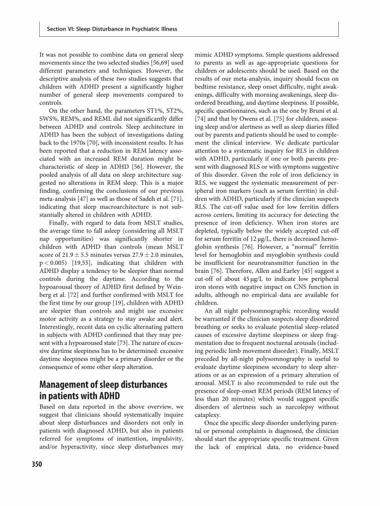
It was not possible to combine data on general sleepmovements since the two selected studies [56,69] useddifferent parameters and techniques. However, thedescriptive analysis of these two studies suggests thatchildren with ADHD present a significantly highernumber of general sleep movements compared tocontrols.
On the other hand, the parameters ST1%, ST2%,SWS%, REM%, and REML did not significantly differbetween ADHD and controls. Sleep architecture inADHD has been the subject of investigations datingback to the 1970s [70], with inconsistent results. It hasbeen reported that a reduction in REM latency asso-ciated with an increased REM duration might becharacteristic of sleep in ADHD [56]. However, thepooled analysis of all data on sleep architecture sug-gested no alterations in REM sleep. This is a majorfinding, confirming the conclusions of our previousmeta-analysis [47] as well as those of Sadeh et al. [71],indicating that sleep macroarchitecture is not sub-stantially altered in children with ADHD.
Finally, with regard to data from MSLT studies,the average time to fall asleep (considering all MSLTnap opportunities) was significantly shorter inchildren with ADHD than controls (mean MSLTscore of 21.9� 5.5 minutes versus 27.9� 2.0 minutes,p< 0.005) [19,55], indicating that children withADHD display a tendency to be sleepier than normalcontrols during the daytime. According to thehypoarousal theory of ADHD first defined by Wein-berg et al. [72] and further confirmed with MSLT forthe first time by our group [19], children with ADHDare sleepier than controls and might use excessivemotor activity as a strategy to stay awake and alert.Interestingly, recent data on cyclic alternating patternin subjects with ADHD confirmed that they may pre-sent with a hypoaroused state [73]. The nature of exces-sive daytime sleepiness has to be determined: excessivedaytime sleepiness might be a primary disorder or theconsequence of some other sleep alteration.
Management of sleep disturbancesin patients with ADHDBased on data reported in the above overview, wesuggest that clinicians should systematically inquireabout sleep disturbances and disorders not only inpatients with diagnosed ADHD, but also in patientsreferred for symptoms of inattention, impulsivity,and/or hyperactivity, since sleep disturbances may
mimic ADHD symptoms. Simple questions addressedto parents as well as age-appropriate questions forchildren or adolescents should be used. Based on theresults of our meta-analysis, inquiry should focus onbedtime resistance, sleep onset difficulty, night awak-enings, difficulty with morning awakenings, sleep dis-ordered breathing, and daytime sleepiness. If possible,specific questionnaires, such as the one by Bruni et al.[74] and that by Owens et al. [75] for children, assess-ing sleep and/or alertness as well as sleep diaries filledout by parents and patients should be used to comple-ment the clinical interview. We dedicate particularattention to a systematic inquiry for RLS in childrenwith ADHD, particularly if one or both parents pre-sent with diagnosed RLS or with symptoms suggestiveof this disorder. Given the role of iron deficiency inRLS, we suggest the systematic measurement of per-ipheral iron markers (such as serum ferritin) in chil-dren with ADHD, particularly if the clinician suspectsRLS. The cut-off value used for low ferritin differsacross centers, limiting its accuracy for detecting thepresence of iron deficiency. When iron stores aredepleted, typically below the widely accepted cut-offfor serum ferritin of 12 μg/L, there is decreased hemo-globin synthesis [76]. However, a “normal” ferritinlevel for hemoglobin and myoglobin synthesis couldbe insufficient for neurotransmitter function in thebrain [76]. Therefore, Allen and Earley [45] suggest acut-off of about 45 μg/L to indicate low peripheraliron stores with negative impact on CNS function inadults, although no empirical data are available forchildren.
An all night polysomnographic recording wouldbe warranted if the clinician suspects sleep disorderedbreathing or seeks to evaluate potential sleep-relatedcauses of excessive daytime sleepiness or sleep frag-mentation due to frequent nocturnal arousals (includ-ing periodic limb movement disorder). Finally, MSLTpreceded by all-night polysomnography is useful toevaluate daytime sleepiness secondary to sleep alter-ations or as an expression of a primary alteration ofarousal. MSLT is also recommended to rule out thepresence of sleep-onset REM periods (REM latency ofless than 20 minutes) which would suggest specificdisorders of alertness such as narcolepsy withoutcataplexy.
Once the specific sleep disorder underlying paren-tal or personal complaints is diagnosed, the clinicianshould start the appropriate specific treatment. Giventhe lack of empirical data, no evidence-based
Section VI: Sleep Disturbance in Psychiatric Illness
350

guidelines are available, particularly in children.Therefore, we discuss the treatment strategies for themost common sleep disorders underlying subjectivecomplaints on the basis of data provided by casereport, case series, and personal experience of ourgroup as well as that of our colleagues in the field.
Sleep disturbances associatedwith RLS/PLMSCase reports [77,78] suggest the efficacy of dopamin-ergic agents (levodopa/carbidopa, pergolide, andropinirole) in children diagnosed with both ADHDand RLS and previously treated with psychostimu-lants with limited efficacy or intolerable side-effects.However, to date, the limited number of patientstreated and the absence of double-blind, placebo-controlled, randomized trials do not allow evidence-based recommendations for treatment to be made. Nostudies on iron supplementation for RLS in childrenhave been published to date. On the other hand,Simakajornboon et al. [79] reported a decrease inperiodic limb movements in sleep in children treatedwith iron sulphate (3mg/kg of elemental iron per dayfor 3months). Non-pharmacological management ofRLS includes consistent and appropriate sleep schedule,limit-setting techniques, exercise program, and restric-tion of caffeinated beverages.
Excessive nocturnal motricityLate afternoon methylphenidate doses may reducenocturnal activity both during sleep and prior to sleeponset and improve sleep quality by consolidatingsleep [80,81]. However, while some studies haveshown that thrice daily methylphenidate does notimpact sleep [82] or causes only a slight decrease insleep duration [40], others have reported that a thirddose of methylphenidate does worsen sleep [41,83].Given these contrasting findings, at present, late after-noon stimulant treatment cannot be recommendedfor ADHD patients with high nocturnal activity.
Circadian rhythm disordersTo date, one open-label study [84] and two random-ized, double-blind, placebo-controlled studies [85,86]have assessed the efficacy and tolerability of melatoninfor the management of SOI, presumably due to circa-dian rhythm disorder, in children with ADHD, usingdoses of 3mg/day for abodyweight<40 kgand6mg/day
for a body weight>40 kg (Van der Heijden et al. [85]),and a unique dose of 5mg/day (Weiss et al. [87]),respectively. In the two randomized studies, in spiteof improvements in sleep onset after 4 weeks, melato-nin given 20 minutes before bedtime by Weiss andcolleagues [87] and at 7 p.m. by Van der Heijden andco-workers [85] demonstrated no effect on behavior,cognitive performance, or quality of life. Although thenumber of acute adverse events did not differ signifi-cantly between melatonin and placebo, since data onpossible long-term effects, such as on the gonadotropicsystem and onset of puberty are not available, furtherevidence is needed before recommending melatoninsystematically for SOI in children with ADHD.
Bearing in mind that a delayed evening increase inendogenous melatonin levels might explain SOI inchildren with ADHD, some authors investigated theefficacy of light therapy (LT). In an open trial, Rybaket al. [88] administered 3 weeks of morning bright LTto 29 adults with ADHD. They found a significantphase advance in circadian preference, as well as asignificant improvement in both subjective and object-ive measures of ADHD. To our knowledge, no con-trolled study has been conducted to assess the efficacyof LT in children with ADHD. Therefore, we think thatresearch in this field should be encouraged.
Sleep problems associated with sleepdisordered breathingGrowing empirical evidence shows a significantimprovement not only of breathing parameters, butalso of ADHD symptoms in children diagnosed withADHD plus SDB, after adenotonsillectomy [61,89].As previously stated, it is not possible to estimateaccurately the prevalence of children with ADHDwho have SDB and might benefit from adenotonsil-lectomy. However, these promising results shouldprompt further studies to assess the effectiveness ofsurgery for sleep disorders and for core ADHD symp-toms in patients with SDB and ADHD. They alsosuggest that appropriate recognition and surgicaltreatment of underlying SDB in children with ADHDmight prevent long-term stimulant treatment.
Sleep disturbances associatedwith ADHD medicationsA panel of ADHD experts [90] proposed several strat-egies to deal with sleep alterations caused by stimulants
Chapter 21: Sleep in ADHD
351
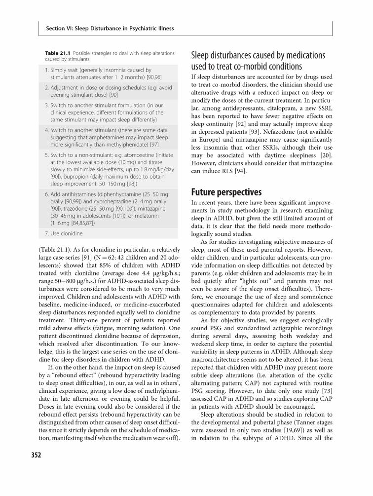
(Table 21.1). As for clonidine in particular, a relativelylarge case series [91] (N¼ 62; 42 children and 20 ado-lescents) showed that 85% of children with ADHDtreated with clonidine (average dose 4.4 μg/kg/h.s.;range 50�800 μg/h.s.) for ADHD-associated sleep dis-turbances were considered to be much to very muchimproved. Children and adolescents with ADHD withbaseline, medicine-induced, or medicine-exacerbatedsleep disturbances responded equally well to clonidinetreatment. Thirty-one percent of patients reportedmild adverse effects (fatigue, morning sedation). Onepatient discontinued clonidine because of depression,which resolved after discontinuation. To our know-ledge, this is the largest case series on the use of cloni-dine for sleep disorders in children with ADHD.
If, on the other hand, the impact on sleep is causedby a “rebound effect” (rebound hyperactivity leadingto sleep onset difficulties), in our, as well as in others’,clinical experience, giving a low dose of methylpheni-date in late afternoon or evening could be helpful.Doses in late evening could also be considered if therebound effect persists (rebound hyperactivity can bedistinguished from other causes of sleep onset difficul-ties since it strictly depends on the schedule of medica-tion,manifesting itself when themedication wears off).
Sleep disturbances caused by medicationsused to treat co-morbid conditionsIf sleep disturbances are accounted for by drugs usedto treat co-morbid disorders, the clinician should usealternative drugs with a reduced impact on sleep ormodify the doses of the current treatment. In particu-lar, among antidepressants, citalopram, a new SSRI,has been reported to have fewer negative effects onsleep continuity [92] and may actually improve sleepin depressed patients [93]. Nefazodone (not availablein Europe) and mirtazapine may cause significantlyless insomnia than other SSRIs, although their usemay be associated with daytime sleepiness [20].However, clinicians should consider that mirtazapinecan induce RLS [94].
Future perspectivesIn recent years, there have been significant improve-ments in study methodology in research examiningsleep in ADHD, but given the still limited amount ofdata, it is clear that the field needs more methodo-logically sound studies.
As for studies investigating subjective measures ofsleep, most of these used parental reports. However,older children, and in particular adolescents, can pro-vide information on sleep difficulties not detected byparents (e.g. older children and adolescents may lie inbed quietly after “lights out” and parents may noteven be aware of the sleep onset difficulties). There-fore, we encourage the use of sleep and somnolencequestionnaires adapted for children and adolescentsas complementary to data provided by parents.
As for objective studies, we suggest ecologicallysound PSG and standardized actigraphic recordingsduring several days, assessing both weekday andweekend sleep time, in order to capture the potentialvariability in sleep patterns in ADHD. Although sleepmacroarchitecture seems not to be altered, it has beenreported that children with ADHD may present moresubtle sleep alterations (i.e. alteration of the cyclicalternating pattern; CAP) not captured with routinePSG scoring. However, to date only one study [73]assessed CAP in ADHD and so studies exploring CAPin patients with ADHD should be encouraged.
Sleep alterations should be studied in relation tothe developmental and pubertal phase (Tanner stageswere assessed in only two studies [19,69]) as well asin relation to the subtype of ADHD. Since all the
Table 21.1 Possible strategies to deal with sleep alterationscaused by stimulants
1. Simply wait (generally insomnia caused bystimulants attenuates after 1 2 months) [90,96]
2. Adjustment in dose or dosing schedules (e.g. avoidevening stimulant dose) [90]
3. Switch to another stimulant formulation (in ourclinical experience, different formulations of thesame stimulant may impact sleep differently)
4. Switch to another stimulant (there are some datasuggesting that amphetamines may impact sleepmore significantly than methylphenidate) [97]
5. Switch to a non-stimulant: e.g. atomoxetine (initiateat the lowest available dose (10mg) and titrateslowly to minimize side-effects, up to 1.8mg/kg/day[90]), bupropion (daily maximum dose to obtainsleep improvement: 50 150mg [98])
6. Add antihistamines (diphenhydramine (25 50 mgorally [90,99]) and cyproheptadine (2 4mg orally[90]), trazodone (25 50mg [90,100]), mirtazapine(30 45mg in adolescents [101]), or melatonin(1 6mg [84,85,87])
7. Use clonidine
Section VI: Sleep Disturbance in Psychiatric Illness
352

available studies in childhood included school-agedsubjects, it would be of interest to assess possibledifferences in sleep patterns in preschool childrenwith ADHD versus healthy controls, and to betterunderstand the potential relationship between sleepalterations and symptoms of ADHD in very youngchildren.
With regard to specific issues, we encourage fur-ther studies using similar methodology to assess gen-eral movement and PLMS in ADHD children, bymeans of PSG or actigraphy with ankle or feet place-ment. This may provide insights into sleep alterationsin ADHD, given that the sleep fragmentation inducedby sleep movements may lead to sleep disruption withpossible daytime consequences. Moreover, we suggestthe systematic measure of iron markers and to studytheir potential correlations with abnormal sleepmovements and, possibly, other sleep parameters. Asfor sleep disordered breathing, we suggest that BMIshould systematically be reported in future studiesassessing SDB in children with ADHD in order toassess the potential effect of obesity on SDB. Westrongly encourage replication studies using MSLT,both in children and adults. Our group has suggestedthat excessive daytime sleepiness may be a paramountand primary alteration in at least a subsample ofADHD children. We have also speculated [95] thatalterations of the hypocretin/orexin system, which isinvolved in alertness and other major functions(which may also be altered in ADHD) such as foodseeking, weight control, and motor activity regula-tion, may contribute to excessive daytime sleepinessin ADHD children. Future studies should address thisissue promptly due to the potential therapeutic impli-cations (use of specific wake-promoting agents forADHD patients with excessive daytime sleepiness).Finally, and perhaps most importantly, we stronglyencourage large, multi-site and randomized treatmentstudies, including pharmacological and psychothera-peutic trials, for sleep disorders in patients withADHD, especially in pediatric patients, given the pau-city of empirical-based data.
This body of future research will advance ourknowledge of subjective and objective sleep alter-ations in patients with ADHD and on the mechan-isms by which objective alterations contribute tosubjective complaints, as well as on the most effectiveand safe treatment strategies. This will form the the-oretical basis for a better assessment and managementof sleep disturbances in patients with ADHD, thus
allowing a better quality of life for these individualsand their families.
References1. American Psychiatric Association. Diagnostic and
statistical manual of mental disorders, 4th ed.Washington, DC: American Psychiatric Association;1994.
2. American Psychiatric Association. Diagnostic andstatistical manual of mental disorders, 4th ed. TextRevision. Washington, DC: American PsychiatricAssociation; 2000.
3. Faraone SV, Sergeant J, Gillberg C, Biederman J. Theworldwide prevalence of ADHD: is it an Americancondition? World Psychiatry. 2003;2:104 113.
4. WHO. The ICD 10 classification of mental andbehavioral disorders: Clinical descriptions anddiagnostic guidelines 1992; diagnostic criteria forresearch 1993. Geneva: WHO; 1992.
5. Taylor E, Sandberg S, Thorley G, Giles S. Theepidemiology of childhood hyperactivity. London:Maudsley Monographs; 1991.
6. Biederman J, Faraone S. Attention deficit/hyperactivity disorder. Lancet. 2005;366:237 248.
7. Kessler RC, Adler LA, Barkley R, et al. Patterns andpredictors of attention deficit/hyperactivity disorderpersistence into adulthood: results from the nationalcomorbidity survey replication. Biol Psychiatry.2005;57:1442 1451.
8. Pliszka S. Practice parameter for the assessment andtreatment of children and adolescents with attentiondeficit/hyperactivity disorder. J Am Acad Child AdolescPsychiatry. 2007;46:894 921.
9. Banaschewski T, Coghill D, Santosh P, et al. Longacting medications for the hyperkinetic disorders:A systematic review and European treatment guideline.Eur Child Adolesc Psychiatry. 2006;15:476 495.
10. Taylor E, Dopfner M, Sergeant J, et al. Europeanclinical guidelines for hyperkinetic disorder: firstupgrade. Eur Child Adolesc Psychiatry. 2004;13(Suppl 1):17 30.
11. Barkley RA. Attention deficit hyperactivity disorder: aclinical handbook, 3rd ed. New York: Guilford; 2006.
12. Faraone SV, Biederman J, Jetton JG, Tsuang MT.Attention deficit disorder and conduct disorder:longitudinal evidence for a familial subtype. PsycholMed. 1997;27:291 300.
13. Biederman J, Wilens T, Mick E, et al. Is ADHD arisk factor for psychoactive substance use disorders?Findings from a four year prospective follow upstudy. J Am Acad Child Adolesc Psychiatry. 1997;36:21 29.
Chapter 21: Sleep in ADHD
353
14. Milberger S, Biederman J, Faraone SV, Chen L, Jones J.ADHD is associated with early initiation of cigarettesmoking in children and adolescents. J Am Acad ChildAdolesc Psychiatry. 1997;36:37 44.
15. Spencer T, Biederman J, Coffey B, et al. Tourettedisorder and ADHD. Adv Neurol. 2001;85:57 77.
16. American Psychiatric Association. Diagnostic andstatistical manual of mental disorders, 3rd ed.Washington, DC: American Psychiatric Association;1980.
17. American Psychiatric Association. Diagnostic andstatistical manual of mental disorders, 3rd ed. Revised.Washington, DC: American Psychiatric Association;1987.
18. Sherman DK, Iacono WG, McGue MK. Attentiondeficit hyperactivity disorder dimensions: a twinstudy of inattention and impulsivity hyperactivity.J Am Acad Child Adolesc Psychiatry. 1997;36:745 753.
19. Lecendreux M, Konofal E, Bouvard M, Falissard B,Mouren Simeoni MC. Sleep and alertness in childrenwith ADHD. J Child Psychol Psychiatry. 2000;41:803 812.
20. Mindell JA, Owens JA. Diagnosis and management ofsleep problems. Philadelphia: Lippincott Williams &Wilkins; 2003.
21. Corkum P, Tannock R, Moldofsky H. Sleepdisturbances in children with attention deficit/hyperactivity disorder. J Am Acad Child AdolescPsychiatry. 1998;37:637 646.
22. Laufer MW, Denhoff E. Hyperkinetic behaviorsyndrome in children. J Pediatr. 1957;50:463 474.
23. Philipsen A, Hornyak M, Riemann D. Sleep and sleepdisorders in adults with attention deficit/hyperactivitydisorder. Sleep Med Rev. 2006;10:399 405.
24. Sobanski E, Schredl M, Kettler N, Alm B. Sleep inadults with attention deficit hyperactivity disorder(ADHD) before and during treatment withmethylphenidate: a controlled polysomnographicstudy. Sleep. 2008;31:375 381.
25. Boonstra AM, Kooij JJ, Oosterlaan J, et al. Hyperactivenight and day? Actigraphy studies in adult ADHD: abaseline comparison and the effect of methylphenidate.Sleep. 2007;30:433 442.
26. Gau SS, Kessler RC, Tseng WL, et al. Associationbetween sleep problems and symptoms of attentiondeficit/hyperactivity disorder in young adults. Sleep.2007;30:195 201.
27. Schredl M, Alm B, Sobanski E. Sleep quality in adultpatients with attention deficit hyperactivity disorder(ADHD). Eur Arch Psychiatry Clin Neurosci.2007;257:164 168.
28. Ryan ND, Puig Antich J, Ambrosini P, et al. Theclinical picture of major depression in children andadolescents. Arch Gen Psychiatry. 1987;44:854 861.
29. Ivanenko A, Crabtree VM, Gozal D. Sleep anddepression in children and adolescents. Sleep Med Rev.2005;9:115 129.
30. Bertocci MA, Dahl RE, Williamson DE, et al.Subjective sleep complaints in pediatric depression: acontrolled study and comparison with EEG measuresof sleep and waking. J Am Acad Child AdolescPsychiatry. 2005;44:1158 1166.
31. Alfano CA, Ginsburg GS, Kingery JN. Sleep relatedproblems among children and adolescents with anxietydisorders. J Am Acad Child Adolesc Psychiatry.2007;46:224 232.
32. Mehl RC, O’Brien LM, Jones JH, et al. Correlates of sleepand pediatric bipolar disorder. Sleep. 2006;29:193 197.
33. Kirov R, Banaschewski T, Uebel H, Kinkelbur J,Rothenberger A. REM sleep alterations in childrenwith co existence of tic disorders and attention deficit/hyperactivity disorder: impact of hypermotorsymptoms. Eur Child Adolesc Psychiatry. 2007;16(Suppl 1):45 50.
34. Kirov R, Kinkelbur J, Banaschewski T, Rothenberger A.Sleep patterns in children with attention deficit/hyperactivity disorder, tic disorder, and comorbidity.J Child Psychol Psychiatry. 2007;48:561 570.
35. Kostanecka Endress T, Banaschewski T, Kinkelbur J,et al. Disturbed sleep in children with Tourettesyndrome: a polysomnographic study. J PsychosomRes. 2003;55:23 29.
36. Ivanenko A, Crabtree VM, Gozal D. Sleep in childrenwith psychiatric disorders. Pediatr Clin North Am.2004;51:51 68.
37. Brown TE, McMullen WJ Jr. Attention deficitdisorders and sleep/arousal disturbances. Ann N YAcad Sci. 2001;931:271 286.
38. Sangal RB, Owens J, Allen AJ, et al. Effects ofatomoxetine and methylphenidate on sleep in childrenwith ADHD. Sleep. 2006;29:1573 1585.
39. Carlson GA, Kelly KL. Stimulant rebound: howcommon is it and what does it mean? J Child AdolescPsychopharmacol. 2003;13:137 142.
40. Ahmann PA, Waltonen SJ, Olson KA, et al. Placebocontrolled evaluation of Ritalin side effects. Pediatrics.1993;91:1101 1106.
41. Stein MA, Blondis TA, Schnitzler ER, et al.Methylphenidate dosing: twice daily versus three timesdaily. Pediatrics. 1996;98:748 756.
42. Jerome L. Can methylphenidate facilitate sleep inchildren with attention deficit hyperactivity disorder?J Child Adolesc Psychopharmacol. 2001;11:109.
Section VI: Sleep Disturbance in Psychiatric Illness
354

43. Kinsbourne M. Stimulants for insomnia. N Engl J Med.1973;288:1129.
44. Allen RP, Picchietti D, Hening WA, et al. Restless legssyndrome: diagnostic criteria, special considerations,and epidemiology: A report from the restless legssyndrome diagnosis and epidemiology workshop at theNational Institutes of Health. Sleep Med. 2003;4:101 119.
45. Allen RP, Earley CJ. The role of iron in restless legssyndrome. Mov Disord. 2007;22(Suppl 18):S440 S448.
46. Picchietti D. Is iron deficiency an underlying cause ofpediatric restless legs syndrome and of attentiondeficit/hyperactivity disorder? Sleep Med. 2007;8(7 8):693 694.
47. Cortese S, Konofal E, Lecendreux M, et al. Restless legssyndrome and attention deficit/hyperactivity disorder:a review of the literature. Sleep. 2005;28:1007 1013.
48. Wagner ML, Walters AS, Fisher BC. Symptoms ofattention deficit/hyperactivity disorder in adults withrestless legs syndrome. Sleep. 2004;27:1499 1504.
49. Cortese S, Lecendreux M, Mouren MC, Konofal E.ADHD and insomnia. J Am Acad Child AdolescPsychiatry. 2006;45:384 385.
50. Chervin RD, Archbold KH, Dillon JE, et al.Associations between symptoms of inattention,hyperactivity, restless legs, and periodic legmovements. Sleep. 2002;25:213 218.
51. American Academy of Sleep Medicine. Theinternational classification of sleep disorders: diagnosticand coding manual, 2nd ed. Westchester, IL: AmericanAcademy of Sleep Medecine; 2005.
52. Cooper J, Tyler L, Wallace I, Burgess KR. No evidenceof sleep apnea in children with attention deficithyperactivity disorder. Clin Pediatr (Phila).2004;43:609 614.
53. Gruber R, Tong X, Frenette S, et al. Sleep disturbancesin pre pubertal children with attention deficithyperactivity disorder: a home polysomnographystudy. Sleep. 2008;32(3):343 350.
54. Huang YS, Chen NH, Li HY, et al. Sleep disorders inTaiwanese children with attention deficit/hyperactivitydisorder. J Sleep Res. 2004;13:269 277.
55. Golan N, Shahar E, Ravid S, Pillar G. Sleep disordersand daytime sleepiness in children with attentiondeficit/hyperactive disorder. Sleep. 2004;27:261 266.
56. Kirov R, Kinkelbur J, Heipke S, et al. Is there a specificpolysomnographic sleep pattern in children withattention deficit/hyperactivity disorder? J Sleep Res.2004;13:87 93.
57. Picchietti DL, England SJ, Walters AS, Willis K,Verrico T. Periodic limb movement disorderand restless legs syndrome in children with
attention deficit hyperactivity disorder. J Child Neurol.1998;13:588 594.
58. Guilleminault C, Pelayo R. Sleep disordered breathingin children. Ann Med. 1998;30:350 356.
59. Chervin RD. How many children with ADHD havesleep apnea or periodic leg movements onpolysomnography? Sleep. 2005;28:1041 1042.
60. Sangal RB, Owens JA, Sangal J. Patients with attentiondeficit/hyperactivity disorder without observed apneicepisodes in sleep or daytime sleepiness have normalsleep on polysomnography. Sleep. 2005;28:1143 1148.
61. Dillon JE, Blunden S, Ruzicka DL et al. DSM IVdiagnoses and obstructive sleep apnea in childrenbefore and 1 year after adenotonsillectomy. J Am AcadChild Adolesc Psychiatry. 2007;46:1425 1436.
62. Van der Heijden KB, Smits MG, Van Someren EJ,Gunning WB. Idiopathic chronic sleep onset insomniain attention deficit/hyperactivity disorder: a circadianrhythm sleep disorder. Chronobiol Int. 2005;22:559 570.
63. Cohen Zion M, Ancoli Israel S. Sleep in children withattention deficit hyperactivity disorder (ADHD): areview of naturalistic and stimulant interventionstudies. Sleep Med Rev. 2004;8:379 402.
64. Cortese S, Konofal E, Yateman N, Mouren MC,Lecendreux M. Sleep and alertness in children withattention deficit/hyperactivity disorder: a systematicreview of the literature. Sleep. 2006;29:504 511.
65. Cortese S, Faraone S, Konofal E, Lecendreux M. Sleepin children with attention deficit/hyperactivitydisorder: meta analysis of subjective and objectivestudies. J Am Acad Child Adolesc Psychiatry. 2009;48(9):894 908.
66. Owens JA. Sleep disorders and attention deficit/hyperactivity disorder. Curr Psychiatry Rep.2008;10:439 444.
67. Sadeh A, Hauri PJ, Kripke DF, Lavie P. The role ofactigraphy in the evaluation of sleep disorders. Sleep.1995;18:288 302.
68. Gruber R, Sadeh A, Raviv A. Instability of sleeppatterns in children with attention deficit/hyperactivity disorder. J Am Acad Child AdolescPsychiatry. 2000;39:495 501.
69. Konofal E, Lecendreux M, Bouvard MP, MourenSimeoni MC. High levels of nocturnal activity inchildren with attention deficit hyperactivity disorder:a video analysis. Psychiatry Clin Neurosci. 2001;55:97 103.
70. O’Brien LM, Gozal D. Sleep in children with attentiondeficit/hyperactivity disorder. Minerva Pediatr.2004;56:585 601.
71. Sadeh A, Pergamin L, Bar Haim Y. Sleep inchildren with attention deficit hyperactivity disorder:
Chapter 21: Sleep in ADHD
355
a meta analysis of polysomnographic studies. SleepMed Rev. 2006;10:381 398.
72. Weinberg WA, Brumback RA. Primary disorder ofvigilance: a novel explanation of inattentiveness,daydreaming, boredom, restlessness, and sleepiness.J Pediatr. 1990;116:720 725.
73. Miano S, Donfrancesco R, Bruni O, et al. NREM sleepinstability is reduced in children with attention deficit/hyperactivity disorder. Sleep. 2006;29:797 803.
74. Bruni O, Ottaviano S, Guidetti V, et al. The SleepDisturbance Scale for Children (SDSC): Constructionand validation of an instrument to evaluate sleepdisturbances in childhood and adolescence. J Sleep Res.1996;5:251 261.
75. Owens JA, Maxim R, Nobile C, McGuinn M, Msall M.Parental and self report of sleep in children withattention deficit/hyperactivity disorder. Arch PediatrAdolesc Med. 2000;154:549 555.
76. Picchietti D. Is iron deficiency an underlying causeof pediatric restless legs syndrome and of attentiondeficit/hyperactivity disorder? Sleep Med. 2007;8:693 694.
77. Konofal E, Arnulf I, Lecendreux M, Mouren MC.Ropinirole in a child with attention deficithyperactivity disorder and restless legs syndrome.Pediatr Neurol. 2005;32:350 351.
78. Walters AS, Mandelbaum DE, Lewin DS, et al.Dopaminergic therapy in children with restless legs/periodic limb movements in sleep and ADHD.Dopaminergic Therapy Study Group. Pediatr Neurol.2000;22:182 186.
79. Simakajornboon N, Gozal D, Vlasic V, et al. Periodiclimb movements in sleep and iron status in children.Sleep. 2003;26:735 738.
80. Konofal E, Lecendreux M, Bouvard M, Mouren MC.Effects of vesperal methylphenidate (MPH)administration on diurnal and nocturnal activity inADHD children: an actigraphic study. Sleep. 2001;Abstract Supplement 24:G363.
81. Kooij JJ, Middelkoop HA, van GK, Buitelaar JK. Theeffect of stimulants on nocturnal motor activity andsleep quality in adults with ADHD: an open label casecontrol study. J Clin Psychiatry. 2001;62:952 956.
82. Kent JD, Blader JC, Koplewicz HS, Abikoff H,Foley CA. Effects of late afternoon methylphenidateadministration on behavior and sleep in attentiondeficit hyperactivity disorder. Pediatrics. 1995;96:320 325.
83. Pelham WE, Gnagy EM, Chronis AM, et al.A comparison of morning only and morning/lateafternoon Adderall to morning only, twice daily, andthree times daily methylphenidate in children with
attention deficit/hyperactivity disorder. Pediatrics.1999;104:1300 1311.
84. Tjon Pian Gi CV, Broeren JP, Starreveld JS,Versteegh FG. Melatonin for treatment of sleepingdisorders in children with attention deficit/hyperactivity disorder: a preliminary open label study.Eur J Pediatr. 2003;162:554 555.
85. Van der Heijden KB, Smits MG, Van Someren EJ,Ridderinkhof KR, Gunning WB. Effect of melatoninon sleep, behavior, and cognition in ADHD andchronic sleep onset insomnia. J Am Acad Child AdolescPsychiatry. 2007;46:233 241.
86. Weiss M, Tannock R, Kratochvil C, et al.A randomized, placebo controlled study of once dailyatomoxetine in the school setting in children withADHD. J Am Acad Child Adolesc Psychiatry.2005;44:647 655.
87. Weiss MD, Wasdell MB, Bomben MM, Rea KJ,Freeman RD. Sleep hygiene and melatonin treatmentfor children and adolescents with ADHD and initialinsomnia. J Am Acad Child Adolesc Psychiatry.2006;45:512 519.
88. Rybak YE, McNeely HE, Mackenzie BE, Jain UR,Levitan RD. An open trial of light therapy in adultattention deficit/hyperactivity disorder. J ClinPsychiatry. 2006;67:1527 1535.
89. Huang YS, Guilleminault C, Li HY, et al. Attentiondeficit/hyperactivity disorder with obstructive sleepapnea: a treatment outcome study. Sleep Med.2007;8:18 30.
90. Kratochvil CJ, Lake M, Pliszka SR, Walkup JT.Pharmacological management of treatment inducedinsomnia in ADHD. J Am Acad Child AdolescPsychiatry. 2005;44:499 501.
91. Prince JB, Wilens TE, Biederman J, Spencer TJ,Wozniak JR. Clonidine for sleep disturbancesassociated with attention deficit hyperactivity disorder:a systematic chart review of 62 cases. J Am Acad ChildAdolesc Psychiatry. 1996;35:599 605.
92. van Bemmel AL, van den Hoofdakker RH,Beersma DG, Bouhuys AL. Changes in sleeppolygraphic variables and clinical state in depressedpatients during treatment with citalopram.Psychopharmacology (Berl). 1993;113:225 230.
93. Lader M, Andersen HF, Baekdal T. The effect ofescitalopram on sleep problems in depressed patients.Hum Psychopharmacol. 2005;20:349 354.
94. Kim SW, Shin IS, Kim JM, et al. Factors potentiatingthe risk of mirtazapine associated restless legssyndrome. Hum Psychopharmacol. 2008;23:615 620.
95. Cortese S, Konofal E, Lecendreux M. Alertness andfeeding behaviors in ADHD: does the hypocretin/
Section VI: Sleep Disturbance in Psychiatric Illness
356
orexin system play a role? Med Hypotheses.2008;71:770 775.
96. Golinko BE. Side effects of dextroamphetamine andmethylphenidate in hyperactive children a brief review.Prog Neuropsychopharmacol Biol Psychiatry. 1984;8:1 8.
97. Efron D, Jarman F, Barker M. Side effects ofmethylphenidate and dexamphetamine in childrenwith attention deficit hyperactivity disorder: a doubleblind, crossover trial. Pediatrics. 1997;100:662 666.
98. Simeon JG, Ferguson HB, Van Wyck FJ. Bupropioneffects in attention deficit and conduct disorders. CanJ Psychiatry. 1986;31:581 585.
99. Merenstein D, Diener West M, Halbower AC, Krist A,Rubin HR. The trial of infant response to
diphenhydramine: the TIRED study a randomized,controlled, patient oriented trial. Arch Pediatr AdolescMed. 2006;160:707 712.
100. Kallepalli BR, Bhatara VS, Fogas BS, Tervo RC,Misra LK. Trazodone is only slightly faster thanfluoxetine in relieving insomnia in adolescents withdepressive disorders. J Child Adolesc Psychopharmacol.1997;7:97 107.
101. Haapasalo Pesu KM, Vuola T, Lahelma L,Marttunen M. Mirtazapine in the treatmentof adolescents with major depression: anopen label, multicenter pilot study. J ChildAdolesc Psychopharmacol. 2004;14:175 184.
Chapter 21: Sleep in ADHD
357
Chapter
22 Sleep in pediatric moodand anxiety disordersValerie McLaughlin Crabtree and Anna Ivanenko
IntroductionSleep complaints such as difficulty initiating andmaintaining sleep, restless sleep, daytime sleepiness,and bedtime anxieties are highly prevalent amongchildren with psychiatric disorders. In particular, chil-dren with mood and anxiety disorders have signifi-cantly more night-time awakenings than childrenwith other psychiatric disorders [1]. Recent attentionhas been paid to the relationship between mood dis-orders and sleep, particularly in adults with majordepressive disorder (MDD). An extension of thiswork has been made investigating sleep in childrenand adolescents with depression and anxiety. Some ofthe work has involved assessing subjective complaintsin children and adolescents with mood and anxietydisorders, while others have studied objective sleepdisruption in this population. Objective studies haveglobally assessed sleep architecture differencesbetween children with mood and anxiety disordersand healthy peers, while others have looked at themicroarchitecture of sleep in children and adolescentswith depression.
Dahl [2] has posited a bidirectional theory of therelationship between emotional and behavioral diffi-culties and sleep in children. Support for his theorycomes from five primary bodies of evidence. First,pediatric patients with emotional disorders haveshown increased rates of sleep disturbance. Corres-pondingly, children with sleep disturbance have asignificantly higher prevalence of psychiatric prob-lems. Third, even in the short term a stressful eventmay induce sleep disruption in children, while insuffi-cient sleep may directly lead to disruptive behaviorduring the day. Fourth, significant overlap in symp-toms is present between those with sleep disordersand those with psychiatric disorders, particularly in
relation to depressive symptomatology. Finally, thesimilar neurobehavioral systems related to both sleepand mood regulation provide evidence of this bidirec-tional relationship between these regulatory systems.
While the majority of focus on sleep disruption inchildren with mood disorders has been on depression,some investigations have examined the sleep of chil-dren and adolescents with bipolar disorder and anx-iety disorders. In the following sections, we willreview the nature of sleep disturbance in pediatricmood and anxiety disorders, focusing on both sub-jective and objective data, and when available, discusstreatment options, with the caveat that the vast major-ity of treatments are based on clinical experiencerather than evidence-based medicine.
Sleep and pediatric depressionSubjective sleep complaints in depressedpediatric patientsSubjective sleep disturbance is extremely common inpediatric depression. Early studies investigating sub-jective complaints of pediatric patients with depres-sion revealed that approximately two-thirds ofchildren with MDD reported difficulty initiating andmaintaining sleep, and half reported early morningawakenings [3]. Ivanenko and colleagues comparedparental report of children’s behavior with sleep com-plaints and found that those with delayed sleep onsetand shorter sleep duration tended to experience morenegative mood during the day. Difficulty maintainingsleep was also associated with anxiety, depressive, andsomatic symptoms. Parental report of excessive day-time sleepiness was significantly more likely to beassociated with depressive and somatic symptomsthan with hyperactivity [1].
Foundations of Psychiatric Sleep Medicine, ed. JohnW.Winkelman and David T. Plante. Published by Cambridge University Press.# Cambridge University Press 2010.
358
In an assessment of sleep complaints in 7- to14-year-old children diagnosed with depression, Liuand colleagues [4] closely examined both insomniaand subjective hypersomnia (report of sleeping morethan usual, napping in the afternoon/after dinner, orhave trouble waking up) in this population. Whileslightly over half of the sample had experiencedinsomnia alone in the past month, 9% reportedhypersomnia alone, and 10% reported experiencingboth hypersomnia and insomnia over the previousmonth. Children with any sleep disturbance exhibitedmore significant depression than those without sleepdisturbance. Children who exhibited both hypersom-nia and insomnia were significantly more likely toexperience a recurrent episode of depression, to havehad a longer course of depression, and to have themost severe depressive symptomatology as comparedto children with either insomnia or hypersomniaalone. Additionally, children with any sleep disturb-ance were significantly more likely to have co-morbidanxiety disorders but were significantly less likely tohave oppositional defiant disorders than depressedchildren without sleep disturbance [4].
In a comparison of depressive symptoms betweenprepubertal children (mean age ¼ 9 years) and ado-lescents (mean age ¼ 14 years) with MDD, symptomsof insomnia were equally prevalent across age groups,with nearly three-quarters of the sample reportingsignificant insomnia. Subjective hypersomnia, on theother hand, was significantly more prevalent in ado-lescents than in prepubertal children (34% vs. 16%).When the age groups were combined, hypersomniaand fatigue were more prevalent in those withendogenous depression subtype, while insomnia wasrelated more to anxious depression [5].
A recent longitudinal clinical assessment of neuro-behavioral factors in pediatric affective disorder dem-onstrated that adolescents with MDD reportedsignificantly longer time to initiate sleep, more fre-quent nocturnal awakenings, more difficulty withmorning awakening, and poorer subjective sleep qual-ity than adolescents without depression. Those withco-morbid anxiety disorders exhibited more noctur-nal awakenings, increased difficulty with morningawakening, and poorer subjective sleep quality thanthose without co-morbid depression [6].
Genetic influences may be important contributorsto the relationship of sleep disturbance and depressivesymptoms. In a study of 8-year-old twins, those withself-reported depressive symptoms were significantly
more likely to have their parents report bedtimeproblems, such as resisting bedtime and delayingsleep onset [7]. Monozygotic twins were significantlymore likely to have concordance with both depressivesymptoms and sleep problems and a higher bivariatecorrelation, indicating sleep problems in one twin anddepressive symptoms in the other, than dizygotictwins. Thus, the association between sleep problemsand depression was due, at least to some extent, togenetic factors, and there was substantial overlapbetween the genes influencing sleep problems andthose influencing depression. The authors also inves-tigated the relationship between child self-reports ofanxiety and sleep disturbance and noted that thisrelationship was far less robust than that betweendepression and sleep problems [7].
There are few studies that examine the effects oftreatment of pediatric depression on co-occurringsleep complaints. Across an 8-week course of treat-ment that included selective serotonin reuptakeinhibitors and/or cognitive behavioral therapy, ado-lescents with MDD exhibited a significant reductionin caffeine use but did not have improvements intheir sleep [6]. One year after initial assessment ofsubjective sleep complaints, Puig-Antich and col-leagues [8] reassessed a subset of children whosedepression had remitted, and only 10% continued toexperience symptoms of insomnia after their remis-sion. However, the children in this study tended tomaintain a similar rise time while having a signifi-cantly later bedtime and sleep onset time. As a result,the children obtained approximately 30 minutes lesssleep each night following their recovery than whileexperiencing their depressive episode, though theysubjectively reported improved sleep [8].
Predictive value of sleep complaintsand future depressionSeveral longitudinal studies probe the relationshipbetween sleep disturbance and depression. Early sleepcomplaints have been posited to be related to laterdevelopment of depression and anxiety; while, con-versely, there is some evidence that early depressivesymptoms may predict persistent sleep difficulties. Ina longitudinal study investigating sleep complaintsand depressive symptoms in adolescents, 75% of thosewho initially reported some symptoms of depression(but did not meet diagnostic criteria for MDD) inaddition to sleep disturbances eventually met criteria
Chapter 22: Sleep in pediatric mood and anxiety disorders
359
for MDD over the course of 12 months [9]. To morespecifically assess the role of sleep disturbance inanxiety and depression in children, Johnson and col-leagues [10], using data from a large study examiningthe psychiatric sequelae of low birth weight, correl-ated sleep disturbance, and anxious/depressive symp-toms in children at the age of 6, as well as thepredictive value of sleep disturbance in anxious/depressive symptoms at the age of 11. At both ages6 and 11, maternal report of difficulty sleeping wassignificantly related to anxious/depressive symptomsin the children, though the relationship was some-what stronger at age 11. Trouble sleeping at age 6 wasnot predictive of anxious/depressive symptoms at age11 in children who were not anxious/depressed atage 6. Unfortunately, this study is limited by the factthat only one question of difficulty sleeping was usedin the analysis.
In a very long-term longitudinal study of childrenwho were 6–23 years of age (mean age ¼ 16 years) atbaseline data collection, Ong and colleagues [11]found that those whose parents reported that theyhad poor sleep rhythmicity as young children, definedas having non-regular sleep habits prior to age 6, weresignificantly more likely to have adolescent-onsetMDD or anxiety disorders, such as generalized anx-iety disorder, obsessive-compulsive disorder, post-traumatic stress disorder (PTSD), panic disorder,agoraphobia, simple phobia, or social phobia wheninterviewed with the Schedule for Affective Disordersand Schizophrenia-Lifetime Version (SADS-L) or theKiddie-SADS, depending upon the age of the partici-pant. On the other hand, no early childhood tempera-ment domain was predictive of adult-onset mooddisturbance.
In a large (>2000 subjects) longitudinal study inHolland, 14 years after being rated as sleeping lessthan others, being overtired, and having difficultysleeping as children (mean age ¼ 9), young adultswere significantly more likely to report symptoms ofanxiety and depression than their peers who had notevidenced such sleep difficulties in childhood. Paren-tal reports of sleeping less than other children alsopredicted aggressive behavior in young adulthood andremained the most robust predictor of future emo-tional difficulties [12]. Similarly, using computer-assisted personal interviews and self-administeredquestionnaires from managed care enrollmentrosters, adolescents with self-reports of insomnia weresignificantly more likely to have symptoms of
depression 1 year later, even when controlling forinsomnia at the second time point [13]. Also, in alongitudinal twin study, sleep problems in 8-year-oldchildren were predictive of depressive symptoms atage 10. Conversely, depressive symptoms at age 8were not predictive of sleep problems at age 10. Thus,the authors suggest early treatment of sleep disturb-ance may provide a protection against later develop-ment of depressive symptoms [14].
There is some evidence that depressive symptomsin adolescents may be predictive of persistent sleepproblems. In a very large cohort of adolescents, thosewith depressive symptoms at both the initial interviewand 4 years later had the highest rate of sleep prob-lems, while those with no depressive symptoms ateither time point had the lowest rates. Those whohad depressive symptoms at baseline that were nolonger present at the 4-year follow-up also had higherrates of sleep problems at the follow-up time pointthan those who had not experienced depressive symp-toms at baseline [15].
Sleep architecture in pediatric depressionWhile adults with depression tend to show consistentdifferences in their sleep architecture from healthyadults, including reduced slow-wave sleep (SWS),shortened REM latency, and increased sleep fragmen-tation [16], depressed children and adolescents tendto show very discrepant findings with respect to sleeparchitecture. In this section, we will provide a pertin-ent overview of sleep architecture in pediatric depres-sion. For a detailed review of polysomnographicfindings in this population, the reader is directed tothe review by Ivanenko and colleagues [17].
Several studies have failed to show any significantdifferences in sleep architecture between childrenwith depression and their healthy peers [3,18–20].However, when subgroups in one study were ana-lyzed, prepubertal children with more severe depres-sion had reduced REM latency, a higher percentage ofREM sleep, and less stage 4 sleep, similar to findingsin depressed adults [19]. To explain these findings,Dahl and colleagues have hypothesized that sleep inchildren and adolescents with depression may bemore “protected” than the sleep of depressed adultsdue to their greater likelihood of having more SWSand greater sleep efficiency than adults, and thuschanges in sleep architecture are seen only in moresevere cases of pediatric depression [19].
Section VI: Sleep Disturbance in Psychiatric Illness
360
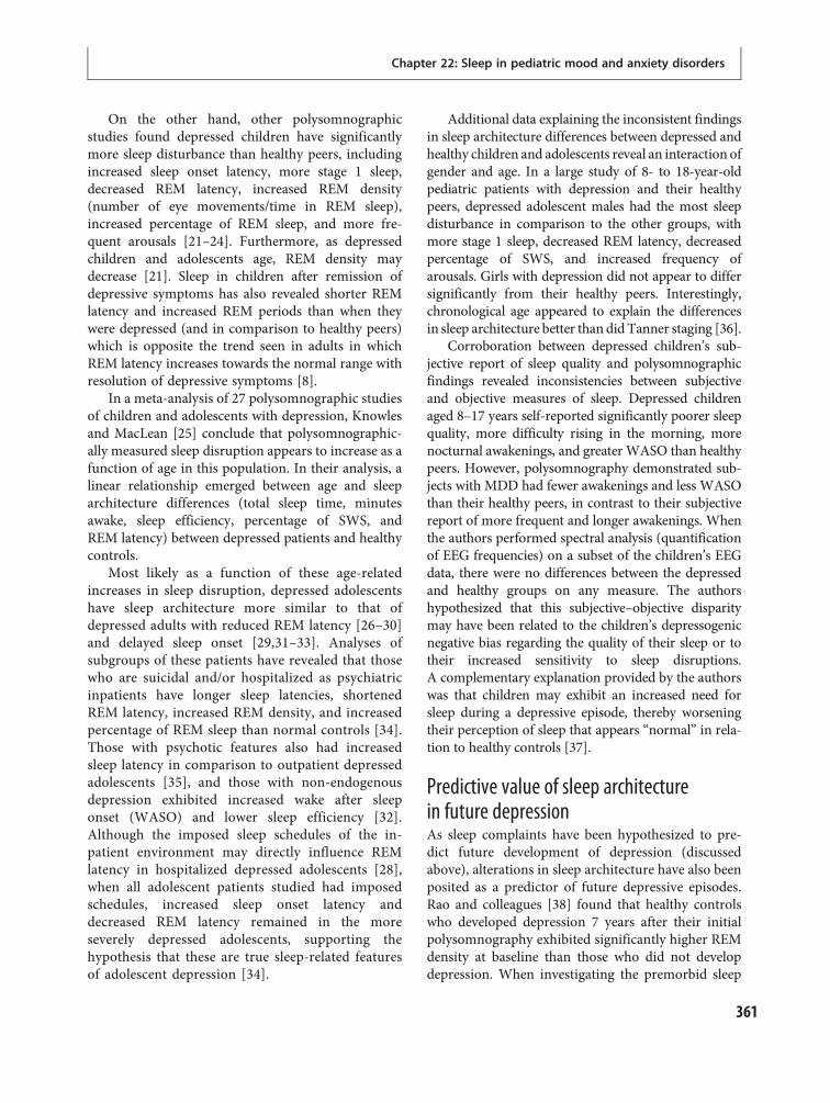
On the other hand, other polysomnographicstudies found depressed children have significantlymore sleep disturbance than healthy peers, includingincreased sleep onset latency, more stage 1 sleep,decreased REM latency, increased REM density(number of eye movements/time in REM sleep),increased percentage of REM sleep, and more fre-quent arousals [21–24]. Furthermore, as depressedchildren and adolescents age, REM density maydecrease [21]. Sleep in children after remission ofdepressive symptoms has also revealed shorter REMlatency and increased REM periods than when theywere depressed (and in comparison to healthy peers)which is opposite the trend seen in adults in whichREM latency increases towards the normal range withresolution of depressive symptoms [8].
In a meta-analysis of 27 polysomnographic studiesof children and adolescents with depression, Knowlesand MacLean [25] conclude that polysomnographic-ally measured sleep disruption appears to increase as afunction of age in this population. In their analysis, alinear relationship emerged between age and sleeparchitecture differences (total sleep time, minutesawake, sleep efficiency, percentage of SWS, andREM latency) between depressed patients and healthycontrols.
Most likely as a function of these age-relatedincreases in sleep disruption, depressed adolescentshave sleep architecture more similar to that ofdepressed adults with reduced REM latency [26–30]and delayed sleep onset [29,31–33]. Analyses ofsubgroups of these patients have revealed that thosewho are suicidal and/or hospitalized as psychiatricinpatients have longer sleep latencies, shortenedREM latency, increased REM density, and increasedpercentage of REM sleep than normal controls [34].Those with psychotic features also had increasedsleep latency in comparison to outpatient depressedadolescents [35], and those with non-endogenousdepression exhibited increased wake after sleeponset (WASO) and lower sleep efficiency [32].Although the imposed sleep schedules of the in-patient environment may directly influence REMlatency in hospitalized depressed adolescents [28],when all adolescent patients studied had imposedschedules, increased sleep onset latency anddecreased REM latency remained in the moreseverely depressed adolescents, supporting thehypothesis that these are true sleep-related featuresof adolescent depression [34].
Additional data explaining the inconsistent findingsin sleep architecture differences between depressed andhealthy children and adolescents reveal an interaction ofgender and age. In a large study of 8- to 18-year-oldpediatric patients with depression and their healthypeers, depressed adolescent males had the most sleepdisturbance in comparison to the other groups, withmore stage 1 sleep, decreased REM latency, decreasedpercentage of SWS, and increased frequency ofarousals. Girls with depression did not appear to differsignificantly from their healthy peers. Interestingly,chronological age appeared to explain the differencesin sleep architecture better than did Tanner staging [36].
Corroboration between depressed children’s sub-jective report of sleep quality and polysomnographicfindings revealed inconsistencies between subjectiveand objective measures of sleep. Depressed childrenaged 8–17 years self-reported significantly poorer sleepquality, more difficulty rising in the morning, morenocturnal awakenings, and greater WASO than healthypeers. However, polysomnography demonstrated sub-jects with MDD had fewer awakenings and less WASOthan their healthy peers, in contrast to their subjectivereport of more frequent and longer awakenings. Whenthe authors performed spectral analysis (quantificationof EEG frequencies) on a subset of the children’s EEGdata, there were no differences between the depressedand healthy groups on any measure. The authorshypothesized that this subjective–objective disparitymay have been related to the children’s depressogenicnegative bias regarding the quality of their sleep or totheir increased sensitivity to sleep disruptions.A complementary explanation provided by the authorswas that children may exhibit an increased need forsleep during a depressive episode, thereby worseningtheir perception of sleep that appears “normal” in rela-tion to healthy controls [37].
Predictive value of sleep architecturein future depressionAs sleep complaints have been hypothesized to pre-dict future development of depression (discussedabove), alterations in sleep architecture have also beenposited as a predictor of future depressive episodes.Rao and colleagues [38] found that healthy controlswho developed depression 7 years after their initialpolysomnography exhibited significantly higher REMdensity at baseline than those who did not developdepression. When investigating the premorbid sleep
Chapter 22: Sleep in pediatric mood and anxiety disorders
361
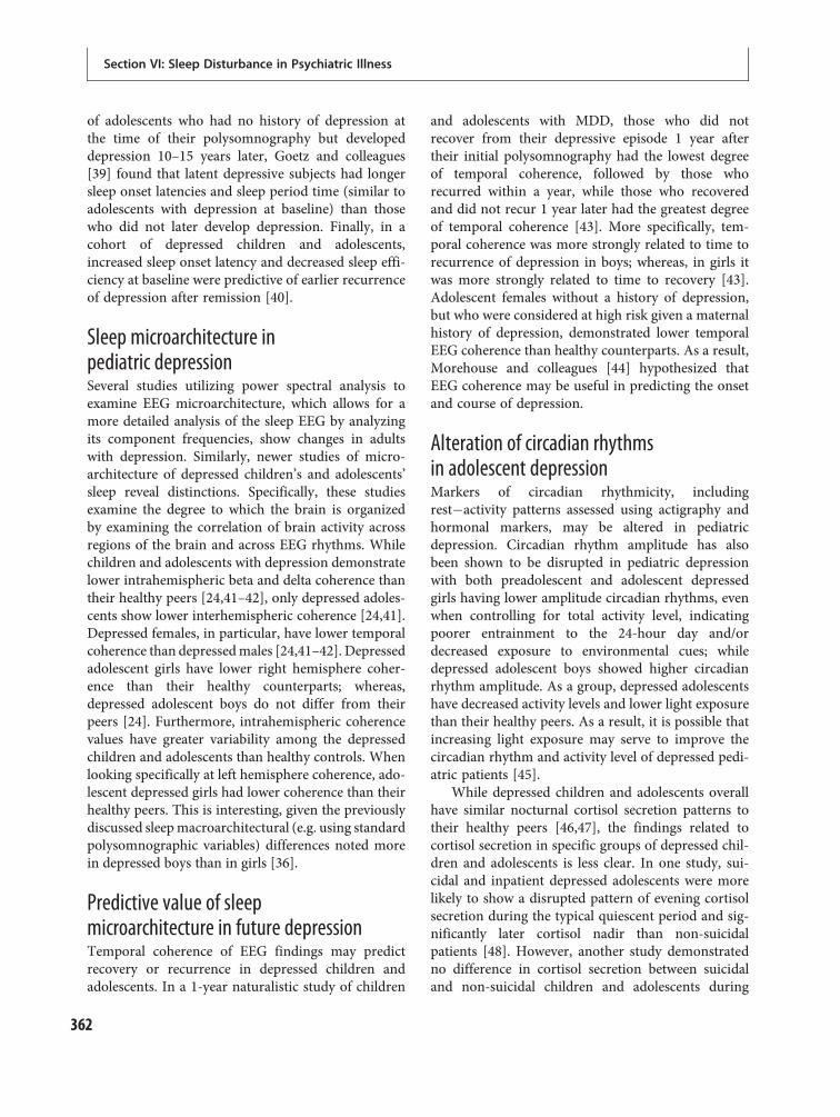
of adolescents who had no history of depression atthe time of their polysomnography but developeddepression 10–15 years later, Goetz and colleagues[39] found that latent depressive subjects had longersleep onset latencies and sleep period time (similar toadolescents with depression at baseline) than thosewho did not later develop depression. Finally, in acohort of depressed children and adolescents,increased sleep onset latency and decreased sleep effi-ciency at baseline were predictive of earlier recurrenceof depression after remission [40].
Sleep microarchitecture inpediatric depressionSeveral studies utilizing power spectral analysis toexamine EEG microarchitecture, which allows for amore detailed analysis of the sleep EEG by analyzingits component frequencies, show changes in adultswith depression. Similarly, newer studies of micro-architecture of depressed children’s and adolescents’sleep reveal distinctions. Specifically, these studiesexamine the degree to which the brain is organizedby examining the correlation of brain activity acrossregions of the brain and across EEG rhythms. Whilechildren and adolescents with depression demonstratelower intrahemispheric beta and delta coherence thantheir healthy peers [24,41–42], only depressed adoles-cents show lower interhemispheric coherence [24,41].Depressed females, in particular, have lower temporalcoherence than depressedmales [24,41–42]. Depressedadolescent girls have lower right hemisphere coher-ence than their healthy counterparts; whereas,depressed adolescent boys do not differ from theirpeers [24]. Furthermore, intrahemispheric coherencevalues have greater variability among the depressedchildren and adolescents than healthy controls. Whenlooking specifically at left hemisphere coherence, ado-lescent depressed girls had lower coherence than theirhealthy peers. This is interesting, given the previouslydiscussed sleepmacroarchitectural (e.g. using standardpolysomnographic variables) differences noted morein depressed boys than in girls [36].
Predictive value of sleepmicroarchitecture in future depressionTemporal coherence of EEG findings may predictrecovery or recurrence in depressed children andadolescents. In a 1-year naturalistic study of children
and adolescents with MDD, those who did notrecover from their depressive episode 1 year aftertheir initial polysomnography had the lowest degreeof temporal coherence, followed by those whorecurred within a year, while those who recoveredand did not recur 1 year later had the greatest degreeof temporal coherence [43]. More specifically, tem-poral coherence was more strongly related to time torecurrence of depression in boys; whereas, in girls itwas more strongly related to time to recovery [43].Adolescent females without a history of depression,but who were considered at high risk given a maternalhistory of depression, demonstrated lower temporalEEG coherence than healthy counterparts. As a result,Morehouse and colleagues [44] hypothesized thatEEG coherence may be useful in predicting the onsetand course of depression.
Alteration of circadian rhythmsin adolescent depressionMarkers of circadian rhythmicity, includingrest�activity patterns assessed using actigraphy andhormonal markers, may be altered in pediatricdepression. Circadian rhythm amplitude has alsobeen shown to be disrupted in pediatric depressionwith both preadolescent and adolescent depressedgirls having lower amplitude circadian rhythms, evenwhen controlling for total activity level, indicatingpoorer entrainment to the 24-hour day and/ordecreased exposure to environmental cues; whiledepressed adolescent boys showed higher circadianrhythm amplitude. As a group, depressed adolescentshave decreased activity levels and lower light exposurethan their healthy peers. As a result, it is possible thatincreasing light exposure may serve to improve thecircadian rhythm and activity level of depressed pedi-atric patients [45].
While depressed children and adolescents overallhave similar nocturnal cortisol secretion patterns totheir healthy peers [46,47], the findings related tocortisol secretion in specific groups of depressed chil-dren and adolescents is less clear. In one study, sui-cidal and inpatient depressed adolescents were morelikely to show a disrupted pattern of evening cortisolsecretion during the typical quiescent period and sig-nificantly later cortisol nadir than non-suicidalpatients [48]. However, another study demonstratedno difference in cortisol secretion between suicidaland non-suicidal children and adolescents during
Section VI: Sleep Disturbance in Psychiatric Illness
362
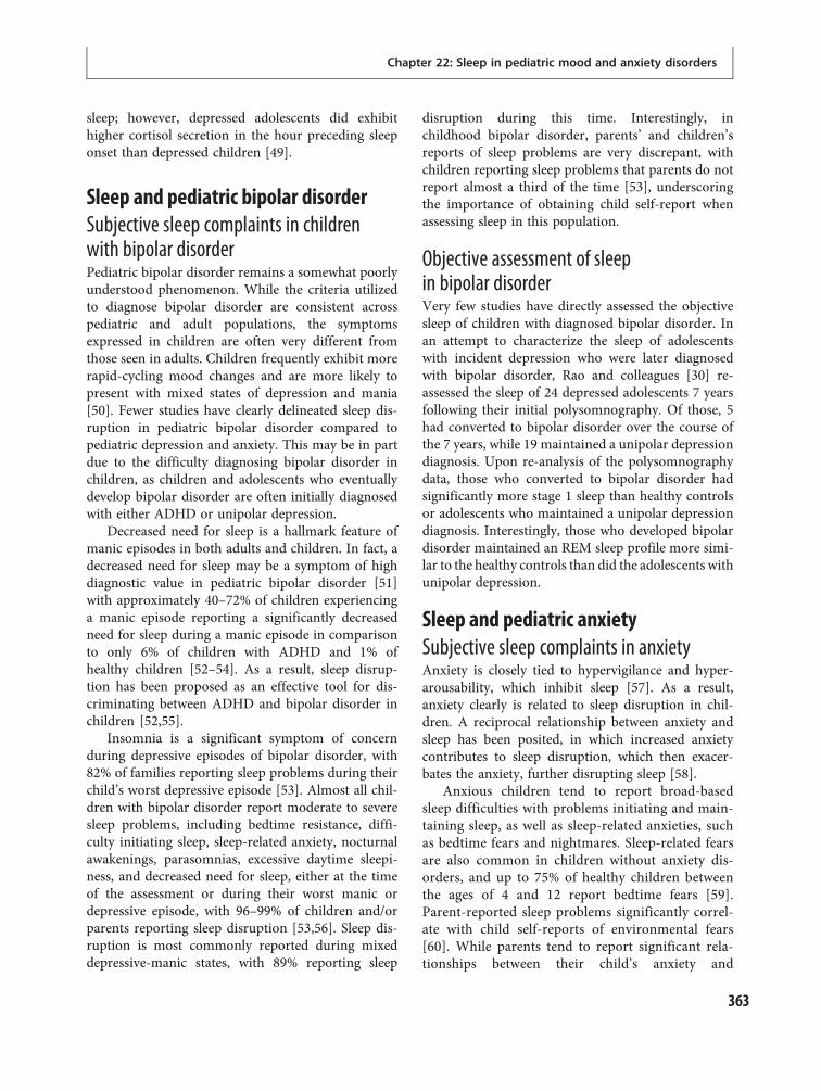
sleep; however, depressed adolescents did exhibithigher cortisol secretion in the hour preceding sleeponset than depressed children [49].
Sleep and pediatric bipolar disorderSubjective sleep complaints in childrenwith bipolar disorderPediatric bipolar disorder remains a somewhat poorlyunderstood phenomenon. While the criteria utilizedto diagnose bipolar disorder are consistent acrosspediatric and adult populations, the symptomsexpressed in children are often very different fromthose seen in adults. Children frequently exhibit morerapid-cycling mood changes and are more likely topresent with mixed states of depression and mania[50]. Fewer studies have clearly delineated sleep dis-ruption in pediatric bipolar disorder compared topediatric depression and anxiety. This may be in partdue to the difficulty diagnosing bipolar disorder inchildren, as children and adolescents who eventuallydevelop bipolar disorder are often initially diagnosedwith either ADHD or unipolar depression.
Decreased need for sleep is a hallmark feature ofmanic episodes in both adults and children. In fact, adecreased need for sleep may be a symptom of highdiagnostic value in pediatric bipolar disorder [51]with approximately 40–72% of children experiencinga manic episode reporting a significantly decreasedneed for sleep during a manic episode in comparisonto only 6% of children with ADHD and 1% ofhealthy children [52–54]. As a result, sleep disrup-tion has been proposed as an effective tool for dis-criminating between ADHD and bipolar disorder inchildren [52,55].
Insomnia is a significant symptom of concernduring depressive episodes of bipolar disorder, with82% of families reporting sleep problems during theirchild’s worst depressive episode [53]. Almost all chil-dren with bipolar disorder report moderate to severesleep problems, including bedtime resistance, diffi-culty initiating sleep, sleep-related anxiety, nocturnalawakenings, parasomnias, excessive daytime sleepi-ness, and decreased need for sleep, either at the timeof the assessment or during their worst manic ordepressive episode, with 96–99% of children and/orparents reporting sleep disruption [53,56]. Sleep dis-ruption is most commonly reported during mixeddepressive-manic states, with 89% reporting sleep
disruption during this time. Interestingly, inchildhood bipolar disorder, parents’ and children’sreports of sleep problems are very discrepant, withchildren reporting sleep problems that parents do notreport almost a third of the time [53], underscoringthe importance of obtaining child self-report whenassessing sleep in this population.
Objective assessment of sleepin bipolar disorderVery few studies have directly assessed the objectivesleep of children with diagnosed bipolar disorder. Inan attempt to characterize the sleep of adolescentswith incident depression who were later diagnosedwith bipolar disorder, Rao and colleagues [30] re-assessed the sleep of 24 depressed adolescents 7 yearsfollowing their initial polysomnography. Of those, 5had converted to bipolar disorder over the course ofthe 7 years, while 19 maintained a unipolar depressiondiagnosis. Upon re-analysis of the polysomnographydata, those who converted to bipolar disorder hadsignificantly more stage 1 sleep than healthy controlsor adolescents who maintained a unipolar depressiondiagnosis. Interestingly, those who developed bipolardisorder maintained an REM sleep profile more simi-lar to the healthy controls than did the adolescents withunipolar depression.
Sleep and pediatric anxietySubjective sleep complaints in anxietyAnxiety is closely tied to hypervigilance and hyper-arousability, which inhibit sleep [57]. As a result,anxiety clearly is related to sleep disruption in chil-dren. A reciprocal relationship between anxiety andsleep has been posited, in which increased anxietycontributes to sleep disruption, which then exacer-bates the anxiety, further disrupting sleep [58].
Anxious children tend to report broad-basedsleep difficulties with problems initiating and main-taining sleep, as well as sleep-related anxieties, suchas bedtime fears and nightmares. Sleep-related fearsare also common in children without anxiety dis-orders, and up to 75% of healthy children betweenthe ages of 4 and 12 report bedtime fears [59].Parent-reported sleep problems significantly correl-ate with child self-reports of environmental fears[60]. While parents tend to report significant rela-tionships between their child’s anxiety and
Chapter 22: Sleep in pediatric mood and anxiety disorders
363

experience of nightmares, children do not mirrorthis report. In fact, there has been no differencefound in children’s self-report of anxiety betweenthose who reported experiencing nightmares andthose who did not. Instead, children report a signifi-cant relationship between anxiety and the distresscaused by their nightmares, rather than the experi-ence of the nightmare itself [61].
When specific sleep complaints are assessed,83–92% of children with anxiety disorders reportat least one sleep-related complaint [58,62–63].Among those with generalized anxiety disorder(GAD), 42–56% of children and 49–57% of adoles-cents report trouble sleeping [64,65]. The mostcommon sleep complaints noted by parents of chil-dren with GAD include nightmares, being over-tired, and trouble sleeping [62]. Two-thirds of ageneral population of children with anxiety dis-orders report insomnia, and over half report fre-quent nightmares. Sixty-one percent of anxiouschildren aged 6–11 reported refusing to sleep alone,and they were significantly more likely to endorsethis complaint than were anxious 12- to 17-year-olds [58]. Nightmares were more frequentlyreported by anxious girls and by younger children[58,63].
When parental report of children’s daytimebehavior was compared with sleep complaints,those with anxiety symptoms were significantlymore likely to have night-time awakenings, sleepwith their lights on, require a security object forsleep, have bedtime fears, require prolonged night-time rituals, and to have nightmares. These associ-ations were hypothesized to be a result of increasedarousal and hypervigilance associated with anxietythat interrupts sleep [1]. Of note, when anxiouschildren’s self-report of sleep problems was com-pared with their polysomnographically recordedsleep, they exhibited a tendency to under-reportproblems. While they had disrupted sleep withrepeated, prolonged awakenings, they did not reportany greater problems with sleep maintenance thantheir peers. However, they did accurately reportprolonged sleep onset latency [20].
Among children and adolescents with obsessive-compulsive disorder, girls and adolescents were sig-nificantly more likely to be reported by their parentsas sleeping more than other children [63]. Alfano andcolleagues found that higher numbers of sleep-relatedproblems were significantly predictive of impaired
home functioning, even when the severity of anxietywas statistically controlled [58]. When the childrenwere treated with family-based cognitive behavioraltherapy, sleep problems significantly decreased,with specific reductions in nightmares, being over-tired, sleeping less and more than other children,difficulty sleeping, and requiring the presence ofanother person to sleep [63].
Sleep characteristics in childrenwith PTSDSleep complaints are a hallmark feature of PTSD.Children and adolescents with PTSD report frequentnightmares and increased nocturnal anxiety, with dif-ficulty initiating and maintaining sleep [66–68].Abuse frequently occurs at night, and as bedtimeapproaches children who have been traumatizedmay feel increasingly anxious and unsafe. Hypervigi-lance due to fear leads to persistent sleep continuitydisruption [69]. Several attempts have been made toobjectively characterize sleep in children with a his-tory of abuse. Results of actigraphic monitoring indi-cated poorer sleep efficiency, increased nocturnalactivity, and reduced quiet sleep among childrenwho were abused compared to non-abused childrenor children with other psychiatric disorders [70,71].Recurrent nightmares and flashbacks of traumaticevents are frequently experienced by both childrenand adults with PTSD.
While acute traumatization is associated withincreased arousal and reduced/fragmented sleep,longstanding significant stressors may lead toincreased sleep. Type II trauma, as described by Terr,which is characterized by chronic overwhelmingstress, may cause symptoms of dissociation, apathy,denial, and excessive sleepiness [69,72].
Objective sleep disturbance inanxiety disordersFewer polysomnographic data are available in chil-dren and adolescents with anxiety disorders thanthose with depression. The polysomnographic stud-ies that have been conducted, however, generallyreveal that children and adolescents with anxietydisorders have poorer sleep than depressed childrenand adolescents, exhibiting longer sleep onsetlatency, more nocturnal awakenings, and decreasedSWS. Furthermore, one study demonstrated that
Section VI: Sleep Disturbance in Psychiatric Illness
364
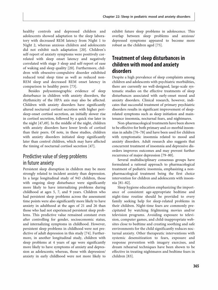
healthy controls and depressed children andadolescents showed adaptation to the sleep labora-tory with decreased REM latency from Night 1 toNight 2, whereas anxious children and adolescentsdid not exhibit such adaptation [20]. Children’sself-report of anxiety symptoms were positively cor-related with sleep onset latency and negativelycorrelated with stage 3 sleep and self-report of easeof waking and sleep quality [20]. Furthermore, chil-dren with obsessive-compulsive disorder exhibitedreduced total sleep time as well as reduced non-REM sleep and decreased REM onset latency incomparison to healthy peers [73].
Besides polysomnographic evidence of sleepdisturbance in children with anxiety disorders, therhythmicity of the HPA axis may also be affected.Children with anxiety disorders have significantlyaltered nocturnal cortisol secretion with higher peri-sleep-onset cortisol secretion, an initially slower risein cortisol secretion, followed by a quick rise later inthe night [47,49]. In the middle of the night, childrenwith anxiety disorders have lower levels of cortisolthan their peers. Of note, in these studies, childrenwith anxiety disorders initiated sleep significantlylater than control children, which may have affectedthe timing of nocturnal cortisol secretion [47].
Predictive value of sleep problemsin future anxietyPersistent sleep disruption in children may be morestrongly related to incident anxiety than depression.In a large longitudinal study of 943 children, thosewith ongoing sleep disturbance were significantlymore likely to have internalizing problems duringchildhood at ages 5, 7, and 9 years. Children whohad persistent sleep problems across the assessmenttime points were also significantly more likely to haveanxiety in adulthood at the ages of 21 and 26 thanthose who had not experienced persistent sleep prob-lems. This predictive value remained constant evenafter controlling for gender, socioeconomic status,and internalizing symptoms in childhood. However,persistent sleep problems in childhood were not pre-dictive of adult depression in this study [74]. Further-more, in another longitudinal study, children withsleep problems at 4 years of age were significantlymore likely to have symptoms of anxiety and depres-sion as adolescents; whereas, those with depression/anxiety in early childhood were not more likely to
exhibit future sleep problems in adolescence. Thisoverlap between sleep problems and anxious/depressed symptoms appeared to become morerobust as the children aged [75].
Treatment of sleep disturbances inchildren with mood and anxietydisordersDespite a high prevalence of sleep complaints amongchildren and adolescents with psychiatric morbidities,there are currently no well-designed, large-scale sys-tematic studies on the effective treatments of sleepdisturbances associated with early-onset mood andanxiety disorders. Clinical research, however, indi-cates that successful treatment of primary psychiatricdisorders results in significant improvement of sleep-related symptoms such as sleep initiation and main-tenance insomnia, nocturnal fears, and nightmares.
Non-pharmacological treatments have been shownto be effective for both primary and co-morbid insom-nias in adults [76–78] and have been used for childrenwith symptomatic insomnia related to mood andanxiety disorders. Adult research also suggests thatconcurrent treatment of insomnia and depressive dis-orders improves outcomes and may prevent furtherrecurrence of major depression [79–80].
Several multidisciplinary consensus groups haveformulated a rational approach to pharmacologicaltreatment of pediatric insomnia, emphasizing a non-pharmacological treatment being the first choiceintervention for children and adolescents with insom-nia [81–82].
Sleep hygiene education emphasizing the import-ance of consistent age-appropriate bedtime andnight-time routine should be provided to everyfamily seeking help for sleep-related problems intheir children. Night-time fears are commonly pre-cipitated by watching frightening movies and/ortelevision programs. Avoiding exposure to televi-sion, computer games, and child-inappropriate web-sites close to bedtime and creating soothing and safeenvironments for the child significantly reduces noc-turnal anxiety. Other therapeutic interventions withsystemic desensitization to fears, exposure andresponse prevention with imagery exercises, anddream rehearsal techniques have been shown to beeffective in treating nightmares and bedtime fears inchildren [83].
Chapter 22: Sleep in pediatric mood and anxiety disorders
365
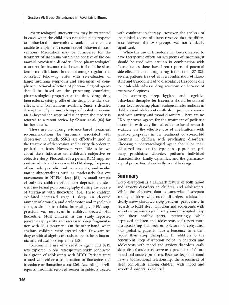
Pharmacological interventions may be warrantedin cases when the child does not adequately respondto behavioral interventions, and/or parents areunable to implement recommended behavioral inter-ventions. Medication may be considered for thetreatment of insomnia within the context of the co-morbid psychiatric disorder. Once pharmacologicaltreatment for insomnia is chosen, it should be shortterm, and clinicians should encourage regular andconsistent follow-up visits with re-evaluation oftarget insomnia symptoms and assessment of com-pliance. Rational selection of pharmacological agentsshould be based on the presenting complaint,pharmacological properties of the drug, drug�druginteractions, safety profile of the drug, potential side-effects, and formulations available. Since a detaileddescription of pharmacotherapy of pediatric insom-nia is beyond the scope of this chapter, the reader isreferred to a recent review by Owens et al. [82] forfurther details.
There are no strong evidence-based treatmentrecommendations for insomnia associated withdepression in youth. SSRIs are effectively used inthe treatment of depression and anxiety disorders inpediatric patients. However, very little is knownabout their influence on children’s subjective orobjective sleep. Fluoxetine is a potent REM suppres-sant in adults and increases NREM sleep, frequencyof arousals, periodic limb movements, and oculo-motor abnormalities such as moderately fast eyemovements in NREM sleep [84]. A small sampleof only six children with major depression under-went nocturnal polysomnography during the courseof treatment with fluoxetine [85]. These childrenexhibited increased stage 1 sleep, an elevatednumber of arousals, and oculomotor and myoclonicchanges similar to adults. Interestingly, REM sup-pression was not seen in children treated withfluoxetine. Most children in this study reportedpoorer sleep quality and increased sleep fragmenta-tion with SSRI treatment. On the other hand, whenanxious children were treated with fluvoxamine,they exhibited significant reductions in both insom-nia and refusal to sleep alone [58].
Concomitant use of a sedative agent and SSRIwas explored in one retrospective study conductedin a group of adolescents with MDD. Patients weretreated with either a combination of fluoxetine andtrazodone or fluoxetine alone [86]. According to self-reports, insomnia resolved sooner in subjects treated
with combination therapy. However, the analysis ofthe clinical course of illness revealed that the differ-ence between the two groups was not clinicallysignificant.
While the use of trazodone has been observed tohave therapeutic effects on symptoms of insomnia, itshould be used with caution in combination withfluoxetine, as there have been reports of potentialside-effects due to drug�drug interaction [87–88].Several patients treated with a combination of fluox-etine and trazodone had to discontinue trazodone dueto intolerable adverse drug reactions or because ofexcessive sleepiness.
In summary, sleep hygiene and cognitivebehavioral therapies for insomnia should be utilizedprior to considering pharmacological interventions inchildren and adolescents with sleep problems associ-ated with anxiety and mood disorders. There are noFDA-approved agents for the treatment of pediatricinsomnia, with very limited evidence-based researchavailable on the effective use of medications withsedative properties in the treatment of co-morbidinsomnia in children with psychiatric disorders.Choosing a pharmacological agent should be indi-vidualized based on the type of sleep problem, pri-mary psychiatric disorder, patient’s individualcharacteristics, family dynamics, and the pharmaco-logical properties of currently available drugs.
SummarySleep disruption is a hallmark feature of both moodand anxiety disorders in children and adolescents.While the objective data is somewhat discrepantamong children with mood disorders, adolescentsclearly show disrupted sleep patterns, particularly inregards to REM sleep. Children and adolescents withanxiety experience significantly more disrupted sleepthan their healthy peers. Interestingly, whiledepressed children and adolescents self-report moredisrupted sleep than seen on polysomnography, anx-ious pediatric patients have a tendency to under-report their sleep disruption. In addition to theconcurrent sleep disruption noted in children andadolescents with mood and anxiety disorders, earlysleep disturbance may serve as a predictor of futuremood and anxiety problems. Because sleep and moodhave a bidirectional relationship, the assessment ofsleep complaints among children with mood andanxiety disorders is essential.
Section VI: Sleep Disturbance in Psychiatric Illness
366
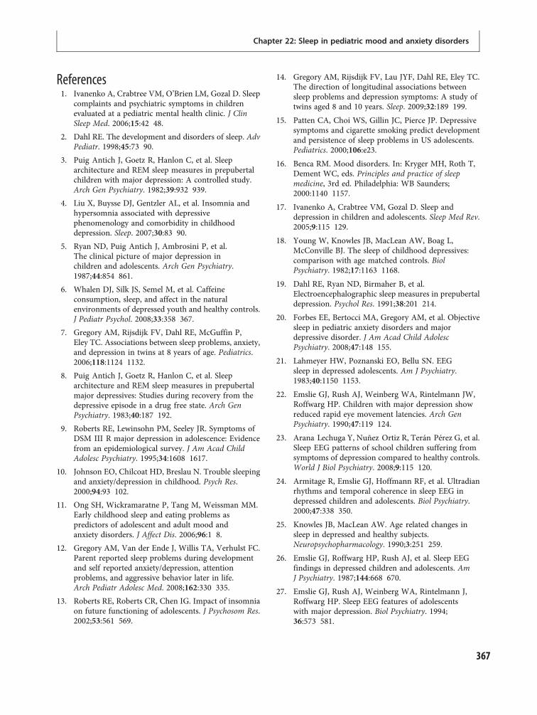
References1. Ivanenko A, Crabtree VM, O’Brien LM, Gozal D. Sleep
complaints and psychiatric symptoms in childrenevaluated at a pediatric mental health clinic. J ClinSleep Med. 2006;15:42 48.
2. Dahl RE. The development and disorders of sleep. AdvPediatr. 1998;45:73 90.
3. Puig Antich J, Goetz R, Hanlon C, et al. Sleeparchitecture and REM sleep measures in prepubertalchildren with major depression: A controlled study.Arch Gen Psychiatry. 1982;39:932 939.
4. Liu X, Buysse DJ, Gentzler AL, et al. Insomnia andhypersomnia associated with depressivephenomenology and comorbidity in childhooddepression. Sleep. 2007;30:83 90.
5. Ryan ND, Puig Antich J, Ambrosini P, et al.The clinical picture of major depression inchildren and adolescents. Arch Gen Psychiatry.1987;44:854 861.
6. Whalen DJ, Silk JS, Semel M, et al. Caffeineconsumption, sleep, and affect in the naturalenvironments of depressed youth and healthy controls.J Pediatr Psychol. 2008;33:358 367.
7. Gregory AM, Rijsdijk FV, Dahl RE, McGuffin P,Eley TC. Associations between sleep problems, anxiety,and depression in twins at 8 years of age. Pediatrics.2006;118:1124 1132.
8. Puig Antich J, Goetz R, Hanlon C, et al. Sleeparchitecture and REM sleep measures in prepubertalmajor depressives: Studies during recovery from thedepressive episode in a drug free state. Arch GenPsychiatry. 1983;40:187 192.
9. Roberts RE, Lewinsohn PM, Seeley JR. Symptoms ofDSM III R major depression in adolescence: Evidencefrom an epidemiological survey. J Am Acad ChildAdolesc Psychiatry. 1995;34:1608 1617.
10. Johnson EO, Chilcoat HD, Breslau N. Trouble sleepingand anxiety/depression in childhood. Psych Res.2000;94:93 102.
11. Ong SH, Wickramaratne P, Tang M, Weissman MM.Early childhood sleep and eating problems aspredictors of adolescent and adult mood andanxiety disorders. J Affect Dis. 2006;96:1 8.
12. Gregory AM, Van der Ende J, Willis TA, Verhulst FC.Parent reported sleep problems during developmentand self reported anxiety/depression, attentionproblems, and aggressive behavior later in life.Arch Pediatr Adolesc Med. 2008;162:330 335.
13. Roberts RE, Roberts CR, Chen IG. Impact of insomniaon future functioning of adolescents. J Psychosom Res.2002;53:561 569.
14. Gregory AM, Rijsdijk FV, Lau JYF, Dahl RE, Eley TC.The direction of longitudinal associations betweensleep problems and depression symptoms: A study oftwins aged 8 and 10 years. Sleep. 2009;32:189 199.
15. Patten CA, Choi WS, Gillin JC, Pierce JP. Depressivesymptoms and cigarette smoking predict developmentand persistence of sleep problems in US adolescents.Pediatrics. 2000;106:e23.
16. Benca RM. Mood disorders. In: Kryger MH, Roth T,Dement WC, eds. Principles and practice of sleepmedicine, 3rd ed. Philadelphia: WB Saunders;2000:1140 1157.
17. Ivanenko A, Crabtree VM, Gozal D. Sleep anddepression in children and adolescents. Sleep Med Rev.2005;9:115 129.
18. Young W, Knowles JB, MacLean AW, Boag L,McConville BJ. The sleep of childhood depressives:comparison with age matched controls. BiolPsychiatry. 1982;17:1163 1168.
19. Dahl RE, Ryan ND, Birmaher B, et al.Electroencephalographic sleep measures in prepubertaldepression. Psychol Res. 1991;38:201 214.
20. Forbes EE, Bertocci MA, Gregory AM, et al. Objectivesleep in pediatric anxiety disorders and majordepressive disorder. J Am Acad Child AdolescPsychiatry. 2008;47:148 155.
21. Lahmeyer HW, Poznanski EO, Bellu SN. EEGsleep in depressed adolescents. Am J Psychiatry.1983;40:1150 1153.
22. Emslie GJ, Rush AJ, Weinberg WA, Rintelmann JW,Roffwarg HP. Children with major depression showreduced rapid eye movement latencies. Arch GenPsychiatry. 1990;47:119 124.
23. Arana Lechuga Y, Nuñez Ortiz R, Terán Pérez G, et al.Sleep EEG patterns of school children suffering fromsymptoms of depression compared to healthy controls.World J Biol Psychiatry. 2008;9:115 120.
24. Armitage R, Emslie GJ, Hoffmann RF, et al. Ultradianrhythms and temporal coherence in sleep EEG indepressed children and adolescents. Biol Psychiatry.2000;47:338 350.
25. Knowles JB, MacLean AW. Age related changes insleep in depressed and healthy subjects.Neuropsychopharmacology. 1990;3:251 259.
26. Emslie GJ, Roffwarg HP, Rush AJ, et al. Sleep EEGfindings in depressed children and adolescents. AmJ Psychiatry. 1987;144:668 670.
27. Emslie GJ, Rush AJ, Weinberg WA, Rintelmann J,Roffwarg HP. Sleep EEG features of adolescentswith major depression. Biol Psychiatry. 1994;36:573 581.
Chapter 22: Sleep in pediatric mood and anxiety disorders
367

28. Goetz RR, Puig Antich J, Dahl RE, et al. EEG sleep ofyoung adults with major depression: A controlledstudy. J Affect Disord. 1991;22:91 100.
29. Dahl RE, Ryan ND, Matty MK, et al. Sleep onsetabnormalities in depressed adolescents. Biol Psychiatry.1996;39:400 410.
30. Rao U, Dahl RE, Ryan ND, et al. Heterogeneity in EEGsleep findings in adolescent depression: Unipolarversus bipolar clinical course. J Affect Disord.2002;70:273 280.
31. Armitage R, Hoffmann R. Sleep electrophysiology ofmajor depressive disorders. Curr Rev Mood AnxietyDisord. 1997;1:139 151.
32. Goetz RR, Puig Antich J, Ryan N, et al.Electroencephalographic sleep of adolescents withmajor depression and normal controls. Arch GenPsychiatry. 1987;44:61 68.
33. Williamson DE, Dahl RE, Birmaher B, et al. Stressfullife events and EEG sleep in depressed and normalcontrol adolescents. Biol Psychiatry. 1995;37:859 865.
34. Dahl RE, Puig Antich J, Ryan ND, et al. EEG sleep inadolescents with major depression: The role ofsuicidality and inpatient status. J Affect Disord.1990;19:63 75.
35. Naylor MW, Shain BN, Shipley JE. REM latency inpsychotically depressed adolescents. Biol Psychiatry.1990;28:161 164.
36. Robert JJT, Hoffman RF, Emslie GJ, et al. Sex and agedifferences in sleep macroarchitecture in childhoodand adolescent depression. Sleep. 2006;29:351 358.
37. Bertocci MA, Dahl RE, Williamson DE, et al.Subjective sleep complaints in pediatric depression:A controlled study and comparison with EEGmeasures of sleep and waking. J Am Acad Child AdolescPsychiatry. 2005;44:1158 1166.
38. Rao U, Dahl RE, Ryan ND, et al. The relationshipbetween longitudinal clinical course and sleep andcortisol changes in adolescent depression. BiolPsychiatry. 1996;40:474 484.
39. Goetz RR, Wolk SI, Coplan JD, Ryan ND,Weissman MM. Premorbid polysomnographic signs indepressed adolescents: A reanalysis of EEG sleep afterlongitudinal follow up in adulthood. Biol Psychiatry.2001;49:930 942.
40. Emslie GJ, Armitage R, Weinberg WA, et al. Sleeppolysomnography as a predictor of recurrence inchildren and adolescents with depressive disorder. IntJ Neuropsychopharmacol. 2001;4:159 168.
41. Armitage R, Emslie GJ, Hoffman RF, Rintelmann J,Rush AJ. Delta sleep EEG in depressed adolescentfemales and healthy controls. J Affect Disord.2001;63:139 148.
42. Armitage R, Hoffmann R, Emslie G, Rintelmann J,Robert J. Sleep microarchitecture in childhood andadolescent depression: Temporal coherence. Clin EEGNeurosci. 2006;37:1 9.
43. Armitage R, Hoffman RF, Emslie GJ, et al. Sleepmicroarchitecture as a predictor of recurrence inchildren and adolescents with depression. IntJ Neuropsychopharmacol. 2002;5:217 228.
44. Morehouse RL, Kusumakar V, Kutcher SP, LeBlanc J,Armitage R. Temporal coherence in ultradiansleep EEG rhythms in a never depressed, high riskcohort of female adolescents. Biol Psychiatry.2002;51:446 456.
45. Armitage R, Hoffmann R, Emslie G, et al. Rest activitycycles in childhood and adolescent depression. J AmAcad Child Adolesc Psychiatry. 2004;43:761 769.
46. Kutcher S, Malkin D, Silverberg J, et al. Nocturnalcortisol, thyroid stimulating hormone, and growthhormone secretory profiles in depressed adolescents.J Am Acad Child Adolesc Psychiatry. 1991;30:407 414.
47. Feder A, Coplan JD, Goetz RR, et al. Twenty four hourcortisol secretion patterns in prepubertal children withanxiety or depressive disorders. Biol Psychiatry.2004;56:198 204.
48. Dahl RE, Ryan ND, Puig Antich J, et al. 24 hourcortisol measures in adolescents with majordepression: A controlled study. Biol Psychiatry.1991;30:25 36.
49. Forbes EE, Williamson DE, Ryan ND, et al. Perisleep onset cortisol levels in children and adolescentswith affective disorders. Biol Psychiatry. 2006;59:24 30.
50. NIMH Roundtable. National Institute of MentalHealth Research Roundtable on Prepubertal BipolarDisorder. J Am Acad Child Adolesc Psychiatry.2001;40:871 878.
51. Geller B, Zimmerman B, Williams M, et al.Phenomenology of prepubertal and early adolescentbipolar disorder: Examples of elated mood, grandiosebehaviors, decreased need for sleep, racing thoughts,and hypersexuality. J Child Adolesc Psychopharmacol.2002;12:3 9.
52. Geller B, Zimmerman B, Williams M, et al. DSM IVmania symptoms in a prepubertal and early adolescentbipolar phenotype compared to attention deficithyperactive and normal controls. J Child AdolescPsychopharmacol. 2002;12:11 25.
53. Lofthouse N, Fristad, M, Splaingard M, Kelleher M.Parent and child reports of sleep problems associatedwith early onset bipolar spectrum disorders. J FamPsychol. 2007;21:114 123.
54. Kowatch RA,YoungstromEA,DanielyanA, Findling RL.Review and meta analysis of the phenomenology and
Section VI: Sleep Disturbance in Psychiatric Illness
368

clinical characteristics of mania in children andadolescents. Bipolar Disorders. 2005;7:483 496.
55. Meyers OI, Youngstrom EA. A parent general behaviorinventory subscale to measure sleep disturbance inpediatric bipolar disorder. J Clin Psychiatry.2008;69:840 843.
56. Lofthouse N, Fristad M, Splaingard M, et al. Websurvey of sleep problems associated with early onsetbipolar spectrum disorders. J Pediatr Psychol.2008;33:349 357.
57. Dahl RE. The regulation of sleep and arousal:Development and psychopathology. Dev Psychopathol.1996;8:3 27.
58. Alfano CA, Ginsburg GS, Kingery JN. Sleep relatedproblems among children and adolescents with anxietydisorders. J Am Acad Child Adolesc Psychiatry.2007;46:224 232.
59. Muris P, Merckelbach H, Gadet B, Moulaert V. Fears,worries, and scary dreams in 4 to 12 year oldchildren: Their content, developmental pattern, andorigins. J Clin Child Psychol. 2000;29:43 52.
60. Dollinger SJ, Molina BS, Campo Monteiro JM.Sleep and anxieties in Brazilian children: The role ofcultural and environmental factors in child sleepdisturbance. Am J Orthopsychiatry. 1996;66:252 261.
61. Mindell JA, Barrett KM. Nightmares and anxiety inelementary aged children: Is there a relationship? ChildCare Health Dev. 2002;28:317 322.
62. Alfano CA, Beidel DC, Turner SM, Lewin DS.Preliminary evidence for sleep complaintsamong children referred for anxiety. Sleep Med.2006;7:467 473.
63. Storch EA, Murphy TK, Lack CW, et al. Sleep relatedproblems in pediatric obsessive compulsive disorder.J Anxiety Disord. 2008;22:877 885.
64. Pina AA, Silverman WK, Alfano CA, Saavedra LM.Diagnostic efficiency of symptoms in the diagnosis ofDSM IV: Generalized anxiety disorder in youth.J Child Psychol Psychiatry. 2002;43:959 967.
65. Masi G, Millepiedi S, Mucci M, Poli P, Bertini N,Milantoni L. Generalized anxiety disorder in referredchildren and adolescents. J Am Acad Child AdolescPsychiatry. 2004;43:752 760.
66. Pynoos RS, Frederick C, Nader K, et al. Life threat andposttraumatic stress in school age children. Arch GenPsychiatry. 1987;44:1057 1063.
67. Ross RJ, Ball WA, Sullivan KA, Caroff SN. Sleepdisturbance as the hallmark of posttraumatic stressdisorder. Am J Psychiatry. 1989;146:697 707.
68. Uhde TW. Anxiety disorders. In: Kryger MH, Roth T,Dement WC, eds. Principles and practice of sleep
medicine, 3rd ed. Philadelphia:WB Saunders;2000:1123 1139.
69. Sadeh A. Stress, trauma and sleep in children. ChildAdolesc Clin North Am. 1996;5:685 700.
70. Teicher MH, Glod CA, Harper D, et al. Locomotoractivity in depressed children and adolescents:I. Circadian dysregulation. J Am Acad Child AdolescPsychiatry. 1993;32:760 769.
71. Glod CA, Teicher MH, Hartman CR, Harakal T.Increased nocturnal activity and impaired sleepmaintenance in abused children. J Am Acad ChildAdolesc Psychiatry. 1997;36:1236 1243.
72. Terr LC. Childhood traumas: An outline and overview.Am J Psychiatry. 1991;148:10 19.
73. Rapoport J, Elkins R, Langer DH, et al. Childhoodobsessive compulsive disorder. Am J Psychiatry.1981;138:1545 1554.
74. Gregory AM, Caspi A, Eley TC, et al. Prospectivelongitudinal associations between persistent sleepproblems in childhood and anxiety and depressiondisorders in adulthood. J Abnorm Child Psychol.2005;33:157 163.
75. Gregory AM, O’Connor TG. Sleep problems inchildhood: A longitudinal study of developmentalchange and association with behavioral problems. J AmAcad Child Adolesc Psychiatry. 2002;41:964 971.
76. Kupfer DJ, Reynolds CF 3rd. Management ofinsomnia. N Engl J Med. 1997;336(5):341 346.
77. Morin CM, Bootzin RR, Buysse DJ, et al. Psychologicaland behavioral treatment of insomnia: update of therecent evidence (1998 2004). Sleep. 2006;29(11):1398 1414.
78. Belanger L, Vallieres A, Ivers H, et al. Meta analysis ofsleep changes in control groups of insomnia treatmenttrials. J Sleep Res. 2007;16(1):77 84.
79. Kupfer DJ. Pathophysiology and management ofinsomnia during depression. Ann Clin Psychiatry.1999;11(4):267 276.
80. Ivanenko A. Sleep and mood disorders in children andadolescents. In: Ivanenko A, ed. Sleep and psychiatricdisorders in children and adolescents. New York:Informa Healthcare; 2008:279 287.
81. Mindell JA, Emslie G, Blumer J, et al. Pharmacologicmanagement of insomnia in children andadolescents: consensus statement. Pediatrics. 2006;117(6):e1223 1232.
82. Owens JA, Babcock D, Blumer J, et al. The use ofpharmacotherapy in the treatment of pediatricinsomnia in primary care: rational approaches.A consensus meeting summary. J Clin Sleep Med.2005;1(1):49 59.
Chapter 22: Sleep in pediatric mood and anxiety disorders
369
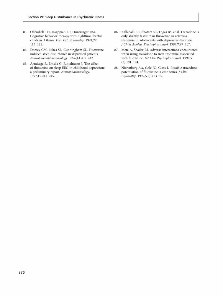
83. Ollendick TH, Hagopian LP, Huntzinger RM.Cognitive behavior therapy with nighttime fearfulchildren. J Behav Ther Exp Psychiatry. 1991;22:113 121.
84. Dorsey CM, Lukas SE, Cunningham SL. Fluoxetineinduced sleep disturbance in depressed patients.Neuropsychopharmacology. 1996;14:437 442.
85. Armitage R, Emslie G, Rintelmann J. The effectof fluoxetine on sleep EEG in childhood depression:a preliminary report. Neuropharmacology.1997;17:241 245.
86. Kallepalli BR, Bhatara VS, Fogas BS, et al. Trazodone isonly slightly faster than fluoxetine in relievinginsomnia in adolescents with depressive disorders.J Child Adolesc Psychopharmacol. 1997;7:97 107.
87. Metz A, Shader RI. Adverse interactions encounteredwhen using trazodone to treat insomnia associatedwith fluoxetine. Int Clin Psychopharmacol. 1990;5(3):191 194.
88. Nierenberg AA, Cole JO, Glass L. Possible trazodonepotentiation of fluoxetine: a case series. J ClinPsychiatry. 1992;53(3):83 85.
Section VI: Sleep Disturbance in Psychiatric Illness
370
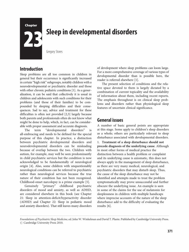
Chapter
23 Sleep in developmental disorders
Gregory Stores
IntroductionSleep problems are all too common in children ingeneral but their occurrence is significantly increasedin certain “high risk” subgroups, notably childrenwith aneurodevelopmental or psychiatric disorder and thosewith other chronic pediatric conditions [1]. As a gener-alization, it can be said that collectively it is usual inchildren and adolescents with such conditions for theirproblems (and those of their families) to be com-pounded by sleeping difficulties and their conse-quences. Sad to say, advice and treatment for thesedifficulties is often not provided [2,3] largely becauseboth parents and professionals often do not know whatmight be done to help, which, in fact, can be consider-able with proper assessment and accurate diagnosis.
The term “developmental disorders” isall-embracing and needs to be defined for the specialpurpose of this chapter. In practice, a distinctionbetween psychiatric developmental disorders andneurodevelopmental disorders can be misleadingbecause of overlap between the two. Children withautism, for example, may well be seen predominantlyin child psychiatric services but the condition is nowacknowledged to be fundamentally of neurologicalorigin [4]. Also, some children with unquestionablyneurological conditions can be referred to psychiatricrather than neurological services because the truenature of their condition has not been recognized.Childhood-onset narcolepsy is a prime example [5].
Genuinely “primary” childhood psychiatricdisorders of mood and anxiety, as well as ADHD,are considered elsewhere in this book (see Chapter21: Sleep in attention-deficit/hyperactivity disorder(ADHD) and Chapter 22: Sleep in pediatric moodand anxiety disorders). That still leaves many disorders
of development where sleep problems can loom large.For a more comprehensive coverage of various types ofdevelopmental disorder than is possible here, thereader is referred elsewhere [1].
The present selection of conditions and the rela-tive space devoted to them is largely dictated by acombination of current topicality and the availabilityof information about them, including recent reports.The emphasis throughout is on clinical sleep prob-lems and disorders rather than physiological sleepfeatures of uncertain clinical significance.
General issuesA number of basic general points are appropriateat this stage. Some apply to children’s sleep disordersas a whole; others are particularly relevant to sleepdisturbance associated with developmental disorders.
1. Treatment of a sleep disturbance should notprecede diagnosis of the underlying cause. Althoughin most other forms of medical practice thedistinction between a health problem or complaintand its underlying cause is axiomatic, this does notalways apply in the management of sleep disturbance,as there are very many medical, neurological, andpsychiatric disorders that may disturb sleep. Thus,the cause of the sleep disturbance may not beidentified and attempts made to treat the problemsymptomatically may prove unsuccessful and furtherobscure the underlying issue. An example is seenin some of the claims for the use of melatonin forsleeplessness in children with multiple handicapswhere imprecise accounts of the nature of the sleepdisturbance add to the difficulty of evaluating theresults [6].
Foundations of Psychiatric SleepMedicine, ed. JohnW.Winkelman andDavid T. Plante. Published by CambridgeUniversity Press.# Cambridge University Press 2010.
371
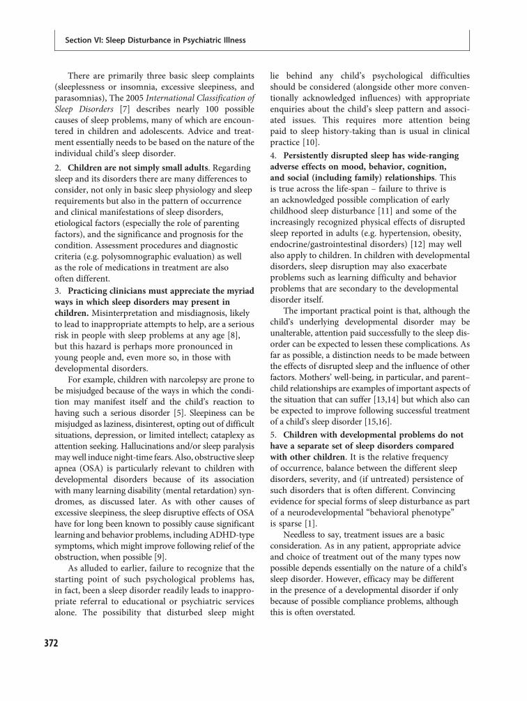
There are primarily three basic sleep complaints(sleeplessness or insomnia, excessive sleepiness, andparasomnias), The 2005 International Classification ofSleep Disorders [7] describes nearly 100 possiblecauses of sleep problems, many of which are encoun-tered in children and adolescents. Advice and treat-ment essentially needs to be based on the nature of theindividual child’s sleep disorder.
2. Children are not simply small adults. Regardingsleep and its disorders there are many differences toconsider, not only in basic sleep physiology and sleeprequirements but also in the pattern of occurrenceand clinical manifestations of sleep disorders,etiological factors (especially the role of parentingfactors), and the significance and prognosis for thecondition. Assessment procedures and diagnosticcriteria (e.g. polysomnographic evaluation) as wellas the role of medications in treatment are alsooften different.3. Practicing clinicians must appreciate the myriadways in which sleep disorders may present inchildren. Misinterpretation and misdiagnosis, likelyto lead to inappropriate attempts to help, are a seriousrisk in people with sleep problems at any age [8],but this hazard is perhaps more pronounced inyoung people and, even more so, in those withdevelopmental disorders.
For example, children with narcolepsy are prone tobe misjudged because of the ways in which the condi-tion may manifest itself and the child’s reaction tohaving such a serious disorder [5]. Sleepiness can bemisjudged as laziness, disinterest, opting out of difficultsituations, depression, or limited intellect; cataplexy asattention seeking. Hallucinations and/or sleep paralysismaywell induce night-time fears. Also, obstructive sleepapnea (OSA) is particularly relevant to children withdevelopmental disorders because of its associationwith many learning disability (mental retardation) syn-dromes, as discussed later. As with other causes ofexcessive sleepiness, the sleep disruptive effects of OSAhave for long been known to possibly cause significantlearning and behavior problems, including ADHD-typesymptoms, which might improve following relief of theobstruction, when possible [9].
As alluded to earlier, failure to recognize that thestarting point of such psychological problems has,in fact, been a sleep disorder readily leads to inappro-priate referral to educational or psychiatric servicesalone. The possibility that disturbed sleep might
lie behind any child’s psychological difficultiesshould be considered (alongside other more conven-tionally acknowledged influences) with appropriateenquiries about the child’s sleep pattern and associ-ated issues. This requires more attention beingpaid to sleep history-taking than is usual in clinicalpractice [10].
4. Persistently disrupted sleep has wide-rangingadverse effects on mood, behavior, cognition,and social (including family) relationships. Thisis true across the life-span – failure to thrive isan acknowledged possible complication of earlychildhood sleep disturbance [11] and some of theincreasingly recognized physical effects of disruptedsleep reported in adults (e.g. hypertension, obesity,endocrine/gastrointestinal disorders) [12] may wellalso apply to children. In children with developmentaldisorders, sleep disruption may also exacerbateproblems such as learning difficulty and behaviorproblems that are secondary to the developmentaldisorder itself.
The important practical point is that, although thechild’s underlying developmental disorder may beunalterable, attention paid successfully to the sleep dis-order can be expected to lessen these complications. Asfar as possible, a distinction needs to be made betweenthe effects of disrupted sleep and the influence of otherfactors. Mothers’ well-being, in particular, and parent–child relationships are examples of important aspects ofthe situation that can suffer [13,14] but which also canbe expected to improve following successful treatmentof a child’s sleep disorder [15,16].
5. Children with developmental problems do nothave a separate set of sleep disorders comparedwith other children. It is the relative frequencyof occurrence, balance between the different sleepdisorders, severity, and (if untreated) persistence ofsuch disorders that is often different. Convincingevidence for special forms of sleep disturbance as partof a neurodevelopmental “behavioral phenotype”is sparse [1].
Needless to say, treatment issues are a basicconsideration. As in any patient, appropriate adviceand choice of treatment out of the many types nowpossible depends essentially on the nature of a child’ssleep disorder. However, efficacy may be differentin the presence of a developmental disorder if onlybecause of possible compliance problems, althoughthis is often overstated.
Section VI: Sleep Disturbance in Psychiatric Illness
372

It needs to be more generally understood that thesleep disorders of developmentally disordered chil-dren, in some cases, may be as treatable as in otherchildren using the same means. If parents are dis-abused of the idea that serious sleep problems(and their consequences for the child and also theirown well-being) are inevitable, the uptake of theavailable treatments, which is poor [2], would beimproved. Of course, this would only be possible ifthere was a corresponding increased awareness byprofessionals of the various treatment possibilities.For example, there is still undue reliance on sedativedrugs in the treatment of sleeplessness in small chil-dren despite the uncertainties associated with theiruse and other drawbacks [17]. In contrast, muchmore appropriate behavioral treatments, such as set-ting limits on uncooperative bedtime behavior, andgradually discouraging the child’s dependence onhis or her parents’ presence when going to sleep atbedtime or after waking during the night, are under-utilized [2].
Behavioral methods for treating sleeplessnesscan be effective in the management of sleep disturb-ance in all children (Table 23.1). Nevertheless, asthere is a general impression that such methods arelikely to be less successful in children with develop-mental disorders, much thought has recently beengiven to the use and further development of medica-tions for this group [18,19]. Clearly, further researchis needed in this area, including evaluation of theplace of melatonin, about which uncertainty remains[20].
6. The possible complexity of factors which mayinfluence the sleep of children with a developmentaldisorder should not be underestimated.Disentangling the various factors requires carefulanalysis in order to define an appropriate treatmentprogram. A combination of physical and behavioralfactors is common in neurodevelopmental disordersgiving rise to sleep disorders of both physical andbehavioral types that call for more than one treatmentapproach.
Learning disability (mental retardation) is themost pervasive general influence on sleep in childrenwith developmental disorders [21]. Surveys have con-sistently shown that sleep problems are particularlycommon in learning disabled children irrespectiveof the cause. Sleep physiology and basic sleep–wakepatterns are severely disturbed if the degree of brain
abnormality is extreme. Persistent sleeplessness (espe-cially settling and night waking problems or earlymorning waking) is often associated with lesserdegrees of brain dysfunction.
Neurodevelopmental disorders are often compli-cated by the types of psychiatric disturbancesdescribed in Chapter 21: Sleep in attention-deficit/hyperactivity disorder (ADHD) and Chapter 22:Sleep in pediatric mood and anxiety disorders orepilepsy. Disturbed sleep appears to be common inchildren with epilepsy although this will vary withthe type of epilepsy and its severity [22]. Sleepmay be disrupted or “fragmented” by nocturnalseizures, but other factors again include associatedpsychological problems or over-permissive parentingpractices. Modern antiepilepsy medication does notgenerally disturb subjective sleep, which, in fact, islikely to be consolidated by improved seizure control.
Sensory deficits may also lead to sleep disturbance,especially severe visual impairment [23], because lightperception is the main cue to day or night and,therefore, the principal factor in entraining sleep–wake rhythms. Blind children often have irregular ornon-24-hour sleep–wake cycle disorders but, again,parenting factors or associated psychiatric disturb-ance may play a part in the development of variousother sleep disorders. Hearing impairment has alsobeen linked to sleep disturbance, including thatapparently caused by the distressing effect of tinnitusat night [24].
Other general factors may operate to disturb sleep.Physical deformity or immobility can cause discomfortat night and some medications used in pediatrics andchild psychiatry (such as bronchodilators, antihista-mines, and stimulant drugs) at least have the reputa-tion of affecting sleep and wakefulness. Childrenneeding repeated admission to hospital face a furtherrisk of sleep disturbance.
It follows from many of the points just made, andalso what follows, that serious attention must be paidto sleep and its possible disturbance in the assessmentof children with a developmental disorder. In clinicalpractice in general, the topic is neglected, with insuffi-cient attention paid to history-taking, which usually,at best, includes very limited enquiries. Medical stu-dents have been reported to be taught very little aboutsleep and its disorders [25] and there is little evidencethat matters have improved generally since that survey.
Chapter 23: Sleep in developmental disorders
373
Table 23.1 Behavioral prevention and treatment options for disorders of initiating and maintaining sleep in children andadolescents [26,27]
Treatment/preventative strategy
Description
Sleep hygiene Basic recommendations for creating internal and external environment conducive to sleep inchildren:
Bedroom environment
� dark and quiet bedroom with temperature comfortably cool
� minimization of environmental noise (potentially utilizing white noise)
Food and fluids
� avoidance of hunger prior to bedtime (appropriately timed snack may be useful)
� avoidance of methylxanthine-containing foods (e.g. caffeinated beverages, chocolate)several hours prior to bedtime
� avoidance of alcohol and caffeine (which can be additives in over-the-countermedications) that can disrupt sleep
� avoidance of excessive fluids before bed which may lead to nocturnal enuresis
The following recommendations apply differently depending on developmental level:
Sleep/wake scheduling and behaviors
� Consistent daytime and bedtime routines from an early age are important. Both parentsmust be consistent with each other
� Promote influence of infants’ biological clock in establishing their circadian sleep wakerhythm. Can be achieved by emphasizing differences between night and day, and notprolonging night-time feeds beyond age at which feeding can physiologically be confinedto daytime
� Have gradual “wind down” period as bedtime approaches with avoidance of vigorous orstimulating activity or experiences
� Children should learn to fall asleep alone and in same circumstances as they willexperience when they wake in the night. This promotes “self-soothing” (i.e. ability to getback to sleep during the night without needing their parents’ presence)
� Set limits to resistant bedtime behavior
� Only put down to sleep when ready for sleep (“sleepy tired”) avoiding association betweenbeing in bed and being awake
� Naps should be developmentally appropriate. Excessive napping, insufficient nappingcausing “overtiredness” at bedtime, and last nap close to bedtime can all lead to settlingproblems at bedtime
Unmodified extinction Parents/caregivers put child to bed at designated bedtime, ignoring the child until morning(except parents do monitor for illness and safety). Goal is to decrease undesired behaviors(e.g. crying) by eliminating parental attention as a reinforcer
Graduated extinction Parents/caregivers ignore bedtime crying/tantrums for predetermined periods before brieflychecking on child. A progressive (graduated) or fixed checking schedule may be used. Goal isfor child to develop “self-soothing” skills and be able to fall asleep independently withoutundesirable sleep associations
Stimulus controltechniques
Target-reduced affective and physiological arousal at bedtime
Section VI: Sleep Disturbance in Psychiatric Illness
374

Autism spectrum disorders (ASD)Recent years has seen increasing interest in sleepdisturbance in children with ASD, the number ofwhom is now considered much greater than oncesupposed. Sleep disturbance in ASD has beenaccorded high priority in recommendations for sleepresearch [19]. For that reason, the condition isemphasized in this review. The cardinal features ofASD are severe impairment of social interaction withother people and of verbal and non-verbal communi-cation, and restricted or repetitive patterns of behav-ior [28], all of which might make the learning of goodsleep habits difficult.
Contrary to the earlier view that “autism” was ofpsychological origin, this group of disorders is nowknown to be undoubtedly neurological in nature [28].ASD is usually considered to principally include acore syndrome of autism, Asperger syndrome or“high functioning autism,” and atypical autism orpervasive developmental disorder (PDD) not other-wise specified but with autistic features. The additionof other conditions is somewhat contentious. Forexample, Rett syndrome is considered by some to bemisplaced on the grounds that it is simply one ofvarious medical disorders associated with autisticsymptomatology [29]. Thus the term “autism” tendsto refer to the core syndrome.
A main point of present relevance is that general-izations about the sleep disorders of “children withASD” as a whole are of limited validity in view of theheterogeneous nature of this group. In fact, the
heterogeneity does not stop there as there are multiplepossible causes of ASD (including genetic, braindamage or dysfunction) and a range of possibleco-morbid developmental, medical, and psychiatricdisorders. Consequently, various factors might becontributing to an individual child’s sleep disturb-ance, each requiring its own type of attention, as faras possible. The same can be said of other categoriesof developmental disorder.
Despite the recent increase in reported studies ofsleep and ASD, with some exceptions [30], the role ofsuch complicating factors has been little explored andthe present literature of sleep disturbance in ASD iscouched, for the most part, in terms of generaliza-tions. However, these do not wholly undermine theconclusions drawn but do make them somewhat pro-visional and might explain certain inconsistencies in,for instance, responses to a given treatment.
Sleep disturbance is not a diagnostic criterion forASD but there are now many reports which consist-ently describe particularly high rates of significantsleep problems in most ASD children across a wideage range [31]. This is shown clearly in comparisonsmade between ASD and “typically developing” chil-dren (i.e. those whose development is normal) and alsobetween ASD and other forms of developmental delay,ASD children having the highest rates of all [32,33].
Most information has been in the form of parentalreports with increasing and appropriate use of stand-ardized and relatively informative assessments ofchildren’s sleep [34] rather than a few perhaps per-functory items. In parallel, the diagnosis of ASD
Table 23.1 (cont.)
Treatment/preventative strategy
Description
� Positive routines: parents/caregivers develop a set of bedtime routines characterized byenjoyable and quiet activities to establish a behavioral pattern leading up to sleep onset
� Faded bedtime: parents/caregivers temporarily delay bedtime to coincide with child’snatural sleep onset time, gradually moving it earlier as the child gains success with fallingasleep
� Response cost: parents/caregivers remove the child from bed for brief periods if the childdoes not fall asleep
Scheduled awakenings Parents/caregivers pre-emptively awaken the child prior to usual spontaneous awakening,and provide responses as if awakening was spontaneous
Parent education Parents/caregivers/older children and adolescents are educated (including teachingregarding behavioral strategies) to limit development or persistence of sleep problems
Chapter 23: Sleep in developmental disorders
375
tends to have become more formalized. The sleepproblems highlighted by parents of children withASD have been mainly difficulty settling to sleep, longperiods of waking during the night, and early morn-ing waking. Some reports have emphasized additionalforms of disturbed sleep [35] such as irregular sleep–wake patterns and problematic sleep routines.An unusual form of sleeplessness has also beendescribed without any expression of discontent, yetshortening the child’s overall time asleep [36].
Sleeplessness problems have been emphasized inthe literature but the range of sleep disturbance maybe wider in reality. For example, parasomnias havebeen reported to be more frequent in ASD childrenthan in other groups [37]. The nature of these para-somnias is unclear from the little information avail-able. This last report referred to nightmares,“disorientated awakening,” and also bruxism. How-ever, distinguishing between the various forms ofdramatic parasomnias in particular is likely to be diffi-cult in ASD children if only because of communicationproblems, although REM sleep behavior disorderhas been confidently described in some ASDchildren [38].
Some investigators have used objective sleepassessment to supplement parental reports. Polysom-nographic (PSG) comparisons between ASD “goodsleepers” and “poor sleepers” have been reported tovalidate parental reports of bedtime settling difficul-ties and length of time their child sleeps; otherwisesleep physiology of both groups was the same astypically developing children [39]. However, anotherstudy using PSG assessment demonstrated ASD chil-dren showed some correspondence with parentalreports but also subtle differences in both non-rapideye movement (NREM) sleep and REM sleep com-pared with controls [40], the clinical significance ofwhich is uncertain.
The more practical (although less detailed) object-ive sleep assessment of actometry (actigraphy) hasalso been used to give simply an overall picture ofthe child’s sleep–wake pattern. Although the resultshave concurred with parental reports in somerespects, disparities have also been reported. Sleeppatterns measured by actigraphy do not always differbetween those ASD children with or without reportedsleeplessness [36]. This finding, and the fact thatimprovement in sleeplessness with treatment, asclaimed by parents, may not be accompanied byobjective change [41] raises the question of whether
the beneficial treatment effect was attitudinal ratherthan physiological.
That parental reports of child sleep problemshave been found to be associated with parental self-reports of stress may well give lie to the mechanismsinvolved [42]. This is likely to add to the other reasonswhy their own sleep suffers more than that of otherparents [43,44] with the parenting and other familyconsequences to which reference was made earlier.Being stressed and tired, they might find it particu-larly difficult to cope with their children’s sleep dis-turbance. Mothers’ accounts of their children’s sleepproblems have been reported to be related to theirown sleep difficulties and to the severity of theirchildren’s ASD symptoms [45].
Various factors (perhaps in combination) mightunderlie sleep problems in children with ASD [35].Most appear to be behavioral in the sense that thechild has not learned appropriate (or has developedinappropriate) ways of falling asleep and stayingasleep, perhaps associated with difficult behavior oranxiety. Inappropriate bedtime routines or rituals arelikely to contribute to settling problems in particular,and social and communication difficulties may wellimpede the process of learning good sleep habits.
Some reports suggest that physiological factorsare implicated, at least in some children, such ascircadian sleep–wake cycle abnormalities due to anabnormal pattern of melatonin production, and epi-lepsy (at least in certain forms) can have a sleep-disrupting effect where it develops. Other possiblyco-morbid conditions such as OSA and mood/anxietydisorders were mentioned earlier.
Given the range of possible sleep disorders in ASD,various treatments might well be required. Most havebeen concerned with behavioral sleep disorders asdefined earlier. A critical eye cast on the efficacy ofthe behavioral treatments for sleep problems that havebeen shown to be effective in children in the generalcommunity in children with ASD concluded that theevidence in their favor was sparse, largely because ofthe few reports and often their methodological limita-tions [46]. As already mentioned, further research isconsidered necessary as a priority [19].
However, more positive opinions have also beenexpressed [47] to the extent that a trial (conductedwith conviction and with the appropriate choice ofbehavioral treatment in the light of parents’ prefer-ence together with support and guidance for theparents) is justified.
Section VI: Sleep Disturbance in Psychiatric Illness
376
A recent study explored the efficacy of behavioraltreatment for severe bedtime and night waking sleepproblems in 39 school-age children with formallydiagnosed ASD [41]. The main issue was whether,given the children’s social and communication diffi-culties, resistance to change, high levels of anxiety,and challenging behavior, this approach would be aseffective as it is in normally developing children.Briefly, despite these reservations about the outcome,the parents’ view was that their children’s sleep hadimproved significantly and that this was still thecase 6–8 months later. The children’s behavior athome was also said to have improved. The fact thatactigraphically the children’s sleep had generally notchanged following treatment was interpreted to sug-gest that a general reduction in tension and friction athome might have altered the parents’ general view ofthe situation.
Doubts have been expressed about the enthusiasticclaims for melatonin in some children with neurode-velopmental disorders because of shortcomings in thescope and design of many studies as well as thepossibility of adverse effects especially in the longterm [6]. In some instances, melatonin has beenadministered in combination with sleep hygieneadvice and behavioral methods making the reasonbehind improved sleep difficult to ascertain. Never-theless, melatonin continues to be used and to be thesubject of positive reports [48,49], although thereported degree of improvement and the methodo-logical sophistication of the studies have varied.
This is also true of preliminary accounts of theapparent value in the treatment of sleep disturbanceof children with ASD of clonidine [50] and of ironsupplements for those ASD children shown to be irondeficient [51]. The place and feasibility of other sleeptreatments, such as chronotherapy and light therapyfor circadian sleep–wake disorders, in children withASD is not possible to judge in general terms becauseof inadequate study. Additional treatments are calledfor if co-morbid conditions, such as epilepsy, arepresent.
Asperger syndrome (AS)Discussion continues about the classification ofAS [52]. Reports of sleep problems in this conditionare few and firm conclusions impossible. The small-scale studies raise questions about how far the find-ings can be generalized and, indeed, the findings are
very variable. In keeping with some small-scale earlierreports [35], more recent accounts have claimed thatsleeplessness or insomnia is common in childrenwith AS [53] and comparable findings have beenreported in adults with this disorder [54]. There issome indication that children with AS may have adifferent pattern of sleep problems than those withautism and that both these groups showed abetter response to medication than typically develop-ing children [55].
The etiology of the sleeplessness problems isobscure. As the affinity between AS and the rest ofthe ASD subgroups is limited in a number of ways(notably the absence of significant intellectual impair-ment in AS), the number of influences likely to dis-turb sleep is less in AS. Anxiety and depression, aswell as rigid adherence to routines (including atbedtime) or limited ability to appreciate social cuesimportant in developing and maintaining satisfactorysleep–wake patterns, are some of the possible explan-ations that deserve further study. At present, it seemsappropriate to follow the same treatment principlesthat apply with the core syndrome of autism.
Learningdisability (mental retardation)syndromesAn important distinction throughout the range oflearning disabilities is that between sleep problemsof behavioral origin (for one reason or another, fail-ure to learn good sleep habits) and those attributableto physical or medical aspects that are part of orassociated with the basic condition. Often both typesof sleep disorder co-exist.
Also, as mentioned before, across the spectrumof learning disability conditions, sleep disturbance islikely to add to the difficulties of the situation for bothchild and carer alike. An optimistic view of successfultreatment prospects is appropriate with possiblybeneficial effects on the child’s behavior [56] andparents’ sleep and emotional state, although more soin mothers compared with fathers [57]. There arepreliminary indications that much can be achievedin the behavioral treatment of (at least) the settlingand night waking problems of children with a learn-ing disability by means of an instructional bookletrather than face-to-face contact with a therapist [58].Given these treatment possibilities, it is unfortunatethat parents often do not seek help for their children’s
Chapter 23: Sleep in developmental disorders
377
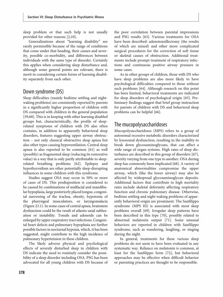
sleep problem or that such help is not usuallyprovided for other reasons [2,10].
Generalizations about “learning disability” arerarely permissible because of the range of conditionsthat come under that heading, their causes and sever-ity, possible co-morbidity, and differences betweenindividuals with the same type of disorder. Certainlythis applies when considering sleep disturbance and,although some general points are relevant, there ismerit in considering certain forms of learning disabil-ity separately from each other.
Down syndrome (DS)Sleep difficulties (mainly bedtime settling and night-waking problems) are consistently reported by parentsin a significantly higher proportion of children withDS compared with children in the general population[59,60]. This is in keeping with other learning disabledgroups but, characteristically, the profile of sleep-related symptoms of children with DS also oftencontains, in addition to apparently behavioral sleepdisorders, features suggesting upper airway obstruc-tion – not only classical obstructive sleep apnea butalso other types causing hypoventilation. Central sleepapnea is also reported to be common [61] as well(possibly) as fragmented sleep (reducing its restorativevalue) in a way that is only partly attributable to sleep-related breathing problems [62]. Epilepsy andhypothyroidism are other potentially sleep-disruptinginfluences in some children with this syndrome.
Studies suggest OSA may occur in 50% or moreof cases of DS. This predisposition is considered tobe caused by combinations of midfacial and mandibu-lar hypoplasia, large posteriorly placed tongue, congeni-tal narrowing of the trachea, obesity, hypotonia ofthe pharyngeal musculature, or laryngomalacia(Figure 23.1). In some cases of central apnea, brainstemdysfunction could be the result of atlanto-axial sublux-ation or instability. Tonsils and adenoids can beenlarged by upper respiratory tract infections. Congeni-tal heart defects and pulmonary hypoplasia are furtherpossible factors in nocturnal hypoxia, which, it has beensuggested, might contribute to the high incidence ofpulmonary hypertension in these children.
The likely adverse physical and psychologicaleffects of severely disturbed sleep in children withDS indicate the need to routinely consider the possi-bility of a sleep disorder including OSA. PSG has beenadvocated for all young children with DS because of
the poor correlation between parental impressionsand PSG results [63]. Various treatments for OSAhave been described: adenotonsillectomy (the resultsof which are mixed) and other more complicatedsurgical procedures for the correction of soft tissueor skeletal causes of obstruction. Additional treat-ments include prompt treatment of respiratory infec-tions and continuous positive airway pressure insome cases.
As in other groups of children, those with DS whohave sleep problems are also more likely to havepsychological difficulties compared to those withoutsuch problems [64]. Although research on this pointhas been limited, behavioral treatments are indicatedfor sleep disorders of psychological origin [65]. Pre-liminary findings suggest that brief group instructionfor parents of children with DS and behavioral sleepproblems can be helpful [66].
The mucopolysaccharidosesMucopolysaccharidoses (MPS) refers to a group ofautosomal recessive metabolic disorders characterizedby lysosomal dysfunction, resulting in the inability tobreak down glycosaminoglycans, that can affect awide range of organ systems. High rates of sleep dis-turbance are described in this group of disorders [67],severity varying from one type to another. OSA duringsleep has commonly been implicated [68]. A variety ofanatomical abnormalities compromise the upperairway, which (like the lower airway) may also beaffected by widespread glycosaminoglycan deposits.Additional factors that contribute to high mortalityrates include skeletal deformity affecting respiratoryfunction and chronic pulmonary disease. Otherwise,bedtime settling and night waking problems of appar-ently behavioral origin are prominent. The Sanfilipposyndrome (MPS III) is associated with most sleepproblems overall [69]. Irregular sleep patterns havebeen described in this type [70], possibly related toabnormal melatonin output [71]. Some unusualbehaviors are reported in children with Sanfilipposyndrome, such as wandering, laughing, or singingduring the night.
In general, treatments for these various sleepproblems do not seem to have been evaluated in anysystematic way. Reliance on melatonin is common, atleast for the Sanfilippo form [72], but behavioralapproaches may be effective when difficult behavioror parenting practices are thought to be responsible.
Section VI: Sleep Disturbance in Psychiatric Illness
378
Prader-Willi syndrome (PWS)PWS, a genetic disorder due to partial deletion ofpaternal chromosome 15q, is classically characterizedby muscular hypotonia, polyphagia and obesity, shortstature, small hands and feet, hypogonadism, and
learning disability (mental retardation). Excessive day-time sleepiness, often seen in this condition, togetherwith the characteristic obesity and noisy respirationduring sleep, suggest that OSA is a usual complication.However, this appears to be so in only a minority ofpatients [73]. Obesity is thought to be a contributory
(c)
(b)
(a)
Figure 23.1 Upper airway anatomy and abnormalities associated with trisomy 21 (Down syndrome). (a) Sagittal MRI demonstratingimportant anatomical regions of the supraglottic airway. Hypopharynx shown as area between posterior aspect of tongue and posterior wall ofpharynx (large arrows). Nasopharynx shown as area between soft palate and region of adenoid tonsils (small arrows). Oropharynx shown asregion between soft palate and anterior aspect of tongue (arrowheads). (b & c) Dynamic (cine) sagittal MRI of a 7 year old girl with trisomy 21diagnosed with obstructive sleep apnea despite tonsillectomy and adenoidectomy at age 3. (b) demonstrates open and (c) demonstratesclosed airway associated with dynamic airway motion. Notice adenoid regrowth seen in both images. (c) Also note hypopharyngeal airwaydecrease in size secondary to glossoptosis and hypopharyngeal collapse (*, adenoids; þ, hypopharyngeal airway; T, tongue; !, glossoptosis).Adapted from Shott SR, Donnelly LF. Cine magnetic resonance imaging: evaluation of persistent airway obstruction after tonsil andadenoidectomy in children with Down syndrome. Laryngoscope. 2004;114(10):1724 1729. Reprinted with permission of John Wiley & Sons, Inc.
379
Chapter 23: Sleep in developmental disorders
factor but not a sufficient explanation for the sleepabnormalities. Instead, both the sleepiness and thevariety of REM sleep abnormalities described in PWSare considered likely to reflect hypothalamic dysfunc-tion resulting in persistent hypoarousal [74]. Centralapnea is also said to be common [75]. Abnormal sleepis considered to be an important but often overlookedfactor contributing to psychological difficulties inpeople with this condition [76].
These etiological uncertainties about the sleep dis-turbance limit treatment possibilities, althoughobesity and OSA should be treated as thoroughly aspossible. Recently, growth hormone treatment of chil-dren with PWS is said to have produced a variety ofbeneficial effects (usually but not always on sleep)including improved psychological functioning [77].
Fetal alcohol spectrum disorders (FASD)FASD refers to combinations of structural, neurocog-nitive, and behavioral effects of prenatal exposure toalcohol. These effects vary in severity, the most severeend of the spectrum being the fetal alcohol syndrome(FAS) in which the children suffer from severe learn-ing disability [78].
Reference is made in some reports of sleep dis-turbance from infancy [79] but, despite the common-ness of FASD, there appears to have been very littlesystematic detailed study of the sleep disorders in thisgroup of children. Experimental animal studies of theeffects of alcohol exposure in early development indi-cate disruption of the suprachiasmatic body clocksystem [80]. This can be expected to cause potentiallyserious sleep–wake cycle disorders.
In addition to this intrinsic predisposition to dis-turbed sleep, a number of other factors forming partof the FASD phenotype (although non-specific in thesense that they commonly accompany other neurode-velopmental disorders) are likely to contribute tosleep problems, in particular behavioral and psychi-atric disturbance [81], including ADHD [82], thereciprocal relationship of which with sleep disturb-ance was mentioned earlier. To the list might beadded parental and psychosocial difficulties withinthe family (which perhaps lay behind the mother’sdrinking while pregnant) and also adverse social cir-cumstances at a later date [83].
In the absence of specific information about thenature and underlying causes of sleep problems inchildren with FASD, in addition to comprehensive
long-term medical and psychiatric care, it is necessaryto fall back on general treatment principles, as men-tioned earlier, such as the possible value of behavioraltreatments for sleep problems of behavioral origin aswell as help for the other features of the conditionas far as possible.
Other learning disability syndromes(see [84] for additional details)Fragile X syndromeFragile X syndrome is a disorder caused by a trinucleo-tide repeat on the X chromosome, leading to intellec-tual disability along with physical characteristics(elongated facies, machroorchidism, and prominentears). Sleep problems are consistently reported byparents of children with this common cause of learn-ing disability, but the nature of the problems is not welldefined. Some children are said to have OSA for whichcontinuous positive airway pressure might be helpfulbut the proportion is unclear. Marked disruption ofsleep microstructure [85] and abnormal melatoninoutput profiles [86] have been reported. The contribu-tions of co-morbid conditions (especially ADHD, aut-istic behavior, and epilepsy) need to be investigatedfurther. Good response to behavioral treatmenthas been claimed [87]. It has also been reported thatclonidine can improve both sleep and behavior.
Tuberous sclerosisTuberous sclerosis complex (TSC) is a multisystemgenetic disorder with prominent signs and symptomsof CNS involvement such as learning disability(mental retardation), epilepsy, and autism. Parents ofchildren with this disorder (of varying degrees ofseverity) also frequently complain of sleep disturbance,mainly settling and night waking problems [88].Again, the origins of this disturbance are difficult toidentify from published accounts. Behavioral factors(including parenting practices) may well need atten-tion. Other likely influences include epilepsy, psychi-atric disorders such as autism, and the various degreesof learning disability associated with the condition.Where children with tuberous sclerosis are reportedto have responded to melatonin for their sleep disturb-ance, it is thought that this has been the result of itshypnotic effect rather than correction of sleep–wakerhythms [89].
Section VI: Sleep Disturbance in Psychiatric Illness
380

Smith-Magenis syndromeSmith-Magenis syndrome is a genetic disorder causedby a mutation of the RAI1 gene on chromosome 17,resulting in learning disability, distinctive facial fea-tures, behavioral difficulties, and sleep disturbance.The various sleep disturbances described in mostchildren with this syndrome are generally non-specific although abnormal patterns of melatonin pro-duction, resulting in an advanced sleep phase, have beensuggested as a cause, possibly reversible with treatment[90]. However, seemingly distinctive features have alsobeen reported to occur from an early age, taking theform of early sleep onset at night, followed by sleepingin short bursts, with prolonged awakenings that termin-ate in very early waking with very active and possibledestructive or hazardous behavior.
Angelman syndromeAngelman syndrome, a genetic disorder due to partialdeletion of maternal chromosome 15q, is typicallycharacterized by learning disability, seizures, stereo-typed movements, and a cheerful demeanor. A highrate of sleep disturbance (especially sleeplessness) hasbeen reported in children with this rare syndromebut the details are yet to be clarified. Sleep-relatedbreathing abnormalities and periodic limbmovementshave recently been reported [91]. Dramatic anddestructive behavior at night has also been reportedin some children with this condition, but its precisenature is unclear. The contribution of behavioral dis-turbance (including ADHD) and epilepsy (bothcommon in children with this condition) is uncertain.Various treatments have been reported including low-dose melatonin, which seemed to improve sleep asjudged by both parental reports and actigraphy [92].
Williams syndromeWilliams syndrome is a genetic disorder caused by adeletion on chromosome 7, resulting in characteristic“elfin” facies, learning disability, and cardiovascularproblems, along with other associated symptoms.Again, the nature of the prominent sleep problemsreported in many children with this syndromeappears to be non-specific and their causes ill defined.Biological factors are suggested by parental reportsimplying respiratory disturbance in some but moreso prominent periodic limb movements in sleep,
which respond to clonazepam, apparently with someimprovements in both sleep and daytime behavior.
Other neurodevelopmental disorders(see [1] for additional details)Craniofacial syndromesUpper airway obstruction is described in the varioussyndromes characterized by craniofacial deformity suchas craniosynostosis syndromes (e.g. Crouzon and Apertsyndrome), micrognathia syndromes (such as Pierre-Robin and Treacher Collins syndrome), Arnold-Chiarimalformation, skeletal disorders such as achondropla-sia, and, as already discussed, Down syndrome [93].
Not all these conditions are necessarily accompan-ied by learning disability, although behavioral disturb-ance likely to affect sleep patterns can be expected inmany cases because of the impact of the conditions onthe child and on the family. This is true, to some extent,of all the other disorders reviewed. The sleep-relatedbreathing disorder usually takes the form of somedegree of OSA, but central apnea is also described inthe skeletal disorders and the Arnold-Chiari malfor-mation. In central sleep apnea no breathing move-ments are seen and there is no air entry into thelungs because the drive to breathe, generated in thebrain, is lost, or because of weakness of the respiratorymuscles. In view of the complexity of the anatomicalabnormalities, treatment may be difficult especially inthe presence of lower airway problems.
Rett syndromeRett syndrome is a neurodevelopmental disordercaused by a mutation on the MECP2 gene on theX chromosome, which typically presents in girls (asmale fetuses with the mutation do not survive to term)with normal development until 6–18 months, followedby regression of language and motor milestones, loss ofpurposeful hand movements, and deceleration in therate of head growth. The vast majority of childrenwith this neurodegenerative condition have been saidto have sleep problems that may worsen as the condi-tion progresses [94]. These are often multiple, takingthe form of various types of sleeplessness and excessivedaytime sleepiness. Laughing, crying, and screamingduring the night has sometimes been reported withsome evidence that this feature lessens with age,although daytime sleepiness might increase [95].
Chapter 23: Sleep in developmental disorders
381

CNS dysfunction, affecting basic circadian sleep–wake rhythms, has been suggested as a cause, butbehavioral factors are also likely in many cases. Epi-lepsy is another common complicating factor as thedisease worsens. Interestingly, the abnormal daytimebreathing patterns, which are common in the syn-drome, do not seem to be closely related to the sleepdisturbance. Only preliminary reports are availableabout the effectiveness of behavioral treatment orthe use of melatonin.
NeurofibromatosisNeurofibromatosis, an autosomal dominant disorderresulting in tumor growth in neural crest cells, mayalso be associated with sleep disturbance. Mention hasbeen made of sleep disturbance as part of the psycho-social problems associated with neurofibromatosis inchildren [96] but without any details being provided.Although sleeplessness was mentioned in this report,in another preliminary parental questionnaire studyonly sleepwalking and sleep terrors were more oftenreported in children with neurofibromatosis type 1compared with children in general [97]. In keepingwith previous findings, much higher rates of behav-ioral disturbance were seen, including ADHD, all ofwhich seem likely to have contributed to (or, at least,been associated with) sleep problems.
Tourette syndrome (TS)This neuropsychiatric disorder characterized by vocaland motor tics poses intriguing questions about theorigin and role of the various associated sleep abnor-malities that have been described from early in thedocumented history of the disorder [98]. It is acknow-ledged that the sleep of children and adolescents withTS is markedly disturbed mainly by difficulty fallingand staying asleep but also by parasomnias.
Interrupted sleep might partly be the result of thepersistence of tics during sleep causing sleep fragmen-tation. An increased tendency to sleepwalking andsleep terrors [99] and frequent periodic limb move-ments during sleep [100] might have the same effect.REM sleep behavior disorder has recently been asso-ciated with TS [101]. However, common co-morbidpsychiatric conditions [102], especially ADHD andobsessive-compulsive disorder, are likely to contrib-ute their own disruptive influence.
Other conditions [1]There have been limited accounts of sleep abnormalitiesin a number of other neurodevelopmental disorders,such as Lesch-Nyhan syndrome, phenylketonuria,Joubert syndrome, Rubinstein-Taybi syndrome, Smith-Lemli-Opitz syndrome, Cornelia de Lange syndrome,and Sotos syndrome. The limited information availabledoes not permit any helpful judgment to be made aboutthe likely nature and extent of the sleep disturbancesinvolved.
SummaryIt is all too evident that sleep disturbance is almost aninevitable accompaniment of the wide range of devel-opmental disorders that have been reviewed. The con-sequences for the child’s psychological development,and the well-being of the family as a whole, can be reallyserious. That being so, it is imperative that carefulattention is routinely paid to the individual child’s sleepas part of overall care, in the hope of early diagnosis ofany sleep disorder and, as far as possible, treatment ofthe sleep disorder itself and associated conditions. Givenaccurate diagnosis and correct choice of treatment usedwith conviction, the prospects of a satisfactory outcomeshould be good for all concerned.
References1. Stores G, Wiggs L. Sleep disturbance in children and
adolescents with disorders of development: itssignificance and management. London: Mac KeithPress; 2001.
2. Wiggs L, Stores G. Sleep problems in children withsevere intellectual disabilities: what help is beingprovided? J Appl Res Intellect. 1996;9:160 165.
3. Robinson AM, Richdale AL. Sleep problems in childrenwith an intellectual disability: parental perceptions ofsleep problems, and views of treatment effectiveness.Child Care Health Dev. 2004;30:139 150.
4. Courchesne E, Karns C, Davis R, et al. Unusual braingrowth patterns in early life in patients with autisticdisorder: an MRI study. Neurology. 2001;57:245 254.
5. Stores G. The protean manifestations of childhoodnarcolepsy and their misinterpretation. Dev Med ChildNeurol. 2006;48:307 310.
6. Stores G. Medication for sleep wake disorders. Arch DisChild. 2003;88:899 903.
7. American Academy of Sleep Medicine. Internationalclassification of sleep disorders, 2nd ed: Diagnostic andCoding Manual. Westchester, IL: American Academy ofSleep Medicine, 2005.
Section VI: Sleep Disturbance in Psychiatric Illness
382
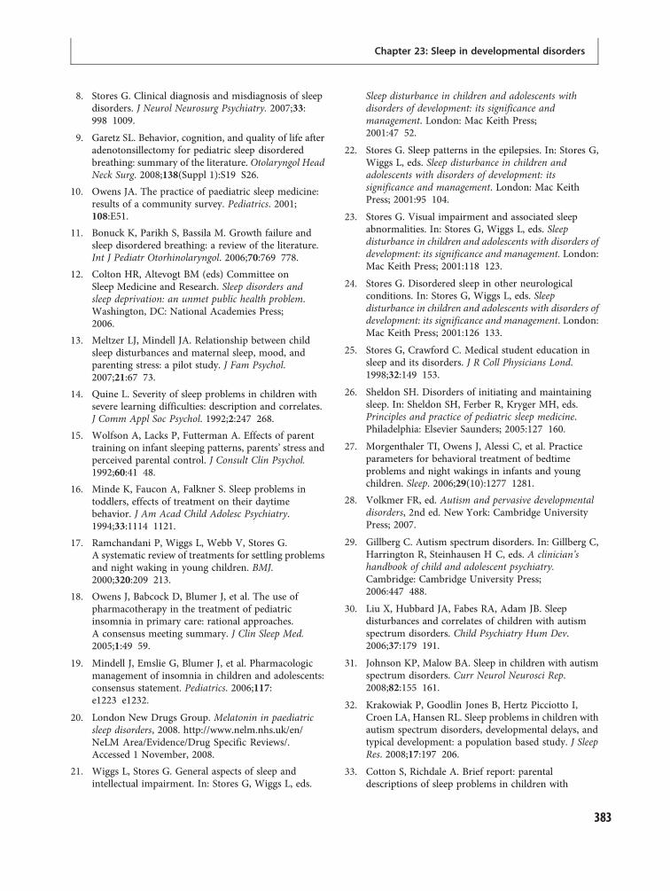
8. Stores G. Clinical diagnosis and misdiagnosis of sleepdisorders. J Neurol Neurosurg Psychiatry. 2007;33:998 1009.
9. Garetz SL. Behavior, cognition, and quality of life afteradenotonsillectomy for pediatric sleep disorderedbreathing: summary of the literature. Otolaryngol HeadNeck Surg. 2008;138(Suppl 1):S19 S26.
10. Owens JA. The practice of paediatric sleep medicine:results of a community survey. Pediatrics. 2001;108:E51.
11. Bonuck K, Parikh S, Bassila M. Growth failure andsleep disordered breathing: a review of the literature.Int J Pediatr Otorhinolaryngol. 2006;70:769 778.
12. Colton HR, Altevogt BM (eds) Committee onSleep Medicine and Research. Sleep disorders andsleep deprivation: an unmet public health problem.Washington, DC: National Academies Press;2006.
13. Meltzer LJ, Mindell JA. Relationship between childsleep disturbances and maternal sleep, mood, andparenting stress: a pilot study. J Fam Psychol.2007;21:67 73.
14. Quine L. Severity of sleep problems in children withsevere learning difficulties: description and correlates.J Comm Appl Soc Psychol. 1992;2:247 268.
15. Wolfson A, Lacks P, Futterman A. Effects of parenttraining on infant sleeping patterns, parents’ stress andperceived parental control. J Consult Clin Psychol.1992;60:41 48.
16. Minde K, Faucon A, Falkner S. Sleep problems intoddlers, effects of treatment on their daytimebehavior. J Am Acad Child Adolesc Psychiatry.1994;33:1114 1121.
17. Ramchandani P, Wiggs L, Webb V, Stores G.A systematic review of treatments for settling problemsand night waking in young children. BMJ.2000;320:209 213.
18. Owens J, Babcock D, Blumer J, et al. The use ofpharmacotherapy in the treatment of pediatricinsomnia in primary care: rational approaches.A consensus meeting summary. J Clin Sleep Med.2005;1:49 59.
19. Mindell J, Emslie G, Blumer J, et al. Pharmacologicmanagement of insomnia in children and adolescents:consensus statement. Pediatrics. 2006;117:e1223 e1232.
20. London New Drugs Group. Melatonin in paediatricsleep disorders, 2008. http://www.nelm.nhs.uk/en/NeLM Area/Evidence/Drug Specific Reviews/.Accessed 1 November, 2008.
21. Wiggs L, Stores G. General aspects of sleep andintellectual impairment. In: Stores G, Wiggs L, eds.
Sleep disturbance in children and adolescents withdisorders of development: its significance andmanagement. London: Mac Keith Press;2001:47 52.
22. Stores G. Sleep patterns in the epilepsies. In: Stores G,Wiggs L, eds. Sleep disturbance in children andadolescents with disorders of development: itssignificance and management. London: Mac KeithPress; 2001:95 104.
23. Stores G. Visual impairment and associated sleepabnormalities. In: Stores G, Wiggs L, eds. Sleepdisturbance in children and adolescents with disorders ofdevelopment: its significance and management. London:Mac Keith Press; 2001:118 123.
24. Stores G. Disordered sleep in other neurologicalconditions. In: Stores G, Wiggs L, eds. Sleepdisturbance in children and adolescents with disorders ofdevelopment: its significance and management. London:Mac Keith Press; 2001:126 133.
25. Stores G, Crawford C. Medical student education insleep and its disorders. J R Coll Physicians Lond.1998;32:149 153.
26. Sheldon SH. Disorders of initiating and maintainingsleep. In: Sheldon SH, Ferber R, Kryger MH, eds.Principles and practice of pediatric sleep medicine.Philadelphia: Elsevier Saunders; 2005:127 160.
27. Morgenthaler TI, Owens J, Alessi C, et al. Practiceparameters for behavioral treatment of bedtimeproblems and night wakings in infants and youngchildren. Sleep. 2006;29(10):1277 1281.
28. Volkmer FR, ed. Autism and pervasive developmentaldisorders, 2nd ed. New York: Cambridge UniversityPress; 2007.
29. Gillberg C. Autism spectrum disorders. In: Gillberg C,Harrington R, Steinhausen H C, eds. A clinician’shandbook of child and adolescent psychiatry.Cambridge: Cambridge University Press;2006:447 488.
30. Liu X, Hubbard JA, Fabes RA, Adam JB. Sleepdisturbances and correlates of children with autismspectrum disorders. Child Psychiatry Hum Dev.2006;37:179 191.
31. Johnson KP, Malow BA. Sleep in children with autismspectrum disorders. Curr Neurol Neurosci Rep.2008;82:155 161.
32. Krakowiak P, Goodlin Jones B, Hertz Picciotto I,Croen LA, Hansen RL. Sleep problems in children withautism spectrum disorders, developmental delays, andtypical development: a population based study. J SleepRes. 2008;17:197 206.
33. Cotton S, Richdale A. Brief report: parentaldescriptions of sleep problems in children with
Chapter 23: Sleep in developmental disorders
383
autism, Down syndrome, and Prader Willisyndrome. Res Dev Disabil. 2006;27:151 161.
34. McGrew S, Malow BA, Henderson L, et al.Developmental and behavioral questionnaire forautism spectrum disorders. Pediatr Neurol.2007;37:108 116.
35. Richdale A. Sleep in children with autism and Aspergersyndrome. In: Stores G, Wiggs L, eds. Sleep disturbancein children and adolescents with disorders ofdevelopment: its significance and management. London:Mac Keith Press; 2001:179 189.
36. Wiggs L, Stores G. Sleep patterns and sleep disorders inchildren with autistic spectrum disorders: insightsusing parent report and actigraphy. Dev Med ChildNeurol. 2004;46:372 380.
37. Schreck KA, Mulick JA. Parental report of sleepproblems in children with autism. J Autism Dev Disord.2000;30:127 135.
38. Thirumalai SS, Shubin RA, Robinson R. Rapid eyemovement sleep behavior disorder in children withautism. J Child Neurol. 2002;17:173 178.
39. Malow BA, Marzec ML, McGrew SG, et al.Characterizing sleep in children with autism spectrumdisorders: a multidimensional approach. Sleep.2006;29:1563 1571.
40. Miano S, Bruni O, Elia M, et al. Sleep in children withautistic spectrum disorder: a questionnaire andpolysomnographic study. Sleep Med. 2007;9:64 70.
41. Wiggs L, Stores G. A randomised controlled trial ofbehavioural intervention for sleeplessness in childrenwith autism spectrum disorders. J Sleep Res. 2006;15(S1):83.
42. Goodlin Jones BL, Tang K, Liu J, Anders TF. Sleeppatterns in preschool age children with autism,developmental delay, and typical development. J AmAcad Child Adolesc Psychiatry. 2008;47:930 938.
43. Lopez Wagner MC, Hoffman CD, Sweeney DP,Hodge D, Gilliam JE. Sleep problems of parents oftypically developing children and parents of childrenwith autism. J Genet Psychol. 2008;169:245 259.
44. Meltzer LJ. Brief report: sleep in parents of childrenwith autism spectrum disorders. J Pediatr Psychol.2008;33:380 386.
45. Hoffman CD, Sweeney DP, Lopez Wagner MC, et al.Children with autism: sleep problems and mothers’stress. Focus on Autism and other DevelopmentalDisabilities. 2008;23:155 165.
46. Schreck KA. Behavioral treatments for sleep disordersin autism: empirically supported or just universallyaccepted? Behav Interventions. 2001;16:265 278.
47. Richdale AL, Wiggs L. Behavioral approaches to thetreatment of sleep problems in children with
developmental disorders: what is the state of the art?Int J Behav Consult Ther. 2005;1:165 189.
48. Anderson M, Kaczmarska J, McGrew SG, Malow BA.Melatonin for insomnia in children with autisticspectrum disorders. J Child Neurol. 2008;23:482 485.
49. Wasdell MB, Jan JE, Bomben MM, et al.A randomized, controlled trial of controlled releasemelatonin treatment of delayed sleep phase syndromeand impaired sleep maintenance in children withneurodevelopmental disabilities. J Pineal Res.2008;44:57 64.
50. Ming X, Gordon E, Kang N, Wagner GC. Use ofclonidine in children with autistic spectrum disorders.Brain Dev. 2008;30:454 460.
51. Dosman CF, Brian JA, Drmic IE, et al. Children withautism: effect of iron supplementation on sleep andferritin. Pediatr Neurol. 2007;36:152 158.
52. Macintosh KE, Dissanayake C. Annotation:The similarities and differences between autisticdisorder and Asperger’s disorder: a review of theempirical evidence. Child Psychol Psychiatry.2004;45:421 434.
53. Allik H, Larsson JO, Smedje H. Sleep patterns inschool age children with Asperger syndrome or highfunctioning autism: a follow up study. J Autism DevDisord. 2008;38:1625 1633.
54. Hare DJ, Jones S, Evershed K. A comparative study ofcircadian rhythm functioning and sleep in people withAsperger syndrome. Autism. 2006;10:565 575.
55. Polimeni MA, Richdale AL, Francis AJ. A survey ofsleep problems in autism, Asperger’s disorder andtypically developing children. J Intellect Disabil Res.2005;9:260 268.
56. Wiggs L, Stores G. Behavioural treatment for sleepproblems in children with severe learning disabilitiesand challenging daytime behaviour: effect ondaytime behaviour. J Child Psychol Psychiat.199;40:627 635.
57. Wiggs L, Stores G. Behavioural treatment for sleepproblems in children with severe intellectualdisabilities and daytime challenging behaviour: effecton mothers and fathers. Br J Health Psychology.2001;6:257 269.
58. Montgomery P, Stores G, Wiggs L. The relativeefficacy of two brief treatments for sleep problems inyoung learning disabled (mentally retarded) children:a randomized controlled trial. Arch Dis Child.2004;89:125 130.
59. Stores R. Sleep and Down syndrome. In: Stores G,Wiggs L, eds. Sleep disturbance in children andadolescents with disorders of development: its
Section VI: Sleep Disturbance in Psychiatric Illness
384
significance and management. London: Mac KeithPress; 2001:53 59.
60. Carter M, McCaughey E, Annaz D, Hill CM. Sleepproblems in a Down syndrome population. Arch DisChild. 2009;94:308 310.
61. Ferri R, Curzi Dascalova L, Del Gracco S, et al.Respiratory patterns during sleep in Down’s syndrome:importance of central apnoea. J Sleep Res.1997;6:34 141.
62. Levanon A, Tarasiuk A, Tal A. Sleep characteristics inchildren with Down syndrome. J Pediatr. 1999;134:755 760.
63. Schott SR, Amin R, Chini B, et al. Obstructive sleepapnoea: should all children with Down syndrome betested? Arch Otolaryngol Head Neck Surg.2006;132:432 436.
64. Stores R, Stores G, Fellows B, Buckley S. A factoranalysis of sleep problems and their psychologicalassociations in children with Down syndrome. J ApplRes Intellect. 2004;17:61 70.
65. Lucas P, Liabo K, Roberts H. Do behaviouraltreatments for sleep disorders in children with Down’ssyndrome work? Arch Dis Child. 2002;87:413 414.
66. Stores R, Stores G. Evaluation of briefgroup administered instruction for parents toprevent or minimize sleep problems in young childrenwith Down syndrome. J Appl Res Intellect. 2004;17:60 71.
67. Colville G, Bax M. Sleep problems in childrenwith mucopolysaccharidosis. In: Stores G, Wiggs L,eds. Sleep disturbance in children and adolescentswith disorders of development: its significance andmanagement. London: Mac Keith Press; 2001:73 78.
68. Leighton SE, Papsin B, Vellodi A, Dinwiddie R,Lane R. Disordered breathing during sleep in patientswith mucopolysaccharidoses. Int J Pediatr Otolaryngol.2001;58:127 138.
69. Fraser J, Gason AA, Wraith JE, Delatychi MB.Sleep disturbance in Sanfillipo syndrome: a parentalquestionnaire. Arch Dis Child. 2005;90:1239 1242.
70. Mariotti P, Della Marca G, Iuvone L, et al. Sleepdisorders in Sanfillipo syndrome: a polygraphic study.Clin Elecroencephalogr. 2003;34:18 22.
71. Guerrero JM, Pozo D, Diaz Rodriguez JL,Martinez Cruz F, Vela Campos F. Impairment of themelatonin rhythm in children with Sanfilliposyndrome. J Pineal Res. 2006;40:192 193.
72. Fraser J, Wraith JE, Delatycki MB. Sleep disturbance inmucopolysaccharidosis type III (Sanfillipo syndrome):a survey of managing clinicians. Clin Genet.2002;62:418 421.
73. Vela Bueno A, Olivan Palacios J, Vgontzas AN. Sleepdisorders and Prader Willi syndrome. In: Stores G,Wiggs L, eds. Sleep disturbance in children andadolescents with disorders of development: itssignificance and management. London: Mac KeithPress; 2001:60 63.
74. Camfferman D, McEvoy RD, O’Donoghue F,Lushington K. Prader Willi Syndrome and excessivedaytime sleepiness. Sleep Med Rev. 2008;12:65 75.
75. Festen DA, de Weerd AW, van den Bossche RA, et al.Sleep related breathing disorders in pre pubertalchildren with Prader Willi syndrome and effects ofgrowth hormone treatment. J Clin Endocrinol Metab.2006;91:4911 4915.
76. Camfferman D, Lushington K, O’Donoghue F,McEvoy RD. Obstructive sleep apnoea syndrome inPrader Willi syndrome: an unrecognized anduntreated cause of cognitive and behavioral deficits.Neuropsychol Rev. 2006;16:123 129.
77. Myers SE, Whitman BY, Carrel AL, et al. Two years ofgrowth hormone therapy in young children with PraderWilli syndrome: physical and neurodevelopmentalbenefits. Am J Genet A. 2007;143:443 448.
78. Manning MA, Hoyme HE. Fetal alcohol spectrumdisorders: a practical clinical approach to diagnosis.Neurosci Biobehav Rev. 2007;31:230 238.
79. Burd L, Wilson H. Fetal, infant, and child mortality ina context of alcohol use. Am J Med Genet C Semin MedGenet. 2004;127C:51 58.
80. Spanagel R, Rosenwasser AM, SchumannG, Sarkar DK.Alcohol consumption and the body’s biological clock.Alcohol Clin Exp Res. 2005;29:1550 1557.
81. Streissguth AP, O’Malley K. Neuropsychiatricimplications and long term consequences of fetalalcohol spectrum disorders. Semin ClinNeuropsychiatry. 2000;5:177 190.
82. O’Malley KD, Storoz L. Fetal alcohol spectrumdisorder and ADHD: diagnostic implications andtherapeutic consequences. Exp Rev Neurotherapeutics.2003;3:477 489.
83. Streissguth AP, Bookstein FL, Barr HM, et al. Riskfactors for adverse life outcomes in fetal alcoholsyndrome and fetal alcohol effects. J Dev Behav Pediatr.2004;25:228 238.
84. Stores G. Aspects of sleep in other neurodevelopmentaldisorders. In: Stores G, Wiggs L, eds. Sleep disturbancein children and adolescents with disorders ofdevelopment: its significance and management. London:Mac Keith Press; 2001:87 93.
85. Miano S, Bruni O, Elia M, et al. Sleep phenotypes ofintellectual disability: A polysomnographic evaluationin subjects with Down syndrome and Fragile Xsyndrome. Clin Neurophysiol. 2008;119:1242 1247.
Chapter 23: Sleep in developmental disorders
385
86. Gould El, Loesch DZ, Martin MJ, et al. Melatoninprofiles and sleep characteristics in boys with fragileX syndrome: a preliminary study. Am J Med Genet.2000;95:307 315.
87. Weiskop S, Richdale A, Matthews J. Behaviouraltreatment to reduce sleep problems in children withautism or fragile X syndrome. Dev Med Child Neurol.2005;47:94 104.
88. Curatolo P, Sen S. Sleep disturbances in tuberoussclerosis complex. In: Stores G, Wiggs L, eds. Sleepdisturbance in children and adolescents with disorders ofdevelopment: its significance and management. London:Mac Keith Press; 2001:79 82.
89. Hancock E, O’Callaghan F, English J, Osborne JP.Melatonin excretion in normal children and in tuberoussclerosis complex with sleep disorder responsive tomelatonin. J Child Neurol. 2005;20:21 25.
90. De Leersnyder H. Inverted rhythm of melatoninsecretion in Smith Magenis syndrome: fromsymptoms to treatment. Trends Endocrinol Metab.2006;17:291 298.
91. Miano S, Bruni O, Elia M, et al. Sleep breathing andperiodic leg movement pattern in Angelmansyndrome: a polysomnographic study. ClinNeurophysiol. 2005;116:2685 2692.
92. Braam W, Didden R, Smits MG, Curfs LM. Melatoninfor chronic insomnia in Angelman syndrome: arandomized placebo controlled trial. Child Neurol.2008;23:649 654.
93. Simakajornboom N, Beckman R. Sleep andcraniofacial syndromes. In: Stores G, Wiggs L, eds.Sleep disturbance in children and adolescents withdisorders of development: its significance andmanagement. London: Mac Keith Press; 2001:64 72.
94. Roane HS, Piazza CC. Sleep disorders and Rettsyndrome. In: Stores G, Wiggs L, eds. Sleep disturbancein children and adolescents with disorders ofdevelopment: its significance and management. London:Mac Keith Press; 2001:83 86.
95. Young D, Nagarajan L, de Klerk N, et al. Sleepproblems in Rett syndrome. Brain Dev. 2007;29:609 617.
96. Wadsby M, Lindehammer H, Eeg Olofsson O.Neurofibromatosis in childhood: neuropsychologicalaspects. Neurofibromatosis. 1989:2:251 260.
97. Johnson H, Wiggs L, Stores G, Huson SM.Psychological disturbance and sleep disorders inchildren with neurofibromatosis type 1. Dev Med ChildNeurol. 2005;47:237 242.
98. Kostanecka Endress T, Banaschewski T, Kinkelbur J,et al. Disturbed sleep in children with Tourettesyndrome: a polysomnographic study. J PsychosomRes. 2003;55:23 29.
99. Barabas G, Matthews WS, Ferrari M. Disorders ofarousal in Gilles de la Tourette’s syndrome. Neurology.1984;34:815 817.
100. Verderholzer U, Muller N, Haag C, Riemann D,Struabe A. Periodic limb movements during sleep are afrequent finding in patients with Gilles de la Tourettesyndrome. J Neurol. 1997;244:521 526.
101. Trajanovic NN, Voloh I, Shapiro, CM, Sandor P.REM sleep behaviour disorder in a child withTourette’s syndrome. Can J Neurol Sci.2004;31:572 575.
102. Freeman RD. Tourette Syndrome InternationalDatabase Consortium. Eur Child Adolesc Psychiatry.2007;16(Suppl 1):15 23.
Section VI: Sleep Disturbance in Psychiatric Illness
386
Section VIIChapter
24Future Directions
The future at the sleep–psychiatry interface
David T. Plante and John W. Winkelman
IntroductionThe preceding chapters in this volume have discussedthe development of sleep medicine and psychiatry,and detailed the current state of knowledge at theinterface of these fields. This chapter will synthesizesome key themes from the book and discuss futureclinical and research directions in psychiatric sleepmedicine. We recognize it is a daunting task to dis-cuss the future of any field of medicine, as unforeseenscientific, economic, and social developments willundoubtedly arise, affecting any predictions made.Thus, the primary goal of this chapter will be todiscuss pending developments on the horizon, prom-ising lines of research that may or may not ultimatelyprove fruitful, and future questions and challengesfaced by both fields.
The challenge and promiseof bidirectionalityAs demonstrated in numerous chapters throughoutthis book, sleep disturbance is far more than a meresymptom, “secondary” to psychiatric illness. Oldermodels in which sleep complaints were assumed tobe caused by an underlying psychiatric disorder areinsufficient to explain the more complex interrela-tionships between sleep and mental illness. The newparadigm which is gaining broader acceptance is onein which sleep disturbance and psychiatric disordersshare a more complicated bidirectional relationship,in which both influence the natural history of eachother.
Evidence for the bidirectional model is clearlystrongest for insomnia and major depressive dis-order [1]. The current evidence suggests insomnia isnot only a symptom of active depression, but plays an
important role in the course of depression itself. Thereis very consistent and substantial evidence that insom-nia puts individuals at risk for the development ofincident depression [2–5] (as well as anxiety and sub-stance use disorders). Furthermore, sleep disturbanceis themost common residual symptom associated withtreated depression, negatively affects quality of life[6,7], and increases the risk of depressive relapse inremitted patients [8,9]. On the other hand, the com-bination of sedative-hypnotic medications with anti-depressant therapies leads to faster andmore thoroughantidepressant responses than can be accounted for bytheir amelioration of sleep disturbance alone [10,11].Similarly, cognitive behavioral therapy for insomnia(CBT-I) may have similar effects when utilized indepressed patients [12]. All of these lines of evidenceargue against the notion that insomnia is a mere epi-phenomenon of psychiatric illness, and provide sub-stantial support for the bidirectional relationshipbetween insomnia and depression.
Despite growing appreciation for the complexitiesof the relationship between sleep and psychiatric dis-orders, much future work will be required to under-stand how and under what circumstances theserelationships might be leveraged into meaningfuladvances in clinical practice. Despite data in majordepression demonstrating that treatment of co-occurring insomnia may improve the psychiatric dis-order in excess of direct effects on sleep, it is not clearhow treating sleep disturbance will affect other func-tional outcomes in depression. For example, althoughthere is evidence that sleep disturbance is associatedwith increased risk of suicidality [13–17], it is notclear whether treating insomnia with hypnotic medi-cations in depressed patients affects the risk of sui-cide, and if it does, whether this is a positive or
Foundations of Psychiatric SleepMedicine, ed. JohnW.Winkelman and David T. Plante. Published by Cambridge University Press.# Cambridge University Press 2010.
387
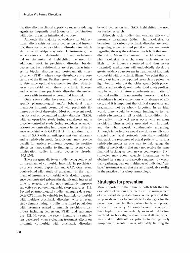
negative effect, as clinical experience suggests sedatingagents are frequently used (alone or in combinationwith other drugs) in intentional overdose.
Although the majority of evidence for bidirec-tional effects exists for major depression and insom-nia, there are other psychiatric disorders for whichsimilar relationships may exist. Unfortunately, theevidence for such relationships is oftentimes inferen-tial or circumstantial, highlighting the need foradditional work in psychiatric disorders besidesdepression. Such relationships are of particular inter-est in bipolar disorder and post-traumatic stressdisorder (PTSD), where sleep disturbance is a corefeature of the illness. Further research will be crucialto determine optimal treatments for sleep disturb-ance co-morbid with these psychiatric illnessesand whether these psychiatric disorders themselvesimprove with treatment of co-morbid insomnia.
Only a few studies have examined the effects ofspecific pharmacological and/or behavioral treat-ments for insomnia co-morbid with psychiatric ill-nesses outside of depression. Most of the recent workhas focused on generalized anxiety disorder (GAD),with an open-label study (using ramelteon) and aplacebo-controlled study (using zolpidem extended-release) demonstrating improvement in sleep disturb-ance associated with GAD [18,19]. In addition, treat-ment of GAD with an antidepressant (escitalopram)and a sedative-hypnotic (eszopiclone) may providebenefit for anxiety symptoms beyond the positiveeffects on sleep, similar to findings in recent coad-ministration studies in major depressive disorder[10,11,20].
There are generally fewer studies being conductedon treatment of co-morbid insomnia in psychiatricdisorders beyond depression and GAD. One recentdouble-blind pilot study of gabapentin in the treat-ment of insomnia co-morbid with alcohol depend-ence demonstrated gabapentin significantly increasedtime to relapse, but did not significantly improvesubjective or polysomnographic sleep measures [21].Beyond pharmacological studies, emerging data sug-gests CBT-I may be valuable for insomnia co-morbidwith multiple psychiatric disorders, with a recentstudy demonstrating its utility in a mixed populationwith insomnia related to multiple psychiatric dis-orders including depression, PTSD, and substanceuse [22]. However, the recent literature is certainlyless developed when evaluating treatment effects oninsomnia co-morbid with psychiatric disorders
beyond depression and GAD, highlighting the needfor further research.
Although such studies that evaluate efficacy ofinsomnia treatments (either pharmacological orbehavioral) in various psychiatric disorders are usefulin guiding evidence-based practice, there are caveatsregarding the way the evidence base is built that meritdiscussion. Given the current financial climate inpharmacological research, many such studies arelikely to be industry sponsored and thus newer(patented) medications will undoubtedly develop agreater evidence base for use in treatment of insomniaco-morbid with psychiatric illness. We point this outnot to cast industry-supported research in a pejorativelight, but to point out that older agents (with provenefficacy and relatively well-understood safety profiles)may be left out of future experiments as a matter offinancial reality. It is noteworthy, however, that lackof evidence is not synonymous with absence of effi-cacy, and it is important that clinical experience andpragmatism not be wholly forgotten. In an idealworld, there would be enough funding to test allsedative-hypnotics in all psychiatric conditions, butthe reality is this will never occur with so manypsychiatric illnesses being associated with insomniaand the pharmacopeia continuously expanding.Although imperfect, we would envision carefully con-structed open-label protocols (potentially multisite)that track the responses of actual patients to varioussedative-hypnotics as one way to help gauge theutility of medications that may not receive the samefinancial backing as their newer counterparts. Suchstrategies may allow valuable information to beobtained in a more cost-effective manner, by essen-tially gathering data on multitudes of individual “off-label” treatment trials that are an unavoidable realityin the practice of psychopharmacology.
Strategies for preventionMore important to the future of both fields than theevaluation of various treatments in the managementof co-morbid sleep disturbance is the potential thatsleep medicine has to contribute to strategies for theprevention of mental illness, which has largely provenelusive in psychiatry. Although beyond the scope ofthis chapter, there are certainly sociocultural factorsinvolved, such as stigma about mental illness, whichmay make it difficult for patients to divulge earlysymptoms of mental illness, ultimately limiting the
Section VII: Future Directions
388
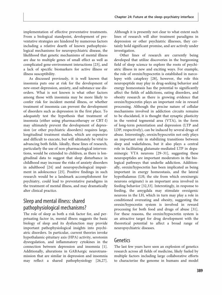
implementation of effective preventative treatments.From a biological standpoint, development of pre-ventative strategies are hindered by numerous factorsincluding a relative dearth of known pathophysio-logical mechanisms for neuropsychiatric disease, thelikelihood that genetic mechanisms of mental illnessare due to multiple genes of small effect as well ascomplicated gene-environment interactions [23], anda lack of specific biomarkers for active illness orillness susceptibility.
As discussed previously, it is well known thatinsomnia puts one at risk for the development ofnew-onset depression, anxiety, and substance use dis-orders. What is not known is what other factorsamong those with insomnia may be more likely toconfer risk for incident mental illness, or whethertreatment of insomnia can prevent the developmentof disorders such as depression in the first place. Toadequately test the hypothesis that treatment ofinsomnia (either using pharmacotherapy or CBT-I)may ultimately prevent the development of depres-sion (or other psychiatric disorders) requires large,longitudinal treatment studies, which are expensiveand difficult to execute, but will ultimately be vital toadvancing both fields. Ideally, these lines of research,particularly the use of non-pharmacological interven-tions, would be extended to children, as there is lon-gitudinal data to suggest that sleep disturbance inchildhood may increase the risks of anxiety disordersin adulthood [24] and neuropsychological impair-ment in adolescence [25]. Positive findings in suchresearch would be a landmark accomplishment forpsychiatry, could lead to preventative paradigms inthe treatment of mental illness, and may dramaticallyalter clinical practice.
Sleep and mental illness: sharedpathophysiological mechanisms?The role of sleep as both a risk factor for, and per-petuating factor in, mental illness suggests the basicbiology of sleep and its dysfunction may provideimportant pathophysiological insights into psychi-atric disorders. In particular, current theories invokehypothalamic-pituitary axis (HPA) activity, serotonindysregulation, and inflammatory cytokines in theconnection between depression and insomnia [1].Additionally, alterations in GABAergic neurotrans-mission that are similar in depression and insomniamay reflect a shared pathophysiology [26,27].
Although it is presently not clear to what extent suchlines of research will alter treatment paradigms indepression or other psychiatric illnesses, they cer-tainly hold significant promise, and are actively underinvestigation.
Other lines of research are currently beingdeveloped that utilize discoveries in the burgeoningfield of sleep science to explore the roots of psychi-atric illness in new and exciting ways. For example,the role of orexin/hypocretin is established in narco-lepsy with cataplexy [28], however, the role thisneuropeptide may play in drug-seeking behavior andenergy homeostasis has the potential to significantlyaffect the fields of addictions, eating disorders, andobesity research as there is growing evidence thatorexin/hypocretin plays an important role in rewardprocessing. Although the precise nature of cellularmechanisms involved in addiction circuits remainsto be elucidated, it is thought that synaptic plasticityin the ventral tegmental area (VTA), in the formof long-term potentiation and depression (LTP andLDP, respectively), can be induced by several drugs ofabuse. Interestingly, orexin/hypocretin not only playsan important role in stabilizing transitions betweensleep and wakefulness, but it also plays a centralrole in facilitating glutamate-mediated LTP in dopa-minergic VTA neurons [29–31], suggesting theseneuropeptides are important moderators in the bio-logical pathways that underlie addiction. Addition-ally, orexin/hypocretin has been implicated as beingimportant in energy homeostasis, and the lateralhypothalamus (LH; the site from which orexinergicneurons originate) is an important area involved infeeding behavior [32,33]. Interestingly, in response tofeeding, the amygdala may stimulate orexigenicneurons in the LH, which in turn may play a role inconditioned overeating and obesity, suggesting theorexin/hypocretin system is involved in rewardprocessing for both food and drugs of abuse [31].For these reasons, the orexin/hypocretin system isan attractive target for drug development with thetheoretical potential to affect a broad range ofneuropsychiatric diseases.
GeneticsThe last few years have seen an explosion of geneticsresearch across all fields of medicine, likely fueled bymultiple factors including large collaborative effortsto characterize the genome in humans and model
Chapter 24: Future at the sleep–psychiatry interface
389

species, the advent of genome-wide associationstudies, and technological advances that increasethroughput and reduce the cost of genetic analyses.Although genetic loci that confer substantial risk forthe development of psychiatric disorders remain elu-sive, some recent findings in the field of pharmaco-genetics may ultimately prove important in theclinical management of psychiatric disorders. Therehave been a host of candidate genes (at several levelsof pharmacobiology including: serotonergic and nor-adrenergic neurotransmission, the cytochrome P450system, brain-derived neurotrophic factor, and G-proteins) that may be linked with treatment responsein mood and anxiety disorders [34]. Interestingly, theshort allele polymorphism of serotonin transportergene (5-HTTLPR), which has been associated withincreased risk for affective disorders (particularly inresponse to stressors), has recently demonstratedassociation with primary insomnia, suggesting a gen-etic link between these disorders [35]. Furthermore,given that other primary sleep disorders such asobstructive sleep apnea (OSA) and restless legs syn-drome (RLS) have high rates of co-morbid psychiatricdisorders and often present with neurocognitive symp-toms, studies that examine the association of thesecandidate genes with primary sleep disorders and/orhow these genes may relate to therapeutic responsemay be a fruitful avenue of research for sleepmedicine.
Just as candidate genes from psychiatric researchmay inform research in primary sleep disorders, gen-etic discoveries from sleep medicine may influencethe field of psychiatry. For example, single nucleotidepolymorphisms (SNPs) have recently been identifiedthat confer the majority of population risk for idio-pathic restless legs syndrome (RLS) and periodic limbmovements of sleep (PLMS) [36–38]. How thesegenes are related to secondary forms of RLS/PLMS(e.g. due to iron deficiency, renal failure) are not wellunderstood and are an active area of research.Notably, since RLS/PLMS is frequently induced byantidepressant treatment [39,40], these polymorph-isms could conceivably be associated with increasedrisk of antidepressant-induced RLS/PLMS, or theor-etically, be linked to treatment resistance or sleepdisturbance in psychiatric patients treated with anti-depressants. Beyond such theoretical links to psychi-atric illness, it is noteworthy that one of the RLS/PLMS-associated SNPs (BTBD9 gene) has alreadybeen associated with Tourette syndrome (particularlywithout obsessive-compulsive disorder), suggesting
a common genetic predisposition among these dis-orders [41], strengthening the argument that thesegenes may be important in neuropsychiatric disease,especially those with hyperkinesia.
In addition to genes related to the risk of primarysleep disorders and their potential associations withneuropsychiatric disorders, strategies that utilize sleepand circadian endophenotypes to untangle the com-plicated genetic risks underlying mental illness mayalso prove valuable. Endophenotypes can broadly bedefined as heritable, primarily state-independent bio-markers that are associated with an illness, with co-segregation of the illness and endophenotype withinfamilies [42]. An example of the potential for sleepand circadian endophenotypes in genetic researchof complex psychiatric diseases is bipolar disorder,with proposed endophenotypes involving circadianrhythm instability, cholinergic sensitivity (and itseffects on REM sleep), and response to sleep depriv-ation. These intermediate phenotypes may ultimatelyhelp parse out the underlying genetic mechanisms ofthis heritable psychiatric disorder, which have thus farremained elusive.
BiomarkersAlthough circadian endophenotypes and their associ-ated genes may eventually prove important in predict-ing the risk, presentation, treatment, course, andpathophysiological mechanisms of psychiatric illness,other sleep-related biomarkers may have similarpotential to one day affect the clinical practice of bothsleep medicine and psychiatry. The lack of reliablebiomarkers for mental illness has limited diagnosticprecision, prediction of at-risk individuals, develop-ment of preventative strategies, understanding of bio-logical pathophysiology, and ultimately, hinderedthe development of successful psychiatric treatments.New technologies in sleep science, particularly theuse of high-density electroencephalography (hd-EEG)and other neuroimaging modalities, offer the hope offinding biomarkers for mental illness beyond what hasbeen typically gleaned from overnight polysomno-graphy. Sleep recordings may be particularly advanta-geous when looking for psychiatric biomarkersbecause confounding activities that may occur duringwakefulness, such as fluctuating levels of attention andmotivation, and the expression of active (e.g. psych-otic, depressive) symptoms, may be minimized. Forexample, using hd-EEG polysomnography, sleep
Section VII: Future Directions
390

spindle activity is significantly decreased incentroparietal regions in schizophrenia, with inte-grated spindle activity providing significant separation(>90%) between schizophrenic subjects and compari-son groups [43]. Although these results requirereplication, the potential for such a sleep-related bio-marker to alter our diagnostic capabilities in psychoticillness is a tantalizing prospect, and might furtherour understanding of the pathophysiology of thedisorder. Beyond schizophrenia, hd-EEG may haveother important applications in sleep medicine andpsychiatry, particularly when combined with othermodalities such as transcranial magnetic stimulation(TMS) and/or used to identify sensitive slow-waveparameters that may reflect changes in synapticstrength across the night [44–46]. Additionally, com-bining other neuroimaging modalities (e.g. functionalmagnetic resonance imaging (fMRI), magnetic reson-ance spectroscopy (MRS), etc.) with hd-EEG in thesame study population may provide greater insightsinto the pathophysiologies of numerous sleep andpsychiatric disorders.
It is not without some hesitancy that we discussthe potential of new sleep-related technologies to alterthe course of psychiatry. In decades past, similardeclarations were made regarding the use of sleeparchitecture, specifically changes in REM sleep asmarkers for depression. Over time, however, overtsleep architectural changes (decreased REM latency,changes in slow-wave sleep, etc.), although physiolo-gically robust markers of depressive illness, failed todemonstrate the sensitivity and specificity sufficientto be useful in clinical psychiatric practice [47]. Still,the use of new technologies that go beyond traditionalsleep staging and better characterize activity in thesleeping brain do seem to hold translational promisein distinguishing psychiatric states, biomarkers fordisease susceptibility, and potentially shedding lighton pathophysiological mechanisms.
Treatments on the horizonPharmacotherapies at the nexus of sleep medicine andpsychiatry are slowly moving from agents discoveredlargely through serendipity to those developed usingtranslational approaches due to an enhanced under-standing of the processes involved in the regulation ofsleep and the pathophysiology of sleep disorders. Inthis way, arousal pathways using cholinergic, adrener-gic, histaminergic, orexinergic, and serotonergic
neurotransmission, as well as inhibitory pathwaysemploying g-aminobutyric acid (GABA), havebecome valid targets of novel pharmacotherapies[48]. Notably, the agents we will discuss in this sectionare not an exhaustive list of pharmacotherapies in thepipeline; however, they are novel and have somepromise to eventually expand the pharmacopeia.Additionally, we recognize that none of these agentsare approved by the Food and Drug Administration(FDA), and may ultimately never be used in clinicalpopulations. However, given the scope of this chapter,a brief discussion of these future pharmacotherapiesis clearly indicated.
Agents that affect serotonergic systems in thebrain have played a key role in the development ofpharmacotherapies for a number of psychiatric dis-eases. More recently, drugs that specifically targetcertain subtypes of the serotonin (5-HT) receptorhave moved into the pipeline as potential treatmentsfor insomnia. Part of the rationale for targeting the5-HT receptor system is related to its effects on slow-wave sleep (SWS). Within the seven serotonin recep-tor subfamilies (5-HT1– 5-HT7), the 5-HT2 receptorsubfamily (composed of three subtypes: 5-HT2A,5-HT2B, and 5-HT2C) seems to play the greatest rolein the modulation of SWS [49,50], with effects onSWS most likely mediated largely through the5-HT2A receptor subtype. In fact, it is thought thatrelatively higher affinities for the 5-HT2A receptor byziprasidone and olanzapine compared to other anti-psychotics may account for the dramatic increases inSWS that can be observed with the use of thesemedications [51]. The use of agents that target5-HT2 receptors for the treatment of insomnia is notnovel, as many antipsychotics and the antidepressantsmirtazapine and trazodone have 5HT2 receptor affin-ity and are used as off-label treatments for insomnia.Unfortunately, these drugs also have significant affin-ities for a number of other receptor types (e.g. D2, a1-adrenergic, H1) which may contribute to their otherknown clinical effects (e.g. neuroleptic and thymolep-tic properties), but lead to unwanted side-effects (e.g.extrapyriamidal symptoms, orthostatic hypotension,and hyperphagia/weight gain) when used to treatinsomnia. As a result, a number of more specific 5-HT2A receptor antagonists or inverse agonists arecurrently under development for the treatment ofinsomnia [52]. Some of these agents have shownpromise in phase II/III clinical trials, but it is unclearif or when any of these agents will come to market, as
Chapter 24: Future at the sleep–psychiatry interface
391
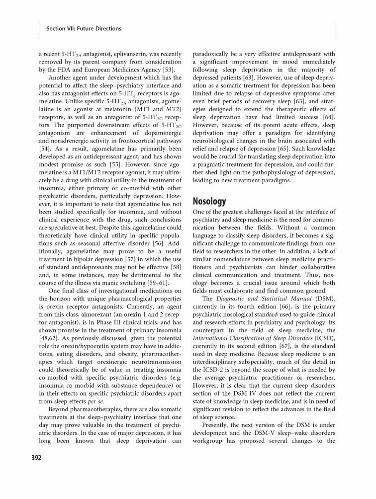
a recent 5-HT2A antagonist, eplivanserin, was recentlyremoved by its parent company from considerationby the FDA and European Medicines Agency [53].
Another agent under development which has thepotential to affect the sleep–psychiatry interface andalso has antagonist effects on 5-HT2 receptors is ago-melatine. Unlike specific 5-HT2A antagonists, agome-latine is an agonist at melatonin (MT1 and MT2)receptors, as well as an antagonist of 5-HT2C recep-tors. The purported downstream effects of 5-HT2C
antagonism are enhancement of dopaminergicand noradrenergic activity in frontocortical pathways[54]. As a result, agomelatine has primarily beendeveloped as an antidepressant agent, and has shownmodest promise as such [55]. However, since ago-melatine is aMT1/MT2 receptor agonist, it may ultim-ately be a drug with clinical utility in the treatment ofinsomnia, either primary or co-morbid with otherpsychiatric disorders, particularly depression. How-ever, it is important to note that agomelatine has notbeen studied specifically for insomnia, and withoutclinical experience with the drug, such conclusionsare speculative at best. Despite this, agomelatine couldtheoretically have clinical utility in specific popula-tions such as seasonal affective disorder [56]. Add-itionally, agomelatine may prove to be a usefultreatment in bipolar depression [57] in which the useof standard antidepressants may not be effective [58]and, in some instances, may be detrimental to thecourse of the illness via manic switching [59–61].
One final class of investigational medications onthe horizon with unique pharmacological propertiesis orexin receptor antagonists. Currently, an agentfrom this class, almorexant (an orexin 1 and 2 recep-tor antagonist), is in Phase III clinical trials, and hasshown promise in the treatment of primary insomnia[48,62]. As previously discussed, given the potentialrole the orexin/hypocretin system may have in addic-tions, eating disorders, and obesity, pharmacother-apies which target orexinergic neurotransmissioncould theoretically be of value in treating insomniaco-morbid with specific psychiatric disorders (e.g.insomnia co-morbid with substance dependence) orin their effects on specific psychiatric disorders apartfrom sleep effects per se.
Beyond pharmacotherapies, there are also somatictreatments at the sleep–psychiatry interface that oneday may prove valuable in the treatment of psychi-atric disorders. In the case of major depression, it haslong been known that sleep deprivation can
paradoxically be a very effective antidepressant witha significant improvement in mood immediatelyfollowing sleep deprivation in the majority ofdepressed patients [63]. However, use of sleep depriv-ation as a somatic treatment for depression has beenlimited due to relapse of depressive symptoms aftereven brief periods of recovery sleep [63], and strat-egies designed to extend the therapeutic effects ofsleep deprivation have had limited success [64].However, because of its potent acute effects, sleepdeprivation may offer a paradigm for identifyingneurobiological changes in the brain associated withrelief and relapse of depression [65]. Such knowledgewould be crucial for translating sleep deprivation intoa pragmatic treatment for depression, and could fur-ther shed light on the pathophysiology of depression,leading to new treatment paradigms.
NosologyOne of the greatest challenges faced at the interface ofpsychiatry and sleep medicine is the need for commu-nication between the fields. Without a commonlanguage to classify sleep disorders, it becomes a sig-nificant challenge to communicate findings from onefield to researchers in the other. In addition, a lack ofsimilar nomenclature between sleep medicine practi-tioners and psychiatrists can hinder collaborativeclinical communication and treatment. Thus, nos-ology becomes a crucial issue around which bothfields must collaborate and find common ground.
The Diagnostic and Statistical Manual (DSM),currently in its fourth edition [66], is the primarypsychiatric nosological standard used to guide clinicaland research efforts in psychiatry and psychology. Itscounterpart in the field of sleep medicine, theInternational Classification of Sleep Disorders (ICSD),currently in its second edition [67], is the standardused in sleep medicine. Because sleep medicine is aninterdisciplinary subspeciality, much of the detail inthe ICSD-2 is beyond the scope of what is needed bythe average psychiatric practitioner or researcher.However, it is clear that the current sleep disorderssection of the DSM-IV does not reflect the currentstate of knowledge in sleep medicine, and is in need ofsignificant revision to reflect the advances in the fieldof sleep science.
Presently, the next version of the DSM is underdevelopment and the DSM-V sleep–wake disordersworkgroup has proposed several changes to the
Section VII: Future Directions
392
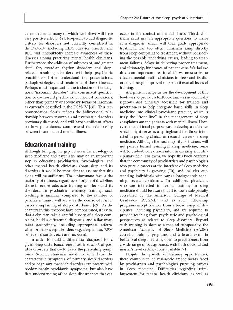
current schema, many of which we believe will havevery positive effects [68]. Proposals to add diagnosticcriteria for disorders not currently considered inthe DSM-IV, including REM behavior disorder andRLS, will undoubtedly increase awareness of theseillnesses among practicing mental health clinicians.Furthermore, the addition of subtypes of, and greaterdetail for, circadian rhythm disorders and sleep-related breathing disorders will help psychiatricpractitioners better understand the presentations,pathophysiologies, and treatments of these illnesses.Perhaps most important is the inclusion of the diag-nosis “insomnia disorder” with concurrent specifica-tion of co-morbid psychiatric or medical conditions,rather than primary or secondary forms of insomniaas currently described in the DSM-IV [68]. This rec-ommendation clearly reflects the bidirectional rela-tionship between insomnia and psychiatric disorderspreviously discussed, and will have significant effectson how practitioners comprehend the relationshipbetween insomnia and mental illness.
Education and trainingAlthough bridging the gap between the nosology ofsleep medicine and psychiatry may be an importantstep in educating psychiatrists, psychologists, andother mental health clinicians about sleep and itsdisorders, it would be imprudent to assume that thisalone will be sufficient. The unfortunate fact is themajority of trainees, regardless of origin of discipline,do not receive adequate training on sleep and itsdisorders. In psychiatric residency training, suchteaching is minimal compared to the number ofpatients a trainee will see over the course of his/hercareer complaining of sleep disturbance [69]. As thechapters in this textbook have demonstrated, it is vitalthat a clinician take a careful history of a sleep com-plaint, build a differential diagnosis, and tailor treat-ment accordingly, including appropriate referralwhen primary sleep disorders (e.g. sleep apnea, REMbehavior disorder, etc.) are suspected.
In order to build a differential diagnosis for agiven sleep disturbance, one must first think of pos-sible disorders that could cause the presenting symp-toms. Second, clinicians must not only know thecharacteristic symptoms of primary sleep disordersand be cognizant that such disorders can present withpredominantly psychiatric symptoms, but also havefirm understanding of the sleep disturbances that can
occur in the context of mental illness. Third, clin-icians must ask the appropriate questions to arriveat a diagnosis, which will then guide appropriatetreatment. Far too often, clinicians jump directlyfrom sleep complaint to treatment, without consider-ing the possible underlying causes, leading to treat-ment failures, delays in delivering proper treatment,and ultimately, hindrance of patient care. We believethis is an important area in which we must strive toeducate mental health clinicians in sleep and its dis-orders, through improved opportunities at all levels oftraining.
A significant impetus for the development of thisbook was to provide a textbook that was academicallyrigorous and clinically accessible for trainees andpractitioners to help integrate basic skills in sleepmedicine into clinical psychiatric practice, which istruly the “front line” in the management of sleepcomplaints among patients with mental illness. How-ever, an additional purpose was to develop a referencewhich might serve as a springboard for those inter-ested in pursuing clinical or research careers in sleepmedicine. Although the vast majority of trainees willnot pursue formal training in sleep medicine, somewill be undoubtedly drawn into this exciting, interdis-ciplinary field. For them, we hope this book confirmsthat the community of psychiatrists and psychologistswho pursue careers at the interface of sleep medicineand psychiatry is growing [70], and includes out-standing individuals with varied backgrounds span-ning several continents. In addition, physicianswho are interested in formal training in sleepmedicine should be aware that it is now a subspecialtyaccredited by the American College of MedicalGraduates (ACGME) and as such, fellowshipprograms accept trainees from a broad range of dis-ciplines, including psychiatry, and are required toprovide teaching from psychiatric and psychologicalperspectives as related to sleep disorders. Beyondsuch training in sleep as a medical subspecialty, theAmerican Academy of Sleep Medicine (AASM)accredits training programs and a board exam inbehavioral sleep medicine, open to practitioners froma wide range of backgrounds, with both doctoral andmaster’s level certifications available [71].
Despite the growth of training opportunities,there continue to be real-world impediments facedby psychiatrists and psychologists pursuing careersin sleep medicine. Difficulties regarding reim-bursement for mental health clinicians, as well as
Chapter 24: Future at the sleep–psychiatry interface
393
interdepartmental politics and economic forces, haveat times led to unfortunate skirmishes as to the pri-mary ownership of sleep medicine as a field as well asthe locations (both physical and proprietary) ofhospital and university-based sleep laboratoriesand clinical services. To combat such “turf wars” anincreasing number of institutions have begun todevelop interdisciplinary centers of sleep medicine,with some degree of autonomy beyond the traditionaldepartmental structure. Additionally, there are pend-ing changes to the field of sleep medicine (e.g. the useof portable monitoring studies to diagnose OSA, andauto-titrating positive airway pressure (PAP) devicesto treat the disorder) that will dramatically alter thefuture landscape of the profession. We anticipate suchchanges will ultimately reduce the number of in-patient sleep laboratories and further concentratethem to academic and tertiary care centers, as in-labpolysomnography will be reserved for more compli-cated sleep disorders cases. Such changes wouldclearly affect financial aspects of sleep medicine andalso would likely increase the importance of clinicalacumen (beyond the interpretation of traditionalpolysomnography) in the evaluation and manage-ment of these disorders. Notwithstanding such issues,sleep medicine is a field with a significant potentialfor further development through collaborationamong specialties, and as evidenced by the extraor-dinary contributors to this volume, one in whichpsychiatrists and psychologists can thrive and arevalued for the clinical and research perspectives theybring to the broader sleep medicine community.
ConclusionThis text is not an attempt to recreate divisions withinsleep medicine, but rather to demonstrate the growthof the field to those in the psychiatric and psycho-logical professions. Some of the early foci of sleepresearchers that initially drew psychiatrists and psych-ologists into the field continue to be of interest today.Moving forward, the potential for research and clin-ical care at the interface of sleep medicine and psych-iatry is quite exciting, with new paradigms and toolsavailable to explore the nature, functions, and path-ologies of sleep and mental illness. We are hopefulthat our growing understanding of the basic scienceof sleep may one day lead to translational advances inthe treatment of psychiatric and sleep disorders.Although the future at the intersection of sleep and
psychiatry is certainly bright, the ability andmotivation to communicate between fields andamong researchers and clinicians of varied back-grounds remains a significant challenge. We arehopeful that this volume will inspire members of bothfields to recognize how interrelated sleep and psych-iatry truly are, and recognize the potential each fieldhas to learn from the other.
References1. Krystal AD. Sleep and psychiatric disorders: future
directions. Psychiatr Clin North Am. 2006;29(4):1115 1130; abstract xi.
2. Breslau N, Roth T, Rosenthal L, Andreski P. Sleepdisturbance and psychiatric disorders: a longitudinalepidemiological study of young adults. BiolPsychiatry. 1996;39(6):411 418.
3. Chang PP, Ford DE, Mead LA, Cooper Patrick L,Klag MJ. Insomnia in young men and subsequentdepression: The Johns Hopkins Precursors Study. AmJ Epidemiol. 1997;146(2):105 114.
4. Ford DE, Kamerow DB. Epidemiologic study of sleepdisturbances and psychiatric disorders. Anopportunity for prevention? JAMA. 1989;262(11):1479 1484.
5. Szklo Coxe M, Young T, Peppard PE, Finn LA,Benca RM. Prospective associations of insomniamarkers and symptoms with depression. AmJ Epidemiol. 2010;171(6):709 720.
6. Nierenberg AA, Keefe BR, Leslie VC, et al. Residualsymptoms in depressed patients who respond acutelyto fluoxetine. J Clin Psychiatry. 1999;60(4):221 225.
7. Carney CE, Segal ZV, Edinger JD, Krystal AD.A comparison of rates of residual insomnia symptomsfollowing pharmacotherapy or cognitive behavioraltherapy for major depressive disorder. J ClinPsychiatry. 2007;68(2):254 260.
8. Reynolds CF 3rd, Frank E, Houck PR, et al. Whichelderly patients with remitted depression remain wellwith continued interpersonal psychotherapy afterdiscontinuation of antidepressant medication? AmJ Psychiatry. 1997;154(7):958 962.
9. Perlis ML, Giles DE, Buysse DJ, Tu X, Kupfer DJ. Selfreported sleep disturbance as a prodromal symptom inrecurrent depression. J Affect Disord. 1997;42(2 3):209 212.
10. Fava M, McCall WV, Krystal A, et al. Eszopicloneco administered with fluoxetine in patients withinsomnia coexisting with major depressive disorder.Biol Psychiatry. 2006;59(11):1052 1060.
11. Asnis GM, Chakraburtty A, DuBoff EA, et al.Zolpidem for persistent insomnia in SSRI treated
Section VII: Future Directions
394
depressed patients. J Clin Psychiatry. 1999;60(10):668 676.
12. Manber R, Edinger JD, Gress JL, et al. Cognitivebehavioral therapy for insomnia enhances depressionoutcome in patients with comorbid major depressivedisorder and insomnia. Sleep. 2008;31(4):489 495.
13. Agargun MY, Kara H, Solmaz M. Sleep disturbancesand suicidal behavior in patients with majordepression. J Clin Psychiatry. 1997;58(6):249 251.
14. Turvey CL, Conwell Y, Jones MP, et al. Risk factors forlate life suicide: a prospective, community basedstudy. Am J Geriatr Psychiatry. 2002;10(4):398 406.
15. Bernert RA, Joiner TE Jr, Cukrowicz KC, Schmidt NB,Krakow B. Suicidality and sleep disturbances. Sleep.2005;28(9):1135 1141.
16. Chellappa SL, Araujo JF. Sleep disorders and suicidalideation in patients with depressive disorder.Psychiatry Res. 2007;153(2):131 136.
17. Wojnar M, Ilgen MA, Wojnar J, McCammon RJ,Valenstein M, Brower KJ. Sleep problems andsuicidality in the National Comorbidity SurveyReplication. J Psychiatr Res. 2009;43(5):526 531.
18. Gross PK, Nourse R, Wasser TE. Ramelteon forinsomnia symptoms in a community sample of adultswith generalized anxiety disorder: an open label study.J Clin Sleep Med. 2009;5(1):28 33.
19. Fava M, Asnis GM, Shrivastava R, et al. Zolpidemextended release improves sleep and next daysymptoms in comorbid insomnia and generalizedanxiety disorder. J Clin Psychopharmacol. 2009;29(3):222 230.
20. Pollack M, Kinrys G, Krystal A, et al. Eszopiclonecoadministered with escitalopram in patients withinsomnia and comorbid generalized anxiety disorder.Arch Gen Psychiatry. 2008;65(5):551 562.
21. Brower KJ, Myra Kim H, Strobbe S, et al.A randomized double blind pilot trial of gabapentinversus placebo to treat alcohol dependence andcomorbid insomnia. Alcohol Clin Exp Res. 2008;32(8):1429 1438.
22. Edinger JD, Olsen MK, Stechuchak KM, et al.Cognitive behavioral therapy for patients with primaryinsomnia or insomnia associated predominantly withmixed psychiatric disorders: a randomized clinicaltrial. Sleep. 2009;32(4):499 510.
23. Taylor L, Faraone SV, Tsuang MT. Family, twin, andadoption studies of bipolar disease. Curr PsychiatryRep. 2002;4(2):130 133.
24. Gregory AM, Caspi A, Eley TC, et al. Prospectivelongitudinal associations between persistent sleepproblems in childhood and anxiety and depression
disorders in adulthood. J Abnorm Child Psychol.2005;33(2):157 163.
25. Gregory AM, Caspi A, Moffitt TE, Poulton R. Sleepproblems in childhood predict neuropsychologicalfunctioning in adolescence. Pediatrics. 2009;123(4):1171 1176.
26. Sanacora G. Cortical inhibition, gamma aminobutyricacid, and major depression: there is plenty of smokebut is there fire? Biol Psychiatry. 2010;67(5):397 398.
27. Winkelman JW, Buxton OM, Jensen JE, et al. Reducedbrain GABA in primary insomnia: preliminary datafrom 4T proton magnetic resonance spectroscopy(1H MRS). Sleep. 2008;31(11):1499 1506.
28. Dauvilliers Y, Arnulf I, Mignot E. Narcolepsy withcataplexy. Lancet. 2007;369(9560):499 511.
29. Bonci A, Borgland S. Role of orexin/hypocretin andCRF in the formation of drug dependent synapticplasticity in the mesolimbic system.Neuropharmacology. 2009;56(Suppl 1):107 111.
30. Borgland SL, Ungless MA, Bonci A. Convergentactions of orexin/hypocretin and CRF on dopamineneurons: Emerging players in addiction. Brain Res.2010;1314C:139 144.
31. Aston Jones G, Smith RJ, Sartor GC, et al. Lateralhypothalamic orexin/hypocretin neurons: A role inreward seeking and addiction. Brain Res.2010;1314C:74 90.
32. Sakurai T. The neural circuit of orexin (hypocretin):maintaining sleep and wakefulness. Nat Rev Neurosci.2007;8(3):171 181.
33. Tsujino N, Sakurai T. Orexin/Hypocretin: aneuropeptide at the interface of sleep, energyhomeostasis, and reward system. Pharmacol Rev.2009;61(2):162 176.
34. Schosser A, Kasper S. The role of pharmacogenetics inthe treatment of depression and anxiety disorders. IntClin Psychopharmacol. 2009;24(6):277 288.
35. Deuschle M, Schredl M, Schilling C, et al. Associationbetween a serotonin transporter length polymorphismand primary insomnia. Sleep. 2010;33(3):343 347.
36. Stefansson H, Rye DB, Hicks A, et al. A genetic riskfactor for periodic limb movements in sleep. N EnglJ Med. 2007;357(7):639 647.
37. Winkelmann J, Schormair B, Lichtner P, et al.Genome wide association study of restless legssyndrome identifies common variants in threegenomic regions. Nat Genet. 2007;39(8):1000 1006.
38. Vilarino Guell C, Farrer MJ, Lin SC. A genetic riskfactor for periodic limb movements in sleep. N EnglJ Med. 2008;358(4):425 427.
Chapter 24: Future at the sleep–psychiatry interface
395
39. Yang C, White DP, Winkelman JW. Antidepressantsand periodic leg movements of sleep. Biol Psychiatry.2005;58(6):510 514.
40. Rottach KG, Schaner BM, Kirch MH, et al. Restless legssyndrome as side effect of second generationantidepressants. J Psychiatr Res. 2008;43(1):70 75.
41. Riviere JB, Xiong L, Levchenko A, et al. Association ofintronic variants of the BTBD9 gene with Tourettesyndrome. Arch Neurol. 2009;66(10):1267 1272.
42. Gottesman II, Gould TD. The endophenotype conceptin psychiatry: etymology and strategic intentions. AmJ Psychiatry. 2003;160(4):636 645.
43. Ferrarelli F, Huber R, Peterson MJ, et al. Reduced sleepspindle activity in schizophrenia patients. AmJ Psychiatry. 2007;164(3):483 492.
44. Esser SK, Hill SL, Tononi G. Sleep homeostasis andcortical synchronization: I. Modeling the effects ofsynaptic strength on sleep slow waves. Sleep. 2007;30(12):1617 1630.
45. Vyazovskiy VV, Riedner BA, Cirelli C, Tononi G.Sleep homeostasis and cortical synchronization: II.A local field potential study of sleep slow waves in therat. Sleep. 2007;30(12):1631 1642.
46. Riedner BA, Vyazovskiy VV, Huber R, et al. Sleephomeostasis and cortical synchronization: III. A highdensity EEG study of sleep slow waves in humans.Sleep. 2007;30(12):1643 1657.
47. Benca RM, Obermeyer WH, Thisted RA, Gillin JC.Sleep and psychiatric disorders: A meta analysis.Arch Gen Psychiatry. 1992;49(8):651 668;discussion 669 670.
48. Sullivan SS, Guilleminault C. Emerging drugs forinsomnia: new frontiers for old and novel targets.Expert Opin Emerg Drugs. 2009;14(3):411 422.
49. Sharpley AL, Solomon RA, Fernando AI, da RozaDavis JM, Cowen PJ. Dose related effects of selective5 HT2 receptor antagonists on slow wave sleep inhumans. Psychopharmacology (Berl). 1990;101(4):568 569.
50. Dugovic C, Wauquier A. 5 HT2 receptors could beprimarily involved in the regulation of slow wave sleepin the rat. Eur J Pharmacol. 1987;137(1):145 146.
51. Cohrs S. Sleep disturbances in patients withschizophrenia: impact and effect of antipsychotics.CNS Drugs. 2008;22(11):939 962.
52. Teegarden BR, Al Shamma H, Xiong Y. 5 HT(2A)inverse agonists for the treatment of insomnia. CurrTop Med Chem. 2008;8(11):969 976.
53. Withdrawal of marketing authorisation application forSliwens (eplivanserin). http://www.ema.europa.eu/humandocs/PDFs/EPAR/sliwens/Sliwens H 1102WQA.pdf. Accessed 9 February, 2010.
54. Millan MJ, Gobert A, Lejeune F, et al. The novelmelatonin agonist agomelatine (S20098) is anantagonist at 5 hydroxytryptamine2C receptors,blockade of which enhances the activity offrontocortical dopaminergic and adrenergic pathways.J Pharmacol Exp Ther. 2003;306(3):954 964.
55. Howland RH. Agomelatine: a novel atypicalantidepressant. J Psychosoc Nurs Ment Health Serv.2007;45(12):13 17.
56. Pjrek E, Winkler D, Konstantinidis A, et al.Agomelatine in the treatment of seasonal affectivedisorder. Psychopharmacology (Berl). 2007;190(4):575 579.
57. Calabrese JR, Guelfi JD, Perdrizet Chevallier C,Agomelatine Bipolar Study Group. Agomelatineadjunctive therapy for acute bipolar depression:preliminary open data. Bipolar Disord. 2007;9(6):628 635.
58. Sachs GS, Nierenberg AA, Calabrese JR, et al.Effectiveness of adjunctive antidepressant treatmentfor bipolar depression. N Engl J Med. 2007;356(17):1711 1722.
59. Peet M. Induction of mania with selective serotoninre uptake inhibitors and tricyclic antidepressants.Br J Psychiatry. 1994;164(4):549 550.
60. Jabeen S, Fisher CJ. Trazodone induced transienthypomanic symptoms and their management.Br J Psychiatry. 1991;158:275 278.
61. Terao T. Comparison of manic switch onset duringfluoxetine and trazodone treatment. Biol Psychiatry.1993;33(6):477 478.
62. Neubauer DN. Almorexant, a dual orexin receptorantagonist for the treatment of insomnia. Curr OpinInvestig Drugs. 2010;11(1):101 110.
63. Wu JC, Bunney WE. The biological basis of anantidepressant response to sleep deprivation andrelapse: review and hypothesis. Am J Psychiatry.1990;147(1):14 21.
64. Wirz Justice A, Quinto C, Cajochen C, Werth E,Hock C. A rapid cycling bipolar patient treated withlong nights, bedrest, and light. Biol Psychiatry. 1999;45(8):1075 1077.
65. Benedetti F, Smeraldi E. Neuroimaging and genetics ofantidepressant response to sleep deprivation:implications for drug development. Curr Pharm Des.2009;15(22):2637 2649.
66. American Psychiatric Association. Diagnostic andstatistical manual of mental disorders, 4th ed, TextRevision. Washington, DC: American PsychiatricAssociation; 2000.
67. American Academy of Sleep Medicine. Internationalclassification of sleep disorders: diagnostic and coding
Section VII: Future Directions
396

manual, 2nd ed. Westchester, IL: American Academyof Sleep Medicine; 2005.
68. Reynolds CF 3rd, Redline S, DSM V Sleep WakeDisorders Workgroup and Advisors. The DSM Vsleep wake disorders nosology: an update and aninvitation to the sleep community. Sleep. 2010;33(1):10 11.
69. Krahn LE, Hansen MR, Tinsley JA. Psychiatricresidents’ exposure to the field of sleep medicine:
a survey of program directors. Acad Psychiatry.2002;26(4):253 256.
70. Lamberg L. More psychiatrists attracted tosleep medicine career. Psychiatric News. 2006;41(6):18.
71. Requirements for the American Board of SleepMedicine Behavioral Sleep Medicine CertificationExaminations. http://www.aasmnet.org/Resources/PDF/BSMGuidelines.pdf. Accessed 10 February, 2010.
Chapter 24: Future at the sleep–psychiatry interface
397

Index
5HT2 receptor antagonistssee serotonin 5HT2 receptorantagonists
5 HT see serotonin 5 HT5 HTTLPR see serotonin 5 HTTLPR
transporter
abused children, 364acetaminophen, 173acetylcholine, 15 16acetylcholinesterase, 305acetylcholinesterase inhibitor, 169, 333
see also memantine;rivastigmine
actigraphy, 41, 107ADHD, 348, 349, 352, 353autism spectrum disorders, 376circadian rhythm phases, 190 1, 306euthymic bipolar subjects, 272excessive daytime sleepiness, 147in mania, 267, 269insomnia, 206schizophrenia, 306sleep onset latency, 349
activation level, AIM model, 3, 6activation synthesis hypothesis, 4 5active sleep, in infancy, 42activity, pre sleep see sleep habits/
hygieneactogramadvanced sleep phase disorder, 188delayed sleep phase disorder, 187irregular sleep wake rhythm, 190non 24 hour sleep wake syndrome,
189, 191addiction, new research, 389adenosine, 25 6, 252adenotonsillectomy, 347, 351,
378, 379ADHD see attention deficit/
hyperactivity disorder(ADHD)
adipocyte derived hormone see leptinadolescents, 374anxiety disorders, 287, 288, 292, 365cannabis use, 316circadian rhythm alterations,
187, 362confusional arousal, 162
depression, 359 60, 361, 362, 363Kleine Levin syndrome, 153obsessive compulsive disorder, 288sleep wake changes, 42 3slow wave sleep deficits, 304social phobia, 287
adrenocorticotrophic hormone(ACTH), 39
adultsADHD, 343dreaming development, 9sleep wake changes, 43
advanced sleep phase disorder(ASPD), 186, 188, 191, 331
affective disorders, 138 40, 195agingsleep changes in, 330,
see also elderly peopleagomelatine, 392AIM model, 6, 7airway obstructionDown syndrome, 379
see also obstructive sleep apnea(OSA)
alcohol abuse induced sleep disorders,43, 314
behavioral therapy, 322, 323insomnia, 314, 322, 323, 388nightmare disorder, 164, 169pharmacotherapy, 224, 226, 227,
322, 323relapse risk factors, 316sleepwalking, 164
alcohol useobstructive sleep apnea, 122REM sleep behavior disorder, 167
alien limb phenomenon, 339almorexant, 392alpha cortical activity, in NREM
sleep, 18alpha waves, on EEG, 14 15, 40alpha 1 adrenoreceptor antagonist, 177REM sleep behavior disorder,
see also prazosinalpha delta sleep, 154alpha methyl paratyrosine (AMPT), 321alprazolam, 217Alzheimer’s disease (AD), 332dementia with Lewy bodies, 335
obstructive sleep apnea in, 334sleep wake changes, 43, 196, 331 4treatment, 196, 333 4vascular dementia and, 334
amantadine, 169, 338American Academy of Sleep Medicine
(AASM), 393American College of Medical
Graduates (ACGME), 393aminergic modulatory ratio, 6aminergic system, 3 4, 8in depression, 257
amphetaminesabuse of, 319, 320, 323effects on sleep, 352for ADHD, 343parasomnias induced by, 169
amygdala medio prefrontalconnectivity, 52
amyloid plaques, 332, 333analytical psychology, on dreams,
79, 88anemia see iron deficiencyAngelman syndrome, 381ANS see autonomic nervous
system (ANS)anterior hypothalamus, damage, 15antiadrenergic drugs, 155 6anticatapleptic agent, 151anticholinergic drugs, 155 6, 223anticonvulsantsfor insomnia, 226, 277, 322for nightmares, 177side effects, 226 see also gabapentin;
lorazepam; pregabalin;tiagabine; topiramate
antidepressant therapy, sleepdeprivation, 392
antidepressants, 44ADHD, 352bipolar disorder, 275cataplexy, 151depression related insomnia, 222 4,
227, 257 8major depressive disorders, 226 7obstructive sleep apnea, 120, 121parasomnias, 164, 169REM sleep behavior disorder, 169side effects
398
periodic leg movements ofsleep, 140
restless legs syndrome, 135 6,139 40, 275
sleep onset REM periodsuppression, 147 8
substance related insomnia, 169, 323wake promoting system blockers, 219
see also doxepin; mirtazapine;selective serotonin reuptakeinhibitors (SSRIs); trazodone
antidopaminergic drugs, 155 6antiepileptic drugs (AEDs), 44obstructive sleep apnea and, 120parasomnias and, 164restless legs syndrome, 138
see also pregabalin; tiagabine;valproic acid
antihistaminesfor insomnia, 225 6in ADHD, 352parasomnias and, 164side effects, 139, 155 6, 225 6, 352
see also cyproheptadine;diphenhydramine;doxylamine; trazodone
antihypertensive drugs, 156antiparkinsonian drugs
see Parkinson’s disease (PD)antipsychotics, 46, 308bipolar disorder, 269 70, 275, 277for insomnia, 224 5, 258, 277substance related, 323
in schizophrenia, 307obstructive sleep apnea, 120, 121post traumatic stress disorder, 293risks in Alzheimer’s disease
patients, 333side effects, 225
see also benzodiazepines;benzodiazepine receptoragonists (BzRAs); olanzapine;quetiapine; risperidone;trazodone
restless legs syndrome, 135, 275sleep disorders, 345
anxietyafter childhood sleep problems, 365depression and, 117extinction by dreaming, 88insomnia in, 98 9benzodiazepines therapy, 217non benzodiazepine adjunctivetherapy, 221, 222
nightmares, 87sleep related breathing disorders,
117, 120anxiety disordersADHD and, 343
management, 286pediatric, 364, 366
and future sleep disorders, 360polysomnography, 364sleep disturbance, 363, 365
restless legs syndrome and, 139sleep disturbance, 43, 248, 286, 293
treatment, 293, 365specific disorders, 287
see also generalized anxietydisorder (GAD)
aphasia, Broca’s area, 50apnea see central sleep apnea;
obstructive sleep apnea (OSA)apnea hypopnea index (AHI)ADHD, 347, 348alcohol dependency, 316
arousal disordersconfusional, 161 2, 163idiopathic hypersomnia, 152parasomnias, 102, 160 1sleep related enuresis, 171
arousal state, in insomnia, 209 10arousal, electrophysiologic
measures, 209arousal related nuclei, 20 1ASDs see autism spectrum disorders
(ASDs)Aserinsky, Eugene, 2, 8ASPD see advanced sleep phase
disorder (ASPD)Asperger syndrome (AS), 377atomoxetine, 319, 343, 346, 352attention deficitsexcessive sleepiness, 101obstructive sleep apnea, 112, 113,
114 15sleep deprivation, 51 2, 148
attention deficit/hyperactivitydisorder (ADHD), 343
adenotonsillectomy, 347, 351future study needs, 352in adulthood, 343obstructive sleep apnea and, 120psychiatric co morbidities, 343, 344sleep disturbance, 343, 344, 350
circadian rhythm sleep disorders,194 5
diagnostic criteria, 343, 363incidence of, 343management, 350medication induced, 319, 320,351, 352
primary, 346restless legs syndrome, 133 4
treatment, 343, 345attitudes and beliefs, insomnia
and, 98 9auditory arousal thresholds, 177
auditory perceptions, in dreams, 82autism spectrum disorders (ASDs),
375, 376, 377autoimmune disordersfatigue defined, 99 100in narcolepsy, 149
autonomic nervous system (ANS),38, 152, 162
awake see wakefulness
bacterial infection, 62basal forebrain (BF), 15 16, 20BD see bipolar depression (BD)bed partnercollateral information, 103injury risk, 160 1, 335, 338sleep disruption, 99, 171
bed, excessive time in, 237bedrest, for manic patients, 270bedtime resistanceADHD, 344, 349autism spectrum disorders, 376
behavior disorders, 165 77behavioral assessments, 100 2, 179behavioral circadian rhythm sleep
disorders, 186 7behavioral conditioning, 171behavioral dysfunctionADHD, 195autism spectrum disorder, 376developmental disorders, 373fetal alcohol spectrum disorder, 380Kleine Levin syndrome, 153mucopolysaccharidoses, 378psychotic disorders, 307
behavioral models, of insomnia, 210behavioral therapiesADHD, 343anxiety disorders, 292autism spectrum disorder, 376manic patients, 270substance related insomnia, 323
behavioral withdrawal, in sleep, 63behaviorally induced sleep loss, 99,
148 9, 191benzodiazepine antagonist, 154benzodiazepine receptor agonists
(BzRAs), 46abuse and dependence, 318anxiety disorders, 292bipolar insomnia, 276 7cognitive behavioral therapy
compared, 234 5depression related insomnia, 258for idiopathic recurring stupor, 154for restless legs syndrome, 138for substance related insomnia, 322,
see also eszopiclone; zaleplon;zolpidem
Index
399
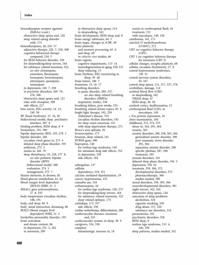
benzodiazepine receptor agonists(BzRAs) (cont.)
obstructive sleep apnea and, 120sleep related eating disorder
with, 173benzodiazepines, 46, 216 17adjunctive therapy, 226 7, 258, 308cognitive behavioral therapy
compared, 234 5for REM behavior disorder, 338for sleepwalking/sleep terrors, 164for substance related insomnia, 322,
see also clonazepam;estazolam; flurazepam;lorazepam; lormetazepam;nitrazepam; quazepam;temazepam
in depression, 226 7, 258in psychotic disorders, 269 70,
276, 308obstructive sleep apnea and, 121risks with clozapine, 308side effects, 217
beta waves, EEG activity, 14 15,249 50
BF (basal forebrain), 15 16, 20bidirectional model, sleep psychiatry
interface, 387 8binding problem, REM sleep, 8 9biomarkers, 191, 390bipolar depression (BD), 193, 270 1bipolar disorder, 266circadian clock genes in, 273 4delayed sleep phase disorder, 195euthymia, 272 3mania in, 266 70sleep disturbance, 43, 228, 277 8,
see also pediatric bipolardisorder (BPD)
bidirectional model, 388evaluation, 274 5management, 275 7
bizarre elements, in dreams, 82blood glucose metabolism, 61, 62blood oxygen level dependent
(BOLD) fMRI, 51 2BMAL1 gene polymorphisms,
27 8, 193body temperature circadian rhythm,
188, 191body, and sleep, 68 9body mind interaction, dreaming, 86BOLD (blood oxygen level
dependent) fMRI, 51 2borderline personality disorder, 195brain activationand dream content, 86in depression, 251 2, 362in insomnia, 209
in obstructive sleep apnea, 114in sleepwalking, 162
brain development, REM sleep and, 8brain energy substrates, 64 5brain maps, changes in rCBF, 49brain plasticityand memory processing, 65 6and sleep, 69in vitro/in vivo studies, 66
brain regionscognitive impairments, 113 14neurodegeneration in aging, 330, 331sleep inducing, 15
brain rhythms, EEG monitoring insleep, 39 40
brain tumor, 186 7brainstem, 15, 16 17breathing disordersin panic disorder, 289, 337,
see also sleep related breathingdisorders (SRBDs)
inspiratory stridor, 338breathing failure, post stroke, 335breathing related dream topics, 85 6bright light therapy, 192, 259Alzheimer’s disease, 333circadian rhythm disorders, 192for sleep onset insomnia, 351with sleep deprivation therapy, 271
Broca’s area aphasia, 50bromocriptine, 173bruxism, sleep related, 141BTBD9 gene, 390bupropion, 140for restless legs syndrome, 140for stimulant drug side effects, 352in depression, 258side effects, 164
cabergoline, 137caffeinedependence, 319, 321
calcium mediated depolarization, 19cancer, hypersomnia, 155cannabis use, 316carbamazepine, 44for restless legs syndrome, 138, 275for sleepwalking/sleep terrors, 164for substance related insomnia, 322sleep related epilepsy, 175
carbidopa, 173, 337side effects, 338
cardiac arrhythmias, differentiation, 289cardiovascular diseases, insomnia
and, 223cardiovascular system, in sleep, 38 9caregivers, 334, 338cataplexyhistaminergic neurons in, 18
orexin in cerebrospinal fluid, 18treatment, 151with narcolepsy, 149, 150
catathrenia, 161, 171catechol O methyltransferase
(COMT), 272CBT see cognitive behavior therapy
(CBT)CBT I see cognitive behavior therapy
for insomnia (CBT I)cellular changes, synaptic plasticity, 68cellular circadian rhythmicity, 27 8central hypersomnia syndromes,
149 51central nervous system disorders,
43, 167central sleep apnea, 111, 317, 337, 378cerebellum, damage, 114cerebral blood flow (CBF)in sleepwalking, 162NREM sleep, 47REM sleep, 48 50
cerebral cortex, deafferenation, 15cerebrospinal fluid (CSF), in
narcolepsy, 149c Fos protein expression, 16chest examination, 104childhood, 372, 374, 375abuse in, 164, 291, 364anxiety, 365anxiety disorders, 286, 358, 363, 366generalized anxiety disorder, 290post traumatic stress disorder,
291, 364separation anxiety disorder, 290specific phobias, 287, 290treatment, 292
arousal disorders, 163delayed sleep phase disorder, 194 5depression, 358 63insomnia, 359, 364, 372developmental disorders, 372pharmacotherapy, 366studies needed, 389
mood disorders, 358, 365, 366neurodevelopmental disorders, 381night terrors, 162, 164obstructive sleep apnea, 120outcomes of sleep problemsalcoholism, 316cigarette smoking, 320drug abuse, 317, 320substance use disorders, 314
parasomnias, 102psychiatric disorders, 358REM sleep, 8restless legs syndrome, 133 4,
346, 350sleep patterns, studies needed, 352
Index
400
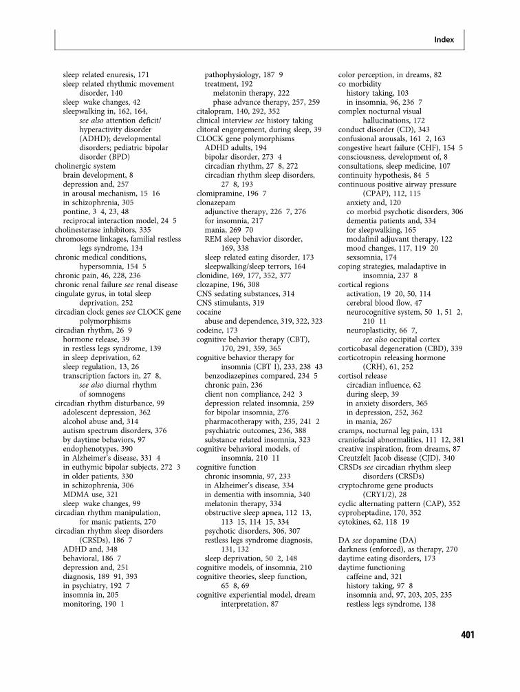
sleep related enuresis, 171sleep related rhythmic movement
disorder, 140sleep wake changes, 42sleepwalking in, 162, 164,
see also attention deficit/hyperactivity disorder(ADHD); developmentaldisorders; pediatric bipolardisorder (BPD)
cholinergic systembrain development, 8depression and, 257in arousal mechanism, 15 16in schizophrenia, 305pontine, 3 4, 23, 48reciprocal interaction model, 24 5
cholinesterase inhibitors, 335chromosome linkages, familial restless
legs syndrome, 134chronic medical conditions,
hypersomnia, 154 5chronic pain, 46, 228, 236chronic renal failure see renal diseasecingulate gyrus, in total sleep
deprivation, 252circadian clock genes see CLOCK gene
polymorphismscircadian rhythm, 26 9hormone release, 39in restless legs syndrome, 139in sleep deprivation, 62sleep regulation, 13, 26transcription factors in, 27 8,
see also diurnal rhythmof somnogens
circadian rhythm disturbance, 99adolescent depression, 362alcohol abuse and, 314autism spectrum disorders, 376by daytime behaviors, 97endophenotypes, 390in Alzheimer’s disease, 331 4in euthymic bipolar subjects, 272 3in older patients, 330in schizophrenia, 306MDMA use, 321sleep wake changes, 99
circadian rhythm manipulation,for manic patients, 270
circadian rhythm sleep disorders(CRSDs), 186 7
ADHD and, 348behavioral, 186 7depression and, 251diagnosis, 189 91, 393in psychiatry, 192 7insomnia in, 205monitoring, 190 1
pathophysiology, 187 9treatment, 192
melatonin therapy, 222phase advance therapy, 257, 259
citalopram, 140, 292, 352clinical interview see history takingclitoral engorgement, during sleep, 39CLOCK gene polymorphismsADHD adults, 194bipolar disorder, 273 4circadian rhythm, 27 8, 272circadian rhythm sleep disorders,
27 8, 193clomipramine, 196 7clonazepamadjunctive therapy, 226 7, 276for insomnia, 217mania, 269 70REM sleep behavior disorder,
169, 338sleep related eating disorder, 173sleepwalking/sleep terrors, 164
clonidine, 169, 177, 352, 377clozapine, 196, 308CNS sedating substances, 314CNS stimulants, 319cocaineabuse and dependence, 319, 322, 323
codeine, 173cognitive behavior therapy (CBT),
170, 291, 359, 365cognitive behavior therapy for
insomnia (CBT I), 233, 238 43benzodiazepines compared, 234 5chronic pain, 236client non compliance, 242 3depression related insomnia, 259for bipolar insomnia, 276pharmacotherapy with, 235, 241 2psychiatric outcomes, 236, 388substance related insomnia, 323
cognitive behavioral models, ofinsomnia, 210 11
cognitive functionchronic insomnia, 97, 233in Alzheimer’s disease, 334in dementia with insomnia, 340melatonin therapy, 334obstructive sleep apnea, 112 13,
113 15, 114 15, 334psychotic disorders, 306, 307restless legs syndrome diagnosis,
131, 132sleep deprivation, 50 2, 148
cognitive models, of insomnia, 210cognitive theories, sleep function,
65 8, 69cognitive experiential model, dream
interpretation, 87
color perception, in dreams, 82co morbidityhistory taking, 103in insomnia, 96, 236 7
complex nocturnal visualhallucinations, 172
conduct disorder (CD), 343confusional arousals, 161 2, 163congestive heart failure (CHF), 154 5consciousness, development of, 8consultations, sleep medicine, 107continuity hypothesis, 84 5continuous positive airway pressure
(CPAP), 112, 115anxiety and, 120co morbid psychotic disorders, 306dementia patients and, 334for sleepwalking, 165modafinil adjuvant therapy, 122mood changes, 117, 119 20sexsomnia, 174
coping strategies, maladaptive ininsomnia, 237 8
cortical regionsactivation, 19 20, 50, 114cerebral blood flow, 47neurocognitive system, 50 1, 51 2,
210 11neuroplasticity, 66 7,
see also occipital cortexcorticobasal degeneration (CBD), 339corticotropin releasing hormone
(CRH), 61, 252cortisol releasecircadian influence, 62during sleep, 39in anxiety disorders, 365in depression, 252, 362in mania, 267
cramps, nocturnal leg pain, 131craniofacial abnormalities, 111 12, 381creative inspiration, from dreams, 87Creutzfelt Jacob disease (CJD), 340CRSDs see circadian rhythm sleep
disorders (CRSDs)cryptochrome gene products
(CRY1/2), 28cyclic alternating pattern (CAP), 352cyproheptadine, 170, 352cytokines, 62, 118 19
DA see dopamine (DA)darkness (enforced), as therapy, 270daytime eating disorders, 173daytime functioningcaffeine and, 321history taking, 97 8insomnia and, 97, 203, 205, 235restless legs syndrome, 138
Index
401
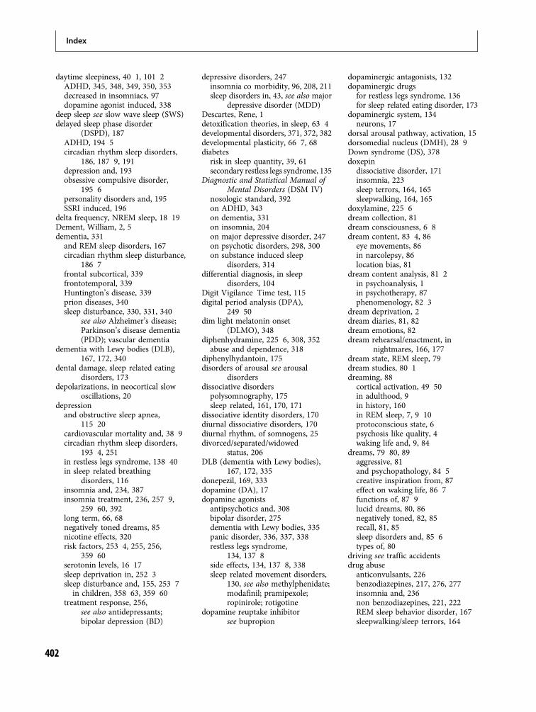
daytime sleepiness, 40 1, 101 2ADHD, 345, 348, 349, 350, 353decreased in insomniacs, 97dopamine agonist induced, 338
deep sleep see slow wave sleep (SWS)delayed sleep phase disorder
(DSPD), 187ADHD, 194 5circadian rhythm sleep disorders,
186, 187 9, 191depression and, 193obsessive compulsive disorder,
195 6personality disorders and, 195SSRI induced, 196
delta frequency, NREM sleep, 18 19Dement, William, 2, 5dementia, 331and REM sleep disorders, 167circadian rhythm sleep disturbance,
186 7frontal subcortical, 339frontotemporal, 339Huntington’s disease, 339prion diseases, 340sleep disturbance, 330, 331, 340
see also Alzheimer’s disease;Parkinson’s disease dementia(PDD); vascular dementia
dementia with Lewy bodies (DLB),167, 172, 340
dental damage, sleep related eatingdisorders, 173
depolarizations, in neocortical slowoscillations, 20
depressionand obstructive sleep apnea,
115 20cardiovascular mortality and, 38 9circadian rhythm sleep disorders,
193 4, 251in restless legs syndrome, 138 40in sleep related breathing
disorders, 116insomnia and, 234, 387insomnia treatment, 236, 257 9,
259 60, 392long term, 66, 68negatively toned dreams, 85nicotine effects, 320risk factors, 253 4, 255, 256,
359 60serotonin levels, 16 17sleep deprivation in, 252 3sleep disturbance and, 155, 253 7in children, 358 63, 359 60
treatment response, 256,see also antidepressants;bipolar depression (BD)
depressive disorders, 247insomnia co morbidity, 96, 208, 211sleep disorders in, 43, see also major
depressive disorder (MDD)Descartes, Rene, 1detoxification theories, in sleep, 63 4developmental disorders, 371, 372, 382developmental plasticity, 66 7, 68diabetesrisk in sleep quantity, 39, 61secondary restless legs syndrome, 135
Diagnostic and Statistical Manual ofMental Disorders (DSM IV)
nosologic standard, 392on ADHD, 343on dementia, 331on insomnia, 204on major depressive disorder, 247on psychotic disorders, 298, 300on substance induced sleep
disorders, 314differential diagnosis, in sleep
disorders, 104Digit Vigilance Time test, 115digital period analysis (DPA),
249 50dim light melatonin onset
(DLMO), 348diphenhydramine, 225 6, 308, 352abuse and dependence, 318
diphenylhydantoin, 175disorders of arousal see arousal
disordersdissociative disorderspolysomnography, 175sleep related, 161, 170, 171
dissociative identity disorders, 170diurnal dissociative disorders, 170diurnal rhythm, of somnogens, 25divorced/separated/widowed
status, 206DLB (dementia with Lewy bodies),
167, 172, 335donepezil, 169, 333dopamine (DA), 17dopamine agonistsantipsychotics and, 308bipolar disorder, 275dementia with Lewy bodies, 335panic disorder, 336, 337, 338restless legs syndrome,
134, 137 8side effects, 134, 137 8, 338sleep related movement disorders,
130, see also methylphenidate;modafinil; pramipexole;ropinirole; rotigotine
dopamine reuptake inhibitorsee bupropion
dopaminergic antagonists, 132dopaminergic drugsfor restless legs syndrome, 136for sleep related eating disorder, 173
dopaminergic system, 134neurons, 17
dorsal arousal pathway, activation, 15dorsomedial nucleus (DMH), 28 9Down syndrome (DS), 378doxepindissociative disorder, 171insomnia, 223sleep terrors, 164, 165sleepwalking, 164, 165
doxylamine, 225 6dream collection, 81dream consciousness, 6 8dream content, 83 4, 86eye movements, 86in narcolepsy, 86location bias, 81
dream content analysis, 81 2in psychoanalysis, 1in psychotherapy, 87phenomenology, 82 3
dream deprivation, 2dream diaries, 81, 82dream emotions, 82dream rehearsal/enactment, in
nightmares, 166, 177dream state, REM sleep, 79dream studies, 80 1dreaming, 88cortical activation, 49 50in adulthood, 9in history, 160in REM sleep, 7, 9 10protoconscious state, 6psychosis like quality, 4waking life and, 9, 84
dreams, 79 80, 89aggressive, 81and psychopathology, 84 5creative inspiration from, 87effect on waking life, 86 7functions of, 87 9lucid dreams, 80, 86negatively toned, 82, 85recall, 81, 85sleep disorders and, 85 6types of, 80
driving see traffic accidentsdrug abuseanticonvulsants, 226benzodiazepines, 217, 276, 277insomnia and, 236non benzodiazepines, 221, 222REM sleep behavior disorder, 167sleepwalking/sleep terrors, 164
Index
402
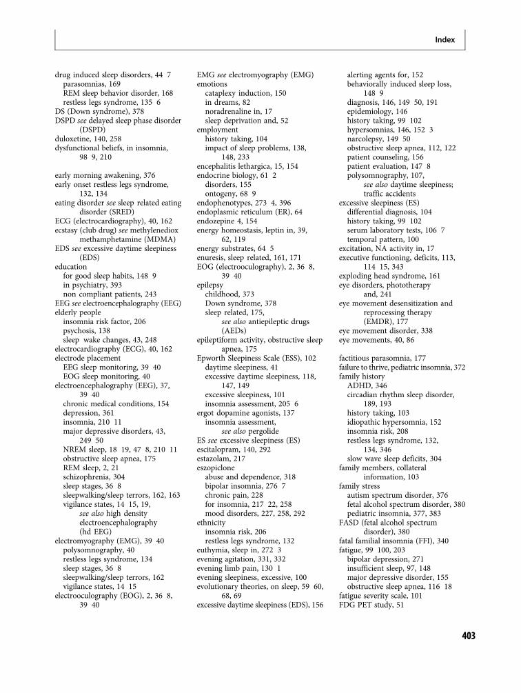
drug induced sleep disorders, 44 7parasomnias, 169REM sleep behavior disorder, 168restless legs syndrome, 135 6
DS (Down syndrome), 378DSPD see delayed sleep phase disorder
(DSPD)duloxetine, 140, 258dysfunctional beliefs, in insomnia,
98 9, 210
early morning awakening, 376early onset restless legs syndrome,
132, 134eating disorder see sleep related eating
disorder (SRED)ECG (electrocardiography), 40, 162ecstasy (club drug) see methylenediox
methamphetamine (MDMA)EDS see excessive daytime sleepiness
(EDS)educationfor good sleep habits, 148 9in psychiatry, 393non compliant patients, 243
EEG see electroencephalography (EEG)elderly peopleinsomnia risk factor, 206psychosis, 138sleep wake changes, 43, 248
electrocardiography (ECG), 40, 162electrode placementEEG sleep monitoring, 39 40EOG sleep monitoring, 40
electroencephalography (EEG), 37,39 40
chronic medical conditions, 154depression, 361insomnia, 210 11major depressive disorders, 43,
249 50NREM sleep, 18 19, 47 8, 210 11obstructive sleep apnea, 175REM sleep, 2, 21schizophrenia, 304sleep stages, 36 8sleepwalking/sleep terrors, 162, 163vigilance states, 14 15, 19,
see also high densityelectroencephalography(hd EEG)
electromyography (EMG), 39 40polysomnography, 40restless legs syndrome, 134sleep stages, 36 8sleepwalking/sleep terrors, 162vigilance states, 14 15
electrooculography (EOG), 2, 36 8,39 40
EMG see electromyography (EMG)emotionscataplexy induction, 150in dreams, 82noradrenaline in, 17sleep deprivation and, 52
employmenthistory taking, 104impact of sleep problems, 138,
148, 233encephalitis lethargica, 15, 154endocrine biology, 61 2disorders, 155ontogeny, 68 9
endophenotypes, 273 4, 396endoplasmic reticulum (ER), 64endozepine 4, 154energy homeostasis, leptin in, 39,
62, 119energy substrates, 64 5enuresis, sleep related, 161, 171EOG (electrooculography), 2, 36 8,
39 40epilepsychildhood, 373Down syndrome, 378sleep related, 175,
see also antiepileptic drugs(AEDs)
epileptiform activity, obstructive sleepapnea, 175
Epworth Sleepiness Scale (ESS), 102daytime sleepiness, 41excessive daytime sleepiness, 118,
147, 149excessive sleepiness, 101insomnia assessment, 205 6
ergot dopamine agonists, 137insomnia assessment,
see also pergolideES see excessive sleepiness (ES)escitalopram, 140, 292estazolam, 217eszopicloneabuse and dependence, 318bipolar insomnia, 276 7chronic pain, 228for insomnia, 217 22, 258mood disorders, 227, 258, 292
ethnicityinsomnia risk, 206restless legs syndrome, 132
euthymia, sleep in, 272 3evening agitation, 331, 332evening limb pain, 130 1evening sleepiness, excessive, 100evolutionary theories, on sleep, 59 60,
68, 69excessive daytime sleepiness (EDS), 156
alerting agents for, 152behaviorally induced sleep loss,
148 9diagnosis, 146, 149 50, 191epidemiology, 146history taking, 99 102hypersomnias, 146, 152 3narcolepsy, 149 50obstructive sleep apnea, 112, 122patient counseling, 156patient evaluation, 147 8polysomnography, 107,
see also daytime sleepiness;traffic accidents
excessive sleepiness (ES)differential diagnosis, 104history taking, 99 102serum laboratory tests, 106 7temporal pattern, 100
excitation, NA activity in, 17executive functioning, deficits, 113,
114 15, 343exploding head syndrome, 161eye disorders, phototherapy
and, 241eye movement desensitization and
reprocessing therapy(EMDR), 177
eye movement disorder, 338eye movements, 40, 86
factitious parasomnia, 177failure to thrive, pediatric insomnia, 372family historyADHD, 346circadian rhythm sleep disorder,
189, 193history taking, 103idiopathic hypersomnia, 152insomnia risk, 208restless legs syndrome, 132,
134, 346slow wave sleep deficits, 304
family members, collateralinformation, 103
family stressautism spectrum disorder, 376fetal alcohol spectrum disorder, 380pediatric insomnia, 377, 383
FASD (fetal alcohol spectrumdisorder), 380
fatal familial insomnia (FFI), 340fatigue, 99 100, 203bipolar depression, 271insufficient sleep, 97, 148major depressive disorder, 155obstructive sleep apnea, 116 18
fatigue severity scale, 101FDG PET study, 51
Index
403

fears, 80 1, 289, 363feeding, and satiety, 62ferritin levels, reduced, 132,
133, 134fetal alcohol spectrum disorder
(FASD), 380fetal life, sleep wake changes, 42fibromyalgia, 154, 226final common pathway
model, 268financial costs, of insomnia, 211fine motor coordination, 114flip flop switch model, 13, 20 1flumazenil, 154fluoxetine, 121adjunctive therapy in insomnia,
217, 221anticatapleptic agent, 151mood disorders, 363pediatric insomnia, 366side effect, 140sleep wake cycle and, 196 7
flupentixol, 196, 197flurazepam, 121, 217fluvoxamine, 196, 197fMRI see functional magnetic
resonance imaging (fMRI)food availability, in circadian
rhythmicity, 28 9fragile X syndrome, 380fragmented sleep see sleep
fragmentationFreud, Sigmund, 1 2, 160frontal lobedysfunction, 113 14,
see also prefrontal model,in obstructive sleep apnea
frontal lobe seizures, 175frontotemporal dementia, 339functional magnetic resonance
imaging (fMRI)BOLD (blood oxygen level
dependent), 51 2functional neuroanatomy, 47 8in new research, 391inhibitory efficiency studies, 52learning and neural plasticity, 50sleep deprivation effects, 51 2
functional neuroanatomyneuroimaging techniques, 47 52of arousal, 209 10
functions of sleep see sleep functionsfunding, research studies, 388future sleep psychiatry interface, 387
GABAGABA A receptors, 216 21GABAergic drugs, 177GABAergic inhibition, 19
GABAergic neurons, 24 5GABAergic preoptic nuclei, 20
gabapentin, 138, 226, 277, 322GAD see generalized anxiety disorder
(GAD)galantamine, 333gamma waves, on EEG, 14 15gamma aminobutyric acid (GABA)
see GABAgastrointestinal diseases, insomnia
and, 236gendercircadian rhythm alterations, 362depression, 361, 362dream content, 83insomnia risk factor, 206restless legs syndrome, 132
generalized anxiety disorder(GAD), 288
ADHD co morbidity, 345diagnosis, 286in childhood, 364insomnia in, 227, 288pharmacotherapy, 388sleep disturbance, 286, 290
genetic expressionBTBD9 gene, 390endoplasmic reticulum protein
biogenesis, 64polymorphismsbipolar depression, 272mood and circadian rhythmdisorders, 193
genetic inheritancefatal familial insomnia, 340Huntington’s disease, 339
genetic mutations, Alzheimer’sdisease, 332
genetic susceptibilitychildhood depression, 359circadian rhythmicity, 27 8, 189restless legs syndrome, 134
genetic upregulation, in sleep, 68genetics, new research, 389geniculate nucleus see lateral
geniculate nucleusghrelin, 39, 62glial glycogen, 65glucose metabolism (CMRGlu), 51glutamate release, 63glutathione, 63glycogen synthase kinase (GSK3 b)
gene polymorphisms, 272,273 4
glycogen, cortical, 65goal setting, and patient compliance,
243gray matter volume, diminished, 114groaning, sleep related, 161, 171
group therapy, for restless legssyndrome patients, 140
growth hormone (GH)deficiency, 155release during sleep, 39, 61sleep deprivation and, 39
growth hormone releasing hormone(GHRH), 252
GSK3 b gene polymorphisms, 272,273 4
habits see sleep habits/hygienehallucinationsdementia with Lewy bodies, 335differential diagnosis, 307excess sleepiness and, 150panic disorder, 337schizophrenia, 84, 300sleep related, 161, 172
haloperidol, 46, 196, 197Hamilton depression scale, 118hd EEG (high density
electroencephalography),47, 390
head noises, 172head traumahypersomnia, 154secondary circadian rhythm
disorder, 186 7health risks, sleep related eating
disorder, 173healthcare costs, of insomnia, 211, 234hearing impairment, insomnia, 373heart disease see cardiovascular
diseases, insomia andHebbian synaptic plasticity, 67heroin, effects on sleep, 46high density electroencephalography
(hd EEG), 47, 390hippocampal neocortical
interactions, 67hippocampuspotential induction, 66REM sleep, 23
histamine, 17 18histamine receptor antagonists, 308REM sleep, see also antihistamines
histaminergic neurons, 17 18history taking, 107bipolar disorder, 274circadian rhythm sleep
disorders, 189dementia with insomnia, 340excessive sleepiness, 99 102, 147 8from family members, 103in ADHD, 350insomnia, 96 9, 205parasomnias, 179parasthesias, 131
Index
404
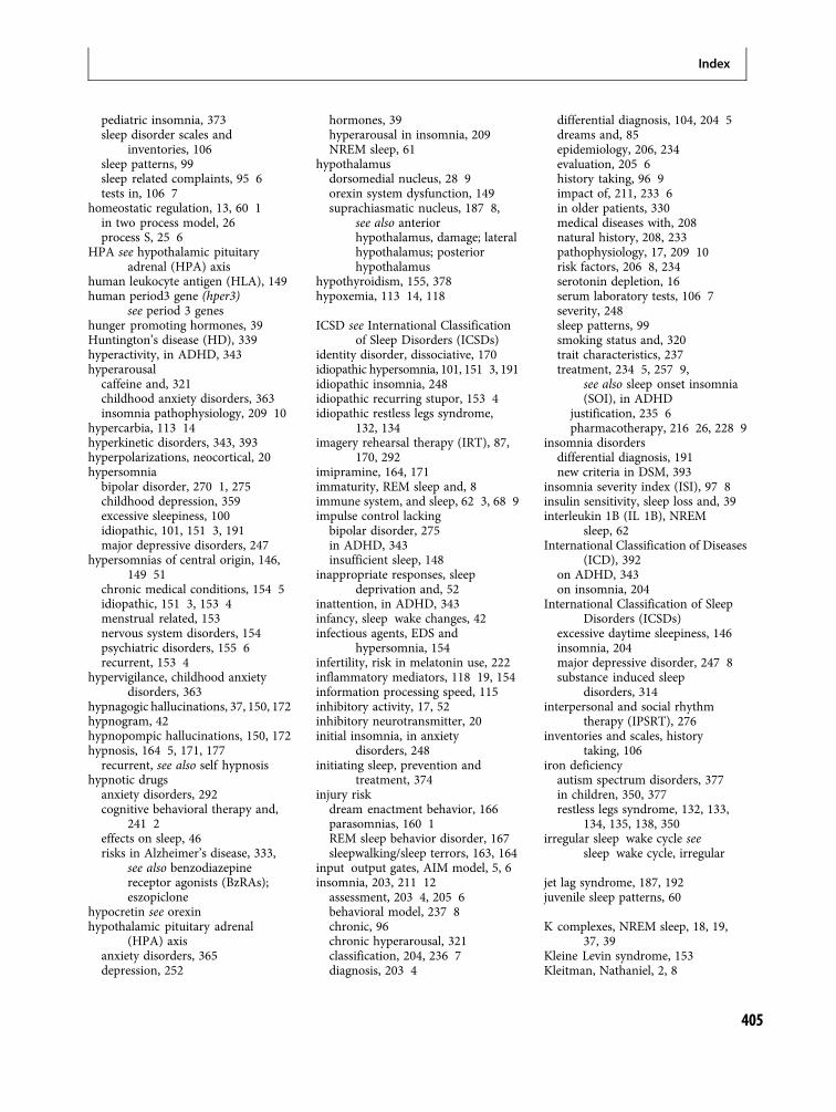
pediatric insomnia, 373sleep disorder scales and
inventories, 106sleep patterns, 99sleep related complaints, 95 6tests in, 106 7
homeostatic regulation, 13, 60 1in two process model, 26process S, 25 6
HPA see hypothalamic pituitaryadrenal (HPA) axis
human leukocyte antigen (HLA), 149human period3 gene (hper3)
see period 3 geneshunger promoting hormones, 39Huntington’s disease (HD), 339hyperactivity, in ADHD, 343hyperarousalcaffeine and, 321childhood anxiety disorders, 363insomnia pathophysiology, 209 10
hypercarbia, 113 14hyperkinetic disorders, 343, 393hyperpolarizations, neocortical, 20hypersomniabipolar disorder, 270 1, 275childhood depression, 359excessive sleepiness, 100idiopathic, 101, 151 3, 191major depressive disorders, 247
hypersomnias of central origin, 146,149 51
chronic medical conditions, 154 5idiopathic, 151 3, 153 4menstrual related, 153nervous system disorders, 154psychiatric disorders, 155 6recurrent, 153 4
hypervigilance, childhood anxietydisorders, 363
hypnagogic hallucinations, 37, 150, 172hypnogram, 42hypnopompic hallucinations, 150, 172hypnosis, 164 5, 171, 177recurrent, see also self hypnosis
hypnotic drugsanxiety disorders, 292cognitive behavioral therapy and,
241 2effects on sleep, 46risks in Alzheimer’s disease, 333,
see also benzodiazepinereceptor agonists (BzRAs);eszopiclone
hypocretin see orexinhypothalamic pituitary adrenal
(HPA) axisanxiety disorders, 365depression, 252
hormones, 39hyperarousal in insomnia, 209NREM sleep, 61
hypothalamusdorsomedial nucleus, 28 9orexin system dysfunction, 149suprachiasmatic nucleus, 187 8,
see also anteriorhypothalamus, damage; lateralhypothalamus; posteriorhypothalamus
hypothyroidism, 155, 378hypoxemia, 113 14, 118
ICSD see International Classificationof Sleep Disorders (ICSDs)
identity disorder, dissociative, 170idiopathic hypersomnia, 101, 151 3, 191idiopathic insomnia, 248idiopathic recurring stupor, 153 4idiopathic restless legs syndrome,
132, 134imagery rehearsal therapy (IRT), 87,
170, 292imipramine, 164, 171immaturity, REM sleep and, 8immune system, and sleep, 62 3, 68 9impulse control lackingbipolar disorder, 275in ADHD, 343insufficient sleep, 148
inappropriate responses, sleepdeprivation and, 52
inattention, in ADHD, 343infancy, sleep wake changes, 42infectious agents, EDS and
hypersomnia, 154infertility, risk in melatonin use, 222inflammatory mediators, 118 19, 154information processing speed, 115inhibitory activity, 17, 52inhibitory neurotransmitter, 20initial insomnia, in anxiety
disorders, 248initiating sleep, prevention and
treatment, 374injury riskdream enactment behavior, 166parasomnias, 160 1REM sleep behavior disorder, 167sleepwalking/sleep terrors, 163, 164
input output gates, AIM model, 5, 6insomnia, 203, 211 12assessment, 203 4, 205 6behavioral model, 237 8chronic, 96chronic hyperarousal, 321classification, 204, 236 7diagnosis, 203 4
differential diagnosis, 104, 204 5dreams and, 85epidemiology, 206, 234evaluation, 205 6history taking, 96 9impact of, 211, 233 6in older patients, 330medical diseases with, 208natural history, 208, 233pathophysiology, 17, 209 10risk factors, 206 8, 234serotonin depletion, 16serum laboratory tests, 106 7severity, 248sleep patterns, 99smoking status and, 320trait characteristics, 237treatment, 234 5, 257 9,
see also sleep onset insomnia(SOI), in ADHD
justification, 235 6pharmacotherapy, 216 26, 228 9
insomnia disordersdifferential diagnosis, 191new criteria in DSM, 393
insomnia severity index (ISI), 97 8insulin sensitivity, sleep loss and, 39interleukin 1B (IL 1B), NREM
sleep, 62International Classification of Diseases
(ICD), 392on ADHD, 343on insomnia, 204
International Classification of SleepDisorders (ICSDs)
excessive daytime sleepiness, 146insomnia, 204major depressive disorder, 247 8substance induced sleep
disorders, 314interpersonal and social rhythm
therapy (IPSRT), 276inventories and scales, history
taking, 106iron deficiencyautism spectrum disorders, 377in children, 350, 377restless legs syndrome, 132, 133,
134, 135, 138, 350irregular sleep wake cycle see
sleep wake cycle, irregular
jet lag syndrome, 187, 192juvenile sleep patterns, 60
K complexes, NREM sleep, 18, 19,37, 39
Kleine Levin syndrome, 153Kleitman, Nathaniel, 2, 8
Index
405
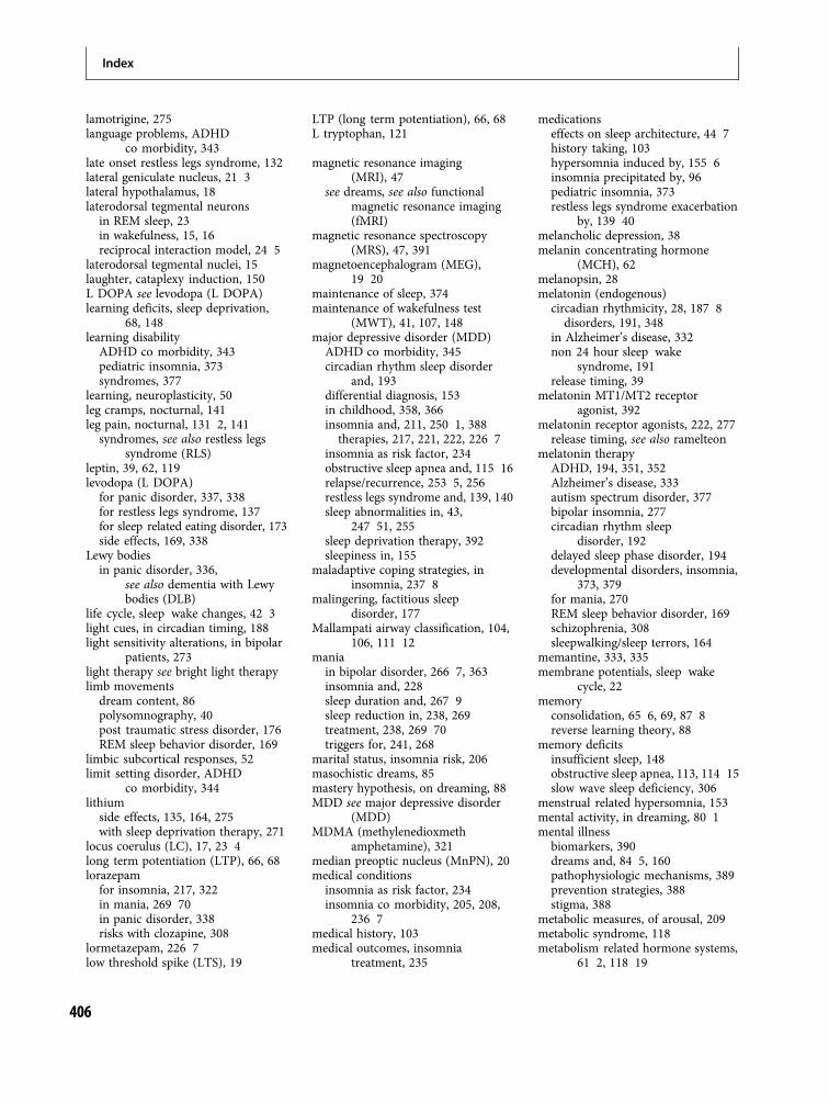
lamotrigine, 275language problems, ADHD
co morbidity, 343late onset restless legs syndrome, 132lateral geniculate nucleus, 21 3lateral hypothalamus, 18laterodorsal tegmental neuronsin REM sleep, 23in wakefulness, 15, 16reciprocal interaction model, 24 5
laterodorsal tegmental nuclei, 15laughter, cataplexy induction, 150L DOPA see levodopa (L DOPA)learning deficits, sleep deprivation,
68, 148learning disabilityADHD co morbidity, 343pediatric insomnia, 373syndromes, 377
learning, neuroplasticity, 50leg cramps, nocturnal, 141leg pain, nocturnal, 131 2, 141syndromes, see also restless legs
syndrome (RLS)leptin, 39, 62, 119levodopa (L DOPA)for panic disorder, 337, 338for restless legs syndrome, 137for sleep related eating disorder, 173side effects, 169, 338
Lewy bodiesin panic disorder, 336,
see also dementia with Lewybodies (DLB)
life cycle, sleep wake changes, 42 3light cues, in circadian timing, 188light sensitivity alterations, in bipolar
patients, 273light therapy see bright light therapylimb movementsdream content, 86polysomnography, 40post traumatic stress disorder, 176REM sleep behavior disorder, 169
limbic subcortical responses, 52limit setting disorder, ADHD
co morbidity, 344lithiumside effects, 135, 164, 275with sleep deprivation therapy, 271
locus coerulus (LC), 17, 23 4long term potentiation (LTP), 66, 68lorazepamfor insomnia, 217, 322in mania, 269 70in panic disorder, 338risks with clozapine, 308
lormetazepam, 226 7low threshold spike (LTS), 19
LTP (long term potentiation), 66, 68L tryptophan, 121
magnetic resonance imaging(MRI), 47
see dreams, see also functionalmagnetic resonance imaging(fMRI)
magnetic resonance spectroscopy(MRS), 47, 391
magnetoencephalogram (MEG),19 20
maintenance of sleep, 374maintenance of wakefulness test
(MWT), 41, 107, 148major depressive disorder (MDD)ADHD co morbidity, 345circadian rhythm sleep disorder
and, 193differential diagnosis, 153in childhood, 358, 366insomnia and, 211, 250 1, 388therapies, 217, 221, 222, 226 7
insomnia as risk factor, 234obstructive sleep apnea and, 115 16relapse/recurrence, 253 5, 256restless legs syndrome and, 139, 140sleep abnormalities in, 43,
247 51, 255sleep deprivation therapy, 392sleepiness in, 155
maladaptive coping strategies, ininsomnia, 237 8
malingering, factitious sleepdisorder, 177
Mallampati airway classification, 104,106, 111 12
maniain bipolar disorder, 266 7, 363insomnia and, 228sleep duration and, 267 9sleep reduction in, 238, 269treatment, 238, 269 70triggers for, 241, 268
marital status, insomnia risk, 206masochistic dreams, 85mastery hypothesis, on dreaming, 88MDD see major depressive disorder
(MDD)MDMA (methylenedioxmeth
amphetamine), 321median preoptic nucleus (MnPN), 20medical conditionsinsomnia as risk factor, 234insomnia co morbidity, 205, 208,
236 7medical history, 103medical outcomes, insomnia
treatment, 235
medicationseffects on sleep architecture, 44 7history taking, 103hypersomnia induced by, 155 6insomnia precipitated by, 96pediatric insomnia, 373restless legs syndrome exacerbation
by, 139 40melancholic depression, 38melanin concentrating hormone
(MCH), 62melanopsin, 28melatonin (endogenous)circadian rhythmicity, 28, 187 8disorders, 191, 348
in Alzheimer’s disease, 332non 24 hour sleep wake
syndrome, 191release timing, 39
melatonin MT1/MT2 receptoragonist, 392
melatonin receptor agonists, 222, 277release timing, see also ramelteon
melatonin therapyADHD, 194, 351, 352Alzheimer’s disease, 333autism spectrum disorder, 377bipolar insomnia, 277circadian rhythm sleep
disorder, 192delayed sleep phase disorder, 194developmental disorders, insomnia,
373, 379for mania, 270REM sleep behavior disorder, 169schizophrenia, 308sleepwalking/sleep terrors, 164
memantine, 333, 335membrane potentials, sleep wake
cycle, 22memoryconsolidation, 65 6, 69, 87 8reverse learning theory, 88
memory deficitsinsufficient sleep, 148obstructive sleep apnea, 113, 114 15slow wave sleep deficiency, 306
menstrual related hypersomnia, 153mental activity, in dreaming, 80 1mental illnessbiomarkers, 390dreams and, 84 5, 160pathophysiologic mechanisms, 389prevention strategies, 388stigma, 388
metabolic measures, of arousal, 209metabolic syndrome, 118metabolism related hormone systems,
61 2, 118 19
Index
406
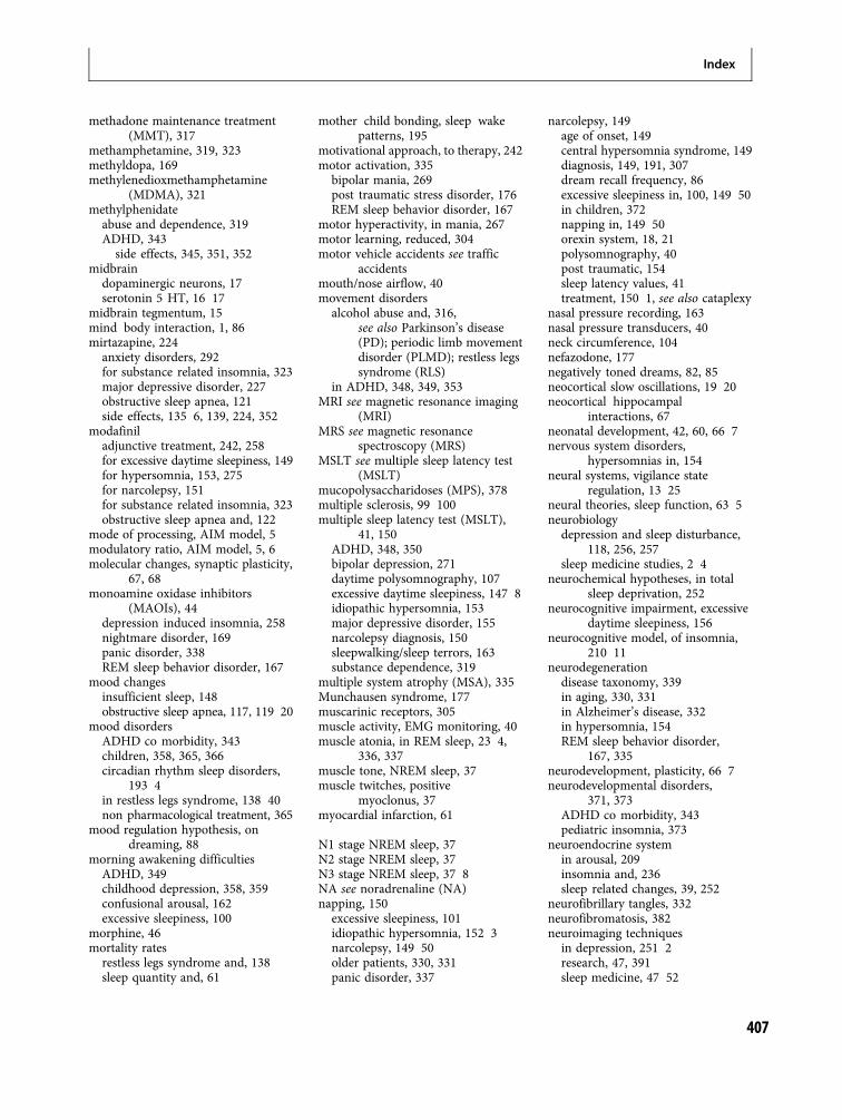
methadone maintenance treatment(MMT), 317
methamphetamine, 319, 323methyldopa, 169methylenedioxmethamphetamine
(MDMA), 321methylphenidateabuse and dependence, 319ADHD, 343side effects, 345, 351, 352
midbraindopaminergic neurons, 17serotonin 5 HT, 16 17
midbrain tegmentum, 15mind body interaction, 1, 86mirtazapine, 224anxiety disorders, 292for substance related insomnia, 323major depressive disorder, 227obstructive sleep apnea, 121side effects, 135 6, 139, 224, 352
modafiniladjunctive treatment, 242, 258for excessive daytime sleepiness, 149for hypersomnia, 153, 275for narcolepsy, 151for substance related insomnia, 323obstructive sleep apnea and, 122
mode of processing, AIM model, 5modulatory ratio, AIM model, 5, 6molecular changes, synaptic plasticity,
67, 68monoamine oxidase inhibitors
(MAOIs), 44depression induced insomnia, 258nightmare disorder, 169panic disorder, 338REM sleep behavior disorder, 167
mood changesinsufficient sleep, 148obstructive sleep apnea, 117, 119 20
mood disordersADHD co morbidity, 343children, 358, 365, 366circadian rhythm sleep disorders,
193 4in restless legs syndrome, 138 40non pharmacological treatment, 365
mood regulation hypothesis, ondreaming, 88
morning awakening difficultiesADHD, 349childhood depression, 358, 359confusional arousal, 162excessive sleepiness, 100
morphine, 46mortality ratesrestless legs syndrome and, 138sleep quantity and, 61
mother child bonding, sleep wakepatterns, 195
motivational approach, to therapy, 242motor activation, 335bipolar mania, 269post traumatic stress disorder, 176REM sleep behavior disorder, 167
motor hyperactivity, in mania, 267motor learning, reduced, 304motor vehicle accidents see traffic
accidentsmouth/nose airflow, 40movement disordersalcohol abuse and, 316,
see also Parkinson’s disease(PD); periodic limb movementdisorder (PLMD); restless legssyndrome (RLS)
in ADHD, 348, 349, 353MRI see magnetic resonance imaging
(MRI)MRS see magnetic resonance
spectroscopy (MRS)MSLT see multiple sleep latency test
(MSLT)mucopolysaccharidoses (MPS), 378multiple sclerosis, 99 100multiple sleep latency test (MSLT),
41, 150ADHD, 348, 350bipolar depression, 271daytime polysomnography, 107excessive daytime sleepiness, 147 8idiopathic hypersomnia, 153major depressive disorder, 155narcolepsy diagnosis, 150sleepwalking/sleep terrors, 163substance dependence, 319
multiple system atrophy (MSA), 335Munchausen syndrome, 177muscarinic receptors, 305muscle activity, EMG monitoring, 40muscle atonia, in REM sleep, 23 4,
336, 337muscle tone, NREM sleep, 37muscle twitches, positive
myoclonus, 37myocardial infarction, 61
N1 stage NREM sleep, 37N2 stage NREM sleep, 37N3 stage NREM sleep, 37 8NA see noradrenaline (NA)napping, 150excessive sleepiness, 101idiopathic hypersomnia, 152 3narcolepsy, 149 50older patients, 330, 331panic disorder, 337
narcolepsy, 149age of onset, 149central hypersomnia syndrome, 149diagnosis, 149, 191, 307dream recall frequency, 86excessive sleepiness in, 100, 149 50in children, 372napping in, 149 50orexin system, 18, 21polysomnography, 40post traumatic, 154sleep latency values, 41treatment, 150 1, see also cataplexy
nasal pressure recording, 163nasal pressure transducers, 40neck circumference, 104nefazodone, 177negatively toned dreams, 82, 85neocortical slow oscillations, 19 20neocortical hippocampal
interactions, 67neonatal development, 42, 60, 66 7nervous system disorders,
hypersomnias in, 154neural systems, vigilance state
regulation, 13 25neural theories, sleep function, 63 5neurobiologydepression and sleep disturbance,
118, 256, 257sleep medicine studies, 2 4
neurochemical hypotheses, in totalsleep deprivation, 252
neurocognitive impairment, excessivedaytime sleepiness, 156
neurocognitive model, of insomnia,210 11
neurodegenerationdisease taxonomy, 339in aging, 330, 331in Alzheimer’s disease, 332in hypersomnia, 154REM sleep behavior disorder,
167, 335neurodevelopment, plasticity, 66 7neurodevelopmental disorders,
371, 373ADHD co morbidity, 343pediatric insomnia, 373
neuroendocrine systemin arousal, 209insomnia and, 236sleep related changes, 39, 252
neurofibrillary tangles, 332neurofibromatosis, 382neuroimaging techniquesin depression, 251 2research, 47, 391sleep medicine, 47 52
Index
407
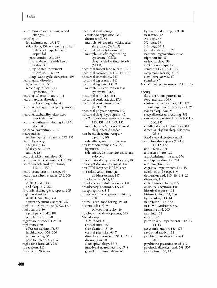
neuroimmune interactions, moodchanges, 119
neurolepticsfor nightmares, 169, 177side effects, 132, see also flupentixol;
haloperidol; quetiapine;risperidol
parasomnias, 164, 169risk in dementia with Lewybodies, 335
sleep related movementdisorders, 130, 139
sleep wake cycle disruption, 196neurological disordershypersomnia, 154secondary restless legs
syndrome, 135neurological examination, 104neuromuscular disorders,
polysomnography, 40neuronal damage, in sleep deprivation,
63 4neuronal excitability, after sleep
deprivation, 64neuronal pathways, binding in REM
sleep, 8 9neuronal restoration, 64 5neuropathiesrestless legs syndrome in, 132, 135
neurophysiologychanges in, 67of sleep, 52 3, 79testing, 134
neuroplasticity, and sleep, 50neuropsychiatric disorders, 112, 382neuropsychological symptoms,
112 13, 153neuroregeneration, in sleep, 69neurotransmitter systems, 272, 308nicotineADHD and, 343and sleep, 319, 320
nicotinic cholinergic receptors, 305night awakeningsADHD, 344, 349, 354autism spectrum disorder, 376
night eating syndrome (NES), 173night terrors, 80age of patient, 42, 102post traumatic, 290
nightmare disorder, 169 70nightmares, 80effect on waking life, 87in childhood, 358, 366in narcolepsy, 86post traumatic, 80, 176
night time fears, 287, 365nitrazepam, 121nitric acid (NO), 26
nocturnal awakeningschildhood depression, 359dream recall, 85multiple, 99, see also waking after
sleep onset (WASO)nocturnal eating behaviors, 43multiple, see also night eating
syndrome (NES);sleep related eating disorder(SRED)
nocturnal frontal lobe seizures, 175nocturnal hypoxemia, 113 14, 118nocturnal immobility, 337nocturnal leg cramps, 141nocturnal leg pain, 131 2multiple, see also restless legs
syndrome (RLS)nocturnal motricity, 351nocturnal panic attacks, 176nocturnal penile tumescence
(NPT), 39nocturnal polysomnagram, 163nocturnal sleep, hypnogram, 42non 24 hour sleep wake syndrome,
189, 191, 192, 193, 195multiple, see also non entrained
sleep phase disordernon benzodiazepine receptor
agonists, 308side effects, see also zopiclone
non benzodiazepines, 217 22hypnotics, 121 2side effects, 221, see also triazolam;
zolpidemnon entrained sleep phase disorder, 186non ergot dopamine agonist, 137non REM sleep see NREM sleepnon selective serotonergic
antidepressants, 167noradrenaline (NA), 17noradrenergic antidepressants, 140noradrenergic neurons, 17, 23norepinephrine, 3 5norepinephrine reuptake inhibitors,
258normal sleep, monitoring, 39 40nose/mouth airflow,
polysomnography, 40nosology, new developments, 393NREM sleepAIM model, 6arousal from, 162classification, 18 19cortical plasticity, 66 7disorders of arousal, 160 1, 161 2dreaming in, 80electrophysiology, 37 8functional neuroanatomy, 47 8growth hormone release, 61
hyperarousal during, 209 10in infancy, 42N1 stage, 37N2 stage, 37N3 stage, 37 8neural systems, 18 21neuronal regeneration in, 64night terrors, 80orthodox sleep, 36rCBF brain maps, 49serotonin (5 HT), 16 17sleep stage scoring, 41 2slow wave activity, 50spindles, 67
NREM sleep parasomnias, 161 2, 178
obesityfat distribution pattern, 104food addiction, 389obstructive sleep apnea, 111, 120and psychotic disorders, 274, 299
risk in sleep loss, 39sleep disordered breathing, 353
obsessive compulsive disorder (OCD),286, 287
childhood anxiety disorders, 364circadian rhythm sleep disorders,
195 6REM sleep disturbances, 43
obstructive sleep apnea (OSA),111 12, 122
and ADHD, 120and alcohol use, 122and Alzheimer’s disease, 334and bipolar disorder, 274and modafinil, 122cognitive impairments, 112 15cytokines and sleep, 119depression and, 115 18, 119 20diagnosis, 112epileptiform activity, 175excessive sleepiness, 100historical reports, 111history taking, 104, 106hypercarbia, 113 14in children, 347, 372in Down syndrome, 378insomnia and, 205napping, 101occult, 120performance impairments, 112 13,
114 15polysomnography, 168, 175prefrontal model, 114psychiatric medications and,
120 2psychiatric presentation of, 112psychotic disorders and, 299, 307risk factors, 106, 121
Index
408
severe, 174sleepwalking/sleep terrors and, 165stroke risk, 335symptoms, 111, 112, 116treatment, 112, 117, 119 20
occipital cortex, 21 3occupation see employmentOCD see obsessive compulsive
disorder (OCD)olanzapine, 309for insomnia, 224 5, 258for post traumatic stress disorder,
177, 287risks in Alzheimer’s disease, 333side effects, 135, 164
older patientsadvanced sleep phase disorder, 331parasomnias, 102 3secondary sleep disorders, 331sleep changes in, 330
Ondine’s curse, 335ontogeny, and sleep, 60, 68opiates/opioidseffects on sleep, 46, 317for restless legs syndrome, 138withdrawal, 135, see also cocaine;
oxycodone; tramadoloppositional defiant disorder (ODD),
343, 344, 346, 348orexin (hypocretin)dysfunction in narcolepsy, 149feeding and satiety, 62in ADHD, 353in flip flop switch, 21new research, 387vigilance state regulation, 18
orexin receptor antagonists, 388organization of sleep, 41 7oronasal airflow, 166oropharyngeal abnormalities, 104orthodox sleep, 36OSA see obstructive sleep apnea (OSA)oxidative processes, 63oximetry, polysomnography, 168oxycodone, 138
P50 suppression, 305paininsomnia and, 217, 228,
see also restless legssyndrome (RLS)
pain reliefanticonvulsants, 226opioid drugs, 317
panic attacks, 176, 289panic disorder (PD), 289as parasomnia, 176diagnosis, 176, 286insomnia, 176, 289
paradoxical insomnia, 210 11, 247paradoxical sleep, 36paralysis (sleep related), 150in history, 160, 170recurrent isolated, 170
parasomnias, 160 1, 178autism spectrum disorder, 376differential diagnosis, 160, 168,
176 7evaluation, 179exploding head syndrome, 172factitious, 177historical view, 160 1history taking, 102 3insomnia in, 205polysomnography, 40recurrent isolated sleep paralysis,
170REM sleep, 166 70sleep related dissociative disorders,
170sleep related eating disorder, 172 4sleep related enuresis, 171sleep related groaning, 171sleep related hallucinations, 172sleepwalking/sleep terrors, 162 4symptoms, 104
parasthesias, 130 1parasympathetic activation, 38parent education, on sleep
disorders, 374parkinsonism, dementia with Lewy
bodies, 335Parkinson’s disease (PD)and bipolar disorder, 275and REM sleep behavior disorder,
167diagnostic features, 336history taking, 104pharmacotherapy, 137, 138,
169, 333polysomnography, 332restless legs syndrome treatment
compared, 137secondary circadian rhythm sleep
disorder, 186 7sleep disturbance, 334
Parkinson’s disease dementia(PDD), 336
Parkinson’s plus syndromes, 334paroxetine, 121, 140, 164, 293paroxysmal arousals, 40partial sleep deprivation, 252 3pathophysiologic mechanisms, 389patient counselingin excessive sleepiness, 156,
see also sleep habits/hygiene,advice
patient non compliance, 242 3, 307
PD see Parkinson’s disease (PD)pediatric bipolar disorder (BPD)ADHD co morbidity, 343, 345mania, 363sleep and, 358, 363
pediatrics see childhoodpedunculopontine tegmental (PPT)
neurons, 15, 16, 23, 24 5pedunculopontine tegmental
nuclei, 15penile tumescence, nocturnal, 39performance impairmentchronic insomnia, 233in obstructive sleep apnea, 112 13,
114 15pergolide, 137perifornical region, 18period 3 gene polymorphisms, 273period 3 genes, 189, 193period gene products (PER1/2), 28periodic limb movement disorder
(PLMD), 130ADHD and, 347diagnosis, 130 2dreams and, 86epidemiology, 132 4genetics, new research, 390in panic disorder, 332polysomnography, 40, 332treatment, 136 8
periodic limb movements duringwakefulness(PLMW),130 1
periodic limb movements of sleep(PLMS), 130 1
ADHD, 346, 347, 351alcohol abuse and, 316and bipolar disorder, 275detection, 133in older patients, 330medication induced, 135 6, 140psychotic disorders and, 299
peripheral neuropathy, 131perpetuating factors, of insomnia,
210personality disorders, 163 4, 195personality factorsin dreams, 81, 84nightmare disorder, 170
PET see positron emissiontomography (PET)
PGD2 see prostaglandin (PGD2)PGO waves see pontine genicul
occipital (PGO) wavespharmacotherapyADHD, 343, 352Alzheimer’s disease, 333 4anxiety disorders, 292, 366bipolar insomnia, 276 7
Index
409

pharmacotherapy (cont.)in dementia with insomnia, 340new treatments, 391panic disorder, 338pediatric depression, 359, 366psychiatric disorders, 354, 388psychotic disorders and sleep
disturbance, 305research study caveats, 388substance related insomnia, 319with cognitive behavioral therapy
for insomnia, 235, 241 2phase advance hypothesis, 257, 348phase advance therapy, 259, 271phasic (episodic) components,
REM sleep, 38phasic muscle activity, 40phenomenology, in dreams, 82 3phenytoin, 44phobic disorders, 287phototherapy, 241physical deformity, pediatric
insomnia, 373physical examinationhistory taking, 104 6in dementia with insomnia, 340
physiological apnea process, 85physiological arousal, 209physiological changesdiscovery of, 5in sleep, 38 9, 107
physiological parameters, indreaming, 80
pindolol, 271pineal gland, 187 8Pittsburgh Sleep Quality Index (PSQI),
205 6, 317PLMD see periodic limb movement
disorder (PLMD)PLMS see periodic limb movements of
sleep (PLMS)polysomnography (PSG), 40, 107, 205ADHD, 348, 349, 350, 352, 353anxiety disorders, 287autism spectrum disorder, 376clinical research, 40dementia with insomnia, 340depression, 361dissociative disorders, 170euthymic bipolar subjects, 272excessive sleepiness, 107in children, 361, 376in multiple sleep latency test,
147 8mania, 267obsessive compulsive disorder, 288obstructive sleep apnea, 165,
166, 175panic disorder, 332, 338
periodic limb movement disorder,130 1, 332
periodic limb movements ofsleep, 133
psychiatric disorders, 42 3, 300REM sleep behavior disorder, 167,
168, 336restless legs syndrome, 130 1schizophrenia, 304sleep terrors, 162, 163sleepwalking, 162, 163smoking and, 320substance misuse, 314, 317, 319
pons, 2 4, 23, 48pontine sublaterodorsal nucleus
activation, 23 4pontine tegmentum, 21 3, 336ponto geniculo occipital (PGO)
waves, 6, 21 3, 49positive airway pressure (PAP),
40, 274positive myoclonus, 37positron emission tomography (PET),
47, 51 2, 209posterior hypothalamus, 17post partum psychosis, 269post traumatic parasomnia, 170post traumatic re enactment
dreams, 80post traumatic stress disorder
(PTSD), 177childhood anxiety disorders, 363diagnosis, 176 7, 290sleep disturbance, 176 7, 290bidirectional model, 388REM sleep, 43
treatment, 177, 292power spectral analysis (PSA),
249 50PPT see pedunculopontine tegmental
(PPT) neuronsPrader Willi syndrome (PWS), 379pramipexolepanic disorder, 337periodic limb movement disorder,
137 8REM sleep behavior disorder, 169restless legs syndrome, 137 8, 275
prazosin, 170, 177, 293precipitating factors, in insomnia,
96 7, 237prefrontal model, in obstructive sleep
apnea, 113 14pregabalin, 226, 292preoptic nuclei, 20prevention strategies, mental
illness, 388priapism, trazodone induced,
224, 258
primacy of sleep, 61primary circadian rhythm sleep
disorders, 186 90, 191primary insomnia, 236 7, 248classification, 204new treatment, 392
primary restless legs syndromesee idiopathic restless legssyndrome
primary sleep disorders, 147 8, 191bipolar disorder and, 274 5in ADHD, 346, 350psychotic disorders and, 298, 305
prion diseases, 340process C, sleep regulation, 26 9process S, sleep regulation, 25 6professional issues, in sleep
medicine, 393progress graphs, client motivation, 243progressive supranuclear palsy (PSP), 338prolactin, 39, 61prostaglandin (PGD2), 26protein biogenesis, 64protoconsciousness, 6, 8 9PSG see polysomnography (PSG)psychiatric assessmentparasomnias, 178 9sleepwalking and, 163 4
psychiatric developmental disorders,371, 380
psychiatric disordersADHD co morbidity, 343, 344circadian rhythm sleep disorders
and, 192 7differential diagnosis, 112, 176 7fatigue defined, 99 100genetics, 390history taking, 95 6, 103hypersomnia, 155 6insomnia as risk factor, 234insomnia in, 205, 208, 236 7prevention, 388treatment, 226 8, 236
neurobiology of, 52 3pediatric, 358REM sleep behavior disorder,
168sleep abnormalities, 42 3, 95 6sleep related eating disorder, 173thermoregulation abnormalities, 39treatment, 44, 120 2, 196 7
psychiatry, xiii xiv, 13early history, 1psychoanalysis in, 1training needs, 393
psychiatry sleep interface, 387psychoanalysis, 1 2, 9on dreams, 79, 88
psychogenic amnesia, 171
Index
410
psychological problemschildhood insomnia, 372, 378dream deprivation, 2in insomniacs, 97
psychomotor deficits, 101, 114 15, 148psychomotor vigilance task, 113psychopathology, 84 5, 160psychophysiologyinsomnia, 247measures of arousal, 209
psychosis, drug induced, 138psychosis like quality, dreaming, 4psychosocial stressors, sleep deficit,
168, 208, 306psychotherapy, 87, 276psychotic disorders, 306classification, 307diagnosis, 300, 307medication withdrawal, 304polysomnography, 300poor sleep hygiene, 299REM abnormalities in, 305sleep deficits in, 298sleep disorder co morbidity, 298sleep disturbance in, 298, 299,
308, 309sleep interactions with, 305slow wave sleep in, 303
psychotropic drugs, 164, 167, 173, 304slow wave sleep in, see also selective
serotonin reuptake inhibitors(SSRIs)
PTSD see post traumatic stressdisorder (PTSD)
public health issue, insomnia as, 211 12PWS (Prader Willi syndrome), 379
quality of life (QoL), 138, 211, 305, 330quazepam, 217, 318questionnairesADHD sleep disorders, 350, 352excessive daytime sleepiness, 147
quetiapine, 224 5bipolar insomnia, 277depression related insomnia, 258parasomnias, 164post traumatic stress disorder,
177, 293side effects, 135, 333substance related insomnia, 323
quiet sleep, in infancy, 42
radiculopathies, 135ramelteon, 222, 277, 318rapid eyemovement sleep see REM sleepRBD see REM sleep behavior
disorder (RBD)rCBF see regional cerebral blood
flow (rCBF)
rebound effect, ADHD medication,345, 352
reboxetine, 164reciprocal interaction model, 3,
4 5, 24 5recurrent hypersomnias, 153 4recurrent isolated sleep paralysis, 170regeneration, neuronal, 64regional cerebral blood flow (rCBF), 49in insomnia, 209 10neuroplasticity and sleep, 50NREM sleep, 47REM sleep, 48 50
relaxation training, 164 5, 241, 291REM density, in depression,
248 9, 255adolescents, 361children, 360, 361
REM latencyin depression, 251, 255
adolescents, 361children, 360
in major depressive disorder,248 9, 256
in schizophrenia, 43, 301REM sleep, 3AIM model, 6and immaturity, 8, 42binding problem, 8 9depressogenic theory, 256deprivation, 64, 253discovery, 2, 9 10dreaming, 2dreaming in, 6, 7, 79, 80dreams, 80, 88electrophysiology, 2 4, 37functions of, 87 8in ADHD, 350in anxiety disorders, in children, 364in major depressive disorders, 43in narcolepsy, 86muscle atonia in, 336, 337neuroimaging, 48 50nightmares, 80panic disorder, 337paradoxical sleep, 36parasomnias, 161, 166 70, 178post traumatic stress disorder, 290Prader Willi syndrome, 379psychiatric disorders, 42 3psychotic disorders, 43, 301, 305rCBF brain maps, 49reciprocal interaction model, 24 5serotonin (5 HT), 16 17sleep stage scoring, 41 2, 147 8substance abuse, 314, 317, 319synaptic plasticity, 66tonic (persistent) components, 38vigilance state regulation, 21 5
REM sleep behavior disorder (RBD),166 8, 169
acute transient, 167dementia with Lewy bodies, 335diagnosis, 167, 393in panic disorder, 337, 338multiple system atrophy, 338muscle atonia in, 337polysomnography, 168time of night, 102treatment, 169
REM active neurons, inhibition, 23REM off neurons, 25REM on neurons, 23, 24renal disease, 135repetitive transcranial magnetic
stimulation (rTMS), 50reproductive system, 39researchCPAP effects on mood, 118dream content, 83modern developments, 36pharmacotherapy, 388
reserpine, 169respiratory depression, 121 2respiratory effortpolysomnography, 40,
see also obstructive sleepapnea (OSA)
respiratory inductiveplethysmography (RIP), 40
respiratory system, during sleep, 39respite care, for caregivers, 334rest induced pain, 130 1restless legs syndrome (RLS), 130 1ADHD, 346, 350, 351age distribution, 132alcohol abuse, 316diagnosis, 106 7, 130 2, 133, 134criteria, 131 2, 393symptoms, 105, 130 1
dopaminergic system in, 134drug induced, 135 6, 139 40epidemiology, 132 4, 136genetics in, 134, 390idiopathic, 132, 134in children, 133 4in older patients, 331insomnia in, 139, 205late onset, 132mood disorders and, 138 40opioid drugs, 46parasthesias in, 130 1psychotic disorders and, 274 5, 299quality of life, 138secondary, 135, 136treatment, 136 8, 140, 226
restlessness, in ADHD, 343RET (reticular nucleus), 19
Index
411
reticular activating system (RAS), 15reticular formation (RF), 15, 23reticular nucleus (RET), 19retinohypothalamic pathway, 28retinoid related orphan nuclear
receptor alpha (Rora), 28Rett syndrome, 381Rev Erba gene polymorphisms, 273 4Rev Erba orphan nuclear receptor, 28reverse learning theory, 88reward processing, 389rheumatoid arthritis/rheumatological
conditionshypersomnia, 154insomnia, 217, 228nocturnal leg pain, 131secondary restless legs syndrome, 135
risperidol, 196risperidone, 135, 333rivastigmine, 333RLS see restless legs syndrome (RLS)ropiniroleperiodic limb movement disorder,
137 8, 336, 337restless legs syndrome, 137 8, 275side effects, 338
rotigotine, 137 8
safety issues, insufficient sleep, 148Sanfilippo syndrome, 378satiety hormones, 39scales and inventories, 106schizoaffective disordersdiagnostic criteria, 300polysomnography, 300sleep disorders and, 195, 298violent behavior, 306
schizophreniadiagnostic criteria, 298, 300hallucinations, 84polysomnography, 300poor sleep hygiene, 299, 300primary sleep disorders and, 298prodromal sleep disturbance, 304REM sleep in, 301risk factors, 304sleep disturbances, 43, 228,
301, 304sleep spindle activity, 40, 390slow wave sleep abnormalities, 303synaptic pruning, in adolescence, 42treatment, side effects, 121, 196
SD see sleep deprivation (SD)SDB see sleep related breathing
disorders (SRBDs)seasonal affective disorder (SAD),
193, 392secondary circadian rhythm sleep
disorder, 186 7
secondary insomnia, 204, 236 7secondary restless legs syndrome,
135, 136secondary sleep disorders, 331sedative antidepressants, 139, 308bipolar insomnia, 277major depressive disorder, 227substance related insomnia, 323,
see also antihistamines;antipsychotics
sedative hypnotics, 121 2, 164, 258 9see also alcohol; antihistamines;benzodiazepines
sedatives, 120pediatric insomnia, 366, 373psychiatric disorders, 155 6, 269 70with SSRIs, 366
see also lorazepamseizure related paroxysmal arousals, 40seizuresalcohol abuse risk, 322in sleep restriction therapy, 238nocturnal, 175
selective extrasynaptic GABAA
receptor antagonists(SEGAs), 46
selective serotonin reuptake inhibitors(SSRIs), 44, 121, 258
for children, 359, 366in anxiety disorders, 292, 293insomnia treatment in
depression, 258side effects, 121, 167,
see also citalopram;clomipramine; escitalopram,fluoxetine; fluvoxamine;paroxetine; sertraline
bruxism exacerbation, 141restless legs syndrome, 135 6, 140sleep disturbances, 196, 345, 352
self hypnosis, 164 5sensory deficits, pediatric insomnia, 373sensory perceptions, in dreams,
82, 83separation anxiety disorder (SAD),
286, 290serotonergic system, 23, 118serotonin, 3 5, 121serotonin 5 HT, 16 17serotonin 5 HT1A beta adrenoceptor
blocking agent, 271serotonin 5 HT2 receptor antagonists,
177, 308, 391serotonin 5 HT2A receptor, T variant,
272serotonin 5 HTTLPR transporter,
272, 390serotonin neurotransmission
deficit, 256
serotonin precursor, 121serotonin norepinephrine reuptake
inhibitors (SNRIs), 44,151, 292
side effect, 135 6, 140,see also duloxetine, venlafaxine
sertraline, 140, 293serum laboratory tests, 106 7sexsomnia, 174sexual abuse, 291sexual behavior, confusional
arousal, 162sexual dreams, 81shift workhistory taking, 104insufficient sleep, 148sleep disturbances, 13, 96, 97, 186treatment, 149, 192
short sleep duration, childhooddepression, 358
short term memory, in obstructivesleep apnea, 113
single nucleotide polymorphisms(SNPs), in movementdisorders, 390
single photon emission computedtomography (SPECT), 47,162, 209 10
sleep, 36 8cerebral cortex deafferentation, 15homeostatic regulation, 60, 250induction, 20normal, 39 40physiology, dream content, 86regulationflip flop hypothesis, 13two process model, 25 9
sleep anxiety disorder, 169 70sleep apnea, 38differential diagnosis, 149 50, 191dream recall frequency, 85endocrine system effects, 39hypersomnia and, 155,
see also central sleep apnea;obstructive sleep apnea (OSA)
sleep architecture, 41 7delayed sleep phase disorder, 189in ADHD, 350in childhood, 360, 361, 362in psychiatric illness, 43mood disorders, 248 50, 255 6,
360, 361, 362sleep attacks see nappingsleep continuity disturbance see sleep
fragmentationsleep deficitsbehaviorally induced, 148 9, 191in depression, 155in psychotic disorders, 305
Index
412
sleep deprivation (SD)and somnogens, 25, 26antidepressant action, 392as therapy, 238, 240, 271 2, 276confusional arousal, 162endocrine effects, 39, 61excessive sleepiness, 149 50fatality in, 61in delayed sleep phase disorder,
188 9in depression, 252 3insomnia and, 203neuroimaging studies, 50 2NREM sleep, 37older patients, 330panic disorder, 337Prader Willi syndrome, 379schizophrenia, 306studies on, 64, 267 8total, 252, see also excessive daytime
sleepiness (EDS)sleep diaries/sleep logs, 100, 207, 212dream diaries, 81, 82excessive sleepiness evaluation, 147in cognitive therapy for insomnia,
239 40insomnia, 99, 206sleep wake cycle monitoring, 190 1
sleep disorder scales andinventories, 106
sleep disordered breathing (SDB)see sleep related breathingdisorders (SRBDs)
sleep disorders, 205, 236ADHD and, 120and dreams, 85 6defining symptoms, 105depression and, 358 63genetics, 390history taking, 95 6in childhood, 42, 358 63, 372, 374in psychiatric illness, 43key parameters, 95polysomnography, 40psychotic co morbidities, 306screening for, 147 8slow wave sleep related, 42symptoms, 104, 105training and education needs, 393
sleep disturbanceADHD and, 120, 350anxiety disorders, 291, 293bipolar disorder, 270 1, 277 8, 363cancer, hypersomnia, 155cognitive deficits, 306depressive disorders, 270 1endocrine system effects, 39in childhoodmood disorders, 361, 363
restless legs syndrome, 133 4in depression, 253 7, 361mania prognosis, 269mood disorders, 247 51parasomnias and, 160 1post traumatic stress disorder,
176 7psychiatric disorders, 42 3, 389psychotic disorders, 300, 307, 309
schizophrenia, 301, 304, 306public health issue, 13restless legs syndrome, 130 1,
133 4, 138, see also insomnia;subjective sleep disturbance
sleep efficiency (SE), 42antidepressants and, 223antipsychotics and, 46in ADHD, 349in psychotic disorders, 300, 301marijuana use, 317
sleep fragmentation, 146, 249chronic, endocrine system in, 39cognitive impairments by, 113 14in ADHD, 349in anxiety disorders, 43in congestive heart failure, 154 5in depressive conditions, 118,
247 51, 253 5in obstructive sleep apnea,
113 14, 118in panic disorder, 337in psychiatric disorders, 42 3nicotine and, 320
sleep functions, 59 61, 68 70cognitive theories, 65 8neural theories, 63 5somatic theories, 61 3unifying theory of, 61
sleep habits/hygiene, 84advice, 148 9
Alzheimer’s disease, 333cognitive behavior therapy,238 9, 240, 291
autism spectrum disorder, 376in sleep disruption, 99mood and anxiety disorders,
291, 365perpetuating insomnia, 210psychotic disorders, 298, 299smoking status and, 320
sleep inertia, in idiopathichypersomnia, 152
sleep logs see sleep diaries/sleep logssleep medicine, 5departments, 5development, 2 6, 9 10recent growth of, 394significance, xiii xivtraining programs, 393
sleep medicine consultations, 107training programs, see also history
takingsleep medicine departments, 393sleep monitoring tools, 39 41hypnogram, 42, see also electroen
cephalography (EEG);polysomnography (PSG)
sleep need decrease, in mania, 266 7sleep onset dreams, 80sleep onset hallucinations, 37sleep onset latency (SOL)childhood mood disorders, 358,
359, 362, 364in ADHD, 345, 349in anxiety disorders, 43, 362in insomnia, 99melatonin receptor agonists in, 222non benzodiazepines for, 221psychiatric disorders, 42 3psychotic disorders, 301, 305sleep architecture variables, 42substance use, 314, 319, 321tricyclic antidepressants in, 223,
see also multiple sleep latencytest (MSLT)
sleep patterns, history taking, 99sleep reductionfinal common pathway model, 268in mania, 268 9mania caused by, 267 8
sleep research see researchsleep restriction therapy, 238, 240,
271 2, 276sleep spindles, 18, 37, 47 8EEG monitoring, 39in schizophrenia, 40, 304, 390synaptic plasticity, 67vigilance state regulation, 19
sleep stages, 36 8, 41 2electrophysiological criteria, 36 8functional neuroanatomy, 47 50physiological changes, 38 9regional cerebral blood flow, 49
sleep terrors (STs), 160, 162 4,165, 165 6
sleep dependent learning, 304, 306sleep disrupting behaviors see sleep
habits/hygienesleep onset insomnia (SOI), in
ADHD, 348, 351sleep onset REM periods (SOREMPs),
147 8sleep promoting medication, 150 1sleep psychiatry interface, the
future, 387sleep related behaviorshistory taking, 97 9,
see also parasomnias
Index
413
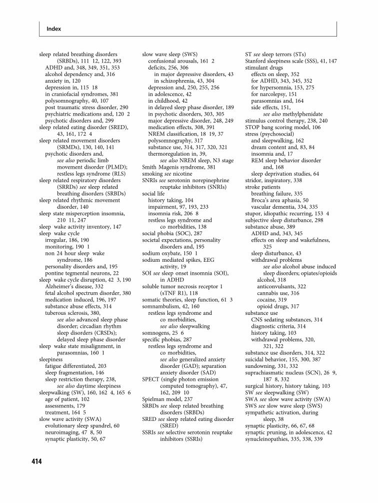
sleep related breathing disorders(SRBDs), 111 12, 122, 393
ADHD and, 348, 349, 351, 353alcohol dependency and, 316anxiety in, 120depression in, 115 18in craniofacial syndromes, 381polysomnography, 40, 107post traumatic stress disorder, 290psychiatric medications and, 120 2psychotic disorders and, 299
sleep related eating disorder (SRED),43, 161, 172 4
sleep related movement disorders(SRMDs), 130, 140, 141
psychotic disorders and,see also periodic limbmovement disorder (PLMD);restless legs syndrome (RLS)
sleep related respiratory disorders(SRRDs) see sleep relatedbreathing disorders (SRBDs)
sleep related rhythmic movementdisorder, 140
sleep state misperception insomnia,210 11, 247
sleep wake activity inventory, 147sleep wake cycleirregular, 186, 190monitoring, 190 1non 24 hour sleep wake
syndrome, 186personality disorders and, 195pontine tegmental neurons, 22
sleep wake cycle disruption, 42 3, 190Alzheimer’s disease, 332fetal alcohol spectrum disorder, 380medication induced, 196, 197substance abuse effects, 314tuberous sclerosis, 380,
see also advanced sleep phasedisorder; circadian rhythmsleep disorders (CRSDs);delayed sleep phase disorder
sleep wake state misalignment, inparasomnias, 160 1
sleepinessfatigue differentiated, 203sleep fragmentation, 146sleep restriction therapy, 238,
see also daytime sleepinesssleepwalking (SW), 160, 162 4, 165 6age of patient, 102assessments, 179treatment, 164 5
slow wave activity (SWA)evolutionary sleep spandrel, 60neuroimaging, 47 8, 50synaptic plasticity, 50, 67
slow wave sleep (SWS)confusional arousals, 161 2deficits, 256, 306in major depressive disorders, 43in schizophrenia, 43, 304
depression and, 250, 255, 256in adolescence, 42in childhood, 42in delayed sleep phase disorder, 189in psychotic disorders, 303, 305major depressive disorder, 248, 249medication effects, 308, 391NREM classification, 18 19, 37polysomnography, 317substance use, 314, 317, 320, 321thermoregulation in, 39,
see also NREM sleep, N3 stageSmith Magenis syndrome, 381smoking see nicotineSNRIs see serotonin norepinephrine
reuptake inhibitors (SNRIs)social lifehistory taking, 104impairment, 97, 193, 233insomnia risk, 206 8restless legs syndrome and
co morbidities, 138social phobia (SOC), 287societal expectations, personality
disorders and, 195sodium oxybate, 150 1sodium mediated spikes, EEG
activity, 19SOI see sleep onset insomnia (SOI),
in ADHDsoluble tumor necrosis receptor 1
(sTNF R1), 118somatic theories, sleep function, 61 3somnambulism, 42, 160restless legs syndrome and
co morbidities,see also sleepwalking
somnogens, 25 6specific phobias, 287restless legs syndrome and
co morbidities,see also generalized anxietydisorder (GAD); separationanxiety disorder (SAD)
SPECT (single photon emissioncomputed tomography), 47,162, 209 10
Spielman model, 237SRBDs see sleep related breathing
disorders (SRBDs)SRED see sleep related eating disorder
(SRED)SSRIs see selective serotonin reuptake
inhibitors (SSRIs)
ST see sleep terrors (STs)Stanford sleepiness scale (SSS), 41, 147stimulant drugseffects on sleep, 352for ADHD, 343, 345, 352for hypersomnia, 153, 275for narcolepsy, 151parasomnias and, 164side effects, 151,
see also methylphenidatestimulus control therapy, 238, 240STOP bang scoring model, 106stress (psychosocial)and sleepwalking, 162dream content and, 83, 84insomnia and, 17REM sleep behavior disorder
and, 168sleep deprivation studies, 64
stridor, inspiratory, 338stroke patientsbreathing failure, 335Broca’s area aphasia, 50vascular dementia, 334, 335
stupor, idiopathic recurring, 153 4subjective sleep disturbance, 298substance abuse, 389ADHD and, 343, 345effects on sleep and wakefulness,
325sleep disturbance, 43withdrawal problems
see also alcohol abuse inducedsleep disorders; opiates/opioids
alcohol, 318anticonvulsants, 322cannabis use, 316cocaine, 319opioid drugs, 317
substance useCNS sedating substances, 314diagnostic criteria, 314history taking, 103withdrawal problems, 320,
321, 322substance use disorders, 314, 322suicidal behavior, 155, 300, 387sundowning, 331, 332suprachiasmatic nucleus (SCN), 26 9,
187 8, 332surgical history, history taking, 103SW see sleepwalking (SW)SWA see slow wave activity (SWA)SWS see slow wave sleep (SWS)sympathetic activation, during
sleep, 38synaptic plasticity, 66, 67, 68synaptic pruning, in adolescence, 42synucleinopathies, 335, 338, 339
Index
414
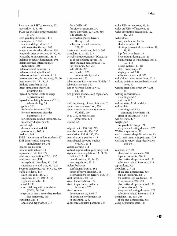
T variant on 5 HT2A receptor, 272tauopathies, 338, 339TCAs see tricyclic antidepressants
(TCAs)teeth grinding (bruxism), 141temazepam, 217, 258abuse and dependence, 318with cognitive therapy, 242
temperature circadian rhythm, 191temporal cortex activation, 49 50, 362tetracyclic antidepressants, 135 6thalamic reticular dysfunction, 304thalamocortical interactions, 67dysfunction, 304
thalamocortical neurons, 19thalamocortical spindles, 67thalamus, reticular nucleus of, 19thermoregulation, during sleep, 39, 60theta waves, 14 15, 18, 23thinking disturbance, 305threat simulation theory, in
dreaming, 88thyroid hormone levels, in sleep
deprivation, 61thyroid stimulating hormone (TSH),
release timing, 39tiagabine, 226for bipolar insomnia, 277for post traumatic disorder
nightmares, 177for substance related insomnia, 322in anxiety disorders, 292
time of nightdream content and, 84parasomnias, 102 3
tiredness, 148TMN (tuberomammillary nucleus), 17TMS (transcranial magnetic
stimulation), 50, 391tobacco see nicotinetonic muscle activity, 40topiramate, 164, 174, 177total sleep deprivation (TSD), 252total sleep time (TST)in psychotic disorders, 301, 333substance use and, 316, 317, 320
Tourette syndrome, 343, 345, 382, 390traffic accidents, 113sleep loss and, 148, 211sleepiness in, 37, 101 2, 156vigilance deficits, 113
tramadol, 138transcranial magnetic stimulation
(TMS), 50, 391transplant patients, secondary restless
legs syndrome, 135trazodone, 223 4abuse and dependence, 318
for ADHD, 352for bipolar insomnia, 277mood disorders, 227, 258, 366side effects, 224sleepwalking/sleep terrors
therapy, 164substance related insomnia,
227, 321treatment compliance, 242 3, 307triazolam, 121, 217, 228tricyclic antidepressants (TCAs), 44as anticatapleptic agents, 151drug induced parasomnias, 169for insomnia, 223, 257side effects, 223sleep quality, 223,
see also trimipraminetrimipramine, 223tuberomammillary nucleus (TMN), 17tuberous sclerosis, 380tumor necrosis factor (TNF),
62, 118two process model, sleep regulation,
13, 25 9
unifying theory, of sleep function, 61upper airway obstruction, 378upper airway resistance syndrome
(UARS), 116U R G E, in restless legs
syndrome, 130uridine, 63
valproic acid, 138, 164, 275vascular dementia, 334, 335venlafaxine, 135 6, 140, 258ventral arousal pathway, 15ventrolateral preoptic nucleus
(VLPO), 20 1verbal learning, 114vertical supranuclear gaze palsy, 338vigilance state regulation, 13 14, 29deficits, 112, 113neural systems, 10, 14 25sleep regulation, 25 9
violent behaviorconfusional arousal, 162schizoaffective disorder, 306sleepwalking/sleep terrors, 163, 164
viral infections, 62, 152visual hallucinations, 172visual impairment, pediatric
insomnia, 373visual systemdevelopment of, 8, 66 7in circadian rhythmicity, 28in dreaming, 9, 82
vocal cord abductor paralysis, 338
wake REM on neurons, 23, 24wake on/REM off neurons, 23wake promoting medication, 122,
242, 258wakefulnessacetylcholine in, 15 16alertness values, 41electrophysiological parameters, 37,
38, 40flip flop hypothesis, 13hypoarousal during, 209 10maintenance of wakefulness test, 41,
107, 148neural systems, 14 18rCBF brain maps, 49staying in bed, 237substance abuse and, 325
wakefulness sleep transitions, 20 1waking activities, neuroplasticity and
sleep, 50waking after sleep onset (WASO),
361waking consciousnessdreaming and, 9unity in, 8 9
waking state, AIM model, 6waking lifedreaming and, 83 4continuity hypothesis, 84
effect of dreams, 86 7, 88war veterans, 177weight gainantipsychotic drugs, 121sleep related eating disorder, 173
Williams syndrome, 381work patterns, sleep disturbance, 13work performance, impairment, 233working memory, sleep deprivation
and, 50 1
zaleplon, 217 22abuse and dependence, 318bipolar insomnia, 276 7obstructive sleep apnea and, 121substance related insomnia, 318
ziprasidone, 303zolpidem, 217 22abuse and dependence, 318bipolar insomnia, 276 7for restless legs syndrome, 138in depression, 227, 258obstructive sleep apnea and, 121parasomnias and, 164sleep related eating disorder, 173substance related insomnia, 318
zopiclone, 121, 138, 308abuse and dependence, 318
Zung depression scale, 115
Index
415