Chemotherapeutic compounds targeting the DNA double-strand break repair pathways: the good, the bad,...
18
REVIEW ARTICLE published: 22 April 2014 doi: 10.3389/fonc.2014.00086 Chemotherapeutic compounds targeting the DNA double-strand break repair pathways: the good, the bad, and the promising Christian Jekimovs, Emma Bolderson, Amila Suraweera, Mark Adams, Kenneth J. O’Byrne and Derek J. Richard* Cancer and Ageing Research Program, Institute of Health and Biomedical Innovation, Queensland University ofTechnology, Brisbane, QLD, Australia Edited by: Megan Chircop, Children’s Medical Research Institute, Australia Reviewed by: Gyorgy Szabadkai, University College London, UK Shiaw-Yih Lin, The University of Texas MD Anderson Cancer Center, USA *Correspondence: Derek J. Richard, Cancer and Ageing Research Program, Institute of Health and Biomedical Innovation, Queensland University ofTechnology, TRI Level 3, 37 Kent Street, Brisbane, QLD 4102, Australia e-mail: [email protected] The repair of DNA double-strand breaks (DSBs) is a critical cellular mechanism that exists to ensure genomic stability. DNA DSBs are the most deleterious type of insult to a cell’s genetic material and can lead to genomic instability, apoptosis, or senescence. Incor- rectly repaired DNA DSBs have the potential to produce chromosomal translocations and genomic instability, potentially leading to cancer.The prevalence of DNA DSBs in cancer due to unregulated growth and errors in repair opens up a potential therapeutic window in the treatment of cancers.The cellular response to DNA DSBs is comprised of two pathways to ensure DNA breaks are repaired: homologous recombination and non-homologous end joining. Identifying chemotherapeutic compounds targeting proteins involved in these DNA repair pathways has shown promise as a cancer therapy for patients, either as a monother- apy or in combination with genotoxic drugs. From the beginning, there have been a number of chemotherapeutic compounds that have yielded successful responses in the clinic, a number that have failed (CGK-733 and iniparib), and a number of promising targets for future studies identified.This review looks in detail at how the cell responds to these DNA DSBs and investigates the chemotherapeutic avenues that have been and are currently being explored to target this repair process. Keywords: DNA damage repair, chemotherapeutic compounds, DNA damage response, cancer, radiosensitize, radioprotective, DNA double-strand break INTRODUCTION Genomic stability at a cellular level requires precise, tightly coor- dinated pathways to detect DNA damage and either repair the damage or, if the damage is too great, ensure the cell dies via apop- tosis or enters senescence. Organisms have evolved complex DNA damage response (DDR) pathways to respond to insults to the DNA either from endogenous (cellular metabolic pathways, reac- tive oxygen species, and errors in DNA replication) or exogenous sources [environmental factors including ionizing radiation (IR) and ultra violet radiation]. Cellular DNA damage that is not repaired correctly can lead to genomic instability, apoptosis, or senescence, which can greatly affect the organism’s development and aging process and in addition can predispose the organism to immunodeficiency, neurological disorders, and cancer. DDR AND REPAIR PATHWAYS Following the initial work on the DDR in yeast, investigations into the DDR in mammals have yielded a highly conserved and elab- orate process. This process mainly controls DNA repair (ensuring genomic stability) and cell cycle checkpoints, however it has also been shown to be involved in circadian rhythms (1), insulin signaling (2), and telomere maintenance (3). The DDR pathway encompasses a set of tightly coordinated processes: detection of DNA damage, a protein cascade to enhance the signal, the accumulation of repair factors at the site of damage, and physical repair of the damage. The DDR also induces cell cycle checkpoints to ensure the damaged cells do not continue dividing until the DNA damage is repaired. To ensure genomic stability, the DDR must be able to recognize all types of DNA structural alter- ations, including nicks, gaps, stalled replication, and double-strand breaks (DSBs). Depending on the type of DNA lesion, there are a num- ber of DNA repair pathways available for the cell to repair the alteration, including homologous recombination (HR) and non- homologous end joining (NHEJ) for DNA DSBs; and mismatch repair (MMR), nucleotide excision repair (NER), and base excision repair (BER) for single DNA strand damage. Highlighting the importance of the DDR, mutations in a num- ber of repair proteins lead to human syndromes, which include multiple cancers, immunodeficiency, and genomic instability phe- notypes. Ataxia telangiectasia mutated (ATM), a protein involved in the DDR is mutated in the syndrome ataxia telangiectasia (AT) (4). AT is a cancer-prone syndrome that also includes progressive cerebellar ataxia, telangiectasia’s of the conjunctivae, and immun- odeficiency. Consistent with ATM’s role in the DDR,AT patients presented a high level of sensitivity to radiation (5). Nijmegen breakage syndrome (NBS) is another syndrome where the key cause of the disease is a mutation in a protein involved in the DDR, NBS1. NBS1 is involved in the detection of DSBs as part www.frontiersin.org April 2014 |Volume 4 | Article 86 | 1
-
Upload
independent -
Category
Documents
-
view
0 -
download
0
Transcript of Chemotherapeutic compounds targeting the DNA double-strand break repair pathways: the good, the bad,...
REVIEW ARTICLEpublished: 22 April 2014
doi: 10.3389/fonc.2014.00086
Chemotherapeutic compounds targeting the DNAdouble-strand break repair pathways: the good, the bad,and the promisingChristian Jekimovs, Emma Bolderson, Amila Suraweera, Mark Adams, Kenneth J. O’Byrne andDerek J. Richard*
Cancer and Ageing Research Program, Institute of Health and Biomedical Innovation, Queensland University of Technology, Brisbane, QLD, Australia
Edited by:Megan Chircop, Children’s MedicalResearch Institute, Australia
Reviewed by:Gyorgy Szabadkai, University CollegeLondon, UKShiaw-Yih Lin, The University of TexasMD Anderson Cancer Center, USA
*Correspondence:Derek J. Richard, Cancer and AgeingResearch Program, Institute of Healthand Biomedical Innovation,Queensland University of Technology,TRI Level 3, 37 Kent Street, Brisbane,QLD 4102, Australiae-mail: [email protected]
The repair of DNA double-strand breaks (DSBs) is a critical cellular mechanism that existsto ensure genomic stability. DNA DSBs are the most deleterious type of insult to a cell’sgenetic material and can lead to genomic instability, apoptosis, or senescence. Incor-rectly repaired DNA DSBs have the potential to produce chromosomal translocations andgenomic instability, potentially leading to cancer. The prevalence of DNA DSBs in cancerdue to unregulated growth and errors in repair opens up a potential therapeutic window inthe treatment of cancers.The cellular response to DNA DSBs is comprised of two pathwaysto ensure DNA breaks are repaired: homologous recombination and non-homologous endjoining. Identifying chemotherapeutic compounds targeting proteins involved in these DNArepair pathways has shown promise as a cancer therapy for patients, either as a monother-apy or in combination with genotoxic drugs. From the beginning, there have been a numberof chemotherapeutic compounds that have yielded successful responses in the clinic, anumber that have failed (CGK-733 and iniparib), and a number of promising targets forfuture studies identified.This review looks in detail at how the cell responds to these DNADSBs and investigates the chemotherapeutic avenues that have been and are currentlybeing explored to target this repair process.
Keywords: DNA damage repair, chemotherapeutic compounds, DNA damage response, cancer, radiosensitize,radioprotective, DNA double-strand break
INTRODUCTIONGenomic stability at a cellular level requires precise, tightly coor-dinated pathways to detect DNA damage and either repair thedamage or, if the damage is too great, ensure the cell dies via apop-tosis or enters senescence. Organisms have evolved complex DNAdamage response (DDR) pathways to respond to insults to theDNA either from endogenous (cellular metabolic pathways, reac-tive oxygen species, and errors in DNA replication) or exogenoussources [environmental factors including ionizing radiation (IR)and ultra violet radiation].
Cellular DNA damage that is not repaired correctly canlead to genomic instability, apoptosis, or senescence, which cangreatly affect the organism’s development and aging process andin addition can predispose the organism to immunodeficiency,neurological disorders, and cancer.
DDR AND REPAIR PATHWAYSFollowing the initial work on the DDR in yeast, investigations intothe DDR in mammals have yielded a highly conserved and elab-orate process. This process mainly controls DNA repair (ensuringgenomic stability) and cell cycle checkpoints, however it has alsobeen shown to be involved in circadian rhythms (1), insulinsignaling (2), and telomere maintenance (3).
The DDR pathway encompasses a set of tightly coordinatedprocesses: detection of DNA damage, a protein cascade to enhance
the signal, the accumulation of repair factors at the site of damage,and physical repair of the damage. The DDR also induces cell cyclecheckpoints to ensure the damaged cells do not continue dividinguntil the DNA damage is repaired. To ensure genomic stability, theDDR must be able to recognize all types of DNA structural alter-ations, including nicks, gaps, stalled replication, and double-strandbreaks (DSBs).
Depending on the type of DNA lesion, there are a num-ber of DNA repair pathways available for the cell to repair thealteration, including homologous recombination (HR) and non-homologous end joining (NHEJ) for DNA DSBs; and mismatchrepair (MMR), nucleotide excision repair (NER), and base excisionrepair (BER) for single DNA strand damage.
Highlighting the importance of the DDR, mutations in a num-ber of repair proteins lead to human syndromes, which includemultiple cancers, immunodeficiency, and genomic instability phe-notypes. Ataxia telangiectasia mutated (ATM), a protein involvedin the DDR is mutated in the syndrome ataxia telangiectasia (AT)(4). AT is a cancer-prone syndrome that also includes progressivecerebellar ataxia, telangiectasia’s of the conjunctivae, and immun-odeficiency. Consistent with ATM’s role in the DDR, AT patientspresented a high level of sensitivity to radiation (5). Nijmegenbreakage syndrome (NBS) is another syndrome where the keycause of the disease is a mutation in a protein involved in theDDR, NBS1. NBS1 is involved in the detection of DSBs as part
www.frontiersin.org April 2014 | Volume 4 | Article 86 | 1
Jekimovs et al. Chemotherapeutics and DNA-double-strand-break repair pathways
of a complex of proteins including Mre11 and Rad50. NBS isa cancer-prone syndrome that is also characterized by progres-sive microcephaly, short stature, and progressive ovarian failure infemales (6).
Current chemotherapeutic compounds development is largelyfocused in targeting proteins specific to pathways important tothe development, growth, and progression of cancer. DSBs arethe most deleterious lesion to cells, where unrepaired DSBs canlead to cell death and incorrectly repaired DSBs have the potentialto produce chromosomal translocations and genomic instability,potentially leading to cancer. Targeting the repair proteins involvedin the repair of DSBs with chemotherapeutic compounds hasthe potential for cancer therapies in conjunction with radiationtherapy or as a monotherapy.
CHEMOTHERAPEUTIC COMPOUNDSDouble-strand breaks are highly cytotoxic and this fact is exploitedin conventional cancer treatment, with radiation therapy andchemotherapeutic drugs treatments generating vast amounts ofDSBs. These include chemotherapeutic drugs that induce DNAcross-links or function as topoisomerase inhibitors, inducing thegeneration of DSB’s in all cells. However, cancer cells are muchmore susceptible to these drugs, as they are rapidly dividing andoften have inactivated components of their DNA repair machineryand deregulated cell cycle checkpoints (7).
However, these chemotherapeutic drugs will also target nor-mal proliferating cells that are dividing as part of their nor-mal processes. These naturally regenerating tissues include bonemarrow, gastrointestinal tract, liver, and hair follicles. The for-mation of secondary hematologic and solid tumors after DNA-damaging therapies is a potential issue for patients undergoingtreatment (8).
A number of chemotherapeutic compounds are used inconjunction with radiotherapy or in combination with otherchemotherapeutic agents to produce a synergistic effect. The use ofradiosensitizing agents that increase the cytotoxic effects of radi-ation on cancer cells and radioprotective agents that decrease theadverse effects of radiation on normal cells (by increasing theirradioresistance) is common. The use of radiosensitizing agentscan greatly enhance the efficacy of radiotherapy and genotoxicdrugs. Recently, chemotherapeutic compounds have been studiedthat may also be useful as a monotherapy, where the chemothera-peutic compound achieves what is termed as “synthetic lethality.”Synthetic lethality exploits the fact that many cancer cells acquiredefects in DNA repair pathways and become dependent on a com-pensatory mechanism in order to survive (9, 10). Inhibition of thecomplementary DNA repair pathway selectively kills cancer cellsthat have a defect in a particular DNA repair pathway.
The safety, tolerability,pharmacokinetics, and efficacy of poten-tial chemotherapeutic compounds have to be carefully validatedbefore they may enter clinical trials to determine the benefits forcancer therapy. This means there is a significant delay between theinitial discovery of a potential chemotherapeutic compound in thelaboratory, to an actual clinical outcome for patients, however thisdelay ensures patient safety.
This review will focus on the pathways responsible for the repairof DSBs, namely HR and NHEJ, and the current chemotherapeutic
compounds that are being investigated that target these repairpathways.
THE DDR TARGETS AND CHEMOTHERAPEUTIC COMPOUNDSDNA DSBs are considered the most cytotoxic of DNA lesions. Cellsare estimated to accumulate around 50 endogenous DSBs per day,mostly induced by reactive oxygen species (11). In response toDSBs, the DDR utilizes two main pathways to repair the damage.During late S-phase and the G2 phase, cells have a sister chro-matid available as a template for targeted HR, which allows forerror-free repair of the DNA damage. However, DSBs that occurwhen there is no sister chromatid available are repaired via NHEJ,which is more error-prone than HR. NHEJ is also active in S andG2 phases of the cell cycle and remains the predominant pathwayby which cells repair DSBs. In NHEJ, the two ends of the break arejoined together (ligated), though this can involve resection withthe consequent loss of genetic material (12). Cancer therapy agentsinduce DSBs including IR and topoisomerase II poisons, and alsoindirectly via single-stranded DNA (ssDNA) lesions which inducereplication forks collapse, leading to DSB formation (13).
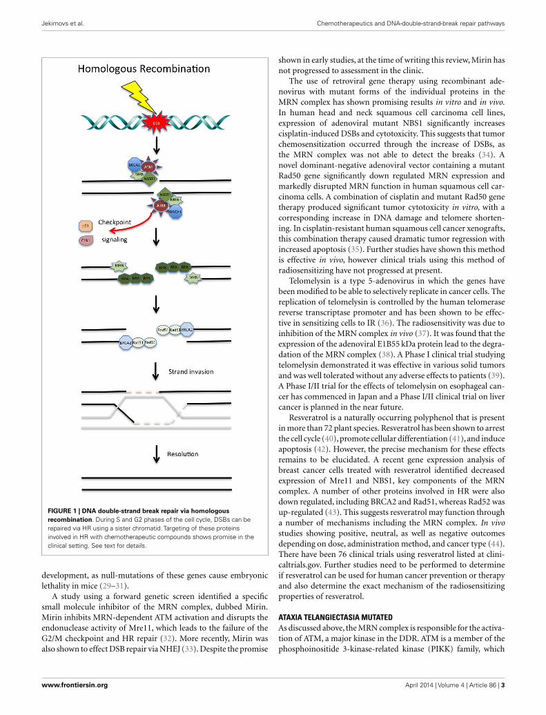
HOMOLOGOUS RECOMBINATION AND CHEMOTHERAPEUTICCOMPOUNDS TARGETSFollowing the induction of a DSB, the Mre11/Rad50/NBS1 (MRN)complex is recruited to the break site by human single-strandedbinding protein 1 (hSSB1) (14–16). MRN binds to the DNA sur-rounding the lesion and resects the DNA around the break in a5′–3′ dependent direction. This acts as a signal to recruit otherDDR proteins. This resection by the MRN complex is stimulatedin the early stages of HR by an interaction with CtIP (17, 18).Following initiation of resection by Mre11, Exo1 performs moreextensive resection to expose long stretches of ssDNA (19–21).Replication protein A (RPA), a ssDNA binding heteromeric com-plex, binds to the exposed ssDNA and is retained at the lesionsite by BRCA1 (22). The binding of RPA to the ssDNA substrateensures that secondary structures are not formed in the DNA andprotects the ssDNA from nucleases. RPA is displaced from theDNA by the recombinase Rad51, which is loaded by BRCA2. Rad51forms a nucleoprotein filament along the ssDNA and functions toallow strand invasion of the sister chromatid (23). Once the DSBis resolved, the DNA is ligated together, completing the processof HR (24). There are a number of key proteins involved in HRthat are currently therapeutic targets or have been identified aspotential targets (see Figure 1).
THE MRN COMPLEXHuman single-stranded binding protein 1 serves as the primarysensor of DSBs and is also involved in the early steps of HR throughthe recruitment of the MRN complex (14–16). Once recruited,the MRN complex specifically functions in the resection of DNAends, activates the ATM kinase, and subsequently activates the cellcycle checkpoints (25). The human syndromes that result frommutations in each component in the MRN complex highlightthe requirement of the MRN complex for the maintenance ofgenomic stability: NBS (26), AT-like disorder (AT-LD) (27), andNBS-like NBS disorder (28) result from NBS1, Mre11, and Rad50mutations, respectively. The MRN complex is also indispensible in
Frontiers in Oncology | Molecular and Cellular Oncology April 2014 | Volume 4 | Article 86 | 2
Jekimovs et al. Chemotherapeutics and DNA-double-strand-break repair pathways
FIGURE 1 | DNA double-strand break repair via homologousrecombination. During S and G2 phases of the cell cycle, DSBs can berepaired via HR using a sister chromatid. Targeting of these proteinsinvolved in HR with chemotherapeutic compounds shows promise in theclinical setting. See text for details.
development, as null-mutations of these genes cause embryoniclethality in mice (29–31).
A study using a forward genetic screen identified a specificsmall molecule inhibitor of the MRN complex, dubbed Mirin.Mirin inhibits MRN-dependent ATM activation and disrupts theendonuclease activity of Mre11, which leads to the failure of theG2/M checkpoint and HR repair (32). More recently, Mirin wasalso shown to effect DSB repair via NHEJ (33). Despite the promise
shown in early studies, at the time of writing this review, Mirin hasnot progressed to assessment in the clinic.
The use of retroviral gene therapy using recombinant ade-novirus with mutant forms of the individual proteins in theMRN complex has shown promising results in vitro and in vivo.In human head and neck squamous cell carcinoma cell lines,expression of adenoviral mutant NBS1 significantly increasescisplatin-induced DSBs and cytotoxicity. This suggests that tumorchemosensitization occurred through the increase of DSBs, asthe MRN complex was not able to detect the breaks (34). Anovel dominant-negative adenoviral vector containing a mutantRad50 gene significantly down regulated MRN expression andmarkedly disrupted MRN function in human squamous cell car-cinoma cells. A combination of cisplatin and mutant Rad50 genetherapy produced significant tumor cytotoxicity in vitro, with acorresponding increase in DNA damage and telomere shorten-ing. In cisplatin-resistant human squamous cell cancer xenografts,this combination therapy caused dramatic tumor regression withincreased apoptosis (35). Further studies have shown this methodis effective in vivo, however clinical trials using this method ofradiosensitizing have not progressed at present.
Telomelysin is a type 5-adenovirus in which the genes havebeen modified to be able to selectively replicate in cancer cells. Thereplication of telomelysin is controlled by the human telomerasereverse transcriptase promoter and has been shown to be effec-tive in sensitizing cells to IR (36). The radiosensitivity was due toinhibition of the MRN complex in vivo (37). It was found that theexpression of the adenoviral E1B55 kDa protein lead to the degra-dation of the MRN complex (38). A Phase I clinical trial studyingtelomelysin demonstrated it was effective in various solid tumorsand was well tolerated without any adverse effects to patients (39).A Phase I/II trial for the effects of telomelysin on esophageal can-cer has commenced in Japan and a Phase I/II clinical trial on livercancer is planned in the near future.
Resveratrol is a naturally occurring polyphenol that is presentin more than 72 plant species. Resveratrol has been shown to arrestthe cell cycle (40), promote cellular differentiation (41), and induceapoptosis (42). However, the precise mechanism for these effectsremains to be elucidated. A recent gene expression analysis ofbreast cancer cells treated with resveratrol identified decreasedexpression of Mre11 and NBS1, key components of the MRNcomplex. A number of other proteins involved in HR were alsodown regulated, including BRCA2 and Rad51, whereas Rad52 wasup-regulated (43). This suggests resveratrol may function througha number of mechanisms including the MRN complex. In vivostudies showing positive, neutral, as well as negative outcomesdepending on dose, administration method, and cancer type (44).There have been 76 clinical trials using resveratrol listed at clini-caltrials.gov. Further studies need to be performed to determineif resveratrol can be used for human cancer prevention or therapyand also determine the exact mechanism of the radiosensitizingproperties of resveratrol.
ATAXIA TELANGIECTASIA MUTATEDAs discussed above, the MRN complex is responsible for the activa-tion of ATM, a major kinase in the DDR. ATM is a member of thephosphoinositide 3-kinase-related kinase (PIKK) family, which
www.frontiersin.org April 2014 | Volume 4 | Article 86 | 3
Jekimovs et al. Chemotherapeutics and DNA-double-strand-break repair pathways
also includes ataxia telangiectasia and Rad3-related (ATR) andDNA-dependent protein kinase catalytic subunit (DNA-PKcs).The MRN complex activates ATM in response to DSBs by recruit-ing it to the sites of damage (45). Activated ATM is responsiblefor the induction of the G1/S, intra-S, and G2/M checkpoints, viathe phosphorylation of a number of down-stream effector kinasesand transcription factors, including p53 and p21 (46). The acti-vation of the cell cycle checkpoints is critical in the DDR to allowfor DNA repair to occur before the cell divides ensuring genomicstability.
A study showed that ATM is responsible for hundreds of phos-phorylation events in the cell in response to DNA damage, high-lighting the key role this kinase plays in the DDR (47). ATM isalso required for the full activation of Akt (also known as proteinkinase B) in response to insulin in the cytoplasm (48). ATM hasalso shown to be involved in the regulation of the expression andstability of ribonucleotide reductase and the mitochondrial home-ostasis through the control of mitochondrial DNA (mtDNA) copynumber dynamics and expression (49). This link with ATM andthe regulation of mtDNA may be involved in the resistance ofgenotoxic stress, highlighted by the potential role of the nuclearco-activators peroxisome proliferator-activated receptor gammaco-activator-1β in DNA damage repair (50). These key roles in theDDR have ensured that ATM has been a prime candidate for inhi-bition in cancer treatment and further investigations into syntheticlethality for AT patients may show promise.
Caffeine and wortmannin were the first ATM inhibitors identi-fied in the lab and were shown to increase sensitivity to radiationand chemotherapeutic compounds (51, 52). Both caffeine andwortmannin were later shown to be non-specific inhibitors ofATM and also inhibited the other PIKK, ATR, and DNA-PKcsand the potency of these drugs rendered them unsuitable for usein a clinical setting.
The flavonoid quercetin was identified as an inhibitor of phos-phoinositide 3-kinase (PI3K) with an IC50 of 3.8 µM (53). Analogsof quercetin were synthesized and investigated for their inhibi-tion of PI3K, which led to the discovery of LY294002, an ATP-competitive inhibitor (54). LY294002 was later found to alsoinhibit ATM (55) and DNA-PKcs (56). However, in the high dosesrequired to inhibit these proteins (>10 µM), LY294002 targetedseveral unrelated proteins including calcium channels and theestrogen receptor (57, 58).
Despite its lack of specificity, LY294002 was used as a researchtool to identify more specific PIKK inhibitors. A study screen-ing a drug-library based on LY294002 identified KU-55933, asmall molecule ATP-competitive inhibitor, which was specific toATM (55). KU-55933 is a potent inhibitor of ATM with an IC50
of 0.013 µM and is highly specific to ATM compared to otherPI3K and PIKK’s. This compound effectively sensitized tumorcells to radiation and DSBs inducing chemotherapeutic com-pounds, such as camptothecin and etoposide, and initial workhas shown this compound may be used as a potential clinicaltreatment (55).
KU-60019, an improved analog of KU-55933 has been shown toinhibit ATM with an IC50 of 0.0063 µM and also inhibits migrationand effectively radiosensitizes human glioma cells (59). Furtherstudies on KU-60019 are currently being performed, specifically
as a radiosensitizer with standard chemotherapy regimes onglioblastoma in preparation for clinical trials (60).
Another ATM specific inhibitor, CP466722, was identified in atargeted compound library screen, looking for inhibitors of ATM-dependent phosphorylation events in vitro. In vivo treatment withCP466722 resulted in transient inhibition of ATM and sensitizedcells to IR however, upon removal, ATM kinase activity and thesubsequent phosphorylation of down-stream targets was com-pletely restored (61). The clinical implications of this transientinhibition of ATM, requires further study.
KU59403 is the latest of the ATM inhibitors that have beenstudied and one that shows the most promise for clinical trialsin patients. KU59403 increased the cytotoxicity of the topoiso-merase I and II poisons camptothecin, etoposide, and doxorubicin,in vitro, in a non-p53-dependent manner. Importantly,upon injec-tion, KU59403 was seen to be distributed to tissues in mice atconcentrations required to inhibit ATM activity, these were shownto be maintained for at least 4 h in colon cancer tumor xenograftsand enhanced the anti-tumor activity of topoisomerase poisons.This chemosensitization was both dose and schedule-dependentand provided the first proof-of-principle pre-clinical data to sup-port future clinical development of ATM inhibitors (62). However,at present, there are no reports of ATM inhibitors in use in clinicaltrials.
It should be noted that CGK-733, a small molecule that was ini-tially reported to inhibit both ATM and ATR kinase activities andblock checkpoint signaling with great selectivity,was later retracted(63). Further studies were completed showing that CGK-733 hasno specific inhibitory effect on ATM or ATR (64). Unfortunately,the compound is still being marketed as an ATM/ATR inhibitor.
hSSB1/2Human single-stranded binding protein 1 is required for the acti-vation of ATM through the recruitment of the MRN complexto the break site (14–16). Until the identification of hSSB1 andhSSB2, as a DNA single strand-binding proteins, RPA was the onlyknown eukaryote member of the single strand-binding proteinfamily (SSB) to be involved in DNA repair (14). As mentionedabove, hSSB1 is required for the efficient recruitment of the MRNcomplex to sites of DSBs and for the efficient initiation of ATM-dependent signaling (15, 16). hSSB1 binds directly to the MRNcomplex through NBS1 and functions to also stimulate its nucleaseactivity. Identification of specific chemotherapeutic compoundsto target hSSB1 and hSSB2 will enable the sensitization of cancercells to radiotherapy and these are currently being explored by ourlaboratory and by industry.
Chk1/2The checkpoint kinases, Chk1 and Chk2, are critical for cell cycleactivation following the induction of DSBs and serve to main-tain the genomic integrity of cells (65). This cell cycle check-point activation is achieved through maintaining or augmentingthe inhibitory phosphorylation of the cyclin-dependent kinases(CDKs) by inhibiting the CDC25 phosphatases. Specifically, phos-phorylation of CDC25A is required for the initiation of the S-phasecheckpoint and phosphorylation of CDC25C for the G2/M check-point (66). Both Chk1 and Chk2 are also required for the activation
Frontiers in Oncology | Molecular and Cellular Oncology April 2014 | Volume 4 | Article 86 | 4
Jekimovs et al. Chemotherapeutics and DNA-double-strand-break repair pathways
or inhibition of a number of other cell cycle checkpoint proteinsand tumor suppressor proteins, including p53 (67).
Of these checkpoint kinases, Chk1 is critical for the inductionof HR, as inhibition of Chk1 in vitro lead to persistent unrepairedDSBs and cell death (68). Rad51 was also shown to be recruited toDSBs at replication forks in a Chk1-dependent manner, thereforeChk1 is essential for HR at stalled replication forks. Highlightingthis critical role, Chk1 null mice were found to be embryonicallylethal (69). Due to its critical role in HR, this review will focus onChk1 inhibitors only, however a number of these Chk1 inhibitorsare also known to target Chk2.
UCN-01, a staurosporine inhibitor was the first inhibitor iden-tified for both Chk1 and Chk2 and treatment with this compoundlead to G2/M checkpoint deficiencies in IR-treated p53-deficienttumor cells (70). However, due to the broad spectrum of targetsfor UCN-01, including protein kinase C, CDK, and CDK2, the useof UCN-01 presented challenges in the clinical setting (71). Sincethe discovery of UCN-01, increasingly specific inhibitors for Chk1have been identified.
XL-844,also known as EXEL-9844, is a potent,ATP-competitiveinhibitor of Chk1 and Chk2. In vitro, XL-844 showed limitedability as a monotherapy but substantially enhanced gemcitabine-induced cell killing. XL-844 increased the gemcitabine-inducedDNA damage, blocked CDC25A phosphorylation, abrogated thegemcitabine-induced S-phase checkpoint, and induced prematuremitotic entry. Interestingly, XL-844 also induced phosphoryla-tion of Chk1 (72). However, a further in vitro study showed that,in response to IR, Chk2, rather than Chk1 appeared to be acti-vated by irradiation and this activation was suppressed by XL-844(73). A Phase I clinical trial of XL-844 as a monotherapy or inconjunction with gemcitabine in patients with advanced malig-nancies was discontinued before completion (clinicaltrials.gov –NCT00475917).
AZD7762 is a potent ATP-competitive checkpoint kinaseinhibitor that was identified via a compound library screen usingChk1. AZD7762 was found to inhibit both Chk1 and Chk2 within vitro and in vivo studies confirming the abrogation of thecheckpoint response to gemcitabine (74). In vivo studies involv-ing human breast cancer xenografts in mice demonstrated thatAZD7762, in combination with irinotecan, improved host sur-vival and reduced tumor growth selectively in p53 mutant tumors(75). A Phase I clinical trial to evaluate the safety, tolerability, andpharmacokinetics of AZD7762, as a monotherapy or in conjunc-tion with gemcitabine, in patients with advanced solid malignan-cies has just been completed, however no published data havebeen released as yet (clinicaltrials.gov – NCT00413686). However,interestingly two Phase I trial of AZD7762 in conjunction withirinotecan (clinicaltrials.gov – NCT00473616) and gemcitabine(clinicaltrials.gov – NCT00937664) were terminated early. TrialNCT00937664 was terminated due to incidence of cardiac toxic-ities reported in the overall Phase I development program (76).This result may affect further clinical development of AZD7762.
PF-00477736 is a potent, selective ATP-competitive small mol-ecule inhibitor of Chk1 and was shown to abrogate cell cycle arrestinduced by DNA damage and enhanced the cytotoxicity of clini-cally important chemotherapeutic agents, including gemcitabineand carboplatin both in vitro and in vivo in mouse xenografts (77).
In combination with docetaxel, PF-00477736 was found to abro-gate the DNA damage checkpoints and resulted in sensitizationto docetaxel (78). The only clinical trial to date with PF-00477736looked at the effects in combination with gemcitabine in advancedsolid tumors, however this study was prematurely terminated(clinicaltrials.gov – NCT00437203).
SCH900776, also known as MK-8776, was identified as a highlypotent Chk1 inhibitor using a high-content, cell-based screen forγ-H2AX induction (γ-H2AX is a surrogate marker for double-strand DNA breaks). SCH900776 also enhanced the anti-tumoreffects of gemcitabine in vivo (79). SCH900776 was also shownto inhibit CDC25C degradation, abrogates S-phase arrest, andinduces DNA damage (80). When SCH900776 was combinedwith low concentrations of hydroxyurea, both p53-deficient andp53-proficient cell lines were sensitive to the combination (81). Itwas also demonstrated, in vitro, that some cell lines were highlysensitive to SCH900776 alone. In vivo models with a human pan-creas tumor xenografts mouse model combined SCH900776 withgemcitabine, this showed a significantly delayed tumor growthcompared to either drug alone (82). A Phase I clinical trial wasundertaken using SCH900776 in combination with cytarabine inpatients with acute leukemia. This trial indicated that SCH900776was tolerated by patients and progressed to a Phase II clinical trial(83). A randomized Phase II clinical trial is currently recruitingto study how well cytarabine, with or without SCH900776, worksin treating adult patients with relapsed acute myeloid leukemia(clinicaltrials.gov – NCT01870596).
LY2603618, a pyrazinyl-urea compound, was identified as aChk1 inhibitor via in vitro kinase assays. LY2603618 is currentlybeing investigated in a number of Phase I and II clinical tri-als and pre-clinical data on their effects in vitro and in vivohave been recently released. LY2603618 was shown in vitro toproduce a cellular phenotype similar to that reported for deple-tion of Chk1 by siRNA and impaired DNA synthesis, elevatedH2AX phosphorylation, which is indicative of DNA damage,and caused premature entry into mitosis. In vivo treatment ofhuman mutant p53 lung cancer cell xenografts in mice, withgemcitabine, resulted in a stimulation of Chk1 kinase activitythat was inhibited by co-administration of LY2603618 (84). Ina Phase 1 dose escalation clinical trial of LY2603618 combinedwith pemetrexed, 9 out of 31 patients achieved stable diseaseand 1 pancreatic cancer patient had a partial response (85). Twoother clinical trials have been completed using LY2603618, inconjunction with gemcitabine, in patients with pancreatic can-cer (clinicaltrials.gov – NCT00839332); and an open-label studyin patients with advanced and/or metastatic solid tumors (clin-icaltrials.gov – NCT01296568), however no results have beenpublished on these clinical trials. There are currently two activeclinical trials currently underway studying LY2603618: the first isstudying the safety and tolerability of LY2603618 in combinationwith gemcitabine in patients with solid advanced or metasta-tic tumors (clinicaltrials.gov – NCT01341457) and the second isinvestigating the safe dose of LY2603618 that can be combinedwith pemetrexed and cisplatin and to test if this triplet offers asignificant improvement in progression-free survival in partici-pants with Stage IV non-squamous non-small cell lung cancer(clinicaltrials.gov – NCT01139775).
www.frontiersin.org April 2014 | Volume 4 | Article 86 | 5
Jekimovs et al. Chemotherapeutics and DNA-double-strand-break repair pathways
LY2606368 has been identified as a Chk1 inhibitor by Eli Lillyand a Phase I clinical trial is currently recruiting to investigate theeffects of LY2606368 in patients with advanced solid tumors (clin-icaltrials.gov – NCT01115790), however no pre-clinical data areavailable for this inhibitor.
Two recently identified Chk1 inhibitors, GDC-0425 and GDC-0575, are currently undergoing clinical trials, however again nopre-clinical data are available. Both compounds are currentlybeing evaluated for the safety, tolerability, and pharmacokineticsadministered as a monotherapy or in combination with gemc-itabine in patients with refractory solid tumors or lymphoma:GDC-0425 (clinicaltrials.gov – NCT01359696); and GDC-0575(clinicaltrials.gov – NCT01564251).
SAR-020106 is a novel, selective, and potent ATP-competitiveinhibitor of Chk1. SAR-020106 has been shown to abrogate theetoposide-induced G2 arrest and significantly enhances the cellkilling of gemcitabine in vitro and in a p53-dependent fashion.In vivo, it was found that irinotecan, gemcitabine, and radiationactivity was enhanced by SAR-020106 with minimal toxicity (86,87). Whilst SAR-020106 has not undergone any clinical trials atpresent, this pre-clinical data suggest it is a prime candidate forinvestigation in p53-defective tumors.
p53The tumor suppressor protein p53 is a transcriptional regulator ofa number of genes involved in DNA repair, cell cycle progression,and apoptosis. Highlighting this function, p53 was found to bemutated in approximately 50% of cancers (88). p53 is activated inresponse to DNA damage by phosphorylation by the ATM, ATR,Chk2, and Chk1 kinases and these phosphorylation events allowfor the stabilization of p53 and activate its transcriptional func-tions allowing the regulation of a number of genes responsible forcell cycle progression and apoptosis (89).
Initial work on p53-mutated cancers has investigated restor-ing the function of p53, thus leading to effective apoptosis inresponse to the chemotherapy. Two main mechanisms have beeninvestigated – restoring p53 to cancer cells using a recombinantadenovirus encoding p53 or using small compounds or shortpeptides to restore the activity of p53.
Pre-clinical in vitro and in vivo studies of adenovirus-mediatedp53 (Ad-p53) cancer gene therapy showed promising results withadvexin (90), gendicine (91), and SCH-58500 (92). Initial clinicaltrials with these Ad-p53 vectors showed that administration was asafe, feasible, and effective strategy against many types of cancers,however, the anti-tumor efficacy has been limited in some cancerpatients. These Ad-p53 vectors have also been used in combi-nation with conventional DNA-damaging treatments, indicatingthe induction of the apoptotic pathway via Ad-p53 can restorethe sensitivity to radiation and chemotherapy in some resistanttumors.
However, issues exist with the low transduction of p53 intocancer cells via these Ad-p53 vectors, to overcome this replica-tion, competent oncolytic adenoviruses have been developed. TheCRAd-p53 vector has been used where the promoters of cancer-related genes are used to regulate virus expression in a tumor-dependent manner. Recent work has focused on AdDelta24-p53(93), SG600-p53 (94), and OBP-700 (95). Initial in vitro and in vivo
studies have shown these CRAd-p53 vectors are a safe and effectivetherapy for inducing anti-tumor effects and have been shown toinduce higher p53 expression and stronger anti-tumor effects thanthe Ad-53 vectors, highlighting their potential in future clinicaltrials.
A number of small compounds and peptides have been shownto be effective in restoring the function of p53 in tumor cells,including CP-31398 (96), PRIMA-1 (97), CDB3 (98), peptide 46(99), and SCH529074 (100). These small compounds and pep-tides act to stabilize p53 in its active biological conformation, thusrestoring its transcriptional activity. Initial in vitro work on thesesmall compounds and peptides have shown promising results andfurther in vivo studies are required to determine their efficacybefore clinical trials can commence.
REPLICATION PROTEIN ADue to its key role in DNA replication and repair, via HR andNER, RPA has been the subject of a number of studies to identifypotential inhibitors. RPA is over-expressed in a number of cancers,including colon (101), esophageal (102), and breast (103). RPA isa heterotrimeric protein, consisting of RPA1 (p70), RPA2 (p32),and RPA3 (p14) subunits. RPA protects ssDNA from nucleolyticattack, prevents DNA hairpin formation, and blocks DNA rean-nealing by binding directly to the ssDNA through four OB-folds.After DNA damage, RPA coats ssDNA and enhances the capacityof Rad51 oligomer formation at sites of damage (104).
Initial work has investigated the disruption of the DNA bindingcapacity of RPA and also inhibition of its protein partner inter-actions using small molecule inhibitors. Selective inhibition ofboth the protein binding and DNA binding capacity of RPA hasthe potential to inhibit the DDR and to sensitize cancer cells toDNA-damaging agents.
TDRL-505, a novel small molecule inhibitor, has recently beenshown to inhibit the RPA–DNA interaction, thereby preventingcell cycle progression, induces cytotoxicity, and increases the effi-cacy of the chemotherapeutic DNA-damaging agent, cisplatin,in vitro (105). TDRL-505 inhibits the DNA binding capacity ofRPA by blocking the OB-folds of RPA1. Further studies need tobe completed in mouse models to determine the efficacy of thiscompound.
Isobornyl derivatives have also been shown to be RPA inhibitorsin a screen of the National Cancer Institute library, with CheSS19shown to interact irreversibly with the OB-folds of RPA1 (106).MCI13E, a haloester modified form of CheSS19, decreased cellviability and induced apoptosis, showing synergistic effects withcisplatin in lung cancer cells (107). However, this compound didnot affect the DNA binding capacity of RPA, but instead may actthrough the alkylation of cysteine residues of RPA. Further stud-ies, both in vitro and in vivo are required to fully understand themechanisms of RPA inhibition by MCI13E prior to clinical studiesbeing undertaken.
The initiation of the DDR by RPA is also mediated by protein–protein interactions involving the N-terminal domain of the p70subunit with partner proteins, including the MRN complex (108),Rad51 (109), and BRCA2 (110). Inhibition of these interactionsincreases sensitivity toward DNA damage and replication stressand may therefore be a potential strategy for cancer drug discovery.
Frontiers in Oncology | Molecular and Cellular Oncology April 2014 | Volume 4 | Article 86 | 6
Jekimovs et al. Chemotherapeutics and DNA-double-strand-break repair pathways
Combining RPA inhibition with radiation therapy could leadto increased cytotoxicity in tumor cells via inhibition of DNADSB repair via NHEJ or HR, both of which have been shown torequire RPA.
Rad51Rad51 plays an important role in maintaining genome stabilitythrough the HR pathway in response to DNA damage. This is high-lighted by the fact that Rad51 knockout mice show early embryoniclethality (111). Rad51 is a DNA recombinase and polymerizes ontoresected DNA ends to form a nucleoprotein filament that promotestrand invasion and exchange between homologous DNA duplexes(112). It is suggested the improper regulation of Rad51 may affecttumorigenesis, as Rad51 has been shown to be over-expressedin a number of cancer phenotypes, including esophageal, pan-creatic, lung, leukemia, and head and neck cancers. Conversely,Rad51 is also under-expressed in a number of cancer cells. Thisvariable expression of Rad51 has been shown to promote the resis-tance of tumors to chemotherapy (113). Using antisense RNA orRNAi to deplete the levels of Rad51 in vitro has been shown tosensitize tumor cells to chemotherapy agents, including cisplatin(114). These effects of Rad51 depletion demonstrate the potentialof Rad51 inhibitors in cancer therapy.
The first identification of a Rad51 inhibitor was a small peptide,homologous to the BRC-motif of the BRCA2 protein, which wasfound to bind Rad51, thereby preventing its DNA binding capacity(115). This peptide inhibited the formation of Rad51 nuclear fociand disrupted HR in vitro. Using an in silico approach on the BRCdomains of BRCA2, a chimeric peptide with an efficiency 10 timeshigher than the original peptide was identified (116). This newpeptide inhibited Rad51 DNA binding and DNA strand exchangeactivity however, although these peptides are currently being usedas a research tool they have not yet found clinical applicability.Further investigation of peptides and peptidomimics inhibitingRad51 function may elucidate novel inhibitors targeting the HRpathway in tumors. However, this approach still holds drawbacksmainly due to the pharmacokinetics of peptide-based inhibitorsand administration of these agents may not be optimal in a clinicalsetting.
More recently, a DNA strand exchange assay was performedand used to identify Rad51 inhibitors by high-throughput screen-ing of the NIH small molecule repository. This study identified 17potential inhibitors, of which 3 were studied further. CompoundB02 was identified that specifically inhibited human Rad51 withtwo other compounds, A03 and A10, which inhibited both Rad51and RecA, but not the structurally unrelated Rad54 protein. B02directly interacts with Rad51 and disrupts its binding to DNA andnucleoprotein filament formation. The interaction of B02 withRad51 disrupted DSB-induced HR and enhanced the sensitivityof cells to cisplatin (117). Further work on these compounds, bothin vitro and in vivo, are required before they can be introducedinto clinical trials.
A small molecule inhibitor to Rad51 was recently identifiedthrough a high-throughput screen of a library of 10,000 smallmolecules (118). The molecule RI-1 covalently binds to Rad51,thereby inhibiting its ability to form filaments on ssDNA. RI-1inhibits the nuclear foci of Rad51 at sites of DNA damage and
sensitizes various cancer cell types to cross-linking chemother-apy, but did not affect Rad51 protein levels. There are limits tothe development of RI-1 in pre-clinical in vivo models due to itsshort half-life in tissue culture media and aqueous buffers. RI-2,a homolog of RI-1, was created that mitigated these effects (119).RI-2 was shown to bind Rad51 and inhibit the nuclear foci ofRad51 at sites of DNA damage. RI-2 is currently the subject offurther in vitro and in vivo studies and is being used to identifythird generation analogs that inhibit the function of Rad51.
A further screen using a yeast-2 hybrid system identified aphenylsulfonyl indolyl isoquinoline compound, IBR2, as a Rad51inhibitor. IBR2 functions to block Rad51 multimerization, acceler-ating proteasome-mediated Rad51 protein degradation, and thusimpairing IR-induced Rad51 foci formation in the nucleus andHR activity. IBR2 inhibited cancer cell growth and induced apop-tosis (120). A synergistic cell-killing effect was produced with acombination of IBR2 and imatinib in vitro. In vivo studies involv-ing breast cancer xenografts in nude mice showed significantlyinhibited tumor growth with no apparent secondary physiologicalabnormalities. Further studies on the effects of IBR2 are requiredbefore moving into a clinical trial.
BRCA1/2The breast cancer susceptibility proteins, BRCA1 and BRCA2, havea key role in efficient HR response to DSBs. Mutations in thesegenes greatly increase the susceptibility to cancer, especially breast,ovarian, and prostate. Mutations in the BRCA genes are responsi-ble for the increased risk of breast cancer, specifically 59–87 and38–80% for BRCA1 and BRCA2 mutations respectively. BRCA1functions in both checkpoint activation and also in the early stepsof HR, by controlling DNA resection (121). BRCA2 functions inRad51 transport and loading (122). Both BRCA1 and BRCA2 arerequired for normal embryonic development in mice (123, 124).
Direct inhibition of BRCA1 and BRCA2 in tumors is generallyproblematic due to the wide expression of these proteins in mosttissues and inhibition may lead to other issues, including cancerdevelopment in healthy tissue. One approach is the possible upregulation of the BRCA1 and BRCA2 proteins, however there isno data to suggest that up regulation blocks tumorigenesis.
POLY ADP-RIBOSE POLYMERASE 1Most of the work in the BRCA therapeutic research area hasfocused on tumors that are known to have mutations in BRCA1or BRCA2. Using synthetic lethality, these studies have focusedon disrupting complimentary pathways to repair DNA damage,with the most interesting results coming from poly ADP-ribosepolymerase 1 (PARP1) inhibitors. PARP1 is involved in DNArepair, replication, transcriptional regulation, chromatin modifi-cation, and apoptosis (125, 126). In regards to DNA repair, PARP1is involved in BER which repairs DNA damage due to reactiveoxygen species and alkylation (127). Inhibition of this pathway,taken together with a loss of HR due to BRCA mutations, cre-ates a synthetic lethality, which can be exacerbated when used inconjunction with chemotherapy agents.
However, PARP1 inhibition and the subsequent syntheticlethality can be used on other cancers that do not have muta-tions in BRCA1 or BRCA2 but that have a defect in the HR
www.frontiersin.org April 2014 | Volume 4 | Article 86 | 7

Jekimovs et al. Chemotherapeutics and DNA-double-strand-break repair pathways
pathway, including mutations in ATM, Chk2, Rad51, and NBS1.However, NHEJ could compensate for the loss of HR in thesecells. Further studies have identified that PARP1 normally func-tions to promote HR by suppressing various components of theNHEJ pathway (128). Inhibition of PARP1 would therefore leadto increased NHEJ, a more error-prone repair mechanism thanHR, this would increase chromosome instability in response tochemotherapy and during S-phase of the cell cycle at stalled DNAreplication forks.
Poly ADP-ribose polymerase catalyzes the cleavage of NAD+ tonuclear acceptor proteins, leading to the formation of ADP-ribosepolymers, realizing nicotinamide in the process. Nicotinamide wasthe first PARP inhibitor identified, although it was not potent(129). Analogs of nicotinamide, including 3-aminobenzamide,were the first generation of PARP inhibitors (130).
The first clinical trial targeting PARP1 in BRCA populationswas with the oral drug olaparib, also known as AZD2281, asa monotherapy (131). Olaparib achieved encouraging responserates of 41 and 33% in patients with BRCA1 or BRCA2 muta-tions, respectively. Olaparib was also used in a combined therapywith carboplatin in vivo and showed a profound decrease intumor growth and increase in patient survival (132). A PhaseII trial with olaparib was conducted on patients with advancedBRCA1/2 mutant breast cancers (133) and ovarian cancers (134).Both of these studies showed a dose-dependent effect of ola-parib. Currently, there is a Phase III olaparib trial being under-taken by AstraZeneca. This trial aims to determine the benefit, byprogression-free survival, of olaparib as a maintenance monother-apy, in BRCA mutated ovarian cancer patients,who are in completeor partial response following platinum-based chemotherapy.
Another PARP inhibitor is veliparib. An in vivo study of veli-parib, also known as ABT-888, confirmed the PARP inhibitoryeffects in paired tumor biopsies and peripheral mononuclear cells(135). A number of Phase I trials have been conducted with veli-parib in combination, including topotecan (136) amongst others.Many of these trials have shown promising results, however myelo-suppression, where bone marrow activity is decreased, has beenshown as the most common adverse event observed.
Iniparib, also known as BSI-201, was the first PARP inhibitor toundergo Phase III clinical trials after showing promising resultsin randomized Phase II trials in patients with triple-negativebreast cancer (137). The results of the subsequent Phase III clin-ical trial were not as expected, missing the co-primary endpointsof overall survival and progression-free survival. However, verylittle pre-clinical data on the effects of iniparib were publishedbefore clinical trials began and iniparib was shown not be relatedto other PARP inhibition and showed very low PARP inhibitionin vitro (138, 139).
Rucaparib, also known as AG014699, was used in a PhaseI clinical trial in combination with temozolomide (140). Ruca-parib was well tolerated in patients and showed promising resultsfor assumed HR-deficient tumors (based on tumor type). Ruca-parib was also used as a monotherapy in a Phase I/II clinicaltrial with patients with solid tumors. Rucaparib was well tol-erated in this trial and showed promising results (clinicaltri-als.gov – NCT01482715). A subsequent Phase II clinical trial,with patients with melanoma, was conducted in combination
with temozolomide, and showed an objective response ratewith 17.4 and 36% of patients remaining progression-free after6 months (141).
A therapeutic index-based strategy was used to identify CEP-8983, a novel 4-methoxy-carbazole inhibitor of PARP1 and PARP2(enzyme IC50 values of 20 and 6 nmol/L, respectively). CEP-8983was found to cause significant sensitization of chemotherapy-resistant tumor cell lines to the effects of temozolomide andcamptothecin in vitro. Administration of CEP-8983, deliveredorally in the form of CEP-9722, attenuated in vivo PARP activ-ity and resulted in significant chemosensitization of temozolomideand irinotecan in chemotherapy-resistant tumor xenografts (142).A Phase I clinical trial with CEP-9722, used as a monotherapyor in conjunction with temozolomide, was recently completedwith patients with advanced solid tumors (clinicaltrials.gov –NCT00920595). A Phase I/II clinical trial using CEP-9722 on solidtumors is currently underway (clinicaltrials.gov – NCT01311713).
MK-4827, also known as niraparib, is a novel 2-phenyl-2H-indazole-7-carboxamide PARP inhibitor that displayed anti-proliferation activities against BRCA1- and BRCA2-deficient can-cer cells in vitro. MK-4827 was found to be well tolerated in vivoand demonstrated efficacy as a single agent in a xenograft model ofBRCA1-deficient cancer (143). A Phase I clinical trial of patientswith solid tumors using MK-4827 was shown to have favorablepharmacokinetics, inhibited PARP activity effectively, is well toler-ated and has anti-tumor activity in carriers of BRCA1 and BRCA2mutations and patients with sporadic cancers (144).
BMN 673, an inhibitor of PARP catalytic activity, has exhib-ited selective anti-tumor activity at much lower concentrations(IC50 = 0.57 nM) than the earlier generation of PARP inhibitors,including olaparib, rucaparib, and veliparib. BMN 673 is read-ily orally bioavailable and in vivo studies with xenograft tumorscarry defects in BRCA1/2 or PTEN were sensitive to BMN 673.Synergistic effects were observed when BMN 673 was combinedwith temozolomide, SN38, or platinum drugs (145). A number ofpre-clinical studies and Phase 1, Phase II, and Phase III clinicaltrials utilizing BMN 673 as a monotherapy or in conjunction withvarious drugs, are currently underway.
It is important to note that not all breast cancer patients withBRCA mutations responded to PARP inhibition (131) and a sub-stantial number of patients with advanced BRCA1-mutant cancersare resistant to these agents. Further studies on PARP inhibitors,along with the current clinical trials, are needed to assess the effi-cacy of PARP inhibition in BRCA mutant and other HR-defectivecancers in conjunction with chemotherapy or as a monotherapy.
A list of all chemotherapeutic compounds targeting the HRpathway is provided in Table 1.
NON-HOMOLOGOUS END JOINING ANDCHEMOTHERAPEUTIC COMPOUNDS TARGETSCLASSICAL-NHEJThe classical-NHEJ (C-NHEJ) pathway is the major pathway ofDSB repair [reviewed in Ref. (146)], estimated to rapidly repair upto 85% of IR-induced DSBs (147). In straightforward terms, thispathway involves simply ligating the two DNA ends back together.Due to the resection of DNA overhangs surrounding the DSB,NHEJ is sometimes considered the error-prone pathway of DNA
Frontiers in Oncology | Molecular and Cellular Oncology April 2014 | Volume 4 | Article 86 | 8
Jekimovs et al. Chemotherapeutics and DNA-double-strand-break repair pathways
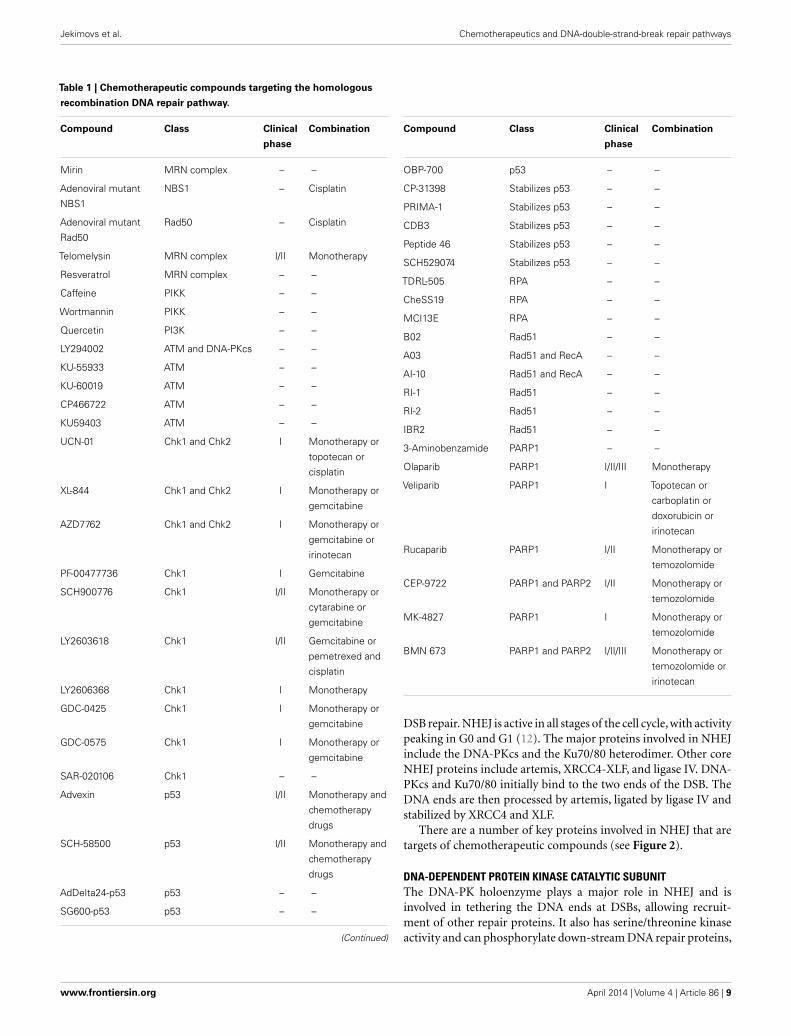
Table 1 | Chemotherapeutic compounds targeting the homologous
recombination DNA repair pathway.
Compound Class Clinical
phase
Combination
Mirin MRN complex – –
Adenoviral mutant
NBS1
NBS1 – Cisplatin
Adenoviral mutant
Rad50
Rad50 – Cisplatin
Telomelysin MRN complex I/II Monotherapy
Resveratrol MRN complex – –
Caffeine PIKK – –
Wortmannin PIKK – –
Quercetin PI3K – –
LY294002 ATM and DNA-PKcs – –
KU-55933 ATM – –
KU-60019 ATM – –
CP466722 ATM – –
KU59403 ATM – –
UCN-01 Chk1 and Chk2 I Monotherapy or
topotecan or
cisplatin
XL-844 Chk1 and Chk2 I Monotherapy or
gemcitabine
AZD7762 Chk1 and Chk2 I Monotherapy or
gemcitabine or
irinotecan
PF-00477736 Chk1 I Gemcitabine
SCH900776 Chk1 I/II Monotherapy or
cytarabine or
gemcitabine
LY2603618 Chk1 I/II Gemcitabine or
pemetrexed and
cisplatin
LY2606368 Chk1 I Monotherapy
GDC-0425 Chk1 I Monotherapy or
gemcitabine
GDC-0575 Chk1 I Monotherapy or
gemcitabine
SAR-020106 Chk1 – –
Advexin p53 I/II Monotherapy and
chemotherapy
drugs
SCH-58500 p53 I/II Monotherapy and
chemotherapy
drugs
AdDelta24-p53 p53 – –
SG600-p53 p53 – –
(Continued)
Compound Class Clinical
phase
Combination
OBP-700 p53 – –
CP-31398 Stabilizes p53 – –
PRIMA-1 Stabilizes p53 – –
CDB3 Stabilizes p53 – –
Peptide 46 Stabilizes p53 – –
SCH529074 Stabilizes p53 – –
TDRL-505 RPA – –
CheSS19 RPA – –
MCI13E RPA – –
B02 Rad51 – –
A03 Rad51 and RecA – –
AI-10 Rad51 and RecA – –
RI-1 Rad51 – –
RI-2 Rad51 – –
IBR2 Rad51 – –
3-Aminobenzamide PARP1 – –
Olaparib PARP1 I/II/III Monotherapy
Veliparib PARP1 I Topotecan or
carboplatin or
doxorubicin or
irinotecan
Rucaparib PARP1 I/II Monotherapy or
temozolomide
CEP-9722 PARP1 and PARP2 I/II Monotherapy or
temozolomide
MK-4827 PARP1 I Monotherapy or
temozolomide
BMN 673 PARP1 and PARP2 I/II/III Monotherapy or
temozolomide or
irinotecan
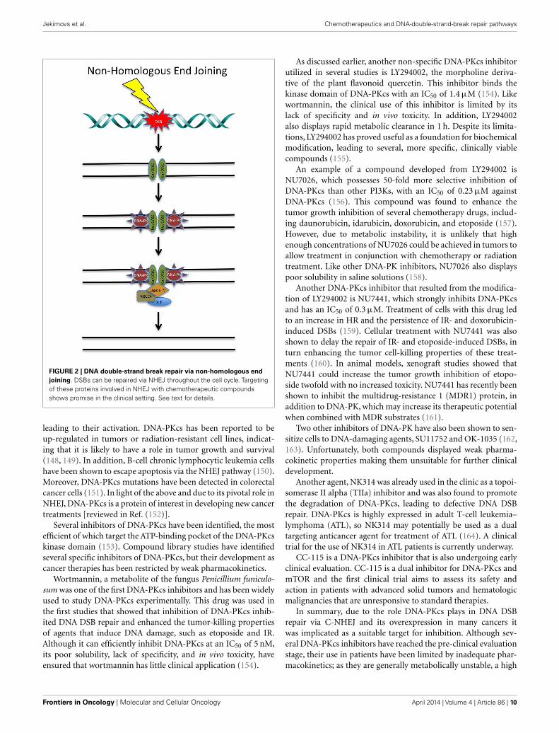
DSB repair. NHEJ is active in all stages of the cell cycle, with activitypeaking in G0 and G1 (12). The major proteins involved in NHEJinclude the DNA-PKcs and the Ku70/80 heterodimer. Other coreNHEJ proteins include artemis, XRCC4-XLF, and ligase IV. DNA-PKcs and Ku70/80 initially bind to the two ends of the DSB. TheDNA ends are then processed by artemis, ligated by ligase IV andstabilized by XRCC4 and XLF.
There are a number of key proteins involved in NHEJ that aretargets of chemotherapeutic compounds (see Figure 2).
DNA-DEPENDENT PROTEIN KINASE CATALYTIC SUBUNITThe DNA-PK holoenzyme plays a major role in NHEJ and isinvolved in tethering the DNA ends at DSBs, allowing recruit-ment of other repair proteins. It also has serine/threonine kinaseactivity and can phosphorylate down-stream DNA repair proteins,
www.frontiersin.org April 2014 | Volume 4 | Article 86 | 9
Jekimovs et al. Chemotherapeutics and DNA-double-strand-break repair pathways
FIGURE 2 | DNA double-strand break repair via non-homologous endjoining. DSBs can be repaired via NHEJ throughout the cell cycle. Targetingof these proteins involved in NHEJ with chemotherapeutic compoundsshows promise in the clinical setting. See text for details.
leading to their activation. DNA-PKcs has been reported to beup-regulated in tumors or radiation-resistant cell lines, indicat-ing that it is likely to have a role in tumor growth and survival(148, 149). In addition, B-cell chronic lymphocytic leukemia cellshave been shown to escape apoptosis via the NHEJ pathway (150).Moreover, DNA-PKcs mutations have been detected in colorectalcancer cells (151). In light of the above and due to its pivotal role inNHEJ, DNA-PKcs is a protein of interest in developing new cancertreatments [reviewed in Ref. (152)].
Several inhibitors of DNA-PKcs have been identified, the mostefficient of which target the ATP-binding pocket of the DNA-PKcskinase domain (153). Compound library studies have identifiedseveral specific inhibitors of DNA-PKcs, but their development ascancer therapies has been restricted by weak pharmacokinetics.
Wortmannin, a metabolite of the fungus Penicillium funiculo-sum was one of the first DNA-PKcs inhibitors and has been widelyused to study DNA-PKcs experimentally. This drug was used inthe first studies that showed that inhibition of DNA-PKcs inhib-ited DNA DSB repair and enhanced the tumor-killing propertiesof agents that induce DNA damage, such as etoposide and IR.Although it can efficiently inhibit DNA-PKcs at an IC50 of 5 nM,its poor solubility, lack of specificity, and in vivo toxicity, haveensured that wortmannin has little clinical application (154).
As discussed earlier, another non-specific DNA-PKcs inhibitorutilized in several studies is LY294002, the morpholine deriva-tive of the plant flavonoid quercetin. This inhibitor binds thekinase domain of DNA-PKcs with an IC50 of 1.4 µM (154). Likewortmannin, the clinical use of this inhibitor is limited by itslack of specificity and in vivo toxicity. In addition, LY294002also displays rapid metabolic clearance in 1 h. Despite its limita-tions, LY294002 has proved useful as a foundation for biochemicalmodification, leading to several, more specific, clinically viablecompounds (155).
An example of a compound developed from LY294002 isNU7026, which possesses 50-fold more selective inhibition ofDNA-PKcs than other PI3Ks, with an IC50 of 0.23 µM againstDNA-PKcs (156). This compound was found to enhance thetumor growth inhibition of several chemotherapy drugs, includ-ing daunorubicin, idarubicin, doxorubicin, and etoposide (157).However, due to metabolic instability, it is unlikely that highenough concentrations of NU7026 could be achieved in tumors toallow treatment in conjunction with chemotherapy or radiationtreatment. Like other DNA-PK inhibitors, NU7026 also displayspoor solubility in saline solutions (158).
Another DNA-PKcs inhibitor that resulted from the modifica-tion of LY294002 is NU7441, which strongly inhibits DNA-PKcsand has an IC50 of 0.3 µM. Treatment of cells with this drug ledto an increase in HR and the persistence of IR- and doxorubicin-induced DSBs (159). Cellular treatment with NU7441 was alsoshown to delay the repair of IR- and etoposide-induced DSBs, inturn enhancing the tumor cell-killing properties of these treat-ments (160). In animal models, xenograft studies showed thatNU7441 could increase the tumor growth inhibition of etopo-side twofold with no increased toxicity. NU7441 has recently beenshown to inhibit the multidrug-resistance 1 (MDR1) protein, inaddition to DNA-PK, which may increase its therapeutic potentialwhen combined with MDR substrates (161).
Two other inhibitors of DNA-PK have also been shown to sen-sitize cells to DNA-damaging agents, SU11752 and OK-1035 (162,163). Unfortunately, both compounds displayed weak pharma-cokinetic properties making them unsuitable for further clinicaldevelopment.
Another agent, NK314 was already used in the clinic as a topoi-somerase II alpha (TIIa) inhibitor and was also found to promotethe degradation of DNA-PKcs, leading to defective DNA DSBrepair. DNA-PKcs is highly expressed in adult T-cell leukemia–lymphoma (ATL), so NK314 may potentially be used as a dualtargeting anticancer agent for treatment of ATL (164). A clinicaltrial for the use of NK314 in ATL patients is currently underway.
CC-115 is a DNA-PKcs inhibitor that is also undergoing earlyclinical evaluation. CC-115 is a dual inhibitor for DNA-PKcs andmTOR and the first clinical trial aims to assess its safety andaction in patients with advanced solid tumors and hematologicmalignancies that are unresponsive to standard therapies.
In summary, due to the role DNA-PKcs plays in DNA DSBrepair via C-NHEJ and its overexpression in many cancers itwas implicated as a suitable target for inhibition. Although sev-eral DNA-PKcs inhibitors have reached the pre-clinical evaluationstage, their use in patients have been limited by inadequate phar-macokinetics; as they are generally metabolically unstable, a high
Frontiers in Oncology | Molecular and Cellular Oncology April 2014 | Volume 4 | Article 86 | 10
Jekimovs et al. Chemotherapeutics and DNA-double-strand-break repair pathways
cellular concentration is unable to be achieved and thereforethey are not clinically viable to potentiate other forms of can-cer therapy. The use of antibody and oligonucleotide approachesto target DNA-PKcs may overcome the pharmacokinetic restric-tions of small molecule inhibitors. However, there is still hope forthis area of treatment as the crystallographic structure of DNA-PKcs was recently reported, allowing more efficient small moleculeinhibitors of DNA-PKcs to be developed (165, 166). Computer-modeled compound design will allow targeting of the DNA-PK auto-phosphorylation sites or the DNA-PK/Ku80 interface,which are predicted to be more efficient than current DNA-PKcsinhibitors.
Ku70/Ku80The levels of the regulatory subunit of the DNA-PKcs holoen-zyme, Ku70/80, like DNA-PKcs, are also increased in many tumors,which suggests that tumors may rely on Ku70/80 for survival (149).Indeed, it was shown that depletion of Ku70 or Ku80 using shRNAinhibited the growth of pancreatic tumor cells (167). Ku70- or80-depletion also sensitized pancreatic cells to IR, suggesting thatit may be a potential target for inhibition in cancer therapy inthe future, although to date specific inhibitors have not beenidentified.
DNA LIGASE IVDNA ligase IV is an ATP-dependent DNA ligase that catalyzes theligation step in NHEJ. Together with XRCC4 and XLF, DNA ligaseIV forms a functional complex that is central to NHEJ (168–171).All DNA ligases catalyze the formation of the DNA phosphodi-ester bond in a three-step process. The initial hydrolysis of ATPleads to the covalent linkage of an AMP moiety to a specific lysineresidue within the active site of DNA ligase and the subsequentrelease of pyrophosphate. A DNA adenylate intermediate is formedthrough the transfer of the AMP moiety from the adenylated lig-ase to the 5′ terminus of a DNA nick with a 5′ phosphate and 3′
hydroxyl terminus. Finally, the non-adenylated DNA ligase inter-acts with the DNA adenylate and the termini are linked togethervia a phosphodiester bond, with the final release of AMP (172).
Inhibiting the activity of DNA ligase IV has become an attrac-tive approach to increase the sensitization of cancer cells to DNAdamage. As DNA ligation is required during DNA repair andreplication, cells deficient in DNA ligases have been shown to besensitive to a variety of DNA-damaging agents (173). To date, thereare two described DNA ligase IV inhibitors, L189 and SCR7.
A computer-aided drug design approach, based on the struc-ture of human DNA ligase I complexed with nicked DNA, wasperformed to identify low molecular weight inhibitors of DNAligases that specifically abrogate functional interactions betweenthe ligase and nicked DNA (174). L189 was 1 of a 192 potentialcandidate inhibitors chosen from this rational approach. L189 wasfurther characterized in vitro, and shown to inhibit DNA ligase I,III, and IV in DNA joining assays using purified protein and inDNA replication, BER, and NHEJ in cell extract assays. Specifi-cally, L189 inhibited the ligase reaction by >90%, however, onlyhad a minimal effect on T4 DNA ligase. In cell culture, L189 wasfound to be cytotoxic, using colony-forming assays. Furthermore,L189 significantly increased the cytotoxicity of the DNA-damaging
agents MMS and IR in three cancer cell lines (breast, cervical, andcolon) but not in a normal breast epithelial cell line. Hence, in vitrodata suggest that L189 is a potential lead compound for the devel-opment of chemotherapeutics (174). However, in vivo data andsubsequent clinical trials are required to further substantiate theseresults.
SCR7 is a L189 derivative that was identified by an in silicodocking approach, as a specific inhibitor of DNA ligase IV. SCR7disrupts the sealing of DSBs by ligase IV by interfering with itsbinding to DNA. In vitro, SCR7 inhibits NHEJ in a ligase IV-dependent manner, leading to the accumulation of DSBs andsubsequent cytotoxicity. SCR7 was used on four different mousemodels to determine tumor progression. Three of the four mousemodels were responsive and SCR7 was found to significantlyreduce tumor progression and increase lifespan, relative to thecontrol. SCR7 slowed the progression of the tumor by activatingthe p53-mediated apoptotic pathway and hence increasing lifes-pan. Additionally, when SCR7 was co-administered with IR andetoposide in mouse models, it significantly increased the sensitivityof tumors (175). This study demonstrates that inhibitors of DNArepair, in combination with existing chemo and radiotherapy, maylead to a better efficacy of treatment.
XRCC4The initial step in NHEJ is the recognition and binding of theKu70/80 heterodimer to the DSB (176). After Ku70/80 is boundto DSB ends, it recruits other NHEJ factors such as XRCC4 to thesite of damage (177). Ku70 and XRCC4 directly interact with eachother and XRCC4 may act as a flexible tether between Ku70/80and DNA ligase IV (176). XRCC4 has no known enzymatic activ-ity, but may function as an additional NHEJ scaffolding protein,responsible for the recruitment of other NHEJ factors to the siteof the damage (177).
In mice, XRCC4 deficiency has been shown to cause late embry-onic lethality (178) and mouse Xrcc4 was found to stimulateadenylation of DNA ligase IV in vitro, the first chemical step inDNA ligation (179).
Since XRCC4 plays a central role in the repair of DSB by NHEJ(177), the presence of active XRCC4 in cells may decrease DSB-mediated apoptosis in cancer cells during radiotherapy. Therefore,the use of potent XRCC4 inhibitors has the potential to enhanceradiotherapy outcomes in patients.
Salvianolic acid B, lithospermic acid, and 2-O-feruloyl tartaricacid were identified as potent agents for interrupting XRCC4-mediated DNA repair, from a screen involving 20,000 compoundsfrom the traditional Chinese medicine (TCM) database (180). Thecompounds were modeled for their binding affinities to the DNAligase IV binding region on XRCC4 and for all three inhibitors,the protein–ligand interactions were focused at Lys188 on chain Aand Lys187 on chain B of XRCC4. From this study, salvianolic acidB, lithospermic acid, and 2-O-feruloyl tartaric acid are potentialenhancers of radiotherapy and furthermore, may have character-ized the key binding elements for inhibiting XRCC4 activity (180).While this study is promising, the efficacy of these inhibitors hasyet to be tested using in vitro and in vivo models.
Inhibiting the XRCC4/DNA ligase IV complex formation couldalso provide a novel strategy for inhibiting NHEJ. The minimal
www.frontiersin.org April 2014 | Volume 4 | Article 86 | 11
Jekimovs et al. Chemotherapeutics and DNA-double-strand-break repair pathways
inhibitory fragment of the XRCC4-interacting region (XIR) capa-ble of abolishing XRCC4/XIR complex was recently identified(181). The key interfaces of ligase IV necessary for interactionwith XRCC4 were identified by the development a competitivedisplacement assay using ESI-MS/MS. The results suggest that bytargeting the interface of helix 2 of DNA ligase IV, modulators thatinhibit the XRCC4/DNA ligase IV complex may be identified. Inaddition, adjuvant compounds to further block the XRCC4/DNAligase IV complex may be discovered by further targeting helix 1and the loop regions of the helix–loop–helix clamp, which offer asecondary target surface (181). While this study has the potentialto identify inhibitors of XRCC4, to date, inhibitors that have beentested in vivo and in clinical trials have not been described.
XCRCC4-LIKE FACTORXCRCC4-like factor/cernunnos (XLF/cer) is a recently discov-ered XRCC4 interaction partner. XLF directly interacts with theXRCC4-ligase IV complex both in vitro and in vivo. Further-more, siRNA knockdown of XLF in mammalian cells gives riseto radiosensitivity and impaired NHEJ and the re-introduction ofwild-type XLF into defective cells corrects the observed defects(171). Data suggest that following DSBs, XLF accumulates atDNA damage sites via constitutive interaction of the XRCC4 headdomains and XLF globular head domains in the XRCC4–DNAligase IV complex and dependent components of the DNA-PKcomplex. Following this, XLF stimulates the ligation of comple-mentary and non-complementary DNA ends via XRCC4 and DNAligase IV. XLF in summary ensures the accuracy of the joining ofDSBs during NHEJ and V(D)J recombination (182).
While there are no inhibitors of XLF in current use, inhibitorsthat abrogate the formation of the XRCC4/XLF/DNA ligase IVfunctional complex that is central to NHEJ may provide a novelstrategy to improve radiotherapy outcomes in patients.
p53-BINDING PROTEIN 1p53-binding protein 1 (53BP1) is a human BRCT protein that wasinitially identified by a yeast 2-hybrid screen as a p53-interactingprotein (183). 53BP1 binds to p53 and enhances p53-mediatedtranscriptional activation. 53BP1 is a central regulator of DNADSB repair and functions to promote the end joining of distalDNA ends induced during V(D)J and class switch recombination.Additionally, 53BP1 is involved in the fusion of unprotected telom-eres (184, 185). 53BP1 is an ATM substrate that forms nuclearfoci in response to DNA damage (186) and promotes NHEJ whilepreventing HR. Recent evidence suggests that 53BP1 recruitmentrequires the direct recognition of a DSB-specific histone code andthe choice of NHEJ vs. HR is dependent on BRCA1 (185).
The identification of specific chemotherapeutic compoundstargeting 53BP1 and thereby sensitizing cancer cells to radiother-apy is an approach that requires further investigation.
ALTERNATIVE NHEJRecent studies have identified another DSB repair pathway, termedalternative NHEJ (A-NHEJ). This pathway comprises anothersimple end joining process that is normally suppressed by theC-NHEJ pathway and only operates when C-NHEJ and HR path-ways are compromised. Therefore,A-NHEJ is generally considered
a backup repair pathway and is implicated to be highly error-prone (187). It has been suggested that A-NHEJ may actuallybe comprised of several pathways due to the functional diver-sity of the A-NHEJ proteins identified so far. However, it has alsobeen suggested that A-NHEJ results from the initiation and failureof C-NHEJ or HR, resulting in C-NHEJ or HR proteins alreadybeing present at the DSB. When the initiation of A-NHEJ followsunsuccessful C-NHEJ, C-NHEJ factors are already at the DSB, butinstead of DNA ligase IV performing the ligation step, this is per-formed by DNA ligase 3 or 1 (188–190). It has also been suggestedthat A-NHEJ may also function to join DNA ends that have beenprocessed by HR factors such as the MRN complex, CTIP, andBRCA1 (190–192). The A-NHEJ pathway has been implicated asenabling tumor cells that have disrupted HR or C-NHEJ pathwaysto survive, making it an attractive target for inhibition.
DNA LIGASE 3αA recent study demonstrated that KRAS mutated leukemic cellshave increased levels of components of the A-NHEJ pathway,including DNA ligase 3α, PARP1, and XRCC1 and that thesecells rely on the A-NHEJ for survival (193). In addition, it wasalso shown that depletion of DNA ligase 3α using RNAi sen-sitized the KRAS-mutant leukemic cells to chemotherapy. Thissuggests that targeting the A-NHEJ pathway may be a promisingavenue for inducing synthetic lethality in combination with DNA-damaging agents in cells bearing KRAS mutations, for which thereis currently no reliable treatment.
A list of all chemotherapeutic compounds targeting the NHEJpathway is provided at Table 2.
CONCLUSIONHuman solid tumors have frequently been found to have pro-nounced genetic and gene expression heterogeneity, of both can-cerous and the normal cells within the tumor. This diversity of cellpopulations within the tumor may explain why cancer is so resis-tant to therapy, including more targeted therapeutic approaches.The increased proliferation of cancer cells also places stress onthe genome, with the fastest growing cell populations havingan advantage in the environment. To increase growth rates and
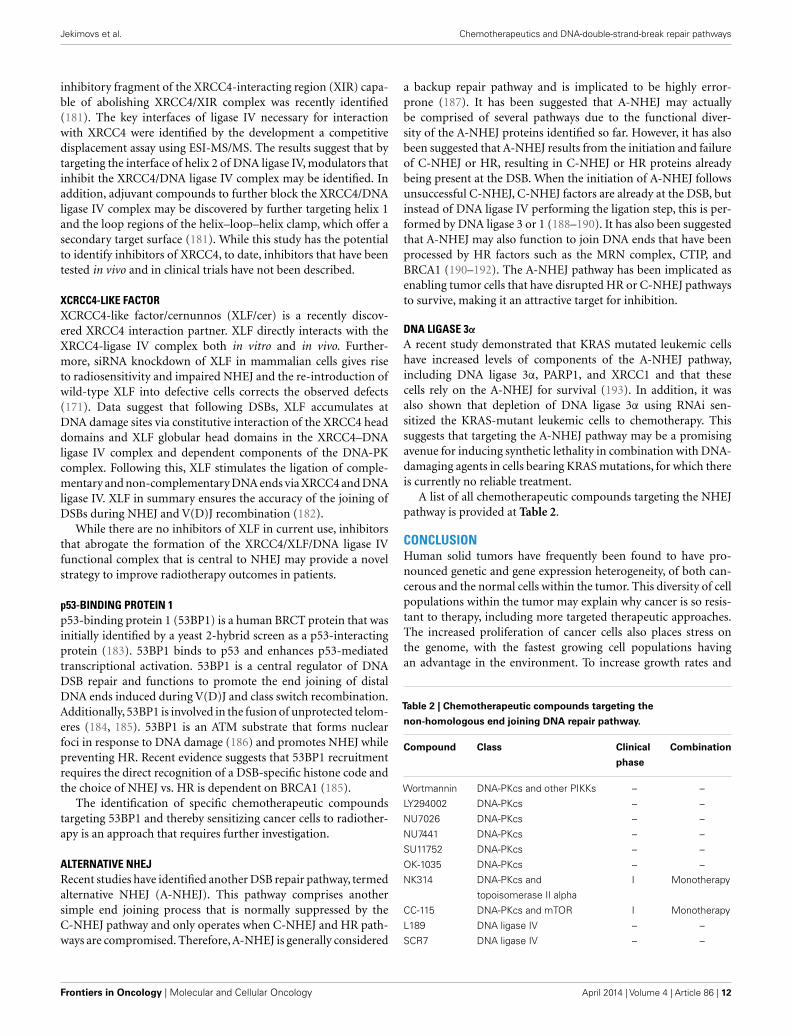
Table 2 | Chemotherapeutic compounds targeting the
non-homologous end joining DNA repair pathway.
Compound Class Clinical
phase
Combination
Wortmannin DNA-PKcs and other PIKKs – –
LY294002 DNA-PKcs – –
NU7026 DNA-PKcs – –
NU7441 DNA-PKcs – –
SU11752 DNA-PKcs – –
OK-1035 DNA-PKcs – –
NK314 DNA-PKcs and
topoisomerase II alpha
I Monotherapy
CC-115 DNA-PKcs and mTOR I Monotherapy
L189 DNA ligase IV – –
SCR7 DNA ligase IV – –
Frontiers in Oncology | Molecular and Cellular Oncology April 2014 | Volume 4 | Article 86 | 12
Jekimovs et al. Chemotherapeutics and DNA-double-strand-break repair pathways
remove normal restrictions on growth, cancer cells evolve to havedefects in the DNA repair pathways and in checkpoint signalingand apoptosis. As a result of these defects and increased metabolicactivity, cancer cells are genomically unstable with the most aggres-sive cancers showing the most genetic instability. However, thisinstability differentiates the cancer cells from normal cells, poten-tially opening up therapeutic windows. The DSB repair pathwayis the most promising of these therapeutic windows, as defectsin this pathway are commonly associated with diseases such ascancer.
The disruption of the DSB repair mechanisms HR and NHEJ,via chemotherapeutic compounds used as either a monotherapyor in conjunction with radiotherapy, has shown promise in theclinical setting for the treatment of various cancers. The targetingof these processes can be further exploited as further investigationsinto the HR and NHEJ pathway lead to the identification of newpotential targets. However, complete inhibition of HR and NHEJfor any extended time period is likely to be lethal to all divid-ing cells, therefore targeted or temporary inhibition is likely to beuseful in conjunction with radiotherapy. Also, complete inhibi-tion of NHEJ may lead to further genomic instability in normalcells, as it is the only pathway for repairing DSBs in non-dividingcells.
Further investigation into synthetic lethality, beyond the identi-fied PARP/BRCA lethality, may lead to additional avenues to be tar-geted and exploited by the use of chemotherapeutic compounds.Promising targets to expand on PARP inhibition using syntheticlethality are other proteins involved in the HR pathway, includ-ing cancers with mutations in ATM, p53, Chk2, Rad51, and NBS1.Inhibition of other proteins involved in the DDR response has alsoshown promise when combined with BRCA mutations. In vitrodepletion of Rad52 in BRCA2-deficient cells showed syntheticlethality when compared with BRCA2-competent cells (194).Recently, synthetic-lethal relationships in chromatin-regulatinggenes have been identified, including chromatin remodeling fac-tors (195) and methyltransferases (196). The concept of syntheticlethality could allow the exploitation of differences between tumorcells and normal cells that have previously been considered to beintractable and it has been shown to be a promising means ofselectively killing tumor cells.
Whilst there have been a number of good and promising resultsusing chemotherapeutic compounds, there have been a numberof failed studies. Ensuring that there are sufficient investigationscompleted both in vitro and in vivo confirming the specificity andpharmacokinetics of these chemotherapeutic compounds beforeintroduction in the clinical setting is critical. As the responseto chemotherapeutic compounds becomes more predictable andwith the identification of specific tumor biomarkers, this will allowfor targeted, more efficient cancer treatments. Inhibition of theseDSB repair proteins holds great promise for the future of cancertherapy in the future.
AUTHOR CONTRIBUTIONSAll authors were involved in the conception of the manuscript,the drafting and/or critically reviewing of the manuscript, haveapproved the final version for publishing, and have agreed to beaccountable for the work.
ACKNOWLEDGMENTSThis work is supported by the Cancer and Ageing Research Pro-gram and grants from the National Health and Medical ResearchCouncil, Cancer Council Queensland, Australian Research Coun-cil, and the Queensland Heath and Medical Research Office.
REFERENCES1. Collis SJ, Boulton SJ. Emerging links between the biological clock and the
DNA damage response. Chromosoma (2007) 116:331–9. doi:10.1007/s00412-007-0108-6
2. Yang DQ, Kastan MB. Participation of ATM in insulin signalling throughphosphorylation of eIF-4E-binding protein 1. Nat Cell Biol (2000) 2:893–8.doi:10.1038/35046542
3. Sprung CN, Bryan TM, Reddel RR, Murnane JP. Normal telomere mainte-nance in immortal ataxia telangiectasia cell lines. Mutat Res (1997) 379:177–84.doi:10.1016/S0027-5107(97)00119-X
4. Savitsky K, Bar-Shira A, Gilad S, Rotman G, Ziv Y, Vanagaite L, et al. A singleataxia telangiectasia gene with a product similar to PI-3 kinase. Science (1995)268:1749–53. doi:10.1126/science.7792600
5. Boder E. Ataxia-telangiectasia: an overview. Kroc Found Ser (1985) 19:1–63.6. Weemaes CM, Hustinx TW, Scheres JM, van Munster PJ, Bakkeren JA, Taal-
man RD. A new chromosomal instability disorder: the Nijmegen breakage syn-drome. Acta Paediatr Scand (1981) 70:557–64. doi:10.1111/j.1651-2227.1981.tb05740.x
7. Hartwell L, Weinert T, Kadyk L, Garvik B. Cell cycle checkpoints, genomicintegrity, and cancer. Cold Spring Harb Symp Quant Biol (1994) 59:259–63.doi:10.1101/SQB.1994.059.01.030
8. Krishnan B, Morgan GJ. Non-Hodgkin lymphoma secondary to cancerchemotherapy. Cancer Epidemiol Biomarkers Prev (2007) 16:377–80. doi:10.1158/1055-9965.EPI-06-1069
9. Farmer H, McCabe N, Lord CJ, Tutt AN, Johnson DA, Richardson TB, et al.Targeting the DNA repair defect in BRCA mutant cells as a therapeutic strategy.Nature (2005) 434:917–21. doi:10.1038/nature03445
10. Bryant HE, Schultz N, Thomas HD, Parker KM, Flower D, Lopez E, et al. Spe-cific killing of BRCA2-deficient tumours with inhibitors of poly(ADP-ribose)polymerase. Nature (2005) 434:913–7. doi:10.1038/nature03443
11. Vilenchik MM, Knudson AG. Endogenous DNA double-strand breaks: pro-duction, fidelity of repair, and induction of cancer. Proc Natl Acad Sci U S A(2003) 100:12871–6. doi:10.1073/pnas.2135498100
12. Shrivastav M, De Haro LP, Nickoloff JA. Regulation of DNA double-strand break repair pathway choice. Cell Res (2008) 18:134–47. doi:10.1038/cr.2007.111
13. Saleh-Gohari N, Bryant HE, Schultz N, Parker KM, Cassel TN, Helleday T.Spontaneous homologous recombination is induced by collapsed replicationforks that are caused by endogenous DNA single-strand breaks. Mol Cell Biol(2005) 25:7158–69. doi:10.1128/MCB.25.16.7158-7169.2005
14. Richard DJ, Bolderson E, Cubeddu L, Wadsworth RI, Savage K, Sharma GG,et al. Single-stranded DNA-binding protein hSSB1 is critical for genomic sta-bility. Nature (2008) 453:677–81. doi:10.1038/nature06883
15. Richard DJ, Cubeddu L, Urquhart AJ, Bain A, Bolderson E, Menon D, et al.hSSB1 interacts directly with the MRN complex stimulating its recruitmentto DNA double-strand breaks and its endo-nuclease activity. Nucleic Acids Res(2011) 39:3643–51. doi:10.1093/nar/gkq1340
16. Richard DJ, Savage K, Bolderson E, Cubeddu L, So S, Ghita M, et al. hSSB1rapidly binds at the sites of DNA double-strand breaks and is required forthe efficient recruitment of the MRN complex. Nucleic Acids Res (2011)39:1692–702. doi:10.1093/nar/gkq1098
17. Limbo O, Chahwan C, Yamada Y, De Bruin RA, Wittenberg C, Russell P. Ctp1is a cell-cycle-regulated protein that functions with Mre11 complex to controldouble-strand break repair by homologous recombination. Mol Cell (2007)28:134–46. doi:10.1016/j.molcel.2007.09.009
18. Takeda S, Nakamura K, Taniguchi Y, Paull TT. Ctp1/CtIP and the MRN com-plex collaborate in the initial steps of homologous recombination. Mol Cell(2007) 28:351–2. doi:10.1016/j.molcel.2007.10.016
19. Tomimatsu N, Mukherjee B, Deland K, Kurimasa A, Bolderson E, Khanna KK,et al. Exo1 plays a major role in DNA end resection in humans and influ-ences double-strand break repair and damage signaling decisions. DNA Repair(2012) 11:441–8. doi:10.1016/j.dnarep.2012.01.006
www.frontiersin.org April 2014 | Volume 4 | Article 86 | 13
Jekimovs et al. Chemotherapeutics and DNA-double-strand-break repair pathways
20. Garcia V, Phelps SE, Gray S, Neale MJ. Bidirectional resection of DNA double-strand breaks by Mre11 and Exo1. Nature (2011) 479:241–4. doi:10.1038/nature10515
21. Bolderson E, Tomimatsu N, Richard DJ, Boucher D, Kumar R, PanditaTK, et al. Phosphorylation of Exo1 modulates homologous recombinationrepair of DNA double-strand breaks. Nucleic Acids Res (2010) 38:1821–31.doi:10.1093/nar/gkp1164
22. Chen L, Nievera CJ, Lee AY, Wu X. Cell cycle-dependent complex formationof BRCA1.CtIP.MRN is important for DNA double-strand break repair. J BiolChem (2008) 283:7713–20. doi:10.1074/jbc.M710245200
23. Yuan SS, Lee SY, Chen G, Song M, Tomlinson GE, Lee EY. BRCA2 is requiredfor ionizing radiation-induced assembly of Rad51 complex in vivo. Cancer Res(1999) 59:3547–51.
24. Helleday T, Lo J, van Gent DC, Engelward BP. DNA double-strand break repair:from mechanistic understanding to cancer treatment. DNA Repair (Amst)(2007) 6:923–35. doi:10.1016/j.dnarep.2007.02.006
25. Lee JH, Paull TT. ATM activation by DNA double-strand breaks through theMre11-Rad50-Nbs1 complex. Science (2005) 308:551–4. doi:10.1126/science.1108297
26. Matsuura S, Tauchi H, Nakamura A, Kondo N, Sakamoto S, Endo S, et al. Posi-tional cloning of the gene for Nijmegen breakage syndrome. Nat Genet (1998)19:179–81. doi:10.1038/549
27. Stewart GS, Maser RS, Stankovic T, Bressan DA, Kaplan MI, Jaspers NG,et al. The DNA double-strand break repair gene hMRE11 is mutated in indi-viduals with an ataxia-telangiectasia-like disorder. Cell (1999) 99:577–87.doi:10.1016/S0092-8674(00)81547-0
28. Waltes R, Kalb R, Gatei M, Kijas AW, Stumm M, Sobeck A, et al. Human RAD50deficiency in a Nijmegen breakage syndrome-like disorder. Am J Hum Genet(2009) 84:605–16. doi:10.1016/j.ajhg.2009.04.010
29. Xiao Y, Weaver DT. Conditional gene targeted deletion by CRE recombinasedemonstrates the requirement for the double-strand break repair Mre11 pro-tein in murine embryonic stem cells. Nucleic Acids Res (1997) 25:2985–91.doi:10.1093/nar/25.15.2985
30. Zhu J, Petersen S, Tessarollo L, Nussenzweig A. Targeted disruption of theNijmegen breakage syndrome gene NBS1 leads to early embryonic lethality inmice. Curr Biol (2001) 11:105–9. doi:10.1016/S0960-9822(01)00019-7
31. Luo G, Yao MS, Bender CF, Mills M, Bladl AR, Bradley A, et al. Disruption ofmRad50 causes embryonic stem cell lethality, abnormal embryonic develop-ment, and sensitivity to ionizing radiation. Proc Natl Acad Sci U S A (1999)96:7376–81. doi:10.1073/pnas.96.13.7376
32. Dupre A, Boyer-Chatenet L, Sattler RM, Modi AP, Lee JH, Nicolette ML, et al.A forward chemical genetic screen reveals an inhibitor of the Mre11-Rad50-Nbs1 complex. Nat Chem Biol (2008) 4:119–25. doi:10.1038/nchembio.63
33. Rass E, Grabarz A, Plo I, Gautier J, Bertrand P, Lopez BS. Role of Mre11 in chro-mosomal nonhomologous end joining in mammalian cells. Nat Struct Mol Biol(2009) 16:819–24. doi:10.1038/nsmb.1641
34. Tran HM, Shi G, Li G, Carney JP, O’Malley B, Li D. Mutant Nbs1enhances cisplatin-induced DNA damage and cytotoxicity in head and neckcancer. Otolaryngol Head Neck Surg (2004) 131:477–84. doi:10.1016/j.otohns.2004.04.019
35. Abuzeid WM, Jiang X, Shi G, Wang H, Paulson D, Araki K, et al. Molecular dis-ruption of RAD50 sensitizes human tumor cells to cisplatin-based chemother-apy. J Clin Invest (2009) 119:1974–85. doi:10.1172/JCI33816
36. Kuroda S, Fujiwara T, Shirakawa Y,Yamasaki Y,Yano S, Uno F, et al. Telomerase-dependent oncolytic adenovirus sensitizes human cancer cells to ionizing radi-ation via inhibition of DNA repair machinery. Cancer Res (2010) 70:9339–48.doi:10.1158/0008-5472.CAN-10-2333
37. Stracker TH, Carson CT, Weitzman MD. Adenovirus oncoproteins inacti-vate the Mre11-Rad50-NBS1 DNA repair complex. Nature (2002) 418:348–52.doi:10.1038/nature00863
38. Kuroda S, Urata Y, Fujiwara T. Ataxia-telangiectasia mutated and the Mre11-Rad50-NBS1 complex: promising targets for radiosensitization. Acta MedOkayama (2012) 66:83–92.
39. Nemunaitis J, Tong AW, Nemunaitis M, Senzer N, Phadke AP, Bedell C, et al.A phase I study of telomerase-specific replication competent oncolytic ade-novirus (telomelysin) for various solid tumors. Mol Ther (2010) 18:429–34.doi:10.1038/mt.2009.262
40. Ragione FD, Cucciolla V, Borriello A, Pietra VD, Racioppi L, Soldati G, et al.Resveratrol arrests the cell division cycle at S/G2 phase transition. BiochemBiophys Res Commun (1998) 250:53–8. doi:10.1006/bbrc.1998.9263
41. Mizutani K, Ikeda K, Kawai Y, Yamori Y. Resveratrol stimulates the prolifera-tion and differentiation of osteoblastic MC3T3-E1 cells. Biochem Biophys ResCommun (1998) 253:859–63. doi:10.1006/bbrc.1998.9870
42. Clement MV, Hirpara JL, Chawdhury SH, Pervaiz S. Chemopreventive agentresveratrol, a natural product derived from grapes, triggers CD95 signaling-dependent apoptosis in human tumor cells. Blood (1998) 92:996–1002.
43. Leon-Galicia I, Diaz-Chavez J, Garcia-Villa E, Uribe-Figueroa L, Hidalgo-Miranda A, Herrera LA, et al. Resveratrol induces downregulation of DNArepair genes in MCF-7 human breast cancer cells. Eur J Cancer Prev (2013)22:11–20. doi:10.1097/CEJ.0b013e328353edcb
44. Carter LG, D’Orazio JA, Pearson KJ. Resveratrol and cancer: a focus on in vivoevidence. Endocr Relat Cancer (2014). doi:10.1530/ERC-13-0171
45. Uziel T, Lerenthal Y, Moyal L, Andegeko Y, Mittelman L, Shiloh Y. Requirementof the MRN complex for ATM activation by DNA damage. EMBO J (2003)22:5612–21. doi:10.1093/emboj/cdg541
46. Shiloh Y, Ziv Y. The ATM protein kinase: regulating the cellular responseto genotoxic stress, and more. Nat Rev Mol Cell Biol (2013) 14:197–210.doi:10.1038/nrm3546
47. Matsuoka S, Ballif BA, Smogorzewska A, McDonald ER III, Hurov KE, LuoJ, et al. ATM and ATR substrate analysis reveals extensive protein networksresponsive to DNA damage. Science (2007) 316:1160–6. doi:10.1126/science.1140321
48. Yang DQ, Halaby MJ, Li Y, Hibma JC, Burn P. Cytoplasmic ATM protein kinase:an emerging therapeutic target for diabetes, cancer and neuronal degeneration.Drug Discov Today (2011) 16:332–8. doi:10.1016/j.drudis.2011.02.001
49. Eaton JS, Lin ZP, Sartorelli AC, Bonawitz ND, Shadel GS. Ataxia-telangiectasiamutated kinase regulates ribonucleotide reductase and mitochondrial home-ostasis. J Clin Invest (2007) 117:2723–34. doi:10.1172/JCI31604
50. Yao Z, Jones AW, Fassone E, Sweeney MG, Lebiedzinska M, Suski JM, et al.Pgc-1 beta mediates adaptive chemoresistance associated with mitochondrialDNA mutations. Oncogene (2013) 32:2592–600. doi:10.1038/onc.2012.259
51. Blasina A, Price BD, Turenne GA, McGowan CH. Caffeine inhibits the check-point kinase ATM. Curr Biol (1999) 9:1135–8. doi:10.1016/S0960-9822(99)80486-2
52. Sarkaria JN, Tibbetts RS, Busby EC, Kennedy AP, Hill DE, Abraham RT. Inhibi-tion of phosphoinositide 3-kinase related kinases by the radiosensitizing agentwortmannin. Cancer Res (1998) 58:4375–82.
53. Matter WF, Brown RF, Vlahos CJ. The inhibition of phosphatidylinositol3-kinase by quercetin and analogs. Biochem Biophys Res Commun (1992)186:624–31. doi:10.1016/0006-291X(92)90792-J
54. Vlahos CJ, Matter WF, Hui KY, Brown RF. A specific inhibitor of phos-phatidylinositol 3-kinase, 2-(4-morpholinyl)-8-phenyl-4H-1-benzopyran-4-one (LY294002). J Biol Chem (1994) 269:5241–8.
55. Hickson I, Zhao Y, Richardson CJ, Green SJ, Martin NM, Orr AI, et al.Identification and characterization of a novel and specific inhibitor of theataxia-telangiectasia mutated kinase ATM. Cancer Res (2004) 64:9152–9.doi:10.1158/0008-5472.CAN-04-2727
56. Rosenzweig KE, Youmell MB, Palayoor ST, Price BD. Radiosensitization ofhuman tumor cells by the phosphatidylinositol 3-kinase inhibitors wortman-nin and LY294002 correlates with inhibition of DNA-dependent protein kinaseand prolonged G2-M delay. Clin Cancer Res (1997) 3:1149–56.
57. Welling A, Hofmann F, Wegener JW. Inhibition of L-type Cav1.2 Ca2+ chan-nels by 2,(4-morpholinyl)-8-phenyl-4H-1-benzopyran-4-one (LY294002)and 2-[1-(3-dimethyl-aminopropyl)-5-methoxy indol-3-yl]-3-(1H-indol-3-yl) maleimide (Go6983). Mol Pharmacol (2005) 67:541–4. doi:10.1124/mol.104.006049
58. Pasapera Limon AM, Herrera-Munoz J, Gutierrez-Sagal R, Ulloa-Aguirre A.The phosphatidylinositol 3-kinase inhibitor LY294002 binds the estrogenreceptor and inhibits 17 beta-estradiol-induced transcriptional activity of anestrogen sensitive reporter gene. Mol Cell Endocrinol (2003) 200:199–202.doi:10.1016/S0303-7207(02)00421-5
59. Golding SE, Rosenberg E, Valerie N, Hussaini I, Frigerio M, Cockcroft XF, et al.Improved ATM kinase inhibitor KU-60019 radiosensitizes glioma cells, com-promises insulin, AKT and ERK prosurvival signaling, and inhibits migration
Frontiers in Oncology | Molecular and Cellular Oncology April 2014 | Volume 4 | Article 86 | 14
Jekimovs et al. Chemotherapeutics and DNA-double-strand-break repair pathways
and invasion. Mol Cancer Ther (2009) 8:2894–902. doi:10.1158/1535-7163.MCT-09-0519
60. Golding SE, Rosenberg E, Adams BR, Wignarajah S, Beckta JM, O’ConnorMJ, et al. Dynamic inhibition of ATM kinase provides a strategy for glioblas-toma multiforme radiosensitization and growth control. Cell Cycle (2012)11:1167–73. doi:10.4161/cc.11.6.19576
61. Rainey MD, Charlton ME, Stanton RV, Kastan MB. Transient inhibition ofATM kinase is sufficient to enhance cellular sensitivity to ionizing radiation.Cancer Res (2008) 68:7466–74. doi:10.1158/0008-5472.CAN-08-0763
62. Batey MA, Zhao Y, Kyle S, Richardson C, Slade A, Martin NM, et al. Preclin-ical evaluation of a novel ATM inhibitor, KU59403, in vitro and in vivo inp53 functional and dysfunctional models of human cancer. Mol Cancer Ther(2013) 12:959–67. doi:10.1158/1535-7163.MCT-12-0707
63. Won J, Kim M, Kim N, Ahn JH, Lee WG, Kim SS, et al. Retraction: smallmolecule-based reversible reprogramming of cellular lifespan. Nat Chem Biol(2008) 4:431. doi:10.1038/nchembio0708-431
64. Choi S, Toledo LI, Fernandez-Capetillo O, Bakkenist CJ. CGK733 does notinhibit ATM or ATR kinase activity in H460 human lung cancer cells. DNARepair (Amst) (2011) 10:1000–1. doi:10.1016/j.dnarep.2011.07.013
65. Bartek J, Lukas J. Chk1 and Chk2 kinases in checkpoint control and cancer.Cancer Cell (2003) 3:421–9. doi:10.1016/S1535-6108(03)00110-7
66. Sanchez Y, Wong C, Thoma RS, Richman R, Wu Z, Piwnica-Worms H, et al.Conservation of the Chk1 checkpoint pathway in mammals: linkage of DNAdamage to CDK regulation through Cdc25. Science (1997) 277:1497–501.doi:10.1126/science.277.5331.1497
67. Taylor WR, Stark GR. Regulation of the G2/M transition by p53. Oncogene(2001) 20:1803–15. doi:10.1038/sj.onc.1204252
68. Sorensen CS, Hansen LT, Dziegielewski J, Syljuasen RG, Lundin C, Bartek J, et al.The cell-cycle checkpoint kinase Chk1 is required for mammalian homologousrecombination repair. Nat Cell Biol (2005) 7:195–201. doi:10.1038/ncb1212
69. Takai H, Tominaga K, Motoyama N, Minamishima YA, Nagahama H,Tsukiyama T, et al. Aberrant cell cycle checkpoint function and early embryonicdeath in Chk1(-/-) mice. Genes Dev (2000) 14:1439–47.
70. Graves PR, Yu L, Schwarz JK, Gales J, Sausville EA, O’Connor PM, et al. TheChk1 protein kinase and the Cdc25C regulatory pathways are targets of theanticancer agent UCN-01. J Biol Chem (2000) 275:5600–5. doi:10.1074/jbc.275.8.5600
71. Busby EC, Leistritz DF, Abraham RT, Karnitz LM, Sarkaria JN. The radiosen-sitizing agent 7-hydroxystaurosporine (UCN-01) inhibits the DNA damagecheckpoint kinase hChk1. Cancer Res (2000) 60:2108–12.
72. Matthews DJ, Yakes FM, Chen J, Tadano M, Bornheim L, Clary DO, et al.Pharmacological abrogation of S-phase checkpoint enhances the anti-tumoractivity of gemcitabine in vivo. Cell Cycle (2007) 6:104–10. doi:10.4161/cc.6.1.3699
73. Riesterer O, Matsumoto F, Wang L, Pickett J, Molkentine D, Giri U, et al.A novel Chk inhibitor, XL-844, increases human cancer cell radiosensitivitythrough promotion of mitotic catastrophe. Invest New Drugs (2011) 29:514–22.doi:10.1007/s10637-009-9361-2
74. Zabludoff SD, Deng C, Grondine MR, Sheehy AM, Ashwell S, Caleb BL, et al.AZD7762, a novel checkpoint kinase inhibitor, drives checkpoint abrogationand potentiates DNA-targeted therapies. Mol Cancer Ther (2008) 7:2955–66.doi:10.1158/1535-7163.MCT-08-0492
75. Ma Z, Yao G, Zhou B, Fan Y, Gao S, Feng X. The Chk1 inhibitor AZD7762sensitises p53 mutant breast cancer cells to radiation in vitro and in vivo. MolMed Rep (2012) 6:897–903. doi:10.3892/mmr.2012.999
76. Seto T, Esaki T, Hirai F, Arita S, Nosaki K, Makiyama A, et al. Phase I, dose-escalation study of AZD7762 alone and in combination with gemcitabine inJapanese patients with advanced solid tumours. Cancer Chemother Pharmacol(2013) 72:619–27. doi:10.1007/s00280-013-2234-6
77. Blasina A, Hallin J, Chen E, Arango ME, Kraynov E, Register J, et al. Breach-ing the DNA damage checkpoint via PF-00477736, a novel small-moleculeinhibitor of checkpoint kinase 1. Mol Cancer Ther (2008) 7:2394–404. doi:10.1158/1535-7163.MCT-07-2391
78. Zhang C, Yan Z, Painter CL, Zhang Q, Chen E, Arango ME, et al. PF-00477736 mediates checkpoint kinase 1 signaling pathway and potentiatesdocetaxel-induced efficacy in xenografts. Clin Cancer Res (2009) 15:4630–40.doi:10.1158/1078-0432.CCR-08-3272
79. Guzi TJ, Paruch K, Dwyer MP, Labroli M, Shanahan F, Davis N, et al. Targetingthe replication checkpoint using SCH 900776, a potent and functionally selec-tive CHK1 inhibitor identified via high content screening. Mol Cancer Ther(2011) 10:591–602. doi:10.1158/1535-7163.MCT-10-0928
80. Schenk EL, Koh BD, Flatten KS, Peterson KL, Parry D, Hess AD, et al. Effects ofselective checkpoint kinase 1 inhibition on cytarabine cytotoxicity in acutemyelogenous leukemia cells in vitro. Clin Cancer Res (2012) 18:5364–73.doi:10.1158/1078-0432.CCR-12-0961
81. Montano R, Chung I, Garner KM, Parry D, Eastman A. Preclinical develop-ment of the novel Chk1 inhibitor SCH900776 in combination with DNA-damaging agents and antimetabolites. Mol Cancer Ther (2012) 11:427–38.doi:10.1158/1535-7163.MCT-11-0406
82. Montano R, Thompson R, Chung I, Hou H, Khan N, Eastman A. Sensitizationof human cancer cells to gemcitabine by the Chk1 inhibitor MK-8776: cell cycleperturbation and impact of administration schedule in vitro and in vivo. BMCCancer (2013) 13:604. doi:10.1186/1471-2407-13-604
83. Karp JE, Thomas BM, Greer JM, Sorge C, Gore SD, Pratz KW, et al. Phase Iand pharmacologic trial of cytosine arabinoside with the selective checkpoint1 inhibitor SCH 900776 in refractory acute leukemias. Clin Cancer Res (2012)18:6723–31. doi:10.1158/1078-0432.CCR-12-2442
84. King C, Diaz H, Barnard D, Barda D, Clawson D, Blosser W, et al. Charac-terization and preclinical development of LY2603618: a selective and potentChk1 inhibitor. Invest New Drugs (2013) 32(2):213–26. doi:10.1007/s10637-013-0036-7
85. Weiss GJ, Donehower RC, Iyengar T, Ramanathan RK, Lewandowski K, WestinE, et al. Phase I dose-escalation study to examine the safety and tolerability ofLY2603618, a checkpoint 1 kinase inhibitor, administered 1 day after peme-trexed 500 mg/m(2) every 21 days in patients with cancer. Invest New Drugs(2013) 31:136–44. doi:10.1007/s10637-012-9815-9
86. Borst GR, McLaughlin M, Kyula JN, Neijenhuis S, Khan A, Good J, et al. Tar-geted radiosensitization by the Chk1 inhibitor SAR-020106. Int J Radiat OncolBiol Phys (2013) 85:1110–8. doi:10.1016/j.ijrobp.2012.08.006
87. Walton MI, Eve PD, Hayes A, Valenti M, De Haven Brandon A, Box G, et al. Thepreclinical pharmacology and therapeutic activity of the novel CHK1 inhibitorSAR-020106. Mol Cancer Ther (2010) 9:89–100. doi:10.1158/1535-7163.MCT-09-0938
88. Greenblatt MS, Bennett WP, Hollstein M, Harris CC. Mutations in the p53tumor suppressor gene: clues to cancer etiology and molecular pathogenesis.Cancer Res (1994) 54:4855–78.
89. Bode AM, Dong Z. Post-translational modification of p53 in tumorigenesis.Nat Rev Cancer (2004) 4:793–805. doi:10.1038/nrc1455
90. Wolf JK, Bodurka DC, Gano JB, Deavers M, Ramondetta L, Ramirez PT,et al. A phase I study of Adp53 (INGN 201; ADVEXIN) for patients withplatinum- and paclitaxel-resistant epithelial ovarian cancer. Gynecol Oncol(2004) 94:442–8. doi:10.1016/j.ygyno.2004.05.041
91. Zhang S, Li Y, Li L, Zhang Y, Gao N, Zhang Z, et al. Phase I study of repeatedintraepithelial delivery of adenoviral p53 in patients with dysplastic oralleukoplakia. J Oral Maxillofac Surg (2009) 67:1074–82. doi:10.1016/j.joms.2008.06.079
92. Buller RE, Runnebaum IB, Karlan BY, Horowitz JA, Shahin M, Buekers T, et al.A phase I/II trial of rAd/p53 (SCH 58500) gene replacement in recurrent ovar-ian cancer. Cancer Gene Ther (2002) 9:553–66. doi:10.1038/sj.cgt.7700472
93. van Beusechem VW, van den Doel PB, Grill J, Pinedo HM, Gerritsen WR. Con-ditionally replicative adenovirus expressing p53 exhibits enhanced oncolyticpotency. Cancer Res (2002) 62:6165–71.
94. Wang X, Su C, Cao H, Li K, Chen J, Jiang L, et al. A novel triple-regulatedoncolytic adenovirus carrying p53 gene exerts potent antitumor efficacyon common human solid cancers. Mol Cancer Ther (2008) 7:1598–603.doi:10.1158/1535-7163.MCT-07-2429
95. Yamasaki Y, Tazawa H, Hashimoto Y, Kojima T, Kuroda S, Yano S, et al. A novelapoptotic mechanism of genetically engineered adenovirus-mediated tumour-specific p53 overexpression through E1A-dependent p21 and MDM2 suppres-sion. Eur J Cancer (2012) 48:2282–91. doi:10.1016/j.ejca.2011.12.020
96. Takimoto R, Wang W, Dicker DT, Rastinejad F, Lyssikatos J, El-Deiry WS. Themutant p53-conformation modifying drug, CP-31398, can induce apoptosis ofhuman cancer cells and can stabilize wild-type p53 protein. Cancer Biol Ther(2002) 1:47–55. doi:10.4161/cbt.1.1.41
www.frontiersin.org April 2014 | Volume 4 | Article 86 | 15
Jekimovs et al. Chemotherapeutics and DNA-double-strand-break repair pathways
97. Bykov VJ, Issaeva N, Shilov A, Hultcrantz M, Pugacheva E, Chumakov P,et al. Restoration of the tumor suppressor function to mutant p53 by alow-molecular-weight compound. Nat Med (2002) 8:282–8. doi:10.1038/nm0302-282
98. Friedler A, Hansson LO, Veprintsev DB, Freund SM, Rippin TM, Nikolova PV,et al. A peptide that binds and stabilizes p53 core domain: chaperone strategyfor rescue of oncogenic mutants. Proc Natl Acad Sci U S A (2002) 99:937–42.doi:10.1073/pnas.241629998
99. Selivanova G, Ryabchenko L, Jansson E, Iotsova V, Wiman KG. Reactivation ofmutant p53 through interaction of a C-terminal peptide with the core domain.Mol Cell Biol (1999) 19:3395–402.
100. Demma M, Maxwell E, Ramos R, Liang L, Li C, Hesk D, et al. SCH529074,a small molecule activator of mutant p53, which binds p53 DNA bindingdomain (DBD), restores growth-suppressive function to mutant p53 and inter-rupts HDM2-mediated ubiquitination of wild type p53. J Biol Chem (2010)285:10198–212. doi:10.1074/jbc.M109.083469
101. Givalos N, Gakiopoulou H, Skliri M, Bousboukea K, Konstantinidou AE,Korkolopoulou P, et al. Replication protein A is an independent prognosticindicator with potential therapeutic implications in colon cancer. Mod Pathol(2007) 20:159–66. doi:10.1038/modpathol.3800719
102. Dahai Y, Sanyuan S, Hong L, Di Z, Chong Z. A relationship between replicationprotein A and occurrence and prognosis of esophageal carcinoma. Cell BiochemBiophys (2013) 67:175–80. doi:10.1007/s12013-013-9530-y
103. Tomkiel JE, Alansari H, Tang N, Virgin JB, Yang X, Vandevord P, et al. Autoim-munity to the M(r) 32,000 subunit of replication protein A in breast cancer.Clin Cancer Res (2002) 8:752–8.
104. Sigurdsson S, Trujillo K, Song B, Stratton S, Sung P. Basis for avid homolo-gous DNA strand exchange by human Rad51 and RPA. J Biol Chem (2001)276:8798–806. doi:10.1074/jbc.M010011200
105. Shuck SC, Turchi JJ. Targeted inhibition of replication protein A reveals cyto-toxic activity, synergy with chemotherapeutic DNA-damaging agents, andinsight into cellular function. Cancer Res (2010) 70:3189–98. doi:10.1158/0008-5472.CAN-09-3422
106. Andrews BJ, Turchi JJ. Development of a high-throughput screen for inhibitorsof replication protein A and its role in nucleotide excision repair. Mol CancerTher (2004) 3:385–91.
107. Neher TM, Bodenmiller D, Fitch RW, Jalal SI, Turchi JJ. Novel irreversiblesmall molecule inhibitors of replication protein A display single-agent activ-ity and synergize with cisplatin. Mol Cancer Ther (2011) 10:1796–806.doi:10.1158/1535-7163.MCT-11-0303
108. Oakley GG, Tillison K, Opiyo SA, Glanzer JG, Horn JM, Patrick SM. Physi-cal interaction between replication protein A (RPA) and MRN: involvementof RPA2 phosphorylation and the N-terminus of RPA1. Biochemistry (2009)48:7473–81. doi:10.1021/bi900694p
109. Stauffer ME, Chazin WJ. Physical interaction between replication protein Aand Rad51 promotes exchange on single-stranded DNA. J Biol Chem (2004)279:25638–45. doi:10.1074/jbc.M400029200
110. Wong JM, Ionescu D, Ingles CJ. Interaction between BRCA2 and replicationprotein A is compromised by a cancer-predisposing mutation in BRCA2. Onco-gene (2003) 22:28–33. doi:10.1038/sj.onc.1206071
111. Tsuzuki T, Fujii Y, Sakumi K, Tominaga Y, Nakao K, Sekiguchi M, et al. Targeteddisruption of the Rad51 gene leads to lethality in embryonic mice. Proc NatlAcad Sci U S A (1996) 93:6236–40. doi:10.1073/pnas.93.13.6236
112. Baumann P, Benson FE, West SC. Human Rad51 protein promotes ATP-dependent homologous pairing and strand transfer reactions in vitro. Cell(1996) 87:757–66. doi:10.1016/S0092-8674(00)81394-X
113. Henning W, Sturzbecher HW. Homologous recombination and cell cyclecheckpoints: Rad51 in tumour progression and therapy resistance. Toxicology(2003) 193:91–109. doi:10.1016/S0300-483X(03)00291-9
114. Ohnishi T, Taki T, Hiraga S, Arita N, Morita T. In vitro and in vivopotentiation of radiosensitivity of malignant gliomas by antisense inhibi-tion of the RAD51 gene. Biochem Biophys Res Commun (1998) 245:319–24.doi:10.1006/bbrc.1998.8440
115. Nomme J, Takizawa Y, Martinez SF, Renodon-Corniere A, Fleury F, Weigel P,et al. Inhibition of filament formation of human Rad51 protein by a smallpeptide derived from the BRC-motif of the BRCA2 protein. Genes Cells (2008)13:471–81. doi:10.1111/j.1365-2443.2008.01180.x
116. Nomme J, Renodon-Corniere A, Asanomi Y, Sakaguchi K, Stasiak AZ, Stasiak A,et al. Design of potent inhibitors of human RAD51 recombinase based on BRCmotifs of BRCA2 protein: modeling and experimental validation of a chimerapeptide. J Med Chem (2010) 53:5782–91. doi:10.1021/jm1002974
117. Huang F, Motlekar NA, Burgwin CM, Napper AD, Diamond SL, Mazin AV.Identification of specific inhibitors of human RAD51 recombinase usinghigh-throughput screening. ACS Chem Biol (2011) 6:628–35. doi:10.1021/cb100428c
118. Budke B, Logan HL, Kalin JH, Zelivianskaia AS, Cameron McGuire W,Miller LL, et al. RI-1: a chemical inhibitor of RAD51 that disrupts homol-ogous recombination in human cells. Nucleic Acids Res (2012) 40:7347–57.doi:10.1093/nar/gks353
119. Budke B, Kalin JH, Pawlowski M, Zelivianskaia AS, Wu M, Kozikowski AP,et al. An optimized RAD51 inhibitor that disrupts homologous recombinationwithout requiring Michael acceptor reactivity. J Med Chem (2013) 56:254–63.doi:10.1021/jm301565b
120. Zhu J, Zhou L, Wu G, Konig H, Lin X, Li G, et al. A novel small mol-ecule RAD51 inactivator overcomes imatinib-resistance in chronic myeloidleukaemia. EMBO Mol Med (2013) 5:353–65. doi:10.1002/emmm.201201760
121. Rosen EM. BRCA1 in the DNA damage response and at telomeres. Front Genet(2013) 4:85. doi:10.3389/fgene.2013.00085
122. Roy R, Chun J, Powell SN. BRCA1 and BRCA2: different roles in a com-mon pathway of genome protection. Nat Rev Cancer (2012) 12:68–78.doi:10.1038/nrc3181
123. Hakem R, De La Pompa JL, Mak TW. Developmental studies of BRCA1 andBRCA2 knock-out mice. J Mammary Gland Biol Neoplasia (1998) 3:431–45.doi:10.1023/A:1018792200700
124. Sharan SK, Morimatsu M, Albrecht U, Lim DS, Regel E, Dinh C, et al. Embry-onic lethality and radiation hypersensitivity mediated by Rad51 in mice lackingBrca2. Nature (1997) 386:804–10. doi:10.1038/386804a0
125. Yu SW, Wang H, Poitras MF, Coombs C, Bowers WJ, Federoff HJ, et al. Medi-ation of poly(ADP-ribose) polymerase-1-dependent cell death by apoptosis-inducing factor. Science (2002) 297:259–63. doi:10.1126/science.1072221
126. Kim MY, Mauro S, Gevry N, Lis JT, Kraus WL. NAD+-dependent modulationof chromatin structure and transcription by nucleosome binding properties ofPARP-1. Cell (2004) 119:803–14. doi:10.1016/j.cell.2004.11.002
127. Leppard JB, Dong Z, Mackey ZB, Tomkinson AE. Physical and functionalinteraction between DNA ligase III alpha and poly(ADP-ribose) polymerase1 in DNA single-strand break repair. Mol Cell Biol (2003) 23:5919–27.doi:10.1128/MCB.23.16.5919-5927.2003
128. Saberi A, Hochegger H, Szuts D, Lan L, Yasui A, Sale JE, et al. RAD18 andpoly(ADP-ribose) polymerase independently suppress the access of nonho-mologous end joining to double-strand breaks and facilitate homologousrecombination-mediated repair. Mol Cell Biol (2007) 27:2562–71. doi:10.1128/MCB.01243-06
129. Clark JB, Ferris GM, Pinder S. Inhibition of nuclear NAD nucleosidase andpoly ADP-ribose polymerase activity from rat liver by nicotinamide and 5’-methyl nicotinamide. Biochim Biophys Acta (1971) 238:82–5. doi:10.1016/0005-2787(71)90012-8
130. Waters R, Ramsay F, Barrett I. 3-Aminobenzamide, an inhibitor of polyADP-ribose polymerase, decreases the frequency of alkaline labile lesions andincreases growth in human fibroblasts exposed to 3-methyl 4-nitroquinoline1-oxide. Carcinogenesis (1982) 3:1463–7. doi:10.1093/carcin/3.12.1463
131. Fong PC, Boss DS,Yap TA, Tutt A,Wu P, Mergui-Roelvink M, et al. Inhibition ofpoly(ADP-ribose) polymerase in tumors from BRCA mutation carriers. N EnglJ Med (2009) 361:123–34. doi:10.1056/NEJMoa0900212
132. Hay T, Matthews JR, Pietzka L, Lau A, Cranston A, Nygren AO, et al. Poly(ADP-ribose) polymerase-1 inhibitor treatment regresses autochthonous Brca2/p53-mutant mammary tumors in vivo and delays tumor relapse in combinationwith carboplatin. Cancer Res (2009) 69:3850–5. doi:10.1158/0008-5472.CAN-08-2388
133. Tutt A, Robson M, Garber JE, Domchek SM, Audeh MW, Weitzel JN, et al. Oralpoly(ADP-ribose) polymerase inhibitor olaparib in patients with BRCA1 orBRCA2 mutations and advanced breast cancer: a proof-of-concept trial. Lancet(2010) 376:235–44. doi:10.1016/S0140-6736(10)60892-6
134. Audeh MW, Carmichael J, Penson RT, Friedlander M, Powell B, Bell-McGuinnKM, et al. Oral poly(ADP-ribose) polymerase inhibitor olaparib in patients
Frontiers in Oncology | Molecular and Cellular Oncology April 2014 | Volume 4 | Article 86 | 16
Jekimovs et al. Chemotherapeutics and DNA-double-strand-break repair pathways
with BRCA1 or BRCA2 mutations and recurrent ovarian cancer: a proof-of-concept trial. Lancet (2010) 376:245–51. doi:10.1016/S0140-6736(10)60893-8
135. Kummar S, Kinders R, Gutierrez ME, Rubinstein L, Parchment RE, Phillips LR,et al. Phase 0 clinical trial of the poly (ADP-ribose) polymerase inhibitor ABT-888 in patients with advanced malignancies. J Clin Oncol (2009) 27:2705–11.doi:10.1200/JCO.2008.19.7681
136. Kummar S, Chen A, Ji J, Zhang Y, Reid JM, Ames M, et al. Phase I studyof PARP inhibitor ABT-888 in combination with topotecan in adults withrefractory solid tumors and lymphomas. Cancer Res (2011) 71:5626–34.doi:10.1158/0008-5472.CAN-11-1227
137. O’Shaughnessy J, Osborne C, Pippen JE, Yoffe M, Patt D, Rocha C, et al. Ini-parib plus chemotherapy in metastatic triple-negative breast cancer. N EnglJ Med (2011) 364:205–14. doi:10.1056/NEJMoa1011418
138. Liu X, Shi Y, Maag DX, Palma JP, Patterson MJ, Ellis PA, et al. Iniparib nonse-lectively modifies cysteine-containing proteins in tumor cells and is not a bonafide PARP inhibitor. Clin Cancer Res (2012) 18:510–23. doi:10.1158/1078-0432.CCR-11-1973
139. Patel AG, De Lorenzo SB, Flatten KS, Poirier GG, Kaufmann SH. Failure of ini-parib to inhibit poly(ADP-ribose) polymerase in vitro. Clin Cancer Res (2012)18:1655–62. doi:10.1158/1078-0432.CCR-11-2890
140. Plummer R, Jones C, Middleton M, Wilson R, Evans J, Olsen A, et al. Phase Istudy of the poly(ADP-ribose) polymerase inhibitor, AG014699, in combina-tion with temozolomide in patients with advanced solid tumors. Clin CancerRes (2008) 14:7917–23. doi:10.1158/1078-0432.CCR-08-1223
141. Plummer R, Lorigan P, Steven N, Scott L, Middleton MR, Wilson RH, et al.A phase II study of the potent PARP inhibitor, rucaparib (PF-01367338,AG014699), with temozolomide in patients with metastatic melanoma demon-strating evidence of chemopotentiation. Cancer Chemother Pharmacol (2013)71:1191–9. doi:10.1007/s00280-013-2113-1
142. Miknyoczki S, Chang H, Grobelny J, Pritchard S, Worrell C, McGann N,et al. The selective poly(ADP-ribose) polymerase-1(2) inhibitor, CEP-8983,increases the sensitivity of chemoresistant tumor cells to temozolomide andirinotecan but does not potentiate myelotoxicity. Mol Cancer Ther (2007)6:2290–302. doi:10.1158/1535-7163.MCT-07-0062
143. Jones P, Altamura S, Boueres J, Ferrigno F, Fonsi M, Giomini C, et al. Discov-ery of 2-{4-[(3S)-piperidin-3-yl]phenyl}-2H-indazole-7-carboxamide (MK-4827): a novel oral poly(ADP-ribose)polymerase (PARP) inhibitor effica-cious in BRCA-1 and -2 mutant tumors. J Med Chem (2009) 52:7170–85.doi:10.1021/jm901188v
144. Sandhu SK, Schelman WR, Wilding G, Moreno V, Baird RD, Miranda S, et al.The poly(ADP-ribose) polymerase inhibitor niraparib (MK4827) in BRCAmutation carriers and patients with sporadic cancer: a phase 1 dose-escalationtrial. Lancet Oncol (2013) 14:882–92. doi:10.1016/S1470-2045(13)70240-7
145. Shen Y, Rehman FL, Feng Y, Boshuizen J, Bajrami I, Elliott R, et al. BMN 673,a novel and highly potent PARP1/2 inhibitor for the treatment of humancancers with DNA repair deficiency. Clin Cancer Res (2013) 19:5003–15.doi:10.1158/1078-0432.CCR-13-1391
146. Chapman JR, Taylor MR, Boulton SJ. Playing the end game: DNA double-strand break repair pathway choice. Mol Cell (2012) 47:497–510. doi:10.1016/j.molcel.2012.07.029
147. Mahaney BL, Meek K, Lees-Miller SP. Repair of ionizing radiation-inducedDNA double-strand breaks by non-homologous end-joining. Biochem J (2009)417:639–50. doi:10.1042/BJ20080413
148. Ader I, Muller C, Bonnet J, Favre G, Cohen-Jonathan E, Salles B, et al. Theradioprotective effect of the 24 kDa Fgf-2 isoform in HeLa cells is related to anincreased expression and activity of the DNA dependent protein kinase (DNA-PK) catalytic subunit. Oncogene (2002) 21:6471–9. doi:10.1038/sj.onc.1205838
149. Beskow C, Skikuniene J, Holgersson A, Nilsson B, Lewensohn R, Kanter L,et al. Radioresistant cervical cancer shows upregulation of the NHEJ proteinsDNA-PKcs, Ku70 and Ku86. Br J Cancer (2009) 101:816–21. doi:10.1038/sj.bjc.6605201
150. Deriano L, Guipaud O, Merle-Beral H, Binet JL, Ricoul M, Potocki-Veronese G,et al. Human chronic lymphocytic leukemia B cells can escape DNA damage-induced apoptosis through the nonhomologous end-joining DNA repair path-way. Blood (2005) 105:4776–83. doi:10.1182/blood-2004-07-2888
151. Miquel C, Jacob S, Grandjouan S, Aime A, Viguier J, Sabourin JC, et al. Fre-quent alteration of DNA damage signalling and repair pathways in human
colorectal cancers with microsatellite instability. Oncogene (2007) 26:5919–26.doi:10.1038/sj.onc.1210419
152. Davidson D, Amrein L, Panasci L, Aloyz R. Small molecules, inhibitors ofDNA-PK, targeting DNA repair, and beyond. Front Pharmacol (2013) 4:5.doi:10.3389/fphar.2013.00005
153. Allen C, Halbrook J, Nickoloff JA. Interactive competition between homolo-gous recombination and non-homologous end joining. Mol Cancer Res (2003)1:913–20.
154. Collis SJ, Deweese TL, Jeggo PA, Parker AR. The life and death of DNA-PK.Oncogene (2005) 24:949–61. doi:10.1038/sj.onc.1208332
155. Fuhrman CB, Kilgore J, Lacoursiere YD, Lee CM, Milash BA, Soisson AP,et al. Radiosensitization of cervical cancer cells via double-strand DNA breakrepair inhibition. Gynecol Oncol (2008) 110:93–8. doi:10.1016/j.ygyno.2007.08.073
156. Peddi P, Loftin CW, Dickey JS, Hair JM, Burns KJ, Aziz K, et al. DNA-PKcs deficiency leads to persistence of oxidatively induced clustered DNAlesions in human tumor cells. Free Radic Biol Med (2010) 48:1435–43.doi:10.1016/j.freeradbiomed.2010.02.033
157. Willmore E, De Caux S, Sunter NJ, Tilby MJ, Jackson GH, Austin CA, et al.A novel DNA-dependent protein kinase inhibitor, NU7026, potentiates thecytotoxicity of topoisomerase II poisons used in the treatment of leukemia.Blood (2004) 103:4659–65. doi:10.1182/blood-2003-07-2527
158. Hollick JJ, Golding BT, Hardcastle IR, Martin N, Richardson C, Rigoreau LJ,et al. 2,6-Disubstituted pyran-4-one and thiopyran-4-one inhibitors of DNA-dependent protein kinase (DNA-PK). Bioorg Med Chem Lett (2003) 13:3083–6.doi:10.1016/S0960-894X(03)00652-8
159. Tavecchio M, Munck JM, Cano C, Newell DR, Curtin NJ. Further characterisa-tion of the cellular activity of the DNA-PK inhibitor, NU7441, reveals poten-tial cross-talk with homologous recombination. Cancer Chemother Pharmacol(2012) 69:155–64. doi:10.1007/s00280-011-1662-4
160. Zhao Y, Thomas HD, Batey MA, Cowell IG, Richardson CJ, Griffin RJ, et al. Pre-clinical evaluation of a potent novel DNA-dependent protein kinase inhibitorNU7441. Cancer Res (2006) 66:5354–62. doi:10.1158/0008-5472.CAN-05-4275
161. Mould E, Berry P, Jamieson D, Hill C, Cano C, Tan N, et al. Identification ofdual DNA-PK MDR1 inhibitors for the potentiation of cytotoxic drug activity.Biochem Pharmacol (2014) 88:58–65. doi:10.1016/j.bcp.2014.01.001
162. Kruszewski M, Wojewodzka M, Iwanenko T, Szumiel I, Okuyama A. Differen-tial inhibitory effect of OK-1035 on DNA repair in L5178Y murine lymphomasublines with functional or defective repair of double strand breaks. Mutat Res(1998) 409:31–6. doi:10.1016/S0921-8777(98)00040-8
163. Ismail IH, Martensson S, Moshinsky D, Rice A, Tang C, Howlett A, et al.SU11752 inhibits the DNA-dependent protein kinase and DNA double-strandbreak repair resulting in ionizing radiation sensitization. Oncogene (2004)23:873–82. doi:10.1038/sj.onc.1207303
164. Hisatomi T, Sueoka-Aragane N, Sato A, Tomimasu R, Ide M, Kurimasa A, et al.NK314 potentiates antitumor activity with adult T-cell leukemia-lymphomacells by inhibition of dual targets on topoisomerase II{alpha} and DNA-dependent protein kinase. Blood (2011) 117:3575–84. doi:10.1182/blood-2010-02-270439
165. Sibanda BL, Chirgadze DY, Blundell TL. Crystal structure of DNA-PKcsreveals a large open-ring cradle comprised of HEAT repeats. Nature (2010)463:118–21. doi:10.1038/nature08648
166. Ihmaid SK, Al-Rawi JM, Bradley CJ, Angove MJ, Robertson MN. Synthe-sis, DNA-PK inhibition, anti-platelet activity studies of 2-(N-substituted-3-aminopyridine)-substituted-1,3-benzoxazines and DNA-PK and PI3K inhibi-tion, homology modelling studies of 2-morpholino-(7,8-di and 8-substituted)-1,3-benzoxazines. Eur J Med Chem (2012) 57:85–101. doi:10.1016/j.ejmech.2012.08.035
167. Li YH, Wang X, Pan Y, Lee DH, Chowdhury D, Kimmelman AC. Inhibitionof non-homologous end joining repair impairs pancreatic cancer growth andenhances radiation response. PLoS One (2012) 7:e39588. doi:10.1371/journal.pone.0039588
168. Wei YF, Robins P, Carter K, Caldecott K, Pappin DJ, Yu GL, et al. Molecularcloning and expression of human cDNAs encoding a novel DNA ligase IV andDNA ligase III, an enzyme active in DNA repair and recombination. Mol CellBiol (1995) 15:3206–16.
www.frontiersin.org April 2014 | Volume 4 | Article 86 | 17
Jekimovs et al. Chemotherapeutics and DNA-double-strand-break repair pathways
169. Critchlow SE, Bowater RP, Jackson SP. Mammalian DNA double-strand breakrepair protein XRCC4 interacts with DNA ligase IV. Curr Biol (1997) 7:588–98.doi:10.1016/S0960-9822(06)00258-2
170. Grawunder U, Wilm M, Wu X, Kulesza P, Wilson TE, Mann M, et al. Activityof DNA ligase IV stimulated by complex formation with XRCC4 protein inmammalian cells. Nature (1997) 388:492–5. doi:10.1038/41358
171. Ahnesorg P, Smith P, Jackson SP. XLF interacts with the XRCC4-DNA lig-ase IV complex to promote DNA nonhomologous end-joining. Cell (2006)124:301–13. doi:10.1016/j.cell.2005.12.031
172. Ellenberger T, Tomkinson AE. Eukaryotic DNA ligases: structural and func-tional insights. Annu Rev Biochem (2008) 77:313–38. doi:10.1146/annurev.biochem.77.061306.123941
173. Tomkinson AE, Vijayakumar S, Pascal JM, Ellenberger T. DNA ligases: struc-ture, reaction mechanism, and function. Chem Rev (2006) 106:687–99.doi:10.1021/cr040498d
174. Chen X, Zhong S, Zhu X, Dziegielewska B, Ellenberger T, Wilson GM, et al.Rational design of human DNA ligase inhibitors that target cellular DNAreplication and repair. Cancer Res (2008) 68:3169–77. doi:10.1158/0008-5472.CAN-07-6636
175. Srivastava M, Nambiar M, Sharma S, Karki SS, Goldsmith G, Hegde M,et al. An inhibitor of nonhomologous end-joining abrogates double-strandbreak repair and impedes cancer progression. Cell (2012) 151:1474–87.doi:10.1016/j.cell.2012.11.054
176. Mari PO, Florea BI, Persengiev SP, Verkaik NS, Bruggenwirth HT, ModestiM, et al. Dynamic assembly of end-joining complexes requires interactionbetween Ku70/80 and XRCC4. Proc Natl Acad Sci U S A (2006) 103:18597–602.doi:10.1073/pnas.0609061103
177. Davis AJ, Chen DJ. DNA double strand break repair via non-homologous end-joining. Transl Cancer Res (2013) 2:130–43.
178. Gao Y, Sun Y, Frank KM, Dikkes P, Fujiwara Y, Seidl KJ, et al. A critical rolefor DNA end-joining proteins in both lymphogenesis and neurogenesis. Cell(1998) 95:891–902. doi:10.1016/S0092-8674(00)81714-6
179. Modesti M, Hesse JE, Gellert M. DNA binding of Xrcc4 protein is associatedwith V(D)J recombination but not with stimulation of DNA ligase IV activity.EMBO J (1999) 18:2008–18. doi:10.1093/emboj/18.7.2008
180. Sun MF, Chen HY, Tsai FJ, Lui SH, Chen CY, Chen CY. Search for novel remediesto augment radiation resistance of inhabitants of Fukushima and Chernobyldisasters: identifying DNA repair protein XRCC4 inhibitors. J Biomol StructDyn (2011) 29:325–37. doi:10.1080/07391102.2011.10507388
181. McFadden MJ, Lee WK, Brennan JD, Junop MS. Delineation of keyXRCC4/ligase IV interfaces for targeted disruption of non-homologous endjoining DNA repair. Proteins (2014) 82:187–94. doi:10.1002/prot.24349
182. Dahm K. Role and regulation of human XRCC4-like factor/cernunnos. J CellBiochem (2008) 104:1534–40. doi:10.1002/jcb.21726
183. Iwabuchi K, Bartel PL, Li B, Marraccino R, Fields S. Two cellular proteinsthat bind to wild-type but not mutant p53. Proc Natl Acad Sci U S A (1994)91:6098–102. doi:10.1073/pnas.91.13.6098
184. Adams MM, Carpenter PB. Tying the loose ends together in DNA double strandbreak repair with 53BP1. Cell Div (2006) 1:19. doi:10.1186/1747-1028-1-19
185. Panier S, Boulton SJ. Double-strand break repair: 53BP1 comes into focus. NatRev Mol Cell Biol (2014) 15:7–18. doi:10.1038/nrm3719
186. Rappold I, Iwabuchi K, Date T, Chen J. Tumor suppressor p53 binding protein1 (53BP1) is involved in DNA damage-signaling pathways. J Cell Biol (2001)153:613–20. doi:10.1083/jcb.153.3.613
187. McVey M, Lee SE. MMEJ repair of double-strand breaks (director’s cut):deleted sequences and alternative endings. Trends Genet (2008) 24:529–38.doi:10.1016/j.tig.2008.08.007
188. Wang H, Rosidi B, Perrault R, Wang M, Zhang L, Windhofer F, et al. DNA lig-ase III as a candidate component of backup pathways of nonhomologous endjoining. Cancer Res (2005) 65:4020–30. doi:10.1158/0008-5472.CAN-04-3055
189. Mladenov E, Magin S, Soni A, Iliakis G. DNA double-strand break repair asdeterminant of cellular radiosensitivity to killing and target in radiation ther-apy. Front Oncol (2013) 3:113. doi:10.3389/fonc.2013.00113
190. Della-Maria J, Zhou Y, Tsai MS, Kuhnlein J, Carney JP, Paull TT, et al. HumanMre11/human Rad50/Nbs1 and DNA ligase III alpha/XRCC1 protein com-plexes act together in an alternative nonhomologous end joining pathway. J BiolChem (2011) 286:33845–53. doi:10.1074/jbc.M111.274159
191. Zhang Y, Jasin M. An essential role for CtIP in chromosomal translocation for-mation through an alternative end-joining pathway. Nat Struct Mol Biol (2011)18:80–4. doi:10.1038/nsmb.1940
192. Zhong Q, Chen CF, Chen PL, Lee WH. BRCA1 facilitates microhomology-mediated end joining of DNA double strand breaks. J Biol Chem (2002)277:28641–7. doi:10.1074/jbc.M200748200
193. Hahnel PS, Enders B, Sasca D, Roos WP, Kaina B, Bullinger L, et al. Target-ing components of the alternative NHEJ pathway sensitizes KRAS-mutantleukemic cells to chemotherapy. Blood (2014) 123:2355–66. doi:10.1182/blood-2013-01-477620
194. Feng Z, Scott SP, Bussen W, Sharma GG, Guo G, Pandita TK, et al. Rad52 inac-tivation is synthetically lethal with BRCA2 deficiency. Proc Natl Acad Sci U S A(2011) 108:686–91. doi:10.1073/pnas.1010959107
195. Oike T, Ogiwara H, Tominaga Y, Ito K, Ando O, Tsuta K, et al. A syn-thetic lethality-based strategy to treat cancers harboring a genetic deficiencyin the chromatin remodeling factor BRG1. Cancer Res (2013) 73:5508–18.doi:10.1158/0008-5472.CAN-12-4593
196. Knutson SK, Warholic NM, Wigle TJ, Klaus CR, Allain CJ, Raimondi A, et al.Durable tumor regression in genetically altered malignant rhabdoid tumorsby inhibition of methyltransferase EZH2. Proc Natl Acad Sci U S A (2013)110:7922–7. doi:10.1073/pnas.1303800110
Conflict of Interest Statement: The authors declare that the research was conductedin the absence of any commercial or financial relationships that could be construedas a potential conflict of interest.
Received: 04 March 2014; paper pending published: 30 March 2014; accepted: 08 April2014; published online: 22 April 2014.Citation: Jekimovs C, Bolderson E, Suraweera A, Adams M, O’Byrne KJ and RichardDJ (2014) Chemotherapeutic compounds targeting the DNA double-strand breakrepair pathways: the good, the bad, and the promising. Front. Oncol. 4:86. doi:10.3389/fonc.2014.00086This article was submitted to Molecular and Cellular Oncology, a section of the journalFrontiers in Oncology.Copyright © 2014 Jekimovs, Bolderson, Suraweera, Adams, O’Byrne and Richard.This is an open-access article distributed under the terms of the Creative CommonsAttribution License (CC BY). The use, distribution or reproduction in other forums ispermitted, provided the original author(s) or licensor are credited and that the originalpublication in this journal is cited, in accordance with accepted academic practice. Nouse, distribution or reproduction is permitted which does not comply with these terms.
Frontiers in Oncology | Molecular and Cellular Oncology April 2014 | Volume 4 | Article 86 | 18