REV7 counteracts DNA double-strand break resection and affects PARP inhibition
Upload
independentCategory
view
2download
0
The involvement of human RECQL4 in DNAdouble-strand break repair
Dharmendra Kumar Singh,1 Parimal Karmakar,2
Maria Aamann,1,3 Shepherd H. Schurman,1 AlfredMay,1 Deborah L. Croteau,1 Lynnette Burks,4 SharonE. Plon4 and Vilhelm A. Bohr1
1Laboratory of Molecular Gerontology, Biomedical Research
Center, 251 Bayview Boulevard, National Institute on Aging, NIH,
Baltimore, MD 21224, USA2Department of Life Sciences and Biotechnology, Jadavpur
University, Kolkata, West Bengal- 700 032, India3Danish Center for Molecular Gerontology, MBI, University of
Aarhus, Denmark4Departments of Molecular and Human Genetics and Pediatrics,
Baylor College of Medicine, Houston, USA
Summary
Rothmund–Thomson syndrome (RTS) is an autosomal
recessive hereditary disorder associated with mutation in
RECQL4 gene, a member of the human RecQ helicases.
The disease is characterized by genomic instability, skele-
tal abnormalities and predisposition to malignant tumors,
especially osteosarcomas. The precise role of RECQL4 in
cellular pathways is largely unknown; however, recent
evidence suggests its involvement in multiple DNA meta-
bolic pathways. This study investigates the roles of REC-
QL4 in DNA double-strand break (DSB) repair. The results
show that RECQL4-deficient fibroblasts are moderately
sensitive to c-irradiation and accumulate more cH2AX and
53BP1 foci than control fibroblasts. This is suggestive of
defects in efficient repair of DSB’s in the RECQL4-deficient
fibroblasts. Real time imaging of live cells using laser con-
focal microscopy shows that RECQL4 is recruited early to
laser-induced DSBs and remains for a shorter duration
than WRN and BLM, indicating its distinct role in repair of
DSBs. Endogenous RECQL4 also colocalizes with cH2AX at
the site of DSBs. The RECQL4 domain responsible for its
DNA damage localization has been mapped to the unique
N-terminus domain between amino acids 363–492, which
shares no homology to recruitment domains of WRN and
BLM to the DSBs. Further, the recruitment of RECQL4 to
laser-induced DNA damage is independent of functional
WRN, BLM or ATM proteins. These results suggest distinct
cellular dynamics for RECQL4 protein at the site of laser-
induced DSB and that it might play important roles in effi-
cient repair of DSB’s.
Key words: Bloom syndrome; BLM; Double-strand break
repair; Premature aging; RecQ helicase; Rothmund-Thom-
son syndrome; RTS; Werner syndrome; WRN.
Introduction
The RecQ helicase family is a group of evolutionarily conserved
DNA unwinding proteins that play diverse roles in multiple DNA
metabolic and repair processes. Five RecQ homologues have
been identified in humans and mice: RECQL1, BLM, WRN, REC-
QL4 and RECQL5. Defects in human RecQ helicases are associ-
ated with chromosomal and developmental abnormalities,
cancer susceptibility and premature aging (Brosh & Bohr, 2007),
and three loci encoding human RecQ helicases are causally
linked to recessive hereditary diseases characterized by genomic
instability and cancer predisposition (Hickson, 2003; Bohr,
2008). Werner syndrome (WS) and Bloom syndrome (BS) are
caused by defects in WRN and BLM helicase, respectively.
Defects in human RECQL4 are linked to Rothmund–Thomson
(RTS) Type 2, RAPADILINO and Baller-Gerold syndromes (Kitao
et al., 1999; Siitonen et al., 2003; Van Maldergem et al., 2006).
Although human WRN and BLM helicases are well character-
ized, relatively little is known about the biochemistry, cellular
biology and function of human RECQL4.
Rothmund–Thomson syndrome is a rare, autosomal recessive
disorder characterized by poikiloderma, growth deficiency, juve-
nile cataracts, premature aging and predisposition to malignant
tumors especially osteosarcomas (Vennos et al., 1992; Wang
et al., 2003; Stinco et al., 2008). Approximately, two-thirds of
patients with RTS have mutations in RECQL4 that are predicted to
result in a truncated protein because of premature termination of
protein synthesis (Lindor et al., 2000; Wang et al., 2003). These
patients are referred to as RTS Type 2 and show strong predisposi-
tion to osteosarcoma (Wang et al., 2003). Cells from patients
with RTS show genomic instability and chromosomal abnormali-
ties such as trisomy, aneuploidy and chromosomal rearrange-
ments (Der Kaloustian et al., 1990; Vennos et al., 1992; Orstavik
et al., 1994; Anbari et al., 2000; Durand et al., 2002).
RECQL4 is a 1208 amino acid protein (133 kDa) with a cen-
tral helicase domain, which is characteristic for RecQ helicases
(Kitao et al., 1998). However, RECQL4 does not share homol-
ogy to two other conserved RecQ motifs, RQC and helicase
and RNaseD C-terminal (HRDC), which are present in human
BLM and WRN helicases. RECQL4 also possesses two N-termi-
nal nuclear localization signal sequences (Burks et al., 2007).
RECQL4 has an intrinsic ATPase activity and single-strand DNA
Correspondence
Vilhelm A. Bohr, Laboratory of Molecular Gerontology, Biomedical Research
Center, National Institute on Aging, NIH, 251, Bayview Boulevard, Suite
100, Baltimore, MD 21224 USA. Tel.: 410 558 8162; fax: 410 558 8157;
e-mail: [email protected]
Accepted for publication 2 February 2010
358 Journal compilation ª Blackwell Publishing Ltd/Anatomical Society of Great Britain and Ireland 2010No claim to original US government works
Aging Cell (2010) 9, pp358–371 Doi: 10.1111/j.1474-9726.2010.00562.xAg
ing
Cell

annealing activity (Yin et al., 2004; Macris et al., 2006). Recent
studies have shown that the RECQL4 also has helicase activity
in vitro (Capp et al., 2009; Suzuki et al., 2009; Xu & Liu,
2009).
The biological functions of RECQL4 are not yet known. How-
ever, it has been proposed that the RecQL4 N-terminal region
plays a role in recruiting DNA polymerase a to nascent DNA rep-
lication forks in Xenopus egg extract (Sangrithi et al., 2005;
Matsuno et al., 2006). Recently, it has been shown that human
RECQL4 is a part of the repliosome complex and interacts with
MCM10, MCM2-7, CDC45 and GINS (Xu et al., 2009). Another
study indicates that the assembly of Cdc45-MCM2-7-GINS
(CMG) complex at the replication fork requires RECQL4 (Im
et al., 2009). These findings suggest an active involvement of
RECQL4 in the assembly of the pre-replication complex and initi-
ation of DNA replication.
Recent findings also indicate the involvement of RECQL4 in
some DNA repair pathways. Fibroblasts from patients with RTS
are sensitive to replication-blocking DNA damaging agents
including hydroxyurea, camptothecin and doxorubicin (Jin et al.,
2008). RTS cells showed sensitivity to H2O2 and accumulate
more H2O2-induced DNA strand breaks than control cells, sug-
gesting that RECQL4 may stimulate repair of H2O2-induced DNA
damage (Werner et al., 2006; Schurman et al., 2009). Biochem-
ically, RECQL4 modulates the intrinsic activities of APE1, FEN1
and DNA polb, indicating its role in base excision repair pathway
(Schurman et al., 2009). RECQL4 also interacts with poly (ADP-
ribose) polymerase 1 (PARP-1), which is implicated in DNA
recombination, DNA repair and transcriptional regulation (Woo
et al., 2006). RECQL4 colocalizes with the xeroderma pigmento-
sum group A protein in human cells treated with UV, suggesting
that RECQL4 might facilitate nucleotide excision repair-medi-
ated repair of UV-induced DNA damage in human cells (Fan &
Luo, 2008). Furthermore, RECQL4 and Rad51 colocalize in the
nuclei of cells treated with etoposide, suggesting that RECQL4
might play a role in homologous recombination (HR)-mediated
repair of DSBs (Petkovic et al., 2005). But the mechanism of this
interaction is not known.
There is also RECQL4 involvement in the repair of DSBs in
Xenopus egg extracts (Kumata et al., 2007). Moreover, there
are inconsistent reports about the sensitivity of RTS cells from
patients and from MEFs derived from the RECQL4-deficient mice
toward c-irradiation, which predominantly forms DSB in the cells
(Vennos et al., 1992; Hoki et al., 2003; Cabral et al., 2008; Jin
et al., 2008).
In this work, we studied the cellular dynamics of RECQL4 in
live cells at the site of laser-induced DSBs using confocal laser
scanning microscopy as well as sensitivity assays. First, we show
that the RTS-deficient primary fibroblasts are moderately sensi-
tive to c-irradiation and accumulate more c-irradiation induced
c-H2AX and 53BP1 foci that do not get efficiently resolved than
control fibroblasts; this suggests that there are DNA repair
defects in RTS cells compared to control cells. RECQL4 is
recruited early to the laser-induced DNA damage site. Recruit-
ment of RECQL4 to DNA damage requires an N-terminal
domain, which does not share homology to WRN and BLM pro-
teins. Furthermore, RECQL4 shows distinct cellular dynamics at
the site of laser-induced DSBs compared to WRN and BLM.
Results
RTS cells are moderately sensitive to c-irradiation
The importance of RECQL4 in DNA DSB repair was examined by
measuring survival of RECQL4-deficient and normal fibroblasts
after c-irradiation. The RTS cells (AG05013 and AG17524) and
normal fibroblasts (GM00323 and GM00969) were treated with
0, 1, 2 or 3 Gy of c-irradiation and survival efficiency was esti-
mated after 14 days by cell proliferation assays. The results
showed that both the RTS cells are moderately more sensitive to
c-irradiation than respective normal cells (the RTS and normal fi-
broblasts were age and sex matched), and that survival
decreases with increasing dose of c-irradiation (Fig. 1). We also
observed that the growth rate of both the RECQL4-deficient fi-
broblasts was approximately 1.5–2-fold slower than the normal
fibroblasts (Supplementary Fig. S1).
(A) (B)
Fig. 1 RECQL4-deficient fibroblasts are sensitive to c-irradiation compared to normal control fibroblasts. (A) Normal (GM00323) and primary Rothmund–
Thomson syndrome (RTS) fibroblasts (AG05013) cell lines were plated on a 60-mm petri dishes and treated with 0, 1, 2 and 3 Gy of c-irradiation. Cells were allowed
to grow for 14 days and fluorescent intensity was measured as described in Experimental procedures. (B) Similar experiments were performed with normal
fibroblasts (GM00969) and RTS cell line (AG17524). The experiments were performed in triplicate and the error bars represent the standard deviation (+ ⁄ )). The
degree of significance was calculated by Student’s t-test method and the (*) represent the significant difference from the normal fibroblast with P value < 0.05.
Involvement of RECQL4 in DSB repair, D. K. Singh et al.
Journal compilation ª Blackwell Publishing Ltd/Anatomical Society of Great Britain and Ireland 2010No claim to original US government works
359

RTS cells are defective in efficient repair of DNA
DSBs
Sensitivity of RECQL4-deficient cells to c-irradiation could be a
result of inefficient processing of DSBs. To examine this possibil-
ity, two RECQL4-deficient (AG05013 and AG18371) and two
normal cells (GM00323 and GM01864) were irradiated with
5 Gy of c-irradiation and allowed to recover for 1, 3, 6 or 10 h.
Cells were then fixed and immunostained with c-H2AX and
53BP1 antibodies and examined by confocal laser scanning
microscopy. c-H2AX and 53BP1 foci are widely used as markers
for DSBs in the cell (Schultz et al., 2000; Kinner et al., 2008).
RTS cells had a higher basal level of c-H2AX foci than normal
cells, suggesting inefficient repair of endogenous DNA DSBs per-
haps because of problems at the replication forks (Supplemen-
tary Fig. S2, lane 1). At early time points (i.e., 1 h after
irradiation), the number of 53BP1 foci in both the RECQL4-defi-
cient cell lines were approximately 2-fold higher than in the con-
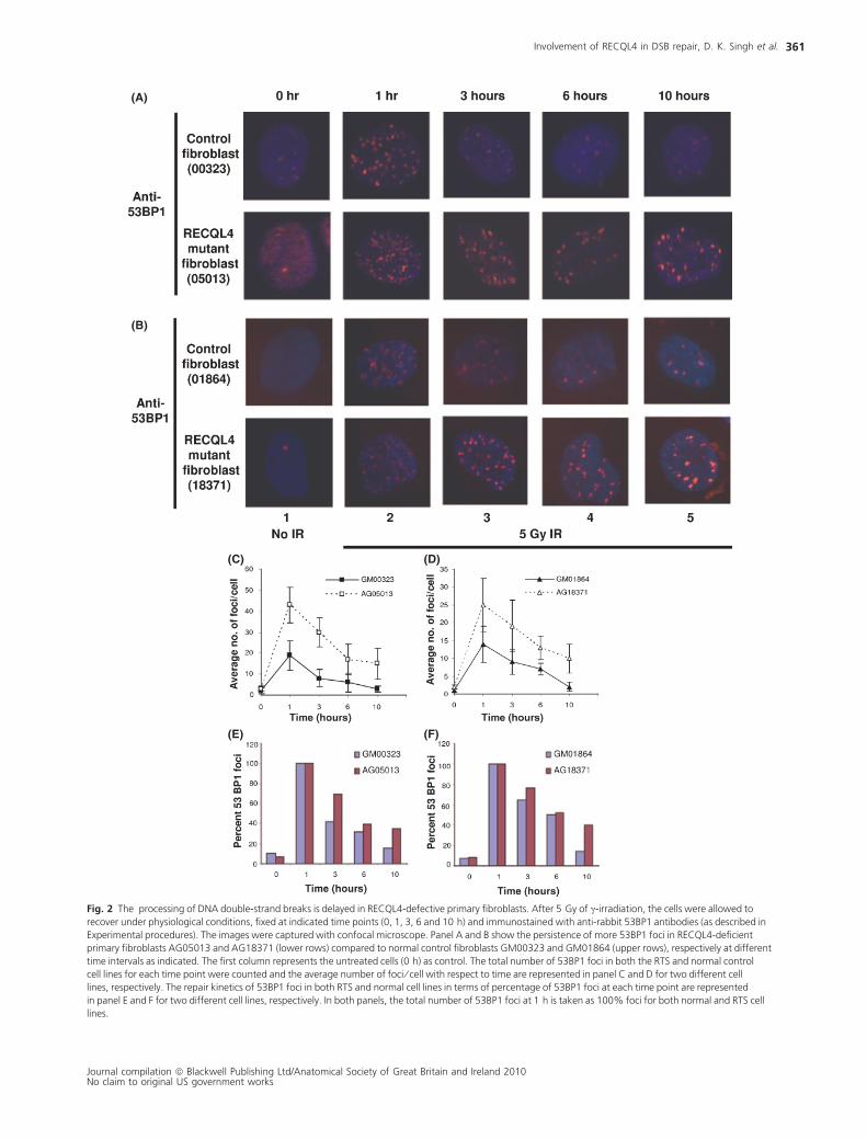
trol cells (Fig. 2, panels A, B, lane 2 and panels C,D).
Furthermore, a significantly higher number of 53BP1 foci (2–3
fold) accumulated in c-irradiated RTS cells than in normal fibro-
blasts (Fig. 2, panels A, B, lanes 3–5 and panels E, F) at late time
points. The graph shows 53BP1 repair kinetics (percent 53BP1
foci with respect to time) and these foci disappeared more
quickly in normal cells than in RTS cells suggesting inefficient
repair of DSBs in RTS cells (Fig. 2, panels A, B, upper rows and
panels E, F). Similar patterns of foci dispersal was also seen for
c-H2AX at the site of DSBs (Supplementary Fig. S2, lanes 3–5).
The slight differences in kinetics of c-H2AX and 53BP1 foci for-
mation and dispersal could be because of differences in recruit-
ment and dissociation properties of c-H2AX and 53BP1 at the
site of DNA damage. These results suggest that RECQL4-defi-
cient cells repair DSBs less efficiently than normal cells, which is
consistent with reduced survival of RECQL4-deficient cells after
exposure to c-irradiation.
Characterization of the laser microirradiation
Confocal laser scanning microscopy is a powerful technique for
studying the spatiotemporal behaviors of DNA repair proteins in
‘real time’ in living cells. This technique has been used to reveal
novel functions of many DNA repair proteins including WRN and
BLM (Mailand et al., 2007; Haince et al., 2008; Yano et al.,
2008). Previous studies also show that WRN and BLM are
recruited very early to laser-induced DSBs via their HRDC domain
(Lan et al., 2005; Karmakar et al., 2006). Laser microirradiation
produces a variety of DNA lesions, [DSBs, single-strand breaks
(SSBs) and base modifications] in a spatially restricted manner in
nuclear DNA. The types of lesions formed after irradiation
depends upon laser intensity and wavelength (Lan et al., 2005;
Dinant et al., 2007). In this study, DSBs were induced with a
435 nm laser without cellular pretreatment. We standardized
the laser intensity so that SSBs could be distinguished from DSBs.
At a laser intensity of < 10%, only SSBs were produced, and
XRCC1 (both green fluorescent protein (GFP)-tagged and
endogenous), but not GFP-53BP1 or endogenous 53BP1 were
recruited to the microirradiated site (Fig. 3A, panels a and b,
lanes 1 and 2). At a laser intensity of > 10%, both SSBs and DSBs
were produced, and both XRCC1 and 53BP1 were recruited to
the microirradiated site (Fig. 3A, panels a and b, lanes 4 and 5).
Live HeLa cells were also microirradiated using 8% or 18% laser
intensity and co-immunostained with antibodies to XRCC1 and
c-H2AX (Fig. 3B). Double-strand breaks and c-H2AX foci were
generated by 18% but not by 8% laser intensity. Under these
experimental conditions, we then tested for the recruitment of
GFP-tagged exogenous and endogenous RECQL4 protein to the
site of DNA damage. The results showed that both endogenous
and GFP-tagged RECQL4 were recruited to the DNA damage site
at high laser intensity (Fig. 3A, compare lanes 3 and 6).
RECQL4 accumulates at laser-induced DNA lesions
Kinetic studies were performed to examine the association and
dissociation of RECQL4, WRN and BLM at laser-induced DNA
damage in living cells. Similar to previous reports, in most cells,
GFP-tagged RECQL4 was localized to the nucleus, but nucleo-
cytoplasmic localization was observed in some cells [data not
shown, (Yin et al., 2004; Burks et al., 2007)]. For this experi-
ment, HeLa cells were transiently transfected with plasmids
expressing GFP-RECQL4, GFP-WRN or GFP-BLM prior to laser
microirradiation, and cells were visualized at different time
points after laser treatment. Previously, using a similar approach,
WRN and BLM have been shown to be recruited to the DNA
damage sites (Lan et al., 2005; Karmakar et al., 2006) and
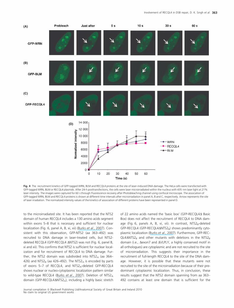
hence were used as positive controls in this study (Fig. 4, panels
A, B, C and D). The results showed that RECQL4 is recruited to
DNA damage site within 5 s of microirradiation (Fig. 4, panels C
and D).
Endogenous c-H2AX and RECQL4 colocalize after
laser-induced DNA damage
Recruitment of endogenous c-H2AX and RECQL4 to the microir-
radiation site was also examined in HeLa cells. Cells were fixed
and immunostained with c-H2AX and RECQL4 antibodies after
5 min of laser microirradiation and imaged. The images were
analyzed using confocal microscopy and Volocity software. The
results showed that endogenous c-H2AX and RECQL4 colocal-
ize within 5 min after laser treatment, suggesting that RECQL4
binds to DSBs (Fig. 5). A significant portion of the endogenous
RECQL4 remains in the cytoplasm after irradiation, as reported
previously (Yin et al., 2004; Werner et al., 2005).
The NTS2 domain of RECQL4 is sufficient for
recruitment to the DSB sites
RECQL4 possesses a characteristic central helicase domain,
which is a shared feature of RecQ helicases (Kitao et al., 1998).
RECQL4 differs from WRN and BLM helicases in that it lacks the
C-terminal RQC and HRDC domains. Two unique nuclear
Involvement of RECQL4 in DSB repair, D. K. Singh et al.
Journal compilation ª Blackwell Publishing Ltd/Anatomical Society of Great Britain and Ireland 2010No claim to original US government works
360
(A)
(B)
(C) (D)
(E) (F)
Fig. 2 The processing of DNA double-strand breaks is delayed in RECQL4-defective primary fibroblasts. After 5 Gy of c-irradiation, the cells were allowed to
recover under physiological conditions, fixed at indicated time points (0, 1, 3, 6 and 10 h) and immunostained with anti-rabbit 53BP1 antibodies (as described in
Experimental procedures). The images were captured with confocal microscope. Panel A and B show the persistence of more 53BP1 foci in RECQL4-deficient
primary fibroblasts AG05013 and AG18371 (lower rows) compared to normal control fibroblasts GM00323 and GM01864 (upper rows), respectively at different
time intervals as indicated. The first column represents the untreated cells (0 h) as control. The total number of 53BP1 foci in both the RTS and normal control
cell lines for each time point were counted and the average number of foci ⁄ cell with respect to time are represented in panel C and D for two different cell
lines, respectively. The repair kinetics of 53BP1 foci in both RTS and normal cell lines in terms of percentage of 53BP1 foci at each time point are represented
in panel E and F for two different cell lines, respectively. In both panels, the total number of 53BP1 foci at 1 h is taken as 100% foci for both normal and RTS cell
lines.
Involvement of RECQL4 in DSB repair, D. K. Singh et al.
Journal compilation ª Blackwell Publishing Ltd/Anatomical Society of Great Britain and Ireland 2010No claim to original US government works
361

localization sequences are also present at the N-terminus of
RECQL4 (Burks et al., 2007). To map the domain of RECQL4
responsible for its recruitment to the laser-induced DSBs sites,
we tested constructs containing either portions of the RECQL4
coding region, or the full-length construct with portions deleted.
The subcellular localization of each mutant was reported previ-
ously (Burks et al., 2007), and we confirmed the expression of
each mutant by Western analyses (Supplementary Fig. S3). First,
we tested three GFP-tagged constructs that contained portions
of the RECQL4 coding region, namely the N-terminus (1–492),
the N-terminus with central helicase domain (1–794) and the C-
terminus (794–1208) (Fig. 6). The results showed that the region
spanning from aa 1–794 and aa 1–492 enabled recruitment to
the DNA damaged site, whereas the C-terminal aa 794–1208
domain was not recruited, however this construct is cytoplasmic
and thus may not have access to the DSB sites. This collective
results suggest that at least one recruitment domain is present
within the N-terminus of the RTS protein (Fig. 6, panels A and B,
ii, iii, iv, v).
To further map the domain within the N-terminus (1–492) of
RECQL4, various mutants within the nuclear targeting
sequence (NTS2, aa 363–492) were tested for their recruitment
(A)
(B)
Fig. 3 The formation of DNA double-strand breaks at the laser microirradiation site requires higher doses of laser irradiation. (A) Panel (a) shows the association
of GFP-tagged XRCC1, 53BP1 and RECQL4 in live HeLa cells at low (8%) and high (21%) doses of laser intensity. Transfected HeLa cells were microirradiated
within the nucleus of the cell, and images were captured for 2 min after photo bleaching. Panel (b) represents the association of endogenous XRCC1, 53BP1 and
RECQL4 proteins at laser-induced DNA damage site irradiated with low (8%) and high (21%) doses of laser intensity. Live HeLa cells were microirradiated and fixed
after 5 min and immunostained with either XRCC1, 53BP1 or RECQL4 antibodies (see Experimental procedures). Arrow indicates the exact site of laser DNA
damage. (B) The live HeLa cells were microirradiated with the laser at 18% and 8% laser intensities. At 18% laser intensity, recruitment of both endogenous
c-H2AX and XRCC1 was observed, whereas at 8% laser intensity, only XRCC1 is recruited and not the c-H2AX. The arrow indicates the site of exact DNA damage.
Asterisks (*) in the lower panel represents the DNA damage at 18% laser intensity.
Involvement of RECQL4 in DSB repair, D. K. Singh et al.
Journal compilation ª Blackwell Publishing Ltd/Anatomical Society of Great Britain and Ireland 2010No claim to original US government works
362
to the microirradiated site. It has been reported that the NTS2
domain of human RECQL4 includes a 130 amino acids segment
within exons 5–8 that is necessary and sufficient for nuclear
localization (Fig. 6, panel A, B, xi, xii) (Burks et al., 2007). Con-
sistent with this observation, GFP-NTS2 (aa 363–492) was
recruited to DNA damage in laser-treated cells, but NTS2-
deleted RECQL4 (GFP-RECQL4 DNTS2) was not (Fig. 6, panel B,
xi and xii). This confirms that NTS2 is sufficient for nuclear local-
ization and for recruitment of RECQL4 to DNA damage. Fur-
ther, the NTS2 domain was subdivided into NTS2A (aa 364–
426) and NTS2B (aa 426–492). The NTS2A is encoded by parts
of exons 5–7 of RECQL4, and NTS2A-deleted GFP-RECQL4
shows nuclear or nucleo-cytoplasmic localization pattern similar
to wild-type RECQL4 (Burks et al., 2007). Deletion of NTS2A
domain (GFP-RECQL4DNTS2A), including a highly basic stretch
of 22 amino acids named the ‘basic box’ (GFP-RECQL4D Basic
Box) does not affect the recruitment of RECQL4 to DNA dam-
age (Fig. 6, panels A, B, vi, vii). In contrast, NTS2B-deleted
GFP-RECQL4 (GFP-RECQL4DNTS2B) shows predominantly cyto-
plasmic localization (Burks et al., 2007). Furthermore, GFP-REC-
QL4DNTS2B and other mutants with deletions in the NTS2B
domain (i.e., Dexon7 and DVLPLY, a highly conserved motif in
all orthologues) are cytoplasmic and are not recruited to the site
of microirradiation. This suggests their importance in the
recruitment of full-length RECQL4 to the site of the DNA dam-
age. However, it is possible that these mutants were not
recruited to the site of the microirradiation because of their pre-
dominant cytoplasmic localization. Thus, in conclusion, these
results suggest that the NTS2 domain spanning from aa 363–
492 contains at least one domain that is sufficient for the
(A)
(B)
(C)
(D)
Fig. 4 The recruitment kinetics of GFP-tagged WRN, BLM and RECQL4 proteins at the site of laser-induced DNA damage. The HeLa cells were transfected with
GFP-tagged WRN, BLM or RECQL4 plasmids. After 24-h posttransfections, the cells were laser microirradiated within the nucleus with 435 nm laser light at 21%
laser intensity. The images were captured for 60 s through Fluorescence recovery after Photobleaching channel using confocal microscope. The association of
GFP-tagged WRN, BLM and RECQL4 proteins is shown at different time intervals after microirradiation in panel A, B and C, respectively. Arrow represents the site
of laser irradiation. The normalized intensity values of the kinetics of association of different proteins have been represented in panel D.
Involvement of RECQL4 in DSB repair, D. K. Singh et al.
Journal compilation ª Blackwell Publishing Ltd/Anatomical Society of Great Britain and Ireland 2010No claim to original US government works
363

recruitment of RECQL4 to the DSBs. However, the possibility of
other domains in the full-length RECQL4 cannot be ruled out
and need further investigation.
GFP-tagged RECQL4 is recruited to DNA damage
independent of functional WRN, BLM and ATM
proteins
WRN and BLM interact physically and functionally, and BLM
inhibits WRN exonuclease (von Kobbe et al., 2002). WRN and
BLM also colocalize after cellular stress (von Kobbe et al.,
2002). Because these results indicate potential interactions
among RecQ helicases, the influence of WRN and BLM on
dynamics of RECQL4 at the sites of DNA damage was investi-
gated using cells lacking WRN or BLM. For this purpose, GFP-
tagged RECQL4 was transiently expressed in SV40-transformed
WRN (AG07066) or BLM (GM08505) and ATM (GM05849)-
deficient fibroblasts. The cells were microirradiated and ana-
lyzed by live cell imaging as described previously. The results
showed that RECQL4 is recruited efficiently to the microirradi-
ated site in the absence of functional WRN, BLM or ATM pro-
teins (Fig. 7).
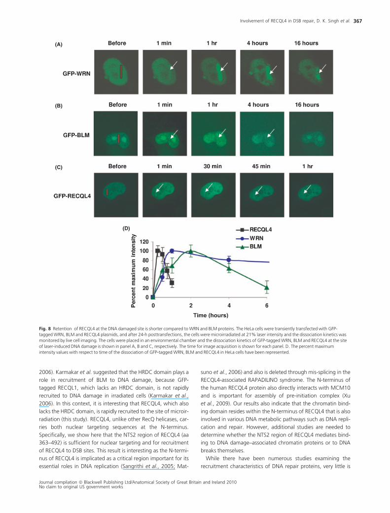
The retention of GFP-tagged RECQL4 at the
microirradiated site is short compared to WRN and
BLM proteins
The efficiency of DNA repair depends on the dynamics of the
DNA repair proteins at the site of DNA damage. WRN, BLM and
RECQL4 are recruited early, but their rate of dissociation also
affects the efficiency of the repair process. While there are many
studies addressing the recruitment of DNA repair proteins to
DNA damage, there is very little information about the disassoci-
ation dynamics of DNA repair proteins from such sites. Such
studies require an environmental chamber, which we have
utilized in these studies. Kinetics of protein dissociation from
DNA damage site was examined in HeLa cells expressing GFP-
tagged WRN, BLM or RECQL4. For this experiment, cells were
microirradiated using 21% laser intensity and placed in an envi-
ronmental chamber, where they could be visualized by confocal
microscopy under normal growth conditions throughout the
experiment. The graph shows the dynamic association of the
various RecQ helicases at the site of a lesion. Interestingly, REC-
QL4 arrives early and departs within 1 h (Fig. 8 panels C and D).
We also observed that the timing at which the WRN protein
reaches its maximum intensity at the DNA damage site is differ-
ent from BLM and RECQL4. WRN reaches maximal intensity at
about one hr and then slowly departs. BLM reaches its maximal
intensity at about 2 h, but its disassociation is faster than for
WRN. Collectively, we conclude that the initial recruitment is
similar for the different RecQ helicases (Fig. 4), but the maximal
recruitment and rate of dissociation varies considerably for the
different RecQ helicases. Thus, the different RecQ helicases have
distinct roles in the response to DNA damage.
Discussion
This report characterizes the role of RECQL4 in DNA DSB repair.
Results presented here show that RECQL4-deficient cells are
moderately more sensitive to c-irradiation than normal fibro-
blasts (Fig. 1). The conflicting results in articles prior to identifi-
cation of the role of RECQL4 in RTS may reflect that 30% of
patients with RTS carry mutations in a yet unidentified gene
(Kitao et al., 1999; Wang et al., 2003). More recently, there
have been divergent reports in the literature regarding the sensi-
tivity of different primary RTS fibroblasts and murine fibroblasts
toward ionizing radiation (Hoki et al., 2003; Cabral et al., 2008;
Jin et al.,2008). Although WRN-deficient primary fibroblasts
also demonstrate only modest sensitivity toward ionizing
radiation (Yannone et al., 2001), WRN plays important roles in
Fig. 5 Endogenous c-H2AX and RECQL4
colocalizes at the laser-induced DNA damage site.
HeLa cells were laser microirradiated at 21% laser
intensity followed by fixation after 5 min and
immunostaining with anti-rabbit c-H2AX and anti-
goat RECQL4 antibodies. The untreated cell is
shown in top left panel, c-H2AX is shown in red
(top middle), RECQL4 is depicted in green (top
right) and the merge images of the two showing
co-localization is shown in yellow (lower panels).
Involvement of RECQL4 in DSB repair, D. K. Singh et al.
Journal compilation ª Blackwell Publishing Ltd/Anatomical Society of Great Britain and Ireland 2010No claim to original US government works
364

regulating HR- and nonhomologous end-joining (NHEJ)-depen-
dent repair of DSBs. Therefore, we propose that RECQL4 might
also play important roles in DSB repair with regard to survival
and efficient repair of damage.
We then examined c-H2AX and 53BP1 foci formation and dis-
persal to show that RECQL4-deficient cells are indeed defective
in efficient repair of DSBs. A comparison of the time course
kinetics of foci dispersal in normal control fibroblast cells and
RECQL4-deficient cells showed that there are approximately 2–3
fold more persistence of 53BP1 foci for > 10 h in RTS cells, sug-
gesting that RECQL4-deficiency impairs efficient repair of DNA
DSBs (Fig. 2). Similarly, WRN-deficient cells also have a higher
basal level of c-H2AX foci than control cells in experiments
where WRN and control cells are age matched. It has been pro-
posed that an age-associated decrease in the efficiency of DSB
repair causes genome instability in the context of both normal
and premature aging (Sedelnikova et al., 2008). Also, in Xeno-
pus, the reduction in DSB-induced c-H2AX was significantly
compromised in RECQL4-depleted extracts suggesting that REC-
QL4 functions in repair of DSB (Kumata et al., 2007).
(A)
(B)
Fig. 6 Analysis of association of GFP-tagged RECQL4 mutants at the site of laser DNA damage. The different GFP-RECQL4 mutant constructs were transiently
transfected in the HeLa cell and association kinetics were monitored at 21% laser intensity. Panel A shows the schematics of different mutants and their
association with DNA damage site (represented with + sign). Two nuclear targeting sequences (NTS1 & NTS2) and the helicase domain are shown. Panel B shows
one representative image of each mutant association at the DNA damaged site.
Involvement of RECQL4 in DSB repair, D. K. Singh et al.
Journal compilation ª Blackwell Publishing Ltd/Anatomical Society of Great Britain and Ireland 2010No claim to original US government works
365

This study used confocal laser scanning microscopy to image
the kinetic behavior of RecQ helicases in response to laser-
induced DNA damage in living cells. As previously shown for
WRN and BLM protein (Lan et al., 2005; Karmakar et al., 2006),
GFP-tagged RECQL4 is recruited rapidly to the site of laser-
induced DNA damage (Fig. 4). The endogenous RECQL4 is also
recruited to DSBs, where it colocalizes with c-H2AX at the micr-
oirradiated site, but not to SSBs (Fig. 5). A previous study
showed that a portion of RECQL4 foci are associated with
regions of ssDNA at DSB sites in cells treated with etoposide and
IR (Petkovic et al., 2005). A study using Xenopus oocyte extracts
also suggested that RECQL4 accumulates in chromatin and pro-
motes repair of DSBs (Kumata et al., 2007). These results sug-
gest that RECQL4 associates with DSBs and promotes efficient
processing and repair of DSBs.
This study shows that the NTS2 domain of RECQL4 is required
and sufficient for recruitment of RECQL4 to the site of microirra-
diation (Fig. 6). RECQL4 is structurally unique and distinct from
other RecQ helicases homologues, in that it does not possess the
RQC and C-terminal HRDC domains, which are present in WRN
and BLM. Both WRN and BLM are recruited to laser-induced
DSBs via their HRDC domain (Lan et al., 2005; Karmakar et al.,
(A)
(B)
(C)
(D)
(E)
Fig. 7 The recruitment of GFP-tagged RECQL4 is independent of functional WRN, BLM and ATM proteins. The association kinetics of transiently transfected GFP-
RECQL4 protein at the site of laser-induced DNA damage in SV40-transformed normal fibroblasts (GM00637), BLM (GM08505), WRN (AG07066) and ATM
(GM05849) mutant fibroblasts cells are shown in panel A, B, C and D, respectively. The normalized intensity value of the kinetics of association of RECQL4 in
normal fibroblasts, BLM, WRN and ATM mutant cells have been represented in panel E.
Involvement of RECQL4 in DSB repair, D. K. Singh et al.
Journal compilation ª Blackwell Publishing Ltd/Anatomical Society of Great Britain and Ireland 2010No claim to original US government works
366
2006). Karmakar et al. suggested that the HRDC domain plays a
role in recruitment of BLM to DNA damage, because GFP-
tagged RECQL1, which lacks an HRDC domain, is not rapidly
recruited to DNA damage in irradiated cells (Karmakar et al.,
2006). In this context, it is interesting that RECQL4, which also
lacks the HRDC domain, is rapidly recruited to the site of microir-
radiation (this study). RECQL4, unlike other RecQ helicases, car-
ries both nuclear targeting sequences at the N-terminus.
Specifically, we show here that the NTS2 region of RECQL4 (aa
363–492) is sufficient for nuclear targeting and for recruitment
of RECQL4 to DSB sites. This result is interesting as the N-termi-
nus of RECQL4 is implicated as a critical region important for its
essential roles in DNA replication (Sangrithi et al., 2005; Mat-
suno et al., 2006) and also is deleted through mis-splicing in the
RECQL4-associated RAPADILINO syndrome. The N-terminus of
the human RECQL4 protein also directly interacts with MCM10
and is important for assembly of pre-initiation complex (Xu
et al., 2009). Our results also indicate that the chromatin bind-
ing domain resides within the N-terminus of RECQL4 that is also
involved in various DNA metabolic pathways such as DNA repli-
cation and repair. However, additional studies are needed to
determine whether the NTS2 region of RECQL4 mediates bind-
ing to DNA damage–associated chromatin proteins or to DNA
breaks themselves.
While there have been numerous studies examining the
recruitment characteristics of DNA repair proteins, very little is
(A)
(B)
(C)
(D)
Fig. 8 Retention of RECQL4 at the DNA damaged site is shorter compared to WRN and BLM proteins. The HeLa cells were transiently transfected with GFP-
tagged WRN, BLM and RECQL4 plasmids, and after 24-h posttransfections, the cells were microirradiated at 21% laser intensity and the dissociation kinetics was
monitored by live cell imaging. The cells were placed in an environmental chamber and the dissociation kinetics of GFP-tagged WRN, BLM and RECQL4 at the site
of laser-induced DNA damage is shown in panel A, B and C, respectively. The time for image acquisition is shown for each panel. D. The percent maximum
intensity values with respect to time of the dissociation of GFP-tagged WRN, BLM and RECQL4 in HeLa cells have been represented.
Involvement of RECQL4 in DSB repair, D. K. Singh et al.
Journal compilation ª Blackwell Publishing Ltd/Anatomical Society of Great Britain and Ireland 2010No claim to original US government works
367

known about the retention kinetics of RECQ helicase at DSB
sites. In our studies, we used an environmental chamber to
maintain the cells under normal conditions over the long period
of study. Interestingly, RECQL4 is retained at sites of DNA dam-
age for only approximately one hour, while WRN and BLM are
retained for longer periods of time (Fig. 8). Thus, RECQL4 might
act at an early step in DSB repair, or it could act on a specialized
transient DNA substrate, which is present at a low level in laser-
treated cells. These results also suggest that different RECQ heli-
cases have distinct and specialized functions in the context of
DSB repair that correlates with their specialized biochemical fea-
tures and their responses to different genotoxic agents.
In cells, DSB’s are repaired either by homologous recombi-
nation repair (HRR) or NHEJ. Nonhomologous end-joining -
mediated DSB repair is largely independent of terminal DNA
sequence homology. Therefore, NHEJ is error-prone and pro-
duces deletions, insertions and translocations (Thompson &
Schild, 2002). The HRR pathway repairs DSBs using homolo-
gous sequences on sister chromatids and to a lesser extent
on chromosome homologues (Liang et al., 1998; Johnson &
Jasin, 2000). Previous studies have indicated that RECQL4
might be involved in both, HRR and NHEJ pathways. RECQL4
has been shown to form complex with Rad51, which is cru-
cial for the repair of DSBs by the HRR pathway (Petkovic
et al., 2005). In another study using Xenopus, RECQL4 has
been shown to be loaded on to the chromatin in response to
DSBs. Further, RECQL4 is loaded adjacent to Ku heterodimer
binding site on damaged chromatin suggesting its possible
involvement in NHEJ (Kumata et al., 2007). However, the
mechanism of RECQL4 involvement in DSB repair is not yet
understood. It is possible that RECQL4 participates in the
repair process once it is loaded on to the chromatin at the
site of DSB. In this complex, RECQL4 can interact directly with
various proteins involved in the HRR and NHEJ pathway at dif-
ferent stages of DSB repair and modulate their functions.
Recent studies have also indicated that the RECQL4 might
also be important in replication restart after repair (Xu et al.,
2009). However, another likely possibility is that the function
of RECQL4 may be indirect. Similar to WRN and BLM heli-
cases, RECQL4 could function in resolving aberrant DNA
structures formed during DNA replication and recombination
repair processes and facilitate the loading of other repair fac-
tors at the site of the break. Therefore, the absence of REC-
QL4 in the cells would lead to genomic instability and could
explain the heterogeneous premature aging phenotype and
cancer susceptibility seen in patients with RTS.
One of the limitations of this study is that most of the experi-
ments are performed with overexpression of transfected pro-
teins to study the cellular dynamics at the site of DNA damage.
Nonetheless, this work strengthens the hypothesis that RECQL4
plays a significant role in the DNA damage response to DSBs.
Additional studies are needed to further understand the molecu-
lar mechanisms that allow RECQL4 to promote genetic stability
and facilitate an efficient response to DNA damage in human
cells.
Experimental procedures
Cell lines and transfection
HeLa cells, SV40-transformed normal (GM00637), WRN
(AG07066), BLM (GM08505), ATM (GM05849) fibroblasts cell
lines, normal primary skin fibroblasts (GM00323, GM00969,
GM01864) and RTS-deficient skin primary fibroblasts
(AG05013, AG18371, AG17524) cell lines from Coriell Cell
Repositories were used in the study. The RTS cell lines have
confirmed RECQL4 mutations and do not show RECQL4 pro-
teins in western analysis of the total cell extract (Kitao et al.,
1999; Petkovic et al., 2005). The pair of RTS cells and normal
fibroblasts cells used for survival assays and the foci formation
assays are age and sex matched. The HeLa cells were cultured
in DMEM media supplemented with 10% fetal bovine serum
(FBS) and 1% penicillin-streptomycin at 37�C in a humidified
atmosphere with 5% CO2. SV40-transformed normal, WRN,
BLM and ATM fibroblasts were cultured in MEM media supple-
mented with 10% FBS. Normal primary skin fibroblasts cells
and RTS-deficient primary fibroblasts were cultured in Amnio-
max II Complete media (Invitrogen, Carlsbad, CA, USA). For
live-cell experiments, the cells were plated in glass bottom Petri
dishes (MatTek Corporation, Ashland, MA, USA) at 50% con-
fluence 24 h before the transfections. For transfection, Fugene
6 was used as per manufacturer’s instructions (Roche, Nutley,
NJ, USA). For immunofluorescence studies, the cells were pla-
ted in 4-well chambered slides 24 h before the treatments.
Cell proliferation assay
Cell proliferation was assessed using CyQuant (Molecular
Probes, Eugene, OR, USA). Both the normal cell line
(GM00323 and GM00969) and the RTS cell line (AG05013
and AG17524) were plated in 60-mm petri dishes at a density
of 1000 cells per plate. The cells were treated with 1, 2, 3 Gy
of c irradiation and then allowed to grow in Amniomax II
Complete media for 14 days at 37�C at 5% CO2. On the day
of the analysis, the plates with adherent cells were washed
once with 1X PBS and incubated with the CyQuant dye for
30 min in dark at 37�C with 5% CO2. Fluorescence intensity,
which is proportional to cell number, was measured on a
Typhoon 8600 fluorescent scanner (GE Healthcare, Piscat-
away, NJ, USA) with filters set at 480 nm excitation and
520 nm emission. Fluorescence data were analyzed, and the
number of colonies in each plate was counted by Image
QuantTL software (GE Healthcare, Piscataway, NJ, USA).
Analysis of c-H2AX and 53BP1 foci
The cH2AX and 53BP1 foci were measured in two normal pri-
mary fibroblasts cell lines (GM00323 and GM01864) and two
RTS cell lines (AG05013 and AG18371). Approximately, 25 000
primary fibroblasts cells were seeded on chambered slides,
grown overnight and then treated with 5 Gy of c-irradiation.
Involvement of RECQL4 in DSB repair, D. K. Singh et al.
Journal compilation ª Blackwell Publishing Ltd/Anatomical Society of Great Britain and Ireland 2010No claim to original US government works
368

Cells were allowed to recover for 1, 3, 6 or 10 h, under normal
growth conditions and then immediately fixed and stained
(described in next section) with either mouse monoclonal
cH2AX antibody (1:200; Upstate Biotechnology, Waltham, MA,
USA) or rabbit polyclonal 53BP1 antibody (1:200, Abcam, Cam-
bridge, MA, USA) and Alexa goat anti-mouse 647 or donkey
anti-rabbit 647 secondary antibody (Invitrogen, Carlsbad, CA,
USA). Ten images representing about 15–20 cells per well were
taken using a Nikon TE2000 spinning disk microscope with five
laser imaging modules and a CCD camera (Hamamatsu). The
data were analyzed using Volocity version 4.3.1 build 6 (Improvi-
sion, Waltham, MA, USA).
Immunocytochemistry and antibodies
The cells were laser microirradiated and stained with antibodies
against cH2AX, 53BP1, XRCC1, RECQL4. After microirradiation,
the cells were fixed with 3.7% formaldehyde in PBS for 10 min at
room temperature. Then, cells were washed three times with
PBS, permeabilized with 0.2% Triton X 100 in PBS for 5 min at
room temperature. The cells were washed with PBS and blocked
in 5% FBS overnight at 4�C. The cells were incubated with differ-
ent primary antibodies for 1 h at 37�C in a humidified chamber.
Mouse monoclonal anti-phosphorylated H2AX (1:200; Upstate
Biotechnology, Waltham, MA, USA), goat polyclonal anti-REC-
QL4 (K16) (1:50; Santa Cruz Biotech, Santa Cruz, CA, USA), rab-
bit polyclonal anti-53BP1 (1:200; Abcam, Cambridge, MA, USA)
and rabbit polyclonal anti-XRCC1 (1:200; Santa Cruz Biotech)
antibodies were used. Bound antibodies were visualized by incu-
bating the cells with secondary antibodies, namely Alexa FITC
(488)–conjugated chicken anti-goat, Alexa 647–conjugated goat
anti-mouse and Alexa-conjugated 647 donkey anti-rabbit. The
images were captured with a confocal microscope (Nikon eclipse
TE2000; Nikon Instruments Inc., Linthicum, MD, USA) and ana-
lyzed by Volocity software (Improvision, Waltham, MA, USA).
Western blotting
After separation in SDS–PAGE gel, the proteins were transferred
to PVDF membrane and the membrane was blocked with 5%
fat-free milk in 1X TBS-T for 1 h at room temperature followed
by incubation with primary rabbit anti-GFP antibody (1: 5000;
Novus Biologicals, Littleton, CO, USA) overnight at 4�C. The
membranes were then washed thrice in 1X TBST, 5 min each
and then incubated in HRP-conjugated secondary anti-rabbit
antibody (1:20,000; Sigma-Aldrich, St. Louis, MO, USA) for 1 h.
After washing, the membrane was developed by ECL plus kit fol-
lowing manufacturer’s protocol (GE Healthcare).
Laser irradiation and confocal microscopy
We employed a Nikon Eclipse 2000E spinning disk confocal
microscope with five laser imaging modules and a CCD camera
(Hamamatsu, Tokyo, Japan). The set-up integrated a Stanford
Research Systems (SRS) NL100 nitrogen laser by Micropoint abla-
tion system (Photonics Instruments, St. Charles, IL, USA). Site-
specific DNA damage was induced using the SRS NL100 nitrogen
laser that was passed through a dye cell to emit at 435 nm
wavelength. The power of the laser was attenuated through
Improvision’s Volocity software 4.3.1 (Improvision ⁄ PerkinElmer,
Coventry, UK) in terms of percent intensity. Positions internal to
the nuclei of either live HeLa cells or HeLa cells transfected with
GFP-tagged plasmids were targeted via a 40X oil objective lens.
The laser intensities of 8% and 21% were used. Images were cap-
tured at various time points and analyzed using Volocity version
4.3.1 build 6 (Improvision). Experiments were performed using
an environmental chamber attached to the microscope to main-
tain the normal atmosphere of the cells (i.e. 37�C and 5% CO2).
Fluorescence recovery after Photobleaching (FRAP)
Fluorescence recovery after Photobleaching analysis was carried
out with live cells transfected with GFP-tagged WRN, BLM and
RECQL4 plasmids. The same set up as the microirradiation was
used to perform the photobleaching. Fluorescence recovery was
monitored as described in the figure legends and the data for
recovery was corrected for the background intensity and loss of
total fluorescence. Each experiment was performed at least
three times and the data presented here are mean intensity val-
ues obtained in given experiments.
Acknowledgments
We thank Drs Suhasini Avvaru and Venkateswarlu Popuri for
critical reading of the manuscript. This work was supported by
funds from the Intramural Research Program of the National
Institute on Aging, NIH. Support is acknowledged from the
Danish Aging Research Center, the Center for Healthy Aging
in Copenhagen, and from the Danish Medical research Council.
Author contributions
Conceived and designed the experiments: VB, DKS, PK, MA.
Performed the experiments: DKS, PK, MA, SS, AM, LB. Analyzed
the data: VB, DKS, PK, MA, DLC, SP. Wrote the article: DKS, VB.
References
Anbari KK, Ierardi-Curto LA, Silber JS, Asada N, Spinner N, Zackai EH,
Belasco J, Morrissette JD, Dormans JP (2000) Two primary osteosar-
comas in a patient with Rothmund-Thomson syndrome. Clin. Ort-
hop. Relat. Res. 378, 213–223.
Bohr VA (2008) Rising from the RecQ-age: the role of human RecQ
helicases in genome maintenance. Trends Biochem. Sci. 33, 609–
620.
Brosh Jr RM, Bohr VA (2007) Human premature aging, DNA repair
and RecQ helicases. Nucleic Acids Res. 35, 7527–7544.
Burks LM, Yin J, Plon SE (2007) Nuclear import and retention domains
in the amino terminus of RECQL4. Gene 391, 26–38.
Cabral RE, Queille S, Bodemer C, de PY, Neto JB, Sarasin A, ya-Gros-
jean L (2008) Identification of new RECQL4 mutations in Caucasian
Involvement of RECQL4 in DSB repair, D. K. Singh et al.
Journal compilation ª Blackwell Publishing Ltd/Anatomical Society of Great Britain and Ireland 2010No claim to original US government works
369

Rothmund-Thomson patients and analysis of sensitivity to a wide
range of genotoxic agents. Mutat. Res. 643, 41–47.
Capp C, Wu J, Hsieh TS (2009) Drosophila RecQ4 has a 3¢-5¢DNA heli-
case activity that is essential for viability. J. Biol. Chem. 284, 30845–
30852.
Der Kaloustian VM, McGill JJ, Vekemans M, Kopelman HR (1990) Clo-
nal lines of aneuploid cells in Rothmund-Thomson syndrome. Am. J.
Med. Genet. 37, 336–339.
Dinant C, de Jager M, Essers J, van Cappellen WA, Kanaar R, Houts-
muller AB, Vermeulen W (2007) Activation of multiple DNA repair
pathways by subnuclear damage induction methods. J. Cell Sci.
120, 2731–2740.
Durand F, Castorina P, Morant C, Delobel B, Barouk E, Modiano P
(2002) Rothmund-Thomson syndrome, trisomy 8 mosaicism and
RECQ4 gene mutation. Ann. Dermatol. Venereol. 129, 892–895.
Fan W, Luo J (2008) RecQ4 Facilitates UV Light-induced DNA Damage
Repair through Interaction with Nucleotide Excision Repair Factor
Xeroderma Pigmentosum Group A (XPA). J. Biol. Chem. 283,
29037–29044.
Haince JF, McDonald D, Rodrigue A, Dery U, Masson JY, Hendzel MJ,
Poirier GG (2008) PARP1-dependent kinetics of recruitment of
MRE11 and NBS1 proteins to multiple DNA damage sites. J. Biol.
Chem. 283, 1197–1208.
Hickson ID (2003) RecQ helicases: Caretakers of the genome. Nat.
Rev. Cancer 3, 169–178.
Hoki Y, Araki R, Fujimori A, Ohhata T, Koseki H, Fukumura R, Nakam-
ura M, Takahashi H, Noda Y, Kito S, Abe M (2003) Growth retarda-
tion and skin abnormalities of the Recql4-deficient mouse. Hum.
Mol. Genet. 12, 2293–2299.
Im JS, Ki SH, Farina A, Jung DS, Hurwitz J, Lee JK (2009) Assembly of
the Cdc45-Mcm2-7-GINS complex in human cells requires the
Ctf4 ⁄ And-1, RecQL4, and Mcm10 proteins. Proc. Natl. Acad. Sci.
U S A 106, 15628–15632.
Jin W, Liu H, Zhang Y, Otta SK, Plon SE, Wang LL (2008) Sensitivity of
RECQL4-deficient fibroblasts from Rothmund-Thomson syndrome
patients to genotoxic agents. Hum. Genet. 123, 643–653.
Johnson RD, Jasin M (2000) Sister chromatid gene conversion is a
prominent double-strand break repair pathway in mammalian cells.
EMBO J. 19, 3398–3407.
Karmakar P, Seki M, Kanamori M, Hashiguchi K, Ohtsuki M, Murata
E, Inoue E, Tada S, Lan L, Yasui A, Enomoto T (2006) BLM is an
early responder to DNA double-strand breaks. Biochem. Biophys.
Res. Commun. 348, 62–69.
Kinner A, Wu W, Staudt C, Iliakis G (2008) Gamma-H2AX in recogni-
tion and signaling of DNA double-strand breaks in the context of
chromatin. Nucleic Acids Res. 36, 5678–5694.
Kitao S, Ohsugi I, Ichikawa K, Goto M, Furuichi Y, Shimamoto A
(1998) Cloning of two new human helicase genes of the RecQ fam-
ily: biological significance of multiple species in higher eukaryotes.
Genomics 54, 443–452.
Kitao S, Shimamoto A, Goto M, Miller RW, Smithson WA, Lindor NM,
Furuichi Y (1999) Mutations in RECQL4 cause a subset of cases of
Rothmund-Thomson syndrome. Nat. Genet. 22, 82–84.
von Kobbe C, Karmakar P, Dawut L, Opresko P, Zeng XM, Brosh RM,
Hickson ID, Bohr VA (2002) Colocalization, physical, and functional
interaction between Werner and Bloom syndrome proteins. J. Biol.
Chem. 277, 22035–22044.
Kumata Y, Tada S, Yamanada Y, Tsuyama T, Kobayashi T, Dong YP,
Ikegami K, Murofushi H, Seki M, Enomoto T (2007) Possible involve-
ment of RecQL4 in the repair of double-strand DNA breaks in Xeno-
pus egg extracts. Biochim Biophys Acta 1773, 556–564.
Lan L, Nakajima S, Komatsu K, Nussenzweig A, Shimamoto A, Oshima
J, Yasui A (2005) Accumulation of Werner protein at DNA double-
strand breaks in human cells. J. Cell Sci. 118, 4153–4162.
Liang F, Han MG, Romanienko PJ, Jasin M (1998) Homology-directed
repair is a major double-strand break repair pathway in mammalian
cells. Proc. Natl. Acad. Sci. U S A 95, 5172–5177.
Lindor NM, Furuichi Y, Kitao S, Shimamoto A, Arndt C, Jalal S (2000)
Rothmund-Thomson syndrome due to RECQ4 helicase mutations:
report and clinical and molecular comparisons with Bloom syndrome
and Werner syndrome. Am. J. Med. Genet. 90, 223–228.
Macris MA, Krejci L, Bussen W, Shimamoto A, Sung P (2006) Bio-
chemical characterization of the RECQ4 protein, mutated in Roth-
mund-Thomson syndrome. DNA Repair 5, 172–180.
Mailand N, Bekker-Jensen S, Faustrup H, Melander F, Bartek J, Lukas
C, Lukas J (2007) RNF8 ubiquitylates histones at DNA double-strand
breaks and promotes assembly of repair proteins. Cell 131, 887–
900.
Matsuno K, Kumano M, Kubota Y, Hashimoto Y, Takisawa H (2006)
The N-terminal noncatalytic region of Xenopus RecQ4 is required
for chromatin binding of DNA polymerase alpha in the initiation of
DNA replication. Mol. Cell. Biol. 26, 4843–4852.
Orstavik KH, Mcfadden N, Hagelsteen J, Ormerod E, Vanderhagen CB
(1994) Instability of Lymphocyte Chromosomes in A Girl with Roth-
mund-Thomson Syndrome. J. Med. Genet. 31, 570–572.
Petkovic M, Dietschy T, Freire R, Jiao RJ, Stagljar I (2005) The human
Rothmund-Thomson syndrome gene product, RECQL4, localizes to
distinct nuclear foci that coincide with proteins involved in the main-
tenance of genome stability. J. Cell Sci. 118, 4261–4269.
Sangrithi MN, Bernal JA, Madine M, Philpott A, Lee J, Dunphy WG,
Venkitaraman AR (2005) Initiation of DNA replication requires the
RECQL4 protein mutated in Rothmund-Thomson syndrome. Cell
121, 887–898.
Schultz LB, Chehab NH, Malikzay A, Halazonetis TD (2000) p53 bind-
ing protein 1 (53BP1) is an early participant in the cellular response
to DNA double-strand breaks. J. Cell Biol. 151, 1381–1390.
Schurman SH, Hedayati M, Wang Z, Singh DK, Speina E, Zhang Y,
Becker K, Macris M, Sung P, Wilson III DM, Croteau DL, Bohr VA
(2009) Direct and indirect roles of RECQL4 in modulating base exci-
sion repair capacity. Hum. Mol. Genet. 18, 3470–3483.
Sedelnikova OA, Horikawa I, Redon C, Nakamura A, Zimonjic DB,
Popescu NC, Bonner WM (2008) Delayed kinetics of DNA double-
strand break processing in normal and pathological aging. Aging
Cell 7, 89–100.
Siitonen HA, Kopra O, Kaariainen H, Haravuori H, Winter RM, Saama-
nen AM, Peltonen L, Kestila M (2003) Molecular defect of RAPADI-
LINO syndrome expands the phenotype spectrum of RECQL
diseases. Hum. Mol. Genet. 12, 2837–2844.
Stinco G, Governatori G, Mattighello P, Patrone P (2008) Multiple
cutaneous neoplasms in a patient with Rothmund-Thomson syn-
drome: case report and published work review. J. Dermatol. 35,
154–161.
Suzuki T, Kohno T, Ishimi Y (2009) DNA helicase activity in purified
human RECQL4 protein. J. Biochem. 146, 327–335.
Thompson LH, Schild D (2002) Recombinational DNA repair and
human disease. Mutat. Res. 509, 49–78.
Van Maldergem L, Siitonen HA, Jalkh N, Chouery E, De Roy M, Dela-
gue V, Muenke M, Jabs EW, Cai J, Wang LL, Plon SE, Fourneau C,
Kestila M, Gillerot Y, Megarbane A, Verloes A (2006) Revisiting the
craniosynostosis-radial ray hypoplasia association: Baller-Gerold syn-
drome caused by mutations in the RECQL4 gene. J. Med. Genet.
43, 148–152.
Involvement of RECQL4 in DSB repair, D. K. Singh et al.
Journal compilation ª Blackwell Publishing Ltd/Anatomical Society of Great Britain and Ireland 2010No claim to original US government works
370
Vennos EM, Collins M, James WD (1992) Rothmund-Thomson Syn-
drome - Review of the World Literature. J. Am. Acad. Dermatol. 27,
750–762.
Wang LL, Gannavarapu A, Kozinetz CA, Levy ML, Lewis RA, Chinta-
gumpala MM, Ruiz-Maldonado R, Contreras-Ruiz J, Cunniff C, Erick-
son RP, Lev D, Rogers M, Zackai EH, Plon SE (2003) Association
between osteosarcoma and deleterious mutations in the RECQL4
gene in Rothmund-Thomson syndrome. J. Natl Cancer Inst. 95,
669–674.
Werner SR, Murthy S, Winata T, Prahalad AK, Yang J, Hock JM (2005)
Recql4 is involved in the cellular response to oxidative stress in oste-
osarcoma cells. J. Bone Miner. Res. 20, 54.
Werner SR, Prahalad AK, Yang J, Hock JM (2006) RECQL4-deficient
cells are hypersensitive to oxidative stress ⁄ damage: insights for oste-
osarcoma prevalence and heterogeneity in Rothmund-Thomson syn-
drome. Biochem. Biophys. Res. Commun. 345, 403–409.
Woo LL, Futami K, Shimamoto A, Furuichi Y, Frank KM (2006) The
Rothmund-Thomson gene product RECQL4 localizes to the nucleo-
lus in response to oxidative stress. Exp. Cell Res. 312, 3443–3457.
Xu X, Liu Y (2009) Dual DNA unwinding activities of the Rothmund-
Thomson syndrome protein, RECQ4. EMBO J. 28, 568–577.
Xu X, Rochette PJ, Feyissa EA, Su TV, Liu Y (2009) MCM10 mediates
RECQ4 association with MCM2-7 helicase complex during DNA rep-
lication. EMBO J. 28, 3005–3014.
Yannone SM, Roy S, Chan DW, Murphy MB, Huang SR, Campisi J,
Chen DJ (2001) Werner syndrome protein is regulated and phos-
phorylated by DNA-dependent protein kinase. J. Biol. Chem. 276,
38242–38248.
Yano K, Morotomi-Yano K, Wang SY, Uematsu N, Lee KJ, Asaithamby
A, Weterings E, Chen DJ (2008) Ku recruits XLF to DNA double-
strand breaks. EMBO Rep. 9, 91–96.
Yin JH, Kwon YT, Varshavsky A, Wang WD (2004) RECQL4, mutated
in the Rothmund-Thomson and RAPADILINO syndromes, interacts
with ubiquitin ligases UBR1 and UBR2 of the N-end rule pathway.
Hum. Mol. Genet. 13, 2421–2430.
Supporting Information
Additional supporting information may be found in the online
version of this article:
Fig. S1 Growth kinetics of normal fibroblasts (GM00323 and
GM00969) and RTS cells (AG05013 and AG17524).
Fig. S2 After 5 Gy of c-irradiation, the cells were allowed to
recover and fixed at indicated time points (0, 1, 3, 6 and 10 h)
and immunostained with anti-mouse c-H2AX antibodies (as
described in Experimental procedures).
Fig. S3 Detection of the expression of GFP-RECQL4 deletion
mutant proteins in HeLa cells.
As a service to our authors and readers, this journal provides
supporting information supplied by the authors. Such materials
are peer-reviewed and may be re-organized for online delivery,
but are not copy-edited or typeset. Technical support issues aris-
ing from supporting information (other than missing files)
should be addressed to the authors.
Involvement of RECQL4 in DSB repair, D. K. Singh et al.
Journal compilation ª Blackwell Publishing Ltd/Anatomical Society of Great Britain and Ireland 2010No claim to original US government works
371
Copyright © 2022 FDOKUMEN