Interplay between human DNA repair proteins at a unique double-strand break in vivo
10
Interplay between human DNA repair proteins at a unique double-strand break in vivo Ame ´ lie Rodrigue 1 , Matthieu Lafrance 1 , Marie-Christine Gauthier 1 , Darin McDonald 2 , Michael Hendzel 2 , Stephen C West 3 , Maria Jasin 4 and Jean-Yves Masson 1, * 1 Genome Stability Laboratory, Laval University Cancer Research Center, Que ´bec city, Que ´bec, Canada, 2 Department of Oncology, Faculty of Medicine, University of Alberta and Cross Cancer Institute, Edmonton, Alberta, Canada, 3 Clare Hall Laboratories, Cancer Research UK, London Research Institute, South Mimms, Hertfordshire, UK and 4 Molecular Biology Program, Memorial Sloan-Kettering Cancer Center, New York, NY, USA DNA repair by homologous recombination is essential for preserving genomic integrity. The RAD51 paralogs (RAD51B, RAD51C, RAD51D, XRCC2 and XRCC3) play important roles in this process. In this study, we show that human RAD51 interacts with RAD51C-XRCC3 or RAD51B-C-D-XRCC2. In addition to being critical for RAD51 focus formation, RAD51C localizes to DNA damage sites. Inhibition of RAD51C results in a decrease in cellular proliferation consistent with a role in repairing double- strand breaks (DSBs) that occur naturally. To monitor a single DNA repair event, we developed immunofluores- cence and chromatin immunoprecipitation (ChIP) meth- ods on human cells where a unique DSB can be created in vivo. Using this system, we observed a single focus of RAD51C, RAD51 and 53BP1, which colocalized with c-H2AX. ChIPs revealed that endogenous human RAD51, RAD51C, RAD51D, XRCC2, XRCC3 and MRE11 proteins are recruited in the S–G2 phase of the cell cycle, while Ku80 is recruited during G1. We propose that RAD51C ensures a tight regulation of RAD51 assembly during DSB repair and plays a direct role in repairing DSBs in vivo. The EMBO Journal (2006) 25, 222–231. doi:10.1038/ sj.emboj.7600914; Published online 5 January 2006 Subject Categories: genome stability & dynamics Keywords: DNA repair; genome stability; homologous recombination; RAD51; RAD51 paralogs Introduction Maintenance of genome stability relies on the accurate repair of double-strand breaks (DSBs) that arise during DNA repli- cation or from DNA-damaging agents. Failure to repair such breaks can lead to the introduction of mutations, chromoso- mal translocations, apoptosis and cancer. Hence, in order to preserve genome integrity, cells have evolved processes to respond and repair DSBs. In higher eukaryotes, the signalling response to DSBs is centered on mammalian ATM (ataxia- telangiectasia mutated), ATR (ATM and Rad3 related) and DNA-PK (DNA-dependent protein kinase). These PI3KKs (phosphatidylinositol-3 kinase-like kinases) trigger cell cycle arrest following DNA damage, therefore allowing DNA repair to take place (Kurz and Lees-Miller, 2004). Moreover, follow- ing the induction of DSBs by ionizing radiation, ATM and DNA-PKcs rapidly phosphorylate the carboxy-terminal SQE motif of H2AX (to form g-H2AX foci) along flanking mega- base chromatin regions (Rogakou et al, 1999; Stiff et al, 2004), a process that might contribute to both detection and repair of DSBs. In addition, ATR and DNA-PKcs phos- phorylate H2AX downstream of replication-associated DSBs (Ward and Chen, 2001). In mammalian cells, homologous recombination (HR) has emerged as the major mechanism for the error-free homo- logy-directed repair of DSBs. The central activity of HR is conferred by the RAD51 protein, a eukaryotic homolog of the Escherichia coli RecA recombinase, which catalyses the inva- sion of the broken ends of the DSB into the intact sister chromatid. Recently, extensive studies have been dedicated to the identification of proteins involved in HR. Five genes (RAD51B, RAD51C, RAD51D, XRCC2 and XRCC3) sharing 20–30% sequence identity with human RAD51 were identi- fied (Thompson and Schild, 2001). Strong evidence implicat- ing the vertebrate paralogs in HR came particularly from studies with mutants in hamster and chicken DT40 cells. In hamster, XRCC3 deficiency was shown to lead to a 25-fold decrease in DSB repair (Pierce et al, 1999), whereas chicken DT40 cells with knocked-out RAD51 paralogs, although viable, were found to be sensitive to DNA crosslinking agents and to ionizing radiation besides exhibiting recombination/ repair-defective phenotypes such as reduced growth rates, chromosomal instability and spontaneous chromosomal breaks (Takata et al, 2001). The sensitivity displayed by the paralog mutants was shown to be partially suppressed by RAD51 overexpression in chicken cells, thereby implicating the RAD51 paralogs as RAD51 cofactors. Moreover, the forma- tion of DNA damaged-induced RAD51 foci was shown to be abolished in the absence of RAD51C, XRCC2 and XRCC3 in hamster cells and in RAD51B-, RAD51C-, RAD51D-, XRCC2- and XRCC3-deficient chicken DT40 cells (Bishop et al, 1998; French et al, 2002; Godthelp et al, 2002). The importance of the RAD51 paralogs in recombination and in genome stability was further emphasized by the embryonic lethality of RAD51B, RAD51D or XRCC2 deficiency observed in mouse (Shu et al, 1999; Deans et al, 2000; Pittman and Schimenti, 2000). At present, there is much to learn about the biochemical properties and the biological functions of the paralogs. Previous studies using yeast two- and three-hybrid assays have shown numerous protein–protein interactions between the RAD51 paralogs, including RAD51B with RAD51C, RAD51C with XRCC3, RAD51C with RAD51D, RAD51D with Received: 11 August 2005; accepted: 22 November 2005; published online: 5 January 2006 *Corresponding author. Genome Stability Laboratory, Laval University Cancer Research Center, Ho ˆtel-Dieu de Que ´bec, 9 McMahon, Que ´bec city, Que ´bec, Canada G1R 2J6. Tel.: þ 1 418 525 4444 ext 15154; Fax: þ 1 418 691 5439; E-mail: [email protected] The EMBO Journal (2006) 25, 222–231 | & 2006 European Molecular Biology Organization | All Rights Reserved 0261-4189/06 www.embojournal.org The EMBO Journal VOL 25 | NO 1 | 2006 & 2006 European Molecular Biology Organization EMBO THE EMBO JOURNAL THE EMBO JOURNAL 222
-
Upload
independent -
Category
Documents
-
view
3 -
download
0
Transcript of Interplay between human DNA repair proteins at a unique double-strand break in vivo
Interplay between human DNA repair proteinsat a unique double-strand break in vivo
Amelie Rodrigue1, Matthieu Lafrance1,Marie-Christine Gauthier1,Darin McDonald2, Michael Hendzel2,Stephen C West3, Maria Jasin4
and Jean-Yves Masson1,*1Genome Stability Laboratory, Laval University Cancer Research Center,Quebec city, Quebec, Canada, 2Department of Oncology, Faculty ofMedicine, University of Alberta and Cross Cancer Institute, Edmonton,Alberta, Canada, 3Clare Hall Laboratories, Cancer Research UK, LondonResearch Institute, South Mimms, Hertfordshire, UK and 4MolecularBiology Program, Memorial Sloan-Kettering Cancer Center, New York,NY, USA
DNA repair by homologous recombination is essential
for preserving genomic integrity. The RAD51 paralogs
(RAD51B, RAD51C, RAD51D, XRCC2 and XRCC3) play
important roles in this process. In this study, we show
that human RAD51 interacts with RAD51C-XRCC3 or
RAD51B-C-D-XRCC2. In addition to being critical for
RAD51 focus formation, RAD51C localizes to DNA damage
sites. Inhibition of RAD51C results in a decrease in cellular
proliferation consistent with a role in repairing double-
strand breaks (DSBs) that occur naturally. To monitor a
single DNA repair event, we developed immunofluores-
cence and chromatin immunoprecipitation (ChIP) meth-
ods on human cells where a unique DSB can be created
in vivo. Using this system, we observed a single focus
of RAD51C, RAD51 and 53BP1, which colocalized with
c-H2AX. ChIPs revealed that endogenous human RAD51,
RAD51C, RAD51D, XRCC2, XRCC3 and MRE11 proteins are
recruited in the S–G2 phase of the cell cycle, while Ku80 is
recruited during G1. We propose that RAD51C ensures
a tight regulation of RAD51 assembly during DSB repair
and plays a direct role in repairing DSBs in vivo.
The EMBO Journal (2006) 25, 222–231. doi:10.1038/
sj.emboj.7600914; Published online 5 January 2006
Subject Categories: genome stability & dynamics
Keywords: DNA repair; genome stability; homologous
recombination; RAD51; RAD51 paralogs
Introduction
Maintenance of genome stability relies on the accurate repair
of double-strand breaks (DSBs) that arise during DNA repli-
cation or from DNA-damaging agents. Failure to repair such
breaks can lead to the introduction of mutations, chromoso-
mal translocations, apoptosis and cancer. Hence, in order to
preserve genome integrity, cells have evolved processes to
respond and repair DSBs. In higher eukaryotes, the signalling
response to DSBs is centered on mammalian ATM (ataxia-
telangiectasia mutated), ATR (ATM and Rad3 related) and
DNA-PK (DNA-dependent protein kinase). These PI3KKs
(phosphatidylinositol-3 kinase-like kinases) trigger cell cycle
arrest following DNA damage, therefore allowing DNA repair
to take place (Kurz and Lees-Miller, 2004). Moreover, follow-
ing the induction of DSBs by ionizing radiation, ATM and
DNA-PKcs rapidly phosphorylate the carboxy-terminal SQE
motif of H2AX (to form g-H2AX foci) along flanking mega-
base chromatin regions (Rogakou et al, 1999; Stiff et al,
2004), a process that might contribute to both detection
and repair of DSBs. In addition, ATR and DNA-PKcs phos-
phorylate H2AX downstream of replication-associated DSBs
(Ward and Chen, 2001).
In mammalian cells, homologous recombination (HR) has
emerged as the major mechanism for the error-free homo-
logy-directed repair of DSBs. The central activity of HR is
conferred by the RAD51 protein, a eukaryotic homolog of the
Escherichia coli RecA recombinase, which catalyses the inva-
sion of the broken ends of the DSB into the intact sister
chromatid. Recently, extensive studies have been dedicated
to the identification of proteins involved in HR. Five genes
(RAD51B, RAD51C, RAD51D, XRCC2 and XRCC3) sharing
20–30% sequence identity with human RAD51 were identi-
fied (Thompson and Schild, 2001). Strong evidence implicat-
ing the vertebrate paralogs in HR came particularly from
studies with mutants in hamster and chicken DT40 cells.
In hamster, XRCC3 deficiency was shown to lead to a 25-fold
decrease in DSB repair (Pierce et al, 1999), whereas chicken
DT40 cells with knocked-out RAD51 paralogs, although
viable, were found to be sensitive to DNA crosslinking agents
and to ionizing radiation besides exhibiting recombination/
repair-defective phenotypes such as reduced growth rates,
chromosomal instability and spontaneous chromosomal
breaks (Takata et al, 2001). The sensitivity displayed by the
paralog mutants was shown to be partially suppressed by
RAD51 overexpression in chicken cells, thereby implicating
the RAD51 paralogs as RAD51 cofactors. Moreover, the forma-
tion of DNA damaged-induced RAD51 foci was shown to be
abolished in the absence of RAD51C, XRCC2 and XRCC3 in
hamster cells and in RAD51B-, RAD51C-, RAD51D-, XRCC2-
and XRCC3-deficient chicken DT40 cells (Bishop et al, 1998;
French et al, 2002; Godthelp et al, 2002). The importance of
the RAD51 paralogs in recombination and in genome stability
was further emphasized by the embryonic lethality of RAD51B,
RAD51D or XRCC2 deficiency observed in mouse (Shu et al,
1999; Deans et al, 2000; Pittman and Schimenti, 2000).
At present, there is much to learn about the biochemical
properties and the biological functions of the paralogs.
Previous studies using yeast two- and three-hybrid assays
have shown numerous protein–protein interactions between
the RAD51 paralogs, including RAD51B with RAD51C,
RAD51C with XRCC3, RAD51C with RAD51D, RAD51D withReceived: 11 August 2005; accepted: 22 November 2005; publishedonline: 5 January 2006
*Corresponding author. Genome Stability Laboratory, Laval UniversityCancer Research Center, Hotel-Dieu de Quebec, 9 McMahon, Quebeccity, Quebec, Canada G1R 2J6. Tel.: þ 1 418 525 4444 ext 15154;Fax: þ 1 418 691 5439; E-mail: [email protected]
The EMBO Journal (2006) 25, 222–231 | & 2006 European Molecular Biology Organization | All Rights Reserved 0261-4189/06
www.embojournal.org
The EMBO Journal VOL 25 | NO 1 | 2006 &2006 European Molecular Biology Organization
EMBO
THE
EMBOJOURNAL
THE
EMBOJOURNAL
222
XRCC2 and XRCC3 with RAD51 (Schild et al, 2000).
Immunoprecipitations experiments from human cells reve-
aled the presence of two complexes of the RAD51 paralogs
(Masson et al, 2001a, b; Liu et al, 2002; Miller et al, 2002;
Wiese et al, 2002). One complex, referred to as BCDX2,
consists of RAD51B, RAD51C, RAD51D and XRCC2, whereas
the CX3 complex contains RAD51C and XRCC3. Evidence for
subcomplexes were also found (Miller et al, 2002). Residues
required for RAD51C binding to XRCC3 (Tyr139 and Phe249)
have been mapped (Kurumizaka et al, 2003), as well as
domains for interactions between the paralogs (Miller et al,
2004). Both complexes of the paralogs might have over-
lapping and distinct functions. In Arabidopsis, for example,
RAD51C and XRCC3 mutants are meiosis deficient, while
XRCC2 and RAD51B mutants are not (Bleuyard et al, 2005;
Li et al, 2005).
RAD51C is the central core of both RAD51C-XRCC3 and
RAD51B-C-D-XRCC2 complexes. A number of studies suggest
that these complexes are involved in HR. RAD51B–RAD51C
alleviates the inhibitory effect of RPA in vitro and also
promotes ATP-independent strand exchange (Sigurdsson
et al, 2001; Lio et al, 2003). BCDX2 has shown preferential
binding to Y-shaped DNA and synthetic Holliday junction
(HJ) (Yokoyama et al, 2004). Branch migration and resolution
activities are dependent on the presence of the RAD51 para-
logs (Liu et al, 2004).
Although these studies show a role for the RAD51 paralogs
in HR in vitro, it is not clear whether these proteins interact
directly with DSBs in vivo and how they mediate repair. For
instance, foci of RAD51C have not been observed by
immunofluorescence in mitotic cells. Using immunoprecipi-
tation analyses, the recombinase RAD51 was found to be
exclusive of the endogenous paralog complexes (Miller et al,
2002). Here, we report the in vitro and in vivo interaction
between RAD51 and RAD51C. Furthermore, using a recombi-
nation reporter system where a single DSB can be created, we
show that RAD51C, together with several repair factors, binds
a DSB in vivo as determined by immunofluorescence and
chromatin immunoprecipitation (ChIP) strategies.
Results
BCDX2 and CX3 complexes interact with RAD51 in vitro
Previous studies have shown that the two paralog complexes
may assist Rad51 during recombinational repair by acting as
cofactors. Two- and three-hybrid studies have shown a net-
work of interactions between the RAD51 paralogs. Hence, we
wanted to test whether purified CX3 or BCDX2 complexes
could interact with RAD51 in vitro (Figure 1A). We observed
that anti-RAD51 pulled down a complex between CX3 and
RAD51 (lanes 7–10). Similarly, BCDX2 was pulled down
together with RAD51 (lanes 11–14). Quantification revealed
that almost all RAD51 was bound to the paralog complexes.
The interactions were strong and complex formation was
resistant to 1 M NaCl washes (lanes 10 and 14). Control
experiments showed that preimmune sera failed to pull
down RAD51 or any of the RAD51 paralogs (lanes 2 and 3)
and anti-RAD51 failed to immunoprecipitate BCDX2 or CX3
(lanes 5 and 6). Additional controls revealed that human
RAD52 failed to interact with CX3 or BCDX2 and that RAD51-
CX3 or RAD51-BCDX2 complexes were detected when incu-
bated in 2 mg of E. coli or HeLa whole-cell extracts (data not
shown). Interestingly, immunoprecipitations of RAD51 and
RAD51C coexpressed in insect cells show that this interaction
is direct (Figure 1B). These results suggest that both RAD51
paralog complexes can interact specifically with RAD51.
RAD51C interacts with RAD51 in vivo and localizes
to DNA damage sites
To provide further evidence for the specific nature of the
BCDX2 and CX3 interaction, we performed immunoprecipita-
tion analyses on cells transfected with YFP-RAD51C or CFP-
RAD51. Given the difficulties to detect interactions between
the endogenous RAD51 and the paralog proteins (Masson
et al, 2001a; Miller et al, 2002), the rationale was to verify
whether we could detect an interaction with endogenous
RAD51C or RAD51 when the corresponding heterologous
partner was expressed transiently. To do this, YFP-RAD51C
and CFP-RAD51 fusions were used and proper fusions were
confirmed by Western blotting (Figure 1C and D). When
cells expressing YFP-RAD51C were immunoprecipitated for
endogenous RAD51, a complex between RAD51 and YFP-
RAD51C was observed (Figure 1E). Conversely, a complex
between endogenous RAD51C and CFP-RAD51 was observed
(Figure 1F). Quantifications revealed that about 20% of
RAD51 is bound to RAD51C. Control experiments revealed
that anti-RAD51 immunoprecipitated endogenous RAD51 and
anti-RAD51C pulled down endogenous RAD51C (Figure 1E
and F, lower panels). Taken together, these results suggest
that RAD51 and RAD51C can interact in vivo.
The interaction between RAD51C and RAD51 suggests that
RAD51C might assist RAD51 in DNA repair during HR in the
nucleus. In order to support this, two experiments were
performed. First, we looked at the localization of RAD51
and RAD51C within the cell. Cellular fractionation revealed
that both RAD51 and RAD51C were located in the nuclear
fraction of human fibroblast cell line DR95. DNA damage
with etoposide did not alter the localization, mobility or the
abundance of RAD51 or RAD51C proteins (Figure 2A).
Consistent with this observation, the RAD51C-green fluores-
cent protein (GFP) fusion was specifically located in the
nucleus in all DR95 cells that were transfected (Figure 2B).
Since DR95 cells can produce a functional GFP following
successful HR (about 5% of the cells by flow cytometry), the
nuclear localization of RAD51C-GFP was also monitored
in other cell lines. RAD51C-GFP was located in the nucleus
in HEK293T and SKN-SH cells (data not shown). It is well
known that DNA damage induced by genotoxic agents results
in the recruitment of several DNA repair proteins, including
RAD51, to DNA damage-induced foci. To our knowledge,
DNA damaged-induced foci of RAD51C have not been ob-
served in mitotic cells. Using a polyclonal antibody generated
against full-length RAD51C, we observed RAD51C foci for-
mation by immunofluorescence after etoposide treatment
(Figure 2C). RAD51C foci colocalized with g-H2AX, a marker
of DNA damage. Identical results were observed with another
RAD51C polyclonal antibody (data not shown). These results
show that the nuclear protein RAD51C localizes to sites of
DNA damage.
RAD51C inhibition results in a decrease of cellular
proliferation and RAD51 foci formation
We reasoned that if RAD51 and RAD51C act in concert during
HR, inhibition of the expression of RAD51C should decrease
DNA repair factors at a defined double-strand breakA Rodrigue et al
&2006 European Molecular Biology Organization The EMBO Journal VOL 25 | NO 1 | 2006 223
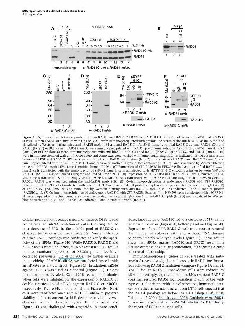
cellular proliferation because natural or induced DSBs would
not be repaired. siRNA inhibition of RAD51C during 24 h led
to a decrease of 80% in the soluble pool of RAD51C as
observed by Western blotting (Figure 3A). Western blotting
of other RAD51 paralogs was conducted to verify the speci-
ficity of the siRNA (Figure 3B). While RAD51B, RAD51D and
XRCC2 levels were unaffected, siRNA against RAD51C results
in a concomitant repression of XRCC3 protein levels as
described previously (Lio et al, 2004). To further evaluate
the specificity of RAD51C siRNA, we transfected the cells with
an siRNA-resistant construct (Figure 3C). In addition, siRNA
against XRCC3 was used as a control (Figure 3D). Colony
formation assays revealed a 92 and 90% reduction of colonies
when cells were inhibited for the expression of RAD51C by
double transfection of siRNA against RAD51C or XRCC3,
respectively (Figure 3E, middle panel and Figure 3F). Next,
cells were transfected once with RAD51C siRNA to preserve
viability before treatment (a 46% decrease in viability was
observed without damage; Figure 3E, top panel and
Figure 3F) and challenged with etoposide. In these condi-
tions, knockdown of RAD51C led to a decrease of 75% in the
number of colonies (Figure 3E, bottom panel and Figure 3F).
Expression of an siRNA RAD51C-resistant construct restored
the number of colonies with and without DNA damage
to approximately wild-type levels (Figure 3F). These results
show that siRNA against RAD51C and XRCC3 result in a
similar decrease of cellular proliferation, highlighting a close
functional relationship.
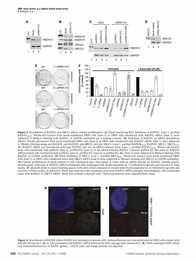
Immunofluorescence studies in cells treated with mito-
mycin C revealed a significant decrease in RAD51 foci forma-
tion following RAD51C inhibition (compare Figure 4A and B).
RAD51 foci in RAD51C knockdown cells were reduced by
38%. Interestingly, expression of the siRNA-resistant RAD51C
construct restored RAD51 foci formation to 91% of the wild-
type cells. Consistent with this observation, immunofluores-
cence studies in hamster and chicken DT40 cells suggest that
the RAD51 paralogs act before RAD51 (Bishop et al, 1998;
Takata et al, 2001; French et al, 2002; Godthelp et al, 2002).
These results establish a pre-RAD51 role for RAD51C during
the repair of DSBs in human cells.
1 2 3
51 pEC
FP
pEC
FP
-51
250 -
98 -64 -50 -
36 -
30 -
Mw
(kD
a)
α -RAD51 mAb
DC
51C
1 2 3
250 -
98 -64 -50 -
36 -
30 -
pEY
FP
pEY
FP
-51C
Mw
(kD
a)
α -RAD51C mAb
- YFP-51C
E
51C
2 3
IgG
Mw
(kD
a)
64 -
50 -
α -RAD51C mAb
- RAD51C- IgG
α 5
1
pEYFP51C
1
α -RAD51 mAb
- RAD51
Mw
(kD
a)
α -RAD51 mAb
F
- RAD51
- CFP-51- IgG
51 IgG
α 5
1C
pECFP51
64 -50 -
36 -2 31
α -RAD51C mAb
- RAD51C
51 +
51C
Immunoblotting: α -RAD51C and α-RAD51 mAbs
- RAD51C-His
- RAD51
1 2 3 4 65 7 8 9 10 11 13 14C
X3
+ 5
1
BC
DX
2 +
51
51 CX
3
BC
DX
2
BCDX2 + 51CX3 + 51
PI 51 α -RAD51 pAb
NaCl (M)0.1 10.25 0.5 0.1 10.25 0.5
A
12
51 51 +
51C
51 p
ur
B
- RAD51
1 2 3
IP 51C
-
-
Figure 1 (A) Interactions between purified human RAD51 and RAD51C-XRCC3 or RAD51B-C-D-XRCC2 and between RAD51 and RAD51Cin vivo. Human RAD51, or a mixture with CX3 or BCX2, were immunoprecipitated with preimmune serum or the anti-hRAD51 as indicated, andvisualized by Western blotting using anti-hRAD51 mAb 14B4 and anti-RAD51C mAb 2H11. Lane 1, purified RAD51Chis10 and RAD51. CX3 andRAD51 (lane 2) or BCDX2 and RAD51 (lane 3) were immunoprecipitated with RAD51 preimmune antibody. As controls, RAD51 (lane 4), CX3(lane 5) or BCDX2 (lane 6) were immunoprecipitated with anti-hRAD51 pAb. CX3 and RAD51 (lanes 7–10) or BCDX2 and RAD51 (lanes 11–14)were immunoprecipitated with anti-hRAD51 pAb and complexes were washed with buffer containing NaCl, as indicated. (B) Direct interactionbetween RAD51 and RAD51C. SF9 cells were infected with RAD51 baculovirus (lane 2) or a mixture of RAD51 and RAD51C (lane 3) andimmunoprecipitated with the anti-hRAD51C. Complexes were washed in lysis buffer containing 1 M NaCl and visualized by Western blottingusing anti-hRAD51 mAb 14B4. Lane 1, purified human RAD51. (C) Expression of YFP-RAD51C in HEK293 cells. Lane 1, purified RAD51Chis10;lane 2, cells transfected with the empty vector pEYFP-N1; lane 3, cells transfected with pEYFP-N1-51C encoding a fusion between YFP andRAD51C. RAD51C was visualized using the anti-RAD51C mAb 2H11. (D) Expression of CFP-RAD51 in HEK293 cells. Lane 1, purified RAD51;lane 2, cells transfected with the empty vector pECFP-N1; lane 3, cells transfected with pECFP-N1-51 encoding a fusion between CFP andRAD51. RAD51 was visualized using the anti-RAD51 mAb 14B4. (E) Co-immunoprecipitation of endogenous RAD51 with YFP-RAD51C.Extracts from HEK293 cells transfected with pEYFP-N1-51C were prepared and protein complexes were precipitated using control IgG (lane 2)or anti-RAD51 pAb (lane 3), and visualized by Western blotting with anti-RAD51C and RAD51, as indicated. Lane 1: marker protein(RAD51Chis10). (F) Co-immunoprecipitation of endogenous RAD51C with CFP-RAD51. Extracts from HEK293 cells transfected with pECFP-N1-51 were prepared and protein complexes were precipitated using control IgG (lane 2) or anti-RAD51 pAb (lane 3) and visualized by Westernblotting with anti-RAD51 and RAD51C, as indicated. Lane 1: marker protein (RAD51).
DNA repair factors at a defined double-strand breakA Rodrigue et al
The EMBO Journal VOL 25 | NO 1 | 2006 &2006 European Molecular Biology Organization224

Localization of endogenous repair proteins
to a single–double strand break in vivo
Most studies use DNA-damaging agents to study foci forma-
tion. However, this causes a difficulty when it is time to look
at a single repair event within the nucleus since DNA damage
is caused randomly. In order to study DNA repair at the
resolution of a single lesion, we used the DR95 cell line,
which bears a modified GFP gene in which an I-SceI restric-
tion site has been engineered. As far as is known, the I-SceI
restriction enzyme does not cut elsewhere in the genome and
therefore is specific for the modified GFP. In this way, a
unique DSB can be created in a known nucleotide sequence.
Following transfection of DR95 cells with pCBASce (a plas-
mid encoding I-SceI), DSB induction was efficient as most of
the cells were cleaved in DR-GFP 4 h after transfection as
judged by LM–PCR (data not shown). Following transfection
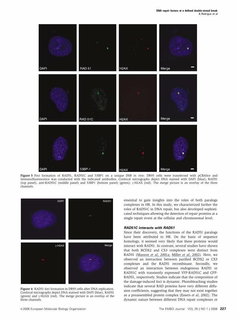
with pCBASce, immunofluorescence studies allowed the
visualization of a single focus formation of RAD51C,
RAD51, and 53BP1, which all colocalized with g-H2AX
(Figure 5). The single focus was not apparent in the absence
of I-SceI expression. A direct role for RAD51C in repairing
DNA is therefore established. Since cells undergo cycles
of I-SceI cleavage and repair, unique DSBs were also present
24 h after transfection. We also synchronized cells in G2
and performed the same analysis. Double RAD51 foci repre-
senting newly replicated sister chromatids that have been
cut by I-SceI were observed (Figure 6).
It is not clear whether RAD51C binds very close to DSBs
in vivo. RAD51C is part of the HJ resolvasome that could
migrate junctions far from the break site (Liu et al, 2004). One
attractive strategy is to examine the assembly of RAD51C on a
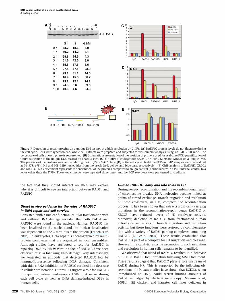
DSB using ChIPs. First, we looked whether the levels of
RAD51C changed during the cell cycle after cell synchroniza-
tion. RAD51C levels did not change during the cell cycle
(Figure 7A). Early after transfection with pCBASce, cells were
fixed with formaldehyde and the chromatin was solubilized
by sonication and purified. Immunoprecipitations were con-
ducted with antibodies raised against RAD51, RAD51C, Ku80
or MRE11. Control experiments revealed that RAD51 anti-
bodies immunoprecipitated endogenous RAD51 (Figure 1E)
and RAD51C antibodies pulled down CX3 and BCDX2 para-
logs complexes (Masson et al, 2001a, b). Similarly, Ku80 and
MRE11-RAD50-NBS1 were pulled down with the correspond-
ing antibodies (data not shown). After reversal of the formal-
dehyde crosslinks, DNA samples were deproteinized. DNA
was isolated and amplified by real-time PCR with primer
pairs specific to regions of interest near the DSB created by
I-SceI (Figure 7B). All reactions were normalized against a
control primer pairs for sequences near the RAD51 gene or
the AFP locus, which allowed us to control for DSB-indepen-
dent effects on protein occupancy. This highly informative
approach allowed the detection of repair proteins on the DSB
(Figure 7C–E). We monitored events in cells arrested at the
G1 and S–G2 phases of the cell cycle. Control experiments
revealed that I-SceI cleavage was equivalent in the G1 and
S–G2 phase based on g-H2AX foci formation. When cells
were synchronized so I-SceI would cut in the G1 phase of the
cell cycle (73.2% of the cells, as determined by flow cyto-
metry), an enrichment in Ku80 (10-fold) was observed at
94–378 bp from the break and decreased further from the
break (3.3-fold at 675–1044 bp and 1.4-fold at 901–1210 bp)
(Figure 7C). MRE11 and RAD51C were not detected, but
RAD51 was found to be present at the DSB, but this may
account for the S–G2 cells present in this sample (24.6%).
When cells were synchronized for DSB formation in S–G2
(70.2% of the cells, as determined by flow cytometry), Ku80
was not detected, whereas the levels of RAD51 (16-fold),
RAD51C (8.5-fold) and MRE11 (13-fold) were vastly increased
very close to the break (94–378 bp) and decreased away from
the break (RAD51 (10.2-fold), RAD51C (7.5-fold), MRE11
(6.1-fold), all at 675–1044 bp from the break) (Figure 7D).
Given that RAD51C is part of BCDX2 and CX3 complexes, we
performed ChIP analysis of RAD51D, XRCC2 and XRCC3 in
S–G2 cells to distinguish whether BCDX2 or CX3 were bound
to DSBs in vivo. Control experiments revealed that all anti-
bodies were able to pull-down paralog complexes. As expec-
ted, the levels of enrichment were lower than those obtained
for RAD51C, since the enrichment of RAD51C on the unique
DSB may represent the sum of each paralog complexes.
RAD51D, XRCC2 and XRCC3 were present at 94–378 bp (1.62-
, 1.56- and 1.65-fold, respectively). The levels of RAD51D and
XRCC2 increased at 675–1044bp (2.58- and 2.2-fold), whereas
XRCC3 was stable (1.65-fold). Surprisingly, both RAD51D
and XRCC2 were present at 901–1210 bp (2.2- and 2.17-fold).
Discussion
Despite the fact that the current model of HR was proposed
about 20 years ago, its biochemical complexity is not fully
understood in mitotic cells. Since RAD51C is at the crossroads
of at least two stable complexes of the RAD51 paralogs
(a dimeric complex of RAD51C-XRCC3 and a larger complex
composed of RAD51B-C-D-XRCC2), its characterization is
A
- γ -H2AX
CY
T
CY
T
NU
C
NU
C
Etoposide
- RAD51C
- RAD51
- GAPDH
Cγ -H2AX MergeRAD51C
GFP-RAD51C
Hoescht
B
Figure 2 RAD51C is a nuclear protein and forms foci followingDNA damage. (A) Cell-free extracts were prepared from mock-treated DR95 cells or DR95 cells treated with etoposide and fractio-nated into cytosolic (Cyt) and nuclear (Nuc) fractions. Thesefractions (25 mg) were analyzed for the presence of RAD51C.RAD51, g-H2AX and GAPDH were used as controls for the cyto-plasmic and nuclear fraction by Western blotting. (B) Localizationof GFP-RAD51C in DR95 cells. (C) RAD51C foci formation followingetoposide treatment (50 mM, 1 h). Immunofluorescence of RAD51C,g-H2AX and merge pictures are depicted.
DNA repair factors at a defined double-strand breakA Rodrigue et al
&2006 European Molecular Biology Organization The EMBO Journal VOL 25 | NO 1 | 2006 225
A
- RAD51C
51C
Moc
k
siR
NA
51C
- GAPDH
E WT
Eto
posi
de
1× siRNA 51C
Unt
reat
edU
ntre
ated
51C
Mock siRNA 51C
- RAD51C
pcD
NA
3
pcD
NA
-51C
pcD
NA
-51C
-Res
pcD
NA
3
pcD
NA
-51C
pcD
NA
-51C
-Res
C
- RAD51B- RAD51D
- XRCC2
- XRCC3
CT
L
Moc
k
siR
NA
51C
B
- GAPDH
- XRCC3
XR
CC
3
Moc
k tr
eate
d
siR
NA
XR
CC
3
- GAPDH
D
1 2 3
4 65 7
1 2 3
1 2 3
1 2 3
F
0
20
40
60
80
100
120
140
160
Con
trol
1× s
iRN
A2×
siR
NA
pcD
NA
3pc
DN
A3-
51C
pcD
NA
3-51
C-R
espc
DN
A3
pcD
NA
-51C
pcD
NA
3-51
C-R
esX
RC
C3
Con
trol
1× s
iRN
Apc
DN
A3
pcD
NA
-51C
3pc
DN
A3-
51C
-Res
XR
CC
3
Co
lon
y-fo
rmin
g u
nit
s(%
co
ntr
ol)
Untreated + Etoposide (50 µM)
1× siRNA 51C
2× siRNA 51CWT
WT
Figure 3 Knockdown of RAD51C and XRCC3 affects cellular proliferation. (A) Small interfering RNA inhibition of RAD51C. Lane 1, purifiedRAD51Chis10. Whole-cell extracts from mock-transfected DR95 cells (lane 2) or DR95 cells transfected with RAD51C siRNA (lane 3) weresubjected to Western blotting with RAD51C or GAPDH antibodies (as a loading control). (B) Inhibition of RAD51C by siRNA destabilizesXRCC3. Whole-cell extracts from mock-transfected DR95 cells (lane 2) or DR95 cells transfected with RAD51C siRNA (lane 3) were subjectedto Western blotting using anti-RAD51B, anti-RAD51D, anti-XRCC2 and anti-XRCC3. Lane 1, purified RAD51Bhis10, RAD51D, XRCC2, XRCC3his6.(C) RAD51C siRNA can knockdown wild-type RAD51C but not an siRNA-resistant form. Lane 1, purified RAD51Chis10. Whole-cell extractsfrom cells transfected with pcDNA3 (lane 2), pcDNA-51C (lane 3) or the siRNA-resistant RAD51C construct pcDNA-51C-Res (lane 4), RAD51CsiRNA-treated cells transfected with pcDNA3 (lane 5), pcDNA-51C (lane 6) or pcDNA-51C-Res (lane 7) were subjected to Western blotting withRAD51C or GAPDH antibodies. (D) RNAi inhibition of XRCC3. Lane 1, purified XRCC3his6. Whole-cell extracts from mock-transfected DR95cells (lane 2) or DR95 cells transfected twice with XRCC3 siRNA (lane 3) were subjected to Western blotting with XRCC3 or GAPDH antibodies.(E) Cellular proliferation of mock-treated or cells transfected once (top panel) or twice with an siRNA specific for RAD51C (middle panel).Bottom panel: wild-type or RAD51C siRNA-transfected cells challenged with 50mM etoposide for 1 h followed by growth and recovery in freshmedia. (F) Quantification of colony-forming assays. Cells were either untreated or treated with 50mM etoposide for 1 h followed by growth andrecovery in fresh media, as indicated. White bars indicate cells transfected once with RAD51C siRNA and gray bars designate cells transfectedtwice with RAD51C or XRCC3 siRNA. Black bars indicate wild-type cells. These experiments were repeated three times.
DAPI
γ -H2AX
RAD51
Merge
A B
siR
NA
RA
D51
C +
MM
C
MM
C
DAPI
γ -H2AX
RAD51
Merge
Figure 4 Knockdown of RAD51C affects RAD51 foci formation in human cells. Immunofluorescence was performed on DR95 cells treated with600 nM mitomycin C (A) or cells pretreated with RAD51C siRNA followed by DNA damage with mitomycin C (B). DNA staining by DAPI (blue)and immunofluorescence of RAD51 (green), g-H2AX (red) and merge pictures are depicted.
DNA repair factors at a defined double-strand breakA Rodrigue et al
The EMBO Journal VOL 25 | NO 1 | 2006 &2006 European Molecular Biology Organization226
essential to gain insights into the roles of both paralogs
complexes in HR. In this study, we characterized further the
roles of RAD51C in DNA repair, but also developed sophisti-
cated techniques allowing the detection of repair proteins at a
single repair event at the cellular and chromosomal level.
RAD51C interacts with RAD51
Since their discovery, the functions of the RAD51 paralogs
have been attributed to HR. On the basis of sequence
homology, it seemed very likely that these proteins would
interact with RAD51. In contrast, several studies have shown
that both BCDX2 and CX3 complexes were distinct from
RAD51 (Masson et al, 2001a; Miller et al, 2002). Here, we
observed an interaction between purified BCDX2 or CX3
complexes and the RAD51 recombinase. Secondly, we
observed an interaction between endogenous RAD51 or
RAD51C with transiently expressed YFP-RAD51C and CFP-
RAD51, respectively. Studies indicate that the composition of
the damage-induced foci is dynamic. Photobleaching studies
indicate that several RAD proteins have very different diffu-
sion coefficients, suggesting that they may not exist together
as a preassembled protein complex (Essers et al, 2002). The
dynamic nature between different DNA repair complexes or
Figure 5 Foci formation of RAD51, RAD51C and 53BP1 on a unique DSB in vivo. DR95 cells were transfected with pCBASce andimmunofluorescence was conducted with the indicated antibodies. Confocal micrographs depict DNA stained with DAPI (blue); RAD51(top panel), anti-RAD51C (middle panel) and 53BP1 (bottom panel) (green); g-H2AX (red). The merge picture is an overlay of the threechannels.
DAPI
γ -H2AX
RAD51
Merge
Figure 6 RAD51 foci formation in DR95 cells after DNA replication.Confocal micrographs depict DNA stained with DAPI (blue), RAD51(green) and g-H2AX (red). The merge picture is an overlay of thethree channels.
DNA repair factors at a defined double-strand breakA Rodrigue et al
&2006 European Molecular Biology Organization The EMBO Journal VOL 25 | NO 1 | 2006 227
the fact that they should interact on DNA may explain
why it is difficult to see an interaction between RAD51 and
RAD51C.
Direct in vivo evidence for the roles of RAD51C
in DNA repair and cell survival
Consistent with a nuclear function, cellular fractionation with
and without DNA damage revealed that both RAD51 and
RAD51C were found in the nucleus. Hamster RAD51C has
been localized to the nucleus and the nuclear localization
was dependent on the C-terminus of the protein (French et al,
2003). In eukaryotes, DNA repair is choreographed by multi-
protein complexes that are organized in focal assemblies.
Although studies have attributed a role for RAD51C in
repairing DNA by HR in vitro, no foci of RAD51C have been
observed in vivo following DNA damage. Very interestingly,
we generated an antibody that detected RAD51C foci by
immunofluorescence following DNA damage. Consistent
with this, siRNA inhibition of RAD51C resulted in a decrease
in cellular proliferation. Our results suggest a role for RAD51C
in repairing natural endogenous DSBs that occur during
each cell cycle as well as DNA damage-induced DSBs in
human cells.
Human RAD51C: early and late roles in HR
During genetic recombination and the recombinational repair
of chromosome breaks, DNA molecules become linked at
points of strand exchange. Branch migration and resolution
of these crossovers, or HJs, complete the recombination
process. It has been shown that extracts from cells carrying
mutations in the recombination/repair genes RAD51C or
XRCC3 have reduced levels of HJ resolvase activity.
Moreover, depletion of RAD51C from fractionated human
extracts caused a loss of branch migration and resolution
activity, but these functions were restored by complementa-
tion with a variety of RAD51 paralog complexes containing
RAD51C (Liu et al, 2004). These results established that
RAD51C is part of a complex for HJ migration and cleavage.
However, the catalytic enzyme promoting branch migration
and resolution in human cells remains to be identified.
We observed that RNAi of RAD51C resulted in a decrease
of 38% in RAD51 foci formation following MMC treatment.
These results suggest that RAD51C plays a role upstream of
RAD51 during HR. This is supported by the following ob-
servations: (i) in vitro studies have shown that BCDX2, when
immobilized on DNA, could recruit limiting amounts of
RAD51 as judged by electron microscopy (Masson et al,
2001b); (ii) chicken and hamster cell lines deficient in
Asy
n
0 h
1 h
2 h
3 h
4 h
5 h
6 h
7 h
8 h
9 h
10 h
-RAD51C
0 h1 h
2 h3 h4 h5 h
6 h7 h8 h9 h10 h
G1 S G2/M
73.2 18.6 6.079.2
68.851.835.627.523.116.913.224.340.6
14.2
24.642.657.547.131.115.612.15.64.6
4.1
4.33.85.623.944.566.774.269.654.3
A C
0123456789
10
IgG RAD51 RAD51C KU80 MRE11
0
2
4
6
8
10
12
14
16
18
IgG RAD51 RAD51C KU80 MRE11
0
0.5
1
1.5
2
2.5
3
IgG RAD51D XRCC2 XRCC3
DSB
901–1210 675–1044 94–378
B
G1
S-G2
S-G2
D
E
94–378675–10441210–901
Figure 7 Detection of repair proteins on a unique DSB in vivo at a high resolution by ChIPs. (A) RAD51C protein levels do not fluctuate duringthe cell cycle. Cells were synchronized, whole-cell extracts were prepared and subjected to Western blot analysis using RAD51C 2H11 mAb. Thepercentage of cells at each phase is represented. (B) Schematic representation of the position of primers used for real-time PCR quantification ofChIPs respective to the unique DSB created by I-SceI in vivo. (C–E) ChIPs of endogenous RAD51, RAD51C, Ku80 and MRE11 on a unique DSB.The presence of the proteins was verified during the G1 (C) or S–G2 phase (D) of the cell cycle. Real-time PCR on ChIP samples were carried outat 94–378, 675–1044 and 901–1210 nucleotides from the break (red, yellow and blue bars, respectively). (E) ChIP analysis of RAD51D, XRCC2and XRCC3. Fold-enrichment represents the enrichment of the proteins compared to an IgG control (normalized with a PCR internal control to alocus other than the DSB). These experiments were repeated three times and the PCR reactions were performed in triplicate.
DNA repair factors at a defined double-strand breakA Rodrigue et al
The EMBO Journal VOL 25 | NO 1 | 2006 &2006 European Molecular Biology Organization228
RAD51C also showed impaired Rad51 foci formation in
response to DNA damage (Takata et al, 2001; French et al,
2002; Godthelp et al, 2002). Hence, our current study pro-
poses that RAD51C is a mediator at early stages of HR, most
likely during the invasion step of HR.
Although BCDX2 and CX3 complexes have been associated
to different stages of HR, it is not totally clear which paralog
complex contribute to early and late steps of the HR pathway.
XRCC3 is recruited early to DSBs (Forget et al, 2004), but
defective processing of recombination intermediates is
observed in both hamster and Arabidopsis XRCC3�/� cells
during the later stages of HR (Brenneman et al, 2002;
Bleuyard and White, 2004). Certainly, CX3 and BCDX2 com-
plexes may have overlapping and different functions since
deletion of RAD51D and XRCC3 in chicken DT40 cells lead to
an additive phenotype (Yonetani et al, 2005). During our ChIP
experiment in S–G2 phase of the cell cycle, we observed that
the levels of RAD51C, unlike those of RAD51 and MRE11, did
not decrease from 94 to 1044 bp. Since the polyclonal anti-
body used is able to pull down both CX3 and BCDX2
complexes, this observation raises the possibility that we
are looking at different paralog complexes depending on the
distance from the I-SceI cut: a RAD51-recruiting complex at
94–378 bp and a HJ migration/resolution complex at 675–
1210 bp. Our data provide new insights into this, as RAD51D,
XRCC2 and XRCC3 levels were similar at 94–378 bp, while
RAD51D and XRCC2 levels increased at 675–1210 bp. This
might suggest that BCDX2 and CX3 are both involved in
RAD51 loading close to the break and RAD51D-XRCC2 might
contribute to branch migration/resolution. Therefore, this
complex is found further away from the break. However, it
should be noticed that ChIPs rely on antibodies. The absence
of RAD51C at 901–1210 bp could mean that the epitopes
recognized by the antibody are masked by another protein.
This could also explain why MRE11 is not found in G1,
although a role in NHEJ was attributed for this protein
(Paull and Gellert, 2000).
Recombination protein foci mark the site of DSB repair
We report here the first molecular-level characterization of
regional DNA repair factors binding a unique DSB in human
cells. To date, these events have been defined primarily at the
resolution of light microscopes. Following I-SceI cleavage,
a single focus of RAD51, RAD51C and 53BP1 was observed
in the nucleus of DR95 cells. All signalling and repair protein
foci colocalized with g-H2AX. 53BP1 and NBS1 appear to
directly bind the phosphorylated SQE motif of H2AX, sug-
gesting that it may act as a scaffold to anchor or facilitate the
assembly of signalling and repair proteins (Kobayashi et al,
2002; Ward et al, 2003). One concern about this system is that
we can also detect endogenous DNA damage rather than the
I-SceI cut. We observed G2 cells with two closely positioned
foci of RAD51 representing the cleavage of both sister chro-
matids. The frequency of cells having these spots argues that
it is very unlikely that this represents random DNA damage in
the nucleus. In agreement with this, two foci of Rad51 has
also been observed at HO endonuclease-induced DSB in the
MAT locus of Saccharomyces cerevisiae (Miyazaki et al,
2004). Our results also suggest that resection of the DSB
goes as far as 1 kb in human cells since RAD51 was not
present at 901–1210 bp from the break.
We took advantage of the DR95 cell line where a unique
DSB can be produced and performed ChIPs on endogenous
DNA repair proteins. In yeast, temporal analysis of the
recruitment of budding yeast proteins on an HO-induced
DSB revealed that Rad51 binds first, followed by Rad52,
Rad55 and finally Rad54. Consistent with biochemical studies
indicating a requirement for Rad55 in mediating Rad51 fila-
ment formation, inactivation of RAD55 lead to a decrease
in the recruitment of Rad51 (Wolner et al, 2003). Certainly,
our system now allows the possibility to study whether this
order of recruitment is conserved in human cells.
Very elegantly, using chicken DT40 cell lines deficient in
Ku70�/� and Rad54�/� cells and synchronised cells, the
Takeda group has found that NHEJ pathway plays a dominant
role in repairing g-radiation-induced DSBs during G1–early S
phase, while recombinational repair is preferentially used in
late S–G2 phase of the cell cycle (Takata et al, 2000). We
extend this study and show directly that Ku80 is bound
to a unique DSB specifically in the G1 phase of the cell
cycle, while proteins representative of the HR pathway
(such as RAD51, RAD51C, RAD51D, XRCC2, XRCC3 and
MRE11) are bound during the S–G2 phase of the cell cycle.
Interestingly, by correlating our immunofluorescence data
with our high-resolution ChIP analysis, these results clearly
define that a focus represents a mark of repair of a DSB
in human cells.
Materials and methods
Cell culture and plasmidsThe DR95hyg-xt cell line was donated by Dr Maria Jasin (Pierceet al, 2001). HEK293T cells are derived from human embryonickidney cells and express Ad5 E1A, E1B proteins and large T antigen(a gift from Dr Josee Lavoie) and SKN-SH are neuroblastoma cells.DR95hyg-xt, HEK293Tand SKN-SH cells were maintained in DMEMsupplemented with 10% fetal bovine serum and 1% penicillin/streptomycin. The human RAD51C and RAD51 genes were insertedin pEYFP-N1 and pECFP-N1 (BD Biosciences) to generate pEYFP-RAD51C and pECFP-RAD51, respectively. pCBASce is an I-SceIexpression vector (Pierce et al, 2001). The siRNA-resistant RAD51Cplasmid was constructed using site-directed mutagenesis ofpcDNA3-RAD51C using the oligonucleotide 50-ATAATCACCTTCTGCTCGGCGCTAGATGATATTCTT.
Cell fractionation, synchronization and colony-forming assayDR95 cells (1�106) were mock treated or treated 2 h with 50mMetoposide. Cells were harvested, washed once with PBS1X,centrifuged and resuspended in 400ml of Schaffner lysis buffer(10 mM HEPES-NaOH, pH 7.9, 10 mM KCl, 0.1 mM EDTA, 0.1 mMEGTA, 1 mM DTT, 0.5 mM PMSF, aprotinin (0.019 TIU/ml) andleupeptin (3 mg/ml)) and allowed to swell on ice for 15 min. NP-40was added to a final concentration of 0.55%, cells were vortexedvigorously for 10 s and centrifuged for 30 s at 13 000 r.p.m. Thesupernatant was kept as the cytoplasmic fraction while the nuclearproteins were extracted from the pellet by incubation in nuclearextraction buffer (20 mM HEPES-NaOH, pH 7.9, 400 mM KCl, 1 mMEDTA, 1 mM EGTA, 1 mM DTT, 0.5 mM PMSF, aprotinin (0.019 TIU/ml) and leupeptin (3 mg/ml)) on a rotary shaker for 15 min at 41C.The nuclei were centrifuged for 5 min and the supernatant wascollected as the nuclear extract.
Cells were synchronized according to Zhu et al (2000) withminor modifications. DR95 cells were treated with 2 mM thymidinefor 14 h. The cells were washed with PBS1X and released in freshmedia for 11 h. Subsequently, cells were treated with aphydicolin at1 mg/ml for 14 h. Cells were then released in fresh media.
Cellular viability of DR95 cells or DR95 cells transfected withRAD51C or XRCC3 siRNA was determined by colony formation.When required, pcDNA3, pcDNA-51C or pcDNA-51C-Res were
DNA repair factors at a defined double-strand breakA Rodrigue et al
&2006 European Molecular Biology Organization The EMBO Journal VOL 25 | NO 1 | 2006 229
electroporated 24 h prior to siRNA transfection. A total of 1–5�103
mock- and siRNA-transfected cells released by trypsinization wereplated onto 100 mm dishes. After 12–16 days, colonies were fixedwith methanol and stained using methylene blue (4 g/l inmethanol) and counted. Cells were treated with 50mM of etoposidefor 1 h, allowed to recover for 1 h in fresh media and their colony-forming ability was monitored as above.
RNA interferenceRNAi-mediated knockdown of RAD51C was conducted using ansiRNA (Qiagen) with CACCTTCTGTTCAGCACTAGA as a targetsequence. Knockdown of XRCC3 was performed using two siRNAagainst CAGAATTATTGCTGCAATTAA and CAGCCAGATCTTCATCGAGCA. siRNA transfection was performed using RNAiFect (Qiagen).In brief, cells were seeded in six-well plates at 2�105 cells/cm2 24 hprior to transfection. For each transfection, 5 mg of siRNA wasdiluted in culture medium (containing serum and antibiotics) to afinal volume of 100 ml and incubated 15 min at room temperaturewith 15ml of RNAiFect. The RNAiFect-siRNA complex was thenadded dropwise to the 75% confluent cells and incubated at 371Cfor 24 h. When required, cells were retransfected the next day foranother 24 h. As a control, cells were mock transfected withRNAiFect alone.
Western blot analysisRAD51C expression was assayed by Western blotting 24 h followingsiRNA transfection. A total of 5�105 cells were harvested, washedwith PBS1X and lysed in 100ml IP buffer (50 mM Tris–Cl, pH 7.5,0.5 M NaCl, 0.5% NP-40) containing 1 mM PMSF, aprotinin(0.019 TIU/ml) and leupeptin (3mg/ml). After 30 min of incubationon ice, cells were sonicated two times for 5 s with a Branson sonifierat 30% burst and centrifuged 10 min at 132 000 r.p.m. at 41C.Cell lysate (25mg) was boiled and subjected to SDS–PAGE electro-phoresis. After transfer to nylon membrane, the membraneswere blocked overnight in PBS1X–0.05% Tween containing 5%skim milk (for RAD51, RAD51C or GAPDH antibodies) or PBS1X–0.05% Tween containing 5% BSA (for g-H2AX mAb). Primaryantibodies RAD51 (Genetex), RAD51C (mAb 2H11), GAPDH(Research diagnostics) or g-H2AX (Upstate cell signaling solutions)were added followed by horseradish peroxidase-conjugatedanti-mouse immunoglobulin and revealed using enhanced chemo-luminescence (Perkin-Elmer).
ImmunoprecipitationPurified human RAD51 (1mg) or CX3 or BCDX2 (1mg) wereincubated in binding buffer (50 mM Tris–Cl, pH 7.5, 100 mM NaCl,0.5% NP-40, 0.5% BSA) for 30 min at 371C. 20ml of preimmuneserum or RAD51 pAb coupled to Aminolink beads (Pierce) in 500mlof buffer were added to the reaction. Protein complexes were pulleddown for 1 h at 41C. Immunoprecipitates were washed with bindingbuffer containing NaCl, as indicated.
Immunoprecipitations from transfected HEK293T cells or bacu-lovirus-infected cells were performed as follows. HEK293T cellswere collected 48 h after transfection with either pEYFP-RAD51C orpECFP-RAD51 and resuspended in lysis buffer (50 mM Tris–HCl, pH7.5, 0.5 M NaCl, 0.5% NP-40) containing the protease inhibitorsPMSF (1 mM), aprotinin (0.019 TIU/ml) and leupeptin (1mg/ml),incubated for 30 min on ice and then lysed by sonication. Insolublematerial was removed by high-speed centrifugation. Proteincomplexes in the supernatant (equivalent to approximately1.5�107 cells) were pulled down for 1.5 h at 41C using preimmuneserum or pAbs raised against RAD51 crosslinked to Aminolinkbeads (Pierce). Complexes were washed four times in lysis buffercontaining salt as indicated, and visualized by Western blottingusing RAD51 14B4 or RAD51C 2H11 mAb.
ImmunofluorescenceTransiently transfected cells grown on coverslips were fixed 18–20 hpost-transfection with 4.0% paraformaldehyde in PBS1X for 5 minat room temperature. Next, cells were permeabilized with PBS1Xcontaining 0.5% Triton X-100 for 5 min and washed twice withPBS1X. Cells were then incubated in primary antibody at theappropriate dilution and incubated at room temperature for 30 min.Coverslips were rinsed with PBS1X containing 0.1% Triton X-100and washed twice with PBS1X prior to a 30-min incubation withan appropriate secondary antibody conjugated to a fluorophore.
Cells were rinsed again with PBS1X containing 0.1% Triton X-100and washed twice with PBS. Coverslips were mounted onto slideswith approximately 10ml of a 90% glycerol–PBS-based mediumcontaining 1 mg of paraphenylenediamine/ml and 0.5 mg of 40,60-diamidino-2-phenylindole (DAPI)/ml. RAD51C was visualizedwith a rabbit polyclonal antibody raised against the full-lengthprotein (JYM pAb20), RAD51 (JYM pAb1) was visualized witha rabbit polyclonal antibody and 53BP1 was obtained from NovusBiologicals. Secondary antibodies included both anti-mouseand anti-rabbit antibodies conjugated with either Alexa-Fluor 488,Alexa-Fluor 555 (Molecular Probes) or cyanin 3 (Jackson Immuno-Research Laboratories Inc.). Images were collected with a ZeissLaser Scanning Confocal Microscope or on a Leica DMIRE2inverted microscope.
ChIPs assaysInduction of a single DSB in DR95 cells was performed throughelectroporation of the I-SceI expression vector pCBASce using theGene Pulser Xcell apparatus (BioRad). For each electroporation, atotal of 2�106 cells suspended in 650 ml PBS were mixed with 50mgof circular plasmid and pulsed at 0.25 kV and 1000 mF in 4 mmcuvettes. Following electroporation, cells were plated onto 100 mmdishes containing fresh medium and returned to incubation at 371Cfor 4 h. Cells were treated with 1% formaldehyde for 8 min tocrosslink proteins to DNA. Glycine (0.125 M) was added to quenchthe reaction. Cells were collected using a cell scraper, washed twicein cold PBS1X, washed for 10 min in solution I (10 mM HEPES, pH7.5, 10 mM EDTA, 0.5 mM EGTA, 0.75% Triton X-100) and 10 min insolution II (10 mM HEPES, pH 7.5, 200 mM NaCl, 1 mM EDTA,0.5 mM EGTA). Cells were resuspended in lysis buffer (25 mMTris–HCl, pH 7.5, 150 mM NaCl, 1% Triton X-100, 0.1% SDS,0.5% deoxycholate) and sonicated for 6�10 s (30% output) toshear chromatin to an average size of 0.5 kb using a Fisher SonicDismembrator sonicator. Once centrifuged until clear, the lysatewas precleared overnight with Sepharose CL-6B. Immunoprecipita-tions were performed for 2 h in lysis buffer with polyclonalantibodies against RAD51C, RAD51D, XRCC2, XRCC3, RAD51,MRE11 and Ku80 proteins. Preimmune serum or rabbit anti-humanIgG (HþL) antibody (Jackson Immunoresearch Laboratories) wasused as a negative control. Complexes were washed twice with RIPAbuffer, once in high salt buffer (50 mM Tris–Cl, pH 8.0, 500 mMNaCl, 0.1% SDS, 0.5% deoxycholate, 1% NP-40, 1 mM EDTA), oncein LiCl buffer (50 mM Tris–Cl, pH 8.0, 250 mM LiCl, 1% NP-40,0.5% deoxycholate, 1 mM EDTA) and twice in TE buffer (10 mMTris–Cl, pH 8.0, 1 mM EDTA, pH 8.0). Beads were resuspended inTE containing 50mg/ml of RNase and incubated for 30 min. Beadswere washed with water and elution buffer (1% SDS, 0.1 MNaHCO3) was added for 15 min. Crosslinks were reversed byadding 200 mM NaCl followed by an incubation for 6 h at 651C.Samples were deproteinized overnight with proteinase K and DNAwas extracted with phenol–chloroform followed by ethanolprecipitation.
Real-time PCRQuantification of the amount of immunoprecipitated DNA wascarried out by real-time PCR using the LightCycler Fast Start DNAMaster SYBR Green I (Roche Applied Sciences), which containedFast Start Taq DNA polymerase and SYBR Green Dye. For eachPCR assay, 0.5 ml aliquots of the DNA sample were amplified intriplicate. The sequence of the primers can be obtained on request.Primers used in the PCR reactions were analyzed for linearity rangeand efficiency using a LightCycler (Roche). Values were calculatedas fold-enrichment compared to the IgG control versus a controllocus.
Acknowledgements
We are grateful to Jacques Cote for advice on chromatin immuno-precipitations and Stephane Richard and Jacques Cote for interest.We also thank Isabelle Brodeur, Rhea Utley and anonymousreviewers for helpful comments. AR is a recipient of a CIHRmaster’s award. JYM is a Canadian Institutes of Health ResearchNew Investigator and this research is supported by funds from theNational Cancer Institute of Canada.
DNA repair factors at a defined double-strand breakA Rodrigue et al
The EMBO Journal VOL 25 | NO 1 | 2006 &2006 European Molecular Biology Organization230
References
Bishop DK, Ear U, Bhattacharyya A, Calderone C, Beckett M,Weichselbaum RR, Shinohara A (1998) Xrcc3 is requiredfor assembly of Rad51 complexes in vivo. J Biol Chem 273:21482–21488
Bleuyard JY, Gallego ME, Savigny F, White CI (2005) Differingrequirements for the Arabidopsis Rad51 paralogs in meiosis andDNA repair. Plant J 41: 533–545
Bleuyard JY, White CI (2004) The Arabidopsis homologue of Xrcc3plays an essential role in meiosis. EMBO J 23: 439–449
Brenneman MA, Wagener BM, Miller CA, Allen C, Nickoloff JA(2002) XRCC3 controls the fidelity of homologous recombination:roles for XRCC3 in late stages of recombination. Mol Cell 10:387–395
Deans B, Griffin CS, Maconochie M, Thacker J (2000) Xrcc2 isrequired for genetic stability, embryonic neurogenesis and viabi-lity in mice. EMBO J 19: 6675–6685
Essers J, Houtsmuller AB, van Veelen L, Paulusma C, Nigg AL,Pastink A, Vermeulen W, Hoeijmakers JH, Kanaar R (2002)Nuclear dynamics of RAD52 group homologous recombinationproteins in response to DNA damage. EMBO J 21: 2030–2037
Forget AL, Bennett BT, Knight KL (2004) Xrcc3 is recruited to DNAdouble strand breaks early and independent of Rad51. J CellBiochem 93: 429–436
French CA, Masson JY, Griffin CS, O’Regan P, West SC, Thacker J(2002) Role of mammalian RAD51L2 (RAD51C) in recombinationand genetic stability. J Biol Chem 277: 19322–19330
French CA, Tambini CE, Thacker J (2003) Identification of func-tional domains in the RAD51L2 (RAD51C) protein and its require-ment for gene conversion. J Biol Chem 278: 45445–45450
Godthelp BC, Wiegant WW, van Duijn-Goedhart A, Scharer OD, vanBuul PP, Kanaar R, Zdzienicka MZ (2002) Mammalian Rad51Ccontributes to DNA cross-link resistance, sister chromatid cohe-sion and genomic stability. Nucleic Acids Res 30: 2172–2182
Kobayashi J, Tauchi H, Sakamoto S, Nakamura A, Morishima K,Matsuura S, Kobayashi T, Tamai K, Tanimoto K, Komatsu K(2002) NBS1 localizes to gamma-H2AX foci through interactionwith the FHA/BRCT domain. Curr Biol 12: 1846–1851
Kurumizaka H, Enomoto R, Nakada M, Eda K, Yokoyama S,Shibata T (2003) Region and amino acid residues required forRad51C binding in the human Xrcc3 protein. Nucleic Acids Res 31:4041–4050
Kurz EU, Lees-Miller SP (2004) DNA damage-induced activation ofATM and ATM-dependent signaling pathways. DNA Repair(Amsterdam) 3: 889–900
Li W, Yang X, Lin Z, Timofejeva L, Xiao R, Makaroff CA, Ma H(2005) The AtRAD51C gene is required for normal meioticchromosome synapsis and double-stranded break repair inArabidopsis. Plant Physiol 138: 965–976
Lio YC, Mazin AV, Kowalczykowski SC, Chen DJ (2003) Complexformation by the human Rad51B and Rad51C DNA repair proteinsand their activities in vitro. J Biol Chem 278: 2469–2478
Lio YC, Schild D, Brenneman MA, Redpath JL, Chen DJ (2004)Human Rad51C deficiency destabilizes XRCC3, impairs recombi-nation, and radiosensitizes S/G2-phase cells. J Biol Chem 279:42313–42320
Liu N, Schild D, Thelen MP, Thompson LH (2002) Involvement ofRad51C in two distinct protein complexes of Rad51 paralogs inhuman cells. Nucleic Acids Res 30: 1009–1015
Liu Y, Masson JY, Shah R, O’Regan P, West SC (2004) RAD51C isrequired for Holliday junction processing in mammalian cells.Science 303: 243–246
Masson J-Y, Stasiak AZ, Stasiak A, Benson FE, West SC (2001a)Complex formation by the human RAD51C and XRCC3 recombi-nation repair proteins. Proc Natl Acad Sci USA 98: 8440–8446
Masson J-Y, Tarsounas MC, Stasiak AZ, Stasiak A, Shah R,McIlwraith MJ, Benson FE, West SC (2001b) Identification andpurification of two distinct complexes containing the five RAD51paralogs. Genes Dev 15: 3296–3307
Miller KA, Sawicka D, Barsky D, Albala JS (2004) Domain mappingof the Rad51 paralog protein complexes. Nucleic Acids Res 32:169–178
Miller KA, Yoshikawa DM, McConnell IR, Clark R, Schild D, AlbalaJS (2002) RAD51C interacts with RAD51B and is central to a larger
protein complex in vivo exclusive of RAD51. J Biol Chem 277:8406–8411
Miyazaki T, Bressan DA, Shinohara M, Haber JE, Shinohara A(2004) In vivo assembly and disassembly of Rad51 and Rad52complexes during double-strand break repair. EMBO J 23:939–949
Paull TT, Gellert M (2000) A mechanistic basis for Mre11-directedDNA joining at microhomologies. Proc Natl Acad Sci USA 97:6409–6414
Pierce AJ, Hu P, Han MG, Ellis N, Jasin M (2001) Ku DNA end-binding protein modulates homologous repair of double-strandbreaks in mammalian cells. Genes Dev 15: 3237–3242
Pierce AJ, Johnson RD, Thompson LH, Jasin M (1999) XRCC3promotes homology-directed repair of DNA damage in mamma-lian cells. Genes Dev 13: 2633–2638
Pittman DL, Schimenti JC (2000) Mid-gestation lethality in micedeficient for the recA-related gene, RAD51D/RAD51L3. Genesis26: 167–173
Rogakou EP, Boon C, Redon C, Bonner WM (1999) Megabasechromatin domains involved in DNA double-strand breaksin vivo. J Cell Biol 146: 905–916
Schild D, Lio YC, Collins DW, Tsomondo T, Chen DJ (2000)Evidence for simultaneous protein interactions between humanRad51 paralogs. J Biol Chem 275: 16443–16449
Shu ZG, Smith S, Wang LJ, Rice MC, Kmiec EB (1999) Disruption ofmuREC2/RAD51L1 in mice results in early embryonic lethalitywhich can be partially rescued in a p53(�/�) background. MolCell Biol 19: 8686–8693
Sigurdsson S, Van Komen S, Bussen W, Schild D, Albala JS, Sung P(2001) Mediator function of the human Rad51B–Rad51C complexin Rad51/RPA-catalyzed DNA strand exchange. Genes Dev 15:3308–3318
Stiff T, O’Driscoll M, Rief N, Iwabuchi K, Lobrich M, Jeggo PA(2004) ATM and DNA-PK function redundantly to phosphorylateH2AX after exposure to ionizing radiation. Cancer Res 64:2390–2396
Takata M, Sasaki MS, Sonoda E, Fukushima T, Morrison C, AlbalaJS, Swagemakers SM, Kanaar R, Thompson LH, Takeda S (2000)The Rad51 paralog Rad51B promotes homologous recombina-tional repair. Mol Cell Biol 20: 6476–6482
Takata M, Sasaki MS, Tachiiri S, Fukushima T, Sonoda E, Schild D,Thompson LH, Takeda S (2001) Chromosome instability anddefective recombinational repair in knockout mutants of thefive Rad51 paralogs. Mol Cell Biol 21: 2858–2866
Thompson LH, Schild D (2001) Homologous recombinational repairof DNA ensures mammalian chromosome stability. Mutat Res477: 131–153
Ward IM, Chen JJ (2001) Histone H2AX is phosphorylated in anATR-dependent manner in response to replicational stress. J BiolChem 276: 47759–47762
Ward IM, Minn K, Jorda KG, Chen J (2003) Accumulationof checkpoint protein 53BP1 at DNA breaks involves itsbinding to phosphorylated histone H2AX. J Biol Chem 278:19579–19582
Wiese C, Collins DW, Albala JS, Thompson LH, Kronenberg A,Schild D (2002) Interactions involving the Rad51 paralogsRad51C and XRCC3 in human cells. Nucleic Acids Res 30:1001–1008
Wolner B, van Komen S, Sung P, Peterson CL (2003) Recruitment ofthe recombinational repair machinery to a DNA double-strandbreak in yeast. Mol Cell 12: 221–232
Yokoyama H, Sarai N, Kagawa W, Enomoto R, Shibata T,Kurumizaka H, Yokoyama S (2004) Preferential binding tobranched DNA strands and strand-annealing activity of thehuman Rad51B, Rad51C, Rad51D and Xrcc2 protein complex.Nucleic Acids Res 32: 2556–2565
Yonetani Y, Hochegger H, Sonoda E, Shinya S, Yoshikawa H, TakedaS, Yamazoe M (2005) Differential and collaborative actions ofRad51 paralog proteins in cellular response to DNA damage.Nucleic Acids Res 33: 4544–4552
Zhu XD, Kuster B, Mann M, Petrini JHJ, de Lange T (2000) Cell-cycle-regulated association of RAD50/MRE11/NBS1 with TRF2and human telomeres. Nat Genet 25: 347–352
DNA repair factors at a defined double-strand breakA Rodrigue et al
&2006 European Molecular Biology Organization The EMBO Journal VOL 25 | NO 1 | 2006 231