REV7 counteracts DNA double-strand break resection and affects PARP inhibition
17
LETTER doi:10.1038/nature14328 REV7 counteracts DNA double-strand break resection and affects PARP inhibition Guotai Xu 1 , J. Ross Chapman 2 *, Inger Brandsma 3 *, Jingsong Yuan 4 , Martin Mistrik 5 , Peter Bouwman 6 , Jirina Bartkova 7 , Ewa Gogola 1 , Danie ¨l Warmerdam 8 , Marco Barazas 1 , Janneke E. Jaspers 1 , Kenji Watanabe 7 , Mark Pieterse 6 , Ariena Kersbergen 1 , Wendy Sol 1 , Patrick H. N. Celie 9 , Philip C. Schouten 6 , Bram van den Broek 8 , Ahmed Salman 2 , Marja Nieuwland 10 , Iris de Rink 10 , Jorma de Ronde 11 , Kees Jalink 8 , Simon J. Boulton 12 , Junjie Chen 4 , Dik C. van Gent 3 , Jiri Bartek 5,7 , Jos Jonkers 6 , Piet Borst 1 & Sven Rottenberg 1,13 Error-free repair of DNA double-strand breaks (DSBs) is achieved by homologous recombination (HR), and BRCA1 is an important factor for this repair pathway 1 . In the absence of BRCA1-mediated HR, the administration of PARP inhibitors induces synthetic lethality of tumour cells of patients with breast or ovarian cancers 2,3 . Despite the benefit of this tailored therapy, drug resistance can occur by HR restoration 4 . Genetic reversion of BRCA1-inactivating mutations can be the underlying mechanism of drug resistance, but this does not explain resistance in all cases 5 . In particular, little is known about BRCA1-independent restoration of HR. Here we show that loss of REV7 (also known as MAD2L2) in mouse and human cell lines re-establishes CTIP-dependent end resection of DSBs in BRCA1- deficient cells, leading to HR restoration and PARP inhibitor resistance, which is reversed by ATM kinase inhibition. REV7 is recruited to DSBs in a manner dependent on the H2AX–MDC1– RNF8–RNF168–53BP1 chromatin pathway, and seems to block HR and promote end joining in addition to its regulatory role in DNA damage tolerance 6 . Finally, we establish that REV7 blocks DSB resection to promote non-homologous end-joining during immunoglobulin class switch recombination. Our results reveal an unexpected crucial function of REV7 downstream of 53BP1 in coordinating pathological DSB repair pathway choices in BRCA1- deficient cells. To identify mechanisms of BRCA1-independent restoration of the HR pathway, we carried out a loss-of-function short hairpin RNA (shRNA) screen using the KB1P-B11 and KB1P-G3 cell lines that we previously derived from Brca1 2/2 p53 2/2 (p53 is also known as Trp53) mouse mammary tumours 7 (Fig. 1a and Supplementary Table 1). Resistant cells were selected with a high concentration of olaparib (500 nM, about 100-fold the half-maximal inhibitory concentration (IC 50 )), which killed cells of the empty vector control. Sequencing of the olaparib-surviving colonies revealed a reproducible enrichment of various individual hairpins targeting Rev7 or 53bp1 (also known as Trp53bp1). To validate the Rev7 hit, we introduced two different hairpins into the KB1P-B11 and KB1P-G3 cell lines; these substantially inhibited Rev7 expression (Fig. 1b, c and Extended Data Fig. 1a). Despite the role of REV7 in metaphase-to-anaphase transition 8 , the level of Rev7 inhibition in these cells did not affect proliferation (Extended Data Fig. 1b, c), allowing long-term clonogenic survival assays. We confirmed that loss of Rev7 resulted in increased resistance to the PARP inhibitors (PARPi) ola- parib and AZD2461 (ref. 7) in both cell lines (Fig. 1d and Extended Data Fig. 1d–g). Resistant cells that survived olaparib treatment (Rev7 sh1/ 2-ola) yielded even lower REV7 expression levels and increased numbers of colonies after PARPi treatment (Fig. 1b–d and Extended Data Fig. 1h). When we reconstituted the Rev7-depleted cells with shRNA-resistant Rev7 complementary DNA resulting in similar REV7 protein levels (Extended Data Fig. 1i), we successfully re-sensitized the tumour cells to PARPi (Fig. 1e, f). Tumours derived from the Brca1 2/2 p53 2/2 cells with stable Rev7 inhibition also showed olaparib resistance in vivo, in contrast to the empty vector controls (Fig. 1g and Extended Data Fig. 1j–l). In addition, we found that Rev7 loss explains some cases of in vivo acquired PARPi resistance in BRCA1-deficient mouse mammary tumours (data not shown). REV7 depletion also resulted in PARPi resistance of the human BRCA1-deficient cell line SUM149PT (Extended Data Fig. 2). To- gether, these data strongly indicate that inhibition of Rev7 confers PARPi resistance in BRCA1-deficient tumour cells. Together with the catalytic subunit REV3, REV7 forms the transle- sion synthesis polymerase f (Polf), and it interacts with REV1 (ref. 9). We therefore investigated whether loss of REV1 or REV3 also confers PARPi resistance in Brca1 2/2 p53 2/2 cells. A 60% inhibition of Rev1 or Rev3 transcripts did not cause olaparib resistance (Extended Data Fig. 3a–d). Moreover, we studied various shRNA-resistant REV7 mu- tants that lack REV1 (Leu186Ala/Gln200Ala/Tyr202Ala and a 1–183- amino-acids truncated protein) or REV3 (Cys70Arg) binding sites 10,11 . In contrast to the truncated 1–140-amino-acid REV7 protein, these mutants are recruited to DNA damage sites (Extended Data Fig. 3e–g), and their expression in Rev7 shRNA KB1P-B11 and KB1P-G3 cells sig- nificantly restored the sensitivity to PARPi to a degree approaching that of wild-type REV7 (Fig. 2a, b; P , 0.001, t-test). The remaining differences of the Leu186Ala/Gln200Ala/Tyr202Ala or Cys70Arg mu- tants with wild-type REV7 may be explained by unequal expression levels (Extended Data Fig. 3f). These data suggest that the REV1 or REV3 in- teraction is not absolutely required for the REV7-mediated function in this context. We observed that green fluorescent protein (GFP)-tagged REV7 co- localizes with 53BP1 shortly after DNA damage induction, suggesting that REV7 acts directly at the site of DNA damage (Fig. 2c). REV7 re- cruitment depends on H2AX, MDC1, RNF8, RNF168 and partly ATM, in both mouse and human cells (Fig. 2d, e and Extended Data Fig. 4a–d). To examine whether PARPi resistance in Rev7-depleted Brca1 2/2 p53 2/2 tumour cells is due to HR restoration, we investigated RAD51 focus *These authors contributed equally to this work. 1 Division of Molecular Oncology, The Netherlands Cancer Institute, Plesmanlaan 121, 1066CX Amsterdam, The Netherlands. 2 The Wellcome Trust Centre for Human Genetics, Roosevelt Drive, Oxford OX3 7BN, UK. 3 Department of Genetics, Erasmus, University Medical Center, 3000 CA Rotterdam, The Netherlands. 4 Department of Experimental Radiation Oncology, University of Texas MD Anderson Cancer Center, Houston, Texas 77030, USA. 5 Institute of Molecular and Translational Medicine, Faculty of Medicine and Dentistry, Palacky University, 779 00 Olomouc, Czech Republic. 6 Division of Molecular Pathology, The Netherlands Cancer Institute, Plesmanlaan 121, 1066CX Amsterdam, The Netherlands. 7 Danish Cancer Society Research Center, 2100 Copenhagen, Denmark. 8 Division of Cell Biology, The Netherlands Cancer Institute, Plesmanlaan 121, 1066CX Amsterdam, The Netherlands. 9 Protein Facility, The Netherlands Cancer Institute, Plesmanlaan 121, 1066CX Amsterdam, The Netherlands. 10 Deep Sequencing Core Facility, The Netherlands Cancer Institute, Plesmanlaan 121, 1066CX Amsterdam, The Netherlands. 11 Division of Molecular Carcinogenesis, The Netherlands Cancer Institute, Plesmanlaan 121, 1066CX Amsterdam, The Netherlands. 12 DNA Damage Response Laboratory, London Research Institute, Cancer Research UK, Clare Hall, South Mimms, Hertfordshire EN6 3LD, UK. 13 Institute of Animal Pathology, Vetsuisse Faculty, University of Bern, Laengassstrasse 122, 3012 Bern, Switzerland. 00 MONTH 2015 | VOL 000 | NATURE | 1 Macmillan Publishers Limited. All rights reserved ©2015
Transcript of REV7 counteracts DNA double-strand break resection and affects PARP inhibition

LETTERdoi:10.1038/nature14328
REV7 counteracts DNA double-strand breakresection and affects PARP inhibitionGuotai Xu1, J. Ross Chapman2*, Inger Brandsma3*, Jingsong Yuan4, Martin Mistrik5, Peter Bouwman6, Jirina Bartkova7,Ewa Gogola1, Daniel Warmerdam8, Marco Barazas1, Janneke E. Jaspers1, Kenji Watanabe7, Mark Pieterse6, Ariena Kersbergen1,Wendy Sol1, Patrick H. N. Celie9, Philip C. Schouten6, Bram van den Broek8, Ahmed Salman2, Marja Nieuwland10, Iris de Rink10,Jorma de Ronde11, Kees Jalink8, Simon J. Boulton12, Junjie Chen4, Dik C. van Gent3, Jiri Bartek5,7, Jos Jonkers6, Piet Borst1
& Sven Rottenberg1,13
Error-free repair of DNA double-strand breaks (DSBs) is achievedby homologous recombination (HR), and BRCA1 is an importantfactor for this repair pathway1. In the absence of BRCA1-mediatedHR, the administration of PARP inhibitors induces syntheticlethality of tumour cells of patients with breast or ovarian cancers2,3.Despite the benefit of this tailored therapy, drug resistance canoccur by HR restoration4. Genetic reversion of BRCA1-inactivatingmutations can be the underlying mechanism of drug resistance, butthis does not explain resistance in all cases5. In particular, little isknown about BRCA1-independent restoration of HR. Here we showthat loss of REV7 (also known as MAD2L2) in mouse and human celllines re-establishes CTIP-dependent end resection of DSBs in BRCA1-deficient cells, leading to HR restoration and PARP inhibitorresistance, which is reversed by ATM kinase inhibition. REV7 isrecruited to DSBs in a manner dependent on the H2AX–MDC1–RNF8–RNF168–53BP1 chromatin pathway, and seems to blockHR and promote end joining in addition to its regulatory role inDNA damage tolerance6. Finally, we establish that REV7 blocksDSB resection to promote non-homologous end-joining duringimmunoglobulin class switch recombination. Our results reveal anunexpected crucial function of REV7 downstream of 53BP1 incoordinating pathological DSB repair pathway choices in BRCA1-deficient cells.
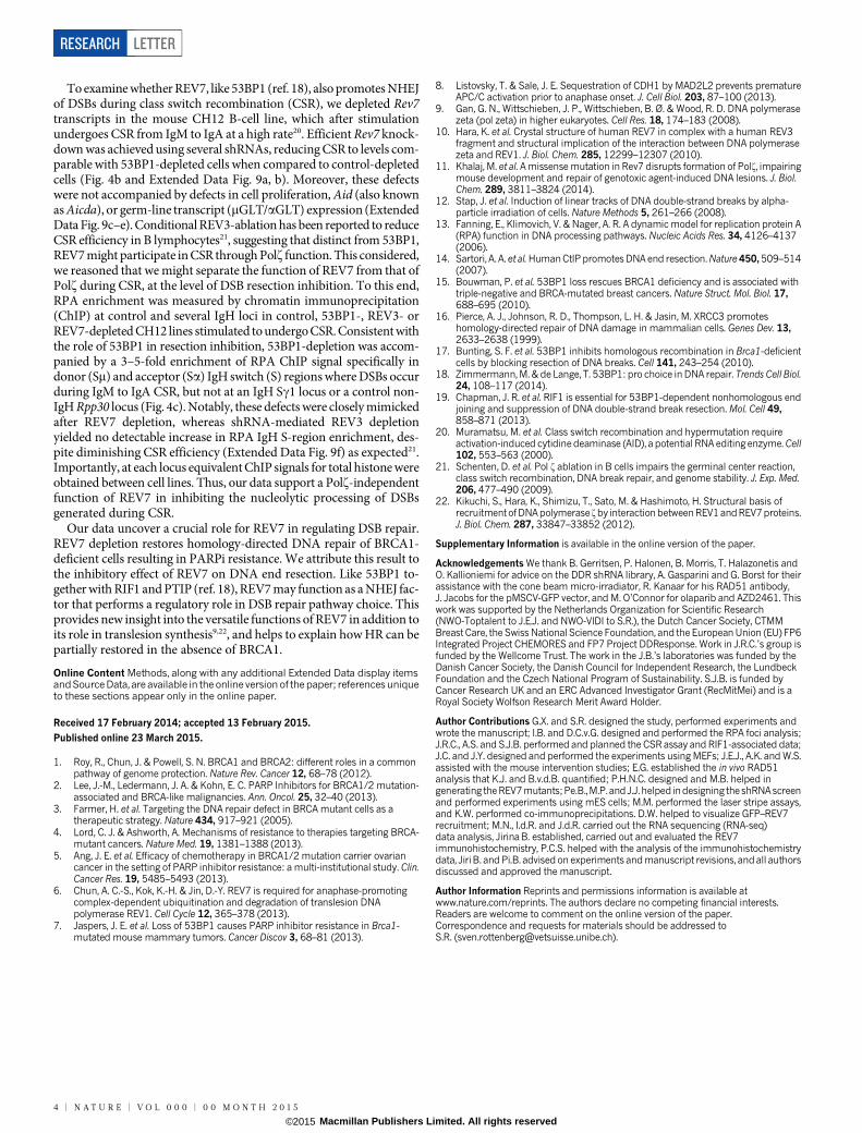
To identify mechanisms of BRCA1-independent restoration of the HRpathway, we carried out a loss-of-function short hairpin RNA (shRNA)screen using the KB1P-B11 and KB1P-G3 cell lines that we previouslyderived from Brca12/2 p53 2/2 (p53 is also known as Trp53) mousemammary tumours7 (Fig. 1a and Supplementary Table 1). Resistantcells were selected with a high concentration of olaparib (500 nM, about100-fold the half-maximal inhibitory concentration (IC50)), which killedcells of the empty vector control. Sequencing of the olaparib-survivingcolonies revealed a reproducible enrichment of various individualhairpins targeting Rev7 or 53bp1 (also known as Trp53bp1). To validatethe Rev7 hit, we introduced two different hairpins into the KB1P-B11and KB1P-G3 cell lines; these substantially inhibited Rev7 expression(Fig. 1b, c and Extended Data Fig. 1a). Despite the role of REV7 inmetaphase-to-anaphase transition8, the level of Rev7 inhibition in thesecells did not affect proliferation (Extended Data Fig. 1b, c), allowinglong-term clonogenic survival assays. We confirmed that loss of Rev7resulted in increased resistance to the PARP inhibitors (PARPi) ola-parib and AZD2461 (ref. 7) in both cell lines (Fig. 1d and Extended Data
Fig. 1d–g). Resistant cells that survived olaparib treatment (Rev7 sh1/2-ola) yielded even lower REV7 expression levels and increased numbersof colonies after PARPi treatment (Fig. 1b–d and Extended Data Fig. 1h).When we reconstituted the Rev7-depleted cells with shRNA-resistantRev7 complementary DNA resulting in similar REV7 protein levels(Extended Data Fig. 1i), we successfully re-sensitized the tumour cellsto PARPi (Fig. 1e, f).
Tumours derived from the Brca12/2 p53 2/2 cells with stable Rev7inhibition also showed olaparib resistance in vivo, in contrast to theempty vector controls (Fig. 1g and Extended Data Fig. 1j–l). In addition,we found that Rev7 loss explains some cases of in vivo acquired PARPiresistance in BRCA1-deficient mouse mammary tumours (data notshown). REV7 depletion also resulted in PARPi resistance of the humanBRCA1-deficient cell line SUM149PT (Extended Data Fig. 2). To-gether, these data strongly indicate that inhibition of Rev7 confersPARPi resistance in BRCA1-deficient tumour cells.
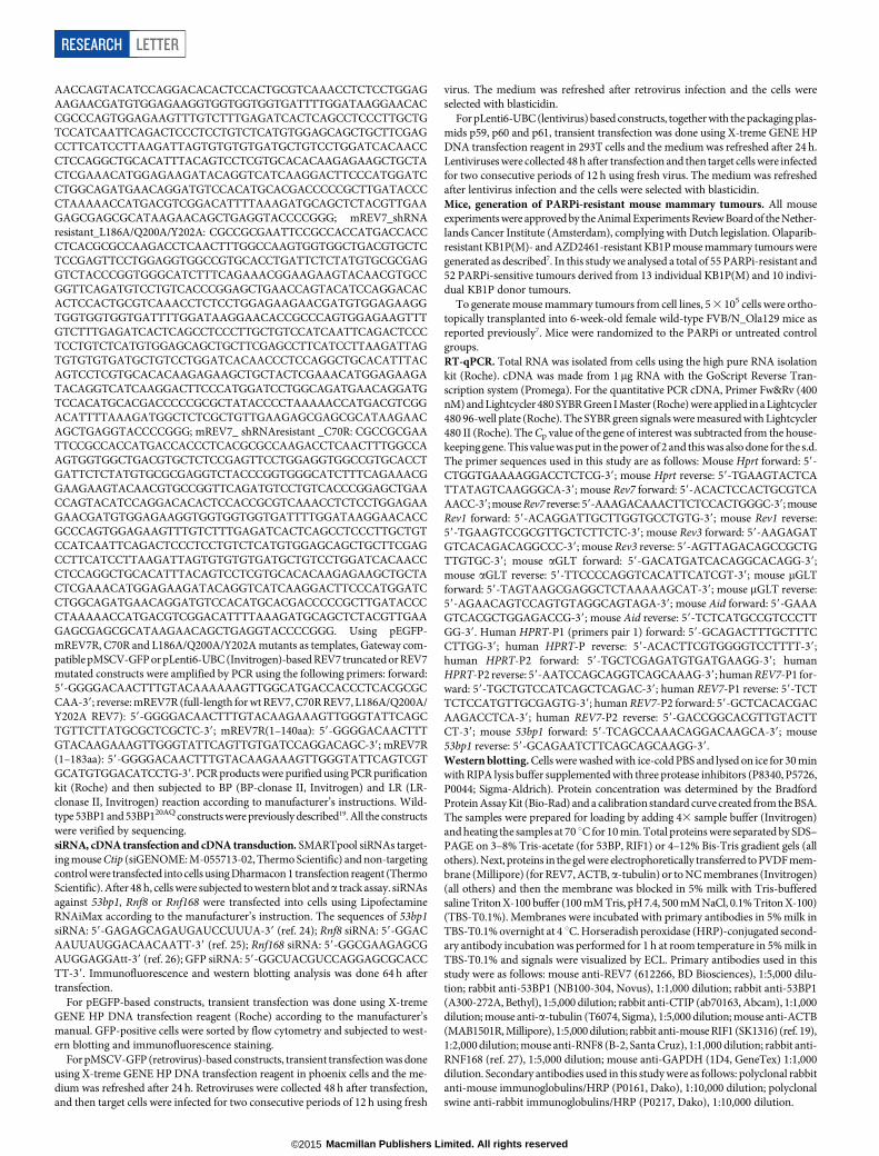
Together with the catalytic subunit REV3, REV7 forms the transle-sion synthesis polymerase f (Polf), and it interacts with REV1 (ref. 9).We therefore investigated whether loss of REV1 or REV3 also confersPARPi resistance in Brca12/2 p53 2/2 cells. A 60% inhibition of Rev1or Rev3 transcripts did not cause olaparib resistance (Extended DataFig. 3a–d). Moreover, we studied various shRNA-resistant REV7 mu-tants that lack REV1 (Leu186Ala/Gln200Ala/Tyr202Ala and a 1–183-amino-acids truncated protein) or REV3 (Cys70Arg) binding sites10,11.In contrast to the truncated 1–140-amino-acid REV7 protein, thesemutants are recruited to DNA damage sites (Extended Data Fig. 3e–g),and their expression in Rev7 shRNA KB1P-B11 and KB1P-G3 cells sig-nificantly restored the sensitivity to PARPi to a degree approachingthat of wild-type REV7 (Fig. 2a, b; P , 0.001, t-test). The remainingdifferences of the Leu186Ala/Gln200Ala/Tyr202Ala or Cys70Arg mu-tants with wild-type REV7 may be explained by unequal expression levels(Extended Data Fig. 3f). These data suggest that the REV1 or REV3 in-teraction is not absolutely required for the REV7-mediated function inthis context.
We observed that green fluorescent protein (GFP)-tagged REV7 co-localizes with 53BP1 shortly after DNA damage induction, suggestingthat REV7 acts directly at the site of DNA damage (Fig. 2c). REV7 re-cruitment depends on H2AX, MDC1, RNF8, RNF168 and partly ATM,in both mouse and human cells (Fig. 2d, e and Extended Data Fig. 4a–d).To examine whether PARPi resistance in Rev7-depleted Brca12/2 p53 2/2
tumour cells is due to HR restoration, we investigated RAD51 focus
*These authors contributed equally to this work.
1Division of Molecular Oncology, The Netherlands Cancer Institute, Plesmanlaan 121, 1066CX Amsterdam, The Netherlands. 2The Wellcome Trust Centre for Human Genetics, Roosevelt Drive, Oxford OX37BN, UK. 3Department of Genetics, Erasmus, University Medical Center, 3000 CA Rotterdam, The Netherlands. 4Department of Experimental Radiation Oncology, University of Texas MD Anderson CancerCenter, Houston, Texas 77030, USA. 5Institute of Molecular and Translational Medicine, Faculty of Medicine and Dentistry, Palacky University, 779 00 Olomouc, Czech Republic. 6Division of MolecularPathology, The Netherlands Cancer Institute, Plesmanlaan 121, 1066CX Amsterdam, The Netherlands. 7Danish Cancer Society Research Center, 2100 Copenhagen, Denmark. 8Division of Cell Biology, TheNetherlands Cancer Institute, Plesmanlaan 121, 1066CX Amsterdam, The Netherlands. 9Protein Facility, The Netherlands Cancer Institute, Plesmanlaan 121, 1066CX Amsterdam, The Netherlands.10Deep Sequencing Core Facility, The Netherlands Cancer Institute, Plesmanlaan 121, 1066CX Amsterdam, The Netherlands. 11Division of Molecular Carcinogenesis, The Netherlands Cancer Institute,Plesmanlaan 121, 1066CX Amsterdam, The Netherlands. 12DNA Damage Response Laboratory, London Research Institute, Cancer Research UK, Clare Hall, South Mimms, Hertfordshire EN6 3LD, UK.13Institute of Animal Pathology, Vetsuisse Faculty, University of Bern, Laengassstrasse 122, 3012 Bern, Switzerland.
0 0 M O N T H 2 0 1 5 | V O L 0 0 0 | N A T U R E | 1
Macmillan Publishers Limited. All rights reserved©2015

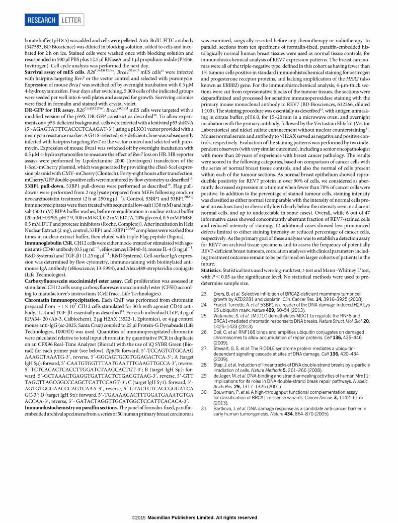
formation after 10 Gy ionizing radiation. As shown in Fig. 3a, b andExtended Data Fig. 4e, f, Rev7 loss resulted in the restoration of RAD51foci formed after DNA damage. To exclude potential off-target effectsof the hairpins, we reconstituted Rev7 shRNA1 and shRNA2 cells withshRNA-resistant mouse or human REV7–GFP fusion proteins (Ex-tended Data Fig. 4g). REV7 re-expression abolished RAD51 focus for-mation after DNA damage in GFP-positive cells (Fig. 3b). We confirmedthe re-appearance of RAD51 foci after tumour irradiation in vivo usingcomputed tomography (CT)-guided high precision cone beam irradi-ation of animals carrying PARPi-resistant KB1P(M) tumours with lowRev7 gene expression (Fig. 3c).
We then tested whether the processing of broken DNA ends requiresATM in Rev7-depleted cells, and found that inhibition of ATM usingKU55933 efficiently suppresses DNA-damage-induced RAD51 foci andincreases olaparib sensitivity (Extended Data Fig. 4h, i). Hence, thepartial restoration of RAD51 focus formation in Brca1-deficient mam-mary tumour cells after DNA damage by inhibition of Rev7 is ATM-dependent.
In contrast to the results with BRCA1-deficient cells, Rev7 depletionin BRCA2-deficient cells did not result in PARPi resistance (ExtendedData Fig. 5a–f). Furthermore, we did not observe increased PARPi
resistance after Rev7 inhibition in the BRCA1/2-proficient p532/2 tumourcell line KP3.33 (Extended Data Fig. 5g–i). This indicates that REV7works upstream of BRCA2 and is antagonized by BRCA1. We there-fore tested whether DNA end resection is altered in the absence of Rev7in BRCA1-deficient cells. Accumulation of the single-strand bindingprotein, RPA, was used as a marker for the generation of single-stranded DNA (ssDNA). Cells were exposed to a particles12, andBRCA1 deficiency resulted in a marked decrease in RPA-positivea trackscompared to BRCA1-proficient cells (Fig. 3d, e). REV7 depletion in theBrca12/2 p53 2/2 cells largely suppressed this defect in both KB1P-G3and KB1P-B11 cells (Fig. 3e and Extended Data Fig. 6a). In addition tocoating resected DNA ends, RPA also interacts with ssDNA gaps (forexample, during replication)13. To exclude that the observed RPA accu-mulation reflected interaction with internal ssDNA gaps, we preventedDNA end resection (without influencing replication) by knockingdown CTIP (ref. 14). This eliminated the increase in RPA-positivetracks induced by Rev7 knockdown in Brca12/2 p53 2/2 cell lines(Fig. 3f and Extended Data Fig. 6b), without affecting cell cycle distri-bution (Extended Data Fig. 6c). We therefore conclude that increasedresection and not binding to ssDNA gaps is responsible for RPAaccumulation.
a
Brca1–/– p53–/–
cells
Packaging
Lentivirus
Infection
PuromycinLong-term
clonogenic assay
Olaparib
(500 nM)
Surviving colonies
gDNA
Amplification of
viral insertions
In vitro/in vivovalidation1,976 hairpins targeting
391 DNA-damage-response-
related genes or vector control
PCR products
Deep sequencing
Comparison before
versus after olaparib
d
eUntreated
AZD2461
(1 μM)
f
Olaparib
(550 nM)
Untreated
AZD2461
(1 μM)
Olaparib
(550 nM)
Rela
tive c
ell
gro
wth
0
0.2
0.4
0.6
100
Surv
ival (%
)
50
00 20 40
G3 vector untreated
Rev7 sh2 untreated
Rev7 sh2 olaparib
G3 vector olaparib
60Days
P = 0.0001
0.8
1.0
1.2
AZD24
61
Olapar
ib
Rev7sh-vector
Rev7
sh-
vect
or
Rev7-sh-wt Rev7
Rev7
sh-
wt R
ev7
g
b
REV7
α-Tubulin
c
Brca1–/– p53–/–
shRNA library
Brca1–/– p53–/–
vector control
0
0.25
0.50
0.75
1.00
Rela
tive
gen
e e
xp
ressio
n
Rev7
sh1
Rev7
sh2
Vecto
r
Rev7
sh1-
ola Rev
7
sh2-
ola
Rev7
sh1
Rev7
sh2
Vecto
r
Rev7
sh1-
ola Rev
7
sh2-
ola
Rev7
sh1
Rev7
sh2
Vecto
r
Rev7
sh1-
ola Rev
7
sh2-
ola
Figure 1 | Identification of loss of Rev7 in PARPi-resistant Brca12/2
p53 2/2 mammary tumour cells. a, Design of the functional shRNA screen.gDNA, genomic DNA. b, c, Quantification of Rev7 transcript (b) or protein(c) levels in KB1P-G3 cells transduced with Rev7-targeting shRNAs or thevector control. Hprt was used as a control for transcript expression, andb-tubulin was used as a control for protein expression. The data represent themean 6 s.d. d, e, Long-term clonogenic assay using KB1P-G3 cells transducedwith the indicated constructs (wt Rev7 stands for pLenti6-wt Rev7) andtreatments. f, Quantification of the clonogenic assay in e by determining theabsorbance of crystal violet at 590 nm. All the groups were normalized tothe absorbance of the vector control. The data represent the mean 6 s.d.g, Overall survival of mice with KB1P-G3-derived Rev7-depleted or controltumours treated with one regimen of 50 mg olaparib per kilogram daily for28 days or left untreated. The P value was calculated using the log-rank test.
a
b
Rela
tive c
ell
gro
wth
c GFP mCherry Overlay
pEGFP+
53BP1–mCherry
pEGFP-REV7+
53BP1–mCherry
Olaparib
550 nM
AZD2461
900 nM
Untreated
shRev
7-GFP
shRev
7-wt
Rev
7 GFP
shRev
7-wt
Rev
7 GFP
shRev
7-Rev
7
(1–1
83aa
) GFP
shRev
7-Rev
7(1–
183a
a) G
FP
KB1P
-G3-
shRev
7-GFP
shRev
7-Rev
7
(1–1
40aa
) GFP
shRev
7-Rev
7(1–
140a
a) G
FP
shRev
7-C70
R
Rev
7 GFP
shRev
7-C70
R R
ev7
GFP
shRev
7-L1
86A/Q
200A
/
Y202A
Rev
7 GFP
shRev
7-L1
86A/Q
200A
/
Y202A
Rev
7 GFP
0
0.2
0.4
0.6
0.8
1.0
1.2
Olaparib 550 nM
AZD2461 900 nM
d
H2ax+/+
H2ax–/–
Mdc1+/+
Mdc1–/–
Rnf8+/+
Rnf8–/–
Atm+/+
Atm–/–
REV7 γH2AX DAPI REV7 γH2AX DAPI
Untreated IR
e P = 0.001
Atm+/+ Atm–/–
20
0
40
60
80
100
120
Fo
ci (%
)
REV7 γH2AX
Figure 2 | Dissection of REV7 function and its dependent factors.a, b, Long-term clonogenic assay (a) and quantification (b) using KB1P-G3cells transduced with the indicated constructs (wt Rev7 stands for pMSCV-GFP-wt Rev7) and treatments. All groups were normalized to the absorbance ofthe shRev7-GFP control. The data represent the mean 6 s.d. c, GFP–REV7recruitment to sites of DNA damage (visualized by 53BP1–mCherry) wasobserved 5 min after 405 nm laser exposure (0.99 mW, 60% laser power, 50 s) inKB1P-B11 cells. pEGFP denotes a mammalian expression vector containingenhanced GFP. Scale bar, 5 mm. d, REV7 foci formation in H2ax2/2 (alsoknown as H2afx2/2), Atm2/2, Mdc12/2 and Rnf82/2 mouse embryonicfibroblast (MEF) cells and their corresponding controls before and 4 h after10 Gy ionizing radiation (IR). DAPI, 49,6-diamidino-2-phenylindole. Scale bar,10mm. e, Quantification of REV7 foci formation (.8 foci per cell) inAtm2/2 and Atm1/1 MEF cells. The quantification of foci-positive cellswas performed by counting a total of 100 cells per sample. Data are presented asmean 6 s.d. from three different experiments. P value calculated usingthe t-test.
RESEARCH LETTER
2 | N A T U R E | V O L 0 0 0 | 0 0 M O N T H 2 0 1 5
Macmillan Publishers Limited. All rights reserved©2015

As Rev7 loss could restore end resection in BRCA1-deficient cells,we analysed whether its depletion could restore full HR proficiency inthis context. Using mouse embryonic stem (mES) cells with a Brca1selectable conditional knockout allele15, we observed that Rev7 loss in-deed prevented cell death of mES cells after BRCA1 deletion, and restoredRAD51 focus formation upon DNA damage (Extended Data Fig. 6d–h).Moreover, we reproducibly observed a partial restoration of HR function
in the DR–GFP reporter assay for homologous recombination16 whenRev7 was depleted in BRCA1-deficient mES cells (Fig. 3g).
Our data for REV7 are reminiscent of previous findings that 53BP1loss occurs in subsets of human breast carcinomas15 and can also restoreHR to BRCA1-deficient cells7,15,17. As with 53BP1, we found a frequentaberrant reduction or loss of the REV7 protein in human triple-negativebreast carcinomas (Extended Data Fig. 7). Addressing the relationshipbetween REV7, 53BP1 and the 53BP1 non-homologous end-joining(NHEJ) effector protein, RIF1 (ref. 18), we found that Rev7 deficiencydid not compromise the formation of endogenous 53BP1 or RIF1 foci(Extended Data Fig. 8a–d). By contrast, endogenous REV7 foci or laser-induced stripes were absent in 53BP1-depleted mouse and human cells(Fig. 4a and Extended Data Fig. 4c, d), strongly suggesting that REV7acts downstream of 53BP1. This is also consistent with our results thatPARPi resistance is not increased when both Rev7 and 53bp1 are de-pleted (data not shown). Despite such strong evidence for a cooperativerole for REV7 and 53BP1, we did not detect REV7 in 53BP1 immuno-complexes isolated from untreated cell lysates, or ATM-phosphorylated53BP1 immunocomplexes containing RIF1 that were induced by DNAdamage19 (Extended Data Fig. 8e and data not shown). Although theintricacies of the interactions remain to be determined, REV7 recruit-ment to DNA damage sites by 53BP1 may result from indirect inter-actions or an activity elicited by 53BP1 protein complexes in chromatinat DSB sites.
b GFP untreated GFP IR GFP hREV7 IRGFP mRev7 IR
0
Vecto
r
KP NIR
KP IR
KB1P
(M) s
ensitiv
e NIR
KB1P
(M) r
esista
nt N
IR
KB1P
(M) r
esista
nt IR
KB1P
(M) s
ensitiv
e IR
Rev7
sh1
Rev7
sh2
Rev7
sh1-
ola
Rev7
sh2-
ola
10
20
30
40
Cells
with
>1
0 R
AD
51
foci (%
)
e
Vector
Vector
Vector
f
CTIP
ACTB
p53–/
–
Ctrl siCtip si
+–
+–
+–+
–+–
+–
0
10
20
30
40
50
0
10
20
30
40
50
60
70R
PA
+ t
racks (%
)
RP
A+ t
racks (%
)
Untreated
10 Gy IR
Rev7 sh1G3 vector Rev7 sh2
0
5
10
15
No
rmaliz
ed
DR
-GF
P+ c
ells
Brca1: +/–
Vector
–/– –/–
****
Rev7
sh1-ola
Rev7sh2-ola
g
c
a
RPA+ track RPA– track
d 53BP1 RPA DAPI 53BP1 RPA DAPI
Brca1
–/– p
53–/
–
vect
or
Brca1
–/– p
53–/
–
Rev7
sh1
Rev7sh1
Rev7sh1
Rev7sh2
Rev7sh2
–/–
Rev7sh3
Rev7sh2
Brca1
–/– p
53–/
–
Rev7
sh2
Brca1
–/– p
53–/
–
Rev7
sh1-
ola
Brca1
–/– p
53–/
–
Rev7
sh2-
ola
Cells
with >
10 R
AD
51
foci (%
)
0
5
10
20
15
25 ****
****
Figure 3 | The effect of REV7 inhibition on RAD51 and RPA focusformation of Brca12/2 p532/2 cells. a, RAD51 focus (red) formation inKB1P-G3 cells before and 5 h after 10 Gy ionizing radiation. Scale bar, 10mm.b, Quantification of RAD51 foci in KB1P-G3 cells (with or without REV7depletion) transfected with an empty vector (GFP) or vectors containing mouseor human Rev7 or REV7, respectively. At least 150 GFP-positive cells wereanalysed per group in three independent experiments each. The data representthe mean 6 s.d. IR denotes 5 h after 10 Gy ionizing radiation. c, In situ analysisof RAD51 foci in PARPi-resistant KB1P(M) tumours with low Rev7 geneexpression. KP (KP3.33) denotes mouse mammary tumour cell line (p532/2);IR denotes 2 h after 15 Gy ionizing radiation; NIR denotes no ionizingradiation. ****P , 0.0001, Mann–Whitney U test. d, e, Representative imagesof 53BP1-labelled a tracks in cells positive or negative for RPA (d) andquantification of RPA-positive tracks 2 h after ionizing radiation (e). KP(p532/2) or KB1P-G3 cells with or without Rev7-targeting shRNAs were tested.Scale bar, 5mm. f, Quantification of RPA- and 53BP1-positive a tracks inKB1P-G3 cells transfected with non-targeting control (ctrl) short interferingRNAs (siRNAs) or siRNAs against mouse Ctip. CTIP protein expressionof the indicated groups was checked by western blotting, with b-actin(ACTB) as a loading control. g, Quantification of HR using the DR-GFPreporter assay. GFP-positive cells normalized to the vector-transducedBrca12/2 p53 shRNA cells are shown. The data represent mean 6 s.d.**P , 0.01, two-tailed t-test.
Vector
53bp1 sh
Vector53bp1 sh
Rev7 sh1
Rev7 sh2
Rev7 sh3
Rev3 sh2Rev7 sh3100
80
60
40
20
0
CS
R (no
rmaliz
ed
Ig
A+ c
ells
) (%
)
– + + + + +CIT:
a
b VDJ Sμ Cμ Sγ1 Sα CαCγ1Cδ
i ii iii iv
Mouse
IgH locus
(Chr.12)
c
0
20
40
60
80
100
120
0
20
40
60
80
100
120
RPA32 ChIP
Histone H2AX ChIP
Rpp30 i ii iii iv
Rpp30 i ii iii iv
ChIP
sig
nal (%
)C
hIP
sig
nal (%
)
53bp1+/+
53bp1–/–
53bp1+/+
53bp1–/–
REV7 53BP1 DAPI
REV7 DAPI
REV7 53BP1 DAPI
Untreated
Untreated
γH2AX REV7 DAPIγH2AX
IR
IR
Figure 4 | REV7 is a downstream effector of 53BP1 on inhibiting endresection and promoting CSR. a, REV7 foci formation in 53bp12/2 and53bp11/1 MEF cells before and 4 h after 10 Gy ionizing radiation. Scale bar,10mm. b, Quantification of CSR to IgA of shRNA-transduced CH12 cells 40 hafter stimulation (CIT denotes CD40 antibody, IL-4 and TGF-b1). Datarepresent mean 6 s.d. from two independent experiments performed intriplicate. c, Schematic of IgH locus shows relative positions of quantitativePCR amplicons used in ChIP experiments. A control non-IgH locus (Rpp30)was also examined. Indicated CH12 cell lines stimulated for 30 h with CITwere subjected to ChIP with IgG (control), histone H2AX and RPA32monoclonal antisera. After background subtraction, values were normalized tothe DNA input signals, followed by the maximum value in each data set.Mean signals, two replicates 6 s.e.m.
LETTER RESEARCH
0 0 M O N T H 2 0 1 5 | V O L 0 0 0 | N A T U R E | 3
Macmillan Publishers Limited. All rights reserved©2015
To examine whether REV7, like 53BP1 (ref. 18), also promotes NHEJof DSBs during class switch recombination (CSR), we depleted Rev7transcripts in the mouse CH12 B-cell line, which after stimulationundergoes CSR from IgM to IgA at a high rate20. Efficient Rev7 knock-down was achieved using several shRNAs, reducing CSR to levels com-parable with 53BP1-depleted cells when compared to control-depletedcells (Fig. 4b and Extended Data Fig. 9a, b). Moreover, these defectswere not accompanied by defects in cell proliferation, Aid (also knownas Aicda), or germ-line transcript (mGLT/aGLT) expression (ExtendedData Fig. 9c–e). Conditional REV3-ablation has been reported to reduceCSR efficiency in B lymphocytes21, suggesting that distinct from 53BP1,REV7 might participate in CSR through Polf function. This considered,we reasoned that we might separate the function of REV7 from that ofPolf during CSR, at the level of DSB resection inhibition. To this end,RPA enrichment was measured by chromatin immunoprecipitation(ChIP) at control and several IgH loci in control, 53BP1-, REV3- orREV7-depleted CH12 lines stimulated to undergo CSR. Consistent withthe role of 53BP1 in resection inhibition, 53BP1-depletion was accom-panied by a 3–5-fold enrichment of RPA ChIP signal specifically indonor (Sm) and acceptor (Sa) IgH switch (S) regions where DSBs occurduring IgM to IgA CSR, but not at an IgH Sc1 locus or a control non-IgH Rpp30 locus (Fig. 4c). Notably, these defects were closely mimickedafter REV7 depletion, whereas shRNA-mediated REV3 depletionyielded no detectable increase in RPA IgH S-region enrichment, des-pite diminishing CSR efficiency (Extended Data Fig. 9f) as expected21.Importantly, at each locus equivalent ChIP signals for total histone wereobtained between cell lines. Thus, our data support a Polf-independentfunction of REV7 in inhibiting the nucleolytic processing of DSBsgenerated during CSR.
Our data uncover a crucial role for REV7 in regulating DSB repair.REV7 depletion restores homology-directed DNA repair of BRCA1-deficient cells resulting in PARPi resistance. We attribute this result tothe inhibitory effect of REV7 on DNA end resection. Like 53BP1 to-gether with RIF1 and PTIP (ref. 18), REV7 may function as a NHEJ fac-tor that performs a regulatory role in DSB repair pathway choice. Thisprovides new insight into the versatile functions of REV7 in addition toits role in translesion synthesis9,22, and helps to explain how HR can bepartially restored in the absence of BRCA1.
Online Content Methods, along with any additional Extended Data display itemsandSourceData, are available in the online version of the paper; references uniqueto these sections appear only in the online paper.
Received 17 February 2014; accepted 13 February 2015.
Published online 23 March 2015.
1. Roy, R., Chun, J. & Powell, S. N. BRCA1 and BRCA2: different roles in a commonpathway of genome protection. Nature Rev. Cancer 12, 68–78 (2012).
2. Lee, J.-M., Ledermann, J. A. & Kohn, E. C. PARP Inhibitors for BRCA1/2 mutation-associated and BRCA-like malignancies. Ann. Oncol. 25, 32–40 (2013).
3. Farmer, H. et al. Targeting the DNA repair defect in BRCA mutant cells as atherapeutic strategy. Nature 434, 917–921 (2005).
4. Lord, C. J. & Ashworth, A. Mechanisms of resistance to therapies targeting BRCA-mutant cancers. Nature Med. 19, 1381–1388 (2013).
5. Ang, J. E. et al. Efficacy of chemotherapy in BRCA1/2 mutation carrier ovariancancer in the setting of PARP inhibitor resistance: a multi-institutional study. Clin.Cancer Res. 19, 5485–5493 (2013).
6. Chun, A. C.-S., Kok, K.-H. & Jin, D.-Y. REV7 is required for anaphase-promotingcomplex-dependent ubiquitination and degradation of translesion DNApolymerase REV1. Cell Cycle 12, 365–378 (2013).
7. Jaspers, J. E. et al. Loss of 53BP1 causes PARP inhibitor resistance in Brca1-mutated mouse mammary tumors. Cancer Discov 3, 68–81 (2013).
8. Listovsky, T. & Sale, J. E. Sequestration of CDH1 by MAD2L2 prevents prematureAPC/C activation prior to anaphase onset. J. Cell Biol. 203, 87–100 (2013).
9. Gan, G. N., Wittschieben, J. P., Wittschieben, B. Ø. & Wood, R. D. DNA polymerasezeta (pol zeta) in higher eukaryotes. Cell Res. 18, 174–183 (2008).
10. Hara, K. et al. Crystal structure of human REV7 in complex with a human REV3fragment and structural implication of the interaction between DNA polymerasezeta and REV1. J. Biol. Chem. 285, 12299–12307 (2010).
11. Khalaj, M. et al. A missense mutation in Rev7 disrupts formation of Polf, impairingmouse development and repair of genotoxic agent-induced DNA lesions. J. Biol.Chem. 289, 3811–3824 (2014).
12. Stap, J. et al. Induction of linear tracks of DNA double-strand breaks by alpha-particle irradiation of cells. Nature Methods 5, 261–266 (2008).
13. Fanning, E., Klimovich, V. & Nager, A. R. A dynamic model for replication protein A(RPA) function in DNA processing pathways. Nucleic Acids Res. 34, 4126–4137(2006).
14. Sartori, A. A. et al. Human CtIP promotes DNA end resection. Nature 450, 509–514(2007).
15. Bouwman, P. et al. 53BP1 loss rescues BRCA1 deficiency and is associated withtriple-negative and BRCA-mutated breast cancers. Nature Struct. Mol. Biol. 17,688–695 (2010).
16. Pierce, A. J., Johnson, R. D., Thompson, L. H. & Jasin, M. XRCC3 promoteshomology-directed repair of DNA damage in mammalian cells. Genes Dev. 13,2633–2638 (1999).
17. Bunting, S. F. et al. 53BP1 inhibits homologous recombination in Brca1-deficientcells by blocking resection of DNA breaks. Cell 141, 243–254 (2010).
18. Zimmermann, M. & de Lange, T. 53BP1: pro choice in DNA repair. Trends Cell Biol.24, 108–117 (2014).
19. Chapman, J. R. et al. RIF1 is essential for 53BP1-dependent nonhomologous endjoining and suppression of DNA double-strand break resection. Mol. Cell 49,858–871 (2013).
20. Muramatsu, M. et al. Class switch recombination and hypermutation requireactivation-induced cytidine deaminase (AID), a potential RNA editing enzyme. Cell102, 553–563 (2000).
21. Schenten, D. et al. Pol f ablation in B cells impairs the germinal center reaction,class switch recombination, DNA break repair, and genome stability. J. Exp. Med.206, 477–490 (2009).
22. Kikuchi, S., Hara, K., Shimizu, T., Sato, M. & Hashimoto, H. Structural basis ofrecruitment ofDNApolymerase fby interaction betweenREV1andREV7proteins.J. Biol. Chem. 287, 33847–33852 (2012).
Supplementary Information is available in the online version of the paper.
Acknowledgements We thank B. Gerritsen, P. Halonen, B. Morris, T. Halazonetis andO. Kallioniemi for advice on the DDR shRNA library, A. Gasparini and G. Borst for theirassistance with the cone beam micro-irradiator, R. Kanaar for his RAD51 antibody,J. Jacobs for the pMSCV-GFP vector, and M. O’Connor for olaparib and AZD2461. Thiswork was supported by the Netherlands Organization for Scientific Research(NWO-Toptalent to J.E.J. and NWO-VIDI to S.R.), the Dutch Cancer Society, CTMMBreast Care, the Swiss National Science Foundation, and the European Union (EU) FP6Integrated Project CHEMORES and FP7 Project DDResponse. Work in J.R.C.’s group isfunded by the Wellcome Trust. The work in the J.B.’s laboratories was funded by theDanish Cancer Society, the Danish Council for Independent Research, the LundbeckFoundation and the Czech National Program of Sustainability. S.J.B. is funded byCancer Research UK and an ERC Advanced Investigator Grant (RecMitMei) and is aRoyal Society Wolfson Research Merit Award Holder.
Author Contributions G.X. and S.R. designed the study, performed experiments andwrote the manuscript; I.B. and D.C.v.G. designed and performed the RPA foci analysis;J.R.C., A.S. and S.J.B. performed and planned the CSR assay and RIF1-associated data;J.C. and J.Y. designed and performed the experiments using MEFs; J.E.J., A.K. and W.S.assisted with the mouse intervention studies; E.G. established the in vivo RAD51analysis that K.J. and B.v.d.B. quantified; P.H.N.C. designed and M.B. helped ingenerating theREV7mutants;Pe.B.,M.P. and J.J. helped indesigning the shRNAscreenand performed experiments using mES cells; M.M. performed the laser stripe assays,and K.W. performed co-immunoprecipitations. D.W. helped to visualize GFP–REV7recruitment; M.N., I.d.R. and J.d.R. carried out the RNA sequencing (RNA-seq)data analysis, Jirina B. established, carried out and evaluated the REV7immunohistochemistry, P.C.S. helped with the analysis of the immunohistochemistrydata, Jiri B. and Pi.B. advised on experiments and manuscript revisions, and all authorsdiscussed and approved the manuscript.
Author Information Reprints and permissions information is available atwww.nature.com/reprints. The authors declare no competing financial interests.Readers are welcome to comment on the online version of the paper.Correspondence and requests for materials should be addressed toS.R. ([email protected]).
RESEARCH LETTER
4 | N A T U R E | V O L 0 0 0 | 0 0 M O N T H 2 0 1 5
Macmillan Publishers Limited. All rights reserved©2015

METHODSCell culture and reagents. KB1P-B11 (B11) and KB1P-G3 (G3) cell lines werederived from a Brca12/2 p532/2 mouse mammary tumour as described7. KB2P-1.21 and KB2P-3.4 cell lines originate from a Brca12/2 p532/2 mouse mammarytumour, and KP3.33 cell line from a p532/2 mouse mammary23. These cell lineswere cultured in DMEM/F-12 (Life Technologies) supplemented with 10% FCS,50 U ml21 penicillin, 50 ng ml21 streptomycin, 5mg ml21 insulin (Sigma), 5 ng ml21
epidermal growth factor (Life Technologies) and 5 ng ml21 cholera toxin (Gentaur)under low oxygen conditions (3% O2, 5% CO2, 37 uC). SUM149PT cells were grownin RPMI1640 (Life Technologies) supplemented with 10% FCS, under normal oxy-gen conditions (21% O2, 5% CO2, 37 uC). U2OS, phoenix, 293T cells were culturedin DMEM (Life Technologies) supplemented with 10% FCS, under normal oxygenconditions (21% O2, 5% CO2, 37 uC). mES cells with a selectable conditional BRCA1deletion (R26CreERT2/wt;Brca1SCo/D)15 were cultured on gelatin-coated plates in60% buffalo red liver cell-conditioned medium supplied with 10% FCS, 0.1 mMb-mercaptoethanol (Merck) and 1 3 103 U ml21 ESGRO LIF (Millipore) under nor-mal oxygen conditions (21% O2, 5% CO2, 37 uC). CH12 cells (CH12F3-2) werecultured in RPMI-1640 medium (Sigma) supplemented with 10% FBS, 5% NCTC109 medium (Sigma), 50mMb-mercaptoethanol, 50 U ml21 penicillin and 50 ng ml21
streptomycin under normal oxygen conditions. Olaparib and AZD2461 were pro-vided by AstraZeneca; KU-55933 (KuDOS) was bought from Selleckchem (S1092).Lentivirus-based transduction of cells with shRNA. Glycerol stocks of shRNAhairpins were obtained from the Sigma Mission library (TRC 1.0) and isolation ofplasmids was carried out with the high pure plasmid Mini Kit or Genopure maxikit (Roche). 293T cells were seeded 16 h before transfection. For each 10-cm dish,0.5 ml 23 HBS (8.18 g NaCl, 0.2 g Na2HPO4-7H2O, 5.95 g HEPES in 500 ml MilliQwater at pH 7.01) was added into a sterile falcon tube. In another sterile falcon tube,6mg plasmid DNA of interest, 2mg pHCMV-G envelope vector (pMD.G), 2mgpRSV-Rev, 2mg packaging vector pMDLg/pRRE, 250ml 0.5 M CaCl2 and distilledwater were added to bring up to 0.5 ml. The CaCl2/plasmid DNA mix was added tothe 23 HBS and incubated for 20 min and then added to the cells. Medium wasrefreshed after 6 h and another 18 h, respectively. The supernatant of 293T cells con-taining lentivirus was collected after 24 h to infect cells with polybrene (6 mg ml21)for 12 h. The medium was refreshed after lentivirus infection and the cells wereselected with puromycin.Individual shRNA vectors used were collected from the TRC library. MouseRev7: sh1: TRCN0000012844_CCAGTGGAGAAGTTTGTCTTT; sh2: TRCN0000012846_CATCTTCCAGAAGCGCAAGAA; sh3: TRCN0000012847_GATACAGGTCATCAAGGACTT; human REV7: sh1: TRCN0000006569_CCCTGATTCCAAGTGCTCTTA; sh2: TRCN0000006570_CCCGGAGCTGAATCAGTATAT; sh3: TRCN0000006571_CCCAGTGGAGAAATTCGTCTT; sh4: TRCN0000006573_CATCTTCCAGAAACGCAAGAA; mouse 53bp1: (puromycin) sh:TRCN0000081778_GCTATTGTGGAGATTGTGTTT; (neomycin) sh: same se-quences as above; human 53BP1: sh1: TRCN0000018866_CCAGTGTGATTAGTATTGATT; sh2: TRCN0000018865_GATACTTGGTCTTACTGGTTT; mousep53: (neomycin) TRCN0000054551_AGAGTATTTCACCCTCAAGAT; mouseRev1: sh1: TRCN0000120298_GCCGAGATCAACTATGGAATA; sh2: TRCN0000120297_CAGCAGTGCTTGTGAGGTATT; mouse Rev3: sh1: TRCN0000119969_CCGTCACATTAGTGAGACTAT; sh2: TRCN0000119970_GCCCACATACACTTTCTTCTT.Loss-of-function screen. In total, 1,976 lentiviral hairpins (pLKO.1) from the SigmaMission library (TRC Mm 1.0) that target 391 mouse genes involved in the DNAdamage response were selected (see Supplementary Table 1). This library was usedto generate pools of lentiviral shRNA in 293T cells to infect target cells. After in-fection, the cells stably expressing integrated shRNA were selected with puromy-cin. Cells with HR restoration were selected with a high concentration of olaparib(500 nM, about 100-fold the IC50), which killed cells of the empty vector group.Surviving cells were pooled and genomic DNA was extracted using the GentraPuregene kit according to the manufacturer’s protocol (Qiagen). shRNA insertswere retrieved from 50 ng genomic DNA by PCR amplification (PCR1 and PCR2)using the following conditions: (1) 95 uC, 5 min; (2) 95 uC, 30 s; (3) 60 uC, 30 s; (4)72 uC, 1 min; (5) go to step (2), 20 cycles; (6) 72 uC, 5 min; (7) 4 uC. The PCR re-action system were as follows: 0.6ml DMSO, 4.0ml phusion HF buffer 53, 0.4mldNTPs, 1.0ml primer f (10 mM), 1.0ml primer r (10 mM), 11.8ml mQ, 0.2ml phu-sion,1.0ml gDNA for PCR1 or 1ml PCR1 products for PCR2. Adaptors and indexesfor deep sequencing (Illumina HiSeq 2000) were incorporated into PCR primers asfollows: PCR1 forward: PCR1_01_PLKO1_f_Integration determination_1, 59-ACACTCTTTCCCTACACGACGCTCTTCCGATCTCGTGATCTTGTGGAAAGGACGAAACACCGG-39; PCR1_02_PLKO1_f_Integration determination_2, 59-ACACTCTTTCCCTACACGACGCTCTTCCGATCTGTAGCCCTTGTGGAAAGGACGAAACACCGG-39; PCR1_03_PLKO1_f_untreated_1, 59-ACACTCTTTCCCTACACGACGCTCTTCCGATCTCACTGTCTTGTGGAAAGGACGAAACACCGG-39; PCR1_04_PLKO1_f_untreated_2, 59-ACACTCTTTCCCTAC
ACGACGCTCTTCCGATCTATTGGCCTTGTGGAAAGGACGAAACACCGG-39; PCR1_05_PLKO1_f_olaparib_1, 59-ACACTCTTTCCCTACACGACGCTCTTCCGATCTGATCTGCTTGTGGAAAGGACGAAACACCGG-39; PCR1_06_PLKO1_f_olaparib_2, 59-ACACTCTTTCCCTACACGACGCTCTTCCGATCTTCAAGTCTTGTGGAAAGGACGAAACACCGG-39; PCR1_07_PLKO1_f_olaparib_3, 59-ACACTCTTTCCCTACACGACGCTCTTCCGATCTCTGATCCTTGTGGAAAGGACGAAACACCGG-39; PCR1_08_PLKO1_f_olaparib_4, 59-ACACTCTTTCCCTACACGACGCTCTTCCGATCTAAGCTACTTGTGGAAAGGACGAAACACCGG-39; PCR1 reverse: P7_pLKO1_r, 59-CAAGCAGAAGACGGCATACGAGATTTCTTTCCCCTGCACTGTACCC-39; PCR2 forward:P5_IlluSeq, 59-AATGATACGGCGACCACCGAGATCTACACTCTTTCCCTACACGACGCTCTTCCGATCT-39; PCR2 reverse: P7, 59-CAAGCAGAAGACGGCATACGAGAT-39.
PCR2 products were purified using the PCR purification kit from Qiagen. TheshRNA stem sequence was segregated and aligned to the TRC library. The reads ofdifferent hairpins were counted and the following criteria were used to select thetop hits for further validation: (1) hairpins targeting the same gene in survival clonesshould have at least 1 3 104 reads (total ,6 3 106 reads); (2) at least two differenthairpins targeting the same gene should be present; (3) hairpins in resistant clonesshould be highly enriched (.8-fold) in cells after olaparib selection; and (4) hair-pins should be present in 4 out of 4 independent screens.PARPi treatment study. Long-term clonogenic assay: on day 0, 1.5 3 104 (B11)or 1 3 104 (G3, KB2P_1.21 or KB2P_3.4) or 6 3 103 (KP3.33) cells were seeded perwell with PARPi (or untreated control) into six-well plates. The medium of thePARPi treatment groups was refreshed with PARPi every 4 days. On day 5, the un-treated control group was stopped and the PARPi treatment groups were stoppedafter another 2–3 weeks and stained with 0.1% crystal violet. Using the SUM149PTcells, 4 3 104 cells were seeded per well with olaparib (or untreated control) into12-well plates. The medium of the PARPi treatment groups was refreshed witholaparib every 4 days. On day 6, the untreated control group was stopped and theolaparib treatment groups were stopped on day 8 and stained with 0.1% crystal vio-let. Quantification of the clonogenic assay was done by determining the absorbanceof crystal violet at 590 nm.
Short-term clonogenic assay: on day 0, 4 3 102 (G3) cells were seeded per wellwith olaparib (or untreated control) into 6-well plates. On day 4, the medium wasrefreshed with olaparib or untreated control. On day 8, all the groups were stainedwith Leishman dye and quantification was done by the relative colony numbers.ATM and PARP inhibitor combination study. On day 0, 1 3 104 (G3) cells wereseeded per well into 6-well plates and then ATM inhibitor or olaparib or their com-bination was added. The medium was refreshed every 3 days with the different drugs.For the combination therapy groups, ATM inhibitor was applied for 6 days. Onday 5, ATM inhibitor alone and untreated control groups were stopped and theother groups were stopped on day 12 and stained with 0.1% crystal violet.Constructs. Human REV7 was amplified by PCR using Platinum Taq polymerase(Invitrogen) from U2OS cDNA using the following primers 59-ATAGAATTCAATGACCACGCTCACACGACAAGAC -39 and 59-ATATGGTACCATCAGCTGCCTTTATGAGCGCGC-39. Mouse Rev7 was amplified from mouse lung cDNAin two parts to introduce silent mutations, making it resistant to Rev7 shRNA2(mRev7R). The following primers were used for part A: [IB11m] 59-ATATGAATTCGATGACCACCCTCACGCGC-39 [IB14m] 59-TACTTCTTCCGTTTCTGAAAGATGCCCACCGGGTA-39 and part B: [IB12m] 59-ATATGGTACC-AT-GCTGTTCTTATGCGCTCGCT-39 [IB13m] 59-GGGCATCTTTCAGAAACGGAAGAAGTACAACGTGC-39. Equal parts of both PCR reactions were mixed andused for a PCR using IB11m and IB12m to create the complete mouse Rev7 sequenceincluding the silent mutations.
The PCR product and pEGFP-C1 vector were digested using EcoRI and KpnIand ligated using the T4 DNA Ligase (Roche) to generate pEGFP-hREV7 andpEGFP-mREV7R.
Using pEGFP-mREV7R as a template, pEGFP-C1-based REV7 truncated con-structs was amplified by PCR using the following primers: forward: 59-ATATGAATTCGATGACCACCCTCACGCGC-39, reverse: mREV7R (1–55aa): 59-ATATGGTACCGGACATCTGAACCGGCAC-39; mREV7R (1–81aa): 59-ATATGGTACCCTCCACATCGTTCTTCTCCAGG-39; mREV7R (1–110aa): 59-ATATGGTACCGATGGACAGCAAGGGAGGC-39; mREV7R (1–140aa): 59-ATATGGTACCGTTGTGATCCAGGACAGC-39; mREV7R (1–183aa): 59-ATATGGTACCGTCGTGCATGTGGACATCCTG-39. pEGFP-REV7 mutants (shRNA-resistant)were ordered as gBlocks (IDT) and cloned into the pEGFP-C1 vector using EcoRIand KpnI restriction enzymes and quick ligase (NEB). The DNA sequences thatwere ordered as gBlocks are: mREV7_shRNAresistant (mREV7R): CGCCGCGAATTCCGCCACCATGACCACCCTCACGCGCCAAGACCTCAACTTTGGCCAAGTGGTGGCTGACGTGCTCTCCGAGTTCCTGGAGGTGGCCGTGCACCTGATTCTCTATGTGCGCGAGGTCTACCCGGTGGGCATCTTTCAGAAACGGAAGAAGTACAACGTGCCGGTTCAGATGTCCTGTCACCCGGAGCTG
LETTER RESEARCH
Macmillan Publishers Limited. All rights reserved©2015
AACCAGTACATCCAGGACACACTCCACTGCGTCAAACCTCTCCTGGAGAAGAACGATGTGGAGAAGGTGGTGGTGGTGATTTTGGATAAGGAACACCGCCCAGTGGAGAAGTTTGTCTTTGAGATCACTCAGCCTCCCTTGCTGTCCATCAATTCAGACTCCCTCCTGTCTCATGTGGAGCAGCTGCTTCGAGCCTTCATCCTTAAGATTAGTGTGTGTGATGCTGTCCTGGATCACAACCCTCCAGGCTGCACATTTACAGTCCTCGTGCACACAAGAGAAGCTGCTACTCGAAACATGGAGAAGATACAGGTCATCAAGGACTTCCCATGGATCCTGGCAGATGAACAGGATGTCCACATGCACGACCCCCGCTTGATACCCCTAAAAACCATGACGTCGGACATTTTAAAGATGCAGCTCTACGTTGAAGAGCGAGCGCATAAGAACAGCTGAGGTACCCCGGG; mREV7_shRNAresistant_L186A/Q200A/Y202A: CGCCGCGAATTCCGCCACCATGACCACCCTCACGCGCCAAGACCTCAACTTTGGCCAAGTGGTGGCTGACGTGCTCTCCGAGTTCCTGGAGGTGGCCGTGCACCTGATTCTCTATGTGCGCGAGGTCTACCCGGTGGGCATCTTTCAGAAACGGAAGAAGTACAACGTGCCGGTTCAGATGTCCTGTCACCCGGAGCTGAACCAGTACATCCAGGACACACTCCACTGCGTCAAACCTCTCCTGGAGAAGAACGATGTGGAGAAGGTGGTGGTGGTGATTTTGGATAAGGAACACCGCCCAGTGGAGAAGTTTGTCTTTGAGATCACTCAGCCTCCCTTGCTGTCCATCAATTCAGACTCCCTCCTGTCTCATGTGGAGCAGCTGCTTCGAGCCTTCATCCTTAAGATTAGTGTGTGTGATGCTGTCCTGGATCACAACCCTCCAGGCTGCACATTTACAGTCCTCGTGCACACAAGAGAAGCTGCTACTCGAAACATGGAGAAGATACAGGTCATCAAGGACTTCCCATGGATCCTGGCAGATGAACAGGATGTCCACATGCACGACCCCCGCGCTATACCCCTAAAAACCATGACGTCGGACATTTTAAAGATGGCTCTCGCTGTTGAAGAGCGAGCGCATAAGAACAGCTGAGGTACCCCGGG; mREV7_ shRNAresistant _C70R: CGCCGCGAATTCCGCCACCATGACCACCCTCACGCGCCAAGACCTCAACTTTGGCCAAGTGGTGGCTGACGTGCTCTCCGAGTTCCTGGAGGTGGCCGTGCACCTGATTCTCTATGTGCGCGAGGTCTACCCGGTGGGCATCTTTCAGAAACGGAAGAAGTACAACGTGCCGGTTCAGATGTCCTGTCACCCGGAGCTGAACCAGTACATCCAGGACACACTCCACCGCGTCAAACCTCTCCTGGAGAAGAACGATGTGGAGAAGGTGGTGGTGGTGATTTTGGATAAGGAACACCGCCCAGTGGAGAAGTTTGTCTTTGAGATCACTCAGCCTCCCTTGCTGTCCATCAATTCAGACTCCCTCCTGTCTCATGTGGAGCAGCTGCTTCGAGCCTTCATCCTTAAGATTAGTGTGTGTGATGCTGTCCTGGATCACAACCCTCCAGGCTGCACATTTACAGTCCTCGTGCACACAAGAGAAGCTGCTACTCGAAACATGGAGAAGATACAGGTCATCAAGGACTTCCCATGGATCCTGGCAGATGAACAGGATGTCCACATGCACGACCCCCGCTTGATACCCCTAAAAACCATGACGTCGGACATTTTAAAGATGCAGCTCTACGTTGAAGAGCGAGCGCATAAGAACAGCTGAGGTACCCCGGG. Using pEGFP-mREV7R, C70R and L186A/Q200A/Y202A mutants as templates, Gateway com-patible pMSCV-GFP or pLenti6-UBC (Invitrogen)-based REV7 truncated or REV7mutated constructs were amplified by PCR using the following primers: forward:59-GGGGACAACTTTGTACAAAAAAGTTGGCATGACCACCCTCACGCGCCAA-39; reverse: mREV7R (full-length for wt REV7, C70R REV7, L186A/Q200A/Y202A REV7): 59-GGGGACAACTTTGTACAAGAAAGTTGGGTATTCAGCTGTTCTTATGCGCTCGCTC-39; mREV7R(1–140aa): 59-GGGGACAACTTTGTACAAGAAAGTTGGGTATTCAGTTGTGATCCAGGACAGC-39; mREV7R(1–183aa): 59-GGGGACAACTTTGTACAAGAAAGTTGGGTATTCAGTCGTGCATGTGGACATCCTG-39. PCR products were purified using PCR purificationkit (Roche) and then subjected to BP (BP-clonase II, Invitrogen) and LR (LR-clonase II, Invitrogen) reaction according to manufacturer’s instructions. Wild-type 53BP1 and 53BP120AQ constructs were previously described19. All the constructswere verified by sequencing.siRNA, cDNA transfection and cDNA transduction. SMARTpool siRNAs target-ing mouse Ctip (siGENOME: M-055713-02, Thermo Scientific) and non-targetingcontrol were transfected into cells using Dharmacon 1 transfection reagent (ThermoScientific). After 48 h, cells were subjected to western blot anda track assay. siRNAsagainst 53bp1, Rnf8 or Rnf168 were transfected into cells using LipofectamineRNAiMax according to the manufacturer’s instruction. The sequences of 53bp1siRNA: 59-GAGAGCAGAUGAUCCUUUA-39 (ref. 24); Rnf8 siRNA: 59-GGACAAUUAUGGACAACAATT-39 (ref. 25); Rnf168 siRNA: 59-GGCGAAGAGCGAUGGAGGAtt-39 (ref. 26); GFP siRNA: 59-GGCUACGUCCAGGAGCGCACCTT-39. Immunofluorescence and western blotting analysis was done 64 h aftertransfection.
For pEGFP-based constructs, transient transfection was done using X-tremeGENE HP DNA transfection reagent (Roche) according to the manufacturer’smanual. GFP-positive cells were sorted by flow cytometry and subjected to west-ern blotting and immunofluorescence staining.
For pMSCV-GFP (retrovirus)-based constructs, transient transfection was doneusing X-treme GENE HP DNA transfection reagent in phoenix cells and the me-dium was refreshed after 24 h. Retroviruses were collected 48 h after transfection,and then target cells were infected for two consecutive periods of 12 h using fresh
virus. The medium was refreshed after retrovirus infection and the cells wereselected with blasticidin.
For pLenti6-UBC (lentivirus) based constructs, together with the packaging plas-mids p59, p60 and p61, transient transfection was done using X-treme GENE HPDNA transfection reagent in 293T cells and the medium was refreshed after 24 h.Lentiviruses were collected 48 h after transfection and then target cells were infectedfor two consecutive periods of 12 h using fresh virus. The medium was refreshedafter lentivirus infection and the cells were selected with blasticidin.Mice, generation of PARPi-resistant mouse mammary tumours. All mouseexperiments were approved by the Animal Experiments Review Board of the Nether-lands Cancer Institute (Amsterdam), complying with Dutch legislation. Olaparib-resistant KB1P(M)- and AZD2461-resistant KB1P mouse mammary tumours weregenerated as described7. In this study we analysed a total of 55 PARPi-resistant and52 PARPi-sensitive tumours derived from 13 individual KB1P(M) and 10 indivi-dual KB1P donor tumours.
To generate mouse mammary tumours from cell lines, 5 3 105 cells were ortho-topically transplanted into 6-week-old female wild-type FVB/N_Ola129 mice asreported previously7. Mice were randomized to the PARPi or untreated controlgroups.RT-qPCR. Total RNA was isolated from cells using the high pure RNA isolationkit (Roche). cDNA was made from 1mg RNA with the GoScript Reverse Tran-scription system (Promega). For the quantitative PCR cDNA, Primer Fw&Rv (400nM) and Lightcycler 480 SYBR Green I Master (Roche) were applied in a Lightcycler480 96-well plate (Roche). The SYBR green signals were measured with Lightcycler480 II (Roche). The Cp value of the gene of interest was subtracted from the house-keeping gene. This value was put in the power of 2 and this was also done for the s.d.The primer sequences used in this study are as follows: Mouse Hprt forward: 59-CTGGTGAAAAGGACCTCTCG-39; mouse Hprt reverse: 59-TGAAGTACTCATTATAGTCAAGGGCA-39; mouse Rev7 forward: 59-ACACTCCACTGCGTCAAACC-39; mouse Rev7 reverse: 59-AAAGACAAACTTCTCCACTGGGC-39; mouseRev1 forward: 59-ACAGGATTGCTTGGTGCCTGTG-39; mouse Rev1 reverse:59-TGAAGTCCGCGTTGCTCTTCTC-39; mouse Rev3 forward: 59-AAGAGATGTCACAGACAGGCCC-39; mouse Rev3 reverse: 59-AGTTAGACAGCCGCTGTTGTGC-39; mouse aGLT forward: 59-GACATGATCACAGGCACAGG-39;mouse aGLT reverse: 59-TTCCCCAGGTCACATTCATCGT-39; mouse mGLTforward: 59-TAGTAAGCGAGGCTCTAAAAAGCAT-39; mouse mGLT reverse:59-AGAACAGTCCAGTGTAGGCAGTAGA-39; mouse Aid forward: 59-GAAAGTCACGCTGGAGACCG-39; mouse Aid reverse: 59-TCTCATGCCGTCCCTTGG-39. Human HPRT-P1 (primers pair 1) forward: 59-GCAGACTTTGCTTTCCTTGG-39; human HPRT-P reverse: 59-ACACTTCGTGGGGTCCTTTT-39;human HPRT-P2 forward: 59-TGCTCGAGATGTGATGAAGG-39; humanHPRT-P2 reverse: 59-AATCCAGCAGGTCAGCAAAG-39; human REV7-P1 for-ward: 59-TGCTGTCCATCAGCTCAGAC-39; human REV7-P1 reverse: 59-TCTTCTCCATGTTGCGAGTG-39; human REV7-P2 forward: 59-GCTCACACGACAAGACCTCA-39; human REV7-P2 reverse: 59-GACCGGCACGTTGTACTTCT-39; mouse 53bp1 forward: 59-TCAGCCAAACAGGACAAGCA-39; mouse53bp1 reverse: 59-GCAGAATCTTCAGCAGCAAGG-39.Western blotting. Cells were washed with ice-cold PBS and lysed on ice for 30 minwith RIPA lysis buffer supplemented with three protease inhibitors (P8340, P5726,P0044; Sigma-Aldrich). Protein concentration was determined by the BradfordProtein Assay Kit (Bio-Rad) and a calibration standard curve created from the BSA.The samples were prepared for loading by adding 43 sample buffer (Invitrogen)and heating the samples at 70 uC for 10 min. Total proteins were separated by SDS–PAGE on 3–8% Tris-acetate (for 53BP, RIF1) or 4–12% Bis-Tris gradient gels (allothers). Next, proteins in the gel were electrophoretically transferred to PVDF mem-brane (Millipore) (for REV7, ACTB,a-tubulin) or to NC membranes (Invitrogen)(all others) and then the membrane was blocked in 5% milk with Tris-bufferedsaline Triton X-100 buffer (100 mM Tris, pH 7.4, 500 mM NaCl, 0.1% Triton X-100)(TBS-T0.1%). Membranes were incubated with primary antibodies in 5% milk inTBS-T0.1% overnight at 4 uC. Horseradish peroxidase (HRP)-conjugated second-ary antibody incubation was performed for 1 h at room temperature in 5% milk inTBS-T0.1% and signals were visualized by ECL. Primary antibodies used in thisstudy were as follows: mouse anti-REV7 (612266, BD Biosciences), 1:5,000 dilu-tion; rabbit anti-53BP1 (NB100-304, Novus), 1:1,000 dilution; rabbit anti-53BP1(A300-272A, Bethyl), 1:5,000 dilution; rabbit anti-CTIP (ab70163, Abcam), 1:1,000dilution; mouse anti-a-tubulin (T6074, Sigma), 1:5,000 dilution; mouse anti-ACTB(MAB1501R, Millipore), 1:5,000 dilution; rabbit anti-mouse RIF1 (SK1316) (ref. 19),1:2,000 dilution; mouse anti-RNF8 (B-2, Santa Cruz), 1:1,000 dilution; rabbit anti-RNF168 (ref. 27), 1:5,000 dilution; mouse anti-GAPDH (1D4, GeneTex) 1:1,000dilution. Secondary antibodies used in this study were as follows: polyclonal rabbitanti-mouse immunoglobulins/HRP (P0161, Dako), 1:10,000 dilution; polyclonalswine anti-rabbit immunoglobulins/HRP (P0217, Dako), 1:10,000 dilution.
RESEARCH LETTER
Macmillan Publishers Limited. All rights reserved©2015

Immunofluorescence. Cells were grown on glass coverslips (12 mm) in 24-wellplates. To induce ionizing radiation-induced foci, cells werec-irradiated (10 Gy) andcompared to non-irradiated controls 5 h after ionizing radiation. For this purposethe cells were pre-extracted using cold CSK buffer (10 mM HEPES-KOH, pH 7.9,100 mM NaCl, 300 mM sucrose, 3 mM MgCl2, 1 mM EGTA and 0.5% (v/v) TritonX-100) on ice for 5 min and then cold CSS buffer (10 mM Tris, pH 7.4, 10 mMNaCl, 3 mM MgCl2, 1% (v/v) Tween and 0.5% (w/v) sodium deoxycholate) on icefor 5 min. Cells were washed with PBS11 (PBS with 1 mM CaCl2 and 0.5 mMMgCl2) and fixed using 2% PFA/PBS11 for 20 min at room temperature. Fixedcells were washed three times with PBS11 and stored at 4 uC. The cells were incu-bated 20 min in 0.2% Triton X-100/PBS11 to be permeabilized. Then the cellswere washed three times in staining buffer (PBS11: 1% BSA, 0.15% glycine, 0.1%Triton X-100), incubated for 30 min in staining buffer at room temperature, incu-bated with the first antibody for 2 h at room temperature in staining buffer, washedthree times in staining buffer, incubated with the second antibody for 1 h at roomtemperature in staining buffer and washed three times in staining buffer. Next, thecells were counter-stained with DAPI for 5 min, washed in staining buffer, washedin PBS11, mounted in Vectashield and sealed with nail polish. Primary antibod-ies used in this study were as follows: rabbit anti-RAD51 (70-001, BioAcademia),1:20,000 dilution; rabbit anti-53BP1 (A300-272A, Bethyl), 1:4,000 dilution. Secon-dary antibodies used in this study were as follows: Alexa Fluor 568 F(ab’)2 Fragmentgoat anti-rabbit (A21069, Invitrogen), 1:1,000 dilution; Alexa Fluor 488 anti-mouseantibody (A11001, Invitrogen), 1:1,000 dilution.
For REV7 staining in MEF cells, cells cultured on coverslips were treated with10 Gy IR and allowed to recover for 4 h. Cells were then washed with PBS, pre-extracted with 0.5% Triton X-100 solution for 3 min and fixed with 3% parafor-maldehyde for 12 min. Coverslips were washed with PBS and then immunostainedwith REV7 antibody (612266, BD Biosciences, 1:200 dilution) and anti-cH2AX oranti-53BP1 in 5% goat serum for 1 h at room temperature. Coverslips were washedand incubated with secondary antibodies conjugated with rhodemine or FITC for30 min at room temperature. Cells were then stained with DAPI to visualize nuclearDNA. The coverslips were mounted onto glass slides with anti-fade solution. ForRIF1 staining in MEF cells, wild-type MEFs stably transduced with indicated shRNA-expressing lentiviruses were examined for RIF1 foci following neocarzinostatintreatment. Automated quantification of RIF1 foci following mock and neocarzi-nostatin treatment was performed using Cell-Profiler software (Broad Institute).Laser irradiation of human cells and immunofluorescence staining. Cells weregrown on plastic disks (17 mm diameter) that were cut using CNC cutter from thebottom of standard 10-cm cultivation dish (TPP) ultraviolet-sterilized and placedinside the wells of a 12-well plate. BrDU (0.5mM) was added into siRNA-transfectedcells 40 h after the transfection to pre-sensitize cells towards UV-A wavelength.Twenty-four hours after BrdU addition, the plastic disks with cells were removedand covered by a coverslip and immediately placed inside Zeiss AxioObserver Z.1inverted microscope combined with LSM 780 confocal module. Cells were irra-diated at 20 uC via340 water immersion objective (Zeiss C-Apo 403/1.2W DICIII),using 355 nm 65 mW laser set on 100% power. The total laser dose that can befurther manipulated by the amount of irradiation cycles was empirically set to sixirradiation cycles. Laser track was pre-defined to cover all the cells within the ac-quisition area with at least one stripe across the nucleus. After the irradiation pro-cess the coverslip was gently removed and plastic disk was quickly placed back intothe same well of the 12-well plate and incubated for another 45 min at standardcultivation conditions. The plastic disks with laser-irradiated cells were first pro-cessed by pre-extraction at 4 uC. It involves washing by PBS (4 uC), equilibrationfor 2 min in sucrose buffer 1 (10 mM PIPES, pH 6.8, 100 mM NaCl, 1.5 mM MgCl2and 300 mM sucrose) on ice and then pre-extraction for 15 min on ice, on slow-moving shaker using sucrose buffer 2 (10 mM PIPES, pH 6.8, 100 mM NaCl, 1.5 mMMgCl2, 300 mM sucrose, 0.5% Triton X-100, 5mg ml21 leupeptin, 2mg ml21 apro-tinin, 0.1 mM phenylmethanesulfonylfluoride (PMSF), 1 mM dithiothreitol (DTT)).After the pre-extraction cells were washed by PBS (4 uC) and fixed by 4% para-formaldehyde (PFA) for 15 min at room temperature. PFA was washed out threetimes by PBS. The disks were further processed as standard coverslips (that is,blocking in blocking solution (DMEM plus 20% FCS) for 1 h followed by incuba-tion with primary antibodies involving REV7 (BD Bioscience, mouse, 1:200), pS139-H2AX (Cell Signaling, 20E3, rabbit, 1:300) and 53BP1 (Santa Cruz, H-300, rabbit1:400) for 2 h, and with appropriate secondary antibodies coupled to Alexa Fluor488 and Alexa Flour 568 fluorophores (dilution 1:1,000) (Life Technologies). Bothprimary and secondary antibodies were dissolved in the blocking solution. Afterwashes in PBS, the disks were incubated in 1mg ml21 DAPI in dH2O at roomtemperature for 5 min and air dried. Dried disks were placed on a standard micro-scopy glass (cell layer face up) and anchored by two rubber bands laced over theglass. Stained cells were mounted using VectaShield (Vector Labs) mounting me-dium and covered by a coverslip. The samples were examined using Zeiss Axio-Observer Z.1 inverted microscope combined with LSM 780 confocal module using
340 oil objective (Zeiss EC PlnN 403/1.3 Oil DICII). It means that after the firstacquisition the plastic disk and the microscopy glass was marked by diamond cutter(to ensure same positioning of the disk in the future), the coverslip was gentlyremoved and disk was washed three times in PBS, 0.5% Tween to remove themounting medium. Next, the disk was incubated in the 13 Re-Blot solution (Re-Blot Plus Mild, Millipore) for 30 min on a slow moving shaker. The solution waswashed out three times in PBS. Such sample was ready for new staining procedureinvolving new set of primary and secondary antibodies following the same pro-tocol as described above.In situ analysis of RAD51 foci formation. Five matched PARPi-resistant and-sensitive KB1P(M) tumours were orthotopically transplanted into wild-type FVB/N recipient mice. When tumours reached ,500 mm3 in volume, the mice wererandomized to be either irradiated (dose: 15 Gy) using a CT-guided high precisioncone beam micro-irradiator (X-RAD 225Cx) or left untreated. As a positive con-trol a BRCA1-proficient KP tumour was taken along. Two hours after irradiationthe tumours were taken out and fixed in 4% formalin. Immunofluorescence stainingwas performed on FFPE slides. RAD51 foci were detected using a non-commercialantibody provided by R. Kanaar in a dilution of 1:5,000. 53BP1 foci were detectedusing rabbit anti-53BP1 (A330-272A, Bethyl), diluted 1:500. As a secondary anti-body goat-anti rabbit-Alexa Fluor568 (Invitrogen) was used at a dilution of 1:1,000(2mg ml21). Images were taken by a ‘blinded’ investigator using a confocal micro-scope (Leica SP5, Leica Microsystems GmbH), equipped with a 3100 objective. Foreach tumour five random areas (246 3 246mm) were imaged. Image stacks (,fourslices) were analysed in ImageJ, using an in-house developed macro to automat-ically and objectively evaluate the RAD51 foci. In brief, nuclei were segmented bythresholding the (median-filtered) DAPI signal, followed by a watershed operationto separate touching nuclei. For every z-stack the maximum-intensity projection ofthe foci signal was background-subtracted using a difference of gaussians method.Next, for each nucleus, foci candidates were identified as locations where the re-sulting pixel values exceeded the background by a factor (typically tenfold) timesthe median standard deviation of all nuclei in the image. In combination with addi-tional filters discriminating for foci size and absolute brightness this procedureyielded a robust and reliable foci count for all nuclei. Results were validated byvisual inspection.REV7 recruitment to local laser-induced DNA damage sites. pEGFP-REV7 orpEGFP and 53BP1–mCherry were co-transfected into Brca12/2 p53 2/2 cells usingX-treme GENE HP DNA Transfection Reagent according to the manufacturer’smanual. GFP and mCherry double-positive cells were sorted by flow cytometry andseeded onto coverslips. Cells were sensitized by pre-incubation with Hoechst33342 andwere subsequently irradiated using a 405-nm diode laser (363 objective, 0.99 mW,60% laser power, 50 s) on a Leica SP5 confocal microscope equipped for live-cellimaging. EGFP-REV7 and 53BP1–mCherry recruitment in living cells was mon-itored by time-lapse imaging.Alpha track assay. Cells were seeded in dishes with a mylar surface as previouslydescribed28, allowing a-particle irradiation through the bottom of the dish. One ortwo hours after irradiation three times for 30 s with a 241americium source, cells werewashed once in ice-cold PBS. Subsequently, cells were extracted with cold CSK buf-fer (10 mM HEPES-KOH, pH 7.9, 100 mM NaCl, 300 mM sucrose, 3 mM MgCl2,1 mM EGTA, 0.5% (v/v) Triton X-100) and cold CSS buffer (10 mM Tris, pH 7.4,10 mM NaCl, 3 mM MgCl2, 1% (v/v) Tween20, 0.5% (w/v) sodium deoxycholate)for 5 min each before fixation in 4% PFA in PBS for 20 min at room temperature.Fixed cells were washed five times in PBS plus 0.1% Triton X-100 and washed oncein blocking solution (0.5% BSA plus 0.15% glycine in PBS). Primary antibodieswere diluted in blocking solution and cells were incubated overnight at 4 uC. Afterincubation, cells were washed five times with PBS plus 0.1% Triton X-100 andwashed once in blocking solution. Secondary antibodies were diluted in blockingsolution and cells were incubated at room temperature for at least 1 h. Afterwards,cells were washed five times in PBS plus 0.1% Triton X-100 and once in PBS.Finally, mylar films were glued on glass slides and cells were mounted usingVectashield with DAPI. For quantification, at least 100 53BP1 or MRE11-positivetracks were scored for the presence of RPA. Primary antibodies used in this studywere as follows: rabbit anti-53BP1 (NB100-304, Novus), 1:1,000 dilution; mouseanti-RPA2 (Ab2175, Abcam), 1:500 dilution; MRE11 antibody29, 1:200 dilution.Secondary antibodies used in this study were as follows: Alexa Fluor 594 goat anti-rabbit IgG (A 31631, Invitrogen), Alexa Fluor 488 goat anti-mouse IgG (A11001,Invitrogen).BrdU propidium iodide cell cycle assay. Cells were seeded in 6-cm dishes andattached overnight. The next day, cells were incubated for 15 min with 5mM BrdUin growth medium, trypsinized, washed in PBS and fixed in 70% ethanol overnight.Fixed cells were washed in PBS, resuspended in pepsin solution (5 mg pepsin in10 ml 0.1 N HCl) and incubated for 20 min at room temperature. Subsequentlyblocking solution (0.5% Tween, 0.1% BSA in PBS) was added to wash. Next, cellswere resuspended in 2 N HCl and incubated 12 min at 37 uC . To neutralize, 100 mM
LETTER RESEARCH
Macmillan Publishers Limited. All rights reserved©2015
borate buffer (pH 8.5) was added and cells were pelleted. Anti-BrdU-FITC antibody(347583, BD Bioscience) was diluted in blocking solution, added to cells and incu-bated for 2 h on ice. Stained cells were washed once with blocking solution andresuspended in 500ml PBS plus 12.5ml RNaseA and 1ml propidium iodide (P3566,Invitrogen). Cell cycle analysis was performed the next day.Survival assay of mES cells. R26CreERT2/wt; Brca1SCo/D mES cells15 were infectedwith hairpins targeting Rev7 or the vector control and selected with puromycin.Expression of mouse Brca1 was switched off by overnight incubation with 0.5mM4-hydroxytamoxifen. Four days after switching, 5,000 cells of the indicated groupswere seeded per well into 6-well plates and assayed for growth. Surviving colonieswere fixed in formalin and stained with crystal violet.DR-GFP for HR assay. R26CreERT2/wt; Brca1SCo/D mES cells were targeted with amodified version of the p59X DR-GFP construct as described30. To allow experi-ments on a p53-deficient background, cells were infected with a lentiviral p53 shRNA(59-AGAGTATTTCACCCTCAAGAT-39) using a pLKO1 vector provided with aneomycin resistance marker. A G418-selected p53-deficient clone was subsequentlyinfected with hairpins targeting Rev7 or the vector control and selected with puro-mycin. Expression of mouse Brca1 was switched off by overnight incubation with0.5mM 4-hydroxytamoxifen to measure the effect of Rev7 loss on HR. HR reporterassays were performed by Lipofectamine 2000 (Invitrogen) transfection of theI-SceI-mCherry plasmid, which was generated by providing the cBasI-SceI expres-sion plasmid with CMV-mCherry (Clontech). Forty-eight hours after transfection,mCherry/GFP double-positive cells were monitored by flow cytometry as described15.53BP1 pull-down. 53BP1 pull-downs were performed as described19. Flag pull-downs were performed from 2 mg lysate prepared from MEFs following mock orneocarzinostatin treatment (2 h at 250 ngml21). Control, 53BP1 and 53BP120AQ
immuneprecipitates were then treated with sequential low-salt (150 mM) and high-salt (500 mM) RIPA buffer washes, before re-equilibration in nuclear extract buffer(20 mM HEPES, pH 7.9, 100 mM KCl, 0.2 mM EDTA, 20% glycerol, 0.5 mM PMSF,0.5 mM DTT and protease inhibitors (Roche, Complete)). After incubation in HelaNuclear Extract (2 mg), control, 53BP1 and 53BP120AQ complexes were washed fourtimes in nuclear extract buffer, then eluted with triple-Flag peptide (Sigma).Immunoglobulin CSR. CH12 cells were either mock-treated or stimulated with ago-nist anti-CD40 antibody (0.5mg ml21; eBioscience; HM40-3), mouse IL-4 (5 ngml21;R&D Systems) and TGF-b1 (1.25 ngml21; R&D Systems). Cell-surface IgA expres-sion was determined by flow cytometry, immunostaining with biotinylated anti-mouse IgA antibody (eBioscience; 13-5994), and Alexa488-streptavidin conjugate(Life Technologies).Carboxyfluorescein succinimidyl ester assay. Cell proliferation was assessed instimulated CH12 cells using carboxyfluorescein succinimidyl ester (CFSE) accord-ing to manufacturer’s instructions (CellTrace; Life Technologies).Chromatin immunoprecipitation. Each ChIP was performed from chromatinprepared from ,1 3 107 CH12 cells stimulated for 30 h with agonist CD40 anti-body, IL-4 and TGF-b1 essentially as described19. For each individual ChIP, 4mg ofRPA34- 20 (Ab-3, Calbiochem), 2mg H2AX (3522-1, Epitomics), or 4mg controlmouse anti-IgG (sc-2025; Santa Cruz) coupled to 25ml Protein-G Dynabeads (LifeTechnologies, 10003D) was used. Quantities of immunoprecipitated chromatinwere calculated relative to total input chromatin by quantitative PCR in duplicateon an CFX96 Real-Time Analyzer (Biorad) with the use of iQ SYBR Green (Bio-rad) for each primer pair (see below). Rpp30: forward, 59-TCCAGTGTGCAAGAAAGCTAAATG-39, reverse, 59-GGCAGTGCGTGGAGACTCA-39; A (targetIgH Sm): forward, 59-CAATGTGGTTTAATGAATTTGAAGTTGCCA-39, reverse,59-TCTCACACTCACCTTGGATCTAAGCACTGT-39; B (target IgH Sm): for-ward, 59-GCTAAACTGAGGTGATTACTCTGAGGTAAG-39, reverse, 59-GTTTAGCTTAGCGGCCCAGCTCATTCCAGT-39; C (target IgH Sc1): forward, 59-AGTGTGGGAACCCAGTCAAA-39, reverse, 59-GTACTCTCACCGGGATCAGC-39; D (target IgH Sa): forward, 59-TGAAAAGACTTTGGATGAAATGTGAACCAA-39, reverse, 59- GATACTAGGTTGCATGGCTCCATTCACACA-39.Immunohistochemistry on paraffin sections. The panel of formalin-fixed, paraffin-embedded archival specimens from a series of 50 human primary breast carcinomas
was examined, surgically resected before any chemotherapy or radiotherapy. Inparallel, sections from ten specimens of formalin-fixed, paraffin-embedded his-tologically normal human breast tissues were used as normal tissue controls, forimmunohistochemical analysis of REV7 expression patterns. The breast carcino-mas were all of the triple-negative type, defined in this cohort as having fewer than1% tumour cells positive in standard immunohistochemical staining for oestrogenand progesterone receptor proteins, and lacking amplification of the HER2 (alsoknown as ERBB2) gene. For the immunohistochemical analysis, 4-mm-thick sec-tions were cut from representative blocks of the tumour tissues, the sections weredeparaffinized and processed for sensitive immunoperoxidase staining with theprimary mouse monoclonal antibody to REV7 (BD Biosciences, 612266, diluted1:100). The staining procedure was essentially as described15, with antigen unmask-ing in citrate buffer, pH 6.0, for 15–20 min in a microwave oven, and overnightincubation with the primary antibody, followed by the Vectastain Elite kit (VectorLaboratories) and nickel sulfate enhancement without nuclear counterstaining31.Mouse normal serum and antibody to cH2AX served as negative and positive con-trols, respectively. Evaluation of the staining patterns was performed by two inde-pendent observers (with very similar outcomes), including a senior oncopathologistwith more than 20 years of experience with breast cancer pathology. The resultswere scored in the following categories, based on comparison of cancer cells withthe series of normal breast tissue controls, and also the normal of cells presentwithin each of the tumour sections. As normal breast epithelium showed repro-ducible positivity for REV7 protein in over 90% of cells, we considered as aber-rantly decreased expression in a tumour when fewer than 70% of cancer cells werepositive. In addition to the percentage of stained tumour cells, staining intensitywas classified as either normal (comparable with the intensity of normal cells pre-sent on each section) or aberrantly low (clearly below the intensity seen in adjacentnormal cells, and up to undetectable in some cases). Overall, while 6 out of 47informative cases showed concomitantly aberrant fraction of REV7-stained cellsand reduced intensity of staining, 12 additional cases showed less pronounceddefects limited to either staining intensity or reduced percentage of cancer cells,respectively. As the primary goal of these analyses was to establish a detection assayfor REV7 on archival tissue specimens and to assess the frequency of potentiallyREV7-deficient breast tumours, correlation analyses with clinical parameters includ-ing treatment outcome remain to be performed on larger cohorts of patients in thefuture.Statistics. Statistical tests used were log-rank test, t-test and Mann–Whitney U test,with P , 0.05 as the significance level. No statistical methods were used to pre-determine sample size.
23. Evers, B. et al. Selective inhibition of BRCA2-deficient mammary tumor cellgrowth by AZD2281 and cisplatin. Clin. Cancer Res. 14, 3916–3925 (2008).
24. Fradet-Turcotte, A. et al. 53BP1 is a reader of the DNA-damage-induced H2A Lys15 ubiquitin mark. Nature 499, 50–54 (2013).
25. Watanabe, S. et al. JMJD1C demethylates MDC1 to regulate the RNF8 andBRCA1-mediatedchromatin response toDNA breaks.NatureStruct. Mol. Biol.20,1425–1433 (2013).
26. Doil, C. et al. RNF168 binds and amplifies ubiquitin conjugates on damagedchromosomes to allow accumulation of repair proteins. Cell 136, 435–446(2009).
27. Stewart, G. S. et al. The RIDDLE syndrome protein mediates a ubiquitin-dependent signaling cascade at sites of DNA damage. Cell 136, 420–434(2009).
28. Stap, J. et al. Induction of linear tracks of DNA double-strand breaks by a-particleirradiation of cells. Nature Methods 5, 261–266 (2008).
29. de Jager, M. et al. DNA-binding and strand-annealing activities of human Mre11:implications for its roles in DNA double-strand break repair pathways. NucleicAcids Res. 29, 1317–1325 (2001).
30. Bouwman, P. et al. A high-throughput functional complementation assayfor classification of BRCA1 missense variants. Cancer Discov. 3, 1142–1155(2013).
31. Bartkova, J. et al. DNA damage response as a candidate anti-cancer barrier inearly human tumorigenesis. Nature 434, 864–870 (2005).
RESEARCH LETTER
Macmillan Publishers Limited. All rights reserved©2015

Extended Data Figure 1 | Loss of Rev7 causes PARPi resistance in vitro andin vivo. a, Quantification of Rev7 transcript levels in KB1P-B11 cellstransduced with Rev7-targeting shRNAs or the vector control. Hprt was used asa control for transcript expression. The data represent mean 6 s.d. b, c, Cellproliferation rates in KB1P-G3 (b) or KB1P-B11 (c) cells analysed usingthe MTT assay. d–g, Long-term clonogenic survival assays and quantificationof KB1P-G3 (d, f) or KB1P-B11 (e, g) cells transduced with the indicatedconstructs and treatments. All the groups were normalized by the absorbance ofthe vector control. The data represent mean 6 s.d. h, Quantification of the realcolony numbers from the short-term clonogenic survival assay of KB1P-G3
cells with or without Rev7 loss exposed to olaparib. i, REV7 protein levels weredetermined by western blotting of lysates derived from KB1P-G3 cellstransduced with the indicated constructs. j, Overall survival of mice withKB1P-B11-derived Rev7-depleted or control tumours treated with one regimenof 50 mg kg21 olaparib daily for 28 days or left untreated. The P valuewas calculated using the log-rank test. k, l, Relative tumour growth of individualKB1P-G3- (k) and KB1P-B11- (l) derived Rev7-depleted or controltumours treated with one regimen of 50 mg kg21 olaparib daily for 28 daysor left untreated.
LETTER RESEARCH
Macmillan Publishers Limited. All rights reserved©2015
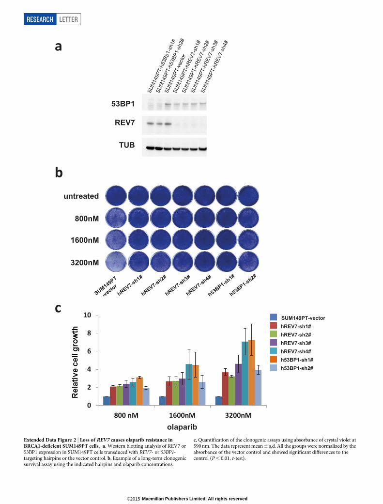
Extended Data Figure 2 | Loss of REV7 causes olaparib resistance inBRCA1-deficient SUM149PT cells. a, Western blotting analysis of REV7 or53BP1 expression in SUM149PT cells transduced with REV7- or 53BP1-targeting hairpins or the vector control. b, Example of a long-term clonogenicsurvival assay using the indicated hairpins and olaparib concentrations.
c, Quantification of the clonogenic assays using absorbance of crystal violet at590 nm. The data represent mean 6 s.d. All the groups were normalized by theabsorbance of the vector control and showed significant differences to thecontrol (P , 0.01, t-test).
RESEARCH LETTER
Macmillan Publishers Limited. All rights reserved©2015

Extended Data Figure 3 | Rev1 or Rev3 inhibition and PARPi sensitivity ofBrca12/2 p532/2 mammary tumour cells. a, b, Quantification of Rev1 (a) orRev3 (b) transcript levels in KB1P-G3 cells transduced with Rev1- or Rev3-targeting shRNAs or the vector control. Hprt was used as a control fortranscript expression. The data represent the mean 6 s.d. c, Long-termclonogenic survival assays of KB1P-G3 cells exposed to the indicated PARPinhibitors. d, Quantification of the clonogenic assay by determining theabsorbance of crystal violet at 590 nm. All the groups were normalized by the
absorbance of the vector control. The data represent the mean 6 s.d.e, f, Quantification of Rev7 transcript (e) or protein (f) levels in KB1P-G3 cellstransduced with Rev7-targeting shRNAs or the vector control. Hprt was used asa control for transcript expression; a-tubulin as a control for proteinexpression. The data represent mean 6 s.d. g, GFP-tagged REV7 mutantsrecruitment to sites of DNA damage was observed 5 min after 405 nm laserexposure (0.99 mW, 60% laser power, 50 s) in KB1P-B11 cells. Scale bar, 10mm.
LETTER RESEARCH
Macmillan Publishers Limited. All rights reserved©2015
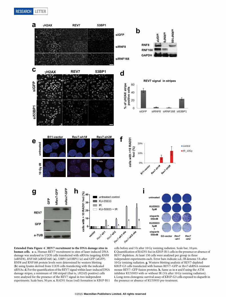
a b
RNF168
GAPDH
siGFP
siRN
F8
siRN
F168
RNF8
d c γH2AX REV7 53BP1
siG
FP
si53
BP1
0
20
40
60
siGFP siRNF8 siRNF168 si53BP1
% o
f γH
2AX
strip
e po
sitiv
e ce
lls
REV7 signal in stripes
γH2AX REV7 53BP1
siGFP
siRNF8
siRNF168
10 G
y IR
untr
eate
d
Rev7-sh1# B11-vector Rev7-sh2#
e
0%
10%
20%control
IR_10Gy
cells
with
>10
RAD
51
foci
(%)
f
REV7
GFP
a-TUB
GFP
hRev
7-G
FP
mR
ev7-
GFP
g h j
Rev7 -sh1
Rev7 -sh2
G3-vector
olaparib 550nM
untreated
KU55933 5μM
KU55933 5μM
+ olaparib 550nM
i
Extended Data Figure 4 | REV7 recruitment to the DNA damage sites inhuman cells. a–c, Human REV7 recruitment to sites of laser-induced DNAdamage was analysed in U2OS cells transfected with siRNAs targeting RNF8(siRNF8), RNF168 (siRNF168) (a), 53BP1 (si53BP1) (c) and GFP (siGFP).RNF8 and RNF168 protein levels were determined by western blotting(b) using lysates derived from U2OS cells transfecting with the indicatedsiRNAs. d, For the quantification of the REV7 signal within laser-induced DNAdamage stripes, a minimum of 100 striped (that is, cH2AX-positive) cellswere analysed for the presence of the REV7 signal in two independentexperiments. Scale bars, 50mm. e, RAD51 focus (red) formation in KB1P-B11
cells before and 5 h after 10 Gy ionizing radiation. Scale bar, 10mm.f, Quantification of RAD51 foci in KB1P-B11 cells in the presence or absence ofREV7 depletion. At least 150 cells were analysed per group in threeindependent experiments each. Error bars indicate s.d.; IR denotes 5 h after10 Gy ionizing radiation. g, Western blotting analysis of REV7-depletedKB1P-G3 cells transfected with human REV7–GFP or Rev7-shRNA-resistantmouse REV7–GFP fusion proteins. h, Same as in e and f using the ATMinhibitor KU55933 with or without IR (5 h after 10 Gy ionizing radiation).i, Long-term clonogenic survival assay of KB1P-G3 cells exposed to olaparib inthe presence or absence of KU55933 pre-treatment.
RESEARCH LETTER
Macmillan Publishers Limited. All rights reserved©2015

Extended Data Figure 5 | Loss of Rev7 does not cause PARPi resistancein Brca22/2 p532/2 or p532/2 mammary tumour cells in vitro.a, b, Quantification of Rev7 transcript levels in Brca22/2 p532/2 (KB2P-1.21 orKB2P-3.4) cells transduced with Rev7-targeting shRNAs or the vector control.Hprt was used as a control for transcript expression. The data represent themean 6 s.d. c–f, Long-term clonogenic survival assays and quantification ofKB2P-1.21 or KB2P-3.4 cells with or without Rev7 depletion exposed to the
indicated treatments. All the groups were normalized by the absorbance of thevector control. The data represent mean 6 s.d. g, Quantification of Rev7transcript levels in p532/2 (KP3.33) cells transduced with the indicatedconstructs. Hprt was used as a control for transcript expression and the datarepresent the mean 6 s.d. h, i, Long-term clonogenic survival assays andquantification of KP3.33 cells exposed to the indicated treatments. The datarepresent the mean 6 s.d.
LETTER RESEARCH
Macmillan Publishers Limited. All rights reserved©2015

Extended Data Figure 6 | Rev7 loss promotes end resection at DSBs inBRCA1-deficient cells after ionizing radiation. a, Quantification of RPA-positive a tracks in KB1P-B11 cells 1 or 2 h after irradiation with a particles.b, Quantification of RPA- and 53BP1-positive a tracks in KB1P-B11 cellstransfected with non-targeting control siRNAs or siRNAs against Ctip.c, Cell cycle analysis (BrdU incorporation and propidium iodine labelling)of KB1P-B11 cells transduced with the indicated constructs and siRNAs.d, e, Quantification of Rev7 transcript (d) or protein (e) levels in
BRCA1-deficient mES cells transduced with Rev7-targeting shRNAs orthe vector control. Hprt was used as a control for transcript expression,a-tubulin as a control for protein expression. The data represent mean 6 s.d.f, Representative images of surviving colonies of Brca12/2 mES cellstransduced with an empty vector control or Rev7-targeting shRNAs.g, Quantification of colony formation normalized to the vector control.h, Quantification of RAD51 foci in Brca12/2 mES cells that weretransduced with the indicated constructs.
RESEARCH LETTER
Macmillan Publishers Limited. All rights reserved©2015
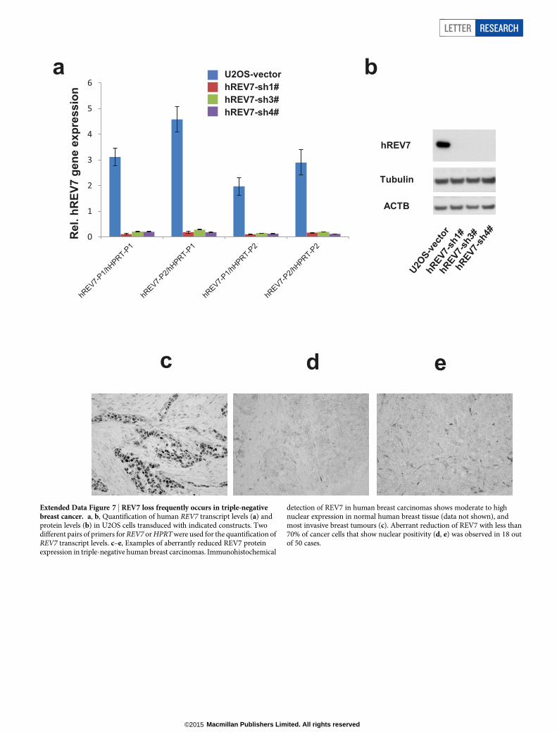
Extended Data Figure 7 | REV7 loss frequently occurs in triple-negativebreast cancer. a, b, Quantification of human REV7 transcript levels (a) andprotein levels (b) in U2OS cells transduced with indicated constructs. Twodifferent pairs of primers for REV7 or HPRT were used for the quantification ofREV7 transcript levels. c–e, Examples of aberrantly reduced REV7 proteinexpression in triple-negative human breast carcinomas. Immunohistochemical
detection of REV7 in human breast carcinomas shows moderate to highnuclear expression in normal human breast tissue (data not shown), andmost invasive breast tumours (c). Aberrant reduction of REV7 with less than70% of cancer cells that show nuclear positivity (d, e) was observed in 18 outof 50 cases.
LETTER RESEARCH
Macmillan Publishers Limited. All rights reserved©2015

Extended Data Figure 8 | REV7 is a downstream effector of 53BP1.a, Quantification of 53BP1 foci in KB1P-G3 cells in the presence or absenceof REV7 depletion. At least 100 cells were analysed per group in threeindependent experiments each. Error bars indicate s.d.; IR denotes 5 h after10 Gy ionizing radiation. b, REV7 or 53BP1 protein levels were determinedby western blotting of lysates derived from MEF cells transduced with theindicated control (CNTL) or Rev7- and 53bp1-targeting shRNA constructs.
c, d, RIF1 foci formation (c) after neocarzinostatin (NCS) treatment andquantification of RIF1 foci (d) in MEF cells in the presence or absence of REV7or 53BP1 depletion. e, Flag pull-downs were performed from 2 mg lysateprepared from 53bp12/2, 53bp12/2 plus 53BP1 and 53bp12/2 plus 53BP120AQ
MEFs after mock or neocarzinostatin treatment. Control, 53BP1 and53BP120AQ immunoprecipitates were incubated in HeLa nuclear extract(HNE, 2 mg) and then eluted with triple-Flag peptide. HA, haemagglutinin.
RESEARCH LETTER
Macmillan Publishers Limited. All rights reserved©2015

%CSR(norm
alizedIgA
+cells)
- + + + +CIT:
100
80
60
40
20
0
120 shCNTL
shREV7(3)
shREV3(1)
shREV3(2)
Extended Data Figure 9 | The effect of REV7 inhibition on CSR afterantigenic stimulation of CH12 cells. a, Rev7 messenger RNA levelsdetermined by qRT–PCR were normalized against b-actin (Actb) transcripts inthe indicated shRNA-transduced CH12 cell lines. The data represent themean 6 s.e.m. from two primer sets specific for Rev7 transcript. CNTL, control.b, 53BP1 protein of each group normalized to vector-transduced cells (CH12)was analysed by western blotting. c, IgH m and a germ-line transcripts (GLT)and Aid mRNA were estimated by semi-quantitative RT–PCR using twofoldserial dilutions of cDNA made from indicated CH12 cell lines 40 h after
stimulation. Hprt was used as a control for transcript expression.d, Representative flow cytometric profiles of shRNA-transduced CH12 B cellsstained with anti-IgA antibody 40 h after stimulation with the indicatedcytokines. e, Cells (CH12) were labelled with CFSE immediately beforecytokine stimulation as in Fig. 4d, and cell proliferation was assessed byflow cytometry at indicated time points. f, Quantification of CSR to IgA ofshRNA-transduced CH12 cells 40 h after stimulation with CD40 antibody, IL-4and TGF-b1 (CIT). Data represent the mean 6 s.d. from two independentexperiments performed in triplicate.
LETTER RESEARCH
Macmillan Publishers Limited. All rights reserved©2015