Prostate Cancer: Basic Mechanisms and Therapeutic ... - ANME
450
-
Upload
khangminh22 -
Category
Documents
-
view
0 -
download
0
Transcript of Prostate Cancer: Basic Mechanisms and Therapeutic ... - ANME


PROSTATECANCER
Basic Mechanisms andTherapeutic Approaches

This page intentionally left blank

Basic Mechanisms and
Therapeutic Approaches
Chawnshang Vhang
Editor
George H. Whipple ProfessorUniversity of Rochester, USA
World ScientificWeNEW JERSEY · LONDON · SINGAPORE · BEIJING · SAHANGHAI · HONG KONG · TAIPEI · CHENNAI

Library of Congress Cataloging-in-Publication DataProstate cancer : basic mechanisms and therapeutic approaches / edited by Chawnshang Chang.
p. cm.Includes bibliographical references and index.ISBN 981-256-067-X (alk. paper) 1. Prostate--Cancer. I. Chang, Chawnshang, 1955-
RC280.P7P75835 2005616.99'463--dc22
2004062902
British Library Cataloguing-in-Publication DataA catalogue record for this book is available from the British Library.
For photocopying of material in this volume, please pay a copying fee through the CopyrightClearance Center, Inc., 222 Rosewood Drive, Danvers, MA 01923, USA. In this case permissionto photocopy is not required from the publisher.
Typeset by Stallion PressEmail: [email protected]
All rights reserved. This book, or parts thereof, may not be reproduced in any form or by any means,electronic or mechanical, including photocopying, recording or any information storage and retrievalsystem now known or to be invented, without written permission from the Publisher.
Copyright © 2005 by World Scientific Publishing Co. Pte. Ltd.
Published by
World Scientific Publishing Co. Pte. Ltd.
5 Toh Tuck Link, Singapore 596224
USA office: 27 Warren Street, Suite 401-402, Hackensack, NJ 07601
UK office: 57 Shelton Street, Covent Garden, London WC2H 9HE
Printed in Singapore.
CONTENTS
List of Contributors and Affiliations xiii
Preface xvii
Editor — Chawnshang Chang, Ph. D. xix
1 Hormonal Therapy for Prostate Cancer: Clinical andExperimental Evidence 1Hiroshi Miyamoto and Chawnshang Chang
Introduction 1The AR and Androgens 1Strategies of Androgen Deprivation 3Concluding Remarks 20References 21
2 Immunotherapies for Prostate Cancer 33Kelley M. Harsch, Jason E. Tasch andWarren D. W. Heston
Introduction 33Inflammation 34The Immune System 36Targets of Immunotherapy 37Cytokines 38Growth Factors 39Tumor Antigens 39Monoclonal Antibody Therapy 40Modulation of T-Cells 42Vaccines 43
v
B245-FM 3/2/05 4:42 PM Page v

T-Bodies 46Summary 46References 47
3 Radiation Therapy and Hormonal Therapy forProstate Cancer 55Ralph A. Brasacchio
Introduction 55Conventional and Conformal Radiation Therapy 56Radiation Therapy With or Without Androgen
Ablation Therapy 58Hormone Therapy and Brachytherapy 65Potential Mechanisms of Androgen Ablation and
Radiation Therapy 65Future Directions 68References 68
4 Gene Therapy for Prostate Cancer 75Juan Antonio Jiménez, Chinghai Kao, Sang-Jin Lee,Chaeyong Jung and Thomas A. Gardner
Introduction 75Gene Therapy Strategy 76Tissue-Specific Promoters 79Past Approaches 84Future Directions 96Conclusion 97References 98
5 Chemotherapy for Prostate Cancer 107Samuel K. Kulp, Kuen-Feng Chen and Ching-Shih Chen
Introduction 107Present Chemotherapies for HRPC 108Future Therapies for HRPC 109Conclusions 124References 125
vi Contents
B245-FM 3/2/05 4:42 PM Page vi

6 Chemoprevention for Prostate Cancer 137Noahiro Fujimoto
Introduction 137Chemopreventive Agents, Rationale and Clinical Trials 138Others 148Conclusions 148References 149
7 Neuroendocrine Differentiation andAndrogen-Independence in Prostate Cancer 157Sonal J. Desai, Clifford G. Tepper andHsing-Jien Kung
Introduction 157Neuroendocrine Differentiation 159Androgen-Independent Growth 169Summary 174References 175
8 Biology of Prostatic Acid Phosphatase andProstate-Specific Antigen and Their Applications inProstate Cancer 191Suresh Veeramani, Ta-Chun Yuan, Siu-Ju Chen,Fen-Fen Lin and Ming-Fong Lin
Introduction 191Historical Review — Discovery and Clinical Applications
of PAcP and PSA 192PAcP 193PSA 197Androgen-Independent PSA Secretion 198Future Perspectives 202Acknowledgments 203References 204
Contents vii
B245-FM 3/2/05 4:42 PM Page vii

9 Epigenetics in Prostate Cancer 213Jose A. Karam, Elie A. Benaim, Hong Chen, Rey-Chen Pong and Jer-Tsong Hsieh
Introduction 213The Role of CpG Dinucleotides in DNA
Methylation 213Methyl Binding Proteins (MBP) 214DNA Methyltransferases (DNMTs) 215DNMT Inhibitors 216Histones 217Histone Deacetylases (HDACs) 220Histone Deacetylase Inhibitors (HDIs) 220The Interaction Between DNA Methylation and
Histone Modifications in Gene Regulation 221Epigenetics in the Pathogenesis and Diagnosis
of PCa 222Membrane Receptors 224Nuclear Receptors 225Nuclear Proteins 227Conclusion 228References 228
10 Significance of 5�-Reductase in Prostate Cancer 243Jun Shimazaki
Introduction 2435�-Reductase 243Formation and Metabolism of DHT in
Cancerous Prostate 245Risk of DHT Formation 246Treatment with Inhibitors 249Prevention 250Conclusion 252References 252
viii Contents
B245-FM 3/2/05 4:42 PM Page viii

11 Roles of Vitamin E in Prostate and Prostate Cancer 263Shuyuan Yeh, Jing Ni, Eugene Chang, Yi Yin and Ming Chen
Introduction 263Vitamin E and its Analogs 264Vitamin E Absorption and Transport 264Functional Mechanisms of Vitamin E in Prostate Cancer 265In vivo Animal Study of Vitamin E’s Role in Prostate and
Prostate Cancer 268Clinical Study of Vitamin E in Prostate Cancer 269Summary 271References 272
12 Vitamin D and Prostate Cancer 277Yi-Fen Lee, Huei-Ju Ting and Bo-Ying Bao
Introduction 277Epidemiology Study 278Vitamin D Action 279Mechanism of Anti-tumor Action in Prostate Cancer
by Vitamin D 280Development of New Vitamin D Analogs and their use
in Combination Therapy for Prostate Cancer 283Vitamin D-Based Clinical Trials 284Loss of Vitamin D Anti-proliferative Responsiveness
in Prostate Cancer 285Future Perspectives 286References 287
13 Functions of Estrogen Receptor in Prostate andProstate Cancer 293Shuyuan Yeh, Ming Chen, Jing Ni, Yi Yin, Eugene Chang, Min Zhang and Xingqing Wen
Introduction 293Distribution of ER� and ER� in Prostate Tissues,
Cancer Specimens and Cancer Cell Lines 294
Contents ix
B245-FM 3/2/05 4:42 PM Page ix

Estrogen Regulated Genes in Prostate Cancer 296ER Coregulators in Prostate 297Histological Changes in Prostates of �ERKO and
�ERKO Mice 298Estrogen Imprinting Effect on the Development of
Prostate 298Estrogen Treatment of �ERKO, �ERKO, and
Hypogonadal (hpg) Mouse Models 299Estrogen Effect on Initiation, Growth, and Progression 302
of Prostate CancerConclusion 305References 305
14 Epidemiology of Prostate Cancer 315Ann W. Hsing and Anand Chokkalingam
Introduction 315Rates and Patterns 321Risk Factors 331Challenges of Studies with Common Polymorphisms 344Summary 346References 347
15 Profiling Gene Expression Changes inProstate Carcinoma 365Peter S. Nelson
Introduction 365Methods for Profiling Gene Expression Alterations 366Microarray Studies of Gene Expression in
Prostate Carcinoma 367Transcript Profiling and Predicting Cancer Outcomes 369Proteomic Approaches for Assessing Gene Expression
in Prostate Carcinoma 374Conclusion and Future Directions 377Acknowledgments 378References 378
x Contents
B245-FM 3/2/05 4:42 PM Page x

16 Study of Androgen–Androgen Receptor Roles inProstate Cancer using Mice Lacking FunctionalProstate Androgen Receptor 383Chun-Te Wu, Shuyuan Yeh, Qingquan Xu, Zhiming Yang, Philip Chang, Yueh-Chiang Hu and Chawnshang Chang
Introduction 383Generation of Androgen Receptor Knockout (ARKO) Mice 384Prostate Development and Carcinogenesis in
Prostate-Specific ARKO Mice 385TRAMP Mice Lacking the Endogenous AR but Carrying
the T877A Mutated Transgene 386ARKO Mice with AR-97Q and AR-24Q
Transgene Expression 387Inducible ARKO and ARKO TRAMP Mice 387ARKO Human Prostate Cancer CWR22R Cells 388Conclusion 390References 390
17 Capturing Signal Anomalies of Human ProstateCancer into Mouse Models 393Hong Wu, Ani Khodavirdi and Pradip Roy-Burman
Introduction 393Cell Surface Signaling Molecules 395Intracellular Signaling Molecules 400Collaboration Between Signaling Molecules 408Summary and Conclusions 410Acknowledgments 411References 412
Index 423
Contents xi
B245-FM 3/2/05 4:42 PM Page xi

This page intentionally left blank

LIST OF CONTRIBUTORS AND AFFILIATIONS
Bo-Ying Bao, Departments of Pathology & Laboratory Medicine andUrology, University of Rochester Medical Center, Rochester, New York
Elie A. Benaim, Department of Urology, UT Southwestern MedicalCenter, Dallas, TX
Ralph A. Brasacchio, Department of Radiation Oncology and JP WilmotCancer Center, University of Rochester Medical Center, Rochester,New York
Chawnshang Chang, George H. Whipple Lab for Cancer Research,Departments of Urology, Pathology, Radiation Oncology, and the CancerCenter, University of Rochester, Rochester, New York
Eugene Chang, Departments of Urology and Pathology, University ofRochester Medical Center, Rochester, New York
Philip Chang, George H. Whipple Lab for Cancer Research, Departmentof Urology, Pathology, Radiation Oncology, and the Cancer Center,University of Rochester, Rochester, New York
Ching-Shih Chen, Division of Medicinal Chemistry and Pharmacognosy,College of Pharmacy, The Ohio State University, Columbus, Ohio
Hong Chen, Department of Urology, UT Southwestern Medical Center,Dallas, TX
Kuen-Feng Chen, Division of Medicinal Chemistry and Pharmacognosy,College of Pharmacy, The Ohio State University, Columbus, Ohio
Ming Chen, Departments of Urology and Pathology, University ofRochester Medical Center, Rochester, New York
xiii
B245-FM 3/2/05 4:42 PM Page xiii

Siu-Ju Chen, Department of Biochemistry and Molecular Biology,University of Nebraska Medical Center, Omaha, NE
Anand Chokkalingam, Celera Diagnostics, LLC, Alameda, California
Sonal J. Desai, Department of Biological Chemistry and Cancer Center,University of California at Davis, Sacramento, CA
Noahiro Fujimoto, Department of Urology, University of Occupationaland Environmental Health, Iseigaoka, Yahatanishiku, Kitakyushu, Japan
Thomas A. Gardner, Urology Research Laboratory, Departments ofUrology, Microbiology and Immunology, Walther Oncology Center,Indiana University Medical Center, Indianapolis, Indiana
Kelley M. Harsch, The Cleveland Clinic Foundation, The Lerner ResearchInstitute, Cleveland, Ohio
Warren D.W. Heston, The Cleveland Clinic Foundation, The LernerResearch Institute, Cleveland, Ohio
Jer-Tsong Hsieh, Department of Urology, UT Southwestern MedicalCenter, Dallas, TX
Ann W. Hsing, Division of Cancer Epidemiology and Genetics, NationalCancer Institute, Bethesda, Maryland
Yueh-Chiang Hu, George H. Whipple Lab for Cancer Research,Departments of Urology, Pathology, Radiation Oncology, and the CancerCenter, University of Rochester, Rochester, New York
Juan Antonio Jiménez, Urology Research Laboratory, Departments ofUrology, Microbiology and Immunology, Walther Oncology Center,Indiana University Medical Center, Indianapolis, Indiana
Chaeyong Jung, Urology Research Laboratory, Departments of Urology,Microbiology and Immunology, Walther Oncology Center, IndianaUniversity Medical Center, Indianapolis, Indiana
Chinghai Kao, Urology Research Laboratory, Departments of Urology,Microbiology, and Immunology, Walther Oncology Center, IndianaUniversity Medical Center, Indianapolis, Indiana
xiv List of Contributors and Affiliations
B245-FM 3/2/05 4:42 PM Page xiv

Jose A. Karam, Department of Urology, UT Southwestern Medical Center,Dallas, TX
Ani Khodavirdi, Department of Pathology, Keck School of Medicine,University of Southern California, Los Angeles, California
Samuel K. Kulp, Division of Medicinal Chemistry and Pharmacognosy,College of Pharmacy, The Ohio State University, Columbus, Ohio
Hsing-Jien Kung, Department of Biological Chemistry and CancerCenter, University of California at Davis, Sacramento, CA
Sang-Jin Lee, Urology Research Laboratory, Departments of Urology,Microbiology and Immunology, Walther Oncology Center, IndianaUniversity Medical Center, Indianapolis, Indiana
Yi-Fen Lee, Departments of Pathology & Laboratory Medicine andUrology, University of Rochester Medical Center, Rochester, New York
Fen-Fen Lin, Department of Biochemistry and Molecular Biology,University of Nebraska Medical Center, Omaha, NE
Ming-Fong Lin, Department of Biochemistry and Molecular Biology,Department of Surgery/Urology, College of Medicine and Eppley Institutefor Cancer Research, University of Nebraska Medical Center, Omaha, NE
Hiroshi Miyamoto, Departments of Pathology & Laboratory Medicine andUrology, University of Rochester Medical Center, Rochester, New York
Peter S. Nelson, Fred Hutchinson Cancer Research Center, Seattle,Washington
Jing Ni, Departments of Urology and Pathology, University of RochesterMedical Center, Rochester, New York
Rey-Chen Pong, Department of Urology, UT Southwestern MedicalCenter, Dallas, TX
Pradip Roy-Burman, Department of Pathology and Department ofBiochemistry and Molecular Biology, Keck School of Medicine,University of Southern California, Los Angeles, California
List of Contributors and Affiliations xv
B245-FM 3/2/05 4:42 PM Page xv

Jun Shimazaki, Department of Urology, Graduate School of Medicine,Chiba University, Chiba, Japan
Jason E. Tasch, The Cleveland Clinic Foundation, The Lerner ResearchInstitute, Cleveland, Ohio
Huei-Ju Ting, Departments of Pathology & Laboratory Medicine andUrology, University of Rochester Medical Center, Rochester, New York
Clifford G. Tepper, Department of Biological Chemistry and CancerCenter, University of California at Davis, Sacramento, CA
Suresh Veeramani, Department of Biochemistry and Molecular Biology,University of Nebraska Medical Center, Omaha, NE
Xingqing Wen, Departments of Urology and Pathology, University ofRochester Medical Center, Rochester, New York
Chun-Te Wu, George H. Whipple Lab for Cancer Research, Departmentsof Urology, Pathology, Radiation Oncology, and the Cancer Center,University of Rochester, Rochester, New York
Hong Wu, Department of Molecular and Medical Pharmacology, Universityof California, Los Angeles School of Medicine, Los Angeles, California
Qingquan Xu, George H. Whipple Lab for Cancer Research, Departmentsof Urology, Pathology, Radiation Oncology, and the Cancer Center,University of Rochester, Rochester, New York
Zhiming Yang, George H. Whipple Lab for Cancer Research, Departmentsof Urology, Pathology, Radiation Oncology, and the Cancer Center,University of Rochester, Rochester, New York
Shuyuan Yeh, Departments of Urology and Pathology, University ofRochester Medical Center, Rochester, New York
Yi Yin, Departments of Urology and Pathology, University of RochesterMedical Center, Rochester, New York
Ta-Chun Yuan, Department of Biochemistry and Molecular Biology,University of Nebraska Medical Center, Omaha, NE
Min Zhang, Departments of Urology and Pathology, University ofRochester Medical Center, Rochester, New York
xvi List of Contributors and Affiliations
B245-FM 3/2/05 4:42 PM Page xvi

xvii
PREFACE
Prostate cancer is the second leading cause of death in men. CharlesHuggins first found that metastatic prostate cancer responds to androgen-ablation therapy, which heralded the beginning of a new era of prostatecancer therapy. Later, Andrew Schally and others showed that advancedprostate cancer responded to the LHRH agonist as decreased serumtestosterone level to 25% and marked reduction in cancer-associated bonepain. The discovery of androgen receptor (AR) led to the screening ofchemical libraries for AR blockers. Since then, antiandrogens, includingflutamide and casodex, have been in continual use as therapeutic agents.Yet, with either androgen ablation via surgical or medical castration, withor without additional combination of various antiandrogens, eventuallymost of, if not all, prostate cancers still progress into the HormoneRefractory stage and the detailed reasons for this remain unclear. Cloningof the AR, generation of AR antibodies, finding of AR coregulators andtheir applications to prostate cancer progression reveals the essential rolesof AR in the prostate cancer progression and opened a new approach forAR ablation therapy by targeting the AR, instead of androgens, to battlethe prostate cancer.
Chapters 1 to 6 discuss current effective hormonal therapy,immunotherapy, chemotherapy, radiation therapy, and gene therapy, aswell as androgen ablation therapy. Chapters 7 to 15 discuss the recentadvances in the field of study of the basic mechanisms behind the growthof prostate cancer and how some of these mechanisms can be used to treatprostate cancer. Chapters 16 and 17 include recent research in the studyof prostate cancer in newly developed mouse model systems. Many ofthese studies have distinct potential advantages as they lead towardadvances in the clinical treatments of and drug therapies for these andro-gen-related diseases. We feel our book should be of interest as both
B245-FM 3/2/05 4:42 PM Page xvii
a study guide and research reference for students, basic scientists, andclinicians.
I would like to dedicate this book to my PhD advisor Dr. Liao, whoseresearch philosophy and Taiwanese dignity deeply influences my contin-ued academic career. I would also like to thank Drs. Carbone, Wilding,Messing, Lardy, and Gorski for their help in establishing my independentacademic research career at the University of Wisconsin and theUniversity of Rochester. Finally, I thank my copyeditors, Mrs. Karen Wolfand Dr. Loretta Collins, for their invaluable editorial and proofreadingassistance.
Chawnshang Chang, PhDUniversity of Rochester, New York, USA
xviii Preface
B245-FM 3/2/05 4:42 PM Page xviii

EDITOR — CHAWNSHANG CHANG, PH.D.
Chawnshang Chang was born and raised inTaiwan. In 1997 he became the GeorgeHoyt Whipple Distinguished Professor at theUniversity of Rochester in Rochester, New York.
He received his B.S. from the NationalTaiwan University and Ph.D. from theUniversity of Chicago in 1985. In 1990, at theUniversity of Wisconsin-Madison he began hisindependent research career and was promotedto full professor in 1996.
Dr. Chang has published more than 250 peer-reviewed articles related to AR and TR2/TR4orphan receptors. His landmark discovery, the first cloning of the androgenreceptor (AR) cDNAs (Science, 240: 324–326, 1988), provided the frame-work for the subsequent studies of basic androgen mechanisms and theirclinical applications. Several androgen-related diseases, such as prostatecancer, Testicular Feminization Syndrome, and Kennedy’s Neuron Diseasewere linked to mutations of the AR. In recent years several new androgensor anti-androgens were developed based on the targets of the AR.
In 1996, Dr. Chang’s Lab isolated the first AR coregulator and withina few years identified and isolated another 10 AR coregulators (EndocrineReview, 23: 155–200, 2000), which modulate AR functions, and enableAR to cross-talk to many signal transduction pathways. Generation of thefirst floxed AR mouse (PNAS, 2002) further provided the first tissue-specific AR knockout mouse, as well as the first female mouse withoutfunctional AR. These mice allow the in vivo study of androgen actions inselective tissues, including prostate, testis, liver, muscle, bone, and thefemale breast and ovary.
xix
B245-FM 3/2/05 4:42 PM Page xix

Many Universities have presented Dr. Chang with various awards andhonorary professorships. His generosity is well known in the academiccommunity and Taiwanese society and more than 700 labs throughout theworld have benefited by collaborations using his AR reagents.
xx Editor
B245-FM 3/2/05 4:42 PM Page xx

1HORMONAL THERAPY FOR PROSTATE CANCER:
CLINICAL AND EXPERIMENTAL EVIDENCE
Hiroshi Miyamoto and Chawnshang Chang
Departments of Pathology & Laboratory Medicine and UrologyUniversity of Rochester Medical Center
Rochester, New York, USA
Introduction
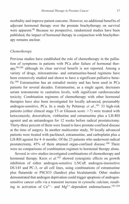
The role and mechanism of androgen function have been studied in a varietyof androgen target organs, including the prostate. As is the case with normalprostate development, the growth of prostatic neoplasms is generallydependent on androgens, especially on 5�-dihydrotestosterone (DHT). Since1941 when Huggins and Hodges1 published their Nobel Prize-winningstudy on the effects of hormone manipulation in patients with metastaticprostate cancer (PCa), hormonal therapy remains the critical therapeuticoption for advanced disease. Multiple strategies have been used to reduceserum levels of androgens or interfere with their function via the androgenreceptor (AR) (Fig. 1). However, considerable uncertainty remains as to theappropriate choice/timing and actual benefits of hormonal therapy in vari-ous situations. Indeed, PCa is still the second leading cause of cancer-relateddeath among men in the United States.2 In this chapter, we systematicallyreview clinical and experimental evidence supporting current strategies ofhormonal therapy in PCa.
The AR and Androgens
The AR, a member of the nuclear receptor superfamily, functions as aligand-inducible transcription factor that regulates the expression of target
1
B245-ch01 3/2/05 4:28 PM Page 1

genes in response to ligands in target cells.3,4 Recent studies have alsorevealed that the AR modulates transcription by recruitment of coregula-tors that influence a number of functional properties of the receptor, includ-ing ligand selectivity and DNA binding capacity (reviewed in Ref. 5).
Testosterone is secreted by Leydig cells in the testis and is the major sexhormone circulating within the blood of males. In a variety of androgen-sensitive tissues like the prostate, testosterone is irreversibly converted by5�-reductases to the more potent androgen, DHT.4,6 Upon binding ofandrogens, the androgen-AR complexes form homodimers, and theytranslocate into the nucleus and bind to androgen responsive elements
2 H. Miyamoto & C. Chang
Pituitary gland
Testis Adrenal gland
LH-RH
LH ACTH
T
DHT5α -R
Adrenal
Estrogens
LH-RH agonists
LH-RH antagonists
Antiandrogens
5α-R inhibitors
Adrenal androgeninhibitors
Hypothalamus
90%5–10%
AR
androgens
Surgicalcastration
Prostatecancer
CRH
Fig. 1. Strategies for hormonal therapy. LH-RH�Luteinizing hormone-releasing hormone;CRH�corticotropin-releasing hormone; LH�luteinizing hormone; ACTH�adrenocorti-cotropic hormone; T�testosterone; 5�-R�5�-reductase; DHT�5�-dihydrotestosterone;AR�androgen receptor.
B245-ch01 3/2/05 4:28 PM Page 2

located on target genes, such as prostate-specific antigen (PSA), which isclinically used for the detection and monitoring of PCa recurrence and pro-gression. Besides testosterone and DHT, several precursors of testosteronemainly secreted by adrenal glands, dehydroepiandrosterone (DHEA),DHEA sulfate, �4-androstenedione, and �5-androstenediol, can also stim-ulate the AR through their conversion to testosterone/DHT in peripheraltissues, including the prostate, or by directly binding to the AR.7–10
Strategies of Androgen Deprivation
Multiple approaches at androgen deprivation have been used for the treat-ment of PCa (Fig. 1). The agents and strategies used for androgen depri-vation therapies are listed in Table 1.
Surgical Castration
Surgical castration by bilateral orchiectomy is the most immediate method toreduce circulating testosterone by �90% within 24 hours,11 and there is norisk of a paradoxical flare of the disease. Since the 1960s, the VeteransAdministration Cooperative Urological Research Group (VACURG) trials,the earliest large-scale randomized studies of hormonal therapy, demon-strated the clinical effectiveness of surgical castration.12,13 Compared toplacebo, orchiectomy retarded cancer progression in advanced cases, but noclear survival advantage for castration over placebo was seen. Recent clini-cal studies (i.e. surgical vs. chemical castration) are discussed later. Althoughsurgical castration may be underused, some studies suggest that manypatients prefer this approach for the reasons of convenience and cost.14 Onthe other hand, other studies suggest that this treatment approach is unac-ceptable to many patients, causing considerable psychological problems,with irreversible impairment in libido and erectile function in most cases.15,16
Medical Castration
Diethylstilbestrol (DES)
In the 1940s, the first reversible medical castration method was achievedby administration of DES, a semi-synthetic estrogen compound.1 The
Hormonal Therapy in Prostate Cancer 3
B245-ch01 3/2/05 4:28 PM Page 3

4H
. Miyam
oto & C
. Chang
Table 1. Treatment Options as Hormonal Therapy for Prostate Cancer
Modality Methodology Mechanism/Action Advantages Disadvantages
Surgical Bilateral Orchiectomy, ↓T Rapid ablation of Definitive castrationcastration orchiectomy testicular T Associated psychological problems
Relatively simple Irreversible loss of libido/procedure, lower cost sexual potency
Reduced muscle mass/energyHot flashesAnemia/osteoporosisUnaffected adrenal androgens
Medical Estrogens (DES) Suppresses LH-RH Cardiovascular events (estrogens)castration secretion, ↓LH, ↓T Flare phenomenon (LH-RH agonists)
Direct effect via ER (?) Reversible castration Reduced muscle mass/energyLH-RH agonists Suppresses LH-RH Ablation of testicular T Loss of libido/sexual potency
(Leuprolide, secretion, ↓LH, ↓T More acceptable than Hot flashesGoserelin) orchiectomy Anemia/osteoporosis
LH-RH antagonists Antagonizes LH-RH Unaffected adrenal androgens(Abarelix) receptor, ↓LH, ↓T
CAB Castration � Ablation of testicular T � More effective (?) Increased side effectsantiandrogen competitive inhibition Antiandrogen withdrawal response
of adrenal androgens
B245-ch01 3/2/05 4:28 PM Page 4
Horm
onal Therapy in P
rostate Cancer
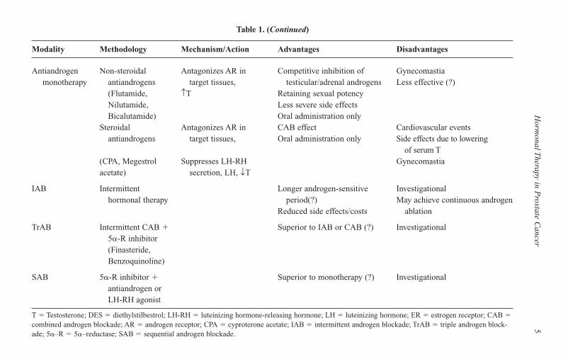
5Table 1. (Continued)
Modality Methodology Mechanism/Action Advantages Disadvantages
Antiandrogen Non-steroidal Antagonizes AR in Competitive inhibition of Gynecomastiamonotherapy antiandrogens target tissues, testicular/adrenal androgens Less effective (?)
(Flutamide, ↑T Retaining sexual potencyNilutamide, Less severe side effectsBicalutamide) Oral administration only
Steroidal Antagonizes AR in CAB effect Cardiovascular eventsantiandrogens target tissues, Oral administration only Side effects due to lowering
of serum T(CPA, Megestrol Suppresses LH-RH Gynecomastiaacetate) secretion, LH, ↓T
IAB Intermittent Longer androgen-sensitive Investigational hormonal therapy period(?) May achieve continuous androgen
Reduced side effects/costs ablation
TrAB Intermittent CAB � Superior to IAB or CAB (?) Investigational5�-R inhibitor (Finasteride, Benzoquinoline)
SAB 5�-R inhibitor � Superior to monotherapy (?) Investigationalantiandrogen or LH-RH agonist
T � Testosterone; DES � diethylstilbestrol; LH-RH � luteinizing hormone-releasing hormone; LH � luteinizing hormone; ER � estrogen receptor; CAB �combined androgen blockade; AR � androgen receptor; CPA � cyproterone acetate; IAB � intermittent androgen blockade; TrAB � triple androgen block-ade; 5�–R � 5�–reductase; SAB � sequential androgen blockade.
B245-ch01 3/2/05 4:28 PM Page 5
VACURG studies identified equivalent overall survival rate in the DESgroup (5 mg/day) to the orchiectomy group, but non-cancer-related deaths,most of which were cardiovascular events, were noted.12,13 Subsequent tri-als have shown that DES at 3 mg/day is equivalent to other treatmentoptions in overall survival rates.17–21 However, cardiovascular toxicity withevents including myocardial infarction, deep vein thrombosis, edema, andtransient ischemic attack was observed in 8%–33% of patients.Gynecomastia was also significantly seen in patients with 3 mg/day DES.A low-dose of DES (1 mg/day) was also evaluated,13,22 but whether DES at1 mg/day is as effective and safe as other treatment options is still contro-versial. After the development of luteinizing hormone-releasing hormone(LH-RH) agonists, with fewer cardiovascular events and no resultinggynecomastia, DES is now only rarely used as a first-line hormonal treat-ment in North America. Instead, several studies have evaluated the efficacyof DES as a salvage therapy after failure of first-line androgen deprivation.Recent studies, using 1–3 mg/day DES with or without anti-thromboticagents, including warfarin and aspirin,23,24 identified response rates byPSA measurement to be 43%–79% with median durations of progressionof 6–7.5 months and with 2.8%–28% cardiovascular events.
It was generally believed that the primary mechanism of action of DESwas to decrease androgen levels through hypothalamic-pituitary suppres-sion, but recent evidence indicates that the mechanism is probably morecomplex. Kitahara et al. reported stronger suppression of testosterone byDES than by surgical castration or other means of chemical castration,such as the administration of a LH-RH agonist.25 The same group alsosuggested that DES might reduce serum DHEA sulfate.26 A direct cyto-toxic effect of estrogens has also been suggested in PCa in vitro, presum-ably through both estrogen receptor (ER)-dependent and ER-independentpathways.27–29 This is consistent with the finding that phytoestrogens,which have steroidal structures similar to estrogens and are found in avariety of plant foods, inhibit PCa cell proliferation.49 Indeed, ER� hasbeen detected in human PCa cell lines, including LNCaP, PC-3 andDU145, and in normal and malignant prostate tissues, whereas ER� isexpressed in PC-3 cells and in stromal (not epithelial) cells of theprostate.30–32 Furthermore, it is suggested that loss of ER� in PCa tissuesis associated with tumor progression.32,33 These findings might be able to
6 H. Miyamoto & C. Chang
B245-ch01 3/2/05 4:28 PM Page 6
explain the evidence that administration of DES could be more effectivethan other androgen ablation therapies in suppressing PCa growth if unfa-vorable side effects of DES are not considered.12,13 On the other hand, wepreviously showed that a natural estrogen, 17�-estradiol, but not DES,increased AR transcriptional activity in PCa cells.34
LH-RH Agonists and Antagonists
The introduction of LH-RH analogues, obtaining medical castration, haslead to a dramatic change in the treatment of advanced PCa.35 In theUnited States, two LH-RH agonists are commercially available: leuprolideacetate and goserelin acetate.
LH-RH is generally secreted by the hypothalamus in pulses, leading topulsatile secretion of LH by the pituitary. This in turn promotes testos-terone secretion by the Leydig cells of the testes. However, constantly highlevels of LH-RH that occur with agonist administration down-regulate thereceptors in the pituitary, inhibit LH secretion, and thereby reduce testos-terone production. In addition, some studies have suggested a directinhibitory effect of LH-RH via LH-RH receptors in PCa cells.36,37
Several randomized studies showed the equivalent effectiveness betweensurgical castration and LH-RH agonist administration.38,39 Recently, depotLH-RH agonist preparations have been developed, which last 3 to 4 monthsand have the same efficacy as classical preparations.40 Thus, the depotpreparations have now become the most widely used form of androgen dep-rivation. Side effects of LH-RH agonists include hot flashes, reduced libido,and osteoporosis.41 In addition, LH-RH agonists often cause an initial surgeof LH release, with a corresponding increase in serum testosterone andDHT lasting 1 to 2 weeks. This surge may stimulate PCa growth with aworsening of related symptoms, which is known as the flare phenomenon.42
Therefore, administration of an antiandrogen or estrogen for a week beforeand during the first few weeks of LH-RH agonist therapy is often used in anattempt to limit the clinical sequelae caused by this hormonal surge.42,43
LH-RH receptor antagonists recently have been developed for andro-gen deprivation.44 Since abarelix, the first peptide antagonist, directlyblocks the binding of LH-RH to its receptor without agonist activity, thereis no initial flare phenomenon as occurs with LH-RH agonists.44,45 Recent
Hormonal Therapy in Prostate Cancer 7
B245-ch01 3/2/05 4:28 PM Page 7

clinical studies have demonstrated that abarelix monotherapy achievesmedical castration and a reduction of serum PSA levels to the same extentachieved by LH-RH agonists.46–48 However, long-term follow-up studiesare necessary to determine whether LH-RH antagonists can be routinelyused for advanced prostate cancer.
Combined Androgen Blockade (CAB)
Monotherapy with surgical or medical castration results in marginal or nodecline of adrenal androgens that not only can be converted to testos-terone/DHT but are likely to possess intrinsic androgenic activity.9,10,49
Thus, men who undergo castration still have relatively high levels (up to40%) of DHT and 5%–10% of testosterone.7,50 The basis of CAB (alsocalled maximal androgen blockade) is concomitant neutralization of bothtesticular and adrenal sources of androgens. CAB consists of treatmentwith a LH-RH agonist or surgical castration combined with a non-steroidalantiandrogen. Antiandrogens include a number of compounds that interferewith the binding of androgens to the AR in the target cell, which ultimatelyprevents the activation of AR pathways in those cells. CAB has been advo-cated as the most effective hormonal treatment for patients with advancedPCa. However, this approach implies increased side effects and cost, andthere are few supportive data showing a meaningful improvement in sur-vival associated with the addition of antiandrogen.51,52
Several early, randomized trials demonstrated a significant survivaladvantage of CAB in patients with advanced PCa, compared to castrationalone.53–56 In 1998, however, Eisenberger et al.57 reported a trial of 1387patients with metastatic PCa who were randomized to surgical castrationand placebo vs. flutamide. There were no differences in progression-free oroverall survival between the two arms. Several factors were hypothesizedto explain the discrepancy between the results of this study and earlierreports. First, patients in this study might have had less aggressive disease.Second, castration with a LH-RH agonist, especially a daily regimen ofleuprolide injections in the first study,53 might not have been as completeas surgical castration. Third, the LH-RH agonist plus placebo group mayhave experienced initial flare leading to worsening the disease. In 2000, theProstate Cancer Trialists’ Collaborative Group52 published a meta-analysis
8 H. Miyamoto & C. Chang
B245-ch01 3/2/05 4:28 PM Page 8

of 27 trials of CAB vs. monotherapy involving 8275 patients with advancedPCa. The difference in the 5-year survival rate was not statistically signifi-cant (25.4% with CAB vs. 23.6% with castration alone). However, a statis-tically significant difference (p � 0.02) in favor of castration plus anon-steroidal antiandrogen was observed. More recently, another meta-analysis of 20 randomized trials concluded that there was a 5% improve-ment in 5-year survival (30% vs. 25%) with CAB.58 However, only 7 of the20 studies might be considered as high-quality trials and no significantimprovement with CAB was seen in the meta-analysis of these 7 studies.In summary, recent data show that CAB provides a minimal advantage (upto 5% improvement in 5-year survival) over castration monotherapy. It isgenerally recommended to use an antiandrogen before and during the firstseveral weeks of LH-RH agonist therapy to prevent possible symptoms ofthe flare. With these data, prolonged treatment beyond 1 month with CABmay not be the first choice of hormonal therapy for advanced PCa.
Antiandrogen Monotherapy
There are two types of antiandrogens, steroidal, such as cyproterone acetate(CPA) and megestrol acetate, and non-steroidal, such as flutamide, nilu-tamide, and bicalutamide. As noted, antiandrogens are generally used in con-junction with castration as CAB. However, castration based approaches areusually associated with side effects, which have a negative impact on qualityof life (QOL). Monotherapy with a (non-steroidal) antiandrogen is becomingan increasingly attractive alternative therapeutic approach. Most of non-steroidal antiandrogens increase within normal physiological range theserum levels of androgens due to the suppression of the pituitary feed-back.Thus, this means of androgen blockade can preserve gonadal function andtherefore provide potential QOL benefits, particularly in terms of retainedpotency and libido, no muscle weakness, and less bone demineralization.
Flutamide
Flutamide was the first non-steroidal antiandrogen that was widely usedas a component of CAB. However, the use of flutamide monotherapy foradvanced PCa has not been extensively studied in phase III trials.59 Initialopen studies assessing the clinical efficacy of flutamide as monotherapy
Hormonal Therapy in Prostate Cancer 9
B245-ch01 3/2/05 4:28 PM Page 9
were reviewed by Delaere and Van Thillo.60 Among approximately 500previously untreated patients with advanced PCa, 68% achieved at least apartial response. But most studies were relatively small, and there seemedto be differences in the criteria of response. Several trials have comparedthe efficacy of flutamide as monotherapy with that of DES, orchiectomy,or CAB. Boccardo reviewed these studies and found no significant differ-ences in response rates/duration among these groups.52 In a double-blindrandomized study to compare the efficacy of flutamide with 3 mg/dayDES,21 however, DES produced significantly longer overall survival thanflutamide (43.2 months vs. 28.5 months). Because some adverse effects,such as hepatotoxicity, were noted, the rate of treatment withdrawal forany drug-related adverse events was highest with flutamide among 3 non-steroidal antiandrogens.59 There have been no comparative studies of theefficacy of different non-steroidal antiandrogens as monotherapy.
Nilutamide
No randomized studies of monotherapy with nilutamide or comparativestudies with any other hormonal therapy have been reported. One smallstudy (26 patients) evaluated the efficacy of nilutamide as monotherapy,demonstrating that 21 (91%) of the 23 evaluable previously untreatedpatients with metastatic PCa had a response, with a median overall sur-vival of 23 months.61 The survival rate in this study might be less than thatachieved by CAB with nilutamide.62 In addition, nilutamide was associ-ated with a high incidence (31%) of visual problems (light-dark adapta-tion disorders).61 Other unique adverse effects of nilutamide, when usedas either monotherapy or a component of CAB, include alcohol intoler-ance and interstitial pneumonitis.61,62 Nilutamide has been reported tocause a higher incidence of nausea and vomiting than the other non-steroidal antiandrogens, whereas the incidence of diarrhea and gyneco-mastia is lower with nilutamide than flutamide.59,62 These results maydiscourage conducting larger trials with nilutamide monotherapy.
Bicalutamide
Of available non-steroidal antiandrogens, bicalutamide as monotherapyhas been most extensively studied. In early comparative trials using
10 H. Miyamoto & C. Chang
B245-ch01 3/2/05 4:28 PM Page 10

bicalutamide at 50 mg/day, castration was shown to be superior to bicalu-tamide monotherapy, in terms of survival rate in patients with metastaticdisease.63 However, subsequent trials with bicalutamide at 100 or150 mg/day have revealed equivalent efficacy between bicalutamidemonotherapy and surgical or medical castration.52,64,65 Other comparativestudies also showed no statistically significant differences in survivalbetween bicalutamide at 150 mg/day monotherapy and CAB (castrationwith flutamide or nilutamide) with better tolerability in the bicalutamidemonotherapy group.66,67 Bicalutamide at 150 mg/day has been shown tohave a more favorable side effect profile than flutamide and nilutamide,59
although there was still a high risk of gynecomastia and breast pain. Sincebicalutamide has a longer elimination half-life of approximately 6 daysthan flutamide (6 hours) or nilutamide (56 hours), it can be given oncedaily vs. flutamide (or nilutamide in many studies) dosed 3 timesdaily.62,68,69 The most recent and largest trials involving 8113 patients con-firmed these observations (clinical efficacy, QOL benefit, and tolerabil-ity).70 Thus, bicalutamide at 150 mg/day is thought to be an appropriatedosage, and this treatment, either alone, referred to as peripheral androgenblockade, or as adjuvant therapy, could be a standard option in patientswith localized or locally advanced PCa.
CPA
CPA, a progestational antiandrogen, was the first antiandrogen used forthe treatment of advanced PCa in Europe. It acts as an AR antagonist, aswell as causes partial suppression of pituitary gonadotropins, whichresults in a rapid and sustained 70% decrease in testosterone levels.71
Therefore, CPA, as a single agent, may yield CAB. In clinical studies,there were no significant differences in tumor response rates or diseasespecific survival between CPA and any other forms of androgen depriva-tion, such as surgical castration, estrogens, LH-RH agonists, and non-steroidal antiandrogens.59,72 Unfortunately, CPA has been reported toinduce severe cardiovascular complications in about 10% of patients,although the rate is lower than those of DES (up to 33%).18 Other com-plications include gynecomastia, loss of libido, erectile dysfunction, andcentral nervous system effects such as headache, fatigue, and weakness
Hormonal Therapy in Prostate Cancer 11
B245-ch01 3/2/05 4:28 PM Page 11

that are possibly attributable to the lowering of serum testosterone levels.Therefore, the use of CPA as monotherapy might be limited to those whofind surgical castration unacceptable. In addition, CPA can be used toblock LH-RH induced flare reactions and to suppress surgical or medicalcastration-related hot flashes.71,72
Neoadjuvant/Adjuvant Hormonal Therapy with Radical Prostatectomy
Neoadjuvant Hormonal Therapy
Radical prostatectomy is a treatment modality which can offer the possi-bility of PCa cure if surgical margins are negative. However, surgicalattempts for a cure in patients with apparently localized PCa often failbecause the cancer is incompletely resected possibly due to clinical under-staging before the surgery or micrometastases existing at the time of sur-gery. The theoretical purposes of neoadjuvant treatment are to lower thepathological stage, reduce the likelihood of positive margins, eliminatemicrometastases, and ultimately increase patient survival.
Laboratory experiments using the Shionogi tumor model support thisrationale.73 Pathologically positive surgical margins were detected in 66%of mice undergoing wide tumor excision (group 1) and in 33% of micetreated with neoadjuvant castration 10 days before wide excision of pro-gressed tumor (group 2). Subsequent androgen-independent tumor recur-rences were seen in 92% of group 1 and in 44% of group 2. There werestatistically significant differences in overall tumor-free survival rates(group 1: 20% vs. group 2: 56%, p � 0.05).
Several prospective randomized trials have been performed to investi-gate the significance of neoadjuvant androgen deprivation for 3 monthsbefore radical prostatectomy.74–77 Most studies demonstrated a significantreduction in prostate volume and margin-positive rates in the patientgroups with neoadjuvant androgen deprivation. Unfortunately, these stud-ies failed to show a significant improvement in seminal vesicle invasion,lymph node involvement, or PSA recurrence. None showed an advantageof neoadjuvant treatment in overall survival. Possible reasons for thisdiscrepancy include an insufficient duration of neoadjuvant hormonaltherapy. Gleave et al.78 observed 547 patients who were randomized to
12 H. Miyamoto & C. Chang
B245-ch01 3/2/05 4:28 PM Page 12

receive neoadjuvant CAB for 3 or 8 months prior to radical prostatectomy.Positive margin rates were significantly lower in the 8-month than3-month group (12% vs. 23%, p � 0.0106), and the authors concluded thatthe optimal duration of neoadjuvant androgen deprivation is longer than3 months. However, rates of local or biochemical recurrence and long-termsurvival were not reported in this study. In addition, an 8-month delay ofsurgery might carry a high risk for patients with androgen-independenttumor. Neoadjuvant hormonal therapy should therefore remain underinvestigation.
Adjuvant Hormonal Therapy
There are a few retrospective studies showing a significantly positiveeffect of adjuvant hormonal therapy following radical prostatectomy ondisease-free survival.79,80 In a large retrospective, non-randomized seriesfrom the Mayo Clinic, continuous hormonal therapy prolonged overallsurvival in patients with nodal metastases who underwent radical prosta-tectomy. However, in earlier analyses, the benefits of this treatment wereseen only in men with DNA diploid cancers.80 Zincke et al. also retro-spectively reviewed 707 patients with stage pT3b disease, including 157patients who received adjuvant hormonal therapy, and found that adjuvanthormonal therapy significantly improved the mean 10-year survivalrates.81 The Eastern Cooperative Oncology Group (ECOG),82 in aprospective randomized clinical trial, investigated the effect of adjuvanthormonal therapy in 98 patients with clinically localized PCa and lymphnode metastases. Androgen deprivation (goserelin or surgical castration)was initiated within 12 weeks of radical prostatectomy and pelviclymphadenectomy in the adjuvant group, whereas, in the observationgroup, androgen deprivation was delayed until disease progression(almost always initiated at diagnosis of metastases). After 7.1 years ofmedian follow-up, immediate treatment was associated with significantadvantages in overall (85% vs. 64%; p � 0.02) and cause-specific(93% vs. 68%; p � 0,001) survival rates. The ECOG study has been criti-cized because of its relatively small number of patients and lack of centralpathological review to determine Gleason grades.83 However, a recentreanalysis of Gleason grades by central pathology review reveals no
Hormonal Therapy in Prostate Cancer 13
B245-ch01 3/2/05 4:28 PM Page 13

significant changes in outcomes, including survival.84 With mean follow-up of 10 years highly significant differences in overall (72% vs. 49%;p � 0.025) and cause-specific (87% vs. 57%; p � 0.001) survival rateswere observed.84 Adjuvant therapy with antiandrogen, such as flutamide85
or bicalutamide,70 has also been reported to reduce biochemical recurrencein a broad spectrum of post-prostatectomy patients. However, these stud-ies are too premature to evaluate survival or other meaningful outcomes.
Hormonal Therapy with Radiation Therapy/Brachytherapy/ Chemotherapy
Radiation Therapy
Zietman et al.86 demonstrated that prior androgen deprivation enhancedthe effect of radiation on eradicating androgen-sensitive Shionogi mousemammary tumors. An additive effect of androgen deprivation and radia-tion on apoptosis was also observed in both Dunning rat prostate tumorsand LNCaP human PCa cells.87,88 Also a recent study using a xenograftmodel demonstrated a synergistic inhibitory effect of castration and radio-therapy.89 LNCaP-bearing mice treated with castration prior to radiationhad significantly decreased mean tumor volume and serum PSA levels,compared to those treated with castration or radiation alone, throughoutthe observation period up to 11 weeks after initiation of treatment.Interestingly, in an androgen-sensitive Dunning rat prostate tumor model,testosterone treatment after castration and radiotherapy failed to stimulatetumor growth, suggesting cancer cells lost their androgen sensitivitythrough irradiation.90,91 Moreover, in this model, castration 14 days prior toradiation was found to be superior in suppressing tumor growth, comparedto androgen deprivation alone, radiation alone, or androgen deprivation3 days after radiation.91
Three prospective studies revealed statistically significant improve-ments in overall survival in favor of early hormonal therapy in the radio-therapy setting. The European Organization for Research and Treatmentof Cancer Genitourinary Group conducted a randomized phase III trialcomparing external irradiation alone with combined therapy, with con-comitant plus adjuvant androgen deprivation plus radiation, in locally
14 H. Miyamoto & C. Chang
B245-ch01 3/2/05 4:28 PM Page 14

advanced PCa patients.92 From 1987 to 1995, 415 patients with WHOgrade 3, stage T1-2 cancers, or stage T3-4 tumors of any grade were ran-domized to (1) radiotherapy plus goserelin, starting on the first day of irra-diation and continuing monthly for 3 years (CPA was also given during thefirst month to prevent flare phenomena), vs. (2) radiotherapy alone fol-lowed by the same hormonal therapy upon clinical progression. Withmedian follow-up of 66 months, 5-year clinical disease-free survival was74% in the early hormonal therapy group and 40% in the control group(p � 0.0001), and 5-year overall survival was 78% and 62%, respectively(p � 0.0002).92 Five-year local disease control was particularly impressive(although biopsies were not done), with 98% in the combined treatmentgroup vs. 74% in the control arm being clinically free of local recurrence.
The Radiation Therapy Oncology Group (RTOG) has conducted severallarge, prospective randomized trials to assess the potential benefit of earlyvs. late and of short-term vs. long-term hormonal therapy in PCa patientstreated with radiotherapy. In the RTOG protocol 85-31, 977 patients withT1-2 N1 or T3 non-metastatic disease, including post-prostatectomy cases,were randomized to receive goserelin starting at the last week of radiother-apy, and continuing indefinitely, or radiotherapy with deferred androgendeprivation at relapse. While initial publication of results at a medianfollow-up of 4.5 years reported that immediate goserelin treatment signif-icantly improved local and distant disease control as well as disease-freesurvival (all p � 0.0001), there was no difference in overall survival.93
However, a recent update of data at a 7.3-year mean follow-up demon-strated significant improvement in overall survival with estimated 10-yearsurvivals being 53% and 38% in the immediate and deferred treatmentgroups, respectively.94
A parallel trial (RTOG 86-10) was performed to evaluate the efficacyof short-term hormonal therapy in PCa patients receiving definitive radi-ation therapy.95 A total of 456 patients with T2-4 tumors were randomizedto receive CAB with goserelin and flutamide for 4 months (2 monthsbefore and 2 months during radiotherapy) with radiotherapy vs. radiother-apy alone, with salvage hormonal therapy with orchiectomy, LH-RH ago-nist, or antiandrogen to be initiated when clinically indicated for relapseor progression of disease. At median follow-up of 6.7 years, early hor-monal therapy was associated with a significant improvement in local and
Hormonal Therapy in Prostate Cancer 15
B245-ch01 3/2/05 4:28 PM Page 15

distant disease control and disease-free survival. Fewer patients in thecombination arm (45%) received salvage hormonal therapy than those inthe control arm (63%) (p � 0.001). However, no significant differencesbetween the two arms were apparent for either overall survival at 5 years(71% vs. 69%) or at 8 years (53% vs. 43%).
Horwitz et al.96 compared the above two studies and concluded thatstatistically significant improvements in biochemical disease-free status,distant metastases failure, and cause-specific failure rates were observedfor adjuvant long-term hormonal therapy compared with short-term adju-vant hormonal therapy or radiotherapy alone in patients with locallyadvanced non-metastatic PCa.
Hanks et al.97 reported the results of another randomized RTOG study(protocol 92-02) comparing short-term and long-term hormonal therapyinvolving 1554 men with T2c-4 disease and a PSA �150 ng/ml whoreceived goserelin and flutamide 2 months before and 2 months duringradiotherapy plus either no further therapy or 24 months of additionalgoserelin alone. With median follow-up of 4.8 years, long-term androgendeprivation led to significantly improved local (p � 0.0001) and distant(p � 0.001) disease control and a trend in longer disease-free survival(92% vs. 87%, p � 0.07). However, there was no significant difference in5-year overall survival between the two arms (78% vs. 79%). A subsetanalysis comparing the results from centrally reviewed Gleason scores8–10 patients from the RTOG 85-31 also showed a statistically significantadvantage in patients receiving long-term androgen deprivation in esti-mated 5-year overall survival (80% vs. 69%; p � 0.02) and disease-freesurvival (90% vs. 78%; p � 0.007) rates.
Brachytherapy
Brachytherapy is increasingly used in patients with localized, low- to inter-mediate-grade PCa. Neoadjuvant androgen deprivation therapy is com-monly given to patients who have a large prostate, to downsize the prostate,making the brachytherapy procedure easier and more feasible. Indeed, ithas been reported that prostate volume was reduced by up to 40% after3 months of androgen deprivation therapy.78 Thus, combining hormonal therapy with prostate brachytherapy may reduce brachytherapy-related
16 H. Miyamoto & C. Chang
B245-ch01 3/2/05 4:28 PM Page 16
morbidity and improve patient outcome. However, no additional benefits ofadjuvant hormonal therapy over the prostate brachytherapy on survivalwere apparent.98 Because no prospective, randomized studies have beenpublished, the impact of hormonal therapy in conjunction with brachyther-apy remains unclear.
Chemotherapy
Previous studies have established the role of chemotherapy in the pallia-tion of symptoms in patients with PCa after failure of hormonal ther-apy,99,100 although its clear survival benefit is not reported. Among avariety of drugs, mitoxantrone- and estramustine-based regimens havebeen extensively studied and shown to have a significant palliative bene-fit.100 Estramustine has an estradiol moiety and has been used in PCapatients for several decades. Estramustine, as a single agent, decreasesserum testosterone to castration levels, with significant cardiovasculartoxicity. Combination regimens of chemotherapy with other hormonaltherapies have also been investigated for locally advanced, presumablyandrogen-sensitive, PCa. In a study by Pettaway et al.,101 33 high-riskpatients (either clinical stage T3 or Gleason score �7) were treated withketoconazole, doxorubicin, vinblastine and estramustine plus a LH-RHagonist and an antiandrogen for 12 weeks before radical prostatectomy.Thirty-three percent of them were found to have prostate-confined diseaseat the time of surgery. In another multicenter study, 50 locally advancedpatients were treated with paclitaxel, estramustine, and carboplatin plus aLH-RH agonist for 4–6 months. Of the 23 patients who underwent radicalprostatectomy, 45% of them attained organ-confined disease.100 Therewere no comparisons of combination regimen to hormonal therapy alone.
Several in vitro studies investigated combinations of chemotherapy andhormonal therapy. Kreis et al.102 showed synergistic effects on growthinhibition of either androgen-sensitive LNCaP, androgen-insensitiveDU145 and PC-3, or all cell lines, using combinations of estramustineplus flutamide or PSC833 (Sandoz) plus bicalutamide. Other studiesdemonstrated that androgen deprivation could trigger apoptosis of androgen-sensitive cancer cells via a transient increase in cytosolic calcium, result-ing in activation of Ca2�- and Mg2�-dependent endonucleases.103,104
Hormonal Therapy in Prostate Cancer 17
B245-ch01 3/2/05 4:28 PM Page 17
Therefore, chemotherapy may become more effective when combinedwith androgen deprivation. In contrast, androgen deprivation was alsoshown to promote androgen-dependent cells to enter the G0 phase of thecell cycle instead of undergoing apoptosis.105 Therefore, these cells mightbe more difficult to eradicate with subsequent chemotherapy.
Intermittent Androgen Deprivation
Intermittent androgen blockade (IAB) aims at delaying the onset of andro-gen-independent growth of PCa, as well as reducing side effects and costs.Laboratory studies have supported the hypothesis that IAB prolongs the ini-tial androgen-sensitive period. Langeler et al.106 showed that intermittentandrogen suppression could delay the emergence of androgen-independentclones induced in LNCaP after long-term culture with androgen depriva-tion. Akakura et al.107 and Sato et al.108 studied IAB in castrated animalsbearing androgen-dependent tumors treated with intermittent exposure toandrogens. The results suggest that IAB induces multiple apoptotic regres-sions of androgen-dependent PCa and prolongs the time to androgen-inde-pendent progression, compared to continuous androgen deprivation.
The first attempt at IAB was reported in 1986.109 Twenty patients withadvanced PCa were treated with intermittent hormonal therapy (DES in 19cases and flutamide in one case) until subjective improvement was noted,with a mean initial treatment duration of 10 months (range 2–70 months).The therapy was then stopped, and re-started when tumors relapsed, witha mean interval time of 8 months (range 1–24 months). All relapsedpatients responded to re-administration of the drug. Patients had betterQOL during the break in the treatment and DES-induced erectile dys-function was reversed in 9 of 10 patients within 3 months of treatmentinterruption.
The availability of agents that induce reversible medical castration, suchas LH-RH agonists, and serial serum PSA measurements after the mid-1980s, made it easier to introduce IAB and to monitor disease activity.Several clinical studies of IAB have been reported.110–113 These intermit-tent hormonal therapies consist of an initial androgen deprivation periodusing a LH-RH agonist with or without a non-steroidal antiandrogenof usually between 6 and 9 months, followed by an off-therapy interval
18 H. Miyamoto & C. Chang
B245-ch01 3/2/05 4:28 PM Page 18
(6–15 months). When PSA values meet threshold criteria (�5–10 ng/ml),treatment is resumed. Most of the initial responders (57%–100%) respondto re-treatment. This cyclic treatment continues until the patient developsandrogen-independent tumors. While off-treatment, many patients hadimprovement in libido, erection, and energy, as well as fewer hot flashes.However, retrospective comparison of survival in these patients wassimilar to those who were treated with continuous androgen blockade.Interestingly, in certain patients, especially in those who received andro-gen deprivation for longer periods, gonadal function and serum testos-terone levels did not recover.114 These findings suggest that intermittentadministration of LH-RH agonists may achieve continuous androgendeprivation, resulting in reduction of cost. A recent study also showedthat the median duration of castration levels of serum testosterone was5.5 months (range 3.5–10 months) after a single injection of long-acting(3-month) depot LH-RH agonist and that the method of re-dosing LH-RHagonists based on serum testosterone levels appeared efficacious, safe,and cost-effective.115
The debate continues as to whether IAB improves survival. Large, ran-domized, phase III clinical trials, comparing intermittent vs. continuousandrogen deprivation are currently ongoing to assess endpoints includingsurvival, time to androgen-independent progression, and QOL.Furthermore, intermittent triple androgen blockade (TrAB), another formof IAB using a 5�-reductase inhibitor, finasteride, during off-treatmentperiods, is also being evaluated.116
5�-Reductase Inhibitors
Two 5�-reductase enzymes have been identified: type 1, the predominantenzyme in extraprostatic tissues, such as skin and liver; and type 2, predom-inantly expressed in the prostate.6 The type 2 5�-reductase has been impli-cated in, at least partially, the regulation of early prostate growth as well aslater hyperplastic growth. Therefore, finasteride, the first 5�-reductaseinhibitor specific for the type 2 enzyme, which significantly decreases lev-els of both serum and intraprostatic DHT by 70%–80%, reduces the totalsize of the prostate gland.117 Thus, finasteride treatment has been a usefulform of androgen deprivation for benign prostatic hyperplasia (BPH), with
Hormonal Therapy in Prostate Cancer 19
B245-ch01 3/2/05 4:28 PM Page 19

fewer adverse effects than antiandrogen treatment. However, the therapeuticactivity of finasteride itself on PCa has not been identified. The effect offinasteride in conjunction with other forms of hormonal therapy has beeninvestigated. In addition to TrAB,116 sequential androgen blockade (SAB), acombination therapy with finasteride plus an antiandrogen or an LH-RHagonist, has been evaluated and has been shown to substantially decrease thePSA levels in men with metastatic PCa while maintaining sexual potency inmost patients.118,119 However, phase III studies, comparing SAB with tradi-tional hormonal therapy, such as CAB, have not been conducted and thesurvival benefit thus remains unknown.
The benzoquinoline, LY320236, is a newer and dual (type 1/2)5�-reductase inhibitor currently in phase I trials of PCa.120 The antitumoractivity of benzoquinoline has been demonstrated in human PCa xenograftmodels.
Concluding Remarks
Many options involving the AR, androgens, and their antagonists areavailable for the treatment of PCa (Fig. 1). Numerous clinical studies haveshown equivalent effects on therapeutic benefits by different hormonaltreatment strategies. Each treatment strategy/hormonal agent has favor-able and unfavorable effects (Table 1). Patients with advanced PCa willclearly benefit from androgen deprivation-based treatments for symptompalliation and improvement of their QOL. However, whether these thera-pies prolong survival when administered before there are symptomscaused by disease progression remains controversial. Thus, despite a num-ber of previous clinical and experimental studies, finding suitablepatients, timing of, and options for hormonal therapy remain problem-atic.121 Data from recent studies support the premise that an earlier treat-ment in patients’ disease course likely leads to better outcomes,82,84,92 butit is not easy to predict the best timing of hormonal therapy for patientswith asymptomatic advanced disease. Observation may still be a reason-able choice for these patients.
Currently, available options for hormonal therapy almost never lead tocures in patients with advanced PCa because these patients eventuallydevelop androgen-independent tumors. In addition, another type of failure
20 H. Miyamoto & C. Chang
B245-ch01 3/2/05 4:28 PM Page 20

of hormonal therapy, antiandrogen withdrawal syndrome, has beenobserved in a significant number (15%–80%) of patients treated withCAB. Although the exact mechanisms for androgen-independent PCa andantiandrogen withdrawal syndrome are far from being fully understood,possible mechanisms were discussed in recent review papers.122,123
To improve overall survival of patients with advanced PCa, novel treat-ment strategies that prolong the androgen-dependent state, but will notinduce antiandrogen withdrawal syndrome, and that are effective againstandrogen-independent disease, need to be identified. Furthermore, it maybe necessary to explore more individualized approaches, such as selec-tively blocking the activated AR pathway in cancer cells. Finally, second-line hormonal therapy and PCa chemoprevention using hormonal therapyare other interesting topics that are not discussed in this chapter (pleaserefer to Chapter 6 in this volume).
References
1. Huggins C, Hodges CV (1941) Studies on prostatic cancer. I. The effects ofcastration, of estrogen and of androgen injection on serum phosphatases inmetastatic carcinoma of the prostate. Cancer Res 1:293–297.
2. Jemal A, Murray T, Samuels A, Ghafoor A, Ward E, Thun M (2003) Cancerstatistics, 2003. CA Cancer J Clin 53:5–26.
3. Chang C, Kokontis J, Liao S (1988) Molecular cloning of human and ratcomplementary DNA encoding androgen receptors. Science 240:324–326.
4. Chang C, Saltzman A, Yeh S, Young W, Keller E, Lee H-J, Wang C,Mizokami A (1995) Androgen receptor: An overview. Crit Rev EukaryotGene Expr 5:97–125.
5. Heinlein CA, Chang C (2002) Androgen receptor (AR) coregulators: Anoverview. Endocr Rev 23:175–200.
6. Russell DW, Wilson JD (1994) Steroid 5�-reductase: Two genes/twoenzymes. Annu Rev Biochem 63:25–61.
7. Geller J (1985) Rationale for blockade of adrenal as well as testicular androgensin the treatment of advanced prostate cancer. Semin Oncol 12(Suppl 1):28–35.
8. Culig Z, Hobisch A, Cronauer MV, Cato ACB, Hittmair A, Radmayr C,Eberle J, Bartsch G, Klocker H (1993) Mutant androgen receptor detected inan advanced-stage prostatic carcinoma is activated by adrenal androgens andprogesterone. Mol Endocrinol 7:1541–1550.
Hormonal Therapy in Prostate Cancer 21
B245-ch01 3/2/05 4:28 PM Page 21

9. Miyamoto H, Yeh S, Lardy H, Messing E, Chang C (1998) �5-androstenediolis a natural hormone with androgenic activity in human prostate cancer cells.Proc Natl Acad Sci USA 95:11083–11088.
10. Miyamoto H, Chang C (2000) Antiandrogens fail to block androstenedione-mediated mutated androgen receptor transactivation in human prostate can-cer cells. Int J Urol 7:32–34.
11. Maatman TJ, Gupta MK, Montie JE (1985) Effectiveness of castrationversus intravenous estrogen therapy in producing rapid endocrine control ofmetastatic cancer of the prostate. J Urol 133:620–621.
12. Byar DP (1973) The Veterans Administration Cooperative UrologicalResearch Group’s studies of cancer of the prostate. Cancer 32:1126–1130.
13. Byar DP, Corle DK (1988) Hormone therapy for prostate cancer: Results ofthe Veterans Administration Cooperative Urological Research Group studies.J Natl Cancer Inst Monogr (NCI Monogr) 7:165–170.
14. Chadwick DJ, Gillatt DA, Gingell JC (1991) Medical or surgical orchiec-tomy: the patients’ choice. BMJ 302:572.
15. Fosså SD, Aass N, Opjordsmoen S (1994) Assessment of quality of life inpatients with prostate cancer. Semin Oncol 21:657–661.
16. Clark JA, Wray NP, Ashton CM (2001) Living with treatment decisions:Regrets and quality of life among men treated for metastatic prostate cancer.J Clin Oncol 19:72–80.
17. The Leuprolide Study Group (1984) Leuprolide versus diethylstilbestrol formetastatic prostate cancer. N Engl J Med 311:1281–1286.
18. deVoogt HJ, Smith PH, Pavone-Macaluso M, de Pauw M, Suciu S, and mem-bers of the European Organization for Research on Treatment of CancerUrological Group (1986) Cardiovascular side effects of diethylstilbestrol,cyproterone acetate, medroxyprogesterone acetate and estramustine phos-phate used for the treatment of advanced prostate cancer: Results fromEuropean Organization for Research on Treatment of Cancer Trials 30761and 30762. J Urol 135:303–307.
19. Emtage LA, Trethowan C, Kelly K, Arkell D, Wallace DM, Hughes M,Hay A, Blacklock R, Jones M, Rouse A, Farrar D, Young C, Blackledge G(1989) A phase III open randomized study of Zoladex 3.6 mg depot vs. DES3 mg per day in untreated advanced prostate cancer: A West MidlandsUrological Research Group Study. Prog Clin Biol Res 303:47–52.
20. Citrin DL, Resnick MI, Guiman P, Al-Bussam N, Scott M, Gau TL,Kennealey GT (1991) A comparison of Zoladex® and DES in the treatmentof advanced prostate cancer: Results of randomized, multicenter trial.Prostate 18:139–146.
22 H. Miyamoto & C. Chang
B245-ch01 3/2/05 4:28 PM Page 22

21. Chang A, Yeap B, Davis T, Blum R, Hahn R, Khanna O, Fisher H, Rosenthal J,White R, Schinella R, Trump D (1996) Double-blind, randomized study of pri-mary hormonal treatment of stage D2 prostate carcinoma: Flutamide versusdiethylstilbestrol. J Clin Oncol 14:2250–2257.
22. Robinson MR, Smith P, Richards B, Newling DW, de Pauw M, Sylvester R(1995) The final analysis of the EORTC genito-urinary tract cancer co-operative group phase III clinical trial (protocol 30805) comparing orchiec-tomy, orchiectomy plus cyproterone acetate and low dose stilboestrol in themanagement of metastatic carcinoma of the prostate. Eur Urol 28:273–283.
23. Orlando M, Chacon M, Salum G, Chacon DR (2000) Low-dose continuousoral fosfestrol is highly active in ‘hormone-refractory’ prostate cancer. AnnOncol 11:177–181.
24. Malkowicz SB (2001) The role of diethylstilbestrol in the treatment ofprostate cancer. Urology 58(Suppl 2A):108–113.
25. Kitahara S, Yoshida K, Ishizaka K, Kageyama Y, Kawakami S, Tsuji T,Oshima H (1997) Stronger suppression of serum testosterone and FSH lev-els by a synthetic estrogen than by castration or an LH-RH agonist. Endocr J44:527–532.
26. Kitahara S, Umeda H, Yano M, Koga F, Sumi S, Moriguchi H, Hosoya Y,Honda M, Yoshida K (1999) Effects of intravenous administration of highdose-diethylstilbestrol diphosphate on serum hormonal levels in patientswith hormone-refractory prostate cancer. Endocr J 46:659–664.
27. Mangan FR, Neal GE, Williams DC (1967) The effects of diethylstilboestroland castration on the nucleic acid and protein metabolism of rat prostategland. Biochem J 104:1075–1081.
28. Ferro MA, Heinemann D, Smith PJB, Symes MO (1988) Effect of stilboe-strol and testosterone on the incorporation of 75selenomethionine by prosta-tic carcinoma cells. Br J Urol 62:166–172.
29. Robertson CN, Roberson KM, Padilla GM, O’Brien ET, Cook JM, Kim L-S,Fine RC (1996) Induction of apoptosis by diethylstilbestrol in hormone-insensitive prostate cancer cells. J Natl Cancer Inst 88:908–917.
30. Castle EP, Thrasher JB (2002) The role of soy phytoestrogens in prostate can-cer. Urol Clin North Am 29:71–81.
31. Lau KM, LaSpina M, Long J, Ho SM (2000) Expression of estrogen recep-tor (ER)-� and ER-� in normal and malignant prostatic epithelial cells:Regulation by methylation and involvement in growth regulation. CancerRes 60:3175–3182.
32. Latil A, Bièche I, Vidaud D, Lidereau R, Berthon P, Cussenot O, Vidaud M(2001) Evaluation of androgen, estrogen (ER� and ER�), and progesterone
Hormonal Therapy in Prostate Cancer 23
B245-ch01 3/2/05 4:28 PM Page 23

receptor expression in human prostate cancer by real-time quantitativereverse transcription-polymerase chain reaction assays. Cancer Res61:1919–1926.
33. Signoretti S, Loda M (2001) Estrogen receptor � in prostate cancer: Brakepedal or accelerator? Am J Pathol 159:13–16.
34. Yeh S, Miyamoto H, Shima H, Chang C (1998) From estrogen to androgenreceptor: A new pathway for sex hormones in prostate. Proc Natl Acad SciUSA 95:5527–5532.
35. Labrie F, Belanger A, Susan L, Labrie C, Simard J, Luu-The V, Diamond P,Gomez J-L, Candas B (1996) Histroy of LHRH agonist and combinationtherapy in prostate cancer. Endocr-Rel Cancer 3:243–278.
36. Dondi D, Limonta P, Moretti RM, Marelli MM, Garattini E, Motta M (1994)Antiproliferative effects of luteinizing hormone-releasing hormone (LHRH)agonists on human androgen-independent prostate cancer cell line DU 145:Evidence for an autocrine-inhibitory LHRH loop. Cancer Res54:4091–4095.
37. Koppán M, Nagy A, Schally AV, Plonowski A, Halmos G, Arencibia JM,Groot K (1999) Targeted cytotoxic analog of luteinizing hormone-releasinghormone AN-207 inhibits the growth of PC-82 human prostate cancer innude mice. Prostate 38:151–158.
38. Denis L (1998) European Organization for Research and Treatment ofCancer (EORTC) prostate cancer trials, 1976–1996. Urology 51(Suppl 5A):50–57.
39. Seidenfeld J, Samson DJ, Hasselblad V, Aronson N, Albertsen PC, Bennett CL,Wilt TJ (2000) Single-therapy androgen suppression in men with advancedprostate cancer: A systematic review and meta-analysis. Ann Intern Med132:566–577.
40. Tunn UW, Bargelloni U, Cosciani S, fiaccavento G, Guazzieri S, Pagano F(1998) Comparison of LH-RH analogue 1-month depot and 3-month depotby their hormone levels and pharmacokinetic profile in patients with advanceprostate cancer. Urol Int 60(Suppl 1):9–16.
41. Stege R (2000) Potential side effects of endocrine treatment of long durationin prostate cancer. Prostate Suppl 10(Suppl):38–42.
42. Bubley GJ (2001) Is the flare phenomenon clinically significant? Urology58(Suppl 2A):5–9.
43. Labrie F, Dupont A, Belanger A, Lachance R (1987) Flutamide eliminatesthe risk of disease flare in prostatic cancer patients treated with a luteinizinghormone-releasing hormone agonist. J Urol 138:804–806.
24 H. Miyamoto & C. Chang
B245-ch01 3/2/05 4:28 PM Page 24

44. Cook T, Sheridan WP (2000) Development of GnRH antagonists for prostatecancer: New approaches to treatment. Oncologist 5:162–168.
45. Stricker HJ (2001) Luteinizing hormone-releasing hormone antagonists inprostate cancer. Urology 58(Suppl 2A):24–27.
46. McLeod D, Zinner N, Tomera K, Gleason D, Fotheringham N, Campion M,Garnick MB for the Abarelix Study Group (2001) A phase 3, multicenter,open-label, randomized study of abarelix versus leuprolide acetate in menwith prostate cancer. Urology 58:756–761.
47. Tomera K, Gleason D, Gittelman M, Moseley W, Zinner N, Murdoch M,Menon M, Campion M, Garnick MB for the Abarelix Study Group (2001)The Gonadotropin-releasing hormone antagonist abarelix depot versusluteinizing hormone releasing hormone agonists leuprolide or goserelin:Initial results of endocrinological and biochemical efficacies in patients withprostate cancer. J Urol 165:1585–1589.
48. Trachtenberg J, Gittleman M, Steidle C, Barzell W, Friedel W, Pessis D,Fotheringham N, Campion M, Garnick MB and Abarelix Study Group(2002) A phase 3, multicenter, open-label, randomized study of abarelix ver-sus leuprolide plus daily antiandrogen in men with prostate cancer. J Urol167:1670–1674.
49. Labrie F, Dupont A, Giguere M, Borsanyi J-P, Lacouraere Y, Montette G,Emond J, Bergeron N (1998) Benefits of combination therapy with flutamidein patients relapsing after castration. Br J Urol 61:341–346.
50. Sandow J, von Rechenberg W, Engelbart K (1988) Pharmacological studieson androgen suppression in therapy of prostate carcinoma. Am J Clin Oncol11(Suppl 1):S6–S10.
51. Prostate Cancer Trialists’ Collaborative Group (2000) Maximum androgenblockade in advanced prostate cancer: An overview of the randomised trials.Lancet 355:1491–1498.
52. Boccardo F (2000) Hormone therapy of prostate cancer: Is there a role forantiandrogen monotherapy? Crit Rev Oncol Hematol 35:121–132.
53. Crawford ED, Eisenberger MA, McLeod DG, Spaulding JT, Benson R,Dorr A, Blumenstein BA, Davis MA, Goodman PJ. (1989) A controlled trialof leuprolide with and without flutamide in prostatic carcinoma. N Engl JMed 321:419–424.
54. Boccardo F, Pace M, Rubagotti A, Guarneri D, Decensi A, Oneto F,Martorana G, Giuliani L, Selvaggi F, Battaglia M, Ponti UD, Petracco S,Cortellini P, Ziveri M, Ferraris U, Bruttini GP, Epis R, Comeri G, Gallo G,and other participants in the Italian Prostatic Cancer Project (PONCAP)
Hormonal Therapy in Prostate Cancer 25
B245-ch01 3/2/05 4:28 PM Page 25
Study Group (1993) Goserelin acetate with or without flutamide in the treat-ment of patients with locally advanced or metastatic prostate cancer. Eur JCancer 29:1088–1093.
55. Denis LJ, Whelan P, de Moura JCL, Newling D, Bono A, De Pauw M,Sylvester R (1993) Goserelin acetate and flutamide versus bilateral orchiec-tomy: A phase III EORTC trial (30853). Urology 42:119–132.
56. Janknegt RA, Abbou CC, Bartoletti R, Bernstein-Hahn L, Bracken B,Brisset JM, Da Silva FC, Chisholm G, Crawford ED, Debruyne FMJ,Dijkman GD, Frick J, Goedhals L, Knönagel H, Venner PM (1993)Orchiectomy and nilutamide or placebo as treatment of metastatic prostaticcancer in a multinational double-blind randomized trial. J Urol 149:77–83.
57. Eisenberger MA, Blumenstein BA, Crawford ED, Miller G, McLeod DG,Loehrer PJ, Wilding G, Sears K, Culkin DJ, Thompson Jr IM, Lowe BA(1998) Bilateral orchiectomy with or without flutamide for metastaticprostate cancer. N Engl J Med 339:1036–1042.
58. Schmitt B, Wilt TJ, Schellhammer PF, De Masi V, Sartor O, Crawford ED,Bennett CL (2001) Combined androgen blockade with nonsteroidal antian-drogens for advanced prostate cancer: A systematic review. Urology57:727–732.
59. Iversen P (2002) Antiandrogen monotherapy: Indications and results.Urology 60(Suppl 3A):64–71.
60. Delaere KPJ, Van Thillo EL (1991) Flutamide monotherapy as primary treat-ment in advanced prostatic carcinoma. Semin Oncol 18(Suppl 6):13–18.
61. Decensi AU, Boccardo F, Guarneri D, Positano N, Parletti MC, Costantini M,Martorano G, Giuliani L for the Italian Prostatic Cancer Project (1991)Monotherapy with nilutamide, a pure nonsteroidal antiandrogen, in untreatedpatients with metastatic carcinoma of the prostate. J Urol 146:377–381.
62. Dole EJ, Holdsworth MT (1997) Nilutamide: An antiandrogen for the treat-ment of prostate cancer. Ann Pharmacother 31:65–75.
63. Bales GT, Chodak GW (1996) A controlled trial of bicalutamide versus cas-tration in patients with advanced prostate cancer. Urology 47(Suppl 1A):38–43.
64. Tyrrell CJ, Kaisary AV, Iversen P, Anderson JB, Baert L, Tammela T,Chamberlain M, Webster A, Blackledge G (1998) A randomized comparisonof ‘Casodex’TM (bicalutamide) 150 mg monotherapy versus castration in thetreatment of metastatic and locally advanced prostate cancer. Eur Urol33:447–456.
65. Iversen P, Tyrrell CJ, Kaisary AV, Anderson JB, Van Poppel H, Tammela TLJ,Chamberlain M, Carroll K, Melezinek I (2000) Bicalutamide monotherapy
26 H. Miyamoto & C. Chang
B245-ch01 3/2/05 4:28 PM Page 26

compared with castration in patients with nonmetastatic locally advancedprostate cancer: 6.3 years of followup. J Urol 164:1579–1582.
66. Chatelain C, Rousseau V, Cosaert J (1994) French multicentre trial compar-ing Casodex (ICI 176,334) monotherapy with castration plus nilutamide inmetastatic prostate cancer: A preliminary report. Eur Urol 26(Suppl):10–14.
67. Boccardo F, Rubagotti A, Barichello M, Battaglia M, Carmignani G, ComeriG, Crucinni G, Dannino S, Delliponti U, Ditonno P, Ferraris V, Lilliu S,Montetiore F, Portoghese F, Spano G for the Italian Prostate Cancer Project(1999) Bicalutamide monotherapy versus flutamide plus goserelin in prostatecancer patients: Results of an Italian Prostate Cancer Project study. J ClinOncol 17:2027–2038.
68. Katchen B, Buxbaum S (1975) Disposition of a new, nonsteroidal, antian-drogen, �,�,�-trifluoro-2-methy-4�-nitro-m-propionotoluidide (flutamide),in men following a single oral 200 mg dose. J Clin Endocrinol Metab41:373–379.
69. McKillop D, Boyle GW, Cockshott ID, Jones DC, Phillips PJ, Yates RA(1993) Metabolism and enantioselective pharmacokinetics of Casodex inman. Xenobiotica 23:1241–1253.
70. See WA, Wirth MP, McLeod DG, Iversen P, Klimberg I, Gleason D,Chodak G, Montie J, Tyrrell C, Wallace DMA, Delaere KPJ, Vaage S,Tammela TLJ, Lukkarinen O, Persson BE, Carroll K, Kolvenbag GJCM(2002) Bicalutamide as immediate therapy either alone or as adjuvant tostandard care of patients with localized or locally advanced prostate cancer:First analysis of the early prostate cancer program. J Urol 168:429–435.
71. Goldenberg SL, Bruchovsky N (1991) Use of cyproterone acetate in prostatecancer. Urol Clin North Am 18:111–122.
72. de Voogt HJ (1992) The position of cyproterone acetate (CPA), a steroidalanti-androgen, in the treatment of prostate cancer. Prostate 4(Suppl):91–95.
73. Gleave ME, Sato N, Goldenberg SL, Stothers L, Bruchovsky N, Sullivan LD(1997) Neoadjuvant androgen withdrawal therapy decreases local recurrencerates following tumor excision in the Shionogi tumor model. J Urol157:1727–1730.
74. Aus G, Abrahamsson P-A, Ahlgren G, Hugosson J, Lundberg S, Schain M,Schelin S, Pedersen K (2002) Three-month neoadjuvant hormonal therapybefore radical prostatectomy: A 7-year follow-up of a randomized controlledtrial. BJU Int 90:561–566.
75. Hurtado-Coll A, Goldenberg SL, Klotz L, Gleave ME (2002) Preoperativeneoadjuvant androgen withdrawal therapy in prostate cancer: The Canadianexperience. Urology 60(Suppl 3A):45–51.
Hormonal Therapy in Prostate Cancer 27
B245-ch01 3/2/05 4:28 PM Page 27
76. Soloway MS, Pareek K, Sharifi R, Wajsman I, McLeod D, Wood Jr DP,Puras-Baez A and the Lupron Depot Neoadjuvant Prostate Cancer StudyGroup (2002) Neoadjuvant androgen ablation before radical prostatectomy incT2bNxM0 prostate cancer: 5-year results. J Urol 167:112–116.
77. Klotz LH, Goldenberg SL, Jewett MAS, Fradet Y, Nam R, Barkin J, Chin J,Chatterjee S and the Canadian Uro-oncology Group (2003) Long-term fol-lowup of a randomized trial of 0 versus 3 months of neoadjuvant androgenablation before radical prostatectomy. J Urol 170:791–794.
78. Gleave ME, Goldenberg L, Chin JL, Warner J, Saad F, Klotz LH, Jewett M,Kassabian V, Chetner M, Dupont C, Van Rensselaer S and the Canadian Uro-oncology Group (2001) Randomized comparative study of 3 versus 8-monthneoadjuvant hormonal therapy before radical prostatectomy: Biochemicaland pathological effects. J Urol 166:500–507.
79. DeKernion JB, Neuwirth H, Stein A, Dorey F, Stenzl A, Hannah J, Blyth B(1990) Prognosis of patients with stage D1 prostate carcinoma followingradical prostatectomy with and without early endocrine therapy. J Urol144:700–703.
80. Zincke H, Bergstralh E, Larson-Keller JJ, Farrow GM, Myers RP,Lieber MM, Barrett DM, Rife CC, Gonochoroff NJ (1992) Stage D1 prostatecancer treated by radical prostatectomy and adjuvant hormonal treatment:evidence for favorable survival in patients with DNA diploid tumors. Cancer70(Suppl 1):311–323.
81. Zincke H, Lau W, Bergstralh E, Blute ML (2001) Role of early adjuvanthormonal therapy after radical prostatectomy for prostate cancer. J Urol166:2208–2215.
82. Messing EM, Manola J, Sarosdy M, Wilding G, Crawford ED, Trump D(1999) Immediate hormonal therapy compared with observation after radicalprostatectomy and pelvic lymphadenectomy in men with node-positiveprostate cancer. N Engl J Med 341:1781–1788.
83. Walsh PC, DeWeese TL, Eisenberger MA (2001) A structures debate:Immediate versus deferred androgen suppression in prostate cancer: evi-dence for deferred treatment. J Urol 166:508–516.
84. Messing EM, Manola J, Yao J, Kiernan M, Crawford ED, Wilding G,di’SantAgnese PA (2004) Immediate vs. delayed hormonal therapy inpatients with nodal positive prostate cancer who had undergone radicalprostatectomy � pelvic lymphadenectomy: Results of central pathologyreview (Abstract). J Urol 171(Suppl 4):383.
85. Wirth M, Frohmüller H, Marx F, Bolten M, Theiß M (1997) Randomizedmulticenter trials on adjuvant flutamide therapy in locally advanced prostate
28 H. Miyamoto & C. Chang
B245-ch01 3/2/05 4:28 PM Page 28

cancer after radical surgery — Interim analysis of treatment effect and prog-nostic factors (Abstract). Br J Urol 80:263.
86. Zietman AL, Nakfoor BM, Prince EA, Gerweck LE (1997) The effect ofandrogen deprivation and radiation therapy on an androgen-sensitive murinetumor: An in vitro and in vivo study. Cancer J Sci Am 3:31–36.
87. Joon DL, Hasegawa M, Sikes C, Khoo VS, Terry NH, Zagars GK,Meistrich ML, Pollack A (1997) Supraadditive apoptotic response of R3327-G rat prostate tumors to androgen ablation and radiation. Int J Radiat OncolBiol Phys 38:1071–1077.
88. Pollack A, Salem N, Ashoori F, Hachem P, Sangha M, von Eschenbach AC,Meistrich ML (2001) Lack of prostate cancer radiosensitization by androgendeprivation. Int J Radiat Oncol Biol Phys 51:1002–1007.
89. Hara I, Miyake H, Yamada Y, Takechi Y, Hara S, Gotoh A, Fujisawa M,Okada H, Arakawa S, Soejima T, Sugimura K, Kamidono S (2002)Neoadjuvant androgen withdrawal prior to external radiotherapy for locallyadvanced adenocarcinoma of the prostate. Int J Urol 9:322–328.
90. Granfors T, Tomic R, Bergh A, Rydh M, Lofroth PO, Widmark A (1999)After radiation testosterone stimulation is unable to increase growth in thedunning R3327-PAP prostate tumor. Urol Res 27:357–361.
91. Kaminski JM, Hanlon A, Joon DL, Meistrich M, Pollack A (2002) The effectof sequencing of androgen ablation and radiation on prostate cancer growth(Abstract). Int J Radiat Oncol Biol Phys 54(Suppl):190–191.
92. Bolla M, Collette L, Blank L, Warde P, Dubois JB, Mirimanoff R-O, Storme G,Bernier I, Kuten A, Sternberg C, Mattelaer J, Torecilla JL, Pfeffer JR (2002)Long-term results with immediate androgen suppression and external irradia-tion in patients with locally advanced prostate cancer (an EORTC study): aphase III randomized trial. Lancet 360:103–108.
93. Pilepich MV, Caplan R, Byhardt RW, Lawton CA, Gallagher MJ, Mesic JB,Hanks GE, Coughlin CT, Porter A, Shipley WU, Grignon D (1997) Phase III trialof androgen suppression using goserelin in unfavorable-prognosis carcinoma ofthe prostate treated with definitive radiotherapy: Report of Radiation TherapyOncology Group protocol 85-31. J Clin Oncol 15:1013–1021.
94. Pilepich MV, Winter K, Lawton C, Krisch RE, Wolkov H, Movsas B, Hug E,Asbell S, Grignon D (2003) Phase III trial of androgen suppression adjuvantto definitive radiotherapy. Long term results of RTOG study 85-31(Abstract). Am Soc Clin Oncol 22:381A.
95. Shipley WU, Lu JD, Pilepich MV, Heydon K, Roach III M, Wolkov HB,Sause WT, Rubin P, Lawton CA, Machtay M (2002) Effect of a short courseof neoadjuvant hormonal therapy on the response to subsequent androgen
Hormonal Therapy in Prostate Cancer 29
B245-ch01 3/2/05 4:28 PM Page 29
suppression in prostate cancer patients with relapse after radiotherapy:A secondary analysis of the randomized protocol RTOG 86-10. Int J RadiatOncol Biol Phys 54:1302–1310.
96. Horwitz EM, Winter K, Hanks GE, Lawton CA, Russell AH, Machtay M(2001) Subset analysis of RTOG 85-31 and 86-10 indicates an advantagefor long-term vs. short-term adjuvant hormones for patients with locallyadvanced nonmetastatic prostate cancer treated with radiation therapy. Int JRadiat Oncol Biol Phys 49:947–956.
97. Hanks GE, Lu JD, Machtay M, Venkatesan VM, Pinover WH, Byhardt RW,Rosenthal SA (2000) RTOG protocol 92-02: A phase III trial of the use oflong term total androgen suppression following neoadjuvant hormonalcytoreduction and radiotherapy in locally advanced carcinoma of theprostate (Abstract). Int J Radiat Oncol Biol Phys 48(Suppl):112.
98. Lee WR (2002) The role of androgen deprivation therapy combined withprostate brachytherapy. Urology 60(Suppl 3A):39–44.
99. Eisenberger MA, Simon R, O’Dwyer PJ, Wittes RE, Friedman MA (1985)A reevaluation of nonhormonal cytotoxic chemotherapy in the treatment ofprostatic carcinoma. J Clin Oncol 3:827–841.
100. Gilligan T, Kantoff PW (2002) Chemotherapy for prostate cancer. Urology60(Suppl 3A):94–100.
101. Pettaway CA, Pisters LL, Troncoso P, Slaton J, Finn L, Kamoi K, Logothetis CJ(1999) Neoadjuvant chemotherapy and hormonal therapy followed by radicalprostatectomy: Feasibility and preliminary results. J Clin Oncol 18:1050–1057.
102. Kreis W, Budman DR, Calabro A (1997) Unique synergism or antagonismof combinations of chemotherapeutic and hormonal agents in humanprostate cancer cell lines. Br J Urol 79:196–202.
103. Martikainen P, Isaacs J (1990) Role of calcium in the programmed death ofrat prostatic glandular cells. Prostate 17:175–187.
104. Wang H-G, Pathan N, Ethell IM, Krajewski S, Yamaguchi Y, Shibazaki F,McKeon F, Babo T, Franke TF, Reed JC (1999) Ca2+-induced apoptosisthrough calcineurin dephosphorylation of BAD. Science 284:339–343.
105. Hussain A, Dawson N, Amin P, Naslund M, Engstrom C, Chen T (2001)Docetaxel followed by hormone therapy after failure of definitive treat-ments for clinically localized/locally advanced prostate cancer: Preliminaryresults. Semin Oncol 28(Suppl 15):22–31.
106. Langeler EG, Connie JC, van Uffelen CJC, Blankenstein MA, vanSteenbrugge GJ, Mulder E (1993) Effect of culture conditions on androgensensitivity of the human prostatic cancer cell line LNCaP. Prostate23:213–223.
30 H. Miyamoto & C. Chang
B245-ch01 3/2/05 4:28 PM Page 30

107. Akakura K, Bruchovsky N, Goldernberg SL, Rennie PS, Buckley AR,Sullivan LD (1993) Effects of intermittent androgen suppression on andro-gen-dependent tumors. Apoptosis and serum prostate-specific antigen.Cancer 71:2782–2790.
108. Sato N, Gleave ME, Bruchovsky N, Rennie PS, Goldenberg SL, Lange PH,Sullivan LD (1996) Intermittent androgen suppression delays progressionto androgen-independent regulation of prostate-specific antigen gene in theLNCaP prostate tumor model. J Steroid Biochem Mol Biol 58:139–146.
109. Klotz LH, Herr HW, Morse MJ, Whitmore Jr WF (1986) Intermittentendocrine therapy for advanced prostate cancer. Cancer 58:2546–2550.
110. Higano CS, Ellis W, Russel K, Lange PH (1996) Intermittent androgen sup-pression with leuprolide and flutamide for prostate cancer: A pilot study.Urology 48:800–804.
111. Crook JM, Szumacher E, Malone S, Huan S, Segal R (1999) Intermittentandrogen suppression in the management of prostate cancer. Urology53:530–534.
112. Egawa S, Takashima R, Matsumoto K, Mizoguchi H, Kuwano S, Baba S(2000) A pilot study of intermittent androgen ablation in advanced prostatecancer in Japanese men. Jpn J Clin Oncol 30:21–26.
113. Hurtado-Coll A, Goldenberg SL, Gleave ME, Klotz L (2002) Intermittentandrogen suppression in prostate cancer: The Canadian experience. Urology60(Suppl 3A):52–56.
114. Hall MC, Fritzsch RJ, Sagalowsky AI, Ahrens A, Petty B, Roehrborn CG(1999) Prospective determination of the hormonal response after cessationof lutenizing hormone-releasing hormone agonist treatment in patients withprostate cancer. Urology 53:898–903.
115. Oefelein MG (2003) Health related quality of life using serum testos-terone as the trigger to re-dose long acting depot luteinizing hormone-releasing hormone agonists in patients with prostate cancer. J Urol 169:251–255.
116. Leibowitz RL, Tucker SJ (2001) Treatment of localized prostate cancer withintermittent triple androgen blockade: Preliminary results in 110 consecu-tive patients. Oncologist 6:177–182.
117. Boyle P, Gould AC, Roehrborn CG (1996) Prostate volume predicts outcomeof treatment of benign prostatic hyperplasia with finasteride: meta-analysisof randomized clinical trials. Urology 48:398–405.
118. Ornstein DK, Smith DS, Andriole GL (1998) Biochemical response to tes-ticular androgen ablation among patients with prostate cancer for whom flu-tamide and/or finasteride therapy failed. Urology 52:1094–1097.
Hormonal Therapy in Prostate Cancer 31
B245-ch01 3/2/05 4:28 PM Page 31

119. Kirby R, Robertson C, Turkes A, Griffiths K, Denis LJ, Boyle P, Altwein J,Schröder F (1999) Finasteride in association with either flutamide orgoserelin as combination hormonal therapy in patients with stage M1 car-cinoma of the prostate gland. International Prostate Health Council (IPHC)Trial Study Group. Prostate 40:105–114.
120. McNulty AM, Audia JE, Bemis KG, Goode RL, Rocco VP, Neubauer BL(2000) Kinetic analysis of LY320236: competitive inhibitor of type I andnon-competitive inhibitor of type II human steroid 5�-reductase. J SteroidBiochem Mol Biol 72:13–21.
121. Miyamoto H, Messing EM (2004) Early versus late hormonal therapy forprostate cancer. Curr Urol Rep 5:188–196.
122. Miyamoto H, Messing EM, Chang C (2004) Androgen deprivation therapyfor prostate cancer: Current status and future prospects. Prostate, in press.
123. Miyamoto H, Rahman MM, Chang C (2004) Molecular basis for the antian-drogen withdrawal syndrome. J Cell Biochem 91:3–12.
32 H. Miyamoto & C. Chang
B245-ch01 3/2/05 4:28 PM Page 32
2IMMUNOTHERAPIES FOR PROSTATE CANCER
Kelley M. Harsch, Jason E. Tasch and Warren D. W. Heston
The Cleveland Clinic Foundation, The Lerner Research InstituteCleveland, Ohio 44195, USA
Introduction
Prostate cancer is the second leading cause of death in men, with approx-imately 220,000 new cases and an expected 28,000 deaths in the year2003.1 A decrease in prostate cancer related deaths has been attributed toearly prostate-specific antigen (PSA) detection, more effectivechemotherapy treatments, and immunotherapies. Although tumors canoften evade an immune response by modulating their tumor antigens,reducing major histocompatibility complex-1 (MHC-I) expression orinhibiting cytotoxic T-cell activity, the use of immune modulation forprostate cancer is a relatively new concept because the prostate is not gen-erally considered a site where immune processes typically occur. Sincetumors arise when cancer cells evade the immune system, the prostate isan ideal target for immunotherapy.2
The four most common types of lesions associated with the prostateare acute/chronic prostatitis (bacterial/abacterial), proliferative inflamma-tory atrophy (PIA), benign prostatic hyperplasia (BPH), and prostate car-cinoma.3 The types of proliferative lesions that occur in the prostate are indifferent regions of the prostate. Most hyperplasias are prevalent in thetransitional and periurethral zones, whereas carcinomas are found mostlyin the peripheral zone.4
PIA, a newly recognized prostate lesion, is hypothesized to be a pre-cursor to prostatic intraepithelial neoplasia (PIN) and to prostate cancer.
33
B245-ch02 3/2/05 4:28 PM Page 33

PIA lesions, which contain proliferating epithelial cells that fail to fullydifferentiate into columnar secretory cells, are typically present in theperipheral zone of the prostate, where prostate cancers arise, and are oftendirectly juxtaposed to PIN and/or prostate cancers.5–9 PIA may linkinflammation with prostatic carcinogenesis.6
Inflammation
Virtually 100% of prostate specimens contain histological and immuno-logical evidence of chronic inflammation.10 Inflammation is a physiolog-ical response to a variety of stimuli such as infection, tissue injury, growthfactors, or chemokines.2,11 The distribution, location and histology ofleukocytes determine the type of inflammation.12 Persistent immune acti-vation resulting in chronic inflammation often has pathological conse-quences. Acute/chronic inflammation is characterized by distinct cellularchanges whereas precancerous lesions are associated with the change inthe balance in the angiogenic and apoptotic cell cycle process.11
During the inflammation process, activated macrophages release vari-ous hydrolytic enzymes and reactive oxygen and nitrogen species thatmay contribute to tissue damage. Chemokines, plasma enzyme mediators(bradykinen, fibrinopeptidases), opsonins, leukotrienes, and prostaglandinsare all mediators that have some role in the inflammatory process.2 The nor-mal prostate is populated by �� T-cells, B-cells and macrophages, with theT-cells being evenly distributed throughout the interstitium and between theepithelial cells.13,14 There is some indication that the number of T-cellsincreases with age,12 which correlates with the incidence of prostate inflam-mation during the aging process.
The proliferation and differentiation of prostate tissue are modulated bygrowth factors15,16 as well as hormonal androgen therapy.17,18 Evidence ofhormonal impact on the prostate is seen in atrophy of the prostate follow-ing castration,4 as well as treatment of BPH samples with a 5�-reductaseinhibitor. When this inhibitor is given to patients, there is a regression ofdihydrotestosterone (DHT) levels and a reduction in prostate volume.19,20
Recent data suggests that mutations in RNase L predispose men toan increased incidence of prostate cancer, which in some cases reflectmore aggressive disease and/or decreased age of onset compared with
34 K. M. Harsch et al.
B245-ch02 3/2/05 4:28 PM Page 34

non-RNase L linked cases. It is proposed that RNase L functions in coun-teracting prostate cancer by virtue of its ability to degrade RNA, thus ini-tiating a cellular stress response that leads to apoptosis.21 RNase L is auniquely regulated endoribonuclease that requires 5�-triphosphorylated,2�,5�-linked oligoadenylates (2-5A) for its activity. The presence of bothgermline mutations in RNase L segregating with disease within HPC-affected families, and loss of heterozygosity (LOH) in tumor tissues sug-gest a novel role in the pathogenesis of prostate cancer. The association ofmutations in RNase L with prostate cancer cases further suggests a rela-tionship between innate immunity and tumor suppression.
Microbial activators may contribute to acute/chronic inflammatoryprocesses, which could then lead to malignancy.11 Differentiating betweenacute/chronic bacterial prostatitis and chronic abacterial prostatitis is doneby quantification of bacterial cultures and microscopic examination ofurine.4 Depending on the severity and duration of the inflammation, acutebacterial prostatitis displays histological evidence of stromal leukocyticinfiltration accompanied by increased elaboration of prostatic secretion orleukocytic infiltration within the glandular spaces.4
In contrast, bacterial/abacterial chronic prostatitis shows histologicalevidence of aggregation of numerous lymphocytes, plasma cells,macrophages, and neutrophils within the prostatic substance. Chemokinesact as chemoattractants, which activate lymphocytes and other immunemodulators into neighboring tissues via extravasation.2 Since antibodiespenetrate the prostate with poor efficiency, this type of inflammation isdifficult to treat.4 The normal aging process in the prostate results inaggregations of lymphocytes, which are prone to appear in the fibromus-cular stroma of the gland. Frequently, this histology of the aging prostateis diagnosed as chronic prostatitis even though the macrophages and neu-trophils are absent.4
Stromal-epithelial interactions are crucial for normal growth andhomeostasis within the prostate.22 These interactions are thought to influ-ence the rate of development of BPH and prostate carcinoma.22 BPH ischaracterized by diffuse infiltrates of activated T-lymphocytes in fibrob-lastic, fibromuscular, and stromal nodules.14,23 Histological evidence ofnodular hyperplasia or BPH is present in 20% of men 40 years of age,70% by age 60, and 90% in men 70 years of age.24 The usual BPH nodule
Immunotherapies for Prostate Cancer 35
B245-ch02 3/2/05 4:28 PM Page 35

weighs between 60–100 grams with some nodules weighing over 200grams.4 Studies indicate that BPH samples also display chronic mononu-clear inflammation, which contain CD3� T-lymphocytes and express theT-cell receptor.25 The epithelial cells associated with the inflammatoryinfiltrate were observed in the periglandular stroma25 and were almostexclusively activated T-cells expressing CD45RO, and producing IL-2 andIFN.3,24 Expression of IFN-, IL-2, and IL-4 mRNA in BPH suggeststhat the disease is associated with Th1 and Th2 response.26
The Immune System
The goal for cancer immunotherapy is to induce antibody and/or T-lymphocyte immune response targeted to the cancer cells. There are severalbranches of the immune system that can be targets for immunotherapy.They include antibody producing B-cells, CD8� cytotoxic T-cells, CD4�
T-helper cells, natural killer (NK) cells, natural killer T (NKT)-cells, andmonocytes. B-cells produce antibodies that kill antigen presenting cells viacomplement, antibody dependent cellular cytotoxicity (ADCC), or apopto-sis.27 Cell mediated response appears to play a major role in a tumorimmune response. Many tumors induce a specific cytotoxic T-cell responsethat recognizes antigens presented by MHC-I, which can elicit a higherresponse.2
NKT-cells share several features with NK cells, such as CD161 andCD122 expression. These cells display intermediate levels of T-cell recep-tor (TCR) and are CD4� or CD4/CD8. NKT-cells produce IL-4, a pro-inflammatory cytokine, in response to engagement of the T-cell receptor.28
In the presence of IL-18 and IL-12, NKT-cells will produce IFN- and killtarget cells in a Fas ligand (FasL) dependent manner without engagementof the TCR.29,30
NK as well as NKT-cells recognize tumor cells through cell-cell con-tact and mediate killing with Fas/FasL or with the induction of cytokinesor lytic enzymes.27 NK cells recognize target cells based on expression ofactivating or inhibitory receptors. Since NK cells do not recognize targetcells based on MHC expression, a decrease in MHC expression does notlimit their activity. Also, some Fc receptors on NK cells can bind to anti-body coated tumor cells leading to ADCC.2
36 K. M. Harsch et al.
B245-ch02 3/2/05 4:28 PM Page 36

Naïve T-cells require more than one signal for activation and subse-quent proliferation into an effector cell. This activation is triggered byrecognition of MHC-peptide complex and a co-stimulatory signal.Frequently, tumor cells give little or no co-stimulatory signals that caninhibit the activation of cytotoxic T-cells. The co-stimulatory signal occursby interaction of B7 on antigen presenting cells and CD28 on the T-cells.CTLA-4 and CD28 are T-surface antigens, which bind to B7-1 or B7-2ligands on antigen presenting cells to activate a T-cell response and con-trol proliferation.2 CD28 is expressed on resting and active cells whileCTLA-4 is virtually undetectable on resting cells. Their ligands, B7-1 andB7-2, are two related forms of immunoglobulin superfamily memberswith similar extracellular domains but with different cytosolic domains.These ligands are constitutively expressed on dendritic cells and can beinduced on macrophage and B-cells. Signaling through CD28 produces apositive co-stimulatory signal and increases CTLA-4 levels on the T-cells.Although CTLA-4 and CD28 are structurally similar, they act antagonis-tically. Surface levels of CTLA-4 are lower than CD28, but it competesfavorably for B7 binding sites due to its high avidity.2
Targets of Immunotherapy
The complexity of the immune system presents many legitimate targets forthe induction of an immune response. One aspect of the innate immunesystem present throughout the body, including the prostate epithelium andstroma, is the presence of toll-like receptors (TLR). TLRs are capable ofrecognizing foreign antigens and act as molecules with pattern recognitioncapabilities and may be soluble or cell-associated receptors. Pattern recog-nition receptors (PRR) are extracellular or present on cell membranes andtarget microbes or components in tissue fluids and blood. Typically, signalstransduced through a TLR result in transcriptional activation, synthesis,and secretion of cytokines. This signaling process results in the activationof antigen presenting cells, all of which are involved in or promoteinflammation.2 For instance, TLR-5 mRNA is found in the prostate, testis,ovaries, and leukocytes. TLR-5 interacts with microbial lipoproteins lead-ing to nuclear factor-kappa B (NF-�B) activation, cytokine secretion, andinflammation.31,32 Other TLR activation induces secretion of cytokines,
Immunotherapies for Prostate Cancer 37
B245-ch02 3/2/05 4:28 PM Page 37

such as IFN-, MAPK pathways,33 or acts as a target for CpG islands(found in bacterial DNA) or double stranded (ds)RNA.11
Cytokines
A second potential target for immunotherapy lies in the world of cytokines.Cytokines are low molecular weight regulatory proteins or glycoproteinsthat regulate the immune response, hematopoiesis, control of cellular pro-liferation and differentiation, and are involved in wound healing. Cytokinesshare many properties with hormones and growth factors in that they aresecreted soluble factors that elicit biological effects. As cytokines are dis-covered, many can be grouped into families based on protein structuralhomology. Several examples of cytokine families are: interferons, tumornecrosis factors, and interleukins. These molecules are redundant and haveoverlapping functions. Once a cytokine encounters the appropriate recep-tor, it acts in an antigen non-specific manner and can induce a series of pro-tein tyrosine phosphorylations. The two main cell types responsible forcytokine secretion are the T-helper cell and macrophage.2
Interferons are one of the major groups of cytokines that have beenused for clinical cancer studies. IFN-� is produced by macrophages andincreases MHC-I expression, activates NK cells, induces an anti-viralstate, and inhibits cell division of normal or malignant transformed cellsin vitro. IFN-�, produced by fibroblasts, increases MHC-I expression andactivates NK cells. IFN- is produced by CD8� T-cells and NK cells andactivates macrophages, increasing both MHC-I and MHC-II expressionwhen foreign antigen is present. Data suggest that malignant tumors dis-play a decrease in MHC-I expression and that the interferons may beresponsible for restoring MHC-I expression, thereby increasing cytotoxicT-cell activity towards the tumor.2 Daily injections of recombinant INF-�have been shown to induce partial or complete regression in hematologi-cal cancers (i.e. leukemia and lymphoma), as well as some solid tumors(i.e. breast and renal cancer).
Tumor necrosis factors TNF-� and TNF-� have been shown to displayanti-tumor activity by direct killing of the tumor cells, reducing proliferationrate (while sparing the normal cells), and inhibiting angiogenesis by damag-ing vascular endothelial cells. Frequently, when treated with either factor, the
38 K. M. Harsch et al.
B245-ch02 3/2/05 4:28 PM Page 38

tumor undergoes hemorrhagic necrosis and regression. Macrophages, mono-cytes, and other cell types including fibroblasts and T-cells secrete TNF-�.However, TNF-� is only produced by activated T-cells and B-cells and is amediator of immune function and involved in wound healing. Both INF-and TNF-� are associated with chronic inflammation.2 The complexity ofcytokines and how they may potentially interact with each other has been onemajor obstacle of this type of therapy. Many cytokines have short half-livesand depending on the circumstances can act as either a pro-inflammatory oranti-inflammatory agent (i.e. IL-7, IL-9).34,35 Systemic administration of alarge amount of cytokines has led to serious consequences and has even beenfatal, therefore these immunotherapies can be limiting.2
Growth Factors
Many tumors display high levels of growth factor receptors on their mem-branes making growth factor receptors a likely target for immunotherapy.Inappropriate expression of either a growth factor or its receptor can resultin uncontrolled proliferation.2 Vascular endothelial growth factor (VEGF)is a potent mitogen for cells and is one of the most well studied growthfactors. VEGF mRNA expression is seen in breast cancer and is associatedwith poor prognosis of colon cancer and non-small cell lung carci-noma.36,37 VEGF can be activated by ras oncogene causing inactivation ofp53 and Von Hippel Landau (VHL), as well as cause activation of PKC.38
VEGF is expressed in the epithelial and stromal areas of the humanprostate, however, hyperplastic glands stain very poorly for the growthfactor.38,39 Several studies have shown that prostate cancer specimens dis-play 32% staining in the stroma and 56% staining in the epithelium.40,41
In contrast, staining for VEGF in BPH displayed 73% staining of thestroma and 50% staining in the epithelium.42,43 Of note is the use of the5�-reductase inhibitor, Finasteride®, which has been shown to decreaseexpression of VEGF in prostatic tissue.44
Tumor Antigens
Tumor specific antigens may result from mutations that cause alteredcellular proteins or may be normally expressed at certain stages of
Immunotherapies for Prostate Cancer 39
B245-ch02 3/2/05 4:28 PM Page 39
differentiation encoded by a variant form of the normal gene or may beexclusively expressed by the tumor.2 Tumor associated glycoprotein-72(TAG-72) is a mucin found on many adenocarcinomas includingcolorectal, pancreatic, gastric, ovarian, endometrial, and mammary, aswell as some prostate cancers.45,46
Tumor antigens, while being specific for tumor tissue, can vary widelyfrom tumor to tumor. The use of tissue specific antigens is usually unde-sirable, as normal tissue would also be targeted with the tumor. The caseof prostate cancer is unique in that the prostate is not a vital organ andcould be targeted without serious harm to the patient. This allows for thetargeting of tissue specific antigens in the prostate. Several prostate spe-cific antigens have been discovered and are targets of immunotherapy.These tissue specific antigens include PSA, prostate alkaline phosphatase(PAP) and prostate specific membrane antigen (PSMA). Although theseself-proteins are not always immunogenic they do provide a basis forfurther development and testing.27
Monoclonal Antibody Therapy
Antigenic modulation in the treatment of many diverse cancers has beenused for a number of years. In fact, the Food and Drug Administrationhas approved several monoclonal antibodies for treatment of variouscancers and non-malignant diseases.47 Ideally, by treating tumor cellswith an antibody, one would hope for complete destruction of the tumorwithout recurrences. However, tumors seem to regenerate once the anti-body treatment has ceased.2 Passive administration of antibodies oractive vaccination to induce antibodies can target cells that expressantigenic proteins on their cell membranes.27 Monoclonal antibodiesare often conjugated to chemotherapeutic agents, biological toxins,radioactive compounds, or immunotoxins.2,48,49 These immunoconju-gates target the neoplastic cells expressing tumor specific or tumor-associated markers.50 Problems with antibody specificity, delivery, andcost are often hurdles for therapy.2,27 Unlike antibodies to Her2-neu forbreast cancer or antibodies to CD20 for non-Hodgkin’s lymphoma, thereare limiting numbers of antibody targets for the treatment of prostatecancer.27
40 K. M. Harsch et al.
B245-ch02 3/2/05 4:28 PM Page 40

CC49, a murine IgG1 antibody, recognizes TAG-72 and shows diseaseresponse when coupled to a radioisotope in ovarian cancer45 and has beenshown to be expressed in prostate cancer cells.46 A clinical trial utilizing131I-CC49 failed to show any clinically relevant data.51 However, when131I-CC49 was used in conjunction with INF-, up-regulation of TAG-72and enhancement of the response was seen. The trial included 16 patientswith androgen independent prostate cancer (AIPC), of which 12 patientshad antibody localization to the tumor. None of the patients had a �50%decline in their PSA or a radiologic response, however several had mod-erate pain relief from bone metastases. Rapid production of anti-mouseantibodies and development of thrombocytopenia precluded further dos-ing.52 In a subsequent clinical study, 14 patients were treated with IFN-�prior to the administration of 131I-CC49.53 Two patients had a minorradiographic response while 3 had a �50% reduction of their serum PSAlevels. Therefore, IFN-� may be acting as an adjuvant yielding a greaterresponse in comparison to just 131I-CC49 therapy.
Since PSA is found in the serum, many researchers have used the anti-gen for a potential target for immunotherapy. In vitro data show that thegeneration of antibodies that recognize PSA and CD3 on T-cells woulddirect non-specific CD3� T-cells to PSA,54 and in turn, this would re-directpreactivated peripheral mononuclear cells to lyse PSA expressing cells.Although demonstrated in vivo, a human trial is necessary. Since PSA is inthe serum, directing antibodies to the prostate tissue would be difficult.54
PSMA is an ideal target for monoclonal antibody therapy since thetarget cell is always internalizing the protein and its internalization isaugmented by monoclonal antibody contact55 and is strongly expressed onnearly 100% of prostate tumors.56,57 Prostacint® (7E11 from Cytogen) isan anti-PSMA antibody used to image the prostate. Prostacint® has beenfound to bind an intracellular epitope of PSMA and, therefore, likely bindsareas of tumor necrosis. Second-generation anti-PSMA antibodies havebeen developed to target the extracellular domain of PSMA due to the factthat internal domain binding antibodies, such as 7E11 and PM2J0004.5(Hybritech) do not bind viable cells.58,59
Most antibody therapies to PSMA have used J591, a mouse mono-clonal antibody that is immunogenic. J591 has been genetically modifiedto eliminate the mouse antigens and is now fully “humanized,” allowing
Immunotherapies for Prostate Cancer 41
B245-ch02 3/2/05 4:28 PM Page 41
repeated dosing without generating anti-mouse antibodies. The unmodi-fied antibody could focus the immune system on tumor sites to comple-ment activation, however, dramatic responses to naked antibodies areinfrequent.47,60 J591 binding to PSMA is rapidly internalized into the celland has been quite useful for imaging of known sites of metastasis.47,60,61
Antibodies to the extracellular domain of PSMA and coupled to toxinsor radioisotopes have been shown to have some effect in prostate cancercell lines and murine models.50,62–64 In one study, J591, PEQ226.5, andPM2P079.1 were conjugated with ricin A chain (RTA), a holotoxin con-taining an � subunit that inactivates protein synthesis and facilitates intra-cellular trafficking of RTA.48,49 Since J591 and PEQ226.5 recognize thesame epitopes that are related to PSMA, a lower cytotoxic effect wasobserved of the antibodies in cell monolayers in comparison to treatmentwith RTA alone, while the specificity of PSMA expression of the tumorswas increased.48,49
A study performed by Dr. Bander and colleagues at the College ofMedicine of Cornell University enrolled 53 patients into a phase-I studyto assess disease staging, metastatic or recurrent disease. Twenty-ninepatients received 111In/90Y-DOTA-J591 while 24 patients received177LU/DOTA-J591. The results indicated 98% of the patients had suc-cessful targeting of J591 to the bone and soft tissue with 87% having radi-ographic evidence of metastasis and 13% had zero visible lesions.Remarkably, 16 of 18 patients with no evidence of metastasis showed pos-itive J591 staining.47 Other biotechnology companies have developedexternal domain specific anti-PSMA antibodies using mice geneticallyengineered to express human antibodies, resulting in the development ofmonoclonal antibodies that are non-immunogenic. These anti-PSMA anti-bodies have demonstrated significant activity in clinical trials.65
Modulation of T-Cells
Modulation of the co-stimulatory signals required for T-cell activationhas been shown to be an effective therapy through blocking cytotoxicT lymphocyte-associated antigen 4 (CTLA-4) with antibodies andprolonging a T-cell response.27 Anti-CTLA-4 blocks B7 binding toCD28, preventing stimulation and decreasing expression of T-cells.66,67
42 K. M. Harsch et al.
B245-ch02 3/2/05 4:28 PM Page 42

Anti-CTLA-4 antibody treatment in animal models can induce tumorrejection in immunogenic tumors.68 Coupled with an anti-tumor vaccina-tion, it can induce rejection of minimally immunogenic tumors in theTRAMP animal model.47 In a study of CTLA-4 blockade administeredimmediately after primary tumor resection, a reduction of metastaticrelapse from 97.4% to 44% was observed. Consistent with this, lymphnodes obtained 2 weeks after treatment reveal marked destruction or com-plete elimination of C2 metastases in 60% of mice receiving adjunctiveanti-CTLA-4 whereas 100% of control antibody-treated mice demon-strated progressive C2 lymph node replacement.69
Adjuvants are often used with various immunotherapies because theycan increase B7 co-stimulation and activate macrophages. Activatedmacrophages can then cluster around tumors and are better T-helper acti-vators. These increase both humoral and cell-mediated responses and cor-relate with tumor regression.2 The Fc portion of human IgG has beenfused to the B7 binding domain of CTLA-4 to produce a chimeric mole-cule, CTLA-Ig.2 The human Fc portion gives the molecule a longer half-life and the B7 binding domain allows binding to CD28. A humanizedantibody for CTLA-4, designated MDX-101 (Medarex, Inc.) has recentlybeen tested in a Phase-I trial, which included 14 patients with AIPC. Thestudy showed successful blocking of the co-stimulatory signaling with noT-cell activation occurring. The therapy was well tolerated and 2 patientshad a �50% decline in PSA levels.70
Vaccines
Tumor vaccines cause induction of a cell-mediated response to antigensand are often composed of tumor-associated proteins mixed with a non-specific antigen.2,27 Demonstrating that antigen specific T-cells are up-regulated by a particular vaccine strategy is important for immunologictherapies.27 Antigen presentation is critical for any immunization tech-nique and its enhancement can modulate tumor immunity. Anti-tumorvaccines that activate cytotoxic lymphocytes (CTLs) and human tumorinfiltrating lymphocytes (HTLs) would be most desirable since HTLsinduce CTLs. HTLs produce both IFN- and granulocyte macrophagecolony stimulating factor (GM-CSF) and kill tumor cells. Therefore,
Immunotherapies for Prostate Cancer 43
B245-ch02 3/2/05 4:28 PM Page 43

vaccines that induce anti-tumor CTLs include MHC-II restricted epitopes,which would trigger a HTL response to tumor-associated antigens.71 Amethod of activating these T-cells may be to use the antigen presentingdendritic cells to initiate the immune response.
Dendritic cells are the most potent antigen-presenting cells, capable ofpresenting antigen to CD8� (MHC-I restricted) and CD4� (MHC-IIrestricted) T-cells.72 Dendritic cell-based vaccines use a patient’s bonemarrow derived antigen presenting cells that are able to sensitize naïveT-cells to new antigens. By combining dendritic cells with tumor antigens,the therapy supposes that the dendritic cells will then activate T-cells withtumor antigen.27
Mouse dendritic cells, pulsed with tumor fragments and incubated withGM-CSF, were re-infused into the mice to activate the TH and CTLresponse to the tumor antigen.73 Mouse tumor cells are immunogenic,therefore animals injected with killed tumor cells do not grow tumorswhen challenged with live tissue, a term designated protective immunity.2
The same is true for humans. When tumor cells were transfected withGM-CSF and given back to the patient, they were able to secrete moreGM-CSF and enhance the differentiation and activation of the host anti-gen presenting cells. As the dendritic cells surround the tumor cells, GM-CSF is secreted by the tumor and enhances presentation of antigen to theTH and CTL cells.2
Denedron Corporation has developed Provenge®, a recombinant fusionprotein with GM-CSF fused to prostate acid phosphatase (PAP), a prostatespecific isozyme of acid phosphatase that is secreted by prostate cells.27,74
This strategy uses autologous dendritic cells combined with human GM-CSF. Thirty-one patients with prostate cancer were enrolled in the clinicalstudy and received three monthly infusions and one final boost at 24months if the disease had not progressed. Results showed 38% of thepatients had a T-cell response against native PAP while some had a declinein their PSA levels. T-cells collected after the treatment revealed the pres-ence of IFN-, a reflection of successful activation.
The use of PAP as a vaccine has also been studied, since serum PAPlevels increase with prostate cancer progression, from 33% up to 92%,making it a more important marker for advanced disease.75 One studyused a xenogenic homologue of PAP (mPAP), which was given to patients
44 K. M. Harsch et al.
B245-ch02 3/2/05 4:28 PM Page 44

with metastatic prostate cancer. The homologous mPAP possessed suffi-cient differences from self-antigen to render it immunogenic, but similarenough that they would cross-react with human PAP. Seven out of21 patients had stable disease following the vaccination beyond one yearwhile three patients had stable disease beyond three years. All of thepatients had T-cell immunity to mPAP and 11out of 21 had induced immu-nity to human PAP.76,77
A PAP peptide (termed PAP-5) capable of binding the HLA-A2 mole-cule was used to pulse an antigen presenting cell fraction containing den-dritic cells isolated from a healthy HLA-A2 donor. The cells wereexpanded and employed to elicit a CD8� CTL response. The peptide lysedprostate tumors in an antigen specific manner. CTLs were evaluated forpeptide specific activity and potency in an in vitro chromium releaseassay. The assay revealed that the CTLs generated after stimulation ofPAP-5 peptide loaded dendritic cells were able to endogenously processthe PAP-5 antigen.74
Human prostate cancer cells were removed at the time of surgery andexpanded in culture. They were transfected to secrete a high amount ofGM-CSF via ex vivo retroviral transduction with GM-CSF cDNA.78 Eightof 11 patients were then irradiated and given a subcutaneous injection oftheir corresponding vaccine every 21 days (3–6 doses). Biopsies showedthe presence of macrophages, dendritic cells, T-cells, and eosinophils.Delayed type hypersensitivity (DTH) vs. irradiated, unmodified, autolo-gous tumor cells and recall antigen were tested pre/post treatment toassess specific tumor cells and recall antigens to determine if a tumor spe-cific response was achieved. Two of eight patients had a DTH responseprior to the vaccination while seven out of eight patients had a DTHresponse post vaccination. Biopsies of the DTH sites showed that 80% ofthe T-cells expressed CD45RO with the presence of Th1 and Th2 cells.78
Expression of CD45RO indicates that the T-cell has switched isoforms andis now acting as an effector cell.79
A vaccine study targeting PSMA enrolled twenty-six patients with var-ious stages of prostate cancer.80 Patients were given either a cDNA plas-mid encoding the extracellular domain of PSMA (with or without CD86),an adenoviral vector expressing PSMA, or both in a prime-and-booststrategy trial. Some of the patients received GM-CSF in addition to their
Immunotherapies for Prostate Cancer 45
B245-ch02 3/2/05 4:28 PM Page 45

treatment. A DTH response to the PSMA expressing plasmid was seen insome of the patients including all 10 patients receiving the adenoviral vec-tor. PSA decline was seen in some patients receiving vaccination only.Due to the various stages of disease and GM-CSF combination treatments,the results of this study are difficult to interpret.27
T-Bodies
A T-cell receptor that has been modified so the intra/extracellular part ofthe domain is the same but the most distal part of the receptor is replacedwith a single chain antibody, is known as a T-body. The distal portion ofthe receptor being modified is the portion that would normally recognizethe peptide antigen complex in the MHC cleft. A T-cell could then be acti-vated to attach to a tumor using a specific antibody to a tumor specificantigen.81 Sadelain et al. have created an artificial T-cell receptor (Pz-1)that is composed of an external PSMA-specific single chain antibody,linked to the CD8 hinge and transmembrane domain, followed by thecytoplasmic T-cell receptor signal transduction domain.57 The receptor iscapable of redirecting the specificity of the T-cell to target PSMA express-ing cells, independent of MHC. In vitro data shows successful lysis ofPSMA expressing prostate cancer cells lines and no effect on the non-PSMA expressing cells. These results indicate proliferation of modifiedT-cells in response to the presence of PSMA expression.82
Summary
More than 80% of prostate carcinoma tissue consists of tumor cells atadvanced stages, with minor infiltration of inflammatory cells.11 Thisindicates that the immune system is not involved. As a result, researchershave the opportunity to tap into a powerful natural defense system thatcan be augmented to involve prostate cancers. Immunotherapy can focusthe immune system on a particular cancer with a wide range ofalternatives that can be used singly or in concert to provide a tremendousbenefit to the patient. By combining therapies involving biologicalresponse modifiers (i.e. cytokines and growth factors), conjugatedmonoclonal antibodies (including toxins and radiolabels), and cancer
46 K. M. Harsch et al.
B245-ch02 3/2/05 4:28 PM Page 46

vaccines (tumor marker proteins with or without dendritic cell augmen-tation), the future of immunotherapeutic treatment of prostate cancerlooks very promising.
References
1. Cancer Facts and Figures 2003 (2004) American Cancer Society Inc., Atlanta,GA, Ref Type: Report.
2. Goldsby RA, Kindt TJ, Osborne BAKJ (2003) Immunology, 5th ed.WH Freeman and Company, NY.
3. Meares EM, Jr (1991) Prostatitis. Med Clin North Am 75:405–424.4. Cotran RS, Kumar VK, Collins TC (2004) Robbins Pathologic Basis of
Disease. WB Saunders Company, Philadelphia.5. De Marzo AM, DeWeese TL, Platz EA, Meeker AK, Nakayama M, Epstein JI,
Isaacs WB, Nelson WG (2004) Pathological and molecular mechanisms ofprostate carcinogenesis: Implications for diagnosis, detection, prevention, andtreatment. J Cell Biochem 91:459–477.
6. De Marzo AM, Marchi VL, Epstein JI, Nelson WG (1999) Proliferativeinflammatory atrophy of the prostate: Implications for prostatic carcinogen-esis. Am J Pathol 155:1985–1992.
7. Putzi MJ, De Marzo AM (2000) Morphologic transitions between prolifera-tive inflammatory atrophy and high-grade prostatic intraepithelial neoplasia.Urology 56:828–832.
8. Shah R, Mucci NR, Amin A, Macoska JA, Rubin MA (2001) Postatrophichyperplasia of the prostate gland: Neoplastic precursor or innocentbystander? Am J Pathol 158:1767–1773.
9. Ruska KM, Sauvageot J, Epstein JI (1998) Histology and cellular kinetics ofprostatic atrophy. Am J Surg Pathol 22:1073–1077.
10. Harmont TJ, Ratliff TL (1999) Prostate immunology. In: Kaisary AV,Murphy GP, Denis L, Griffiths K, eds. Textbook of Prostate Cancer Pathology,Diagnosis and Treatment. Martin Dunitz, London. 367 pp., illustrated (ISBN1–85317–422–X).
11. Konig JE, Senge T, Allhoff EP, Konig W (2004) Analysis of the inflamma-tory network in benign prostate hyperplasia and prostate cancer. Prostate58:121–129.
12. Steiner GE, Djavan B, Kramer G, Handisurya A, Newman ME, Lee C,Marberger M (2002) The picture of prostatic lymphokine network is becom-ing increasingly complex. Rev Urol 4:4171–4177.
Immunotherapies for Prostate Cancer 47
B245-ch02 3/2/05 4:28 PM Page 47

13. Theyer G, Kramer G, Assmann I, Sherwood E, Preinfalk W, Marberger M,Zechner O, Steiner GE (1992) Phenotypic characterization of infiltratingleukocytes in benign prostatic hyperplasia. Lab Invest 66:96–107.
14. Steiner G, Gessl A, Kramer G, Schollhammer A, Forster O, Marberger M(1994) Phenotype and function of peripheral and prostatic lymphocytes inpatients with benign prostatic hyperplasia. J Urol 151:480–484.
15. Gann PH, Klein KG, Chatterton RT, Ellman AE, Grayhack JT, Nadler RB,Lee C (1999) Growth factors in expressed prostatic fluid from menwith prostate cancer, BPH, and clinically normal prostates. Prostate 40:248–255.
16. Pisters LL, Troncoso P, Zhau HE, Li W, von Eschenbach AC, Chung LW(1995) c-met proto-oncogene expression in benign and malignant humanprostate tissues. J Urol 154:293–298.
17. Bostwich DG (2000) Immunohistochemical changes in prostate cancer afterandrogen deprivation therapy. Mol Urol 4(3):101–106 (discussion 107).
18. Droller MJ (1997) Medical approaches in the management of prostatic dis-ease. Br J Urol 79 (Suppl 2):42–52.
19. Walsh PC (1996) Treatment of benign prostatic hyperplasia. N Engl J Med335:586–587.
20. Smith CJ, Gardner A (1987) Current Concepts and Approaches to the Studyof Prostate Cancer. AR Liss, New York.
21. Silverman RH (2003) Implications for RNase L in prostate cancer biology.Biochemistry 42:1805–1812.
22. Robbins SG, Conaway JR, Ford BL, Roberto KA, Penn JS (1997) Detectionof vascular endothelial growth factor (VEGF) protein in vascular and non-vascular cells of the normal and oxygen-injured rat retina. Growth Factors14:229–241.
23. Bierhoff E, Vogel J, Benz M, Giefer T, Wernert N, Pfeifer U (1996) Stromalnodules in benign prostatic hyperplasia. Eur Urol 29:345–354.
24. Arrighi HM, Metter EJ, Guess HA, Fozzard JL (1991) Natural history ofbenign prostatic hyperplasia and risk of prostatectomy. The BaltimoreLongitudinal Study of Aging. Urology 38:4–8.
25. Nadler RB, Humphrey PA, Smith DS, Catalona WJ, Ratliff TL (1995) Effectof inflammation and benign prostatic hyperplasia on elevated serum prostatespecific antigen levels. J Urol 154:407–413.
26. Steiner GE, Stix U, Handisurya A, Willheim M, Haitel A, Reithmayr F,Paikl D, Ecker RC, Hrachowitz K, Kramer G, Lee C, Marberger M (2003)Cytokine expression pattern in benign prostatic hyperplasia infiltratingT cells and impact of lymphocytic infiltration on cytokine mRNA profile inprostatic tissue. Lab Invest 83:1131–1146.
48 K. M. Harsch et al.
B245-ch02 3/2/05 4:28 PM Page 48

27. Fong L, Small EJ (2003) Immunotherapy for prostate cancer. Semin Oncol30:649–658.
28. Parkinson DR, Lotzova E (1990) Interleukin-2, killer cells and cancer ther-apy: an overview. Nat Immun Cell Growth Regul 9(4):237–241.
29. Leite-De-Moraes MC, Hameg A, Arnould A, Machavoine F, Koezuka Y,Schneider E, Herbelin A, Dy M (1999) A distinct IL-18-induced pathway tofully activate NK T lymphocytes independently from TCR engagement.J Immunol 163:5871–5876.
30. Leite-De-Moraes MC, Herbelin A, Gouarin C, Koezuka Y, Schneider E,Dy M (2000) Fas/Fas ligand interactions promote activation-induced celldeath of NK T lymphocytes. J Immunol 165:4367–4371.
31. Rock FL, Hardiman G, Timans JC, Kastelein RA, Bazan JF (1998) A familyof human receptors structurally related to Drosophila Toll. Proc Natl AcadSci USA 95:588–593.
32. Chaudhary PM, Ferguson C, Nguyen V, Nguyen O, Massa HF, Eby M,Jasmin A, Trask BJ, Hood L, Nelson PS (1998) Cloning and characterizationof two Toll/Interleukin-1 receptor-like genes TIL3 and TIL4: Evidence for amulti-gene receptor family in humans. Blood 91:4020–4027.
33. Papatsoris AG, Papavassiliou AG (2001) Molecular ‘palpation’ of BPH:A tale of MAPK signalling? Trends Mol Med 7:288–292.
34. Feghali CA, Wright TM (1997) Cytokines in acute and chronic inflamma-tion. Front Biosci 2:d12–d26.
35. Fisman EZ, Motro M, Tenenbaum A (2003) Cardiovascular diabetology inthe core of a novel interleukins classification: The bad, the good and thealoof. Cardiovasc Diabetol 2:11.
36. Leung DW, Cachianes G, Kuang WJ, Goeddel DV, Ferrara N (1989) Vascularendothelial growth factor is a secreted angiogenic mitogen. Science 246:1306–1309.
37. Fontanini G, Faviana P, Lucchi M, Boldrini L, Mussi A, Camacci T,Mariani MA, Angeletti CA, Basolo F, Pingitore R (2002) A high vascularcount and overexpression of vascular endothelial growth factor are associ-ated with unfavorable prognosis in operated small cell lung carcinoma. Br JCancer 86:558–563.
38. Walsh K, Sriprasad S, Hopster D, Codd J, Mulvin D (2002) Distribution ofvascular endothelial growth factor (VEGF) in prostate disease. ProstateCancer Prostatic Dis 5:119–122.
39. Ferrer FA, Miller LJ, Andrawis RI, Kurtzman SH, Albertsen PC, Laudone VP,Kreutzer DL (1997) Vascular endothelial growth factor (VEGF) expressionin human prostate cancer: in situ and in vitro expression of VEGF by humanprostate cancer cells. J Urol 157:2329–2333.
Immunotherapies for Prostate Cancer 49
B245-ch02 3/2/05 4:28 PM Page 49

40. Berkman RA, Merrill MJ, Reinhold WC, Monacci WT, Saxena A, Clark WC,Robertson JT, Ali IU, Oldfield EH (1993) Expression of the vascular perme-ability factor/vascular endothelial growth factor gene in central nervoussystem neoplasms. J Clin Invest 91:153–159.
41. Warren RS, Yuan H, Matli MR, Gillett NA, Ferrara N (1995) Regulation byvascular endothelial growth factor of human colon cancer tumorigenesis in amouse model of experimental liver metastasis. J Clin Invest 95:1789–1797.
42. Brown LF, Berse B, Jackman RW, Tognazzi K, Manseau EJ, Dvorak HF,Senger DR (1993) Increased expression of vascular permeability factor (vas-cular endothelial growth factor) and its receptors in kidney and bladder car-cinomas. Am J Pathol 143:1255–1262.
43. Suzuki K, Hayashi N, Miyamoto Y, Yamamoto M, Ohkawa K, Ito Y, Sasaki Y,Yamaguchi Y, Nakase H, Noda K, Enomoto N, Arai K, Yamada Y, YoshiharaH, Tujimura T, Kawano K, Yoshikawa K, Kamada T (1996) Expression ofvascular permeability factor/vascular endothelial growth factor in humanhepatocellular carcinoma. Cancer Res 56:3004–3009.
44. Steinberg DM, Sauvageot J, Piantadosi S, Epstein JI (1997) Correlation ofprostate needle biopsy and radical prostatectomy Gleason grade in academicand community settings. Am J Surg Pathol 21:566–576.
45. Alvarez RD, Partridge EE, Khazaeli MB, Plott G, Austin M, Kilgore L,Russell CD, Liu T, Grizzle WE, Schlom J, LoBuglio AF, Meredith RF (1997)Intraperitoneal radioimmunotherapy of ovarian cancer with 177Lu-CC49: Aphase I/II study. Gynecol Oncol 65:94–101.
46. Myers RB, Meredith RF, Schlom J, LoBuglio AF, Bueschen AJ, Wheeler RH,Stockard CR, Grizzle WE (1994) Tumor associated glycoprotein-72 is highlyexpressed in prostatic adenocarcinomas. J Urol 152:243–246.
47. Bander NH, Trabulsi EJ, Kostakoglu L, Yao D, Vallabhajosula S, Smith-Jones P, Joyce MA, Milowsky M, Nanus DM, Goldsmith SJ (2003) Targetingmetastatic prostate cancer with radiolabeled monoclonal antibody J591 to theextracellular domain of prostate specific membrane antigen. J Urol 170:1717–1721.
48. Thrush GR, Lark LR, Clinchy BC, Vitetta ES (1996) Immunotoxins: Anupdate. Annu Rev Immunol 14:49–71.
49. Kreitman RJ (2001) Quantification of immunotoxin number for completetherapeutic response. Methods Mol Biol 166:111–123.
50. Fracasso G, Bellisola G, Cingarlini S, Castelletti D, Prayer-Galetti T, Pagano F,Tridente G, Colombatti M (2002) Anti-tumor effects of toxins targeted to theprostate specific membrane antigen. Prostate 53:9–23.
50 K. M. Harsch et al.
B245-ch02 3/2/05 4:28 PM Page 50

51. Meredith RF, Bueschen AJ, Khazaeli MB, Plott WE, Grizzle WE,Wheeler RH, Schlom J, Russell CD, Liu T, LoBuglio AF (1994) Treatment ofmetastatic prostate carcinoma with radiolabeled antibody CC49. J Nucl Med35:1017–1022.
52. Slovin SF, Scher HI, Divgi CR, Reuter V, Sgouros G, Moore M, Weingard K,Pettengall R, Imbriaco M, El Shirbiny A, Finn R, Bronstein J, Brett C,Milenic D, Dnistrian A, Shapiro L, Schlom J, Larson SM (1998)Interferon-gamma and monoclonal antibody 131I-labeled CC49: Outcomesin patients with androgen-independent prostate cancer. Clin Cancer Res4:643–651.
53. Meredith RF, Khazaeli MB, Macey DJ, Grizzle WE, Mayo M, Schlom J,Russell CD, LoBuglio AF (1999) Phase II study of interferon-enhanced 131I-labeled high affinity CC49 monoclonal antibody therapy in patients withmetastatic prostate cancer. Clin Cancer Res 5:3254s–3258s.
54. Katzenwadel A, Schleer H, Gierschner D, Wetterauer U, Elsasser-Beile U(2000) Construction and in vivo evaluation of an anti-PSA x anti-CD3 bis-pecific antibody for the immunotherapy of prostate cancer. Anticancer Res20:1551–1555.
55. Liu H, Rajasekaran AK, Moy P, Xia Y, Kim S, Navarro V, Rahmati R,Bander NH (1998) Constitutive and antibody-induced internalization ofprostate-specific membrane antigen. Cancer Res 58:4055–4060.
56. Ghosh A, Heston WD (2003) Effect of carbohydrate moieties on the folatehydrolysis activity of the prostate specific membrane antigen. Prostate 57:140–151.
57. Tasch J, Gong M, Sadelain M, Heston WD (2001) A unique folate hydrolase,prostate-specific membrane antigen (PSMA): A target for immunotherapy?Crit Rev Immunol 21:249–261.
58. Danshe M, Gardner JM, Donovan GP, Schulke N, Hopf C, Cohen M (2003)Fully human anti-PSMA antibodies for prostate cancer therapy. Proc AmAssoc Cancer Res 44:1483 (Ref Type: Abstract).
59. Chang SS, Reuter VE, Heston WD, Bander NH, Grauer LS, Gaudin PB(1999) Five different anti-prostate-specific membrane antigen (PSMA) anti-bodies confirm PSMA expression in tumor-associated neovasculature.Cancer Res 59:3192–3198.
60. Bander NH, Nanus DM, Milowsky MI, Kostakoglu L, Vallabahajosula S,Goldsmith SJ (2003) Targeted systemic therapy of prostate cancer with amonoclonal antibody to prostate-specific membrane antigen. Semin Oncol30:667–676.
Immunotherapies for Prostate Cancer 51
B245-ch02 3/2/05 4:28 PM Page 51

61. Chang SS, Bander NH, Heston WD (2004) Biology of PSMA as a diagnos-tic and therapeutic target. In: Klein EA, ed. Management of Prostate Cancer,2nd ed. Humana Press, Totowa NJ.
62. Vallabhajosula S, Smith-Jones PM, Navarro V, Goldsmith SJ, Bander NH(2004) Radioimmunotherapy of prostate cancer in human xenografts usingmonoclonal antibodies specific to prostate specific membrane antigen(PSMA): Studies in nude mice. Prostate 58:145–155.
63. Nanus DM, Milowsky MI, Kostakoglu L, Smith-Jones PM, Vallabahajosula S,Goldsmith SJ, Bander NH (2003) Clinical use of monoclonal antibody HuJ591therapy: Targeting prostate specific membrane antigen. J Urol 170:S84–S88.
64. Smith-Jones PM, Vallabhajosula S, Navarro V, Bastidas D, Goldsmith SJ,Bander NH (2003) Radiolabeled monoclonal antibodies specific to the extra-cellular domain of prostate-specific membrane antigen: Preclinical studies innude mice bearing LNCaP human prostate tumor. J Nucl Med 44:610–617.
65. Smith S (2001) Technology evaluation: C242-DM1, ImmunoGen Inc. CurrOpin Mol Ther 3:198–203.
66. Egen JG, Kuhns MS, Allison JP (2002) CTLA-4: New insights into its bio-logical function and use in tumor immunotherapy. Nat Immunol 3:611–618.
67. Chambers CA, Kuhns MS, Egen JG, Allison JP (2001) CTLA-4-mediatedinhibition in regulation of T cell responses: Mechanisms and manipulationin tumor immunotherapy. Annu Rev Immunol 19:565–594.
68. Hurwitz AA, Foster BA, Kwon ED, Truong T, Choi EM, Greenberg NM,Burg MB, Allison JP (2000) Combination immunotherapy of primaryprostate cancer in a transgenic mouse model using CTLA-4 blockade.Cancer Res 60:2444–2448.
69. Kwon ED, Foster BA, Hurwitz AA, Madias C, Allison JP, Greenberg NM,Burg MB (1999) Elimination of residual metastatic prostate cancer after sur-gery and adjunctive cytotoxic T lymphocyte-associated antigen 4 (CTLA-4)blockade immunotherapy. Proc Natl Acad Sci USA 96:15074–15079.
70. Davis TA, Korman A, Keler T (2002) MDX-010 (human anti-CTLA4): Aphase I trial in hormone refractory prostate carcinoma (HRPC). Proc Am SocClin Oncol 21:74 (Ref Type: Abstract).
71. Kobayashi H, Omiya R, Sodey B, Yanai M, Oikawa K, Sato K, Kimura S,Senju S, Nishimura Y, Tateno M, Celis E (2003) Identification of naturallyprocessed helper T-cell epitopes from prostate-specific membrane antigenusing peptide-based in vitro stimulation. Clin Cancer Res 9:5386–5393.
72. Guery JC, Adorini L (1995) Dendritic cells are the most efficient in present-ing endogenous naturally processed self-epitopes to class II-restricted T cells.J Immunol 154:536–544.
52 K. M. Harsch et al.
B245-ch02 3/2/05 4:28 PM Page 52
73. Dranoff G, Jaffee E, Lazenby A, Golumbek P, Levitsky H, Brose K,Jackson V, Hamada H, Pardoll D, Mulligan RC (1993) Vaccination with irra-diated tumor cells engineered to secrete murine granulocyte-macrophagecolony-stimulating factor stimulates potent, specific, and long-lasting anti-tumor immunity. Proc Natl Acad Sci USA 90:3539–3543.
74. Peshwa MV, Shi JD, Ruegg C, Laus R, van Schooten WC (1998) Inductionof prostate tumor-specific CD8� cytotoxic T-lymphocytes in vitro usingantigen-presenting cells pulsed with prostatic acid phosphatase peptide.Prostate 36:129–138.
75. Jacobs EL, Haskell CM (1991) Clinical use of tumor markers in oncology.Curr Probl Cancer 15:299–360.
76. Fong L, Brockstedt D, Benike C, Breen JK, Strang G, Ruegg CL, Engleman EG(2001) Dendritic cell-based xenoantigen vaccination for prostate cancerimmunotherapy. J Immunol 167:7150–7156.
77. Fong L, Benike C, Brockstedt D, et al. (1999) Immunization with dendriticcells pulsed with xenogenic prostatic acid phosphatase administered via dif-ferent routes induces cellular immune response in prostate cancer patients.Proc Am Assoc Cancer Res 40:85 (Ref Type: Abstract).
78. Simmons SJ, Tjoa BA, Rogers M, Elgamal A, Kenny GM, Ragde H,Troychak MJ, Boynton AL, Murphy GP (1999) GM-CSF as a systemic adju-vant in a phase II prostate cancer vaccine trial. Prostate 39:291–297.
79. Suarez A, Mozo L, Gutierrez C (2002) Generation of CD4(�)CD45RA(�)effector T cells by stimulation in the presence of cyclic adenosine5�-monophosphate-elevating agents. J Immunol 169:1159–1167.
80. Mincheff M, Tchakarov S, Zoubak S, Loukinov D, Botev C, Altankova I,Georgiev G, Petrov S, Meryman HT (2000) Naked DNA and adenoviralimmunizations for immunotherapy of prostate cancer: A phase I/II clinicaltrial. Eur Urol 38:208–217.
81. Kramer G, Steiner GE, Handisurya A, Stix U, Haitel A, Knerer B, Gessl A,Lee C, Marberger M (2002) Increased expression of lymphocyte-derivedcytokines in benign hyperplastic prostate tissue, identification of the produc-ing cell types, and effect of differentially expressed cytokines on stromal cellproliferation. Prostate 52:43–58.
82. Gong MC, Latouche JB, Krause A, Heston WD, Bander NH, Sadelain M(1999) Cancer patient T cells genetically targeted to prostate-specific mem-brane antigen specifically lyse prostate cancer cells and release cytokines inresponse to prostate-specific membrane antigen. Neoplasia 1:123–127.
Immunotherapies for Prostate Cancer 53
B245-ch02 3/2/05 4:28 PM Page 53

This page intentionally left blank

3RADIATION THERAPY AND HORMONAL THERAPY
FOR PROSTATE CANCER
Ralph A. Brasacchio
Department of Radiation Oncology and JP Wilmot Cancer CenterUniversity of Rochester Medical Center
Rochester, New York, USA
Introduction
Prostate cancer is the most common cancer among American men(33% of the estimated 699,560 new cancer cases in males in 2004)and is second only to lung cancer in male cancer-related deaths (10%of the estimated 290,890 cancer deaths in males in 2004).1 Becausethe incidence of prostate cancer increases more rapidly with age,and the average age of the American male is increasing, the number ofpatients with prostate cancer is expected to steadily increase overthe next decade. Therefore, prostate cancer has become a major healthconcern.
Several treatment options exist for prostate cancer depending on thestage of disease and other prognostic factors. These include orchiectomy,prostatectomy, radiation therapy (RT) [external beam (EBRT) andbrachytherapy], hormonal therapy, and chemotherapy. Clinicians fre-quently combine these options in treating the disease in order to optimizeresults.
This review will focus on the use of RT in the treatment of localizedprostate cancer and the role and mechanisms of hormonal therapy incombination with RT.
55
B245-ch03 3/2/05 4:29 PM Page 55

Conventional and Conformal Radiation Therapy
Localized Prostate Cancer
External beam radiation therapy (EBRT), conventional and more recentlythree-dimensional conformal radiation therapy (3D-CRT) with doseescalation, including intensity modulated radiation therapy (IMRT), hasproven to be a highly effective treatment for men with localized prostatecancer. Treatment results from several series for patients with favorableprognostic factors of prostate-specific antigen (PSA) 10 ng/ml, Gleasonscore � 7 (i.e. well-moderately differentiated), and T1–T2a (i.e. locally-confined, low volume) disease, have been excellent. In a large series fromMD Anderson Cancer Center, patients with favorable risk factors experi-enced a freedom from biochemical failure rate of 88% at 5 years and 84%at 10 years.2 Similar results have been seen in a number of other seriesacross multiple institutions.3–5
Despite these reports showing the clinical benefit of RT in early-stage,low-risk prostate cancer, patients with intermediate risk prognostic fac-tors and with high risk/locally advanced disease [i.e. clinical stage T2B,extensive unilateral disease, or Gleason score 7 (i.e. moderately–poorlydifferentiated), PSA 10–20 ng/ml; and clinical stage � T2C, bilateral oftenbulky disease, Gleason score � 8 (i.e. poorly differentiated) andPSA � 20 ng/ml] have a 25%–50% and a greater than 50% risk of bio-chemical recurrence in 5 years.6 Ten-year survival rates of only 40% havebeen observed with standard RT alone for patients with locally advanceddisease.7,8 The rates of survival free of biochemical failure at 5 years were69% for intermediate risk patients and 47% for high risk patients in amulti-institutional review.3
These relatively poor results of RT in patients with intermediate andhigh risk disease have led to trials investigating radiation dose-escalationusing conformal RT methods, such as 3D-CRT and IMRT, and externalbeam combined with hormonal therapy, which is the main subject of thisreview.
In one trial of dose escalation performed at Fox Chase Cancer Center, animprovement in PSA-relapse free survival (RFS) was observed among thesubgroup of patients with PSA � 10 when the RT dose was increased from68 Gy to 79 Gy.9 The 5-year biochemical disease-free survival (DFS) was
56 R. A. Brasacchio
B245-ch03 3/2/05 4:29 PM Page 56

significantly improved for patients with pre-treatment PSA�10 whoreceived greater than 76 Gy. Patients with an initial PSA of 10.0–19.9 had a5-year biochemical DFS of 29%, 57%, and 73% if they received �71.5 Gy,71.5–75.6 Gy, and �75.6 Gy respectively (p �0.02). Patients with an initialPSA �20 had a 5-year biochemical DFS rate of 8%, 28% and 30% if theyreceived �71.5 Gy, 71.5–75.6 Gy and �75.6 Gy, respectively (p �0.02).10
Similar dose-response results have been observed at Memorial SloanKettering Cancer Center, indicating improvement in biochemical DFS forpatients with intermediate and high-risk disease who received a dose of�75.6 Gy (p 0.05).11 The study from Memorial Sloan Kettering showedthat clinical response was dose dependent, with 90% of the patientsreceiving 75.6 Gy or 81.0 Gy achieving a PSA nadir � 1.0 ng/ml com-pared with 76% and 56% for those treated with 70.2 Gy and 64.8 Gy(p � 0.001). Five-year actuarial PSA-RFS for patients with favorableprognostic indicators (stage T1–2, PSA � 10.0, Gleason score � 6/10) was85% compared with 65% for those with intermediate prognosis and 35%for the group with unfavorable prognosis (�2 indicators with high riskfeatures) (p � 0.001).11 Positive biopsy rates also were significantly lessin those receiving the higher doses.
In a large series of patients, analyzed at the University of Michigan,12
3D-CRT with dose escalation reduced the risk of biochemical failureamong intermediate risk patients. Hormonal therapy was not associatedwith reduced rate of failure in patients with intermediate risk features,although for patients with high-risk features, adjuvant and neoadjuvanthormonal therapy significantly increased failure-free survival in radiationtreated patients (p � 0.05), as is also reported in the randomized trialdiscussed below.
A French study accrued 164 patients to a dose escalation trial. Thepatients had clinical T1b–T3 disease and were prescribed doses rangingfrom 66 Gy to 80 Gy, with group 1 patients receiving a standard dose of66–70 Gy, and group 2 patients receiving a dose of 74–80 Gy. Althoughmean follow-up was short for both groups (32 months for group 1, n � 46;17.5 months for group 2, n � 118) the probability of achieving nadirPSA 1 ng/ml was significantly higher for group 2 patients.13
The results of a randomized study from MD Anderson Cancer Centercomparing 70 Gy vs. 78 Gy showed an improvement in 5-year freedom
Radiation Therapy and Hormonal Therapy for Prostate Cancer 57
B245-ch03 3/2/05 4:29 PM Page 57
from PSA failure, most significant in patients with pre-treatmentPSA � 10 ng/ml.2,14 The 5-year freedom from failure (FFF) rate was 75%for the patients in the high risk arm vs. 48% for those in the control group(p � 0.011). Updated results at 60 months showed an improvement incrude biochemical failure rate for the experimental group (21% vs. 32%,p � 0.03).15 Kaplan-Meier analysis16 showed that although FFF rates wereimproved in the experimental group the differences narrowed, with longerfollow-up, to 6% at 6 years. Improvements in FFF were most significantfor patients with intermediate to high risk disease (6-year FFF rate of 43%vs. 62% for the control and experimental arms, respectively). There alsoappeared to be a reduction in freedom from distant metastasis at 6 yearsfor patients with pre-treatment PSA � 10 ng/ml (88% for the 70 Gy armvs. 98% for the 78 Gy arm [p � 0.056]). There was no overall survival dif-ference seen at 6 years.
Therefore, both radiation dose escalation and randomized series showa more pronounced effect of dose escalation in patients with intermediaterisk prostate cancer. Patients with more favorable disease may not benefitfrom higher doses because conventional doses are adequate to eradicatethese tumors. Patients with high risk disease may have a local controlbenefit from high dose RT; however, due to the high likelihood of occultmetastases, long term biochemical no evidence of disease (NED) and DFSmay not be affected. For this high-risk group of patients, adjuvant hor-monal therapy and possibly chemotherapy may be necessary in additionto dose escalation. It is also not clear that dose escalation is withoutpotential toxicity with longer follow-up15 and particularly when higherdoses are used, although studies in general report an improvement in latesequelae of 3D-CRT and IMRT dose escalation.17–19
Radiation Therapy With or Without Androgen Ablation Therapy
As discussed in the previous section, several retrospective and prospectivedose escalation trials have suggested that the use of higher radiation doses,particularly in intermediate risk groups (clinical stage T2B or Gleasonscore 7, PSA 10–20 ng/ml) of prostate cancer patients, may lead toimprovements in biochemical control and DFS. However, the optimumradiation dose has yet to be determined, and in especially high-risk
58 R. A. Brasacchio
B245-ch03 3/2/05 4:29 PM Page 58

patients, RT dose escalation alone may not be adequate. In addition, themore long-term results of dose-escalation have yet to be assessed.Therefore, investigators have attempted to improve the outcome forpatients with localized but locally-advanced prostate cancer by the addi-tion of hormonal therapy to EBRT. Green et al.20 was one of the earlyworkers in this area. In 1975, he reported a study including 80 patientstreated with RT for prostate cancer, 35 of whom also received estrogenduring the radiation and for the subsequent 3 months. The majority of thepatients had locally advanced but non-metastatic disease. The authornoted that the most favorable tumor response was seen in those whoreceived estrogen along with radiation compared with those who receivedradiation or estrogens alone.
This same investigator later reported on a group of 35 patients withpelvic or pelvic and para-aortic lymph node metastasis. These patientswere treated with 3 mg of diethylstilbesterol (DES) for 8 weeks prior toand continuing during RT, then discontinued after 6 months in the major-ity of patients. The study suggested a benefit of DES regarding cytore-duction and the role of estrogen therapy in allowing RT to improve controlof disease.21 Green et al. also reported on 36 patients with bulky prostatecancer of whom 25 received 3 mg DES before RT and were comparedwith a similar group of 11 patients treated with RT alone. With a medianfollow-up of 4 years, the group reported a local control rate of 72% for thecombined group compared to 53% local control for the RT alone group.22
Other nonrandomized studies have investigated the role of hormonaltherapy (estrogens) with RT. van der Werf-Messing et al.23 retrospectivelyanalyzed patients with T3–T4NXM0 prostate cancer, 26 of whom receivedorchiectomy and hormones (1 mg DES) alone, 30 were treated with exter-nal RT alone, and 30 received hormones and external RT. Patients in theRT alone group had a significantly greater overall survival at 4 years thanthe other groups. There were 10 cardiovascular deaths in the groupstreated with hormones that likely accounted for the decreased survival.The combined therapy group also had a greater percentage of patientswith poorly-differentiated tumors.
The first randomized series comparing hormonal therapy plusradiation to radiation alone was initiated by Del Regato in 1967.24 Thiswas a multi-institutional trial including patients with locally-advanced
Radiation Therapy and Hormonal Therapy for Prostate Cancer 59
B245-ch03 3/2/05 4:29 PM Page 59

(stage T3/T4) prostate cancer. DES was started at the end of RT and con-tinued indefinitely, most patients continuing for several years and manypermanently. The long-term results of the trial encompassed patientstreated at MD Anderson. Seventy-eight patients were randomized to RTplus DES at 2 mg or 5 mg daily, or RT alone. There was an improvementin disease freedom, although a survival difference due to adjuvant DESwas not observed, despite a median follow-up of 14.5 years.25 This mayhave been related to small patient numbers since large numbers of patientsmust be studied because of the large number of intercurrent deaths in theelderly prostate cancer population; and to the association of DES withincreased cardiac mortality, which may have counterbalanced any poten-tial survival benefit from the hormonal therapy.
Pilepich et al. have reported a Radiation Therapy Oncology Group(RTOG) Phase II trial investigating potential benefit of hormonal cytore-duction in locally advanced prostate cancer. This trial compared the effi-cacy and toxicity of megastrol versus DES as cytoreductive agents beforeand during RT. Patients had locally-advanced adenocarcinoma of theprostate, stage B2 and C with or without pelvic lymph node involvement,and were stratified by stage, grade and nodal status. They were random-ized to receive either 40 mg megastrol 3 times daily (tid) or 1 mg DESorally, tid. The drugs were started 2 months before RT, continued duringRT, and then discontinued. This study accrued 203 patients, of which 197were evaluable. With a median follow-up of 32 months, only 6.5% of allevaluable patients manifested evidence of local failure. There was no sig-nificant difference between DES and megastrol regarding tumor clear-ance, although the DES appeared to be more effective in suppressingtestosterone. This came with an increased rate of toxicity, especiallygynecomastia and fluid retention. Patients with medical conditions pre-disposing them to thromboembolic phenomenon were excluded from thistrial because of the risk of DES contributing to cardiovascular events.26
These preliminary investigations evaluating the role of neoadjuvantand/or adjuvant androgen deprivation therapy plus RT in the treatment oflocally advanced prostate cancer have led to a number of randomized trials.
RTOG 85-31 randomized 977 patients with clinical stage T3 disease(57%), post-prostatectomy patients with seminal vesicle or extracapsulardisease (15%), and T1/T2 patients with node positive disease (28%), to
60 R. A. Brasacchio
B245-ch03 3/2/05 4:29 PM Page 60

either indefinite androgen deprivation vs. no androgen deprivation therapywith goserelin (LHRH analog therapy) following EBRT and goserelincontinued indefinitely. Improved outcome was observed for those treatedwith the combination with regard to local, biochemical, and distant con-trol (p � 0.0001). At median follow-up of 5.6 years there was numerical,although not statistically significant (p � 0.52), improvement in overallsurvival at 5 and 8 years for all patients. In a subgroup of patients withcentrally reviewed biopsy (Gleason score 8–10), and who did not undergoprostatectomy, there was a significant improvement in the estimated8-year overall and cause-specific survival (p � 0.036 and 0.019, respec-tively) with immediate hormonal therapy. However, results of subsetanalysis should be reviewed with caution and require confirmation in anappropriately randomized and stratified study.27,28
The EORTC studied 415 men with T1–T4 prostate cancer, the majorityof whom had T3–T4 disease without evidence of lymph node involvement.Men with clinically localized disease had a Gleason score of 8–10.Patients were randomized to immediate hormonal therapy with LHRHagonist goserelin monthly starting on the first day of radiotherapy andcontinuing for 3 years, also receiving an antiandrogen for 1 week beforeand for 2–4 weeks after the first LHRH agonist injection in order to ame-liorate possible LHRH agonist flare reaction. Those in the control armreceived the same hormonal therapy at clinical progression. The initialanalysis was reported in 1997, and showed overall survival at 5 years forthe RT plus adjuvant hormonal therapy of 79% vs. 62% for the RT alonegroup. PSA-determined DFS at 5 years was significantly improved withthe addition of hormonal manipulation, 85% vs. 48%. Local control was97% for the combined group vs. 77% for the RT alone group. An updatewith median follow-up of 66 months continued to show a statisticallysignificant survival benefit (78% vs. 62%; p � 0.0002) and clinical DFSbenefit (74% vs. 40%; p � 0.0001) for the patients treated with combinedtherapy vs. RT alone respectively.29,30
The RTOG has conducted a number of randomized trials addressingthe potential benefit of cytoreduction given neoadjuvantly to RT. In theRTOG 86-10 trial, patients with locally advanced prostate cancer, i.e.bulky T2–T4 disease with palpable tumor �25 cc, with or without pelviclymph node involvement, were randomized to receive goserelin 3.6 mg
Radiation Therapy and Hormonal Therapy for Prostate Cancer 61
B245-ch03 3/2/05 4:29 PM Page 61

subcutaneously every 4 weeks and the antiandrogen flutamide, 250 mg tid,for 2 months before and 2 months during RT (arm I) or RT alone (arm II).Of the 456 patients determined to be eligible at median follow-up of4.5 years (471 patients accrued), arm I showed a significant improvementin local progression compared with arm II, 46% vs. 75% (p � 0.001) andprogression-free survival 36% vs. 15% (p � 0.001). There was a trendtoward improvement in distant metastasis-free survival (p � 0.09) but notimprovement in overall survival.31 Their data indicated that the combinedtreatment results in an additive effect over RT alone in freedom fromrelapse or rising PSA.
In an update of this trial32 with median follow-up of 6.7 years and8.6 years for surviving patients, there was a statistical benefit for neoad-juvant hormonal therapy plus RT in local failure, NED survival, bio-chemical NED survival with PSA � 4 ng/ml, biochemical survival withPSA � 1.5 ng/ml, and cause-specific failure. Patients who seemed tobenefit the most from cytoreduction therapy were patients with Gleasonscore 2–6, who also had an improvement in overall survival. The survivalimprovement was not seen in the higher Gleason score groups who onlyshowed an improvement in biochemical NED survival.
Subset analysis of RTOG 85-71 and 86-10 supported that the use oflong-term hormonal therapy for locally-advanced but non-metastaticprostate cancer, was beneficial for patients with Gleason score 7 and8–10. These patients showed an improvement in both cause-specific andoverall survival with long-term hormonal therapy.33
In order to attempt to answer the question of long vs. short-term hor-monal therapy with RT, RTOG protocol 92-02 randomized 1554 menwith clinical stage T2–T4 disease and PSA � 150 ng/ml, to goserelin givenfor 2 years after neoadjuvant total androgen blockade (goserelin and -flutamide for 2 months before and 2 months during radiotherapy) andradiotherapy, or to neoadjuvant total androgen blockade and radiationfollowed by no further therapy. At a median follow-up of 4.8 years, thegroup receiving long term hormonal therapy showed a statisticallysignificant improvement in estimated 5-year DFS, 46% vs. 28%(p < 0.001); clinical local progression, 6% vs. 12% (p � 0.001); distantmetastasis, 12% vs. 17% (p � 0.0035); and biochemical failure 28% vs.56% (p � 0.001). The 5-year survival rate for both groups was comparable
62 R. A. Brasacchio
B245-ch03 3/2/05 4:29 PM Page 62

at this early endpoint. Patients in the study were stratified accordingto pretreatment Gleason score. In the subset of patients with Gleasonscore 8–10, there was an improvement in estimated 5-year overall survival(80% vs. 69%; p � 0.02) and disease-specific survival (90% vs. 75%;p � 0.0007) favoring the long term arm.34
RTOG 94-13 was a 4-arm randomized trial which accrued 1327 patientswith clinical stage T2C–T4 disease with pretreatment PSA � 100 ng/mland estimated �15% risk of lymph node involvement based on the equa-tion: % � LN � 2/3PSA � ([GS 6] � 10).35 The randomization was toneoadjuvant hormonal manipulation with LHRH agonist plus antiandro-gen for 2 months before and during RT or the same hormonal manipula-tion for 4 months after RT. Patients were also randomized betweenwhole-pelvis RT plus prostate boost versus radiation to the prostate only.With a median follow-up of nearly 5 years, patients receiving neoadjuvanthormonal therapy and RT had a 4-year progression-free survival of 53%vs. 48% for the adjuvant hormonal arm (p � 0.33). Patients treated withwhole-pelvis RT plus a boost had a 4-year progression-free survival rateof 56% vs. 46% for the prostate only RT (p � 0.014). In the whole pelvisRT plus neoadjuvant hormonal manipulation arm, the progression-freesurvival rate was 61% at 4.5 years vs. 45% for the prostate only plusneoadjuvant hormonal manipulation, 49% for the whole-pelvis plus -adjuvant hormonal therapy arm, and 47% for the prostate only withoutadjuvant hormonal therapy (p � 0.005). Overall survival was not statisti-cally significant for any of the arms (p � 0.15), being 88% for the whole-pelvis plus neoadjuvant hormonal therapy vs. 81–83% for each of theremaining three arms.36
Two additional randomized studies have been reported: Laverdiereaccrued 120 patients to a 3-arm trial of RT alone, vs. RT plus 3 months ofneoadjuvant hormonal manipulation with LHRH-agonist, vs. RT plusneoadjuvant LHRH-agonist and an additional 6 months of adjuvantLHRH-agonist. Patients were eligible if they had clinically measurabledisease by digital-rectal exam and no distant metastasis. At 12 and 24months there was a statistically significant decrease in the positive biopsyrate in favor of the radiation plus hormonal therapy arms and an increasein biochemical control at 12 months in the hormonal and radiation arms,although this was not apparent at 24 months.37
Radiation Therapy and Hormonal Therapy for Prostate Cancer 63
B245-ch03 3/2/05 4:29 PM Page 63

Finally, Granfors et al. randomized men to hormonal therapy and radi-ation versus RT alone, with hormonal therapy deferred until clinical dis-ease progression. All patients underwent pelvic lymph node staging priorto randomization and both node-positive and node-negative patients wereincluded. The patient population in this study was small since the studywas closed early due to a significant difference in the two arms. Thehormonal therapy was orchiectomy performed approximately one monthbefore radiotherapy. Pelvic radiation fields were given. At a medianfollow-up of 9.3 years, immediate orchiectomy was associated with anincrease in median survival of more than 3 years. Overall survival was62% in the immediate therapy group vs. 39% in the deferred therapygroup (p � 0.02). When the results were analyzed by node status at studyentry, the survival difference favoring immediate therapy was only seenin the node positive patients (p � 0.007), which may reflect the lowernumber of events in node negative, i.e. more favorable patients.38
In summary, the available data from trials comparing RT plus androgenablation to RT alone have shown significant differences based on end-points such as biochemical failure and biopsy positivity. Two studies havedemonstrated an improved overall survival for immediate hormonaltherapy.29,30,38 Two have shown no significant difference in survivalbetween immediate and deferred therapy8,25,28 and it is too early to drawdefinitive conclusions from the RTOG 92-02 and RTOG 94-13 studies.A difference in the studies, which may partially explain these differencesin survival, may be that the hormonal therapy was commenced before thepatients underwent the first RT treatment, whereas in those studies inwhich no survival benefit was seen, the hormonal therapy was commencedat the end of the course of RT. Patients with clinical T3 tumor with favor-able Gleason score ( 6) seem to benefit from short course neoadjuvanttotal androgen blockade with LHRH agonist and antiandrogen (flutamide)for 4 months (2 months before RT and during RT). Patients with anyT stage with Gleason score 8–10 seem to benefit from LHRH treatment(�2 years). Based on a metanalysis of RTOG protocols, patients with T3
Gleason score 7 tumors also appear to benefit from long term hormonalmanipulation. This may more likely reflect the control of micrometastaticdisease.39 Finally, a study recently completed by the RTOG (RTOG 99-10)is investigating, in a randomized fashion, 2 months vs. 7 months total
64 R. A. Brasacchio
B245-ch03 3/2/05 4:29 PM Page 64
androgen blockade prior to definitive RT in intermediate risk prostate can-cer patients.
It appears therefore, that there are benefits to the combination of hor-monal therapy and RT for patients with adenocarcinoma of the prostate,likely as a result of its potential for cytoreduction, as well as for control ofmetastatic disease.
Hormone Therapy and Brachytherapy
No randomized trial has been completed analyzing the role of hormonesuppression in patients treated with brachytherapy, i.e. the placement ofradioactive seeds within the prostate. A retrospective matched-pair analy-sis failed to identify an improvement in survival for low risk patientstreated with hormone therapy and brachytherapy vs. brachytherapy alone.Stratification by Gleason score and pretreatment PSA also failed to reveala subpopulation that benefited from hormonal therapy.40
In a series from Seattle, the addition of hormone suppression did notsignificantly increase freedom from PSA failure in patients treated withthe combination of EBRT and brachytherapy.41 However, Stone andStock42 reported improved freedom from biochemical failure at 4 yearsfor intermediate-risk patients treated with 6 months of hormonal ther-apy and brachytherapy compared to brachytherapy alone. High riskpatients appeared to have a similar benefit in another report.43
However, at this time the primary benefit of short-term use of hormonaltherapy with brachytherapy is limited to reduction of prostate volume.It is possible that intermediate-risk patients may benefit from hormonaltherapy in association with brachytherapy, although the majority ofstudies have short follow-up and therefore the effect of hormonal ther-apy is difficult to interpret, as it may be masking PSA failure in thecombination arm.
Potential Mechanisms of Androgen Ablation and Radiation Therapy
Androgen dependence of the prostate was described more than 100 yearsago when the effect of castration on prostate hypertrophy was demon-strated.44 Higgins and Hodges in 1941 showed the androgen dependence
Radiation Therapy and Hormonal Therapy for Prostate Cancer 65
B245-ch03 3/2/05 4:29 PM Page 65

of prostate adenocarcinoma through orchiectomy and measuring the levelsof acid phosphatase in patients with metastatic disease.45 This work ulti-mately led to that of Walsh, who summarized the mechanism of action ofandrogens on prostate growth.46 Histologic changes resulting from andro-gen deprivation in malignant prostate cancer cells include glandularshrinkage, cytoplasmic vacuolization, nuclear pyknosis, and degenerationof the tumor cells.47
The mechanism by which malignant and non-malignant tissues bal-ance cell numbers are being better elucidated. Various factors are capableof tilting the balance in one direction or another. Active prostate cellcycling, for example, may be promoted by androgens through stimulationof growth factors and through bcl-2-mediated gene products. Programmedcell death (apoptosis) may conversely be triggered by androgen depriva-tion, influencing expression of cathepsin-D by abnormal p53 gene expres-sion and by radiation. The role of apoptosis is supported by the presenceof pyknosis and nuclear fragments.48
The mechanism of radiation cell kill in prostate cancer remains uncer-tain. DNA double-strand breaks are responsible, as well as induction ofapoptosis. A number of investigators have shown radiation-induced apop-tosis in a variety of cell systems.49,50 Pollack et al. demonstrated apopto-sis following radiation of prostate adenocarcinoma in the Dunning R3327rat model.51
The rationale for androgen ablation (AA) plus RT may be divided bythe treatment strategies of neoadjuvant vs. adjuvant AA. Neoadjuvant AAdecreases clonogen numbers, leaving fewer cells for radiation to eradi-cate. The addition of RT leads to an additive cell kill, which is more likelyto be curative to patients who otherwise might have failed single modal-ity treatment. Neoadjuvant AA plus RT may also enhance cell death by asupra-additive effect, i.e. cells “weakened” (but not eradicated) by thehormonal therapy become more susceptible to cell killing by RT. This lat-ter mechanism may also be that by which adjuvant AA, i.e. hormonaltherapy started during or immediately after RT has its beneficial effect.This may occur via apoptosis, with radiation and hormonal therapiesshunting more cells down the apoptotic pathway than either therapyalone. Additive cell killing may also be a mechanism for the effect ofadjuvant AA.
66 R. A. Brasacchio
B245-ch03 3/2/05 4:29 PM Page 66

Work by Joon et al. using the Dunning R3327 rat model showed thatradiation alone or castration alone induced apoptotic indices of about 2%or less, primarily through propagation of the cell cycle, but when AApreceded RT, approximately 10% apoptotic indices were noted, represent-ing a 5-fold increase. In a protracted course of fractionated RT this maytotal a significant degree of additional cell kill. The maximum effect wassequence and timing dependent, since apoptosis was significantlyenhanced when AA was given 3 days before 7 Gy radiation and no supra-additive effect was achieved when AA was initiated at the same time asradiation or when radiation was given 3–4 weeks after AA.52 These datasuggest that neoadjuvant AA may be preferable to adjuvant AA althoughfractionation studies are needed to investigate the impact of fractionationon cell killing. It should also be noted, somewhat paradoxically, that it hasbeen reported that some patients with abnormal p53 expression mayfare more poorly if androgen deprivation is part of their therapy, than ifit is not.53
The Shionogi tumor system has been an important model for prostatecancer as it is androgen-sensitive and progresses in an androgen-independentmanner following androgen deprivation. In a series of studies, the radiationdose required to control 50% of the tumors (TCD50) was reduced 50% byboth neoadjuvant and adjuvant androgen deprivation; however, the effect ofandrogen deprivation was greatest when it preceded radiation and if radia-tion was deferred until maximal tumor regression. If the period of neoadju-vant deprivation is extended too far, however, the advantage is largely lost.The degree of response to neoadjuvant androgen deprivation can also beused in the system as a predictor of response to subsequent radiation, sincesome tumors which showed a slow response to androgen deprivation had asignificantly higher TCD50, then those that had a rapid response to andro-gen deprivation. Androgen deprivation given after RT also reduces TCD50,but to a lesser degree than neoadjuvant therapy. It is possible that the neoad-juvant approach leads to a synergistic effect with RT, while the adjuvantapproach reflects the effect of independent cell killing.54,55
Other mechanisms may also be involved in the favorable results of hor-monal therapy and RT, including volume reduction leading to improvedoxygenation of tumor, which through the production of free oxygen radi-cals, leads to increased radiation sensitivity.
Radiation Therapy and Hormonal Therapy for Prostate Cancer 67
B245-ch03 3/2/05 4:29 PM Page 67
Future Directions
The use of hormonal therapy in the treatment of prostate cancer hasmoved from castration, to the use of estrogen and similar compounds, andnow to the use of LHRH agonists. The use of GnRH antagonists, whichinhibit LH production and cause suppression of testosterone and dihy-drotestosterone, is also being investigated.56–58 GnRH antagonists do notstimulate LH and testosterone production, which may cause a temporaryworsening of cancer or symptoms, as is the case when LHRH agonists areinitiated. The optimal combinations of hormonal therapy, whether LHRHalone, or with antiandrogen, or the use of other new agents to be used inconjunction with RT, have yet to be fully defined. In addition, the optimaltiming and duration of hormonal therapy and which group of patientsstand to benefit the most, taking into account potential for disease control,side effects, and possible survival benefit, still need to be elucidated. Theintegration of chemotherapy and other therapies, e.g. gene therapy59 andAA, with RT in the treatment of prostate cancer also represents importantchallenges to improving the results of treatment. It is hoped that suchimprovements will lead to a positive impact on the lives of the manypatients with this disease.
References
1. Jemal A, Tiwari RC, Murray T, Ghafoor A, Samuels A, Ward E, Fewer EJ,Thun MJ (2004) Cancer Statistics, 2004. CA Cancer J Clin 54:8–29.
2. Pollack A, Zagars GK (1997) External beam radiotherapy dose response ofprostate cancer. Int J Radiat Oncol Biol Phys 39:1011–1018.
3. Shipley WU, Thames HD, Sandler HM, Hanks GE, Zietman AL, Perez CA,Kuban DA, Hancock SL, Smith CD (1999) Radiation therapy for clinicallylocalized prostate cancer: A multi-institutional pooled analysis. JAMA 281:1598–1604.
4. Keyser D, Kupelian PA, Zippe CD, Levin HS, Klein EA (1997) Stage T1-2prostate cancer with pretreatment prostate-specific antigen level � or �
10 ng/ml: Radiation therapy or surgery? Int J Radiat Oncol Biol Phys38:723–729.
5. Brachman DG, Thomas T, Hilbe J, Beyer DC (2000) Failure-free survivalfollowing brachytherapy alone or external beam irradiation alone for T1-2
68 R. A. Brasacchio
B245-ch03 3/2/05 4:29 PM Page 68

prostate tumors in 2222 patients: Results from a single practice. Int J RadiatOncol Biol Phys 48:111–117.
6. D’Amico AV, Whittington R, Malkowicz SB, Schultz D, Blank K,Broderick GA, Tomaszewski JE, Renshaw AA, Kaplan I, Beard CJ, Wein A(1998) Biochemical outcome after radical prostatectomy, external beam radi-ation therapy, or interstitial radiation therapy for clinically localized prostatecancer. JAMA 280:969–974.
7. del Regato JA, Trailins AH, Pittman DD (1993) Twenty years follow-up ofpatients with inoperable cancer of the prostate (stage C) treated by radio-therapy: Report of a national cooperative study. Int J Radiat Oncol Biol Phys26:197–201.
8. Zagars GK, von Eschenbach AC, Ayala AG (1993) Prognostic factors inprostate cancer. Analysis of 874 patients treated with radiation therapy.Cancer 72:1709–1725.
9. Hanks GE, Lee WR, Hanlon AL, Hunt M, Kaplan E, Epstein BE, Movsas B,Schultheiss TE (1996) Conformal technique dose escalation for prostatecancer: Biochemical evidence of improved cancer control with higher dosesin patients with pretreatment prostate-specific antigen � or � 10 ng/ml. IntJ Radiat Oncol Biol Phys 35:861–868.
10. Hanks GE, Hanlon AL, Pinover WH, Horwitz EM, Price RA, Schultheiss T(2000) Dose selection for prostate cancer patients based on dose comparisonand dose response studies. Int J Radiat Oncol Biol Phys 46:823–832.
11. Zelefsky MJ, Leibel SA, Gaudin PB, Kutcher GJ, Fleshner NE,Venkatramen ES, Reuter VE, Fair WR, Ling CC, Fuks Z (1998) Dose escala-tion with three-dimensional conformal radiation therapy affects the outcomein prostate cancer. Int J Radiat Oncol Biol Phys 41:491–500.
12. Symon Z, Griffith KA, McLaughlin PW, Sullivan M, Sandler HM (2003)Dose escalation for localized prostate cancer: Substantial benefit observedwith 3D conformal therapy. Int J Radiat Oncol Biol Phys 57:384–390.
13. Bey P, Carrie C, Beckendorf V, Ginestet C, Aletti P, Madelis G, Luporsi E,Pommier P, Cowen D, Gonzague-Casablanca L, Simonian-Sauve M,Maingon P, Naudy S, Lagrange JL, Marcie S (2000) Dose escalation with3D-CRT in prostate cancer: French study of dose escalation with conformal3D radiotherapy in prostate cancer—preliminary results. Int J Radiat OncolBiol Phys 48:513–517.
14. Pollack A, Zagars GK, Smith LG, Lee JJ/von Eschenbach AC, Antolak JA,Starkschall G, Rosen I (2000) Preliminary results of a randomized radio-therapy dose-escalation study comparing 70 Gy with 78 Gy for prostatecancer. J Clin Oncol 18:3904–3911.
Radiation Therapy and Hormonal Therapy for Prostate Cancer 69
B245-ch03 3/2/05 4:29 PM Page 69

15. Pollack A, Zagars GK, Starkschall G, Antolak JA, Lee JJ, Huang E,von Eschenbach AC, Kuban DA, Rosen I (2002) Prostate cancer radiationdose response: Results of the M. D. Anderson phase III randomized trial.Int J Radiat Oncol Biol Phys 53:1097–1105.
16. Kaplan EL, Meier P (1958) Nonparametric estimation from incompleteobservations. J Am Stat Assoc 53:457–481.
17. Michalski JM, Winter K, Purdy JA, Wilder R, Perez CA, Roach M,Parliament M, Pollack A, Markoe A, Harms WB, Sandler H, Cox JD (2002)Trade-off to low-grade toxicity with conformal radiation therapy for prostatecancer on Radiation Therapy Oncology Group 9406. Semin Radiat Oncol12:75–80.
18. Perez CA, Michalski JM, Purdy JA, Wasserman TH, Williams K, Lockett MA(2000) Three-dimensional conformal therapy or standard irradiation in local-ized carcinoma of prostate: Preliminary results of a nonrandomized compari-son. Int J Radiat Oncol Biol Phys 47:629–637.
19. Zelefsky MJ, Cowen D, Fuks Z, Shike M, Burman C, Jackson A,Venkatramen ES, Leibel SA (1999) Long term tolerance of high dose three-dimensional conformal radiotherapy in patients with localized prostate car-cinoma. Cancer 85:2460–2468.
20. Green N, Melbye RW, Lipsett J, Kurohara SS, George FW 3rd, Cosgrove M,Morrow J (1975) Prostate carcinoma: Measures to improve therapeuticresponse and prevent complications. Urology 6:287–290.
21. Green N, Bodner H, Broth E, Garrett G, Goldberg H, Goldstein A, Polse S,Skaist L, Treible D, Wallack H (1981) Response of lymph node metastasis tosequential estrogen and radiation therapy in prostate carcinoma. Urology18:137–142.
22. Green N, Bodner H, Broth E, Chiang C, Garrett J, Goldstein A, Goldberg H,Gualtieri V, Gray R, Jaffe J, Kaplan R, Polse S, Ross S, Skaist L, Treible D,Valz A, Wallack H (1984) Improved control of bulky prostate carcinoma withsequential estrogen and radiation therapy. Int J Radiat Oncol Biol Phys10:971–976.
23. van der Werf-Messing B, Sourek-Zikova V, Blonk DI (1976) Localizedadvanced carcinoma of the prostate: Radiation therapy versus hormonaltherapy. Int J Radiat Oncol Biol Phys 1:1043–1048.
24. del Regato JA (1979) Long-term curative results of radiotherapy of patientswith inoperable prostatic carcinoma. Erskine Memorial Lecture 1978.Radiology 131:291–297.
25. Zagars GK, Johnson DE, von Eschenbach AC, Hussey DH (1988) Adjuvantestrogen following radiation therapy for stage C adenocarcinoma of the
70 R. A. Brasacchio
B245-ch03 3/2/05 4:29 PM Page 70

prostate: Long-term results of a prospective randomized study. Int J RadiatOncol Biol Phys 14:1085–1091.
26. Pilepich MV, Krall JM, John MJ, Rubin P, Porter AT, Marcial VA, Martz KL(1989) Hormonal cytoreduction in locally advanced carcinoma of theprostate treated with definitive radiotherapy: Preliminary results of RTOG83-07. Int J Radiat Oncol Biol Phys 16:813–817.
27. Pilepich MV, Caplan R, Byhardt RW, Lawton CA, Gallagher MJ, Mesic JB,Hanks GE, Coughlin CT, Porter A, Shipley WU, Grignon D (1997)Phase III trial of androgen suppression using goserelin in unfavorable-prognosis carcinoma of the prostate treated with definitive radiotherapy:Report of Radiation Therapy Oncology Group Protocol 85-31. J Clin Oncol15:1013–1021.
28. Lawton CA, Winter K, Murray K, Machtay M, Mesic JB, Hanks GE,Coughlin CT, Pilepich MV (2001) Updated results of the phase III RadiationTherapy Oncology Group (RTOG) trial 85-31 evaluating the potential bene-fit of androgen suppression following standard radiation therapy for unfa-vorable prognosis carcinoma of the prostate. Int J Radiat Oncol Biol Phys49:937–946.
29. Bolla M, Collette L, Blank L, Warde P, Dubois JB, Mirimanoff RO,Storme G, Bernier J, Kuten A, Sternberg C, Mattelaer J, Lopez Torecilla J,Pfeffer JR, Lino Cutajar C, Zurlo A, Pierart M (2002) Long-term results withimmediate androgen suppression and external irradiation in patients withlocally advanced prostate cancer (an EORTC study): A phase III randomizedtrial. Lancet 360:103–106.
30. Bolla M, Gonzalez D, Warde P, Dubois JB, Mirimanoff RO, Storme G,Bernier J, Kuten A, Sternberg C, Gil T, Collette L, Pierart M (1997) Improvedsurvival in patients with locally advanced prostate cancer treated with radio-therapy and goserelin. N Engl J Med 337:295–300.
31. Pilepich MV, Krall JM, al-Sarraf M, John MJ, Doggett R, Sause WT,Lawton CA, Abrams RA, Rotman M, Rubin P, Shipley WU, Grignon D,Caplan R, Cox JD (1995) Androgen deprivation with radiation therapy com-pared with radiation therapy alone for locally advanced prostatic carcinoma:A randomized comparative trial of the Radiation Therapy Oncology Group.Urology 45:616–623.
32. Pilepich MV, Winter K, John MJ, Mesic JB, Sause W, Rubin P, Lawton C,Machtay M, Grignon D (2001) Phase III radiation therapy oncology group(RTOG) trial 86-10 of androgen deprivation adjuvant to definitive radiother-apy in locally advanced carcinoma of the prostate. Int J Radiat Oncol BiolPhys 50:1243–1252.
Radiation Therapy and Hormonal Therapy for Prostate Cancer 71
B245-ch03 3/2/05 4:29 PM Page 71

33. Horwitz EM, Winter K, Hanks GE, Lawton CA, Russell AH, Machtay M(2001) Subset analysis of RTOG 85-31 and 86-10 indicates an advantagefor long-term vs. short-term adjuvant hormones for patients with locallyadvanced nonmetastatic prostate cancer treated with radiation therapy. Int JRadiat Oncol Biol Phys 49:947–956.
34. Hanks GE, Pajak TF, Porter A, Grignon D, Brereton H, Venkatesan V,Horwitz EM, Lawton C, Rosenthal SA, Sandler HM, Shipley WU (2003)Radiation Therapy Oncology Group. Phase III trial of long-term adjuvantandrogen deprivation after neoadjuvant hormonal cytoreduction and radio-therapy in locally advanced carcinoma of the prostate: The RadiationTherapy Oncology Group Protocol 92-02. J Clin Oncol 21:3972–3978.
35. Roach M 3rd, Marquez C, Yuo HS, Narayan P, Coleman L, Nseyo UO,Navvab Z, Carroll PR (1994) Predicting the risk of lymph node involvementusing the pre-treatment prostate specific antigen and Gleason score in menwith clinically localized prostate cancer. Int J Radiat Oncol Biol Phys28:33–37.
36. Roach M 3rd, Lu JD, Lawton C, Hsu IC, Machtay M, Seider MJ, Rotman M,Jones C, Asbell SO, Valicenti RK, Han S, Thomas CR, Jr., Shipley WS(2001) A phase III trial comparing whole-pelvic (WP) to prostate only (PO)radiotherapy and neoadjuvant to adjuvant total androgen suppression (TAS):Preliminary analysis of RTOG 94-13. Int J Radiat Oncol Biol Phys 51:3.
37. Laverdiere J, Gomez JL, Cusan L, Suburu ER, Diamond P, Lemay M,Candas B, Fortin A, Labrie F (1997) Beneficial effect of combination hor-monal therapy administered prior and following external beam radiation ther-apy in localized prostate cancer. Int J Radiat Oncol Biol Phys 37:247–252.
38. Granfors T, Modig H, Damber JE, Tomic R (1998) Combined orchiectomyand external radiotherapy versus radiotherapy alone for nonmetastaticprostate cancer with or without pelvic lymph node involvement: A prospec-tive randomized study. J Urol 159:2030–2034.
39. Roach M 3rd, Lu J, Pilepich MV, Asbell SO, Mohuidden M, Terry R,Grignon D, Lawton C, Shipley W, Cox J (2000) Predicting long-term survival,and the need for hormonal therapy: A meta-analysis of RTOG prostate cancertrials. Int J Radiat Oncol Biol Phys 47:617–627.
40. Potters L, Torre T, Ashley R, Leibel S (2000) Examining the role of neoad-juvant androgen deprivation in patients undergoing prostate brachytherapy.J Clin Oncol 18:1187–1192.
41. Sylvester JE, Blasko JC, Grimm PD, Meier R, Malmgren JA (2003) Ten-yearbiochemical relapse-free survival after external beam radiation and
72 R. A. Brasacchio
B245-ch03 3/2/05 4:29 PM Page 72

brachytherapy for localized prostate cancer: The Seattle experience. Int JRadiat Oncol Biol Phys 57:944–952.
42. Stone NN, Stock RG (1999) Prostate brachytherapy: Treatment strategies. JUrol 162:421–426.
43. Lee LN, Stock RG, Stone NN (2002) Role of hormonal therapy in the man-agement of intermediate- to high-risk prostate cancer treated with permanentradioactive seed implantation. Int J Radiat Oncol Biol Phys 52:444–452.
44. White JW (1893) The present position of the surgery of the hypertrophiedprostate. Ann Surg 18:152–188.
45. Higgins C, Hodges C (1941) The effects of castration, of estrogen and ofandrogen injection of serum phosphatase in metastatic carcinoma of theprostate: Studies of prostate cancer. Cancer Res 1:293–297.
46. Walsh PC (1975) Physiologic basis for hormonal therapy in carcinoma of theprostate. Urol Clin North Am 2:125–140.
47. Hellstrom M, Haggman M, Brandstedt S, de la Torre M, Pedersen K,Jarlsfeldt I, Wijkstrom H, Busch C (1993) Histopathological changes inandrogen-deprived localized prostatic cancer. A study in total prostatectomyspecimens. Eur Urol 24:461–465.
48. Gleave M, Hsieh JT, Gao CA, von Eschenbach AC, Chung LW (1991)Acceleration of human prostate cancer growth in vivo by factors produced byprostate and bone fibroblasts. Cancer Res 51:3753–3761.
49. Fuks Z, Haimowitz-Friedman A, Kolesnick RN (1995) The role of the sphin-gomyelin pathway and protein kinase C in radiation-induced cell kill. In:Devita VT, Hellman S, Rosenberg SA, eds. Important Advances in Oncology.JB Lippincott, Philadelphia, pp. 15–31.
50. Meyn RE, Stephens LC, Ang KK, Hunter NR, Brock WA, Milas L, Peters LJ(1993) Heterogeneity in the development of apoptosis in irradiated murinetumours of different histologies. Int J Radiat Oncol Biol Phys 64:583–591.
51. Joon DL, Hasegawa M, Wu CS, Sikes C, Khoo VS, Zagars GK, Terry NHA,Meistrich ML, Pollack A (1996) Supra-additive apoptotic response in pre-dominantly quiescent prostate tumors when treated with androgen ablationand radiotherapy. Int J Radiat Oncol Biol Phys 36:191 (Abstract).
52. Joon DL, Hasegawa M, Sikes C, Khoo VS, Terry NHA, Zagars GK,Meistrich ML, Pollack A (1997) Supraadditive apoptotic response of R3327-Grat prostate tumors to androgen ablation and radiation. Int J Radiat Oncol BiolPhys 38:1071–1077.
53. Grignon DJ, Caplan R, Sarkar FH, Lawton CA, Hammond EH, Pilepich MV,Forman JD, Mesic J, Fu KK, Abrams RA, Pajak TF, Shipley WU, Cox JD
Radiation Therapy and Hormonal Therapy for Prostate Cancer 73
B245-ch03 3/2/05 4:29 PM Page 73
(1997) p53 status and prognosis of locally advanced prostatic adenocarci-noma: A study based on RTOG 86-10. J Natl Cancer Inst 89:158–165.
54. Zietman AL, Nakfoor BM, Prince EA, Gerweck LE (1997) The effect ofandrogen deprivation and radiation therapy on an androgen-sensitive murinetumor: An in vitro and in vivo study. Cancer J Sc Am 3:31–36.
55. Zietman AL, Prince EA, Nakfoor BM, Park JJ (1997) Androgen deprivationand radiation therapy: Sequencing studies using the Shionogi in vivo tumorsystem. Int J Radiat Oncol Biol Phys 38:1067–1070.
56. Campion M, Kuca B, Garnick MB (1999) The magnitude of Lupron® (L) orZoladex® (Z) testosterone (T) and Luteinizing Hormone (LH) surge isdependent on formulation: Comparative results of L and Z vs abarelix-Depot(A-D), a GnRH antagonist devoid of androgen surge. Proc Am Soc ClinOncol 18:320a (Abstract).
57. Garnick MB, Gittelman M, Steidle C, et al. (1998) Abarelix (PPI-149), anovel and potent GnRH antagonist, induces a rapid and profound prostategland volume reduction (PGVR) and androgen suppression beforebrachytherapy (BT) or radiation therapy (XRT). J Urol 159:222a (Abstract).
58. Garnick MB, Tomera K, Campion M et al. (1999) Abarelix-depot (A-DA sustained-release (SR) formulation of a potent GnRH pure antagonist inpatients (pts) with prostate cancer (PrCA): Phase II clinical results andendocrine comparison with superagonist Lupron® (L) and Zoladex® (Z).Proc Am Soc Clin Oncol 18:321a (Abstract).
59. Teh BS, Aguilar-Cordova E, Kernen K, Chou CC, Shalev M, Vlachaki MT,Miles B, Kadmon D, Mai WY, Caillouet J, Davis M, Ayala G, Wheeler T,Brady J, Carpenter LS, Lu HH, Chiu JK, Woo SY, Thompson T, Butler EB(2001) Phase I/II trial evaluating combined radiotherapy and in situ genetherapy with or without hormonal therapy in the treatment of prostatecancer — a preliminary report. Int J Radiat Oncol Biol Phys 51:605–613.
74 R. A. Brasacchio
B245-ch03 3/2/05 4:29 PM Page 74

4GENE THERAPY FOR PROSTATE CANCER
Juan Antonio Jiménez, Chinghai Kao, Sang-Jin Lee,Chaeyong Jung and Thomas A. Gardner
Urology Research Laboratory, Departments of Urology, Microbiologyand Immunology, Walther Oncology Center
Indiana University Medical CenterIndianapolis, Indiana, USA
Introduction
In 2003, it was estimated that prostate cancer would account for the mostnew cancer diagnoses, aside from skin cancer, at 220,900 men in theUnited States and was the second most common cause of cancer deaths at28,900 men. A steady decline in the annual age-adjusted prostate cancerdeath rates over the past five years as recorded in the SEER CancerStatistics Review would suggest improvements in early detection andtreatment of locally confined prostate cancer.1 While this may be true, atthe present time, treatment options prolong life; however, most patientswill eventually experience local recurrence or develop advanced disease.Androgen ablation therapy slows the dissemination of the disease but oncethe cancer changes its androgen status, tumors become refractory to hor-monal treatment. A greater understanding of the molecular events under-lying cancer and the subsequent development of metastatic disease allowsgene therapy approaches targeted against these molecular events to bedeveloped. Our laboratory has focused its energy on the development andapplication of tissue-specific promoters such as osteocalcin (OC) andprostate-specific enhancer sequence (PSES) as well as the development ofmolecular therapy for androgen-independent primary and metastaticprostate cancer.2–6
75
B245-ch04 3/2/05 4:30 PM Page 75

Gene Therapy Strategy
The ultimate goals of research in this area are to develop treatment modal-ities that increase survival, enhance quality of life, and cure men afflictedwith prostate cancer. In order to do so, four factors must be consideredwhen developing strategies. These include the selection of the disease tobe targeted, the genetic material to transfer, the method of delivery, and theroute of administration. The safety and efficacy of the trial underlie eachof these factors.
The discussion begins with the selection of the disease and patient totarget. The progression of prostate cancer from benign dysplasia to malig-nancy to metastatic disease is well known and results from the accumula-tion of multiple genetic defects. Several stages of prostate cancer aresusceptible to treatment with gene therapy. Currently, most of the stagesin the progression from localized prostate cancer to metastatic diseasehave been targeted; however, many more can be targeted as vector devel-opment and understanding of the molecular events continue. Lack of con-ventional therapies for locally advanced, locally recurrent, or metastaticprostate cancer makes these patients excellent candidates to target. Ofcourse, as with most therapies, the ability to treat men with low volumedisease should enhance the success of the therapy. This is a critical pointfor the therapeutic approaches discussed below.
With further research into the genetic determinants of prostate cancerprogression, a protective approach could be used to decrease the morbid-ity and mortality associated with conditions such as benign prostatichyperplasia (BPH) and prostatic intraepithelial neoplasia (PIN). Currentstudies have demonstrated a prophylactic effect against the developmentof prostate cancer with conventional therapies for BPH.7 As prostate-specific targeting and tumor killing efficacy continue to increase, the lab-oratory vision of a molecular prostatectomy may soon translate into aclinical reality. Because the prostate is a non-essential organ post-fertilityand the side effects from chemotherapy, radiation, and surgical interven-tion are so great, molecular ablation has vast appeal.
The genetic material for transfer is determined by the objective ofthe therapy. For example, gene therapy for prostate cancer can providecorrective therapy for genetic alterations that give the cancer a survival
76 J. A. Jiménez et al.
B245-ch04 3/2/05 4:30 PM Page 76

advantage such as those affecting tumor suppressors or growth-promotingoncogenes; however, because multiple mutations occur in the pathogene-sis and progression of cancer, correction of one genetic insult may not besufficient to change the cancer cell phenotype. Nevertheless, in vivo cor-rection of single gene defects has been successful in several preclinicalstudies.8–11 Targets for corrective therapy include the proto-oncogenesp53, p21, p16, and retinoblastoma (Rb), and certain cell adhesion mole-cules (CAMs), as well as oncogenes ras, myc, and bcl-2. A second objec-tive of molecular therapy for prostate cancer is to deliver genes that arecapable of destroying tumor cells either directly or indirectly. Such cyto-reductive therapies include the direct killing of prostate cancer cells byreplication-competent oncolytic viruses and the indirect killing of cancercells through the delivery of suicide genes such as pro-drug enzyme andapoptosis-inducing genes. The final objective of gene therapy for prostatecancer is to enhance the body’s antitumor immune response. Currentapproaches involve ex vivo gene therapy of autologous tumor cells andsubsequent vaccination with the irradiated cells now expressing cytokinessuch as interleukin-2 (IL-2) and granulocyte-macrophage colony-stimulating factor (GM-CSF), ex vivo gene transfer of genes encodingtumor antigens and subsequent vaccination leading to enhanced inductionof T-cell immunity, in vivo intratumoral gene transfer of cytokine genes,and delivery of naked tumor DNA or RNA and the subsequent uptake andexpression by antigen presenting cells such as dendrites.12 Table 1 sum-marizes these objectives as they appear in clinical trials registered with theOffice of Biologic Activities (OBA).
The currently available methods of delivery of genetic information arelisted in Table 2. The ideal method of delivery would transfer the geneticmaterial efficiently and specifically to the targeted organ, not be harmfulto the patient, and be inexpensive to produce and administer. Each vectorhas advantages and disadvantages as couriers of genetic information.Adenovirus, perhaps the most commonly administered vector, clearly hasits advantages and disadvantages. For example, adenovirus can deliverlarge amounts of genetic information with high efficiency regardless ofthe cell cycle status; however, it is highly immunogenic. To further com-pound this issue, it is believed that up to 75% of the population havehumoral immunity to several serotypes of adenovirus due to prior infection.
Gene Therapy for Prostate Cancer 77
B245-ch04 3/2/05 4:30 PM Page 77

Efforts have been made to create less immunogenic forms of the virus aswell as gutless forms of the virus that are incapable of replicating inimmunocompromised patients. Such replication-deficient adenovirusesare achieved by deleting the early genes responsible for the control of viralreplication.13
Finally, the route of administration is determined by a combination ofthe previous three factors. At the present time, most prostate cancer genetherapy trials involve the intratumoral injection of the vector which isquite suitable given the ability to visualize the prostate using ultrasoundand its convenient transrectal access. Ultimately, the desired route ofadministration is systemic intravenous transfusion of the vector. Limitingfactors include vector half-life, hematologic inactivation of the vector, and
78 J. A. Jiménez et al.
Table 1. Current Approaches for Prostate Cancer Gene Therapy
Strategy Vector(s) DNA Transferred
Corrective Retrovirus p53Adenovirus p16
c-myc
Cytoreductive Adenovirus TK(suicide) CD
TK/CDNISOC promoter
(oncolytic) PB/PSA promoterPB/PSE promoter
Immunotherapy Retrovirus GM-CSFVaccinia/fowl MUC-1/IL-2
PSALiposome PSA
hTERTPSMAIL-2
RNA Tumor RNAAAV GM-CSFAdenovirus IL-12
Inf-�
B245-ch04 3/2/05 4:30 PM Page 78

infection of non-target organs. As prostate-specific promoter systemsbecome more effective, gene therapy may become the silver-bullet thatseeks and destroys cancer cells in the prostate and distant metastatic sites.
Tissue-Specific Promoters
Recently, much effort has been made to develop tissue-specific deliverysystems that eliminate the threat of harm to the patient. Several studieshave demonstrated the importance of tissue-specific vectors, revealingsystemic toxicity with the administration of high doses of nonspecificvectors.14,15 Essentially, viral vectors can transfer their therapeutic genesto any cell in the body, provided that it expresses the correct receptors forthe virus. Through the use of prostate-specific promoters and enhancers,the expression of a therapeutic gene can be limited to cells that contain the
Gene Therapy for Prostate Cancer 79
Table 2. Comparison of Vector Systems in Gene Therapy
Attribute Retrovirus/ Adenovirus Adeno- Vaccinia/ Non-viralLentivirus (Ad)/‘gutless’ associated Fowlpox (Liposome)
Virus Virus(AAV)
In vivo gene Low/High High High High Lowtransfer rate
Size limit of 8 kb 10/35 kb �5 kb �30 kb ? LimitDNA transfer
Cell cycling Yes/No No No No Nodependent
Genome Yes No Yes No Nointegrating
DNA Stable Transient/ Stable Transient Transientexpression stablestability
Immunoreactive No Yes/No No Yes No
Use in previous Yes/No Yes/No Yes Yes Yesclinical trial
This table lists the important attributes of the currently used gene therapy vectors for prostate cancer.The bold items are felt to be the particular vector.
B245-ch04 3/2/05 4:30 PM Page 79

appropriate activators and transcription factors. One limitation to thistechnology is low level leaky activation of prostate-specific promoters innon-prostatic tissues; however, the development of chimeric promoterspromises greater prostate-specificity.
Osteocalcin Promoter
OC is a highly conserved bone gamma-carboxyglutamic acid protein(BGP) that has been shown to be transcriptionally regulated by 1,25-dihydroxyvitamin D3.16 This noncollagenous bone protein constitutes1–2% of the total protein in bone, and its expression is limited to differ-entiated osteoblasts and osteotropic tumors.17 Figure 1 depicts expressionof OC RNA and protein in primary and metastatic prostate cancers. Theosteoblastic nature of osseous prostate cancer metastases is well charac-terized,18 and the mechanism is believed to be via its osteomimetic prop-erties, specifically its ability to express bone-related proteins such asOC.19 The human OC promoter contains numerous regulatory elementsincluding a vitamin D-responsive element (VDRE), making it inducibleby vitamin D3 administration,20,21 a glucocorticoid response element(GRE), an AP-1 binding site,22 and an AML-1 binding site which has beenshown to be responsible for 75% of OC expression.23 The OC promoterretains its tissue specificity in a recombinant OC promoter-driven thymi-dine kinase (TK)-expressing adenoviral vector (Ad-OC-TK). Followinginfection with Ad-OC-TK, only cells of osteoblastic lineage expressedTK; furthermore, Ko et al. demonstrated that the addition of acyclovir(ACV) resulted in osteoblast-specific cell toxicity.24 A similar strategy hasbeen developed for the intralesional injection of Ad-OC-TK to osseousprostate cancer metastases followed by administration of valcyclovir(VAL). In phase I clinical trials, this therapy induced apoptosis in everylesion treated without serious adverse effects to the patients.3,6
Prostate-Specific Enhancer Sequence
Prostate-specific proteins such as prostate-specific antigen (PSA) andprostate-specific membrane antigen (PSMA) are released into the blood-stream when the prostatic basement membrane is compromised, such as
80 J. A. Jiménez et al.
B245-ch04 3/2/05 4:30 PM Page 80
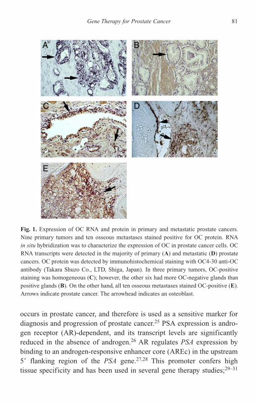
occurs in prostate cancer, and therefore is used as a sensitive marker fordiagnosis and progression of prostate cancer.25 PSA expression is andro-gen receptor (AR)-dependent, and its transcript levels are significantlyreduced in the absence of androgen.26 AR regulates PSA expression bybinding to an androgen-responsive enhancer core (AREc) in the upstream5� flanking region of the PSA gene.27,28 This promoter confers high tissue specificity and has been used in several gene therapy studies;29–31
Gene Therapy for Prostate Cancer 81
Fig. 1. Expression of OC RNA and protein in primary and metastatic prostate cancers.Nine primary tumors and ten osseous metastases stained positive for OC protein. RNAin situ hybridization was to characterize the expression of OC in prostate cancer cells. OCRNA transcripts were detected in the majority of primary (A) and metastatic (D) prostatecancers. OC protein was detected by immunohistochemical staining with OC4-30 anti-OCantibody (Takara Shuzo Co., LTD, Shiga, Japan). In three primary tumors, OC-positivestaining was homogeneous (C); however, the other six had more OC-negative glands thanpositive glands (B). On the other hand, all ten osseous metastases stained OC-positive (E).Arrows indicate prostate cancer. The arrowhead indicates an osteoblast.
B245-ch04 3/2/05 4:30 PM Page 81

however, its utility in men undergoing androgen ablation therapy is lim-ited. On the other hand, PSMA expression is upregulated under androgen-depleted conditions.32 Its expression is elevated higher in prostate cancerthan in benign hyperplasia or normal prostate.33 In addition, serum PSMAlevels are highest in patients with metastatic disease, suggesting enhancedPSMA expression as prostate cancer progresses.34 Recently, the PSMAenhancer (PSME) was discovered within the third intron of the PSMAgene, FOLH135 and has been used for prostate-specific gene deliveryunder low androgen levels.36 Our laboratory hypothesized that AREc andPSME could function synergistically and developed a novel chimeric pro-moter, prostate-specific enhancer sequence (PSES), with high transcrip-tional activity and strong prostate specificity.
Through deletion and linker scan mutagenesis, PSES was developed bylocating the minimal sequences, AREc3 and PSME(del2) in AREc andPSME, respectively and placing AREc3 upstream from PSME(del2).AREc3 contains six GATA transcription factor binding sites and three ARbinding sites leading to high enhancer activity once surrounding silencerregions are deleted. PSME(del2) contains eight AP-1 and three AP-337
binding sites acting as positive regulators in the absence of androgen anda downstream deletion of an Alu repeat that functions as a transcriptionalsilencer. As depicted in Fig. 2, PSES drives luciferase activity five-foldhigher than universal promoter RSV and slightly higher than CMV pro-moter, and luciferase expression was detected in several PSA- and PSMA-positive prostate cancer cell lines, but not in PSA- and PSMA-negativeprostate cells or non-prostate cell lines. PSES retains its prostate-specificnature in recombinant adenoviral vectors as well. Figure 3 shows the resultsafter injection of BALB/c nude mice with Ad-CMV-luc and Ad-PSES-luc.4
Due to its small size, high level of tissue specificity, and strong promoteractivity in the presence or absence of androgen, PSES is an ideal promoterfor use in prostate cancer gene therapy.
Human Telomerase Promoter
Telomeres are tandem repeat structures found at the termini of chromo-somes that maintain chromosomal integrity by preventing DNArearrangements, degradation, and end-to-end fusions. In most normal
82 J. A. Jiménez et al.
B245-ch04 3/2/05 4:30 PM Page 82
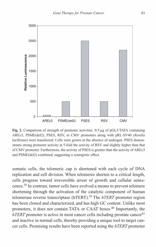
somatic cells, the telomeric cap is shortened with each cycle of DNAreplication and cell division. When telomeres shorten to a critical length,cells progress toward irreversible arrest of growth and cellular senes-cence.38 In contrast, tumor cells have evolved a means to prevent telomereshortening through the activation of the catalytic component of humantelomerase reverse transcriptase (hTERT).39 The hTERT promoter regionhas been cloned and characterized, and has high GC content. Unlike mostpromoters, it does not contain TATA or CAAT boxes.40 Importantly, thehTERT promoter is active in most cancer cells including prostate cancer41
and inactive in normal cells, thereby providing a unique tool to target can-cer cells. Promising results have been reported using the hTERT promoter
Gene Therapy for Prostate Cancer 83
0
500
1000
1500
2000
2500
3000
AREc3 PSME(del2) PSES RSV CMV
Rel
ativ
e L
um
ines
cen
ce
Fig. 2. Comparison of strength of promoter activities. 0.5 �g of pGL3/TATA containingAREc3, PSME(del2), PSES, RSV, or CMV promoters along with pRL-SV40 (Renillaluciferase) were transfected. Cells were grown in the absence of androgen. PSES demon-strates strong promoter activity at 5-fold the activity of RSV and slightly higher than thatof CMV promoter. Furthermore, the activity of PSES is greater than the activity of AREc3and PSME(del2) combined, suggesting a synergistic effect.
B245-ch04 3/2/05 4:30 PM Page 83

to deliver TRAIL, an inducer of apoptosis42 and to control the replicationof an oncolytic adenovirus.43 Our laboratory is investigating the use of thehTERT promoter in conjunction with a prostate-specific promoter to con-trol adenoviral replication in a prostate cancer-specific manner.
Past Approaches
Over the past ten years, three categories of gene therapy approaches forprostate cancer have emerged, corrective gene therapy, cytoreductive genetherapy, and immunotherapy. Each molecular therapy has a strong foun-dation of preclinical data allowing for the approval of several clinical stud-ies. Currently, 56 gene transfer protocols registered with the OBA aretargeted against prostate cancer. This accounts for 15% of all cancer pro-tocols listed to date.44 Table 3 highlights the details of the prostate cancer
84 J. A. Jiménez et al.
Fig. 3. In vivo PSES prostate-specificity comparison. Recombinant adenovirusesAd-CMV-luc and Ad-PSES-luc were injected into male athymic mice, 7 � 1010 virus par-ticles by tail vein injection or 1.4 � 1010 virus particles by intra-prostate injection. Micewere sacrificed and organs were harvested for luciferase activity 2 days post-injection.Following systemic injection, negligible levels of PSES-driven luciferase activity weredetected in organs expected to take up adenovirus, mainly liver, spleen, and lung. To over-come the low infectivity of the prostate, intraprostatic injections were performed, reveal-ing high PSES activity in the prostate.
B245-ch04 3/2/05 4:30 PM Page 84

Gene Therapy for Prostate Cancer 85
Table 3. Prostate Cancer Gene Therapy Trials (OBA ProtocolList, 17 December 2003)44
Principal Institution Vector Genetic YearInvestigator Material Reviewed
1. Simons, JW Johns Hopkins, Retrovirus GM-CSF 1994MD
2. Steiner, MS Vanderbilt Univ., Retrovirus c-myc 1995TN
3. Chen, AP Nat. Naval Med, Vaccinia/ PSA 1995MD fowlpox
4. Paulson, DF Duke Univ., NC Liposome IL-2 19955. Scardino, PT MSKCC, NY Adenovirus HSV-TK 19966. Eder, JP Dana-Farber, Vaccinia/ PSA 1996
MD fowlpox7. Sanda, MG Univ. of Michigan, Vaccinia/ PSA 1997
MI fowlpox8. Belldegrun, AS UCLA, CA Liposome IL-2 19979. Hall, SJ Mt. Sinai, NY Adenovirus HSV-TK 1997
10. Belldegrun, AS UCLA, CA Adenovirus p53 199711. Simons, JW Johns Hopkins, Retrovirus GM-CSF 1997
MD12. Logothetis, CJ MD Anderson, Adenovirus p53 1997
TX13. Kadmon, D Baylor College, Adenovirus HSV-TK 1998
TX14. Simons, JW Johns Hopkins, Adenovirus PSA 1998
MD15. Figlin, RA UCLA, CA Vaccinia/ MUC-1/IL-2 1998
fowlpox16. Gardner, TA Univ. of Virginia, Adenovirus OC-HSV-TK 1998
VA17. Eder, JP Dana-Farber, Vaccinia/ PSA 1999
MD fowlpox18. Small, EJ UCSF, CA Retrovirus GM-CSF 199919. Kaufman, HL Albert Einstein, Vaccinia/ PSA 1999
NY fowlpox20. Vieweg, J Duke Univ., NC RNA PSA 199921. Belldegrun, AS UCLA, CA Liposome IL-2 199922. Small, EJ UCSF, CA Retrovirus GM-SCF 1999
B245-ch04 3/2/05 4:30 PM Page 85

86 J. A. Jiménez et al.
Table 3 (Continued)
Principal Institution Vector Genetic YearInvestigator Material Reviewed
23. Kim, JH Henry Ford Hosp., Adenovirus CD/TK 1999MI
24. Aguilar- Harvard Univ., Adenovirus TK 1999Cordova, E MA
25. Gingrich, JR Univ. of Tenn., TN Adenovirus p16 199926. Terris, MK Stanford Univ., CA Adenovirus PSA 199927. Wilding, G Univ. Of Wisc., WI Adenovirus PSA 199928. Belldegrun, AS UCLA, CA Liposome IL-2 199929. Dahut, WL NIH/NCI Vaccinia/ PSA 1999
fowlpox30. Arlen, PM NIH/NCI Vaccinia/ PSA 2000
fowlpox31. Vieweg, J Duke Univ., NC RNA Tumor RNA 200032. Pollack, A Univ. of Texas, TX Adenovirus p53 200033. Gardner, TA Indiana Univ., IN Adenovirus OC promoter 200034. Freytag, SO Henry Ford Hosp., Adenovirus CD/TK 2000
MI35. Lubaroff, DM Univ. of Iowa, IA Adenovirus PSA 200136. Miles, BJ Baylor College, TX Adenovirus IL-12 200137. DeWeese, TL Johns Hopkins, Adenovirus PB/PSE 2001
MD promoter38. Small, EJ UCSF, CA Adenovirus PB/PSE 2001
promoter39. Dula, E West Coast AAV GM-CSF 2001
Clin. Res.40. Freytag, SO Henry Ford Hosp., Adenovirus CD/TK 2001
MI41. Scher, H MSKCC, NY Liposome PSMA 200142. Corman, J VA Puget Sound, VA AAV GM-CSF 200143. Pantuck, AJ UCLA, LA Vaccinia/ MUC-1/IL-2 2001
fowlpox44. Vieweg, J Duke, NC RNA hTERT 200145. Corman, J VA Puget Sound, Adenovirus PSE promoter 2001
VA46. Vieweg, J Duke, NC RNA PSA 200147. Dinney, CP MD Anderson, TX Adenovirus Inf-� 200248. Dahut, W NIH/NCI Vaccinia/ PSA 2002
fowlpox
B245-ch04 3/2/05 4:30 PM Page 86

trials registered with the OBA. Prostate cancer will remain a focus for lab-oratories due to the limited availability of treatments for advanced diseaseand the large population that is affected.
Corrective Gene Therapy
This approach repairs inherited or acquired genetic defects affecting theregulation of the cell growth cycle. A single prostate cancer cell may har-bor several such mutations, giving it a survival advantage. The replacementof a damaged gene with the wild-type is often sufficient to suppress thegrowth of the cancer or lead to apoptosis, as evidenced in the pre-clinicaland clinical trials outlined below.
p53
Tumor suppressor p53 is referred to as the molecular gatekeeper, protectingthe integrity of the genome.45 When cellular DNA damage occurs, wildtype p53 is activated and stimulates the expression of GADD45 (growtharrest and DNA damage) and the cyclin-dependent kinase (CDK) inhibitor,p21. p21 inhibits the CDK-cyclin D complex required to phosphorylate Rb,
Gene Therapy for Prostate Cancer 87
Table 3 (Continued)
Principal Institution Vector Genetic YearInvestigator Material Reviewed
49. Morris, J Mayo Clinic, MN Adenovirus NIS 200250. Kaufman, HL Columbia, NY Vaccinia/ PSA 2002
fowlpox51. Zanetti, M Univ. of Calif., CA Liposome hTERT 200252. Malkowicz, SB Univ. of Penn., PA Liposome PSA 200253. Arlen, PM NIH/NCI Vaccinia/ PSA 2002
fowlpox54. Corman, J Virginia Mason, WI Adenovirus PSA 200355. Freytag, S Henry Ford Hosp., Adenovirus HSV-TK 2003
MI56. Kantoff, P Dana-Farber, MD Vaccinia/ PSA 2003
fowlpox
B245-ch04 3/2/05 4:30 PM Page 87

thereby halting the cell at the G1/S checkpoint to allow for DNA repair. IfGADD45-mediated DNA repair is unsuccessful, p53 activates bax whichmediates apoptosis.46 p53 mutations occur in approximately one third ofearly prostate cancers,47 and this increases in patients with advanced andmetastatic disease.48 Replacement of wild-type p53 with recombinant ade-noviral vectors (Ad-p53) has resulted in growth inhibition and induction ofapoptosis in prostate cancer both in vitro9,49 and in vivo.50 In addition, intra-tumoral administration of Ad-p53 has been shown to slow the progressionof prostate cancer to metastatic disease.51 Perhaps the most powerful useof p53 replacement is in combination with conventional therapies. Ad-p53has been shown to sensitize prostate cancer cells in vitro and in vivo tochemotherapeutic agents52 and in vitro to radiation therapy.53 Clinical trialsare ongoing to determine the safety of such therapies.54,55
p16
Similar to p53, tumor suppressor p16 is a negative regulator of the cellgrowth cycle, preventing the phosphorylation of Rb by sequesteringCDK4 of the CDK-cyclin D complex. Hypophosphorylated Rb arrests thecell at G1, and loss of normal p16 function is common in prostate cancer.46
Small homozygous deletions have been identified as the major mecha-nism of inactivation of p16, which occurs in 40% of primary prostatecancers and 71% of advanced androgen-independent prostate cancers.56–58
Replacement of p16 using adenoviral vectors suppressed cell growth andinduced senescence in several prostate cancer cell lines including LNCaP(androgen-dependent) and C4-2, DU-145, PPC-1 and PC-3 (androgen-independent).10,59,60 Furthermore, intratumoral injection of Ad-CMV-p16inhibited the growth of PC-3 tumor xenografts in experimental animalmodels and prolonged the animals’ survival.60 Currently, one clinical trialutilizing p16 is in progress.
c-myc
The oncogene c-myc plays an important role in the progression of thecell cycle. As a transcription factor, it regulates the expression of CDC25phosphatases that control CDK activity.61 c-myc amplification is a
88 J. A. Jiménez et al.
B245-ch04 3/2/05 4:30 PM Page 88

common mutation in prostate cancer and its levels correlate with increas-ing tumor grade.62 The approach developed to suppress the overexpressionof oncogenes delivers antisense RNA complimentary to the sense strandfor that gene. This approach not only inhibits cancer cell growth, but alsoinduces apoptosis through the down-regulation of bcl-2 resulting fromc-myc suppression. Steiner et al. developed a replication-incompetentretroviral vector to deliver antisense c-myc transcripts intratumorily inDU-145 nude mice xenografts. Reduction in tumor size and even completetumor obliteration were observed.11 One c-myc clinical trial is in progress.
Cytoreductive Gene Therapy
This approach to the molecular therapy of prostate cancer results inthe direct or indirect killing of prostate cancer cells by replication-competent oncolytic viruses such as Ad-OC-E1a, pro-drug enzyme genessuch as thymidine kinase, and apoptosis-inducing genes such as TRAIL.Perhaps one of the most successful approaches to the molecular therapy ofprostate cancer, several clinical trials are ongoing to explore cytoreductivetreatments.
Oncolytic Virus Therapy
Previous studies have shown that a replication-competent adenovirusinjected intra-organ is sufficient to kill prostate cancer cells.63 Althoughthis therapy alone might be effective, adenovirus is taken up by the liverand lungs. This has the potential to cause hepatic and respiratory distressin immune-compromised cancer patients.64 Controlling the replication ofadenovirus solely to prostate cancer cells increases the safety of thistumor-eliminating therapy. The replication of adenovirus can be controlledby placing the early gene, E1a, a transcriptional activator of adenovirallate genes, under the control of a tissue-specific promoter. Without theexpression of the essential late genes, the virus cannot reassemble andpropagate in the host cell.65 Recently, Henderson et al. demonstrated theability to conditionally drive the replication of adenovirus by a prostate-specific enhancer (PSE) resulting in regression of in vivo androgen-independent LNCaP tumors.66
Gene Therapy for Prostate Cancer 89
B245-ch04 3/2/05 4:30 PM Page 89
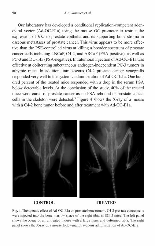
Our laboratory has developed a conditional replication-competent aden-oviral vector (Ad-OC-E1a) using the mouse OC promoter to restrict theexpression of E1a to prostate epithelia and its supporting bone stroma inosseous metastases of prostate cancer. This virus appears to be more effec-tive than the PSE-controlled virus at killing a broader spectrum of prostatecancer cells including LNCaP, C4-2, and ARCaP (PSA-positive), as well asPC-3 and DU-145 (PSA-negative). Intratumoral injection of Ad-OC-E1a waseffective at obliterating subcutaneous androgen-independent PC-3 tumors inathymic mice. In addition, intraosseous C4-2 prostate cancer xenograftsresponded very well to the systemic administration of Ad-OC-E1a. One hun-dred percent of the treated mice responded with a drop in the serum PSA below detectable levels. At the conclusion of the study, 40% of the treatedmice were cured of prostate cancer as no PSA rebound or prostate cancercells in the skeleton were detected.5 Figure 4 shows the X-ray of a mousewith a C4-2 bone tumor before and after treatment with Ad-OC-E1a.
90 J. A. Jiménez et al.
CONTROL TREATED
Fig. 4. Therapeutic effect of Ad-OC-E1a on prostate bone tumors. C4-2 prostate cancer cellswere injected into the bone marrow space of the right tibia in SCID mice. The left panelshows the X-ray of an untreated mouse with a large mass and deformed tibia. The rightpanel shows the X-ray of a mouse following intravenous administration of Ad-OC-E1a.
B245-ch04 3/2/05 4:30 PM Page 90

It has been shown that controlling the expression of the early gene E1b,in addition to E1a, results in better viral replication control.67 For thisreason, we developed a second replication-competent adenoviral vector,Ad-hOC-E1, containing a single bidirectional human OC promoter to con-trol the expression of both E1a and E1b. Under the control of this VDRE-containing promoter, viral replication is induced 10-fold higher thanwild-type viral replication and cytotoxicity is enhanced with the administra-tion of vitamin D.68 Although still controversial,69 some preclinical studiesindicate that vitamin D has an antiproliferative effect on androgen independ-ent prostate cancer.70,71 In our preclinical studies, administration of vitaminD3 in nude mice with subcutaneous DU-145 xenografts demonstrated a ther-apeutic effect; however, the systemic administration of Ad-hOC-E1 in com-bination with vitamin D showed marked repression of the tumors, indicatingthe potential for clinical use.68 The previously outlined preclinical findingshave translated into a phase I clinical trial of OC-driven oncolytic adenoviralintratumoral therapy for androgen-independent prostate cancer.
Pro-drug Enzyme Gene Therapy
The efficacy of this approach depends on the conversion of a non-toxicpro-drug to an active cytotoxic drug by the enzymatic product of adelivered gene not normally expressed in human cells. Following systemicadministration of the pro-drug, high concentrations of the lethal metabo-lite are only found locally at the tumor site, avoiding systemic toxicity.Fortunately, the toxic effect is not limited to the cells that produced thecytotoxic drug, but extends to neighboring cells via the bystander effect.This bystander effect is mediated by intercellular gap junctions andphagocytosis of debris from dying cells. By these means, the cytotoxiceffect is amplified, compensating for low gene transfer efficiencies. Themost widely applied pro-drug gene therapy in prostate cancer utilizesthymidine kinase from the herpes simplex virus (HSV-TK) and any one ofseveral anti-herpetic agents such as ganciclovir (GCV), acyclovir (ACV),or valacyclovir (VAL). These nucleoside analogues are phosphorylatedspecifically by HSV-TK. The phosphorylated forms of the drugs areincorporated into cellular DNA during DNA replication resulting in chaintermination and ultimately cell death.
Gene Therapy for Prostate Cancer 91
B245-ch04 3/2/05 4:30 PM Page 91

The TK/pro-drug system is widely favored because it is a safeapproach for cancer gene therapy for a number of reasons. First, apopto-sis is induced in the transduced cell only when it divides, allowing thegene therapist to target cancer cells that divide more rapidly than non-cancerous cells. Second, the toxic effect only occurs when the pro-drug isadministered allowing the cessation of treatment in the event of adverseeffects. Third, several anti-herpetic nucleoside analogue drugs are clini-cally available, simplifying the approval process for the use of the pro-drug in clinical trials. Finally, the bystander effect greatly increases thekilling efficiency of the therapy.
Previously, Eastham et al. demonstrated the sensitivity of humanprostate cancer cells PC-3 and DU-145 to GCV cytotoxicity following thein vitro transduction of the cells with HSV-TK using a recombinant replica-tion-deficient adenoviral vector. Similar results were obtained in vivo insubcutaneous xenografts of murine and human cancer models following theintralesional injection of Ad-RSV-TK and Ad-CMV-TK.72–74 Placing TKunder the control of universal promoters such as RSV or CMV allows forthe potential of TK activation in any cell without discrimination betweennormal and cancer cells. Therefore, intratumoral injection of the vector isrequired to prevent systemic dissemination of the virus. Scardino et al.developed the initial TK clinical trial in which a replication-deficient adeno-viral vector was injected intralesionally to deliver HSV-TK, preceding theadministration of GCV, in men with locally recurrent prostate cancer one ormore years after definitive external beam radiotherapy. This trial realizedthe potential of this therapy by demonstrating the tumoricidal activity of thisTK/GCV therapy as evidenced by sustained decreases in serum PSA andimproved biopsies. As a result of adenoviral leakage from the injection sitethrough the bloodstream and tracking to the liver, several of the patientsexperienced a self-limiting toxicity and one patient experienced moderatebut reversible hepatic dysfunction and thrombocytopenia.15 To circumventtoxicity to the patient, Chung et al. developed a replication-deficient aden-ovirus to deliver HSV-TK driven by the PSA promoter.29
Our laboratory developed a clinical protocol to test the hypothesis thatthe OC promoter can regulate HSV-TK expression specifically within aprostate cancer cell and the supportive stroma of a metastasis. We per-formed a phase I clinical trial enrolling 11 patients with locally recurrent
92 J. A. Jiménez et al.
B245-ch04 3/2/05 4:30 PM Page 92

or metastatic prostate cancer. Two post-surgical local recurrences and ninemetastatic lesions (five osseous and 4 lymph node) were injected with repli-cation-defective Ad-OC-TK vector followed by the administration of oralvalacyclovir.3 All patients tolerated this therapy with no severe adverseeffects. Of the eleven men, local cancer cell death was observed in sevenpatients; however, the treated lesions of all eleven men showed histologicalchanges as a result of the treatment. One patient demonstrated regressionand stabilization of the treated lesion for 317 days post-treatment withoutalternative treatments, as demonstrated in Fig. 5.6 This trial opened the doorto the development of future adenoviral vectors for the systemic treatmentof osseous and visceral prostate cancer metastases.
Suicide Gene Therapy
Tumor necrosis factor-related apoptosis-inducing ligand (TRAIL), alsoknown as Apo-2 ligand, is a member of the tumor necrosis factor (TNF)
Gene Therapy for Prostate Cancer 93
Fig. 5. Histomorphologic study of prostate cancer bone metastases treated with Ad-OC-TK.Tissue biopsies were collected from one patient and H&E-stained before (A), during (day8) (B), and after (day 30) (C) treatment with Ad-OC-TK and VAL. Malignant prostate can-cer cells surround healthy bone tissue before treatment (A). During treatment, cancer cellsare replaced and fibrosis occurs (B). By day 30, malignant cells disappear and are replacedcompletely by fibrosis and inflammatory cells (C).
B245-ch04 3/2/05 4:30 PM Page 93

family and has been shown to preferentially kill tumor cells. Originallydiscovered because of its similarity to Fas-ligand, TRAIL is a 32 kDa typeII transmembrane protein, whose C-terminal extracellular domain (aminoacids 114–281) is homologous with other members of the TNF family.75,76
TRAIL induces apoptosis by binding to the death domain-containingreceptors DR4 and DR5; however, the death signal is not transduced viathe adaptor molecule FADD. Instead, the death protease FLICE2 isbelieved to be engaged, cleaving the initiating caspase 8 and beginning thecaspase cascade.77
The selectivity of TRAIL for cancer cells over normal cells makes ita prime candidate for anticancer therapy. TRAIL expression has beendetected in several normal human tissues which suggests that TRAIL is nottoxic to those cells in vivo.78 In essence, these cells are protected fromthe apoptotic effects of TRAIL by an antagonistic decoy receptor, TRID,which lacks an intracellular domain and is found on the surface membraneof TRAIL-resistant cells.77 Many prostate cancer cell lines includingALVA-31, DU-145, and PC-3 are extremely sensitive to TRAIL andundergo apoptosis when exposed; however, other cell lines such as LNCaPare highly resistant.79 This resistance has been shown to be reversed bysimultaneous administration of the chemotherapeutic agents doxorubicin,cisplatin, or etoposide80 or by infection of those cells with adenovirus.81
For this reason, TRAIL is a promising suicide gene to consider for prostatecancer gene therapy; however, recent studies suggest that cultured humanhepatocytes may be sensitive to TRAIL.82 Recombinant forms of TRAILwith reduced hepatotoxicity are being investigated83 in addition to mono-clonal antibodies that antagonize the TRAIL receptor in hepatocytes.84
Immunotherapy
Prostate cancer, like most cancers, has developed mechanisms to evadethe host immune system. Such mechanisms include the down-regulationof class I major histocompatibility complex (MHC) molecules on thetumor cell surface85 as well as the down-regulation of the co-stimulatoryB7 molecules.86 These means of evasion result in decreased presentationof tumor antigens to cytotoxic T lymphocytes (CTLs). The goal ofimmunotherapy is to enhance the host immune response to prostate cancer
94 J. A. Jiménez et al.
B245-ch04 3/2/05 4:30 PM Page 94

cells. Although several clinical trials have been initiated, only limitedresults have been published.
GM-CSF
After demonstrating that immunostimulatory molecules could transformcancer cells to activate a tumor-directed immune response,87 Simons et al.developed the first gene therapy clinical trial protocol for prostate cancer. Inthis study, eight men with metastatic prostate cancer discovered during rad-ical prostatectomy were administered autologous, ex vivo GM-CSF trans-duced, irradiated cancer vaccines. Unfortunately, due to the large numberof ex vivo manipulations, the vaccination rate decreased, from the originalenrollment number of eleven men to eight. Side effects were limited topruritis, erythema, and swelling at the site of vaccination. Delayed-typehypersensitivity (DTH) reactions to untransduced autologous prostate can-cer cells were positive in seven of the eight men, demonstrating the abilityto mount a T cell response. Furthermore, new antibodies against prostatecancer polypeptides were discovered in the sera of three of the eight men,suggesting a B cell response to vaccination.88 A subsequent clinical trialdemonstrated no therapeutic value of systemic administration of recombi-nant GM-CSF, thereby suggesting the importance of local secretion ofGM-CSF by the cancer vaccines.89 Multiple clinical trials are in progressthat employ a simple vaccine composed of irradiated prostate cancer cellline allografts expressing GM-CSF, which allows for multiple dosing.
IL-2
More recently, in vivo immunotherapy approaches have been used.Belldegrun et al. developed a gene therapy protocol in which 24 men withlocally advanced prostate cancer were administered a DNA-liposomecomplex encoding the IL-2 gene intraprostatically. This therapy was welltolerated, with side effects including mild hematuria, transient rectalbleeding, and perineal discomfort. Immunohistochemical analysis of thetumor site demonstrated T cell infiltration. Decreased serum PSA levelswere observed in 16 of the 24 men on day 1 and 14 of the 24 by day 8.90
Similar clinical trials are still in progress.
Gene Therapy for Prostate Cancer 95
B245-ch04 3/2/05 4:30 PM Page 95

Future Directions
Among clinical trials that have been launched to demonstrate the safetyand efficacy of adenoviral-based gene therapy on prostate cancer in thepast few years, the most exciting results have come from the study oftumor/tissue-restricted replicative adenovirus (TRRA). This strategyallows the viral vector to propagate from a limited number of infected cellsto the whole tumor mass, overcoming the problem of inadequate in vivoinfectivity or biodistribution of the vector; however, TRRA-based therapyis not without its limitations. One of the major limitations of this therapy isthe induction of the host immune system, targeted to eliminate the aden-ovirus from the body.
It will become critical to temporarily suppress the host’s immune sys-tem or enhance the killing activity of the virus so that it can eliminatetumors within a shorter period of time, allowing it to escape from hostimmune attack. Among immune regulators, TGF-� and Fas-ligand arelikely the best candidates for incorporation into TRRA for cancer genetherapy. There are five members reported in the TGF-� family; three ofthem (TGF-�1, TGF-�2, and TGF-�3) are expressed in mammals. Thesethree isoforms share a high degree of sequence homology in the maturedomain, and have similar actions on cells in tissue culture. Suppressionof the immune response includes the inhibition of T and B cell prolifera-tion, the down-regulation of natural killer cell activity and CTL response,and the regulation of macrophage activation.91 TGF-� is a mediator ofimmune suppression that allows tumors to escape from immune surveil-lance, and its use has been explored to suppress the inflammatory andalloreactive immune responses in liver transplantation.92 Besides TGF-�,Fas-ligand is another immune modulator that has been explored for use inkidney transplant patients to suppress alloreactive lymphocytes.93 Fas-ligand, also known as CD95 or APO-1, is a membrane-bound protein of theTNF family, and it is expressed in several cell types including tumors,T cells, and B cells. Cells expressing the Fas receptor undergo apoptosis when they encounter Fas-ligand.94 Besides their immune modulation func-tion, TGF-� and Fas-ligand are also strong growth inhibitors and induceapoptosis in a variety of cancers, including prostate cancer.95 Therefore,incorporating TGF-� or Fas-ligand into a TRRA will potentially enhance
96 J. A. Jiménez et al.
B245-ch04 3/2/05 4:30 PM Page 96
the tumoricidal activity of the vector and blanket the tumor site from theimmune system, allowing the TRRA to complete its mission.
Once the TRRA is armed with immune suppressors, the need for tightregulation of viral replication increases. In addition to controlling E1a andE1b with prostate-specific promoters, E4 should be considered. The E4transcription unit encodes regulatory genes that are critical for viral repli-cation, involving the shut-down of host gene expression and the facilita-tion of late viral gene expression.96 Mutant adenoviral vectors lackingthe E4 region are severely replication defective and can only propagate inE4-expressing cells.97 Placing E4 under the control of a prostate-specificpromoter should provide additional safety. TK should also be consideredfor incorporation into a TRRA. In addition to enhancing the tumor-killingactivity of a TRRA, TK will allow a gene therapist to monitor adenoviralreplication in vivo via positron emission tomography (PET) imaging.98
Finally, because all tissue-specific promoters have basal activity levelsin non-target tissues, the potential exists for accumulation of adenovirusin the liver, resulting in undesirable toxic effects caused by significantexpression of therapeutic genes in the liver. Modification of the adenovi-ral fiber knob can increase viral tropism toward cancers and away from theliver and other vital organs.99,100 This is also beneficial because it is wellknown that expression of the coxsackievirus-adenovirus receptor (CAR)is frequently down-regulated in cancers. Our laboratory is currently inves-tigating an Ad-5/35 hybrid adenovirus for use in prostate cancer genetherapy whose infectivity is CAR-independent and lacks liver tropism.
Conclusion
The safety and efficacy of gene therapy for prostate cancer has beendemonstrated through various preclinical and clinical trials. In recentyears, interest in this field has expanded and will continue to do so. It isconceivable that, in the near future, a safe gene therapy modality will bedeveloped to replace hormone ablation therapy which causes unpleasantside effects, decreases the quality of life of the patient, and only tem-porarily controls the disease. Due to its convenient administration throughultrasound-guided transrectal injection, it is also conceivable that genetherapy applied in an outpatient clinic may someday replace radical
Gene Therapy for Prostate Cancer 97
B245-ch04 3/2/05 4:30 PM Page 97

prostatectomy and radiation therapy to treat early prostate cancer. Factorsthat limit the disseminated use of gene therapy as a standard of careinclude time, funding, and fear from the general public; however, theseshould diminish as the number of successful clinical trials increase.
References
1. Jemal A, Murray T, Samuels A, Ghafoor A, Ward E, Thun MJ (2003) CancerStatistics, 2003. CA Cancer J Clin 53:5–26.
2. Gardner TA, Ko SC, Kao C, Shirakawa T, Cheon J, Gotoh A, Wu T, Sikes RA,Zhau HE, Cui Q, Balian G, Chung LWK (1998) Exploiting stromal-epithelialinteraction for model development and new strategies of gene therapy forprostate cancer and osteosarcoma metastases. Gene Therapy Mol Bio 2:41–58.
3. Koeneman KS, Kao C, Ko SC, Yang L, Wada Y, Kallmes DF, Gillenwater JY,Zhau HE, Chung LW, Gardner TA (2000) Osteocalcin-directed gene therapyfor prostate-cancer bone metastasis. World J Urol 18:102.
4. Lee SJ, Kim HS, Yu R, et al. (2002) Novel prostate-specific promoter derivedfrom PSA and PSMA enhancers. Mol Ther 6:415–421.
5. Matsubara S, Wada Y, Gardner TA, et al. (2001) A conditional replication-competent adenoviral vector, Ad-OC-E1a, to cotarget prostate cancer andbone stroma in an experimental model of androgen-independent prostatecancer bone metastasis. Cancer Res 61:6012–6019.
6. Kubo H, Gardner TA, Wada Y, et al. (2003) Phase I dose escalation clinicaltrial of adenovirus vector carrying osteocalcin promoter-driven herpes sim-plex virus thymidine kinase in localized and metastatic hormone-refractoryprostate cancer. Hum Gene Ther 14:227–241.
7. Thompson IM, Goodman PJ, Tangen CM, et al. (2003) The influence offinasteride on the development of prostate cancer. N Engl J Med 349:215–224.
8. Bookstein R, Shew JY, Chen PL, Scully P, Lee WH (1990) Suppression oftumorigenicity of human prostate carcinoma cells by replacing a mutated RBgene. Science 247:712–715.
9. Eastham JA, Hall SJ, Sehgal I, et al. (1995) In vivo gene therapy with p53 orp21 adenovirus for prostate cancer. Cancer Res 55:5151–5155.
10. Allay JA, Steiner MS, Zhang Y, Reed CP, Cockroft J, Lu Y (2000) Adenovirusp16 gene therapy for prostate cancer. World J Urol 18:111–120.
11. Steiner MS, Anthony CT, Lu Y, Holt JT (1998) Antisense c-myc retroviral vec-tor suppresses established human prostate cancer. Hum Gene Ther 9:747–755.
98 J. A. Jiménez et al.
B245-ch04 3/2/05 4:30 PM Page 98

12. Wei C, Willis RA, Tilton BR, et al. (1997) Tissue-specific expression of thehuman prostate-specific antigen gene in transgenic mice: Implications fortolerance and immunotherapy. Proc Natl Acad Sci USA 94:6369–6374.
13. Hay RT (1985) The origin of adenovirus DNA replication: Minimal DNAsequence requirement in vivo. EMBO J 4:421–426.
14. Brand K, Arnold W, Bartels T, et al. (1997) Liver-associated toxicity of theHSV-tk/GCV approach and adenoviral vectors. Cancer Gene Ther 4:9–16.
15. Herman JR, Adler HL, Aguilar-Cordova E, et al. (1999) In situ gene therapyfor adenocarcinoma of the prostate: A phase I clinical trial. Hum Gene Ther10:1239–1249.
16. Pan LC, Price PA (1984) The effect of transcriptional inhibitors on the bonegamma-carboxyglutamic acid protein response to 1,25-dihydroxyvitamin D3in osteosarcoma cells. J Biol Chem 259:5844–5847.
17. Jung C, Ou YC, Yeung F, Frierson HF, Jr, Kao C (2001) Osteocalcin isincompletely spliced in non-osseous tissues. Gene 271:143–150.
18. Wu TT, Sikes RA, Cui Q, et al. (1998) Establishing human prostate cancercell xenografts in bone: Induction of osteoblastic reaction by prostate-specific antigen-producing tumors in athymic and SCID/bg mice usingLNCaP and lineage-derived metastatic sublines. Int J Cancer 77:8878–8894.
19. Koeneman KS, Yeung F, Chung LW (1999) Osteomimetic properties ofprostate cancer cells: A hypothesis supporting the predilection of prostatecancer metastasis and growth in the bone environment. Prostate 39:246–261.
20. Bortell R, Owen TA, Bidwell JP, et al. (1992) Vitamin D-responsive protein-DNA interactions at multiple promoter regulatory elements that contribute tothe level of rat osteocalcin gene expression. Proc Natl Acad Sci USA89:6119–6123.
21. Lian JB, Stein GS, Stein JL, van Wijnen AJ (1999) Regulated expression ofthe bone-specific osteocalcin gene by vitamins and hormones. Vitam Horm55:443–509.
22. Banerjee C, Stein JL, Van Wijnen AJ, Frenkel B, Lian JB, Stein GS (1996)Transforming growth factor-beta 1 responsiveness of the rat osteocalcin geneis mediated by an activator protein-1 binding site. Endocrinology 137:1991–2000.
23. Banerjee C, Hiebert SW, Stein JL, Lian JB, Stein GS (1996) An AML-1 con-sensus sequence binds an osteoblast-specific complex and transcriptionallyactivates the osteocalcin gene. Proc Natl Acad Sci USA 93:4968–4973.
24. Ko SC, Cheon J, Kao C, et al. (1996) Osteocalcin promoter-based toxic genetherapy for the treatment of osteosarcoma in experimental models. CancerRes 56:4614–4619.
Gene Therapy for Prostate Cancer 99
B245-ch04 3/2/05 4:30 PM Page 99

25. Stamey TA, Yang N, Hay AR, McNeal JE, Freiha FS, Redwine E (1987)Prostate-specific antigen as a serum marker for adenocarcinoma of theprostate. N Engl J Med 317:909–916.
26. Cleutjens KB, van der Korput HA, van Eekelen CC, van Rooij HC,Faber PW, Trapman J (1997) An androgen response element in a far upstreamenhancer region is essential for high, androgen-regulated activity of theprostate-specific antigen promoter. Mol Endocrinol 11:148–161.
27. Schuur ER, Henderson GA, Kmetec LA, Miller JD, Lamparski HG,Henderson DR (1996) Prostate-specific antigen expression is regulated by anupstream enhancer. J Biol Chem 271:7043–7051.
28. Pang S, Dannull J, Kaboo R, et al. (1997) Identification of a positive regula-tory element responsible for tissue-specific expression of prostate-specificantigen. Cancer Res 57:495–499.
29. Gotoh A, Ko SC, Shirakawa T, et al. (1998) Development of prostate-specificantigen promoter-based gene therapy for androgen-independent humanprostate cancer. J Urol 160:220–229.
30. Wu L, Matherly J, Smallwood A, et al. (2001) Chimeric PSA enhancersexhibit augmented activity in prostate cancer gene therapy vectors. GeneTher 8:1416–1426.
31. Lu Y, Carraher J, Zhang Y, et al. (1999) Delivery of adenoviral vectors to theprostate for gene therapy. Cancer Gene Ther 6:64–72.
32. Wright GL, Jr, Grob BM, Haley C, et al. (1996) Upregulation of prostate-specific membrane antigen after androgen-deprivation therapy. Urology48:326–334.
33. Xiao Z, Adam BL, Cazares LH, et al. (2001) Quantitation of serum prostate-specific membrane antigen by a novel protein biochip immunoassay discrim-inates benign from malignant prostate disease. Cancer Res 61:6029–6033.
34. Sweat SD, Pacelli A, Murphy GP, Bostwick DG (1998) Prostate-specificmembrane antigen expression is greatest in prostate adenocarcinoma andlymph node metastases. Urology 52:637–640.
35. Watt F, Martorana A, Brookes DE, et al. (2001) A tissue-specific enhancer of theprostate-specific membrane antigen gene, FOLH1. Genomics 73:2432–2454.
36. Uchida A, O’Keefe DS, Bacich DJ, Molloy PL, Heston WD (2001) In vivosuicide gene therapy model using a newly discovered prostate-specific mem-brane antigen promoter/enhancer: A potential alternative approach to andro-gen deprivation therapy. Urology 58:13213–13219.
37. Lee SJ, Lee K, Yang X, et al. (2003) NFATc1 with AP-3 site binding specificitymediates gene expression of prostate-specific-membrane-antigen. J Mol Biol330:749–760.
100 J. A. Jiménez et al.
B245-ch04 3/2/05 4:30 PM Page 100
38. Chiu CP, Harley CB (1997) Replicative senescence and cell immortality: Therole of telomeres and telomerase. Proc Soc Exp Biol Med 214:99–106.
39. Kim NW, Piatyszek MA, Prowse KR, et al. (1994) Specific association ofhuman telomerase activity with immortal cells and cancer. Science 266:2011–2015.
40. Horikawa I, Cable PL, Afshari C, Barrett JC (1999) Cloning and characteri-zation of the promoter region of human telomerase reverse transcriptasegene. Cancer Res 59:826–830.
41. Sommerfeld HJ, Meeker AK, Piatyszek MA, Bova GS, Shay JW, Coffey DS(1996) Telomerase activity: A prevalent marker of malignant human prostatetissue. Cancer Res 56:218–222.
42. Lin T, Huang X, Gu J, et al. (2002) Long-term tumor-free survival from treat-ment with the GFP-TRAIL fusion gene expressed from the hTERT promoterin breast cancer cells. Oncogene 21:8020–8028.
43. Lanson NA, Jr, Friedlander PL, Schwarzenberger P, Kolls JK, Wang G (2003)Replication of an adenoviral vector controlled by the human telomerasereverse transcriptase promoter causes tumor-selective tumor lysis. CancerRes 63:7936–7941.
44. 2003 Office of Biotechnology Activities’ Recombinant DNA and GeneTransfer Web Page, http://www4.od.nih.gov/oba/rdna.htm
45. Levine AJ (1997) p53, the cellular gatekeeper for growth and division. Cell88:323–331.
46. Sherr CJ (1996) Cancer cell cycles. Science 274:1672–1677.47. Downing SR, Russell PJ, Jackson P (2003) Alterations of p53 are common in
early stage prostate cancer. Can J Urol 10:1924–1933.48. Eastham JA, Stapleton AM, Gousse AE, et al. (1995) Association of
p53 mutations with metastatic prostate cancer. Clin Cancer Res 1:1111–1118.
49. Yang C, Cirielli C, Capogrossi MC, Passaniti A (1995) Adenovirus-mediatedwild-type p53 expression induces apoptosis and suppresses tumorigenesis ofprostatic tumor cells. Cancer Res 55:4210–4213.
50. Ko SC, Gotoh A, Thalmann GN, et al. (1996) Molecular therapy with recom-binant p53 adenovirus in an androgen-independent, metastatic humanprostate cancer model. Hum Gene Ther 7:1683–1691.
51. Eastham JA, Grafton W, Martin CM, Williams BJ (2000) Suppression ofprimary tumor growth and the progression to metastasis with p53 adenovirusin human prostate cancer. J Urol 164:814–819.
52. Gurnani M, Lipari P, Dell J, Shi B, Nielsen LL (1999) Adenovirus-mediatedp53 gene therapy has greater efficacy when combined with chemotherapy
Gene Therapy for Prostate Cancer 101
B245-ch04 3/2/05 4:30 PM Page 101

against human head and neck, ovarian, prostate, and breast cancer. CancerChemother Pharmacol 44:143–151.
53. Colletier PJ, Ashoori F, Cowen D, et al. (2000) Adenoviral-mediated p53transgene expression sensitizes both wild-type and null p53 prostate cancercells in vitro to radiation. Int J Radiat Oncol Biol Phys 48:1507–1512.
54. Pantuck AJ, Zisman A, Belldegrun AS (2000) Gene therapy for prostate can-cer at the University of California, Los Angeles: Preliminary results andfuture directions. World J Urol 18:143–147.
55. Sweeney P, Pisters LL (2000) Ad5CMVp53 gene therapy for locallyadvanced prostate cancer — where do we stand? World J Urol 18:121–124.
56. Jarrard DF, Bova GS, Ewing CM, et al. (1997) Deletional, mutational, andmethylation analyses of CDKN2 (p16/MTS1) in primary and metastaticprostate cancer. Genes Chromosomes Cancer 19:90–96.
57. Liggett WH Jr, Sidransky D (1998) Role of the p16 tumor suppressor genein cancer. J Clin Oncol 16:1197–1206.
58. Isaacs WB (1995) Molecular genetics of prostate cancer. Cancer Surv25:357–379.
59. Steiner MS, Zhang Y, Farooq F, Lerner J, Wang Y, Lu Y (2000) Adenoviralvector containing wild-type p16 suppresses prostate cancer growth and pro-longs survival by inducing cell senescence. Cancer Gene Ther 7:360–372.
60. Gotoh A, Kao C, Ko SC, Hamada K, Liu TJ, Chung LW (1997) Cytotoxiceffects of recombinant adenovirus p53 and cell cycle regulator genes (p21WAF1/CIP1 and p16CDKN4) in human prostate cancers. J Urol 158:636–641.
61. Galaktionov K, Chen X, Beach D (1996) Cdc25 cell-cycle phosphatase as atarget of c-myc. Nature 382:511–517.
62. Jenkins RB, Qian J, Lieber MM, Bostwick DG (1997) Detection of c-myconcogene amplification and chromosomal anomalies in metastatic prostaticcarcinoma by fluorescence in situ hybridization. Cancer Res 57:524–531.
63. Deng J, Xia W, Hung MC (1998) Adenovirus 5 E1A-mediated tumor sup-pression associated with E1A-mediated apoptosis in vivo. Oncogene17:2167–2175.
64. van der Eb MM, Cramer SJ, Vergouwe Y, et al. (1998) Severe hepatic dys-function after adenovirus-mediated transfer of the herpes simplex virusthymidine kinase gene and ganciclovir administration. Gene Ther 5:451–458.
65. Robbins PD, Tahara H, Ghivizzani SC (1998) Viral vectors for gene therapy.Trends Biotechnol 16:35–40.
66. Rodriguez R, Schuur ER, Lim HY, Henderson GA, Simons JW, Henderson DR(1997) Prostate attenuated replication competent adenovirus (ARCA)
102 J. A. Jiménez et al.
B245-ch04 3/2/05 4:30 PM Page 102

CN706: A selective cytotoxic for prostate-specific antigen-positive prostatecancer cells. Cancer Res 57:2559–2563.
67. Yu DC, Sakamoto GT, Henderson DR (1999) Identification of the transcrip-tional regulatory sequences of human kallikrein 2 and their use in the con-struction of calydon virus 764, an attenuated replication competentadenovirus for prostate cancer therapy. Cancer Res 59:1498–1504.
68. Hsieh CL, Yang L, Miao L, et al. (2002) A novel targeting modality toenhance adenoviral replication by vitamin D(3) in androgen-independenthuman prostate cancer cells and tumors. Cancer Res 62:3084–3092.
69. Konety BR, Johnson CS, Trump DL, Getzenberg RH (1999) Vitamin D in theprevention and treatment of prostate cancer. Semin Urol Oncol 17:77–84.
70. Getzenberg RH, Light BW, Lapco PE, et al. (1997) Vitamin D inhibition ofprostate adenocarcinoma growth and metastasis in the Dunning rat prostatemodel system. Urology 50:999–1006.
71. Zhao XY, Feldman D (2001) The role of vitamin D in prostate cancer.Steroids 66:293–300.
72. Eastham JA, Chen SH, Sehgal I, et al. (1996) Prostate cancer gene therapy:Herpes simplex virus thymidine kinase gene transduction followed by ganci-clovir in mouse and human prostate cancer models. Hum Gene Ther 7:515–523.
73. Hall SJ, Mutchnik SE, Chen SH, Woo SL, Thompson TC (1997) Adenovirus-mediated herpes simplex virus thymidine kinase gene and ganciclovir ther-apy leads to systemic activity against spontaneous and induced metastasis inan orthotopic mouse model of prostate cancer. Int J Cancer 70:183–187.
74. Cheon J, Kim HK, Moon DG, Yoon DK, Cho JH, Koh SK (2000)Adenovirus-mediated suicide-gene therapy using the herpes simplex virusthymidine kinase gene in cell and animal models of human prostate cancer:Changes in tumour cell proliferative activity. BJU Int 85:759–766.
75. Wiley SR, Schooley K, Smolak PJ, et al. (1995) Identification and charac-terization of a new member of the TNF family that induces apoptosis.Immunity 3:673–682.
76. Pitti RM, Marsters SA, Ruppert S, Donahue CJ, Moore A, Ashkenazi A(1996) Induction of apoptosis by Apo-2 ligand, a new member of the tumornecrosis factor cytokine family. J Biol Chem 271:12687–12690.
77. Pan G, Ni J, Wei YF, Yu G, Gentz R, Dixit VM (1997) An antagonist decoyreceptor and a death domain-containing receptor for TRAIL. Science277:815–818.
78. Pan G, O’Rourke K, Chinnaiyan AM, et al. (1997) The receptor for the cyto-toxic ligand TRAIL. Science 276:111–113.
Gene Therapy for Prostate Cancer 103
B245-ch04 3/2/05 4:30 PM Page 103

79. Nesterov A, Lu X, Johnson M, Miller GJ, Ivashchenko Y, Kraft AS (2001)Elevated AKT activity protects the prostate cancer cell line LNCaP fromTRAIL-induced apoptosis. J Biol Chem 276:10767–10774.
80. Munshi A, McDonnell TJ, Meyn RE (2002) Chemotherapeutic agentsenhance TRAIL-induced apoptosis in prostate cancer cells. CancerChemother Pharmacol 50:46–52.
81. Voelkel-Johnson C, King DL, Norris JS (2002) Resistance of prostate cancercells to soluble TNF-related apoptosis-inducing ligand (TRAIL/Apo2L) canbe overcome by doxorubicin or adenoviral delivery of full-length TRAIL.Cancer Gene Ther 9:164–172.
82. Jo M, Kim TH, Seol DW, et al. (2000) Apoptosis induced in normal humanhepatocytes by tumor necrosis factor-related apoptosis-inducing ligand. NatMed 6:564–567.
83. Lawrence D, Shahrokh Z, Marsters S, et al. (2001) Differential hepatocytetoxicity of recombinant Apo2L/TRAIL versions. Nat Med 7:383–385.
84. Mori E, Thomas M, Motoki K, et al. (2003) Human normal hepatocytes aresusceptible to apoptosis signal mediated by both TRAIL-R1 and TRAIL-R2.Cell Death Differ 11:203–207.
85. Bander NH, Yao D, Liu H, et al. (1997) MHC class I and II expression inprostate carcinoma and modulation by interferon-alpha and -gamma.Prostate 33:233–239.
86. Kwon ED, Hurwitz AA, Foster BA, et al. (1997) Manipulation of T cell cos-timulatory and inhibitory signals for immunotherapy of prostate cancer. ProcNatl Acad Sci USA 94:8099–8103.
87. Sanda MG, Ayyagari SR, Jaffee EM, et al. (1994) Demonstration of arational strategy for human prostate cancer gene therapy. J Urol 151:622–628.
88. Simons JW, Mikhak B, Chang JF, et al. (1999) Induction of immunity toprostate cancer antigens: Results of a clinical trial of vaccination with irradi-ated autologous prostate tumor cells engineered to secrete granulocyte-macrophage colony-stimulating factor using ex vivo gene transfer. CancerRes 59:5160–5168.
89. Simmons SJ, Tjoa BA, Rogers M, et al. (1999) GM-CSF as a systemic adju-vant in a phase II prostate cancer vaccine trial. Prostate 39:291–297.
90. Belldegrun A, Tso CL, Zisman A, et al. (2001) Interleukin 2 gene therapy forprostate cancer: Phase I clinical trial and basic biology. Hum Gene Ther12:883–892.
91. Huang X, Lee C (2003) From TGF-beta to cancer therapy. Curr Drug Targets4:243–250.
104 J. A. Jiménez et al.
B245-ch04 3/2/05 4:30 PM Page 104

92. Narumoto K, Saibara T, Maeda T, et al. (2000) Transforming growth factor-beta 1 derived from biliary epithelial cells may attenuate alloantigen-specific immune responses. Transpl Int 13:21–27.
93. Ke B, Coito AJ, Kato H, et al. (2000) Fas ligand gene transfer prolongsrat renal allograft survival and down-regulates anti-apoptotic Bag-1 inparallel with enhanced Th2-type cytokine expression. Transplantation 69:1690–1694.
94. Winoto A (1997) Cell death in the regulation of immune responses. CurrOpin Immunol 9:365–370.
95. Norris JS, Hyer ML, Voelkel-Johnson C, Lowe SL, Rubinchik S, Dong JY(2001) The use of Fas Ligand, TRAIL and Bax in gene therapy of prostatecancer. Curr Gene Ther 1:123–136.
96. Tauber B, Dobner T (2001) Molecular regulation and biological function ofadenovirus early genes: The E4 ORFs. Gene 278:1–23.
97. Krougliak V, Graham FL (1995) Development of cell lines capable of com-plementing E1, E4, and protein IX defective adenovirus type 5 mutants.Hum Gene Ther 6:1575–1586.
98. Pantuck AJ, Berger F, Zisman A, et al. (2002) CL1-SR39: A noninvasivemolecular imaging model of prostate cancer suicide gene therapy usingpositron emission tomography. J Urol 168:1193–1198.
99. Campbell M, Qu S, Wells S, Sugandha H, Jensen RA (2003) An adenoviralvector containing an arg-gly-asp (RGD) motif in the fiber knob enhancesprotein product levels from transgenes refractory to expression. CancerGene Ther 10:559–570.
100. Vigne E, Dedieu JF, Brie A, et al. (2003) Genetic manipulations of aden-ovirus type 5 fiber resulting in liver tropism attenuation. Gene Ther 10:153–162.
Gene Therapy for Prostate Cancer 105
B245-ch04 3/2/05 4:30 PM Page 105

This page intentionally left blank
5CHEMOTHERAPY FOR PROSTATE CANCER
Samuel K. Kulp, Kuen-Feng Chenand Ching-Shih Chen
Division of Medicinal Chemistry and PharmacognosyCollege of Pharmacy, The Ohio State University
Columbus, OH, USA
Introduction
For several decades, androgen deprivation has been the mainstay oftherapy for metastatic prostate cancer. The high response rates to thisapproach, which near 90% by PSA criteria,1 underscore the activity of thistreatment modality in prostate cancer patients. However, satisfaction withthe efficacy of androgen deprivation therapy is tempered by the transienceof its effects on disease progression. The median time to progression hasbeen reported to range from 9 to 30 months.1,2 Although secondary hor-monal maneuvers will further benefit a subset of these patients, they arecharacterized by even shorter durations of response (�6 months).3,4
Continued progression in the face of castrate levels of androgen and thelack of responsiveness to hormonal therapies defines the emergence ofhormone-refractory prostate cancer (HRPC) for which the prognosis isbleak. The treatment of hormone-refractory tumors constitutes a signifi-cant challenge in the management of patients with prostate cancer.
Although HRPC has historically been considered a chemorefractorydisease, chemotherapy has an established role in the management ofpatients with HRPC. Evidence obtained over the last decade has shownsome chemotherapeutic regimens to be unquestionably active in thesepatients.5 Indeed, guidelines set forth by the National Comprehensive
107
B245-ch05 3/2/05 4:30 PM Page 107
Cancer Network (NCCN) currently identify six chemotherapeutic regi-mens as options for the management of HRPC.6 These options, however,are solely palliative. The oft-cited caveat that applies to these activechemotherapeutic regimens is that none have been shown definitively toextend the survival of HRPC patients beyond the typically reported medianof 12–16 months.3,7 Certainly, palliation and improvement or maintenanceof quality of life are important and valuable clinical goals in the manage-ment of HRPC patients, but the short duration of response and the lack ofclear survival benefit of current chemotherapeutic options emphasize theneed for new agents and strategies for the treatment of HRPC.
Opportunities for the development of therapies that achieve, not onlypalliation, but also prolonged survival, emerge from the elucidation of thecellular and genetic abnormalities that underlie prostate carcinogenesis,malignant progression, prostate cancer cell survival, and resistance totherapy. Such findings from the “bench” have revealed promising targetsthat can guide the rational development of novel therapeutic agents thatmake sense in the context of prostate cancer biology.
This chapter is broadly organized into two parts. The first is a briefoverview of the current combinatorial chemotherapeutic regimens identi-fied by the NCCN as options for treatment of HRPC. Thorough reviews ofthe clinical findings of trials utilizing these regimens, as well as their com-ponent agents as monotherapy have been presented elsewhere.3,8–11 Thesecond section discusses investigational agents designed to target specificmolecules within pathways involved in cancer cell survival with a specificfocus on signaling through phosphoinositide-3-kinase (PI3K)/Akt.
Present Chemotherapies for HRPC
The NCCN recommends combination chemotherapies as a treatmentoption in the management of patients with HRPC.6 It is understood thatthese regimens are solely palliative and yield no definitive survival bene-fit. Nonetheless, their inclusion in these guidelines illustrates the dimin-ished pessimism associated with chemotherapy for HRPC. Theseregimens include ketoconazole/doxorubicin, mitoxantrone/prednisone,and various estramustine-based combinations that include etoposide, vin-blastine, paclitaxel and docetaxel. Among these combinations are two
108 S. K. Kulp et al.
B245-ch05 3/2/05 4:30 PM Page 108
major chemotherapeutic approaches that represent the most frequentlyused regimens in the management of HRPC: mitoxantrone/prednisone andestramustine/antimicrotubule agents.3,12
Mitoxantrone-Based Regimens
Mitoxantrone is a semisynthetic anthracenedione. Two large, randomizedtrials of corticosteroids alone vs. in combination with mitoxantronedemonstrated that the combination regimens (mitoxantrone plus pred-nisone or hydrocortisone) achieved greater palliation and better quality oflife in symptomatic HRPC patients. Despite these important clinicalbenefits, overall survival was not affected.3,12 Nonetheless, the improvedpalliation and improvements in quality of life led to the FDA approval ofthis combination for patients with HRPC.
Estramustine Phosphate/Antimicrotubule Agent Combinations
Estramustine phosphate (EMP) is a conjugate of 17�-estradiol and nitro-gen mustard. As such, it possesses both estrogenic and alkylating activi-ties; however, its cytotoxic effects on cancer cells are apparently unrelatedto its alkylating actions. EMP induces cell death through its binding tomicrotubule-associated proteins resulting in microtubule disassembly.13,14
In combination with cytotoxic antimicrotubule agents, such as the vincaalkaloids and taxanes, antitumor effects were potentiated preclinically andtranslated to clinical trials in which enhanced palliation and antitumoractivity were documented.3,15 No significant effects on overall survival,however, were observed. However, tantalizing findings from Phase IItrials of combinations containing EMP and paclitaxel or docetaxel revealmedian overall survival durations as high as 20 months.3 These promisingresults have yet to be confirmed in larger, randomized clinical trials.
Future Therapies for HRPC
Despite the valuable palliative role of current chemotherapeutic regimensfor HRPC patients, improvement in the survival rates of these patients will
Chemotherapy for Prostate Cancer 109
B245-ch05 3/2/05 4:30 PM Page 109

require novel approaches. Efforts to develop new, more selective, andpotentially less toxic, molecularly targeted agents have benefited fromrecent experimental advances identifying genetic and cellular aberrationsimplicated in prostate cancer cell survival and progression against whichthese agents can be directed. Signal transduction pathways and networksare frequently altered in cancer cells and represent promising targets fornovel therapeutic interventions. The recent clinical successes oftrastuzumab (Herceptin, Genentech) and imatinib mesylate (STI571;Gleevec; Novartis) in patients with defined abnormalities in HER2 andbcr-abl or c-kit, respectively,16–18 has spurred enthusiasm for the discov-ery and development of novel therapeutic agents targeting aberrant sig-naling molecules in cancer cells. The PI3K/Akt pathway is upregulated inmultiple human cancers, including that of the prostate. Substantial evi-dence points to a prominent role for abnormal PI3K/Akt signaling in pro-static malignancy and establishes it as a relevant and attractive target fortherapeutic intervention.
This section of the review focuses on components of the PI3K/Akt sig-naling pathway. It is organized by molecular target and includes overviewsof the corresponding pathways, their relevance to prostate cancer, anddescriptions of new agents directed at these molecules that are currentlybeing developed and evaluated. Agents that have been evaluated clinicallyin prostate cancer patients are emphasized. Information on clinical trialswas gathered from the NCI clinical trials website (http://www.cancer.gov/search/clinical_trials/), unless otherwise indicated.
PI3K/Akt Signaling Pathway
The PI3K/Akt signaling cascade occupies a well-documented, central rolein promoting cell survival and proliferation downstream of various extra-cellular stimuli,19,20 commonly those transduced by receptor tyrosinekinases (RTKs). Signaling through this pathway is initiated by RTK-mediated activation of PI3K, which catalyzes the phosphorylation ofplasma membrane lipids to form phosphatidylinositol-3,4-bisphosphate[PI(3,4)P2] and PI-3,4,5-triphosphate (PIP3). Signaling proteins containingpleckstrin homology (PH) domains, such as Akt and phosphoinositide-dependent kinase-1 (PDK-1), directly bind to these lipid products resulting
110 S. K. Kulp et al.
B245-ch05 3/2/05 4:30 PM Page 110

in their co-localization at the plasma membrane, where Akt is phosphory-lated at Thr308 by PDK-1 and at Ser473 by an undefined mechanism.21–23
Phosphorylation at both sites fully stimulates Akt’s kinase activity.Activated Akt conveys pro-survival signals by phosphorylating multiplesubstrates involved in the regulation of apoptosis and cell prolifera-tion.20,24,25 Proapoptotic and growth inhibitory substrates that are inacti-vated by Akt include the proapoptotic proteins, BAD and procaspase 9,transcriptional factors of the forkhead family, which support transcriptionof Fas ligand and the cyclin-dependent kinase inhibitor p27kip1, and glyco-gen synthase kinase 3 (GSK3), which inhibits cyclin D1. Substrates acti-vated by Akt include I�B-kinase-� (IKK�), which frees NF-�B to inducegene expression.26 Negative regulation of PI3K/Akt signaling can beachieved by two phosphatases, phosphatase and tensin homologue deletedon chromosome ten (PTEN) and protein phosphatase 2A (PP2A).Through its lipid phosphatase activity, PTEN dephosphorylates PIP3 at the3-position, thereby countering the action of PI3K and inhibiting activationof Akt. PP2A opposes the activity of PDK-1 by directly dephosphorylat-ing activated Akt.
Substantial evidence indicates that the PI3K/Akt signaling pathway isaccentuated in cancer cells. Activation of the pathway occurs indirectly viainactivation of PTEN, the product of one of the most commonly mutatedtumor suppressor genes in human cancers.27,28 The functional loss ofPTEN leading to overactive phosphoinositide signaling and subsequentAkt phosphorylation is a feature of prostate cancer, particularly inadvanced disease.29–34 Functional activation of this pathway also occurs asPI3K/Akt propagates signals from many of the RTKs involved in growthfactor signaling known to be aberrantly upregulated in prostate cancer.35
Indeed, elevated levels of phosphorylated Akt and Akt activity have beendemonstrated in prostate cancer cell lines and human tumor specimens inassociation with corresponding changes in downstream signaling compo-nents, such as p27kip1 and BAD.36–39 Recently, in patient-matched normalepithelium, PIN, and invasive carcinoma, Paweletz et al.38 combined lasercapture microdissection and protein microarray analysis to show thatdisease progression was marked by significant elevations in phospho-Aktlevels, which were directly correlated with decreased apoptosis; therebylinking suppression of apoptosis with activated Akt. Evidence of an
Chemotherapy for Prostate Cancer 111
B245-ch05 3/2/05 4:30 PM Page 111

oncogenic role for dysregulated signaling through PI3K/Akt in prostatecancer is derived from transgenic mouse models. Pten heterozygous mice(Pten�/) and mice with prostate-specific overexpression of Akt exhibitphenotypic overlap in the development of PIN.40,41 While a Pten�/background accelerates prostate tumorigenesis in TRAMP mice and micewith inactivating mutations of the homeobox gene Nkx3.1 or the gene forp27kip1,40,42,43 prostate-specific biallelic inactivation of Pten alone is suf-ficient to initiate early onset prostate tumorigenesis and metastasis.44,45 Inaddition to oncogenesis, Akt is important for endothelial cell survival andangiogenesis,46 and is a determinant of chemotherapeutic resistance inmultiple cancers.36,47–57
Together, these clinical and experimental findings underscore theprominent role of aberrant PI3K/Akt signaling in prostatic malignancy.This, along with its position as a convergence point for multiple signalingpathways, establishes PI3K/Akt signaling as a relevant and attractive tar-get for therapeutic intervention.
Receptor Tyrosine Kinases
Receptor tyrosine kinases are transmembrane receptors that are activatedby a wide variety of ligands. High affinity ligand binding triggers dimer-ization of receptors resulting in the close apposition of the intracellulartyrosine kinase domains and subsequent tyrosine autophosphorylation.The activated receptors provide docking sites for other intracellular sig-naling molecules, which are in turn activated by phosphorylation or con-formational changes thereby initiating signaling cascades that ultimatelyalter the transcription of genes regulating proliferation, apoptosis, anddifferentiation.58 Aberrant up-regulation of signaling through RTKs andits subsequent activation of the PI3K/Akt pathway is a frequent defect inmany cancers, including prostate cancer.35,59 Mechanisms that have beenshown to up-regulate RTK activity include receptor overexpression, acti-vating mutations or chromosomal rearrangements, and overproductionof ligands.60 The frequent implication of dysregulated growth factor-RTK pathways in prostate carcinogenesis and androgen-independentprogression represents a rationale for directing therapeutic approachesagainst RTK signaling in this disease. Thus far, two approaches for the
112 S. K. Kulp et al.
B245-ch05 3/2/05 4:30 PM Page 112

direct inhibition of RTKs have achieved degrees of clinical success:(a) Targeting the extracellular ligand binding domains with monoclonalantibodies (Fig. 1, site 1), and (b) targeting the intracellular ATP-bindingsites with small molecules (Fig. 1, site 2).
Chemotherapy for Prostate Cancer 113
PP
RTK
Ras
Raf
MEK
ERK
PI3K
PTEN
PDK-1 Akt
mTOR
Cell membrane
Cell proliferationCell survival
PI(3,4)P2 /PI(3,4,5)P3 PI(4,5)P2
3
1
24
5 6
7
VEGF
PP
RTK
RasPI3K
PTEN
PDK-1 Akt
mTOR
PI(3,4)P2 /PI(3,4,5)P3 PI(4,5)P2
1
VEGF
Fig. 1. Therapeutic targets within the PI3K/Akt pathway. The figure shows a schematicrepresentation of the PI3K/Akt signaling pathway and its components that are targeted bythe investigational agents discussed in this chapter. The numbered triangles identify sitesof action of these agents which are described in the text: (1) ectodomain of receptor tyro-sine kinases (targeted by monoclonal antibodies), (2) ATP-binding site of receptor tyrosinekinases (small molecule inhibitors), (3) vascular endothelial growth factor (monoclonalantibody), (4) Ras (farnesyl transferase inhibitors and antisense agents), (5) PDK-1 (smallmolecule inhibitors), (6) Akt (phospholipid analogs), and (7) mTOR (rapamycin analogs).mTOR, mammalian target of rapamycin; PDK-1, phosphoinositide-dependent kinase-1,PI3K, phosphatidylinositol-3-kinase; PI(4,5)P2, phosphatidylinositol-4, 5-bisphosphate;PI(3,4,5)P3, phosphatidylinositol-3,4,5-triphosphate; PTEN, phosphatase and tensinhomologue deleted on chromosome 10; RTK, receptor tyrosine kinase; VEGF, vascularendothelial growth factor.
B245-ch05 3/2/05 4:30 PM Page 113

Epidermal Growth Factor Receptor (EGFR) Family
The EGFR superfamily is composed of four structurally related RTKs:HER1 (erbB1, EGFR), HER2 (neu, erbB2), HER3 (erbB3) and HER4(erbB4). Of these, both HER1 and HER2 have been implicated in prostatecancer progression to androgen-independence. HER1 was expressed innearly 90% of the androgen-independent metastases examined, in additionto evidence of a shift to an autocrine mechanism of receptor activation inthese refractory metastatic tumor specimens.61 In addition, a mutantHER1 carrying a deletion in the extracellular domain was shown to beconstitutively active in the absence of ligand and to be highly expressed inmalignant prostate tissue. Moreover, the level of expression of this variantincreased in parallel with progression to malignancy.62
The case for HER2’s role in prostate cancer biology has been consid-ered less definitive. This uncertainty stems from highly variable data onexpression levels of the receptor in HRPC tissues, and the absence ofHER2 gene amplification as occurs in a subset of breast cancer patients,3,63
in whom dramatic clinical success was achieved with HER2 inhibition. Onthe other hand, two independent groups reported nearly identical findingsshowing that the frequency of HER2 overexpression increased with pro-gression from androgen-dependent to androgen-independent cancer,64,65
suggesting a role for HER2 in the acquisition of hormone-independence.Supporting this contention is data from experimental systems which showthat signaling through HER2 activates two parallel cascades, the Akt andMAPK pathways, which converge to phosphorylate and activate AR in aligand-independent manner.66,67
In light of the likely importance of HER1 and HER2 in prostate cancerprogression, therapeutic strategies for the direct suppression of theseRTKs have been developed. Cetuximab (C225, Erbitux; ImClone) andABX-EGF (Abgenix) are monoclonal antibodies directed against theextracellular ligand-binding domain of HER1. These antibodies blocknative ligand binding, HER1 autophosphorylation, and activation of theRTK.68 Cetuximab has completed Phase I/II evaluation in which diseasestabilization was achieved in combination with doxorubicin for androgen-independent prostate cancer.59 Phase II evaluation of the fully humanizedABX-EGF in patients with HER1-overexpressing, hormone-resistant
114 S. K. Kulp et al.
B245-ch05 3/2/05 4:30 PM Page 114
prostate tumors is underway. Trastuzumab (Herceptin, Genentech), ahumanized monoclonal antibody directed against HER2, is clinicallyeffective in women with HER2-overexpressing breast tumors. Againstprostate cancer, it displayed antitumor activity in both androgen-dependentand -independent human prostate cancer xenograft models in combinationwith paclitaxel. As a single agent, however, androgen-independent tumorswere unaffected by treatment.69 The results of a recent Phase II studyrevealed a lack of efficacy in prostate cancer patients that did not overex-press HER2. In addition, difficulties in collecting metastatic tumor tissuethwarted identification of potentially responsive, HER2-overexpressingpatients.70 Another humanized monoclonal antibody directed against theHER2 ectodomain is 2C4 (pertuzumab; Genentech), which hinders therecruitment of HER2 into heterodimers with other HER receptors.71 2C4inhibited androgen-independent prostate tumor growth in the 22Rv1xenograft model72 and will be evaluated in HRPC patients in a recentlyinitiated Phase II trial.
A second strategy for therapeutic targeting of HER1 and HER2 signal-ing is the inhibition of tyrosine kinase activity using membrane-permeablesmall molecules. These compounds interfere with the ATP binding site in theintracellular domain thereby preventing ligand-induced receptor activation.Gefitinib (ZD1839; Iressa; AstraZeneca) is a highly selective HER1 tyrosinekinase inhibitor with a broad spectrum of antitumor activity against multipletypes of human tumor xenografts, including prostate tumors. Combinationsof gefitinib with conventional cytotoxic agents displayed additive and supra-additive effects in suppressing tumor growth.73 Promising Phase I results inpatients with a variety of solid tumors that express HER1, includingadvanced prostate cancer, has led to current Phase II studies in patients withHRPC and other malignancies.74 Other small molecule inhibitors includethe HER1/HER2 dual tyrosine kinase inhibitor, PKI-166 (Novartis), whichexhibited antitumor activity against both androgen-dependent and androgen-independent prostate cancer xenografts.75
Platelet-Derived Growth Factor Receptor (PDGFR) Family
PDGFRs are RTKs that occur as �-homodimers (PDGFR�) or�-homodimers (PDGFR�). Their ligands are PDGFs, which compose a
Chemotherapy for Prostate Cancer 115
B245-ch05 3/2/05 4:30 PM Page 115

family of disulfide-bonded dimeric isoforms. Four different PDGF chainshave been identified: PDGF-A, -B, -C and -D, which exist as AA, BB, AB,CC and DD dimers. Ligand-induced receptor activation results in signal-ing through phosphoinositide-3-kinase (PI3K), phospholipase C andRas/Raf/mitogen-activated protein kinase (MAPK) pathways.76–78 PDGFstypically target connective tissue cell types, like fibroblasts and smoothmuscle cells, to influence their growth, survival and function.78
The co-expression of PDGF and PDGFRs has been shown in numeroushuman malignancies, including prostate cancer,79 suggesting a role inautocrine/paracrine growth stimulation. Furthermore, the PDGF-A chainand PDGFR� were preferentially expressed over the B chain and �-receptorin prostate tumors cells as determined by immunohistochemistry, butcould not be detected in the normal cells surrounding the lesion.79
Similarly, these two proteins were expressed in prostatic intraepithelialneoplasia (PIN) and in the tumor cells of prostate cancer bone metastases,but not in the adjacent normal bone stroma.80,81 More recently, PDGFRwas detected immunohistochemically in �80% of primary and metastaticprostate tumors.82 These observations suggest that PDGFR expression isa feature of prostate cancer cells, occurs early in carcinogenesis, and ismaintained throughout progression.
Small molecule inhibitors of PDGFR kinase activity that interfere with the ATP-binding pocket have been evaluated preclinically and inearly clinical trials for efficacy against prostate cancer. Leflunomide(SU101; SUGEN Pharmaceuticals) is a potent inhibitor of PDGFR kinaseactivity in vitro and possesses antitumor activity against PDGFR-drivengrowth of xenograft tumors, including those derived from prostate cancer.83
Phase II evaluation of leflunomide in pretreated patients with HRPCexhibited only modest clinical benefits, despite the immunohistochemicaldetection of PDGFR in �80% of primary and metastatic prostate tumors.This modest activity was attributed in part to the lability of the drugin vivo.82 Imatinib mesylate (Gleevec, Novartis), the well known inhibitorof the Bcr-abl and c-kit tyrosine kinases, is an equally potent inhibitor ofPDGFR kinase activity in vitro.84 In a nude mouse xenograft model ofbone metastasis, systemic treatment with imatinib mesylate reduced bonedestruction caused by growth of prostate cancer cells after intratibialimplantation. This effect was enhanced by co-treatment with paclitaxel
116 S. K. Kulp et al.
B245-ch05 3/2/05 4:30 PM Page 116

and correlated with decreased levels of phosphorylated PDGFR. Notably,in untreated animals, strong expression of PDGFs and PDGFRs weredetected in tumor cells within bone, but not in those growing outside thebone.85 The findings support the clinical utility of PDGFR inhibition inthe context of metastatic prostate cancer, and identify differences in tumormicroenvironment as a factor that could influence the efficacy of RTKinhibitors.
Tropomyosin Receptor Kinase (trk) Family
Members of the trk family of RTKs, a subtype of the neurotrophin recep-tor family, are activated by the neurotrophin ligands: nerve growth factor(NGF), brain-derived neurotrophic factor (BDNF) and neurotrophins(NT)-3 and -4. Activation of trk receptors leads to downstream signalingthrough the Ras/Raf/MEK/MAPK and PI3K/Akt pathways.86 The func-tional relevance of the trk receptor-neurotrophin axis to prostate carcino-genesis and progression is evident in the changes in the expressionpatterns of trk receptor types and neurotrophin ligands that occur duringthese processes. Specifically, in the normal prostate epithelium, a stromal-to-epithelial directed NGF-trkA paracrine axis exists, upon which theepithelial cells are not critically dependent for survival. During progres-sion to adenocarcinoma, however, cancer cells acquire the ectopic expres-sion of neurotrophins BDNT and NT-3, and their cognate receptors,trkB and trkC, thereby establishing a functional autocrine pathway.Also, cancer cells lose expression of p75NTR, which is a low affinityNT receptor that, in the appropriate context, can suppress trkA activityand induce apoptosis. Notably, tumor cells acquire a unique depend-ence on these acquired neurotrophin-trk pathways for survival.87 Thus,neurotrophin-trk signaling represents a rational target for therapeuticintervention in HRPC.
Small molecule indolocarbazole analogs, designated CEP-751 andCEP-701 (Cephalon), are potent inhibitors of the tyrosine kinase activity oftrk receptors. In a series of very careful studies, these compounds exhibitedsignificant antitumor and antimetastatic activities in Dunning rat prostatecancer models, as well as human tumor xenografts through inductionof apoptosis. By inhibiting trk activities, these agents blocked signaling
Chemotherapy for Prostate Cancer 117
B245-ch05 3/2/05 4:30 PM Page 117

through both pathways, Ras/Raf/MEK/MAPK and PI3K/Akt, with thelatter pathway identified as the critical survival pathway emanating fromligand-activated trk receptors.88–90 Currently, oral CEP-701 is being evalu-ated in a Phase II trial for patients with HRPC (www.hopkinskimmel-cancercenter.org/clinicaltrials/).
Vascular Endothelial Growth Factor Receptors (VEGFR)
Tumor growth requires new blood vessel formation, i.e. angiogenesis,which is dependent upon the paracrine action of proangiogenic moleculesthat are produced by tumor cells and act on the vascular endothelium.Among the proangiogenic factors that have been identified, VEGF(VEGF-A) plays a central role in tumor angiogenesis.91 Other members ofthe VEGF family include VEGF-B, -C, -D and -E. VEGF induces angio-genesis by stimulating the proliferation, differentiation, migration and sur-vival of endothelial cells through activation of the RTK, VEGFR-2 (Flk-1,KDR).92,93 In prostate cancer, tumor growth and metastasis are dependentupon angiogenesis, and intratumoral vascular density is positively corre-lated with invasiveness and poor prognosis.91,94 Moreover, establishedhuman prostate cancer cells lines, as well as human prostate cancer tissuespecimens, can express functional VEGFRs.95,96 These latter findings sug-gest the existence of an autocrine pathway through which prostate cancercells themselves serve as a target for the stimulatory actions of VEGF.Thus, VEGF-induced signaling through VEGFRs is a logical therapeutictarget in prostate cancer.
In the preclinical setting, the immunoneutralization of VEGF withmonoclonal antibodies (Fig. 1, site 3) has been a successful strategy forthe inhibition of angiogenesis and prostate tumor growth in animal mod-els.97–99 Bevacizumab (Avastin; Genentech), the recombinant humanizedanti-VEGF monoclonal antibody, completed initial Phase I trials100 and iscurrently being evaluated in numerous Phase II/III clinical trials for a vari-ety of malignancies. Updated findings of a Phase III trial for patients withHRPC who received bevacizumab in combination with docetaxel andestramustine revealed high rates for PSA response (79%), measurableresponse (42%) and stable disease for 6 weeks or more (32%). Survivaldata from this study is not yet mature.101
118 S. K. Kulp et al.
B245-ch05 3/2/05 4:30 PM Page 118

ZD6474 (AstraZeneca), an orally available small molecule designed tobe an inhibitor of the VEGFR family tyrosine kinase domain, caused pro-found regression of established PC-3 xenograft tumors in mice.102
Interestingly, this compound also inhibits HER1 signaling which may addto its therapeutic efficacy.103 It is currently being evaluated in a series ofactive Phase II trials for non-prostate cancers. SU5416 (semaxanib;Sugen) is another small molecule antiangiogenic agent that is a selectiveinhibitor of VEGFR-2 tyrosine kinase activity. Despite promising resultsfrom preclinical in vivo studies in prostate cancer models104 and Phase I/IItrials,105 development of the agent was halted at Phase III.106
Ras
The Ras family of small GTP-binding proteins mediates signal transduc-tion between the membrane and the nucleus. Its activation is a downstreamcomponent of signaling pathways emanating largely from a variety ofRTKs which ultimately affect cell proliferation, differentiation, and sur-vival. In order for Ras to function in signal transduction, it must be local-ized to the cell membrane. This is accomplished by the covalent additionof a farnesyl moiety to the C-terminal Cys of Ras by the enzyme farnesyltransferase.107 Ras is inactive when bound to GDP. Ras activation occursthrough its association with son of sevenless (SOS), which is bound to theactivated RTK via the adaptor protein, Grb2. The subsequent conforma-tional change in Ras permits the exchange of GTP for the GDP. Active,GTP-bound Ras then engages a variety of effectors that initiate a multi-tude of downstream signaling cascades of which the best characterized arethe PI3K/Akt and Raf/MEK/MAPK pathways.108 In the latter pathway,active Ras recruits Raf to the membrane, and then activates it. Raf in turnphosphorylates and activates MAPK kinases (MEK1/2), which then phos-phorylate and activate MAPKs (extracellular signal-regulated kinases[ERK1/2]). ERK1/2 phosphorylate a number of substrates, including tran-scription factors, which can result in mitogenesis.
Activating mutations of Ras are common defects in human cancers;109
however, such mutations are infrequent in prostate cancers.110–112
Nonetheless, accumulating evidence supports the contention that wild-type Ras can be chronically activated by the aberrant autocrine/paracrine
Chemotherapy for Prostate Cancer 119
B245-ch05 3/2/05 4:30 PM Page 119

growth factor signaling that is characteristic of advanced prostate cancer35
and implicates signaling through Ras/Raf/MAPK in prostate cancer pro-gression to androgen-independence.113,114 Thus, multiple components ofthis pathway appear to be potential targets for therapeutic interventionin this disease. For the purposes of this chapter, however, cross-talkbetween the PI3K/Akt and Ras/Raf/MEK/MAPK pathways at the level ofRas-induced activation of PI3K108 identifies Ras as a therapeutic target forinhibiting PI3K/Akt signaling (Fig. 1, site 4).
The antisense compound, ISIS 2503 (Isis Pharmaceuticals), selectivelyinhibits the expression of H-Ras, and has been reported to inhibit cancercell growth and to suppress H-Ras protein levels both in vitro and inxenograft models.115 Notably, ISIS 2503 was shown to be effective even incell lines with mutated K-Ras, as well as those without ras mutations, indi-cating a spectrum of activity not limited to cancers with mutated H-ras.116
Although the results of Phase I trials were promising, patients with prostatecancer were not included in these studies,116,117 or in any current Phase IItrial evaluating this drug. Interestingly, ISIS 5132 (Isis Pharmaceuticals),an antisense drug targeting the downstream kinase, c-Raf, exhibited signif-icant antitumor activity against a broad range of cancer cell types, includ-ing prostate cancer, in preclinical models,118,119 leading to recent Phase Iand II trials in patients with HRPC.120 The farnesylation and subsequentlocalization of Ras to the cell membrane is a prerequisite for its ability toengage its partners and participate in signaling. Thus, the development ofsmall molecule farnesyl transferase inhibitors (FTI) has been a focus forthe therapeutic targeting of this signaling molecule.121 Preclinically, FTinhibition has proven to be an effective strategy for growth suppression ofprostate cancer cells in vitro and in prostate xenograft tumors in vivo.122–126
The nonpeptidomimetic FTI, tipifarnib (R115777; Zarnestra; Johnson &Johnson) is undergoing evaluation in a number of clinical trials includinga Phase II trial in HRPC patients. However, preliminary findings indicatedno significant antitumor activity in these HRPC patients with the dose andschedule used, despite partial inhibition of FT activity and evidence of pro-tein prenylation.127 Other FTIs that have reached clinical trials, lonafarnib(SCH66336; Sarasar; Schering-Plough), BMS-214662 (Bristol-MyersSquibb), and L-778,123 (Merck) have not been evaluated in patients withprostate cancer.
120 S. K. Kulp et al.
B245-ch05 3/2/05 4:30 PM Page 120
PDK-1/Akt
The roles of PDK-1 and Akt in PI3K signaling and the importance of thepathway to prostate carcinogenesis and disease progression have been dis-cussed. Despite the potential value of inhibiting PDK-1/Akt signaling,there exists a relative lack of clinically available agents directed at thesemolecules, in contrast to drugs targeting RTKs and the more downstreamcomponent of the pathway, mTOR (mammalian target of rapamycin;discussed below). Thus, the development of specific and effective smallmolecule inhibitors of PDK-1 and Akt (Fig. 1, sites 5 and 6, respectively)would fulfill a need in the development of therapies targeting this path-way. Industry efforts to develop drugs against the proximal componentsof the PI3K pathway include agents in preclinical studies targetingPI3K (Lilly, Iconix, Echelon-ComGenex), Akt (Kinetek, Celgene,Abbott, Kinacia), and integrin-linked kinase (ILK), a PDK-2 candidate(Kinetek).128
Despite industrial and academic efforts, no direct, small moleculeinhibitor of Akt is available. Worth noting, however, is recent preclinicalwork demonstrating the efficacy of a novel alkylphospholipid, perifosine,against PTEN-null human PC-3 prostate cancer cells in which Akt is iden-tified as an important target of perifosine action.129 Perifosine inhibitedPC-3 cell growth at low micromolar concentrations and caused rapid lossof Akt phosphorylation and activity in association with induction of p21.This phospholipid analog had no direct effects on PI3K, PDK-1, or Aktactivities, but decreased membrane localization of Akt. Myristoylated-Aktabrogated these drug-induced effects, leading the authors to hypothesizethat perifosine targets Akt by interacting with its PH-domain, therebyinhibiting its recruitment to the cell membrane by 3�-phosphorylatedproducts of PI3K. Currently, two Phase II clinical trials are evaluating per-ifosine in prostate cancer patients with recurrent hormone-sensitive, andmetastatic androgen-independent disease.
The only inhibitor of PDK-1, the upstream, activating kinase of Akt,currently in clinical trials is UCN-01 (7-hydroxystaurosporine; KyowaHakko Kogyo). However, this agent is not specific, having been shown toinhibit multiple kinases, including PDK-1, PKC, and cyclin-dependentkinases.130,131 The apparent basis for this lack of specificity is its interaction
Chemotherapy for Prostate Cancer 121
B245-ch05 3/2/05 4:30 PM Page 121

with the ATP binding site, a mechanism associated with the broad-spectrum kinase inhibition that characterizes other competitive inhibitorsof ATP binding, such as flavopiridol.132 Nonetheless, Phase I evaluation ofUCN-01 as monotherapy in patients with resistant solid tumors has beencompleted with some promising results in non-prostate cancer patients,and active Phase I trials are evaluating alternative dosing schedules andcombinations with other chemotherapeutics.132
Another class of novel small molecule inhibitors of PDK-1/Aktsignaling is being developed by the authors of this chapter. Althoughnot yet in clinical trials, the lead compound, OSU-03012, is in preclini-cal development as part of the NCI’s Rapid Access to InterventionDevelopment (RAID) program and will be described briefly.Development of OSU-03012 evolved from mechanistic studies on theantitumor effects of the cyclooxygenase-2 (COX-2) inhibitor, celecoxib,in prostate cancer cells, which demonstrated that the two pharmacologi-cal effects of celecoxib (COX-2 inhibition and apoptosis induction) wereseparable, and which identified the structural requirements for each ofthese activities.133,134 Moreover, blocking Akt activation through the inhi-bition of PDK-1 was shown to be a major COX-2-independent, antitumortarget for celecoxib in prostate cancer cells.135 These findings permittedthe structure-based optimization of celecoxib to generate novel potentPDK-1 inhibitors that are devoid of COX-2-inhibitory activity. PDK-1inhibition by these agents is achieved through interaction with the ATPbinding site on the enzyme. OSU-03012 exhibits low micromolar IC50
values for PDK-1 inhibition, Akt dephosphorylation, and apoptosisinduction in PC-3 cells. Moreover, the drug suppresses the growth ofestablished PC-3 xenograft tumors after oral administration (C.S. Chen,unpublished findings).
mTOR
mTOR (also called FRAP and RAFT-6) is a serine-threonine kinase thatis a downstream component in the PI3K/Akt signaling pathway.136,137
Activation of mTOR by phosphorylation stimulates the translation ofproteins important for cell cycle progression, such as cyclin D1, therebycoupling growth stimuli mediated by the PI3K/Akt pathway to cell
122 S. K. Kulp et al.
B245-ch05 3/2/05 4:30 PM Page 122

proliferation. Specifically, mTOR phosphorylates two regulators of trans-lation, the 40S ribosomal protein S6 kinase (p70S6K) and eukaryotic ini-tiation factor 4E-binding protein-1 (4E-BP1). Activated p70S6K isimportant for ribosome biogenesis, while 4E-BP1, a repressor of mRNAtranslation, is inactivated by phosphorylation causing the release ofactive eukaryotic initiation factor 4E (eIF-4E). As a component of thetranslation initiation complex, eIF-4E promotes ribosome recruitment tomRNA and stimulates the translation of proteins essential for cellcycle progression from G1 to S phase.63,138,139 The importance of dys-regulated PI3K/Akt signaling in prostate cancer, particularly in advanceddisease, as described above, suggests that mTOR is a relevant target fortherapeutic intervention (Fig. 1, site 7). Indeed, both in vitro and in vivostudies, including those using prostate cancer cells, demonstratedthat PTEN-inactive cells were preferentially sensitive to the growthinhibitory effects of mTOR inhibition.140–143 These findings suggest thatpatients with tumors characterized by elevated PI3K/Akt signaling,such as HRPC, may represent a population responsive to mTOR-targetedtherapies.
Currently, two investigational drugs that inhibit mTOR are in clinicaltrials as anticancer agents. Both of these agents, CCI-779 (Wyeth) andRAD001 (everolimus; Novartis), are soluble ester derivatives of themacrolide rapamycin, which is currently approved as an immunosuppres-sant for organ transplant patients (Rapamune; sirolimus; Wyeth).Rapamycin and its derivatives inhibit mTOR by binding to theimmunophilin, FK506-binding protein 12 (FKBP12). This complex sub-sequently binds mTOR and inhibits its activation, thereby suppressingphosphorylation of downstream targets and cell proliferation.144
Preclinically, pharmacological inhibition of mTOR has been shown tosuppress cancer cell growth and to overcome chemotherapeutic resistanceconferred by PTEN inactivation in both in vitro and in vivo models ofprostate cancer.140,145–149 CCI-779 has completed phase I studies withoutdose-limiting toxicity and with partial responses in some solid tumors(non-prostate).63,139 It is currently being evaluated in two Phase II trials inprostate cancer patients; an open trial in the neoadjuvant setting and aclosed trial in patients with androgen-independent metastatic tumors.RAD001 is currently in Phase I trials.150
Chemotherapy for Prostate Cancer 123
B245-ch05 3/2/05 4:30 PM Page 123

Conclusions
Enthusiasm for the development of molecularly targeted agents has beenspurred by the clinical successes of trastuzumab and imatinib mesylate,and the more recent approvals of gefitinib, cetuximab, and bevacizumab.One of the lessons learned from the development of these agents is theimportance of target selection. Appropriate targets are the aberrant path-ways or molecules upon which the cancer cell is critically dependent. Thechallenge here is whether targeting a single pathway or molecule willimpact tumor burden given the redundancies in signaling pathways andthe molecular heterogeneity inherent in the tumor cell population. In lightof this problem, an approach that might prove effective is the targeting ofconvergence points of multiple signaling pathways, such as Akt.Modulation of such targets may provide more complete inhibition thanthose farther upstream, such as cell surface receptors. Of course, thepotential benefits of such an approach will have to be balanced by carefulevaluation of potential toxicities, as such points of signaling confluenceare utilized in multiple cellular functions, including those in normal cellpopulations. Nevertheless, given the importance of signaling throughPI3K/Akt in prostate cancer, targeting components of this pathway, eitheralone or in combination with cytotoxic agents or other targeted therapeu-tics, holds substantial promise.
Molecularly targeted agents that alter pathways critical to cancer cellsurvival and progression will undoubtedly compose, either alone orin combination, the foundation for future chemotherapeutic strategies forHRPC. The rate at which these strategies can mature and be translatedto the clinical setting will rely in part on the identification and validationof appropriate targets, the consideration of more global signaling profilesor signatures in tumor cells that will facilitate the identification of crit-ical pathways that can be targeted in combination, utilization of moderndrug discovery/medicinal chemistry resources, access of clinician-scientists to new investigational agents developed by these resources,development of innovative means to identify patients who harbor arelevant target, and to assess drug responses in these patients, and onsubstantive and informative interactions among basic scientists andclinicians.
124 S. K. Kulp et al.
B245-ch05 3/2/05 4:30 PM Page 124

References
1. Oefelein MG, Resnick MI (2003) Effective testosterone suppression forpatients with prostate cancer: Is there a best castration? Urology 62:207–213.
2. Goktas S, Crawford ED (1999) Optimal hormonal therapy for advanced pro-static carcinoma. Semin Oncol 26:162–173.
3. David AK, Khwaja R, Hudes GR (2003) Treatments for improving survivalof patients with prostate cancer. Drugs Aging 20:683–699.
4. Small EJ, Vogelzang NJ (1997) Second-line hormonal therapy for advancedprostate cancer: A shifting paradigm. J Clin Oncol 15:382–388.
5. Roth BJ (1999) Androgen-independent prostate cancer: Not so chemorefrac-tory after all. Semin Oncol 26:43–50.
6. Scherr D, Swindle PW, Scardino PT (2003) National Comprehensive CancerNetwork guidelines for the management of prostate cancer. Urology 61:14–24.
7. Smaletz O, Scher HI, Small EJ, et al. (2002) Nomogram for overall survivalof patients with progressive metastatic prostate cancer after castration. J ClinOncol 20:3972–3982.
8. Kamradt JM, Pienta KJ (2001) Prostate cancer chemotherapy: Closing out acentury and opening a new one. In: Chung LWK, Isaacs WB, Simons JW,eds. Prostate Cancer: Biology, Genetics and the New Therapeutics. HumanPress, Inc., Totowa, NJ, pp. 415–431.
9. Goodin S, Rao KV, DiPaola RS (2002) State-of-the-art treatment of metasta-tic hormone-refractory prostate cancer. Oncologist 7:360–370.
10. Solit DB, Kelly WK (2002) Chemotherapy for androgen-independentprostate cancer. In: Kantoff PW, Carroll PR, D’Amico AV, eds. ProstateCancer: Principles and Practice. Lippincott, Williams and Wilkins,Philadelphia, PA, pp. 665–681.
11. Martel CL, Gumerlock PH, Meyers FJ, Lara PN (2003) Current strategies inthe management of hormone refractory prostate cancer. Cancer Treat Rev29:171–187.
12. Trump D, Lau YK (2003) Chemotherapy of prostate cancer: Present andfuture. Curr Urol Rep 4:229–232.
13. Stearns ME, Tew KD (1988) Estramustine binds MAP-2 to inhibit micro-tubule assembly in vitro. J Cell Sci 89(Pt 3):331–342.
14. Tew KD, Glusker JP, Hartley-Asp B, Hudes G, Speicher LA (1992)Preclinical and clinical perspectives on the use of estramustine as an antimi-totic drug. Pharmacol Ther 56:323–339.
Chemotherapy for Prostate Cancer 125
B245-ch05 3/2/05 4:30 PM Page 125

15. Obasaju C, Hudes GR (2001) Paclitaxel and docetaxel in prostate cancer.Hematol Oncol Clin North Am 15:525–545.
16. Slamon DJ, Leyland-Jones B, Shak S, et al. (2001) Use of chemotherapy plusa monoclonal antibody against HER2 for metastatic breast cancer that over-expresses HER2. N Engl J Med 344:783–792.
17. Druker BJ, Sawyers CL, Kantarjian H, et al. (2001) Activity of a specificinhibitor of the BCR-ABL tyrosine kinase in the blast crisis of chronicmyeloid leukemia and acute lymphoblastic leukemia with the Philadelphiachromosome. N Engl J Med 344:1038–1042.
18. Joensuu H, Roberts PJ, Sarlomo-Rikala M, et al. (2001) Effect of the tyro-sine kinase inhibitor STI571 in a patient with a metastatic gastrointestinalstromal tumor. N Engl J Med 344:1052–1056.
19. Chan TO, Rittenhouse SE, Tsichlis PN (1999) AKT/PKB and other D3 phos-phoinositide-regulated kinases: Kinase activation by phosphoinositide-dependent phosphorylation. Annu Rev Biochem 68:965–1014.
20. Datta SR, Brunet A, Greenberg ME (1999) Cellular survival: A play in threeAkts. Genes Dev 13:2905–2927.
21. Toker A, Cantley LC (1997) Signalling through the lipid products ofphosphoinositide-3-OH kinase. Nature 387:673–676.
22. Storz P, Toker A (2002) 3’-phosphoinositide-dependent kinase-1 (PDK-1) inPI 3-kinase signaling. Front Biosci 7:886–902.
23. Scheid MP, Woodgett JR (2003) Unravelling the activation mechanisms ofprotein kinase B/Akt. FEBS Lett 546:108–112.
24. Cantley LC (2002) The phosphoinositide 3-kinase pathway. Science 296:1655–1657.
25. Hill MM, Hemmings BA (2002) Inhibition of protein kinase B/Akt.Implications for cancer therapy. Pharmacol Therap 93:243–251.
26. Ozes ON, Mayo LD, Gustin JA, Pfeffer SR, Pfeffer LM, Donner DB (1999)NF-kappaB activation by tumour necrosis factor requires the Akt serine-threonine kinase. Nature 401:82–85.
27. Cantley LC, Neel BG (1999) New insights into tumor suppression:PTEN suppresses tumor formation by restraining the phosphoinositide3-kinase/AKT pathway. Proc Natl Acad Sci USA 96:4240–4245.
28. Di Cristofano A, Pandolfi PP (2000) The multiple roles of PTEN in tumorsuppression. Cell 100:387–390.
29. Wang SI, Parsons R, Ittmann M (1998) Homozygous deletion of the PTENtumor suppressor gene in a subset of prostate adenocarcinomas. Clin CancerRes 4:811–815.
126 S. K. Kulp et al.
B245-ch05 3/2/05 4:30 PM Page 126
30. Whang YE, Wu X, Suzuki H, et al. (1998) Inactivation of the tumor sup-pressor PTEN/MMAC1 in advanced human prostate cancer through loss ofexpression. Proc Natl Acad Sci USA 95:5246–5250.
31. McMenamin ME, Soung P, Perera S, Kaplan I, Loda M, Sellers WR (1999)Loss of PTEN expression in paraffin-embedded primary prostate cancercorrelates with high Gleason score and advanced stage. Cancer Res 59:4291–4296.
32. Vlietstra RJ, van Alewijk DC, Hermans KG, van Steenbrugge GJ, Trapman J(1998) Frequent inactivation of PTEN in prostate cancer cell lines andxenografts. Cancer Res 58:2720–2723.
33. Abate-Shen C, Shen MM (2000) Molecular genetics of prostate cancer.Genes Dev 14:2410–2434.
34. DeMarzo AM, Nelson WG, Isaacs WB, Epstein JI (2003) Pathological andmolecular aspects of prostate cancer. Lancet 361:955–964.
35. Djakiew D (2000) Dysregulated expression of growth factors and theirreceptors in the development of prostate cancer. Prostate 42:150–160.
36. Page C, Lin HJ, Jin Y, et al. (2000) Overexpression of Akt/AKT can modu-late chemotherapy-induced apoptosis. Anticancer Res 20:407–416.
37. Graff JR, Konicek BW, McNulty AM, et al. (2000) Increased AKT activitycontributes to prostate cancer progression by dramatically acceleratingprostate tumor growth and diminishing p27Kip1 expression. J Biol Chem275:24500–24505.
38. Paweletz CP, Charboneau L, Bichsel VE, et al. (2001) Reverse phase pro-tein microarrays which capture disease progression show activation ofpro-survival pathways at the cancer invasion front. Oncogene 20:1981–1989.
39. Malik SN, Brattain M, Ghosh PM, et al. (2002) Immunohistochemicaldemonstration of phospho-Akt in high Gleason grade prostate cancer. ClinCancer Res 8:1168–1171.
40. Di Cristofano A, De Acetis M, Koff A, Cordon-Cardo C, Pandolfi PP (2001)Pten and p27KIP1 cooperate in prostate cancer tumor suppression in themouse. Nat Genet 27:222–224.
41. Majumder PK, Yeh JJ, George DJ, et al. (2003) Prostate intraepithelialneoplasia induced by prostate restricted Akt activation: The MPAKT model.Proc Natl Acad Sci USA 100:7841–7846.
42. Kwabi-Addo B, Giri D, Schmidt K, et al. (2001) Haploinsufficiency of thePten tumor suppressor gene promotes prostate cancer progression. Proc NatlAcad Sci USA 98:11563–11568.
Chemotherapy for Prostate Cancer 127
B245-ch05 3/2/05 4:30 PM Page 127

43. Kim MJ, Cardiff RD, Desai N, et al. (2002) Cooperativity of Nkx3.1 and Ptenloss of function in a mouse model of prostate carcinogenesis. Proc Natl AcadSci USA 99:2884–2889.
44. Backman SA, Ghazarian D, So K, et al. (2004) Early onset of neoplasia inthe prostate and skin of mice with tissue-specific deletion of Pten. Proc NatlAcad Sci USA 101:1725–1730.
45. Wang S, Gao J, Lei Q, et al. (2003) Prostate-specific deletion of the murinePten tumor suppressor gene leads to metastatic prostate cancer. Cancer Cell4:209–221.
46. Dimmeler S, Zeiher AM (2000) Akt takes center stage in angiogenesis sig-naling. Circ Res 86:4–5.
47. Clark AS, West K, Streicher S, Dennis PA (2002) Constitutive and inducibleAkt activity promotes resistance to chemotherapy, trastuzumab, or tamoxifenin breast cancer cells. Mol Cancer Ther 1:707–717.
48. Wu K, Wang C, D’Amico M, et al. (2002) Flavopiridol and trastuzumabsynergistically inhibit proliferation of breast cancer cells: Association withselective cooperative inhibition of cyclin D1-dependent kinase and Aktsignaling pathways. Mol Cancer Ther 1:695–706.
49. Hayakawa J, Ohmichi M, Kurachi H, et al. (2000) Inhibition of BAD phos-phorylation either at serine 112 via extracellular signal-regulated proteinkinase cascade or at serine 136 via Akt cascade sensitizes human ovariancancer cells to cisplatin. Cancer Res 60:5988–5994.
50. Mabuchi S, Ohmichi M, Kimura A, et al. (2002) Inhibition of phosphoryla-tion of BAD and Raf-1 by Akt sensitizes human ovarian cancer cells to pacli-taxel. J Biol Chem 277:33490–33500.
51. Yip-Schneider MT, Wiesenauer CA, Schmidt CM (2003) Inhibition of thephosphatidylinositol 3�-kinase signaling pathway increases the responsive-ness of pancreatic carcinoma cells to sulindac. J Gastrointest Surg 7:354–363.
52. Kraus AC, Ferber I, Bachmann SO, et al. (2002) In vitro chemo- and radio-resistance in small cell lung cancer correlates with cell adhesion and constitu-tive activation of AKT and MAP kinase pathways. Oncogene 21:8683–8695.
53. Krystal GW, Sulanke G, Litz J (2002) Inhibition of phosphatidylinositol3-kinase-Akt signaling blocks growth, promotes apoptosis, and enhancessensitivity of small cell lung cancer cells to chemotherapy. Mol Cancer Ther1:913–922.
54. O’Gorman DM, McKenna SL, McGahon AJ, Knox KA, Cotter TG (2000)Sensitisation of HL60 human leukaemic cells to cytotoxic drug-inducedapoptosis by inhibition of PI3-kinase survival signals. Leukemia 14:602–611.
128 S. K. Kulp et al.
B245-ch05 3/2/05 4:30 PM Page 128

55. Neri LM, Borgatti P, Tazzari PL, et al. (2003) The phosphoinositide 3-kinase/AKT1 pathway involvement in drug and all-trans-retinoic acid resistance ofleukemia cells. Mol Cancer Res 1:234–246.
56. Beresford SA, Davies MA, Gallick GE, Donato NJ (2001) Differentialeffects of phosphatidylinositol-3/Akt-kinase inhibition on apoptotic sensiti-zation to cytokines in LNCaP and PCc-3 prostate cancer cells. J InterferonCytokine Res 21:313–322.
57. West KA, Sianna Castillo S, Dennis PA (2002) Activation of the PI3K/Aktpathway and chemotherapeutic resistance. Drug Resist Updat 5:234–248.
58. Schlessinger J (2000) Cell signaling by receptor tyrosine kinases. Cell 103:211–225.
59. Barton J, Blackledge G, Wakeling A (2001) Growth factors and their recep-tors: New targets for prostate cancer therapy. Urology 58:114–122.
60. Fabbro D, Parkinson D, Matter A (2002) Protein tyrosine kinase inhibitors:New treatment modalities? Curr Opin Pharmacol 2:374–381.
61. Scher HI, Sarkis A, Reuter V, et al. (1995) Changing pattern of expression ofthe epidermal growth factor receptor and transforming growth factor alpha inthe progression of prostatic neoplasms. Clin Cancer Res 1:545–550.
62. Olapade-Olaopa EO, Moscatello DK, MacKay EH, et al. (2000) Evidencefor the differential expression of a variant EGF receptor protein in humanprostate cancer. Br J Cancer 82:186–194.
63. Hudes GR (2002) Signaling inhibitors in the treatment of prostate cancer.Invest New Drugs 20:159–172.
64. Signoretti S, Montironi R, Manola J, et al. (2000) Her-2-neu expression andprogression toward androgen independence in human prostate cancer. J NatlCancer Inst 92:1918–1925.
65. Osman I, Scher HI, Drobnjak M, et al. (2001) HER-2/neu (p185neu) proteinexpression in the natural or treated history of prostate cancer. Clin CancerRes 7:2643–2647.
66. Yeh S, Lin HK, Kang HY, Thin TH, Lin MF, Chang C (1999) From HER2/Neu signal cascade to androgen receptor and its coactivators: A novel path-way by induction of androgen target genes through MAP kinase in prostatecancer cells. Proc Natl Acad Sci USA 96:5458–5463.
67. Wen Y, Hu MC, Makino K, et al. (2000) HER-2/neu promotes androgen-independent survival and growth of prostate cancer cells through the Aktpathway. Cancer Res 60:6841–6845.
68. Ratan HL, Gescher A, Steward WP, Mellon JK (2003) ErbB receptors:Possible therapeutic targets in prostate cancer? BJU Int 92:890–895.
Chemotherapy for Prostate Cancer 129
B245-ch05 3/2/05 4:30 PM Page 129
69. Agus DB, Scher HI, Higgins B, et al. (1999) Response of prostate cancer toanti-Her-2/neu antibody in androgen-dependent and -independent humanxenograft models. Cancer Res 59:4761–4764.
70. Morris MJ, Reuter VE, Kelly WK, et al. (2002) HER-2 profiling and target-ing in prostate carcinoma. Cancer 94:980–986.
71. Albanell J, Codony J, Rovira A, Mellado B, Gascon P (2003) Mechanismof action of anti-HER2 monoclonal antibodies: Scientific update ontrastuzumab and 2C4. Adv Exp Med Biol 532:253–268.
72. Mendoza N, Phillips GL, Silva J, Schwall R, Wickramasinghe D (2002)Inhibition of ligand-mediated HER2 activation in androgen-independentprostate cancer. Cancer Res 62:5485–5488.
73. Sirotnak FM, Zakowski MF, Miller VA, Scher HI, Kris MG (2000) Efficacyof cytotoxic agents against human tumor xenografts is markedly enhanced bycoadministration of ZD1839 (Iressa), an inhibitor of EGFR tyrosine kinase.Clin Cancer Res 6:4885–4892.
74. Lorusso PM (2003) Phase I studies of ZD1839 in patients with commonsolid tumors. Semin Oncol 30:21–29.
75. Mellinghoff IK, Tran C, Sawyers CL (2002) Growth inhibitory effects of thedual ErbB1/ErbB2 tyrosine kinase inhibitor PKI-166 on human prostate can-cer xenografts. Cancer Res 62:5254–5259.
76. Bergsten E, Uutela M, Li X, et al. (2001) PDGF-D is a specific, protease-activated ligand for the PDGF beta-receptor. Nat Cell Biol 3:512–516.
77. LaRochelle WJ, Jeffers M, McDonald WF, et al. (2001) PDGF-D, a newprotease-activated growth factor. Nat Cell Biol 3:517–521.
78. Heldin CH, Westermark B (1999) Mechanism of action and in vivo role ofplatelet-derived growth factor. Physiol Rev 79:1283–1316.
79. Fudge K, Wang CY, Stearns ME (1994) Immunohistochemistry analysis ofplatelet-derived growth factor A and B chains and platelet-derived growthfactor alpha and beta receptor expression in benign prostatic hyperplasiasand Gleason-graded human prostate adenocarcinomas. Mod Pathol 7:549–554.
80. Chott A, Sun Z, Morganstern D, et al. (1999) Tyrosine kinases expressedin vivo by human prostate cancer bone marrow metastases and loss of thetype 1 insulin-like growth factor receptor. Am J Pathol 155:1271–1279.
81. Fudge K, Bostwick DG, Stearns ME (1996) Platelet-derived growth factor Aand B chains and the alpha and beta receptors in prostatic intraepithelialneoplasia. Prostate 29:282–286.
82. Ko YJ, Small EJ, Kabbinavar F, et al. (2001) A multi-institutional phase iistudy of SU101, a platelet-derived growth factor receptor inhibitor, for
130 S. K. Kulp et al.
B245-ch05 3/2/05 4:30 PM Page 130
patients with hormone-refractory prostate cancer. Clin Cancer Res 7:800–805.
83. Shawver LK, Schwartz DP, Mann E, et al. (1997) Inhibition of platelet-derived growth factor-mediated signal transduction and tumor growthby N-[4-(trifluoromethyl)-phenyl]5-methylisoxazole-4-carboxamide. ClinCancer Res 3:1167–1177.
84. Buchdunger E, Cioffi CL, Law N, et al. (2000) Abl protein-tyrosine kinaseinhibitor STI571 inhibits in vitro signal transduction mediated by c-kit andplatelet-derived growth factor receptors. J Pharmacol Exp Ther 295:139–145.
85. Uehara H, Kim SJ, Karashima T, et al. (2003) Effects of blocking platelet-derived growth factor-receptor signaling in a mouse model of experimentalprostate cancer bone metastases. J Natl Cancer Inst 95:458–470.
86. Kaplan DR, Miller FD (1997) Signal transduction by the neurotrophin recep-tors. Curr Opin Cell Biol 9:213–221.
87. Weeraratna AT, Arnold JT, George DJ, DeMarzo A, Isaacs JT (2000) Rationalbasis for Trk inhibition therapy for prostate cancer. Prostate 45:140–148.
88. Dionne CA, Camoratto AM, Jani JP, et al. (1998) Cell cycle-independentdeath of prostate adenocarcinoma is induced by the trk tyrosine kinaseinhibitor CEP-751 (KT6587). Clin Cancer Res 4:1887–1898.
89. George DJ, Dionne CA, Jani J, et al. (1999) Sustained in vivo regression ofDunning H rat prostate cancers treated with combinations of androgenablation and Trk tyrosine kinase inhibitors, CEP-751 (KT-6587) or CEP-701(KT-5555). Cancer Res 59:2395–2401.
90. Weeraratna AT, Dalrymple SL, Lamb JC, et al. (2001) Pan-trk inhibitiondecreases metastasis and enhances host survival in experimental models as aresult of its selective induction of apoptosis of prostate cancer cells. ClinCancer Res 7:2237–2245.
91. Choy M, Rafii S (2001) Role of angiogenesis in the progression and treat-ment of prostate cancer. Cancer Invest 19:181–191.
92. Cross MJ, Dixelius J, Matsumoto T, Claesson-Welsh L (2003) VEGF-receptorsignal transduction. Trends Biochem Sci 28:488–494.
93. Gerber HP, McMurtrey A, Kowalski J, et al. (1998) Vascular endothelialgrowth factor regulates endothelial cell survival through the phosphatidyli-nositol 3�-kinase/Akt signal transduction pathway. Requirement for Flk-1/KDR activation. J Biol Chem 273:30336–30343.
94. Weidner N, Carroll PR, Flax J, Blumenfeld W, Folkman J (1993) Tumorangiogenesis correlates with metastasis in invasive prostate carcinoma. Am JPathol 143:401–409.
Chemotherapy for Prostate Cancer 131
B245-ch05 3/2/05 4:30 PM Page 131
95. Ferrer FA, Miller LJ, Lindquist R, et al. (1999) Expression of vascularendothelial growth factor receptors in human prostate cancer. Urology54:567–572.
96. Chevalier S, Defoy I, Lacoste J, et al. (2002) Vascular endothelial growthfactor and signaling in the prostate: More than angiogenesis. Mol CellEndocrinol 189:169–179.
97. Borgstrom P, Bourdon MA, Hillan KJ, Sriramarao P, Ferrara N (1998)Neutralizing anti-vascular endothelial growth factor antibody completelyinhibits angiogenesis and growth of human prostate carcinoma microtumors in vivo. Prostate 35:1–10.
98. Melnyk O, Zimmerman M, Kim KJ, Shuman M (1999) Neutralizinganti-vascular endothelial growth factor antibody inhibits further growth ofestablished prostate cancer and metastases in a pre-clinical model. J Urol161:960–963.
99. Fox WD, Higgins B, Maiese KM, et al. (2002) Antibody to vascularendothelial growth factor slows growth of an androgen-independentxenograft model of prostate cancer. Clin Cancer Res 8:3226–3231.
100. Gordon MS, Margolin K, Talpaz M, et al. (2001) Phase I safety and phar-macokinetic study of recombinant human anti-vascular endothelial growthfactor in patients with advanced cancer. J Clin Oncol 19:843–850.
101. Picus J (2004) Docetaxel/Bevacizumab (avastin) in prostate cancer. CancerInvest 22(Suppl 1):60 (Abstract 46).
102. Wedge SR, Ogilvie DJ, Dukes M, et al. (2002) ZD6474 inhibits vascularendothelial growth factor signaling, angiogenesis, and tumor growth fol-lowing oral administration. Cancer Res 62:4645–4655.
103. Ciardiello F, Caputo R, Damiano V, et al. (2003) Antitumor effects ofZD6474, a small molecule vascular endothelial growth factor receptor tyro-sine kinase inhibitor, with additional activity against epidermal growth fac-tor receptor tyrosine kinase. Clin Cancer Res 9:1546–1556.
104. Fong TA, Shawver LK, Sun L, et al. (1999) SU5416 is a potent and selec-tive inhibitor of the vascular endothelial growth factor receptor (Flk-1/KDR) that inhibits tyrosine kinase catalysis, tumor vascularization, andgrowth of multiple tumor types. Cancer Res 59:99–106.
105. Rothenberg ML, Berlin JD, Cropp GF, et al. (2001) Phase I/II study ofSU5416 in combination with irinotecan/5-FU/LV (IFL) in patients withmetastatic colorectal cancer. Proc Am Soc Clin Oncol 20:75a (Abstract 298).
106. Unknown (2002) Trials terminated (News Update). Appl Clin Trials 11:16.107. Hancock JF (2003) Ras proteins: Different signals from different locations.
Nat Rev Mol Cell Biol 4:373–384.
132 S. K. Kulp et al.
B245-ch05 3/2/05 4:30 PM Page 132

108. Shields JM, Pruitt K, McFall A, Shaub A, Der CJ (2000) UnderstandingRas: ‘It ain’t over ’til it’s over’. Trends Cell Biol 10:147–154.
109. Bos JL (1989) ras oncogenes in human cancer: A review. Cancer Res 49:4682–4689.
110. Carter BS, Epstein JI, Isaacs WB (1990) ras gene mutations in humanprostate cancer. Cancer Res 50:6830–6832.
111. Gumerlock PH, Poonamallee UR, Meyers FJ, deVere White RW (1991)Activated ras alleles in human carcinoma of the prostate are rare. CancerRes 51:1632–1637.
112. Moul JW, Friedrichs PA, Lance RS, Theune SM, Chang EH (1992)Infrequent RAS oncogene mutations in human prostate cancer. Prostate20:327–338.
113. Gioeli D, Mandell JW, Petroni GR, Frierson HF Jr, Weber MJ (1999)Activation of mitogen-activated protein kinase associated with prostate can-cer progression. Cancer Res 59:279–284.
114. Bakin RE, Gioeli D, Sikes RA, Bissonette EA, Weber MJ (2003)Constitutive activation of the Ras/mitogen-activated protein kinase signal-ing pathway promotes androgen hypersensitivity in LNCaP prostate cancercells. Cancer Res 63:1981–1989.
115. Holmlund JT, Monia BP, Kwoh TJ, Dorr FA (1999) Toward antisenseoligonucleotide therapy for cancer: ISIS compounds in clinical develop-ment. Curr Opin Mol Ther 1:372–385.
116. Cunningham CC, Holmlund JT, Geary RS, et al. (2001) A Phase I trial ofH-ras antisense oligonucleotide ISIS 2503 administered as a continuous intra-venous infusion in patients with advanced carcinoma. Cancer 92:1265–1271.
117. Adjei AA, Dy GK, Erlichman C, et al. (2003) A phase I trial of ISIS 2503,an antisense inhibitor of H-ras, in combination with gemcitabine in patientswith advanced cancer. Clin Cancer Res 9:115–123.
118. Monia BP, Johnston JF, Sasmor H, Cummins LL (1996) Nuclease resistanceand antisense activity of modified oligonucleotides targeted to Ha-ras. JBiol Chem 271:14533–14540.
119. Geiger T, Muller M, Monia BP, Fabbro D (1997) Antitumor activity of aC-raf antisense oligonucleotide in combination with standard chemothera-peutic agents against various human tumors transplanted subcutaneouslyinto nude mice. Clin Cancer Res 3:1179–1185.
120. Tolcher AW, Reyno L, Venner PM, et al. (2002) A randomized phase II andpharmacokinetic study of the antisense oligonucleotides ISIS 3521 and ISIS5132 in patients with hormone-refractory prostate cancer. Clin Cancer Res8:2530–2535.
Chemotherapy for Prostate Cancer 133
B245-ch05 3/2/05 4:30 PM Page 133

121. Kohl NE (1999) Farnesyltransferase inhibitors. Preclinical development.Ann N Y Acad Sci 886:91–102.
122. Sepp-Lorenzino L, Tjaden G, Moasser MM, et al. (2001) Farnesyl: Proteintransferase inhibitors as potential agents for the management of humanprostate cancer. Prostate Cancer Prostatic Dis 4:33–43.
123. Shi B, Yaremko B, Hajian G, et al. (2000) The farnesyl protein transferaseinhibitor SCH66336 synergizes with taxanes in vitro and enhances theirantitumor activity in vivo. Cancer Chemother Pharmacol 46:387–393.
124. Liu M, Bryant MS, Chen J, et al. (1998) Antitumor activity of SCH 66336,an orally bioavailable tricyclic inhibitor of farnesyl protein transferase, inhuman tumor xenograft models and wap-ras transgenic mice. Cancer Res58:4947–4956.
125. Nielsen LL, Shi B, Hajian G, et al. (1999) Combination therapy with thefarnesyl protein transferase inhibitor SCH66336 and SCH58500 (p53 ade-novirus) in preclinical cancer models. Cancer Res 59:5896–5901.
126. Sirotnak FM, Sepp-Lorenzino L, Kohl NE, Rosen N, Scher HI (2000) Apeptidomimetic inhibitor of ras functionality markedly suppresses growthof human prostate tumor xenografts in mice. Prospects for long-term clini-cal utility. Cancer Chemother Pharmacol 46:79–83.
127. Haas N, Peereboom D, Rangnanathan S, Thistle A, Greenberg R (2002)Phase II trial of R115777, an inhibitor of farnesyltransferase, in patientswith hormone refractory prostate cancer. Proc Am Soc Clin Oncol 21:181a(Abstract 721).
128. Mills GB, Kohn E, Lu Y, et al. (2003) Linking molecular diagnostics tomolecular therapeutics: Targeting the PI3K pathway in breast cancer. SeminOncol 30:93–104.
129. Kondapaka SB, Singh SS, Dasmahapatra GP, Sausville EA, Roy KK (2003)Perifosine, a novel alkylphospholipid, inhibits protein kinase B activation.Mol Cancer Ther 2:1093–1103.
130. Sato S, Fujita N, Tsuruo T (2002) Interference with PDK1-Akt survival sig-naling pathway by UCN-01 (7-hydroxystaurosporine). Oncogene 21:1727–1738.
131. Shao RG, Shimizu T, Pommier Y (1997) 7-Hydroxystaurosporine (UCN-01) induces apoptosis in human colon carcinoma and leukemia cells inde-pendently of p53. Exp Cell Res 234:388–397.
132. Senderowicz AM (2002) The cell cycle as a target for cancer therapy: Basicand clinical findings with the small molecule inhibitors flavopiridol andUCN-01. Oncologist 7(Suppl 3):12–19.
134 S. K. Kulp et al.
B245-ch05 3/2/05 4:30 PM Page 134

133. Song X, Lin HP, Johnson AJ, et al. (2002) Cyclooxygenase-2, player orspectator in cyclooxygenase-2 inhibitor-induced apoptosis in prostate can-cer cells. J Natl Cancer Inst 94:585–591.
134. Zhu J, Song X, Lin HP, et al. (2002) Using cyclooxygenase-2 inhibitors asmolecular platforms to develop a new class of apoptosis-inducing agents. JNatl Cancer Inst 94:1745–1757.
135. Kulp SK, Yang YT, Hung CC, et al. (2004) 3-phosphoinositide-dependentprotein kinase-1/Akt signaling represents a major cyclooxygenase-2-independent target for celecoxib in prostate cancer cells. Cancer Res 64:1444–1451.
136. Scott PH, Brunn GJ, Kohn AD, Roth RA, Lawrence JC Jr (1998) Evidenceof insulin-stimulated phosphorylation and activation of the mammalian tar-get of rapamycin mediated by a protein kinase B signaling pathway. ProcNatl Acad Sci USA 95:7772–7777.
137. Nave BT, Ouwens M, Withers DJ, Alessi DR, Shepherd PR (1999)Mammalian target of rapamycin is a direct target for protein kinase B:Identification of a convergence point for opposing effects of insulin andamino-acid deficiency on protein translation. Biochem J 344(Pt 2):427–431.
138. Dutcher JP (2004) Mammalian target of rapamycin (mTOR) inhibitors.Curr Oncol Rep 6:111–115.
139. Tolcher AW (2004) Novel therapeutic molecular targets for prostate cancer:The mTOR signaling pathway and epidermal growth factor receptor. J Urol171:S41–43 (discussion S44).
140. Neshat MS, Mellinghoff IK, Tran C, et al. (2001) Enhanced sensitivity ofPTEN-deficient tumors to inhibition of FRAP/mTOR. Proc Natl Acad SciUSA 98:10314–10319.
141. Podsypanina K, Lee RT, Politis C, et al. (2001) An inhibitor of mTORreduces neoplasia and normalizes p70/S6 kinase activity in Pten�/ mice.Proc Natl Acad Sci USA 98:10320–10325.
142. Shi Y, Gera J, Hu L, et al. (2002) Enhanced sensitivity of multiplemyeloma cells containing PTEN mutations to CCI-779. Cancer Res 62:5027–5034.
143. Yu K, Toral-Barza L, Discafani C, et al. (2001) mTOR, a novel target inbreast cancer: The effect of CCI-779, an mTOR inhibitor, in preclinicalmodels of breast cancer. Endocr Relat Cancer 8:249–258.
144. Hidalgo M, Rowinsky EK (2000) The rapamycin-sensitive signal transduc-tion pathway as a target for cancer therapy. Oncogene 19:6680–6686.
Chemotherapy for Prostate Cancer 135
B245-ch05 3/2/05 4:30 PM Page 135
145. Mousses S, Wagner U, Chen Y, et al. (2001) Failure of hormone therapyin prostate cancer involves systematic restoration of androgen responsivegenes and activation of rapamycin sensitive signaling. Oncogene 20:6718–6723.
146. Grunwald V, DeGraffenried L, Russel D, Friedrichs WE, Ray RB, Hidalgo M(2002) Inhibitors of mTOR reverse doxorubicin resistance conferred byPTEN status in prostate cancer cells. Cancer Res 62:6141–6145.
147. van der Poel HG, Hanrahan C, Zhong H, Simons JW (2003) Rapamycininduces Smad activity in prostate cancer cell lines. Urol Res 30:380–386.
148. Gao N, Zhang Z, Jiang BH, Shi X (2003) Role of PI3K/AKT/mTOR signal-ing in the cell cycle progression of human prostate cancer. Biochem BiophysRes Commun 310:1124–1132.
149. Gera JF, Mellinghoff IK, Shi Y, et al. (2004) AKT activity determines sen-sitivity to mammalian target of rapamycin (mTOR) inhibitors by regulatingcyclin D1 and c-myc expression. J Biol Chem 279:2737–2746.
150. Huang S, Houghton PJ (2003) Targeting mTOR signaling for cancer therapy.Curr Opin Pharmacol 3:371–377.
136 S. K. Kulp et al.
B245-ch05 3/2/05 4:30 PM Page 136

6CHEMOPREVENTION FOR PROSTATE CANCER
Noahiro Fujimoto
Department of UrologyUniversity of Occupational and Environmental Health
Iseigaoka, Yahatanishiku, Kitakyushu, 807–8555, Japan
Introduction
Chemoprevention is defined as the use of specific agents to suppress orreverse carcinogenesis and to prevent the development of cancer.1
Chemoprevention trials are ongoing for various cancers including prostatecancer. There are significant epidemiological differences related to thehigher incidence of clinical prostate cancer observed in Western comparedto Asian countries. However, the incidence of occult prostate cancer isvery similar between Western and Asian countries. The development ofprostate cancer is a long-term process involving multiple steps. Normalepithelia may require 10–30 years to develop into local clinical cancer andanother 10–20 years to metastasize (Fig. 1).2 Sex steroids, especiallyandrogens, influence the development of prostate cancer. These findingsprovide multiple opportunities for preventing prostate cancer and investi-gation of chemoprevention is very attractive. The goal of prostate cancerchemoprevention is to find agents that modulate the progression fromnormal epithelium to clinically significant and localized cancer, and alsoprevent the progression from localized cancer to locally advanced, then tometastatic, and finally to hormone refractory cancer (Fig. 2).
The development of chemoprevention strategies against prostate can-cer would have great overall impact both medically and economically.Various phase I–III chemoprevention trials were conducted by NationalCancer Institute (NCI)3 and several other trials are ongoing (Table 1).
137
B245-ch06 3/2/05 4:31 PM Page 137

Chemopreventive Agents, Rationale and Clinical Trials
There are many candidate treatment strategies for prostate cancer preven-tion (Table 2).4 Here we will focus on several chemopreventive agents andclinical trials.
Sex Steroid Signaling: Antiandrogens and Antiestrogens
It is well known that sex hormones influence the development of prostatecancer. Thus, the strategies altering sex steroid signaling may be usefultools for preventing prostate cancer.
5-� Reductase Inhibitor (Finasteride)
Finasteride is an inhibitor of 5-� reductase, the enzyme that convertstestosterone to the more potent androgen, dihydrotestosterone (DHT) and
138 N. Fujimoto
10–20 Years 10–20 Years 1–10 Years
Clonal evolution and development of hormonal escape
Normal LGPIN HGPIN Carcinoma Metastasis
Fig. 1. Natural history of prostate carcinogenesis.2 Early stages are androgen dependent.However, as disease progresses over time, androgen-dependent clones emerge, eitherde novo or in response to androgen deprivation therapy and clonal selection. Clear circlesrepresent androgen-dependent or sensitive cells, filled circles represent androgen-independent or resistant cells. LGPIN: Low-grade prostatic intraepithelial neoplasia,HGPIN: high-grade prostatic intraepithelial neoplasia.
B245-ch06 3/2/05 4:31 PM Page 138

can lower the androgen level in the prostate. Thus, it was anticipated thatfinasteride would reduce the risk of prostate cancer. In 1993, The ProstateCancer Prevention Trial (PCPT) was initiated and funded by the NCI toinvestigate chemoprevention of prostate cancer with finasteride. The PCPTwas the first large-scale prospective phase III trial and demonstrated thatfinasteride significantly reduced the period-prevalence of prostate cancer.In this trial, 18,882 men, aged 55 years and older, with PSA level of3.0 ng/ml or lower, and normal digital rectal examination, were randomizedto treatment with finasteride (5 mg/day) or placebo for 7 years. In 2003, thePCPT was stopped and the results were published.5 Prostate cancer wasdetected in 803 of the 4,368 men in the finasteride group (18.4%), and in1,147 of the 4,692 men in the placebo group (24.4%), for a 24.8% reduc-tion in prevalence over the seven-year-period (p 0.001). The differencein the incidence of prostate cancer between the two groups was observedearly, and continued through late in the follow-up period. The early
Chemoprevention for Prostate Cancer 139
Preclinical stage
Normal epithelium
LGPIN
HGPIN
Occult cancer
Localized cancer
Local advanced cancer
Metastatic cancer
Hormone refractory cancer
Prevention
Clinically significant stage
Fig. 2. Chemoprevention in the multiple-step process of prostate cancer progression.LGPIN: Low-grade prostatic intraepithelial neoplasia, HGPIN: high-grade prostaticintraepithelial neoplasia.
B245-ch06 3/2/05 4:31 PM Page 139

140 N. Fujimoto
Table 1. Ongoing Prostate Cancer Prevention Trials (National Cancer Institute)
Title of Trial Protocol IDs
Phase I Randomized Study of JHOC-J0007, JHOC-00030801, Neoadjuvant Celecoxib Followed NCI-N01-95129, NCI-P01-0186by Prostatectomy in Patients withLocalized Prostate Cancer
Phase I Study of Lycopene for the UIC-H-99-058, NCI-P00-0143,Chemoprevention of Prostate Cancer UIC-N01-CN-85081
Phase II Randomized Prevention Study CCUM-0202, DUMC-1385-02-7R3ER,of Fat- and/or Flaxseed-Modified Diets NCI-P02-0235, UMCC-0202in Patients with Newly DiagnosedProstate Cancer
Phase II Randomized Study of Dietary Soy CALGB-79806, NCI-P02-0207in Patients with Elevated PSA Levels
Phase II Randomized Study of WCCC-CO-99802, Doxercalciferol in Patients with NCI-N01-CN-95130, NCI-P01-0188, Localized Prostate Cancer WCCC-CO-2000169
Phase II Randomized Study of the UCLA-0001030, NCI-G01-1973Effects of a Low Fat, High FiberDiet on Serum Factors in Patientswith Prostate Cancer
Phase II Randomized Study of Toremifene PCI-00-105, NCI-P01-0181,Followed by Radical Prostatectomy in PCI-N01-CN-75018Patients with Stage I or IIAdenocarcinoma of the Prostate
Phase II Randomized Study of Vitamin E, CAN-NCIC-PRP1Selenium and Soy Protein Isolate inPatients with High-Grade ProstaticIntraepithelial Neoplasia
Phase IIB Randomized Chemoprevention UCIRVINE-97-18, NCI-P00-0164,Study of Eflornithine (DFMO) in Patients UCIRVINE-U01-CA-81886-01at High Genetic Risk for Prostate Cancer
Phase III Randomized Study of Selenium SWOG-S0000, CALGB-S0000,and Vitamin E for the Prevention of CAN-NCIC-S0000, ECOG-S0000,Prostate Cancer (SELECT Trial) NCCAM, NCI-P00-0172
Phase III Randomized Study of SWOG-S9917, CALGB-SWOG-S9917,Selenium as Chemoprevention of prostate ECOG-SWOG-S9917, NCI-P02-0203Cancer in Patients with High GradeProstatic Intraepithelial Neoplasia
B245-ch06 3/2/05 4:31 PM Page 140

difference suggests that finasteride may have treated subclinical, micro-scopical cancer early in the study, and the fact that the difference continuedto increase suggests that it prevented or delayed the onset of cancer.
However, the high-grade tumors (Gleason scores 7–10) were signifi-cantly common in the finasteride group. The high-grade tumors werenoted in 6.4% of the men in the finasteride group, as compared with 5.1%of those in the placebo group. Possible explanations for this differencewere (1) grading bias; histologic changes that mimic those of high gradedisease are caused by androgen-deprivation therapy, and (2) finasteridemight promote an environment that favors high-grade tumors, becausethere is clinical evidence that hypogonadal patients tend to have highergrade and more aggressive prostate cancers than patients with normal tes-ticular function.6–8 Finasteride may select for high-grade tumors by selec-tively inhibiting low-grade tumors. Long-term follow-up is needed toelucidate the association between finasteride and high-grade prostate can-cer. Sexual side effects were more common, but urinary symptoms wereless common in finasteride-treated men.
In summary, finasteride prevents or delays the appearance of prostatecancer, but this possible benefit and a reduced risk of urinary problemsmust be weighed against sexual side effects and the increased risk of high-grade prostate cancer.
Antiestrogen and Selective Estrogen Receptor Modulators (SERMs)
Shibata et al. reported that an age-dependent decrease in DHT, with anincrease in the estradiol/DHT ratio in the aging prostate, leads to a
Chemoprevention for Prostate Cancer 141
Table 1 (Continued)
Title of Trial Protocol IDs
Randomized Pilot Study of Isoflavones MCC-0105, NCI-3811, NCI-P02-0216versus Lycopene Prior to RadicalProstatectomy in Patients with LocalizedProstate Cancer
Randomized Study of Isoflavones in MCC-0002, NCI-4031, NCI-P01-0195Reducing Risk Factors in Patients withStage I or II Prostate Cancer
B245-ch06 3/2/05 4:31 PM Page 141

142 N. Fujimoto
Table 2. Candidates of Chemopreventive Agents for Prostate Cancer4
Sex steroid signaling 5� Reductase inhibitors (finasteride)Antiandrogens (receptor antagonist)Selective estrogen receptor modulators
(SERMs)Differentiation/antiproliferation Retinoids (RAR, RXR, selective agonists)
Vitamin D analogsOrnithine decarboxylase inhibitors (DMFO)
Growth signaling pathways PDGF receptor antagonists(angiogenesis) VEGF receptor antagonists
Famestyl-protein transferase inhibitorsProtein tyrosine kinase inhibitors
(soy isoflavones)Arachinoid acid-associated signaling Nonselective cyclooxygenase inhibitors
(proapoptosis) (NSAIDs)Selective cyclooxygenase-2 inhibitors
(celecoxib, rofecoxib)5-Lipoxygenase inhibitorsOther anti-inflammatory agents
(R-flurbiprofen)PPAR modulators (sulindac sulfone)
Gene therapy Genetically modified vaccinesIn situ delivery of immunostimulatory genesIn situ delivery of cytotoxic genesReplication-restricted cytolytic viruses
Growth factors Endothelin-1 antagonistsMatrix metalloproteinase inhibitorsIGF-1 pathway inhibitorsPSA protease inhibitorsPPAR modulators (glitazones)
Antioxidants Vitamin ESeleniumCarotenoidsOthers (green tea polyphenols)
relatively estrogen dominant environment.9 African-Americans have thehighest levels of serum estrone and estradiol, whereas Japanese man havethe lowest, which parallels their risk for prostate cancer.10 Rising estro-gens appear to increase sensitivity of the prostate tissue to androgensby up-regulation of the androgen receptor (AR).11–13 Estradiol, in the
B245-ch06 3/2/05 4:31 PM Page 142
presence of androgens, has been shown to stimulate carcinoma in situ andadenocarcinoma of the prostate in Noble rats.14–17 Estradiol can inducehigh-grade prostatic intraepithelial neoplasia (PIN) and prostate cancer inthe aging dog.18,19 Those observations suggest that estrogenic stimulationwith decreasing androgen levels contributes to the genesis of prostaticdysplasia and subsequent prostate cancer.
Phytoestrogens
Phytoestrogens, such as isoflavonoids and lignans, have weak estrogenicactivity. Soybean is a major source of isoflavonoids mainly in the formcalled genistein. Large amounts of soy are consumed in China and Japan,where the incidence of prostate cancer is low.20 A direct inverse correla-tion was observed between serum isoflavonoids levels and prostate cancerincidence.21 Possible mechanisms of phytoestrogens’ chemopreventionof prostate cancer are lowering the 5�-reductase activity, increasing sexhormone binding globlin, lowering free testosterone, decreasing tyrosinespecific protein kinase activity, and reducing p450 aromatase activity.21
Phytoestrogens are thought to prevent prostate cancer and are being inves-tigated in NCI clinical trials.
SERMs
SERMs are nonsteroidal compounds that are generally considered to beweak estrogens and also possess cancer-suppressing activity. Toremifenis a chlorinated derivative of tamoxifen, widely used to treat breast cancer.In the transgenic adenocarcinoma of mouse prostate model, toremifenesuppressed the development of high-grade PIN, decreased prostatecancer incidence, and increased survival. The molecular mechanism oftoremifene’s chemopreventive effects appears to be through estrogenreceptor (ER)-� and to be androgen-independent.22 Estrogen stimulatescellular proliferation by inducing local production of stimulatory growthfactors such as transforming growth factor (TGF)-�, insulin-like growthfactor, and epidermal growth factor, and by suppressing inhibitory effectsof factors such as TGF-�.23,24 Thus, SERMs would be expected to decreasethe levels of stimulatory growth factors and augment the production of
Chemoprevention for Prostate Cancer 143
B245-ch06 3/2/05 4:31 PM Page 143

TGF�. The antiproliferative effects of SERMs may also be mediatedby binding and sequestration of calmodulin,25 inhibition of proteinkinase C,26 and induction of p21.27 In addition, SERMs can bind to ERand the formation of SERM-ER complexes results in the inactivation ofthe estrogen-regulated genes, thereby decreasing cellular proliferation.Toremifene has been evaluated in a phase II exploratory trial in men withhigh-grade PIN. A four-month toremifene treatment regimen significantlyreduced high-grade PIN28 and the chemopreventive effect of toremifene isbeing investigated by placebo controlled, randomized, dose-finding phaseII–III clinical trials. Raloxifen, one of the SERMs, has been shown toinduce apoptosis in androgen-independent prostate cancer cell lines29 andcould be a candidate agent for prostate cancer chemoprevention.
Antiproliferation/Differentiation
Difluoromethylornithine (DFMO)
As polyamines are ubiquitous and essential for cell survival, limiting orinhibiting polyamine synthesis results in reduced cycling of highly prolif-erative cancer cells. DFMO is an irreversible inhibitor of ornithine decar-boxylase (ODC), and is involved in the synthesis of polyamines and,consequently, related to cell proliferation. DFMO has been demonstratedto inhibit carcinogenesis in animal models of epithelial cancers includingurinary bladder, colon, skin and breast. The concentrations of somepolyamines such as spermine and spermidine are higher in the prostatethan in most other tissues. Heston et al. demonstrated that DFMO inhib-ited tumor growth in the prostate.30 Because a significant limiting sideeffect of DFMO is ototoxicity, optimal doses must be determined. Thus,DFMO and other inhibitors of polyamine synthesis are thought to bechemopreventive agents of prostate cancer.
Vitamin A and Related Compounds
Vitamin A and metabolites (retinoids) possess a close relationship withimportant cellular functions such as morphogenesis, proliferation anddifferentiation. Peehl et al.31 demonstrated that clonal growth of prostatic
144 N. Fujimoto
B245-ch06 3/2/05 4:31 PM Page 144

epithelial cells was inhibited by retinoic acid (RA). The cellular changesprovided evidence that the retinoids play a role in prostate cell apoptosisand differentiation. Many vitamin A metabolites bind nonspecifically toboth retinoic acid receptor (RAR) and retinoid X receptor (RXR).The receptor dimers, such as RXR-RAR, RXR-peroxisome proliferatoractivated receptor (PPAR), and RXR-vitamin D receptor (VDR), areknown to induce differentiation when activated. Several retinoids, such as9-cis retinoic acid (a pan-agonist for retinoic acid receptor and RXR)and an RXR selective agonist, are under investigation for treatment ofprostate cancer.
Short-Chain Fatty Acids
This class of differentiating agents includes the bioactively stable analogsof sodium butyrate, phenylbutyrate (PB) and phenylacetate (PA). Samidet al.32 found that PA-treated human prostate cancer cells (PC-3, DU145,LNCaP) lost their ability to invade a basement membrane and showeddiminished ability to form tumors when transplanted into athymic mice.Because a Phase I study did not show a benefit of PA, the focus of inves-tigation turned to PB. PB is 1.5 to 2.5 times more active at inhibitinggrowth and inducing apoptosis than PA.33,34 The mechanisms of action forshort-chain fatty acids are most likely not mutually exclusive. Two accept-able mechanisms of action are inhibition of histone deacetylase and acti-vation of the PPAR nuclear receptor.34 A Phase I study suggested that PBdelayed the progression of heavily pretreated androgen-independentprostate cancer.35 The activities and low toxicity profile make PB attrac-tive for further investigation as a chemopreventive agent.
Arachidonic Acid Signaling/Proapoptotics
Anti-inflammatory Drugs, COX Inhibitors
Epidemiologic studies, preclinical models, and randomized clinical trialssupport an association between nonsteroidal anti-inflammatory drugs(NSAIDs) and decreased incidence of cancers, such as colon and prostate.NSAIDs are potent inhibitors of cyclooxygenases, enzymes that catalyze
Chemoprevention for Prostate Cancer 145
B245-ch06 3/2/05 4:31 PM Page 145

the synthesis of prostaglandins from arachidonic acid. Cyclooxygenase 2(COX2) is a key enzyme in the synthesis of proinflammatoryprostaglandins (prostaglandin E2, PGE2). In humans, COX2 is upregu-lated in precancer and in prostatic carcinomas. NSAIDs were initiallydeveloped to suppress inflammation and pain by inhibiting the productionof PGE2 and its metabolites. NSAIDs are active against prostate cancer inlaboratory and clinical studies. Inhibition of COX2 by celecoxib, the firstCOX2-selective drug, inhibits carcinogenesis and suppresses tumorgrowth.36,37 In addition, celecoxib is effective at inhibiting angiogenesisinduced by basic fibroblast growth factor. Exisulind (sulindac sulfone) isa potent inhibitor of both COX1 and COX2. Anti-tumor activity wasproven in clinical trials to treat familial adenomatous polyposis.38,39
Exisulind has proven to be active in rodent models of chemical carcino-genesis against breast, colon, lung and bladder.40–44 In mouse xenograftmodels of prostate cancer, exisulind induced apoptosis of cancer cells andshowed anticancer activity.45,46
Antioxidants
Epidemiological studies have provided evidence for an inverse associationbetween exposure to antioxidant nutrients (e.g. lycopene, soy isoflavones,selenium, vitamin E, green tea polyphenols) and prostate cancer incidenceand mortality.
Selenium
Selenium is a nonmetallic trace element recognized as a nutrient essentialto human health. Selenium inhibits tumorigenesis in a variety of experi-mental models. Many animal models, including prostate cancer models,have shown reductions in tumor incidence in response to selenium sup-plementation.47,48 Possible mechanisms of selenium antitumorigeniceffects are antioxidant effects, enhancement of immune function, induc-tion of apoptosis, inhibition of cell proliferation, alteration of carcinogenmetabolism, cytotoxicity of metabolities formed under high-seleniumconditions, and an influence on testosterone production.49–55 Two large,
146 N. Fujimoto
B245-ch06 3/2/05 4:31 PM Page 146

randomized trials have demonstrated that selenium supplementation couldreduce overall cancer mortality, as well as mortality from stomach andesophageal cancers.56,57 In addition, a case-control study demonstratedthat the highest selenium status tested reduced the risk of advancedprostate cancer.58 A large scale, randomized study demonstrated thechemopreventive effect of selenium.59 In this study, 1,312 subjects with aprior history of skin cancer were randomized to receive selenium orplacebo, and followed for an average of 4.5 years. While no difference wasfound in rate of recurrence of skin cancer, prostate cancer incidence wasreduced by two thirds among those in the selenium-supplemented group.
Vitamin E (�-Tocopherol)
Vitamin E functions as the major lipid-soluble antioxidant in cell mem-branes: it is a chain-breaking, free-radical scavenger and specificallyinhibits lipid peroxidation, the biologic activity relevant to carcinogen-induced DNA damage.60 �-Tocopherol is the most active form of vitamin Eand may influence the development of cancer through several mechanismssuch as antioxidation,61 inhibition of protein kinase C,62,63 inducion of thedetoxification enzyme NADHP, and inhibition of arachadonic acid andprostaglandin metabolism.64,65 A study of 2,974 subjects, over 17-yearfollow-up period, found low �-tocopherol to be associated with higherprostate cancer risk.66 A large-scale, randomized, placebo-controlled trialalso suggested that vitamin E is effective for chemoprevention of prostatecancer. This trial, the Alpha-Tocopherol, Beta-Carotene Cancer PreventionTrial (ATBC), was conducted in Finland.67 In this trial, 29,133 men, aged50–69 years, were randomized and the median follow-up period was 6.1years. A statistically significant 32% reduction in prostate cancer wasobserved in the �-tocopherol group.
As selenium and vitamin E are thought to be promising candidates forprostate cancer prevention, a new randomized, prospective, double-blindstudy was initiated in 2001.68 This trial is called SELECT, the Seleniumand Vitamin E Cancer Prevention Trial. SELECT was designed to deter-mine whether selenium and vitamin E can reduce the risk of prostatecancer among healthy men, and the final results are anticipated in 2013.
Chemoprevention for Prostate Cancer 147
B245-ch06 3/2/05 4:31 PM Page 147

Lycopene
Lycopene is a carotenoid naturally present in tomatoes and other fruits, isa potent antioxidant, and is the most significant free radical scavenger inthe carotenoid family.69 Mills et al. reported that tomato consumption wasmost strongly associated with reduced prostate cancer risk.70 A nestedcase-control study within the context of the Health Professional Follow-up study showed that individuals consuming more than 10 servings perweek of tomato-based products had an adjusted odds ratio of 0.65 fordeveloping advanced/aggressive prostate cancers.71 These observationssuggest that lycopene supplementation may be beneficial in preventingthe progression of prostate cancer.
Others
Protein tyrosine kinase inhibitors, pan-inhibitors of platelet-derivedgrowth factor, vascular endothelial growth factor, and fibroblast growthfactor, endothelin-1 antagonist and matrix metalloproteinase inhibitorsare candidate prostate cancer chemopreventive agents and are underinvestigation.
Conclusions
For prostate cancer prevention, Lieberman72 proposed a dual strategy; apublic health approach and a cost effective pharmacologic, medically ori-ented, translational science strategy. A public health approach includes(1) changes in dietary practices with increased fruits and vegetables,decreased carbohydrates and charbroiled meat, (2) caloric restriction/obesity control, (3) increased physical activity and stress reduction, and(4) early detection of precancerous lesions. A pharmacologic, medicallyoriented, translational science strategy includes (1) identification of indi-viduals at risk using clinical, histologic, genetic and proteomic profiles,(2) risk reduction by using chemopreventive agents to modulate surrogateendpoints, (3) suppression/reversal of promoter methylation in targetgenes and (4) suppression/reversal of signature protein patterns of earlyprostate cancer to delay the onset of clinically active prostate cancer.
148 N. Fujimoto
B245-ch06 3/2/05 4:31 PM Page 148
As mentioned above, there are many candidate chemopreventiveagents, and some clinical trials demonstrated promising results in the pre-vention of carcinogenesis and progression of prostate cancer. Large-scale,prospective, randomized trials are absolutely necessary to obtain usefulstrategies to prevent prostate cancer. However, we still need to overcomeor better understand several issues. For example, which populations areoptimal targets? When the target population is a low-risk group (generalpopulation), targets are easily definable, readily available, and results arewidely applicable, but large study population and long follow-up periodsare required. In contrast, when high-risk groups such as populations withHGPIN, are targeted, sample size and duration can be reduced, but thediagnosis is subjective, and sampling errors will occur, and results are notwidely applicable. We also must consider when chemoprevention shouldbe introduced and how long it should be continued. Are combinationsof agents or strategies more useful than single ones? Furthermore, theprinciples of combination of various agents must be considered, including,(1) using two or more agents with different mechanisms of action,(2) using agents with non-overlapping toxicity and (3) reducing the doseof each agent.
The economic aspect of clinical trials should also be considered. Torun a Phase III clinical trial in EORTC, the average cost per patientinvolved is between $1,000 and $2,000 USD. When a sample size is30,000 men, $20–$40 million are needed.
In addition, it is necessary to evaluate multi-organ systems for effec-tiveness of any agent, because each agent may prevent cancer in one organbut may promote cancer in others. For example, tamoxifen reduced therisk of invasive breast cancer by 49%, but increased invasive endometrialcancer (2.53 times greater risk compared to placebo group).73
Active basic and clinical research regarding the chemoprevention ofprostate cancer is ongoing and will provide effective and safe strategiesthat result in clinical benefit.
References
1. Greenwald P, Kelloff GJ (1996) The role of chemoprevention in cancer control.Principles of chemoprevention. In: Stewart BW, McGregor D, Kleihues P, eds.
Chemoprevention for Prostate Cancer 149
B245-ch06 3/2/05 4:31 PM Page 149

Principles of Chemoprevention. IARC Scientific Publication No.139, Lyon,IARC, pp. 13–22.
2. Liebarman R (2001) Androgen deprivation therapy for prostate cancer chemo-prevention: Current status and future directions for agent development.Urology 58(Suppl 2A):83–90.
3. Lieberman R (2002) Chemoprevention of prostate cancer: Current status andfuture directions. Cancer Metastasis Rev 21:279–309.
4. Lieberman R, Nelson WG (2001) Executive summary of the National CancerInstitute workshop: Highlights and recommendation. Urology 57(Suppl 1):4–27.
5. Thompson IM, Goodman PJ (2003) The influence of finasteride on thedevelopment of prostate cancer. N Engl J Med 349:215–224.
6. Ishikawa S, Soloway MS, van der Zwaag R, Todd B (1989) Prognostic fac-tors in survival free progression after androgen deprivation therapy for treat-ment of prostate cancer. J Urol 141:1139–1142.
7. Hoffman MA, DeWolf WC, Morgentaler A (2000) Is low serum free testos-terone a marker for high grade prostate cancer? J Urol 163:824–827.
8. Schatzl G, Madersbacher S, Haitel A, Gsur A, Preyer M, Haidinger G,Gassner C, Ochsner M, Marberger M (2003) Association of serum testos-terone with microvessel density, androgen receptor gene polymorphism inprostate cancer. J Urol 169:1312–1315.
9. Shibata Y, Ito K, Suzuki K, Nakano K, Fukabori Y, Suzuki R, Kawabe Y,Honma S, Yamanaka H (2000) Changes in the endocrine environment of thehuman prostate transition zone with aging: Simultaneous quantitative analy-sis of prostatic sex steroids and comparison with human prostatic histologi-cal composition. Prostate 42:25–55.
10. Ross R, Bernstein L, Judd H, Hanisch R, Pike M, Henderson B (1986) Serumtestosterone levels in healthy young black and white men. J Natl Cancer Inst76:45–48.
11. Moore RJ, Gazac JM, Wilson JD (1979) Regulation of cytoplasmic dihy-drotestosterone binding in dog prostate by 17�-estradiol. J Clin Invest63:351–357.
12. Mobbs BG, Johnson IE, Connolly JG, Thompson J (1983) Concentration andcellular distribution of androgen receptor in human prostatic neoplasia: Canestrogen treatment increase androgen receptor content? J Steroid Biochem19:1279–1290.
13. Blanchere M, Berthaut I, Portois MC, Mestayer C, Mowszowicz I (1998)Hormonal regulation of the androgen receptor expression in human prostaticcells in culture. J Steroid Biochem Mol Biol 66:319–326.
150 N. Fujimoto
B245-ch06 3/2/05 4:31 PM Page 150

14. Leav I, Ho SM, Ofner P, Merk FB, Kwan PW, Damassa D (1988)Biochemical alterations in sex hormone-induced hyperplasia and dysplasiaof the dorsolateral prostates of Noble rats. J Natl Cancer Inst 80:1045–1053.
15. Leave I, Merk FB, Kwan PW, Ho SM (1989) Androgen-supported estrogen-enhanced epithelial proliferation in the prostates of intact Noble rats.Prostate 15:23–40.
16. Ofner P, Bosl MC, Vena RL (1992) Differential effects of diethylstilbesteroland estradiol-17� in combination with testosterone on rat prostate lobes.Toxicol Appl Pharmacol 112:300–309.
17. Lau K, Leav I, Ho SM (1998) Rat estrogen receptor-� and -� and proges-terone receptor mRNA expression in various prostatic lobes and microdis-sected normal and dysplastic epithelial tissues of the Noble rats.Endocrinology 139:424–427.
18. Ho SM, Lee KF. Lane K (1997) Neoplastic transformation of the prostate.In: Naz RK, ed. Prostate: Basic and Clinical Aspects. CRC Press, New York,pp. 74–114.
19. Waters DJ, Bostwick DG (1997) The canine prostate is a spontaneous modelof intraepithelial neoplasia and prostate cancer progression. Anticancer Res17:1467–1470.
20. Price KR, Fenwick GR (1985) Naturally occurring oestrogens in foods:A review. Food Addit Contam 2:73–106.
21. Denis L, Morton MS, Griffiths K (1999) Diet and its preventive role inprostatic disease. Eur Urol 35:377–387.
22. Raghow S, Hooshdaran MZ, Katiyar S, Steiner MS (2002) Toremifene pre-vents prostate cancer in the transgenic adenocarcinoma of mouse model.Cancer Res 62:1370–1376.
23. Steiner MS (1993) The role of peptide growth factors in the prostate:A review. Urology 42:99–110.
24. Steiner MS (1995) Review of peptide growth factors in benign prostatichyperplasia and urologic malignancy. J Urol 153:1085–1096.
25. Lam HY (1984) Tamoxifen is a calmodulin antagonist in the activation ofcAMP phosphodiestrase. Biochem Biophys Res Commun 118:27–32.
26. O’Brein CA, Liskamp RM, Solomon DH (1985) Inhibition of protein kinase Cby tamoxifen. Cancer Res 45:2462–2465.
27. Rohlff C, Blagosklonny MV, Kyle E, Kesari A, Kim IY, Zelner DJ, Hakim F,Trepel J, Bergan RC (1998) Prostate cancer cell growth inhibition of proteinkinase C and induction of p21(waf1/cip1). Prostate 37:51–59.
28. Steiner MS, Pound CR, Gingrich JR, Patterson AI, Wake RW, Conrad LW,Kisber RH, McSwain M, Shelton T (2002) Acapodene (GTx-006) reduces
Chemoprevention for Prostate Cancer 151
B245-ch06 3/2/05 4:31 PM Page 151

high grade prostate intraepithelial neoplasia (HGPIN) in phase 2 clinicaltrial. Program and abstracts from the American Society of Clinical Oncology38th Annual Meeting, 18–21, Orlando (Abstract 719).
29. Kim IY, Kim BC, Seong DH, Lee DK, Seo JM, Hong YJ, Kim HT,Morton RA, Kim SJ (2002) Raloxifen, a mixed estrogen agonist/antagonist,induces apoptosis in androgen-independent human prostate cancer cell lines.Cancer Res 62:5365–5369.
30. Heston WD, Kadmon D, Lazan DW, Fair WR (1982) Copenhagen rat prosta-tic tumor ornithine decarboxylase activity (ODC) and the effect of the ODCinhibitor alpha-difluoromethulornithine. Prostate 3:383–389.
31. Peehl DM, Wong ST, Stamey TA (1993) Vitamin A regulates proliferationand differentiation of human prostatic epithelial cells. Prostate 23:69–78.
32. Samid D, Shack S, Myers CE (1993) J Clin Invest 91:2288–2295.33. Carducci MA, Nelson JB, Chan-Tack KM, Ayyagari SR, Sweatt WH,
Campbell PA, Nelson WG, Simons JW, Carducci MA, Nelson JB, Chan-TackKM (1996) Selective growth arrest and phenotypic reversion of prostatecancer cells in vitro by nontoxic pharmacological concentrations of phenyl-acetate. Clin Cancer Res 2:379–387.
34. Pineau T, Hudgins WR, Liu L, Chen LC, Sher T, Gonzalez FJ, Samid D (1996)Activation of a human peroxisome proliferator-activated receptor by the anti-tumor agent phenylacetate and its analogs. Biochem Pharmacol 52:659–667.
35. Carducci MA, Bowling MK, Eisenberger M (1997) Phenylbutyrate (PB) forrefractory solid tumors: Phase 1 clinical and pharmacologic evaluation ofintravenous and oral PB. Phenylbutyrate (PB) for refractory solid tumors:Phase 1 clinical and pharmacologic evaluation of intravenous and oral PB.Anticancer Res 17:3972a (Abstract 103).
36. Reddy BS, Hirose Y, Lubet R, Steele V, Kelloff G, Paulson S, Seibert K, Rao CV(2000) Chemoprevention of colon cancer by specific cyclooxygenase-2inhibitor, celecoxib, administrated during different stages of carcinogenesis.Cancer Res 60:293–297.
37. Kawamori T, Rao CV, Seibert K, Reddy BS (1998) Chemopreventive activity ofcelecoxib, a specific cyclooxygenase-2 inhibitor, against colon carcinogenesis.Cancer Res 58:409–412.
38. Burke C, van Stolk R, Arber N (2000) Exislund prevents adenoma formationin familial adenomatous polyposis (FAP). Gastroenterology 118(4 Suppl 2):A657 (Abstract 3604).
39. Burke C, Arber N, Phillips R (2000) Exislund continues to prevent colonicadenoma formation in familial adenomatous polyposis (FAP) patients treatedfor 18 months. Gastroenterology 118(4 Suppl 2):A657 (Abstract 3605).
152 N. Fujimoto
B245-ch06 3/2/05 4:31 PM Page 152
40. Thompson HJ, Briggs S, Paranka NS (1995) Sulfone metabolite of sulindacinhibits mammary carcinogenesis. J Natl Cancer Inst 85:1259–1261.
41. Thompson HJ, Jiang C, Lu J, Mehta RG, Piazza GA, Paranka NS, Pamukcu R(1997) Sulfone metabolite of sulindac inhibits mammary carcinogenesis.Cancer Res 57:267–271.
42. Piazza GA, Alberts DS, Hixson LJ, Paranka NS, Li H, Finn T, Bogert C,Guillen JM, Brendel K, Gross PH, Sperl G, Ritchie J, Burt RW, Ellsworth L,Ahnen DJ, Pamukcu R (1997) Sulindac sulfone inhibits azoxymethane-induced colon carcinogenesis in rats without reducing prostaglandin levels.Cancer Res 57:2909–2915.
43. Malkinson AM, Koski KM, Dwyer-Nield LD, Rice PL, Rioux N, CastonguayA, Ahnen DJ, Thompson H, Pamukcu R, Piazza GA (1998) Inhibition of4-(methylnitrosoamino)-1-(3-pyridyl)-1-butanone-induced mouse lungtumor formation by FGN-1 (sulindac sulfone). Carcinogenesis 8:1353–1356.
44. Piazza GA, Pamukcu R, Thompson WJ, Alila DL, Hill I, Grubbs CJ,Horsham PA, Birmingham LA (2000) Inhibition of urinary bladder tumorge-nesis in rats by exisulind (Aptosyn). J Urol 163(Suppl 4):122.
45. Lim JT, Piazza GA, Han EK, Delohery TM, Li H, Finn TS, Buttyan R,Yamamoto H, Sperl GJ, Brendel K, Gross PH, Pamukcu R, Weinstein IB(1999) Sulindac derivatives inhibit growth and induce apoptosis in humanprostate cancer cell lines. Biochem Pharmacol 58:1097–1107.
46. Goluboff ET, Shabsigh A, Saidi JA, Weinstein IB, Mitra N, Heitjan D,Piazza GA, Pamukcu R, Buttyan R, Olsson CA (1997) Exisulind (sulindacsulfone) suppresses growth of human prostate cancer in a nude mousexenograft model by increasing apoptosis. Urology 53:440–445.
47. Nakamura A, Shirai T, Takahashi S, Ogawa K, Hirose M, Ito N (1991) Lackof modification by naturally occurring antioxidants of 3, 2�-dimethyl-4-aminobiphenyl initiated rate prostate carcinogenesis. Cancer Lett 58:241–246.
48. Webber MM, Perez-Ripoli EA, James GT (1985) Inhibitory effects of sele-nium on the growth of DU145 human prostate carcinoma cells in vitro.Biochem Biophys Res Comm 130:603–609.
49. Ip C, Medina D (1987). In: Medina D, Kidwell W, Heppner G, Anderson EP,eds. Current Concept of Selenium and Mammary Tumorigenesis. Cellularand Molecular Biology of Breast Cancer. Plenum Press, New York, p. 479.
50. Kiremindjian-Scheumacher L, Stotzky G (1987) Review: Selenium andimmune response. Env Res 42:277–303.
51. Thompson HJ, Wilson A, Lu J, Singh M, Jiang C, Upadhyaya P, El-Bayoumy K,Ip C (1994) Comparison of the effects of an organic and an inorganic form ofselenium on a mammary carcinoma cell line. Carcinogenesis 15:183–186.
Chemoprevention for Prostate Cancer 153
B245-ch06 3/2/05 4:31 PM Page 153

52. Redman C, Scott JA, Baines AT, Basye JL, Clark LC, Calley C, Roe D, PayneCM, Nelson MA (1998) Inhibitory effect of selenomethionine on the growthof three selected human tumor cell lines. Cancer Lett 125:103–110.
53. Shimada T, El-Bayoumy K, Upadhyaya P, Sutter TR, Guengerich FP,Yamazaki H (1997) Inhibition of human cytochrome P450-catalyzed oxida-tions of xenobiotics and procarcinogens by synthetic organoselenium com-pounds. Cancer Res 57:4757–4764.
54. El-Bayoumy K (1991) The role of selenium in cancer prevention. In:DeVita, VT, Hellman S, Rosenberg SS, eds. Practice of Oncology.Lippincott, Philadelphia, pp. 1–15.
55. Bedwal RS, Nair N, Sharma MP, Mathurk RS (1993) Selenium: Its biologi-cal perspectives. Med Hyp 41:150–159.
56. Blot WJ, Li JY, Taylor PR, Guo W, Dawsey S, Wang GQ, Yang CS, Zheng SF,Gail M, Li GY, Yu Y, Liu BQ, Tangrea J, Sun YH, Liu F, Fraumeni Jr JF,Zhang YH, Li B (1999) Nutrition intervention trials in Linxian, China:Supplementation with specific vitamin/mineral combination, cancer inci-dence, and disease-specific mortality in the general population. J NatlCancer Inst 85:1483–1492.
57. Li JY, Taylor PR, Li B, Dawsey S, Wang GQ, Ershow AG, Guo W, Li SF,Yang CS, Shen Q, Wang W, Mark SD, Zuo XN, Greenwald P, Wu YP,Blot WJ (1999) Nutrition intervention trial in Linxian, China: Multiplevitamin/mineral supplementation, cancer incidence, and disease-specificmortality among adults with esophageal dysplasia. J Natl Cancer Inst 85:1492–1498.
58. Yoshizawa K, Willett WC, Morris SJ, Stampfer MJ, Spiegelman D, RimmEB, Giovannucci E (1998) Study of prediagnostic selenium level in toenailsand the risk of advanced prostate cancer. J Natl Cancer Inst 90:1219–1224.
59. Clark LC, Combs GF Jr, Turnbull BW, Slate EH, Chalker DK, Chow J, DavisLS, Glover RA, Graham GF, Gross EG, Krongrad A, Lesher JL Jr, Park HK,Sanders BB Jr, Smith CL, Taylor JR (1996) Effects of selenium supplemen-tation for cancer prevention in patients with carcinoma of the skin. A ran-domized controlled trial. Nutritional Prevention of Cancer Study Group.JAMA 276:1957–1963.
60. Barton GW, Ingold KU (1981) Autoxidation of biological molecules. 1. Theantioxidant activity of vitamin E and related chain-breaking phenolic antiox-idants in vitro. J Am Chem Soc 103:6472.
61. Fleshner NE, Kucuk O (2001) Antioxidant dietary supplements: Rationaleand current status as chemopreventive agents for prostate cancer. Urology57(Suppl 4A):90–94.
154 N. Fujimoto
B245-ch06 3/2/05 4:31 PM Page 154

62. Mahoney CW, Azzi A (1998) Vitamin E inhibits protein kinase C activity.Biochem Biophys Res Commun 154:694–697.
63. Chatelain E, Boscoboinik DO, Bartoli GM, Kagan VE, Gey FK, Packer L,Azzi A (1993) Inhibition of smooth muscle cell proliferation and proteinkinase C activity by tocopherols and tocotrienols. Biochem Biophys Acta117:83–89.
64. Wang W, Higuchi CM (1995) Induction of NAD(P)H: Quinone reductase byvitamins A, E and C in Colo205 colon cancer cells. Cancer Lett 98:63–69.
65. Traber MG, Packer L (1995) Vitamin E: Beyond antioxidant function.Am J Clin Nutr 62:1501–1509.
66. Hsing AW, Comstock GW, Abbey H, Polk BF (1990) Serologic precursors ofcancer retinol, carotenoids, and tocopherol and risk of prostate cancer. J NatlCancer Inst 82:941–946.
67. Heinonen OP, Albanes D, Virtamo J, Taylor PR, Huttunen JK, Hartman AM,Haapakoski J, Malila N, Rautalahti M, Ripatti S, Maenpaa H, Teerenhovi L,Koss L, Virolainen M, Edwards BK, Heinonen OP, Albanes D, Huttunen JK(1998) Prostate cancer and supplementation with �-tocopherol and �-carotene:Incidence and mortality in a controlled trial. J Natl Cancer Inst 90:440–446.
68. Klein EA, Thompson IM, Lippman SM, Goodman PJ, Albanes D, Taylor PR,Coltman C (2001) SELECT: The next prostate cancer prevention trial. J Urol166:1311–1315.
69. Stahl W, Nicolai S, Briviba K, Hanusch M, Broszeit G, Peters M, Martin HD,Sies H, Stahl W, Nicolai S, Briviba K (1997) Biological activities of naturaland synthetic carotenoids: Induction of gap junctional communication andsinglet oxygen quenching. Carcinogenesis 18:89–92.
70. Mills PK, Beeson WL, Phillips RL, Fraser GE (1989) Cohort study of diet,lifestyle and prostate cancer in Adventist men. Cancer 64:598–604.
71. Giovannucci E, Ascherio A, Rimm EB, Stampfer MJ, Colditz GA, WillettWC (1995) Intake of carotenoids and retinol in relationship to risk of prostatecancer. J Natl Cancer Inst 87:1767–1776.
72. Liebarman R (2003) Evolving strategies for prostate cancer chemopreven-tion trials. World J Urol 21:3–8.
73. Fisher B, Costantino JP, Wickerham DL, Redmond CK, Kavanah M,Cronin WM, Vogel V, Robidoux A, Dimitrov N, Atkins J, Daly M, Wieand S,Tan-Chiu E, Ford L, Wolmark N (1998) Tamoxifen for prevention of breastcancer: Report of the National Surgical Adjuvant Breast and Bowel ProjectP-1 Study. J Natl Cancer Inst 90:1371–1288.
Chemoprevention for Prostate Cancer 155
B245-ch06 3/2/05 4:31 PM Page 155

This page intentionally left blank

7NEUROENDOCRINE DIFFERENTIATION
AND ANDROGEN-INDEPENDENCEIN PROSTATE CANCER
Sonal J. Desai, Clifford G. Tepper and Hsing-Jien Kung
Department of Biological Chemistry and Cancer CenterUniversity of California at Davis
Sacramento, CA 95817, USA
Introduction
One of the most troubling aspects of prostate cancer (PCa) is the emergenceof hormone-refractory cells, which currently defy treatment.1,2 Increasingevidence suggests that a majority of hormone-refractory PCa cells, whileandrogen-independent, retain the expression and activity of the androgenreceptor (AR).3 In fact, the level of AR or its coactivators in these tumorsmay actually be higher,4,5 suggesting that the receptor is activated byamplification or other cellular factors, independent of the action of andro-gen (or in the presence of castrate levels of androgen).6 Understandingthese factors is the key to the identification of targets and the developmentof intervention therapy for androgen-independent tumors. Among the fac-tors accompanying the progression of PCa to an androgen-independentstate, are the appearance of an increased number of neuroendocrine (NE)cells and the increased serum level of chromogranin A (CgA), IL-6, andIL-8, soluble factors relevant to neuroendocrine differentiation (NED). Inthis chapter, we will describe the molecular pathways leading to NED andto androgen-independence, and their potential causal relationship. Therehave been excellent reviews on the subject of either NED or androgen-independence. The readers are referred to those publications2,7–14 for moredetails.
157
B245-ch07 3/2/05 4:32 PM Page 157

As discussed above, an association of increased NED as well as highserum concentrations of growth factors, cytokines, and neuropeptideshas been frequently observed in androgen-independent tumors.Interestingly, growing evidence suggests androgen-withdrawal itself (andby implication androgen-ablation therapy)15 or in combination with circu-lating factors such as IL-6,16–19 epinephrine,20,21 or stromal factors such asHB-EGF,22,23 induces NED in the prostate. These differentiated cellsbecome growth arrested, but are strongly resistant to apoptosis andat some point begin secreting factors (e.g., neuropeptides) that elicitsignals that contribute to the survival and growth of surrounding, undif-ferentiated PCa cells, which would have otherwise died under androgen-deprived conditions. These surviving cells, upon further geneticaberrations evolve to become androgen-independent, untreatable clones.This is the central hypothesis of this chapter (Fig. 1) and we will elaborateon the current understanding of the molecular and cellular details of theseprocesses.
158 S. J. Desai et al.
Fig. 1. PCa disease progression model. Initial occurrence of PCa is as an androgen-dependent tumor. Androgen-ablation therapy causes the tumor to regress, but, by itself orin combination with other soluble factors also induces NED of some cells. These NEDcells then secrete neurokines that act on surrounding non-NED cells to promote androgen-independent growth of the tumor.
B245-ch07 3/2/05 4:32 PM Page 158

Neuroendocrine Differentiation
NE cells are terminally differentiated, postmitotic cells. They are charac-terized by the presence of neuritic outgrowths and neurosecretorygranules as well as by the expression and secretion of neuron-specificproteins.13 NE cells are present in all regions of the prostate at birth, butsoon disappear from the peripheral regions. At the onset of puberty, NEcells reappear and increase to a low but apparently optimum level.1,24–26
The origin of NE cells is unclear. Based on cell line studies describedin the next section, interconversion (or trans-differentiation) betweenprostate epithelial cells and prostate NE cells can occur. A current modelis that the three cell types comprising the prostate epithelium (epithelial,basal, and NE) originate from a common endodermal pluripotent stemcell. The epithelial cells are androgen-dependent while the basal and NEcells are androgen-independent.24,25 Little is known about the function ofNE cells in the prostate, but they appear to be essential for growth and dif-ferentiation as well as homeostatic regulation of the secretory processes inthe mature prostate. Tumor cell populations have been reported to becomeenriched for NE cells and are correlated with elevated levels of NE mark-ers.1,19,27 While the occurrence of purely NE, or small cell carcinoma andcarcinoid or carcinoid-like tumors, is rare (1% of all prostate cancers),28,29
an increase in the number of cells in the prostate with NE characteristicshas been reported as a marker for the development of androgen-independence in PCa. Furthermore, overexpression of SV40 large T anti-gen in prostatic tissue of mice models intended for prostate carcinomagives rise to NE tumors, again suggesting a kinship of these two cell typesin prostate carcinogenesis.30–32 There has been a great deal of research incharacterizing NE cells and identifying markers that could be used indetermining the prognosis and possible treatment of PCa.33–42 However,the factors that induce these changes are relatively unknown. NE cellsfound in PCa posses phenotypic traits of both normal NE cells, such as theexpression of NE markers (i.e., CgA and NSE) as well as epithelial char-acteristics, such as PSA, and secrete neuropeptides, such as neurotensin,serotonin, and the bombesin homologue gastrin-releasing peptide (GRP).One theory is that, due to its origin from a pluripotent stem cell, a malignantcancer cell can mobilize a mix of genes that are normally differentially
Neuroendocrine Differentiation and Androgen-Independence 159
B245-ch07 3/2/05 4:32 PM Page 159

expressed in basal, epithelial, or NE cells, which would explain the cell’sability to transform as well as the duality of characteristics within one celland the ability of a usually postmitotic, terminally differentiated cell to pro-liferate. This would generate a cancer cell more adaptable to environmentalchanges, such as androgen depletion, and enhance its ability to respond tocirculating aberrant growth signals.1,24,25 A summary of the signaling path-ways involved in NED, which we will discuss below, is presented in Fig. 2A.
Androgen-Withdrawal Induced Neuroendocrine Differentiation
An increase in the number of NED cells in the prostate as evidenced bychromogranin A (CgA) staining has been associated with androgen-ablation therapy.43,44 The aggressive nature of these cells is underscoredby the fact that while TUNEL staining demonstrated the occurrence ofapoptosis in the exocrine cells of prostate tumors, CgA-positive NE cellswere resistant to apoptosis.45 This phenomenon was also seen in thecanine prostate46 and in human PCa xenograft PC-31047,48 grown in nudemice subjected to castration or anti-androgen treatment. In addition, invitro withdrawal of androgen from the androgen-dependent PCa cell line,LNCaP, results in cell cycle arrest49 and NE trans-differentiation15 char-acterized by distinctive morphological changes including rounding up ofthe cell bodies and the extension of neuritic processes (Fig. 3B). This isaccompanied biochemically by an increase in the expression of NSE,50
CgA,41 GRP34 and neurotensin.51 Similar to in vivo observations, LNCaPcells are resistant to apoptosis when androgen is removed.52 While apop-tosis appears to be initiated, ascertained by the appearance of DNA frag-mentation, it is not completed. Instead, the cells are growth arrested andmRNA expression of anti-apoptotic proteins is seen. Substantial evidencesupports the fact that suppression of AR signaling is obligatory for NED.53
AR serves as the pivotal effector of androgen deprivation, as eliminationof its ligand essentially shuts down the AR pathway by simultaneouslyterminating signals emanating from the receptor and causing a markeddown-regulation in AR protein levels.54 Accordingly, NED of LNCaP canbe induced by antagonism of AR function with the anti-androgen bicalu-tamide (Casodex)49,55 or by AR silencing with siRNAs as evidenced by theappearance of the typical NE phenotype and expression of NE markers,
160 S. J. Desai et al.
B245-ch07 3/2/05 4:32 PM Page 160
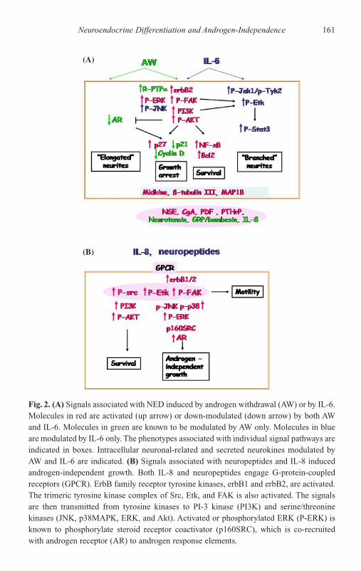
Neuroendocrine Differentiation and Androgen-Independence 161
(A)
(B)
Fig. 2. (A) Signals associated with NED induced by androgen withdrawal (AW) or by IL-6.Molecules in red are activated (up arrow) or down-modulated (down arrow) by both AWand IL-6. Molecules in green are known to be modulated by AW only. Molecules in blueare modulated by IL-6 only. The phenotypes associated with individual signal pathways areindicated in boxes. Intracellular neuronal-related and secreted neurokines modulated byAW and IL-6 are indicated. (B) Signals associated with neuropeptides and IL-8 inducedandrogen-independent growth. Both IL-8 and neuropeptides engage G-protein-coupledreceptors (GPCR). ErbB family receptor tyrosine kinases, erbB1 and erbB2, are activated.The trimeric tyrosine kinase complex of Src, Etk, and FAK is also activated. The signalsare then transmitted from tyrosine kinases to PI-3 kinase (PI3K) and serine/threoninekinases (JNK, p38MAPK, ERK, and Akt). Activated or phosphorylated ERK (P-ERK) isknown to phosphorylate steroid receptor coactivator (p160SRC), which is co-recruitedwith androgen receptor (AR) to androgen response elements.
B245-ch07 3/2/05 4:32 PM Page 161

such as NSE and beta-tubulin III.53 Conversely, addition of androgen tohormone-depleted media inhibited NED of LNCaP and could restorebasal expression levels of NSE and neurotensin.49 In contrast, whileandrogen deprivation for 4–7 days induces a terminal differentiation bymorphological criteria, the cells retain the capacity to respond to androgenand overcome growth arrest (Desai, unpublished data).
Investigation into signaling events that occur upon androgen withdrawalin LNCaP cells have implicated kinases and cell cycle regulators in theprocess of growth arrest and resistance to apoptosis. Notably, PI3K signal-ing is deregulated and augmented in the LNCaP model as a consequenceof mutational inactivation of the PTEN lipid phosphatase.56,57 As a result,this aberrant signal poses as a dominant mediator of survival primarilythrough the resultant elevation in the basal level of active Akt/PKB,58 amajor PI3K target known to negatively regulate apoptosis via the phospho-rylation and inactivation of pro-apoptotic molecules.59,60 Akt emerged as
162 S. J. Desai et al.
(A) (B)
(C) (D)
Fig. 3. Neuroendocrine differentiation in LNCaP cells. Cells were plated in RPMI 1640 sup-plemented with 2% FBS. The following day, cells were either (A) left untreated or treated with(B) androgen withdrawal (RPMI 1640 supplemented with 2% CDS-FBS), (C) IL-6 (50ng/mlIL-6 in RPMI 1640 supplemented with 2% FBS), or (D) Forskolin (10�M Forskolin in RPMIsupplemented with 2% FBS). Photographs were taken 96 hours after treatment.
B245-ch07 3/2/05 4:32 PM Page 162

a critical mediator of survival during androgen-deprivation by studiesdemonstrating its rapid hyper-activation observable within 8 hours ofandrogen withdrawal49 and persisting for up to 60 days (Tepper, unpublisheddata). Although AR is a bona fide Akt substrate,61,62 Akt-mediated phos-phorylation paradoxically promotes AR instability by targeting it forMdm2-mediated ubiquitination and subsequent proteasome-mediateddegradation.63 Similar to other PCa models, chronic androgen ablation ofLNCaP results in the transition from an androgen-dependent to -independentstate in which growth arrest is overcome.49,64,65 In accord with elevatedserum PSA serving as the clinical benchmark for the occurrence of hor-mone-refractory disease, reinstatement of the AR pathway (i.e., AR expres-sion, PSA secretion) in experimental models is one hallmark of thetransition to androgen-independence.49,64–67 The reappearance of AR isdue, at least in part, to enhanced stability of the receptor.68 In late passageand androgen-independent LNCaP cell lines (e.g., LNCaP-AI, LNCaP-rf,LNCaP-cds)49,64,67,69 this potentially involves a conversion of persistentPI3K/Akt activation from a degradation signal to an AR activator.67
Consistent with androgen withdrawal-induced Akt activation, treat-ment of LNCaP cells with R1881, a synthetic androgen, can decreasePI3K and Akt activation in both parental LNCaP and LNCaP-rf celllines.49 The mechanism by which this occurs is yet to be determined. Thereceptor tyrosine kinase erbB2, a member of the EGFR family, is anupstream activator of the PI3K pathway. ErbB2 is found expressed in NEcells of patients who have undergone radical prostatectomy70 and is bothupregulated and activated in androgen-deprived LNCaP cells (Tepper,unpublished data). However, it is unlikely that erbB2 is solely requiredfor Akt activation by androgen withdrawal since functional knockout oferbB2, by its sequestration in the ER via expression of an erbB2-specificsingle chain antibody,71 did not diminish Akt phosphorylation (Tepper,unpublished data). Therefore, multiple pathways for PI3K activationprobably exist. ErbB2, a known activator of MAPK, could be responsiblefor constitutive activation of MAPK after androgen withdrawal (Desai,unpublished data).72 RPTP-� also increases in androgen deprived NE dif-ferentiated LNCaP cells and contributes to MAPK activation.50,55 Oneinteresting possibility is that erbB2 and RPTP-� may cross-talk duringthe differentiation process leading to MAPK activation. The induction of
Neuroendocrine Differentiation and Androgen-Independence 163
B245-ch07 3/2/05 4:32 PM Page 163

PI3K and Akt appears to be responsible for LNCaP resistance to apopto-sis in the absence of androgen. Inhibiting PI3K either by treatment withpharmacological agents such as wortmannin or LY294002 or by overex-pressing PTEN can selectively induce apoptosis in LNCaP cells duringandrogen deprivation.49 This is accompanied by decreased Akt activationand triggering of the apoptotic proteolytic cascade, evidenced by caspase-3activation. Short-term (3-day) androgen withdrawal prior to the additionof PI3K inhibitors further sensitizes LNCaP to apoptosis induced by PI3Kinhibitors. PI3K inhibition might translate into enhanced apoptosis sus-ceptibility via down-regulation of the anti-apoptotic protein Bcl-2, whichincreases upon androgen withdrawal.73 In contrast, LNCaP-rf cells areremarkably resistant to treatment with PI3K inhibitors which might beexplained by their markedly elevated levels of activated Akt and by Bcl-2only being decreased by 50% upon treatment with the inhibitors (Tepper,unpublished data). Together, this suggests that immediate signaling stim-uli are replaced by other factors as LNCaP cells progress to androgen-independence. These factors may be secreted by NE cells themselves, in aparacrine or autocrine fashion, allowing differentiated cells to eventuallyobtain signaling and growth autonomy.
PI3K signaling may contribute to overcoming growth arrest, in addi-tion to suppressing apoptosis. Downstream effects of PI3K pathway dys-regulation can certainly manifest as inappropriate progression through theRb-mediated restriction point. Specifically, while cyclin-dependent kinaseinhibitor p27kip1 and p16Ink4a protein levels increased upon acute androgenwithdrawal in LNCaP cells, they are significantly decreased in androgen-independent LNCaP and CWR22 cells and xenografts.49,54,69 AlthoughPI3K/Akt activation can suppress forkhead-mediated p27kip1 transcrip-tion,74 the primary mechanism for p27kip1 protein down-regulation in thismodel was via increasing its rate of ubiquitin-mediated degradation,75 asLY294002 treatment of androgen-independent LNCaP cells in the pres-ence of the protein synthesis inhibitor cycloheximide markedly enhancedp27kip1 stability.49 At present, it is rather unclear as to how androgenwithdrawal augments p27kip1 expression level in the presence of PI3K/Akt activation. However, the expression of p27kip1 seems to be a deter-mining factor for the growth arrest of NE cells. p21Waf1/Cip1 exhibits theopposite expression pattern to p27kip1, decreasing during acute androgen
164 S. J. Desai et al.
B245-ch07 3/2/05 4:32 PM Page 164

withdrawal followed by reinstatement in the refractory stage.49 This seemsto contradict the conventional role of p21Waf1/Cip1 as an inducer of cellcycle arrest. However, it may be explained by recent findings thatp21Waf1/Cip1 can be either cell-cycle inhibitory or mitogenic, depending onits cellular localization.76–78 In the nucleus, p21Waf1/Cip1 binds to andinhibits cyclin/CDK activity, thereby blocking cell cycle progression.79
Upon phosphorylation of p21Waf1/Cip1 by Akt, p21Waf1/Cip1 translocates tothe cytoplasm, where it promotes cell growth.80 In addition, p21Waf1/Cip1
has been demonstrated to be critical for the assembly of cyclin D-cdk4complexes.81 Thus the restoration of p21Waf1/Cip1 expression might be apivotal event for the resumption of G1-S progression in the refractorystage. The AR and PI3K pathways play complimentary roles in positivelyregulating p21Waf1/Cip1 expression. While androgen stimulates transcrip-tion of the p21Waf1/Cip1 gene through AR binding to a consensus bindingsite in the p21Waf1/Cip1 promoter,82 Akt stabilizes the translated product byphosphorylation on residues Thr145 and Ser146.83 Accordingly, inhibitionof PI3K causes a decrease in p21Waf1/Cip1. Usurping p53-mediated growtharrest through mutational inactivation84 or reduction in p53 expression54,85
also figures as a prominent mechanism underlying androgen-independentgrowth. Recent data implicates PI3K as a potent suppressor of the p53pathway as Akt-mediated phosphorylation of Mdm2 promotes its nuclearlocalization, high affinity binding to p53-p300 complexes, ubiquitination,and enhanced targeting of p53 for proteasome-mediated degradation.80
However, the role of these cell cycle proteins and their requirements dur-ing NED has yet to be uncovered. Importantly, the theme of PI3K activa-tion promoting hormone-refractory PCa appears to be conserved even inmodels that do not possess genetic defects in PI3K pathway regulation,such as CWR22. For instance, microarray analyses of CWR22 recurrenttumors demonstrated elevated expression of genes encoding growth fac-tors (e.g., HGF, VEGFC, FGF2, PDGFA) known to stimulate PI3K/Akt.86
IL-6-Induced Neuroendocrine Differentiation
Another potential inducer of NED in PCa is Interleukin-6 (IL-6), a 212amino acid glycoprotein that was first identified as a regulator of immuneand inflammatory responses, but later shown to be involved in cell growth,
Neuroendocrine Differentiation and Androgen-Independence 165
B245-ch07 3/2/05 4:32 PM Page 165

differentiation, and metastasis.87 IL-6 has been implicated as a drivingfactor in the progression of PCa and elevated serum IL-6 levels are oftenfound in PCa patients.88 The receptors for IL-6, an 80-kd IL-6 specificreceptor subunit (alpha chain) and a 130-kd signal transducer calledgp130,87,89,90 are expressed in prostate carcinomas and the PCa cell linesLNCaP, PC-3, and DU145.91 The androgen-independent PCa cell linesPC-3 and DU145 express IL-6, but LNCaP cells do not.91 An antibody toeither IL-6 or the 80-kd subunit IL-6 receptor can inhibit growth of PC-3cells suggesting that IL-6 is part of an autocrine loop promoting thegrowth and androgen-independence of PC-3 cells.92 In contrast, IL-6treatment of LNCaP cells induces p27kip1 and consequent growtharrest18,19 and NED, as evidenced by morphological criteria (Fig. 3C) andthe expression of NE markers NSE, CgA, and beta-tubulin III.18,19,50,93 Thereceptors for IL-6 are found in cholesterol rich lipid rafts of the plasmamembrane and the formation of these rafts appear to be required for IL-6induced signaling and NED.94 IL-6 stimulation of gp130 has been shownto interact with and activate erbB2 and erbB3 receptors in LNCaP cells.Under these conditions, erbB2 appears to be required for IL-6 inducedmitogen activated protein kinase (MAPK) phosphorylation.95 IL-6 alsoinduces expression of erbB2 (Desai, unpublished data). The above prop-erties are thus shared with androgen-deprived LNCaP cells. Signaling inLNCaP cells induced by IL-6 utilizes the customary JAK-STAT family asmediators of signal transduction.96 The binding of IL-6 to its receptorcomplex results in the autophosphorylation and activation of JAK1, JAK2,and Tyk2, which in turn leads to the phosphorylation of and transcriptionalactivation by STAT3. STAT1, STAT5a, and STAT5b are expressed inLNCaP, but do not become activated by IL-6 signaling.16 STAT3 appearsto be the key player in NED induced by IL-6, as ectopic expression of adominant-negative form of STAT3 in LNCaP cells abrogate their differ-entiation into NE cells and the expression of NSE and CgA.93 IL-6 doesnot induce differentiation or NSE expression in PC-3 cells, which do notexpress STAT3. However, STAT3 overexpression in PC-3 cells results inan NED like morphology. Treatment with IL-6 does not further increasethe NED phenotype, but does slightly induce NSE expression. Consistentwith IL-6 induced NED, STAT3 overexpression in PC-3 cells leads to con-stitutive activation of STAT3 and growth inhibition of PC-3.93
166 S. J. Desai et al.
B245-ch07 3/2/05 4:32 PM Page 166

IL-6 can also mediate PCa aggressiveness through antagonism of apop-tosis. Similar to androgen withdrawal, IL-6 triggers the PI3K pathway. IL-6has previously been reported to protect Hep3B hepatocarcinoma cells fromTGF-� induced apoptosis through PI3K-Akt activation and by inhibitingcaspase-3 activation.17 Accordingly, inhibition of IL-6 signaling sensitizesPC-3 cells to etoposide-mediated cytotoxicity.92 In LNCaP cells, IL-6activates Etk, a member of the Btk tyrosine kinase family,97 in a PI3K-dependent manner.19 Etk overexpression protects LNCaP cells from apop-tosis induced by oxidative stress or thapsigargin.98 This protection wasabolished by treatment with LY294002 prior to challenge with either stress.Indeed, Etk poses as a critical anti-apoptotic component of the IL6R-PI3Kaxis parallel to Akt signaling. IL-6 induced STAT3 activation has beenimplicated as an anti-apoptotic signal by virtue of affording protection toHep3B cells,17 and by gp130-STAT3-mediated induction of Bcl-2 in BAF-B03 mouse pro-B cells.99 However, the role played by STAT3 in IL-6-treatedLNCaP cells with respect to apoptosis has yet to be determined.
Etk appears to play a critical role in modulating the cytoskeletalchanges that manifest as the characteristic NE morphology induced byIL-6. Treatment of LNCaP cells with IL-6 promotes the extension of shortneuritic processes having a high level of branching complexity. While thiscan be recapitulated by overexpression of Etk in the absence of ligand,abrogation of Etk function using a dominant-negative approach alters themorphological changes to long, unbranched outgrowths19 more typical ofthe response to androgen withdrawal. Consistent with these observations,we have been unable to detect Etk activation in the latter scenario (Desai,unpublished data) thereby implicating Etk as a strong effector of branch-ing seen in IL-6 induced NED. Currently, the mechanism via which Etkactivation translates into extension of neurites is speculative. The smallGTPase RhoA, but not Rac1 or Cdc24, has recently been shown to be acti-vated by Etk.100 Since RhoA controls the organization of cytoskeletal actinand mediates the formation of stress fibers, RhoA activation by Etk wouldbe consistent with the requirement of Etk in the IL-6 induced NED phe-notype. Etk can also associate with focal adhesion kinase (FAK) in LNCaPcells.101,102 FAK has been shown to promote neurite outgrowth in PC12cells103 and appears to be necessary for this process in LNCaP cells sinceexpression of a dominant negative form of FAK prevents differentiation by
Neuroendocrine Differentiation and Androgen-Independence 167
B245-ch07 3/2/05 4:32 PM Page 167

IL-6 stimulation or androgen withdrawal (Desai, unpublished data). Aswith nonmalignant cells, cellular morphology and the processes regulat-ing cell shape have functional implications. This is exemplified by theEtk-FAK association also serving as a migration signal in responseto GRP or integrin stimulation of LNCaP or HUVEC cells, respec-tively.101,102 Interestingly, Etk is highly expressed in PC-3 cells, whichhave a greater migratory potential compared to LNCaP cells.102 Etk hasbeen shown to interact with Cas, a docking protein, and enhance the inter-action of Cas with Crk.104 The Cas/Crk complex is involved in regulatingactin and cell motility and loss of Etk inhibits migration in COS-7 cells.Both Cas and Crk are also recruited by FAK, and it will be interesting tosee whether an Etk/FAK/Cas/Crk complex is formed at the cellular mem-brane during IL-6 induced NED of LNCaP cells. This suggests that thefinal NE morphology and aggressiveness of the cell might be determinedby levels of Etk, FAK, and the molecules with which they interact.
Other Factors Inducing Neuroendocrine Differentiation
While other factors have been shown to induce NED, less is knownabout the pathways involved. Inducers of cyclic AMP, such as epineph-rine, forskolin (Fig. 3D), and PAPCA, can induce NED of LNCaPcells.16,20,21,105,106 Cyclic AMP elevation leads to growth arrest andexpression of NSE, CgA, PTHrP, neurotensin, and serotonin. PKA andMAPK activation are thought to be involved16,20 and may lead to activa-tion of the transcription factor CREB.106 Contrary to IL-6 or androgen-withdrawal-induced differentiation, cAMP-induced differentiation doesnot appear to be terminal. Removal of cAMP inducers results in the lossof the NED phenotype and an increase in protein kinase activities.21 Co-treatment of IL-6 with cAMP enhances the extent of NED suggesting thatthese agents may cooperate to induce differentiation in PCa.16 HB-EGF, aprostate stromal-derived factor that promotes survival, can induce differ-entiation and increase NSE expression through MAPK activation.23 Incontrast to other NED inducers, HB-EGF does not induce cell cyclearrest,23 but instead promotes androgen-independent growth of LNCaPcells and down-modulates AR.22 IL-1� and IL-2,107,108 and anticancer
168 S. J. Desai et al.
B245-ch07 3/2/05 4:32 PM Page 168

agents that induce growth arrest such as jolkinolide B,109 the COX-2inhibitor NS-398,110 and silibinin111 also induce NED and an increase inNSE and CgA expression in LNCaP cells suggesting that NED may be adefault pathway in response to cellular stress in androgen-dependent cells.
As discussed above, a wealth of clinical and experimental data impli-cates NED as a common feature of hormone-refractory disease. AlthoughNED can be induced by circulating cytokines or passively as a result ofandrogen ablative therapy, NE cells possess the potential to actively andpotently drive the metastatic growth of PCa cells in the absence of andro-gens and/or in the presence of AR antagonists. This is primarily accom-plished through the paracrine, and possibly autocrine, secretion ofpolypeptide factors such as neurotrophins and cytokines. Below, we willdiscuss the signaling pathways triggered by several of these polypeptideligands and how they engender one or more of the vital components ofandrogen-independent growth such as reinstatement of AR signaling andstimulation of metastasis through enhanced motility, invasiveness, andangiogenesis. Since there is significant diversity in these ligands, thereceptor-mediated signals they evoke, and their ultimate biological effects,the combinatorial outcome of “Neurokine” signaling might be sufficientto support androgen-independent growth. A summary of the signal path-ways involved in neurokine-induced androgen-independence is presentedin Fig. 2B.
Androgen-Independent Growth
The absence of androgen and subsequent down-regulation of AR duringandrogen-ablation therapy would suggest that AR is no longer required inandrogen-independent tumors. However, AR is still expressed in almostall androgen-independent tumors3 and this suggests that AR is involved inandrogen-independent growth of PCa. Amplification of AR and its coac-tivators such as p160 SRCs4–6 are also detected in relapsed and metastatictumors. Androgen-independent LNCaP cell lines, which can grow in theabsence of androgen, show an increased level of AR expression.49,64,112
Mutations in AR can lead to the activation of AR by other steroids suchas estrogens,113 glucocorticoids,114 and even nonsteroidal anti-androgens
Neuroendocrine Differentiation and Androgen-Independence 169
B245-ch07 3/2/05 4:32 PM Page 169

used to treat PCa.115 However, the low frequency of mutations in androgen-independent tumors is leading investigators to investigate alternativemechanisms of AR activation, either being ligand-independent or insynergy with castrate levels of androgen.
Three classes of activators have been identified (1) circulating growthfactors that bind to transmembrane receptor tyrosine kinases (EGF, IGF-1,and KGF); (2) neuropeptides (GRP and neurotensin) and chemokines(IL-8) secreted by neuroendocrine cells that activate G-protein-coupledreceptors (GPCR); and (3) interleukins (e.g., IL-6) that bind to cytokinereceptors. These factors, while differing in the detailed signal pathwaysthey transmit, share certain common traits. For instance, they all inducephosphorylation cascades and activate tyrosine and serine/threoninekinases. It has been suggested that phosphorylation of AR, or its activa-tors, facilitates the assembly of a transcriptional complex that mimics therecruitment by androgen-bound AR, and may be an alternate route to ARactivation.116 Since AR and its coactivators are known to be phosphory-lated only at serines and threonines, serine/threonine kinases are thoughtto be immediate activators of the AR complex, whereas tyrosine kinasesare the upstream activators of serine/threonine kinases. Phosphorylationand activation of AR by the serine/threonine kinases ERK,117,118 Akt,62
PKA,119,120 PKC,121,122 and CAK123 have been reported. Examples ofserine/threonine kinase activation of AR coactivators include ERK phos-phorylation of SRC-164 and phosphorylation of SRC-24 by IL-6 and EGFstimulation, respectively. Most of these studies were based on in vitrostudies or transient expression systems. The in vivo phosphorylation sitesof AR have recently been identified,124–126 and ironically none of the sitesare consensus phosphorylation sites of ERK or Akt,124 raising the possi-bility that other novel kinases are involved. Studies to identify proteinkinases associated with or induced by AR have resulted in the discoveryof several novel kinases, ANPK,127 SPAK128 MAK,129 and PAK6.130,131
These kinases modulate (ANPK, SPAK, and MAK activate, while PAK6represses) AR activity by directly binding to AR, but do not seem todirectly phosphorylate AR or affect cellular growth induced by androgen.Despite these new twists, it seems clear that AR activity can be enhancedby kinases. In the ensuing sections, we will review how soluble factorsinduce the activation of these kinases.
170 S. J. Desai et al.
B245-ch07 3/2/05 4:32 PM Page 170

Growth Factors
Growth factors such as KGF, IGF-1, and EGF have been shown to activateAR in the absence of androgen,132 via the activation of their respectivereceptor tyrosine kinases (FGF-R, IGF-R, and EGFR). ErbB2, when over-expressed, can induce PSA expression in an androgen-independent, butnot AR-dependent manner.117,118 All of the receptor tyrosine kinases areknown to activate serine/threonine kinases, such as MAPK117,118 andAkt,62 which in turn activate AR activity as discussed above.
Neuropeptides
Of growing interest is elucidating the role of neuropeptides in the androgen-independent growth of PCa. Since neuropeptides are secreted by NE cells,the potential contribution of the prostate micro-environment to cancergrowth becomes significant. As mentioned in the previous section, NEcells in the prostate and NE-differentiated LNCaP cells secrete neuropep-tides such as GRP and neurotensin. Androgen-independent PCas and PCacell lines also express GRP and neurotensin, as well as chemokines suchas IL-8, that appear to be involved in an autocrine loop that stimulatesgrowth. GRP receptors133,134 and neurotensin receptors135 are found inPCas and PCa cell lines. GRP and neurotensin have been shown to stim-ulate androgen-independent growth in PC-3, DU145, and LNCaPcells.51,101,135–138 Conversely, GRP antagonists139–142 and neurotensinantagonists135 have been shown to inhibit PC-3 and DU145 growth. Themajority of information about the mechanism mediating neuropeptide-induced growth of PCa pertains to GRP signaling. GRP binds to andactivates GPCR proteins (Fig. 2B), which engage the heterotrimeric G proteins �, �, and , which in turn leads to sequential Ca� mobilization,PLC and PKC activation, and ultimately cellular proliferation.143 The spe-cific response in the cell is dependent on which G protein is activated andwhether it is stimulatory or inhibitory. In the case of GRP, GRP-R hasbeen shown to activate G�q, which activates the PLC� pathway and G�12,which activates the Rho pathway. However, activation of G�q and G�12 byGRP in PCa cells has not been verified. In PC-3 and DU145, cross-talkbetween GPCR proteins and receptor tyrosine kinases144 has been
Neuroendocrine Differentiation and Androgen-Independence 171
B245-ch07 3/2/05 4:32 PM Page 171

demonstrated by the ability of GRP to transactivate EGFR and that itrequires Src activation and Ca� mobilization. Once EGFR is activated itleads to Ras and MAPK activation and DNA synthesis. The transcriptionfactor Elk-1, and immediate early genes c-fos and c-myc are also up-regulated by GRP presenting a possible pathway for growth induction.145
Evidence for GRP as a progression factor is provided by its ability to up-regulate proangiogenic factors such as MMP-9,146,147 VEGF, and IL-8,148
the latter two being mediated through NF-�B. Furthermore, cross-talkbetween GRP signaling and �1 integrin results in enhanced processing ofpro-MMP-9 into its active form as a result of Src and PI3K activation cul-minating in the up-regulation of membrane-bound uPA.146 GRP activationof the PKC pathway in PC-3 cells leads to the activation of FAK andincreased motility suggesting that GRP may also play an important role intissue invasion, increasing the potential for metastasis.149
GRP and neurotensin have also been shown to activate AR and induceandrogen-independent growth of LNCaP cells.101 GRP can act synergisti-cally with low levels of androgen to activate AR, leading to the possibilitythat residual circulating androgen present after androgen-ablation therapycould cooperate with GRP in achieving an androgen-independent state.150
GRP-induced activation and complex formation of Etk, FAK, and Src arerequired for AR activation. Interestingly, Etk activation is dependent uponSrc and FAK, but not on PI3K, as is the case with IL-6 stimulated Etkactivation. Activation of AR supports the notion that neuropeptides mightmediate the transition from an androgen-dependent to -independentstate in PCa. Further indication that GRP may be a transition-promotingfactor is the evidence that neutral endopeptidase (NEP), which cleavesneuropeptides, is down-regulated in androgen-independent tumors,151
and is down-regulated by GRP in LNCaP.152 Reintroduction of NEP inandrogen-independent cells interferes with GRP signaling153 and cellularmigration.154
Chemokines
As mentioned above, the chemokine IL-8 is an angiogenic factor involvedin the migration and invasion of cancer cells. Serum IL-8 levels are ele-vated in PCa patients155 and IL-8 is overexpressed in PC-3 cells.156 The
172 S. J. Desai et al.
B245-ch07 3/2/05 4:32 PM Page 172
IL-8 receptors, CXCR1 and CXCR2, are expressed in DU145 and PC-3cells157 and antisense-mediated suppression of IL-8 expression reducesinvasion and metastatic potential of PC-3 xenografts in nude mice.158
Many studies have focused on the role of IL-8 in tissue invasion andmetastasis.158–160 However, a recent finding showed that IL-8 couldstimulate the androgen-independent growth of LNCaP cells via recruit-ment of the AR transcription complex to the proximal region of the PSApromoter.161 Analogous to GRP signaling, IL-8 induces Src, FAK, andMAPK activation (Fig. 2B) and promotes migration of LNCaP cells. Srcand MAPK appear to be required for AR activity and LNCaP prolifera-tion, while Src and FAK are required for LNCaP migration. Together,these data indicate that GRP and IL-8 may be progression factors of PCacontributing to the androgen-independence and metastatic potential ofadvanced cancers.
Cytokines
As described in the previous section, our work, as well as that of others,demonstrated that IL-6 treatment of LNCaP induced NED and growtharrest.19,162 Paradoxically, IL-6 has also been found to induce growth ofPCa cells, including LNCaP. When subjected to prolonged IL-6 treatment,eventual selection of proliferating LNCaP cells occured. While the molec-ular details of the genetic adaptation caused by IL-6 is not clear, onceachieved, the cells become androgen-independent and can grow in thepresence of IL-6. This may be related to the observation that in low-passageLNCaP cells, activated Akt down-modulates AR, whereas in high-passageLNCaP cells, it enhances AR activity.67 In fact, a very interesting aspectof the pleiotropic influence of IL-6 upon the transition to androgen-independence is its ability to directly regulate AR.163–167 Although IL-6activation of AR can occur without androgen, IL-6 can also act synergisti-cally with low levels of androgen.163–165 The pathway of IL-6-mediatedAR activation is complex and involves multiple signal transducers. Tworeports demonstrated that JAK-STAT3 activation is the principal path-way responsible for AR activation via direct STAT3-AR interaction.163,164
This was confirmed by inhibition of AR activation in the presence of over-expressed dominant negative STAT3. However, a third report implicated
Neuroendocrine Differentiation and Androgen-Independence 173
B245-ch07 3/2/05 4:32 PM Page 173
MEK/MAPK, PKA, and PKC, since AR activation could be inhibited withpharmacological inhibition of each of these kinases.165 Yet another papershowed that IL-6 activation of AR depends on ERK and p300.167 Onereason for this diversity may lie in differences in cellular milieu and theoutcome of competition for cellular resources that can activate AR.166
For instance, STAT3 is the dominant mediator of IL-6 signaling and ARactivation in LNCaP cells, but PKA or PKC might predominate to activateectopically-expressed AR in the STAT3-deficient cell line PC-3. Anintriguing question is how IL-6 and AR cooperate under diverse conditionsof NED. In contrast to down-regulation of AR activity and protein levelsduring androgen withdrawal, IL-6 might contribute to cell survival duringandrogen ablation therapy through its ability to activate the AR. This couldresult in maintenance of AR expression so that other growth factors areable to utilize AR to circumvent androgen dependence.
Summary
In summary, there is considerable evidence that NED represents an integraland potentially important part of prostate carcinogenesis. The propensityof prostate carcinoma cells to undergo NED under a variety of conditionsincluding androgen withdrawal suggest that it is an innate property ofprostate epithelial cells to survive and to overcome adverse conditions.Although the cellular signals involved in this trans-differentiation processvary with different inducing conditions, the differentiated cells have threecommon characteristics: resistance to apoptosis, growth arrest, and expres-sion of neuronal phenotypes. The release by these cells of neurokines,which are survival and growth factors, is likely to have an impact on sur-rounding undifferentiated prostate cancer cells undergoing androgen-deprivation crisis. Accumulating evidence suggests that these neurokinesare capable of activating AR, presumably via kinase signaling. This maybe one way a fraction of PCas survive androgen ablation and eventuallydevelop into androgen-independent clones. If true, targeting neurokinesand the downstream protein kinases in conjunction with hormone therapyoffers a new intervention strategy for treatment of prostate cancer and inparticular, androgen-independent tumors. (This work from the authors’labs was supported by grants from the NIH and DOD.)
174 S. J. Desai et al.
B245-ch07 3/2/05 4:32 PM Page 174
References
1. Abrahamsson PA (1999) Neuroendocrine differentiation and hormone-refractory prostate cancer. Prostate 6(Suppl):3–8.
2. Arnold JT, Isaacs JT (2002) Mechanisms involved in the progression ofandrogen-independent prostate cancers: It is not only the cancer cell’s fault.Endocr Relat Cancer 9:61–73.
3. Culig Z, Hobisch A, Hittmair A, Peterziel H, Cato A, Bartsch G, Klocker H(1998) Expression, structure, and function of androgen receptor in advancedprostatic carcinoma. Prostate 35:63–70.
4. Gregory CW, Fei X, Ponguta LA, He B, Bill HM, French FS, Wilson EM(2004) Epidermal growth factor increases coactivation of the androgenreceptor in recurrent prostate cancer. J Biol Chem 279:7119–7130.
5. Zhou G, Hashimoto Y, Kwak I, Tsai SY, Tsai MJ (2003) Role of thesteroid receptor coactivator SRC-3 in cell growth. Mol Cell Biol 23:7742–7755.
6. Chen CD, Welsbie DS, Tran C, Baek SH, Chen R, Vessella R, Rosenfeld MG,Sawyers CL (2004) Molecular determinants of resistance to antiandrogentherapy. Nat Med 10:33–39.
7. Hanson J, Abrahamsson PA (2001) Neuroendocrine pathogenesis in adeno-carcinoma of the prostate. Ann Oncol 12(Suppl 2):S145–152.
8. Bonkoff H (2001) Neuroendocrine differentiation in human prostate cancer.Morphogenesis, proliferation and androgen receptor status. Ann Oncol12(Suppl 2):S141–144.
9. di Sant’Agnese PA (2001) Neuroendocrine differentiation in prostatic carci-noma: An update on recent developments. Ann Oncol 12(Suppl 2):S135–140.
10. Abrahamsson PA (1999) Neuroendocrine cell in tumor growth in theprostate. Endocr Relat Cancer 6:503–519.
11. di Sant’Agnese PA (1998) Neuroendocrine differentiation in prostatic carci-noma: An updatel. Prostate 8(Suppl):74–79.
12. Aprikian AG, Han K, Guy L, Landry F, Begin LR, Chevalier S (1998)Neuroendocrine differentiation and the bombesin/gastrin-releasing peptidefamily of neuropeptides in the progression of human prostate cancer.Prostate 8(Suppl):52–61.
13. di Sant’Agnese PA (1998) Neuroendocrine cell of the prostate and neuroen-docrine differentiation in prostatic carcinoma: A review of morphologicaspects. Urology 51:121–124.
14. Gkonos PJ, Krongrad A, Roos BA (1995) Neuroendocrine peptides in theprostate. Urol Res 23:81–87.
Neuroendocrine Differentiation and Androgen-Independence 175
B245-ch07 3/2/05 4:32 PM Page 175

15. Burchardt T, Burchardt M, Chen MW, Cao Y, de la Taille A, Shabsigh A,Hayek O, Dorai T, Buttyan R (1999) Transdifferentiation of prostate cancer cellto a neuroendocrine cell phenotype in vitro and in vivo. J Urol 162:1800–1805.
16. Deeble PD, Murphy DJ, Parsons SJ, Cox ME (2001) Interleukin-6-and cyclicAMP mediated signaling potentiates NED of LNCaP prostate tumor cells.Mol Cell Bio 21:8471–8482.
17. Chen RH, Chang MC, Su YS, Tsai YT, Kuo ML (1999) Interleukin-6 inhibitstransforming growth factor-beta-induced apoptosis through the phos-phatidylinositol 3�-kinase/Atk and signal transducers and activators of tran-scription 3 pathway. J Bio Chem 274:23013–23019.
18. Mori S, Murakami-Mori K, Bonavida B (1999) Interleukin-6 induces G1arrest through induction of p27Kip1, a cyclin-dependent kinase inhibitor, andneuron-like morphology in LNCaP prostate tumor cells. Biochem BiophysRes Commun 257:609–614.
19. Qui Y, Robinson D, Pretlow TG, Kung HJ (1998) Etk/Bmx, a tyrosine kinasewith a pleckstrin-homology domain, is an effector of phosphatidylinositol3�-kinase and is involved in interleukin 6-induced NED of PCa cells. ProcNatl Acad Sci 95:3644–3649.
20. Cox ME, Deeble PD, Bissonette EA, Parsons SJ (2000) Activated3�,5�-cyclic AMP-dependent protein kinase is sufficient to induce neuroen-docrine-like differentiation of the LNCaP prostate tumor cell line. J BiolChem 275:13812–13818.
21. Cox ME, Deeble PD, Lakhani S, Parsons SJ (1999) Acquisition of neuroen-docrine characteristics by prostate tumor cells is reversible: Implications forprostate cancer progression. Cancer Res 59:3821–3830.
22. Adam RM, Kim J, Lin J, Orsola A, Zhuang L, Rice DC, Freeman MR (2002)Heparin-binding epidermal growth factor-like growth factor stimulatesandrogen-independent prostate tumor growth and antagonizes androgenreceptor function. Endocrinology 143:4599–4608.
23. Kim J, Adam RM, Freeman MR (2002) Activation of the Erk mitogen-activated protein kinase pathway stimulates neuroendocrine differentiation inLNCaP cells independently of cell cycle withdrawal and STAT3 phosphory-lation. Cancer Res 62:1549–1554.
24. Schalken JA, van Leenders G (2003) Cellular and molecular biology of theprostate: Stem cell biology. Urology 62:11–20.
25. Xue Y, Smedts F, Verhofstad A, Debruyne F, de la Rosette J, Schalken J(1998) Cell kinetics of prostate exocrine and neuroendocrine epitheliumand their differential interrelationship: New perspectives. Prostate8(Suppl):62–73.
176 S. J. Desai et al.
B245-ch07 3/2/05 4:32 PM Page 176
26. Bonkhoff H, Remberger K (1996) Differentiation pathways and histogeneticaspects of normal and abnormal prostatic growth: A stem cell model. Prostate22:98–106.
27. di Sant’Agnese PA, de Mesy Jensen KL (1987) Neuroendocrine differentia-tion in prostatic carcinoma. Human Pathol 18:849–856.
28. Helpap B, Kollermann J, Oehler U (1999) Neuroendocrine differentiation inprostatic carcinomas: Histogenesis, biology, clinical relevance and futuretherapeutical perspectives. Urol Int 62:133–138.
29. di Sant’Agnese PA (1992) Neuroendocrine differentiation in carcinoma ofthe prostate. Diagnostic, prognostic, and therapeutic implications. Cancer70:254–268.
30. Hu Y, Ippolito JE, Garabedian EM, Humphrey PA, Gordon JI (2002)Molecular characterization of a metastatic neuroendocrine cell cancer arisingin the prostates of transgenic mice. J Biol Chem 277:44462–44474.
31. Masumori N, Thomas TZ, Chaurand P, Case T, Paul M, Kasper S,PCarioli RM, Tsukamoto T, Shappell SB, Matusik RJ (2001) A probasin-largeT antigen transgenic mouse line develops prostate adenocarcinoma and neu-roendocrine carcinoma with metastatic potential. Cancer Res 61:2239–2249.
32. Garabedian EM, Humphrey PA, Gordon JI (1998) A transgenic mouse modelof metastatic prostate cancer originating from neuroendocrine cells. ProcNatl Acad Sci USA 95:15382–15387.
33. Clegg N, Ferguson C, True LD, Arnold H, Moorman A, Quinn JA,Vessella RL, Nelson PS (2003) Molecular characterization of prostatic small-cell neuroendocrine carcinoma. Prostate 55:55–64.
34. Hvamstad T, Jordal A, Hekmat N, Paus E, Fossa SD (2003) Neuroendocrineserum tumour markers in hormone-resistant prostate cancer. Eur Urol 44:215–221.
35. Kamiya N, Akakura K, Suzuki H, Isshiki S, Komiya A, Ueda T, Ito H (2003)Pretreatment serum level of neuron specific enolase (NSE) as a prognosticfactor in metastatic prostate cancer patients treated with endocrine therapy.Eur Urol 44:309–314.
36. Berruti A, Dogliotti L, Mosca A, Gorzegno G, Bollito E, Mari M,Tarabuzzi R, Poggio M, Torta M, Fontana D, Angeli A (2001) Potential clin-ical value of circulating chromogranin A in patients with prostate carcinoma.Ann Oncol 12(Suppl 2):S153–157.
37. Bollito E, Berruti A, Bellina M, Mosca A, Leonardo E, Tarabuzzi R, Capia S,Ari MM, Tampellini M, Fontana D, Gubetta L, Angeli A, Dogliotti L (2001)Relationship between neuroendocrine features and prognostic parameters inhuman prostate adenocarcinoma. Ann Oncol 12(Suppl 2):S159–164.
Neuroendocrine Differentiation and Androgen-Independence 177
B245-ch07 3/2/05 4:32 PM Page 177
38. Ferrero-Poüs M, Hersant AM, Pecking A, Brésard-Leroy M, Pichon MF(2001) Serum chromogranin-A in advanced prostate cancer. BJU Int88:790–796.
39. Ather MH, Abbas F (2000) Prognostic significance of neuroendocrine dif-ferentiation in prostate cancer. Eur Urol 38:535–542.
40. Abrahamsson PA, Cockett ATK, di Sant’Agnese PA (1998) Prognostic sig-nificance of neuroendocrine differentiation in clinically localized prostaticcarcinoma. Prostate 8(Suppl):37–42.
41. Deftos LJ (1998) Granin-A, parathyroid hormone-related protein, andcalcitonin gene products in neuroendocrine prostate cancer. Prostate8(Suppl):23–31.
42. Deftos LJ, Abrahamsson PA (1998) Granins and prostate cancer. Urology51:141–145.
43. Jiborn T, Bjartell A, Abrahamsson P-A (1998) NED in prostatic carcinomaduring hormonal treatment. Urology 51:585–589.
44. Tarle M, Ahel MZ, Kovacic K (2002) Aquired NE-positivity during maximalandrogen blockade in prostate cancer patients. Anticancer Res 22:2525–2529.
45. Fixemer T, Remberger K, Bonkhoff H (2002) Apoptosis resistance of NEphenotypes in prostatic adenocarcinoma. Prostate 53:118–123.
46. Ismail A HR, Landry F, Aprikian AG, Chevalier S (2002) Androgen ablationpromotes NE cell differentiation in dog and human prostate. Prostate51:117–125.
47. Jongsma J, Oomen MH, Noordzij MA, van Weerden WM, Martens GJM, vander Kwast TH, Schröder FH, van Steenbrugge GJ (2000) Androgen depriva-tion of the prohormone convertase-310 human prostate cancer model systeminduces NED. Cancer Res 60:741–748.
48. Jongsma J, Oomen MH, Noordzij MA, van Weerden WM, Martens GJM, vander Kwast TH, Schröder FH, van Steenbrugge GJ (2002) Different profilesof NE cell differentiation evolve in the PC-310 human prostate cancer modelduring long-term androgen deprivation. Prostate 50:203–215.
49. Murillo H, Huang H, Schmidt LJ, Smith DI, Tindall DJ (2001) Role of PI3Ksignaling in survival and progression of LNCaP prostate cancer cells to theandrogen refractory state. Endocrinology 142:4795–4805.
50. Zelivianski S, Verni M, Moore C, Kondrikov D, Taylor R, Lin M-F (2001)Multipathways for transdifferentiation of human prostate cancer cells intoNE-like phenotype. Biochim Biophys Acta 1539:28–43.
51. Sehgal I, Powers S, Huntley B, Powis G, Pittelkow M, Maihle NJ (1994)Neurotensin is an autocrine trophic factor stimulated by androgen withdrawalin human prostate cancer. Proc Natl Acad Sci USA 91:4673–4677.
178 S. J. Desai et al.
B245-ch07 3/2/05 4:32 PM Page 178
52. Saeed B, Zhang H, Ng SC (1997) Apoptotic program is initiated but notcompleted in LNCaP cells in response to growth in charcoal-stripped media.Prostate 31:145–152.
53. Wright ME, Tsai MJ, Aebersold R (2003) AR represses the NE transdiffer-entiation process in prostate cancer cells. Mol Endocrinol 17:1726–1737.
54. Agus DB, Cordon-Cardo C, Fox W, Drobnjak M, Koff A, Golde DW, Scher HI(1999) Prostate cancer cell cycle regulators: Response to androgen withdrawaland development of androgen independence. J Natl Cancer Inst 91:1869–1876.
55. Zhang XQ, Kondrikov D, Yuan TC, Lin FF, Hansen J, Lin MF (2003)Receptor protein tyrosine phosphatase alpha signaling is involved in andro-gen depletion-induced NED of androgen-sensitive LNCaP human prostatecancer cells. Oncogene 22:6704–6716.
56. Teng DH, Hu R, Lin H, Davis T, Iliev D, Frye C, Swedlund B, Hansen KL,Vinson VL, Gumpper KL, Ellis L, El-Naggar A, Frazier M, Jasser S,Langford LA, Lee J, Mills GB, Pershouse MA, Pollack RE, Tornos C,Troncoso P, Yung WK, Fujii G, Berson A, Steck PA (1997) MMAC1/PTENmutations in primary tumor specimens and tumor cell lines. Cancer Res57:5221–5225.
57. Carson JP, Kulik G, Weber MJ (1999) Antiapoptotic signaling in LNCaPprostate cancer cells: A survival signaling pathway independent of phos-phatidylinositol 3�-kinase and Akt/protein kinase B. Cancer ResI 59:1449–1453.
58. Chan TO, Rittenhouse SE, Tsichlis PN (1999) AKT/PKB and other D3phosphoinositide-regulated kinases: Kinase activation by phosphoinositide-dependent phosphorylation. Annu Rev Biochem 68:965–1014.
59. Lawlor MA, Alessi DR (2001) PKB/Akt: A key mediator of cell prolifera-tion, survival and insulin responses? J Cell Sci 114:2903–2910.
60. Milburn CC, Deak M, Kelly SM, Price NC, Alessi DR, Van Aalten DM(2003) Binding of phosphatidylinositol 3,4,5-trisphosphate to the pleckstrinhomology domain of protein kinase B induces a conformational change.Biochem J 375:531–538.
61. Lin HK, Yeh S, Kang HY, Chang C (2001) Akt suppresses androgen-inducedapoptosis by phosphorylating and inhibiting androgen receptor. Proc NatlAcad Sci USA 98:7200–7205.
62. Wen Y, Hu MC, Makino K, Spohn B, Bartholomeusz G, Yan DH, Hung MC(2000) HER-2/neu promotes androgen-independent survival and growth ofprostate cancer cells through the Akt pathway. Cancer Res 60:6841–6845.
63. Lin HK, Wang L, Hu YC, Altuwaijri S, Chang C (2002) Phosphorylation-dependent ubiquitylation and degradation of androgen receptor by Aktrequire Mdm2 E3 ligase. EMBO J 21:4037–4048.
Neuroendocrine Differentiation and Androgen-Independence 179
B245-ch07 3/2/05 4:32 PM Page 179

64. Shi XB, Ma AH, Tepper CG, Xia L, Gregg JP, Gandour-Edwards R,Mack PC, Kung HJ, de Vere White RW (2004) Molecular alterations associ-ated with LNCaP cell progression to androgen independence. Prostate,in press.
65. Kokontis JM, Hay N, Liao S (1998) Progression of LNCaP prostate tumorcells during androgen deprivation: Hormone-independent growth, repressionof proliferation by androgen, and role for p27Kip1 in androgen-induced cellcycle arrest. Mol Endocrinol 12:941–953.
66. Nagabhushan M, Miller CM, Pretlow TP, Giaconia JM, Edgehouse NL,Schwartz S, Kung HJ, de Vere White RW, Gumerlock PH, Resnick MI,Amini SB, Pretlow TG (1996) CWR22: The first human prostate cancerxenograft with strongly androgen-dependent and relapsed strains both in vivoand in soft agar. Cancer Res 56:3042–3046.
67. Lin HK, Hu YC, Yang L, Altuwaijri S, Chen YT, Kang HY, Chang C (2003)Suppression versus induction of androgen receptor functions by the phos-phatidylinositol 3-kinase/Akt pathway in prostate cancer LNCaP cells withdifferent passage numbers. J Biol Chem 278:50902–50907.
68. Gregory CW, Johnson RT Jr, Mohler JL, French FS, Wilson EM (2001)Androgen receptor stabilization in recurrent prostate cancer is associatedwith hypersensitivity to low androgen. Cancer Res 61:2892–2898.
69. Graff JR, Konicek BW, McNulty AM, Wang Z, Houck K, Allen S, Paul JD,Hbaiu A, Goode RG, Sandusky GE, Vessella RL, Neubauer BL (2000)Increased AKT activity contributes to prostate cancer progression by dra-matically accelerating prostate tumor growth and diminishing p27Kip1expression. J Biol Chem 275:24500–24505.
70. Iwamura M, Koshiba K, Cockett ATK (1998) Receptors for BPH growth fac-tors are located in some NE cells. Prostate 8:14–17.
71. Beerli RR, Wels W, Hynes NE (1994) Intracellular expression of single chainantibodies reverts ErbB-2 transformation. J Biol Chem 269:23931–23936.
72. Gioeli D, Mandell JW, Petroni GR, Frierson HF Jr, Weber MJ (1999)Activation of mitogen-activated protein kinase associated with prostate can-cer progression. Cancer Res 59:279–284.
73. Pugazhenthi S, Nesterova A, Sable C, Heidenreich KA, Boxer LM,Heasley LE, Reusch JE (2000) Akt/protein kinase B up-regulates Bcl-2expression through cAMP-response element-binding protein. J Biol Chem275:10761–10766.
74. Medema RH, Kops GJ, Bos JL, Burgering BM (2000) AFX-like Forkheadtranscription factors mediate cell-cycle regulation by Ras and PKB throughp27kip1. Nature 404:782–787.
180 S. J. Desai et al.
B245-ch07 3/2/05 4:32 PM Page 180

75. Slingerland J, Pagano M (2000) Regulation of the cdk inhibitor p27 and itsderegulation in cancer. J Cell Physiol 183:10–17.
76. Dong Y, Chi SL, Borowsky AD, Fan Y, Weiss RH (2004) Cytosolicp21Waf1/Cip1 increases cell cycle transit in vascular smooth muscle cells.Cell Signal 16:263–269.
77. Dupont J, Karas M, LeRoith D (2003) The cyclin-dependent kinase inhibitorp21CIP/WAF1 is a positive regulator of insulin-like growth factor I-inducedcell proliferation in MCF-7 human breast cancer cells. J Biol Chem 278:37256–37264.
78. Weiss RH (2003) p21Waf1/Cip1 as a therapeutic target in breast and othercancers. Cancer Cell 4:425–429.
79. Hengst L, Gopfert U, Lashuel HA, Reed SI (1998) Complete inhibition ofCdk/cyclin by on molecule of p21 (Cip1). Genes Dev 12:3882–3888.
80. Zhou BP, Liao Y, Xia W, Zou Y, Spohn B, Hung MC (2001) HER-2/neuinduces p53 ubiquitination via Akt-mediated MDM2 phosphorylation. NatCell Biol 3:973–982.
81. Cheng M, Olivier P, Diehl JA, Fero M, Roussel MF, Roberts JM, Sherr CJ(1999) The p21(Cip1) and p27(Kip1) CDK ‘inhibitors’ are essential activa-tors of cyclin D-dependent kinases in murine fibroblasts. EMBO J 18:1571–1583.
82. Lu S, Liu M, Epner DE, Tsai SY, Tsai MJ (1999) Androgen regulation of thecyclin-dependent kinase inhibitor p21 gene through an androgen responseelement in the proximal promoter. Mol Endocrinol 13:376–384.
83. Li Y, Dowbenko D, Lasky LA (2002) AKT/PKB phosphorylation ofp21Cip/WAF1 enhances protein stability of p21Cip/WAF1 and promotes cellsurvival. J Biol Chem 277:11352–11361.
84. Meyers FJ, Gumerlock PH, Chi SG, Borchers H, Deitch AD, deVereWhite RW (1998) Very frequent p53 mutations in metastatic prostate carci-noma and in matched primary tumors. Cancer 83:2534–2539.
85. Burchardt M, Burchardt T, Shabsigh A, Ghafar M, Chen MW, Anastasiadis A,de la Taille A, Kiss A, Buttyan R (2001) Reduction of wild type p53 functionconfers a hormone resistant phenotype on LNCaP prostate cancer cells.Prostate 48:225–230.
86. Mousses S, Wagner U, Chen Y, Kim JW, Bubendorf L, Bittner M, Pretlow T,Elkahloun AG, Trepel JB, Kallioniemi OP (2001) Failure of hormone therapyin prostate cancer involves systematic restoration of androgen responsive genesand activation of rapamycin sensitive signaling. Oncogene 20:6718–6723.
87. Bonkhoff H (1998) NE cells in benign and malignant prostate tissue: mor-phogenesis, proliferation, and AR status. Prostate 8(Suppl):18–22.
Neuroendocrine Differentiation and Androgen-Independence 181
B245-ch07 3/2/05 4:32 PM Page 181

88. Twillie DA, Eisenberger MA, Carducci MA, Hseih WS, Kim WY,Simons JW (1995) Interleukin-6: A candidate mediator of human prostatecancer morbidity. Urology 45:542–549.
89. Hibi M, Murakami M, Saito M, Hirano T, Taga T, Kishimoto T, Hirata Y,Yamasaki K, Yasukawa K, Matsuda T (1990) Molecular cloning and expres-sion of an IL-6 signal transducer, gp130. Cell 63:1149–1157.
90. Taga T, Hibi M, Hirata Y, Yamasaki K, Yasukawa K, Matsuda T, Hirano T,Kishimoto T (1989) Interleukin-6 triggers the association of its receptorwith a possible signal transducer, gp130. Cell 58:573–581.
91. Lou W, Ni Z, Dyer K, Tweardy DJ, Gao AC (2000) Interleukin-6 inducesprostate cancer cell growth accompanied by activation of Stat3 signalingpathway. Prostate 42:239–242.
92. Borsellino N, Bonavida B, Ciliberto G, Toniatti C, Travali S, D’Alessandro N(1999) Blocking signaling through the gp130 receptor chain by interleukin-6 and oncostatin M inhibits PC-3 cell growth and sensitizes the tumor cellsto etoposide and cisplatin-mediated cytotoxicity. Cancer 85:134–144.
93. Spiotto MT, Chung TDK (2000) STAT3 mediates IL-6-induced NED inprostate cancer cells. Prostate 42:186–195.
94. Kim J, Adam RM, Solomon K, Freeman MR (2004) Involvement ofcholesterol-rich lipid rafts in interleukine-6-induced NED of LNCaPprostate cancer cells. Endocrinology 145:613–619.
95. Qui Y, Ravi L, Kung HJ (1998) Requirement of ErbB2 for signaling byinterleukin-6 in prostate carcinoma cells. Nature 393:83–85.
96. Heinrich PC, Behrmann I, Haan S, Hermanns HM, Muller-Newen G,Schaper F (2003) Principles of interleukin (IL)-6-type cytokine signalingand its regulation. Biochem J 374:1–20.
97. Qui Y, Kung HJ (2000) Signaling network of the Btk family kinases.Oncogene 19:5651–5661.
98. Xue LY, Qiu Y, He J, Kung HJ, Oleinick NL (1999) Etk/Bmx a PH-domain containing tyrosine kinase, protects prostate cancer cells fromapoptosis induced by photodynamic therapy or thapsigargin. Oncogene 18:3391–3398.
99. Fukada T, Hibi M, Yamanaka Y, Takahashi-Tezuka M, Fujitani Y, YamaguchiY, Nakajima K, Hirano T (1996) Two signals are necessary for cell prolifer-ation induced by a cytokine receptor gp130: Involvement of STAT3 in anti-apoptosis. Immunity 5:449–460.
100. Kim O, Yang J, Qiu Y (2002) Selective activation of small GTPase RhoA bytyrosine kinase Etk through its pleckstrin homology domain. J Biol Chem33:30066–30071.
182 S. J. Desai et al.
B245-ch07 3/2/05 4:32 PM Page 182
101. Lee LF, Guan J, Qiu Y, Kung HJ (2001) Neuropeptide-induced androgenindependence in prostate cancer cells: roles of nonreceptor tyrosine kinasesEtk/Bmx, Src, and focal adhesion kinase. Mol Cell Biol 21:8385–8397.
102. Chen R, Kim O, Li M, Xiong X, Guan JL, Kung HJ, Chen H, Shimizu Y,Qiu Y (2001) Regulation of the PH-domain-containing tyrosine kinaseEtk by focal adhesion kinase through the FERM domain. Nat Cell Biol3:439–444.
103. Ivankovic-Dikic I, Grönroos E, Blaukat A, Barth BU, Dikic I (2000) Pyk2and FAK regulate neurite outgrowth induced by growth factors and inte-grins. Nature Cell Biol 2:574–581.
104. Abassi YA, Rehn M, Ekman N, Alitalo K, Vuori K (2003) p130Cas couplesthe tyrosine kinase Bmx/Etk with regulation of the actin cytoskeleton andcell migration. J Biol Chem 278:35636–35643.
105. Bang YJ, Pirnia F, Fang WG, Kang WK, Sartor O, Whitesell L, Ha MJ,Tsokos M, Sheahan MD, Nguyen P, Niklinski WT, Myers CE, Trepel JB(1994) Terminal neuroendocrine differentiation of human prostate carci-noma cells in response to increased intracellular cyclic AMP. Proc NatlAcad Sci USA 91:5330–5334.
106. Farini D, Puglianiello A, Mammi C, Siracusa G, Moretti C (2003) Dualeffect of pituitary adenylate cyclase activating polypeptide on prostatetumor LNCaP cells: Short- and long-term exposure affect proliferation andneuroendocrine differentiation. Endocrinology 144:1631–1643.
107. Diaz M, Abdul M, Hoosein N (1998) Modulation of neuroendocrine differ-entiation in prostate cancer by interleukin-1 and -2. Prostate 8(Suppl):32–36.
108. Chiao JW, Hsieh TC, Xu W, Sklarew RJ, Kancherla R (1999) Developmentof human prostate cancer cells to neuroendocrine-like cells by interleukin-1.Int J Oncol 15:1033–1037.
109. Liu WK, Ho JC, Qin G, Che CT (2002) Jolkinolide B induces neuroen-docrine differentiation of human prostate LNCaP cancer cell line. BiochemPharmacol 63:951–957.
110. Meyer-Siegler K (2001) COX-2 specific inhibitor, NS-398, increasesmacrophage migration inhibitory factor expression and induces neuroen-docrine differentiation in C4–2b prostate cancer cells. Mol Med 7:850–860.
111. Tyagi A, Agarwal C, Agarwal R (2002) Inhibition of retinoblastomaprotein (Rb) phosphorylation at serine sites and an increase in Rb-E2Fcomplex formation by silibinin in androgen-dependent human prostatecarcinoma LNCaP cells: Role in prostate cancer prevention. Mol CancerTher 7:525–532.
Neuroendocrine Differentiation and Androgen-Independence 183
B245-ch07 3/2/05 4:32 PM Page 183

112. Kokontis J, Takakura K, Hay N, Liao S (1994) Increased androgen receptorand altered c-myc expression in prostate cancer cells after long-term andro-gen deprivation. Cancer Res 54:1566–1573.
113. Yeh S, Miyamoto H, Shima H, Chang C (1998) From estrogen to androgenreceptor: A new pathway for sex hormones in prostate. Proc Natl Acad SciUSA 95:5527–5532.
114. Zhao XY, Malloy PJ, Krishnan AV, Swami S, Navone NM, Peehl DM,Feldman D (2000) Glucocorticoids can promote androgen-independentgrowth of prostate cancer cells through a mutated androgen receptor. NatMed 6:703–706.
115. Reid P, Kantoff P, Oh W (1999) Antiandrogens in prostate cancer. InvestNew Drugs 17:271–289.
116. Denhardt DT (1996) Signal-transducing protein phosphorylation cascadesmediated by Ras/Rho proteins in the mammalian cell: The potential for mul-tiplex signaling. Biochem J 318:729–747.
117. Craft N, Shostak Y, Carey M, Sawyers CL (1999) A mechanism for hormoneindependent prostate cancer through modulation of androgen receptorsignaling by the HER-2/neu tyrosine kinase. Nat Med 5:280–285.
118. Yeh S, Lin HK, Kang HY, Thin TH, Lin MF, Chang C (1999) FromHER2/neu signal cascade to androgen receptor and its coactivators: A novelpathway by induction of androgen target genes through MAP kinase inprostate cancer cells. Proc Natl Acad Sci USA 96:5458–5463.
119. Rawlings DJ, Scharenberg AM, Park H, Wahl MI, Lin S, Kato RM,Fluckinger AC, Witte ON, Kiner JP (1996) Activation of BTK by a phospho-rylation mechanism initiated by SRC family kinases. Science 271:822–825.
120. Sadar MD (1999) Androgen-independent induction of prostate-specific anti-gen gene expression via cross-talk between the androgen receptor and proteinkinase A signal transduction pathways. J Biol Chem 274: 7777–7783.
121. Ikonen T, Palvimo JJ, Kallio PJ, Reinikainen P, Janne OA (1994)Stimulation of androgen-regulated transactivation by modulators of proteinphosphorylation. Endocrinology 135:1359–1366.
122. de Ruiter PE, Teuwen R, Trapman J, Dijkema R, Brinkmann AO (1995)Synergism between androgens and protein kinase-C on androgen-regulatedgene expression. Mol Cell Endocrinol 110:R1–6.
123. Lee DK, Duan HO, Chang C (2000) From androgen receptor to the generaltranscription factor TFIIH. Identification of cdk activation kinase (CAK) asan androgen receptor NH(2)-terminal associated coactivator. J Biol Chem275:9308–9313.
184 S. J. Desai et al.
B245-ch07 3/2/05 4:32 PM Page 184

124. Gioeli D, Ficarro SB, Kwiek JJ, Aaronson D, Hancock M, Catling AD,White FM, Christian RE, Settlage RE, Shabanowitz J, Hunt DF, Weber MJ(2002) Androgen receptor phosphorylation. Regulation and identificationof the phosphorylation sites. J Biol Chem 277:29304–29314.
125. Zhu Z, Becklin RR, Desiderio DM, Dalton JT (2001) Identification of anovel phosphorylation site in human androgen receptor by mass spectrom-etry. Biochem Biophys Res Commun 284:836–844.
126. Zhou ZX, Kemppainen JA, Wilson EM (1995) Identification of threepraline-directed phosphorylation sites in the human androgen receptor. MolEndocrinol 9:605–615.
127. Moilanen AM, Poukka H, Karvonen U, Hakli M, Janne OA, Palvimo JJ(1998) Identification of a novel RING finger protein as a coregulator insteroid receptor-mediated gene transcription. Mol Cell Biol 18:5128–5129.
128. Qi H, Labrie Y, Grenier J, Fournier A, Fillion C, Labrie C (2001) Androgensinduce expression of SPAK, a STE20/SPS1-related kinase, in LNCaPhuman prostate cancer cells. Mol Cell Endocrinol 182:181–192.
129. Xia L, Robinson D, Ma AH, Chen HC, Wu F, Qui Y, Kung HJ (2002)Identification of human male germ cell-associated kinase, a kinase tran-scriptionally activated by androgen in prostate cancer cells. J Biol Chem227:35422–35433.
130. Schrantz N, da Silva Correia J, Fowler B, Ge Q, Sun Z, Bokoch GM (2004)Mechanism of p21-activated kinase 6-mediated inhibition of androgenreceptor signaling. J Biol Chem 279:1922–1931.
131. Schuur ER, Henderson GA, Kmetec LA, Miller JD, Lamparski HG,Henderson DR (1996) Prostate-specific antigen expression is regulated byan upstream enhancer. J Biol Chem 271:7043–7051.
132. Culig Z, Hobisch A, Cronauer MV, Radmayr C, Trapman J, Hittmair A,Bartsch G, Klocker H (1994) Androgen receptor activation in prostatictumor cell lines by insulin-like growth factor-I, keratinocyte growth factor,and epidermal growth factor. Cancer Res 54:5474–5478.
133. Markwalder R, Reubi JC (1999) Gastrin-releasing peptide receptors in thehuman prostate: Relation to neoplastic transformation. Cancer Res 59:1152–1159.
134. Reubi JC, Wenger S, Schmuckli-Maurer J, Schaer JC, Gugger M (2002)Bombesin receptor subtypes in human cancers: Detection with the univer-sal radioligand 125I-[D-TYR6, �-ALA11, PHE13, NLE14] bombesin (6–14)Clin Cancer Res 8:1139–1146.
Neuroendocrine Differentiation and Androgen-Independence 185
B245-ch07 3/2/05 4:32 PM Page 185

135. Seethalakshmi L, Mitra SP, Dobner PR, Menon M, Carraway RE (1997)Neurotensin receptor expression in prostate cancer cell line and growtheffect of NT at physiological concentrations. Prostate 31:183–192.
136. Jongsma J, Oomen MH, Noordzij MA, Romijn JC, van der Kwast TH,Schröder FH, van Steenbrugge GJ (2000) Androgen-independent growth isinduced by neuropeptides in human prostate cancer cell lines. Prostate42:34–44.
137. Bologna M, Festuccia C, Muzi P, Biordi L, Ciomei M (1989) Bombesin stim-ulates growth of human prostatic cancer cell in vitro. Cancer 63:1714–1720.
138. Pinski J, Schally AV, Halmos G, Szepeshazi K (1993) Effect of somatostatinanalog RC-160 and bombesin/gastrin releasing peptide antagonist RC-3095on growth of PC-3 human prostate-cancer xenografts in nude mice. Int JCancer 55:963–967.
139. Milovanovic SR, Radulovic S, Groot K, Schally AV (1992) Inhibition ofgrowth of PC-82 human prostate cancer line xenocrafts in nude mice bybombesin antagonist RC-3095 or combination of agonist [D-Trp6]-luteiniz-ing hormone-releasing hormone and somatostatin analog RC-160. Prostate20:269–280.
140. Plonowski A, Nagy A, Schally AV, Sun B, Groot K, Halmos G (2000)In vivo inhibition of PC-3 human androgen-independent prostate cancer bytargeted cytotoxic bombesin analogue, AN-215. Int J Cancer 88:652–657.
141. Jungwirth A, Galvan G, Pinski J, Halmos G, Szepeshazi K, Cai RZ,Groot K, Schally AV (1997) Luteinizing hormone-releasing hormoneantagonist cetrorelix (SB-75) and bombesin antagonist RC-3940-II inhibitthe growth of androgen-independent PC-3 prostate cancer in nude mice.Prostate 32:164–172.
142. Jungwirth A, Pinski J, Galvan G, Halmos G, Szepeshazi K, Cai RZ,Groot K, Vadillo-Buenfil M, Schally AV (1997) Inhibition of growth ofandrogen-independent DU-145 prostate cancer in vivo by luteinizinghormone-releasing hormone antagonist cetrorelix and bombesin antagonistsRC-3940-II and RC3950-II. Eur J Cancer 33:1141–1148.
143. Rozengurt E (1998) Signal transduction pathways in the mitogenic response toG protein-coupled neuropeptide receptor agonists. J Cell Physiol 177: 507–517.
144. Xiao D, Qu X, Webe HC (2003) Activation of extracellular signal-regulatedkinase mediates bombesin-induced mitogenic responses in prostate cancercells. Cell Signal 15:945–953.
145. Xiao D, Qu X, Webe HC (2002) GRP receptor-mediated immediate earlygene expression and transcription factor Elk-1 activation in prostate cancercells. Regulatory Peptides 109:141–148.
186 S. J. Desai et al.
B245-ch07 3/2/05 4:32 PM Page 186

146. Festuccia C, Angelucci A, Gravina GL, Eleuterio E, Vicentini C, Bologna M(2002) Bombesin-dependent pro-MMP-9 activation in prostatic cancer cellsrequires �1 integrin engagement. Exp Cell Res 280:1–11.
147. Ishimaru H, Yukio K, Tetsuo H, Tetsuo N, Yoshinobu E, Kazunori K (2002)Expression of matrix metalloproteinase-9 and bombesin/gastrin-releasingpeptide in human prostate cancer and their lymph node metastases. ActaOncol 41:289–296.
148. Levine L, Lucci JA, Pazdrak B, Cheng JZ, Guo YS, Townsend Jr CM,Hellmich MR (2003) Bombesin stimulates nuclear factor �B activation andexpression of proangiogenic factors in prostate cancer cells. Cancer Res63:3495–3502.
149. Aprikian AG, Tremblay L, Han K, Chevalier S (1997) Bombesin stimulatesthe motility of human prostate-carcinoma cells through tyrosine phospho-rylation of focal adhesion kinase and of integrin-associated proteins. Int JCancer 72:498–504.
150. Dai J, Shen R, Sumitomo M, Stahl R, Navarro D, Gershengorn MC,Nanus DM (2002) Synergistic activation of the androgen receptor bybombesin and low-dose androgen. Clin Cancer Res 8:2399–2405.
151. Dai J, Shen R, Sumitomo M, Goldberg JS, Geng Y, Navarro D, Xu S,Koutcher JA, Garzotto M, Powell CT, Nanus DM (2001) Tumor-suppressiveeffects of neutral endopeptidase in androgen-independent prostate cancercells. Clin Cancer Res 7:1370–1377.
152. Albrecht M, Doroszewicz J, Gillen S, Gomes I, Wilhelm B, Stief T,Aumüller G (2004) Proliferation of prostate cancer cells and activity of neu-tral endopeptidase is regulated by bombesin and IL-1� with IL-1� acting asa modulator of cellular differentiation. Prostate 58:82–94.
153. Sumitomo M, Milowsky MI, Shen R, Navarro D, Dai J, Asano T, Hayakawa M,Nanus DM (2001) Neutral endopeptidase inhibits neuropeptide-mediatedtransactivation of the insulin-like growth factor receptor-Akt cell survival path-way. Cancer Res 61:3294–3298.
154. Sumitomo M, Shen R, Walburg M, Dai J, Geng Y, Navarro D, Boileau G,Papandreou CN, Giancotti FG, Knudsen B, Nanus DM (2000) Neutralendopeptidase inhibits prostate cancer cell migration by blocking focaladhesion kinase signaling. J Clin Invest 106:1399–1407.
155. Veltri RW, Miller MC, Zhao G, Ng A, Marley GM, Wright GL Jr, Vessella RL,Ralph D (1999) Interleukin-8 serum levels in patients with benign prostatichyperplasia and prostate cancer. Urology 53:139–147.
156. Moore BB, Arenberg DA, Stoy K, Morgan T, Addison CL, Morris SB,Glass M, Wilke C, Xue YY, Sitterding S, Kunkel SL, Burdick MD,
Neuroendocrine Differentiation and Androgen-Independence 187
B245-ch07 3/2/05 4:32 PM Page 187

Strieter RM (1999) Distinct CXC chemokines mediate tumorigenicity ofprostate cancer cells. Am J Path 162:5511–5518.
157. Reiland J, Furcht LT, McCarthy JB (1999) CXC-chemokines stimulateinvasion and chemotaxis in prostate carcinoma cells through the CXCR2receptor. Prostate 41:78–88.
158. Inoue K, Slaton JW, Eve BY, Kim SJ, Perrotte P, Balbay MD, Yano S,Bar-Eli M, Radinsky R, Pettaway CA, Dinney CPN (2000) Interleukin 8expression regulates tumorigenicity and metastases in androgen-independentprostate cancer. Clin Cancer Res 6:2104–2119.
159. Greene GF, Kitadai Y, Pettaway CA, von Eschenbach AC, Bucana CD,Fidler IJ (1997) Correlation of metastasis-related gene expression withmetastatic potential in human prostate carcinoma cells implanted in nudemice using an in situ messenger RNA hybridization technique. Am J Pathol150:1571–1582.
160. Ferrer FA, Miller LJ, Andrawis RI, Kurtzman SH, Albertsen PC,Laudone VP, Kreutzer DL (1998) Angiogenesis and prostate cancer: In vivoand in vitro expression of angiogenesis factors by prostate cancer cells.Urology 51:161–167.
161. Lee LF, Louie MC, Desai SJ, Yang J, Chen HW, Evans CP, Kung HJ (2004)Interleukin-8 confers androgen-independent growth and migrationof LNCaP: Differential effects of tyrosine kinases Src and FAK.Oncogene 23:2197–2205.
162. Culig Z, Bartsch G, Hobisch A (2002) Interleukin-6 regulates androgenreceptor activity and prostate cancer cell growth. Mol Cell Endocrinol197:231–238.
163. Ueda T, Bruchovsky N, Sadar MD (2001) Activation of the androgen recep-tor N-terminal domain by Interleukin-6 via MAPK and STAT3 signal trans-duction pathways. J Biol Chem 277:7076–7085.
164. Chen T, Wang LH, Farrar WL (2000) Interleukin 6 activates androgenreceptor-mediated gene expression through a signal transducer and activa-tor of transcription 3-dependent pathway in LNCaP prostate cancer cells.Cancer Res 60:2132–2135.
165. Hobisch A, Eder IE, Putz T, Horninger W, Bartsch G, Klocker H, Culig Z(1998) Interleukin-6 regulates prostate-specific protein expression inprostate carcinoma cells by activation of the androgen receptor. Cancer Res58:4640–4645.
166. Yang L, Wang L, Lin HK, Kan PY, Xie S, Tsai MY, Wang PH, Chen YT,Chang C (2003) Interleukin-6 differentially regulates androgen receptor
188 S. J. Desai et al.
B245-ch07 3/2/05 4:32 PM Page 188

transactivation via PI3K-Akt, STAT3, and MAPK, three distinct signalpathways in prostate cancer cells. Biochem Biophys Res Commun 305:462–469.
167. Debes JD, Sebo TJ, Lohse CM, Murphy LM, Haugen de AL, Tindall DJ(2003) P300 in prostate cancer proliferation and progression. Cancer Res63:7638–7640.
Neuroendocrine Differentiation and Androgen-Independence 189
B245-ch07 3/2/05 4:32 PM Page 189

This page intentionally left blank

8BIOLOGY OF PROSTATIC ACID PHOSPHATASE
AND PROSTATE-SPECIFIC ANTIGEN AND THEIRAPPLICATIONS IN PROSTATE CANCER
Suresh Veeramani*, Ta-Chun Yuan*, Siu-Ju Chen*Fen-Fen Lin* and Ming-Fong Lin*,†,‡
*Department of Biochemistry and Molecular Biology†Department of Surgery/Urology, College of Medicine and
‡Eppley Institute for Cancer ResearchUniversity of Nebraska Medical Center
Omaha, NE 68198, USA
Introduction
Prostatic acid phosphatase (PAcP) and prostate-specific antigen (PSA) aretwo well-known biomarkers of prostate epithelial cells and are of immensevalue historically in the detection of prostate cancer (PCa). Because bothare elevated significantly in the circulation of patients with PCa, correlat-ing with the stages of clinical PCa, PAcP is an old marker for PCa diag-nosis and PSA is the new gold standard. Early detection of PCa has beenaccomplished in recent years utilizing PSA-based screening methods thathave led to the early treatment of PCa and decreased incidence of PCadeath. In this chapter, we focus our discussion on the recent advances inthe biology of PAcP and PSA, and include a brief overview of the historyof PAcP and PSA in PCa diagnosis. Various clinical tests based on PSAthat have revolutionized the field of PCa diagnosis are beyond the scopeof this chapter but are described elsewhere.1–3 In the first section, wefocus on the role of PAcP in androgen-regulation of cell proliferation andthe tumorigenicity of PCa cells. In the second section, we discuss the bio-logical function of PSA. The major impediment in the treatment of PCa is
191
B245-ch08 3/2/05 4:33 PM Page 191

the development of hormone-refractory cancer, which is indicated bythe rebound of the circulating PSA level. We focus our discussion, inthe latter part of the chapter, on the possible mechanisms of androgen-independent PSA secretion by hormone-refractory PCa.
Historical Review — Discovery and Clinical Applications of PAcPand PSA
PCa has become the most predominant cancer in American men, withapproximately 30,000 deaths each year.4 With increased testing andscreening programs, most PCa cases are diagnosed while they are stillconfined to the prostate.1,2 Since the early 1930s, the quest for a suitablemarker for the diagnosis of PCa has been the center of attraction. In 1936,Gutman and his colleagues made the seminal observations that the activ-ity of PAcP is increased in the circulation of patients with PCa, especiallythose with bone metastasis, to a level higher than that in normal adultmales.5 Subsequent studies confirmed that secretory PAcP (sPAcP) activ-ity correlates with prostate tumor progression and can serve as an indica-tor of treatment.6 Since then, sPAcP had been studied extensively as amarker for PCa diagnosis and therapeutic efficacy until the usefulness ofPSA was appreciated.1,6
PSA was discovered by Chu and associates in a major research effort insearch of promising markers for PCa diagnosis.7 Quantitative assessmentsof PSA in the early 1980s revealed that the level of PSA is elevated in thecirculation of patients with PCa1,8 and correlates with cancer stage. PSA isof immense value in predicting the survival rate of patients with advancedPCa.9 Within a few years of its discovery, the clinical potential of PSA wasfully appreciated and PSA test kits shortly became available. Those basicefforts have led to early diagnosis and decreased PCa death rates.1
Although there is a general concern that PSA may detect too manyclinically insignificant tumors, it should be emphasized that with theavailable modifications to increase the specificity of PSA-based tests, thedetection of organ-confined clinical cancer has increased 2-fold, withreduction in disseminated cancer between 1986 and 1992, and reductionin death rates from 34,902 to 31,078 in the years 1994 and 2000.1,4 Hence,the introduction of PSA-based tests has not only led to the increased
192 S. Veeramani et al.
B245-ch08 3/2/05 4:33 PM Page 192

detection of early PCa but also led to timely therapeutic interventionresulting in reduced mortality rates.
PAcP
Biochemical Properties of PAcP
The active form of PAcP protein contains two subunits with a similarmolecular size of approximately 50 kDa. The PAcP subunit was initiallytranslated as a proprotein with 386 amino acids containing a 32-aminoacid signal peptide.10 Post-translational modifications in the endoplasmicreticulum are apparently required for the stability of PAcP protein becausethe expression of a signal peptide-null PAcP from a cDNA expression vec-tor occurs at very low levels (X. Q. Zhang and M. F. Lin, unpublishedobservations). Post-translational processings generate several intermedi-ate species of PAcP proteins.11
PAcP is the major acid phosphatase in normal, well-differentiatedprostate epithelial cells.12–14 There are two forms of PAcP: the cellular(cPAcP) and the sPAcP forms. Both forms of PAcP can hydrolyze a broadvariety of small organic phosphomonoesters in vitro in acidic condi-tions.12–15 Physiologically, cPAcP level is negligible before adolescence inmales. In normal adults, cPAcP is found at a high level of approximately0.5 mg/gm wet tissue.12,16 sPAcP is secreted into seminal fluid at concen-trations of approximately 1 mg/ml.17 Thus, sPAcP may have a functionalrole in fertility. Interestingly, sPAcP might also exhibit amidolytic activ-ity on seminal proteins.18 Further studies are needed to clarify the rolesof sPAcP.
PAcP — An Authentic Protein Tyrosine Phosphatase
PAcP efficiently hydrolyzes small organic compounds, including phospho-amino acids, with Km values at mM ranges.19 Interestingly, PAcP prefer-ably hydrolyzes aromatic compounds over aliphatic compounds.20 PAcPwas initially shown to be co-purified with the major protein tyrosinephosphatase (PTP) activity in non-cancerous prostatic cells.21 Furtherstudies revealed that biochemically, PAcP exhibits dual-specific protein
Biological Roles of PAcP and PSA 193
B245-ch08 3/2/05 4:33 PM Page 193

phosphatase activity.19,22,23 It has a very high affinity for the p-Tyr linkagein protein substrates with Km values in the nM range, which is over50-fold higher than for p-Ser/p-Thr linkages and several orders of magni-tude higher than for free phospho-amino acids.19 Unexpectedly, a lowVmax is observed, which could be due to high affinity interactionsbetween PAcP and p-Tyr protein substrates resulting in a delay in therelease of the dephosphorylated substrate.
The PTP activity of PAcP has been demonstrated by variousapproaches.24–29 The site-directed mutagenesis approach confirmed thatPAcP indeed exhibits authentic PTP activity.30 It is interesting to note thatPAcP contains neither the characteristic signature motif of the PTP super-family nor the signature motif of the dual-specific protein phosphatase.10,31
Based on the three-dimensional structure of rat PAcP, it was proposed thatthe Cys183 residue of PAcP functions as the phosphate acceptor and parti-cipates in the dephosphorylation reaction.32 The reactivity of Cys183 wasconfirmed by titration experiments.33 Additionally, Asp258 is conserved inthe PTP family.32 Nevertheless, His12 and Asp258, but not Cys183 or Cys281,are required for the PTP activity of PAcP.30 The results thus indicated thatPAcP is a novel PTP and uses the same active site and catalytic mechanismof acid phosphatase (AcP) to execute its PTP activity.
ErbB-2/HER-2/Neu (ErbB-2) — An In Vivo Substrate of PacP
cPAcP is implicated to be the major PTP in non-cancerous prostateepithelial cells.19,21 Using the autophosphorylated epidermal growth fac-tor receptor as a substrate, p-Tyr dephosphorylation by PAcP was optimalat a neutral pH.34 The putative substrate of cPAcP was established in themid-1990s. Incorporation of PAcP protein into PAcP-null DU145 PCacells showed decreased tyrosine phosphorylation of a 185 kDa phospho-protein35 that was later identified to be the ErbB-2 protein. Thus, PAcPinteracts with ErbB-2 and dephosphorylates the p-Tyr linkage ofErbB-2.36 Initial analyses showed that cPAcP dephosphorylates ErbB-2protein primarily at the Tyr1248 residue30 that interacts with p52Shc protein,which is involved in regulating androgen action.37 ErbB-2 is a putativegrowth factor receptor with an intrinsic tyrosine kinase activity.38 Thus,the low cPAcP activity in prostate carcinomas29,39–41 may infer unregulated
194 S. Veeramani et al.
B245-ch08 3/2/05 4:33 PM Page 194

tyrosine phosphorylation of ErbB-2, leading to aberrant tyrosine phos-phorylation signaling.29 The function of cPAcP in PCa cells is, at least inpart, to cause p-Tyr dephosphorylation of ErbB-2 protein.29,30,36
Biological Activity of cPAcP
Effect of cPAcP on the Growth and the Tumorigenicity of PCa Cells
In PCa cells, the level of cPAcP inversely correlates with the proliferation rateand the tumorigenicity (Table 1).42,43 For example, growth stimulation ofLNCaP cells by various stimuli results in decreased cPAcP activity.Furthermore, the introduction of exogenous PAcP into PC-3 cells, by a cDNAexpression vector, diminishes growth rates and tumorigenicity (Table 1).26,29
Biological Roles of PAcP and PSA 195
Table 1. Correlation of cPAcP with the Proliferation and the Tumorigenicityof PCa Cells
C-33 C-51 C-81 LN-28 PC-3 PC-41120 → 33 → 80 → 125 and -40 and -416
Androgen ��� �� � ��/��� �
SensitivityAR ��� ��� ��� ��� � �
cPAcP ��� �� � �� �
ExpressionpErbB-2 Level � �� ��� � ��� �
ErbB-2 Protein ��� ��� ��� ��� �� ��
Cell Growth* 112 57 49 86 33 52PSA Secretion � �� ��� � N/A† N/A†
(Steroid-ReducedCondition)
Tumorigenicity‡ � �� ��� � ���� ��/���
*The doubling time in hours was estimated in the steroid-reduced condition.36
†Not applicable because PC-3 cells do not express PSA.‡Tumorigenicity was analyzed utilizing xenograft animal models.29
Note: LNCaP C-33, C-51 and C-81 cells are LNCaP-FGC cells that have passage numbers between20–33, 35–80 and 81–125, respectively. LN-28 and LN-40 cells are stable subclones of C-81 cellstransfected with a PAcP cDNA expression vector, while PC-411 and PC-416 cells are stable subclonesof PC-3 cells transfected with PAcP cDNA expression vector.27,36 PAcP expression gradually decreasesupon passage in LNCaP cells. This results in aberrant tyrosine phosphorylation of ErbB-2 protein lead-ing to increased cell growth, PSA secretion, and tumorigenicity even under androgen-depleted condi-tions. The LNCaP cell model closely resembles the observations in the clinical PCa specimens.
B245-ch08 3/2/05 4:33 PM Page 195

We have established an interesting cell model system using the LNCaPcell line, which recapitulates PCa progression in the clinic.27,43 Prolongedpassage of androgen-sensitive parental LNCaP C-33 cells (starting at pas-sages 20–33) in regular culture conditions resulted in loss of androgen-sensitivity and development of androgen independence by passage 81(LNCaP C-81) (Table 1). Although C-81 cells express a similar level offunctional androgen receptor (AR) as their parental C-33 cells, theybehave as androgen-independent cells, resembling the clinical progressionto advanced hormone-refractory PCa.43 Importantly, even in the absenceof androgen, PSA secretion in C-81 cells is much higher than in C-33cells.43 Interestingly, C-81 cells showed decreased cPAcP expression, andincreased proliferation rate and malignancy as compared to the C-33 cells.
Several lines of evidence show that cPAcP exhibits the characteristicsof a tumor suppressor. First, the expression of cPAcP is decreased orabsent in prostate carcinomas, correlating with cancer progression.39–41
Second, the loss of cPAcP expression in PCa cells correlates withincreased tumorigenicity.29 Finally, ectopic expression of cPAcP directlysuppresses tumor growth in xenograft animal models.28,29 Collectively,decreased expression of cPAcP correlates with PCa progression, rapidtumor formation, and androgen-independent cell proliferation. Thus, thelevel of cPAcP may serve as a useful marker for predicting the metastaticpotential of PCa cells.
Effect of cPAcP on Androgen Sensitivity of PCa Cells
In AR-positive PCa cells, the expression of cPAcP correlates with androgensensitivity (Table 1). In AR-positive, androgen-independent PCa cells,ectopic expression of cPAcP restores androgen-stimulated proliferation.27,44
One possible explanation of this phenomenon is that in AR-positive, cPAcP-expressing cells the cell growth is decreased, in part, due to ErbB-2 dephos-phorylation (Fig. 1). Upon androgen stimulation, cPAcP activity decreases,resulting in increased tyrophosphorylation of ErbB-2 and subsequentpromotion of cell proliferation.25,26,44 In PAcP-null PCa cells, ErbB-2 ishyper-tyrophosphorylated, which promotes cell proliferation and decreasesandrogen dependence, despite the expression of functional AR.27,44 Theseresults provide an explanation of the clinical phenomenon of advanced
196 S. Veeramani et al.
B245-ch08 3/2/05 4:33 PM Page 196
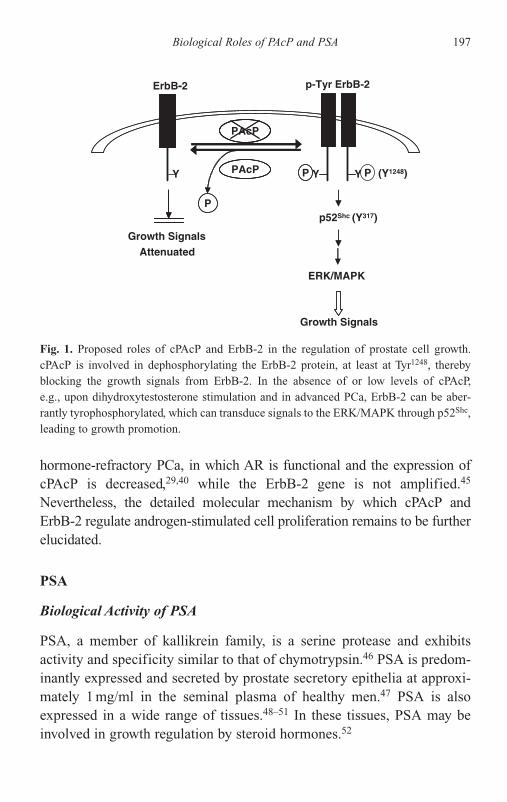
hormone-refractory PCa, in which AR is functional and the expression ofcPAcP is decreased,29,40 while the ErbB-2 gene is not amplified.45
Nevertheless, the detailed molecular mechanism by which cPAcP andErbB-2 regulate androgen-stimulated cell proliferation remains to be furtherelucidated.
PSA
Biological Activity of PSA
PSA, a member of kallikrein family, is a serine protease and exhibitsactivity and specificity similar to that of chymotrypsin.46 PSA is predom-inantly expressed and secreted by prostate secretory epithelia at approxi-mately 1 mg/ml in the seminal plasma of healthy men.47 PSA is alsoexpressed in a wide range of tissues.48–51 In these tissues, PSA may beinvolved in growth regulation by steroid hormones.52
Biological Roles of PAcP and PSA 197
p-Tyr ErbB-2ErbB-2
Growth SignalsAttenuated
PYY
p52Shc (Y317)
ERK/MAPK
Growth Signals
PAcP
P
P Y (Y1248)
PAcP
-
Fig. 1. Proposed roles of cPAcP and ErbB-2 in the regulation of prostate cell growth.cPAcP is involved in dephosphorylating the ErbB-2 protein, at least at Tyr1248, therebyblocking the growth signals from ErbB-2. In the absence of or low levels of cPAcP,e.g., upon dihydroxytestosterone stimulation and in advanced PCa, ErbB-2 can be aber-rantly tyrophosphorylated, which can transduce signals to the ERK/MAPK through p52Shc,leading to growth promotion.
B245-ch08 3/2/05 4:33 PM Page 197

The synthesis and control of PSA expression in prostatic cells are welldocumented. Naïve translated PSA, or proPSA, is a 244-amino acid pro-tein with a 17-amino acid leader sequence.53 Several inactive truncatedforms of PSA are found to be increased in benign prostatic hyperplasia(BPH).54,55 The ratio of truncated PSA to total PSA (PSA index) aidsin the discrimination between non-cancerous and cancerous prostategland.1 Serum PSA may form complexes with �-antichymotrypsin and�2-macroglobulin3 and circulate as complexed PSA.1,2
PSA may be involved in the proteolytic cleavage of seminal proteins.Semenogelin I/seminal plasma motility inhibitor (SPMI) precursor and itsactive product SPMI may be the substrates for PSA.56,57 Other putativesubstrates of PSA include IGFBP-3,58 PTHrP,59 and extracellular matrix(ECM) proteins, such as fibronectin and laminin.60 Since PSA can modu-late in vitro interactions between IGF-I and IGFBP-3, PSA may play a rolein regulating the growth of prostatic fibromuscular cells.61
Effect of PSA on Prostate Tumorigenicity
The highly invasive property of androgen-independent PCa cells could bedue in part to the degradation of the ECM proteins by increased secretionof PSA, since a monoclonal antibody (mAb) against PSA blocked PCainvasion in a dose-dependent manner.60 This is in parallel with the clinicalobservation that patients with metastatic PCa have elevated levels ofserum PSA. On the contrary, PSA exhibits anti-angiogenic activity62 andhas a direct suppressive effect on new blood vessel formation in PCa.63
Furthermore, recombinant PSA inhibits endothelial cell proliferation andtube formation, which are essential features of angiogenesis.62 It is alsosuggested that anti-angiogenic drugs such as TNP-470 might act throughincreasing PSA secretion.64 Future studies are needed to clarify the role ofPSA in PCa metastasis.
Androgen-Independent PSA Secretion
Androgens are well documented as key factors regulating PSA geneexpression.65 Since PCa cells initially depend on androgens for
198 S. Veeramani et al.
B245-ch08 3/2/05 4:33 PM Page 198

proliferation, androgen ablation therapy is the mainstay of treatment foradvanced PCa, and leads to a drop in circulating PSA. Unfortunately, can-cer cells eventually escape from the steroid requirement and progress toandrogen independence, which is indicated by the rebound of PSA levelsin circulation.66 The ability to treat these patients is limited. Interestingly,most of the hormone-refractory PCa cells express functional AR and PSA.The molecular mechanism of androgen-independent PSA secretiondeserves careful analysis.
Biochemically, upon androgen binding, AR exhibits conformationalchanges, induced by modifications including phosphorylation and dimer-ization, for its activation.67 It is proposed that in hormone-refractorycancers, in the absence of androgen, AR is also activated by phosphoryla-tion.68 Hence, a direct cross-talk between AR and cellular kinases may bean important factor involved in the elevation of androgen-independentPSA secretion. Our understanding of the cross-talk between AR andprotein kinases is discussed briefly as follows.
ErbB-2/HER-2/Neu (ErbB-2) → ERK/MAPK Pathway
Data from our lab strongly indicate that ErbB-2 plays a critical role inhormone-refractory proliferation of PCa cells as well as in their enhancedandrogen-independent PSA secretion, and are of clinical relevance(Fig. 2). This notion of ErbB-2 regulation is shown in ErbB-2 cDNA-transfected C-33 cells in which the growth rate and PSA secretion areelevated, even in the absence of androgen.38,44,69 Conversely, in C-81 cellsin which ErbB-2 is activated and PSA secretion is increased, the expres-sion of a dominant-negative mutant of ErbB-2, or addition of ErbB-2inhibitors, reduces the elevated androgen-independent PSA secretion.38
In androgen-deprived environments, ErbB-2, via the ERK/MAPK orAkt pathway, might activate AR by serine phosphorylation (Ser514 forERK/MAPK and Ser791 for Akt).69–71 Our data indicate that ERK/MAPKplays a key role in the early stage of androgen-independent PSA secretion,as well as in tumor progression. For example, in androgen-depleted condi-tions, elevated PSA secretion is decreased by MEK1 inhibitor, PD98059,in C-81 cells, while PSA secretion is increased by MEK1 cDNA transfec-tion in C-33 cells.38 Furthermore, in ErbB-2 cDNA-transfected C-33 cells,
Biological Roles of PAcP and PSA 199
B245-ch08 3/2/05 4:33 PM Page 199
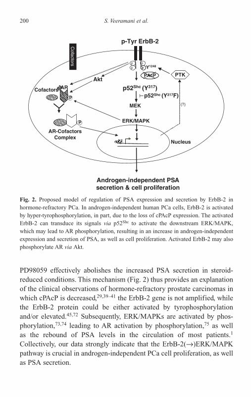
PD98059 effectively abolishes the increased PSA secretion in steroid-reduced conditions. This mechanism (Fig. 2) thus provides an explanationof the clinical observations of hormone-refractory prostate carcinomas inwhich cPAcP is decreased,29,39–41 the ErbB-2 gene is not amplified, whilethe ErbB-2 protein could be either activated by tyrophosphorylationand/or elevated.45,72 Subsequently, ERK/MAPKs are activated by phos-phorylation,73,74 leading to AR activation by phosphorylation,75 as wellas the rebound of PSA levels in the circulation of most patients.1
Collectively, our data strongly indicate that the ErbB-2(→)ERK/MAPKpathway is crucial in androgen-independent PCa cell proliferation, as wellas PSA secretion.
200 S. Veeramani et al.
MEK
AR-CofactorsComplex
AR
PAcP PTK
p
p ppp
p52Shc (Y317)
ERK/MAPK
Akt
Androgen-independent PSAsecretion & cell proliferation
(?)
Y1248
Nucleusp
p-Tyr ErbB-2
pp
pp
Cofactorsp52Shc (Y317F)
Cofactors
Fig. 2. Proposed model of regulation of PSA expression and secretion by ErbB-2 inhormone-refractory PCa. In androgen-independent human PCa cells, ErbB-2 is activatedby hyper-tyrophosphorylation, in part, due to the loss of cPAcP expression. The activatedErbB-2 can transduce its signals via p52Shc to activate the downstream ERK/MAPK,which may lead to AR phosphorylation, resulting in an increase in androgen-independentexpression and secretion of PSA, as well as cell proliferation. Activated ErbB-2 may alsophosphorylate AR via Akt.
B245-ch08 3/2/05 4:33 PM Page 200
ErbB-2 can up-regulate PSA secretion at two levels: the transcriptionallevel and the post-translational level in the secretory pathway. In C-81cells in which ErbB-2 is activated by hyper-tyrophosphorylation, the levelof PSA mRNA is elevated in the steroid-reduced condition, compared toC-33 cells,27,43 which may be due in part to the activation of the PSA pro-moter by ErbB-2 via ERK/MAPK and AR.69 Alternatively, ErbB-2 mayactivate MEK1, leading to the up-regulation of a transcription factor(s)that subsequently activates the PSA promoter.76 ErbB-2 may also activatethe secretory pathway of PSA in those cells (M. S. Lee and M. F. Lin,unpublished observations). Nevertheless, further experiments are needed.
Akt Pathway
AR phosphorylation might also occur through the PI3K → Akt pathwayin hormone-refractory PCa cells.71 Androgen-responsive, low passageLNCaP cells have a lower Akt activity than the androgen-independenthigh passage cells,77 which implies that this pathway could be importantin later stages of advanced metastatic cancer, where PTEN mutation leadsto constant Akt activation. Nevertheless, it should be noted that pathwaysother than PTEN could be involved in activating Akt in higher passageLNCaP cells because PTEN is also mutated in the lower passage LNCaPcells.78,79 Interestingly, the anti-androgen flutamide blocks Akt-inducedPSA secretion in IL-4-treated LNCaP cells,80 while it stimulates thegrowth of LNCaP cells,81 indicating the differential roles of Akt.Nevertheless, IGFBP-5 cDNA-transfected LNCaP cells express hyperac-tivated Akt and exhibit higher growth rates and PSA secretion levels incastrated mice than do control cells.82 Further experiments are requiredto clarify the role of Akt in androgen-independent PSA secretion vs.hormone-refractory PCa cell proliferation and its clinical relevance.
Cytokine Pathway
Interleukin-6 (IL-6) has been proposed to play a role in the developmentof androgen-independence in PCa cells because IL-6 enhances the growthof LNCaP cells in androgen-depleted environment and serum IL-6 levelsare found to be elevated in some patients with advanced PCa. Since
Biological Roles of PAcP and PSA 201
B245-ch08 3/2/05 4:33 PM Page 201

androgen-independent LNCaP-IL-6� cells, which were selected by con-tinuous passage in media containing 5 ng/ml IL-6, are resistant to the cellcycle arrest mediated by IL-6,83 it is inferred that IL-6 may contribute toPCa progression, and via the ErbB-2(→)ERK/MAPK pathway, stimulateandrogen-independent PSA secretion.84,85 IL-4 might also have a role inthe androgen-independent, AR-mediated PSA secretion,80 because IL-4level is elevated in some PCa patients with hormone-refractory cancer.86
PKA Pathway
Forskolin, a PKA activator, can increase the activity of the PSA promoterin the absence of androgen.87 The activation of PKA by forskolin alsoresults in increased PSA expression in androgen-depleted LNCaP cells.88
Butyrate, a differentiation agent, increased circulating PSA levels in cas-trated animals. Electromobility shift assay showed an increased AR-AREcomplex formation in butyrate-treated LNCaP cells that is partially inhib-ited by a PKA blocker, indicating that butyrate action on LNCaP cellsmight, in part, involve the PKA pathway.88
Other Downstream Mechanisms
Protein phosphorylation may activate other regulatory factors, e.g., ARcofactors, leading to increased PSA secretion. Alternatively, aberrantlyregulated transcriptional factors may play a role in this mode of regula-tion. For example, nuclear factor-Kappa B (NF-�B), which is increased inandrogen-independent PCa cells, can activate PSA expression,89 while thegrowth of those cells is inhibited by NF-�B inhibitors.90 A 45-kDa cell-specific transcription factor (p45) has also been implicated in androgen-independent expression of the PSA gene.91
Future Perspectives
The decreased expression of cPAcP is implicated as a crucial step in car-cinogenesis and/or progression of human PCa. More studies of the basicbiology of PAcP may provide new insights into its potential roles in tumor
202 S. Veeramani et al.
B245-ch08 3/2/05 4:33 PM Page 202
suppression and potential uses for therapy of PCa. In xenograft animal mod-els, a single intratumoral injection of an expression vector encoding the wildtype PAcP resulted in the growth suppression of prostate tumors.28 Therestoration of cPAcP expression in PCa may provide a new avenue for treat-ing patients with advanced PCa in which the expression of PAcP is lost.
Functional molecules involved in androgen-independent pathwaysmay serve as potential targets for clinical therapy. Inhibitors of androgen-independent pathways such as ErbB-2 and ERK/MAPK blockers mightprove valuable in the treatment of hormone-refractory PCa. Studies indi-cated that co-administration of the ERK/MAPK inhibitor PD98059 withdocetaxel increases the apoptotic death of androgen-independent PCacells.92 Chemointerventions for hormone-refractory PCa might be mademore useful with co-administration of these pathway blockers with theregular therapeutic regime.
Immunotherapy for PCa also has potential value. Phase I studies usingPSA expressing vaccinia viral vectors indicated that T cells could beinduced in the patients.93 Vaccination with dendritic cells preloaded withPSA mRNA resulted in the reduction of circulating PSA levels in 80% ofthe patients.94 Similar experiments with PAcP also showed some effi-cacy.95 Future work is needed in the clinical utilization of PAcP and PSAas targets of immunotherapy against PCa.
The proteolytic property of PSA has been exploited in the developmentof therapeutic interventions for PCa. For example, peptide-conjugated -doxorubicin96 and vinblastine,97,98 which can be activated in prostate tissueby PSA, has shown some promising results. Similar strategies have beenapplied for PAcP (M. F. Lin, unpublished observations). Nevertheless, pro-motion of such strategies to clinical application requires further studies. Inconclusion, due to the recent advances in understanding the biology ofPAcP and PSA, as well as their functional roles in PCa progression andmetastasis, new avenues through which to develop novel strategies forimproving the efficacy of treating hormone-refractory PCa are expected.
Acknowledgments
This project is in part supported by NIH, NCI, CA88184, and UNMCGraduate Student Fellowship (T. C. Y; S. J. C). We thank all group
Biological Roles of PAcP and PSA 203
B245-ch08 3/2/05 4:33 PM Page 203

members for their efforts toward conquering this cancer. We are alsoindebted to Dr. T. M. Chu and his colleagues for the discovery of PSA.
References
1. Chu TM, Lin MF (1998) PSA and acid phosphatase in the diagnosis ofprostate cancer. J Clin Ligand Assay 21:24–34.
2. Chu TM (1997) Prostate-specific antigen (PSA) and early detection ofprostate cancer. Tumour Biol 18:123–134.
3. Armbruster DA (1993) Prostate-specific antigen: biochemistry, analyticalmethods, and clinical application. Clin Chem 39:181–195.
4. Jemal A, Murray T, Samuels A, Ghafoor A, Ward E, Thun MJ (2003) CancerStatistics 2003. CA Cancer J Clin 53:5–26.
5. Gutman EB, Sproul EE, Gutman AB (1936) Significance of increased phos-phatase activity at the site of osteoplastic metastases secondary to carcinomaof the prostate gland. Am J Cancer 28:485–495.
6. Huggins C, Hodges CV (1941) Studies on prostatic cancer: the effect ofcastration, of estrogen and of androgen injection on serum phosphatases inmetastatic carcinoma of the prostate. Cancer Res 1:293–297.
7. Wang MC, Valenzuela LA, Murphy GP, Chu TM (1979) Purification of ahuman prostate specific antigen. Invest Urol 17:159–163.
8. Kuriyama M, Wang MC, Lee CL, Killian CS, Papsidero LD, Inaji H,Loor RM, Lin MF, Nishiura T, Slack NH, Murphy GP, Chu TM (1982)Multiple marker evaluation in human prostate cancer with the use of tissue-specific antigens. J Natl Cancer Inst 68:99–105.
9. Kuriyama M, Wang MC, Lee CL, Papsidero LD, Killian CS, Inaji H,Slack NH, Nishiura T, Murphy CP, Chu TM (1981) Use of human prostate-specific antigen in monitoring prostate cancer. Cancer Res 41:3874–3876.
10. Vihko P, Virkkunen P, Henttu P, Roiko K, Solin T, Huhtala ML (1988)Molecular cloning and sequence analysis of cDNA encoding human prosta-tic acid phosphatase. FEBS Lett 236:275–281.
11. Lin MF, Garcia-Arenas R, Kawachi M, Lin FF (1993) Regulation of theexpression of prostatic acid phosphatase in LNCaP human prostate carci-noma cells. Cell Mol Biol Res 39:739–750.
12. Yam LT (1974) Clinical significance of the human acid phosphatases: areview. Am J Med 56:604–616.
13. Vihko P (1979) Human prostatic acid phosphatases: purification of a minorenzyme and comparisons of the enzymes. Invest Urol 16:349–352.
204 S. Veeramani et al.
B245-ch08 3/2/05 4:33 PM Page 204

14. Lin MF, Clinton GM (1987) Human prostatic acid phosphatase and itsphosphotyrosyl-protein phosphatase activity. Adv Protein Phosphatases 4:199–228.
15. Lin MF, Lee CL, Li SS, Chu TM (1983) Purification and characterizationof a new human prostatic acid phosphatase isoenzyme. Biochemistry 22:1055–1062.
16. Shaw LM, Yang N, Neat M, Croop W (1982) Immunological and clinicalspecificity of the immunochemical determination of prostatic acid phos-phatase. Ann NY Acad Sci 390:73–88.
17. Ronnberg L, Vihko P, Sajanti E, Vihko R (1981) Clomiphene citrate admin-istration to normogonadotropic subfertile men: blood hormone changesand activation of acid phosphatase in seminal fluid. Int J Androl 4:372–378.
18. Brillard-Bourdet M, Rehault S, Juliano L, Ferrer M, Moreau T, Gauthier F(2002) Amidolytic activity of prostatic acid phosphatase on humansemenogelins and semenogelin-derived synthetic substrates. Eur J Biochem269:390–395.
19. Lin MF, Clinton GM (1986) Human prostatic acid phosphatase has phos-photyrosyl protein phosphatase activity. Biochem J 235:351–357.
20. Nguyen L, Chapdelaine A, Chevalier S (1990) Prostatic acid phosphatase inserum of patients with prostatic cancer is a specific phosphotyrosine acidphosphatase. Clin Chem 36:1450–1455.
21. Li HC, Chernoff J, Chen LB, Kirschonbaum A (1984) A phosphotyrosyl-protein phosphatase activity associated with acid phosphatase from humanprostate gland. Eur J Biochem 138:45–51.
22. Wasylewska E, Czubak J, Ostrowski WS (1983) Phosphoprotein phosphataseactivity of human prostate acid phosphatase. Acta Biochim Pol 30:175–184.
23. Lee H, Chu TM, Lee CL (1991) Endogenous protein substrates for prostaticacid phosphatase in human prostate. Prostate 19:251–263.
24. Vihko P, Kurkela R, Porvari K, Herrala A, Lindfors A, Lindqvist Y,Schneider G (1993) Rat acid phosphatase: overexpression of active, secretedenzyme by recombinant baculovirus-infected insect cells, molecular proper-ties, and crystallization. Proc Natl Acad Sci USA 90:799–803.
25. Lin MF, DaVolio J, Garcia-Arenas R (1992) Expression of human prostaticacid phosphatase activity and the growth of prostate carcinoma cells. CancerRes 52:4600–4607.
26. Lin MF, Garcia-Arenas R, Xia XZ, Biela B, Lin FF (1994) The cellular levelof prostatic acid phosphatase and the growth of human prostate carcinomacells. Differentiation 57:143–149.
Biological Roles of PAcP and PSA 205
B245-ch08 3/2/05 4:33 PM Page 205

27. Lin MF, Meng TC, Rao PS, Chang C, Schonthal AH, Lin FF (1998)Expression of human prostatic acid phosphatase correlates with androgen-stimulated cell proliferation in prostate cancer cell lines. J Biol Chem 273:5939–5947.
28. Igawa T, Lin FF, Rao P, Lin MF (2003) Suppression of LNCaP prostatecancer xenograft tumors by a prostate-specific protein tyrosine phosphatase,prostatic acid phosphatase. Prostate 55:247–258.
29. Lin MF, Lee MS, Zhou XW, Andressen JC, Meng TC, Johansson SL,West WW, Taylor RJ, Anderson JR, Lin FF (2001) Decreased expressionof cellular prostatic acid phosphatase increases tumorigenicity of humanprostate cancer cells. J Urol 166:1943–1950.
30. Zhang XQ, Lee MS, Zelivianski S, Lin MF (2001) Characterization ofa prostate-specific tyrosine phosphatase by mutagenesis and expression inhuman prostate cancer cells. J Biol Chem 276:2544–2550.
31. Tonks NK, Neel BG (2001) Combinatorial control of the specificity of pro-tein tyrosine phosphatases. Curr Opin Cell Biol 13:182–195.
32. Schneider G, Lindqvist Y, Vihko P (1993) Three-dimensional structure of ratacid phosphatase. EMBO J 12:2609–2615.
33. Ostanin K, Saeed A, van Etten RL (1994) Heterologous expression of humanprostatic acid phosphatase and site-directed mutagenesis of the enzymeactive site. J Biol Chem 269:8971–8978.
34. Lin MF, Clinton GM (1988) The epidermal growth factor receptor fromprostate cells is dephosphorylated by a prostate-specific phosphotyrosylphosphatase. Mol Cell Biol 8:5477–5485.
35. Lin MF, Meng TC (1996) Tyrosine phosphorylation of a 185 kDa phospho-protein (pp185) inversely correlates with the cellular activity of human pro-static acid phosphatase. Biochem Biophys Res Commun 226:206–213.
36. Meng TC, Lin MF (1998) Tyrosine phosphorylation of c-ErbB-2 is regulatedby the cellular form of prostatic acid phosphatase in human prostate cancercells. J Biol Chem 273:22096–22104.
37. Lee MS, Igawa T, Lin MF (2004) Tyrosine-317 of p52Shc mediates androgen-stimulated proliferation signals in human prostate cancer cells. Oncogene,in press.
38. Lee MS, Igawa T, Yuan TC, Zhang XQ, Lin FF, Lin MF (2003) ErbB-2signaling is involved in regulating PSA secretion in androgen-independenthuman prostate cancer LNCaP C-81 cells. Oncogene 22:781–796.
39. Reif AE, Schlesinger RM, Fish CA, Robinson CM (1973) Acid phosphataseisozymes in cancer of the prostate. Cancer 31:689–699.
206 S. Veeramani et al.
B245-ch08 3/2/05 4:33 PM Page 206

40. Loor R, Wang MC, Valenzuela L, Chu TM (1981) Expression of prostaticacid phosphatase in human prostate cancer. Cancer Lett 14:63–69.
41. Hakalahti L, Vihko P, Henttu P, Autio-Harmainen H, Soini Y, Vihko R (1993)Evaluation of PAP and PSA gene expression in prostatic hyperplasia andprostatic carcinoma using northern-blot analyses, in situ hybridization andimmunohistochemical staining with monoclonal and bispecific antibodies.Int J Cancer 55:590–597.
42. Lin MF, DaVolio J, Garcia-Arenas R (1992) Expression of human prostaticacid phosphatase activity and the growth of prostate carcinoma cells. CancerRes 52:4600–4607.
43. Igawa T, Lin FF, Lee MS, Karan D, Batra SK, Lin MF (2002) Establishmentand characterization of androgen-independent human prostate cancer LNCaPcell model. Prostate 50:222–235.
44. Meng TC, Lee MS, Lin MF (2000) Interaction between protein tyrosinephosphatase and protein tyrosine kinase is involved in androgen-promotedgrowth of human prostate cancer cells. Oncogene 19:2664–2677.
45. Signoretti S, Montironi R, Manola J, Altimari A, Tam C, Bubley G, Balk S,Thomas G, Kaplan I, Hlatky L, Hahnfeldt P, Kantoff P, Loda M (2000)HER-2-neu expression and progression toward androgen independence inhuman prostate cancer. J Natl Cancer Inst 92:1918–1925.
46. Yousef GM, Diamandis EP (2003) An overview of the kallikrein gene fami-lies in humans and other species: emerging candidate tumour markers. ClinBiochem 36:443–452.
47. Lovgren J, Valtonen-Andre C, Marsal K, Lilja H, Lundwall A (1999)Measurement of prostate-specific antigen and human glandular kallikrein 2in different body fluids. J Androl 20:348–355.
48. Frazer HA, Humphrey PA, Burchette JL, Paulson DF (1992) Immunoreactiveprostate-specific antigen in male periurethral glands. J Urol 147:246–248.
49. Clements A, Mukhtar A (1994) Glandular kallikreins and prostate specificantigen are expressed in the human endometrium. J Clin Endocrinol Metab78:1536–1539.
50. Howarth DJ, Aronson IB, Diamandis EP (1997) Immunohistochemical local-ization of prostate-specific antigen in benign and malignant breast tissues.Br J Cancer 75:1646–1651.
51. Malatesta M, Mannello F, Luchetti F, Marcheggiani F, Condemi L, Papa S,Gazzanelli G (2000) Prostate-specific antigen synthesis and secretion byhuman placenta: a physiological kallikrein source during pregnancy. J ClinEndocrinol Metab 85:317–321.
Biological Roles of PAcP and PSA 207
B245-ch08 3/2/05 4:33 PM Page 207

52. Diamandis EP, Yu H (1995) Editorial: New biological functions of prostate-specific antigen? J Clin Endocrinol Metab 80:1515–1517.
53. Lilja H (2003) Biology of prostate-specific antigen. Urology 62:27–33.54. Mikolajczyk SD, Millar LS, Wang TJ, Rittenhouse HG, Wolfert RL, Marks
LS, Song W, Wheeler TM, Slawin KM (2000) “BPSA,” a specific molecularform of free prostate-specific antigen, is found predominantly in the transitionzone of patients with nodular benign prostatic hyperplasia. Urology 55:41–45.
55. Wang TJ, Slawin KM, Rittenhouse HG, Millar LS, Mikolajczyk SD (2000)Benign prostatic hyperplasia-associated prostate-specific antigen (BPSA)shows unique immunoreactivity with anti-PSA monoclonal antibodies. Eur JBiochem 267:4040–4045.
56. Robert M, Gibbs BF, Jacobson E, Gagnon C (1997) Characterization ofprostate-specific antigen proteolytic activity on its major physiological sub-strate, the sperm motility inhibitor precursor/semenogelin I. Biochemistry36:3811–3819.
57. Rehault S, Brillard-Bourdet M, Bourgeois L, Frenette G, Juliano L,Gauthier F, Moreau T (2002) Design of new and sensitive fluorogenic sub-strates for human kallikrein hK3 (prostate-specific antigen) derived fromsemenogelin sequences. Biochim Biophys Acta 1596:55–62.
58. Okabe E, Kajihara J, Usami Y, Hirano K (1999) The cleavage site specificityof human prostate specific antigen for insulin-like growth factor bindingprotein-3. FEBS Lett 447:87–90.
59. Cramer SD, Chen Z, Peehl DM (1996) Prostate specific antigen cleavesparathyroid hormone-related protein in the PTH-like domain: Inactivation ofPTHrP-stimulated cAMP accumulation in mouse osteoblasts. J Urol 156(2 Pt 1):526–531.
60. Webber MM, Waghray A, Bello D (1995) Prostate-specific antigen, a serineprotease, facilitates human prostate cancer cell invasion. Clin Cancer Res1:1089–1094.
61. Sutkowski DM, Goode RL, Baniel J, Teater C, Cohen P, McNulty AM,Hsiung HM, Becker GW, Neubauer BL (1999) Growth regulation of prostaticstromal cells by prostate-specific antigen. J Natl Cancer Inst 91:1663–1669.
62. Fortier AH, Holaday JW, Liang H, Dey C, Grella DK, Holland-Linn J, Vu H,Plum SM, Nelson BJ (2003) Recombinant prostate specific antigen inhibitsangiogenesis in vitro and in vivo. Prostate 56:212–219.
63. Papadopoulos I, Sivridis E, Giatromanolaki A, Koukourakis MI (2001)Tumor angiogenesis is associated with MUC1 overexpression and lossof prostate-specific antigen expression in prostate cancer. Clin Cancer Res7:1533–1538.
208 S. Veeramani et al.
B245-ch08 3/2/05 4:33 PM Page 208
64. Horti J, Dixon SC, Logothetis CJ, Guo Y, Reed E, Figg WD (1999) Increasedtranscriptional activity of prostate-specific antigen in the presence ofTNP-470, an angiogenesis inhibitor. Br J Cancer 79:1588–1593.
65. Cleutjens KB, van der Korput HA, van Eekelen CC, van Rooij HC,Faber PW, Trapman J (1997) An androgen response element in a far upstreamenhancer region is essential for high, androgen-regulated activity of theprostate-specific antigen promoter. Mol Endocrinol 11:148–161.
66. Pilat MJ, Kamradt JM, Pienta KJ (1998–99) Hormone resistance in prostatecancer. Cancer Metastasis Rev 17:373–381.
67. Kuil CW, Berrevoets CA, Mulder E (1995) Ligand-induced conformationalalterations of the androgen receptor analyzed by limited trypsinization. Studieson the mechanism of antiandrogen action. J Biol Chem 270:27569–27576.
68. Sadar MD, Hussain M, Bruchovsky N (1999) Prostate cancer: molecularbiology of early progression to androgen independence. Endocr RelatCancer 6:487–502.
69. Yeh S, Lin HK, Kang HY, Thin TH, Lin MF, Chang C (1999) FromHER2/Neu signal cascade to androgen receptor and its coactivators: a novelpathway by induction of androgen target genes through MAP kinase inprostate cancer cells. Proc Natl Acad Sci USA 96:5448–5463.
70. Craft N, Shostak Y, Carey M, Sawyers CL (1999) A mechanism for hormone-independent prostate cancer through modulation of androgen receptor sig-naling by the HER-2/neu tyrosine kinase. Nat Med 5:280–285.
71. Wen Y, Hu MC, Makino K, Spohn B, Bartholomeusz G, Yan DH, Hung MC(2000) HER-2/neu promotes androgen-independent survival and growth ofprostate cancer cells through the Akt pathway. Cancer Res 60:6841–6845.
72. Osman I, Scher HI, Drobnjak M, Verbel D, Morris M, Agus D, Ross JS,Cordon-Cardo C (2001) HER-2/neu (p185neu) protein expression in the nat-ural or treated history of prostate cancer. Clin Cancer Res 7:2643–2647.
73. Price DT, Rocca GD, Guo C, Ballo MS, Schwinn DA, Luttrell LM (1999)Activation of extracellular signal-regulated kinase in human prostate cancer.J Urol 162:1537–1542.
74. Gioeli D, Mandell JW, Petroni GR, Frierson HF Jr, Weber MJ (1999)Activation of mitogen-activated protein kinase associated with prostate can-cer progression. Cancer Res 59:279–284.
75. Ofelia L, Zegarra-Moro OL, Schmidt LJ, Huang H, Tindall DJ (2002)Disruption of androgen receptor function inhibits proliferation of androgen-refractory prostate cancer cells. Cancer Res 62:1008–1013.
76. Franco OE, Onishi T, Yamakawa K, Arima K, Yanagawa M, Sugimura Y,Kawamura J (2003) Mitogen-activated protein kinase pathway is involved in
Biological Roles of PAcP and PSA 209
B245-ch08 3/2/05 4:33 PM Page 209

androgen-independent PSA gene expression in LNCaP cells. Prostate56:319–325.
77. Lin HK, Hu YC, Yang L, Altuwaijri S, Chen YT, Kang HY, Chang C (2003)Suppression versus induction of androgen receptor functions by the phos-phatidylinositol 3-kinase/Akt pathway in prostate cancer LNCaP cells withdifferent passage numbers. J Biol Chem 278:50902–50907.
78. Li J, Yen C, Liaw D, Podsypanina K, Bose S, Wang SI, Puc J, Miliaresis C,Rodgers L, McCombie R, Bigner SH, Giovanella BC, Ittmann M, Tycko B,Hibshoosh H, Wigler MH, Parsons R (1997) PTEN, a putative protein tyro-sine phosphatase gene mutated in human brain, breast, and prostate cancer.Science 275:51943–1947.
79. Reiss K, Wang JY, Romano G, Furnari FB, Cavenee WK, Morrione A, Tu X,Baserga R (2000) IGF-I receptor signaling in a prostatic cancer cell line witha PTEN mutation. Oncogene 19:2687–2694.
80. Lee SO, Lou W, Hou M, Onate SA, Gao AC (2003) Interleukin-4 enhancesprostate-specific antigen expression by activation of the androgen receptorand Akt pathway. Oncogene 22:7981–7988.
81. Furr BJ, Tucker H (1996) The preclinical development of bicalutamide: phar-macodynamics and mechanism of action. Urology 47(Suppl):13–45.
82. Miyake H, Nelson C, Rennie PS, Gleave ME (2000) Overexpressionof insulin-like growth factor binding protein-5 helps accelerate progressionto androgen-independence in the human prostate LNCaP tumor modelthrough activation of phosphatidylinositol 3�-kinase pathway. Endocrinol141:2257–2265.
83. Culig Z, Bartsch G, Hobisch A (2002) Interleukin-6 regulates androgenreceptor activity and prostate cancer cell growth. Mol Cell Endocrinol 197:231–238.
84. Lin DL, Whitney MC, Yao Z, Keller ET (2001) Interleukin-6 induces andro-gen responsiveness in prostate cancer cells through up-regulation of andro-gen receptor expression. Clin Cancer Res 7:1773–1781.
85. Qiu Y, Ravi L, Kung HJ (1998) Requirement of ErbB2 for signalling byinterleukin-6 in prostate carcinoma cells. Nature 393:83–85.
86. Wise GJ, Marella VK, Talluri G, Shirazian D (2000) Cytokine variations inpatients with hormone treated prostate cancer. J Urol 164:722–725.
87. Nazareth LV, Weigel NL (1996) Activation of the human androgen receptorthrough a protein kinase A signaling pathway. J Biol Chem 16:19900–19907.
88. Sadar MD, Gleave ME (2000) Ligand-independent activation of the andro-gen receptor by the differentiation agent butyrate in human prostate cancercells. Cancer Res 15:5825–5831.
210 S. Veeramani et al.
B245-ch08 3/2/05 4:33 PM Page 210
89. Chen CD, Sawyers CL (2002) NF-kappa B activates prostate-specific anti-gen expression and is upregulated in androgen-independent prostate cancer.Mol Cell Biol 22:2862–2670.
90. Kikuchi E, Horiguchi Y, Nakashima J, Kuroda K, Oya M, Ohigashi T,Takahashi N, Shima Y, Umezawa K, Murai M (2003) Suppression ofhormone-refractory prostate cancer by a novel nuclear factor kappaBinhibitor in nude mice. Cancer Res 63:107–110.
91. Yeung F, Li X, Ellett J, Trapman J, Kao C, Chung LW (2000) Regions ofprostate-specific antigen (PSA) promoter confer androgen-independentexpression of PSA in prostate cancer cells. J Biol Chem 275:40846–40855.
92. Zelivianski S, Spellman M, Kellerman M, Kakitelashvilli V, Zhou XW,Lugo E, Lee MS, Taylor R, Davis TL, Hauke R, Lin MF (2003) ERKinhibitor PD98059 enhances docetaxel-induced apoptosis of androgen-independent human prostate cancer cells. Int J Cancer 107:478–485.
93. Eder JP, Kantoff PW, Roper K, Xu GX, Bubley GJ, Boyden J, Gritz L,Mazzara G, Oh WK, Arlen P, Tsang KY, Panicali D, Schlom J, Kufe DW(2000) A phase I trial of a recombinant vaccinia virus expressing prostate-specific antigen in advanced prostate cancer. Clin Cancer Res 6:1632–1638.
94. Heiser A, Coleman D, Dannull J, Yancey D, Maurice MA, Lallas CD, Dahm P,Niedzwiecki D, Gilboa E, Vieweg J (2002) Autologous dendritic cells trans-fected with prostate-specific antigen RNA stimulate CTL responses againstmetastatic prostate tumors. J Clin Invest 109:409–417.
95. Fong L, Small EJ (2003) Immunotherapy for prostate cancer. Semin Oncol30:649–658.
96. Schwartz MS, Matuszewski BK (2002) Determination of a peptide-doxorubicin, prostate-specific antigen activated prodrug, and its activemetabolites in human plasma using high-performance liquid chromatogra-phy with fluorescence detection. Stabilization of the peptide prodrug withEDTA. J Chromatogr B Analyt Technol Biomed Life Sci 780:171–182.
97. DeFeo-Jones D, Brady SF, Feng DM, Wong BK, Bolyar T, Haskell K,Kiefer DM, Leander K, McAvoy E, Lumma P, Pawluczyk JM, Wai J, Motzel SL,Keenan K, Van Zwieten M, Lin JH, Garsky VM, Freidinger R, Oliff A, Jones RE(2002) A prostate-specific antigen (PSA)-activated vinblastine prodrug selec-tively kills PSA-secreting cells in vivo. Mol Cancer Ther 1:451–459.
98. Brady SF, Pawluczyk JM, Lumma PK, Feng DM, Wai JM, Jones R,DeFeo-Jones D, Wong BK, Miller-Stein C, Lin JH, Oliff A, Freidinger RM,Garsky VM (2002) Design and synthesis of a pro-drug of vinblastine targetedat treatment of prostate cancer with enhanced efficacy and reduced systemictoxicity. J Med Chem 45:4706–4715.
Biological Roles of PAcP and PSA 211
B245-ch08 3/2/05 4:33 PM Page 211

This page intentionally left blank

213
9EPIGENETICS IN PROSTATE CANCER
Jose A. Karam, Elie A. Benaim, Hong Chen, Rey-Chen Pong and Jer-Tsong Hsieh
Department of Urology, UT Southwestern Medical Center5323 Harry Hines Blvd., Dallas, TX, USA
Introduction
Prostate cancer (PCa) is the most common non-cutaneous cancer affect-ing males in the USA. One in six American men will be diagnosed withPCa in their lifetime. In 2004, a total of 230,110 new cases and 29,900deaths are estimated to occur in the USA.1 It has been well documentedthat alterations in genomic DNA, such as point mutations, homozygousdeletions and loss of heterozygosity are linked to the pathogenesis of can-cer,2 including PCa.3 However, the majority of studies have focused on theDNA sequence and to a lesser extent on DNA structure and its surround-ing environment. Recently, investigators started looking at epigenetics asan alternative and complementary mechanism in the pathogenesis of can-cer. The term “epigenetic” refers to the heritable changes in gene expres-sion that are caused by mechanisms other than the alteration in thenucleotide sequence.4 This concept generated tremendous new knowledgein understanding gene expression in mammalian cells.
The Role of CpG Dinucleotides in DNA Methylation
CpG dinucleotides can be either clustered as CpG islands or dispersed. Inthe era before completion of the human genome project, a CpG island wasdefined as a stretch of 200-bp of DNA with a C � G content of 50% and
B245-ch09 3/2/05 4:34 PM Page 213

an observed CpG/expected CpG in excess of 0.6.5 However, this definitionincluded inactive and viral sequences within the human genome. Whencorrected for these “intrusive” sequences, a more biologically appropriateand more stringent definition of a CpG island would be a stretch of 500-bpof DNA with a C � G content of 55% and an observed CpG/expected CpGof more than 0.65.6 Therefore, the definition of a CpG island is still arbi-trary. More than half of the human genes are associated with unique CpGislands.7 Reports from the human genome project have estimated that thehuman genome contains almost 30,000 CpG islands.8
CpG dinucleotides are present throughout the genome and are usuallymethylated when not associated with CpG islands. However, when theCpG dinucleotides are found in CpG islands, they are usually unmethy-lated, in particular if they are found in the promoter region of an activegene,9 or methylated, when found in the 3� end of the gene. However, CpGmethylation the at 3� end of the gene does not affect gene transcription.10
In general, it is a rare event to find de novo CpG methylation in normalsomatic cells. However, as cells age, especially when cultured in vitro, anincrease in methylation has been documented.11,12
Cytosine methylation prevents gene expression by interfering withtranscription initiation. DNA methylation has no effect on base pairing butcan alter protein-DNA interaction by protruding into the major DNAgroove.13 Another explanation is that methylation results in decreasedbinding affinity of the transcription factors to gene promoter regions.14
Methyl Binding Proteins (MBP)
The first true MBP to be identified was MeCP2 (Methyl CpG-binding pro-tein 2). It was found to specifically bind to the methyl group at position 5 ofcytosine and to be active in somatic mammalian cells but not necessarily inembryonic stem cells.15 This protein has been found most abundantly in thepericentromeric heterochromatin region16 and its localization to this regionhas been shown to be dependent on the presence of methylated DNA.17
MeCP2 specifically represses methylated promoters through a transcriptionrepressor domain (TRD). MeCP2 can bind to DNA that contains one sym-metrically methylated CpG pair and does not need chromatin disassemblyin order to bind to DNA.18 In fact, MeCP2 can displace histone H1 from
214 J. A. Karam et al.
B245-ch09 3/2/05 4:34 PM Page 214

DNA and then exert its repression function, at least in vitro.19 MeCP1 canalso discriminate between methylated and unmethylated DNA, althoughneeding a high amount of methylated CpG for its binding (more than 10methylated CpGs).20
Four other methyl-binding domain (MBD)-containing proteins were iden-tified by comparing the MBD domain of MeCP2 to Expressed SequenceTags (EST) databases.20 MBD1-mediated activity is Histone Deacetylase(HDAC)-dependent, as its actions are partially inhibited by HDAC inhibitors(HDIs). However, the identity of this HDAC is still unknown.21 MBD1 inter-acts with a histone H3 methyltransferase as well as heterochromatin protein 1(a methyl-lysine binding protein) resulting in chromatin compaction andtranscription repression.22 In the prostate, MBD1 is highly expressed inbenign prostate hyperplasia and low grade PCa. However, MBD1 expressiondecreases in high grade PCa.23 MBD2, which is a part of the MeCP1 com-plex, has transcription repression activity24 as well as DNA demethylaseactivity.25,26 MBD3 is similar in structure to MBD2, however no demethylaseactivity has been demonstrated.27 MBD4 has DNA glycosylase activity,therefore effectively removing uracil or thymidine from a mismatched CpGlocation, at least in vitro. Binding experiments showed that mutations atmethyl CpG sites can be reduced by the activity of MBD4.28
DNA Methyltransferases (DNMTs)
The conversion of cytosine (C) into 5-methylcytosine (5mC) is catalyzedby enzymes termed DNA methyl transferases (DNMTs), using S-adenosyl-methionine (SAM) as a methyl donor.29 Three active DNMTs have beencharacterized. DNMT1 is responsible for methylating daughter DNAstrands during the S-phase, through its interaction with Proliferating CellNuclear Antigen (PCNA).30 DNMT1 is involved in maintaining themethylation status of genes and this task is possible due to its high affin-ity to hemi-methylated DNA.31 When DNMT1 is absent in mice, DNAmethylation is severely affected, resulting in an embryonic lethal pheno-type.32 DNMT3a and DNMT3b have some structural similarities, havingthe N-terminal as the regulatory subunit and the C-terminal as the cat-alytic subunit. In contrast to DNMT1, both DNMT3a and DNMT3b canmethylate unmethylated and hemi-methylated DNA.33
Epigenetics in Prostate Cancer 215
B245-ch09 3/2/05 4:34 PM Page 215
216 J. A. Karam et al.
An elegant study in Drosophila showed that DNMT3a, but notDNMT1, acts as a de novo methyltransferase.34 Therefore, DNMT3aestablishes the gene methylation pattern and then DNMT1 maintains thispattern in the future generations. When DNMT3a or DNMT3b wereknocked out, the resulting mice died at 4 weeks of life or during embry-onic development, respectively.35 Increased DNMT mRNA has been doc-umented in colon and lung cancer.36–39 However, DNA hypermethylationis not necessarily due to overexpression of DNMT.40
DNMT Inhibitors
DNMT inhibitors act mainly by inhibiting methylation; however, othermechanisms are involved. After phosphorylation, these compounds incor-porate into DNA or RNA.41 After incorporating into DNA, they covalentlybind DNMT resulting in its inhibition42–44 without causing DNAdemethylation per se.44 Additionally, their incorporation into DNA causesstructural instability resulting in DNA damage.45
Currently, five DNMT inhibitors have been used in preclinical/clinicaltrials, namely 5-azacytidine (azacytidine), 5-aza-2�-deoxycytidine(decitabine), dihydro-5-azacytidine (DHAC), arabinofuranosyl-5-azacytosine (fazarabine),46 and most recently zebularine. Zebularine is theonly DNMT inhibitor that can be given orally, while the other DNMTinhibitors are given parenterally.47
DNMT inhibitors have been used in preclinical studies with verygood tumor control rate. However, in clinical trials, the results are not sodramatic, especially with solid tumors, where limited efficacy has beenencountered.46 Decitabine has been used in phase II trial in patients withhormone-independent metastatic prostate cancer, however, the resultswere not encouraging.48 Most of the DNMT inhibitors have a relativelyhigh cytotoxicity profile and can only demethylate genes as long as theyare present in the cell surroundings, therefore limiting their potential clin-ical usefulness, as they cannot be administered for prolonged periods oftime. However, a recent in vitro study showed that zebularine can suppressDNMT1 expression, demethylate p16, and restore p16 expression for up to40 days in continuously treated T24 bladder cancer cells, without causingpronounced cytotoxicity. In addition, when zebularine is given at a lower
B245-ch09 3/2/05 4:34 PM Page 216

Epigenetics in Prostate Cancer 217
dose following an initial dose of decitabine, a profound demethylationof p16 is observed. These results suggest the usefulness of combining aninitial bolus of a parenteral DNMT inhibitor followed by a maintenancedose of the oral agent zebularine.49 However, proof of their usefulness ina preclinical and ultimately clinical setting is yet to be established.
Recently, RNA interference (RNAi) has been used to knock outDNMT1,50 which composes the majority of DNMT activity in cancercells,51 resulting in reactivation of repressed genes. Another study, how-ever, showed that both DNMT1 and DNMT3b need to be shut down tore-express silenced genes.52 These results imply the need to treat cancercells with compounds that can affect more than one type of DNMT, ascancer cells might be able to overcome the deficiency in one DNMT.53
Two phase I clinical trials using MG98, an antisense oligodeoxynucleotideagainst DNMT1, have been recently conducted to study the MG98 safetyprofile and dosage regimens in patients with cancer.54,55
Histones
In mammalian cells, genomic DNA is always associated with histone pro-teins to form chromatin structure. The basic unit of chromatin is the nucle-osome, which is composed of 146 base pairs of DNA that are wrappedtwice around a disk-like complex made of 8 histone proteins (2 of each ofhistones H2A, H2B, H3, and H4). A stable nucleosome core is assembledby H3 and H4 heterodimerization, followed by H3 dimerization resultingin a (H3-H4)2 tetramer.56 Subsequently, H2A and H2B heterodimerize andeach dimer attaches to one side of the (H3-H4)2 tetramer.56 This yields anoctamer of histones on which DNA can be wrapped.57 Adjacent nucleo-somes are brought together by histone H1. The N- and C-termini of his-tone H1 are able to bind to DNA within the nucleosome in addition to the“linker” DNA between the nucleosomes. This binding and subsequentneutralization of the acidic DNA results in the formation of higher orderchromatin.58–60 The amino acids at the N-terminal of the histone protrudeout of the nucleosome, thereby allowing for specific reversible modifica-tions and interaction with surrounding factors. This N-terminal is rich inthe basic (positively charged) amino acids like lysine and serine, whichallows for reversible histone modification by methylation, acetylation,
B245-ch09 3/2/05 4:34 PM Page 217

phosphorylation, ubiquitination, and most recently sumoylation. When thispositively charged N-terminal of the amino acid is free, it results in tightbinding of histones to DNA phosphates, which are negatively charged,therefore inhibiting access of transcription factors to the gene promoterregion and subsequently silencing gene expression.
Acetylation of histones is mediated by histone acetyltransferases(HATs) using acetyl CoA as the acetyl moiety donor, resulting in activa-tion of gene transcription. HATs do not directly bind to DNA, instead, theyare subunits in coactivator complexes that mediate transcriptional activityrecovery. Examples of such coactivator HATs in humans are p300/CBP61
and TAFII250,62 which is a component of human RNA polymerase II,an enzyme involved in transcription. Steroid Receptor Coactivator-1(SRC-1)63 and ACTR64 are also coactivators with HAT activity that inter-act with nuclear receptors in a hormone-dependent fashion. pCAF65 hasthe ability to associate with p300/CBP,66 ACTR64 and SRC-1,63 resultingin a multitude of HATs in the same complex. Acetylation of histones hasbeen linked with transcriptional activation.
The second histone modification is phosphorylation of the Serine-10of histone H3. This modification has been implicated in both activation oftranscription67 and chromosomal condensation in mitosis. This phospho-rylation corresponds to the activation of early-immediate genes, such asc-Fos68 and has been shown to occur through the Rsk-2,69 Msk-170, andSnf-1 kinases.71 Interestingly, the latter histone kinase works in concertwith HAT to activate gene transcription.71,72 Also, I kappa B kinase �
(IKK�) has been reported to phosphorylate histone H3, subsequentlyallowing for specific H3 acetylation through CREB-binding protein(CBP), resulting in activation of nuclear factor kappa B (NF-�B) respon-sive genes.73 It is not clear how histone phosphorylation activates genetranscription; however, it could be related to the fact that adding the neg-atively charged phosphates to histone N-termini disrupts the histone-DNAinteraction, resulting in increased access of transcription factors to pro-moter regions.74
The third histone modification is due to methylation of either arginine orlysine residues. Arginine methylation at histone H3 and H4 is mediated bythe histone methyltransferase (HMT) CARM175,76 and PRMT1,77 respec-tively, through transfer of a methyl group from SAM. The end product
218 J. A. Karam et al.
B245-ch09 3/2/05 4:34 PM Page 218

results in gene activation. Histone lysine methylation, on the other hand, canresult in either gene transcription activation or repression. Suvar3-9 hasHMT activity, specifically methylating Lysine-9 of histone H3 (K9-H3),78
subsequently attracting the heterochromatin protein HP179,80 resulting inheterochromatin assembly81 and gene silencing. Human HMTs at K9-H3include SUV39H182 (causing retinoblastoma (Rb)-mediated transcriptionrepression) and EZH2 (a member of the polycomb group of transcriptionalrepressors).83,84 In addition to methylation at K9-H3, EZH2 has the abilityto methylate K27-H3, resulting also in transcription repression.85 EZH2expression was reported to be increased in metastatic prostate cancer and inlocalized prostate cancer. In the latter case, it indicates a poor patient prog-nosis and outcome.85 Interestingly, both monomethylation86 and trimethyla-tion87 at K9-H3 can control and trigger DNA methylation per se. Recently,it has been shown that methylation levels of K9-H3 differ depending ontheir location in the chromosome and that different HMTs are responsiblefor their methylation at each of these sites: monomethylated and dimethy-lated K9-H3 are localized in silenced euchromatin regions and their methy-lation is mediated by G9a HMT, while trimethylated K9-H3 was abundantin pericentromeric regions, with SUV39H1 and SUV39H2 responsible fortheir methylation.88 In contrast, methylation at Lysine-4 of histone H3 (K4-H3) by SET7/Set989,90 results in transcription activation. In LNCaPprostate cancer cells, the activated androgen receptor (AR) binds to theandrogen responsive element (ARE) of the prostate specific antigen (PSA)gene, resulting in decreased methylation of K4-H3 of the PSA enhancer andpromoter and increased PSA transcription.91
The fourth covalent histone modification is ubiquitination.92 Ubiquitinis a 76-amino acid peptide that can attach to the C-terminus of histoneH2A and likely to that of H2B as well. Ubiquitination of histone H2Acoincides with transcriptional activity.93 However, ubiquitination of his-tone H2B results in histone H3 methylation, thereby causing transcriptionrepression.94
Recently, histone sumoylation has been implicated in transcriptionalrepression of gene activity. SUMO (small ubiquitin-related modifier) isinvolved in post-translational modification of several proteins and it ismediated by the same enzymatic cascade that catalyzes ubiquitination.Sumoylation of histone H4 results in recruitment of HDAC and the
Epigenetics in Prostate Cancer 219
B245-ch09 3/2/05 4:34 PM Page 219
heterochromatin protein HP1, therefore causing transcriptional repres-sion.95 Collectively, these histone modifications may constitute a “Histonecode” that is able to specify patterns of gene expression.96
Histone Deacetylases (HDACs)
In mammals, HDACs have been grouped into 3 classes. Class I HDACs(HDAC 1, 2, 3, 8) have catalytic site homology at their C-termini andclass II HDACs (HDAC 4, 5, 6, 7, 9,10) share homology at their catalyticC-termini and regulatory N-termini.97 Class III is the conserved NAD-dependent Sir2 group of deacetylases.97 Interestingly, class I and II, butnot class III HDACs, are inhibited by Trichostatin A (TSA) and suberoy-lanilide hydroxamic acid (SAHA). Similar to HATs, there is evidencethat HDACs do not directly bind to DNA. Instead, they are part of thetranscription regulator complexes.98 HDACs do not show complete redun-dancy in their functions. Class I HDACs are almost exclusively located inthe nucleus. On the other hand, class II HDACs translocate between thecytoplasm and nucleus.97,99
Histone Deacetylase Inhibitors (HDIs)
HDIs can be classified on the basis of their different structures intohydroxamates (TSA and SAHA), cyclic tetrapeptides (apidicin depsipep-tide and depudecin), carboxylates (sodium phenylbutyrate and valproicacid) and benzamides (CI-994 and MS-27-275).100–108
The inhibition of HDACs, as mediated by HDIs via interacting with azinc active site in the HDAC moiety,109 could result in induction of differ-entiation, growth arrest, and/or apoptosis.110 These effects are attributed toa subset of genes whose expression has been altered, or returned back tonormal upon exposure to HDIs and histone hyperacetylation,111 as seen insingle-gene,112 as well as high-throughput studies.113
Several HDIs, including SAHA, phenylbutyrate, and depsipeptide havebeen used in Phase I and Phase II clinical trials.114 SAHA can be admin-istered orally or intravenously and has been shown to increase the levelsof acetylated histones in peripheral blood mononuclear cells and reducetumor activity in pretreated patients with hematologic and solid
220 J. A. Karam et al.
B245-ch09 3/2/05 4:34 PM Page 220

cancers.115 SAHA was also effective in increasing the intratumor acety-lated histone levels and inhibiting the growth of prostate cancer xenograftsin nude mice.116
The Interaction Between DNA Methylation and HistoneModifications in Gene Regulation
Recently, an interaction between histone modifications and DNA methy-lation has been discovered and has been intensively studied. Jones andcolleagues described a system where methylated cytosine attracts themethyl-binding protein MeCP2, which in turn binds a HDAC-corepressorcomplex, therefore resulting in transcription inhibition.117 In addition,DNA methylation and histone methylation also interact, as MeCP2 canrecruit a HMT, resulting in methylation of K9-H3.118 More recently, it hasbeen shown that DNMT1 and DNMT3a interact with SUV39H1 (a K9-H3HMT) as well as HP1, providing more evidence for the close associationbetween DNA and histone methylation.119
A question that has been recently addressed is whether methylation ofDNA precedes histone modifications or vice versa.120,121 Most of the cur-rently available data supports the dominant role of methylation in the epi-genetic control of promoter activation, as DNMT inhibitors are moreimportant than HDAC inhibitors in initiating gene reactivation.122 In thesecases, even if HDAC inhibitors cause histone acetylation, methyl bindingproteins such as MeCP2 are still bound to methylated cytosine, therebyneeding a DNMT inhibitor to release the methylation and subsequentlydisengaging from MeCP2.123 However, recent data showed that methyla-tion of K9-H3 and subsequent gene silencing can occur prior to methyla-tion of CpG in the gene promoter.53 Both phenomena are present to someextent and the relative importance of one or the other could be dependenton specific tumor tissue, cell type, experimental conditions, and the spe-cific gene under study.
The fact that DNA methylation and histone deacetylation cooperate tosilence genes has been exploited to develop new therapeutic regimens.DNA microarray analysis113 experiments show that the use of DNMTinhibitors and HDAC inhibitors results in a synergistic activation of spe-cific tumor suppressor genes,122,124,125 as well as affecting global gene
Epigenetics in Prostate Cancer 221
B245-ch09 3/2/05 4:34 PM Page 221

expression. In addition, low doses of DNMT inhibitors and HDIs arecombined in order to decrease the occurrence of side effects and cause asynergistic inhibition of tumor growth.126 Decitabine and phenylbutyratewas found to have a synergistic effect in preventing the formation of lungtumors in mice.127
Epigenetics in the Pathogenesis and Diagnosis of PCa
Patra and colleagues have demonstrated that elevated DNMT and HDACexpression is associated with PCa, indicating that epigenetic regulation playsan important role in PCa pathogenesis.128 Several genes have been describedas being methylated in PCa and only to a limited extent or not at all innormal prostates. Some genes are methylated in early stages of PCa, poten-tially making them useful as diagnostic markers. Other genes are methylatedin advanced stages of PCa and can be used as prognostic markers.
GSTP1
The most commonly studied epigenetically-controlled gene in PCa isthe Glutathione-S-transferase-�1 (GSTP1) gene. GSTP1 is responsiblefor intracellular detoxification reactions by conjugating free radicals toglutathione.129,130 The first evidence of GSTP1 methylation in PCawas demonstrated by Lee and colleagues,131 when they described thatGSTP1 was not expressed in several PCa cell lines due to promoterhypermethylation.
The revolution in detection of methylated DNA came in 1996 whenHerman and colleagues described methylation-specific PCR (MSP), whichcan offer higher sensitivity and specificity in detecting minute amounts ofmethylated DNA.132 Recently, Harden and colleagues employed a quantita-tive MSP (qMSP) assay for GSTP1 in conjunction with routine prostatebiopsy histology in order to improve the PCa detection. They found that his-tology alone detected 39/61 tumors (64% sensitivity), while GSTP1 qMSPwith histology detected 48/61 tumors (79% sensitivity) with a 100% speci-ficity. However, when the threshold of qMSP is decreased, 4/11 samples thatwere labeled as normal on histology were positive on qMSP. This raisesthe question that such patients should be monitored more intensively, as
222 J. A. Karam et al.
B245-ch09 3/2/05 4:34 PM Page 222
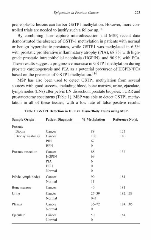
preneoplastic lesions can harbor GSTP1 methylation. However, more con-trolled trials are needed to justify such a follow up.133
By combining laser capture microdissection and MSP, recent datademonstrated the absence of GSTP-1 methylation in patients with normalor benign hyperplastic prostates, while GSTP1 was methylated in 6.3%with prostatic proliferative inflammatory atrophy (PIA), 68.8% with high-grade prostatic intraepithelial neoplasia (HGPIN), and 90.9% with PCa.These results suggest a progressive increase in GSTP1 methylation duringprostate carcinogenesis and PIA as a potential precursor of HGPIN/PCabased on the presence of GSTP1 methylation.134
MSP has also been used to detect GSTP1 methylation from severalsources with good success, including blood, bone marrow, urine, ejaculate,lymph nodes (LNs) after pelvic LN dissection, prostate biopsies, TURP, andprostatectomy specimens (Table 1). MSP was able to detect GSTP1 methy-lation in all of these tissues, with a low rate of false positive results.
Epigenetics in Prostate Cancer 223
Table 1. GSTP1 Detection in Human Tissue/Body Fluids using MSP
Sample Origin Patient Diagnosis % Methylation Reference No(s).
ProstateBiopsy Cancer 89 133Biopsy washings Cancer 100 180
PIN 67BPH 0
Prostate resection Cancer 88 134HGPIN 69PIA 6BPH 0Normal 0
Pelvic lymph nodes Cancer 90 181Normal 11
Bone marrow Cancer 40 181
Urine Cancer 27–39 182, 183Normal 0–3
Plasma Cancer 36–72 184, 185Normal 0
Ejaculate Cancer 50 184Normal 0
B245-ch09 3/2/05 4:34 PM Page 223

Detection of cancer in LN allows for a more accurate assessment ofmetastatic disease and better adjuvant therapy. Detection of methylatedGSTP1 in blood, urine, and ejaculate potentially allows for earlier detectionof PCa. In addition, its presence in blood, urine, or bone marrow could proveto be a useful prognostic marker, surrogate marker for predicting cancervolume during therapy and a means of follow up after definitive therapy.
Membrane Receptors
CD44
CD44 is an integral membrane glycoprotein involved in cell to cell adhe-sion and cell-extracellular matrix interaction.135 It has been shown thatCD44 promoter hypermethylation inversely correlates with the expressionlevel of CD44 in PCa cell lines.136 The methylation status of CD44 wasexamined in prostatectomy specimens, which revealed that 77.5% of PCaspecimens harbored CD44 methylation, in contrast to only 10% of thematched normal controls, suggesting the possible involvement of CD44methylation and transcriptional repression in the prostate carcinogenesis.137
Additionally, the hypermethylation of CD44 has been found to be morecommon in advanced stages of PCa.138 Recently, Woodson and colleaguesreported that hypermethylation of CD44 was 1.7 times more common inblack patients than in white patients with PCa.139 Ekici and colleagues stud-ied CD44 expression in a cohort of patients with PCa and reported that thepresence of CD44 is significantly higher in non-metastatic versus metasta-tic groups, correlates inversely with pathologic stage and disease progres-sion and positively with PSA-free survival.140
CAR
Coxsackie and adenovirus receptor (CAR), first identified as the highaffinity receptor for both coxsackie and adenovirus (type 2 and 5)141,142
is a typical immunoglobulin (Ig)-like membrane protein with two Igdomains that may have adhesion activity.143 Like many other cell adhe-sion molecules, the loss of CAR is often detected in several cancertypes.144–149 In PCa, decreased expression of CAR is found in primarytumors146 and increased expression of CAR can reduce tumor growth of
224 J. A. Karam et al.
B245-ch09 3/2/05 4:34 PM Page 224
PCa in vitro and in vivo.150 Data from our laboratory indicate that theCpG islands in the CAR promoter are unmethylated; however, thedecreased expression of CAR is due to histone deacetylation at the CARpromoter.151 In PCa, the use of HDIs increases the expression of CAR,which further enhances the adenovirus susceptibility of PCa cells.151
Thus, combining HDIs with recombinant adenovirus could lead to amore effective treatment regimen for PCa patients.
Nuclear Receptors
AR
The AR has been shown to be down-regulated as a result of its promotermethylation.152,153 AR is methylated in only 8% of PCas and in none of thenormal prostates. The PCas with methylated AR were exclusively highstage, indicating that AR methylation is a relatively late event in prostatecarcinogenesis.154
Estrogen Receptor (ER)
When studied in clinical specimens, ER methylation was detected in 60%of BPH, 80% of low-stage PCa and 100% of high-stage PCa specimens.The difference of ER methylation was highly significant in PCa comparedto BPH.155 Another study investigated the different subtypes of ERs inpaired PCa samples and discovered that ER�-A and ER�-B gene promot-ers were methylated in 98% and 92% of PCa samples, respectively, whilethey were unmethylated in normal prostate samples. ER�-C was notmethylated in any PCa samples. In addition, ER� was methylated in 78%of PCas, with no methylation in normal prostates.154 Such differences inER gene methylation could imply a different role of each subtype of ERin the pathogenesis of PCa.
Retinoid Acid Receptor-�2 (RAR�2)
RAR�2 is a nuclear receptor that binds to the retinoic acid responsiveelement (RARE) found in retinoic acid pathway genes and other tran-scription factor genes.156 The hypermethylation of RAR�2 has been
Epigenetics in Prostate Cancer 225
B245-ch09 3/2/05 4:34 PM Page 225

reported in 53%–83% of PCas, compared to 0%–3% in normaltissues/BPH.157–159 It has been shown that decitabine and TSA can induceRAR�2 gene expression, indicating that histone deacetylation and DNAmethylation are responsible for silencing of the RAR�2 gene.157
Ras Effector Proteins
In PCa, enhanced expression of Ras protein correlates with increasedtumor grade.160 Several studies indicated that most metastatic tumorsexpressed Ras protein, while only a fifth of primary tumors did.160–162
Surprisingly, PCa cells demonstrate an extremely low rate of mutation inthe Ras gene.162–164 This implies that other effectors regulating Ras activ-ity may be involved in increasing Ras protein levels in PCa.
RASSF1A
RASSF1A, a Ras effector homologue, is a tumor suppressor gene thatis down-regulated due to its promoter hypermethylation in several can-cers165,166including PCa.158,167 RASSF1A hypermethylation was found in53%–71% of PCa specimens and it was more prevalent in patients withGleason scores 7–10.158,167 These results suggest that RASSF1A genemethylation may reflect an aggressive PCa phenotype and could poten-tially be used as a marker in this context.
Human Disabled-2 Interacting Protein (hDAB2IP)
hDAB2IP is a new member of Ras-GTPase activating protein family thatmaintains Ras inactivation status.168 The down-regulation of hDAB2IPis often associated with many androgen-independent PCa cell lines andincreased expression of hDAB2IP can suppress the growth of PCa.168
Analysis of hDAB2IP gene promoter reveals that it is hypermethylated inPCa cell lines but not in normal epithelial cells. Both DNMT inhibitorsand HDIs can induce the expression level of hDAB2IP,169,170 indicatingthat the epigenetic machinery plays an important role in modulatinghDAB2IP expression during PCa progression.
226 J. A. Karam et al.
B245-ch09 3/2/05 4:34 PM Page 226
Nuclear Proteins
Cyclin D2
Cyclins are proteins involved in cell cycle regulation by interacting withcyclin-dependent kinases.171 Cyclin D2 is involved in the transition fromG1 to S phase during mitosis.172 Padar and colleagues found that PCas aremore commonly methylated (32%) than normal prostate tissue (6%) inde-pendent of age. In addition, methylation was significantly higher inpatients with Gleason scores �7.173 This finding suggests that loss ofcyclin D2 could be involved in PCa progression.
ZNF185
ZNF185, or Zinc Finger 185 is a protein that belongs to the LIMdomain protein family.174 LIM proteins play a role in cellular growthand differentiation.175 In a recent study, ZNF185 was found to be down-regulated in a genomic screening of PCas. After treatment of PCa celllines with a DNMT inhibitor, ZNF185 expression was re-established.Using MSP, ZNF185 hypermethylation was detected in PCa, but not inany normal prostate samples. When stratified into different stages,ZNF185 is methylated in 36.3%, 50% and 100% of PCa samples withGleason 6, Gleason 9 and from metastatic sites, respectively. These dataindicate a role for ZNF185 promoter hypermethylation in theprogression of PCa and its potential application as a tumor markerfor PCa.176
Bone Morphogenic Protein-6 (BMP-6)
BMP-6 belongs to the transforming growth factor-� superfamily, whichis involved in the formation of bone and cartilage.177 In contrast to theabove genes, where their expression has been silenced by promoterhypermethylation, BMP-6 gene methylation is decreased in PCa. Thisloss of BMP-6 promoter methylation and subsequent gene activationleads to overexpression of BMP-6 protein in both primary and secondarysites of PCa with advanced metastasis.178 BMP-6 expression in PCa is
Epigenetics in Prostate Cancer 227
B245-ch09 3/2/05 4:34 PM Page 227

associated with a decreased bone-metastasis-free survival as compared tothose with no BMP-6 expression.179 The re-expression of BMP-6 byaggressive PCa could be one way through which these cancers metasta-size to bone.
Conclusion
In addition to the genetic mutations of tumor suppressor genes associatedwith PCa, recent data clearly indicate that epigenetic alterations are alsoinvolved in this silencing. Unlike genetic changes, epigenetic modifica-tions are potentially reversible, which can open a new avenue of cancertherapy; several inhibitors of DNMT and HDAC are currently being stud-ied in clinical trials for several cancers. Even so, more controlled studieson epigenetics in PCa are needed to develop new methods for early can-cer detection, to predict patient prognosis and to ultimately treat patientswith PCa.
References
1. Jemal A, Tiwari RC, Murray T, et al. (2004) Cancer statistics, 2004. CACancer J Clin 54:8–29.
2. Knudson AG, Jr (1985) Hereditary cancer, oncogenes, and antioncogenes.Cancer Res 45:1437–1443.
3. Ozen M, Pathak S (2000) Genetic alterations in human prostate cancer: areview of current literature. Anticancer Res 20:1905–1912.
4. Bird A (2002) DNA methylation patterns and epigenetic memory. Genes Dev16:6–21.
5. Gardiner-Garden M, Frommer M (1987) CpG islands in vertebrate genomes.J Mol Biol 196:261–282.
6. Takai D, Jones PA (2002) Comprehensive analysis of CpG islands in humanchromosomes 21 and 22. Proc Natl Acad Sci USA 99:3740–3745.
7. Antequera F, Bird A (1993) Number of CpG islands and genes in human andmouse. Proc Natl Acad Sci USA 90:11995–11999.
8. Lander ES, Linton LM, Birren B, et al. (2001) Initial sequencing and analy-sis of the human genome. Nature 409:860–921.
9. Bird AP (1986) CpG-rich islands and the function of DNA methylation.Nature 321:209–213.
228 J. A. Karam et al.
B245-ch09 3/2/05 4:34 PM Page 228

10. Larsen F, Solheim J, Prydz H (1993) A methylated CpG island 3� in theapolipoprotein-E gene does not repress its transcription. Hum Mol Genet2:775–780.
11. Jones PA, Wolkowicz MJ, Rideout WM, 3rd, et al. (1990) De novo methyla-tion of the MyoD1 CpG island during the establishment of immortal celllines. Proc Natl Acad Sci USA 87:6117–6121.
12. Antequera F, Boyes J, Bird A (1990) High levels of de novo methylation andaltered chromatin structure at CpG islands in cell lines. Cell 62:503–514.
13. Razin A, Riggs AD (1980) DNA methylation and gene function. Science210:604–610.
14. Clark SJ, Harrison J, Molloy PL (1997) Sp1 binding is inhibited by(m)Cp(m)CpG methylation. Gene 195:67–71.
15. Meehan RR, Lewis JD, Bird AP (1992) Characterization of MeCP2, a verte-brate DNA binding protein with affinity for methylated DNA. Nucleic AcidsRes 20:5085–5092.
16. Lewis JD, Meehan RR, Henzel WJ, et al. (1992) Purification, sequence, andcellular localization of a novel chromosomal protein that binds to methylatedDNA. Cell 69:905–914.
17. Nan X, Tate P, Li E, Bird A (1996) DNA methylation specifies chromosomallocalization of MeCP2. Mol Cell Biol 16:414–421.
18. Ballestar E, Wolffe AP (2001) Methyl-CpG-binding proteins. Targeting spe-cific gene repression. Eur J Biochem 268:1–6.
19. Nan X, Campoy FJ, Bird A (1997) MeCP2 is a transcriptional repressor withabundant binding sites in genomic chromatin. Cell 88:471–481.
20. Hendrich B, Bird A (1998) Identification and characterization of a family ofmammalian methyl-CpG binding proteins. Mol Cell Biol 18:6538–6547.
21. Ng HH, Jeppesen P, Bird A (2000) Active repression of methylated genes bythe chromosomal protein MBD1. Mol Cell Biol 20:1394–1406.
22. Fujita N, Watanabe S, Ichimura T, et al. (2003) Methyl-CpG bindingdomain 1 (MBD1) interacts with the Suv39h1-HP1 heterochromatic com-plex for DNA methylation-based transcriptional repression. J Biol Chem278:24132–24138.
23. Patra SK, Patra A, Zhao H, Carroll P, Dahiya R (2003) Methyl-CpG-DNAbinding proteins in human prostate cancer: expression of CXXC sequencecontaining MBD1 and repression of MBD2 and MeCP2. Biochem BiophysRes Commun 302:759–766.
24. Ng HH, Zhang Y, Hendrich B, et al. (1999) MBD2 is a transcriptional repres-sor belonging to the MeCP1 histone deacetylase complex. Nat Genet 23:58–61.
Epigenetics in Prostate Cancer 229
B245-ch09 3/2/05 4:34 PM Page 229

25. Bhattacharya SK, Ramchandani S, Cervoni N, Szyf M (1999) A mammalianprotein with specific demethylase activity for mCpG DNA. Nature397:579–583.
26. Detich N, Theberge J, Szyf M (2002) Promoter-specific activation anddemethylation by MBD2/demethylase. J Biol Chem 277:35791–35794.
27. Wade PA, Gegonne A, Jones PL, Ballestar E, Aubry F, Wolffe AP (1999)Mi-2 complex couples DNA methylation to chromatin remodelling andhistone deacetylation. Nat Genet 23:62–66.
28. Hendrich B, Hardeland U, Ng HH, Jiricny J, Bird A (1999) The thymine gly-cosylase MBD4 can bind to the product of deamination at methylated CpGsites. Nature 401:301–304.
29. Sano H, Sager R (1980) Deoxyribonucleic acid methyltransferase from theeukaryote, Chlamydomonas reinhardi. Eur J Biochem 105:471–480.
30. Chuang LS, Ian HI, Koh TW, Ng HH, Xu G, Li BF (1997) Human DNA-(cytosine-5) methyltransferase-PCNA complex as a target for p21WAF1.Science 277:1996–2000.
31. Pradhan S, Bacolla A, Wells RD, Roberts RJ (1999) Recombinant humanDNA (cytosine-5) methyltransferase. I. Expression, purification, and com-parison of de novo and maintenance methylation. J Biol Chem 274:33002–33010.
32. Li E, Bestor TH, Jaenisch R (1992) Targeted mutation of the DNA methyl-transferase gene results in embryonic lethality. Cell 69:915–926.
33. Okano M, Xie S, Li E (1998) Cloning and characterization of a family ofnovel mammalian DNA (cytosine-5) methyltransferases. Nat Genet 19:219–220.
34. Lyko F, Ramsahoye BH, Kashevsky H, et al. (1999) Mammalian (cytosine-5)methyltransferases cause genomic DNA methylation and lethality inDrosophila. Nat Genet 23:363–366.
35. Okano M, Bell DW, Haber DA, Li E (1999) DNA methyltransferases Dnmt3aand Dnmt3b are essential for de novo methylation and mammalian develop-ment. Cell 99:247–257.
36. Issa JP, Vertino PM, Wu J, et al. (1993) Increased cytosine DNA-methyltransferase activity during colon cancer progression. J Natl CancerInst 85:1235–1240.
37. Robertson KD, Uzvolgyi E, Liang G, et al. (1999) The human DNAmethyltransferases (DNMTs) 1, 3a and 3b: coordinate mRNA expression innormal tissues and overexpression in tumors. Nucleic Acids Res 27:2291–2298.
230 J. A. Karam et al.
B245-ch09 3/2/05 4:34 PM Page 230

38. Belinsky SA, Nikula KJ, Baylin SB, Issa JP (1996) Increased cytosine DNA-methyltransferase activity is target-cell-specific and an early event in lungcancer. Proc Natl Acad Sci USA 93:4045–4050.
39. Melki JR, Warnecke P, Vincent PC, Clark SJ (1998) Increased DNA methyl-transferase expression in leukaemia. Leukemia 12:311–316.
40. Eads CA, Danenberg KD, Kawakami K, Saltz LB, Danenberg PV, Laird PW(1999) CpG island hypermethylation in human colorectal tumors is notassociated with DNA methyltransferase overexpression. Cancer Res 59:2302–2306.
41. Lubbert M (2000) DNA methylation inhibitors in the treatment of leukemias,myelodysplastic syndromes and hemoglobinopathies: clinical results andpossible mechanisms of action. Curr Top Microbiol Immunol 249:135–164.
42. Bouchard J, Momparler RL (1983) Incorporation of 5-Aza-2�-deoxycytidine-5�-triphosphate into DNA. Interactions with mammalian DNA polymerasealpha and DNA methylase. Mol Pharmacol 24:109–114.
43. Santi DV, Garrett CE, Barr PJ (1983) On the mechanism of inhibition ofDNA-cytosine methyltransferases by cytosine analogs. Cell 33:9–10.
44. Juttermann R, Li E, Jaenisch R (1994) Toxicity of 5-aza-2�-deoxycytidine tomammalian cells is mediated primarily by covalent trapping of DNA methyl-transferase rather than DNA demethylation. Proc Natl Acad Sci USA 91:11797–11801.
45. Lin KT, Momparler RL, Rivard GE (1981) High-performance liquid chro-matographic analysis of chemical stability of 5-aza-2�-deoxycytidine.J Pharm Sci 70:1228–1232.
46. Goffin J, Eisenhauer E (2002) DNA methyltransferase inhibitors-state of theart. Ann Oncol 13:1699–1716.
47. Cheng JC, Matsen CB, Gonzales FA, et al. (2003) Inhibition of DNA methy-lation and reactivation of silenced genes by zebularine. J Natl Cancer Inst95:399–409.
48. Thibault A, Figg WD, Bergan RC, et al. (1998) A phase II study of 5-aza-2�deoxycytidine (decitabine) in hormone independent metastatic (D2)prostate cancer. Tumori 84:87–89.
49. Cheng JC, Weisenberger DJ, Gonzales FA, et al. (2004) Continuous zebular-ine treatment effectively sustains demethylation in human bladder cancercells. Mol Cell Biol 24:1270–1278.
50. Robert MF, Morin S, Beaulieu N, et al. (2003) DNMT1 is required to main-tain CpG methylation and aberrant gene silencing in human cancer cells.Nat Genet 33:61–65.
Epigenetics in Prostate Cancer 231
B245-ch09 3/2/05 4:34 PM Page 231

51. Rhee I, Jair KW, Yen RW, et al. (2000) CpG methylation is maintained inhuman cancer cells lacking DNMT1. Nature 404:1003–1007.
52. Rhee I, Bachman KE, Park BH, et al. (2002) DNMT1 and DNMT3b coop-erate to silence genes in human cancer cells. Nature 416:552–556.
53. Bachman KE, Park BH, Rhee I, et al. (2003) Histone modifications and silenc-ing prior to DNA methylation of a tumor suppressor gene. Cancer Cell 3:89–95.
54. Davis AJ, Gelmon KA, Siu LL, et al. (2003) Phase I and pharmacologicstudy of the human DNA methyltransferase antisense oligodeoxynucleotideMG98 given as a 21-day continuous infusion every 4 weeks. Invest NewDrugs 21:85–97.
55. Stewart DJ, Donehower RC, Eisenhauer EA, et al. (2003) A phase I pharma-cokinetic and pharmacodynamic study of the DNA methyltransferase 1inhibitor MG98 administered twice weekly. Ann Oncol 14:766–774.
56. Eickbush TH, Moudrianakis EN (1978) The histone core complex: anoctamer assembled by two sets of protein-protein interactions. Biochemistry17:4955–4964.
57. Luger K, Mader AW, Richmond RK, Sargent DF, Richmond TJ (1997)Crystal structure of the nucleosome core particle at 2.8 A resolution. Nature389:251–260.
58. Allan J, Mitchell T, Harborne N, Bohm L, Crane-Robinson C (1986) Rolesof H1 domains in determining higher order chromatin structure and H1 loca-tion. J Mol Biol 187:591–601.
59. Carruthers LM, Bednar J, Woodcock CL, Hansen JC (1998) Linker histonesstabilize the intrinsic salt-dependent folding of nucleosomal arrays: mecha-nistic ramifications for higher-order chromatin folding. Biochemistry37:14776–14787.
60. Howe L, Iskandar M, Ausio J (1998) Folding of chromatin in the presenceof heterogeneous histone H1 binding to nucleosomes. J Biol Chem 273:11625–11629.
61. Ogryzko VV, Schiltz RL, Russanova V, Howard BH, Nakatani Y (1996) Thetranscriptional coactivators p300 and CBP are histone acetyltransferases.Cell 87:953–959.
62. Mizzen CA, Yang XJ, Kokubo T, et al. (1996) The TAF(II)250 subunit ofTFIID has histone acetyltransferase activity. Cell 87:1261.
63. Spencer TE, Jenster G, Burcin MM, et al. (1997) Steroid receptor coactivator-1is a histone acetyltransferase. Nature 389:194–198.
64. Chen H, Lin RJ, Schiltz RL, et al. (1997) Nuclear receptor coactivator ACTRis a novel histone acetyltransferase and forms a multimeric activation com-plex with P/CAF and CBP/p300. Cell 90:569–580.
232 J. A. Karam et al.
B245-ch09 3/2/05 4:34 PM Page 232
65. Yang WM, Inouye C, Zeng Y, Bearss D, Seto E (1996) Transcriptionalrepression by YY1 is mediated by interaction with a mammalian homologof the yeast global regulator RPD3. Proc Natl Acad Sci USA 93:12845–12850.
66. Yang XJ, Ogryzko VV, Nishikawa J, Howard BH, Nakatani Y (1996) Ap300/CBP-associated factor that competes with the adenoviral oncoproteinE1A. Nature 382:319–324.
67. Nowak SJ, Corces VG (2000) Phosphorylation of histone H3 correlates withtranscriptionally active loci. Genes Dev 14:3003–3013.
68. Mahadevan LC, Willis AC, Barratt MJ (1991) Rapid histone H3 phosphory-lation in response to growth factors, phorbol esters, okadaic acid, and proteinsynthesis inhibitors. Cell 65:775–783.
69. Sassone-Corsi P, Mizzen CA, Cheung P, et al. (1999) Requirement of Rsk-2for epidermal growth factor-activated phosphorylation of histone H3.Science 285:886–891.
70. Thomson S, Clayton AL, Hazzalin CA, Rose S, Barratt MJ, Mahadevan LC(1999) The nucleosomal response associated with immediate-early geneinduction is mediated via alternative MAP kinase cascades: MSK1 as apotential histone H3/HMG-14 kinase. EMBO J 18:4779–4793.
71. Lo WS, Duggan L, Tolga NC, et al. (2001) Snf1-a histone kinase that worksin concert with the histone acetyltransferase Gcn5 to regulate transcription.Science 293:1142–1146.
72. Cheung P, Tanner KG, Cheung WL, Sassone-Corsi P, Denu JM, Allis CD(2000) Synergistic coupling of histone H3 phosphorylation and acetyla-tion in response to epidermal growth factor stimulation. Mol Cell 5:905–915.
73. Yamamoto Y, Verma UN, Prajapati S, Kwak YT, Gaynor RB (2003) HistoneH3 phosphorylation by IKK-alpha is critical for cytokine-induced geneexpression. Nature 423:655–659.
74. Cheung P, Allis CD, Sassone-Corsi P (2000) Signaling to chromatin throughhistone modifications. Cell 103:263–271.
75. Schurter BT, Koh SS, Chen D, et al. (2001) Methylation of histone H3by coactivator-associated arginine methyltransferase 1. Biochemistry 40:5747–5756.
76. Chen D, Ma H, Hong H, et al. (1999) Regulation of transcription by a pro-tein methyltransferase. Science 284:2174–2177.
77. Wang H, Huang ZQ, Xia L, et al. (2001) Methylation of histone H4 at argi-nine 3 facilitating transcriptional activation by nuclear hormone receptor.Science 293:853–857.
Epigenetics in Prostate Cancer 233
B245-ch09 3/2/05 4:34 PM Page 233

78. Rea S, Eisenhaber F, O’Carroll D, et al. (2000) Regulation of chromatin struc-ture by site-specific histone H3 methyltransferases. Nature 406:593–599.
79. Bannister AJ, Zegerman P, Partridge JF, et al. (2001) Selective recognition ofmethylated lysine 9 on histone H3 by the HP1 chromo domain. Nature410:120–124.
80. Lachner M, O’Carroll D, Rea S, Mechtler K, Jenuwein T (2001) Methylationof histone H3 lysine 9 creates a binding site for HP1 proteins. Nature410:116–120.
81. Nakayama J, Rice JC, Strahl BD, Allis CD, Grewal SI (2001) Role of histoneH3 lysine 9 methylation in epigenetic control of heterochromatin assembly.Science 292:110–113.
82. Nielsen SJ, Schneider R, Bauer UM, et al. (2001) Rb targets histone H3methylation and HP1 to promoters. Nature 412:561–565.
83. Kuzmichev A, Nishioka K, Erdjument-Bromage H, Tempst P, Reinberg D(2002) Histone methyltransferase activity associated with a human multiproteincomplex containing the Enhancer of Zeste protein. Genes Dev 16: 2893–2905.
84. Czermin B, Melfi R, McCabe D, Seitz V, Imhof A, Pirrotta V (2002)Drosophila enhancer of Zeste/ESC complexes have a histone H3 methyltrans-ferase activity that marks chromosomal Polycomb sites. Cell 111:185–196.
85. Varambally S, Dhanasekaran SM, Zhou M, et al. (2002) The polycomb groupprotein EZH2 is involved in progression of prostate cancer. Nature419:624–629.
86. Tamaru H, Selker EU (2001) A histone H3 methyltransferase controls DNAmethylation in Neurospora crassa. Nature 414:277–283.
87. Tamaru H, Zhang X, McMillen D, et al. (2003) Trimethylated lysine 9 of his-tone H3 is a mark for DNA methylation in Neurospora crassa. Nat Genet34:75–79.
88. Rice JC, Briggs SD, Ueberheide B, et al. (2003) Histone methyltransferasesdirect different degrees of methylation to define distinct chromatin domains.Mol Cell 12:1591–1598.
89. Wang H, Cao R, Xia L, et al. (2001) Purification and functional characteri-zation of a histone H3-lysine 4-specific methyltransferase. Mol Cell 8:1207–1217.
90. Nishioka K, Chuikov S, Sarma K, et al. (2002) Set9, a novel histone H3methyltransferase that facilitates transcription by precluding histone tail mod-ifications required for heterochromatin formation. Genes Dev 16:479–489.
91. Kim J, Jia L, Tilley WD, Coetzee GA (2003) Dynamic methylation of histoneH3 at lysine 4 in transcriptional regulation by the androgen receptor. NucleicAcids Res 31:6741–6747.
234 J. A. Karam et al.
B245-ch09 3/2/05 4:34 PM Page 234

92. Goldknopf IL, Taylor CW, Baum RM, et al. (1975) Isolation and character-ization of protein A24, a “histone-like” non-histone chromosomal protein.J Biol Chem 250:7182–7187.
93. Pham AD, Sauer F (2000) Ubiquitin-activating/conjugating activity ofTAFII250, a mediator of activation of gene expression in Drosophila.Science 289:2357–2360.
94. Sun ZW, Allis CD (2002) Ubiquitination of histone H2B regulates H3methylation and gene silencing in yeast. Nature 418:104–108.
95. Shiio Y, Eisenman RN (2003) Histone sumoylation is associated with tran-scriptional repression. Proc Natl Acad Sci USA 100:13225–13230.
96. Turner BM (2002) Cellular memory and the histone code. Cell 111:285–291.
97. Grozinger CM, Chao ED, Blackwell HE, Moazed D, Schreiber SL (2001)Identification of a class of small molecule inhibitors of the sirtuin family ofNAD-dependent deacetylases by phenotypic screening. J Biol Chem 276:38837–38843.
98. Khochbin S, Verdel A, Lemercier C, Seigneurin-Berny D (2001) Functionalsignificance of histone deacetylase diversity. Curr Opin Genet Dev 11:162–166.
99. de Ruijter AJ, van Gennip AH, Caron HN, Kemp S, van Kuilenburg AB(2003) Histone deacetylases (HDACs): characterization of the classicalHDAC family. Biochem J 370:737–749.
100. Yoshida M, Kijima M, Akita M, Beppu T (1990) Potent and specific inhi-bition of mammalian histone deacetylase both in vivo and in vitro by tri-chostatin A. J Biol Chem 265:17174–17179.
101. Richon VM, Emiliani S, Verdin E, et al. (1998) A class of hybrid polarinducers of transformed cell differentiation inhibits histone deacetylases.Proc Natl Acad Sci USA 95:3003–3007.
102. Singh SB, Zink DL, Liesch JM, et al. (2002) Structure and chemistry of api-cidins, a class of novel cyclic tetrapeptides without a terminal alpha-ketoepoxide as inhibitors of histone deacetylase with potent antiprotozoal activ-ities. J Org Chem 67:815–825.
103. Furumai R, Matsuyama A, Kobashi N, et al. (2002) FK228 (depsipeptide)as a natural prodrug that inhibits class I histone deacetylases. Cancer Res62:4916–4921.
104. Kwon HJ, Owa T, Hassig CA, Shimada J, Schreiber SL (1998) Depudecininduces morphological reversion of transformed fibroblasts via theinhibition of histone deacetylase. Proc Natl Acad Sci USA 95:3356–3361.
Epigenetics in Prostate Cancer 235
B245-ch09 3/2/05 4:34 PM Page 235

105. Boivin AJ, Momparler LF, Hurtubise A, Momparler RL (2002)Antineoplastic action of 5-aza-2�-deoxycytidine and phenylbutyrate onhuman lung carcinoma cells. Anticancer Drugs 13:869–874.
106. Phiel CJ, Zhang F, Huang EY, Guenther MG, Lazar MA, Klein PS (2001)Histone deacetylase is a direct target of valproic acid, a potent anticonvul-sant, mood stabilizer, and teratogen. J Biol Chem 276:36734–36741.
107. Prakash S, Foster BJ, Meyer M, et al. (2001) Chronic oral administration ofCI-994: a phase 1 study. Invest New Drugs 19:1–11.
108. Saito A, Yamashita T, Mariko Y, et al. (1999) A synthetic inhibitor of his-tone deacetylase, MS-27-275, with marked in vivo antitumor activityagainst human tumors. Proc Natl Acad Sci USA 96:4592–4597.
109. Finnin MS, Donigian JR, Cohen A, et al. (1999) Structures of a histonedeacetylase homologue bound to the TSA and SAHA inhibitors. Nature401:188–193.
110. Marks P, Rifkind RA, Richon VM, Breslow R, Miller T, Kelly WK (2001)Histone deacetylases and cancer: causes and therapies. Nat Rev Cancer1:194–202.
111. Van Lint C, Emiliani S, Verdin E (1996) The expression of a small fractionof cellular genes is changed in response to histone hyperacetylation. GeneExpr 5:245–253.
112. Butler LM, Zhou X, Xu WS, et al. (2002) The histone deacetylase inhibitorSAHA arrests cancer cell growth, up-regulates thioredoxin-bindingprotein-2, and down-regulates thioredoxin. Proc Natl Acad Sci USA99:11700–11705.
113. Suzuki H, Gabrielson E, Chen W, et al. (2002) A genomic screen for genesupregulated by demethylation and histone deacetylase inhibition in humancolorectal cancer. Nat Genet 31:141–149.
114. Marks PA, Miller T, Richon VM (2003) Histone deacetylases. Curr OpinPharmacol 3:344–351.
115. Kelly WK, Richon VM, O’Connor O, et al. (2003) Phase I clinical trial ofhistone deacetylase inhibitor: suberoylanilide hydroxamic acid adminis-tered intravenously. Clin Cancer Res 9:3578–3588.
116. Butler LM, Agus DB, Scher HI, et al. (2000) Suberoylanilide hydroxamicacid, an inhibitor of histone deacetylase, suppresses the growth of prostatecancer cells in vitro and in vivo. Cancer Res 60:5165–5170.
117. Jones PL, Veenstra GJ, Wade PA, et al. (1998) Methylated DNA andMeCP2 recruit histone deacetylase to repress transcription. Nat Genet19:187–191.
236 J. A. Karam et al.
B245-ch09 3/2/05 4:34 PM Page 236
118. Fuks F, Hurd PJ, Wolf D, Nan X, Bird AP, Kouzarides T (2003) The methyl-CpG-binding protein MeCP2 links DNA methylation to histone methyla-tion. J Biol Chem 278:4035–4040.
119. Fuks F, Hurd PJ, Deplus R, Kouzarides T (2003) The DNA methyltrans-ferases associate with HP1 and the SUV39H1 histone methyltransferase.Nucleic Acids Res 31:2305–2312.
120. Jones PA, Baylin SB (2002) The fundamental role of epigenetic events incancer. Nat Rev Genet 3:415–428.
121. Li E (2002) Chromatin modification and epigenetic reprogramming inmammalian development. Nat Rev Genet 3:662–673.
122. Cameron EE, Bachman KE, Myohanen S, Herman JG, Baylin SB (1999)Synergy of demethylation and histone deacetylase inhibition in the re-expression of genes silenced in cancer. Nat Genet 21:103–107.
123. El-Osta A, Kantharidis P, Zalcberg JR, Wolffe AP (2002) Precipitous releaseof methyl-CpG binding protein 2 and histone deacetylase 1 from the methy-lated human multidrug resistance gene (MDR1) on activation. Mol CellBiol 22:1844–1857.
124. Shaker S, Bernstein M, Momparler LF, Momparler RL (2003) Preclinicalevaluation of antineoplastic activity of inhibitors of DNA methylation(5-aza-2�-deoxycytidine) and histone deacetylation (trichostatin A, dep-sipeptide) in combination against myeloid leukemic cells. Leuk Res27:437–444.
125. Gagnon J, Shaker S, Primeau M, Hurtubise A, Momparler RL (2003)Interaction of 5-aza-2�-deoxycytidine and depsipeptide on antineoplasticactivity and activation of 14-3-3sigma, E-cadherin and tissue inhibitor ofmetalloproteinase 3 expression in human breast carcinoma cells. AnticancerDrugs 14:193–202.
126. Zhu WG, Otterson GA (2003) The interaction of histone deacetylaseinhibitors and DNA methyltransferase inhibitors in the treatment of humancancer cells. Curr Med Chem Anti-Canc Agents 3:187–199.
127. Belinsky SA, Klinge DM, Stidley CA, et al. (2003) Inhibition of DNAmethylation and histone deacetylation prevents murine lung cancer. CancerRes 63:7089–7093.
128. Patra SK, Patra A, Dahiya R (2001) Histone deacetylase and DNA methyl-transferase in human prostate cancer. Biochem Biophys Res Commun287:705–713.
129. Coles B, Ketterer B (1990) The role of glutathione and glutathione trans-ferases in chemical carcinogenesis. Crit Rev Biochem Mol Biol 25:47–70.
Epigenetics in Prostate Cancer 237
B245-ch09 3/2/05 4:34 PM Page 237

130. Rushmore TH, Pickett CB (1993) Glutathione S-transferases, structure, reg-ulation, and therapeutic implications. J Biol Chem 268:11475–11478.
131. Lee WH, Morton RA, Epstein JI, et al. (1994) Cytidine methylation ofregulatory sequences near the pi-class glutathione S-transferase geneaccompanies human prostatic carcinogenesis. Proc Natl Acad Sci USA 91:11733–11737.
132. Herman JG, Graff JR, Myohanen S, Nelkin BD, Baylin SB (1996)Methylation-specific PCR: a novel PCR assay for methylation status ofCpG islands. Proc Natl Acad Sci USA 93:9821–9826.
133. Harden SV, Sanderson H, Goodman SN, et al. (2003) Quantitative GSTP1methylation and the detection of prostate adenocarcinoma in sextant biop-sies. J Natl Cancer Inst 95:1634–1637.
134. Nakayama M, Bennett CJ, Hicks JL, et al. (2003) Hypermethylation of thehuman glutathione S-transferase-pi gene (GSTP1) CpG island is present ina subset of proliferative inflammatory atrophy lesions but not in normal orhyperplastic epithelium of the prostate: a detailed study using laser-capturemicrodissection. Am J Pathol 163:923–933.
135. Naot D, Sionov RV, Ish-Shalom D (1997) CD44: structure, function, andassociation with the malignant process. Adv Cancer Res 71:241–319.
136. Verkaik NS, Trapman J, Romijn JC, van der Kwast TH, van Steenbrugge GJ(1999) Down-regulation of CD44 expression in human prostatic carcinomacell lines is correlated with DNA hypermethylation. Int J Cancer 80:439–443.
137. Lou W, Krill D, Dhir R, et al. (1999) Methylation of the CD44 metastasissuppressor gene in human prostate cancer. Cancer Res 59:2329–2331.
138. Kito H, Suzuki H, Ichikawa T, et al. (2001) Hypermethylation of the CD44gene is associated with progression and metastasis of human prostatecancer. Prostate 49:110–115.
139. Woodson K, Hayes R, Wideroff L, Villaruz L, Tangrea J (2003)Hypermethylation of GSTP1, CD44, and E-cadherin genes in prostate can-cer among US Blacks and Whites. Prostate 55:199–205.
140. Ekici S, Ayhan A, Kendi S, Ozen H (2002) Determination of prognosis inpatients with prostate cancer treated with radical prostatectomy: prognosticvalue of CD44v6 score. J Urol 167:2037–2041.
141. Bergelson JM, Cunningham JA, Droguett G, et al. (1997) Isolation of acommon receptor for Coxsackie B viruses and adenoviruses 2 and 5.Science 275:1320–1323.
142. Tomko RP, Xu R, Philipson L (1997) HCAR and MCAR: the human andmouse cellular receptors for subgroup C adenoviruses and group B cox-sackieviruses. Proc Natl Acad Sci USA 94:3352–3356.
238 J. A. Karam et al.
B245-ch09 3/2/05 4:34 PM Page 238

143. Okegawa T, Pong RC, Li Y, Bergelson JM, Sagalowsky AI, Hsieh JT (2001)The mechanism of the growth-inhibitory effect of coxsackie and adenovirusreceptor (CAR) on human bladder cancer: a functional analysis of car pro-tein structure. Cancer Res 61:6592–6600.
144. Li Y, Pong RC, Bergelson JM, et al. (1999) Loss of adenoviral receptorexpression in human bladder cancer cells: a potential impact on the efficacyof gene therapy. Cancer Res 59:325–330.
145. Sachs MD, Rauen KA, Ramamurthy M, et al. (2002) Integrin alpha(v) andcoxsackie adenovirus receptor expression in clinical bladder cancer.Urology 60:531–536.
146. Rauen KA, Sudilovsky D, Le JL, et al. (2002) Expression of the coxsackieadenovirus receptor in normal prostate and in primary and metastatic prostatecarcinoma: potential relevance to gene therapy. Cancer Res 62:3812–3818.
147. Hemmi S, Geertsen R, Mezzacasa A, Peter I, Dummer R (1998) The pres-ence of human coxsackievirus and adenovirus receptor is associated withefficient adenovirus-mediated transgene expression in human melanomacell cultures. Hum Gene Ther 9:2363–2373.
148. Miller CR, Buchsbaum DJ, Reynolds PN, et al. (1998) Differential suscep-tibility of primary and established human glioma cells to adenovirus infec-tion: targeting via the epidermal growth factor receptor achieves fiberreceptor-independent gene transfer. Cancer Res 58:5738–5748.
149. Okegawa T, Li Y, Pong RC, Bergelson JM, Zhou J, Hsieh JT (2000) The dualimpact of coxsackie and adenovirus receptor expression on human prostatecancer gene therapy. Cancer Res 60:5031–5036.
150. Lucas A, Kremer EJ, Hemmi S, Luis J, Vignon F, Lazennec G (2003)Comparative transductions of breast cancer cells by three DNA viruses.Biochem Biophys Res Commun 309:1011–1016.
151. Pong RC, Lai YJ, Chen H, et al. (2003) Epigenetic regulation of coxsackieand adenovirus receptor (CAR) gene promoter in urogenital cancer cells.Cancer Res 63:8680–8686.
152. Jarrard DF, Kinoshita H, Shi Y, et al. (1998) Methylation of the androgenreceptor promoter CpG island is associated with loss of androgen receptorexpression in prostate cancer cells. Cancer Res 58:5310–5314.
153. Kinoshita H, Shi Y, Sandefur C, et al. (2000) Methylation of the androgenreceptor minimal promoter silences transcription in human prostate cancer.Cancer Res 60:3623–3630.
154. Sasaki M, Tanaka Y, Perinchery G, et al. (2002) Methylation and inactiva-tion of estrogen, progesterone, and androgen receptors in prostate cancer.J Natl Cancer Inst 94:384–390.
Epigenetics in Prostate Cancer 239
B245-ch09 3/2/05 4:34 PM Page 239

155. Li LC, Chui R, Nakajima K, Oh BR, Au HC, Dahiya R (2000) Frequentmethylation of estrogen receptor in prostate cancer: correlation with tumorprogression. Cancer Res 60:702–706.
156. Freemantle SJ, Spinella MJ, Dmitrovsky E (2003) Retinoids in cancer therapyand chemoprevention: promise meets resistance. Oncogene 22:7305–7315.
157. Nakayama T, Watanabe M, Yamanaka M, et al. (2001) The role of epige-netic modifications in retinoic acid receptor beta2 gene expression inhuman prostate cancers. Lab Invest 81:1049–1057.
158. Maruyama R, Toyooka S, Toyooka KO, et al. (2002) Aberrant promotermethylation profile of prostate cancers and its relationship to clinicopatho-logical features. Clin Cancer Res 8:514–519.
159. Yamanaka M, Watanabe M, Yamada Y, et al. (2003) Altered methylation ofmultiple genes in carcinogenesis of the prostate. Int J Cancer 106:382–387.
160. Viola MV, Fromowitz F, Oravez S, et al. (1986) Expression of ras oncogenep21 in prostate cancer. N Engl J Med 314:133–137.
161. Sumiya H, Masai M, Akimoto S, Yatani R, Shimazaki J (1990)Histochemical examination of expression of ras p21 protein and R 1881-binding protein in human prostatic cancers. Eur J Cancer 26:786–789.
162. Carter BS, Epstein JI, Isaacs WB (1990) ras gene mutations in humanprostate cancer. Cancer Res 50:6830–6832.
163. Pergolizzi RG, Kreis W, Rottach C, Susin M, Broome JD (1993) Mutationalstatus of codons 12 and 13 of the N- and K-ras genes in tissue and cell linesderived from primary and metastatic prostate carcinomas. Cancer Invest11:25–32.
164. Gumerlock PH, Poonamallee UR, Meyers FJ, deVere White RW (1991)Activated ras alleles in human carcinoma of the prostate are rare. CancerRes 51:1632–1637.
165. Burbee DG, Forgacs E, Zochbauer-Muller S, et al. (2001) Epigenetic inac-tivation of RASSF1A in lung and breast cancers and malignant phenotypesuppression. J Natl Cancer Inst 93:691–699.
166. Dammann R, Li C, Yoon JH, Chin PL, Bates S, Pfeifer GP (2000)Epigenetic inactivation of a RAS association domain family protein fromthe lung tumour suppressor locus 3p21.3. Nat Genet 25:315–319.
167. Liu L, Yoon JH, Dammann R, Pfeifer GP (2002) Frequent hypermethylationof the RASSF1A gene in prostate cancer. Oncogene 21:6835–6840.
168. Wang Z, Tseng CP, Pong RC, et al. (2002) The mechanism of growth-inhibitory effect of DOC-2/DAB2 in prostate cancer. Characterization of anovel GTPase-activating protein associated with N-terminal domain ofDOC-2/DAB2. J Biol Chem 277:12622–12631.
240 J. A. Karam et al.
B245-ch09 3/2/05 4:34 PM Page 240
169. Chen H, Pong RC, Wang Z, Hsieh JT (2002) Differential regulation of thehuman gene DAB2IP in normal and malignant prostatic epithelia: cloningand characterization. Genomics 79:573–581.
170. Chen H, Toyooka S, Gazdar AF, Hsieh JT (2003) Epigenetic regulation of anovel tumor suppressor gene (hDAB2IP) in prostate cancer cell lines. J BiolChem 278:3121–3130.
171. Tsihlias J, Kapusta L, Slingerland J (1999) The prognostic significance ofaltered cyclin-dependent kinase inhibitors in human cancer. Annu Rev Med50:401–423.
172. Zhang P (1999) The cell cycle and development: redundant roles of cellcycle regulators. Curr Opin Cell Biol 11:655–662.
173. Padar A, Sathyanarayana UG, Suzuki M, et al. (2003) Inactivation of cyclinD2 gene in prostate cancers by aberrant promoter methylation. Clin CancerRes 9:4730–4734.
174. Heiss NS, Gloeckner G, Bachner D, et al. (1997) Genomic structure of anovel LIM domain gene (ZNF185) in Xq28 and comparisons with theorthologous murine transcript. Genomics 43:329–338.
175. Dawid IB, Toyama R, Taira M (1995) LIM domain proteins. CR Acad Sci III318:295–306.
176. Vanaja DK, Cheville JC, Iturria SJ, Young CY (2003) Transcriptional silenc-ing of zinc finger protein 185 identified by expression profiling is associ-ated with prostate cancer progression. Cancer Res 63:3877–3882.
177. Gitelman SE, Kirk M, Ye JQ, Filvaroff EH, Kahn AJ, Derynck R (1995)Vgr-1/BMP-6 induces osteoblastic differentiation of pluripotential mes-enchymal cells. Cell Growth Differ 6:827–836.
178. Tamada H, Kitazawa R, Gohji K, Kitazawa S (2001) Epigenetic regulationof human bone morphogenetic protein 6 gene expression in prostate cancer.J Bone Miner Res 16:487–496.
179. De Pinieux G, Flam T, Zerbib M, et al. (2001) Bone sialoprotein, bone mor-phogenetic protein 6 and thymidine phosphorylase expression in localizedhuman prostatic adenocarcinoma as predictors of clinical outcome: a clini-copathological and immunohistochemical study of 43 cases. J Urol 166:1924–1930.
180. Goessl C, Muller M, Heicappell R, et al. (2002) Methylation-specificPCR for detection of neoplastic DNA in biopsy washings. J Pathol 196:331–334.
181. Kollermann J, Muller M, Goessl C, et al. (2003) Methylation-specific PCRfor DNA-based detection of occult tumor cells in lymph nodes of prostatecancer patients. Eur Urol 44:533–538.
Epigenetics in Prostate Cancer 241
B245-ch09 3/2/05 4:34 PM Page 241

182. Cairns P, Esteller M, Herman JG, et al. (2001) Molecular detection ofprostate cancer in urine by GSTP1 hypermethylation. Clin Cancer Res 7:2727–2730.
183. Gonzalgo ML, Pavlovich CP, Lee SM, Nelson WG (2003) Prostate cancerdetection by GSTP1 methylation analysis of postbiopsy urine specimens.Clin Cancer Res 9:2673–2677.
184. Goessl C, Krause H, Muller M, et al. (2000) Fluorescent methylation-specific polymerase chain reaction for DNA-based detection of prostatecancer in bodily fluids. Cancer Res 60:5941–5945.
185. Jeronimo C, Usadel H, Henrique R, et al. (2002) Quantitative GSTP1hypermethylation in bodily fluids of patients with prostate cancer. Urology60:1131–1135.
242 J. A. Karam et al.
B245-ch09 3/2/05 4:34 PM Page 242

10SIGNIFICANCE OF 5�-REDUCTASE IN PROSTATE CANCER
Jun Shimazaki
Department of Urology, Graduate School of MedicineChiba University, Chiba, Japan 262-8670
Introduction
Since Huggins and Hodges1 alleviated the pain of patients with prostatecancer by androgen removal, endocrine therapy has become an importantstrategy for treating this disease. Of the androgens in circulation, morethan 90% is testosterone secreted from the testis, and the remainder isdehydroepiandrosterone and its sulfate, and androst-4-ene-3,17-dione,from the adrenal gland. These androgens are converted to dihydrotesto-sterone (DHT, 5�-androstane-17�-ol-3-one) by 5�-reductase (EC1.3.99.5) in the prostate, and then bound to androgen receptor, whichmediates the effects of androgens. Since DHT regulates prostate growth,including the growth of malignant prostate tissues, the role of 5�-reductasein cancer development has become a focus of research.
5�-Reductase
Two types of 5�-reductase have been cloned.2 In the human enzymes,type 1 (SRD5A1) has a deduced molecular weight of 29,000 and 259 aminoacids, and type 2 (SRD5A2) has a deduced molecular weight of 28,000 and254 amino acids. Amino acid sequence identity is approximately 50%between types 1 and 2. Sequence identity shared by human and ratisozymes is 60% for type 1 and 77% for type 2. The kinetic parameters of
243
B245-ch10 3/2/05 4:34 PM Page 243

human enzymes are different between the two types: optimal pH and Km’sfor testosterone are pH 6–8.5 and 1–5 �M for type 1, and pH 5.0–5.5 and0.075–1.0 �M for type 2.3 Enzyme reactions of both types require NADPH as cofactor.
In the rat prostate, SRD5A1 is found in the basal cells4 or tall secretorycells5 of the glandular epithelium, and SRD5A2 is detected in the stromalcells. In the human prostate, early reports did not detect SRD5A1 butfound SRD5A2 in the basal and stromal cells of non-malignant and can-cerous prostates.6,7 Subsequently, both types of isozymes have beenshown in normal and abnormal prostates by means of immunohistochem-istry,8 RT-PCR,9 and microarray.10 To date, SRD5A1 and SRD5A2 havebeen found in many tissues such as prostate, genital and non-genital skin,and male genital organs.
Expression of SRD5A2 mRNA correlated well with the level of dihy-drotestosterone estimated by 5�-reductase activity in the prostate, but noassociation was found between the expression of SRD5A1 mRNA andenzyme activity.11 Using biochemical measurement, values of Km andVmax in stroma were higher than those in epithelium from both non-malignant and cancerous tissues, indicating a higher activity of stromal5�-reductase, although the tissue concentrations of DHT were in similarranges between the two fractions.12 Cancer cells reveal some 5�-reductaseactivity: SRD5A1 in LNCaP, PC 3 and DU145,13–16 and SRD5A2 inLNCaP.14,16 When cancer cells obtained from surgery were grown in cul-ture, expression of both types of 5�-reductase could be detected, however,SRDA1 was preferentially expressed.17 Of the cancer specimens studied,28% showed high intensity staining for SRD5A1 in the tumor epithelium,but the hyperplastic tissues showed less intense staining in the epithe-lium.18 Although some cancer cells exhibit SRD5A1, SRD5A2 seems toact as the main enzyme in the prostate, as suggested by clinical evidences.For example, the small prostate of a patient with a specific type of malepseudohermaphroditism who had normal SRD5A1 but mutatedSRD5A2,19 shrunk as the result of treatment with the SRD5A2 inhibitor,finasteride. Therefore, SRD5A2 in the stromal component may mainly actin the formation of DHT in both non-malignant and cancerous prostates,thus the SRD5A2 gene, in relation to DHT metabolism, has been widelystudied in order to understand the development of prostate cancer.
244 J. Shimazaki
B245-ch10 3/2/05 4:34 PM Page 244

Formation and Metabolism of DHT in Cancerous Prostate
In 1965, Shimazaki et al. first found that incubation of radiolabled testos-terone with tissue extracts of rat prostate or minced tissues of normal andpathological human prostates formed radiolabled DHT and androst-4-ene-3,17-dione.20,21 Three years later, Bruchovsky and Wilson found thatDHT could be detected in isolated prostatic nuclei 2 hours after adminis-tration of 3H-testosterone to rats.22 Several months later, Anderson andLiao confirmed that nuclei of both rat ventral prostate and seminal vesiclesselectively retained 3H-DHT.23 The metabolic rate of DHT formation ishigh in hyperplastic tissues, moderate in normal tissues, and low in cancer-ous tissues.20,21 Reduced activity of 5�-reductase in the cancerous tissueswas further confirmed by various procedures: incubation with mincedtissues24 and with homogenate,25,26 RT-PCR,11,27 and microarray.10 Poorlydifferentiated cancerous tissues showed less activity than well differenti-ated ones. Metabolic foci in lymph nodes revealed scant activity28 and noexpression of SRD5A1 or SRD5A2 mRNA.29 Although a few opposingfindings are present,8,9 decreased 5�-reductase activity in cancer tissueshas been indicated in many other reports. In this context, hormone concen-trations (pM/dry weight) in normal, benign hyperplastic, and cancerousprostate tissues were: 12.6 � 2.3, 14.1 � 2.4, and 39.6 � 6.2 of testosterone;12.9 � 1.9, 45.5 � 5.8, and 22.4 � 2.4 of DHT; and no detectable level,30.0 � 7.6, and 42.0 � 7.9 of androst-4-ene-3,17-dione, respectively.30
DHT is further metabolized to 5�-androstane-3�,17�-diol and, to alesser extent, 5�-androstane-3�,17�-diol in the prostate. The former isconverted to androsterone.31 To assess the activity of 5�-reductase, meas-urements of 5�-androstane-3�,17�-diol glucuronide and androsteroneglucuronide in serum are commonly used. The oxidative reaction of5�-androstane-3�,17�-diol has been discussed, since dog prostate hyper-plasia can effectively be induced with this steroid, perhaps by its conver-sion to DHT. However, in cases of prostate cancer, this reaction does notseem to be substantial.32
Percutaneous administration of dehydroepiandrosterone to healthyaged male volunteers caused an increase of androst-4-ene-3,17-dioneand conjugated metabolites of DHT (glucuronides of androsterone,5�-androstane-3�,17�-diol and 5�-androstane-3�,17�-diol) in serum,
5�-Reductase in Prostate Cancer 245
B245-ch10 3/2/05 4:34 PM Page 245

246 J. Shimazaki
despite unchanged levels of testosterone and DHT.33 In patients whounderwent castration, the serum ratio of DHT/testosterone increased,34
and 58% of DHT remained in the serum, in contrast to the 92% reductionof serum testosterone,35 suggesting that adrenal androgen is a precursor ofDHT. The prostate contains metabolizing enzymes for the conversionof dehydroepiandrosterone and its sulfate, and androst-4-ene-3,17-dioneto DHT, including sulfatase,36 3�-hydroxysteroid dehydrogenase,37 and17�-hydroxysteroid dehydrogenase.38,39 An estimated 30%–50% of totalandrogens in men are synthesized in peripheral tissues, including theprostate, from inactive adrenal precursors.40 In a 14-year follow-up of1008 men, the total testosterone, estrone, estradiol, and sex hormone-binding globulin levels did not correlate with the incidence of prostatecancer, but androst-4-ene-3,17-dione showed a positive dose-responsegradient, suggesting an adrenal contribution.41 Although the proportion ofadrenal androgen acting as precursors of DHT is uncertain, adrenal andro-gen must be considered in striving for complete block of DHT formationfor prostate cancer treatment. It is a clinical issue whether endocrine ther-apy can be achieved by suppression of the testicular hormone alone orwhether elimination of influence from adrenal androgens is necessary.
Risk of DHT Formation
Men castrated at a young age and patients with male hypogonadism sel-dom experience prostate cancer. Ethnic differences in the incidence ofclinical prostate cancer are evident, although differences in the incidenceof latent cancer among the races are small. Therefore, the activity of5�-reductase in the formation of DHT may be an etiological and/or pro-gressive factor in clinical cancer.42
The levels of total and free testosterone in African-Americans were19% and 21% higher than those in whites (both p � 0.02), respectively.43
Three reports, in which testosterone, DHT, estradiol, and sex hormone-binding globulin were measured in prospective cohort studies, werereviewed by meta-analysis.44 Men whose total testosterone level was inthe highest quartile were 2.34-fold more likely to develop prostate cancerthan men in the lowest quartile. Levels of DHT and estradiol did not dif-fer significantly, but low sex hormone-binding globulin was revealed as a
B245-ch10 3/2/05 4:34 PM Page 246

5�-Reductase in Prostate Cancer 247
risk factor. Thus, levels of circulating bioavailable testosterone may beassociated with prostate cancer. On the contrary, the serum testosteronelevel in Japanese youths was not different from that of Caucasian andAfrican-American youths in the USA, but the latter two groups hadincreased serum levels of 3�,17�-androstanediol glucuronide and andros-terone glucuronide, suggesting high DHT formation.45 Racial differencesin the activity of SRD5A2 continue as men age.46 A high serum testo-sterone level correlates with high grade cancer47 and increased risk ofrelapse.48 Conversely, patients with high grade cancer had correlated lowlevel of serum testosterone,49,50 however, the initial step in the develop-ment of prostate cancer may be proceeded more by increased levels ofandrogen. The activity of 5�-reductase in the prostate increased by vari-ous androgens via feed-forward control.51–53 Therefore, an increased levelof testosterone, even a small increase, serves as a substrate and raises theactivity of 5�-reductase, which consequently elevates the DHT level.
The gene encoding SRD5A2 contains five exons and is located on chro-mosome 2p23. No specific mutations or polymorphisms of 5�-reductasehave been detected in the families of patients with hereditary prostate can-cer.54 Therefore, polymorphisms of SRD5A2 in a comparison between eth-nic groups or in a case-control study have widely been examined. Thenumber of dinucleotide repeats (TA)n in the 3� untranslated region of the5�-reductase gene was discussed in relation to the risk of prostate cancer,55
because longer alleles may cause a modest reduction in the activity of theenzyme. Initially, differences in the number of repeats were found amongthe races, but no evidence revealed any correlation between the differencesand the risk.56 The (TA)n repeats were not correlated with the level ofandrostane-3�,17�-diol glucuronide.57 On the contrary, in the Chinesepopulation, which has the lowest incidence of prostate cancer, men who areheterozygous for the (TA)0/(TA)n allele had a modest risk reduction com-pared with men who are homozygous for the (TA)0 allele and have higherserum DHT levels.58 Men who are homozygous for the (TA)9 or (TA)18alleles and men who are of the (TA)9/(TA)18 genotype have a modestlyreduced risk.59 The number of (TA)n repeats was related with age at theonset of prostate cancer.60 Of 208 patients with localized prostate cancer,30 were (TA)n heterozygotes in peripheral lymphocytes, and this genotypewas then compared with that of tumor DNA. Fifty-seven percent of the
B245-ch10 3/2/05 4:34 PM Page 247

tumors showed loss of heterozygosity (LOH) or microsatellite instability inthis marker. Tumors showing these somatic mutations tend to be of a highgrade and stage.61 Together with these reports, (TA)n repeat length maypartly be viewed as an etiologic/progressive factor.62
Screening among healthy, racially/ethnically diverse male populationsdetected 10 missense substitutions and three double mutations that are allnaturally found in human males.63 Of the ten single amino acid substitu-tions, V89L (substitution of leucine for valine at codon 89) is on almost33% of the chromosomes, followed by A49T (substitution of threonine foralanine at codon 49) on 2.0%. Other mutations are rare, at less than 2%.The Vmax of A49T is 5-fold higher than that of the wild type, and theVmax of V89L shows 50% reduced activity.
In men with the LL genotype of V89L, the serum levels of testosteroneand free testosterone were reduced by 12% and 16% respectively, and thatof 5�-androstane-3�,17�-diol glucuronide was reduced by 10%.64 Thefrequency of the VV genotype was the highest in African-Americans at58.9%, lower in Caucasians at 57.1%, and the lowest in Asians at29.4 %.65 The VV genotype is associated with a higher serum level of5�-androstane-3�,17�-diol glucuronide than the VL and LL genotype,thus a correlation with the higher incidence of prostate cancer in African-Americans was suggested. Men with the V allele are at a 2-fold greaterrisk for prostate cancer development and an additional 2-fold increase inthe risk of progression when compared to men with the LL genotype.66
Similarly, men with the VV or VL genotypes had an increased risk forprostate cancer compared with those with the LL genotype.67 On the con-trary, no correlation between the V89L genotype and prostate cancer wasreported,58–60,68 or conversely, the L allele may be of a risk but not signi-ficantly.69–72 Finally, meta-analysis from nine studies concluded no asso-ciation between SRD5A2 genotype and prostate cancer risk.62
An association between A49T polymorphism and prostate cancer wasreported in African-Americans and Hispanic men.73 The missense muta-tion in healthy men of both populations is rare, but men with AT/TT geno-types have increased risk of clinically significant cancer at 7.2-fold inAfrican-Americans and 3.6-fold in Hispanic men. The polymorphism wascorrelated with extracapsular extension in Caucasians.74 On the contrary,no association was found between this polymorphism and tumor stage,
248 J. Shimazaki
B245-ch10 3/2/05 4:34 PM Page 248
grade, or family history.75 Men with one or two copies of the variantT allele had a 24% lower androstanediol glucuronide level than menhomozygous for the wild type allele.57 Meta-analysis, however, revealed amodest effect of T allele on cancer.62 The T allele was not detected in theJapanese population, indicating racial differences in the polymorphism.R227Q (substitution of glutamine for arginine at codon 227) significantlyreduces enzyme activity, but men with RQ phenotype are rare, and thispolymorphism was not associated with prostate cancer.58
From these reports, it seems to be difficult to determine the risk forprostate cancer attributable to polymorphisms of SRD5A2. In each case-control study, selection of the control group could not completely excludemen with tiny foci of cancer or benign hyperplastic prostate, which is anandrogen-dependent disease. The Japanese population is considered to beethnically homogeneous, and the incidence of prostate cancer was for-merly low. However, the incidence is increasing at the fastest rate of allmale malignancies, therefore, the different incidences among countriescan not be explained by racial differences only. Finally, reports of geneticassociation are influenced by study size and time of publication, thusresults have been variable.76
The diet of Japanese men has changed to include foods with high fatand protein compositions since World War II. A diet of 25% reduced fatand the same total calories caused a 10% decrease in serum testosteroneof healthy volunteers.77 Isoflavonoids (genistein and daidzein) derivedfrom soybean and green tea gallates (epigallocatechin-3-gallate andepicatechin-3-gallate) are favorite foods for Oriental men, and are also5�-reductase inhibitors.78,79 -Linolenic acid,80 eicosapentaenoic acid,81
and free fatty acids82 are also inhibitors. Ingested foods may influenceandrogen metabolism, and consequently the incidence of prostate cancer.
Treatment with Inhibitors
The 5�-reductase activity in rat prostate and human skin fibroblasts wasinhibited by estrogen,83,84 progesterone, and deoxycorticosteroneacetate.85 6-Methyleneprogesterone is also an inhibitor.86 These inhibitorsare natural steroids or their derivatives and have other hormonal effect.Subsequently, pure inhibitors have been synthesized; some specifically
5�-Reductase in Prostate Cancer 249
B245-ch10 3/2/05 4:34 PM Page 249

inhibit one type of enzyme (SRD5A1 or SRD5A2), while others inhibitboth types of enzyme.87
4-aza-3-oxo-1-ene (finasteride, dutasteride, PNU157706), 4-aza-3-oxo-4-methyl (4MA, turosteride), 3-carboxylic steroid (SKF105657), andother androstane derivatives88 are steroidal inhibitors, and benzoquinoli-none (bexlosteride, izonsteride), ONO 3805, and FK 143 are nonsteroidalinhibitors. Bexlosteride, a SRD5A1 inhibitor, inhibits testosterone-stimulated LNCaP (possessing SRD5A1) cell growth in culture at 10 nMand 100 nM, but the compound shows no effect without testosterone.89 Intransplantable androgen-dependent tumors in animals, growth retardationwas shown with the following systems; 4MA for the Noble tumor,90 andPNU 157706 and turosteride for the Dunning R3327 tumor.91,92 Treatmentwith inhibitors evokes retardation of tumor growth, but the effect is ratherweak when compared with castration, suggesting that the remainingtestosterone promotes tumor growth. Alternatively, weak retardation of thegrowth may be due to incomplete suppression of DHT formation, espe-cially in cases where a specific SRD5A2 inhibitor is used.
Asymptomatic patients with stage D prostate cancer were treated with10 mg of finasteride daily for up to 12 weeks, and their clinical data werecompared with those from the placebo group.93 Treatment with finasteridedecreased the serum levels of PSA and DHT, but did not affect prostaticacid phosphatase, testosterone, prostate volume, or appearance of bonescan. Patients with untreated M1 cancers were treated with luteinizinghormone-releasing hormone (LHRH) analog and finasteride, flutamide,or both. After 12 weeks, PSA levels decreased in all three groups, but nodifferences were noticed on bone scan scores or in performance status.94
Treatment with finasteride did not change the histological pattern of can-cer.95 Therefore, finasteride may be, if necessary, applied with other drugsto bolster the effects. Treatment with finasteride (5 mg/day) and flutamide(250 mg tid) for relapsed or metastatic cancer evoked a considerableresult, accounting for 65% of the overall survival at 5 years.96
Prevention
The ACI/Seg rat has a 63% incidence rate of spontaneous prostate cancerby 140 weeks of age. Treatment with 20 ppm of FK 143 in the diet reduced
250 J. Shimazaki
B245-ch10 3/2/05 4:34 PM Page 250

the incidence of tumor formation to some extent, but treatment with200 ppm of the same inhibitor showed no effect, suggesting that the lack ofeffect is due, at least partly, to increased testosterone in the prostate.97 InF344 rats, treatment with dimethylaminobiphenyl causes prostate cancer.Finasteride reduced the number of visible tumors, but the total number ofvisible and microscopic tumors was the same as that for the control, thusfinasteride may delay the development of tumors to macroscopic size.98
Finasteride reduced prostate tumor incidence by 10% in Wistar rats treatedwith methylnitrosourea/testosterone propionate, and the chemopreventiveactivity is suspected to occur by suppression of prostatic polyamine syn-thesis.99 These animal experiments suggest rather weak, if any, inhibitingeffect of 5�-reductase inhibitors on the development of prostate cancer.
During treatment of benign hyperplastic prostate with a daily dose of5 mg finasteride, occurrence of prostate cancer was surveyed. Incidenceof cancer was 1% and 2% in the finasteride and placebo groups, respec-tively, during the 2 years of a Canadian trial.100 In the USA, both groupsshowed the same cancer incidence at 5% for 4 years.101 Fifty-two menwith increased PSA but negative biopsy were divided into the finasterideand placebo groups.102 The finasteride group showed a decrease in serumPSA by 48% and in DHT by 67%, but an increase in testosterone by 21%.The incidence of cancer was 4% (one man) in the placebo group but 30%(eight men) in the finasteride group. These results indicate that the short-term administration of finasteride cannot restrain the clinical outbreak ofprostate cancer.
The Prostate Cancer Prevention Trial was started in the USA in 1993;18,882 men 55 years of age or older with normal digital rectal examina-tion and 3 ng/ml or less of PSA were divided into the finasteride(5 mg daily) or placebo groups.103 After 7 years, prostate cancer wasdetected in 18.4% men in the finasteride group and in 24.4% of men inthe placebo group, thus a 24.8% reduction in the occurrence of cancer wascalculated during this period. Tumors of Gleason score 7–10 accounted for37.0% in the finasteride group and 22.2% in the placebo group, thus therate of malignancy increased by 1.7% with finasteride. Additionally, moresexual side effects and fewer urinary symptoms were noticed in the finas-teride group. This raises the question as to whether finasteride retarded thegrowth of low grade cancers but could not suppress high grade cancers.
5�-Reductase in Prostate Cancer 251
B245-ch10 3/2/05 4:34 PM Page 251

From this report, an argument on balancing diminished incidence rate andincreased tumor malignancy due to finasteride has arisen. A consensus ofopinion has not yet been reached (see New Engl J Med 2003, 349:Scardino et al., p. 297, and Rubin et al., p. 1569). However, chemopre-ventative treatments involving 5�-reductase inhibitor remain in use.
Conclusion
It is widely accepted that DHT, formed by 5�-reductase in the prostate,plays a pivotal role in the development of prostate cancer. Therefore,metabolism of DHT in the prostate is an etiologic and progressive factor.At present, the association between structural variants of 5�-reductase andprostate cancer risk might not clearly be demonstrated from the results ofstudies considering ethnicity as well as case-control differences. Otherfactors involved in androgen synthesis and metabolism may cooperate toinfluence the risk of prostate cancer. Application of 5�-reductaseinhibitors in therapy and prevention seem to be ineffectual, thus furtherstudies are required.
References
1. Huggins C, Hodges CV (1941) Studies of prostate cancer: I Effect of castra-tion, estrogen and androgen injections on serum phosphatases in metastaticcarcinoma of the prostate. Cancer Res 1:293–297.
2. Russell DW, Wilson JD (1994) Steroid 5�-reductase: Two genes/twoenzymes. Ann Rev Biochem 63:25–61.
3. Normington K, Russell DW (1992) Tissue distribution and kinetic char-acteristics of rat steroid 5�-reductase isozymes. J Biol Chem 267:19548–19554.
4. Berman DM, Russell DW (1993) Cell-type-specific expression of rat steroid5�-reductase isozymes. Proc Natl Acad Sci USA 90:9359–9363.
5. Miyamoto T, Kagawa S, Kitagawa S, Futaki S, Yokoi H, Tsuruo Y, Ishimura K(1996) Immunocytochemical localization of 5�-reductase type I in theprostate of normal and castrated rats. Histochem Cell Biol 105:101–109.
6. Thigpen AE, Silver RI, Guileyardo JM, Casey ML, McConnell JD,Russell DW (1993) Tissue distribution and ontogeny of steroid 5�-reductaseisozyme expression. J Clin Invest 92:903–910.
252 J. Shimazaki
B245-ch10 3/2/05 4:34 PM Page 252

7. Silver RI, Wiley EL, Thigpen AE, Guileyardo JM, McConnell JD,Russell DW (1994) Cell type specific expression of 5�-reductase 2. J Urol152:438–442.
8. Bonkhoff H, Stein U, Aumuller G, Remberger K (1996) Differential expres-sion of 5�-reductase isozymes in the human prostate and prostatic carcino-mas. Prostate 29:261–267.
9. Iehle C, Radvanyi F, de Medina SGD, Ouafik LH, Gerard H, Chopin D,Raynaud J-P, Martin P-M (1999) Differences in steroid 5�-reductaseiso-enzymes expression between normal and pathological human prostatetissue. J Steroid Biochem Mol Biol 68:189–195.
10. Luo J, Dunn TA, Ewing CM, Walsh PC, Isaacs WB (2003) Decreased geneexpression of steroid 5alpha-reductase 2 in human prostate cancer:Implications for finasteride therapy of prostate carcinoma. Prostate 57:134–139.
11. Soderstrom TG, Bjelfman C, Brekkan E, Ask B, Egevad L, Norlen BJ,Rane A (2001) Messenger ribonucleic acid levels of steroid 5�-reductase 2in human prostate predict the enzyme activity. J Clin Endocrinol Metab86:855–858.
12. Bruchovsky N, Rennie P, Batzold FH, Goldenberg SL, Fletcher T,Mc Loughlin MG (1988) Kinetic parameters of 5�-reductase activity instroma and epithelium of normal, hyperplastic, and carcinomatous humanprostates. J Clin Endocrinol Metab 67:806–816.
13. Negri-Cesi P, Poletti A, Colciago A, Magni P, Martini P, Motta M (1998)Presence of 5alpha-reductase isozymes and aromatase in human prostatecancer cells and in benign prostate hyperplastic tissue. Prostate 34:283–291.
14. Negri-Cesi P, Colciago A, Poletti A, Motta M (1999) 5alpha-reductaseisozymes and aromatase are differentially expressed and active in theandrogen-independent human prostate cancer cell lines DU 145 and PC-3.Prostate 41:224–232.
15. Delos S, Iehle C Martin P-M, Raynaud J-P (1994) Inhibition of the activityof “basic” 5�-reductase (type 1) detected in DU145 cells and expressed ininsect cells. J Steroid Biochem Mol Biol 48:347–352.
16. Zhu YS, Cai LQ, You X, Cordero JJ, Huang Y, Imperato-McGinley J (2003)Androgen-induced prostate-specific antigen gene expression is mediated viadihydrotestosterone in LNCaP cells. J Androl 24:681–687.
17. Delos S, Carsol J-L, Fina F, Raynaud J-P, Martin P-M (1998) 5�-reductaseand 17�-hydroxysteroid dehydrogenase expression in epithelial cells fromhyperplastic and malignant human prostate. Int J Cancer 75:840–846.
5�-Reductase in Prostate Cancer 253
B245-ch10 3/2/05 4:34 PM Page 253
18. Thomas LN, Douglas RC, Vessey JP, Gupta R, Fontaine D, Norman RW,Thompson IM, Troyer DA, Rittmaster RS, Lazier CB (2003) 5alpha-reductase type 1 immunostaining is enhanced in some prostate cancerscompared with benign prostatic hyperplasia epithelium. J Urol 170:2019–2025.
19. Thigpen AE, Davis DL, Milatovich A, Mendonca BB, Imperato-McGinley J,Griffin JE, Francke U, Wilson JD, Russell DW (1992) Molecular genetics ofsteroid 5alpha-reductase 2 deficiency. J Clin Invest 90:799–809.
20. Shimazaki J, Kurihara H, Ito Y, Shida K (1965) Testosterone metabolism inprostate: Formation of androstane-17�-ol-3-one and androst-4-ene-3,17-dione, and inhibitory effect of natural and synthetic estrogens. Gunma J MedSci 14:313–325.
21. Shimazaki J, Kurihara H, Ito Y, Shida K (1965) Metabolism of testosteronein prostate (2nd report): Separation of prostatic 17�-ol-dehydrogenase and5�-reductase. Gunma J Med Sci 14:326–333.
22. Bruchovsky N, Wilson JD (1968) The conversion of testosterone to5�-androstane-17�-ol-3-one by rat prostate in vivo and in vitro. J Biol Chem243:2012–2021.
23. Anderson KM, Liao S (1968) Selective retention of dihydrotestosterone byprostatic nuclei. Nature (London) 219:277–279.
24. Prout GR Jr, Kliman B, Daly JJ, McLaughlin RA, Griffin PP (1976) In vitrouptake of 3H testosterone and its conversion to dihydrotestosterone by pro-static carcinoma and other tissues. J Urol 116:603–610.
25. Ghanadian R, Smith CB (1982) Metabolism of steroids in the prostate.In: Ghanadian R, ed. The Endocrinology of Prostate Tumors. MTP Press,Lancaster, pp. 113–142.
26. Brendler CB, Follansbee AL, Isaacs JT (1985) Discrimination between nor-mal, hyperplastic, and malignant humans’ prostatic tissues by enzymatic pro-files. J Urol 133:495–501.
27. Bjelfman C, Soderstrom TG, Brekkan E, Norlen BJ, Egevad L, Unge T,Andersson S, Rane A (1997) Differential gene expression of steroid5�-reductase 2 in core needle biopsies from malignant and benign prostatictissue. J Clin Endocrinol Metab 82:2210–2214.
28. Ghanadian R, Masters JRW, Smith CB (1981) Altered androgen metabolismin carcinoma of the prostate. Eur Urol 7:169–170.
29. Habib FK, Ross M, Bayne CW, Bollina P, Grigor K, Chapman K (2003) Theloss of 5�-reductase type I and type II mRNA expression in metastaticprostate cancer to bone and lymph node metastasis. Clin Cancer Res 9:1815–1819.
254 J. Shimazaki
B245-ch10 3/2/05 4:34 PM Page 254

30. Habib FK (1982) Factors controlling abnormal growth. In: Chisholm GD,Williams DI, eds. Scientific Foundations of Urology, 2nd ed. WilliamHeineman Medical Books, London, pp. 499–505.
31. Kliman B, Prout GR, McLaughlin RA, Daly JJ, Griffin PP (1978) Alteredandrogen metabolism in metastatic prostate cancer. J Urol 119: 623–626.
32. Rizner TL, Lin HK, Peehl DM, Steckelbroeck S, Bauman DR, Penning TM(2003) Human type 3 3�-hydroxysteroid dehydrogenase (aldo-keto reductase1C2) and androgen metabolism in prostate cells. Endocrinology 144:2922–2932.
33. Labrie F, Belanger A, Cusan L, Candas B (1997) Physiological changes indehydroepiandrosterone are not reflected by serum levels of active androgensand estrogens but of their metabolites: Intracrinology. J Clin EndocrinolMetab 82:2403–2409.
34. Rohl HF, Beuke H-P (1992) Effect of orchidectomy on serum concentrationsof testosterone and dihydrotestosterone in patients with prostate cancer.Scand J Urol Nephrol 26:11–14.
35. Ying J, Yao DH, Ren XM (2003) Study of sexual hormone changes inprostate cancer patients before and after androgen deprivation therapy.Zhonghua Nan Ke Xue 9:191–192.
36. Farnsworth WE (1973) Human prostatic dehydroepiandrosterone sulfate sul-fatase. Steroids 21:647–664.
37. Labrie F, Luu-The V, Lin SX, Simard J, Labrie C, El-Alfy M, Pelletier G,Belanger A (2000) Intracrinology: role of the family of 17�-hydroxysteroiddehydrogenases in human physiology and disease. J Mol Endocrinol25:1–16.
38. Lin H-K, Jez JM, Schlegel BP, Peehl DM, Pachter JA, Penning TM (1997)Expression and characterization of recombinant type 2 3�-hydroxysteroiddehydrogenase (HSD) from human prostate: Demonstration of bifunctional3�/17�-HSD activity and cellular distribution. Mol Endocrinol 11:1971–1984.
39. Dufort I, Rheault P, Huang X-F, Soucy P, Luu-The V (1999) Characteristicsof a highly labile human type 5 17�-hydroxysteroid dehydrogenase.Endocrinology 140:568–574.
40. Labrie F, Belanger A, Luu-The V, Labrie C, Simard J, Cusan L, Gomez J-L,Candas B (1998) DHEA and the intracrine formation of androgens and estro-gens in peripheral target tissues: Its role during aging. Steroids 63: 322–328.
41. Barrett-Connor E, Garland C, McPhillips JB, Khaw K-T, Wingard DL (1990)A prospective, population-based study of androstenedione, estrogens andprostate cancer. Cancer Res 50:169–173.
5�-Reductase in Prostate Cancer 255
B245-ch10 3/2/05 4:34 PM Page 255
42. Ross RK, Reichardt JKV, Ingles SA, Coetzee GA (2001) The genetic epi-demiology of prostate cancer. In: Chung LWK, Isaacs WB, Simons JW, eds.Prostate Cancer. Humana Press, Totowa, NJ, pp. 111–121.
43. Ross R, Bernstein L, Judd H, Hanisch R, Pike M, Henderson B (1986) Serumtestosterone levels in healthy young black and white men. J Natl Cancer Inst76:45–48.
44. Shaneyfelt T, Husein R, Bubley G, Mantzoros CS (2000) Hormonal predic-tors of prostate cancer: A meta-analysis. J Clin Oncol 18:847–853.
45. Ross RK, Bernstein L, Lobo RA, Shimizu H, Stanczyk FZ, Pike MC,Henderson BE (1992) 5-alpha-reductase activity and risk of prostate canceramong Japanese and US white and black males. Lancet 339:887–889.
46. Wu AH, Whittemore AS, Kolonel LN, Stanczyk FZ, John EM, Gallagher RP,West DW (2001) Lifestyle determinants of 5�-reductase metabolites in olderAfrican-American, White, and Asian-American men. Cancer EpidemiolBiomarkers Prev 10:533–538.
47. Gustafsson O, Norming U, Gustafsson S, Eneroth P, Astrom G, Nyman CR(1996) Dihydrotestosterone and testosterone levels in men screened forprostate cancer: a study of a randomized population. Br J Urol 77:433–440.
48. Zagars GK, Pollack A, von Eschenbach AC (1997) Serum testosterone — asignificant determinant of metastatic relapse for irradiated localized prostatecancer. Urology 49:327–334.
49. Schatzl G, Madersbacher S, Thurridl T, Waldmuller J, Kramer G, Haitel A,Marberger M (2001) High-grade prostate cancer is associated with lowserum testosterone levels. Prostate 47:52–58.
50. Zhang PL, Rosen S, Veeramachaneni R, Kao J, De Wolf WC, Bubley G(2002) Association between prostate cancer and serum testosterone levels.Prostate 53:179–182.
51. Shimazaki J, Ohki Y, Matsushita M, Tanaka M, Shida K (1972) Further stud-ies on testosterone 5�-reduction in rat ventral prostate. Endocrinol Japan19:69–75.
52. Wilson JD (1975) Metabolism of testicular androgens, In: Hamilton DW,Greep RO, eds. Handbook of Physiology, Section 7, Endocrinology, Vol 7.American Physiological Society, Washington, pp. 491–508.
53. Torres JM, Ruiz E, Ortega E (2003) Development of a quantitative RT-PCRmethod to study 5�-reductase mRNA isozymes in rat prostate in differentandrogen status. Prostate 56:74–79.
54. Chang BL, Zheng SL, Isaacs CD, Turner AR, Bleecker ER, Walsh PC, MeyersDA, Isaacs WB, Xu J (2003) Evaluation of SDR5A2 sequence variants in sus-ceptibility to hereditary and sporadic prostate cancer. Prostate 56:37–44.
256 J. Shimazaki
B245-ch10 3/2/05 4:34 PM Page 256

55. Kantoff P, Febbo P, Giovannuci E, Krithivas K, Dahl D, Chang G, Hennekens C,Brown M, Stampfer M (1997) A polymorphism of the 5�-reductase gene andits association with prostate cancer: A case-control analysis. Cancer EpidemiolBiomarkers Prev 6:189–192.
56. Ross RK, Pike MC, Coetzee GA, Reichardt JKV, Yu MC, Feigelson H,Stanczyk FZ, Kolonel LN, Henderson BE (1998) Androgen metabolism andprostate cancer: Establishing a model of genetic susceptibility. Cancer Res58:4497–4504.
57. Allen NE, Reichardt JK, Nguyen H, Key TJ (2003) Association between twopolymorphisms in the SRD5A2 gene and serum androgen concentrations inBritish men. Cancer Epidemiol Biomarkers Prev 12:578–581.
58. Hsing AW, Chen C, Chokkalingam AP, Gao YT, Dightman DA, Nguyen HT,Deng J, Cheng J, Sesterhenn IA, Mostofi FK, Stanczyk FZ, Reichardt JK(2001) Polymorphic markers in the SRD5A2 gene and prostate cancer risk:a population-based case-control study. Cancer Epidemiol Biomarkers Prev10:1077–1082.
59. Lamharzi N, Johnson MM, Goodman G, Etzioni R, Weiss NS, Dightman DA,Barnett M, Di Tommaso D, Chen C (2003) Polymorphic markers in the 5alpha-reductase type II gene and the incidence of prostate cancer. Int J Cancer105:480–483.
60. Latil AG, Azzouzi R, Cancel GS, Guillaume EC, Cochan-Priollet B, BerthonPL, Cussenot O (2001) Prostate carcinoma risk and allelic variants of genesinvolved in androgen biosynthesis and metabolism pathways. Cancer92:1130–1137.
61. Akalu A, Elmajian DA, Highshaw RA, Nicholls PW, Reichardt JK (1999)Somatic mutations at the SRD5A2 locus encoding prostatic steroid 5alpha-reductase during prostate cancer progression. J Urol 161:1355–1358.
62. Ntais C, Polycarpou A, Ioannidis JP (2003) SRD5A2 gene polymorphismsand the risk of prostate cancer: a meta-analysis. Cancer EpidemiolBiomarkers Prev 12:618–624.
63. Makridakis NM, di Salle E, Reichardt JK (2000) Biochemical and pharma-cogenetic dissection of human steroid 5alpha-reductase type II.Pharmacogenetics 10:407–413.
64. Allen NE, Forrest MS, Key TJ (2001) The association between poly-morphisms in the CYP 17 and 5�-reductase (SRD5A2) genes and serumandrogen concentrations in men. Cancer Epidemiol Biomarkers Prev 10:185–189.
65. Makridakis N, Ross RK, Pike MC, Chang L, Stanczyk FZ, Kolonel LN,Shi CY, Yu MC, Henderson BE, Reichardt JK (1997) A prevalent missense
5�-Reductase in Prostate Cancer 257
B245-ch10 3/2/05 4:34 PM Page 257

substitution that modulates activity of prostatic steroid 5alpha-reductase.Cancer Res 57:1020–1022.
66. Nam RK, Toi A, Vesprini D, Ho M, Chu W, Harvie S, Sweet J, Trachtenberg J,Jewett MAS, Narod SA (2001) V89L polymorphism of type-2, 5-alpha reduc-tase enzyme gene predicts prostate cancer presence and progression. Urology57:199–205.
67. Li Z, Habuchi T, Mitsumori K, Kamoto T, Kinoshitu H, Segawa T, Ogawa O,Kato T (2003) Association of V89L SRD5A2 polymorphism with prostatecancer development in a Japanese population. J Urol 169: 2378–2381.
68. Febbo PG, Kantoff PW, Platz EA, Casey D, Batter S, Giovannucci E,Hennekens CH, Stampfer MJ (1999) The V89L polymorphism in the5�-reductase type 2 gene and risk of prostate cancer. Cancer Res 59:5878–5881.
69. Lunn RM, Bell DA, Mohler JL, Taylor JA (1999) Prostate cancer risk andpolymorphism in 17 hydroxylase (CYP 17) and steroid reductase (SRD5A2).Carcinogenesis 20:1727–1731.
70. Yamada Y, Watanabe M, Murata M, Yamanaka M, Kubota Y, Ito H, Kato T,Kawamura J, Yatani R, Shiraishi T (2001) Impact of genetic polymorphismsof 17-hydroxylase cytochrome P-450 (CYP 17) and steroid 5�-reductasetype II (SRD5A2) genes on prostate-cancer risk among the Japanese popula-tion. Int J Cancer 92:683–686.
71. Soderstrom T, Wadelius M, Andersson SO, Johansson JE, Johansson S,Granath F, Rane A (2002) 5alpha-reductase 2 polymorphisms as risk factorsin prostate cancer. Phamacogenetics 12:307–312.
72. Shibata A, Gurcia MI, Cheng I, Stamey TA, McNeal JE, Brooks JD,Henderson S, Yemoto CE, Peehl DM (2002) Polymorphisms in the androgenreceptor and type II 5�-reductase genes and prostate cancer prognosis.Prostate 52:269–278.
73. Makridakis NM, Ross RK, Pike MC, Crocitto LE, Kolonel LN, Pearce CL,Henderson BE, Reichardt JK (1999) Association of missense substitution inSRD5A2 gene with prostate cancer in African-American and Hispanic menin Los Angeles, USA. Lancet 354:975–978.
74. Jaffe JM, Malkowicz SB, Walker AH, MacBride S, Peschel R,Tomaszewski J, van Arsdalen K, Wein AJ, Rebbeck TR (2000) Association ofSRD5A2 genotype and pathological characteristics of prostate tumors.Cancer Res 60:1626–1630.
75. Mononen N, Ikonen T, Syrijakoski K, Matikainen M, Schleutker J,Tammela TL, Koivisto PA, Kallioniemi OP (2001) A missense substitution
258 J. Shimazaki
B245-ch10 3/2/05 4:34 PM Page 258
5�-Reductase in Prostate Cancer 259
A49T in the steroid 5-�-reductase gene (SRD5A2) is not associated withprostate cancer in Finland. Br J Cancer 84:1344–1347.
76. Loannidis JPA, Trikalinos TA, Ntzani EE, Contopoulos-Ioannidis DG (2003)Genetic associations in large versus small studies: an empirical assessment.Lancet 361:567–571.
77. Hamalainen E, Adlercreutz H, Puska P, Pietinen P (1984) Diet and serum sexhormones in healthy men. J Steroid Biochem 20:459–464.
78. Evans BAJ, Griffiths K, Morton M (1995) Inhibition of 5alpha-reductasein genital skin fibroblasts and prostate tissue by dietary ligands andisoflavonoids. J Endocrinol 147:295–302.
79. Liao S, Hiipakka RA (1995) Selective inhibition of steroid 5�-reductaseisozymes by tea epicatechin-3-gallate and epigallocatechin-3-gallate.Biochem Biophys Res Commun 214:833–838.
80. Kokontis JM, Liao S (1999) Molecular action of androgen in the normal andneoplastic prostate. Vitamin Hormone 55:219–307.
81. Pham H, Ziboh VA (2002) 5alpha-reductase-catalyzed conversion of testos-terone to dihydrotestosterone is increased in prostatic adenocarcinoma cells:suppression by 15-lipoxygenase metabolites of gamma-linolenic and eicos-apentaenoic acids. J Steroid Biochem Mol Biol 82:393–400.
82. Raynaud JP, Cousse H, Martin PM (2002) Inhibition of type 1 and type 25alpha reductase activity by free fatty acids, active ingredients of permixon.J Steroid Biochem Mol Biol 82:233–239.
83. Shimazaki J, Ohki Y, Koya A, Shida K (1972) Inhibition of nuclear testos-terone 5�-reductase in rat ventral prostate by estrogens and anti-androgens.Endocrinol Japan 19:585–588.
84. Lee DKH, Young JC, Tamura Y, Patterson DC, Bird CE, Clark AF (1973)In vitro effects of estrogens on the S4–reduction of testosterone by ratprostate and liver preparations. Can J Biochem 51:735–740.
85. Voigt W, Fernandez EP, Hsia S (1970) Transformation of testosterone into17�-hydroxy-5�-androstan-3-one by microsomal preparations of humanskin. J Biol Chem 245:5594–5499.
86. Petrow V, Wang Y-S, Lack L, Sandberg A, Kadohama N, Kendle K (1983)Prostate cancer II. Inhibitors of rat prostatic 4-ene-3-ketosteroid 5�-reductasederived from 6-methylene-4-androsten-3-ones. J Steroid Biochem 19:1491–1502.
87. Shimazaki J (2002) The role of 5�-reductase in prostate disease and malepattern baldness. In: Chang C, ed. Androgens and Androgen Receptor.Kluwer Academic Pub, pp. 155–196.
B245-ch10 3/2/05 4:34 PM Page 259

88. Nnane IP, Kato K, Liu Y, Lu Q, Wang X, Ling Y-Z, Brodie A (1998) Effectsof some novel inhibitors of C17,20-lyase and 5�-reductase in vitro and in vivoand their potential role in the treatment of prostate cancer. Cancer Res58:3826–3832.
89. Sutkowski DM, Audia JE, Goodie RL, Hsiao KC, Leibovitch IY,McNulty AM and Neubauer BL (1996) Responses of LNCaP prostaticadenocarcinoma cell cultures to LY 300502, a benzoquinolinone human type1 5�-reductase inhibitor. Prostate 6(Suppl):62–66.
90. Kadohama N, Wakisaka M, Kim U, Karr JP, Murphy GP, Sandberg AA(1985) Retardation of prostate tumor progression in the Noble rat by4-methyl-4-aza-steroidal inhibitors of 5�-reductase. J Natl Cancer Inst74:475–486.
91. Di Salle E, Giudici D, Racice A, Zaccheo T, Ornati G, Nesi M, Panzeri A,Delos S, Martin PM (1998) PNU 157706, a novel dual type I and II5�-reductase inhibitor. J Steroid Biochem Mol Biol 64:179–186.
92. Zaccheo T, Giudici D, di Salle E (1998) Effect of early treatment of prostatecancer with the 5�-reductase inhibitor turosteride in Dunning R3327 prosta-tic carcinoma in rats. Prostate 35:237–242.
93. Presti JC Jr, Fair WR, Andriole G, Sogami PC, Seidmon EJ, Ferguson D,Ng J, Gormley GJ (1992) Multicenter, randomized, double-blind, placebocontrolled study to investigate the effect of finasteride (MK-906) on stage Dprostate cancer. J Urol 148:1201–1204.
94. Kirby R, Robertson C, Turkes A, Griffiths K, Denis LJ, Boyle P, Altwein J,Schroeder F (1999) Finasteride in association with either flutamide orgoserelin as combination hormonal therapy in patients with stage M1 carci-noma of the prostate gland. Prostate 40:105–114.
95. Yang XJ, Lecksell K, Short K, Gottesman J, Peterson L, Bannow J,Schellhammer PF, Fitch WP, Hodge GB, Perra R, Rouse S, Waldsteicher J,Epstein JI (1999) PLESS study group. Does long-term finasteride therapyaffect the histologic features of benign prostatic tissue and prostate cancer onneedle biopsy? Urology 53:696–700.
96. Oh WK, Manola J, Bittmann L, Brufsky A, Kaplan ID, Smith MR,Kaufman DS, Kantoff PW (2003) Finasteride and flutamide therapy inpatients with advanced prostate cancer: response to subsequent castrationand long-term follow-up. Urology 62:99–104.
97. Homma Y, Kaneko M, Kondo Y, Kawabe K, Kakizoe T (1997) Inhibition ofrat prostate carcinogenesis by a 5alpha-reductase inhibitor, FK 143. J NatlCancer Inst 89:803–807.
260 J. Shimazaki
B245-ch10 3/2/05 4:34 PM Page 260

98. Tsukamoto S, Akaza H, Onozawa M, Shirai T, Ideyama Y (1998) A five-alpha reductase inhibitor or antiandrogen prevents the progression of micro-scopic prostate carcinoma to macroscopic carcinoma in rats. Cancer 82:531–537.
99. Esmat AY, Refaie FM, Shaheen MH, Said MM (2002) Chemoprevention ofprostate carcinogenesis by DFMO and/or finasteride treatment in maleWistar rats. Tumori 88:513–521.
100. Nickel JC, Fradet Y, Boake RC, Pommerville PJ, Perreault J-P, Afridi SK,Elhilali MM (1996) Prospect Study Group 1996. Efficacy and safety offinasteride therapy for benign prostatic hyperplasia: results of a 2-yearrandomized controlled trial (the PROSPECT Study). Can Med Assoc J155:1251–1259.
101. McConnell JD, Bruskewitz R, Walsh P, Andriole G, Lieber M, Holtgrewe HL,Albertsen P, Roehrborn CG, Nickel JC, Wang DZ, Taylor AM, Waldstreicher J(1998) The effect of finasteride on the risk of acute urinary retention andthe need for surgical treatment among men with benign prostatic hyperplasia.New Engl J Med 338:557–563.
102. Cote RJ, Skinner EC, Salem CE, Mertes SJ, Stanczyk FZ, Henderson BE,Pike MC, Ross RK (1998) The effect of finasteride on the prostate gland inmen with elevated serum prostate-specific antigen levels. Br J Cancer78:413–418.
103. Thompson IM, Goodman PJ, Tangen CM, Lucia MS, Miller GJ, Ford LG,Lieber MM, Cespedes RD, Atkins JN, Lippman SM, Carlin SM, Ryan A,Szczepanek CM, Crowley JJ, Coltman Jr CA (2003) The influence of finas-teride on the development of prostate cancer. New Engl J Med 349:215–224.
5�-Reductase in Prostate Cancer 261
B245-ch10 3/2/05 4:34 PM Page 261

This page intentionally left blank
11ROLES OF VITAMIN E IN PROSTATE AND
PROSTATE CANCER
Shuyuan Yeh, Jing Ni, Eugene Chang, Yi Yin and Ming Chen
Departments of Urology and PathologyUniversity of Rochester Medical Center, Rochester, New York
Introduction
In the United States, prostate cancer is the second leading cause of newcancer cases, accounting for approximately 33% of all new cases in malesand is the second leading cause of cancer-related death of men, behindlung cancer.1 Factors such as diet, exercise, and environment are allaspects related in the subsequent development of cancers. For example,statistics from the American Cancer Society show that the death rates ofprostate cancer are greater in the United States compared to China.1
Epidemiological studies have indicated the protective roles of certainvitamins and minerals in prostate cancer.2–4 Among these vitamins andminerals, vitamin E has been identified by researchers as a potent anticanceragent in the delay or prevention of prostate cancer. There is a variety offactors that affect the performance and effectiveness of vitamin E in thebody. The method by which vitamin E is taken into the body is an areaof concern. Another consideration is the efficacy of different isoforms ofvitamin E including four tocopherols and four tocotrienols.5 There are alsonumerous synthetic vitamin E analogs that have been proven to be effectivein vivo, such as �-vitamin E succinate (VES).
In vivo animal and clinical studies support in vitro data on the effec-tiveness of vitamin E in curbing the growth of cancers through a variety ofmechanisms including cell cycle inhibition, apoptosis, gene regulation, and
263
B245-ch11 3/2/05 4:35 PM Page 263

antioxidation. This review discusses anticancer mechanisms of vitamin Eother than its antioxidant activity, and supporting animal and clinical data.Upcoming clinical trials and a look at future study directions are alsoincluded.
Vitamin E and Its Analogs
Vitamin E refers to a family of compounds called tocopherols andtocotrienols. Both groups are further divided into four isoforms: �, �, ,and �. �-Tocopherol is the most commonly found natural form of vitamin Eas it accounts for about 90% of all tocopherols in most mammaliantissues.5 It has several esterified analogs including �-tocopheryl acetate (�-vitamin E acetate, VEA), �-tocopheryl nicotinate (�-vitamin E nicoti-nate, VEN), and �-tocopheryl succinate (�-vitamin E succinate, VES). Ofall these forms, VES (vitamin E succinate) is the most effective in terms ofits anticancer properties. Both in vitro and in vivo studies have shown thatVES is capable of inducing apoptosis and inhibiting cell proliferation incancer cells without affecting the proliferation of most normal cells.6 VEScan be hydrolyzed by esterase in the gastrointestinal tract and may thus losesome of its potency. In vivo animal studies have shown that an intraperi-toneal injection is an effective delivery strategy of the VES.7 A non-hydrolyzable ether forms, �-tocopheryl oxybutyric acid and ether aceticacid analogs, have been created as a solution for this loss of potency.8–10
In addition, Birringer et al. have shown that the modification of differ-ent functional moieties of the vitamin E molecule can enhance its proapop-totic properties. Analogs of VES with a lower number of methylsubstitutions on the aromatic ring were less active than VES itself.Replacement of the succinyl group with a maleyl group greatly enhancedthe activity. However, methylation of the free succinyl carboxyl group onVES completely abolished the apoptogenic activity of these compounds.11
Vitamin E Absorption and Transport
Major dietary sources of vitamin E include vegetable oils, margarine, nuts,seeds, whole grains, soybeans, eggs, and avocados. The recommendeddietary allowance of vitamin E is 15 mg/day (15 mg � 22.5 IU),12 however,
264 S. Yeh et al.
B245-ch11 3/2/05 4:35 PM Page 264

common doses range from 100 to 800 IU/day with no significant adverseside effects, and have not been associated with mutagenic or teratogenicproperties.13
Vitamin E is absorbed in the intestine and circulates through thelymphatic system. It is absorbed together with lipids, packed intochylomicrons, and transported to the liver. This process is similar for thevarious forms of vitamin E. After transport to the liver, �-tocopherol willbe absorbed and released to the plasma. Most other ingested �-, -, and �-tocopherols and tocotrienols are secreted into bile, or not absorbedand excreted in the feces.5
In the American diet the level of consumption of -tocopherol rangesfrom two to four times higher than the level of �-tocopherol. Both forms areequally well absorbed by the intestines, bound with chylomicron lipopro-tein, and transported to liver, yet the plasma level of �-tocopherol is five toten times higher than plasma levels of -tocopherol. This is attributed to �-tocopherol’s higher affinity to a liver cytosolic tocopherol transfer protein(TTP) compared to -tocopherol. Therefore, TTP is a major determinant ofplasma tocopherol levels. There have been studies that show that dietary sup-plementation with �-tocopherol will actually decrease levels of -tocopherolin the blood and adipose tissue14,15 due to the limited binding capacity of thehepatic TTP.
Furthermore, Bonina’s study indicates that �-tocopherol and VES(�-tocopheryl succinate) are incorporated into erythrocyte membraneswith the help of specific transport proteins. The study suggested thatother vitamin E transfer or binding proteins could exist and differences inmembrane incorporation of �-tocopherol and VES might contribute totheir variant cytoprotective properties.16 Indeed, other vitamin E bindingproteins, the tocopherol associated protein and tocopherol binding protein,were identified, but their functions were not fully characterized.17–19
Functional Mechanisms of Vitamin E in Prostate Cancer
Currently, a new clinical trial, SELECT,20,21 has been initiated in the US,and an earlier epidemiological study also indicated that daily supplementsof Vitamin E could reduce the incidence and mortality of prostate cancer.2
However, the functional mechanisms remain largely unclear. We summa-rize the functional mechanisms of vitamin E as follows.
Roles of Vitamin E in Prostate and Prostate Cancer 265
B245-ch11 3/2/05 4:35 PM Page 265

Vitamin E and Its Analogs Induce Proapoptotic Properties in Prostate Cancer Cells
�-Vitamin E (�-tocopherol) has been shown by researchers to haveproapoptotic properties in human prostate cancer LNCaP cells. Their datashowed that vitamin E administration resulted in reduced DNA synthesisand enhanced DNA fragmentation, as well as a general inhibition of cellproliferation.22
Another study by Gunawardena and colleagues, showed that �-tocopherol stimulated apoptosis in three different human prostatecancer cell lines: DU-145 (androgen-unresponsive), LNCaP (androgen-responsive), and ALVA-101 (moderately androgen-responsive). The groupcited nucleosome fragmentation as evidence of apoptosis following �-tocopherol treatment in these different prostate cancer cell lines.23
Furthermore, results from other researchers indicated that the apoptosis-triggering properties of VES may be due to its modulation of Fas signaling.Fas belongs to the tumor necrosis factor receptor and nerve growth factorreceptor superfamily, and contains a cytoplasmic death domain that caninitiate an apoptotic cascade. Using two prostate cancer cell lines (LNCaPand PC-3) and the normal prostate epithelial cells (PrEC) the investigatorsshowed that VES induced apoptosis only in cancer cells. They also showedthat VES administration enhanced Fas ligand expression and increased Faslevels in the membrane, both of which are important events in Fas-inducedapoptosis.24
Vitamin E and Its Analogs Inhibit Cell Cycle Progression
VES has been shown to inhibit the proliferation of prostate cancer cell linesthrough the inhibition of cell cycle. Our group has found that VES effec-tively inhibits prostate cancer LNCaP cell growth by causing cell cyclearrest in the G1 phase with a reduction of cells in S phase. VES decreasesthe expression of several cell cycle regulatory proteins such as cyclin D1,D3, E, cdk2, and cdk4, but not cdk6.25 In addition, Ni et al. also found thatVES can inhibit the phosphorylation of retinoblastoma (Rb), and conse-quently inhibit the E2F activity.25 Another group, Venkasteswaran et al., hasobserved similar inhibitory effects of VES. Their data shows VES-induced
266 S. Yeh et al.
B245-ch11 3/2/05 4:35 PM Page 266

Roles of Vitamin E in Prostate and Prostate Cancer 267
G1 arrest in LNCaP and G2/M arrest in PC-3 prostate cancer cells. Theirdata also showed a G1 phase arrest, and the mechanism credited for theincreased amounts of p27 by VES.26
In addition to �-tocopherol, -tocopherol is another isoform of vita-min E that has demonstrated anticancer properties. A study by Gysin andcolleagues showed that -tocopherol inhibits cell proliferation moresignificantly than �-tocopherol in DU-145 and LNCaP cells. They alsoshowed that the mechanisms of -tocopherol are through inhibition ofDNA synthesis, defects of cell cycle with decreased S-phase cell popula-tion, and down-regulation of cyclin D1 and cyclin E levels. Based on theirresults, -tocopherol is more potent than �-tocopherol in those two celllines.27 However, the absorption efficiency of -tocopherol is lower thanthat of �-tocopherol in human.14
VES Inhibits the Expressions of PSA and the Androgen Receptor (AR)
A functional AR is essential for the development and progression ofprostate cancer.28–30 In prostate, AR can bind to the promoter of andregulate the expression of prostate-specific antigen (PSA), the most popu-lar detection marker for prostate cancer. Results from our earlier reportsuggested that VES, at a non-toxic concentration and in vivo achievablelevel, could selectively inhibit the expression of both AR and PSA, butnot retinoid X receptor � (RXR�) and peroxisome proliferators-activatedreceptor � (PPAR�), in prostate cancer LNCaP cells. Results from furtherinvestigation indicated that VES can affect the translational efficiency ofAR. Overall, the results suggested that VES-mediated inhibition of prostatecancer cell growth can be partly due to the inhibition of AR function.31
VES Inhibits Activity of MMP9 Secreted From Prostate Cancer Cells
Most of the tumors of prostate cancer patients become incurable oncetheir cancers progress to metastatic stage. Metastasis of cancer cellsemploys complicated processes including degradation of extracellularmatrix, migration, homing and angiogenesis. Our group has found that
B245-ch11 3/2/05 4:35 PM Page 267

VES can affect the invasiveness of prostate cancer PC-3 and DU-145cells. Results from mechanism investigation suggested that VES couldaffect the matrix metalloproteinase-9 (MMP-9) activity, but not the tissueinhibitor of metalloproteinases (TIMPs). The inhibition of MMP-9 activ-ity and cancer metastasis through matrigel could be observed with 24 htreatment of VES. This time frame is shorter then the event of VES medi-ated disturbance of the cell cycle distribution and cell growth, which takesaction upon longer VES treatment. Thus, the inhibition of MMP-9 activ-ity could be an event independent of VES-mediated cell growth inhibitionand cell cycle progression of prostate cancer cells.32
In vivo Animal Study of Vitamin E’s Role in Prostate and Prostate Cancer
One study has investigated the effects of vitamin E deficiency on pubertalgrowth and maturation of the rat prostate. The rats were placed on avitamin E deficient diet at four weeks of age and were followed for 15–26weeks. The study showed that vitamin E deficiency in the body led to aless significant increase in weight, DNA, and protein in the lateral lobe ofprostate compared with control rats. The group also concluded that vitaminE deficiency may contribute to the delay of prostate differentiation.33
In spite of the constructive role of vitamin E in the development ofprostate, the role of vitamin E in the incidence and progression of prostatecancer are controversial in animal models. In 3,2�-dimethyl-4-aminobiphenyl(DMBA)-initiated rat prostate carcinogenesis, the modifying effects of sixnaturally occurring antioxidants, including �-tocopherol, were investigated.Atypical hyperplasias and carcinomas of the prostate were observed inthe ventral lobe in all groups treated with DMAB. However, the incidenceof these lesions was not significantly different between carcinogen controland antioxidant-treated groups. There were also no significant increases ordecreases in the incidence of tumors in any other organs.34
Fleshner et al. also investigated the effects between vitamin E anda high-fat diet on prostate cancer development. Tumors were induced bysubcutaneous injection of LNCaP cells in nude mice and they were treatedwith dl-�-tocopherol at 11.4 mg/kg body weight/day while divided into fourdifferent groups (regular diet, high-fat diet, regular diet with supplemental
268 S. Yeh et al.
B245-ch11 3/2/05 4:35 PM Page 268

vitamin E, high-fat diet with supplemental vitamin E). They concluded thatthe mechanism for fat induced growth of prostate cancer cells is mediatedby oxidative stress, thus making vitamin E an effective solution.35
Vitamin E has been shown to enhance the growth-inhibitory effects ofadriamycin on human prostatic carcinoma cells in vitro. Vitamin E used incombination with adriamycin was evaluated in the treatment of Nb ratprostate adenocarcinoma. The adriamycin-vitamin E treatment groups hadthe lowest average final tumor volume, but the mortality rate increased.These results suggest that vitamin E may play a role in enhancing the cyto-toxic effects of adriamycin.36 Recently, the antineoplastic effects of VES,selenium, and lycopene were tested on Lady mouse prostate cancer model.The results suggested that oral feeding of VES, selenium, and lycopenewill elicit a better antineoplastic effects.37 Overall the antineoplasticeffects of vitamin E and its analogs need further evaluation in animals.
Clinical Study of Vitamin E in Prostate Cancer
There have been numerous studies focusing on the effect of vitamin Esupplement and its correlation to cancer occurrence. Results from a finishedAlpha-Tocopherol, Beta-Carotene (ATBC) Cancer Prevention Study (n � 29,133) of male smokers suggested that the daily supplement of�-tocopherol could reduce the incidence and mortality of prostate cancer.2
In addition, the association between prostate cancer and baselinevitamin E and selenium was evaluated in the trial-based cohort of theATBC study. During nine years of follow-up, 317 men developed incidentprostate cancer. The report found that there were no significant associa-tions between baseline serum �-tocopherol, regular dietary vitamin E, orselenium and prostate cancer.38 Overall, their results indicated a protectiveeffect for total vitamin E among those who received the �-tocopherol inter-vention.38 In another study, the investigators examined the associationsbetween prediagnostic blood levels of micronutrients and prostate cancerrisk in two nested case-control studies of 9,804 and 10,456 male residentsof Washington County, Maryland, who donated blood in 1974 (CLUE I)and 1989 (CLUE II), respectively. They found serum �-tocopherol mightbe weakly associated with prostate cancer risk.39 Furthermore, a study wasdone on 10,456 male residents from Washington County, Maryland using
Roles of Vitamin E in Prostate and Prostate Cancer 269
B245-ch11 3/2/05 4:35 PM Page 269

donated blood samples. The results showed that the risk of prostate cancerdeclined with increasing concentrations of -tocopherol although notlinearly. Men having -tocopherol levels in the top fifth had a five-foldreduced risk compared to the bottom fifth. In this study, statistically sig-nificant protective effects are associated with high levels of selenium and�-tocopherol are observed only when -tocopherol concentrations arehigh.3,40
An additional population-based, case-control study in King County,Washington examined supplement dietary use in 697 incident prostatecancer cases (ages 40–64). The results suggested that individual supple-ments of zinc, vitamin C, and vitamin E may be protective.3 However, thesuggested supplemental vitamin E may not have a beneficial effect on therisk of prostate cancer in non-smokers.41
Furthermore, the relation of baseline levels of serum �-tocopherol andserum sex hormones in older men was studied.36 Their results suggestedthat serum �-tocopherol was significantly inversely associated withserum androstenedione, testosterone, sex hormone-binding globulin, andestrogen levels. These results indicated that �-tocopherol may affectconcentrations of several sex hormones in older men and may have impli-cations for the protective effect of vitamin E in relation to prostate cancerin the ATBC Study.42 Moreover, another group analyzed whether long-term supplementation with �-tocopherol affected VEGF levels, a cytokineintegrally involved in endothelial cell proliferation, vascular permeability,and regulation of angiogenesis. Two hundred Finnish men were randomlyassigned to placebo and �-tocopherol pills (50 mg) daily. There was an11% reduction in VEGF levels in the �-tocopherol group compared to a10% increase for the control group. The result suggests that �-tocopherolmay suppress prostate cancer angiogenesis through reducing VEGFlevels.3,43
Due to controversial data from various studies, whether the supplementof �-tocopherol and its analogs could lower the risk of prostate cancerremains to be elucidated. Currently, there is a big clinical trial, Seleniumand Vitamin E Clinical Trial (SELECT) in the US. The SELECT trial is arandomized, prospective, double-blind study that intends to determinewhether selenium and vitamin E, singly or in combination, can reduce therisk of prostate cancer in healthy men. The study will provide 200 �g
270 S. Yeh et al.
B245-ch11 3/2/05 4:35 PM Page 270
L-selenomethionine and 400 mg racemic �-tocopheryl acetate. The trialbegan in 2001 and final results are expected in 2013.21 The outcomes of thisclinical trial will provide a comprehensive investigation of the associationbetween the prostate cancer risk and the supplement of vitamin E and/orselenium.
Summary
Although some controversial data exists, the epidemiological and clinicalstudies suggested that the incidence and mortality of prostate cancer may bereduced with daily supplement of �-tocopherol analogs. We and otherresearchers have devoted efforts on exploring these underlying mechanisms.Currently, the identified mechanisms include inhibition of DNA synthesis,22
inducing apoptosis and FAS ligand activity,24 affecting the expression andfunction of AR and PSA,31 targeting on cell cycle molecules,25,26 andinhibiting the invasiveness of prostate cancer cells.32 There are still severalfields have not been addressed. First, it is of great interest to know whether�-tocopherol and its analogs can affect the growth factor and kinase signals,oncogene function, bone metastasis, and angiogenesis in prostate cancercells. Second, as most of the published results rely on the in vitro cell linestudies, it is important to use animal models to test the anticancer effects of�-tocopherol and its analogs. The application of animal models, includingcancer cell xenografts on nude mice, transgenic mice such as TRAMPmodel,44 LADY prostate cancer model,45,46 knockout mice models suchas NKX3.1knockout mice,47 pTENknockout mice48 and others, couldadvance our insights of how �-tocopherol affects the development ofprostate cancer in vivo. Third, there is a need to identify the �-tocopherylderivatives with better efficacy and stability than the parental chemicalversion. Fourth, the exploration of the roles of tocopherol-associated protein(TAP) and tocopherol transfer protein (TTP) are also important. Till now,TTP is the only protein that has been linked to the absorption of tocopherolinto liver and then subsequently into circulating systems.49,50 However,the roles of another tocopherol binding protein, TAP, have not been well-explored. Fifth, among these fat-soluble vitamins, A, D, E, and K, thereceptors of vitamin A and vitamin D have been identified. It is interestingto investigate whether vitamin E has a specific receptor, which can bind
Roles of Vitamin E in Prostate and Prostate Cancer 271
B245-ch11 3/2/05 4:35 PM Page 271

and propagate the function and signal of vitamin E. Overall, the anticancereffects of vitamin E and its analogs have been observed, yet the underlyingmechanisms need more intensive investigations. Although there have beenmany advances in radiotherapy, chemotherapy, and surgery, the use ofcomplementary therapies for cancer remains of key interest. Understandingthe functional mechanisms of vitamin E are important, as they will providea base for the combinational therapy with other compounds. In the long run,cancer patients could benefit from a cocktail therapy via combining differ-ent treatments with complementary or synergistic effects.
References
1. Jemal A, Tiwari RC, Murray T, Ghafoor A, Samuels A, Ward E, Feuer EJ,Thun MJ (2004) Cancer statistics, 2004. CA Cancer J Clin 54:8–29.
2. The Alpha-Tocopherol, Beta Carotene Cancer Prevention Study Group (1994)The effect of vitamin E and beta carotene on the incidence of lung cancer andother cancers in male smokers. N Engl J Med 330:1029–1035.
3. Kristal AR, Stanford JL, Cohen JH, Wicklund K, Patterson RE (1999)Vitamin and mineral supplement use is associated with reduced risk ofprostate cancer. Cancer Epidemiol Biomarkers Prev 8:887–892.
4. Chiu BC, Ji BT, Dai Q, Gridley G, McLaughlin JK, Gao YT, Fraumeni JF,Jr, Chow WH (2003) Dietary factors and risk of colon cancer in Shanghai,China. Cancer Epidemiol Biomarkers Prev 12:201–208.
5. Brigelius-Flohe R, Traber MG (1999) Vitamin E: function and metabolism.FASEB J 13:1145–1155.
6. Prasad KN, Kumar B, Yan XD, Hanson AJ, Cole WC (2003) Alpha-tocopherylsuccinate, the most effective form of vitamin E for adjuvant cancer treatment:a review. J Am Coll Nutr 22:108–117.
7. Malafa MP, Neitzel LT (2000) Vitamin E succinate promotes breast cancertumor dormancy. J Surg Res 93:163–170.
8. Fariss MW, Fortuna MB, Everett CK, Smith JD, Trent DF, Djuric Z (1994)The selective antiproliferative effects of alpha-tocopheryl hemisuccinate andcholesteryl hemisuccinate on murine leukemia cells result from the action ofthe intact compounds. Cancer Res 54:3346–3351.
9. Lawson KA, Anderson K, Menchaca M, Atkinson J, Sun L, Knight V,Gilbert BE, Conti C, Sanders BG, Kline K (2003) Novel vitamin E analoguedecreases syngeneic mouse mammary tumor burden and reduces lung metas-tasis. Mol Cancer Ther 2:437–444.
272 S. Yeh et al.
B245-ch11 3/2/05 4:35 PM Page 272
10. Anderson K, Simmons-Menchaca M, Lawson KA, Atkinson J, Sanders BG,Kline K (2004) Differential response of human ovarian cancer cells to induc-tion of apoptosis by vitamin E succinate and vitamin E analogue, alpha-TEA.Cancer Res 64:4263–4269.
11. Birringer M, EyTina JH, Salvatore BA, Neuzil J (2003) Vitamin E analogues asinducers of apoptosis: structure-function relation. Br J Cancer 88: 1948–1955.
12. Institute of Medicine (2000) Dietary Reference Intakes for Vitamin C,Vitamin E, Selenium, and Carotenoids. National Academies Press,Washington, DC.
13. Fleshner NE (2002) Vitamin E and prostate cancer. Urol Clin North Am29:107–113, ix.
14. Huang HY, Appel LJ (2003) Supplementation of diets with alpha-tocopherolreduces serum concentrations of gamma- and delta-tocopherol in humans.J Nutr 133:3137–3140.
15. Baker H, Handelman GJ, Short S, Machlin LJ, Bhagavan HN, Dratz EA,Frank O (1986) Comparison of plasma alpha and gamma tocopherol levelsfollowing chronic oral administration of either all-rac-alpha-tocopherylacetate or RRR-alpha-tocopheryl acetate in normal adult male subjects.Am J Clin Nutr 43:382–387.
16. Bonina F, Lanza M, Montenegro L, Salerno L, Smeriglio P, Trombetta D,Saija A (1996) Transport of alpha-tocopherol and its derivatives througherythrocyte membranes. Pharm Res 13:1343–1347.
17. Zimmer S, Stocker A, Sarbolouki MN, Spycher SE, Sassoon J, Azzi A (2000)A novel human tocopherol-associated protein: cloning, in vitro expression,and characterization. J Biol Chem 275:25672–25680.
18. Dutta-Roy AK, Gordon MJ, Leishman DJ, Paterson BJ, Duthie GG, James WP(1993) Purification and partial characterisation of an alpha-tocopherol-bindingprotein from rabbit heart cytosol. Mol Cell Biochem 123:139–144.
19. Gordon MJ, Campbell FM, Duthie GG, Dutta-Roy AK (1995) Characterizationof a novel alpha-tocopherol-binding protein from bovine heart cytosol. ArchBiochem Biophys 318:140–146.
20. Klein EA, Thompson IM, Lippman SM, Goodman PJ, Albanes D, Taylor PR,Coltman C (2001) SELECT: the next prostate cancer prevention trial.Selenum and Vitamin E Cancer Prevention Trial. J Urol 166:1311–1315.
21. Klein EA, Lippman SM, Thompson IM, Goodman PJ, Albanes D, Taylor PR,Coltman C (2003) The selenium and vitamin E cancer prevention trial. WorldJ Urol 21:21–27.
22. Sigounas G, Anagnostou A, Steiner M (1997) dl-alpha-tocopherol inducesapoptosis in erythroleukemia, prostate, and breast cancer cells. Nutr Cancer28:30–35.
Roles of Vitamin E in Prostate and Prostate Cancer 273
B245-ch11 3/2/05 4:35 PM Page 273

23. Gunawardena K, Murray DK, Meikle AW (2000) Vitamin E and other anti-oxidants inhibit human prostate cancer cells through apoptosis. Prostate44:287–295.
24. Israel K, Yu W, Sanders BG, Kline K (2000) Vitamin E succinate inducesapoptosis in human prostate cancer cells: role for Fas in vitamin E succinate-triggered apoptosis. Nutr Cancer 36:90–100.
25. Ni J, Chen M, Zhang Y, Li R, Huang J, Yeh S (2003) Vitamin E succinateinhibits human prostate cancer cell growth via modulating cell cycle regula-tory machinery. Biochem Biophys Res Commun 300:357–363.
26. Venkateswaran V, Fleshner NE, Klotz LH (2002) Modulation of cell prolif-eration and cell cycle regulators by vitamin E in human prostate carcinomacell lines. J Urol 168:1578–1582.
27. Gysin R, Azzi A, Visarius T (2002) Gamma-tocopherol inhibits humancancer cell cycle progression and cell proliferation by down-regulation ofcyclins. FASEB J 16:1952–1954.
28. Heinlein CA, Chang C (2002) Androgen receptor (AR) coregulators: anoverview. Endocr Rev 23:175–200.
29. Yeh S, Kang HY, Miyamoto H, Nishimura K, Chang HC, Ting HJ, Rahman M,Lin HK, Fujimoto N, Hu YC, Mizokami A, Huang KE, Chang C (1999)Differential induction of androgen receptor transactivation by different andro-gen receptor coactivators in human prostate cancer DU145 cells. Endocrine11:195–202.
30. Yeh S, Lin HK, Kang HY, Thin TH, Lin MF, Chang C (1999) FromHER2/Neu signal cascade to androgen receptor and its coactivators: a novelpathway by induction of androgen target genes through MAP kinase inprostate cancer cells. Proc Natl Acad Sci USA 96:5458–5463.
31. Zhang Y, Ni J, Messing EM, Chang E, Yang CR, Yeh S (2002) Vitamin Esuccinate inhibits the function of androgen receptor and the expression ofprostate-specific antigen in prostate cancer cells. Proc Natl Acad Sci USA99:7408–7413.
32. Zhang M, Altuwaijri S, Yeh S (2004) RRR-alpha-tocopheryl succinateinhibits human prostate cancer cell invasiveness. Oncogene 23:3080–3088.
33. Wilson MJ, Kaye D, Smith WE, Quach HT, Sinha AA, Vatassery GT (2003)Effect of vitamin E deficiency on the growth and secretory function of therat prostatic complex. Exp Mol Pathol 74:267–275.
34. Nakamura A, Shirai T, Takahashi S, Ogawa K, Hirose M, Ito N (1991) Lackof modification by naturally occurring antioxidants of 3,2�-dimethyl-4-aminobiphenyl-initiated rat prostate carcinogenesis. Cancer Lett58:241–246.
274 S. Yeh et al.
B245-ch11 3/2/05 4:35 PM Page 274

35. Fleshner N, Fair WR, Huryk R, Heston WD (1999) Vitamin E inhibits thehigh-fat diet promoted growth of established human prostate LNCaP tumorsin nude mice. J Urol 161:1651–1654.
36. Nesbitt JA, Smith J, McDowell G, Drago JR (1988) Adriamycin-vitamin Ecombination therapy for treatment of prostate adenocarcinoma in the Nb ratmodel. J Surg Oncol 38:283–284.
37. Venkateswaran V, Fleshner NE, Sugar LM, Klotz LH (2004) Antioxidantsblock prostate cancer in lady transgenic mice. Cancer Res 64:5891–5896.
38. Hartman TJ, Albanes D, Pietinen P, Hartman AM, Rautalahti M, Tangrea JA,Taylor PR (1998) The association between baseline vitamin E, selenium, andprostate cancer in the alpha-tocopherol, beta-carotene cancer preventionstudy. Cancer Epidemiol Biomarkers Prev 7:335–340.
39. Huang HY, Alberg AJ, Norkus EP, Hoffman SC, Comstock GW, HelzlsouerKJ (2003) Prospective study of antioxidant micronutrients in the blood andthe risk of developing prostate cancer. Am J Epidemiol 157:335–344.
40. Helzlsouer KJ, Huang HY, Alberg AJ, Hoffman S, Burke A, Norkus EP,Morris JS, Comstock GW (2000) Association between alpha-tocopherol,gamma-tocopherol, selenium, and subsequent prostate cancer. J Natl CancerInst 92:2018–2023.
41. Chan JM, Stampfer MJ, Ma J, Rimm EB, Willett WC, Giovannucci EL (1999) Supplemental vitamin E intake and prostate cancer risk in a largecohort of men in the United States. Cancer Epidemiol Biomarkers Prev8:893–899.
42. Hartman TJ, Dorgan JF, Virtamo J, Tangrea JA, Taylor PR, Albanes D (1999)Association between serum alpha-tocopherol and serum androgens andestrogens in older men. Nutr Cancer 35:10–15.
43. Woodson K, Triantos S, Hartman T, Taylor PR, Virtamo J, Albanes D (2002)Long-term alpha-tocopherol supplementation is associated with lower serumvascular endothelial growth factor levels. Anticancer Res 22:375–378.
44. Greenberg NM, DeMayo FJ, Sheppard PC, Barrios R, Lebovitz R, FinegoldM, Angelopoulou R, Dodd JG, Duckworth ML, Rosen JM, et al. (1994) Therat probasin gene promoter directs hormonally and developmentally regulatedexpression of a heterologous gene specifically to the prostate in transgenicmice. Mol Endocrinol 8:230–239.
45. Kasper S, Sheppard PC, Yan Y, Pettigrew N, Borowsky AD, Prins GS,Dodd JG, Duckworth ML, Matusik RJ (1998) Development, progression, andandrogen-dependence of prostate tumors in probasin-large T antigen trans-genic mice: a model for prostate cancer. Lab Invest 78:319–333.
Roles of Vitamin E in Prostate and Prostate Cancer 275
B245-ch11 3/2/05 4:35 PM Page 275
46. Masumori N, Thomas TZ, Chaurand P, Case T, Paul M, Kasper S, Caprioli RM,Tsukamoto T, Shappell SB, Matusik RJ (2001) A probasin-large T antigentransgenic mouse line develops prostate adenocarcinoma and neuroendocrinecarcinoma with metastatic potential. Cancer Res 61:2239–2249.
47. Bhatia-Gaur R, Donjacour AA, Sciavolino PJ, Kim M, Desai N, Young P,Norton CR, Gridley T, Cardiff RD, Cunha GR, Abate-Shen C, Shen MM(1999) Roles for Nkx3.1 in prostate development and cancer. Genes Dev13:966–977.
48. Kim MJ, Cardiff RD, Desai N, Banach-Petrosky WA, Parsons R, Shen MM,Abate-Shen C (2002) Cooperativity of Nkx3.1 and Pten loss of functionin a mouse model of prostate carcinogenesis. Proc Natl Acad Sci USA99:2884–2889.
49. Leonard SW, Terasawa Y, Farese RV, Jr, Traber MG (2002) Incorporationof deuterated RRR- or all-rac-alpha-tocopherol in plasma and tissues ofalpha-tocopherol transfer protein–null mice. Am J Clin Nutr 75:555–560.
50. Ouahchi K, Arita M, Kayden H, Hentati F, Ben Hamida M, Sokol R, Arai H,Inoue K, Mandel JL, Koenig M (1995) Ataxia with isolated vitamin Edeficiency is caused by mutations in the alpha-tocopherol transfer protein.Nat Genet 9:141–145.
276 S. Yeh et al.
B245-ch11 3/2/05 4:35 PM Page 276

277
12VITAMIN D AND PROSTATE CANCER
Yi-Fen Lee, Huei-Ju Ting and Bo-Ying Bao
Departments of Pathology & Laboratory Medicine and UrologyUniversity of Rochester Medical Center
Rochester, New York, USA
Introduction
Nutritional factors have been hypothesized to be critical in the develop-ment of numerous cancers, and this holds true for prostate cancer. On thebasis of geographic patterns of ultraviolet radiation throughout thecontiguous United States, and on epidemiological data on prostate cancerincidence, the hypothesis was raised by Schwartz and colleagues thatvitamin D deficiency may be a prostate cancer risk factor, and thatincreased exposure to sunlight may protect against clinical prostate cancer.1
In vitro studies have shown that treatment with 1�, 25(OH)2D3 (1,25-VD),a active form of vitamin D, decreases proliferation and increases differen-tiation of prostate cancer cells. Prostate cancer cells contain a specificreceptor for 1,25-VD (the vitamin D receptor, VDR) that is known to medi-ate inhibition of proliferation, tumor invasiveness, the induction of apopto-sis, and differentiation of prostate cancer cells. In this chapter, we willsummarize recent progress in the study of vitamin D in prostate cancer,including epidemiological analyses, the vitamin D anti-tumor mechanisms,and vitamin D-based treatment in clinical trials. Understanding how vita-min D acts in the prostate is of great importance for the continued advance-ment of prostate cancer therapy and prevention.
B245-ch12 3/2/05 4:35 PM Page 277

Epidemiology Study
On the basis of geographic patterns of ultraviolet radiation throughoutthe United States and epidemiological data on prostate cancer incidence,a hypothesis was raised by Schwartz and colleagues that vitamin Ddeficiency might be a risk factor and that increased sunlight exposure mayprotect against prostate cancer.1 It was initially found that prostate cancermortality rates were higher in northern latitudes. Because casual exposureto sunlight results in the production of vitamin D, it was postulated thatvitamin D promotes differentiation of prostate cells and that inadequatelevels of vitamin D may permit the growth of prostate cancer. Later, it wasreported that prostate cancer mortality rates by county within the UnitedStates were inversely correlated with the availability of UV radiation,lending further support to the vitamin D hypothesis.2 Men with prostatecancer have been found to have lower serum levels of 1,25-VD beforediagnosis of the disease.3 This is further supported by the Ahonen groupstudy comprised of a 13-year follow-up of about 19,000 middle-aged menwho were free of clinically verified prostate cancer. They found thatamong those who developed prostate cancer, more than half had vitamin Ddeficiency (serum level of vitamin D3 [1,25-VD3] below 50 nmol/l). Low1,25-VD3 concentrations in the younger men were associated with moreaggressive prostate cancer, and high 1,25-VD3 levels delayed the appear-ance of clinically verified prostate cancer by 1.8 years. Therefore, VitaminD deficiency may increase the risk of initiation and progression ofprostate cancer.4
One of the most recent studies of 200,000 samples from Nordicmen, including men from Norway, Finland, and Sweden, found thatthe normal average serum concentration of 25(OH)-vitamin D(40–60 nmol/l) was associated with the lowest risk of prostate cancer,and that both low ( 19 nmol/l) and high (�80 nmol/l) 25(OH)-vitaminD serum concentrations were associated with higher prostate cancerrisk. High vitamin D levels might enhance expression of 24-hydroxylase,the enzyme that inactivates 1,25-VD. Therefore, the U-shaped risk ofprostate cancer might be due to the availability of 1,25-VD within theprostate.5
278 Y.-F. Lee et al.
B245-ch12 3/2/05 4:35 PM Page 278

Vitamin D Action
1,25-VD, the bioactive form of vitamin D, was identified as a nutrient acentury ago, and is the major regulator of calcium and phosphate home-ostasis, responsible for maintenance of bone integrity, and also plays animportant role in hair follicle cycling and mammary gland development.The biological effects of 1,25-VD are mainly mediated through the VDR,a member of the nuclear receptor superfamily, which heterodimerizes withthe retinoid X receptor (RXR) and binds to response elements located inthe promoters of target genes in various tissues, such as bone (osteocalcin,osteopontin, and beta 3 integrin), kidney (24-hydroxylase), and intestine(calbindin).6,7 Like other nuclear receptors, VDR contains an amino-terminal activation function 1 (AF-1) domain, a DNA binding domain(DBD), composed of two hexameric half-sites organized as direct repeatsthat recognize specific DNA response elements, and a ligand bindingdomain (LBD) that contains a ligand-regulated transcriptional activationfunction 2 (AF-2) region.8 Ligand-induced conformational changes in theLBD of VDR create an interface for protein-protein interaction betweenreceptors and a group of intermediate proteins, termed coregulators, thatmay connect liganded receptors to basal transcription machinery, or thatremodel the chromatin structure to facilitate transcription initiation.9
Several coregulators that modulate VDR activity have been identi-fied. Included are positive coregulators such as SRC-1, TIF2/GRIP-1,SRC-3/RAC3/AIB-1, HMG-1/2, TAF(II)55, Smad3, and TRAP220, aswell as negative coregulators such as NCoRs, Smad7, and Stat1.10–18
The VDR-interacting protein (DRIP) complex, identified by affinitycolumn assay, consists of several components, promotes transactivationof VDR in vitro, and demonstrates chromatin remodeling activity.19
Large numbers of coregulators have been identified, and it is believedthat they may form several multiprotein complexes to serve particularfunctions in modulating receptor transactivation. For example, theSWI/SNF complex possesses ATP-dependent chromatin remodelingactivity, the CBP and p/CAF complexes possess histone acetyltrans-ferase activity, and the DRIP complex functions to recruit basal tran-scription factors.20
Vitamin D and Prostate Cancer 279
B245-ch12 3/2/05 4:35 PM Page 279

280 Y.-F. Lee et al.
The phosphorylation status of VDR is also important for modulation ofreceptor function. For example, phosphorylation of VDR by caseinkinase-II promotes its transactivation activity, while phosphorylation ofVDR by PKC is thought to negatively regulate its activity. In addition, thetyrosine-phosphorylated VDR exerts increasing affinity for DRIP205,a component of a coregulator complex that acts as a bridge between thebasal transcription machinery and VDR.21 A recent report showed thatthe p38 and JNK-triggered c-Jun/AP-1 pathways transactivate VDR andsensitize breast cancer to vitamin D-induced growth inhibition.22 Theselines of evidence indicate that cross-talk between growth factors andVDR-mediated vitamin D action may be involved in modulating vitaminD-mediated growth arrest in cancer cells.
Mechanism of Anti-tumor Action in Prostate Cancer by Vitamin D
A key feature of cancer cells is their increased rate of growth relative tonormal cells, and this can be attributed to increased cell proliferation,decreased cell death, or a combination of the two. 1,25-VD has beendemonstrated to regulate cellular differentiation and proliferation in anumber of normal and malignant cells, including prostate cancer cells,however the response to 1,25-VD in cancer cells appears to be cell type-specific. The majority of the 1,25-VD-mediated signals function throughthe VDR. The sensitivity and responsiveness of cells to vitamin D aretherefore partly dependent on the activation of VDR. Miller et al. firstdemonstrated the presence of VDR in the LNCaP human prostate cancercell line.23 VDRs have since been found in other prostate cancer cell lines,as well as in normal prostate epithelial and stromal cells grown in cul-ture.24 Several studies have demonstrated that 1,25-VD and its analogsinhibit the growth of prostate cancer cell lines as well as primary prostatecancer cells in culture.23–26 The 1,25-VD responsiveness is variableamong cell lines tested, however the context of VDR and the 1,25-VD-mediated VDR transcriptional activity did not fully explain the differentvitamin D anti-proliferative responses among cell lines. Our data and thatof other investigators show that the cross-talk between vitamin D andandrogen/AR signaling contributes to some degree of growth inhibition.The vitamin D anti-proliferation effect is greater in AR-expressing
B245-ch12 3/2/05 4:35 PM Page 280
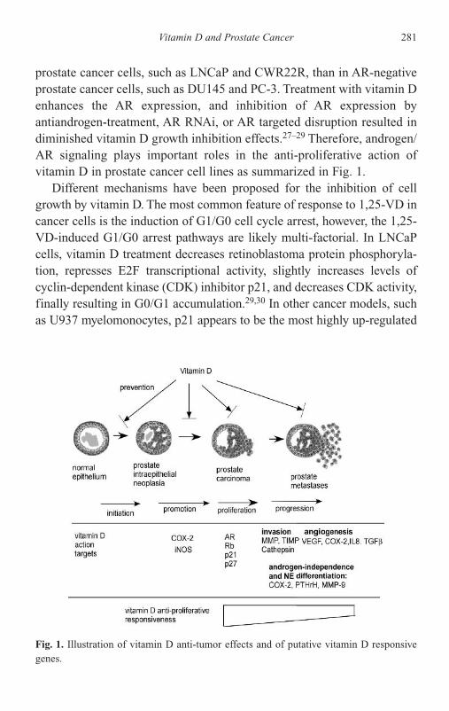
Vitamin D and Prostate Cancer 281
prostate cancer cells, such as LNCaP and CWR22R, than in AR-negativeprostate cancer cells, such as DU145 and PC-3. Treatment with vitamin Denhances the AR expression, and inhibition of AR expression byantiandrogen-treatment, AR RNAi, or AR targeted disruption resulted indiminished vitamin D growth inhibition effects.27–29 Therefore, androgen/AR signaling plays important roles in the anti-proliferative action ofvitamin D in prostate cancer cell lines as summarized in Fig. 1.
Different mechanisms have been proposed for the inhibition of cellgrowth by vitamin D. The most common feature of response to 1,25-VD incancer cells is the induction of G1/G0 cell cycle arrest, however, the 1,25-VD-induced G1/G0 arrest pathways are likely multi-factorial. In LNCaPcells, vitamin D treatment decreases retinoblastoma protein phosphoryla-tion, represses E2F transcriptional activity, slightly increases levels ofcyclin-dependent kinase (CDK) inhibitor p21, and decreases CDK activity,finally resulting in G0/G1 accumulation.29,30 In other cancer models, suchas U937 myelomonocytes, p21 appears to be the most highly up-regulated
Fig. 1. Illustration of vitamin D anti-tumor effects and of putative vitamin D responsivegenes.
B245-ch12 3/2/05 4:35 PM Page 281

gene, yet, in head and neck squamous cell carcinoma (SCC), p21 is notincreased, but p27 is up-regulated. Identification of a vitamin D responseelement (VDRE) in the p21 promoter regions suggested direct regulationof p21 activity by vitamin D/VDR, and the cell context is likely to beanother determining factor. In addition to G1/G0 arrest, induction ofapoptosis and differentiation of the prostate cancer cells may also contributeto the antiproliferative activity of vitamin D in some prostate cancer cells.31
Studies in breast cancer cells suggest that the vitamin D/VDR pathwayseems to be involved in stimulating the mitochondria to release cytochromeC rather than a pathway relying on upstream caspases.32 The mechanisms bywhich 1,25-VD induces apoptosis remain to be further investigated.
In addition to its antiproliferative effect, 1,25-VD can inhibit prostatecancer cell invasion, adhesion, and migration through modulation of selectcell surface adhesion molecules, such as integrins (alpha 4 and beta 4),33
and by inhibition of metallo-proteases (MMP-2 and MMP-9).34 These datasuggest that 1,25-VD can not only inhibit the growth of the tumor cells, butcan also stop tumor cell migration, penetration, and metastasis. Such datastimulated clinical trials of vitamin D as therapeutic agents for treatmentof prostate cancer.35,36 In one of our most recent studies, we found thatvitamin D prevents the prostate cancer cell invasion via modulation ofselective proteinase activity. In our current study using the zymographicassay, we found that 1,25-VD inhibited MMP-9 activity and enhancedtissue inhibitors of metalloproteinase-1 (TIMP-1) activity, but had less ofan effect on plasminogen activators and Cathepsin.37 Real-time PCR analy-sis demonstrated that 1,25-VD inhibited MMP-9 and induced TIMP-1 tran-script expression in a time dependent manner. The vitamin D effects on theTIMP-1 promoter activity further suggested a potential cross-talk betweenAP-1 and vitamin D/VDR signalings, which might contribute to the sup-pression of TIMP-1. In contrast, the regulation of MMP-9 by vitamin D didnot seem to be regulated at the transcriptional level, suggesting some post-transcriptional modifications, or RNA stability might contribute to the sup-pression of MMP-9 activity by vitamin D.37 From our study, we concludedthat vitamin D modulates the activity of selective proteinases, such asMMP-9 and TIMP-1, to inhibit the prostate cancer cell invasion, and thusprovides proof of the concept for treating advanced stage prostate cancerpatients with vitamin D to stop disease progression.
282 Y.-F. Lee et al.
B245-ch12 3/2/05 4:35 PM Page 282

Development of New Vitamin D Analogs and Their Usein Combination Therapy for Prostate Cancer
The beneficial effects of 1,25-VD against cancer cell proliferation havebeen supported by many in vitro studies, yet the therapeutic window of1,25-VD usefulness is extremely narrow and effective doses cannot beadministered without inducing hypercalcemia. The increase in calcium isachieved both by enhanced intestinal absorption and by liberation of cal-cium from the bone, eventually leading to decreased bone mass at higherdoses. Therefore, much effort has been directed toward the identificationof new analogs, which retain the favorable activities of 1,25-VD whileavoiding the side effects. Several synthetic vitamin D analogs have beenreported to exert promising anti-cancer effects with reduced hypercal-cemia. Skowronski et al. showed that selected vitamin D analogs dis-played reduced calcemic effects and that potency was even greater than1,25-VD.38 Among those vitamin D analogs, one of the most promisingsynthetic analogs is Seocalcitol (EB1089, Leo Pharmaceutical Products).A considerable number of in vitro studies have been carried out withEB1089 and show that the analog is more potent than 1,25-VD withrespect to regulation of cancer cell growth and differentiation, and theeffect of EB1089 on calcium metabolism in vivo is approximately 50%less than that of 1,25-VD. The anti-cancer effects of EB1089 withoutinduction of hypercalcemia were also demonstrated in vivo in a rat modelof mammary gland carcinoma. Similar effects were seen in an in vivoprostate cancer study where EB1089 inhibited prostate cancer cell prolif-eration and reduced tumorigenesis as well as tumor metastases. Severalother vitamin D analogs or structural VDR activators, such asMaxacalcitol (OCT) (Chugai Pharmaceutical Co. Ltd.), 16-ene analogs(Hoffmann LaRoche, Inc.), 19-nor analogs (Hoffmann LaRoche, Inc.),1�-hydroxyvitamin D5.39 LG190119 (Ligand Pharmaceuticals Inc.), and1�-hydroxyvitamin D2 have been developed and tested in clinical trials inthe advanced prostate cancer patients.40,41 These compounds may havepromise as therapeutic agents for cancer and other diseases, with fewerside effects than 1,25-VD.
Another strategy to preserve vitamin D anti-tumor effects while avoid-ing the side effects is vitamin D-based combination therapy in which other
Vitamin D and Prostate Cancer 283
B245-ch12 3/2/05 4:35 PM Page 283
agents that either promote vitamin D responsiveness or reduce the hyper-calcemia are used. The regimens used for vitamin D combination therapyinclude the chemotherapy drug (Paclitaxel, Taxotere),42,43 anti-mitoticagents (Genistein),44 differentiation agents (retinoic acid),45,46 and theagents that modulate vitamin D/VDR action (Dexamethasone).47 All reg-imens were shown to potentiate the anti-tumor effects of vitamin D andreduce vitamin-D associated hypercalcemia in prostate cancer cells andanimal models, yet detailed mechanisms are not yet known and moreclinical trials need to be done.
Vitamin D-Based Clinical Trials
Evidence accumulated from both experimental and epidemiological stud-ies supports the idea that vitamin D could be used as a therapeutic drugto control prostate cancer, and several vitamin D-based clinical trialshave been conducted, mainly enlisting patients with advanced androgen-independent prostate cancer (AIPC). Pilot studies from small numbers ofpatients that took daily orally doses of calcitriol found that the PSA levelsin those patients were significantly lower before calcitriol therapy.However, there is incidence of hypercalciuria or hypercalcemia, and thedevelopment of renal stones in some patients, which limits its clinicalusefulness.35 One Phase I clinical trial was conducted by injection ofcalcitriol subcutaneously every other day in the advanced malignancypatients and tested the tolerable toxicity via this route.48 Then, differentcalcitriol-based clinical trials, with either modifications in the schedulesand route of administration, or combinations with dexamethasone, pacli-taxel have been developed.49 In addition to the calcitriol, its analogs suchas 1�-hydroxyvitamin D2 have been tested in clinical trial Phase I and IIin the AIPC patients.40,41 The results from all these clinical trials suggestfurther clinical investigation of this disease with vitamin D, its analogs, orin combination with other agents, such as chemotherapy, should be furtherpursued.
In the past, chemotherapy was considered ineffective, however, newerchemotherapeutic drugs and drug combinations are now demonstratingimproved response rates. Among those chemotherapy drugs, taxotere,a semisynthetic taxane, is commonly used as a chemotherapeutic agent
284 Y.-F. Lee et al.
B245-ch12 3/2/05 4:35 PM Page 284

by itself and in combination regimens in the treatment of hormone refrac-tory prostate cancer (HRPC). 1,25-VD has been shown to sensitizeprostate cancer cells to cytotoxic drugs, such as paclitaxel, platinum com-pounds, and Taxotere. The Phase II clinical trial of Taxotere® plus cal-citriol has shown approximately twice the PSA decline response ratecompared with taxotere alone,43 which provide very promising strategiesfor treatment of AIPC. Quality of life (QOL) and pain relief were alsochecked during the calcitriol and taxotere combination therapy.Significant analgesic activity was demonstrated, yet worsening in severalQOL parameters was also observed in patients experiencing relativelylow pain intensity.50
Loss of Vitamin D Anti-proliferative Responsivenessin Prostate Cancer
While current efforts focus on developing strategies to use vitamin Danalogs to control prostate cancer, it is possible that prostate cancer cellscould become resistant to the tumor suppressive effects of vitamin D.Some prostate cancer cells are already known to be resistant to growthinhibition by vitamin D. Analyses of experimental model systems revealthat prostate cancer cells become less sensitive to vitamin D through lossof vitamin D receptors, loss of signaling molecules that modulate vitaminD action, or through changes in metabolic enzymes, such as 1�-hydroxylaseand 24-hydroxylase, that sensitize or degrade vitamin D compounds.51
Such changes have been found in experimental models, yet whether thesealterations occur in human prostate cancer tissues in vivo, and the associ-ated frequencies of occurrences still need to be determined. One studysuggested that VDR levels were decreased in the prostate after age 60,which might be linked with increased incidence of prostate cancerwith age,52 and development of vitamin D resistance during the diseaseprogression.
In one of our current studies, we identified a prostate cancer cell sub-line, CWR22R-2, which displays more aggressive behavior in the tumorinvasiveness than its parental CWR22R cells. This aggressive CWR22R-2line displays reduced vitamin D anti-proliferative effects compared toCWR22R cells, which provides evidence that prostate cancer cells might
Vitamin D and Prostate Cancer 285
B245-ch12 3/2/05 4:35 PM Page 285
develop resistance to vitamin D during disease progression. Further char-acterization of these two cell lines found reduced vitamin D transcrip-tional activity in CWR22R-2 cells, which might correlate with higherexpression of VDR co-repressors such as SMRT and NCoR, and eventu-ally result in reduced vitamin D responsiveness.53 More evidence fromother prostate cancer cell lines with different vitamin D responses andfrom prostate cancer patient samples is needed to confirm our findings.
Future Perspectives
With increasing numbers of people being diagnosed with prostate cancereach year, there is great interest in developing better strategies to treat, andeven to prevent, prostate cancer. Vitamin D has been proven to inhibitprostate cancer growth, progression, and metastases in both in vitro andin vivo animal models. However, the therapeutic use of vitamin D is lim-ited. Development of vitamin D-based treatment with improved therapeu-tic indices, so that desired activity can be maximized while the tendencytoward hypercalcemia can be minimized, is necessary.
The Figure summarizes our studies and those of others regardingvitamin D action in prostate cancer. Vitamin D may work through multi-ple pathways to prevent prostate cancer growth and progression via director indirect modulation of intracellular signals or secreted proteins, whichare involved in the tumor growth, invasion, and angiogenesis. Whether thevitamin D-mediated changes of gene profiles occur in vivo as they occurin vitro, remain to be answered. Development of the prostate cancerpatient tissue array technique by combination of classical histochemicalanalyses with advanced molecular techniques might provide powerfultools to study prostate cancer progression and its correlation with the clin-ical features at the molecular level. Through analysis of the expression ofthose vitamin D responsive genes identified from in vitro studies usingprostate cancer tissue array samples, we might be able to generate a vita-min D-response human database in prostate cancer and thus directly trans-late in vitro study results into clinical application. These will certainlyhelp to predict outcomes of vitamin D-based treatment and enhance thetreatment efficacy based on individual patients’ biochemical and molecu-lar profiles.
286 Y.-F. Lee et al.
B245-ch12 3/2/05 4:35 PM Page 286

References
1. Schwartz GG, Hulka BS (1990) Is vitamin D deficiency a risk factor forprostate cancer? (Hypothesis). Anticancer Res 10:1307–1311.
2. Hanchette CL, Schwartz GG (1992) Geographic patterns of prostate cancermortality. Evidence for a protective effect of ultraviolet radiation. Cancer70:2861–2869.
3. Corder EH, Guess HA, Hulka BS, et al. (1993) Vitamin D and prostate can-cer: a prediagnostic study with stored sera. Cancer Epidemiol BiomarkersPrev 2:467–472.
4. Ahonen MH, Tenkanen L, Teppo L, Hakama M, Tuohimaa P (2000) Prostatecancer risk and prediagnostic serum 25-hydroxyvitamin D levels (Finland).Cancer Causes Control 11:847–852.
5. Tuohimaa P, Tenkanen L, Ahonen M, et al. (2004) Both high and low levelsof blood vitamin D are associated with a higher prostate cancer risk: a longi-tudinal, nested case-control study in the Nordic countries. Int J Cancer108:104–108.
6. Glass CK (1994) Differential recognition of target genes by nuclear receptormonomers, dimers, and heterodimers. Endocr Rev 15:391–407.
7. Kato S (2000) The function of vitamin D receptor in vitamin D action.J Biochem (Tokyo) 127:717–722.
8. Haussler MR, Haussler CA, Jurutka PW, et al. (1997) The vitamin Dhormone and its nuclear receptor: molecular actions and disease states.J Endocrinol 154(Suppl):S57–73.
9. Freedman LP (1999) Multimeric coactivator complexes for steroid/nuclearreceptors. Trends Endocrinol Metab 10:403–407.
10. Boonyaratanakornkit V, Melvin V, Prendergast P, et al. (1998) High-mobilitygroup chromatin proteins 1 and 2 functionally interact with steroid hormonereceptors to enhance their DNA binding in vitro and transcriptional activityin mammalian cells. Mol Cell Biol 18:4471–4487.
11. Leo C, Li H, Chen JD (2000) Differential mechanisms of nuclear receptorregulation by receptor-associated coactivator 3. J Biol Chem 275:5976–5982.
12. Liu YY, Nguyen C, Peleg S (2000) Regulation of ligand-induced het-erodimerization and coactivator interaction by the activation function-2domain of the vitamin D receptor. Mol Endocrinol 14:1776–1787.
13. May M, Mengus G, Lavigne AC, Chambon P, Davidson I (1996) HumanTAF(II28) promotes transcriptional stimulation by activation function 2 ofthe retinoid X receptors. EMBO J 15:3093–3104.
Vitamin D and Prostate Cancer 287
B245-ch12 3/2/05 4:35 PM Page 287

14. Subramaniam N, Leong GM, Cock TA, et al. (2001) Cross-talk between1,25-dihydroxyvitamin D3 and transforming growth factor-beta signalingrequires binding of VDR and Smad3 proteins to their cognate DNA recogni-tion elements. J Biol Chem 276:15741–15746.
15. Tagami T, Lutz WH, Kumar R, Jameson JL (1998) The interaction of thevitamin D receptor with nuclear receptor corepressors and coactivators.Biochem Biophys Res Commun 253:358–363.
16. Takeshita A, Ozawa Y, Chin WW (2000) Nuclear receptor coactivators facil-itate vitamin D receptor homodimer action on direct repeat hormoneresponse elements. Endocrinology 141:1281–1284.
17. Vidal M, Ramana CV, Dusso AS (2002) Stat1-vitamin D receptor interac-tions antagonize 1,25-dihydroxyvitamin D transcriptional activity andenhance stat1-mediated transcription. Mol Cell Biol 22:2777–2787.
18. Takeyama K, Masuhiro Y, Fuse H, et al. (1999) Selective interaction ofvitamin D receptor with transcriptional coactivators by a vitamin D analog.Mol Cell Biol 19:1049–1055.
19. Rachez C, Suldan Z, Ward J, et al. (1998) A novel protein complex that inter-acts with the vitamin D3 receptor in a ligand-dependent manner andenhances VDR transactivation in a cell-free system. Genes Dev 12:1787–1800.
20. Rosenfeld MG, Glass CK (2001) Coregulator codes of transcriptional regu-lation by nuclear receptors. J Biol Chem 276:36865–36868.
21. Barletta F, Freedman LP, Christakos S (2002) Enhancement of VDR-mediated transcription by phosphorylation: correlation with increased inter-action between the VDR and DRIP205, a subunit of the VDR-interactingprotein coactivator complex. Mol Endocrinol 16:301–314.
22. Qi X, Pramanik R, Wang J, et al. (2002) The p38 and JNK pathways cooper-ate to trans-activate vitamin D receptor via c-Jun/AP-1 and sensitize humanbreast cancer cells to vitamin D(3)-induced growth inhibition. J Biol Chem277:25884–25892.
23. Miller GJ, Stapleton GE, Ferrara JA, et al. (1992) The human prostatic car-cinoma cell line LNCaP expresses biologically active, specific receptors for1 alpha,25-dihydroxyvitamin D3. Cancer Res 52:515–520.
24. Peehl DM, Skowronski RJ, Leung GK, Wong ST, Stamey TA, Feldman D(1994) Antiproliferative effects of 1,25-dihydroxyvitamin D3 on primarycultures of human prostatic cells. Cancer Res 54:805–810.
25. Skowronski RJ, Peehl DM, Feldman D (1993) Vitamin D and prostate cancer:1,25 dihydroxyvitamin D3 receptors and actions in human prostate cancercell lines. Endocrinology 132:1952–1960.
288 Y.-F. Lee et al.
B245-ch12 3/2/05 4:35 PM Page 288
Vitamin D and Prostate Cancer 289
26. Schwartz GG, Oeler TA, Uskokovic MR, Bahnson RR (1994) Humanprostate cancer cells: inhibition of proliferation by vitamin D analogs.Anticancer Res 14:1077–1078.
27. Hsieh TY, Ng CY, Mallouh C, Tazaki H, Wu JM (1996) Regulation ofgrowth, PSA/PAP and androgen receptor expression by 1 alpha,25-dihydrox-yvitamin D3 in the androgen-dependent LNCaP cells. Biochem Biophys ResCommun 223:141–146.
28. Hsieh T, Wu JM (1997) Induction of apoptosis and altered nuclear/cytoplas-mic distribution of the androgen receptor and prostate-specific antigen by1alpha,25-dihydroxyvitamin D3 in androgen-responsive LNCaP cells.Biochem Biophys Res Commun 235:539–544.
29. Bao BY, Ting HJ, Hu YC, Lee YF (2004) Androgen signaling is required invitamin D mediated growth inhibition in prostate cancer cells. Oncogene(in press).
30. Zhuang SH, Burnstein KL (1998) Antiproliferative effect of 1alpha,25-dihy-droxyvitamin D3 in human prostate cancer cell line LNCaP involves reduc-tion of cyclin-dependent kinase 2 activity and persistent G1 accumulation.Endocrinology 139:1197–1207.
31. Blutt SE, McDonnell TJ, Polek TC, Weigel NL (2000) Calcitriol-inducedapoptosis in LNCaP cells is blocked by overexpression of Bcl-2.Endocrinology 141:10–17.
32. Weitsman GE, Ravid A, Liberman UA, Koren R (2003) Vitamin D enhancescaspase-dependent and -independent TNFalpha-induced breast cancer celldeath: The role of reactive oxygen species and mitochondria. Int J Cancer106:178–186.
33. Sung V, Feldman D (2000) 1,25-Dihydroxyvitamin D3 decreases humanprostate cancer cell adhesion and migration. Mol Cell Endocrinol 164:133–143.
34. Schwartz GG, Wang MH, Zang M, Singh RK, Siegal GP (1997) 1 alpha,25-Dihydroxyvitamin D (calcitriol) inhibits the invasiveness of human prostatecancer cells. Cancer Epidemiol Biomarkers Prev 6:727–732.
35. Gross C, Stamey T, Hancock S, Feldman D (1998) Treatment of early recur-rent prostate cancer with 1,25-dihydroxyvitamin D3 (calcitriol). J Urol159:2035–2039; discussion pp. 2039–2040.
36. Trump D, Lau YK (2003) Chemotherapy of prostate cancer: present andfuture. Curr Urol Rep 4:229–232.
37. Bao BY, Lee YF (2004) 1,25-vitamin D3 inhibits prostate cancer cell inva-sion by modulation MMPs, TIPs, and cystein protease activities. Oncogene(submitted).
B245-ch12 3/2/05 4:35 PM Page 289

290 Y.-F. Lee et al.
38. Skowronski RJ, Peehl DM, Feldman D (1995) Actions of vitamin D3,analogs on human prostate cancer cell lines: comparison with 1,25-dihydroxyvitamin D3. Endocrinology 136:20–26.
39. Mehta RG, Mehta RR (2002) Vitamin D and cancer. J Nutr Biochem13:252–264.
40. Liu G, Oettel K, Ripple G, et al. (2002) Phase I trial of 1alpha-hydroxyvita-min D(2) in patients with hormone refractory prostate cancer. Clin CancerRes 8:2820–2827.
41. Liu G, Wilding G, Staab MJ, et al. (2003) Phase II study of 1alpha-hydroxyvitamin D(2) in the treatment of advanced androgen-independentprostate cancer. Clin Cancer Res 9:4077–4083.
42. Hershberger PA, Yu WD, Modzelewski RA, Rueger RM, Johnson CS, TrumpDL (2001) Calcitriol (1,25-dihydroxycholecalciferol) enhances paclitaxelantitumor activity in vitro and in vivo and accelerates paclitaxel-inducedapoptosis. Clin Cancer Res 7:1043–1051.
43. Beer TM, Eilers KM, Garzotto M, Egorin MJ, Lowe BA, Henner WD (2003)Weekly high-dose calcitriol and docetaxel in metastatic androgen-independentprostate cancer. J Clin Oncol 21:123–128.
44. Rao A, Woodruff RD, Wade WN, Kute TE, Cramer SD (2002) Genistein andvitamin D synergistically inhibit human prostatic epithelial cell growth. JNutr 132:3191–3194.
45. Zhao XY, Ly LH, Peehl DM, Feldman D (1999) Induction of androgen recep-tor by 1alpha,25-dihydroxyvitamin D3 and 9-cis retinoic acid in LNCaPhuman prostate cancer cells. Endocrinology 140:1205–1212.
46. Ikeda N, Uemura H, Ishiguro H, et al. (2003) Combination treatment with1alpha,25-dihydroxyvitamin D3 and 9-cis-retinoic acid directly inhibitshuman telomerase reverse transcriptase transcription in prostate cancer cells.Mol Cancer Ther 2:739–746.
47. Johnson CS, Hershberger PA, Bernardi RJ, McGuire TF, Trump DL (2002)Vitamin D receptor: a potential target for intervention. Urology 60:123–130;discussion pp. 130–131.
48. Smith DC, Johnson CS, Freeman CC, Muindi J, Wilson JW, Trump DL(1999) A Phase I trial of calcitriol (1,25-dihydroxycholecalciferol) in patientswith advanced malignancy. Clin Cancer Res 5:1339–1345.
49. Blutt SE, Weigel NL (1999) Vitamin D and prostate cancer. Proc Soc ExpBiol Med 221:89–98.
50. Beer TM, Eilers KM, Garzotto M, Hsieh YC, Mori M (2004) Quality of lifeand pain relief during treatment with calcitriol and docetaxel in symptomaticmetastatic androgen-independent prostate carcinoma. Cancer 100:758–763.
B245-ch12 3/2/05 4:35 PM Page 290

51. Peehl DM, Feldman D (2003) The role of vitamin D and retinoids in con-trolling prostate cancer progression. Endocr Relat Cancer 10:131–140.
52. Krill D, DeFlavia P, Dhir R, et al. (2001) Expression patterns of vitamin Dreceptor in human prostate. J Cell Biochem 82:566–572.
53. Ting HJ, Bao BY, Lee YF (2004) Loss of 1,25-dihydroxyvitamin D3 respon-siveness in aggressive androgen-independent prostate cancer cells. Mol BiolCell (submitted).
Vitamin D and Prostate Cancer 291
B245-ch12 3/2/05 4:35 PM Page 291

This page intentionally left blank

13FUNCTIONS OF ESTROGEN RECEPTOR IN
PROSTATE AND PROSTATE CANCER
Shuyuan Yeh, Ming Chen, Jing Ni, Yi Yin, Eugene Chang, Min Zhang and Xingqing Wen
Departments of Urology and PathologyUniversity of Rochester Medical Center, Rochester, New York
Introduction
It has long been known that the functions of estrogen and estrogen recep-tor (ER) are important for the female reproductive system, mammarygland development, and cancer progression.1 Accumulated evidence fromvarious studies indicated that ER is expressed and functions in the malereproductive system, including prostate. Earlier studies only identifiedone traditional ER, ERalpha (ER�), which was thought to be responsiblefor all the estrogen-mediated responses in prostate. Recently, ERbeta(ER�) was identified and shown to express in prostate epithelia.2 Resultsfrom PCR, immunohistochemical staining and receptor knockout (KO)mice suggested that estrogens, ER� and ER� play significant rolesin prostate development and the initiation and progression of prostate cancer. In this chapter, we will focus on the distribution of ER� andER�, potential target genes, ER association proteins, their potentialroles in the development of prostate, and the progression of prostatecancer based on the results from prostate tissues, cancer cell lines, andanimal models, including �ERKO, �ERKO, ��ERKO, and hypogonadal(hpg) mice.
293
B245-ch13 3/2/05 4:39 PM Page 293
Distribution of ER� and ER� in Prostate Tissues, Cancer Specimens and Cancer Cell Lines
Estrogen has been used for the treatment of prostate cancer since the early1940s.3 It is generally believed this action is indirectly mediated at the hypo-thalamic level to suppress the circulating androgens.4 However, in the early1960s, a direct action of estrogen via their own receptors in the prostate wasproposed by Mangan et al.5 Recently, the evidences that ER expressed innormal prostate, benign prostatic hyperplasia (BPH), prostate cancer speci-mens, and different prostate cancer cell lines, along with the demonstrationof the stimulatory or inhibitory effects of estrogen on prostate cancer cellsgrowth suggested estrogen may exert direct effects on prostate via their ownreceptors.6–11 The following sections will discuss the distribution of ER�
and ER� in prostate tissues, cancer specimens, and cancer cell lines.Both ER� and ER� express in the human prostate tissue.2,12 In normal
prostate tissue ER� is mostly expressed in stromal cells with occasionalexpression in the basal epithelial cells,8,11,13–15 whereas ER� is abundantlypresent in basal epithelial cells.8,11,13 The complementary location of bothER� and ER� might explain why some early studies reported ER� in boththe stroma and epithelium of the normal prostate, which may be caused bycross reaction against both ER subtypes.16
In BPH, ER� immunostaining could not be detected in severalstudies.15,16 Using enzyme immunoassay, Mobbs et al. (1990) found highpositivity of ER� in BPH.17 The latter immunostaining studies found ER�
was stronger in BPH than in normal prostates and localized in both epithe-lial and stromal cells.8 Immunoexpression of ER� in BPH was also higherthan in normal prostates and localized only in the epithelium.8
In prostate cancer specimens, the expressions of both ER� andER� have been studied for years,6,8,13,18 however, the relative expression levels and location of ERs in prostate cancer are still controversial. Usingmethods of immunohistochemistry staining, loss or down-regulation ofER� in prostate cancer has been reported.19,20 It appears ER� gene is tran-scriptionally inactivated by DNA methylation in most prostate cancer cellslines and specimens.11,21 Conversely, several groups reported the higherER� expression in the prostate cancer specimens than in BPH and normalprostate tissue, which is consistent with other reports that abundance ofER� is positively correlated with the malignancy of prostate cancer.7,17
294 S. Yeh et al.
B245-ch13 3/2/05 4:39 PM Page 294

In terms of ER� expression, Royuela et al. (2001) reported the ER� hasincreasing epithelial staining in both BPH and prostate cancer, and somestromal cells acquire ER� in prostate cancer specimens, not in normalprostate and BPH. These findings suggest the involvement of ER� inprostate cancer. However, recent reports showed a frequent loss ofER� expression in prostate cancer samples relative to normal prostatetissue.13,18,22 In the primary prostate tumor sites, ER� was stronglyexpressed in low grade prostate carcinoma and was markedly diminishedin higher grade tumor.18,22,23 These results suggested ER� may protectagainst abnormal prostate cell growth.11,13,22 Loss of ER� may causeprostate epithelial cells to escape the control of proliferation by ER� inprostate cancer. Further studies indiated ER� protein expression was sig-nificantly elevated in metastatic prostate tumor.13 Currently it is unclearwhy the ER� expression will be decreased in the higher grade prostatecancer and re-elevated in metastases. One hypothesis is that the failure tolose ER� may allow the cancer cells to gain the metastatic potential, andanother could be the local environment in the distal metastatic sites mayinduce ER� expression. Although the controversy of ERs expressionexists, the accumulating evidence shows the abnormality of ER signalingmay contribute to the pathogenesis of prostate cancer.
The expression patterns of both ERs in different prostate cancer cell lineshas been studied recently. The earlier studies on the effect of estrogens onprostate cancer cells growth showed the direct role of estrogen via their ownreceptors, but the specific ER has not been defined. Using RT-PCR, Lauet al. (1999) showed ER�, but not ER�, was found in normal prostate epithe-lia cells, along with pS2 and PR mRNA expression. Only ER� is detectablein LNCaP and DU145 cell lines, whereas PC-3 and BPH-1 expressed bothER� and ER�.11 Currently, there are two ER� isoforms, ER�1 and ER�2.Only ER�1 was detectable in PC-3, DU145, and LNCaP cell lines, whereasER�2 was absent.24 Together, these studies suggested ER� could be the keyplayer of estrogen-mediated effects in prostate cancer cell lines.
In rodents, it was found that ER� mRNA was the major type ofER expressed in two-week-old rat prostate and that as the rat aged ER�
became the dominant expression.25 In agreement with the results fromhuman prostate, it was found that ER� protein is predominantly located inthe stroma cells of ventral prostate of adult rodents, with over 90% of theepithelial cells stained positive for ER�.26
Functions of Estrogen Receptor in Prostate and Prostate Cancer 295
B245-ch13 3/2/05 4:39 PM Page 295

Estrogen Regulated Genes in Prostate Cancer
Estrogens and ER have been shown to play important roles in prostatecarcinogenesis.27 The detailed mechanisms however, are not well known.Currently, some estrogen target genes have been identified, which may contribute to the diversified estrogen effect on prostate cancer. Those genes include telomerase,27 endothelin-1,28 eNOS,29 E-cadherin,30 metalloth-ionein II,31 CGA gene,32 androgen receptor,26 and glutathione-peroxidase.33
In addition, PS2 and progesterone receptor (PR), have an estrogenresponsive element (ERE) in the promoter regions, and are also expressedin the prostate, suggesting they can be regulated by estrogen in humanprostate.11 In mouse prostate, diethylstilbestrol (DES) treatment inducesPR synthesis in secretory epithelial cells, which is usually low in theuntreated prostate. Although a functional role for progesterone in regulat-ing prostate growth is unknown, PR synthesis is an effective marker ofestrogen action in the prostate. It was found that the acute DES exposureof WT male mice, the prostatic responses were similar, including induc-tion of squamous metaplasia (SQM), and up-regulation of PR.34 Table 1lists the identified estrogen target genes in the prostate. In comparison,many other estrogen target genes have been identified in the breastor other estrogen target organs. For better understanding of the estrogensignal in prostate, ER� and ER� target genes and their roles in prostateneed to be identified and characterized.
296 S. Yeh et al.
Table 1. Estrogen Regulated Genes in Prostate
Function Genes Up- or Down- Estrogen Responseregulation Element (ERE)
Carcinogenesis Telomerase ↑ �
Angiogenesis eNOS ↑ N/AMetastasis E-cadherin ↑ N/AMetastasis Endothelin-1 ↓ N/AHomeostasis Metallothionein II ↑ N/AHormones CGA gene ↑ N/A (link with ER�)Oxidant enzyme Glutathione-peroxidase ↑ N/ATranscription factor Androgen receptor ↓ N/ATranscription factor Progesterone receptor ↑ �
Unknown PS2 ↑ �
B245-ch13 3/2/05 4:39 PM Page 296

Functions of Estrogen Receptor in Prostate and Prostate Cancer 297
ER Coregulators in Prostate
ERs are ligand-dependent transcription factors which bind to the ERE toregulate the transcription of target genes. The transcription initiation is acomplex process which involves the cooperative interaction between ERsand multiple factors at the promoter region of target genes. The occur-rence of transcriptional interference between nuclear receptors in earlytransient receptor/reporter co-transfection assays suggested the existenceof common rate limiting cofactors, other than general transcription factors(GTFs), required for ERs activation function.35,36 In this regard, coregu-lators were identified, and defined as coactivators or corepressors requiredfor transcriptional regulation.37 In this chapter, we will mainly focus onthe expression pattern of some identified ER coactivators in the prostatetissue and prostate cancer cell lines.
To date, there are numerous ER coregulators that have been identifiedand the most well characterized ER coactivators belong to the p160 fam-ily, including SRC-1, SRC-2 (TIF2/GRIP1) and SRC-3 (AIB1/RAC3).37
Among these identified ER coactivators, SRC-3/RAC3/AIB1 is themost well studied in prostate cancer cell lines. Using Western blotting,Gnanapragasam et al. showed that SRC3/RAC3/AIB1 protein has thehighest expression in LNCaP cells, moderate expression in PC-3 cells,and low-level expression in DU145 cells.38 In the human prostate tissue,the levels of SRC-3/RAC3/AIB1 expression is significantly correlatedwith tumor grade and stage of disease, but not with serum PSA levels.38
SRC-1 is expressed as a major RNA transcript of 7.5 kb in many tissues,including prostate. The expression levels of SRC-1 were found to be elevated in the cancer specimens with a higher grade or poor responseto endocrine therapy, than in those with a lower grade or good responseto endocrine therapy.39 In addition to SRC-1 and SRC-3, other ERco-regulators, CBP/p300, SMRT, and N-CoR, are also expressed inprostate.40
Overall, most of ER co-regulators identified to date are not specificto ER, and can interact with other nuclear receptors, including AR.41
Therefore, how these co-regulators regulate ER function in prostatedevelopment and neoplastic transformation remains to be elucidated. Inaddition, whether any ER specific associated proteins exist in prostateneeds further investigation.
B245-ch13 3/2/05 4:39 PM Page 297
Histological Changes in Prostates of �ERKO and �ERKO Mice
To understand the roles of estrogen, ER� and ER� in the development ofprostate glands, the gene knockout strategy has been used. The adult male�ERKO mouse prostate shows normal development and histology, speci-fically in the ventral prostate (VP)42,43 and anterior prostate (AP).44 Withaging, the weights of �ERKO seminal vehicle (SV) and AP increased,45
although they remain histologically indistinguishable from that of WTlittermates.42,44
The initial description of targeted disruption of the gene encoding ER�
reported some evidence of epithelial hyperplasia in �ERKO prostates.46
Weihua et al.26 further reported that �ERKO mice had multiple hyperplas-tic foci in the peripheral and central zones of the VP at five months; by oneyear of age, eight of ten �ERKO VPs had multiple hyperplastic lesions, andmost epithelial cells stained positively for the proliferation antigen Ki-67.However, other studies of the prostate phenotype in ER�-deficientmice,42,43,47 failed to corroborate the initial report of Krege et al.,46 andno hyperplasia was seen in prostate lobes of �ERKO line at 12–20months.42,43,47 The reason for discrepant results from mice with the sameapparent genotype is unclear, yet other factors might affect the mice, suchas diet or infection. In general, there is no significant histological changesin prostates of �ERKO mice, and with controversial observation in prostateof �ERKO Mice.
Estrogen Imprinting Effect on the Development of Prostate
Although estrogen levels are low or undetectable in adult male mice,administration of exogenous estrogens during development48 dramaticallyaffects prostate growth and function.49–54 The effects of estrogens varyaccording to timing and duration of exposure, in addition to the type anddose of estrogen administered. Furthermore, the individual lobes of theprostate exhibit varied degrees of response to estrogens and androgens.
The neonatal period after birth is very fundamental for the rodent prostatedevelopment in which the prostate involves branching morphogenesisfollowed by functional differentiation. In this period, brief exposure of malerats or mice to high-level estrogens will cause irreversible alterations in
298 S. Yeh et al.
B245-ch13 3/2/05 4:39 PM Page 298
development and function of the prostate gland and a reduced responsivenessto androgens during adulthood.50,55–57 The estrogen imprinting effect is asso-ciated with an aging-related prostatic lesion, which includes the hyperplasiaof prostatic epithelial cells or severe dysplasia similar to high grade prostaticintra-epithelial neoplasia (PIN) and extensive immune cell infiltrate.51,58–61
As early as 1978, Rajfer and Coffey reported that if high dose of 17�-estradiol at 500 �g per day, or estradiol benzoate at 250 �g per day, orestradiol dipropionate at 100 �g per day, is administered to intact malerats for 2 days during the 1st week after birth, the prostate, at adulthood, isdiminutive in size and is inert to the action of exogenous androgens.If immature rodents are exposed to exogenous estrogen before puberty, theaging animals develop prostatic epithelial hyperplasia50 or even dysplasia ifandrogen is given together with estrogen.61 A series of studies by McLachlanet al. demonstrated that perinatal exposure to the synthetic estrogen, DES,results in an assortment of apparently direct defects in the murine malereproductive tract, including undescended testes, epididymal cysts, aberrantexpression of estrogen inducible genes, adenocarcinoma, and sterility.61–65
In addition to morphological and histological changes, several groupscharacterized the other hallmarks of neonatal estrogen imprinting in therat model as well as in different mouse strains. These included transientup-regulation of ER�, down-regulation of androgen receptor (AR),decreased ER� levels in adult prostate epithelium, lack of dorsal lateralprostate (DLP) secretory protein, up-regulated proto-oncogene c-fos,reduced TGF� type I receptor levels in the prostate epithelium, but not instroma, whereas there was no effect on TGF� type II receptor. p21 (cip-1/waf-1) is a cyclin-dependent kinase inhibitor, and is known to beinducible by TGF�1 in the prostate. Neonatal estrogenization preventedthis transient expression of p21 (cip-1/waf-1) in the prostate epitheliumcells. Those changes in molecular levels suggested they could be goodmarkers for estrogenization in the prostate.66–72
Estrogen Treatment of �ERKO, �ERKO, and Hypogonadal (hpg) Mouse Models
Intact adult �ERKO mice treated with DES showed no change in organweight, no induction of SQM, or synthesis of CK10 or PR in any prostate
Functions of Estrogen Receptor in Prostate and Prostate Cancer 299
B245-ch13 3/2/05 4:39 PM Page 299

lobe.44 In contrast, the intact adult �ERKO mouse treated with DESshowed a full response, similar to that of WT mice.44 In spite of normalER� levels in the �ERKO prostate, these animals showed no morpholog-ical or molecular effects of estrogen in the prostate. These data suggestthat these actions or effects of exogenous estrogen on the adult mouseprostate are predominantly mediated through ER�. ER� is thus the pre-dominant receptor mediating mitogenic estrogen action in the prostate,and a role for ER� remains to be identified.
Because classic estrogen responses (SQM and up-regulation of PR)were seen in the �ERKO but not in the �ERKO mice, it is assumed thatER� mediates these responses in the adult prostate. A recent reportinvolving tissue recombination examined the role of ER� in mediatingestrogenic responses in prostate, using combinations of WT and �ERKOprostate epithelial and stromal cells.44 The recombinant tissues weregrafted under the kidney capsule of immunologically deprived nudemice exposed to DES treatment for 21 days. ER� is required for theinduction of SQM, but not for the up-regulation of PR synthesis, whichoccurred in all recombinants, even though this response was absent afterthe in vivo DES treatment of �ERKO mice.44 The functional significanceof the separation of these two end points (SQM and PR synthesis)suggests that additional mechanisms are involved in mediating estrogenresponses.
Exposure to estrogens during the neonatal period causes both acute andlong-term effects on the prostate gland. Neonatally estrogenized WTanimals show inhibited prostate growth during development and subse-quently display evidence of dysplasia and an attenuated response toandrogens.50,52–54,67 When estrogenized animals are castrated and thentreated with androgen (testosterone) implants, the observed effects are notcompletely blocked, suggesting that much of the phenotype is the result ofpermanent alterations within the prostate tissue caused by direct estrogenaction.67 The neonatal studies described above were all conducted in a ratmodel system with high doses of DES. By contrast, a single report showedincreased prostate size following low-dose maternal estrogen administra-tion to male mouse fetuses.49,73 The discrepancies between rat and mousemodels, and high- and low-dose estrogen, emphasize the compounding
300 S. Yeh et al.
B245-ch13 3/2/05 4:39 PM Page 300

influences of timing, duration, and dose of estrogen in similar studies.Recently, Prins et al.42 utilized �ERKO and �ERKO mice to demonstratethat both acute and chronic effects of neonatal DES treatment are pre-dominantly mediated by ER� in mouse prostate.
Investigation of intact male mice with prostatic response to estrogentreatment requires analysis of both indirect and direct effects. Indirectactions occur via regulation of the hypothalamic-pituitary-gonadal (HPG)axis. They alter androgen production and the endocrine status of the mice.Direct responses to estrogen by the prostate differ and occur in response tolocal or intraprostatic changes in estrogen synthesis via aromatase enzymeactivity or modulation of the ERs. More recently, the direct actions ofestrogens in vivo were evaluated using the hypogonadal (hpg) mousemodel74 that is deficient in pituitary gonadotropin and sex steroid produc-tion. Mature male hpg mice were exposed to estradiol for 6 weeks, andproliferative changes were recorded in the prostate with specific effectsobserved in the stroma and epithelium; estradiol administration stimulatedgrowth and expansion of the stromal, epithelial and luminal compartmentsof the mouse prostate lobes.75 The epithelial cells became multi-layeredand squamous, showing immunoexpression of high molecular weightcytokeratins and cytokeratin 10 (CK10) that is characteristic of the pathol-ogy known as squamous metaplasia (SQM).44,76 The stromal response wascharacterized by an increase in fibroblastic stroma that penetrated thesmooth muscle layer, accompanied by a reduction and disorganization in�-actin-positive smooth muscle cells surrounding the glandular ducts.Additionally, neutrophils were identified in the stroma and were shown tomigrate through the epithelium to the lumen. Although secretory activity ofprostate epithelial cells was significantly reduced by estradiol exposure,the lumen was distended as a result of accumulated cellular debriscomprising epithelial cells, inflammatory cells, and anuclear keratiniseddeposits.75
Overall, results from these genomic-manipulated mice suggested thatER� is the dominant ER form mediating the developmental estrogeniza-tion of the prostate gland. Estrogens induced direct proliferative changesin the prostate of male hpg mice, which do not have physiological levelsof androgen.
Functions of Estrogen Receptor in Prostate and Prostate Cancer 301
B245-ch13 3/2/05 4:39 PM Page 301

Estrogen Effect on Initiation, Growth, and Progression of Prostate Cancer
While androgens play essential roles in the development and growth ofprostate and pathogenesis of prostate cancer, extensive in vivo and in vitrostudies suggested that estrogens are required for carcinogenesis ofprostate cancer. It was demonstrated that treatment with both estrogen andtestosterone will induce 100% dorsolaterol prostate carcinoma in rats.77 Inhuman, direct estrogen effects and the balance of androgens and estrogensmay contribute to these estrogen activities. Men synthesize both estrogensand androgens and as men age, the plasma levels of androgen decreaseswhile estrogen remains constant. However, the expression pattern andactivity of aromatase, the enzyme that turns testosterone into estrogen,suggests that local synthesized estrogen may have significant conse-quences in tumorigenesis of prostate.78,79 Here we will focus on estrogenseffects on prostate cancer cells from in vitro and in vivo approaches. Whilethis approach provides some insights to explain the in vivo observationfrom clinical and mouse study, many unclear and controversial dataremain.
In prostate, estrogen induced cancerous transformation may be par-tially due to their genotoxic metabolites, including 2-hydroxy-estrogens,4-hydroxy-estrogens, quinone, and semiquinone intermediates. Thesemetabolites may directly induce genomic damage or function via forma-tion of reactive oxygen species (ROS).80,81 In addition, the enzymesresponsible for the formation or inactivation of these estrogen metabolitesmay also play critical roles in the estrogen mediated transformationin prostate. These enzymes include (1) cytochrome p4501A1, whichcan convert estrogen into 2-hydroxy-estrogens, (2) cytochrome p4501B1,which can concert estrogens to 4-hydroxy-estrogens, (3) cathecol-O-methyl-transferase, which can inactivate 2- and 4-hydroxy-estrogens, and(4) glutathione-S-transferases that detoxify ROS.81–83 Although these maybe important factors for estrogen mediated effects in prostate, few of themhave been investigated for their contribution in prostate cancer.
As the aromatase can convert androgens into estrogens, thus it could beone of the risk factor of prostate cancer.84,85 However, the aromatase trans-genic mice and knockout mice have numerous endocrine defects,34,86 thus
302 S. Yeh et al.
B245-ch13 3/2/05 4:39 PM Page 302

these mice cannot be used as good models to study the relationshipbetween estrogen formation and prostate cancer risk. The studies will beadvanced by producing mice with prostate specific expression or knock-out of aromatase in the future.
In adult WT mice, administration of high-dose DES, a potent syntheticestrogen, causes regression of the prostate and induces SQM in the epithe-lium. The reduced organ size is associated with declining androgen levels,whereas SQM is considered to be a direct, ER� mediated response toestrogen.44 Prostatic SQM is characterized by proliferation of basal cells,leading to formation of stratified squamous epithelium and altered CK10synthesis. In mouse prostate, DES treatment induces PR synthesis insecretory epithelial cells, which is usually low in the untreated prostate.
Although studies suggested that estrogens are required for carcinogen-esis of prostate cancer, yet extensive evidences also suggest that estrogenscan suppress prostate cancer growth. It has been found that estrogeninhibits the cell growth of PC-3, an androgen independent prostate cancercell line,9 while it can stimulate the growth of LNCaP, an androgenresponsive prostate cancer cell line.10 This difference may be due to dif-ferent ER forms and ER levels in these two cell lines85,86 and other mech-anisms. Although estrogen stimulates both ER� and ER�, it is generallybelieved that ER� stimulates cell proliferation whereas ER� counteractsER� activity. In the prostate, ER� is exclusively expressed in stroma,while ER� is largely expressed in epithelial cells. In tumors, ER� isexpressed in both stroma and epithelial. Many studies support the expres-sion pattern of ER� and ER�, and the interaction between stroma andepithelial of prostate largely contributes to the different estrogen effects onthe prostate cancer cells growth in vivo and in vitro.11,87–89 However, theexpression pattern of ER� and ER� in normal and tumor prostate tissue,and prostate cancer cell lines are different. More studies need to beelucidated.
The effect of estrogen regulation of prostate cancer cell growth wasalso tested with the estrogen metabolite, 2-methoxyestradiol (2-ME),which can arrest the cell cycle and induce apoptosis.90,91 However, the10 �M dose of 2-ME used in the study is much higher than physiologicalconcentration. Cellular component concentrations are another importantfactor for the cellular growth or signal. Homeostatic changes might also
Functions of Estrogen Receptor in Prostate and Prostate Cancer 303
B245-ch13 3/2/05 4:39 PM Page 303

contribute to malignant growth. Estrogen can increase intracellularCa (2�) in PC-3 cells.92 Estrogen also reduces the uptake of rubidiumchloride, suggesting an estrogen effect on ion transport and change ofcellular membrane permeability.93
Although estrogen effects on prostate cancer cell growth are somewhatcontroversial, the fact that estrogen is required for prostate carcinogenesisis consistent. It was reported that a low ratio of androgen to estrogenresults in a higher risk for the prostate cancer. In vivo animal studies alsosupported that estrogen, in a milieu of decreasing androgen, contributessignificantly to the prostate hyperplasia, prostate dysplasia, and prostatecancer. �ERKO mice cannot be induced by estrogen treatment to growprostate cancer.76,94 Therefore, estrogens and ERs, together with androgen,are required for the pathogenesis of prostate cancer. Recently, telomerase,whose activity increased during tumorigenesis, has been shown to bestimulated by estrogen in primary cultured cells from BPH and normalprostate tissue. Telomerase contains ERE in its promoter, thus could bea target gene of ER.27 This may be one of the mechanisms by whichestrogens stimulate carcinogenesis. Antiestrogen treatment therefore mayprevent prostate cancer.
It is very significant that in the transgenic adenocarcinoma mouseprostate (TRAMP) mouse model of adenocarcinoma of the ventralprostate,95 the selective estrogen receptor modulator (SERM), toremifene,96
and the phytoestrogen genistein,97 prevent the development of cancer.Although it has been suggested that this protection results from inhibitionof ER� in the prostate stroma, an equally acceptable explanation is thatthese agents, acting as ER� agonists, prevent proliferation of the prostateepithelium. Such a mechanism is likely in view of the role of ER� in pre-venting proliferation of the ventral prostate epithelium in the mouse andstrongly suggests a role for ER� agonists in the treatment and/or preventionof prostate cancer.
Implantation of prostate cancer xenografts (LuCaP 35, LuCaP 49,LuCaP 58, LuCaP 73, PC-3, and LNCaP) into intact and ovariectomizedfemale mice was done by Corey et al. to characterize growth and uptakerates in the absence of androgens.98 Significant inhibition of prostate cancergrowth in intact vs. ovariectomized female animals was observed in five ofsix prostate cancer xenograft lines (except for PC-3). E2 supplements givento ovariectomized female mice led to inhibition of tumor establishment and
304 S. Yeh et al.
B245-ch13 3/2/05 4:39 PM Page 304

diminished growth of LuCaP 35, similar to that observed in intact femalemice. RT-PCR showed that these xenografts express the ER� message. Twohypothetical mechanisms may be suggested to explain how E2 can affectprostate cancer tumor growth in female mice in the absence of androgens:(1) E2 exerts direct inhibitory effects via ER expressed on prostate cancercells or via other, as yet unidentified, mechanisms; and (2) E2 exerts effectson other cells, which then secrete signaling molecules that inhibit prostatecancer growth.
Together, these in vivo and in vitro studies suggested that estrogens mayhave dual roles: (1) estrogens are required for tumorigenecity of prostatecancer; and (2) Estrogens may suppress growth of prostate cancer.
Conclusion
To date, results from various studies clearly demonstrated that estrogensand ERs play important roles in prostate development and tumorigenesis.However, the roles are still not completely elucidated. Results from�ERKO male mice suggest that ER� plays roles in the initiation of BPHand SQM.86 Results from �ERKO male mice indicated that when miceaged they may develop hypertrophy26 suggesting that ER� plays a nega-tive role or is a factor controlling prostatic cell growth. However, resultsfrom other groups are not entirely consistent with that conclusion.99 It iscurrently unclear whether nutritional and environmental factors contributeto the discrepancy. Furthermore, the number and function of differentisoforms of ER� that exist in the prostate have not been conclusivelydetermined.
References
1. Russo IH, Russo J (1998) Role of hormones in mammary cancer initiationand progression. J Mammary Gland Biol Neoplasia 3:49–61.
2. Mosselman S, Polman J, Dijkema R (1996) ER beta: identification and char-acterization of a novel human estrogen receptor. FEBS Lett 392:49–53.
3. Huggins C, Hodges CV (1941) Studies on prostatic cancer. I. The effect ofcastration, of estrogen and androgen injection on serum phosphatases inmetastatic carcinoma of the prostate. Cancer Res 1:293–297.
Functions of Estrogen Receptor in Prostate and Prostate Cancer 305
B245-ch13 3/2/05 4:39 PM Page 305

4. Huggins C, Hodges CV (1972) Studies on prostatic cancer. I. The effect ofcastration, of estrogen and androgen injection on serum phosphatases inmetastatic carcinoma of the prostate. CA Cancer J Clin 22:232–240.
5. Mangan FR, Neal GE, Williams DC (1967) The effects of diethylstilboestroland castration on the nucleic acid and protein metabolism of rat prostategland. Biochem J 104:1075–1081.
6. Brolin J, Skoog L, Ekman P (1992) Immunohistochemistry and biochemistryin detection of androgen, progesterone and estrogen receptors in benign andmalignant human prostatic tissue. Prostate 20:281–295.
7. Bonkhoff H, Fixemer T, Hunsicker I, Remberger K (1999) Estrogen receptorexpression in prostate cancer and premalignant prostatic lesions. Am J Pathol155:641–647.
8. Royuela M, de Miguel MP, Bethencourt FR, Sanchez-Chapado M, Fraile B,Arenas MI, Paniagua R (2001) Estrogen receptors alpha and beta in the nor-mal, hyperplastic and carcinomatous human prostate. J Endocrinol168:447–54.
9. Carruba G, Pfeffer U, Fecarotta E, Coviello DA, D’Amato E, Lo Castro M, VidaliG, Castagnetta L (1994) Estradiol inhibits growth of hormone-nonresponsivePC3 human prostate cancer cells. Cancer Res 54:1190–1193.
10. Castagnetta LA, Carruba G (1995) Human prostate cancer: a direct role foroestrogens. Ciba Found Symp 191:269–286; discussion 286–289.
11. Lau KM, LaSpina M, Long J, Ho SM (2000) Expression of estrogen recep-tor (ER)-alpha and ER-beta in normal and malignant prostatic epithelialcells: regulation by methylation and involvement in growth regulation.Cancer Res 60:3175–3182.
12. Kuiper GG, Carlsson B, Grandien K, Enmark E, Haggblad J, Nilsson S,Gustafsson JA (1997) Comparison of the ligand binding specificity and tran-script tissue distribution of estrogen receptors alpha and beta. Endocrinology138:863–870.
13. Leav I, Lau KM, Adams JY, McNeal JE, Taplin ME, Wang J, Singh H,Ho SM (2001) Comparative studies of the estrogen receptors beta and alphaand the androgen receptor in normal human prostate glands, dysplasia, andin primary and metastatic carcinoma. Am J Pathol 159:79–92.
14. Chaisiri N, Pierrepoint CG (1980) Examination of the distribution of oestro-gen receptor between the stromal and epithelial compartments of the canineprostate. Prostate 1:357–366.
15. Ehara H, Koji T, Deguchi T, Yoshii A, Nakano M, Nakane PK, Kawada Y(1995) Expression of estrogen receptor in diseased human prostate assessedby non-radioactive in situ hybridization and immunohistochemistry. Prostate27:304–313.
306 S. Yeh et al.
B245-ch13 3/2/05 4:39 PM Page 306

16. Schulze H, Claus S (1990) Histological localization of estrogen receptors innormal and diseased human prostates by immunocytochemistry. Prostate16:331–343.
17. Mobbs BG, Johnson IE, Liu Y (1990) Quantitation of cytosolic and nuclearestrogen and progesterone receptor in benign, untreated and treated malignanthuman prostatic tissue by radioligand binding and enzyme-immunoassays.Prostate 16:235–244.
18. Latil A, Bieche I, Vidaud D, Lidereau R, Berthon P, Cussenot O, Vidaud M(2001) Evaluation of androgen, estrogen (ER alpha and ER beta), andprogesterone receptor expression in human prostate cancer by real-timequantitative reverse transcription-polymerase chain reaction assays. CancerRes 61:1919–1926.
19. Konishi N, Nakaoka S, Hiasa Y, Kitahori Y, Ohshima M, Samma S, Okajima E(1993) Immunohistochemical evaluation of estrogen receptor status in benignprostatic hypertrophy and in prostate carcinoma and the relationship to efficacyof endocrine therapy. Oncology 50:259–263.
20. Hobisch A, Hittmair A, Daxenbichler G, Wille S, Radmayr C, Hobisch-Hagen P, Bartsch G, Klocker H, Culig Z (1997) Metastatic lesions fromprostate cancer do not express oestrogen and progesterone receptors. J Pathol182:356–361.
21. Li LC, Chui R, Nakajima K, Oh BR, Au HC, Dahiya R (2000) Frequentmethylation of estrogen receptor in prostate cancer: correlation with tumorprogression. Cancer Res 60:702–706.
22. Horvath LG, Henshall SM, Lee CS, Head DR, Quinn DI, Makela S,Delprado W, Golovsky D, Brenner PC, O’Neill G, Kooner R, Stricker PD,Grygiel JJ, Gustafsson JA, Sutherland RL (2001) Frequent loss of estrogenreceptor-beta expression in prostate cancer. Cancer Res 61:5331–5335.
23. Tsurusaki T, Aoki D, Kanetake H, Inoue S, Muramatsu M, Hishikawa Y,Koji T (2003) Zone-dependent expression of estrogen receptors alpha andbeta in human benign prostatic hyperplasia. J Clin Endocrinol Metab88:1333–1340.
24. Hanstein B, Liu H, Yancisin MC, Brown M (1999) Functional analysis ofa novel estrogen receptor-beta isoform. Mol Endocrinol 13:129–137.
25. Asano K, Maruyama S, Usui T, Fujimoto N (2003) Regulation of estrogenreceptor alpha and beta expression by testosterone in the rat prostate gland.Endocr J 50:281–287.
26. Weihua Z, Makela S, Andersson LC, Salmi S, Saji S, Webster JI, Jensen EV,Nilsson S, Warner M, Gustafsson JA (2001) A role for estrogen receptor betain the regulation of growth of the ventral prostate. Proc Natl Acad Sci USA98:6330–6335.
Functions of Estrogen Receptor in Prostate and Prostate Cancer 307
B245-ch13 3/2/05 4:39 PM Page 307

27. Nanni S, Narducci M, Della Pietra L, Moretti F, Grasselli A, De Carli P, SacchiA,Pontecorvi A, Farsetti A (2002) Signaling through estrogen receptors modulatestelomerase activity in human prostate cancer. J Clin Invest 110:219–227.
28. Grande M, Carlstrom K, Stege R, Pousette A, Faxen M (2002) Estrogensaffect endothelin-1 mRNA expression in LNCaP human prostate carcinomacells. Eur Urol 41:568–572; discussion 573–574.
29. Grande M, Carlstrom K, Stege R, Pousette A, Faxen M (2000) Estrogensincrease the endothelial nitric oxide synthase (ecNOS) mRNA level inLNCaP human prostate carcinoma cells. Prostate 45:232–237.
30. Carruba G, Miceli D, D’Amico D, Farruggio R, Comito L, Montesanti A,Polito L, Castagnetta LA (1995) Sex steroids up-regulate E-cadherin expres-sion in hormone-responsive LNCaP human prostate cancer cells. BiochemBiophys Res Commun 212:624–631.
31. Harris H, Henderson R, Bhat R, Komm B (2001) Regulation of metalloth-ionein II messenger ribonucleic acid measures exogenous estrogen receptor-beta activity in SAOS-2 and LNCaPLN3 cells. Endocrinology 142:645–652.
32. Bieche I, Latil A, Parfait B, Vidaud D, Laurendeau I, Lidereau R, Cussenot O,Vidaud M (2002) CGA gene (coding for the alpha subunit of glycoproteinhormones) overexpression in ER alpha-positive prostate tumors. Eur Urol41:335–341.
33. Weihua Z, Warner M, Gustafsson JA (2002) Estrogen receptor beta in theprostate. Mol Cell Endocrinol 193:1–5.
34. McPherson SJ, Wang H, Jones ME, Pedersen J, Iismaa TP, Wreford N,Simpson ER, Risbridger GP (2001) Elevated androgens and prolactin inaromatase-deficient mice cause enlargement, but not malignancy, of theprostate gland. Endocrinology 142:2458–2467.
35. Meyer ME, Gronemeyer H, Turcotte B, Bocquel MT, Tasset D, Chambon P(1989) Steroid hormone receptors compete for factors that mediate theirenhancer function. Cell 57:433–442.
36. Tora L, White J, Brou C, Tasset D, Webster N, Scheer E, Chambon P (1989)The human estrogen receptor has two independent nonacidic transcriptionalactivation functions. Cell 59:477–487.
37. McKenna NJ, Lanz RB, O’Malley BW (1999) Nuclear receptor coregulators:cellular and molecular biology. Endocr Rev 20:321–344.
38. Gnanapragasam VJ, Leung HY, Pulimood AS, Neal DE, Robson CN (2001)Expression of RAC 3, a steroid hormone receptor co-activator in prostatecancer. Br J Cancer 85:1928–1936.
39. Fujimoto N, Mizokami A, Harada S, Matsumoto T (2001) Different expressionof androgen receptor coactivators in human prostate. Urology 58:289–294.
308 S. Yeh et al.
B245-ch13 3/2/05 4:39 PM Page 308

40. Misiti S, Schomburg L, Yen PM, Chin WW (1998) Expression and hormonal regulation of coactivator and corepressor genes. Endocrinology139:2493–2500.
41. McKenna NJ, O’Malley BW (2002) Minireview: nuclear receptorcoactivators — an update. Endocrinology 143:2461–2465.
42. Prins GS, Birch L, Couse JF, Choi I, Katzenellenbogen B, Korach KS (2001)Estrogen imprinting of the developing prostate gland is mediated throughstromal estrogen receptor alpha: studies with alphaERKO and betaERKOmice. Cancer Res 61:6089–6097.
43. Couse JF, Curtis Hewitt S, Korach KS (2000) Receptor null mice revealcontrasting roles for estrogen receptor alpha and beta in reproductive tissues.J Steroid Biochem Mol Biol 74:287–296.
44. Risbridger G, Wang H, Young P, Kurita T, Wang YZ, Lubahn D, Gustafsson JA,Cunha G, Wong YZ (2001) Evidence that epithelial and mesenchymal estrogenreceptor-alpha mediates effects of estrogen on prostatic epithelium. Dev Biol229:432–442.
45. Couse JF, Korach KS (1999) Estrogen receptor null mice: what have welearned and where will they lead us? Endocr Rev 20:358–417.
46. Krege JH, Hodgin JB, Couse JF, Enmark E, Warner M, Mahler JF, Sar M,Korach KS, Gustafsson JA, Smithies O (1998) Generation and reproductivephenotypes of mice lacking estrogen receptor beta. Proc Natl Acad Sci USA95:15677–15682.
47. Dupont S, Krust A, Gansmuller A, Dierich A, Chambon P, Mark M (2000)Effect of single and compound knockouts of estrogen receptors alpha(ERalpha) and beta (ERbeta) on mouse reproductive phenotypes. Development127:4277–4291.
48. Jarred RA, Cancilla B, Prins GS, Thayer KA, Cunha GR, Risbridger GP(2000) Evidence that estrogens directly alter androgen-regulated prostatedevelopment. Endocrinology 141:3471–3477.
49. vom Saal FS, Timms BG, Montano MM, Palanza P, Thayer KA, Nagel SC,Dhar MD, Ganjam VK, Parmigiani S, Welshons WV (1997) Prostateenlargement in mice due to fetal exposure to low doses of estradiol ordiethylstilbestrol and opposite effects at high doses. Proc Natl Acad Sci USA94:2056–2061.
50. Prins GS (1992) Neonatal estrogen exposure induces lobe-specific alterationsin adult rat prostate androgen receptor expression. Endocrinology130:3703–3714.
51. Prins GS (1997) Developmental estrogenization of the prostate gland. In:Naz RK, ed. Prostate: Basic and Clinical Aspects, pp. 247–265.
Functions of Estrogen Receptor in Prostate and Prostate Cancer 309
B245-ch13 3/2/05 4:39 PM Page 309

52. Prins GS, Birch L (1995) The developmental pattern of androgen receptorexpression in rat prostate lobes is altered after neonatal exposure to estrogen.Endocrinology 136:1303–1314.
53. Prins GS (1989) Differential regulation of androgen receptors in the separaterat prostate lobes: androgen independent expression in the lateral lobe.J Steroid Biochem 33:319–326.
54. Prins GS, Jung MH, Vellanoweth RL, Chatterjee B, Roy AK (1996) Age-dependent expression of the androgen receptor gene in the prostate andits implication in glandular differentiation and hyperplasia. Dev Genet18:99–106.
55. Rajfer J, Coffey DS (1979) Effects of neonatal steroids on male sex tissues.Invest Urol 17:3–8.
56. Rajfer J, Coffey DS (1978) Sex steroid imprinting of the immature prostate.Long-term effects. Invest Urol 16:186–190.
57. Naslund MJ, Coffey DS (1986) The differential effects of neonatal androgen,estrogen and progesterone on adult rat prostate growth. J Urol 136:1136–1140.
58. Prins GS, Woodham C, Lepinske M, Birch L (1993) Effects of neonatalestrogen exposure on prostatic secretory genes and their correlation withandrogen receptor expression in the separate prostate lobes of the adult rat.Endocrinology 132:2387–2398.
59. Pylkkanen L, Makela S, Valve E, Harkonen P, Toikkanen S, Santti R (1993)Prostatic dysplasia associated with increased expression of c-myc in neonatallyestrogenized mice. J Urol 149:1593–1601.
60. Arai Y, Chen CY, Nishizuka Y (1978) Cancer development in malereproductive tract in rats given diethylstilbestrol at neonatal age. Gann69:861–862.
61. Santti R, Newbold RR, Makela S, Pylkkanen L, McLachlan JA (1994)Developmental estrogenization and prostatic neoplasia. Prostate 24:67–78.
62. McLachlan JA, Newbold RR, Bullock B (1975) Reproductive tract lesions inmale mice exposed prenatally to diethylstilbestrol. Science 190:991–992.
63. Newbold RR, Bullock BC, McLachlan JA (1986) Adenocarcinoma ofthe rete testis. Diethylstilbestrol-induced lesions of the mouse rete testis.Am J Pathol 125:625–628.
64. Newbold RR, Bullock BC, McLachlan JA (1985) Lesions of the rete testis inmice exposed prenatally to diethylstilbestrol. Cancer Res 45:5145–5150.
65. Beckman WC, Jr, Newbold RR, Teng CT, McLachlan JA (1994) Molecularfeminization of mouse seminal vesicle by prenatal exposure to diethylstilbe-strol: altered expression of messenger RNA. J Urol 151:1370–1378.
310 S. Yeh et al.
B245-ch13 3/2/05 4:39 PM Page 310

66. Driscoll SG, Taylor SH (1980) Effects of prenatal maternal estrogen on themale urogenital system. Obstet Gynecol 56:537–542.
67. Prins GS, Birch L (1997) Neonatal estrogen exposure up-regulates estrogenreceptor expression in the developing and adult rat prostate lobes.Endocrinology 138:1801–1809.
68. Prins GS, Marmer M, Woodham C, Chang W, Kuiper G, Gustafsson JA,Birch L (1998) Estrogen receptor-beta messenger ribonucleic acid ontogenyin the prostate of normal and neonatally estrogenized rats. Endocrinology139:874–883.
69. Chang WY, Wilson MJ, Birch L, Prins GS (1999) Neonatal estrogen stimu-lates proliferation of periductal fibroblasts and alters the extracellular matrixcomposition in the rat prostate. Endocrinology 140:405–415.
70. Chang WY, Birch L, Woodham C, Gold LI, Prins GS (1999) Neonatalestrogen exposure alters the transforming growth factor-beta signaling sys-tem in the developing rat prostate and blocks the transient p21(cip1/waf1)expression associated with epithelial differentiation. Endocrinology140:2801–2813.
71. Sabharwal V, Putz O, Prins GS (2000) Neonatal estrogen exposure inducesprogesterone receptor expression in the developing prostate gland. In: 95thAnnual Meeting of the American Urologic Association, p. 97.
72. Habermann H, Chang WY, Birch L, Mehta P, Prins GS (2001) Developmentalexposure to estrogens alters epithelial cell adhesion and gap junction proteinsin the adult rat prostate. Endocrinology 142:359–369.
73. Putz O, Schwartz CB, Kim S, LeBlanc GA, Cooper RL, Prins GS (2001)Neonatal low- and high-dose exposure to estradiol benzoate in the male rat: I.Effects on the prostate gland. Biol Reprod 65:1496–1505.
74. Cattanach BM, Iddon CA, Charlton HM, Chiappa SA, Fink G (1977)Gonadotrophin-releasing hormone deficiency in a mutant mouse withhypogonadism. Nature 269:338–340.
75. Bianco JJ, Handelsman DJ, Pedersen JS, Risbridger GP (2002) Directresponse of the murine prostate gland and seminal vesicles to estradiol.Endocrinology 143:4922–4933.
76. Cunha GR, Wang YZ, Hayward SW, Risbridger GP (2001) Estrogenic effectson prostatic differentiation and carcinogenesis. Reprod Fertil Dev 13:285–296.
77. Han X, Liehr JG, Bosland MC (1995) Induction of a DNA adduct detectableby 32P-postlabeling in the dorsolateral prostate of NBL/Cr rats treated withestradiol-17 beta and testosterone. Carcinogenesis 16:951–954.
78. Cunha GR, Hayward SW, Wang YZ, Ricke WA (2003) Role of the stromalmicroenvironment in carcinogenesis of the prostate. Int J Cancer 107:1–10.
Functions of Estrogen Receptor in Prostate and Prostate Cancer 311
B245-ch13 3/2/05 4:39 PM Page 311
79. Risbridger GP, Bianco JJ, Ellem SJ, McPherson SJ (2003) Oestrogens andprostate cancer. Endocr Relat Cancer 10:187–191.
80. Cavalieri E, Frenkel K, Liehr JG, Rogan E, Roy D (2000) Estrogens asendogenous genotoxic agents — DNA adducts and mutations. J Natl CancerInst Monogr 75–93.
81. Yager JD (2000) Endogenous estrogens as carcinogens through metabolicactivation. J Natl Cancer Inst Monogr 67–73.
82. Yager JD, Liehr JG (1996) Molecular mechanisms of estrogen carcinogenesis.Annu Rev Pharmacol Toxicol 36:203–232.
83. Tanaka Y, Sasaki M, Kaneuchi M, Shiina H, Igawa M, Dahiya R (2002)Polymorphisms of the CYP1B1 gene have higher risk for prostate cancer.Biochem Biophys Res Commun 296:820–826.
84. Di Salle E, Briatico G, Giudici D, Ornati G, Panzeri A (1994) Endocrineproperties of the testosterone 5-alpha-reductase inhibitor turosteride(FCE 26073). J Steroid Biochem Mol Biol 48:241–248.
85. Hiramatsu M, Maehara I, Ozaki M, Harada N, Orikasa S, Sasano H (1997)Aromatase in hyperplasia and carcinoma of the human prostate. Prostate31:118–124.
86. Li X, Nokkala E, Yan W, Streng T, Saarinen N, Warri A, Huhtaniemi I,Santti R, Makela S, Poutanen M (2001) Altered structure and functionof reproductive organs in transgenic male mice overexpressing humanaromatase. Endocrinology 142:2435–2442.
87. Ito T, Tachibana M, Yamamoto S, Nakashima J, Murai M (2001) Expressionof estrogen receptor (ER-alpha and ER-beta) mRNA in human prostatecancer. Eur Urol 40:557–563.
88. Linja MJ, Savinainen KJ, Tammela TL, Isola JJ, Visakorpi T (2003)Expression of ERalpha and ERbeta in prostate cancer. Prostate 55:180–186.
89. Maruyama S, Fujimoto N, Asano K, Ito A, Usui T (2000) Expression ofestrogen receptor alpha and beta mRNAs in prostate cancers treated withleuprorelin acetate. Eur Urol 38:635–639.
90. Bu S, Blaukat A, Fu X, Heldin NE, Landstrom M (2002) Mechanisms for2-methoxyestradiol-induced apoptosis of prostate cancer cells. FEBS Lett531:141–151.
91. Qadan LR, Perez-Stable CM, Anderson C, D’Ippolito G, Herron A,Howard GA, Roos BA (2001) 2-Methoxyestradiol induces G2/M arrest andapoptosis in prostate cancer. Biochem Biophys Res Commun 285:1259–1266.
92. Huang JK, Jan CR (2001) Mechanism of estrogens-induced increases inintracellular Ca(2+) in PC3 human prostate cancer cells. Prostate 47:141–148.
312 S. Yeh et al.
B245-ch13 3/2/05 4:39 PM Page 312

93. Widmark A, Grankvist K, Bergh A, Henriksson R, Damber JE (1995) Effectsof estrogens and progestogens on the membrane permeability and growth ofhuman prostatic carcinoma cells (PC-3) in vitro. Prostate 26:5–11.
94. Jarred RA, McPherson SJ, Bianco JJ, Couse JF, Korach KS, Risbridger GP(2002) Prostate phenotypes in estrogen-modulated transgenic mice. TrendsEndocrinol Metab 13:163–168.
95. Gingrich JR, Barrios RJ, Morton RA, Boyce BF, DeMayo FJ, Finegold MJ,Angelopoulou R, Rosen JM, Greenberg NM (1996) Metastatic prostatecancer in a transgenic mouse. Cancer Res 56:4096–4102.
96. Raghow S, Hooshdaran MZ, Katiyar S, Steiner MS (2002) Toremifeneprevents prostate cancer in the transgenic adenocarcinoma of mouse prostatemodel. Cancer Res 62:1370–1376.
97. Mentor-Marcel R, Lamartiniere CA, Eltoum IE, Greenberg NM, Elgavish A(2001) Genistein in the diet reduces the incidence of poorly differentiatedprostatic adenocarcinoma in transgenic mice (TRAMP). Cancer Res 61:6777–6782.
98. Corey E, Quinn JE, Emond MJ, Buhler KR, Brown LG, Vessella RL (2002)Inhibition of androgen-independent growth of prostate cancer xenografts by17beta-estradiol. Clin Cancer Res 8:1003–1007.
99. Couse JE, Mahato D, Eddy EM, Korach KS (2001) Molecular mechanism ofestrogen action in the male: insights from the estrogen receptor null mice.Reprod Fertil Dev 13:211–219.
Functions of Estrogen Receptor in Prostate and Prostate Cancer 313
B245-ch13 3/2/05 4:39 PM Page 313

This page intentionally left blank

14EPIDEMIOLOGY OF PROSTATE CANCER
Ann W. Hsing
Division of Cancer Epidemiology and GeneticsNational Cancer Institute,
Bethesda, Maryland 20852-7234, USA
Anand P. Chokkalingam
Celera Diagnostics, LLCAlameda, California 94502, USA
Introduction
Prostate cancer is the most common non-skin cancer in most westernpopulations, and although worldwide incidence rates were on the risethrough the 1990s,1 they now appear to be declining slightly.2 In westerncountries, the rise in incidence in the late 1970s and early 1980s was due,in part, to the increased use of transurethral resection of the prostate forbenign prostatic hyperplasia (BPH).3 However, the increase in incidencebetween 1986 and 1992 was largely due to the increasing use of prostatespecific antigen (PSA) testing for early detection of prostate cancer.4
Although incidence rates in Asian countries are low, their recent relativeincreases are larger than those of western countries and have been attrib-uted to increased westernization.1
Despite prostate cancer’s high morbidity, its etiology remains obscure.The only established risk factors are age, race and a family history ofprostate cancer. Many putative factors, such as hormones, diet, obesity,physical inactivity, occupation, vasectomy, smoking, sexual factors, andgenetic susceptibility, have been implicated, but the epidemiologicevidence is inconclusive. An overview of these factors is presented below.
315
B245-ch14 3/2/05 4:40 PM Page 315

316A
. W. H
sing & A
. P. Chokkalingam
Table 1. Summary of Epidemiologic Risk Factors for Prostate Cancer
Observation Evidence Implications
Established FactorsAge Incidence rises with age Consistent Latency is long and progression is slowRace African-Americans have the highest Consistent Suggests both environmental and genetic
reported rates in the world, while factors may have a role in prostate cancerChinese men living in China havethe lowest reported rates.
Migrants have much higher risk than their Consistent Suggests a role of environmental factorscounterparts in ancestral countries
Suggests westernization may be related toan increased risk
Family history of Familial aggregation Consistent Suggests a role of genetic predispositionprostate cancer
Probable FactorsDiet Animal fat and red meat intake is associated Somewhat Suggests fat or other constituents in meat may
with an increased risk consistent contribute to prostate carcinogenesisSelenium and vitamin E are associated Somewhat Suggests anti-carcinogenic effect of these
with a reduced risk consistent compoundsChemoprevention trials are underway to
evaluate these effectsConsumption of tomato products is Somewhat Lycopene may protect against prostate cancer
associated with a decreased risk consistent
B245-ch14 3/2/05 4:40 PM Page 316

Epidem
iology of Prostate C
ancer317
Table 1 (Continued)
Intake of cruciferous vegetables may be Suggestive Suggests intake of broccoli, cauliflower, associated with decreased risk Brussels sprouts and other cruciferous
vegetables may protect againstprostate cancer
Allium vegetable intake may be associated Needs with decreased risk confirmation
Intake of fish and marine fats may be Needs associated with a decreased risk confirmation
Calcium may be associated with Inconsistentincreased risk
Intake of total vegetables may be associated Inconsistentwith decreased risk
IGFs Higher serum/plasma levels of IGF-I and Somewhat Suggests IGFs may be related to the lower levels of IGFBP-3 may be related consistent progression of prostate cancerto an increased risk Clinical utility of IGFs is under evaluation
Occupation Farmers have ~10% excess risk Consistent Suggests exposures to herbicides or pesticidesor lifestyles among farmers may be relatedto prostate cancer risk
Workers in heavy metal and rubber Suggestive Suggests exposures to certain chemicals may industries may have an increased risk increase prostate cancer risk
Androgens Higher serum levels of androgens may be Suggestive Suggests androgenic action is involved in associated with an increased risk prostate carcinogenesis
Obesity Abdominal obesity may be related to an Suggestive Suggests that alteration of hormone synthesis or increased risk metabolism may have a role in prostate
cancer etiology
B245-ch14 3/2/05 4:40 PM Page 317

318A
. W. H
sing & A
. P. Chokkalingam
Table 1 (Continued)
Observation Evidence Implications
Chronic inflammation Inflammation is found in prostate biopsies Suggestive Suggests that factors contributing to and resected prostate tissue, and inflammatory states may have a role pro-inflammatory markers are associated in prostate cancer initiation or promotionwith increased risk
Vitamin D Higher serum levels of vitamin D may be Inconsistentassociated with a reduced risk
Sexual factors Sexual factors, especially sexually Inconsistenttransmitted infections such as HPVinfection and syphilis, may be relatedto an increased risk
Vasectomy Vasectomy may be associated with an Inconsistentincreased risk
Physical activity Long-term physical activity may be associated Inconsistentwith a reduced risk of prostate cancer
Liver cirrhosis Patients with liver cirrhosis may have Inconsistenta lower risk
Diabetes Diabetic patients may have a lower risk InconsistentSmoking Smoking may be associated with an Inconsistent
increased risk
B245-ch14 3/2/05 4:40 PM Page 318
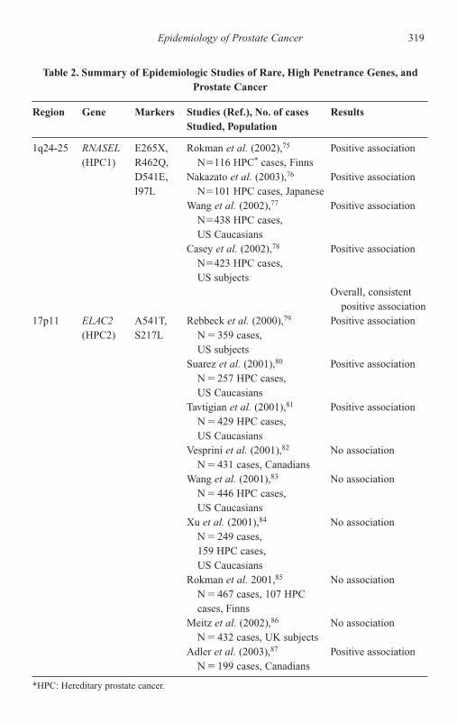
Epidemiology of Prostate Cancer 319
Table 2. Summary of Epidemiologic Studies of Rare, High Penetrance Genes, andProstate Cancer
Region Gene Markers Studies (Ref.), No. of cases ResultsStudied, Population
1q24-25 RNASEL E265X, Rokman et al. (2002),75 Positive association(HPC1) R462Q, N�116 HPC* cases, Finns
D541E, Nakazato et al. (2003),76 Positive associationI97L N�101 HPC cases, Japanese
Wang et al. (2002),77 Positive associationN�438 HPC cases, US Caucasians
Casey et al. (2002),78 Positive associationN�423 HPC cases,US subjects
Overall, consistentpositive association
17p11 ELAC2 A541T, Rebbeck et al. (2000),79 Positive association(HPC2) S217L N � 359 cases,
US subjectsSuarez et al. (2001),80 Positive association
N � 257 HPC cases,US Caucasians
Tavtigian et al. (2001),81 Positive associationN � 429 HPC cases, US Caucasians
Vesprini et al. (2001),82 No associationN � 431 cases, Canadians
Wang et al. (2001),83 No associationN � 446 HPC cases,US Caucasians
Xu et al. (2001),84 No associationN � 249 cases, 159 HPC cases,US Caucasians
Rokman et al. 2001,85 No associationN � 467 cases, 107 HPC cases, Finns
Meitz et al. (2002),86 No associationN � 432 cases, UK subjects
Adler et al. (2003),87 Positive associationN � 199 cases, Canadians
*HPC: Hereditary prostate cancer.
B245-ch14 3/2/05 4:40 PM Page 319

320 A. W. Hsing & A. P. Chokkalingam
Table 2 (Continued)
Region Gene Markers Studies (Ref.), No. of cases ResultsStudied, Population
Stanford et al. (2003),88 Positive associationN � 591 cases, US subjects
Takahashi et al. (2003),89 Positive associationN � 98 cases (BPHcontrols), Japanese
Severi et al. (2003),90 No associationN � 825 cases, Australians
Meta-analysis: Camp and Association only Tavtigian (2002)91 for HPC Overall,
weak, inconsistentassociations
May be associatedonly with HPC, not sporadic disease
xq27-28 None Linkage studies AR (also on X(HPCX) chromosome)
unlikely to beHPCXsusceptibility gene
20q13 None Linkage studies Linkage studies(HPC20) need further
confirmation1p36 None Linkage studies Most consistent
(CAPB) linkage to strong family history withearly onset disease
1q42.2-43 PCTA-1 Linkage studies PCTA is possible(PCAP) candidate gene,
but no functionalmarkers
8p22-23 MSR1 PRO3, Xu et al. (2003),92 Positive associationP275A, N � 301 cases,D174Y, US CaucasiansIVS5-59, Miller et al., 2003,93 Positive associationR293X N � 134 cases,
African-Americans
B245-ch14 3/2/05 4:40 PM Page 320

Epidemiology of Prostate Cancer 321
Table 2 (Continued)
Region Gene Markers Studies (Ref.), No. of cases ResultsStudied, Population
Wang et al. (2003),94 Null associationN � 499 cases, 438 HPCcases, US Caucasians
Seppala et al. (2003),95 Null associationN � 537 cases, Finns Overall, weak
results, withlarger studiesshowing nullassociations evenfor HPC
Rates and Patterns
Incidence
There is considerable variation in reported incidence rates of prostatecancer worldwide.5,6 Age-adjusted prostate cancer incidence rates amongAfrican-Americans are the highest in the world (185.4 per 100,000 person-years), and rates among Caucasian-Americans are second (107.8 per100,000 person-years) (Fig. 1). Reported rates in the Caribbean and inBrazil, where there are large populations of African descent (92–96 per100,000 person-years), are comparable to the high rates amongCaucasian-Americans. In contrast, in Central America and other parts ofSouth America, rates are much lower (28–42 per 100,000 person-years).Rates within Europe vary almost seven-fold (from 15–100 per 100,000person-years), with Austria having the highest reported rates. Althoughrates in Canada, Oceania (including Australia and New Zealand), WesternEurope and Scandinavia (50–103 per 100,000 person-years) are generallynot as high as the rates reported in the US, they are 2–3 times higher thanrates in Eastern Europe (15–36 per 100,000 person-years). Within Asia,where the rates are the lowest, there is also considerable variation inreported incidence, with more westernized Asian countries such as Israeland the Philippines (22–47 per 100,000 person-years) showing markedly
B245-ch14 3/2/05 4:40 PM Page 321

322A
. W. H
sing & A
. P. Chokkalingam
Table 3. Summary of Epidemiologic Studies of Common, Low Penetrance Genes and Prostate Cancer
Gene Marker Studies (Ref.), No. of Cases Studied, Population Results and Comments
Androgen Biosynthesis/Metabolism PathwayCYP17 MspA1 Lunn et al. (1999),120 N � 108 cases, US subjects Positive association for Caucasians, null for
African-AmericansWadelius et al. (1999),134 N � 178 cases, Positive association
Swedish CaucasiansGsur et al. (2000),135 N � 63 cases, Austrians Positive associationHabuchi et al. (2000),136 N � 252 cases, Japanese Positive associationHaiman et al. (2001),137 N � 600 cases, Null association
US CaucasiansYamada et al. (2001),125 N � 105 cases, Japanese Positive associationKittles et al. (2001),138 N � 71 cases, Positive association
African-AmericansLatil et al. (2001),110 N � 226 cases, Null association
French CaucasiansChang et al. (2001),139 N � 225 cases, Null association
US CaucasiansStanford et al. (2002),140 N � 596 cases, Null association overall, positive association
US Caucasians and African-Americans among Caucasians with family historyMadigan et al. (2003),141 N � 174 cases, Chinese Null associationLin et al. (2003),142 N � 93 cases, Taiwanese Null associationNam et al. (2003),119 N � 483 cases, Canadians Null associationReview: Ntais et al. (2003)98 Meta-analysis indicates no overall association,
but A2 allele may be associated with risk inAfrican-Americans.98 A1 is reported to berisk allele in Asians.
B245-ch14 3/2/05 4:40 PM Page 322

Epidem
iology of Prostate C
ancer323
Table 3 (Continued)
CYP19 TTTA repeats, Latil et al. (2001),110 N � 226 cases, Positive associationN264C French Caucasians
Modugno et al. (2001),111 N � 88 cases, Positive associationUS Caucasians
Suzuki et al. (2003),143 N � 99 HPC* cases, Positive associationJapanese
Overall, suggestive but mixed results — longerTTTA alleles associated with higher risk inCaucasians, but lower risk in Asians. Furtherinvestigation needed.
CYP1A1 2455A�G Murata et al. (1998),144 N � 115 cases, Japanese Positive associaton3801T�C Suzuki et al. (2003),145 N � 81 HPC cases, Japanese Positive association2453C�A Chang et al. (2003),146 N � 245 cases, Positive association
US CaucasiansCYP3A4 5� promoter Rebbeck et al. (1998),147 N � 230 cases, Positive association
variant US CaucasiansParis et al. (1999),148 N � 174 cases, Positive association
African-AmericansNam et al. (2003),119 N � 483 cases, Canadians Null association
SRD5A2 V89L, A49T, Lunn et al. 1999,120 N � 108 cases, US Null associationR227Q, TA Caucasians and African-Americansrepeats Kantoff et al. (1997),121 N � 590 cases, US Null association
CaucasiansFebbo et al. (1999),122 N � 592 cases, US Null association
CaucasiansMakridakis et al. (1999),123 N � 388 cases, US Positive association
Hispanics and African-Americans
*HPC: Hereditary prostate cancer.
B245-ch14 3/2/05 4:40 PM Page 323

324A
. W. H
sing & A
. P. Chokkalingam
Table 3 (Continued)
Gene Marker Studies (Ref.), No. of Cases Studied, Population Results and Comments
Margiotti et al. (2000),124 N � 108 cases, Italians Positive associationYamada et al. (2001),125 N � 105 cases, Japanese Null associationNam et al. (2001),126 N � 158 cases, Canadians Positive associationLatil et al. (2001),110 226 cases, French Null associationMononen et al. (2001),127 N � 449 cases, Finns Null associationHsing et al. (2001),128 N � 191 cases, Chinese Null associationPearce et al. (2002),129 N � 921 cases, US subjects Null associationSoderstrom et al. (2002),130 N � 176 cases, Swedes Null associationLamharzi et al. (2003),131 N � 300 cases, US subjects Positive associationChang et al. (2003),132 N � 245 cases, 159 HPC cases, Null association
US CaucasiansLi et al. (2003),133 N � 302 cases, Japanese Positive associationNam et al. (2003),119 N � 483 cases, Canadians Null associationReview: Ntais et al. (2003)96 Overall, the T allele of A49T (associated with
higher enzymatic activity) and shorter TArepeats may be associated with a modestincrease in risk.96
While results are mixed, the V89L marker’s LL genotype, which is associated with lower serum levels of androgens, may be associated with a reduced risk.
R227Q is very rare, observed only in Asians.
B245-ch14 3/2/05 4:40 PM Page 324

Epidem
iology of Prostate C
ancer325
Table 3 (Continued)
AR CAG repeats, Ingles et al. (1997),100 N � 57 cases, US Caucasians Positive associationGGN Stanford et al. (1997),101 N � 301 cases, US Positive associationrepeats Caucasians
Giovannucci et al. (1997),102 N � 587 cases, US Positive associationCaucasians (and Platz et al. (1998),103 N � 582 cases)
Correa-Cerro et al. (1999),104 N � 132 cases, Null associationFrench and Germans
Ekman et al. (1999),105 N � 93 cases, 59 HPC cases, Positive associationSwedes and Japanese
Edwards et al. (1999),106 N � 178 cases, Positive associationU.K. Caucasians
Hsing et al. (2000),107 N � 190 cases, Chinese Positive associationMiller et al. (2001),108 N � 140 cases, US subjects Null associationBeilin et al. (2001),109 N � 445 cases, Australians Null associationLatil et al. (2001),110 N � 226 cases, French Null associationModugno et al. (2001),111 N � 88 cases, US Positive association
CaucasiansChang et al. (2002),112 N � 245 cases, 159 HPC cases Positive associationMononen et al. (2002),113 N � 449 cases, Finns Positive associationGsur et al. (2002),114 N � 190 cases, Austrians Null associationChen et al. (2002),115 N � 300 cases, US subjects Null associationBalic et al. (2002),116 N � 82 cases, Hispanics Positive associationSantos et al. (2003),117 N � 133 cases, Brazilians Null associationHuang et al. (2003),118 N � 66 cases, Taiwanese Null associationNam et al. (2003),119 N � 483 cases, Canadians Null association
Although overall results are mixed, shorterCAG repeat lengths may be associatedwith increased prostate cancer risk.
B245-ch14 3/2/05 4:40 PM Page 325

326A
. W. H
sing & A
. P. Chokkalingam
Table 3 (Continued)
Gene Marker Studies (Ref.), No. of Cases Studied, Population Results and Comments
HSD3B1 N367T, Chang et al. (2002),149 N � 245 cases, 159 HPC Positive associationc7062t cases, US Caucasians
HSD3B2 c7159g, Chang et al. (2002),149 N � 245 cases, 159 HPC Null associationc7474t cases, US Caucasians
HSD17B3 G289S Margiotti et al. (2002),150 N � 103 cases, Italians Positive association
Growth Factors and Non-androgenic Hormone PathwaysVDR BsmI, TaqI, Taylor et al. (1996),154 N � 108 cases, US Caucasians Positive association
polyA, ApaI, Ingles et al. (1997),100 N � 57 cases, US Caucasians Positive associationFokI Ingles et al. (1998),155 N � 151 cases, Null association
African-AmericansMa et al. (1998),156 N � 372 cases, US Caucasians Null associationCorrea-Cerro et al. (1999),157 N � 131 cases, Europeans Null associationHabuchi et al. (2000),158 N � 222 cases, Japanese Positive associationFuruya et al. (1999),159 N � 66 cases, Japanese Null associationWatanabe et al. (1999),160 N � 100 cases, Japanese Null associationBlazer et al. (2000),161 N � 77 cases, US Caucasians Null associationChokkalingam et al. (2001),162 N � 191 cases, Chinese Null associationGsur et al. (2002),163 N � 190 cases, Austrians Null associationHamasaki et al. (2002),164 N � 110 cases, Japanese Positive association for aggressive diseaseMedieros et al. (2002),165 N � 163 cases, Portugese Positive association for late-onset diseaseSuzuki et al. (2003),166 N � 81 HPC cases, Japanese Null associationNam et al. (2003),119 N � 483 cases, Canadians Null association
B245-ch14 3/2/05 4:40 PM Page 326
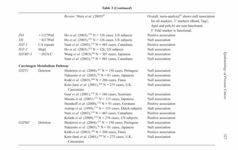
Epidem
iology of Prostate C
ancer327
Table 3 (Continued)
Review: Ntais et al. (2003)97 Overall, meta-analysis97 shows null associationfor all markers. 3� markers (BsmI, Taq1,ApaI and polyA) are non-functional,5� FokI marker is functional.
INS �1127PstI Ho et al. (2003),153 N � 126 cases, US subjects Positive associationTH 4217PstI Ho et al. (2003),153 N � 126 cases, US subjects Null associationIGF-1 CA repeats Nam et al. (2003),119 N � 483 cases, Canadians Positive associationIGF-2 MspI Ho et al. (2003),153 N � 126, US subjects Null associationIGFBP-3 202A/C Wang et al. (2003),94 N � 307 cases, Japanese Null association
Nam et al. (2003),119 N � 483 cases, Canadians Null association
Carcinogen Metabolism PathwayGSTT1 Deletion Medeiros et al. (2004),167 N � 150 cases, Portugese Null association
Nakazato et al. (2003),76 N � 81 cases, Japanese Null associationKidd et al. (2003),168 N � 206 cases, Finns Null associationKote-Jarai et al. (2001),169 N � 275 cases, U.K. Null association
CaucasiansGsur et al. (2001),170 N � 166 cases, Austrians Null associationMurata et al. (2001),171 N � 115 cases, Japanese Null associationSteinhoff et al. (2000),172 N � 91 cases, Germans Positive associationAutrup et al. (1999),173 N � 153 cases, Dutch subjects Null associationNam et al. (2003),119 N � 483 cases, Canadians Positive associationKelada et al. (2000),174 N � 276 cases, US subjects Positive association
GSTM1 Deletion Medeiros et al. (2004),167 N � 150 cases, Portugese Null associationNakazato et al. (2003),76 N � 81 cases, Japanese Null associationKidd et al. (2003),168 N � 206 cases, Finns Positive associationKote-Jarai et al. (2001),169 N � 275 cases, U.K. Null association
Caucasians
B245-ch14 3/2/05 4:40 PM Page 327

328A
. W. H
sing & A
. P. Chokkalingam
Table 3 (Continued)
Gene Marker Studies (Ref.), No. of Cases Studied, Population Results and Comments
Gsur et al. (2001),170 N � 166 cases, Austrians Null associationMurata et al. (2001),171 N � 115 cases, Japanese Positive associationSteinhoff et al. (2000),172 N � 91 cases, Germans Null associationAutrup et al., 1999,173 N � 153 cases, Dutch subjects Null associationNam et al. (2003),119 N � 483 cases, Canadians Null associationKelada et al. (2000),174 N � 276 cases, US subjects Null association
GSTM3 Medeiros et al. (2004),167 N � 150 cases, Portugese Positive associationGSTP1 I105V Nakazato et al. (2003),76 N � 81 cases, Japanese Positive association
Kidd et al. (2003),168 N � 206 cases, Finns Null associationKote-Jarai et al. (2001),169 N � 275 cases, U.K. Positive association
CaucasiansGsur et al. (2001),170 N � 166 cases, Austrians Positive associationSteinhoff et al. (2000),172 N � 91 cases, Germans Null associationShepard et al. (2000),175 N � 590 cases, US Null association
CaucasiansAutrup et al. (1999),173 N � 153 cases, Dutch Null associationWadelius et al. (1999),134 N � 850 subjects, Swedes Null association
and DanesNam et al. (2003),119 N � 483 cases, Canadians Null association
NAT2 Wadelius et al. (1999),134 N � 850 subjects, Swedes Null associationand Danes
DNA Repair PathwayXRCC1 R399Q, Rybicki et al. (2004),177 N � 637 cases, US Null association
R194W, CaucasiansR280H van Gils et al. (2002),178 N � 77 cases, US subjects Positive association
B245-ch14 3/2/05 4:40 PM Page 328

Epidem
iology of Prostate C
ancer329
Table 3 (Continued)
XPD D312N, Rybicki et al. (2004),177 N � 637 cases, US Positive association, needs furtherK751Q Caucasians investigation
hOGG1 S326C, Xu et al. (2002),179 N � 245 cases, US Caucasians Positive association�11657A/G Chen et al. (2003),180 N � 84 cases, US Caucasians Positive association
Inflammation/Angiogenesis/Cytokine PathwaysVEGF VEGF-1154, McCarron et al. (2002),183 N � 247 cases, U.K. Positive association
VEGF-460 CaucasiansLin et al. (2003),142 N � 96 cases, Taiwanese Positive association
TNF-� TNF-�-308 McCarron et al. (2002),183 N � 247 cases, U.K. Null associationCaucasians
IL-1-� IL-1�-511 McCarron et al. (2002),183 N � 247 cases, U.K. Null associationCaucasians
IL-8 IL-8-251 McCarron et al. (2002),183 N � 247 cases, U.K. Positive associationCaucasians
IL-10 IL-10-1082 McCarron et al. (2002),183 N � 247 cases, U.K. Positive associationCaucasians
PPAR- P12A Paltoo et al. (2003),184 N � 193 cases, Finns Null associationTGF-� L10P Li et al. (2004),181 N � 351 cases, Japanese Positive associationCOX-2 1285A/G Panguluri et al. (2004),182 N � 288, 264 and 184 cases, Positive association in all ethnic groups
1265G/A African-Americans, Nigerians, and US Caucasians899G/C297C/G
B245-ch14 3/2/05 4:40 PM Page 329

330 A. W. Hsing & A. P. Chokkalingam
Fig. 1. Age-adjusted incidence rates (per 100,000 person-years) for prostate cancer in48 countries, 1993–1997.
Source: Parkin DM, Whelan SL, Ferlay J, Teppo L, and Thomas DB. Cancer Incidence in Five Continents, Vol VIII, IARC Sci Publ 155, 2003.
South AMERICA
North AMERICA
OCEANIA
EUROPE
ASIA
AFRICA
B245-ch14 3/2/05 4:40 PM Page 330

higher rates than Thailand, India, Pakistan and Shanghai, China (3–7 per100,000 person-years). There are few data on incidence in Africa, withonly four registries included in the IARC report.6 The rates within theAfrican continent vary widely, from 5–37 per 100,000 person-years. Partof the difference in incidence rates in various countries is related to theextent of prostate cancer screening, especially the use of prostate-specificantigen (PSA) testing. However, since screening is less common in devel-oping countries, it is not likely to explain the nearly 60-fold difference inprostate cancer risk between high- and low-risk populations.
Mortality
In the US, only one in six men diagnosed with prostate cancer willeventually die from it. Nevertheless, 29,900 prostate cancer deaths areexpected in 2004, making prostate cancer the second leading cause of cancerdeath among US men after lung cancer.7 Age-adjusted prostate cancer mor-tality rates from 38 countries in 1998 are shown in Fig. 2. Overall, mortalitypatterns mimic those of incidence in various countries, although mortalityrates show less diversity worldwide than do incidence rates, but are stillhigher in Western nations than in lower-risk, Asian countries (Fig. 2). Of spe-cial interest is the observation that the Caribbean nations of Barbados, theBahamas and Trinidad and Tobago, where there are large populations of menof African descent, had the world’s highest mortality rates (30.3 to 47.9 per100,000 person-years). Mortality was higher in Scandinavian countries andparts of northern Europe than in the US (18.7–23.6 versus 14.0 per 100,000person-years), and lowest of all in the Asian countries of South Korea,Philippines and Japan (1.6–4.4 per 100,000 person-years).
Risk Factors
Demographic Factors
Age
Over 80% of prostate tumors in the US are diagnosed among men overage 65,8 and the incidence of prostate cancer increases exponentially
Epidemiology of Prostate Cancer 331
B245-ch14 3/2/05 4:40 PM Page 331

332 A. W. Hsing & A. P. Chokkalingam
Fig. 2. Age-adjusted mortality rates (per 100,000 person-years) for prostate cancer in38 countries, 1998.
Source: http://www-depdb.iarc.fr/who/menu.htm* Rates are from 1994
South AMERICA
North AMERICA
OCEANIA
EUROPE
ASIA
, ,
B245-ch14 3/2/05 4:40 PM Page 332

with advancing age — an increase that is faster than that for any othermalignancy (Table 1). Estimates from the Surveillance, Epidemiology,and End Results (SEER) program from 1996–2000 indicate that for USmen under 65 years of age and 65 years and over, age-adjusted prostatecancer incidence rates were 56.8 and 974.7 per 100,000 person-years,respectively.2
Racial/Ethnic Variation
Another consistently observed but poorly understood risk factor is ethnic-ity. African-Americans have the highest incidence rate in the world,roughly 60 times that of the ethnic group with the world’s lowest rates, inShanghai, China1 (Fig. 1).
Adjustment of incidence rates for prevalence of latent disease atautopsy and proportion of localized tumors among all cancers of theprostate revealed that Japanese men still experience a markedly lowerincidence than Americans, indicating that the international variation can-not be explained by differences in detection alone.9 This bolsters theresults of migrant studies suggesting that ethnic factors, includinggenetic, lifestyle, and environmental factors, may affect prostate cancerrisk and explain many of the differences in risk between high- and low-risk populations.9,10
Hormonal, Behavioral and Lifestyle Factors
Hormones and Growth Factors
Androgens play a key role in the development and maintenance of theprostate gland; however, the precise role of androgens in the etiology ofprostate cancer is unclear. Prostate cancer is notably absent in castratedmen, and laboratory studies show that administration of testosteroneinduces prostate cancer in rats and that androgens promote cell prolifera-tion and inhibit prostate cell death.11–13 However, epidemiologic datasupporting a role of androgens are inconclusive.14–16 To date, over 13prospective studies have investigated the role of circulating androgens,and only one was able to show that men with higher serum testosterone
Epidemiology of Prostate Cancer 333
B245-ch14 3/2/05 4:40 PM Page 333

levels have a higher risk of prostate cancer.17 More comprehensivereviews of this topic are reported elsewhere.14–16 Studies of genetic mark-ers involved in the androgen pathways offer further insight into thisavenue of research, and are reviewed later in this chapter.
In addition to androgens, insulin-like growth factors (IGFs), insulinand vitamin D have been implicated in prostate cancer. IGF-I and IGF-IIare polypeptides that function as both tissue growth factors and endocrinehormones with mitogenic and anti-apoptotic effects on prostate epithelialcells. There are at least six known IGF binding proteins (IGFBPs) that canbind to IGFs and thus prevent activation of the IGF receptor, which medi-ates IGF effects. At least nine epidemiologic studies have evaluated theroles of the IGF axis in prostate cancer, and most have reported a positiveassociation with IGF-I and an inverse association with IGFBP3.18,19
However, the role of IGF-II is less clear.Vitamin D is a steroid hormone obtained primarily from dermal syn-
thesis in response to sunlight exposure. Vitamin D and its analogs havepotent anti-proliferative, pro-differentiative, and pro-apoptotic effects onprostate cancer cells. In addition, vitamin D inhibits prostate tumor growthin vivo. In general, laboratory data are consistent and support the hypothe-sis that vitamin D may protect against prostate cancer. However, resultsfrom epidemiologic studies investigating serum vitamin D levels have beeninconsistent.20 The reasons for these conflicting results are unclear.
Diet
Ecologic studies have shown a strong correlation between the incidence ofprostate cancer and dietary fat intake.21 A western diet has been linked toa higher risk of prostate cancer, and it has been suggested that the westerndiet, high in fat, increases production and availability of both androgenand estrogen, while Asian (low-fat, high-fiber) and vegetarian diets leadto lower circulating levels of these hormones.21
Fat is the most studied dietary factor in relation to prostate cancer.Most epidemiologic studies have investigated the role of total, saturated,and/or animal fat. Findings from these studies suggest a possible positiveassociation with monounsaturated, animal and saturated fats, and aninverse association with omega-3 fat. The results for polyunsaturated fat are
334 A. W. Hsing & A. P. Chokkalingam
B245-ch14 3/2/05 4:40 PM Page 334

less consistent.22,23 Consumption of meat, particularly red meat, is also con-sistently linked to an increased risk of prostate cancer. However, it is unclearwhether the excess risk is due to the fat content in red meat, mutagens suchas heterocyclic amines that are induced during high-temperature cooking ofmeat products, animal proteins, or other unidentified factors.24
Several epidemiologic studies have also investigated whether intake offatty fish, rich in potentially tumor-inhibitory marine fatty acids, is associ-ated with reduced prostate cancer risk. However, a recent review of 17 stud-ies, including eight prospective studies, found suggestive but inconsistentresults, possibly due to inadequate assessment of fish intake or lack of infor-mation on specific marine fatty acids, particularly the polyunsaturated fattyacids eicosapentaenoic and docosahexaenoic acids,25 in these studies.
Although consumption of fruits and vegetables is associated with areduced risk of several cancers, their role in prostate cancer is less clear.The only consistent finding is an inverse association with consumptionof tomatoes and tomato paste, which has been largely attributed to theantioxidant effect of lycopene.26 Cruciferous and allium vegetables havebeen implicated. A recent review concluded that there is modest evidencethat intake of cruciferous vegetables, including broccoli, cabbage, cauli-flower, and Brussels sprouts, is inversely associated with prostate cancerrisk, possibly due to their content of isothiocyanates.27 Intake of alliumvegetables, including onions, garlic, and chives, was associated with areduced risk in a case-control study in China.28 This protective effect maybe due to the tumor inhibitory properties of organosulfur compounds.
Dietary calcium, from either dairy intake or supplements, has alsobeen linked to prostate cancer. Because of its role in regulation of vita-min D synthesis, calcium may down-regulate vitamin D’s anti-proliferativeeffects on prostate cancer. However, the epidemiologic evidence for cal-cium is as yet unclear, complicated by differences in assessment of cal-cium (dietary intake versus circulating levels).29 Recent data suggest athreshold effect in that only very high calcium intake (� 2000 mg/day)appears to be associated with disease.30
Chronic excess of zinc, another mineral obtained largely throughdietary supplements, may be positively associated with prostate cancerrisk, although in vitro studies demonstrating mitogenic effects of zinc onprostate cancer suggest that it may reduce risk.31
Epidemiology of Prostate Cancer 335
B245-ch14 3/2/05 4:40 PM Page 335

A large body of epidemiological evidence, including observational,case-control, cohort and randomized controlled clinical trials, supports thehypothesis that selenium may prevent prostate cancer in humans.32
Molecular data show that selenium prevents clonal expansion of tumorsby causing cell cycle arrest, promoting apoptosis, and modulating p53-dependent DNA repair mechanisms. Clinical trials have also shown thatvitamin E supplementation is associated with a reduced risk of prostatecancer.33,34 Currently a clinical trial is under way to test the chemopre-ventive efficacy of these two compounds.35
Obesity
In epidemiologic studies, overall obesity is usually measured by bodymass index (weight in kg divided by the square of height in meters, kg/m2)and abdominal obesity by the ratio of waist to hip circumference. Thefindings on overall obesity are mixed. However, recent data suggest thatabdominal obesity may be associated with an increased risk of prostatecancer even in relatively lean men.36,37 In addition, higher serum levels ofinsulin were associated with an increased risk of prostate cancer inChina,38 and higher serum levels of leptin were associated with largertumor volume (� 5 cm3).39 Although the role of obesity in prostate canceris not clearly defined, future studies should attempt to clarify it furtherbecause obesity is linked to numerous putative risk factors for prostatecancer, including high intakes of meat and fat intake, hormone metabolism,and serum level IGFs and insulin. Furthermore, the prevalence of obesitycorrelates with prostate cancer risk across populations. It is likely thatobesity may thus provide a link between westernization and increasedprostate cancer risk. With the epidemic of obesity in both developed anddeveloping countries, the role of obesity needs to be clarified further.
Physical Activity
Physical activity may decrease levels of total and free testosterone, reduceobesity, and enhance immune function,40 all of which may lead to protec-tion from prostate cancer. However, perhaps due to challenges in classify-ing physical activity and/or identifying the age/time period at which
336 A. W. Hsing & A. P. Chokkalingam
B245-ch14 3/2/05 4:40 PM Page 336

activity may be most protective, results from numerous epidemiologicstudies are equivocal.40,41
Occupation
Occupation is highly correlated with socioeconomic status and life-style factors. There is a large body of literature on prostate cancer andoccupation, and one consistent result from these studies is that farmersand other agricultural workers have a 7–12% increased risk.42,43 Whilethis excess could reflect lifestyle factors such as increased intake of meatand fats, chemical exposures may also play a role. These chemicals, whichhave a wide variety of poorly characterized effects, may include fertiliz-ers, solvents, pesticides and herbicides.44 Organochlorines present inmany pesticides and herbicides can affect circulating hormone levels;however the epidemiologic evidence linking specific pesticide or herbi-cide exposures to prostate cancer is weak. In addition to agriculture, work-ers in the heavy metals industry, rubber manufacturing, and newspaperprinting may be at elevated risk,42 suggesting that exposure to certainchemicals common in these work environments may increase the risk ofprostate cancer.
Vasectomy
Several, but not all, studies investigating the association between vasectomyand prostate cancer risk suggest a modest positive association. The role ofvasectomy remains controversial, however, since most studies are unable toexclude the possible effect of detection bias: men undergoing vasectomies aremore likely to have prostate cancer detected than men who do not. Vasectomyis linked to elevations in anti-spermatozoa antibodies, decreased seminal hor-mone concentrations and decreased prostatic secretion.45 Whether these con-ditions can influence prostate carcinogenesis needs to be clarified.
Chronic Inflammation
Evidence for chronic inflammation and prostate cancer is just emerging,46
but an association of prostate cancer with chronic inflammation of theprostate (chronic prostatitis) has long been suspected. Inflammation is
Epidemiology of Prostate Cancer 337
B245-ch14 3/2/05 4:40 PM Page 337

frequently found in prostate biopsy specimens obtained from both radicalprostatectomy and surgical treatment for BPH,47,48 however, epidemio-logic findings have been mixed. A recent meta-analysis of 11 studies ofprostatitis and prostate cancer reported an overall relative risk of 1.6.49
Results from pathologic and molecular surveys suggest that the earlieststages of prostate cancer may develop in lesions generally associated withchronic inflammation.50,51 De Marzo et al. showed that almost all formsof focal prostatic glandular atrophy, thought to be precursors of prostaticadenocarcinoma, are proliferative, and that such proliferative inflammatoryatrophy (PIA) lesions often contain inflammatory infiltrates and arefrequently found adjacent to or near high-grade prostatic intraepithelialneoplasia (PIN).50,51 Inflammation may lead to tumorigenesis by stimulat-ing angiogenesis, enhancing cell proliferation, and damaging DNA throughradical oxygen species such as nitric oxide.
Additional support for a role for chronic inflammation in prostatecancer comes from the observation that a higher intake of fish and useof aspirin and other non-steroidal anti-inflammation drugs (NSAIDs)has been associated with reduced prostate cancer risk.52 In two largeprospective studies, higher intake of fish was associated with a lower riskof total prostate cancer and metastatic prostate cancer.53,54 Abundant infatty fish, omega-3 fatty acids are known antagonists of arachidonicacid and suppress the production of pro-inflammatory cytokines.55 Inaddition, use of anti-inflammatory agents, especially NSAIDs such asibuprofen or aspirin, has been related to lower prostate cancer risk in epi-demiologic studies,56–58 and a recent meta-analysis of 12 of these studiesconcluded that aspirin use was associated with a 15% reduction in prostatecancer risk.59 Taken together, these data suggest chronic inflammation mayincrease the risk of prostate cancer. However, there are few epidemiologicstudies investigating this directly, possibly due to the difficulty in diagnos-ing chronic prostatitis and in measuring cytokine levels reliably in serumsamples. This is likely to be a fruitful area for future research.
Sexually Transmitted Diseases
Chronic inflammation induced by bacterial or viral agents has been impli-cated as a potential underlying mechanism for the link between STDs and
338 A. W. Hsing & A. P. Chokkalingam
B245-ch14 3/2/05 4:40 PM Page 338
prostate cancer. One recent large, population-based study showed two- tothree-fold increased prostate cancer risks associated with STDs, particu-larly syphilis and recurrent gonorrhea infections.60 Other studies reportedassociations of human papillomavirus-16, -18 and -33 serology with anincreased risk of prostate cancer.61,62 In addition, epidemiological dataare accumulating to suggest that sexual history may be associated withprostate cancer risk,63 and a recent meta-analysis of 17 studies concludedthat increased sexual frequency and number of partners are associatedwith increased prostate cancer risk.49
Benign Prostatic Hyperplasia
The relationship between BPH and prostate cancer is not well established.BPH is currently not considered a precursor to prostate cancer, sinceprostate cancer occurs mostly in the peripheral zone of the prostate andBPH is more common in the transition and periurethral zones. However,because both conditions are common in elderly men, and because theymay coexist within the prostate, they appear to share risk profiles, makingit difficult to elucidate the independent role, if any, of BPH in prostatecancer etiology. Detection bias also complicates investigation: excessprostate cancer risk in men who are symptomatic for BPH may be simplya reflection of the increased intensity of evaluation and medical surveil-lance in such patients. In addition, in most epidemiologic studies, it hasbeen difficult to completely rule out the presence of BPH in controlpopulations, since the prevalence of BPH is very common in elderly men.Due in part to these limitations, the epidemiologic evidence for BPH asa risk factor for prostate cancer remains weak and inconsistent,64 withthe largest study to date (over 85,000 BPH patients) showing only a mar-ginally elevated risk of prostate cancer versus the general population(� 2% in 10 years).65
Other Factors
Several other risk factors, such as smoking, use of alcohol, diabetes andliver cirrhosis, have been investigated, but their roles in prostate cancer areweak or unclear based on data in the current literature.66–68
Epidemiology of Prostate Cancer 339
B245-ch14 3/2/05 4:40 PM Page 339

Genetic Factors
Family History of Cancer
Prostate cancer etiology has a hereditary component. Numerous studieshave consistently reported familial aggregation of prostate cancer,showing a two- to three-fold increased risk of prostate cancer among menwho have a first-degree male relative (father, brother, son) with a historyof prostate cancer.69 Recent data from a large twin study suggests that asmuch as 42% (95% CI 29–50%) of the risk of prostate cancer may beaccounted for by genetic factors.70 Genetic factors involved in prostatecancer include individual and combined effects of rare, highly penetrantgenes, more common weakly penetrant genes and genes acting in concertwith each other.
High-Penetrance Markers
Segregation and linkage analyses have shown that certain early-onsetprostate cancers may be inherited in an autosomal dominant fashion,71 andit is estimated that such hereditary prostate cancers (HPCs) due to highlypenetrant genes may account for about 10% of all prostate cancer cases.70
Several family studies are currently underway to identify hereditaryprostate cancer candidate genes. However, these investigations haveproven to be difficult for several reasons.72 One is that, due to the highincidence of prostate cancer and the heterogeneity of tumors, it is possiblethat sporadic cases are included in HPC families, thereby reducing thestatistical power to detect genes for HPC. In addition, because prostatecancer is generally diagnosed at a late age, it is often impossible to obtainDNA specimens from fathers of HPC cases, and sons of HPC cases areoften too young to have developed prostate cancer. Therefore, studies ofHPC families are often unable to include more than one generation. Finally,the genetic heterogeneity of prostate cancer makes it difficult to deviseappropriate statistical transmission models that also account for multiplesusceptibility genes, many of which may be at only moderate penetrance.Despite these challenges, seven loci have been described to date, includingHPC1, ELAC2, HPCX, HPC20, CAPB, PCAP, and an unnamed locus at8p22-23 (Table 2), and fine mapping has led to the identification of a
340 A. W. Hsing & A. P. Chokkalingam
B245-ch14 3/2/05 4:40 PM Page 340
number of candidate genes, including RNASEL, ELAC2 and MSR-1.73,74
The results of studies of these loci,75–95 which have been extensivelyreviewed elsewhere,73 have largely been mixed, with subsequent studiesfailing to replicate promising earlier findings. The absence of strong,consistent results for high penetrance markers strongly suggests that theheritable component of prostate cancer largely comprises effects of multi-ple factors, including common, weakly penetrant markers, possibly inter-acting with one another and with environmental factors.
Common Low-Penetrance Markers
Results of epidemiologic studies of common polymorphisms are sum-marized below and in Table 3 by biological pathway; several of these mark-ers have been reviewed elsewhere.73,96–99 In reviewing these results, it isimportant to note that, as with any other epidemiologic exposure, replica-tion of findings is critical to establishing causality. This is particularly trueof genetic association studies, because the recent explosion of genetic datahas increased the potential for publication bias as investigators and pub-lishers become more selective about writing up and publishing findings.
Androgen Biosynthesis and Metabolism Pathway
Because prostate cancer is an androgen-dependent tumor, it is likelythat markers in genes whose gene products are involved in androgenbiosynthesis and metabolism (Fig. 3) may be associated with disease.Recent epidemiologic studies have investigated the role of polymor-phisms of over 10 genes involved in androgen biosynthesis, metabolism,transport, and regulation. These data are promising and accumulatingat a remarkable pace but still are too sparse to support a role for anyparticular gene.
Results for the androgen receptor (AR), which is involved in androgenbinding and transport, are fairly consistent, showing that shorter CAGrepeat lengths are associated with increased risk in most, but not all, pop-ulations.100–119 For the type II steroid 5�-reductase (SRD5A2), which con-verts testosterone to the more active androgen dihydrotestosterone, theresults are mixed,110,119–133 with a recent meta-analysis showing modest
Epidemiology of Prostate Cancer 341
B245-ch14 3/2/05 4:40 PM Page 341
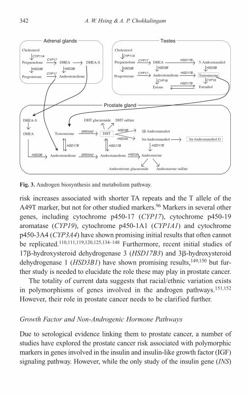
342 A. W. Hsing & A. P. Chokkalingam
Fig. 3. Androgen biosynthesis and metabolism pathway.
Cholesterol
Pregnenolone
Progesterone
CYP11ACYP17 DHEA 5-Androstenediol
Testosterone
Estradiol
Androstenedione
Estone
CYP19
CYP17 HSD17B
Testosterone DHT
AndrostanedioneAndrostenedione
HSD17B
HSD3A Androsterone
Androsterone glucuronide Androsterone sulfate
DHT sulfateDHT glucuronide
HSD3B 3β-Androstanediol
HSD3A 3α-Androstanediol 3α-Androstanediol G
HSD17B
DHEA-S
DHEA
HSD3B
Cholesterol
Pregnenolone
Progesterone
DHEACYP17
Androstenedione
HSD3B
CYP11A
CYP17
DHEA-S
HSD3B HSD3B
HSD17B
HSD3B
HSD17BCYP19
HSD3B
SRD5A2
HSD17B
Adrenal glands
SRD5A2
Prostate gland
Testes
risk increases associated with shorter TA repeats and the T allele of theA49T marker, but not for other studied markers.96 Markers in several othergenes, including cytochrome p450-17 (CYP17), cytochrome p450-19aromatase (CYP19), cytochrome p450-1A1 (CYP1A1) and cytochromep450-3A4 (CYP3A4) have shown promising initial results that often cannotbe replicated.110,111,119,120,125,134–148 Furthermore, recent initial studies of17�-hydroxysteroid dehydrogenase 3 (HSD17B3) and 3�-hydroxysteroiddehydrogenase 1 (HSD3B1) have shown promising results,149,150 but fur-ther study is needed to elucidate the role these may play in prostate cancer.
The totality of current data suggests that racial/ethnic variation existsin polymorphisms of genes involved in the androgen pathways.151,152
However, their role in prostate cancer needs to be clarified further.
Growth Factor and Non-Androgenic Hormone Pathways
Due to serological evidence linking them to prostate cancer, a number ofstudies have explored the prostate cancer risk associated with polymorphicmarkers in genes involved in the insulin and insulin-like growth factor (IGF)signaling pathway. However, while the only study of the insulin gene (INS)
B245-ch14 3/2/05 4:40 PM Page 342

Epidemiology of Prostate Cancer 343
has shown promising results, early studies of markers in the IGF-II and IGFbinding protein-3 (IGFBP-3) genes have shown null results.94,119,153
Strong laboratory evidence showing chemoprotection of vitamin Dagainst prostate cancer, in addition to suggestive but inconsistent sero-epidemiological studies, has led to numerous studies of the vitamin Dreceptor gene (VDR).100,119,154–166 However, despite promising early stud-ies, a recent comprehensive meta-analysis showed no overall associationsand concluded that markers in the VDR gene are unlikely to be majorgenetic determinants of prostate cancer risk.97
Carcinogen Metabolism Pathway
Genes encoding enzymes that metabolize carcinogens and other toxinsmay play a role in prostate cancer. However, results from several studiesof markers in different glutathione-S-transferases (GSTs), includingGSTT1, GSTP1 and GSTM1, have mostly been null.76,119,134,167–175 Recentinitial epidemiologic studies of other genes in these pathways, includingGSTM3 and N-acetyl transferase 2 (NAT2), have been positive but requireconfirmation.134,167
DNA Repair Pathway
The DNA repair pathway serves to prevent disruptions in DNA integritythat might otherwise lead to gene rearrangements, translocations, ampli-fications and deletions that may contribute to cancer development.176
Initial reports of markers in genes encoding DNA repair enzymes,including the X-ray repair cross-complementing group (XRCC1), human8-oxoguanine glycosylase I (hOGG1) and the xeroderma pigmentosumgroup D (XPD), show promising results.177–180 These results, combinedwith strong biological plausibility, suggest that this may be a fruitful areafor further research.
Chronic Inflammation Pathway
Several lines of evidence point to a role of inflammation in prostate can-cer etiology, and studies of markers in the genes involved in inflammationare emerging.46 Initial studies show positive results for transforming
B245-ch14 3/2/05 4:40 PM Page 343

growth factor-� (TGF-�) and COX-2181,182 and negative results for tumornecrosis factor-�-308 (TNF-�-308), interleukin-1� (IL-1�) and peroxi-some proliferator-activated receptor- (PPAR-).183,184 Evidence for a roleof inflammation markers in prostate cancer is increasing. Given thebiological plausibility of this hypothesis, this should be a fruitful area forfuture research.
Angiogenesis Pathways
The need for increased vasculature to support cancer growth is an area ofresearch that is currently gaining momentum. Genetic investigations ofangiogenesis in prostate cancer have thus far involved the vascularendothelial growth factor (VEGF) gene as well as the genes for IL-8 andIL-10, and the handful of studies conducted to date have shown positiveresults.142,183 These findings await further confirmation and support thenotion that angiogenesis may indeed be involved in prostate cancer.
Biological Pathways Related to Dietary Factors
It is clear that genetic susceptibility to both Phase I and II enzymes(cytochrome p450) affects the association between certain dietary factorsand prostate cancer risk. For example, the effect of cruciferous vegetablesis related to both their high glycosinolate content and functional variationsin enzymes, particularly GSTM1 and GSTT1, that metabolize glycosino-lates to isothiocyanates (ITCs).27 Thus, to better assess the role of ITCs inprostate cancer, studies with both comprehensive and reliable assessmentof cruciferous vegetable intake and genetic polymorphisms in GSTM1 andGSTT1 will be required. Moreover, genetic polymorphisms in receptorsand transcription factors that interact with these compounds may con-tribute to variations in response to cruciferous vegetable intake. With suf-ficiently large sample size and careful assessment of diet and geneticfactors, this important area should be investigated further.
Challenges of Studies with Common Polymorphisms
Currently, the totality of data suggests that racial/ethnic variation existsin common polymorphisms of certain genes, such as the SRD5A2, AR, and
344 A. W. Hsing & A. P. Chokkalingam
B245-ch14 3/2/05 4:40 PM Page 344

ELAC2/HPC2, but few variants or genes have been firmly shown to con-tribute to prostate cancer susceptibility. Challenges in molecular epidemi-ology studies of common polymorphisms include the selection of relevantsingle nucleotide polymorphisms (SNPs) for genotyping and the diffi-culty in replicating results. The difficulty in replicating earlier findings insubsequent association studies is due, in part, to (1) the relatively small tomodest effects of most common polymorphisms, ranging from 10 to 80%,(2) the relatively small sample size in most previous studies, ranging from100 to 500 cases, and the limited power of these studies to detect a modesteffect on the order of 10 to 50%, (3) the tendency of small studies toproduce false positive findings, and (4) differences in study design andpopulations, including differences in the severity of cases. Thus, studieswith large sample size (�1000 cases) are needed to clarify further therole of these polymorphic markers. In addition, it is becoming clear thata single gene or SNP alone is unlikely to explain most of the variation inprostate cancer susceptibility, thereby requiring even larger sample sizes(�3000 cases) to evaluate the effect of multiple variants.
Another challenge in epidemiologic studies investigating the role ofgenetic variants in complex disease (e.g., prostate cancer) is the limitedability to identify “causal SNPs” through association studies. This is partlyrelated to two factors, (1) the difficulty in selecting biologically relevantSNPs for genotyping and (2) the inability to tease out causal SNPs fromblocks of SNPs that are in high linkage disequilibrium (LD). For each geneof interest, there may be a dozen to a few hundred SNPs. The conventionalapproach is to choose SNPs with functional significance for genotyping.This is a difficult task in practice, given the very large pool of known SNPsand the limited information on the functional significance of many SNPs.In some studies, a haplotype-tagging approach has been used to identifyinformative SNPs by exploiting blocks of SNPs that are in high LD.185–187
Rapid progress in molecular epidemiology during the next few yearsis likely to hinge upon several factors, including the availability of largewell-designed interdisciplinary epidemiologic studies, development ofnovel approaches, and statistical methods to deal with the vast amount ofdata, and innovative laboratory methods, such as DNA pooling188 or wholegenome scans, that permit typing multiple genetic markers at a much lowercost with higher throughput.
Epidemiology of Prostate Cancer 345
B245-ch14 3/2/05 4:40 PM Page 345

It is clear that prostate cancer etiology involves an intricate interplaybetween lifestyle and genetic factors. To fully explore the complexity ofinterrelationships between the numerous elements in these pathways willrequire large cohort studies in which blood is sampled prior to diagnosis.Such studies will be important for identifying which modifiable aspects oflifestyle (such as diet, obesity, and physical activity) can be targeted forprevention and risk reduction. To this end, studies such as the CohortConsortium, a collaborative agreement launched in 2003 involving over10 large, prospective cohorts with a combined total of over 7000 incidentprostate cancer cases, have been organized to provide unique opportuni-ties to evaluate the complex relationships between lifestyle and genetics inprostate cancer etiology with sufficient statistical power.
The widespread use of PSA testing in western populations has changedthe characteristics of cases included in epidemiologic studies.189 Prostatecancer cases diagnosed in the PSA era are more likely to have early lesions,which may differ in etiology from advanced lesions and more aggressivetumors. This is frequently reflected in recent epidemiologic investigationsthat include a large number of cases with both early and advanced lesions,which frequently show positive associations for advanced stage or moreaggressive tumors but not for early stage or localized tumors. It is impor-tant that future studies include prostate tumor subclassification, such asmethods of detection, markers of biological aggressiveness, and geneticchanges, in order to provide more accurate risk estimates related to specificrisk factors.
Summary
Epidemiologic observations provide important clues to the etiology ofprostate cancer. Although the causes of prostate cancer remain unclear,there are many intriguing leads, including both environmental and geneticfactors. The pathogenesis of prostate cancer reflects complex interactionsbetween several environmental and genetic factors. With newly availabletools in molecular biology and genomics, a new generation of large-scalemultidisciplinary population-based studies is beginning to investigate theindividual and combined effects of environmental and genetic factors.These studies are likely to provide unique information on risk factors and
346 A. W. Hsing & A. P. Chokkalingam
B245-ch14 3/2/05 4:40 PM Page 346

help identify subsets of the population that are more susceptible toprostate cancer through certain environmental insults.
References
1. Hsing AW, Tsao L, Devesa SS (2000) International trends and patterns ofprostate cancer incidence and mortality. Int J Cancer 85:60–67.
2. Ries LAG, Eisner MP, Kosary CL, Hankey BF, Miller BA, Clegg L,Mariotto A, Fay MP, Feuer EJ, Edwards BK (eds.) (2003) SEER CancerStatistics Review, 1975–2000. National Cancer Institute, Bethesda, MD.
3. Potosky AL, Kessler L, Gridley G, Brown CC, Horm JW (1990) Rise in pro-static cancer incidence associated with increased use of transurethral resec-tion. J Natl Cancer Inst 82:1624–1628.
4. Potosky AL, Miller BA, Albertsen PC, Kramer BS (1995) The role of increas-ing detection in the rising incidence of prostate cancer. JAMA 273:548–552.
5. Hsing AW, Devesa SS (2001) Trends and patterns of prostate cancer: what dothey suggest? Epidemiol Rev 23:3–13.
6. Parkin DM, Whelan SJ, Ferlay J, Teppo L, Thomas DB (eds.) (2003) CancerIncidence in Five Continents, Vol VIII. IARC Press, Lyon.
7. Jemal A, Tiwari RC, Murray T, Ghafoor A, Samuels A, Ward E, Feuer EJ,Thun MJ (2004) Cancer statistics, 2004. CA Cancer J Clin 54:8–29.
8. Parkin DM, Pisani P, Ferlay J (1999) Global cancer statistics. CA Cancer JClin 49:33–64.
9. Shimizu H, Ross RK, Bernstein L, Yatani R, Henderson BE, Mack TM(1991) Cancers of the prostate and breast among Japanese and white immi-grants in Los Angeles County. Br J Cancer 63:963–966.
10. Cook LS, Goldoft M, Schwartz SM, Weiss NS (1999) Incidence of adeno-carcinoma of the prostate in Asian immigrants to the United States and theirdescendants. J Urol 161:152–155.
11. Huggins C, Hodges CV (1941) Studies on prostatic cancer: Effect of castra-tion, of estrogen, and of androgen injection on serum phosphatases inmetastatic carcinoma of the prostate. Cancer Res. 1:293–297.
12. Niu Y, Xu Y, Zhang J, Bai J, Yang H, Ma T (2001) Proliferation and differen-tiation of prostatic stromal cells. BJU Int 87:386–393.
13. Noble RL (1977) The development of prostatic adenocarcinoma in Nb ratsfollowing prolonged sex hormone administration. Cancer Res 37:1929–1933.
14. Hsing AW (2001) Hormones and prostate cancer: what’s next? EpidemiolRev 23:42–58.
Epidemiology of Prostate Cancer 347
B245-ch14 3/2/05 4:40 PM Page 347
15. Hsing AW (1996) Hormones and prostate cancer: where do we go from here?J Natl Cancer Inst 88:1093–1095.
16. Eaton NE, Reeves GK, Appleby PN, Key TJ (1999) Endogenous sex hor-mones and prostate cancer: a quantitative review of prospective studies. Br JCancer 80:930–934.
17. Gann PH, Hennekens CH, Ma J, Longcope C, Stampfer MJ (1996)Prospective study of sex hormone levels and risk of prostate cancer. J NatlCancer Inst 88:1118–1126.
18. Yu H, Rohan T (2000) Role of the insulin-like growth factor family in can-cer development and progression. J Natl Cancer Inst 92:1472–1489.
19. LeRoith D, Roberts CT Jr (2003) The insulin-like growth factor system andcancer. Cancer Lett 195:127–317.
20. Zhao XY, Feldman D (2001) The role of vitamin D in prostate cancer.Steroids 66:293–300.
21. Hill P, Wynder EL, Garbaczewski L, Garnes H, Walker AR (1979) Diet andurinary steroids in black and white North American men and black SouthAfrican men. Cancer Res 39:5101–5105.
22. Kolonel LN, Nomura AM, Cooney RV (1999) Dietary fat and prostate can-cer: current status. J Natl Cancer Inst 91:414–428.
23. Kolonel LN (2001) Fat, meat, and prostate cancer. Epidemiol Rev 23:72–81.24. Norrish AE, Ferguson LR, Knize MG, Felton JS, Sharpe SJ, Jackson RT
(1999) Heterocyclic amine content of cooked meat and risk of prostate can-cer. J Natl Cancer Inst 91:2038–2044.
25. Terry PD, Rohan TE, Wolk A (2003) Intakes of fish and marine fatty acids andthe risks of cancers of the breast and prostate and of other hormone-relatedcancers: a review of the epidemiologic evidence. Am J Clin Nutr 77:532–543.
26. Giovannucci E (1999) Nutritional factors in human cancers. Adv Exp MedBiol 472:29–42.
27. Kristal AR, Lampe JW (2002) Brassica vegetables and prostate cancer risk:a review of the epidemiological evidence. Nutr Cancer 42:1–9.
28. Hsing AW, Chokkalingam AP, Gao YT, Madigan MP, Deng J, Gridley G,Fraumeni JF, Jr (2002) Allium vegetables and risk of prostate cancer: apopulation-based study. J Natl Cancer Inst 94:1648–1651.
29. Chan JM, Giovannucci EL (2001) Dairy products, calcium, and vitamin Dand risk of prostate cancer. Epidemiol Rev 23:87–92.
30. Rodriguez C, McCullough ML, Mondul AM, Jacobs EJ, Fakhrabadi-Shokoohi D, Giovannucci EL, Thun MJ, Calle EE (2003) Calcium, dairyproducts, and risk of prostate cancer in a prospective cohort of United Statesmen. Cancer Epidemiol Biomarkers Prev 12:597–603.
348 A. W. Hsing & A. P. Chokkalingam
B245-ch14 3/2/05 4:40 PM Page 348

31. Leitzmann MF, Stampfer MJ, Wu K, Colditz GA, Willett WC, Giovannucci EL(2003) Zinc supplement use and risk of prostate cancer. J Natl Cancer Inst95:1004–1007.
32. Klein EA (2004) Selenium: epidemiology and basic science. J Urol 171:S50–53; discussion p. S53.
33. Virtamo J, Pietinen P, Huttunen JK, Korhonen P, Malila N, Virtanen MJ,Albanes D, Taylor PR, Albert P (2003) Incidence of cancer and mortalityfollowing alpha-tocopherol and beta-carotene supplementation: a post-intervention follow-up. JAMA 290:476–485.
34. Pak RW, Lanteri VJ, Scheuch JR, Sawczuk IS (2002) Review of vitamin E andselenium in the prevention of prostate cancer: implications of the seleniumand vitamin E chemoprevention trial. Integr Cancer Ther 1:338–344.
35. Klein EA, Thompson IM, Lippman SM, Goodman PJ, Albanes D, Taylor PR,Coltman C (2001) SELECT: the next prostate cancer prevention trial.Selenum and Vitamin E Cancer Prevention Trial. J Urol 166:1311–1315.
36. Hsing AW, Deng J, Sesterhenn IA, Mostofi FK, Stanczyk FZ, Benichou J,Xie T, Gao YT (2000) Body size and prostate cancer: a population-based case-control study in China. Cancer Epidemiol Biomarkers Prev 9:1335–1341.
37. Hubbard JS, Rohrmann S, Landis PK, Metter EJ, Muller DC, Andres R,Carter HB, Platz EA (2004) Association of prostate cancer risk with insulin,glucose, and anthropometry in the Baltimore longitudinal study of aging.Urology 63:253–258.
38. Hsing AW, Chua S, Jr, Gao YT, Gentzschein E, Chang L, Deng J,Stanczyk FZ (2001) Prostate cancer risk and serum levels of insulin andleptin: a population-based study. J Natl Cancer Inst 93:783–789.
39. Chang S, Hursting SD, Contois JH, Strom SS, Yamamura Y, Babaian RJ,Troncoso P, Scardino PS, Wheeler TM, Amos CI, Spitz MR (2001) Leptinand prostate cancer. Prostate 46:62–67.
40. Lee IM, Sesso HD, Chen JJ, Paffenbarger RS, Jr (2001) Does physical activityplay a role in the prevention of prostate cancer? Epidemiol Rev 23:132–137.
41. Lee IM (2003) Physical activity and cancer prevention — data from epi-demiologic studies. Med Sci Sports Exerc 35:1823–1827.
42. Sharma-Wagner S, Chokkalingam AP, Malker HS, Stone BJ, McLaughlin JK,Hsing AW (2000) Occupation and prostate cancer risk in Sweden. J OccupEnviron Med 42:517–525.
43. van der Gulden JW, Kolk JJ, Verbeek AL (1995) Work environment andprostate cancer risk. Prostate 27:250–257.
44. Alavanja MC, Samanic C, Dosemeci M, Lubin J, Tarone R, Lynch CF,Knott C, Thomas K, Hoppin JA, Barker J, Coble J, Sandler DP, Blair A
Epidemiology of Prostate Cancer 349
B245-ch14 3/2/05 4:40 PM Page 349

(2003) Use of agricultural pesticides and prostate cancer risk in theAgricultural Health Study cohort. Am J Epidemiol 157:800–814.
45. Bernal-Delgado E, Latour-Perez J, Pradas-Arnal F, Gomez-Lopez LI (1998)The association between vasectomy and prostate cancer: a systematic reviewof the literature. Fertil Steril 70:191–200.
46. Platz EA, De Marzo AM (2004) Epidemiology of inflammation and prostatecancer. J Urol 171:S36–40.
47. Nickel JC, True LD, Krieger JN, Berger RE, Boag AH, Young ID (2001)Consensus development of a histopathological classification system forchronic prostatic inflammation. BJU Int 87:797–805.
48. Di Silverio F, Gentile V, De Matteis A, Mariotti G, Giuseppe V, Luigi PA,Sciarra A (2003) Distribution of inflammation, pre-malignant lesions, inci-dental carcinoma in histologically confirmed benign prostatic hyperplasia: aretrospective analysis. Eur Urol 43:164–175.
49. Dennis LK, Dawson DV (2002) Meta-analysis of measures of sexual activityand prostate cancer. Epidemiology 13:72–79.
50. De Marzo AM, Marchi VL, Epstein JI, Nelson WG (1999) Proliferativeinflammatory atrophy of the prostate: implications for prostatic carcinogen-esis. Am J Pathol 155:1985–1992.
51. DeMarzo AM, Nelson WG, Isaacs WB, Epstein JI (2003) Pathological andmolecular aspects of prostate cancer. Lancet 361:955–964.
52. Nelson JE, Harris RE (2000) Inverse association of prostate cancer and non-steroidal anti-inflammatory drugs (NSAIDs): results of a case-control study.Oncol Rep 7:169–170.
53. Terry P, Lichtenstein P, Feychting M, Ahlbom A, Wolk A (2001) Fatty fishconsumption and risk of prostate cancer. Lancet 357:1764–1766.
54. Augustsson K, Michaud DS, Rimm EB, Leitzmann MF, Stampfer MJ,Willett WC, Giovannucci E (2003) A prospective study of intake of fish andmarine fatty acids and prostate cancer. Cancer Epidemiol Biomarkers Prev12:64–67.
55. Calder PC (2002) Fatty acids and gene expression related to inflammation.Nestle Nutr Workshop Ser Clin Perform Programme 7:19–36; discussionpp. 36–40.
56. Norrish AE, Jackson RT, McRae CU (1998) Non-steroidal anti-inflammatorydrugs and prostate cancer progression. Int J Cancer 77:511–515.
57. Leitzmann MF, Stampfer MJ, Ma J, Chan JM, Colditz GA, Willett WC,Giovannucci E (2002) Aspirin use in relation to risk of prostate cancer.Cancer Epidemiol Biomarkers Prev 11:1108–1111.
350 A. W. Hsing & A. P. Chokkalingam
B245-ch14 3/2/05 4:40 PM Page 350

58. Thun MJ, Namboodiri MM, Calle EE, Flanders WD, Heath CW, Jr (1993)Aspirin use and risk of fatal cancer. Cancer Res 53:1322–1327.
59. Mahmud S, Franco E, Aprikian A (2004) Prostate cancer and use of non-steroidal anti-inflammatory drugs: systematic review and meta-analysis. Br JCancer 90:93–99.
60. Hayes RB, Pottern LM, Strickler H, Rabkin C, Pope V, Swanson GM,Greenberg RS, Schoenberg JB, Liff J, Schwartz AG, Hoover RN, Fraumeni JF,Jr (2000) Sexual behaviour, STDs and risks for prostate cancer. Br J Cancer82:718–725.
61. Adami HO, Kuper H, Andersson SO, Bergstrom R, Dillner J (2003) Prostatecancer risk and serologic evidence of human papilloma virus infection: apopulation-based case-control study. Cancer Epidemiol Biomarkers Prev12:872–875.
62. Rosenblatt KA, Carter JJ, Iwasaki LM, Galloway DA, Stanford JL (2003)Serologic evidence of human papillomavirus 16 and 18 infections and risk ofprostate cancer. Cancer Epidemiol Biomarkers Prev 12:763–768.
63. Strickler HD, Goedert JJ (2001) Sexual behavior and evidence for an infec-tious cause of prostate cancer. Epidemiol Rev 23:144–151.
64. Guess HA (2001) Benign prostatic hyperplasia and prostate cancer.Epidemiol Rev 23:152–158.
65. Chokkalingam AP, Nyren O, Johansson JE, Gridley G, McLaughlin JK,Adami HO, Hsing AW (2003) Prostate carcinoma risk subsequent to diagno-sis of benign prostatic hyperplasia: a population-based cohort study inSweden. Cancer 98:1727–1734.
66. Giovannucci E (2001) Medical history and etiology of prostate cancer.Epidemiol Rev 23:159–162.
67. Dennis LK, Hayes RB (2001) Alcohol and prostate cancer. Epidemiol Rev23:110–114.
68. Hickey K, Do KA, Green A (2001) Smoking and prostate cancer. EpidemiolRev 23:115–125.
69. Stanford JL, Ostrander EA (2001) Familial prostate cancer. Epidemiol Rev23:19–23.
70. Lichtenstein P, Holm NV, Verkasalo PK, Iliadou A, Kaprio J, Koskenvuo M,Pukkala E, Skytthe A, Hemminki K (2000) Environmental and heritable fac-tors in the causation of cancer-analyses of cohorts of twins from Sweden,Denmark, and Finland. N Engl J Med 343:78–85.
71. Carter BS, Bova GS, Beaty TH, Steinberg GD, Childs B, Isaacs WB,Walsh PC (1993) Hereditary prostate cancer: epidemiologic and clinicalfeatures. J Urol 150:797–802.
Epidemiology of Prostate Cancer 351
B245-ch14 3/2/05 4:40 PM Page 351

72. Simard J, Dumont M, Soucy P, Labrie F (2002) Perspective: prostate cancersusceptibility genes. Endocrinology 143:2029–2040.
73. Schaid DJ (2004) The complex genetic epidemiology of prostate cancer.Hum Mol Genet 13:R103–121 (Epub 2004 Jan 28).
74. Ostrander EA, Stanford JL (2000) Genetics of prostate cancer: too many loci,too few genes. Am J Hum Genet 67:1367–1375 (Epub 2000 Nov 7).
75. Rokman A, Ikonen T, Seppala EH, Nupponen N, Autio V, Mononen N,Bailey-Wilson J, Trent J, Carpten J, Matikainen MP, Koivisto PA,Tammela TL, Kallioniemi OP, Schleutker J (2002) Germline alterations of theRNASEL gene, a candidate HPC1 gene at 1q25, in patients and families withprostate cancer. Am J Hum Genet 70:1299–1304 (Epub 2002 Apr 8).
76. Nakazato H, Suzuki K, Matsui H, Koike H, Okugi H, Ohtake N, Takei T,Nakata S, Hasumi M, Ito K, Kurokawa K, Yamanaka H (2003) Associationof genetic polymorphisms of glutathione-S-transferase genes (GSTM1,GSTT1 and GSTP1) with familial prostate cancer risk in a Japanese popula-tion. Anticancer Res 23:2897–2902.
77. Wang L, McDonnell SK, Elkins DA, Slager SL, Christensen E, Marks AF,Cunningham JM, Peterson BJ, Jacobsen SJ, Cerhan JR, Blute ML, Schaid DJ,Thibodeau SN (2002) Analysis of the RNASEL gene in familial and sporadicprostate cancer. Am J Hum Genet 71:116–123 (Epub 2002 May 17).
78. Casey G, Neville PJ, Plummer SJ, Xiang Y, Krumroy LM, Klein EA,Catalona WJ, Nupponen N, Carpten JD, Trent JM, Silverman RH, Witte JS(2002) RNASEL Arg462Gln variant is implicated in up to 13% of prostatecancer cases. Nat Genet 32:581–583.
79. Rebbeck TR, Walker AH, Zeigler-Johnson C, Weisburg S, Martin AM,Nathanson KL, Wein AJ, Malkowicz SB (2000) Association of HPC2/ELAC2 genotypes and prostate cancer. Am J Hum Genet 67:1014–1019(Epub 2000 Sep 12).
80. Suarez BK, Gerhard DS, Lin J, Haberer B, Nguyen L, Kesterson NK,Catalona WJ (2001) Polymorphisms in the prostate cancer susceptibilitygene HPC2/ELAC2 in multiplex families and healthy controls. Cancer Res61:4982–4984.
81. Tavtigian SV, Simard J, Teng DH, Abtin V, Baumgard M, Beck A, Camp NJ,Carillo AR, Chen Y, Dayananth P, Desrochers M, Dumont M, Farnham JM,Frank D, Frye C, Ghaffari S, Gupte JS, Hu R, Iliev D, Janecki T, Kort EN,Laity KE, Leavitt A, Leblanc G, McArthur-Morrison J, Pederson A, Penn B,Peterson KT, Reid JE, Richards S, Schroeder M, Smith R, Snyder SC,Swedlund B, Swensen J, Thomas A, Tranchant M, Woodland AM, Labrie F,
352 A. W. Hsing & A. P. Chokkalingam
B245-ch14 3/2/05 4:40 PM Page 352

Skolnick MH, Neuhausen S, Rommens J, Cannon-Albright LA (2001)A candidate prostate cancer susceptibility gene at chromosome 17p. NatGenet 27:172–180.
82. Vesprini D, Nam RK, Trachtenberg J, Jewett MA, Tavtigian SV, Emami M,Ho M, Toi A, Narod SA (2001) HPC2 variants and screen-detected prostatecancer. Am J Hum Genet 68:912–917 (Epub 2001 Mar 14).
83. Wang L, McDonnell SK, Elkins DA, Slager SL, Christensen E, Marks AF,Cunningham JM, Peterson BJ, Jacobsen SJ, Cerhan JR, Blute ML, Schaid DJ,Thibodeau SN (2001) Role of HPC2/ELAC2 in hereditary prostate cancer.Cancer Res 61:6494–6499.
84. Xu J, Zheng SL, Carpten JD, Nupponen NN, Robbins CM, Mestre J,Moses TY, Faith DA, Kelly BD, Isaacs SD, Wiley KE, Ewing CM,Bujnovszky P, Chang B, Bailey-Wilson J, Bleecker ER, Walsh PC, Trent JM,Meyers DA, Isaacs WB (2001) Evaluation of linkage and association ofHPC2/ELAC2 in patients with familial or sporadic prostate cancer. Am JHum Genet 68:901–911 (Epub 2001 Mar 14).
85. Rokman A, Ikonen T, Mononen N, Autio V, Matikainen MP, Koivisto PA,Tammela TL, Kallioniemi OP, Schleutker J (2001) ELAC2/HPC2 involve-ment in hereditary and sporadic prostate cancer. Cancer Res 61:6038–6041.
86. Meitz JC, Edwards SM, Easton DF, Murkin A, Ardern-Jones A, Jackson RA,Williams S, Dearnaley DP, Stratton MR, Houlston RS, Eeles RA (2002)HPC2/ELAC2 polymorphisms and prostate cancer risk: analysis by age ofonset of disease. Br J Cancer 87:905–908.
87. Adler D, Kanji N, Trpkov K, Fick G, Hughes RM (2003) HPC2/ELAC2 genevariants associated with incident prostate cancer. J Hum Genet 48:634–638(Epub 2003 Nov 19).
88. Stanford JL, Sabacan LP, Noonan EA, Iwasaki L, Shu J, Feng Z,Ostrander EA (2003) Association of HPC2/ELAC2 polymorphisms with riskof prostate cancer in a population-based study. Cancer EpidemiolBiomarkers Prev 12:876–881.
89. Takahashi H, Lu W, Watanabe M, Katoh T, Furusato M, Tsukino H, Nakao H,Sudo A, Suzuki H, Akakura K, Ikemoto I, Asano K, Ito T, Wakui S, Muto T,Hano H (2003) Ser217Leu polymorphism of the HPC2/ELAC2 gene associ-ated with prostatic cancer risk in Japanese men. Int J Cancer 107:224–228.
90. Severi G, Giles GG, Southey MC, Tesoriero A, Tilley W, Neufing P,Morris H, English DR, McCredie MR, Boyle P, Hopper JL (2003)ELAC2/HPC2 polymorphisms, prostate-specific antigen levels, and prostatecancer. J Natl Cancer Inst 95:818–824.
Epidemiology of Prostate Cancer 353
B245-ch14 3/2/05 4:40 PM Page 353

91. Camp NJ, Tavtigian SV (2002) Meta-analysis of associations of theSer217Leu and Ala541Thr variants in ELAC2 (HPC2) and prostate cancer.Am J Hum Genet 71:1475–1478.
92. Xu J, Zheng SL, Komiya A, Mychaleckyj JC, Isaacs SD, Chang B, TurnerAR, Ewing CM, Wiley KE, Hawkins GA, Bleecker ER, Walsh PC, MeyersDA, Isaacs WB (2003) Common sequence variants of the macrophage scav-enger receptor 1 gene are associated with prostate cancer risk. Am J HumGenet 72:208–212.
93. Miller DC, Zheng SL, Dunn RL, Sarma AV, Montie JE, Lange EM,Meyers DA, Xu J, Cooney KA (2003) Germ-line mutations of themacrophage scavenger receptor 1 gene: association with prostate cancerrisk in African-American men. Cancer Res 63:3486–3489.
94. Wang L, Habuchi T, Tsuchiya N, Mitsumori K, Ohyama C, Sato K,Kinoshita H, Kamoto T, Nakamura A, Ogawa O, Kato T (2003) Insulin-likegrowth factor-binding protein-3 gene -202 A/C polymorphism is correlatedwith advanced disease status in prostate cancer. Cancer Res 63:4407–4411.
95. Seppala EH, Ikonen T, Autio V, Rokman A, Mononen N, Matikainen MP,Tammela TL, Schleutker J (2003) Germ-line alterations in MSR1 gene andprostate cancer risk. Clin Cancer Res 9:5252–5256.
96. Ntais C, Polycarpou A, Ioannidis JP (2003) SRD5A2 gene polymorphismsand the risk of prostate cancer: a meta-analysis. Cancer EpidemiolBiomarkers Prev 12:618–624.
97. Ntais C, Polycarpou A, Ioannidis JP (2003) Vitamin D receptor gene poly-morphisms and risk of prostate cancer: a meta-analysis. Cancer EpidemiolBiomarkers Prev 12:1395–1402.
98. Ntais C, Polycarpou A, Ioannidis JP (2003) Association of the CYP17 genepolymorphism with the risk of prostate cancer: a meta-analysis. CancerEpidemiol Biomarkers Prev 12:120–126.
99. Gsur A, Feik E, Madersbacher S (2004) Genetic polymorphisms andprostate cancer risk. World J Urol 21:414–423 (Epub 2003 Nov 26).
100. Ingles SA, Ross RK, Yu MC, Irvine RA, La Pera G, Haile RW, Coetzee GA(1997) Association of prostate cancer risk with genetic polymorphismsin vitamin D receptor and androgen receptor. J Natl Cancer Inst 89:166–170.
101. Stanford JL, Just JJ, Gibbs M, Wicklund KG, Neal CL, Blumenstein BA,Ostrander EA (1997) Polymorphic repeats in the androgen receptor gene:molecular markers of prostate cancer risk. Cancer Res 57:1194–1198.
102. Giovannucci E, Stampfer MJ, Krithivas K, Brown M, Dahl D, Brufsky A,Talcott J, Hennekens CH, Kantoff PW (1997) The CAG repeat within the
354 A. W. Hsing & A. P. Chokkalingam
B245-ch14 3/2/05 4:40 PM Page 354
androgen receptor gene and its relationship to prostate cancer. Proc NatlAcad Sci USA 94:3320–3323.
103. Platz EA, Giovannucci E, Dahl DM, Krithivas K, Hennekens CH,Brown M, Stampfer MJ, Kantoff PW (1998) The androgen receptor geneGGN microsatellite and prostate cancer risk. Cancer Epidemiol BiomarkersPrev 7:379–384.
104. Correa-Cerro L, Wohr G, Haussler J, Berthon P, Drelon E, Mangin P,Fournier G, Cussenot O, Kraus P, Just W, Paiss T, Cantu JM, Vogel W (1999)(CAG)nCAA and GGN repeats in the human androgen receptor gene arenot associated with prostate cancer in a French-German population. Eur JHum Genet 7:357–362.
105. Ekman P, Gronberg H, Matsuyama H, Kivineva M, Bergerheim US, Li C(1999) Links between genetic and environmental factors and prostatecancer risk. Prostate 39:262–268.
106. Edwards SM, Badzioch MD, Minter R, Hamoudi R, Collins N, Ardern-Jones A, Dowe A, Osborne S, Kelly J, Shearer R, Easton DF, Saunders GF,Dearnaley DP, Eeles RA (1999) Androgen receptor polymorphisms: associ-ation with prostate cancer risk, relapse and overall survival. Int J Cancer84:458–465.
107. Hsing AW, Gao YT, Wu G, Wang X, Deng J, Chen YL, Sesterhenn IA,Mostofi FK, Benichou J, Chang C (2000) Polymorphic CAG andGGN repeat lengths in the androgen receptor gene and prostate cancerrisk: a population-based case-control study in China. Cancer Res 60:5111–5116.
108. Miller EA, Stanford JL, Hsu L, Noonan EA, Ostrander EA (2001)Polymorphic repeats in the androgen receptor gene in high-risk sibships.Prostate 48:200–205.
109. Beilin J, Harewood L, Frydenberg M, Mameghan H, Martyres RF, Farish SJ,Yue C, Deam DR, Byron KA, Zajac JD (2001) A case-control study of theandrogen receptor gene CAG repeat polymorphism in Australian prostatecarcinoma subjects. Cancer 92:941–949.
110. Latil AG, Azzouzi R, Cancel GS, Guillaume EC, Cochan-Priollet B,Berthon PL, Cussenot O (2001) Prostate carcinoma risk and allelic variantsof genes involved in androgen biosynthesis and metabolism pathways.Cancer 92:1130–1137.
111. Modugno F, Weissfeld JL, Trump DL, Zmuda JM, Shea P, Cauley JA,Ferrell RE (2001) Allelic variants of aromatase and the androgen andestrogen receptors: toward a multigenic model of prostate cancer risk. ClinCancer Res 7:3092–3096.
Epidemiology of Prostate Cancer 355
B245-ch14 3/2/05 4:40 PM Page 355

112. Chang BL, Zheng SL, Hawkins GA, Isaacs SD, Wiley KE, Turner A,Carpten JD, Bleecker ER, Walsh PC, Trent JM, Meyers DA, Isaacs WB,Xu J (2002) Polymorphic GGC repeats in the androgen receptor gene areassociated with hereditary and sporadic prostate cancer risk. Hum Genet110:122–129 (Epub 2002 Jan 23).
113. Mononen N, Ikonen T, Autio V, Rokman A, Matikainen MP, Tammela TL,Kallioniemi OP, Koivisto PA, Schleutker J (2002) Androgen receptor CAGpolymorphism and prostate cancer risk. Hum Genet 111:166–171 (Epub2002 Jul 3).
114. Gsur A, Preyer M, Haidinger G, Zidek T, Madersbacher S, Schatzl G,Marberger M, Vutuc C, Micksche M (2002) Polymorphic CAG repeats inthe androgen receptor gene, prostate-specific antigen polymorphism andprostate cancer risk. Carcinogenesis 23:1647–1651.
115. Chen C, Lamharzi N, Weiss NS, Etzioni R, Dightman DA, Barnett M,DiTommaso D, Goodman G (2002) Androgen receptor polymorphisms andthe incidence of prostate cancer. Cancer Epidemiol Biomarkers Prev 11:1033–1040.
116. Balic I, Graham ST, Troyer DA, Higgins BA, Pollock BH, Johnson-Pais TL,Thompson IM, Leach RJ (2002) Androgen receptor length polymorphismassociated with prostate cancer risk in Hispanic men. J Urol 168:2245–2248.
117. Santos ML, Sarkis AS, Nishimoto IN, Nagai MA (2003) Androgen recep-tor CAG repeat polymorphism in prostate cancer from a Brazilian popula-tion. Cancer Detect Prev 27:321–326.
118. Huang SP, Chou YH, Chang WS, Wu MT, Yu CC, Wu T, Lee YH, Huang JK,Wu WJ, Huang CH (2003) Androgen receptor gene polymorphism andprostate cancer in Taiwan. J Formos Med Assoc 102:680–686.
119. Nam RK, Zhang WW, Trachtenberg J, Jewett MA, Emami M, Vesprini D,Chu W, Ho M, Sweet J, Evans A, Toi A, Pollak M, Narod SA (2003)Comprehensive assessment of candidate genes and serological markersfor the detection of prostate cancer. Cancer Epidemiol Biomarkers Prev12:1429–1437.
120. Lunn RM, Bell DA, Mohler JL, Taylor JA (1999) Prostate cancer risk andpolymorphism in 17 hydroxylase (CYP17) and steroid reductase(SRD5A2). Carcinogenesis 20:1727–1731.
121. Kantoff PW, Febbo PG, Giovannucci E, Krithivas K, Dahl DM, Chang G,Hennekens CH, Brown M, Stampfer MJ (1997) A polymorphism of the5 alpha-reductase gene and its association with prostate cancer: a case-control analysis. Cancer Epidemiol Biomarkers Prev 6:189–192.
356 A. W. Hsing & A. P. Chokkalingam
B245-ch14 3/2/05 4:40 PM Page 356

122. Febbo PG, Kantoff PW, Platz EA, Casey D, Batter S, Giovannucci E,Hennekens CH, Stampfer MJ (1999) The V89L polymorphism in the5alpha-reductase type 2 gene and risk of prostate cancer. Cancer Res 59:5878–5881.
123. Makridakis NM, Ross RK, Pike MC, Crocitto LE, Kolonel LN, Pearce CL,Henderson BE, Reichardt JK (1999) Association of mis-sense substitutionin SRD5A2 gene with prostate cancer in African-American and Hispanicmen in Los Angeles, USA. Lancet 354:975–978.
124. Margiotti K, Sangiuolo F, De Luca A, Froio F, Pearce CL, Ricci-Barbini V,Micali F, Bonafe M, Franceschi C, Dallapiccola B, Novelli G, Reichardt JK(2000) Evidence for an association between the SRD5A2 (type II steroid5 alpha-reductase) locus and prostate cancer in Italian patients. Dis Markers16:147–150.
125. Yamada Y, Watanabe M, Murata M, Yamanaka M, Kubota Y, Ito H, KatohT, Kawamura J, Yatani R, Shiraishi T (2001) Impact of genetic polymor-phisms of 17-hydroxylase cytochrome P-450 (CYP17) and steroid 5alpha-reductase type II (SRD5A2) genes on prostate-cancer risk among theJapanese population. Int J Cancer 92:683–686.
126. Nam RK, Toi A, Vesprini D, Ho M, Chu W, Harvie S, Sweet J, Trachtenberg J,Jewett MA, Narod SA (2001) V89L polymorphism of type-2, 5-alpha reduc-tase enzyme gene predicts prostate cancer presence and progression. Urology57:199–204.
127. Mononen N, Ikonen T, Syrjakoski K, Matikainen MP, Schleutker J,Tammela TL, Koivisto PA, Kallioniemi OP (2001) A missense substitutionA49T in the steroid 5-alpha-reductase gene (SRD5A2) is not associatedwith prostate cancer in Finland. Br J Cancer 84:1344–1347.
128. Hsing AW, Chen C, Chokkalingam AP, Gao YT, Dightman DA, Nguyen HT,Deng J, Cheng J, Sesterhenn IA, Mostofi FK, Stanczyk FZ, Reichardt JK(2001) Polymorphic markers in the SRD5A2 gene and prostate cancerrisk: a population-based case-control study. Cancer Epidemiol BiomarkersPrev 10:1077–1082.
129. Pearce CL, Makridakis NM, Ross RK, Pike MC, Kolonel LN,Henderson BE, Reichardt JK (2002) Steroid 5-alpha reductase type IIV89L substitution is not associated with risk of prostate cancer in amultiethnic population study. Cancer Epidemiol Biomarkers Prev 11:417–418.
130. Soderstrom T, Wadelius M, Andersson SO, Johansson JE, Johansson S,Granath F, Rane A (2002) 5alpha-reductase 2 polymorphisms as risk factorsin prostate cancer. Pharmacogenetics 12:307–312.
Epidemiology of Prostate Cancer 357
B245-ch14 3/2/05 4:40 PM Page 357

131. Lamharzi N, Johnson MM, Goodman G, Etzioni R, Weiss NS, DightmanDA, Barnett M, DiTommaso D, Chen C (2003) Polymorphic markers in the5alpha-reductase type II gene and the incidence of prostate cancer. Int JCancer 105:480–483.
132. Chang BL, Zheng SL, Isaacs SD, Turner AR, Bleecker ER, Walsh PC,Meyers DA, Isaacs WB, Xu J (2003) Evaluation of SRD5A2 sequence variantsin susceptibility to hereditary and sporadic prostate cancer. Prostate 56:37–44.
133. Li Z, Habuchi T, Mitsumori K, Kamoto T, Kinoshitu H, Segawa T, Ogawa O,Kato T (2003) Association of V89L SRD5A2 polymorphism with prostatecancer development in a Japanese population. J Urol 169:2378–2381.
134. Wadelius M, Andersson AO, Johansson JE, Wadelius C, Rane E (1999)Prostate cancer associated with CYP17 genotype. Pharmacogenetics9:635–639.
135. Gsur A, Bernhofer G, Hinteregger S, Haidinger G, Schatzl G,Madersbacher S, Marberger M, Vutuc C, Micksche M (2000) A polymor-phism in the CYP17 gene is associated with prostate cancer risk. Int JCancer 87:434–437.
136. Habuchi T, Liqing Z, Suzuki T, Sasaki R, Tsuchiya N, Tachiki H, ShimodaN, Satoh S, Sato K, Kakehi Y, Kamoto T, Ogawa O, Kato T (2000) Increasedrisk of prostate cancer and benign prostatic hyperplasia associated with aCYP17 gene polymorphism with a gene dosage effect. Cancer Res 60:5710–5713.
137. Haiman CA, Stampfer MJ, Giovannucci E, Ma J, Decalo NE, Kantoff PW,Hunter DJ (2001) The relationship between a polymorphism in CYP17 withplasma hormone levels and prostate cancer. Cancer Epidemiol BiomarkersPrev 10:743–748.
138. Kittles RA, Panguluri RK, Chen W, Massac A, Ahaghotu C, Jackson A,Ukoli F, Adams-Campbell L, Isaacs W, Dunston GM (2001) Cyp17 pro-moter variant associated with prostate cancer aggressiveness in AfricanAmericans. Cancer Epidemiol Biomarkers Prev 10:943–947.
139. Chang B, Zheng SL, Isaacs SD, Wiley KE, Carpten JD, Hawkins GA,Bleecker ER, Walsh PC, Trent JM, Meyers DA, Isaacs WB, Xu J (2001)Linkage and association of CYP17 gene in hereditary and sporadic prostatecancer. Int J Cancer 95:354–359.
140. Stanford JL, Noonan EA, Iwasaki L, Kolb S, Chadwick RB, Feng Z,Ostrander EA (2002) A polymorphism in the CYP17 gene and risk ofprostate cancer. Cancer Epidemiol Biomarkers Prev 11:243–247.
141. Madigan MP, Gao YT, Deng J, Pfeiffer RM, Chang BL, Zheng S,Meyers DA, Stanczyk FZ, Xu J, Hsing AW (2003) CYP17 polymorphisms
358 A. W. Hsing & A. P. Chokkalingam
B245-ch14 3/2/05 4:40 PM Page 358

in relation to risks of prostate cancer and benign prostatic hyperplasia: apopulation-based study in China. Int J Cancer 107:271–275.
142. Lin CC, Wu HC, Tsai FJ, Chen HY, Chen WC (2003) Vascular endothelialgrowth factor gene-460 C/T polymorphism is a biomarker for prostate can-cer. Urology 62:374–377.
143. Suzuki K, Nakazato H, Matsui H, Koike H, Okugi H, Ohtake N, Takei T,Nakata S, Hasumi M, Yamanaka H (2003) Association of the genetic poly-morphism of the CYP19 intron 4[TTTA]n repeat with familial prostate can-cer risk in a Japanese population. Anticancer Res 23:4941–4946.
144. Murata M, Shiraishi T, Fukutome K, Watanabe M, Nagao M, Kubota Y,Ito H, Kawamura J, Yatani R (1998) Cytochrome P4501A1 and glutathioneS-transferase M1 genotypes as risk factors for prostate cancer in Japan. JpnJ Clin Oncol 28:657–660.
145. Suzuki K, Matsui H, Nakazato H, Koike H, Okugi H, Hasumi M, OhtakeN, Nakata S, Takei T, Hatori M, Ito K, Yamanaka H (2003) Association ofthe genetic polymorphism in cytochrome P450 (CYP) 1A1 with risk offamilial prostate cancer in a Japanese population: a case-control study.Cancer Lett 195:177–183.
146. Chang BL, Zheng SL, Isaacs SD, Turner A, Hawkins GA, Wiley KE,Bleecker ER, Walsh PC, Meyers DA, Isaacs WB, Xu J (2003)Polymorphisms in the CYP1A1 gene are associated with prostate cancerrisk. Int J Cancer 106:375–358.
147. Rebbeck TR, Jaffe JM, Walker AH, Wein AJ, Malkowicz SB (1998)Modification of clinical presentation of prostate tumors by a novel geneticvariant in CYP3A4. J Natl Cancer Inst 90:1225–1229.
148. Paris PL, Kupelian PA, Hall JM, Williams TL, Levin H, Klein EA, Casey G,Witte JS (1999) Association between a CYP3A4 genetic variant and clini-cal presentation in African-American prostate cancer patients. CancerEpidemiol Biomarkers Prev 8:901–905.
149. Chang BL, Zheng SL, Hawkins GA, Isaacs SD, Wiley KE, Turner A,Carpten JD, Bleecker ER, Walsh PC, Trent JM, Meyers DA, Isaacs WB,Xu J (2002) Joint effect of HSD3B1 and HSD3B2 genes is associatedwith hereditary and sporadic prostate cancer susceptibility. Cancer Res 62:1784–1789.
150. Margiotti K, Kim E, Pearce CL, Spera E, Novelli G, Reichardt JK (2002)Association of the G289S single nucleotide polymorphism in the HSD17B3gene with prostate cancer in Italian men. Prostate 53:65–68.
151. Hsing AW, Reichardt JK, Stanczyk FZ (2002) Hormones and prostate can-cer: current perspectives and future directions. Prostate 52:213–235.
Epidemiology of Prostate Cancer 359
B245-ch14 3/2/05 4:40 PM Page 359

152. Ross RK, Pike MC, Coetzee GA, Reichardt JK, Yu MC, Feigelson H,Stanczyk FZ, Kolonel LN, Henderson BE (1998) Androgen metabolism andprostate cancer: establishing a model of genetic susceptibility. Cancer Res58:4497–4504.
153. Ho GY, Melman A, Liu SM, Li M, Yu H, Negassa A, Burk RD, Hsing AW,Ghavamian R, Chua SC, Jr (2003) Polymorphism of the insulin gene isassociated with increased prostate cancer risk. Br J Cancer 88:263–269.
154. Taylor JA, Hirvonen A, Watson M, Pittman G, Mohler JL, Bell DA (1996)Association of prostate cancer with vitamin D receptor gene polymorphism.Cancer Res 56:4108–4110.
155. Ingles SA, Coetzee GA, Ross RK, Henderson BE, Kolonel LN, Crocitto L,Wang W, Haile RW (1998) Association of prostate cancer with vitamin Dreceptor haplotypes in African-Americans. Cancer Res 58:1620–1623.
156. Ma J, Stampfer MJ, Gann PH, Hough HL, Giovannucci E, Kelsey KT,Hennekens CH, Hunter DJ (1998) Vitamin D receptor polymorphisms, cir-culating vitamin D metabolites, and risk of prostate cancer in United Statesphysicians. Cancer Epidemiol Biomarkers Prev 7:385–390.
157. Correa-Cerro L, Berthon P, Haussler J, Bochum S, Drelon E, Mangin P,Fournier G, Paiss T, Cussenot O, Vogel W (1999) Vitamin D receptor poly-morphisms as markers in prostate cancer. Hum Genet 105:281–287.
158. Habuchi T, Suzuki T, Sasaki R, Wang L, Sato K, Satoh S, Akao T,Tsuchiya N, Shimoda N, Wada Y, Koizumi A, Chihara J, Ogawa O, Kato T(2000) Association of vitamin D receptor gene polymorphism with prostatecancer and benign prostatic hyperplasia in a Japanese population. CancerRes 60:305–308.
159. Furuya Y, Akakura K, Masai M, Ito H (1999) Vitamin D receptor gene poly-morphism in Japanese patients with prostate cancer. Endocr J 46:467–470.
160. Watanabe M, Fukutome K, Murata M, Uemura H, Kubota Y, Kawamura J,Yatani R (1999) Significance of vitamin D receptor gene polymorphism forprostate cancer risk in Japanese. Anticancer Res 19:4511–4514.
161. Blazer DG, 3rd, Umbach DM, Bostick RM, Taylor JA (2000) Vitamin Dreceptor polymorphisms and prostate cancer. Mol Carcinog 27:18–23.
162. Chokkalingam AP, McGlynn KA, Gao YT, Pollak M, Deng J, Sesterhenn IA,Mostofi FK, Fraumeni JF, Jr, Hsing AW (2001) Vitamin D receptor genepolymorphisms, insulin-like growth factors, and prostate cancer risk: apopulation-based case-control study in China. Cancer Res 61:4333–4336.
163. Gsur A, Madersbacher S, Haidinger G, Schatzl G, Marberger M, Vutuc C,Micksche M (2002) Vitamin D receptor gene polymorphism and prostatecancer risk. Prostate 51:30–34.
360 A. W. Hsing & A. P. Chokkalingam
B245-ch14 3/2/05 4:40 PM Page 360

164. Hamasaki T, Inatomi H, Katoh T, Ikuyama T, Matsumoto T (2002)Significance of vitamin D receptor gene polymorphism for risk and diseaseseverity of prostate cancer and benign prostatic hyperplasia in Japanese.Urol Int 68:226–231.
165. Medeiros R, Morais A, Vasconcelos A, Costa S, Pinto D, Oliveira J, Lopes C(2002) The role of vitamin D receptor gene polymorphisms in the suscepti-bility to prostate cancer of a southern European population. J Hum Genet47:413–418.
166. Suzuki K, Matsui H, Ohtake N, Nakata S, Takei T, Koike H, Nakazato H,Okugi H, Hasumi M, Fukabori Y, Kurokawa K, Yamanaka H (2003)Vitamin D receptor gene polymorphism in familial prostate cancer in aJapanese population. Int J Urol 10:261–266.
167. Medeiros R, Vasconcelos A, Costa S, Pinto D, Ferreira P, Lobo F, Morais A,Oliveira J, Lopes C (2004) Metabolic susceptibility genes and prostatecancer risk in a southern European population: The role of glutathioneS-transferases GSTM1, GSTM3, and GSTT1 genetic polymorphisms.Prostate 58:414–420.
168. Kidd LC, Woodson K, Taylor PR, Albanes D, Virtamo J, Tangrea JA(2003) Polymorphisms in glutathione-S-transferase genes (GST-M1,GST-T1 and GST-P1) and susceptibility to prostate cancer among malesmokers of the ATBC cancer prevention study. Eur J Cancer Prev 12:317–320.
169. Kote-Jarai Z, Easton D, Edwards SM, Jefferies S, Durocher F, Jackson RA,Singh R, Ardern-Jones A, Murkin A, Dearnaley DP, Shearer R, Kirby R,Houlston R, Eeles R (2001) Relationship between glutathione S-transferaseM1, P1 and T1 polymorphisms and early onset prostate cancer.Pharmacogenetics 11:325–330.
170. Gsur A, Haidinger G, Hinteregger S, Bernhofer G, Schatzl G,Madersbacher S, Marberger M, Vutuc C, Micksche M (2001)Polymorphisms of glutathione-S-transferase genes (GSTP1, GSTM1 andGSTT1) and prostate-cancer risk. Int J Cancer 95:152–155.
171. Murata M, Watanabe M, Yamanaka M, Kubota Y, Ito H, Nagao M,Katoh T, Kamataki T, Kawamura J, Yatani R, Shiraishi T (2001) Geneticpolymorphisms in cytochrome P450 (CYP) 1A1, CYP1A2, CYP2E1, glu-tathione S-transferase (GST) M1 and GSTT1 and susceptibility to prostatecancer in the Japanese population. Cancer Lett 165:171–177.
172. Steinhoff C, Franke KH, Golka K, Thier R, Romer HC, Rotzel C,Ackermann R, Schulz WA (2000) Glutathione transferase isozyme genotypesin patients with prostate and bladder carcinoma. Arch Toxicol 74:521–526.
Epidemiology of Prostate Cancer 361
B245-ch14 3/2/05 4:40 PM Page 361
173. Autrup JL, Thomassen LH, Olsen JH, Wolf H, Autrup H (1999) GlutathioneS-transferases as risk factors in prostate cancer. Eur J Cancer Prev 8:525–532.
174. Kelada SN, Kardia SL, Walker AH, Wein AJ, Malkowicz SB, Rebbeck TR(2000) The glutathione S-transferase-mu and -theta genotypes in the etiol-ogy of prostate cancer: genotype-environment interactions with smoking.Cancer Epidemiol Biomarkers Prev 9:1329–1334.
175. Shepard TF, Platz EA, Kantoff PW, Nelson WG, Isaacs WB, Freije D,Febbo PG, Stampfer MJ, Giovannucci E (2000) No association between theI105V polymorphism of the glutathione S-transferase P1 gene (GSTP1) andprostate cancer risk: a prospective study. Cancer Epidemiol BiomarkersPrev 9:1267–1268.
176. Berwick M, Vineis P (2000) Markers of DNA repair and susceptibilityto cancer in humans: an epidemiologic review. J Natl Cancer Inst 92:874–897.
177. Rybicki BA, Conti DV, Moreira A, Cicek M, Casey G, Witte JS (2004) DNArepair gene XRCC1 and XPD polymorphisms and risk of prostate cancer.Cancer Epidemiol Biomarkers Prev 13:23–29.
178. van Gils CH, Bostick RM, Stern MC, Taylor JA (2002) Differences in baseexcision repair capacity may modulate the effect of dietary antioxidantintake on prostate cancer risk: an example of polymorphisms in the XRCC1gene. Cancer Epidemiol Biomarkers Prev 11:1279–1284.
179. Xu J, Zheng SL, Turner A, Isaacs SD, Wiley KE, Hawkins GA, Chang BL,Bleecker ER, Walsh PC, Meyers DA, Isaacs WB (2002) Associationsbetween hOGG1 sequence variants and prostate cancer susceptibility.Cancer Res 62:2253–2257.
180. Chen L, Elahi A, Pow-Sang J, Lazarus P, Park J (2003) Association betweenpolymorphism of human oxoguanine glycosylase 1 and risk of prostate can-cer. J Urol 170:2471–2474.
181. Li Z, Habuchi T, Tsuchiya N, Mitsumori K, Wang L, Ohyama C, Sato K,Kamoto T, Ogawa O, Kato T (2004) Increased risk of prostate cancer andbenign prostatic hyperplasia associated with transforming growth factor-beta 1 gene polymorphism at codon10. Carcinogenesis 25:237–240 (Epub2003 Nov 6).
182. Panguluri RC, Long LO, Chen W, Wang S, Coulibaly A, Ukoli F, Jackson A,Weinrich S, Ahaghotu C, Isaacs W, Kittles RA (2004) COX-2 gene promoterhaplotypes and prostate cancer risk. Carcinogenesis 30:30
183. McCarron SL, Edwards S, Evans PR, Gibbs R, Dearnaley DP, Dowe A,Southgate C, Easton DF, Eeles RA, Howell WM (2002) Influence of
362 A. W. Hsing & A. P. Chokkalingam
B245-ch14 3/2/05 4:40 PM Page 362
cytokine gene polymorphisms on the development of prostate cancer.Cancer Res 62:3369–3372.
184. Paltoo D, Woodson K, Taylor P, Albanes D, Virtamo J, Tangrea J (2003)Pro12Ala polymorphism in the peroxisome proliferator-activated receptor-gamma (PPAR-gamma) gene and risk of prostate cancer among men in alarge cancer prevention study. Cancer Lett 191:67–74.
185. Loukola A, Chadha M, Penn SG, Rank D, Conti DV, Thompson D, Cicek M,Love B, Bivolarevic V, Yang Q, Jiang Y, Hanzel DK, Dains K, Paris PL,Casey G, Witte JS (2003) Comprehensive evaluation of the associationbetween prostate cancer and genotypes/haplotypes in CYP17A1, CYP3A4,and SRD5A2. Eur J Hum Genet 15:15.
186. Haiman CA, Stram DO, Pike MC, Kolonel LN, Burtt NP, Altshuler D,Hirschhorn J, Henderson BE (2003) A comprehensive haplotype analysis ofCYP19 and breast cancer risk: the Multiethnic Cohort. Hum Mol Genet12:2679–2692.
187. Stram DO, Haiman CA, Hirschhorn J, Altshuler D, Kolonel LN,Henderson BE, Pike MC (2003) Choosing haplotype-tagging SNPS basedon unphased genotype data using a preliminary sample of unrelated sub-jects with an example from the Multiethnic Cohort Study. Hum Hered55:27–36.
188. Risch N, Teng J (1998) The relative power of family-based and case-controldesigns for linkage disequilibrium studies of complex human diseases I.DNA pooling. Genome Res 8:1273–1288.
189. Platz EA, De Marzo AM, Giovannucci E (2004) Prostate cancer associationstudies: pitfalls and solutions to cancer misclassification in the PSA era.J Cell Biochem 91:553–571.
Epidemiology of Prostate Cancer 363
B245-ch14 3/2/05 4:40 PM Page 363

This page intentionally left blank

15PROFILING GENE EXPRESSION CHANGES
IN PROSTATE CARCINOMA
Peter S. Nelson
Fred Hutchinson Cancer Research CenterSeattle, Washington, USA
Introduction
Prostate carcinogenesis represents a complex process involving a seriesof overlapping molecular events that occur in the transformation from anormal to a malignant cell. The accumulation of mutations and epigeneticmodifications in critical genes involved in growth regulatory pathwaysresult in sporadic cancers. This process can be accelerated with inheritedpredispositions resulting from the transmission of one or more of thenecessary genetic alterations to successive generations. Despite greatadvances over the last several decades in identifying and characterizingindividual molecular alterations mediating various aspects of prostatecancer growth and invasion, it is now clear that a more comprehensiveapproach may be required to fully understand the complex network ofgenes, their protein products and the various intra- and inter-cellular interactions that together comprise the neoplastic phenotype. Advancesbrought about by the Human Genome Project now allow for comprehen-sive analyses of DNA, transcripts, and proteins such that the complexitiesof the normal cellular machinery and the attendant perturbations inneoplasia, can at least be thoroughly identified. This knowledge providesa starting point for understanding how molecular alterations actuallydysregulate cellular controls governing growth, death, invasion, and ulti-mately, responses to therapy.
365
B245-ch15 3/2/05 4:40 PM Page 365

Methods for Profiling Gene Expression Alterations
Several methods have been developed for thoroughly assessing and com-paring gene expression between two or more tissues or cell types. Mostapproaches have centered on studies of messenger RNA (mRNA) mole-cules or transcripts, due to the versatility and stability offered by the con-version of mRNA into complementary DNA (cDNA) and the ability to use hybridization and amplification strategies to evaluate RNA extractedfrom small quantities of cells and tissues. One approach, termed subtrac-tive hybridization, relies on the molecular binding of complementarynucleic acids to remove transcripts in common between tumor cells andnormal cells, leaving behind for analyses those transcripts differing inabundance between the two cell states.1 The method of differential displayutilizes the polymerase chain reaction to amplify transcripts comprisedof regions complimentary to combinations of primer sequences such thatmRNAs expressed in different quantities between cells of interest can bevisualized as bands following separation by polyacrylamide gel elec-trophoresis.2 Several genes with altered levels in prostate carcinoma havebeen identified using this technique including Hevin,3 dd3,4 PCGEM,5
and COSVIc.6 The large scale partial sequencing of clones from cDNAlibraries constructed from normal and neoplastic tissues generatedexpressed sequence tags (ESTs) that can be assessed to provide qualitativeand quantitative data on gene expression.7 Statistical algorithms allow forcomputational in silico comparisons of EST numbers between multipletissue types, termed virtual Northern or Digital Differential Display com-parisons.8 Using EST profiles generated through the Cancer GenomeAnatomy Project (CGAP), several genes such as PATE,9 PRAC10 andGDEP11 have been determined to be specifically expressed in prostateepithelium, or differentially expressed between normal prostate andprostate carcinomas. An elegant enhancement of the EST approachinvolves the truncation of cDNAs by restriction enzymes, followed byconcatenation and amplification such that multiple transcript tags can beidentified and quantitated with a single sequence analysis.12 This method,termed the Serial Analysis of Gene Expression (SAGE) has been used toprofile the genes expressed by normal and neoplastic prostate tissues;identifying PMEPA113 and E2F414 as up-regulated in malignancy. Whileeach of these approaches can provide a deep sampling of a tissue or
366 P. S. Nelson
B245-ch15 3/2/05 4:40 PM Page 366

cellular gene expression repertoire, they remain poorly suited for theanalysis and comparisons of multiple different tissue samples due to thelabor entailed in conducting the procedures.
The current method of choice for high-throughput comprehensivestudies of gene expression involves the analyses of microarrays: wafers,chips, or slides with spatially-defined immobilized DNA fragments repre-senting genes of interest.15,16 This technique combines the proven chem-istry of nucleic acid hybridization with advanced automation andimage-analysis technology to quantitatively monitor changes in geneexpression patterns. Briefly, individual cDNAs or oligonucleotides repre-senting known or unknown genes are spotted or synthesized on a solidsupport such as a glass slide. Replicates are made of the arrays using high-precision robotics. Complex tissue probes are constructed with radioac-tive or fluorescent labels and hybridized to the arrays. Individual or groupsof transcripts with differential expression signals are identified usingquantitative image analysis software. This methodology allows the simul-taneous assessment of expression levels of thousands of genes representedat 0.01%–0.001% abundance in a population, making it ideally suited forthe identification of the numerous molecular alterations that occur in thedevelopment and progression of human cancers (see Fig. 1).
Microarray Studies of Gene Expression in Prostate Carcinoma
Microarray analysis has been used in several published studies designed toprofile gene expression alterations in prostate carcinoma (Table 1).Importantly, although all of these experiments used microarrays, there areimportant differences that preclude a simple comparison of the reportedresults. These include the use of different patient samples, microarrays withdifferent genes represented and variations in experimental and analyticalapproaches. Magee et al. used oligonucleotide arrays to characterize theexpression of 4712 genes in four benign and 11 prostate cancer samples.17
Most of the neoplastic samples represented primary tumors of variousGleason grades, though two metastatic lesions were also characterized. Thesamples were enriched for epithelial cells using macrodissection proce-dures. Analyses of the gene-expression profiles identified four genes withsignificant changes associated with carcinoma, including Hepsin, a type-IImembrane-bound serine protease. Luo et al. used microarrays comprised
Gene Expression Changes in Prostate Carcinoma 367
B245-ch15 3/2/05 4:40 PM Page 367

368 P. S. Nelson
NormalProstate
NeoplasticProstate
RNA
LabeledcDNA
CombineEqualAmounts
ClinicalOutcomes Data
LowRisk
IntermediateRisk
HighRisk
ObservationDietaryChange
Chemoprevention
PrimaryTreatment
PrimaryTreatment+
AdjuvantTherapy
Fig. 1. Prostate cancer outcomes determined by microarray expression profiles. MessengerRNA (mRNA) is isolated separately from normal and neoplastic prostate tissues, prefer-ably from specific microdissected cell types. RNA samples are either labeled directly withfluorescent probes or first converted to complementary cDNA followed by hybridizationto microarrays of spotted or synthesized DNAs representing genes of interest. Geneexpression measurements are determined by analyzing the fluorescent signal intensity foreach gene on the microarray and subsequently comparing the signal levels between normal
B245-ch15 3/2/05 4:40 PM Page 368

Gene Expression Changes in Prostate Carcinoma 369
and neoplastic cell types and neoplastic cell types representing different clinical outcomes.Expression data are correlated with known clinical outcomes to determine a profile or fin-gerprint capable of predicting the risk of local and distant cancer progression. Patients witha minimal risk of progression (i.e. with indolent disease) would receive no primary inter-vention but could be considered for dietary alteration or chemoprevention trials. Patientsat intermediate risk would receive primary curative therapy such as radical prostatectomyor radiation therapy. Patients at high risk would receive primary therapy with the additionof systemic adjuvant or neoadjuvant therapy.
of 6500 spotted cDNAs to profile gene expression in nine benign prostatichyperplasia (BPH) specimens and 16 prostate cancer samples.18 A commonreference standard was used to facilitate comparisons and clearlydiscernable patterns that discriminated prostate cancer and BPH was evi-dent. The study identified 210 genes with statistically significant expressiondifferences between the two tissue types. This study also identified Hepsinoverexpression in cancer epithelium relative to benign epithelium. A studyby Dhanasekaran et al. characterized the gene expression signatures of morethan 50 normal and cancerous prostate specimens and three prostate cancercell lines using arrays comprised of 9984 cDNAs.19 Cohorts of genes dis-tinguishing normal prostate, BPH, localized prostate cancer and metastatichormone-refractory prostate cancer were identified. Examples of genes dif-ferentially-expressed between benign and neoplastic prostate tissue includeHepsin, PIM1, IGFBP-5, DAN1, FAT, RAB5A and HEVIN. This study fur-ther explored the correlation of Hepsin and PIM1 protein expression usingtissue microarrays comprised of 700 prostate-cancer specimens with knownclinical attributes. The expression levels of Hepsin and PIM1 were shown toindependently correlate with cancer progression. Overall, these microarray-based studies of prostate gene expression demonstrate consistent alterationsin a subset of genes that can be grouped into a cancer-associated profile.These results provide important substrates for mechanistic studies designedto evaluate their utility as diagnostic, prognostic and therapeutic targets.
Transcript Profiling and Predicting Cancer Outcomes
Despite a variety of clinical and histological parameters used to classifyprostate and other cancers, patients receiving the same diagnosis can havestrikingly different disease progression rates and responses to treatment.
B245-ch15 3/2/05 4:40 PM Page 369
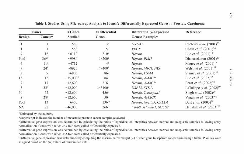
370P.S. N
elson
Table 1. Studies Using Microarray Analysis to Identify Differentially Expressed Genes in Prostate Carcinoma
Tissues # Genes # Differential Differentially-Expressed ReferenceBenign Cancer* Studied Genes Genes: Examples
1 1 588 13a GSTM1 Chetcuti et al. (2001)53
1 1 588 15b VEGF Chaib et al. (2001)54
9 16 ~6112 210c Hepsin Luo et al. (2001)18
Pool 3620 ~9984 �200d Hepsin, PIM1 Dhanasekaran (2001)19
4 113 ~4712 4e Hepsin Magee et al. (2001)17
9 241 ~8920 �400f Hepsin, MIC1, FAS Welsh et al. (2001)55
8 9 ~6800 86g Hepsin, PSMA Stamey et al. (2001)56
15 15 ~35,000# 84h Hepsin, AMACR Luo et al. (2002)57
9 17 ~12,600 216i Hepsin, AMACR Ernst et al. (2002)58
3 329 ~12,000 �3400j USP13, STK11 LaTulippe et al. (2002)59
50 52 ~12,600 456k Hepsin, Tetraspan1 Singh et al. (2002)29
8 285 ~12,600 50l Hepsin, AMACR Vanaja et al. (2003)60
Pool 13 6400 136m Hepsin, Nectin3, CALLA Best et al. (2003)28
NA 72 ~46,000 266n trp-p8, seladin-1, SOCS2 Henshall et al. (2003)31
#Estimated by the authors.*Superscript indicates the number of metastatic prostate cancer samples analyzed.aDifferential gene expression was determined by calculating the ratios of hybridization intensities between normal and neoplastic samples following arraynormalization. Genes with ratios � 3-fold were called differentially expressed.bDifferential gene expression was determined by calculating the ratios of hybridization intensities between normal and neoplastic samples following arraynormalization. Genes with ratios � 2-fold were called differentially expressed.cDifferential gene expression was determined by computing the discriminative weight (w) of each gene to separate cancer from benign tissue. P values wereassigned based on the (w) values of randomized data.
B245-ch15 3/2/05 4:40 PM Page 370

Gene E
xpression Changes in P
rostate Carcinom
a371
dSeveral approaches used: t-statistics (of prostate cancer versus benign tissue) ranked based on effect size: 200 genes reported. An analysis using fold-changeidentified 1520 genes with 2-fold changes between 50% of cancer samples and normal adjacent tissue and 1006 genes with 3-fold change in 75% of can-cer samples compared with a normal commercial pool.eDifferential expression between benign and neoplastic prostate tissue; � 3-fold change in all 11 tumors compared with four benign samples. P values werecalculated by a two-tailed t-test for independent data sets of unequal size and variance.fDifferential expression between benign and neoplastic prostate tissue was determined by equally weighing contributions from differences in hybridizationintensities, the quotient of hybridization intensities and the result of an unpaired t-test between expression levels in tumor and normal tissues.gDifferential expression between BPH and neoplastic prostate tissue; p � 0.0005 and expressed in all samples examined.hDifferential expression between benign and neoplastic prostate tissue; p � 0.05 by Student’s t-test; signal intensity �500 units.iDifferential expression between benign and neoplastic tissue; p � 0.05 by Student’s t-test and fold change �2.5.jDifferential expression between non-recurrent primary prostate cancers and metastasis; fold-difference �3-fold.kDifferential expression between normal versus tumor samples was determined using a variation of a signal-to-noise metric. The statistical significance ofthe gene expression correlations was determined by comparing the observed correlations to the results mined by comparing the observed correlations to theresults derived from permutations of the class labels (normal and tumor).lDifferential expression between benign and cancer samples was determined by combining equally weighted contributions from differences in hybridizationintensities, the quotient of the hybridization intensities, and the results of an unpaired t-test between expression levels. Selection criteria were further nar-rowed to a fold-change �2.35 and a p � 0.001 by Student’s t-test.mDifferential expression between normal and cancer tissue was determined at the p � 0.001 level by a one sample t-test and Wilcoxian ranking.nThis report was designed to identify gene expression changes associated with disease progression. Cox proportional hazard survival analysis predictingrelapse was used to evaluate each gene (probe set). Gene expression predictors of relapse at the p � 0.01 level were reported. A False-Discovery Rate (FDR)was calculated to be 23% (61 of the 266 findings are false positives).
B245-ch15 3/2/05 4:40 PM Page 371

A major objective in the field of oncology centers on accurate predictionof outcome and patient stratification for planned interventions such assurgery, radiation treatment and chemotherapy. Clues to tumor behaviormay be found in the molecular heterogeneity within individual cancerdiagnostic categories such as chromosomal translocations, deletions oftumor suppressor genes, amplifications or mutations of oncogenes, andnumerous chromosomal abnormalities. Ultimately, the majority of thesemolecular alterations will produce changes in gene expression that mani-fest in phenotypic behaviors of invasion, metastasis and drug resistance.
A landmark study involving the molecular analysis of Non-HodgkinsLymphoma (NHL) proved the hypothesis that gene expression profilescould be used to stratify cancers into clinically relevant categories. Thisstudy compared the variability in the natural history of a NHL subtype withanalyses of molecular heterogeneity in the tumors as assessed by cDNAmicroarray fingerprints.20 Although histologically-indistinguishable, two distinct forms of this NHL subtype were identified by expression pat-terns that reflected different stages of B-cell differentiation. The outcomeof patients with different expression profiles was demonstrated by diver-gent survival curves following the best available therapy. The study con-cluded that a molecular classification of tumors on the basis of geneexpression can identify previously undetected and clinically significantsubtypes of cancer.20 Similar studies have now been performed in othertumor types. van’t Veer et al. used microarrays to profile genes expressedin breast cancers.21 A classifier comprised of 70 “prognosis reportergenes” was developed that distinguished which primary tumors were des-tined to progress to distant metastases versus those that did not relapse.These and other studies point to an emerging use for genomics in themanagement of patients with neoplastic diseases.
The strongest predictive factors for prostate cancer disease progressionare the Gleason score, serum PSA, and clinical stage.22 The Gleason grad-ing system in biopsy or prostatectomy specimens is a measure of biologi-cal aggressiveness.22–24 Several groups have recently combined clinicalstage, serum PSA level, and Gleason score to generate “nomograms” thatpredict pathological stage or outcomes23,25–27 and allow prognostic esti-mates to be made with readily available clinical data. To identify biologicalcorrelates of the Gleason histology, Best et al. used microarray analysis to
372 P. S. Nelson
B245-ch15 3/2/05 4:40 PM Page 372

profile transcripts expressed in 13 high and moderate-grade prostate can-cers.28 Using permutation t-tests, 21 genes were found to segregate basedon tumor grade (p � 0.001). Genes with this discriminatory powerincluded tumor protein D52, BRCA1 binding protein 1 and CD69. Futurestudies will demonstrate the utility of these genes in delineating patientoutcomes prospectively.
To date, three studies have been published with designs aimed towardindependently identifying molecular signatures correlating with outcomesof relapsed disease following radical prostatectomy. Singh et al. usedoligonucleotide microarrays to characterize the expression levels of~12,600 transcripts in primary prostate carcinomas.29 The study identified456 differentially expressed genes between benign and malignant prostatetissues. A molecular classifier comprised of five genes (ITPR3, sialyl-transferase 1, PDGFR-�, chromogranin A and HoxC6) was capable ofsegregating primary cancers into two groups, those cancers that relapsedwithin four years of radical prostatectomy and those that did not recurwithin four years. In a second study, Ramaswamy et al. used microarraysto define a gene expression signature of metastasis present in multipletumor types, including prostate cancer.30 Comparing metastases with pri-mary cancers identified a group of 17 genes capable of discriminatingmetastases from primary tumors. A subset of primary tumors was alsofound to contain the metastasis signature and these tumors exhibited ahigh likelihood of disease relapse and poor clinical outcome following pri-mary therapy. Henshall et al. used microarrays comprised of 46,000unique sequences to profile 72 cases of primary prostate carcinoma.31 Atthe time of analysis, 17 patients were known to have relapsed as deter-mined by rising PSA measurements and the expression levels of 266genes were shown to correlate with disease relapse. This cohort includedthe putative calcium channel protein trp-p8, a transcript whose loss ofexpression in neoplastic tissue was strongly associated with diseaserelapse (p 0.001) independently of pre-treatment PSA levels.
Although both the five-gene molecular classifier defined by Singhet al.21 and the 17-gene metastasis signature defined by Ramaswamy et al.30
independently predicted disease relapse, no genes are shared betweenthe two predictive cohorts. Similarly, none of the progression-associatedgenes identified by Henshall et al.31 are included in the five-gene outcome
Gene Expression Changes in Prostate Carcinoma 373
B245-ch15 3/2/05 4:40 PM Page 373

classifier, though there may be overlaps in the biochemical pathways inwhich these genes participate. Of the 70 genes used to predict breast cancerprogression in the van’t Veer study,21 none are shared with the five or 17-geneprostate cancer outcome predictors. Therefore, while quite provocative andstatistically valid, the utility of these predictive signatures will require vali-dation in additional independent data sets and in prospective studies.
Proteomic Approaches for Assessing Gene Expression in Prostate Carcinoma
While important information has been gained through the profiling oftranscripts, it is important to keep in mind that the end-point for geneexpression is the protein, as proteins represent the actual scaffolds, molec-ular engines and communication mechanisms utilized by cells. In addi-tion, the development of most biomedical interventions center on proteinendpoints. Large-scale efforts are underway to analyze the proteome: thetotal protein complement of the genome.32 However, the proteome repre-sents a complex dynamic entity due to the many forms of a given proteinthat result from alternative transcript splicing and the numerous post-translational modifications that often define functional protein states.33,34
A core technology developed for the global analysis of proteinsinvolves the electrophoretic separation of proteins along two dimensions:size and charge, using polyacrylamide gels (2D-PAGE).35 Comparisons ofgel profiles from two different cell states (e.g. normal versus cancer) canidentify differential protein expression or protein modifications. The iden-tity of individual protein spots can be determined by immunodetection orby calculating the theoretical locations of known proteins based uponcharge and mass. More recently, microsequencing and mass spectrome-try have gained widespread use for characterizing protein spots of inter-est.36,37 The 2D-PAGE technique is theoretically capable of resolvingmore than 10,000 proteins and peptides from complex mixtures. However,practical applications of 2D-PAGE have rarely identified more than asmall percentage of proteins comprising the cellular proteome.38 Despitethis drawback, 2D-PAGE has been used to identify androgen-regulatedgenes in prostate epithelium39 and prostate cancer-associated proteinalterations such as NEDD8 and calponin.40
374 P. S. Nelson
B245-ch15 3/2/05 4:40 PM Page 374

Mass spectrometry (MS) has evolved to become a key technologyin proteomics research. The basic mass spectrometer is comprised of anion source, a mass analyzer, and a detector to record the spectra of ionintensity versus the mass to charge ratio of proteins or peptides underanalysis.36 Currently, surface enhanced laser assisted desorption (SELDI),matrix assisted laser desorption (MALDI) and electrospray ionization(ESI) represent the preferred methods for ionizing peptides and proteins.Comparing the mass spectra of complex protein mixtures such as humanserum offers the potential for identifying proteins or protein fragmentsthat are present in different abundances and that associate with diseasestates.33,41 Several reports have used SELDI mass spectrometry as a bio-marker discovery method to distinguish serum profiles or fingerprints thatreflect the presence of prostate carcinoma in comparison to serum proteinprofiles derived from normal controls.42,43 A major drawback to theseprofiling approaches involves a lack of reproducible quantitative accuracyand a difficulty in positively identifying the peptides and parent proteinsthat account for the fingerprint differences.
Several strategies have been devised to provide MS with the ability toaccurately quantitate differences in protein levels between two cellularstates.44–47 One technique employs Isotope Coded Affinity Tags (ICAT)comprised of three components: a biotin affinity tag, a linker with eithereight hydrogen or eight deuterium atoms (generating a light and a heavyform of the molecule), and a SH-reactive group capable of covalentlylinking to cysteine residues (Fig. 2).48 The proteins of one cell state(e.g. normal epithelium) are labeled with the light reagent and those of asecond cell state (e.g. neoplastic epithelium) with the heavy reagent.Equal quantities of labeled cells are mixed and the proteins are separated,digested with a proteolytic enzyme and the resulting mixture of peptidesare passed over an avidin column to isolate the cysteine labeled peptides(about 90% of proteins have cysteine residues). These can be fractionatedand analyzed by tandem mass spectrometry (MS/MS). The first MSanalysis gives the areas under the curves of the paired isotopic peptides(hence, their relative abundances); the second MS analysis providesa peptide fingerprint that can be used to identify the parent protein.Thus, the ICAT method dramatically increases throughput by reducingsample redundancy (only cysteine-containing peptides are assessed) and
Gene Expression Changes in Prostate Carcinoma 375
B245-ch15 3/2/05 4:40 PM Page 375
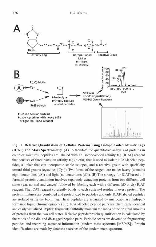
376 P. S. Nelson
Fig . 2. Relative Quantitation of Cellular Proteins using Isotope Coded Affinity Tags(ICAT) and Mass Spectrometry. (A) To facilitate the quantitative analysis of proteins incomplex mixtures, peptides are labeled with an isotope-coded affinity tag (ICAT) reagentthat consists of three parts: an affinity tag (biotin) that is used to isolate ICAT-labeled pep-tides, a linker that can incorporate stable isotopes, and a reactive group with specificitytoward thiol groups (cysteines [Cys]). Two forms of the reagent are made: heavy (containseight deuteriums [d8]) and light (no deuteriums [d0]). (B) The strategy for ICAT-based dif-ferential protein quantitation involves separately extracting proteins from two different cellstates (e.g. normal and cancer) followed by labeling each with a different (d0 or d8) ICATreagent. The ICAT reagent covalently bonds to each cysteinyl residue in every protein. Theprotein mixtures are combined and proteolyzed to peptides and only ICAT-labeled peptidesare isolated using the biotin tag. These peptides are separated by microcapillary high-per-formance liquid chromatography (LC). ICAT-labeled peptide pairs are chemically identicaland easily visualized. Peptide fragments faithfully maintain the ratios of the original amountsof proteins from the two cell states. Relative peptide/protein quantification is calculated bythe ratios of the d0- and d8-tagged peptide pairs. Periodic scans are devoted to fragmentingpeptides and recording sequence information (tandem mass spectrum [MS/MS]). Proteinidentifications are made by database searches of the tandem mass spectrum.
B245-ch15 3/2/05 4:40 PM Page 376

retains sample complexity while allowing for accurate relative proteinquantification.48,49
The ICAT approach has been used to identify secreted50 and androgen-regulated proteins51 expressed by prostate cancer cells. In addition toidentifying genes previously not known to be regulated by androgens, com-parisons of global transcript and protein abundance levels showed that formost genes (�90%), protein levels were concordant with transcript abun-dance. However, there were distinct outliers that indicate multiple levels ofgene expression regulation (e.g. post-transcriptional, post-translational).These results suggest immediately testable hypotheses for characterizingmechanism(s) of gene expression regulation. The results also demonstratethat to fully delineate a gene expression profile, measurements of proteinlevels are necessary, since, for some genes, protein alterations would nothave been predicted by transcript abundance measurements.
Conclusion and Future Directions
Gene expression profiles offer an extraordinary assessment of both the diver-sity and consistency of genetic alterations in prostate carcinomas. Publishedstudies have provided the first glimpse of the potential of profiling technolo-gies to identify individual genes that participate in neoplastic growth and todefine molecular predictors of cancer behavior. However, there clearly arelimitations to the approach and significant hurdles to be overcome. It issomewhat disconcerting that cohorts of genes statistically associated withdisease outcome in different studies do not correlate with each other. Thismay be explained by the extreme heterogeneity of neoplastic prostate lesions.In addition, tissue samples used in most studies are comprised of variousamounts of normal epithelium, stromal cells, inflammatory cells, blood ves-sels, and other constituents. While gene expression changes in these celltypes may influence neoplastic transformation, growth, and invasion, thevariable amounts of these cell types between different studies may makeinter-study comparisons difficult. To address this problem, microdissectiontechniques that compare only specific cell types can be employed.52 The ulti-mate endpoint of such an approach is the characterization of individual cellexpression profiles within the tumor environment. Although such anapproach represents a daunting task, the pace of biotechnological advances
Gene Expression Changes in Prostate Carcinoma 377
B245-ch15 3/2/05 4:40 PM Page 377
do not rule out single-cell analysis using high-speed flow cytometers andnanotechnology in the near future. Finally, a major factor that could influencethe outcomes of tumors with identical gene expression profiles centers onvariables within the host. Most expression profiling studies have focused ondefining molecular determinants within tumor tissue. However, host charac-teristics involving immune response, dietary factors and the hormone milieumay influence tumor cell proliferation, invasion and metastasis. In the future,we will likely gain important additional knowledge of tumor behavior andresponse to therapy through the integration of profiles reflecting both tumorand host gene expression.
Acknowledgments
Peter Nelson is supported by a Scholar Award from the Damon-RunyonCancer Research Foundation.
References
1. Diatchenko L, Lau YF, Campbell AP, et al. (1996) Suppression subtractivehybridization: a method for generating differentially regulated or tissue-specific cDNA probes and libraries. Proc Natl Acad Sci USA 93:6025–6030.
2. Liang P, Pardee AB (1992) Differential display of eukaryotic messengerRNA by means of the polymerase chain reaction. Science 257:967–971.
3. Nelson PS, Plymate SR, Wang K, et al. (1998) Hevin, an antiadhesive extra-cellular matrix protein, is down-regulated in metastatic prostate adenocarci-noma. Cancer Res 58:232–236.
4. Bussemakers MJ, van Bokhoven A, Verhaegh GW, et al. (1999) DD3: a newprostate-specific gene, highly overexpressed in prostate cancer. Cancer Res59:5975–5979.
5. Srikantan V, Zou Z, Petrovics G, et al. (2000) PCGEM1, a prostate-specificgene, is overexpressed in prostate cancer. Proc Natl Acad Sci USA 97:12216–12221.
6. Wang FL, Wang Y, Wong WK, et al. (1996) Two differentially expressedgenes in normal and human prostate tissue and in carcinoma. Cancer Res56:3634–3637.
7. Nelson PS, Ng WL, Schummer M, et al. (1998) An expressed-sequence-tagdatabase of the human prostate: sequence analysis of 1168 cDNA clones [InProcess Citation]. Genomics 47:12–25.
378 P. S. Nelson
B245-ch15 3/2/05 4:40 PM Page 378

8. Scheurle D, DeYoung MP, Binninger DM, Page H, Jahanzeb M, NarayananR (2000) Cancer gene discovery using digital differential display [In ProcessCitation]. Cancer Res 60:4037–4043.
9. Bera TK, Maitra R, Iavarone C, et al. (2002) PATE, a gene expressed inprostate cancer, normal prostate, and testis, identified by a functionalgenomic approach. Proc Natl Acad Sci USA 99:3058–3063.
10. Liu XF, Olsson P, Wolfgang CD, et al. (2001) PRAC: A novel small nuclearprotein that is specifically expressed in human prostate and colon. Prostate47:125–131.
11. Olsson P, Bera TK, Essand M, et al. (2001) GDEP, a new gene differentiallyexpressed in normal prostate and prostate cancer. Prostate 48:231–241.
12. Velculescu VE, Zhang L, Vogelstein B, Kinzler KW (1995) Serial analysis ofgene expression. Science 270:384–387.
13. Xu LL, Shanmugam N, Segawa T, et al. (2000) A novel androgen-regulatedgene, PMEPA1, located on chromosome 20q13 exhibits high level expres-sion in prostate [In Process Citation]. Genomics 66:257–263.
14. Waghray A, Schober M, Feroze F, Yao F, Virgin J, Chen YQ (2001)Identification of differentially expressed genes by serial analysis of geneexpression in human prostate cancer. Cancer Res 61:4283–4286.
15. Schena M, Shalon D, Davis RW, Brown PO (1995) Quantitative monitoringof gene expression patterns with a complementary DNA microarray. Science270:467–470.
16. DeRisi J, Penland L, Brown PO, et al. (1996) Use of a cDNA microarrayto analyse gene expression patterns in human cancer. Nature Genet 14:457–460.
17. Magee JA, Araki T, Patil S, et al. (2001) Expression profiling reveals Hepsinoverexpression in prostate cancer. Cancer Res 61:5692–5696.
18. Luo J, Duggan DJ, Chen Y, et al. (2001) Human prostate cancer and benignprostatic hyperplasia: molecular dissection by gene expression profiling.Cancer Res 61:4683–4688.
19. Dhanasekaran SM, Barrette TR, Ghosh D, et al. (2001) Delineation of prog-nostic biomarkers in prostate cancer. Nature 412:822–826.
20. Alizadeh AA, Eisen MB, Davis RE, et al. (2000) Distinct types of diffuselarge B-cell lymphoma identified by gene expression profiling. Nature403:503–511.
21. van ‘t Veer LJ, Dai H, van de Vijver MJ, et al. (2002) Gene expression pro-filing predicts clinical outcome of breast cancer. Nature 415:530–536.
22. Partin AW, Yoo J, Carter HB, et al. (1993) The use of prostate specific anti-gen, clinical stage and Gleason score to predict pathological stage in menwith localized prostate cancer [see comments]. J Urol 150:110–114.
Gene Expression Changes in Prostate Carcinoma 379
B245-ch15 3/2/05 4:40 PM Page 379

23. Partin AW, Kattan MW, Subong EN, et al. (1997) Combination of prostate-specific antigen, clinical stage, and Gleason score to predict pathologicalstage of localized prostate cancer. A multi-institutional update. JAMA277:1445–1451.
24. Stamey TA, McNeal JE, Yemoto CM, Sigal BM, Johnstone IM (1999)Biological determinants of cancer progression in men with prostate cancer.JAMA 281:1395–1400.
25. Kattan MW, Stapleton AM, Wheeler TM, Scardino PT (1997) Evaluation ofa nomogram used to predict the pathologic stage of clinically localizedprostate carcinoma. Cancer 79:528–537.
26. Kattan MW, Eastham JA, Stapleton AM, Wheeler TM, Scardino PT (1998) A preoperative nomogram for disease recurrence following radical prostatec-tomy for prostate cancer. J Natl Cancer Inst 90:766–771.
27. Kattan MW, Wheeler TM, Scardino PT (1999) Postoperative nomogram fordisease recurrence after radical prostatectomy for prostate cancer. J ClinOncol 17:1499–1507.
28. Best CJ, Leiva IM, Chuaqui RF, et al. (2003) Molecular differentiation ofhigh- and moderate-grade human prostate cancer by cDNA microarrayanalysis. Diagn Mol Pathol 12:63–70.
29. Singh D, Febbo PG, Ross K, et al. (2002) Gene expression correlates of clin-ical prostate cancer behavior. Cancer Cell 1:203–209.
30. Ramaswamy S, Ross KN, Lander ES, Golub TR (2003) A molecular signa-ture of metastasis in primary solid tumors. Nat Genet 33:49–54.
31. Henshall SM, Afar DE, Hiller J, et al. (2003) Survival analysis of genome-wide gene expression profiles of prostate cancers identifies new prognostictargets of disease relapse. Cancer Res 63:4196–4203.
32. Humphery-Smith I, Blackstock W (1997) Proteome analysis: genomics viathe output rather than the input code. J Protein Chem 16:537–544.
33. Corthals GL, Nelson PS (2001) Large-scale proteomics and its future impacton medicine. Pharmacogenomics J 1:15–19.
34. Martin DB, Nelson PS (2001) From genomics to proteomics: techniques andapplications in cancer research. Trends Cell Biol 11:S60–65.
35. O’Farrell PH (1975) High resolution two-dimensional electrophoresis of pro-teins. J Biol Chem 250:4007–4021.
36. Corthals G, Gygi SP, Aebersold R, Patterson SD (1999) Identification ofproteins by mass spectrometry. In: Rabilloud T, ed. Proteome Research: 2D GelElectrophoresis and Detection Methods. Springer, New York, pp. 197–232.
37. Yates JR, 3rd (1998) Database searching using mass spectrometry data.Electrophoresis 19:893–900.
380 P. S. Nelson
B245-ch15 3/2/05 4:40 PM Page 380

38. Gygi SP, Corthals GL, Zhang Y, Rochon Y, Aebersold R (2000) Evaluationof two-dimensional gel electrophoresis-based proteome analysis technology.Proc Natl Acad Sci USA 97:9390–9395.
39. Waghray A, Feroze F, Schober MS, et al. (2001) Identification ofandrogen-regulated genes in the prostate cancer cell line LNCaP by serialanalysis of gene expression and proteomic analysis. Proteomics 1:1327–1338.
40. Meehan KL, Holland JW, Dawkins HJ (2002) Proteomic analysis of normaland malignant prostate tissue to identify novel proteins lost in cancer.Prostate 50:54–63.
41. Issaq HJ, Veenstra TD, Conrads TP, Felschow D (2002) The SELDI-TOF MSapproach to proteomics: protein profiling and biomarker identification.Biochem Biophys Res Commun 292:587–592.
42. Qu Y, Adam BL, Yasui Y, et al. (2002) Boosted decision tree analysis of surface-enhanced laser desorption/ionization mass spectral serum profilesdiscriminates prostate cancer from noncancer patients. Clin Chem 48:1835–1843.
43. Petricoin EF, 3rd, Ornstein DK, Paweletz CP, et al. (2002) Serum pro-teomic patterns for detection of prostate cancer. J Natl Cancer Inst 94:1576–1578.
44. Olsen JV, Andersen JR, Nielsen PA, et al. (2003) Hystag — a novel proteomicquantification tool applied to differential display analysis of membrane pro-teins from distinct areas of mouse brain. Mol Cell Proteom 10:10.
45. Espina V, Mehta AI, Winters ME, et al. (2003) Protein microarrays: molecu-lar profiling technologies for clinical specimens. Proteomics 3:2091–2100.
46. Grubb RL, Calvert VS, Wulkuhle JD, et al. (2003) Signal pathway profiling ofprostate cancer using reverse phase protein arrays. Proteomics 3: 2142–2146.
47. Haab BB, Dunham MJ, Brown PO (2001) Protein microarrays for highlyparallel detection and quantitation of specific proteins and antibodies incomplex solutions. Genome Biol 2:RESEARCH0004.
48. Gygi SP, Rist B, Gerber SA, Turecek F, Gelb MH, Aebersold R (1999)Quantitative analysis of complex protein mixtures using isotope-coded affin-ity tags. Nat Biotechnol 17:994–999.
49. Han DK, Eng J, Zhou H, Aebersold R (2001) Quantitative profiling of differentiation-induced microsomal proteins using isotope-coded affinitytags and mass spectrometry. Nat Biotechnol 19:946–951.
50. Martin DB, Gifford DR, Wright ME, Keller A, Yi E, Goodlett DR, Aebersold R,Nelson PS (2004) Quantitative proteomic analysis of proteins released by neo-plastic prostate epithelium. Cancer Res 64:347–355.
Gene Expression Changes in Prostate Carcinoma 381
B245-ch15 3/2/05 4:40 PM Page 381

51. Wright ME, Eng J, Sherman J, et al. (2003) Identification of androgen-coregulated protein networks from the microsomes of human prostate cancercells. Genome Biol 5:R4.
52. Emmert-Buck MR, Bonner RF, Smith PD, et al. (1996) Laser capturemicrodissection [see comments]. Science 274:998–1001.
53. Chetcuti A, Margan S, Mann S, et al. (2001) Identification of differentiallyexpressed genes in organ-confined prostate cancer by gene expression array.Prostate 47:132–140.
54. Chaib H, Cockrell EK, Rubin MA, Macoska JA (2001) Profiling and verifi-cation of gene expression patterns in normal and malignant human prostatetissues by cDNA microarray analysis. Neoplasia 3:43–52.
55. Welsh JB, Sapinoso LM, Su AI, et al. (2001) Analysis of gene expressionidentifies candidate markers and pharmacological targets in prostate cancer.Cancer Res 61:5974–5978.
56. Stamey TA, Warrington JA, Caldwell MC, et al. (2001) Molecular geneticprofiling of Gleason grade 4/5 prostate cancers compared to benign prosta-tic hyperplasia. J Urol 166:2171–2177.
57. Luo JH, Yu YP, Cieply K, et al. (2002) Gene expression analysis of prostatecancers. Mol Carcinog 33:25–35.
58. Ernst T, Hergenhahn M, Kenzelmann M, et al. (2002) Decrease and gain ofgene expression are equally discriminatory markers for prostate carcinoma:a gene expression analysis on total and microdissected prostate tissue. Am J Pathol 160:2169–2180.
59. LaTulippe E, Satagopan J, Smith A, et al. (2002) Comprehensive geneexpression analysis of prostate cancer reveals distinct transcriptional pro-grams associated with metastatic disease. Cancer Res 62:4499–4506.
60. Vanaja DK, Cheville JC, Iturria SJ, Young CY (2003) Transcriptional silenc-ing of zinc finger protein 185 identified by expression profiling is associatedwith prostate cancer progression. Cancer Res 63:3877–3882.
382 P. S. Nelson
B245-ch15 3/2/05 4:40 PM Page 382

16STUDY OF ANDROGEN–ANDROGEN RECEPTOR ROLES
IN PROSTATE CANCER USING MICE LACKINGFUNCTIONAL PROSTATE ANDROGEN RECEPTOR
Chun-Te Wu, Shuyuan Yeh, Qingquan Xu, Zhiming Yang,Philip Chang, Yueh-Chiang Hu and Chawnshang Chang
George H. Whipple Lab for Cancer ResearchDepartments of Urology, Pathology and Radiation Oncology, and
the Cancer Center, University of RochesterRochester, New York 14642, USA
Introduction
Prostate cancer is an androgen-dependent disease and a leading causeof cancer mortality in men. The vast majority of prostate cancers expressthe androgen receptor (AR), have androgen-dependent growth, initiallyrespond to androgen ablation therapy, but eventually become androgen-independent (hormone refractory stage).1 The mechanisms for transitionof tumors from androgen-dependent to androgen-independent are gener-ally explained by clonal selection, adaption, alternative pathways of sig-nal transduction and aberrant AR signaling.2–4 The AR is now known toparticipate in tumor progression through three mechanisms: expression,development of point mutations, and ligand-independent activation viacoregulators.5 Histological and epidemiological data have indicated therole of AR mutations and poly-Q polymorphisms in the anti-androgenwithdrawal syndrome and prostate cancer susceptibility.6 The AR pointmutation T877A is common in the hormone-refractory prostate cancer.7
In vitro studies have indicated the transactivation of this mutated AR can beinduced by the anti-androgen, flutamide, in human prostate cancer LNCaPcells, which contain the mutant T877A (tyrptophen to alanine) AR.8
383
B245-ch16 3/2/05 4:41 PM Page 383

The involvement of this mutation in the transition to hormone-refractorystage and the anti-androgen withdrawal syndrome, remains controversial.Alleles of the CAG repeat in the first exon of the human AR gene have beenshown to be associated with the risk of prostate cancer. Even in low-riskAsian populations that are favored by higher median CAG repeat lengths, ashorter CAG repeat length influences prostate cancer risk.9 Animal modelswithout functional AR in the prostate provide excellent tools to investigatethe AR role in normal prostate development, prostate cancer carcinogenesis,progression, metastasis and transition to hormone-refractory. It is hoped thatcontinuing research on AR expression and function in prostate cancer willprovide new therapeutic strategies.
Generation of Androgen Receptor Knockout (ARKO) Mice
The AR, a member of the nuclear receptor superfamily, was first clonedin 1988.10–11 It contains an N-terminal transactivation domain (NTD), a central DNA-binding domain (DBD) and a C-terminal ligand-bindingdomain (LBD). To generate ARKO mice, a cre-lox strategy for condi-tional KO is necessary. The cre-lox system utilizes the expression of P1phage cre recombinase (Cre) to catalyze the excision of DNA locatedbetween flanking lox sites in exon 2 of the DBD.12 We generated two sep-arate lines of transgenic mice to carry the Cre and the homozygous floxedAR genes in both X chromosomes, as illustrated in Fig. 1. The generationof ar/Y-ACTB/Cre male mice in F3 resulted in total ARKO in all of thecells. Phenotype analyses showed that ARKO male mice have a low bodyweight and the external genitalia was ambiguous or appeared feminized,had a microphallic penis, and the urethra showed hypospadia. The scro-tum is poorly developed and all vas deferens, epididymis, seminal vesicle and prostate are agenetic. Testes are small in size and cryptorchidin the low abdominal area close to the internal inguinal ring. This studyindicated that disruption of AR gene in early embryonic stage had greatimpact in the development of normal prostate.12 However, the systemiceffects of lower androgen level in the total ARKO mice should be furtherexplored.
384 C.-T. Wu et al.
B245-ch16 3/2/05 4:41 PM Page 384

Prostate Development and Carcinogenesis in Prostate-SpecificARKO Mice
The cre-lox ARKO mouse model provides a much-needed in vivo animalmodel system to study androgen functions in the selective androgen targettissues in male mice. In this regard, it is of paramount importance to derivea cre recombinant transgenic animal system with robust expression of abiologically active Cre protein in a prostate epithelial cell-specific man-ner. We selected a rat probasin (PB) promoter to drive expression of theCre gene.13 The AR gene was gradually deleted during the adolescencestage when increased androgen levels activated the probasin promoterexpression. Specific ARKO in prostate epithelium was evidenced by
Androgen–Androgen Receptor Roles in Prostate Cancer 385
Fig. 1. Generation of ARKO female mice by mating among floxed AR (fAR) founders andcre founders. The chimera founder is B6/129 mosaic strain. The mating of the founder withthe B6 female created female mice heterozygous with fAR. The following F2 generationresulted in fAR male mice. The mating between the heterozygous fAR female and thehomozygous FVB/N-TgN ACTB cre male that carry Cre under �-actin promoter createda heterozygous KO of AR female carrying the Cre. The mating between the fAR male andthe heterozygous ARKO female carrying the Cre generated the homozygous ARKO Cre�
female mice with a 1:8 ratio.
B245-ch16 3/2/05 4:41 PM Page 385

genotyping, RT-PCR and immunohistochemistry (IHC). Hormonal stud-ies revealed normal serum testosterone levels in prostate-specific ARKOas compared to wild-type (WT) mice. The growth of urogenital organsother than prostate remained unchanged after the prostate epithelium nolonger has a functional AR, however the size and weight of prostate glandswere slightly decreased. The secretion proteins, probasin and prostaticsecretory protein 94 (PSP94), were dramatically decreased in prostate-specific ARKO mice. When androgen ablation by castration was inducedat 12 weeks old, both WT and prostate-specific ARKO mice showed dra-matic regression of prostate and seminal vesicle growth. This modelimplies that the AR plays specific roles in epithelium and in stromaldevelopment. By crossing male probasin-cre mice to flox AR TRAMPfemale mice, the prostate-specific ARKO TRAMP mice were generated.This model will add more clues about the role of AR in the prostate can-cer carcinogenesis and progression.
TRAMP Mice Lacking the Endogenous AR but Carrying theT877A Mutated Transgene
In a subgroup of patients with endocrine therapy-resistant prostate can-cers, amino acid substitutions in the AR LBD have been found, whichresult in a broadened ligand response spectrum. The most common sub-stitution in these cancers was T877A, which was first also described in theLNCaP prostate cancer cell line. The T877A substitution renders the ARresponsive to natural low-affinity ligands and anti-androgens, such as flu-tamide. This mutation is believed to contribute to the prostate cancer pro-gression and its transition to the hormone-refractory stage as well as theanti-androgen withdrawal syndrome.
By crossing the T877A transgene knock-in male mice with ourARKO female mice, we are in the process of generating ARKO male miceexpressing prostate T877A AR transgene only. This will enable us to com-pare the effects of mutated AR and WT AR on the development of prostategland. Further crossing female ARKO mice with T877A transgene maleTRAMP mice will generate TRAMP mice which only express T877Amutated AR in the prostate. By these strategies, we can continue toexplore the AR mutation on prostate cancer carcinogenesis and compare
386 C.-T. Wu et al.
B245-ch16 3/2/05 4:41 PM Page 386

the tumor initiation and progression in the TRAMP mice with two differ-ent AR expressions.
ARKO Mice with AR-97Q and AR-24Q Transgene Expression
Despite the substantial public health impact of prostate cancer, little isknown about its etiology. The generally accepted risk factors for the devel-opment of prostate cancer are advanced age, familial predisposition andperhaps ethnicity. The exon 1 of the AR contains several polymorphicrepeats; the most variable is a polymorphic CAG repeat, which encodes apolyglutamine (poly-Q) chain. The range of CAG repeat lengths is from14 to 35 repeats in man and may vary somewhat with ethnicity and race.Because the length of the polymorphic CAG trinucleotide repeat isinversely correlated with the transactivation function of the AR in vitro, ithas been proposed that men with shorter repeats will be at higher risk forprostate cancer.14 Several previous studies have shown that shorter CAGrepeat length in AR NTD is associated with the occurrence of moreaggressive prostate cancer, earlier age of onset and likelihood of recur-rence.15 However, one small study in a European population failed to sup-port the association between CAG repeat length and prostate cancer risk.16
The role of CAG repeats in prostate needs to be determined more clearly.By crossing AR-97Q and AR-24Q transgene male mice with our ARKOfemale mice, we are generating the ARKO mice carrying only 97-Q and24-Q transgenic AR to compare the effects of different poly-Q lengths onthe development of the prostate gland. Further crossing to TRAMP micewill create ARKO TRAMP mice with different poly-Q AR transgenes.By studying the differences in carcinogenesis and tumor progression inthese mice, we will determine the effect of poly-Q lengths on the prostatecancer in vivo.
Inducible ARKO and ARKO TRAMP Mice
The normal prostate development depends on the androgen-AR signaling.In the total ARKO model, agenesis of prostate revealed that AR playsa critical role in the embryonic development of prostate. The prostate-specific ARKO model using probasin-cre lox strategy had nearly normal
Androgen–Androgen Receptor Roles in Prostate Cancer 387
B245-ch16 3/2/05 4:41 PM Page 387

prostate growth after adolescence. These two models suggested the time-specific role of AR in the development of prostate. What is the role of ARbetween embryonic and adolescence? The time-specific role of AR in thenormal prostate development can also be elucidated by the study of theinducible ARKO mice. The method uses an interferon-responsive pro-moter to control the expression of Cre recombinase. Here, Cre was usedto delete a segment of the AR gene flanked by loxP recombinase recogni-tion sites. Deletion was complete in liver and nearly complete in lympho-cytes within a few days, whereas partial deletion was obtained in othertissues. Crossing the flox-AR female mice with Mx-1-inducible Cre malemice17 will generate interferon-inducible ARKO mice. Our preliminarydata (Xu and Chang, unpublished data), showed the efficiency of deletionof AR gene in prostate tissue was about 20% using the intraperitonealinjection of pI-pC. Some modifications can be made to improve thepercentage of AR deletion, such as multiple injections and intra-prostateinjection. Yet the partial deletion of AR in prostate may be sufficient forgene inactivation and result in easily detectable phenotypes. By crossingTRAMP male mice with flox-AR-cre-MX-1 female mice, the inducibleARKO TRAMP mice can be generated. As in Fig. 2, induction of IFN in5-week-old inducible ARKO TRAMP mice resulted in a prostate tumorin one out of three mice, while all three mice without the induction ofIFN had prominent prostate tumors at 20-week-old. If the AR gene wasinducibly knocked out in 20-week-old mice, all the mice in WT AR andinducible ARKO groups grew prostate cancer, but the tumor size wassmaller in the ARKO group. This model indicated that AR deletion inTRAMP mice might inhibit carcinogenesis at an earlier stage and delaycancer progression at a late stage. Further study is necessary to confirmthe results of the preliminary observations and to explore the actual roleof AR in prostate cancer carcinogenesis and progression.
ARKO Human Prostate Cancer CWR22R Cells
The advantage of using gene targeting in human somatic cells is that itprovides a tool to study the roles of the signaling of interest in human cellsinstead of ARKO animal model. For this purpose, we attempted to disruptAR signaling in human androgen-refractory prostate cancer cells in order
388 C.-T. Wu et al.
B245-ch16 3/2/05 4:41 PM Page 388

to investigate the biological importance of AR signaling in those prostatecancers. Human prostate cancer CWR22R cells were transfected with theAR targeting vector using Superfect transfection kit (Qiagen) and selectedwith 400 �g/ml neomycin reagent.18 The genotypes of the survivingclones were detected by Southern blotting using a DNA probe containingthe 5�-UTR sequences of the AR gene. The untargeted and targeted lociproduced approximately 9.0-kb and 3.46-kb bands, respectively. ARexpression was reduced in those heterozygous clones compared to that inthe parental CWR22R cells19 confirming that one of the AR genes is tar-geted by homologous recombination. Therefore, this method provides apromising result and indicates that it is possible that we could target ARgenes in any other cell lines of interest. Efforts to obtain homologous AR-null CWR22R cells failed after extensive screening, although homologous
Androgen–Androgen Receptor Roles in Prostate Cancer 389
Fig. 2. Model for development of inducible ARKO-TRAMP mice.
Flox-AR (+) MX-1 Cre (+)
Flox-AR-MX-1 Cre (+)
TRAMP (+)
TRAMP-Flox-AR-MX-1 Cre (+)
Induce IFN at 5th week Induce IFN at 20th week
(–) (+) (–) (+)
TRAMP 3/3tumor at 20th
week
ARKO-TRAMP1/3 tumor at20th week
TRAMP 2/2big tumor at28th week
ARKO-TRAMP2/2 small tumorat 28th week
B245-ch16 3/2/05 4:41 PM Page 389

AR-null MCF cells were successfully established. This suggests that ARis an essential survival factor in prostate cancer cells. The essential role ofAR in androgen-refractory prostate cancer has been depicted in manyother studies.20,21 In order to knockout both AR genes, we are trying tostably transfect the tetracycline-inducible AR construct into heterozygousCWR22R cells and then target the second allele of AR in the cells. Webelieve this will provide a powerful tool for us to manipulate AR expres-sion in the prostate cancer studies.
Conclusion
Although AR plays the pivot role in prostate development and prostatecancer carcinogenesis and progression, several aspects of AR function inthe prostate are still not clear due to lack of appropriate animal models.One advantage of creating the floxed AR mice is to provide a base to gen-erate tissue-specific ARKO in the prostate. Combined with different Cre-recombinases, a time-specific role of AR can also be studied by using theinducible Mx-1 mice. Based on the ARKO mice, other mutated genes canbe tested as a combination of transgenic and knockout mice, such asT877A and poly-Q ARKO mice. Continued expansion of our knowledgeof AR action in prostate cancer will contribute to more effective therapies.It is likely that in the future, AR ablation with a target to degenerate and/oreliminate AR protein, rather than androgen ablation to block androgenbinding to the AR, will become the treatment of choice for advancedprostate cancer and hormone-refractory prostate cancer.
References
1. van der Kwast TH, Schalken J, Ruizeveld de Winter JA, van Vronnhoven CCJ,Mulder E, Boersma W, Trapman J (1991) Androgen receptors in endocrinetherapy resistant human prostate cancer. Int J Cancer 48:189–193.
2. Visakorpi T (1999) New pieces to the prostate cancer puzzle. Nat Med5:264–265.
3. Gelmann E (2002) Molecular biology of the androgen receptor. J Clin Oncol20:3001–3015.
4. Suzuki H, Ueda T, Ichikawa T, Ito H (2003) Androgen receptor involvementin the progression of prostate cancer. Endocr Relat Cancer 10:209–216.
390 C.-T. Wu et al.
B245-ch16 3/2/05 4:41 PM Page 390

5. Miyamoto H, Yeh S, Wilding G, Chang C (1998) Promotion of agonist activ-ity of anti-androgens by the androgen receptor coactivator, ARA70, in humanprostate DU145 cells. Proc Natl Acad Sci USA 95:7379–7384.
6. Wang Q, Udayakumar TS, Vasaitis TS, Brodie AM, Fondell JD (2004)Mechanistic relationship between androgen receptor polyglutamine tracttruncation and androgen-dependent transcriptional hyperactivity in prostatecancer cells. J Biol Chem 279:17319–17328.
7. Gaddipati JP, McLeod DG, Heidenberg HB, Sesterhenn IA, Finger MJ,Moul JW, Srivastava S (1994) Frequent detection of codon 877 mutation in theandrogen receptor gene in advanced prostate cancer. Cancer Res 54:2861–2864.
8. Veldscholte J, Ris-Stalpers C, Kuiper G, Jenster G, Berrevoets C, Claassen E,van Rooij HCJ, Trapman AO, Brinkmann AO, Mulder E (1990) A mutationin the ligand binding domain of the androgen receptor of human LNCaP cellsaffects steroid binding characteristics and response to anti-androgens.Biochem Biophys Res Commun 173:534–540.
9. Hsing AW, Gao YT, Wu G, Wang X, Deng J, Chen YL, Sesterhenn IA,Mostofi FK, Benichou J, Chang C (2000) Polymorphic CAG and GGN repeatlengths in the androgen receptor and prostate cancer risk: a population-basedcase control study in China. Cancer Res 60:5111–5116.
10. Chang C, Kokontis J, Liao S (1988) Molecular cloning of human and ratcomplementary DNA encoding androgen receptors. Science 240:324–326.
11. Lubahn DB, Joseph DR, Sullivan PM, Willard HF, French FS, Wilson EM(1988) Cloning of human androgen receptor complementary DNA and local-ization to the X chromosome. Science 240:327–330.
12. Yeh S, Tsai MY, Xu Q, Mu XM, Lardy H, Huang KE, Lin H, Yeh SD,Altuwaijri S, Zhou X, Xing L, Gan L, Chang C (2002) Generation and char-acterization of androgen receptor knockout (ARKO) mice: an in vivo modelfor the study of androgen functions in selective tissue. Proc Natl Acad SciUSA 99:13498–13503.
13. Wu X, Wu J, Huang J, Powell WC, Zhang J, Matusik RJ, Sangiorgi FO,Maxson RE, Sucov HM, Roy-Burman P (2001) Generation of a prostateepithelial cell-specific Cre transgenic mouse model for tissue-specific geneablation. Mech Dev 101:61–69.
14. Irvine RA, Yu MC, Ross RK, Coetzee GA (1995) The CAG and GGCmicrosatellites of the androgen receptor gene are in linkage disequilibrium inmen with prostate cancer. Cancer Res 55:1937–1940.
15. Nam RK, Elhaji Y, Krahn MD (2000) Significance of the CAG repeat poly-morphism of the androgen receptor gene in prostate cancer progression.J Urol 164:672–676.
Androgen–Androgen Receptor Roles in Prostate Cancer 391
B245-ch16 3/2/05 4:41 PM Page 391

16. Correa-Cerro L, Wohr G, Haussler J (1999) (CAG)nCAA and GGN repeatsin the human androgen receptor gene are not associated with prostate cancerin a French-German population. Eur J Hum Genet 7:357–362.
17. Kuhn R, Schwenk F, Aguet M, Rajewsky K (1995) Inducible gene targetingin mice. Science 269:1427–1429.
18. Yeh S, Hu YC, Wang PH, Xie C, Xu Q, Tsai MY, Dong Z, Wang RS, Lee TH,Chang C (2003) Abnormal mammary gland development and growth retar-dation in female mice and MCF7 breast cancer cells lacking androgen recep-tor. J Exp Med 198:1899–1908.
19. Bao BY, Hu YC, Ting HJ, Lee YF (2004) Androgen signaling is required forthe vitamin D-mediated growth inhibition in human prostate cancer cells.Oncogene 23:3350–3360.
20. Zegarra-Moro OL, Schmidt LJ, Huang H, Tindall DJ (2002) Disruption ofandrogen receptor function inhibits proliferation of androgen-refractoryprostate cancer cells. Cancer Res 62:1008–1013.
21. Chen CD, Welsbie DS, Tran C, Baek SH, Chen R, Vessella R, Rosenfeld MG,Sawyers CL (2004) Molecular determinants of resistance to anti-androgentherapy. Nat Med 10:33–39.
392 C.-T. Wu et al.
B245-ch16 3/2/05 4:41 PM Page 392

17CAPTURING SIGNAL ANOMALIES OF HUMAN
PROSTATE CANCER INTO MOUSE MODELS
Hong Wu
Department of Molecular and Medical PharmacologyUniversity of California Los Angeles School of Medicine
Los Angeles, California 90095, USA
Ani Khodavirdi* and Pradip Roy-Burman*,†
*Department of Pathology and†Department of Biochemistry and Molecular Biology
Keck School of MedicineUniversity of Southern California, Los Angeles
California 90033, USA
Introduction
Prostatic adenocarcinoma, the most common malignant visceral neoplasmof men, displays extensive phenotypic heterogeneity, both morphologicallyand genetically, and is typically also admixed with non-cancerous cellswithin the glands.1,2 The disease is further confounded by the presenceof multiple malignant foci in a majority of cancerous glands.3 Such com-plexities have hindered progress in elucidating the clinical course and themolecular parameters for the development of prostate cancer. Some evi-dence for molecular anomalies has been documented, albeit in a fragmen-tary manner. In terms of the genesis of the disease, it is recognized that themost likely primary precursor of human prostate cancer is the histopatho-logical lesion known as intraepithelial neoplasia (PIN). PIN lesions couldbe described as a low grade (LG) or high grade (HG), and it is widelyperceived that HGPIN is a precursor of prostatic adenocarcinoma.4–7 Since
393
B245-ch17 3/2/05 4:41 PM Page 393
the initial growth of prostate tumor or cancer is dependent on androgens,hormone therapy in the form of medical or surgical castration constitutesa common approach for systemic treatment. However, over a period oftime, most cancers will develop androgen-independence, thereby makingthe continued androgen deprivation therapy ineffective.8–10 While themechanisms that drive the genesis of subclinical, microscopic PIN lesions,their progression to invasive cancer and androgen independence remainlargely unknown and evidence collected in recent years point to certainmolecular aberrations that pave the path of disease progression. For exam-ple, anomalies in specific signaling molecules, including extracellulargrowth factors, protein tyrosine kinase cell surface receptors, intracellulartranscription factors, nuclear receptors and their ligands, growth suppres-sors, cell cycle regulators, and others, have been indicated in some prostatecarcinomas.
Some of these issues have been well described in a number of recentreviews.2,11–16 There is currently a strong focus on the genetic alterations oraberrations in gene expression that are frequently encountered in humanprostate cancer in the design of mouse models. The goal is to recapture thepathophysiologic characteristics of the human disease in a “natural” man-ner in immuno-competent mice to facilitate exploration of the molecularmechanism underlying prostate cancer as well as for development or test-ing of new targeted therapies. The strategies, however, are logical exten-sions of the earlier successful efforts to derive transgenic mouse modelswith prostate-specific expression of viral oncogenes.17–21 The presentreport only considers the mouse models that have been engineered todepict one or more genetic defects seen in human prostate cancer.
Another important issue to note at the onset is that, although both menand mice harbor functionally equivalent prostate glands, there are similar-ities as well as differences in the anatomy and histology of the prostate inthe two species. Similar epithelial cell types, namely, secretory, basal, andneuroendocrine are found in both mouse and human prostate, althoughtheir proportions vary. While human prostate has a robust fibromuscularstroma, the mouse contains a modest stromal component. Anatomically,the human prostate gland is a single alobular structure with central,peripheral, and transitional zones. In contrast, the mouse prostate is
394 H. Wu et al.
B245-ch17 3/2/05 4:41 PM Page 394

composed of four paired lobes, namely, anterior (AP), dorsal (DP), lateral(LP), and ventral (VP) prostate. Since DP and LP share a ductal system,they are often dissected together and referred to as the dorsolateralprostate (DLP). The mouse DLP is perceived to be most similar to thehuman peripheral zone in which the majority of clinically diagnosedprostate cancers are found. The mouse VP does not appear to have ahuman homologue and the human transitional zone does not have amurine homologue. The transitional zone constitutes a site where humannodular hyperplasia (BPH) is commonly seen. The mouse AP is analogousto the human central zone, which only infrequently represents a site ofneoplasia in humans. A summary of the comparative aspects of anatomy,histology, and pathology of the human and mouse prostates can be foundin a recent review article.16
Cell Surface Signaling Molecules
Signaling interactions between various extracellular growth factors andthe corresponding cell surface receptors converge to determine the fate ofthe cell with respect to proliferation, survival, or death. In this context,dysregulation of several growth factors or their receptors has been impli-cated in prostate tumorigenesis. A number of transgenic mouse lines havebeen produced in which genes that are known to be overexpressed inhuman prostate cancer are targets. All of the models reviewed here,however, concern usage of a robust prostate-specific promoter to drive theexpression of the transgene, and thus exclude those which were based onpromoters that are not sufficiently prostate specific. For example, the sur-vival factor, insulin-like growth factor 1 (IGF1), which is generally over-expressed in human prostate and which may potentially be a good tumormarker in prostate cancer,22 was a target in a transgenic line. Its expres-sion in the mouse tissues was designed by using the bovine keratin 5 pro-moter.23 These mice develop squamous papillomas, some of whichprogress to carcinomas of the skin. The increased IGF1 levels also lead topathologic changes in the prostate and in other male accessory glands ofthese animals.24 The severity of the lesions in the prostate ranges from PINto carcinoma in situ as well as tumors with neuroendocrine differentiation.
Capturing Signal Anomalies of Human Prostate Cancer 395
B245-ch17 3/2/05 4:41 PM Page 395

Fibroblast Growth Factors (FGFs)
The FGF family of heparin-binding proteins is intercellular signaling mol-ecules of which at least 23 different members (FGF1–FGF23) have beenidentified to date. FGF proteins are generally secreted and their effects aremediated by a complex system of FGF receptor (FGFR) tyrosine kinases,either through autocrine or paracrine mechanisms, or both.25,26 Whiledysregulation of several FGFs has been described in prostate developmentand tumorigenesis,27,28 two members, FGF7 and FGF8, have been furtherpursued through mouse modeling experiments.
Alternative splicing of the mouse fgf8 mRNA gives rise to eight poten-tial protein isoforms that vary in their amino termini.29,30 In humans, how-ever, only four isoforms (FGF8a, FGF8b, FGF8e and FGF8f) arepredicted due to a blocked reading frame in an exon of the human gene.Of the four possible isoforms, FGF8b has been demonstrated to possessthe most transforming and tumorigenic potential.31–33 While expression ofFGF8b appears to represent the primary species in prostatic epithelial celllines or malignant epithelium, its expression is practically undetected inthe stromal component of prostate cancer.29,31,34,35 Increased expressionof FGF8b in prostatic lesions beginning from PIN to adenocarcinoma andits persistence in androgen-independent disease has been described.34,35
The overexpression of FGF8b in prostate cancer cells has been shown toincrease proliferative and invasive properties of the affected cells directly,and proliferation of prostatic stromal cells indirectly.36 Consistent withthese results, antisense down-regulation of FGF8 mRNA reduces thegrowth rate, inhibits clonogenic activity, and decreases in vivo tumori-genicity of prostate tumor cells.37 Recent demonstrations that FGF8expression in prostate cancer is, at least in part, regulated by the androgenreceptor at the transcriptional level,38 and that FGF8b is angiogenic39 fur-ther enhance the biological relevance of this factor in prostate cancer.
To assess the effects of FGF8b overexpression in the normal prostatesecretory epithelium, transgenic mice were produced by using animproved rat probasin gene promoter.40 This promoter, ARR2PB, has beenfound to confer a high level of reporter transgene expression specificallyin the prostatic luminal epithelium and is strongly regulated by andro-gens.41,42 Prostatic hyperplasia appears in the LP and VP in some FGF8b
396 H. Wu et al.
B245-ch17 3/2/05 4:41 PM Page 396

transgenic animals as early as 2 to 3 months, and in DP and AP between6 to 16 months. LGPIN lesions manifest from 5 to 7 months. One hundredpercent of the mice display multi-focal prostatic epithelial hyperplasiaduring the first 14 months, with 35% also having areas of LGPIN. In sub-sequent months (15 to 24 months) the profile changes to a higher inci-dence of LGPIN (66%) along with HGPIN (51%). Occasionally, HGPINlesions resemble the histopathology of human prostatic carcinoma in situ.Figure 1 represents the progression pattern in the FGF8b transgenics. LikeHGPIN, stromal proliferation and appearance of papillary hyperplasiawith atypia, display a delayed pattern Together, these findings suggest thatFGF8b is an etiological factor in prostate tumorigenesis. The stochasticpattern of disease progression from hyperplasia to carcinoma in situ alsoimplies that hyperplasia initiated by the FGF8b mitogen is conducive tothe manifestation of other genetic lesions, which may represent the rate-limiting factors responsible for a temporal progression and the severityof the lesions. Additionally, the delayed development of stromal hypercel-lularity in the prostate of these mice mimics the results of the epithelial-stromal cocultures in vitro,36 invoking an indirect effect of FGF8bsignaling in the epithelial cells on the stromal cells. Thus, besides being
Capturing Signal Anomalies of Human Prostate Cancer 397
Fig. 1. Temporal incidence of appearance of prostatic lesions in FGF8b transgenic mice.40
0
10
20
30
40
50
60
70
80
90
100
2 to 13 months 15-24 months
Age
Hyp
erpl
asia
Pap
illar
y hy
perp
lasi
a
LG
PIN
HG
PIN
Hyp
erpl
asia
Pap
illar
y hy
perp
lasi
a LG
PIN
HG
PIN
Inci
den
ce(%
)
B245-ch17 3/2/05 4:41 PM Page 397

an initiation factor, FGF8b is likely to act as a progression factor in theprostate disease. The reported properties of FGF8b, such as inductionof tumorigenic and invasive activity to weakly tumorigenic prostate carci-noma cells or the ability to induce angiogenesis,36,39 are noteworthy in thisregard.
While FGF8 is produced in the prostate epithelium, FGF7 (keratinocytegrowth factor, KGF) is made by the stroma and plays a role as a mediatorof stromal-epithelial interactions in the prostate development.43–45 It isalso reported that a switch in the spatial pattern of FGF7 expression mayoccur in clinical prostate cancer such that cancer cells, rather than stromalcells, display stronger expression.46 The cognate receptor for FGF7 isFGFR2 isoform b, which is normally found on the epithelium. It is alsointeresting to note that during prostate cancer progression, FGFR2 mayswitch from isoform b to isoform c in the rat Dunning tumor system.26,47
Thus, it is possible that changes in FGF7 and its receptors may be linkedto malignancy or gain of independence from stromal control. To convertFGF7 from a paracrine to autocrine factor, transgenic mice were producedin which the minimal rat probasin promoter was used to target FGF7expression directly to the prostatic epithelium.48 After one year of age,most of these mice develop prostatic epithelial hyperplasia, which, how-ever, does not progress to dysplasia or tumors. These results imply thatwhile FGF7 misexpression in the prostate could be a contributing factor,at least, in the induction of proliferation of the epithelial cells, the primaryrole of FGF7 may actually rest on its paracrine signaling in prostatebiology and disease.
Fibroblast Growth Factor Receptors (FGFRs)
The role of FGFR signaling axis, focusing on FGFR1 and FGFR2, has beenevaluated in the prostate of several transgenic mouse models. Consideringthat growth factor receptors like FGFRs49 are naturally activated byoligomerization, a chemically induced dimerization technology was cleverlyemployed to achieve temporal control of FGFR expression.50 Transgenicmice generated for this study had ARR2PB driven expression of eitherligand-inducible FGFR1 or FGFR2. Examination of the prostates of theseanimals indicates that while activation of FGFR1 triggers hyperplasia
398 H. Wu et al.
B245-ch17 3/2/05 4:41 PM Page 398

followed by PIN lesions, such activation of FGFR2 does not elicit any observ-able changes even after 12 weeks of treatment with the inducer. All mice withthe FGFR1 transgene show PIN in virtually every acinus when treated for12 weeks with the inducer, and by timed removal of this extra FGFR1 sig-naling, the investigators show that induced hyperplasia is reversible untilintraductal vascularization occurs. After that stage, however, it appears thathyperplastic cells become independent of FGFR1 signaling.
Depression of the FGFR2 signaling in the prostate was approachedby targeting expression of a dominant negative, truncated FGFR2 form byusing the minimal rat probasin promoter.48 As with the overexpression ofFGFR2 in the prostate, interference of signaling by the dominant negativemutant also does not yield gross changes in the prostate except that thesize is smaller than the littermate controls. However, blocking of FGFR2appears to lead to some histological changes, such as disorganization ofthe prostatic ducts and areas of hyperplastic stroma. Additionally, a trendin the emergence of epithelial-neuroendocrine transition is noted in theprostate gland of these mice.
In other studies, a potential cooperation between ectopic FGFR1 anddown-regulation of FGFR2 in the induction of PIN lesions in the mouseprostate was examined.51,52 Both probasin minimal promoter and thestronger ARR2PB promoters were used in developing separate lines ofmice with either low or high expression of a constitutively active FGFR1mutant in the prostate. While low levels of expression lead to LGPINlesions only when the mice become older than 18 months, increasedexpressions dramatically accelerate PIN development. HGPIN is detectedin the ARR2PB driven transgenic mice within 8 months. The resultsindicate a correlation between the FGFR1 expression level and the devel-opment or progression of PIN lesions. Studies of bigenic mice withrepressed FGFR2 via dominant negative mutant and either low or higherexpression of the constitutively active FGFR1 also led to interestingresults. While a combination of depressed FGFR2 and low level ofFGFR1 appears to contribute to preneoplastic lesions in the prostate,expression of the dominant negative form of FGFR2 does not display anymeasurable synergy with high expression of FGFR1. The combination oflow FGFR1 and dominant negative FGFR2 also synergistically increasethe population of neuroendocrine cells in the prostate.
Capturing Signal Anomalies of Human Prostate Cancer 399
B245-ch17 3/2/05 4:41 PM Page 399

Together, these different models highlight the physiological differ-ences between FGFR1 and FGFR2 receptor functions in the prostateepithelium. Similar to what is described for FGF8 and FGF7 transgenicmice, these studies underscore the specificity and potency of the individ-ual players as well as their dose-dependent effects in FGF-FGFR signal-ing, which could potentially be exploited for targeted intervention ofprostate tumorigenesis.
Intracellular Signaling Molecules
A number of transgenic mouse lines have been produced in which a singlegene encoding an intracellular signaling molecule is overexpressed or dis-rupted in the prostate epithelium. The design to increase expression con-cerns genes that are known to be overexpressed in human prostate cancer.Similarly, the targets of inactivation are those genes each of whose functionis diminished or abolished in human prostate cancer. While the firstapproach is straightforward transgenic development, the latter invokesbigenics, particularly when conventional knockout is unproductive becauseof embryonic lethality or premature death. The powerful approach ofconditional, cell-type, and tissue-specific deletion of genes, through thecommonly referred Cre-loxP recombination, has been productive.Although three reports have recently appeared describing alternativeprostate-specific Cre systems,42,53,54 the power and utility of the PB-Cre4line42 has been fully documented.
Androgen Receptor (AR)
AR is expressed in normal prostate in the secretory epithelial cells at highlevels and in a subset of smooth muscle cells.55,56 All phases of prostatetumorigenesis ranging from preneoplastic lesions to androgen-dependentadenocarcinoma to androgen-independent disease require the activity ofthis nuclear receptor.9,10,15 The failure of androgen ablation therapy anddevelopment of “androgen-resistant” prostate cancer is thought to be,at least partly, due to hypersensitized AR signaling, either by gain-of-function mutations or through increased AR levels, that operates in theabsence of optimal levels of the ligand.10,15,57 To assess the effects of AR
400 H. Wu et al.
B245-ch17 3/2/05 4:41 PM Page 400

overexpression in the normal prostate secretory epithelium, a transgenicmouse line was produced by using the minimal PB promoter to drive amurine AR transgene expression.58
Although the effect of AR overexpression in the model is not significantup to one year of age, with further aging focal areas of VP and DLP beginto display PIN lesions. Interestingly, hyperplasia is not prominent in theprostate of these animals, which may be a consequence of simultaneousincrease in the rate of apoptosis. Since PIN lesions are focal and increasedwith age, it is suggested that a balanced increase in proliferation and apop-tosis may be a conducive factor for secondary genetic or epigenetic eventsthat lead to dysplasia.
Homeobox Transcription Factor, NKX3.1
NKX3.1, which displays prostate-specific expression, is a candidatetumor suppressor in its properties. NKX3.1 gene is mapped in a chromo-somal region (8p21), which is found to undergo allelic deletion in about80% of prostate cancers.59 Unlike the conventional inactivation of theremaining allele of a tumor suppressor gene by mutation, epigeneticmodifications, yet to be clearly understood, may be involved in loss ofNKX3.1 protein expression in prostate cancer.60,61 Both conventional andconditional knockouts of the Nkx3.1 gene in mice were attempted todefine the role of NKX3.1 in prostate tumorigenesis.53,62–64 All modelspoint to this homeobox transcription factor as a regulator of ductal branch-ing and secretory activity of the glandular epithelium, and when lost, asa factor in the development of LGPIN by one year of age, largely in AP.Mice with further aging up to two years reveal progression of lesions butnot beyond HGPIN.62 To extend the “life” of these lesions, when tissuerecombination/transplantation approach65 was used, the lesions didprogress to a more advanced stage of PIN, but did not convert to invasivecancer.66 The results implied that other secondary events for progressionmight not manifest in the Nkx3.1 knockout model. However, studies ofboth homozygous and heterozygous mutants clearly indicated that notonly null status, but also reduced NKX3.1 expression, may be directlyrelated to initiation of preneoplastic lesions in the prostate, a point whichis clinically very relevant.62,67
Capturing Signal Anomalies of Human Prostate Cancer 401
B245-ch17 3/2/05 4:41 PM Page 401
Retinoid Receptor, RXR�
There has been long term interest in retinoids and prostate biology.68
Actions of retinoids are largely mediated by their nuclear receptorscomprising retinoic acid receptor (RAR) and retinoid X receptor (RXR)families. The physiological consequences of RARs and RXRs inactivationhave been investigated via conventional knockout technology.69
Considering that the active retinoid receptor is mostly a heterodimer ofone RAR and one RXR,70 it was noteworthy that RAR null mutant micedevelop squamous metaplasia of the prostate.71 Since mice lacking bothRXR� and RXR are normal in terms of prostate morphology andfunction,72 the critical RXR in prostate biology appears to be RXR�. Thehuman chromosomal region, 9q34.3, in which RXR� gene is mapped, ischaracterized by a high rate of recombination,73 and, the incidence of lossof heterozygosity at this locus has been reported to be 20% in prostatecancer.74 In a recent study, it was demonstrated that the nuclear expressionof RXR� is generally down-regulated in human prostate cancer celllines and specimens, and that manipulated overexpression of just RXR�
subtype in prostate cancer cells can significantly induce cell death byapoptosis.75,76
Since conventional disruption of the RXR� gene is embryonic lethal,the PB-Cre4/loxP system was used to inactivate RXR� alleles in themouse prostate.77 Developmentally, prostatic branching is increased fromthe loss of RXR� function. Histopathologically, homozygous RXR�-deficient prostates show multifocal hyperplasia as early as 4 monthsof age. Lesions, which are like LGPINs, are detected after 5 months.Subsequently, beginning at about 10 months of age, HGPINs develop insome animals, and could be present in any of the lobes. Similar tohomozygous mice, the monoallelic mice appear to develop hyperplasia,LGPIN, and HGPIN in a temporal fashion, except that incidence issubstantially delayed by several months (Fig. 2). Thus, it appears that,like NKX3.1, haploinsufficiency of RXR� could be a factor in prostatedisease, the reduced production or delayed accumulation of which mightpromote a positive environment for proliferation and, perhaps followingother secondary events, transformation to preneoplastic lesions. All in all,results from the model underscore that a major component of retinoid
402 H. Wu et al.
B245-ch17 3/2/05 4:41 PM Page 402

action in the prostate is mediated by a retinoid receptor, RXR�, theinactivation of which in the prostatic epithelium leads to early stageneoplastic disease.
Cell Cycle Inhibitor p27Kip1 and Its Regulator SKP2
Protein ubiquitinylation for proteolysis of regulatory proteins in the cell isa complex process involving a series of components, both invariable andvariable, which is targeted towards a specific protein. A ubiquitin E3
Capturing Signal Anomalies of Human Prostate Cancer 403
Fig. 2. Incidence and onset of prostate lesions in mice with monoallelic or biallelic inactiva-tion of RXR� in the prostate epithelium. (A) Homozygous mutant prostate displays LGPINafter 5 months, while HGPIN begin to appear after 10 months. (B) The heterozygous mutantmice also develop similar phenotypes, but in a delayed manner, implying a dose effect.77
(A)
(B)
B245-ch17 3/2/05 4:41 PM Page 403

ligase, the SCF SKP2 complex, consisting of SKP1, Cull, Rbx1, andthe F-box protein SKP2, and mediates the polyubiquitination of theCDK inhibitor p27Kip1.78–80 In this complex SKP2 serves as a substrate-targeting subunit that binds to the phosphorylated p27. Based on theknowledge of inverse relationship between the levels of SKP2 and p27,and by the general observation that low or lack of p27 expression may beassociated with human cancers including prostate cancer,81–83 transgenicmouse lines were generated that express SKP2 under the control of theARR2PB promoter in the prostate epithelium.84 Overexpression of SKP2down-regulates p27 and is associated with incidences of epithelial hyper-plasia and dysplasia up to HGPIN in both VP and DLP glands. It is likelythat SKP2 acts, at least partly, as an oncoprotein in the mouse prostatethrough induction of ubiquitin-dependent degradation of p27. Thereappear to be other mechanisms in play which, however, remain to bedefined. For instance, although hyperplasia and dysplasia are prominentby 3 to 7 months of age in SKP2 transgenic mice, only hyperplasia, butnot dysplasia, develops in p27-deficient mice at the advanced age of14 months.83,85 One possibility could be a consequence of differences inthe mouse strains used, but potentially more intriguing would be a rela-tionship to differential levels of p27. The p27 protein, besides being amajor CDK inhibitor in the cell cycle, may also function in the assemblyof the cyclin D/CDK4 or CDK6 complex.86 It is thought that if SKP2expression fails to completely abolish p27 levels, as in p27 null mice, theresidual p27 may be critical in sustaining the assembly role.84 Still anotherpossibility could be that SKP2 might promote earlier onset of hyperplasiaand subsequent progression to dysplastic lesions by not only targetingp27, but perhaps other cell cycle regulators for proteolysis.
c-MYC Transcription Factor
c-MYC has been widely implicated in various human cancers.87
Involvement of c-MYC in the multi-step tumorigenesis was first demon-strated in Burkitt’s lymphoma in which the translocation from chromosome8 to chromosome 14 results in deregulated c-MYC expression. Increasedc-MYC gene copy number has been described in a significant portionof human prostate cancers88,89 and overexpression of c-MYC mRNA isdetected in several prostate cancers.90 To address the role of c-MYC in the
404 H. Wu et al.
B245-ch17 3/2/05 4:41 PM Page 404
prostate, transgenic mice were generated in which the transgene isexpressed from two different strength prostate epithelium-specific promot-ers, namely minimal PB and ARR2PB.91 Several interesting observationswere made from the examination of these two model systems, designatedLo-Myc and Hi-Myc, respectively.
Multifocal proliferative lesions and dysplasia develop in both Lo-Mycand Hi-Myc mice mostly in VP and DLP. The PIN lesions appear toprogress to invasive adenocarcinomas by 3 to 6 months in the Hi-Mycmice and by 10 to 12 months in the Lo-Myc mice. These results imply adose effect of c-MYC in the rate of disease progression. Invasion noted todate is mostly local as determined from the penetration through the fibro-muscular layer, and in some cancers, with detection of foci indicative oflymphovascular invasion. On the molecular level, c-MYC is known to nor-mally regulate early cell growth and proliferation. In certain contexts,c-MYC also induces apoptosis, and, in fact, in c-myc transgenic models,both proliferation and apoptosis are evident, although it appears thatproliferation may outpace apoptosis to some extent.91 It is also noteworthythat c-MYC levels are increased in the prostate after castration.92
Furthermore, a recent study describes that overexpression of c-MYC maylead to defects in double-strand DNA break repair and induction of chro-mosome translocations, suggesting that c-MYC may serve as a “dominantmutator” to accelerate tumor development.93
Microarray-based expression profiling of the c-myc transgenics has ledto identification of some signature gene activities, which are sharedbetween human and mouse prostate cancers. For example, while NKX3.1protein is detected in PINs at variable levels, it is not detected in theprostate tumor in the transgenics. Another gene, pim1, whose serine/threonine kinase product is known to cooperate with c-MYC in murinelymphomagenesis, is also overexpressed in tumors in the c-myc transgenicmice. Interestingly, PIM1 expression is increased in a subset of humanprostate cancers that also display poor clinical outcome.94
Protein Kinase AKT and Master Tumor Suppressor PTEN
The cellular proto-oncogene c-Akt was originally identified as a homo-logue of the viral oncogene v-Akt.95 AKT is a protein kinase whose activ-ity is positively regulated by PI3-kinase.96 Activated AKT, in turn, controls
Capturing Signal Anomalies of Human Prostate Cancer 405
B245-ch17 3/2/05 4:41 PM Page 405

multiple signaling pathways, including cell survival and cell proliferation.Phosphatase and tensin homologue detected on chromosome 10 (PTEN)is a tumor suppressor gene, which is most frequently mutated or deletedin various human cancers. Germ line mutations in the PTEN gene areassociated with Cowden Syndrome and related diseases in which patientsdevelop hyperplastic lesions (harmatomas) in multiple organs withincreased risks of malignant transformation.97,98 PTEN alteration isstrongly implicated in prostate cancer development. Its deletions and/ormutations are found in 30% of primary prostate cancers97,99 and 63% ofmetastatic prostate cancers,100 placing PTEN aberrations among the mostcommon genetic alterations reported in human prostate cancers. Themajor function of PTEN relies on its lipid phosphatase activity that antag-onizes the PI3K/AKT pathway.101–103 Loss of PTEN function results inaccumulation of PIP3 and activation of its downstream effectors includingAKT/PKB.42,104,105 As a serine/threonine protein kinase, AKT functionsby phosphorylating key intermediate signaling molecules, such as glyco-gen synthase kinase 3, BAD, caspase 9, I�B, and others leading toincreased cell metabolism, cell growth, and cell survival.102,106,107
To determine whether AKT activation is sufficient for the transformationof normal prostatic epithelial cells, a transgenic mouse line, with the use ofa probasin promoter driving the expression of a constitutively activated formof Akt, was generated.108 These animals develop hyperplastic/dysplasticlesions, which do not progress further, and the longevity of the animals iscompromised apparently related to a bladder outlet obstruction. Analyses ofgene expression profiles in the VP of this transgenic line led to some inter-esting findings that have relevance to human prostate tumorigenesis. Forexample, the up-regulated genes include prostate stem cell antigen (PSCA),which is expressed in prostate ductal tips during prostate development,109
osteocalcin (or gla protein), which is overexpressed in prostate cancer,110,111
and angiogenin-3 and other family members which induce angiogenesis.To directly assess the role of prostate-specific Pten deletion, mice
harboring floxed alleles of Pten were crossed with the PB-Cre4line.42,112,113 In another study, Cre/loxP strategy involving PB-Cre442 orPB-Cre,54 was used to define the effect of Pten deletion in the prostate.114
A “hypomorphic” strain of mice was also developed which exhibits only25%–35% of PTEN compared to heterozygous Pten alleles (50%) and
406 H. Wu et al.
B245-ch17 3/2/05 4:41 PM Page 406

wild-type (100%).114 Several exciting sets of information have beenderived from these new models. In homozygous Pten null prostates,113
multifocal hyperplasia is evident starting from DLP and VP from 4 weeksand later reaching AP. These mice develop PIN at 6 weeks and prostaticadenocarcinoma at 9 weeks. The latency of PIN formation is shorter thanheterozygous animals that display these lesions from 8 to 10 months.While heterozygous mice of this strain progress only up to HGPIN in theirlate life, mice with homozygous Pten deletion display invasive adenocar-cinoma as early as 9 weeks. Practically all lobes are involved, and from15 weeks, there is evidence for the cancer cell invasion into lymph andblood vessel systems. Some of the cancer cells appear to survive in thecirculation, as seeding into distant sites such as subcapsular sinus oflymph nodes and the lung is evident. The metastatic tumor cells remainAR positive and null for PTEN immunostaining. The progression of thedisease in this conditional Pten null mouse strain is depicted in Fig. 3.
Capturing Signal Anomalies of Human Prostate Cancer 407
Fig. 3. A summary of onset and progression of prostate cancer development in mice withhomozygous Pten deletion in the prostate epithelium.113
B245-ch17 3/2/05 4:41 PM Page 407
Androgen ablation in these mice reveals that the cancer cells do respondto the treatment, as there is a significant reduction of prostate volume.However, even though the survival of Pten null prostate cancer cellsappears to be androgen-dependent, their proliferation is not sensitive toandrogen withdrawal. This property of androgen-independent growthobserved in this model is likely to contribute to formation of hormone-resistant cancer.
Although the mixed genetic backgrounds of the mouse strains used indifferent laboratories do limit direct correlation of the results, it is clearthat the activity of PTEN is critical in suppression of prostate tumorigen-esis. Inclusion of the hypomorphic Pten allele in the work of Trotmanet al.114 underscores the point that PTEN dose dictates cancer progressionin the prostate. Furthermore, this study also illustrates that qualitativemorphological changes are a function of the number of cells sufferingcomplete Pten inactivation as well as the strength of Cre expression.
As expected, in all Pten null prostate mouse lines, the AKT is activated.AKT-activated prostate epithelial cells are also larger than the wild type orheterozygous control cells, consistent with the role of PTEN in controllingcell size.115,116 While AKT activation is likely to be an important mecha-nism in this model, there must be other PTEN-directed signaling pathwaysthat allow progression to invasive prostate cancer, since disease progres-sion stops at dysplasia with AKT activation alone. In this regard, interest-ing clues are emerging from the global assessment of molecular changescaused by homozygous Pten deletion in the prostate, including changes inlevels of factors like NKX3.1, PSCA, clusterin and osteoponin.113
Collaboration Between Signaling Molecules
Since carcinogenesis is a multi-step process involving a number of geneticchanges or aberrations, successive development of increasingly complexmouse models with respect to these aberrations should be important incritical analysis of the disease progression. To date, only a few modelshave been developed incorporating up to two gene functions in a singlesystem for prostate tumorigenesis, although most of them may lack thedesired degree of tissue specificity. As the field is still at an early stage,only two examples are described below.
408 H. Wu et al.
B245-ch17 3/2/05 4:41 PM Page 408

NKX3.1 and PTEN
The conventional knockout mice for Nkx3.1 and Pten alleles were cross-bred to examine progression of prostatic lesions. The latency of PINdevelopment in the NKX3.1 insufficiency background is reduced by halfin the Nkx3.1 �/; Pten �/ mice.67 Furthermore, evidence is obtainedfor the loss of NKX3.1 protein as well as the loss of the second allele ofPten in focal areas of tumors formed in the prostate of these compoundmutants carrying each of the two genes as heterozygous alleles. Themalignant potential of the pre-invasive lesions from the compoundmice have further been investigated using a serial tissue recombination/transplantation assay or as a consequence of aging beyond one year oflife.117 Such prolonged observation or propagation appears conducive tothe development of invasive adenocarcinoma, which is also frequentlyaccompanied by metastases to lymph nodes. These results are quite con-sistent with the observations made with the conditional inactivation ofPten alleles in the prostate, in which there is also concomitant disease inNKX3.1 protein levels.113
p27Kip1 and PTEN
Since loss of p27 and PTEN is a frequent observation in human prostatecancer, one approach was to attempt concomitant inactivation of onePten allele and one or both p27 alleles.85 p27 is a downstream target ofthe PTEN controlled signaling pathway. Activated AKT phosphorylatesthe forkhead transcription factors, such as AFX, FKHR, and FKHR-L1,which, in turn, act to reduce p27 mRNA and protein levels, leading toaccelerated G1-S cell cycle transition.105,118,119 PTEN and PI3K pathwaysalso regulate p27 protein stability. For example, PTEN deficiency inembryonic stem (ES) cells causes a decrease of p27 levels and concomi-tant increase of SKP2, a key component of the SCF SKP2 ubiquitinE3 ligase complex as described above.
While most of the Pten �/ heterozygous mice die within eightmonths from massive lympho-splenomegaly, long-term survivors developa spectrum of epithelial tumors with propensities for those of adrenal,endometrial, and thyroid tissues. They also manifest a high incidence of
Capturing Signal Anomalies of Human Prostate Cancer 409
B245-ch17 3/2/05 4:41 PM Page 409

PIN after nine months.85,120 Mice null for p27 alleles do not displayenhanced spontaneous tumors.121,122 In this regard, it is noteworthy thatwhen Pten �/ mice are produced in the background of p27 null geno-type, the compound mutants develop prostate carcinoma within 3 monthspostnatally with complete penetrance along with tumors of various otherhistological origins.85 The prostate cancer is formed in DLP and AP, butnot in VP, and is described to be locally invasive as rupture of the basalmembrane of the epithelium is frequently seen. These results testify toa cooperative role of PTEN and p27 in tumor progression in epithelialtissues including the prostate.
Summary and Conclusions
Evidence has been collected to demonstrate that dysregulated expressionof endogenous genes in the mouse prostate can lead to stages of prostatetumorigenesis. Transgenic mouse models that represent robust and tissue-specific activation of FGF8b, FGFR1, or SKP2 in the prostatic secretoryepithelium, and those that address genome-wide knockout of a targetgene, such as Nkx3.1 or conditional prostate-specific gene inactivation,such as RXRalpha, display a stochastic pattern of increasing degree ofphenotypic abnormalities of lesions, beginning with epithelial hyperplasiafollowed by presentations of preneoplastic lesions. Other types of model-ing, like overexpression of a strong proto-oncogene c-myc or disruptionof the master tumor suppressor gene Pten, in the prostate epithelium,lead to the rapid development of invasive adenocarcinoma of the prostatewith 100% penetrance. Evidence is accumulating to suggest that some ofprostate cancer models, such as Pten null prostate, further progress to dis-tant metastases in lung and lymph nodes, and possibly also to the bonesfor which more compelling data remain to be derived. While the adeno-carcinomas in the advanced disease models respond to androgen ablationthrough increased apoptosis, the Pten null prostate cancer cells do appearto survive and eventually proliferate. With time, it is expected that theability to proliferate under the condition of androgen deprivation mayactually lead to development of hormone refractory prostate cancer as ithappens in humans after prolonged hormone therapy.
410 H. Wu et al.
B245-ch17 3/2/05 4:41 PM Page 410

The models generated to date are beginning to shed light into themolecular features of the disease. Global assessment of molecularchanges in the advanced disease models has already identified key genesknown to be relevant to prostate cancer. The preneoplastic disease modelsare being similarly scrutinized to identify molecular expression changesthat are associated with progression from one early step to the next intime. Revelations of new molecular targets will likely contribute to thecurrent theme of compound mutants to recapture the pathologic charac-teristics of the human disease to a fuller extent. In this regard, it would beimportant to increase focus on the models that favor the genesis and pro-gression of the tumor in the DLP so as to increase relevance to the pathol-ogy that is frequently seen in the peripheral zone of the human prostate.
It is predicted that the next generation mouse models of prostate can-cer, which are founded on prostate-specific, regulatable expression ofappropriate proto-oncogenes in combination with conditional as well astemporal inactivation of relevant tumor suppressor or metastasis suppres-sor genes, will be forthcoming not too far in the future. These models willbe of further value if simultaneously engineered with the power for non-invasive quantitation of tumor burden, its spread or colonization to distantsites. This capability is reflected in the recent successful generation of areporter mouse that facilitates visualization of spontaneous tumor devel-opment via in vivo bioluminescence imaging.123 Potentially, any Cre/loxPmodels for prostate cancer can be combined with this reporter line tomake them amenable to longitudinal monitoring of tumor growth. Thus,the future complex models mimicking the sporadic prostate cancer andequipped with new approaches to follow the disease progression are antic-ipated to compliment the current models to a great extent. All of these willcontribute to new insights into the pathogenesis of this common disease aswell as for initiating the essential preclinical tests for therapeutic orchemopreventive regimens.
Acknowledgments
The research concerning modeling human prostate cancer in mice isprimarily supported by NIH grant R01CA59705 (to P.R-B), and grants
Capturing Signal Anomalies of Human Prostate Cancer 411
B245-ch17 3/2/05 4:41 PM Page 411

U01CA98013 and DOD PC991538 (to H.W.). The authors thank LeslieWauke for secretarial assistance in the preparation of this manuscript.
References
1. Roy-Burman P, Zheng J, Miller GJ (1997) Molecular heterogeneity inprostate cancer: can TP53 mutation unravel tumorigenesis? Mol Med Today3:476–482.
2. DeMarzo AM, Nelson WG, Isaacs WB, Epstein JI (2003) Pathological andmolecular aspects of prostate cancer. Lancet 361:955–964.
3. Miller GJ, Cygan JM (1994) Morphology of prostate cancer: the effects ofmultifocality on histological grade, tumor volume and capsule penetration.J Urol 152:1709–1713.
4. McNeal JE, Villers A, Redwine EA, Freiha FS, Stamey TA (1991)Microcarcinoma in the prostate: its association with duct-acinar dysplasia.Hum Pathol 22:644–652.
5. Haggman MJ, Macoska JA, Wojno KJ, Oesterling JE (1997) The relationshipbetween prostatic intraepithelial neoplasia and prostate cancer: criticalissues. J Urol 158:12–22.
6. Isaacs W, De Marzo A, Nelson WG (2002) Focus on prostate cancer. CancerCell 2:113–116.
7. Park JH, Walls JE, Galvez JJ, et al. (2002) Prostatic intraepithelial neoplasiain genetically engineered mice. Am J Pathol 161:727–735.
8. Pilat MJ, Kamradt JM, Pienta KJ (1998) Hormone resistance in prostatecancer. Cancer Metastasis Rev 17:373–381.
9. Feldman BJ, Feldman D (2001) The development of androgen-independentprostate cancer. Nat Rev Cancer 1:34–45.
10. Taplin ME, Balk SP (2004) Androgen receptor: a key molecule in the pro-gression of prostate cancer to hormone independence. J Cell Biochem91:483–490.
11. Abate-Shen C, Shen MM (2000) Molecular genetics of prostate cancer.Genes Dev 14:2410–2434.
12. Elo JP, Visakorpi T (2001) Molecular genetics of prostate cancer. Ann Med33:130–141.
13. Gao AC, Isaacs JT (2000) The molecular basis of prostate carcinogenesis. In:Coleman WB, Tsongalis GJ, eds. Molecular Basis of Human Cancer. TheHumana Press Inc., Totowa, NJ, pp. 365–379.
412 H. Wu et al.
B245-ch17 3/2/05 4:41 PM Page 412

14. Powell WC, Cardiff RD, Cohen MB, Miller GJ, Roy-Burman P (2003) Mousestrains for prostate tumorigenesis based on genes altered in human prostatecancer. Curr Drug Targets 4:263–279.
15. Litvinov IV, De Marzo AM, Isaacs JT (2003) Is the Achilles’ heel for prostatecancer therapy a gain of function in androgen receptor signaling? J ClinEndocrinol Metab 88:2972–2982.
16. Roy-Burman P, Wu H, Powell WC, Hagenkord J, Cohen MB (2004)Genetically defined mouse models that mimic natural aspects of humanprostate cancer development. Endocr Relat Cancer 11:225–254.
17. Greenberg NM, DeMayo F, Finegold MJ, et al. (1995) Prostate cancer in atransgenic mouse. Proc Natl Acad Sci USA 92:3439–3443.
18. Gingrich JR, Barrios RJ, Kattan MW, Nahm HS, Finegold MJ, GreenbergNM (1997) Androgen-independent prostate cancer progression in theTRAMP model. Cancer Res 57:4687–4691.
19. Kasper S, Sheppard PC, Yan Y, et al. (1998) Development, progression, andandrogen-dependence of prostate tumors in probasin-large T antigen trans-genic mice: a model for prostate cancer. Lab Invest 78:i–xv.
20. Masumori N, Thomas TZ, Chaurand P, et al. (2001) A probasin-large Tantigen transgenic mouse line develops prostate adenocarcinoma and neu-roendocrine carcinoma with metastatic potential. Cancer Res 61:2239–2249.
21. Winter SF, Cooper AB, Greenberg NM (2003) Models of metastatic prostatecancer: a transgenic perspective. Prostate Cancer Prostatic Dis 6:204–211.
22. Woodson K, Tangrea JA, Pollak M, et al. (2003) Serum insulin-like growthfactor I: tumor marker or etiologic factor? A prospective study of prostatecancer among Finnish men. Cancer Res 63:3991–3994.
23. DiGiovanni J, Bol DK, Wilker E, et al. (2000) Constitutive expression ofinsulin-like growth factor-1 in epidermal basal cells of transgenic mice leadsto spontaneous tumor promotion. Cancer Res 60:1561–1570.
24. DiGiovanni J, Kiguchi K, Frijhoff A, et al. (2000) Deregulated expression ofinsulin-like growth factor 1 in prostate epithelium leads to neoplasia in trans-genic mice. Proc Natl Acad Sci USA 97:3455–3460.
25. Wilkie AO, Morriss-Kay GM, Jones EY, Heath JK (1995) Functions offibroblast growth factors and their receptors. Curr Biol 5:5000–5007.
26. McKeehan WL, Wang F, Kan M (1998) The heparan sulfate-fibroblastgrowth factor family: diversity of structure and function. Prog Nucleic AcidRes Mol Biol 59:135–176.
27. Djakiew D (2000) Dysregulated expression of growth factors and theirreceptors in the development of prostate cancer. Prostate 42:150–160.
Capturing Signal Anomalies of Human Prostate Cancer 413
B245-ch17 3/2/05 4:41 PM Page 413

28. Thomson AA (2001) Role of androgens and fibroblast growth factors in pro-static development. Reproduction 121:187–195.
29. Tanaka A, Furuya A, Yamasaki M, et al. (1998) High frequency of fibroblastgrowth factor (FGF) 8 expression in clinical prostate cancers and breast tis-sues, immunohistochemically demonstrated by a newly established neutral-izing monoclonal antibody against FGF 8. Cancer Res 58:2053–2056.
30. Gemel J, Gorry M, Ehrlich GD, MacArthur CA (1996) Structure andsequence of human FGF8. Genomics 35:253–257.
31. Ghosh AK, Shankar DB, Shackleford GM, et al. (1996) Molecular cloningand characterization of human FGF8 alternative messenger RNA forms. CellGrowth Differ 7:1425–1434.
32. MacArthur CA, Lawshe A, Shankar DB, Heikinheimo M, Shackleford GM(1995) FGF-8 isoforms differ in NIH3T3 cell transforming potential. CellGrowth Differ 6:817–825.
33. Daphna-Iken D, Shankar DB, Lawshe A, Ornitz DM, Shackleford GM,MacArthur CA (1998) MMTV-Fgf8 transgenic mice develop mammary andsalivary gland neoplasia and ovarian stromal hyperplasia. Oncogene 17:2711–2717.
34. Dorkin TJ, Robinson MC, Marsh C, Bjartell A, Neal DE, Leung HY (1999)FGF8 over-expression in prostate cancer is associated with decreased patientsurvival and persists in androgen independent disease. Oncogene 18:2755–2761.
35. Valve EM, Nevalainen MT, Nurmi MJ, Laato MK, Martikainen PM,Harkonen PL (2001) Increased expression of FGF-8 isoforms and FGFreceptors in human premalignant prostatic intraepithelial neoplasia lesionsand prostate cancer. Lab Invest 81:815–826.
36. Song Z, Powell WC, Kasahara N, van Bokhoven A, Miller GJ, Roy-Burman P(2000) The effect of fibroblast growth factor 8, isoform b, on the biology ofprostate carcinoma cells and their interaction with stromal cells. Cancer Res60:6730–6736.
37. Rudra-Ganguly N, Zheng J, Hoang AT, Roy-Burman P (1998) Downregulationof human FGF8 activity by antisense constructs in murine fibroblastic andhuman prostatic carcinoma cell systems. Oncogene 16:1487–1492.
38. Gnanapragasam VJ, Robson CN, Neal DE, Leung HY (2002) Regulation ofFGF8 expression by the androgen receptor in human prostate cancer.Oncogene 21:5069–8500.
39. Mattila MM, Ruohola JK, Valve EM, Tasanen MJ, Seppanen JA, Harkonen PL(2001) FGF-8b increases angiogenic capacity and tumor growth of androgen-regulated S115 breast cancer cells. Oncogene 20:2791–2804.
414 H. Wu et al.
B245-ch17 3/2/05 4:41 PM Page 414

40. Song Z, Wu X, Powell WC, et al. (2002) Fibroblast growth factor 8isoform B overexpression in prostate epithelium: a new mouse model forprostatic intraepithelial neoplasia. Cancer Res 62:5096–5105.
41. Zhang J, Thomas TZ, Kasper S, Matusik RJ (2000) A small compositeprobasin promoter confers high levels of prostate-specific gene expressionthrough regulation by androgens and glucocorticoids in vitro and in vivo.Endocrinology 141:4698–4710.
42. Wu X, Wu J, Huang J, et al. (2001) Generation of a prostate epithelial cell-specific Cre transgenic mouse model for tissue-specific gene ablation. MechDev 101:61–69.
43. Leung HY, Mehta P, Gray LB, Collins AT, Robson CN, Neal DE (1997)Keratinocyte growth factor expression in hormone insensitive prostatecancer. Oncogene 15:1115–1120.
44. Matsubara A, Yasumoto H, Usui T (1999) Hormone refractory prostatecancer and fibroblast growth factor receptor. Breast Cancer 6:320–324.
45. Sugimura Y, Foster BA, Hom YK, et al. (1996) Keratinocyte growth factor(KGF) can replace testosterone in the ductal branching morphogenesis of therat ventral prostate. Int J Dev Biol 40:941–951.
46. McGarvey TW, Stearns ME (1995) Keratinocyte growth factor and receptormRNA expression in benign and malignant human prostate. Exp Mol Pathol63:52–62.
47. Yan G, Fukabori Y, McBride G, Nikolaropolous S, McKeehan WL (1993)Exon switching and activation of stromal and embryonic fibroblastgrowth factor (FGF)-FGF receptor genes in prostate epithelial cellsaccompany stromal independence and malignancy. Mol Cell Biol 13:4513–4522.
48. Foster BA, Evangelou A, Gingrich JR, Kaplan PJ, DeMayo F, Greenberg NM(2002) Enforced expression of FGF-7 promotes epithelial hyperplasiawhereas a dominant negative FGFR2iiib promotes the emergence of neuro-endocrine phenotype in prostate glands of transgenic mice. Differentiation70:624–632.
49. Schlessinger J, Plotnikov AN, Ibrahimi OA, et al. (2000) Crystal structure ofa ternary FGF-FGFR-heparin complex reveals a dual role for heparin inFGFR binding and dimerization. Mol Cell 6:743–750.
50. Freeman KW, Gangula RD, Welm BE, et al. (2003) Conditional activationof fibroblast growth factor receptor (FGFR) 1, but not FGFR2, in prostatecancer cells leads to increased osteopontin induction, extracellular signal-regulated kinase activation, and in vivo proliferation. Cancer Res 63:6237–6243.
Capturing Signal Anomalies of Human Prostate Cancer 415
B245-ch17 3/2/05 4:41 PM Page 415

51. Jin C, McKeehan K, Guo W, et al. (2003) Cooperation between ectopicFGFR1 and depression of FGFR2 in induction of prostatic intraepithelialneoplasia in the mouse prostate. Cancer Res 63:8784–8790.
52. Wang F, McKeehan K, Yu C, Ittmann M, McKeehan WL (2004) Chronicactivity of ectopic type 1 fibroblast growth factor receptor tyrosine kinase inprostate epithelium results in hyperplasia accompanied by intraepithelialneoplasia. Prostate 58:1–12.
53. Abdulkadir SA, Magee JA, Peters TJ, et al. (2002) Conditional loss ofNkx3.1 in adult mice induces prostatic intraepithelial neoplasia. Mol CellBiol 22:1495–1503.
54. Maddison LA, Nahm H, DeMayo F, Greenberg NM (2000) Prostatespecific expression of Cre recombinase in transgenic mice. Genesis 26:154–156.
55. Cussenot O, Berthon P, Cochand-Priollet B, Maitland NJ, Le Duc A (1994)Immunocytochemical comparison of cultured normal epithelial prostaticcells with prostatic tissue sections. Exp Cell Res 214:83–92.
56. Leav I, McNeal JE, Kwan PW, Komminoth P, Merk FB (1996) Androgenreceptor expression in prostatic dysplasia (prostatic intraepithelial neoplasia)in the human prostate: an immunohistochemical and in situ hybridizationstudy. Prostate 29:137–145.
57. Chen CD, Welsbie DS, Tran C, et al. (2004) Molecular determinants ofresistance to antiandrogen therapy. Nat Med 10:33–39.
58. Stanbrough M, Leav I, Kwan PW, Bubley GJ, Balk SP (2001) Prostaticintraepithelial neoplasia in mice expressing an androgen receptor transgenein prostate epithelium. Proc Natl Acad Sci USA 98:10823–10828.
59. Dong JT (2001) Chromosomal deletions and tumor suppressor genes inprostate cancer. Cancer Metastasis Rev 20:173–193.
60. Voeller HJ, Augustus M, Madike V, Bova GS, Carter KC, Gelmann EP (1997)Coding region of NKX3.1, a prostate-specific homeobox gene on 8p21, isnot mutated in human prostate cancers. Cancer Res 57:4455–4559.
61. Bowen C, Bubendorf L, Voeller HJ, et al. (2000) Loss of NKX3.1 expressionin human prostate cancers correlates with tumor progression. Cancer Res60:6111–6115.
62. Bhatia-Gaur R, Donjacour AA, Sciavolino PJ, et al. (1999) Roles for Nkx3.1in prostate development and cancer. Genes Dev 13:966–977.
63. Schneider A, Brand T, Zweigerdt R, Arnold H (2000) Targeted disruption ofthe Nkx3.1 gene in mice results in morphogenetic defects of minor salivaryglands: parallels to glandular duct morphogenesis in prostate. Mech Dev 95:163–174.
416 H. Wu et al.
B245-ch17 3/2/05 4:41 PM Page 416
64. Tanaka M, Komuro I, Inagaki H, Jenkins NA, Copeland NG, Izumo S (2000)Nkx3.1, a murine homolog of Drosophila bagpipe, regulates epithelial ductalbranching and proliferation of the prostate and palatine glands. Dev Dyn219:248–260.
65. Cunha GR, Hayward SW, Wang YZ (2002) Role of stroma in carcinogenesisof the prostate. Differentiation 70:473–485.
66. Kim MJ, Bhatia-Gaur R, Banach-Petrosky WA, et al. (2002) Nkx3.1 mutantmice recapitulate early stages of prostate carcinogenesis. Cancer Res 62:2999–3004.
67. Kim MJ, Cardiff RD, Desai N, et al. (2002) Cooperativity of Nkx3.1 and Ptenloss of function in a mouse model of prostate carcinogenesis. Proc Natl AcadSci USA 99:2884–2889.
68. Peehl DM, Feldman D (2003) The role of vitamin D and retinoids in con-trolling prostate cancer progression. Endocr Relat Cancer 10:131–140.
69. Chambon P (1996) A decade of molecular biology of retinoic acid receptors.FASEB J 10:940–954.
70. Mangelsdorf DJ, Umesone K, Evans RM (1994) The retinoid receptors.In: Sporn MB and Goodman DS, eds. The Retinoids. Raven Press, New York,pp. 319–349.
71. Lohnes D, Kastner P, Dierich A, Mark M, LeMeur M, Chambon P (1993)Function of retinoic acid receptor gamma in the mouse. Cell 73:643–658.
72. Krezel W, Dupe V, Mark M, Dierich A, Kastner P, Chambon P (1996) RXRgamma null mice are apparently normal and compound RXR alpha �//RXR beta //RXR gamma / mutant mice are viable. Proc Natl AcadSci USA 93:9010–9014.
73. Almasan A, Mangelsdorf DJ, Ong ES, Wahl GM, Evans RM (1994)Chromosomal localization of the human retinoid X receptors. Genomics 20:397–403.
74. Ruijter E, van de Kaa C, Miller G, Ruiter D, Debruyne F, Schalken J (1999)Molecular genetics and epidemiology of prostate carcinoma. Endocr Rev20:22–45.
75. Zhong C, Yang S, Huang J, Cohen MB, Roy-Burman P (2003) Aberration inthe expression of the retinoid receptor, RXRalpha, in prostate cancer. CancerBiol Ther 2:179–184.
76. Pandey KK, Batra SK (2003) RXRalpha: a novel target for prostate cancer.Cancer Biol Ther 2:185–186.
77. Huang J, Powell WC, Khodavirdi AC, et al. (2002) Prostatic intraepithelialneoplasia in mice with conditional disruption of the retinoid X receptor alphaallele in the prostate epithelium. Cancer Res 62:4812–4819.
Capturing Signal Anomalies of Human Prostate Cancer 417
B245-ch17 3/2/05 4:41 PM Page 417
78. Tsvetkov LM, Yeh KH, Lee SJ, Sun H, Zhang H (1999) p27(Kip1) ubiquiti-nation and degradation is regulated by the SCF(Skp2) complex throughphosphorylated Thr187 in p27. Curr Biol 9:661–664.
79. Carrano AC, Eytan E, Hershko A, Pagano M (1999) SKP2 is required forubiquitin-mediated degradation of the CDK inhibitor p27. Nat Cell Biol1:193–199.
80. Sutterluty H, Chatelain E, Marti A, et al. (1999) p45SKP2 promotes p27Kip1degradation and induces S phase in quiescent cells. Nat Cell Biol 1:207–214.
81. Slingerland J, Pagano M (2000) Regulation of the cdk inhibitor p27 and itsderegulation in cancer. J Cell Physiol 183:10–17.
82. Lloyd RV, Erickson LA, Jin L, et al. (1999) p27kip1: a multifunctionalcyclin-dependent kinase inhibitor with prognostic significance in humancancers. Am J Pathol 154:313–323.
83. Cordon-Cardo C, Koff A, Drobnjak M, et al. (1998) Distinct altered patternsof p27KIP1 gene expression in benign prostatic hyperplasia and prostaticcarcinoma. J Natl Cancer Inst 90:1284–1291.
84. Shim EH, Johnson L, Noh HL, et al. (2003) Expression of the F-box proteinSKP2 induces hyperplasia, dysplasia, and low-grade carcinoma in the mouseprostate. Cancer Res 63:1583–1588.
85. Di Cristofano A, De Acetis M, Koff A, Cordon-Cardo C, Pandolfi PP (2001)Pten and p27KIP1 cooperate in prostate cancer tumor suppression in themouse. Nat Genet 27:222–224.
86. Cheng M, Olivier P, Diehl JA, et al. (1999) The p21(Cip1) and p27(Kip1)CDK ‘inhibitors’ are essential activators of cyclin D-dependent kinases inmurine fibroblasts. EMBO J 18:1571–1583.
87. Nesbit CE, Tersak JM, Prochownik EV (1999) MYC oncogenes and humanneoplastic disease. Oncogene 18:3004–3016.
88. Jenkins RB, Qian J, Lieber MM, Bostwick DG (1997) Detection of c-myconcogene amplification and chromosomal anomalies in metastatic prostaticcarcinoma by fluorescence in situ hybridization. Cancer Res 57:524–531.
89. Qian J, Jenkins RB, Bostwick DG (1997) Detection of chromosomal anom-alies and c-myc gene amplification in the cribriform pattern of prostaticintraepithelial neoplasia and carcinoma by fluorescence in situ hybridization.Mod Pathol 10:1113–1119.
90. Latil A, Vidaud D, Valeri A, et al. (2000) htert expression correlates withMYC over-expression in human prostate cancer. Int J Cancer 89:172–176.
91. Ellwood-Yen K, Graeber TG, Wongvipat J, et al. (2003) Myc-driven murineprostate cancer shares molecular features with human prostate tumors.Cancer Cell 4:223–238.
418 H. Wu et al.
B245-ch17 3/2/05 4:41 PM Page 418

92. Lim K, Park C, Kim YK, et al. (2000) Association of castration-dependentearly induction of c-myc expression with a cell proliferation of the ventralprostate gland in rat. Exp Mol Med 32:216–221.
93. Karlsson A, Deb-Basu D, Cherry A, Turner S, Ford J, Felsher DW (2003)Defective double-strand DNA break repair and chromosomal translocationsby MYC overexpression. Proc Natl Acad Sci USA 100:9974–9979.
94. Dhanasekaran SM, Barrette TR, Ghosh D, et al. (2001) Delineation of prog-nostic biomarkers in prostate cancer. Nature 412:822–826.
95. Staal SP, Hartley JW, Rowe WP (1977) Isolation of transforming murineleukemia viruses from mice with a high incidence of spontaneous lym-phoma. Proc Natl Acad Sci USA 74:3065–3067.
96. Franke TF, Kaplan DR, Cantley LC, Toker A (1997) Direct regulation of theAkt proto-oncogene product by phosphatidylinositol-3,4-bisphosphate.Science 275:665–668.
97. Dahia PL (2000) PTEN, a unique tumor suppressor gene. Endocr RelatCancer 7:115–129.
98. Liaw D, Marsh DJ, Li J, et al. (1997) Germline mutations of the PTEN genein Cowden disease, an inherited breast and thyroid cancer syndrome. NatGenet 16:64–67.
99. Sellers WR, and Sawyers CL (2002) Somatic Genetics of Prostate Cancer:Oncogenes and Tumor Suppressors. Lippincott Williams & Wilkins,Philadelphia.
100. Suzuki H, Freije D, Nusskern DR, et al. (1998) Interfocal heterogeneityof PTEN/MMAC1 gene alterations in multiple metastatic prostate cancertissues. Cancer Res 58:204–209.
101. Cantley LC, Neel BG (1999) New insights into tumor suppression:PTEN suppresses tumor formation by restraining the phosphoinositide3-kinase/AKT pathway. Proc Natl Acad Sci USA 96:4240–4245.
102. Di Cristofano A, Pandolfi PP (2000) The multiple roles of PTEN in tumorsuppression. Cell 100:387–390.
103. Maehama T, Taylor GS, Dixon JE (2001) PTEN and myotubularin: novelphosphoinositide phosphatases. Annu Rev Biochem 70:247–279.
104. Stambolic V, Suzuki A, de la Pompa JL, et al. (1998) Negative regulationof PKB/Akt-dependent cell survival by the tumor suppressor PTEN. Cell95:29–39.
105. Sun H, Lesche R, Li DM, et al. (1999) PTEN modulates cell cycle progres-sion and cell survival by regulating phosphatidylinositol 3,4,5,-trisphosphateand Akt/protein kinase B signaling pathway. Proc Natl Acad Sci USA 96:6199–6204.
Capturing Signal Anomalies of Human Prostate Cancer 419
B245-ch17 3/2/05 4:41 PM Page 419
106. Hanahan D, Weinberg RA (2000) The hallmarks of cancer. Cell 100:57–70.107. Vivanco I, Sawyers CL (2002) The phosphatidylinositol 3-Kinase AKT
pathway in human cancer. Nat Rev Cancer 2:489–501.108. Majumder PK, Yeh JJ, George DJ, et al. (2003) Prostate intraepithelial
neoplasia induced by prostate restricted Akt activation: the MPAKT model.Proc Natl Acad Sci USA 100:7841–7846.
109. Reiter RE, Gu Z, Watabe T, et al. (1998) Prostate stem cell antigen: a cellsurface marker overexpressed in prostate cancer. Proc Natl Acad Sci USA95:1735–1740.
110. Levedakou EN, Strohmeyer TG, Effert PJ, Liu ET (1992) Expression of thematrix Gla protein in urogenital malignancies. Int J Cancer 52:534–537.
111. Coleman RE, Mashiter G, Fogelman I, et al. (1988) Osteocalcin: a potentialmarker of metastatic bone disease and response to treatment. Eur J CancerClin Oncol 24:1211–1217.
112. Lesche R, Groszer M, Gao J, et al. (2002) Cre/loxP-mediated inactivationof the murine Pten tumor suppressor gene. Genesis 32:148–149.
113. Wang S, Gao J, Lei Q, et al. (2003) Prostate-specific deletion of the murinePten tumor suppressor gene leads to metastatic prostate cancer. Cancer Cell4:209–221.
114. Trotman LC, Niki M, Dotan ZA, et al. (2003) Pten dose dictates cancer pro-gression in the prostate. PLoS Biol 1:385–396.
115. Backman S, Stambolic V, Mak T (2002) PTEN function in mammalian cellsize regulation. Curr Opin Neurobiol 12:516–522.
116. Groszer M, Erickson R, Scripture-Adams DD, et al. (2001) Negativeregulation of neural stem/progenitor cell proliferation by the Pten tumorsuppressor gene in vivo. Science 294:2186–2189.
117. Abate-Shen C, Banach-Petrosky WA, Sun X, et al. (2003) Nkx3.1; Ptenmutant mice develop invasive prostate adenocarcinoma and lymph nodemetastases. Cancer Res 63:3886–3890.
118. Medema RH, Kops GJ, Bos JL, Burgering BM (2000) AFX-like Forkheadtranscription factors mediate cell-cycle regulation by Ras and PKB throughp27kip1. Nature 404:782–787.
119. Stiles B, Gilman V, Khanzenzon N, et al. (2002) Essential role ofAKT-1/protein kinase B alpha in PTEN-controlled tumorigenesis. Mol CellBiol 22:3842–3851.
120. Di Cristofano A, Kotsi P, Peng YF, Cordon-Cardo C, Elkon KB, Pandolfi PP(1999) Impaired Fas response and autoimmunity in Pten�/ mice. Science285:2122–2125.
420 H. Wu et al.
B245-ch17 3/2/05 4:41 PM Page 420

121. Kiyokawa H, Kineman RD, Manova-Todorova KO, et al. (1996) Enhancedgrowth of mice lacking the cyclin-dependent kinase inhibitor function ofp27(Kip1). Cell 85:721–732.
122. Nakayama K, Ishida N, Shirane M, et al. (1996) Mice lacking p27(Kip1)display increased body size, multiple organ hyperplasia, retinal dysplasia,and pituitary tumors. Cell 85:707–720.
123. Lyons SK, Meuwissen R, Krimpenfort P, Berns A (2003) The generation ofa conditional reporter that enables bioluminescence imaging of Cre/loxP-dependent tumorigenesis in mice. Cancer Res 63:7042–7046.
Capturing Signal Anomalies of Human Prostate Cancer 421
B245-ch17 3/2/05 4:41 PM Page 421

This page intentionally left blank

INDEX
423
1�, 25(OH)2D3 (1,25-VD) 2772-methoxyestradiol (2-ME) 3033,29-dimethyl-4-aminobiphenyl
(DMAB) 2685�-androstane-3�,17�-diol 2455�-dihydrotestosterone (DHT) 15�-reductase 2, 34, 13824-hydroxylase 278
A49T 248�-tocopherol 147, 264–271�-vitamin E acetate (VEA) 264�-vitamin E succinate (VES) 263,
264adenovirus 77adjuvant androgen deprivation 67adjuvant hormonal therapy 13, 63�ER knockout (�ERKO) 293, 298–301,
305Akt 108, 110–114, 117–124, 161–165,
167, 170, 171, 173, 199–201AKT, protein kinase 405androgen 34, 333, 334, 341, 342androgen ablation (AA) 66
androgen independent prostate cancer(AIPC) 41
androgen receptor (AR) 1, 81, 82, 196, 219androgen receptor knockout (ARKO)
384androgen receptor phosphorylation 170androgen-independence 157, 163, 166,
169, 173, 192, 196, 198–203androgen-sensitive 196androst-4-ene-3,17-dione 243angiogenesis 38, 118, 338, 344antiandrogen 8, 62antibody dependent cellular cytotoxicity
(ADCC) 36antioxidant 335apoptosis 66, 80, 84, 88, 89, 92, 94,
96, 144AR-24Q 387AR-97Q 387
B7 37B-cell 34BAD 111Bcl-2 164, 167
B245-Index 3/2/05 4:44 PM Page 423

benign prostatic hyperplasia (BPH) 19,33, 198
�ER knockout (�ERKO) 293, 298–301,304, 305
beta-tubulin III 162, 166bicalutamide 10bombesin 159bone morphogenic protein 227brachytherapy 14, 65butyrate 202bystander effect 91, 92
c-myc 88, 89c-MYC transcription factor 404calbindin 279cancer therapy 228carboplatin 17castration 3CC49 (a murine IgG1) 41CD28 37CD4� T-helper cell 36CD8� 38CD122 36CD161 36cell adhesion molecule (CAM) 77cellular PAcP (cPAcP) 193chemokine 170–172chemoprevention 137chemotherapy 14chromatin 214, 215, 217chromogranin A (CgA) 157, 160chymotrypsin 197coactivator 157, 169, 170combined androgen blockade (CAB) 8complementary DNA (cDNA) 366conformal radiation therapy
(3D-CRT) 56corrective gene therapy 77, 87COX inhibitor 145Coxsackie and adenovirus receptor 224CpG island 38, 213, 214, 225cre recombinase (Cre) 384
Cre-loxP recombination 400cyclic AMP 168cyclin D2 227cyclin-dependent kinase (CDK) 281cyclooxygenase-2 (COX-2) 122, 146cyproterone acetate (CPA) 9, 11cytogen 41cytokine 38, 158, 169, 173cytoreduction 59cytoreductive gene therapy 77, 89cytotoxic lymphocyte (CTL) 43cytotoxic T lymphocyte-associated antigen
4 (CTLA-4) 42cytotoxic T-cell 36
dehydroepiandrosterone 245delayed type hypersensitivity (DTH) 45dendritic cell 44Denedron Corporation 44dexamethasone 284diet 315, 334, 344, 346diethylstilbestrol (DES) 3, 33, 59difluoromethylornithine (DFMO)
144dihydroxytestosterone (DHT) 197,
243dinucleotide repeat (TA)n 247disease-free survival (DFS) 56DNA methylation 214, 215, 219,
221, 226DNA methyltransferase 215DNMT inhibitor 216, 217, 221, 222,
226, 227docetaxel 203dose escalation 56dose-response 57doxorubicin 203
endoplasmic reticulum 193epidemiology 315, 333, 345epidermal growth factor receptor
(EGRF) 114
424 Index
B245-Index 3/2/05 4:44 PM Page 424

epinephrine 158, 168ER alpha (ER�) 293ER beta (ER�) 6, 293erbB1 114erbB2 114, 161, 163, 166, 171erbB3 114erbB4 114ErbB-2/HER-2/Neu 194, 199ERK 161, 170, 174ERK/MAPK pathway 199, 200, 202estramustine 17estramustine phosphate (EMP) 109estrogen receptor (ER) 6, 225, 293estrogen responsive element (ERE) 296etiology 315, 333, 339, 340, 343, 346Etk 161, 167, 168, 172exisulind 146expressed sequence tag (EST) 366expression profiling 378external beam radiation therapy (EBRT)
55, 56extracellular matrix (ECM) 198
FAK 161, 167, 168, 172, 173Fas ligand (FasL) 36, 96FGF7 396, 398, 400FGF8, isoform b 398FGFR1 398–400, 410FGFR2 398–400fibroblast growth factor (FGF) 396fibroblast growth factor receptor
(FGFR) 398finasteride 39, 138Flox-AR 389flutamide 9, 62, 201forskolin 162, 168, 202
G1/G0 cell cycle arrest 281G-protein-coupled receptor (GPCR) 161,
170-tocopherol 265, 267, 270gastrin-releasing peptide (GRP) 159
gene regulation 221gene therapy 75general transcription factor (GTF) 297genetic factor 333, 340, 344, 346genetic susceptibility 315, 344Genistein 284genomics 372Gleason score 17glucocorticoid response element
(GRE) 80glutathione-S-transferase (GST) 222,
343glycogen synthase kinase 3 (GSK3) 111goserelin 13, 62granulocyte-macrophage colony-
stimulating factor (GM-CSF) 43, 77, 95growth factor 342, 344
HB-EGF 158, 168hDAB2IP 226HDAC inhibitor 215, 221HER1 114HER2 114HER3 114HER4 114Her2-neu 40high grade PIN (HGPIN) 138, 393, 397,
399, 401–404, 407histone 214, 215, 217––221, 225, 226
acetylation 221methyltransferase 218sumoylation 219ubiquitination 218, 219
histone deacetylase (HDAC) 215, 220holotoxin 42homeobox transcription factor,
NKX3.1 401hormonal therapy 14hormone 315, 333, 334
metabolism 336hormone refractory prostate cancer
(HRPC) 107, 285
Index 425
B245-Index 3/2/05 4:44 PM Page 425

hormone-refractory cancer 192, 199, 202hTERT promoter 83, 84human telomerase promoter 82human telomerase reverse transcriptase
(hTERT) 83human tumor infiltrating lymphocyte
(HTL) 43hypogonadal (Hpg) 293, 299, 301hypothalamic-pituitary-gonadal
(HPG) 301
IGF binding protein (IGFBP) 334IGFBP-3 198IGFBP-5 201
immunotherapy 77, 94incidence 315, 321, 331, 333, 334, 340inflammation 34insulin-like growth factor (IGF) 334integrin (alpha 4 and beta 4) 282intensity modulated radiation therapy
(IMRT) 56interferon (IFN) 38
IFN-� 38IFN-� 38IFN- 38
interleukin (IL) 38IL-2 77, 95IL-4 201, 202IL-6 165, 170, 201
intermittent androgen blockade (IAB) 18isoflavone 141isoflavonoid 143
J591 41JAK 166, 173
kallikrein 197
LNCaP 195, 196, 201, 202loss of heterozygosity (LOH) 35low grade PIN (LGPIN) 138, 397,
399, 403
lox 384luteinizing hormone-releasing hormone
(LHRH) 6, 250agonist 63analog therapy 61
lycopene 140
macrophage 34major histocompatibility complex-I
(MHC-I) 33mammalian target of rapamycin 113,
121mass spectrometry (MS) 374, 375matrix metalloproteinase-9 (MMP-9)
268MEK1 199, 201messenger RNA (mRNA) 366metallo-protease (MMP-2 and
MMP-9) 282metalloproteinase-1 (TIMP-1) 282methyl binding protein 214, 221microarray 367, 369, 372mitogen 39mitogen-activated protein kinase (MAPK)
114, 116–120, 163, 166, 168, 171–174mitogenesis 119mitoxantrone 109molecular epidemiology 345monoclonal antibody (mAb) 40, 198monocyte 36mouse models for prostate cancer
collaboration between signaling molecules 408
cell surface signaling molecules 395intracellular signaling molecules 400
mouse prostate lobesanterior prostate (AP) 395dorsal prostate (DP) 395dorsolateral prostate (DLP) 395lateral prostate (LP) 395ventral prostate (VP) 395
MX-1 Cre 389
426 Index
B245-Index 3/2/05 4:44 PM Page 426
NADPH 244natural killer (NK) cell 36natural killer T (NKT)-cell 36neoadjuvant hormonal therapy 12, 67neurite 167neuritic outgrowth 159neuroendocrine (NE) 157, 159, 160, 162,
165, 168, 170neurokine 158, 161, 174neuron-specific antigen (NSE) 159, 160,
162, 166, 168, 169neuropeptide 158, 159, 161, 170–172neurotensin 159, 160, 162, 168, 170–172nilutamide 10no evidence of disease (NED) 58nonsteroidal anti-inflammatory drug
(NSAID) 145, 338nuclear factor-Kappa B (NF-�B) 202
activation 37nucleosome 217
obesity 315, 317, 336, 346oncolytic virus 89orchiectomy 59ornithine decarboxylase (ODC) 144osteocalcin (OC) 75, 81
OC promoter 80, 90–92
p16 77, 88p16Ink4a 164p21Waf1/Cip1 164, 165p27 regulator 403, 404p27kip1 164, 166p52Shc 194, 197, 200p53 39, 77, 87, 88, 165Paclitaxel 17, 284papillary hyperplasia 397pattern recognition receptor (PRR) 37PB-Cre4 line 400, 406PC-3 195PD98059 199, 200, 203PDK-1/Akt 121, 122
peroxisome-activated receptor �
(PPAR�) 267phosphatase and tensin homologue deleted
on chromosome ten (PTEN) 111phosphoinositide-3-kinase (PI3K) 108,
116, 121phosphoinositide-dependent kinase-1
(PDK-1) 110, 111, 113, 121, 122physical activity 318, 336, 346phytoestrogen 143pI3k 161–165, 167, 172PKA 168, 170, 174
pathway 202PKC 39, 121, 170–172, 174platelet-derived growth factor receptor
(PDGFR) 115PM2J0004.5 41polyacrylamide gel (2D-PAGE) 374polyglutamine (poly-Q) 387polymorphic CAG repeat 387positron emission tomography
(PET) 97prevention 250pro-drug 91, 92probasin (PB) 385probasin gene promoter construct
(ARR2PB) 396procaspase 9 111progesterone receptor (PR) 296prognosis 372proliferative inflammatory atrophy
(PIA) 33, 338promoter 214, 218, 219, 221, 222,
224–227proPSA 198Prostacint® 41prostate acid phosphatase (PAcP) 44prostate alkaline phosphatase (PAP) 40prostate cancer 315, 321, 331, 333–347Prostate Cancer Prevention Trial
(PCPT) 139prostate epithelial cells (PrEC) 266
Index 427
B245-Index 3/2/05 4:44 PM Page 427

prostate specific membrane antigen(PSMA) 40, 80, 82
prostate-specific antigen (PSA) 3, 80, 81,90, 159, 163, 171, 173, 191
prostate-specific enhancer sequence(PSES) 75, 80, 82
prostate-specific promoter 79, 80prostatic acid phosphatase (PAcP) 191prostatic intraepithelial neoplasia (PIN)
33, 338, 393–396, 401, 405, 407,409, 410
prostatic secretory protein 94 (PSP94)386
prostatitis 33protective immunity 44protein tyrosine phosphatase 193proteomics 375Provenge® 44PTEN, tumor suppressor 405PTHrP 198
quality of life (QOL) 9, 285
radiation therapy (RT) 14, 55Radiation Therapy Oncology Group
(RTOG) 15rapamycin 123ras oncogene 39RASSF1A 226reactive oxygen species (ROS) 302receptor tyrosine kinase (RTK) 110, 112,
113relapse free survival (RFS) 56retinoblastoma (Rb) 266retinoic acid (RA) 145retinoic acid receptor (RAR) 145, 225retinoid receptor, RXRa 402, 403retinoid X receptor (RXR) 145, 279retinoid X receptor � (RXR�) 267ricin A chain (RTA) 42risk factor 315, 331, 333, 337, 339RNase L 34
secretory PAcP (sPAcP) 192selective estrogen receptor modulator
(SERM) 141, 304selenium 146semenogelin I/seminal plasma motility
inhibitor (SPMI) 198seminal fluid 193Seocalcitol (EB1089) 283sequential androgen blockade (SAB) 20sexually transmitted disease (STD) 338short-chain fatty acid 145signal transduction 110, 119single nucleotide polymorphism
(SNP) 345squamous metaplasia (SQM) 296, 301STAT3 166, 167, 173, 174sulindac sulfone 146
T877A 383T-body 46T-cell 34
receptor 36T-lymphocyte 35Taxotere 284TGF-� 96Th1 45Th2 45thymidine kinase (TK) 80, 91, 97
TK activation 92tissue inhibitor of metalloproteinase
(TIMP) 268TNP-470 198tocopherol transfer protein (TTP) 265, 271tocopherol-associated protein (TAP) 271tocotrienol 263–265toll-like receptor (TLR) 37toremifen 143toremifene 140total androgen blockade 62TRAMP 43, 386, 389tropomyosin receptor kinase (trk) 117 truncated PSA 198
428 Index
B245-Index 3/2/05 4:44 PM Page 428

tumor associated glycoprotein-72 (TAG-72) 40
tumor necrosis factor (TNF) 38, 93TNF-� 38TNF-� 38
tumor necrosis factor-related apoptosis-inducing ligand (TRAIL) 84,89, 93, 94
tumor suppressor gene 221, 226, 228tumor suppressor p16 88tumor suppressor p53 87tumor/tissue-restricted replicative
adenovirus (TRRA) 96, 97tumorigenesis 338type 1 (SRD5A1) 243type 2 (SRD5A2) 243
V89L 248vaccine 43vascular endothelial growth
factor (VEGF) 39vascular endothelial growth factor
receptor (VEGFR) 118VDR-interacting protein (DRIP) 279vinblastine 203vitamin A 144vitamin D 277vitamin D receptor (VDR) 145, 277vitamin D response element
(VDRE) 80, 282Von Hippel Landau (VHL) 39
ZNF185 227
Index 429
B245-Index 3/2/05 4:44 PM Page 429