Biodistributions of 177 Lu and 111 InLabeled 7E11 Antibodies to Prostate-Specific Membrane Antigen...
17
Biodistributions of 177 Lu- and 111 In- labeled 7E11 Antibodies to Prostate-Specific Membrane Antigen in Xenograft Model of Prostate Cancer and Potential Use of 111 In-7E11 as a Pretherapeutic Agent for 177 Lu-7E11 Radioimmunotherapy Mei-Hsiu Pan 1 , Dong-Wei Gao 1 , Jinjin Feng 1 , Jiang He 1 , Youngho Seo 1,* , John Tedesco 2 , John G. Wolodzko 2 , Bruce H. Hasegawa 1 , and Benjamin L. Franc 1 1 Center for Molecular and Functional Imaging, Department of Radiology and Biomedical Imaging, University of California, San Francisco, CA 94143, USA 2 Cytogen Corporation, Princeton, NJ 08540, USA Abstract Prostate-specific membrane antigen (PSMA) is a transmembrane glycoprotein highly expressed in many prostate cancers, and can be targeted with radiolabeled antibodies for diagnosis and treatment of this disease. To serve as a radioimmunotherapeutic agent, a kinetically inert conjugate is desired to maximize tumor uptake and tumor radiation dose with minimal nonspecific exposure to bone marrow and other major organs. In this study, we assessed the pharmacokinetics and biodistribution of the 7E11 monoclonal antibody (MAb) radiolabeled with the lutetium-177 ( 177 Lu) - tetraazacyclododecanetetraacetic acid (DOTA) conjugate system ( 177 Lu-7E11) versus those of the 7E11 MAb radiolabeled with the indium-111 ( 111 In) – glycyl-tyrosyl-(N,- diethylenetriaminepentaacetic acid)-lysine hydrochloride (DTPA) conjugate system ( 111 In-7E11, also known as ProstaScint ® ), to determine the feasibility of using 111 In-7E11 as a pretherapeutic agent for 177 Lu-7E11 radioimmunotherapy. Pharmacokinetic and biodistribution studies of 177 Lu-7E11 in LNCaP xenograft mice were performed at 2, 8, 12, 24, 72, and 168 hours after radiopharmaceutical administration. For 111 In-7E11, pharmacokinetic and biodistribution studies were performed at 8, 24, and 72 hours. Parallel studies of 177 Lu-7E11 in nontumor bearing mice at 8, 24, and 72 hours postinjection served as controls. Gamma scintigraphy was performed, followed by autoradiography and tissue counting to demonstrate and quantify the distributions of radioconjugated MAb in the tumor and normal tissues. Both 177 Lu- and 111 In- 7E11 conjugates demonstrated an early blood pool phase in which uptake was dominated by the blood, lung, spleen and liver, followed by uptake and retention of the radiolabeled antibody in the tumor which was most prominent at 24 h. Total accumulation of radioconjugated MAb in tumor at 24 h was greater in the case of 177 Lu-7E11 in comparison to that of 111 In-7E11. Continued accumulation in tumor was observed for the entire time course studied for both 177 Lu-7E11 and 111 In-7E11. The liver was the only major organ demonstrating a significant difference in accumulation between the two conjugates. In conclusion, pharmacokinetic and biodistribution studies of 177 Lu-7E11 in LNCaP xenograft mouse models support its potential application as a radioimmunotherapeutic agent targeting prostate cancer, and the distribution and tumor uptake of 111 In-7E11 appear to be similar to those of 177 Lu-7E11, supporting its use as a pretherapeutic tool to assess the potential accumulation of 177 Lu-7E11 radioimmunotherapeutic at sites of prostate cancer. However, the different accumulation patterns of the 111 In and 177 Lu immunoconjugates in liver will likely prevent the use of 111 In-7E11 as a true dosimetry tool for 177 Lu-7E11 radioimmunotherapy. Correspondence to: Youngho Seo, Ph.D.; [email protected]. NIH Public Access Author Manuscript Mol Imaging Biol. Author manuscript; available in PMC 2010 March 18. Published in final edited form as: Mol Imaging Biol. 2009 ; 11(3): 159–166. doi:10.1007/s11307-008-0185-9. NIH-PA Author Manuscript NIH-PA Author Manuscript NIH-PA Author Manuscript
-
Upload
independent -
Category
Documents
-
view
2 -
download
0
Transcript of Biodistributions of 177 Lu and 111 InLabeled 7E11 Antibodies to Prostate-Specific Membrane Antigen...

Biodistributions of 177Lu- and 111In- labeled 7E11 Antibodies toProstate-Specific Membrane Antigen in Xenograft Model ofProstate Cancer and Potential Use of 111In-7E11 as aPretherapeutic Agent for 177Lu-7E11 Radioimmunotherapy
Mei-Hsiu Pan1, Dong-Wei Gao1, Jinjin Feng1, Jiang He1, Youngho Seo1,*, John Tedesco2,John G. Wolodzko2, Bruce H. Hasegawa1, and Benjamin L. Franc11Center for Molecular and Functional Imaging, Department of Radiology and Biomedical Imaging,University of California, San Francisco, CA 94143, USA2Cytogen Corporation, Princeton, NJ 08540, USA
AbstractProstate-specific membrane antigen (PSMA) is a transmembrane glycoprotein highly expressed inmany prostate cancers, and can be targeted with radiolabeled antibodies for diagnosis and treatmentof this disease. To serve as a radioimmunotherapeutic agent, a kinetically inert conjugate is desiredto maximize tumor uptake and tumor radiation dose with minimal nonspecific exposure to bonemarrow and other major organs. In this study, we assessed the pharmacokinetics and biodistributionof the 7E11 monoclonal antibody (MAb) radiolabeled with the lutetium-177 (177Lu) -tetraazacyclododecanetetraacetic acid (DOTA) conjugate system (177Lu-7E11) versus those of the7E11 MAb radiolabeled with the indium-111 (111In) – glycyl-tyrosyl-(N,-diethylenetriaminepentaacetic acid)-lysine hydrochloride (DTPA) conjugate system (111In-7E11,also known as ProstaScint®), to determine the feasibility of using 111In-7E11 as a pretherapeuticagent for 177Lu-7E11 radioimmunotherapy. Pharmacokinetic and biodistribution studiesof 177Lu-7E11 in LNCaP xenograft mice were performed at 2, 8, 12, 24, 72, and 168 hours afterradiopharmaceutical administration. For 111In-7E11, pharmacokinetic and biodistribution studieswere performed at 8, 24, and 72 hours. Parallel studies of 177Lu-7E11 in nontumor bearing mice at8, 24, and 72 hours postinjection served as controls. Gamma scintigraphy was performed, followedby autoradiography and tissue counting to demonstrate and quantify the distributions ofradioconjugated MAb in the tumor and normal tissues. Both 177Lu- and 111In- 7E11 conjugatesdemonstrated an early blood pool phase in which uptake was dominated by the blood, lung, spleenand liver, followed by uptake and retention of the radiolabeled antibody in the tumor which was mostprominent at 24 h. Total accumulation of radioconjugated MAb in tumor at 24 h was greater in thecase of 177Lu-7E11 in comparison to that of 111In-7E11. Continued accumulation in tumor wasobserved for the entire time course studied for both 177Lu-7E11 and 111In-7E11. The liver was theonly major organ demonstrating a significant difference in accumulation between the two conjugates.In conclusion, pharmacokinetic and biodistribution studies of 177Lu-7E11 in LNCaP xenograftmouse models support its potential application as a radioimmunotherapeutic agent targeting prostatecancer, and the distribution and tumor uptake of 111In-7E11 appear to be similar to thoseof 177Lu-7E11, supporting its use as a pretherapeutic tool to assess the potential accumulationof 177Lu-7E11 radioimmunotherapeutic at sites of prostate cancer. However, the differentaccumulation patterns of the 111In and 177 Lu immunoconjugates in liver will likely prevent the useof 111In-7E11 as a true dosimetry tool for 177Lu-7E11 radioimmunotherapy.
Correspondence to: Youngho Seo, Ph.D.; [email protected].
NIH Public AccessAuthor ManuscriptMol Imaging Biol. Author manuscript; available in PMC 2010 March 18.
Published in final edited form as:Mol Imaging Biol. 2009 ; 11(3): 159–166. doi:10.1007/s11307-008-0185-9.
NIH
-PA Author Manuscript
NIH
-PA Author Manuscript
NIH
-PA Author Manuscript

Keywordsprostate cancer; immunoscintigraphy; lutetium-177; indium-111; radioimmunotherapy; prostate-specific membrane antigen; bioluminescence imaging
IntroductionMetastatic prostate cancer may be treated using androgen ablation, surgery, or radiation therapy[1,2]. Currently, there is no single treatment that appears superior to the others for metastaticdisease, and none of them significantly prolongs survival [3]. There has been continued interestin utilizing antibodies to deliver radionuclides and other therapeutics to prostate cancermetastases with the goal of decreasing toxicity to normal tissues and increasing therapeuticefficacy by carrying the therapeutic directly to the tumor target [4–6]. A similar strategypreviously has been used successfully to target prostate cancer for scintigraphic imaging usingradiolabeled antibodies to prostate-specific membrane antigen (PSMA). PSMA is a 100 kDtransmembrane glycoprotein that is overexpressed in poorly differentiated and metastaticprostate carcinoma, and appears to be upregulated following androgen-ablation therapy [7,8].The specific role of PSMA in the evolution of prostate cancer itself is not fully understood, butPSMA is recognized to affect tumor angiogenesis in general [9]. The murine monoclonalantibody (MAb) 7E11 recognizes a PSMA epitope located on the cytoplasmic side of theprostate cancer cell membrane; therefore it is able to bind to PSMA after prostate cancer cellapoptosis or necrosis [10–12]. The 7E11.C5.3 antibody was developed using lymph nodecancer of the prostate (LNCaP), a cell line that was developed from a heavily pretreated patientwith hormone refractory prostate carcinoma. The cell line has been used to characterize 7E11uptake in many studies [8,13].
A radiolabeled form of 7E11 MAb (7E11/CYT356), 111In-capromab pendetide(ProstaScint®, Cytogen Corporation, Princeton NJ), has been utilized for several years as asingle photon emission computed tomography (SPECT) radionuclide imaging agent to identifysites of residual disease, local recurrence, or metastases in prostate cancer patients. 111In-capromab pendetide binds to sites of disease in soft tissues, lymph nodes, and bone, and hasbeen found to be particularly useful in the exclusion of lymph node metastases when planningdefinitive therapy, in the assessment of response to primary therapy, in the determination ofpatterns of relapse or progression, and in the localization of disease foci in cases of otherpositive indicators of disease (e.g., rising PSA) without evidence of relapse by computedtomography (CT) or bone scan [14,15]. Given its proven utility in the imaging realm, thepotential of this antibody to target prostate cancer for therapy with a radionuclide is evident.
When 111In-capromab pendetide is used for radioimmunoscintigraphy, the bifunctionalchelator glycyl-tyrosyl-(N,-diethylenetriaminepentaacetic acid)-lysine hydrochloride (DTPA)is linked to the antibody to chelate the 111In-radiometal. Toward the goal of optimizing theantibody for radioimmunotherapy using lutetium-177 (177Lu), several linker-chelatorsfor 177Lu conjugation with 7E11/CYT356 PSMA have been tested in an attempt to decreasenonspecific uptake, increase the rate of blood clearance, and minimize toxicity to normaltissues. Investigators have reported recently that the 7E11/CYT356 antibody linked to thetetraazacyclododecanetetraacetic acid (DOTA) chelator demonstrates a highly stable chelation[16,17]. The 177Lu-DOTA-7E11/CYT500 radioimmunoconjugate therefore is expected toprovide little nonspecific radiation dose due to secondary loss of the radionuclide from thebifunctional chelator, and is a promising candidate for radioimmunotherapy in targeting smallvolume prostate cancer metastases.
Pan et al. Page 2
Mol Imaging Biol. Author manuscript; available in PMC 2010 March 18.
NIH
-PA Author Manuscript
NIH
-PA Author Manuscript
NIH
-PA Author Manuscript
In order to further explore its potential as a PSMA targeting agent for prostate cancerradioimmunotherapy, the biodistribution of 177Lu-DOTA-7E11/CYT500 (177Lu-7E11) in theLNCaP xenograft mouse model of prostate cancer was evaluated. In addition, thebiodistributions and tumor targeting capabilities of 177Lu-DOTA-7E11/CYT500 (177Lu-7E11)and 111In-DTPA-7E11/CYT356 (111In-7E11) were compared to assess their potential ascomplementary radioimmunoimaging and radioimmunotherapy of prostate cancer.
Materials and MethodsCell Culture
LNCaP cells transfected with luciferase were supplied by the Contag laboratory at StanfordUniversity. Cells were grown in RPMI 1640, supplemented with 10% fetal calf serum and 1%penicillin-streptomycin, at 37°C. The cells were then trypsinzed, counted, and suspended inMatrigel (BD Bioscience, San Jose, CA) before implantation.
Xenograft PreparationMale SCID (severe combined immunodeficiency) mice 7–9 weeks old were inoculatedsubcutaneously in the right flank with ~106 LNCaP cells in a suspension of 0.2 ml Matrigel.The LNCaP cell line was a stable luciferase-transfected cell line allowing bioluminescentimaging to establish viable tumor presence. Sham control mice were prepared by injecting 0.2ml Matrigel into the right flank at the time of inoculation as test xenograft mice.
7E11 Radiolabeling ProcedureDOTA-7E11/CYT500 and DTPA-7E11/CYT356 were provided by Cytogen Corporation(Princeton, NJ). The DOTA-7E11/CYT500 was labeled with 177Lu (PerkinElmer, Inc.,Waltham, MA) to the specific activity of 123 MBq/mg. The DTPA-7E11/CYT356 wasradiolabeled with 111In (PerkinElmer, Inc., Waltham, MA) according to the vendor’sprocedure. Following labeling, the radioimmunoconjugates were isolated using a centrifugalfilter (Microcon YM-3, Millipore, Bilerica, MA). Thin-layer chromatography (TLC) wasperformed to determine the labeling efficiency.
Biodistribution StudiesFollowing 8–10 weeks of xenograft growth, viable tumor volume was established using acombination of calliper measurement and bioluminescent imaging. Bioluminescent imagingusing IVIS 50 (Xenogen Corporation, Alameda, CA) was performed under isofluraneanesthesia following the administration of luciferin (150 mg/kg body weight) viaintraperitoneal (IP) injection. Mice with tumors ranging from 8 mm to 15 mm in diameter(approximately 1 cm3 in volume) were selected for biodistribution studies. 177Lu (40 ± 0.5MBq) or 111In (28 ±0.34 MBq) conjugated antibodies in approximately 0.2 ml were injectedvia tail vein. Animal cohorts (n=3 per cohort) were euthanized at 2, 8, 12, 24, 72 hours, and 7days following radiolabeled MAb injection in 177Lu conjugated studies, and at 8, 24, and 72hours for studies with 111In-7E11. Control sham mice were injected with 177Lu-7E11or 111In-7E11 radioimmunoconjugate and euthanized at 8, 24, and 72 hours (n=3 per cohort).Radionuclide imaging was performed using small animal SPECT/CT (FLEX™ X-SPECT™/X-O™, Gamma Medica-Ideas, Inc., Northridge, CA) at each time point immediately followedby euthanasia. Mice were anesthetized with 1.2% isoflurane. SPECT was performed using a2-mm pinhole collimator (360° of rotation, 64 projections, 15 seconds per projection, and an82×82 imaging matrix), and volumetric SPECT images were reconstructed using 10 iterationsand 8 subsets of an ordered subsets-expectation maximization (OS-EM) reconstructionalgorithm. For anatomical localization of SPECT images, x-ray CT data were acquired at 75kVp and 315 µA, and volumetric CT images were reconstructed in a 512×512×512 format
Pan et al. Page 3
Mol Imaging Biol. Author manuscript; available in PMC 2010 March 18.
NIH
-PA Author Manuscript
NIH
-PA Author Manuscript
NIH
-PA Author Manuscript

with voxel dimensions of (170 µm)3 using a generalized cone beam Feldkamp algorithmprovided by the manufacturer.
Following euthanasia, major organs including heart, lungs, brain, liver, pancreas, kidneys,small intestine, and large intestine were isolated, the tumor was resected, and samples of bone(femur), skin, and muscle were dissected. These samples were weighed and counted withappropriate standards using a calibrated gamma scintillation counter (Wizard 1480, PerkinElmer, Wellesley, MA) to determine the localization of the specific radiolabeled antibody ineach organ. The results of scintillation counting were expressed as percentage of the injecteddose per gram of tissue (%ID/g), and were corrected for physical radionuclide decay. Todetermine differences in the biodistributions of antibodies labeled with two different isotopes(177Lu and 111In), statistical analysis (Student’s t-test) was performed using SPSS 11 softwarepackage (SPSS, Inc., Chicago, IL).
AutoradiographyTumor tissue samples were immediately frozen in crushed dry ice and embedded in tissuemedium (O.C.T. 4583, Sakura Finetec, Torrance, CA). Tissue samples were cryosectioned at20 µm thickness. Sections were mounted on glass coverslips, placed on cardboard, and exposedon a storage phosphor image plate (35×43 cm) for 1 h. The plate then was scanned with aphosphor imaging system (PhosphorImager 445 SI, Molecular Dynamics, Sunnyvale, CA).
Results177Lu and 111In Radiolabeling
Both 177Lu- and 111In- 7E11 MAb conjugates were labeled with 98% efficiency as determinedby the thin layer chromatography.
Mouse xenograft mass, model survival, and tolerance of radioimmunoconjugatesThe average tumor mass of all mice included in the biodistribution study was 1056 ± 422 mgin the 177Lu-7E11 group and 2072±478 mg in 111In-7E11 group. One out of 30 (3.3%) LNCaP-bearing SCID mice died prior to completion of the biodistribution study. This event occurredimmediately following the injection of 177Lu radiolabeled conjugates, and was believed to besecondary to air embolization.
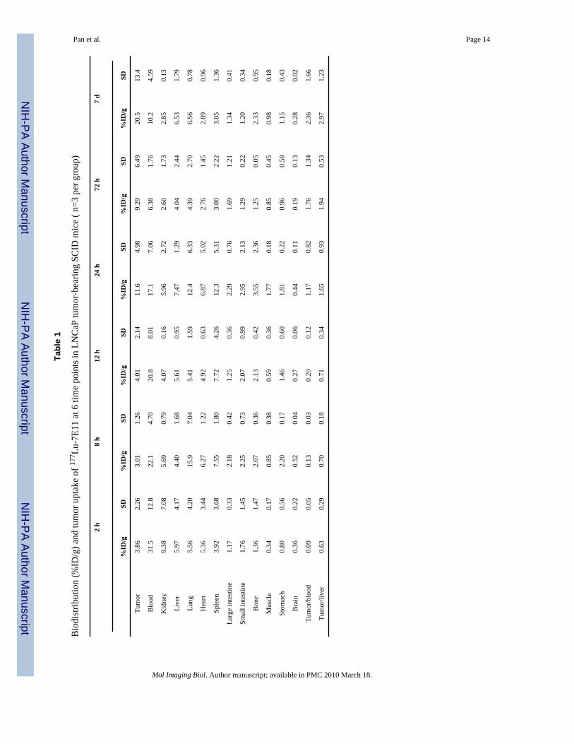
Biodistribution StudiesThe results of all biodistribution studies in xenograft-containing mice are summarized in Figure1 and Figure 2 and detailed in Table 1 and Table 2.
Uptake and retention of the 177Lu-7E11 conjugates within tumor was most prominent after 24hours, following a blood pool phase in which uptake was dominated by the blood, lung, andliver. Tumor uptake of the radioimmunoconjugates continued for the entire time course studied(7 days), and ratio of uptake within the tumor compared to other organs increased over the 7day time course following initial administration. Percentage of injected dose per gram (%ID/g) for LNCaP xenograft was 11.6 and 20.5 at 24 h and 7 d, respectively. The tumor to bloodratio (%ID/g / %ID/g) was 0.094 ± 0.051 at 2 h, 1.7± 1.3 at 72 h, and 2.00±1.7 at 7 d followinginitial injection (Figure 5). The presence of radiolabeled conjugates in the blood pool decreasedover time, the uptake in liver remained constant over 7 days.
The patterns of 111In-7E11 conjugates in tumor and blood did not differ significantly betweenmice administered with 177Lu-7E11 versus 111In-7E11; however uptakes (%ID/g)of 111In-7E11 in tumor and blood were less than those of 177Lu-7E11. Total accumulation intumor using 177Lu-7E11 was notably higher than 111In-7E11 conjugates 24 hours
Pan et al. Page 4
Mol Imaging Biol. Author manuscript; available in PMC 2010 March 18.
NIH
-PA Author Manuscript
NIH
-PA Author Manuscript
NIH
-PA Author Manuscript

postinjection. 177Lu-7E11 and 111In-7E11 %ID/g were 11.6 and 4.5 at 24 hours, and 9.3 and4.0 at 72 hours, respectively. The uptake in tumor reached a plateau after 24 hours; whereasthe uptake in other organs (lung, blood, kidney and spleen) had initial prominent retentionwhich decreased with time. Hepatic uptake, unlike other organs, continued to rise after 72hours. Uptake of 111In-7E11 was significantly lower (p<0.05) than that of 177Lu-7E11 studiesat all time points. This result was likely due to the DOTA chelator for 177Lu-7E11 which couldhave resulted in little nonspecific radiation dose without secondary loss of the radionuclidefrom the bifunctional DTPA chelator used for 111In-7E11. In addition, although the initialtumor sizes for both groups were similar, tumor sizes for the 177Lu-7E11 group were relativelysmaller (p<0.15) at the time points since 24 h, which could have resulted in higher %ID/g intumors for the 177Lu-7E11 group. This tumor size change could be due to the therapeuticradiation dose from 177Lu.
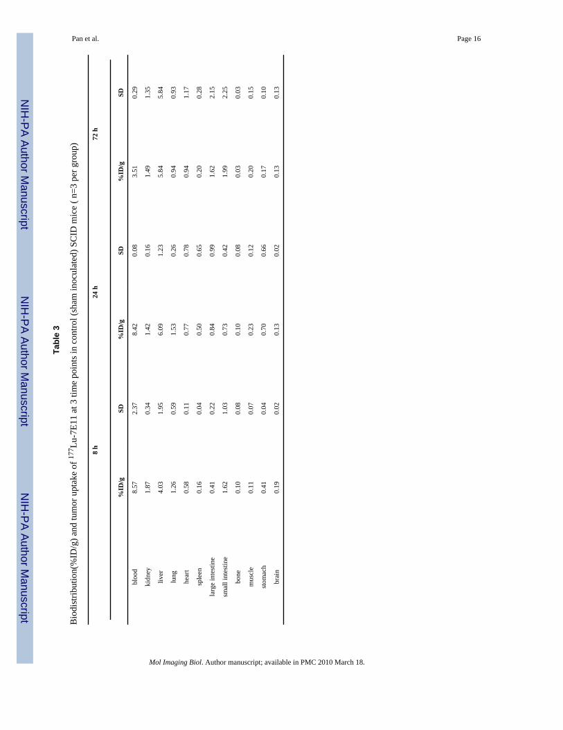
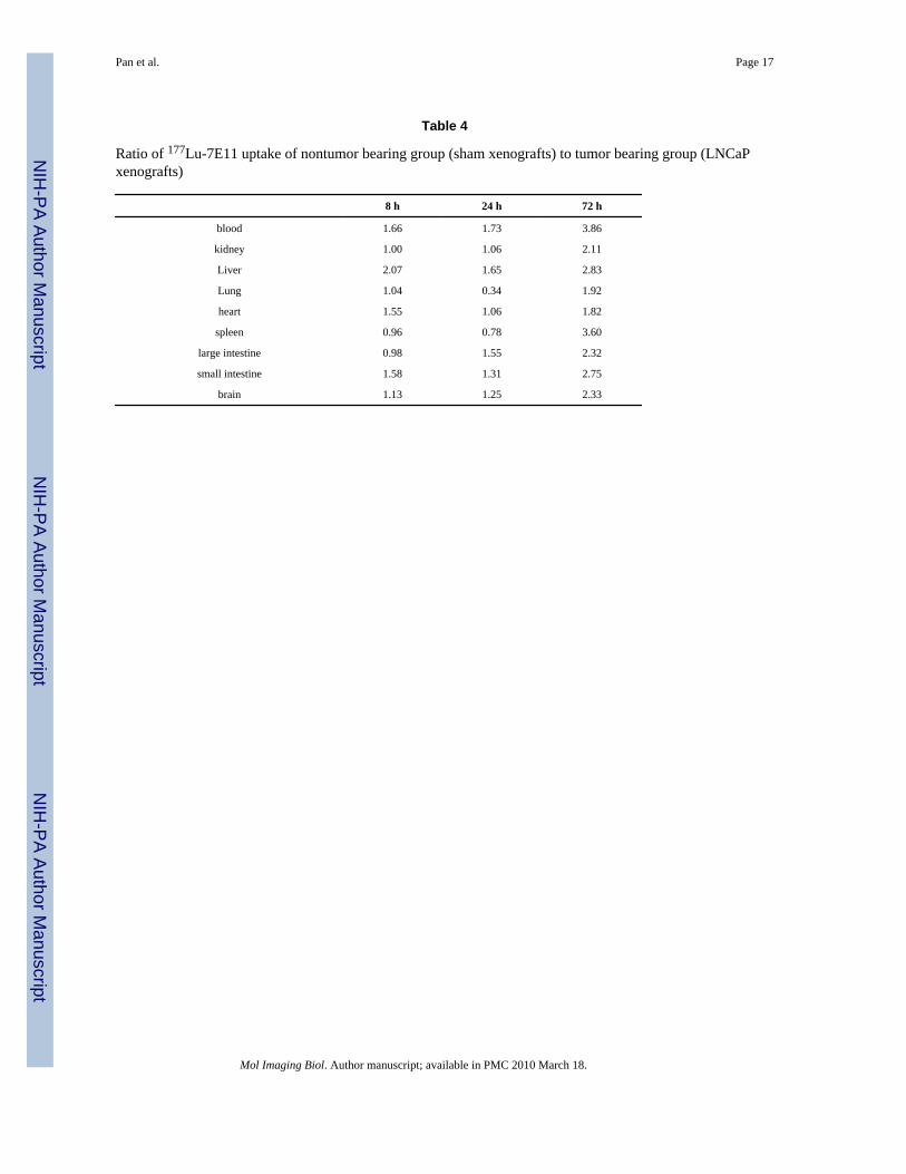
177Lu-7E11 results from sham xenograft control mice did not demonstrate measurable uptakeof the radioimmunoconjugates in the region of the sham tumor inoculation over that ofbackground soft tissue (Table 3). Compared to the tumor-bearing groups at matched timepoints, the %ID/g uptake and retention of radioconjugated MAb were increased in the bloodand liver at 8 and 24 hours following administration (p<0.05) with a 2 to 3-fold increase in allmajor organs except lung and heart at 72 hours after injection (p<0.05) (Table 4). Results forthe 111In-7E11 biodistribution studies in sham xenograft control mice were similar.
Bioluminescence ImagingBioluminescence imaging performed prior to administration of the radioimmunoconjugatesdemonstrated presence of viable LNCaP tumor in all mice studied. SPECT imagingof 177Lu-7E11 and 111In-7E11 confirmed the ex vivo biodistribution studies, and demonstrateda high tumor-to-background ratio with tumor uptake easily distinguishable from uptake insurrounding soft tissues on SPECT images at the 24 h time point and beyond. Representativebioluminescence and SPECT images of 177Lu-7E11 distribution at 24 h after initialradiopharmaceutical administration from the same animal are shown in Figure 5.
AutoradiographyRepresentative autoradiography images of 177Lu-7E11 tumor localization at 2, 24, and 72 hare shown in Figure 5. At later time points, signs of necrosis were shown particularly in theautoradiograph at 72 h. Nevertheless, within the tumor boundaries, 7E11 MAb distributionwas homogeneous within all tumors.
DiscussionThe history of the use of 111In-7E11 as an FDA-approved imaging agent (capromab pendetide),its retained targeting ability during concurrent hormonal therapy, and the recent success anddemonstrated stability of the conjugation of 7E11 with DOTA support the potential useof 177Lu-7E11 as a radioimmunotherapeutic for prostate cancer.
The 7E11 PSMA antibody has been constantly compared with other PSMA antibodies, mostnotably J591. The major difference between 7E11 and J591 is that 7E11 has been shown totarget an epitope on the intracellular portion of PSMA, while J591 targets an external membranePSMA epitope [18]. It has been suggested that the external membrane PSMA epitope shouldbe available for binding on viable prostate cancer cells; whereas the internal epitope, targetedby the 7E11, is only available once cells have undergone necrosis or apoptosis. PSMA isexpressed on the apical surface of the prostate cancer epithelium where tight junctions actuallyimpede accessibility of intravenously-injected agents to the apical side of the epithelial cell,so even antibodies to the extracellular surface would probably not be able to reach the intact
Pan et al. Page 5
Mol Imaging Biol. Author manuscript; available in PMC 2010 March 18.
NIH
-PA Author Manuscript
NIH
-PA Author Manuscript
NIH
-PA Author Manuscript
tissues of viable well-differentiated prostate cancer cells [19]. This suggests that 7E11 is notnecessarily at a true disadvantage when compared to antibodies targeting the external epitopeof PSMA when used as potential radiotherapeutics for prostate cancer. However, as suggestedby other authors, there may be a need for enhanced vascular permeability and intraepithelialtransport of any of these agents, an area that will require further research and innovation [19].
In addition, the choice of 177Lu as a radiotherapeutic agent for the 7E11 PSMA antibody iswell compared with the current Phase I clinical trial of J591 antibody. The clinical trials ofJ591 antibodies radiolabeled with either yttrium-90 (90Y) or lutetium-177 (177Lu) have beenperformed to assess toxicity and initial potential to treat large (28 to 42 mm diameter) and small(1.2 to 3 mm diameter) prostate tumor sites [20,21]. These trials have demonstrated a highpotential for delivering multiple doses of radiation to prostate cancer with acceptable toxicity.Due to the long range of their β− particle, 90Y-labeled agents provide a greater radiation doseto tumors with larger diameters. 177Lu-labeled agents emit a shorter range β− particle enablinghigh radiation doses to tumors of smaller diameters with minimal radiation to nearby structures.The choice of 177Lu as the therapeutic radionuclide in the present study represents a decisionto focus on smaller foci of prostate cancer present within lymph nodes or other small metastases[22]. Our choice of 177Lu as a radioimmunotherapy agent for the 7E11 antibody lies in thesame rationale. Besides, because of gamma emission from 177Lu, 177Lu-labeled agents can beimaged in its own right, and produce generally good images comparable to even imaging-only 111In-labeled agents due to less septal penetration of gamma camera collimators shownin our studies presented in this paper.
7E11 labeled with 111In has been used for targeted prostate cancer imaging with SPECT forseveral years, but this study represents the first published report of investigation into itssuitability for radioimmunotherapy when labeled with 177Lu. Using the LNCaP xenograftmouse model, this study investigated the biodistribution of 177Lu-labeled 7E11 over a 7 dayperiod. The presented findings indicate that 177Lu-7E11 localizes within LNCaP PSMA-expressing tumors. The tumor-to-background uptake of 177Lu-7E11 was observed to increaseover a 7 day period and its uptake was measured to be 20.5% ±13.3%ID/g at 7 days followingadministration. This high fractional uptake suggests the potential radioimmunotherapeuticutility of this agent. However, there were also relatively high blood values for all the timepoints, which may have contributed also high fractional tumor uptake of 177Lu-7E11 at latertime points. This prolonged blood uptake values may underestimate the potential efficacy ofthe 177Lu-7E11 for tumor sites, and may limit the administration dose for radioimmunotherapybecause of high bone marrow exposure from the blood uptake. For this reason, a carefuldosimetry study of 177Lu-7E11 will be needed.
This study also investigated the similarities and differences in the biodistributions of 111In-and 177Lu-labeled 7E11 PSMA antibody. The extensive experience with 111In-7E11 in NuclearMedicine community suggests that this diagnostic agent could provide pretherapeuticdosimetry estimates for 177Lu-7E11 therapy. 111In-7E11 is recognized to present somechallenges when used alone as imaging agent. These challenges include high nonspecificuptake in background soft tissues, and long tumor uptake times. Nevertheless, 111In-7E11 hasbeen successfully implemented as a prostate cancer imaging tool at many healthcare centers.Furthermore, the use of combined dual-modality SPECT/CT imaging has potential of increasedsensitivity of radioimmunoscintigraphy in general, and the addition of corrections for photonattenuation and geometric blurs caused by radionuclide collimator has shown the increase ofthe tumor target-to-background ratio of 111In-capromab pendetide imaging [23]. As a result,the overall sophistication of PSMA immunoscintigraphy with 111In-capromab pendetide hasincreased significantly over the past few years, and it would be optimal to capture thatexperience by utilizing 111In-7E11 for pretherapeutic imaging. Thus, the comparison study ofthe biodistributions of 111In-7E11 and 177Lu-7E11, presented in this paper, is the first step in
Pan et al. Page 6
Mol Imaging Biol. Author manuscript; available in PMC 2010 March 18.
NIH
-PA Author Manuscript
NIH
-PA Author Manuscript
NIH
-PA Author Manuscript

determining the potential role of 111In-7E11 as a pretherapeutic imaging agent. The presentstudy demonstrates the feasibility to apply 111In-7E11 as a pretherapeutic agent to predictuptake of the radioimmunotherapeutic in tumor and major organs, except liver. In order todetermine whether 111In-7E11 reflects the anticipated uptake of 177Lu-7E11 accurately, furtherstudies are required to determine whether their uptake is proportional within the same animaland tumor focus. One of the future studies needs to focus on 111In-7E11 with the DOTAchelator that is used in 177Lu-7E11 so that there is little variability of chelation of 7E11 antibodywith radiometals (111In and 177Lu) to potentially minimize the differences betweenbiodistributions of these two agents.
ConclusionsBiodistribution studies of 177Lu-7E11 in LNCaP xenograft mouse models support its potentialapplication as a radioimmunotherapeutic for prostate cancer by demonstrating its accumulationin prostate cancer xenografts. Companion studies of 111In-7E11 (DTPA) versus 177Lu-7E11(DOTA) biodistributions support its potential as a pretherapy imaging agent to predict thepotential uptake of the 177Lu-7E11 agent within prostate tumor. The gamma emission fromthe 177Lu-7E11 radiotherapeutic may also be used for posttreatment dosimetry studies andcomparison to pretherapy 111In-7E11 imaging. To further validate these points, biodistributionstudies with a larger number of animals and the same DOTA chelators for 111In-7E11and 177Lu-7E11 would be desirable.
AcknowledgmentsResearch Funding and 7E11 antibody were provided by the Cytogen Corporation for this project. LNCaP cellstransfected with luciferase were kindly supplied by the Contag laboratory at Stanford University. The authors alsoacknowledge support from NIH Grant 5R01EB000348 and 5K25CA114254.
References1. Swanson GP, Thompson IM, Basler J. Current status of lymph node-positive prostate cancer: Incidence
and predictors of outcome. Cancer 2006;107:439–450. [PubMed: 16795064]2. Swanson GP, Thompson IM, Basler J. Treatment options in lymph node-positive prostate cancer.
Cancer 2006;106:2531–2539. [PubMed: 16700035]3. Yagoda A, Petrylak D. Cytotoxic chemotherapy for advanced hormone-resistant prostate cancer.
Cancer 1993;71:1098–1109. [PubMed: 7679039]4. Bander NH, Milowsky MI, Nanus DM, Kostakoglu L, Vallabhajosula S, Goldsmith SJ. Phase I trial
of 177lutetium-labeled J591, a monoclonal antibody to prostate-specific membrane antigen, in patientswith androgen-independent prostate cancer. J Clin Oncol 2005;23:4591–4601. [PubMed: 15837970]
5. Russell PJ, Hewish D, Carter T, et al. Cytotoxic properties of immunoconjugates containing melittin-like peptide 101 against prostate cancer: in vitro and in vivo studies. Cancer Immunol Immunother2004;53:411–421. [PubMed: 14722668]
6. Smith-Jones PM, Vallabahajosula S, Goldsmith SJ, Navarro V, Hunter CJ, Bastidas D, Bander NH.In vitro characterization of radiolabeled monoclonal antibodies specific for the extracellular domainof prostate-specific membrane antigen. Cancer Res 2000;60:5237–5243. [PubMed: 11016653]
7. Wright GL Jr, Grob BM, Haley C, et al. Upregulation of prostate-specific membrane antigen afterandrogen-deprivation therapy. Urology 1996;48:326–334. [PubMed: 8753752]
8. Fair WR, Israeli RS, Heston WD. Prostate-specific membrane antigen. Prostate 1997;32:140–148.[PubMed: 9215402]
9. Conway RE, Petrovic N, Li Z, Heston W, Wu D, Shapiro LH. Prostate-specific membrane antigenregulates angiogenesis by modulating integrin signal transduction. Mol Cell Biol 2006;26:5310–5324.[PubMed: 16809768]
Pan et al. Page 7
Mol Imaging Biol. Author manuscript; available in PMC 2010 March 18.
NIH
-PA Author Manuscript
NIH
-PA Author Manuscript
NIH
-PA Author Manuscript
10. Troyer JK, Beckett ML, Wright GL Jr. Detection and characterization of the prostate-specificmembrane antigen (PSMA) in tissue extracts and body fluids. Int J Cancer 1995;62:552–558.[PubMed: 7665226]
11. Troyer JK, Beckett ML, Wright GL Jr. Location of prostate-specific membrane antigen in the LNCaPprostate carcinoma cell line. Prostate 1997;30:232–242. [PubMed: 9111600]
12. Horoszewicz JS, Kawinski E, Murphy GP. Monoclonal antibodies to a new antigenic marker inepithelial prostatic cells and serum of prostatic cancer patients. Anticancer Res 1987;7:927–935.[PubMed: 2449118]
13. Barren RJ 3rd, Holmes EH, Boynton AL, Misrock SL, Murphy GP. Monoclonal antibody 7E11.C5staining of viable LNCaP cells. Prostate 1997;30:65–68. [PubMed: 9018338]
14. Elgamal AA, Troychak MJ, Murphy GP. ProstaScint scan may enhance identification of prostatecancer recurrences after prostatectomy, radiation, or hormone therapy: analysis of 136 scans of 100patients. Prostate 1998;37:261–269. [PubMed: 9831223]
15. Feneley MR, Jan H, Granowska M, et al. Imaging with prostate-specific membrane antigen (PSMA)in prostate cancer. Prostate Cancer Prostatic Dis 2000;3:47–52. [PubMed: 12497162]
16. Tedesco J, Goeckeler W, Becker M, Frank K, Gulyas G, Young S. Development of Op-timal Lu-177Labeled Monoclonal Antibody (7E11) Construct (CYT-500) for Radioimmunotherapy of HormoneRefractory Prostate Cancer. J Clin Oncol 2005;23(16S):4765.
17. Tedesco J, Wolodzco J, Goeckeler W, Becker M, Hasegawa B, Franc B. Development andcharacterization of a novel 177Lu-MeO-DOTA-7E11 antibody construct (CYT-500) for thetreatment and imaging of prostate adenocarcinoma. AACR Annual Meeting. 2006
18. Nargund V, Al Hashmi D, Kumar P, et al. Imaging with radiolabelled monoclonal antibody (MUJ591)to prostate-specific membrane antigen in staging of clinically localized prostatic carcinoma:comparison with clinical, surgical and histological staging. BJU Int 2005;95:1232–1236. [PubMed:15892807]
19. Christiansen JJ, Rajasekaran SA, Inge L, Cheng L, Anilkumar G, Bander NH, Rajasekaran AK. N-glycosylation and microtubule integrity are involved in apical targeting of prostate-specificmembrane antigen: implications for immunotherapy. Mol Cancer Ther 2005;4:704–714. [PubMed:15897234]
20. Bander NH, Trabulsi EJ, Kostakoglu L, et al. Targeting metastatic prostate cancer with radiolabeledmonoclonal antibody J591 to the extracellular domain of prostate specific membrane antigen. J Urol2003;170:1717–1721. [PubMed: 14532761]
21. Milowsky MI, Nanus DM, Kostakoglu L, Vallabhajosula S, Goldsmith SJ, Bander NH. Phase I trialof yttrium-90-labeled anti-prostate-specific membrane antigen monoclonal antibody J591 forandrogen-independent prostate cancer. J Clin Oncol 2004;22:2522–2531. [PubMed: 15173215]
22. O'Donoghue JA, Bardies M, Wheldon TE. Relationships between tumor size and curability foruniformly targeted therapy with beta-emitting radionuclides. J Nucl Med 1995;36:1902–1909.[PubMed: 7562062]
23. Seo Y, Wong KH, Sun M, Franc BL, Hawkins RA, Hasegawa BH. Correction of photon attenuationand collimator response for a body-contouring SPECT/CT imaging system. J Nucl Med2005;46:868–877. [PubMed: 15872362]
Pan et al. Page 8
Mol Imaging Biol. Author manuscript; available in PMC 2010 March 18.
NIH
-PA Author Manuscript
NIH
-PA Author Manuscript
NIH
-PA Author Manuscript

Figure 1.Summary of pharmacodynamics for 177Lu-7E11 in LNCaP xenograft murine model.
Pan et al. Page 9
Mol Imaging Biol. Author manuscript; available in PMC 2010 March 18.
NIH
-PA Author Manuscript
NIH
-PA Author Manuscript
NIH
-PA Author Manuscript
Figure 2.Summary of pharmacodynamics 111In-7E11 in LNCaP xenograft murine model.
Pan et al. Page 10
Mol Imaging Biol. Author manuscript; available in PMC 2010 March 18.
NIH
-PA Author Manuscript
NIH
-PA Author Manuscript
NIH
-PA Author Manuscript

Figure 3.Graphical representation of tumor-to-blood uptake ratio of 177Lu-7E11 and 111In-7E11 overtime (%ID/g).
Pan et al. Page 11
Mol Imaging Biol. Author manuscript; available in PMC 2010 March 18.
NIH
-PA Author Manuscript
NIH
-PA Author Manuscript
NIH
-PA Author Manuscript
Figure 4.a) In vivo Bioluminescence image demonstrating location of luciferase-transfected LNCaPtumor and b) representative coronal image from in vivo SPECT imaging 24 hoursfollowing 177Lu-7E11 (40 MBq) administration.
Pan et al. Page 12
Mol Imaging Biol. Author manuscript; available in PMC 2010 March 18.
NIH
-PA Author Manuscript
NIH
-PA Author Manuscript
NIH
-PA Author Manuscript
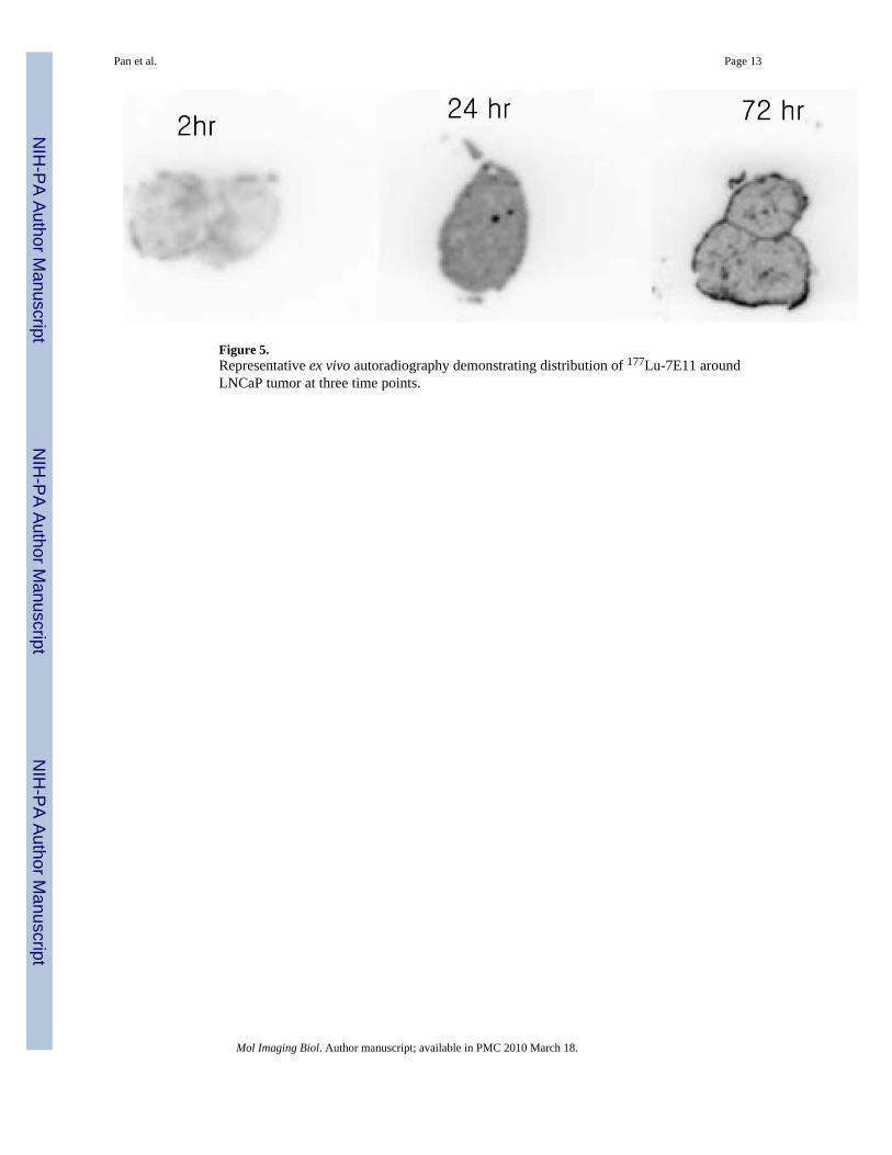
Figure 5.Representative ex vivo autoradiography demonstrating distribution of 177Lu-7E11 aroundLNCaP tumor at three time points.
Pan et al. Page 13
Mol Imaging Biol. Author manuscript; available in PMC 2010 March 18.
NIH
-PA Author Manuscript
NIH
-PA Author Manuscript
NIH
-PA Author Manuscript
NIH
-PA Author Manuscript
NIH
-PA Author Manuscript
NIH
-PA Author Manuscript
Pan et al. Page 14
Tabl
e 1
Bio
dist
ribut
ion
(%ID
/g) a
nd tu
mor
upt
ake
of 17
7 Lu-
7E11
at 6
tim
e po
ints
in L
NC
aP tu
mor
-bea
ring
SCID
mic
e ( n
=3 p
er g
roup
)
2 h
8 h
12 h
24 h
72 h
7 d
%ID
/gSD
%ID
/gSD
%ID
/gSD
%ID
/gSD
%ID
/gSD
%ID
/gSD
Tum
or3.
862.
263.
011.
264.
012.
1411
.64.
989.
296.
4920
.513
.4
Blo
od31
.512
.822
.14.
7020
.88.
0117
.17.
066.
381.
7610
.24.
59
Kid
ney
9.38
7.08
5.69
0.79
4.07
0.16
5.96
2.72
2.60
1.73
2.85
0.13
Live
r5.
974.
174.
401.
685.
610.
957.
471.
294.
042.
446.
531.
79
Lung
5.56
4.20
15.9
7.04
5.41
1.59
12.4
6.33
4.39
2.70
6.56
0.78
Hea
rt5.
363.
446.
271.
224.
920.
636.
875.
022.
761.
452.
890.
96
Sple
en3.
923.
687.
551.
807.
724.
2612
.35.
313.
002.
223.
051.
36
Larg
e in
test
ine
1.17
0.33
2.18
0.42
1.25
0.36
2.29
0.76
1.69
1.21
1.34
0.41
Smal
l int
estin
e1.
761.
452.
250.
732.
070.
992.
952.
131.
290.
221.
200.
34
Bon
e1.
361.
472.
070.
362.
130.
423.
552.
361.
250.
052.
330.
95
Mus
cle
0.34
0.17
0.85
0.38
0.59
0.36
1.77
0.18
0.85
0.45
0.98
0.18
Stom
ach
0.80
0.56
2.20
0.17
1.46
0.60
1.81
0.22
0.96
0.58
1.15
0.43
Bra
in0.
360.
220.
520.
040.
270.
060.
440.
110.
190.
130.
280.
02
Tum
or/b
lood
0.09
0.05
0.13
0.03
0.20
0.12
1.17
0.82
1.76
1.34
2.36
1.66
Tum
or/li
ver
0.63
0.29
0.70
0.18
0.71
0.34
1.65
0.93
1.94
0.53
2.97
1.23
Mol Imaging Biol. Author manuscript; available in PMC 2010 March 18.

NIH
-PA Author Manuscript
NIH
-PA Author Manuscript
NIH
-PA Author Manuscript
Pan et al. Page 15
Tabl
e 2
Bio
dist
ribut
ion(
%ID
/g) a
nd tu
mor
upt
ake
of 11
1 In-
7E11
at 4
tim
e po
ints
in L
NC
aP tu
mor
-bea
ring
SCID
mic
e ( n
=3 p
er g
roup
)
8 h
12 h
24 h
72 h
%ID
/gSD
%ID
/gSD
%ID
/gSD
%ID
/gSD
Tum
or3.
380.
903.
001.
724.
476.
014.
014.
14
Blo
od5.
823.
012.
641.
714.
811.
643.
100.
95
Kid
ney
6.76
3.12
2.20
0.77
3.67
0.64
3.77
0.22
Live
r2.
050.
321.
300.
542.
481.
153.
180.
35
Lung
11.1
48.
264.
391.
323.
971.
922.
110.
65
Hea
rt9.
604.
453.
850.
494.
282.
821.
210.
17
Sple
en7.
824.
425.
801.
742.
730.
441.
851.
14
Larg
e in
test
ine
2.56
1.49
1.48
0.35
0.98
0.28
0.62
0.18
Smal
l int
estin
e2.
601.
092.
180.
611.
200.
130.
660.
38
Bon
e0.
620.
271.
580.
191.
330.
790.
470.
14
Mus
cle
0.51
0.25
0.94
0.14
0.74
0.43
0.43
0.01
Stom
ach
1.38
0.24
1.60
0.39
0.70
0.26
0.34
0.02
Bra
in0.
850.
420.
310.
040.
270.
060.
120.
00
Tum
or/b
lood
0.58
1.14
0.93
1.30
Tum
or/li
ver
1.65
2.32
1.80
1.26
Mol Imaging Biol. Author manuscript; available in PMC 2010 March 18.
NIH
-PA Author Manuscript
NIH
-PA Author Manuscript
NIH
-PA Author Manuscript
Pan et al. Page 16
Tabl
e 3
Bio
dist
ribut
ion(
%ID
/g) a
nd tu
mor
upt
ake
of 17
7 Lu-
7E11
at 3
tim
e po
ints
in c
ontro
l (sh
am in
ocul
ated
) SC
ID m
ice
( n=3
per
gro
up)
8 h
24 h
72 h
%ID
/gSD
%ID
/gSD
%ID
/gSD
bloo
d8.
572.
378.
420.
083.
510.
29
kidn
ey1.
870.
341.
420.
161.
491.
35
liver
4.03
1.95
6.09
1.23
5.84
5.84
lung
1.26
0.59
1.53
0.26
0.94
0.93
hear
t0.
580.
110.
770.
780.
941.
17
sple
en0.
160.
040.
500.
650.
200.
28
larg
e in
test
ine
0.41
0.22
0.84
0.99
1.62
2.15
smal
l int
estin
e1.
621.
030.
730.
421.
992.
25
bone
0.10
0.08
0.10
0.08
0.03
0.03
mus
cle
0.11
0.07
0.23
0.12
0.20
0.15
stom
ach
0.41
0.04
0.70
0.66
0.17
0.10
brai
n0.
190.
020.
130.
020.
130.
13
Mol Imaging Biol. Author manuscript; available in PMC 2010 March 18.
NIH
-PA Author Manuscript
NIH
-PA Author Manuscript
NIH
-PA Author Manuscript
Pan et al. Page 17
Table 4
Ratio of 177Lu-7E11 uptake of nontumor bearing group (sham xenografts) to tumor bearing group (LNCaPxenografts)
8 h 24 h 72 h
blood 1.66 1.73 3.86
kidney 1.00 1.06 2.11
Liver 2.07 1.65 2.83
Lung 1.04 0.34 1.92
heart 1.55 1.06 1.82
spleen 0.96 0.78 3.60
large intestine 0.98 1.55 2.32
small intestine 1.58 1.31 2.75
brain 1.13 1.25 2.33
Mol Imaging Biol. Author manuscript; available in PMC 2010 March 18.