Alpha-tocopheryl succinate (α-TOS) modulates human prostate LNCaP xenograft growth and gene...
10
Arpita Basu Bruce Grossie Michael Bennett Nat Mills Vicky Imrhan Alpha-tocopheryl succinate (a-TOS) modulates human prostate LNCaP xenograft growth and gene expression in BALB/c nude mice fed two levels of dietary soybean oil Received: 26 August 2006 Accepted: 31 October 2006 Published online: 15 December 2006 j Abstract Background Malig- nancy of the prostate constitutes a leading cause of cancer-related deaths in America and Europe. Alpha-tocopheryl succinate (a- TOS) has been shown to inhibit human prostate cancer growth in vitro, via several mechanisms, including inhibiting prostate-spe- cific antigen (PSA) and vascular endothelial growth factor (VEGF) expressions. The route of a-TOS administration has a profound effect on its antitumor activity, and few studies have investigated its effects on prostate cancer growth in vivo. Aim of the Study The present study tested the hypothesis that a-TOS wil reduce the growth of human prostate LNCaP tumors in mice fed low (7%) and high (20%) levels of dietary soybean oil, compared to the controls receiving vehicle, by modulating PSA and VEGF gene expressions in the tumor tissue. Methods BALB/c nude mice (n = 42) were subcutaneously inocu- lated with 1 · 10 6 LNCaP cells and assigned to one of four dietary groups; 7% or 20% soybean oil diet with or without a-TOS treat- ment. Three weeks later, mice received daily intraperitoneal injections of a-TOS (100 mg/kg body weight) in sesame seed oil (SSO) for two weeks; controls received SSO injections. Tumor volumes were recorded weekly. Sera, liver, and tumor tissues were collected at seven weeks for serum PSA, testosterone and a-tocoph- erol analyses, histopathological examination, and reverse tran- scription and polymerase chain reaction (RT-PCR) amplification of PSA and VEGF gene fragments in tumors. Relative quantification of gene expression was performed using real-time PCR. P £ 0.05 was considered significant. Results Intraperitoneal injections of a- TOS caused decreased tumor growth in both groups (7% and 20% fat, P < 0.05), versus controls. a-TOS treatment significantly re- duced serum PSA and testosterone levels in comparison to the SSO- treated controls (P < 0.05). Con- trol tumors had a greater degree of angiogenesis than a-TOS tumors, as demonstrated by the greater number of blood-filled vessels. PSA and VEGF mRNA expres- sions, were also reduced with a- TOS treatment (P < 0.05), reveal- ing the possible molecular mech- anisms of growth inhibition of LNCaP xenografts by a-TOS. Con- clusions Our study shows signifi- cant reduction in LNCaP xenograft growth with a-TOS treatment in nude mice fed a low (7%) and high (20%) fat soybean oil diets versus ORIGINAL CONTRIBUTION Eur J Nutr (2007) 46:34–43 DOI 10.1007/s00394-006-0629-4 EJN 629 This research work won three best poster awards at Experimental Biology 2005 (San Diego, CA, USA), at three different Research Interest Sections (RIS) competi- tions, organized by the American Society for Nutritional Sciences (ASNS)- Diet & Cancer RIS, Vitamin & Mineral RIS, Nutrient-Gene RIS. A. Basu, PhD (&) Nutritional Sciences, 301 Human Environmental Sciences Oklahoma State University Stillwater (OK) 74078, USA Tel.: +1-405/744-4437 Fax: +1-405/744-1357 E-Mail: [email protected] B. Grossie V. Imrhan Dept. of Nutrition & Food Sciences Texas Woman’s University Denton (TX), USA M. Bennett Pathology UT Southwestern Medical Center Dallas (TX), USA N. Mills Dept. of Biology Texas Woman’s University Denton (TX), USA
-
Upload
independent -
Category
Documents
-
view
0 -
download
0
Transcript of Alpha-tocopheryl succinate (α-TOS) modulates human prostate LNCaP xenograft growth and gene...
Arpita BasuBruce GrossieMichael BennettNat MillsVicky Imrhan
Alpha-tocopheryl succinate (a-TOS)modulates human prostate LNCaPxenograft growth and gene expression inBALB/c nude mice fed two levels of dietarysoybean oil
Received: 26 August 2006Accepted: 31 October 2006Published online: 15 December 2006
j Abstract Background Malig-nancy of the prostate constitutes aleading cause of cancer-relateddeaths in America and Europe.Alpha-tocopheryl succinate (a-TOS) has been shown to inhibithuman prostate cancer growth invitro, via several mechanisms,including inhibiting prostate-spe-cific antigen (PSA) and vascularendothelial growth factor (VEGF)expressions. The route of a-TOSadministration has a profoundeffect on its antitumor activity,and few studies have investigatedits effects on prostate cancergrowth in vivo. Aim of the StudyThe present study tested thehypothesis that a-TOS wil reducethe growth of human prostateLNCaP tumors in mice fed low(7%) and high (20%) levels ofdietary soybean oil, compared tothe controls receiving vehicle, bymodulating PSA and VEGF geneexpressions in the tumor tissue.Methods BALB/c nude mice (n =42) were subcutaneously inocu-lated with 1 · 106 LNCaP cells andassigned to one of four dietarygroups; 7% or 20% soybean oildiet with or without a-TOS treat-ment. Three weeks later, micereceived daily intraperitonealinjections of a-TOS (100 mg/kgbody weight) in sesame seed oil(SSO) for two weeks; controls
received SSO injections. Tumorvolumes were recorded weekly.Sera, liver, and tumor tissues werecollected at seven weeks for serumPSA, testosterone and a-tocoph-erol analyses, histopathologicalexamination, and reverse tran-scription and polymerase chainreaction (RT-PCR) amplificationof PSA and VEGF gene fragmentsin tumors. Relative quantificationof gene expression was performedusing real-time PCR. P £ 0.05 wasconsidered significant. ResultsIntraperitoneal injections of a-TOS caused decreased tumorgrowth in both groups (7% and20% fat, P < 0.05), versus controls.a-TOS treatment significantly re-duced serum PSA and testosteronelevels in comparison to the SSO-treated controls (P < 0.05). Con-trol tumors had a greater degree ofangiogenesis than a-TOS tumors,as demonstrated by the greaternumber of blood-filled vessels.PSA and VEGF mRNA expres-sions, were also reduced with a-TOS treatment (P < 0.05), reveal-ing the possible molecular mech-anisms of growth inhibition ofLNCaP xenografts by a-TOS. Con-clusions Our study shows signifi-cant reduction in LNCaP xenograftgrowth with a-TOS treatment innude mice fed a low (7%) and high(20%) fat soybean oil diets versus
ORIGINAL CONTRIBUTIONEur J Nutr (2007) 46:34–43DOI 10.1007/s00394-006-0629-4
EJN
629
This research work won three best posterawards at Experimental Biology 2005 (SanDiego, CA, USA), at three differentResearch Interest Sections (RIS) competi-tions, organized by the American Societyfor Nutritional Sciences (ASNS)- Diet &Cancer RIS, Vitamin & Mineral RIS,Nutrient-Gene RIS.
A. Basu, PhD (&)Nutritional Sciences, 301 HumanEnvironmental SciencesOklahoma State UniversityStillwater (OK) 74078, USATel.: +1-405/744-4437Fax: +1-405/744-1357E-Mail: [email protected]
B. Grossie Æ V. ImrhanDept. of Nutrition & Food SciencesTexas Woman’s UniversityDenton (TX), USA
M. BennettPathologyUT Southwestern Medical CenterDallas (TX), USA
N. MillsDept. of BiologyTexas Woman’s UniversityDenton (TX), USA
Introduction
Malignancy of the prostate constitutes the secondleading cause of cancer-related deaths in Americanmen, and is also the third most frequent cause ofcancer mortality among the European countries [1, 2].The androgen receptor (AR) is required for thedevelopment of both the normal prostate gland andprostate cancer. In the early stages of prostate cancer,almost all cancer cells are androgen-dependent andhighly sensitive to antiandrogen therapy. However,prostate cancer usually recurs after a few years ofandrogen ablative treatment, and most cancer cellsbecome androgen-independent, rendering antiandro-gen therapy useless. As androgen-independent pros-tate tumors are incurable at the present time, theprevention of such aberrant AR activation is anattractive therapeutic target [3].
Studies have shown that dietary micronutrientslike, carotenoids and lycopenes, retinoids and vitaminA, vitamin E [alpha (a) tocopherol and gamma (c)tocopherol], vitamin C, selenium, and phenol-con-taining dietary substances have effects that extendbeyond their antioxidant properties [4]. Alpha-toc-opheryl succinate (a-TOS) has been reported to be avery effective chemotherapeutic agent in prostatecancer. The proposed mechanisms of a-TOS action onhuman prostate cancer cells by various authors in-clude enhancing the chemotherapeutic effects ofadriamycin on human prostate cancer cells [5],inducing apoptosis by causing mobilization of Fasfrom the cytosol to the plasma membrane in humanprostate cancer cells [6], and decreasing the produc-tion of vascular endothelial growth factor (VEGF) byprostate cancer cells [7]. a-TOS has also been reportedto inhibit the transcription as well as translation ofprostate-specific antigen (PSA) and AR in humanprostate cancer cells, exert a synergistic effect incombination with hydroxyflutamide on growth inhi-bition of prostate cancer [8], and also inhibit humanprostate cancer cell invasiveness by inhibiting theactivity of matrix metalloproteinases [9]. Zu and Ip
[10] compared the antineoplastic action of a-tocoph-erol and its two ester derivatives, a-tocopheryl acetateand a-TOS on the growth of human prostate PC-3cancer cells in culture. Interestingly, a concentrationof 20 lM of a-TOS was sufficient to achieve a 40%growth inhibition of PC-3 cells after 72 h of treatment,whereas, neither a-tocopherol nor a-tocopheryl ace-tate was able to achieve the same after the same lengthof exposure and with a dose 10 times higher than thatof a-TOS. While various in vitro studies report thesuperiority of a-TOS as a chemopreventive agent, veryfew studies have reported similar in vivo effects of a-TOS. A recent study by Venkateswaran et al. [11]showed a significant reduction in prostate cancerincidence in male Lady transgenic mice fed a stan-dard-fat (25% kcal-fat) or a high-fat (40% kcal-fat)diet with antioxidant supplementation (800 IU of a-TOS, 200 lg of seleno-DL-methionine and 50 mg oflycopene) versus the controls. However, a-TOS is notan antioxidant, but becomes one once it is taken up bythe intestinal epithelial cells, and hydrolyzed to a-tocopherol. While normal cells, including intestinalepithelial cells, are capable of hydrolyzing a-TOS to a-tocopherol, cancer cells lack this ability. As a result,accumulation of the intact molecule of a-TOS in can-cer cells causes apoptosis [12]. Malafa and Neitzel [13]demonstrated that nude mice injected intraperitone-ally (ip) with a-TOS significantly lowered final weightof human breast cancer xenografts than the controlvehicle-treated group, whereas, no difference was no-ted between mice treated subcutaneously (sc) with a-TOS versus the control sc treated group. Thus, theroute of a-TOS administration has a profound effecton its antitumor activity in vivo. This effect remains tobe established in prostate cancer growth in vivo andmay produce a similar or even greater inhibitory effectthan reported with oral ingestion of a-TOS [11].
This study investigated the effects of a-TOS alone,administered intraperitoneally, on androgen-depen-dent human prostate LNCaP xenograft growth innude mice. Our study tested the hypothesis that a-TOS will reduce the growth of human prostate LNCaPtumors in mice fed low (7%) and high (20%) levels of
controls. Serum PSA and testos-terone, tumor angiogenesis, andPSA and VEGF mRNA expressionswere markedly reduced by a-TOSadministration, suggesting a pos-sible role of a-TOS as a chemo-
therapeutic agent in humanprostate cancer, and warrants fur-ther investigations on the doseand delivery of a-TOS in humans.
j Key words alpha-tocopherylsuccinate – prostate cancer –angiogenesis – prostate-specificantigen – xenograft growth
A. Basu et al. 35a-TOS modulates human prostate LNCaP xenograft growth
dietary soybean oil, compared to the controlsreceiving vehicle, by modulating PSA and VEGF geneexpressions in the tumor tissue.
Materials and methods
j Cell culture
The LNCaP cells, serum and the medium were ob-tained from the American Type Tissue Culture Col-lection (ATCC, Rockville, MD). The cells were grownin RPMI-1640 medium supplemented with 5% fetalcalf serum, nonessential amino acids, and L-glutaminein an atmosphere of 5% CO2/air at 37�C. The cells weregrown in flasks and passaged when they reachedsubconfluence and were treated briefly with trypsin/EDTA to put them in solution for each passage. Thecells were counted using a hemocytometer, tested forviability with exclusion by trypan blue dye, and weretaken for injection into mice when most of the cellswere in log phase growth; usually covering only 1/2 thesurface of the flask. Only cells with >90% viability wereused. The cells were kept on ice (4�C) until injected.
j Animals and diets
Forty-two male BALB/c nude mice, aged 8 weeks,were obtained from National Cancer Institute (Fred-erick, MD) and housed 4–5 animals per cage, in apathogen-free environment with microbe filter tops infiltered laminar-flow hoods. All mice were used forthe tumor implantation and were randomly assignedto the dietary experimental groups. All animals werecared for in accordance with the Institutional AnimalCare and Use Committee (IUCAC) guidelines of theUniversity of Texas at Southwestern Medical Center(Dallas, TX). The water and bedding were heat ster-ilized (autoclaved). In order to prevent sepsis byPseudomonus aeruginosa bacteria, 750 ll of Chlorox/bottle of 400 ml of drinking water were added.
The composition of the AIN-93G diets [14] wasmodified with added methionine and high-fat (20%soybean oil). The diets were isonitrogenous in termsof kilocalorie of the diet to ensure similar proteinintake levels in both groups. The diets were pelletedand irradiated by Bio-Serv (Frenchtown, NJ).
Food consumption, tumor volume and animalweights were recorded weekly. The diets were storedat 4�C and fresh diets were given every 3–4 days. Thetumor size was measured using a caliper and tumorvolume was calculated using the formula (cm3):length (L) · width (W) · height (H) · 0.5236. Tumorweight was estimated from tumor volume, as thedensity of tissue is 1 g/ml. Body weight of each animal
was then determined by subtracting tumor weightfrom total body weight.
j Experimental design
This was a 7-week study design, starting from inoc-ulation of the animals with LNCaP cells. LNCaP cellsin culture were resuspended in ice-cold Matrigel(Collaborative Biomedical Products, Bedford, MA) ata concentration of 1 · 106/0.2 ml. Mice were theninoculated (27 gauge needle) subcutaneously in thehindlimb with 1 · 106 cells. All animals were fed thecontrol diet containing 7%-fat ad libitum for 1 weekprior to inoculation. Following inoculation, the ani-mals were randomly assigned to one of the fourgroups: 7% or 20% soybean oil diet, with or withouta-TOS treatment. All animals remained on theirrespective diets until the termination of the study.Three weeks after inoculation, when measurable tu-mors were formed in most of the animals (at least0.02 ± 0.005 cm3 in 80% of the mice in each group),the experimental groups received daily intraperito-neal (ip) injections of a-TOS in sesame oil (100 mg/kgbody weight) while the controls received a vehicle ofsesame oil for 2 weeks and the animals remained ontheir respective diets for the remaining 2 weeks of thestudy. a-TOS [all racemic (all rac) a-tocopheryl suc-cinate] was purchased from EMD Biosciences (SanDiego, CA) and sesame oil obtained from SigmaChemical Co. (St. Louis, MO). All animals were sac-rificed 7 weeks after inoculation and blood was col-lected through cardiac puncture. Sera were separatedand stored at )80 �C until analyses. Two hundredmilligrams of the LNCaP tumor tissue were weighedand preserved in RNAlater� (Ambion Inc.) for sub-sequent RNA extraction and reverse transcription-polymerase chain reaction (RT-PCR). A sample tumortissue was stored in 10% formalin in PBS for laterhistopathologic examination. Liver tissues were alsocollected and snap-frozen for a- tocopherol analyses.
j Analyses
Serum PSA and testosterone: Human PSA levels inserum were determined using an enzyme immuno-assay kit (ALPCO Diagnostics, Windham, NH). Totalserum testosterone was determined using enzyme-linked immunosorbent assays (ALPCO Diagnostics,Windham, NH). All the above assays were performedin duplicate. The interassay variability was <5.0%.
Liver tissue a-tocopherol analysis was doneaccording to the protocol of Devaraj and Jialal [15].Separation was performed on a reverse-phase column(C18) at 25�C and a-tocopherol was detected at
36 European Journal of Nutrition (2007) Vol. 46, Number 1� Steinkopff Verlag 2006
292 nm. The mobile phase used was methanol:aceto-nitrile:dichloroethane (60:20:20) with a flow rate of0.8 ml/min. a-tocopherol was extracted following thelipid extraction from the liver tissues according toHara & Radin [16].
Total RNA isolation and RT-PCR: Total cellularRNA was isolated from vehicle and a-TOS -treatedLNCaP xenografts (preserved in RNAlater� AmbionInc.) using TRIZOL reagent (Invitrogen, Carlsbad,CA) according to protocols provided by the manu-facturer. RNA purity was established by agarose gelelectrophoresis, and the amount of RNA was quanti-fied by measurement of the A260/280 nm ratio. Reversetranscription (RT) of RNA into cDNA was accom-plished with SuperScript� III reverse transcriptase(Invitrogen, Carlsbad, CA) according to the manu-facturer’s protocol. For real-time PCR, after a 1:200dilution, two microliters of the RT reaction and thecorresponding PCR primer set plus 25 ll of theSYBR� Green qPCR master mix (modified DyNAmohot start DNA polymerase, SYBR Green I, optimizedPCR buffer, 5 mM MgCl2, dNTP mix including dUTP)(MJ Research, Reno, NV) was added and then ad-justed to 50 ll with ultra pure (UP) H2O. Primers(5 ll each) were added for the following: PSA forwardand backward, 5¢-GGAACAAAAGCGTGATCTTGCTGGG-3¢ and 5¢-GGGTGAACTTGCGCACACACGTC-3¢, expected size 363 bp; VEGF forward and back-ward, 5¢-ATCTTCAAGCCATCCTGTGTGC-3¢ and 5¢-TCACCGCCTCGGCTTGTCACAT-3¢, expected size391, 365 and 291 bp (3 isoforms); and b2-microglob-ulin (housekeeping gene, internal control) forwardand backward, 5¢-ATGCCTGCCGTGTGAACCATGTG-3¢ and 5¢-AGAGCTACCTGTGGAGCAACCTG-3¢,expected size 280 bp [17–19].
Reactions were assembled at room temperature,and then loaded on the DNA Engine Opticon� 2System for continuous fluorescence detection (MJResearch, Inc. Watertown, MA). The thermal cyclingprotocol was 95�C for 15 min at hot start followed by40 cycles (60�C or in accord to primer melting pointsless 4–6�C for 1 min; 72�C for 2 min for primerextension and 94�C for 30 s for dsDNA templatedenaturation). Following PCR, in order to confirm asingle band dsDNA product as well as match thedsDNA size to the DNA standard, ten microliters ofeach PCR sample plus 2.0 ll of 30% glycerol withtracking dyes were loaded on a 2% (w/v) agarose geland separated by size using electrophoresis at 80 Vfor 90 min. DNA was then visualized by fluorescencefrom ethidium bromide staining (0.5 lg/ml) anddocumented photographically.
The Opticon Monitor software, version 2.02, wasused to calculate the initial quantity of template in asample. Each total RNA sample was analyzed twiceand in duplicate for amount of dsDNA from RT-PCR
for each gene. The ratio of PSA or VEGF/b2micro-globulin expression were used for comparison amongdifferent treatment groups at different time points.
j Tumor histology
Formalin-fixed tumor specimens were embedded inparaffin and sectioned at 5 lm thickness. Hematox-ylin and eosin (H & E)-stained slides were examinedby light microscopy, using a Nikon microscope. Wecharacterized tumor morphology and cytologic fea-tures. Tissue density was determined by measuringtotal tumor area and quantitating the proportion(expressed as %) occupied by tumor cells, compactconnective tissue matrix, blood-filled vessels, andareas of necrosis. Many blood vessels were readilyidentifiable and were quantified on the hematoxylin-eosin-stained sections because of the presence of eo-sin-stained red blood cells.
j Statistical analysis
All samples were analyzed using a two-way ANOVAfollowed by Tukey–Kramer multiple comparison teststo investigate the effects of dietary fat levels and a-TOS treatment on the growth of human prostateLNCaP tumor tissues. A P £ 0.05 was consideredsignificant. Simple correlation and linear regressiontests were performed to identify potential relation-ships among experimental parameters. The statisticalsoftware used was SPSS, version 11.5 for Windows.
Results
Table 1 shows average initial and final body weights,weight gain and daily intake of total feed and selectednutrients of nude mice from treatment and controlgroups. Mice fed ad libitum the 20%-fat diet con-sumed higher amounts of calorie and fats than thosefed the 7%-fat diet. In this study, a hypercaloric, high-fat diet (20%-fat) was shown to promote prostatetumor growth in nude mice in comparison to thetumor growth in mice that had a lower fat and ahigher carbohydrate intake.
The final tumor volumes at 7 weeks after inocula-tion (Fig. 1) were significantly lower in the a-TOS-treated groups than the controls for both levels ofdietary fat. Also, the mean tumor volume was lower forthe 7%-fat group (0.09 ± 0.05 cm3) compared to thosein the 20%-fat group (0.19 ± 0.1 cm3, P < 0.001). Theoverall treatment effect (P < 0.0001) showed that thea-TOS-treated mice had a lower tumor volume(0.06 ± 0.02 cm3) when compared to the vehicle-
A. Basu et al. 37a-TOS modulates human prostate LNCaP xenograft growth
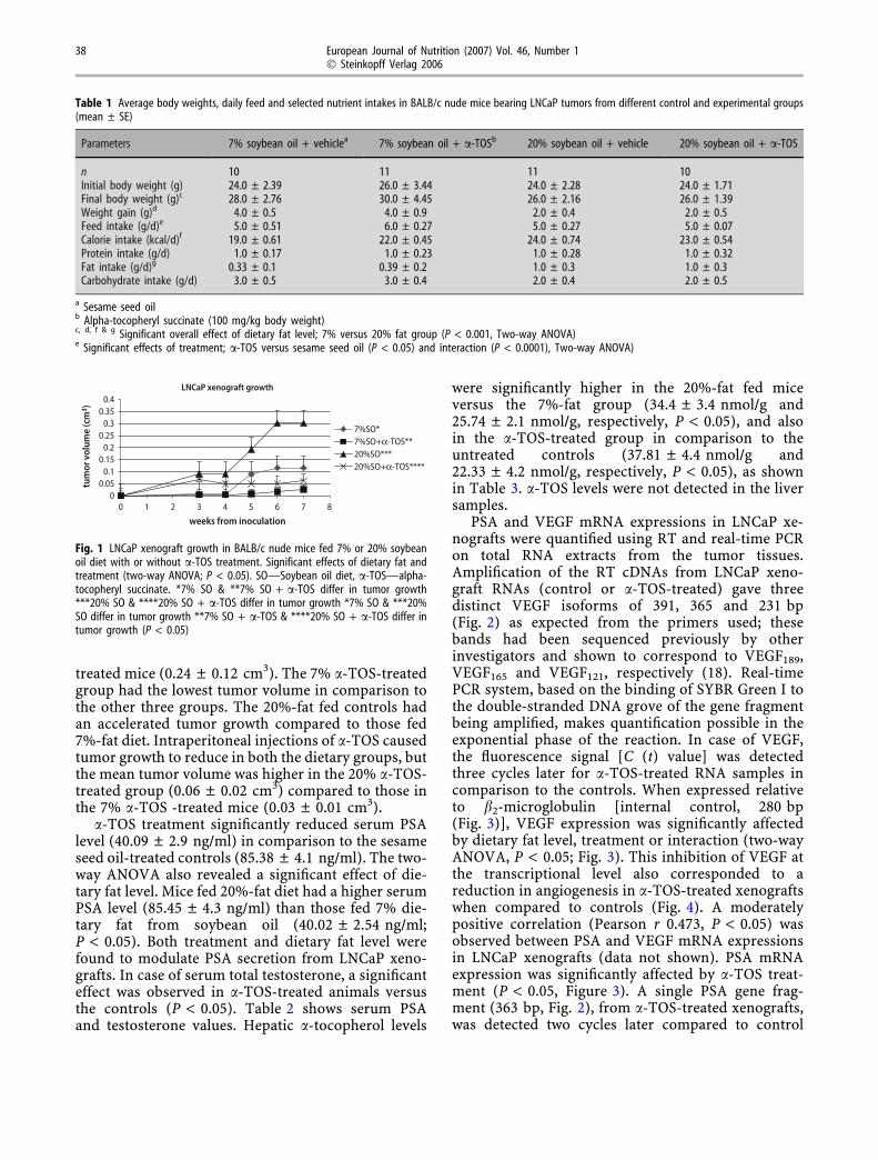
treated mice (0.24 ± 0.12 cm3). The 7% a-TOS-treatedgroup had the lowest tumor volume in comparison tothe other three groups. The 20%-fat fed controls hadan accelerated tumor growth compared to those fed7%-fat diet. Intraperitoneal injections of a-TOS causedtumor growth to reduce in both the dietary groups, butthe mean tumor volume was higher in the 20% a-TOS-treated group (0.06 ± 0.02 cm3) compared to those inthe 7% a-TOS -treated mice (0.03 ± 0.01 cm3).
a-TOS treatment significantly reduced serum PSAlevel (40.09 ± 2.9 ng/ml) in comparison to the sesameseed oil-treated controls (85.38 ± 4.1 ng/ml). The two-way ANOVA also revealed a significant effect of die-tary fat level. Mice fed 20%-fat diet had a higher serumPSA level (85.45 ± 4.3 ng/ml) than those fed 7% die-tary fat from soybean oil (40.02 ± 2.54 ng/ml;P < 0.05). Both treatment and dietary fat level werefound to modulate PSA secretion from LNCaP xeno-grafts. In case of serum total testosterone, a significanteffect was observed in a-TOS-treated animals versusthe controls (P < 0.05). Table 2 shows serum PSAand testosterone values. Hepatic a-tocopherol levels
were significantly higher in the 20%-fat fed miceversus the 7%-fat group (34.4 ± 3.4 nmol/g and25.74 ± 2.1 nmol/g, respectively, P < 0.05), and alsoin the a-TOS-treated group in comparison to theuntreated controls (37.81 ± 4.4 nmol/g and22.33 ± 4.2 nmol/g, respectively, P < 0.05), as shownin Table 3. a-TOS levels were not detected in the liversamples.
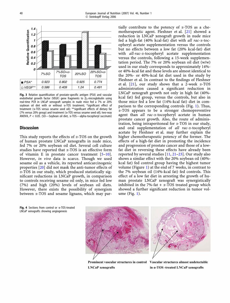
PSA and VEGF mRNA expressions in LNCaP xe-nografts were quantified using RT and real-time PCRon total RNA extracts from the tumor tissues.Amplification of the RT cDNAs from LNCaP xeno-graft RNAs (control or a-TOS-treated) gave threedistinct VEGF isoforms of 391, 365 and 231 bp(Fig. 2) as expected from the primers used; thesebands had been sequenced previously by otherinvestigators and shown to correspond to VEGF189,VEGF165 and VEGF121, respectively (18). Real-timePCR system, based on the binding of SYBR Green I tothe double-stranded DNA grove of the gene fragmentbeing amplified, makes quantification possible in theexponential phase of the reaction. In case of VEGF,the fluorescence signal [C (t) value] was detectedthree cycles later for a-TOS-treated RNA samples incomparison to the controls. When expressed relativeto b2-microglobulin [internal control, 280 bp(Fig. 3)], VEGF expression was significantly affectedby dietary fat level, treatment or interaction (two-wayANOVA, P < 0.05; Fig. 3). This inhibition of VEGF atthe transcriptional level also corresponded to areduction in angiogenesis in a-TOS-treated xenograftswhen compared to controls (Fig. 4). A moderatelypositive correlation (Pearson r 0.473, P < 0.05) wasobserved between PSA and VEGF mRNA expressionsin LNCaP xenografts (data not shown). PSA mRNAexpression was significantly affected by a-TOS treat-ment (P < 0.05, Figure 3). A single PSA gene frag-ment (363 bp, Fig. 2), from a-TOS-treated xenografts,was detected two cycles later compared to control
Table 1 Average body weights, daily feed and selected nutrient intakes in BALB/c nude mice bearing LNCaP tumors from different control and experimental groups(mean ± SE)
Parameters 7% soybean oil + vehiclea 7% soybean oil + a-TOSb 20% soybean oil + vehicle 20% soybean oil + a-TOS
n 10 11 11 10Initial body weight (g) 24.0 ± 2.39 26.0 ± 3.44 24.0 ± 2.28 24.0 ± 1.71Final body weight (g)c 28.0 ± 2.76 30.0 ± 4.45 26.0 ± 2.16 26.0 ± 1.39Weight gain (g)d 4.0 ± 0.5 4.0 ± 0.9 2.0 ± 0.4 2.0 ± 0.5Feed intake (g/d)e 5.0 ± 0.51 6.0 ± 0.27 5.0 ± 0.27 5.0 ± 0.07Calorie intake (kcal/d)f 19.0 ± 0.61 22.0 ± 0.45 24.0 ± 0.74 23.0 ± 0.54Protein intake (g/d) 1.0 ± 0.17 1.0 ± 0.23 1.0 ± 0.28 1.0 ± 0.32Fat intake (g/d)g 0.33 ± 0.1 0.39 ± 0.2 1.0 ± 0.3 1.0 ± 0.3Carbohydrate intake (g/d) 3.0 ± 0.5 3.0 ± 0.4 2.0 ± 0.4 2.0 ± 0.5
a Sesame seed oilb Alpha-tocopheryl succinate (100 mg/kg body weight)c, d, f & g Significant overall effect of dietary fat level; 7% versus 20% fat group (P < 0.001, Two-way ANOVA)e Significant effects of treatment; a-TOS versus sesame seed oil (P < 0.05) and interaction (P < 0.0001), Two-way ANOVA)
LNCaP xenograft growth
00.050.10.150.20.250.30.350.4
0 1 2 3 4 5 6 7 8
weeks from inoculation
tum
or v
olum
e (c
m3 )
7%SO*7%SO+α-TOS**20%SO***20%SO+α-TOS****
Fig. 1 LNCaP xenograft growth in BALB/c nude mice fed 7% or 20% soybeanoil diet with or without a-TOS treatment. Significant effects of dietary fat andtreatment (two-way ANOVA; P < 0.05). SO—Soybean oil diet, a-TOS—alpha-tocopheryl succinate. *7% SO & **7% SO + a-TOS differ in tumor growth***20% SO & ****20% SO + a-TOS differ in tumor growth *7% SO & ***20%SO differ in tumor growth **7% SO + a-TOS & ****20% SO + a-TOS differ intumor growth (P < 0.05)
38 European Journal of Nutrition (2007) Vol. 46, Number 1� Steinkopff Verlag 2006
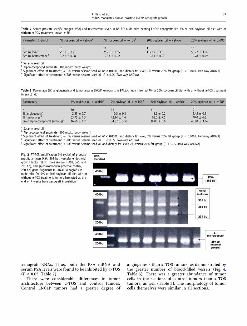
xenograft RNAs. Thus, both the PSA mRNA andserum PSA levels were found to be inhibited by a-TOS(P < 0.05, Table 2).
There were considerable differences in tumorarchitecture between a-TOS and control tumors.Control LNCaP tumors had a greater degree of
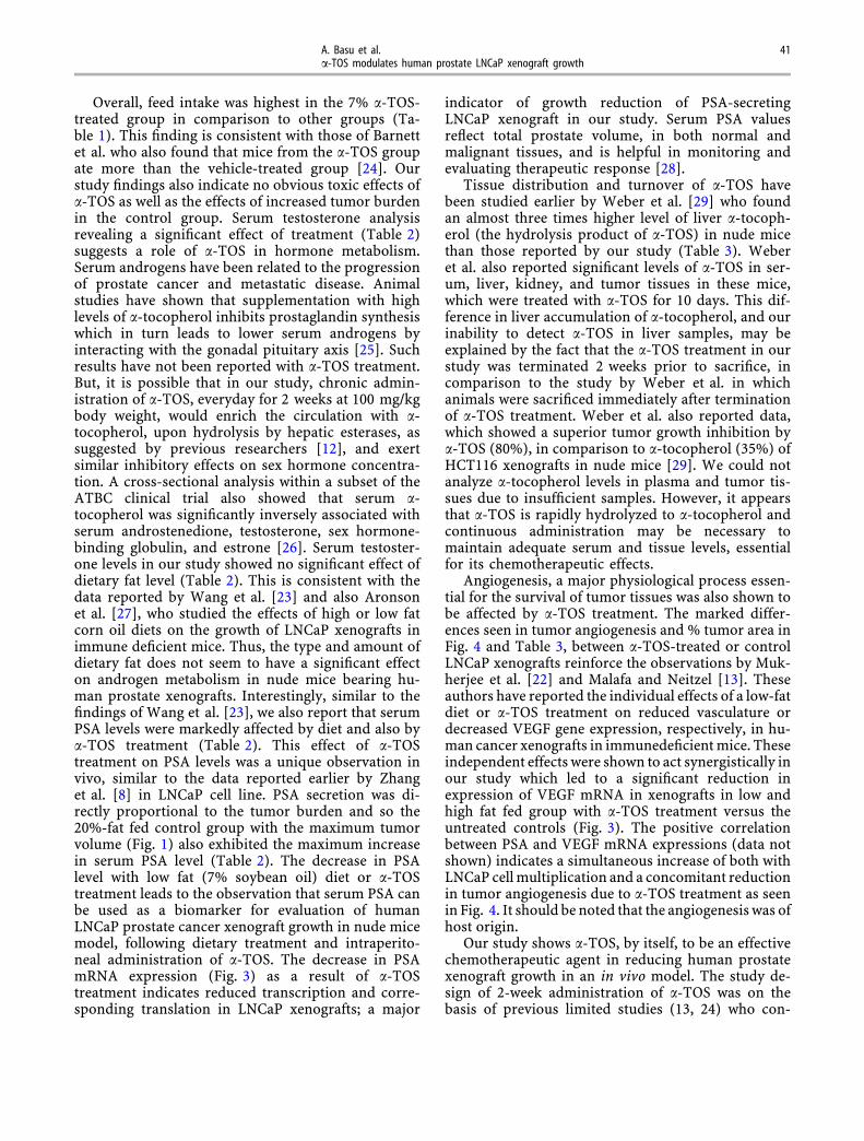
angiogenesis than a-TOS tumors, as demonstrated bythe greater number of blood-filled vessels (Fig. 4,Table 3). There was a greater abundance of tumorcells in the sections of control tumors than a-TOStumors, as well (Table 3). The morphology of tumorcells themselves were similar in all sections.
Table 2 Serum prostate-specific antigen (PSA) and testosterone levels in BALB/c nude mice bearing LNCaP xenografts fed 7% or 20% soybean oil diet with orwithout a-TOS treatment (mean ± SE)
Parameters (ng/mL) 7% soybean oil + vehiclea 7% soybean oil + a-TOSb 20% soybean oil + vehicle 20% soybean oil + a-TOS
n 10 11 11 10Serum PSAc 55.12 ± 2.7 26.28 ± 2.55 112.89 ± 3.6 55.27 ± 3.64Serum Testosteroned 0.52 ± 0.08 0.33 ± 0.02 0.61 ± 0.07 0.28 ± 0.09
a Sesame seed oilb Alpha-tocopheryl succinate (100 mg/kg body weight)c Significant effect of treatment; a-TOS versus sesame seed oil (P < 0.0001) and dietary fat level; 7% versus 20% fat group (P < 0.0001, Two-way ANOVA)d Significant effect of treatment; a-TOS versus sesame seed oil (P < 0.05, Two-way ANOVA)
Table 3 Percentage (%) angiogenesis and tumor area in LNCaP xenografts in BALB/c nude mice fed 7% or 20% soybean oil diet with or without a-TOS treatment(mean ± SE)
Parameters 7% soybean oil + vehiclea 7% soybean oil + a-TOSb 20% soybean oil + vehicle 20% soybean oil + a-TOS
n 10 11 11 10% angiogenesisc 2.25 ± 0.7 0.8 ± 0.3 1.9 ± 0.2 1.45 ± 0.4% tumor aread 63.75 ± 1.3 42.14 ± 1.6 68.0 ± 7.5 40.0 ± 6.6Liver alpha-tocopherol (nmol/g)e 16.66 ± 1.7 34.82 ± 3.50 28.00 ± 5.6 40.80 ± 3.04
a Sesame seed oilb Alpha-tocopheryl succinate (100 mg/kg body weight)c Significant effect of treatment; a-TOS versus sesame seed oil (P < 0.0001) and dietary fat level; 7% versus 20% fat group (P < 0.0001, Two-way ANOVA)d Significant effect of treatment; a-TOS versus sesame seed oil (P < 0.05, Two-way ANOVA)e Significant effect of treatment; a-TOS versus sesame seed oil and dietary fat level; 7% versus 20% fat group (P < 0.05, Two-way ANOVA)
Fig. 2 RT-PCR amplification (40 cycles) of prostate-specific antigen (PSA, 363 bp), vascular endothelialgrowth factor (VEGF, three isoforms: 391, 365, and231 bp), and b2-microglobulin (internal control,280 bp) gene fragments in LNCaP xenografts innude mice fed 7% or 20% soybean oil diet with orwithout a-TOS treatment; tumors harvested at theend of 7 weeks from xenograft inoculation
A. Basu et al. 39a-TOS modulates human prostate LNCaP xenograft growth
Discussion
This study reports the effects of a-TOS on the growthof human prostate LNCaP xenografts in nude mice,fed 7% or 20% soybean oil diet. Several cell culturestudies have reported that a-TOS is an effective formof vitamin E in prostate cancer treatment [5–10].However, in vivo data is scarce. Though we usedsesame oil as a vehicle, its reported anticarcinogenicproperties [20] did not mask the anti-tumor effects ofa-TOS in our study, which produced statistically sig-nificant reductions in LNCaP growth, in comparisonto controls receiving sesame oil only, in mice fed low(7%) and high (20%) levels of soybean oil diets.However, there exists the possibility of synergismbetween a-TOS and sesame lignans, which may par-
tially contribute to the potency of a-TOS as a che-motherapeutic agent. Fleshner et al. [21] showed areduction in LNCaP xenograft growth in nude micefed a high-fat (40% kcal-fat) diet with all rac-a-toc-opheryl acetate supplementation versus the controlsbut no effects between a low fat (20% kcal-fat) dietwith all-rac-a-tocopheryl acetate supplementationversus the controls, following a 15-week supplemen-tation period. The 7% or 20% soybean oil diet (w/w)used in our study corresponds to approximately 14%-or 40%-kcal fat and these levels are almost identical tothe 20%- or 40%-kcal fat diet used in the study byFleshner et al. In contrast to the findings of Fleshneret al. [21], our study shows that a 2-week a-TOSadministration caused a significant reduction inLNCaP xenograft growth not only in high fat (40%-kcal fat) fed group, versus the controls, but also inthose mice fed a low fat (14%-kcal fat) diet in com-parison to the corresponding controls (Fig. 1). Thus,a-TOS appears to be a stronger chemopreventiveagent than all rac-a-tocopheryl acetate in humanprostate cancer growth. Also, the route of adminis-tration, being intraperitoneal for a-TOS in our study,and oral supplementation of all rac-a-tocopherylacetate by Fleshner et al. may further explain thehigher chemotherapeutic potency of the former. Theeffects of a high-fat diet in promoting the incidenceand progression of prostate cancer and those of a low-fat diet in reversing these effects have already beenreported by several studies [11, 21–23]. Our study alsoshows a similar effect with the 20% soybean oil (40%-kcal fat) fed control group having the highest tumorvolume (Figure 1) at the end of 7 weeks, in contrast tothe 7% soybean oil (14%-kcal fat) fed controls. Thiseffect of a low fat diet in arresting the growth of hu-man prostate LNCaP xenograft was synergisticallyinhibited in the 7%-fat + a-TOS treated group whichshowed a further significant reduction in tumor vol-ume (Fig. 1).
0
0.2
0.4
0.6
0.8
1
1.2
1.4R
ela
tive
Qu
an
tifi
cati
on
PSA* 0.923 0.802 0.925 0.774
VEGF** 0.586 0.409 1.24 0.491
7%SO7%SO+a-
TOS20%SO
20%SO+a-TOS
Fig. 3 Relative quantification of prostate-specific antigen (PSA) and vascularendothelial growth factor (VEGF) gene fragments to b2-microglobulin usingreal-time PCR in LNCaP xenograft samples in nude mice fed a 7% or 20%soybean oil diet with or without a-TOS treatment; *significant effect oftreatment (a-TOS versus sesame seed oil); **significant effects of dietary fat(7% versus 20% group) and treatment (a-TOS versus sesame seed oil); two-wayANOVA, P < 0.05. (SO—Soybean oil diet, a-TOS—alpha-tocopheryl succinate)
Fig. 4 Sections from control or a-TOS-treatedLNCaP xenografts showing angiogenesis
40 European Journal of Nutrition (2007) Vol. 46, Number 1� Steinkopff Verlag 2006
Overall, feed intake was highest in the 7% a-TOS-treated group in comparison to other groups (Ta-ble 1). This finding is consistent with those of Barnettet al. who also found that mice from the a-TOS groupate more than the vehicle-treated group [24]. Ourstudy findings also indicate no obvious toxic effects ofa-TOS as well as the effects of increased tumor burdenin the control group. Serum testosterone analysisrevealing a significant effect of treatment (Table 2)suggests a role of a-TOS in hormone metabolism.Serum androgens have been related to the progressionof prostate cancer and metastatic disease. Animalstudies have shown that supplementation with highlevels of a-tocopherol inhibits prostaglandin synthesiswhich in turn leads to lower serum androgens byinteracting with the gonadal pituitary axis [25]. Suchresults have not been reported with a-TOS treatment.But, it is possible that in our study, chronic admin-istration of a-TOS, everyday for 2 weeks at 100 mg/kgbody weight, would enrich the circulation with a-tocopherol, upon hydrolysis by hepatic esterases, assuggested by previous researchers [12], and exertsimilar inhibitory effects on sex hormone concentra-tion. A cross-sectional analysis within a subset of theATBC clinical trial also showed that serum a-tocopherol was significantly inversely associated withserum androstenedione, testosterone, sex hormone-binding globulin, and estrone [26]. Serum testoster-one levels in our study showed no significant effect ofdietary fat level (Table 2). This is consistent with thedata reported by Wang et al. [23] and also Aronsonet al. [27], who studied the effects of high or low fatcorn oil diets on the growth of LNCaP xenografts inimmune deficient mice. Thus, the type and amount ofdietary fat does not seem to have a significant effecton androgen metabolism in nude mice bearing hu-man prostate xenografts. Interestingly, similar to thefindings of Wang et al. [23], we also report that serumPSA levels were markedly affected by diet and also bya-TOS treatment (Table 2). This effect of a-TOStreatment on PSA levels was a unique observation invivo, similar to the data reported earlier by Zhanget al. [8] in LNCaP cell line. PSA secretion was di-rectly proportional to the tumor burden and so the20%-fat fed control group with the maximum tumorvolume (Fig. 1) also exhibited the maximum increasein serum PSA level (Table 2). The decrease in PSAlevel with low fat (7% soybean oil) diet or a-TOStreatment leads to the observation that serum PSA canbe used as a biomarker for evaluation of humanLNCaP prostate cancer xenograft growth in nude micemodel, following dietary treatment and intraperito-neal administration of a-TOS. The decrease in PSAmRNA expression (Fig. 3) as a result of a-TOStreatment indicates reduced transcription and corre-sponding translation in LNCaP xenografts; a major
indicator of growth reduction of PSA-secretingLNCaP xenograft in our study. Serum PSA valuesreflect total prostate volume, in both normal andmalignant tissues, and is helpful in monitoring andevaluating therapeutic response [28].
Tissue distribution and turnover of a-TOS havebeen studied earlier by Weber et al. [29] who foundan almost three times higher level of liver a-tocoph-erol (the hydrolysis product of a-TOS) in nude micethan those reported by our study (Table 3). Weberet al. also reported significant levels of a-TOS in ser-um, liver, kidney, and tumor tissues in these mice,which were treated with a-TOS for 10 days. This dif-ference in liver accumulation of a-tocopherol, and ourinability to detect a-TOS in liver samples, may beexplained by the fact that the a-TOS treatment in ourstudy was terminated 2 weeks prior to sacrifice, incomparison to the study by Weber et al. in whichanimals were sacrificed immediately after terminationof a-TOS treatment. Weber et al. also reported data,which showed a superior tumor growth inhibition bya-TOS (80%), in comparison to a-tocopherol (35%) ofHCT116 xenografts in nude mice [29]. We could notanalyze a-tocopherol levels in plasma and tumor tis-sues due to insufficient samples. However, it appearsthat a-TOS is rapidly hydrolyzed to a-tocopherol andcontinuous administration may be necessary tomaintain adequate serum and tissue levels, essentialfor its chemotherapeutic effects.
Angiogenesis, a major physiological process essen-tial for the survival of tumor tissues was also shown tobe affected by a-TOS treatment. The marked differ-ences seen in tumor angiogenesis and % tumor area inFig. 4 and Table 3, between a-TOS-treated or controlLNCaP xenografts reinforce the observations by Muk-herjee et al. [22] and Malafa and Neitzel [13]. Theseauthors have reported the individual effects of a low-fatdiet or a-TOS treatment on reduced vasculature ordecreased VEGF gene expression, respectively, in hu-man cancer xenografts in immunedeficient mice. Theseindependent effects were shown to act synergistically inour study which led to a significant reduction inexpression of VEGF mRNA in xenografts in low andhigh fat fed group with a-TOS treatment versus theuntreated controls (Fig. 3). The positive correlationbetween PSA and VEGF mRNA expressions (data notshown) indicates a simultaneous increase of both withLNCaP cell multiplication and a concomitant reductionin tumor angiogenesis due to a-TOS treatment as seenin Fig. 4. It should be noted that the angiogenesis was ofhost origin.
Our study shows a-TOS, by itself, to be an effectivechemotherapeutic agent in reducing human prostatexenograft growth in an in vivo model. The study de-sign of 2-week administration of a-TOS was on thebasis of previous limited studies (13, 24) who con-
A. Basu et al. 41a-TOS modulates human prostate LNCaP xenograft growth
ducted similar in vivo administration of a-TOS. Onthe basis of our novel data, future studies may involvea long-term administration of a-TOS. An importantlimitation of our study was the time gap between thetermination of a-TOS treatment and the sacrifice ofthe animals, as a result of which we were unable toshow the accumulation of a-TOS in liver tissuesin these animals. However, the challenge remains inadministering this vitamin E analog to humans, inwhich case daily intraperitoneal doses may not befeasible. Further studies in animals with higher dosesof a-TOS, given intraperitoneally or intravenously[12] every two or three days, are warranted to detectany difference in the chemotherapeutic action of a-TOS. a-TOS has repeatedly shown to be highlyselective for cancer cells, and remains non-toxic to
normal cells even when accumulated at high levels inthe biological system [29].
In summary, our study shows significant reductionin human prostate LNCaP xenograft growth with a-TOS treatment in nude mice fed a low (7%) and high(20%) fat soybean oil diets verus controls. Serum PSAand testosterone, tumor angiogenesis, and PSA andVEGF mRNA expressions were markedly reduced bya-TOS administration.
j Acknowledgements The authors thank Dr. Veera Arora at UT-Southwestern Medical Center. This study was jointly funded by theHuman Nutrition Fund, Texas and the Research EnhancementProgram (REP) at Texas Woman’s University.
References
1. American Cancer Society. Cancer Facts& Figures 2005 http://www.cancer.org/docroot/STT/stt_0.asp [accessed June2006]
2. Granado de la OS, Saa RC, Quintas VA(2006) Epidemiological situation ofprostate cancer in Spain. Actas UrolEsp 30:574–582
3. Sogani PC, Whitmore WF (1988)Flutamide and other antiandrogens inthe treatment of advanced prostaticcarcinoma. Cancer Treat Res 39:131–145
4. Willis MS, Wians FH (2003) The role ofnutrition in preventing prostate cancer:a review of the proposed mechanism ofaction of various dietary substances.Clin Chim Acta 330:57–83
5. Ripoll EA, Rama BN, Webber MM(1986) Vitamin E enhances the che-motherapeutic effects of adriamycin onhuman prostatic carcinoma cells in vi-tro. J Urol 136:529–531
6. Israel K, Yu W, Sanders BG, Kline K(2000) Vitamin E succinate inducesapoptosis in human prostate cancercells: role for Fas in vitamin E succi-nate-triggered apoptosis. Nutr Cancer36:90–100
7. Yu A, Somasundar P, BalsubramaniamA, Rose AT, Vona DL (2002) Vitamin Eand the Y4 agonist BA-129 decreaseprostate cancer growth and productionof vascular endothelial growth factor. JSurg Res 105:65–68
8. Zhang Y, Ni J, Messing EM, Chang E,Yang CR (2002) Vitamin E succinateinhibits the function of androgenreceptor and the expression of pros-tate-specific antigen in prostate cancercells. Proc Natl Acad Sci USA 99:7408–7413
9. Zhang M, Altuwaijri S, Yeh S (2004)RRR-a-tocopheryl succinate inhibitshuman prostate cancer cell invasive-ness. Oncogene 23:3080–3088
10. Zu K, Ip C (2003) Synergy betweenselenium and vitamin E in apoptosisinduction is associated with activationof distinctive initiator caspases in hu-man prostate cancer cells. Cancer Res63:6988–6995
11. Venkateswaran V, Fleshner NE, SugarLM, Klotz LH (2004) Antioxidantsblock prostate cancer in Lady trans-genic mice. Cancer Res 64:5891–5896
12. Neuzil J (2003) Vitamin E succinateand cancer treatment: a vitamin Eprototype for selective antitumoractivity. Br J Cancer 89:1822–1826
13. Malafa MP, Neitzel LT (2000) VitaminE succinate promotes breast cancertumor dormancy. J Surg Res 93:163–170
14. Reeves PG, Nielsen FH, Fahey GC(1993) AIN-93 purified diets for labo-ratory rodents: final report of theAmerican Institute of Nutrition Ad HocWriting Committee on the reformula-tion of the AIN-76A rodent diet. J Nutr123:1939–1951
15. Devaraj S, Jialal I (2000) Low-densitylipoprotein postsecretory modification,monocyte function, and circulatingadhesion molecules in type 2 diabeticpatients with and without macrovas-cular complications: the effect of alpha-tocopherol supplementation. Circula-tion 102:191–196
16. Hara A, Radin N (1978) Lipid extrac-tion of tissues with a low-toxicity sol-vent. Annal Biochem 90:420–426
17. Edelstein RA, Krane RJ, Babayan RK,Morenas A, Moreland RB (1995) A ra-pid and simple method for the detec-tion of prostate-specific antigen mRNAin archival tissue specimens using areverse transcription-polymerase chainreaction assay. Urology 45:597–603
18. Collado B, Gutierrez-Canas I, Rodri-guez-Henche N, Prieto JC, Carmena MJ(2004) Vasoactive intestinal peptideincreases vascular endothelial growthfactor expression and neuroendocrinedifferentiation in human prostate can-cer LNCaP cells. Regul Pept 119:69–75
19. Eder IE, Culig Z, Ramoner R, ThurnherM, Putz T (2000) Inhibition of LNCaPprostate cancer cells by means ofandrogen receptor antisense oligonu-cleotides. Cancer Gene Ther 7:997–1007
20. Jacklin A, Ratledge C, Welham K, BilkoD, Newton CJ (2003) The sesame seedoil constituent, sesamol, inducesgrowth arrest and apoptosis of cancerand cardiovascular cells. Ann N Y AcadSci 1010:374–380
21. Fleshner N, Fair WR, Huryk R, HestonWDW (1999) Vitamin E inhibits thehigh-fat diet promoted growth ofestablished human prostate LNCaP tu-mors in nude mice. J Urol 161:1651–1654
22. Mukherjee P, Sotnikov AV, MangianHJ, Zhou JR, Visek WJ (1999) Energyintake and prostate tumor growth,angiogenesis, and vascular endothelialgrowth factor expression. J Natl CancerInst 91:512–523
23. Wang Y, Corr JG, Thaler HT, Tao Y,Fair WR (1995) Decreased growth ofestablished human prostate LNCaP tu-mors in nude mice fed on low-fat diet. JNatl Cancer Inst 87:1456–1461
42 European Journal of Nutrition (2007) Vol. 46, Number 1� Steinkopff Verlag 2006
24. Barnett KT, Fokum FD, Malafa MP(2002) Vitamin E succinate inhibitscolon cancer liver metastases. J SurgRes 106:292–298
25. Gilbert VA, Zebrowski EF, Chan AC(1983) Differential effects of megavitamin E on prostacyclin and throm-boxane synthesis in streptozotocin-in-duced diabetic rats. Horm Metab Res15:320–325
26. Hartman TJ, Dorgan JF, Virtamo J(1999) Association between serum al-pha-tocopherol and serum androgensand estrogens in older men. NutrCancer 35:10–15
27. Aronson WJ, Tymchuk CN, ElashoffRM (1999) Decreased growth of humanprostate LNCaP tumors in SCID micefed a low-fat, soy protein diet withisoflavones. Nutr Cancer 35:130–136
28. Thalmann GN, Sikes RA, Chang SM(1996) Suramin-induced decrease inprostate-specific antigen expressionwith no effect on tumor growth in theLNCaP model of human prostate can-cer. J Natl Cancer Inst 88:794–801
29. Weber T, Lu M, Andera L, Lahm H,Gellert N, Fariss MW, Korinek V, Sat-tler W, Ucker DS, Terman A, SchroderA, Erl W, Brunk UT, Coffey RJ, WeberC, Neuzil J (2002) Vitamin E succinateis a potent novel antineoplastic agentwith high selectivity and cooperativitywith tumor necrosis factor-relatedapoptosis-inducing ligand (Apo2 li-gand) in vivo. Clin Cancer Res 8:863–869
A. Basu et al. 43a-TOS modulates human prostate LNCaP xenograft growth