BK Ca Channels Activating at Resting Potential without Calcium in LNCaP Prostate Cancer Cells
12
BK Ca Channels Activating at Resting Potential without Calcium in LNCaP Prostate Cancer Cells G. Gessner 1 , K. Scho¨nherr 1 , M. Soom 1 , A. Hansel 1 , M. Asim 2 , A. Baniahmad 2 , C. Derst 3 , T. Hoshi 4 , S.H. Heinemann 1 1 Institute of Molecular Cell Biology, Molecular and Cellular Biophysics, Friedrich Schiller University Jena, Germany 2 Institute of Human Genetics and Anthropology, Molecular Genetics, Friedrich Schiller University Jena, Germany 3 Center for Anatomy, Electronmicroscopy and Molecular Neuroanatomy, Charite´ Berlin, Germany 4 Department of Physiology, University of Pennsylvania, Philadelphia, PA, USA Received: 29 July 2005/Revised: 15 December 2005 Abstract. Large-conductance Ca 2+ -dependent K + (BK Ca ) channels are activated by intracellular Ca 2+ and membrane depolarization in an allosteric man- ner. We investigated the pharmacological and bio- physical characteristics of a BK Ca -type K + channel in androgen-dependent LNCaP (lymph node carci- noma of the prostate) cells with novel functional properties, here termed BK L .K + selectivity, high conductance, activation by Mg 2+ or NS1619, and inhibition by paxilline and penitrem A largely resembled the properties of recombinant BK Ca channels. However, unlike conventional BK Ca chan- nels, BK L channels activated in the absence of free cytosolic Ca 2+ at physiological membrane potentials; the half-maximal activation voltage was shifted by about )100 mV compared with BK Ca channels. Half- maximal Ca 2+ -dependent activation was observed at 0.4 lM for BK L (at )20 mV) and at 4.1 lM for BK Ca channels (at +50 mV). Heterologous expression of hSlo1 in LNCaP cells increased the BK L conduc- tance. Expression of hSlo-b1 in LNCaP cells shifted voltage-dependent activation to values between that of BK L and BK Ca channels and reduced the slope of the P open (open probability)-voltage curve. We pro- pose that LNCaP cells harbor a so far unknown type of BK Ca subunit, which is responsible for the BK L phenotype in a dominant manner. BK L -like channels are also expressed in the human breast cancer cell line T47D. In addition, functional expression of BK L in LNCaP cells is regulated by serum-derived factors, however not by androgens. Key words: BK Ca channel — Ca 2+ -activated K + channel — Patch clamp — K + channel blocker — K + channel opener — b subunit Introduction Depolarization and free cytosolic calcium ([Ca 2+ ] in ) synergistically activate large-conductance Ca 2+ -acti- vated potassium (BK Ca ) channels (Kaczorowski et al., 1996). In a functional BK Ca channel complex, each of the four (Shen et al., 1994) a (Slo1) subunits likely contains six major segments (S1 to S6) plus an addi- tional N-terminal S0 segment, thought to interact with an accessory b subunit, and a large cytoplasmic C-terminus important for Ca 2+ binding (Meera et al., 1997; Xia et al., 2002). A high-affinity (< 10 lM [Ca 2+ ] in ) and a low-affinity (>100 lM [Ca 2+ ] in or [Mg 2+ ] in ) binding site located in the C-terminus enable BK Ca channels to respond to changes in [Ca 2+ ] in ranging from 500 nM to 50 mM (Xia et al., 2002). Even in the absence of Ca 2+ , depolarization alone can open BK Ca channels but the half-maximal activation volt- age (V 0.5 ) under such a condition is typically far outside the physiological range (>+100 mV) (Horrigan et al., 1999; Meera et al., 1996). Activation of typical BK Ca channels at physiological voltages requires micromo- lar levels of [Ca 2+ ] in . In excitable cells, activation of BK Ca channels facilitates hyperpolarization and thereby reduces Ca 2+ entry through voltage-gated channels. By pro- viding an inhibitory influence on cellular excitability, BK Ca channels regulate the shape of action poten- tials (Shao et al., 1999), neurotransmitter release (Kaczorowski et al., 1996), smooth muscle contraction (Brenner et al., 2000), and electrical tuning of hair cells Correspondence to: S.H. Heinemann; email: Stefan.H.Heinemann @uni-jena.de J. Membrane Biol. 208, 229–240 (2005) DOI: 10.1007/s00232-005-0830-z
-
Upload
independent -
Category
Documents
-
view
1 -
download
0
Transcript of BK Ca Channels Activating at Resting Potential without Calcium in LNCaP Prostate Cancer Cells

BKCa Channels Activating at Resting Potential without Calcium in LNCaP Prostate
Cancer Cells
G. Gessner1, K. Schonherr1, M. Soom1, A. Hansel1, M. Asim2, A. Baniahmad2, C. Derst3, T. Hoshi4,
S.H. Heinemann1
1Institute of Molecular Cell Biology, Molecular and Cellular Biophysics, Friedrich Schiller University Jena, Germany2Institute of Human Genetics and Anthropology, Molecular Genetics, Friedrich Schiller University Jena, Germany3Center for Anatomy, Electronmicroscopy and Molecular Neuroanatomy, Charite Berlin, Germany4Department of Physiology, University of Pennsylvania, Philadelphia, PA, USA
Received: 29 July 2005/Revised: 15 December 2005
Abstract. Large-conductance Ca2+-dependent K+
(BKCa) channels are activated by intracellular Ca2+
and membrane depolarization in an allosteric man-ner. We investigated the pharmacological and bio-physical characteristics of a BKCa-type K
+ channelin androgen-dependent LNCaP (lymph node carci-noma of the prostate) cells with novel functionalproperties, here termed BKL. K
+ selectivity, highconductance, activation by Mg2+ or NS1619, andinhibition by paxilline and penitrem A largelyresembled the properties of recombinant BKCachannels. However, unlike conventional BKCa chan-nels, BKL channels activated in the absence of freecytosolic Ca2+ at physiological membrane potentials;the half-maximal activation voltage was shifted byabout )100 mV compared with BKCa channels. Half-maximal Ca2+-dependent activation was observed at0.4 lM for BKL (at )20 mV) and at 4.1 lM for BKCachannels (at +50 mV). Heterologous expression ofhSlo1 in LNCaP cells increased the BKL conduc-tance. Expression of hSlo-b1 in LNCaP cells shiftedvoltage-dependent activation to values between thatof BKL and BKCa channels and reduced the slope ofthe Popen (open probability)-voltage curve. We pro-pose that LNCaP cells harbor a so far unknown typeof BKCa subunit, which is responsible for the BKLphenotype in a dominant manner. BKL-like channelsare also expressed in the human breast cancer cell lineT47D. In addition, functional expression of BKL inLNCaP cells is regulated by serum-derived factors,however not by androgens.
Key words: BKCa channel — Ca2+-activated K+
channel — Patch clamp — K+ channel blocker —K+ channel opener — b subunit
Introduction
Depolarization and free cytosolic calcium ([Ca2+]in)synergistically activate large-conductance Ca2+-acti-vated potassium (BKCa) channels (Kaczorowski et al.,1996). In a functional BKCa channel complex, each ofthe four (Shen et al., 1994) a (Slo1) subunits likelycontains six major segments (S1 to S6) plus an addi-tional N-terminal S0 segment, thought to interact withan accessory b subunit, and a large cytoplasmicC-terminus important for Ca2+ binding (Meera et al.,1997; Xia et al., 2002). A high-affinity (< 10 lM[Ca2+]in) and a low-affinity (>100 lM [Ca2+]in or[Mg2+]in) binding site located in theC-terminus enableBKCa channels to respond to changes in [Ca
2+]inranging from 500 nM to 50 mM (Xia et al., 2002). Evenin the absence of Ca2+, depolarization alone can openBKCa channels but the half-maximal activation volt-age (V0.5) under such a condition is typically far outsidethe physiological range (>+100mV) (Horrigan et al.,1999; Meera et al., 1996). Activation of typical BKCachannels at physiological voltages requires micromo-lar levels of [Ca2+]in.
In excitable cells, activation of BKCa channelsfacilitates hyperpolarization and thereby reducesCa2+ entry through voltage-gated channels. By pro-viding an inhibitory influence on cellular excitability,BKCa channels regulate the shape of action poten-tials (Shao et al., 1999), neurotransmitter release(Kaczorowski et al., 1996), smoothmuscle contraction(Brenner et al., 2000), and electrical tuning of hair cells
Correspondence to: S.H. Heinemann; email: Stefan.H.Heinemann
@uni-jena.de
J. Membrane Biol. 208, 229–240 (2005)
DOI: 10.1007/s00232-005-0830-z

(Ramanathan et al., 1999). To fulfill this broad spec-trum of functions, properties of BKCa channels areremarkably diverse and at least three mechanismscontribute to this functional diversity. First, alterna-tive splicing of the underlying single gene slo1 orKCNMA1, regulated at least in part by steroid hor-mones (Xie & McCobb, 1998; Lai & McCobb, 2002),creates multiple pore-forming a subunit isoforms(Tseng-Crank et al., 1994). Second, auxiliary b su-bunits encoded by four different genes (slob1–b4 orKCNMB1–4) coassemble with a subunits to furtherincrease the diversity (Orio et al., 2002). Third, post-translational modifications of BKCa a and/or b su-bunits, such as phosphorylation and oxidation(reviewed in Weiger et al., 2002), markedly alter thefunctional properties of BKCa channel complexes.
BKCa channels are found in virtually all celltypes, including some cancer cells such as humanglioma (Liu et al., 2002) and astrocytoma (Basraiet al., 2002) where they may play critical roles incontrol of cell proliferation and malignancy grade. Inlymph node carcinoma of the prostate (LNCaP) cells(Horoszewicz et al., 1983), which serve as a modelsystem in which to study androgen-dependent pro-liferation and apoptosis of cancer cells, the existenceof BKCa channels has been also speculated but notunequivocally demonstrated: Gutierrez et al. (1999)inferred that hyperpolarization caused by an apop-tosis inducer was mediated by activation of BKCachannels. The postulated BKCa channel should beclearly distinguishable from the Ca2+-inhibited K+
channel described in LNCaP cells (Skryma et al.,1999). This voltage-dependent 78-pS K+ channel,accounting for most of the whole-cell K+ current(<600 pA at 40 mV), was half-maximally inhibitedby 0.5 lM [Ca2+]in but insensitive to charybdotoxin(ChTx) and iberiotoxin (IbTx), two toxins that nor-mally inhibit BKCa channels (Dworetzky et al., 1996).
The aim of this study was to demonstrate thatLNCaP cells express BKCa channels and to examinewhether their functional features are altered byandrogens. We found a novel type of BKCa channel(termed BKL) with an unusual voltage and [Ca
2+]independence: activation at physiological voltages evenin the absence of Ca2+. Similar BKCa channels werealso present in T47D breast cancer cells. Thus weconclude that these BKCa channels with the afore-mentioned unusual activation properties are physio-logically relevant for functions not only in prostatecancer cells.
Materials and Methods
CELL CULTURE AND MOLECULAR BIOLOGY
LNCaP cells (DSMZ) and T47D cells (ATCC) were grown at 37�Cin a humidified atmosphere with 5% CO2 in RPMI 1640 medium
(Invitrogen, Karlsruhe, Germany) supplemented with L-glutamine
and 10% fetal calf serum (FCS). Cells were seeded on gelatin-
coated 35-mn plates in culture medium 2–5 days before electro-
physiological measurements. HEK293 cells (DSMZ) were main-
tained in DMEM / F-12 medium (1:1; Invitrogen) supplemented
with 10% FCS. A second LNCaP cell line (ATCC) was cultured in
RPMI 1640 medium supplemented with L-glutamine, sodium
pyruvate, 10% FCS, 1% penicillin, and 1% streptomycin (Sigma,
Taufkirchen, Germany). Charcoal-treated serum was generated by
incubating 500 ml FCS with activated charcoal (30 g; Sigma) for 4
h at 4�C, followed by centrifugation and sterile filtration. C4-2 cells(kindly provided by G. Thalmann) were maintained in 10% FCS-
containing T-medium (Zhau et al., 1996). All other cell lines were
maintained according to the distributor�s instructions (DSMZ).Cells were used within passage numbers 3 to 20. R1881 (methyl-
trienolone) was a gift from Zeneca Pharmaceuticals (Wilmington,
DE). Casodex was obtained from Perkin Elmer (Rodgau, Ger-
many).
The Polyfect and Superfect transfection kits (Qiagen, Hilden,
Germany) were used for transient transfection. hKCNMA1 (hSlo1,
U11058) was subcloned into pCI-neo (Promega, Mannheim, Ger-
many). hKCNMB1 (hSlo-b1) was amplified from a mixed brain andheart cDNA (Clontech, Heidelberg, Germany) and cloned into
pcDNA3 (Invitrogen). An EGFP expression plasmid (pEGFPN1,
Clontech) was co-transfected (20% of DNA) allowing identification
of transfected, fluorescent cells. mRNA from �5 · 106 LNCaPcells, isolated with the OligoTex Direct mRNA Kit (Qiagen), was
used for RT-PCR with the Titan One Tube RT-PCR Kit (Roche,
Mannheim, Germany). Gene-specific primers (Jena Bioscience,
Jena, Germany and MWG Biotech, Munich, Germany) were de-
signed according to the published human sequences. PCR frag-
ments were subcloned into pGEM-T (Promega) and sequenced
using thermosequenase (Fermentas, St. Leon-Rot, Germany) and
the Li-Cor 4000 automated sequencer (MWG Biotech).
ELECTROPHYSIOLOGICAL RECORDINGS
Electrophysiological data were obtained with an EPC-9 patch-
clamp amplifier (HEKA Elektronik, Lambrecht, Germany). Data
acquisition and analysis were controlled with Pulse+PulseFit,
PatchMaster (HEKA), and IgorPro (WaveMetrics. Lake Oswego,
OR) software. Patch pipettes fabricated from borosilicate glass had
initial resistances of 1–2 and 5–15 MW for whole-cell and single-
channel experiments, respectively. The internal solutions contained
(in mM): 140 KCl, 2 MgCl2, 10 HEPES, pH 7.4. Free intracellular
calcium concentrations ([Ca2+]in) were adjusted by adding CaCl2to either 10 mM EGTA (1–300 nM [Ca2+]in with or without 2 mM
[Mg2+]) or 10 mM HEDTA (0.3–100 lM [Ca2+]in) buffered solu-tions, as calculated by Patcher�s Power Tools (http: //www.mpibpc.gwdg.de/abteilungen/140/software/), considering Mg2+ and ATP
concentrations (if added). The external solution contained (in mM):
135 NaCl, 5 KCl, 2 MgCl2, 2 CaCl2, 10 HEPES, pH 7.4. For
measurements under bi-ionic conditions (likewise in symmetrical
(140 mM) K+) NaCl and KCl were replaced by the chloride salt of
the monovalent species under consideration. In the whole-cell
configuration, currents were measured with access resistances be-
low 4 MW. Series resistance compensation was applied up to 90%.To measure the voltage dependence, 100-ms depolarization
pulses to various voltages were applied. Instantaneous tail-current
amplitudes (Iinst) were plotted against voltage and fitted with a
Boltzmann function:
Iinst ¼ Iinst;max�1 þ exp V0:5 � Vð Þ=kað Þð Þ ð1Þ
where Iinst,max is the maximum instantaneous current, V0.5 is the
voltage for half-maximal activation, and ka is a factor
characterizing the steepness of the voltage dependence. Alterna-
tively, voltage dependence was quantified by fitting steady-state
230 G. Gessner et al.: BKCa Channels in LNCaP Cells

currents with a combined Boltzmann / GHK constant-field equa-
tion:
I ðVÞ ¼ Gmaxð1þ expðV0:5 � VÞ=kaÞ1� exp Vrev � Vð Þ=25mVð ÞÞ= 1� exp �V=25mVð Þð Þð
ð2Þ
where Gmax is the maximum conductance, Vrev is the reversal po-
tential, and V0.5 and ka have the same meaning as in Eq. (1).
Reversal potentials, measured under bi-ionic conditions, were used
to calculate permeability coefficients.
All chemicals used were of high grade, obtained from Sigma,
except iberiotoxin, paxilline, and penitrem A were purchased from
Alomone (Jerusalem, Israel). Blockers were applied with an
application pipette and the time course of block was monitored by
repetitive stimulation. Inhibition was analyzed by measuring the
steady-state current, plotting the fractional current block against
the blocker concentration, and fitting a Hill equation to these data:
I=Icontrol ¼ 1.1 þ ½blocker� IC50
� �h� ��ð3Þ
where IC50 represents the inhibitor concentration yielding 50%
block and h is the Hill coefficient.
Single-channel analysis was performed on continuous current
records of patches containing 1–2 channels. Records were low-pass
filtered at 2 kHz and sampled at 10 kHz. Fits with double Gaussian
functions to all-points amplitude histograms were used to estimate
the unitary current amplitudes (i). Single-channel conductances were
determined from the linear i / V slope between )30 and + 80 mV.
Open probabilities (Popen) were calculated from the integrated peak
areaswith a 50% threshold value betweenmeanopen and closed state
currents. The maximal number of simultaneously open channels at
high voltage (with aPopen>50%) was defined as number of channels
per patch. To assay the voltage dependence, Popen / V data were
fitted with a Boltzmann function (Eq. 1) with Iinst replaced by Popendata.
Calcium dependence was assayed by plotting Popen at con-
stant voltage against [Ca2+]in and fitting a modified Hill function to
the data:
Popen¼Popen;minþ Popen;max�Popen;min� �
= 1þ Ca2þ� �
=EC50� �h� �
ð4Þ
with the half-maximally activating concentration EC50 and the Hill
coefficient h.
All data are given as mean ± SEM (n), where n is the number
of independent experiments. Statistical comparisons were made
using a two-sided Student�s t-test. Statistical significance was as-sumed at P < 0.05.
Results
POTASSIUM CHANNELS IN LNCaP CELLS WITH ANDWITHOUT Ca2+
LNCaP cells are reported to contain Ca2+-activated(Gutierrez et al., 1999) as well as Ca2+-inhibited(Skryma et al., 1999) voltage-dependent K+ (Kv)channels. We recorded robust voltage-dependentwhole-cell currents from LNCaP cells with 0 and 3lMfree [Ca2+]in (Fig. 1A, B). At 100 mV, the currentswere often greater than 20 nA and the average cur-rent densities were 1.40 ± 0.15 nA / pF (n = 28) and1.65 ± 0.31 nA / pF (n = 35) in 0 and 3 lM [Ca2+]in,respectively. While the average current densities were
not different in 0 and 3 lM [Ca2+]in, our densityestimates were �20-fold greater than observed bySkryma et al. (1999).
Voltage dependence of the whole-cell currentsdepended on [Ca2+]in. Increasing [Ca
2+]in from 0 to 3lM changed the mean V0.5 value estimated from thetail currents from +32.9 ± 1.3 mV to )89.7 ± 1.5mV (P < 0.001), a )120 mV shift to the negativedirection. The same change in [Ca2+]in also increasedthe slope factor ka from 12.1 ± 0.5 mV to16.9 ± 0.6 mV (P < 0.001). The activation kinetics,well characterized by a single exponential, was alsomarkedly dependent on [Ca2+]in, being faster in 3 lM[Ca2+]in (Fig. 1A, D).
The channels underlying the whole-cell currentswere highly selective for K+. Under bi-ionic condi-tions (140 mM K+ inside), inward tail currents at)120 mV were detected only with (140 mM) K+ or
Fig. 1. Ca2+-sensitive whole-cell currents in LNCaP cells. (A)
Representative whole-cell currents of two different LNCaP cells
with 0 or 3 lM [Ca2+]in. (B) Steady-state current amplitudes wereaveraged for 28 and 35 cells, respectively, and plotted against
voltage. (C) Tail currents were measured immediately following the
voltage step to )40 mV. Mean tail currents were plotted against thevoltage and fitted with a Boltzmann function (line). (D) Activation
time constants in 0 and 3 lM [Ca2+]in, as obtained by single
exponential fits (superimposed in A) at different voltages. (E)
Concentration-response curves of whole-cell conductances in 0
(empty symbols) or 3 lM (filled symbols) free [Ca2+]in. Steady-statecurrents were normalized to the control current before blocker
application and averaged for 4–6 cells. Solid lines are best fits with
a Hill function (Eq. 3).
G. Gessner et al.: BKCa Channels in LNCaP Cells 231

Rb+ (PK/PRb = 1.2), but with Na+, Cs+, or Li+ inthe external solution. The permeability coefficient forK+ over Na+, Cs+, and Li+ was estimated to begreater than 115, according to their correspondingVrev values of <)120 mV.
Activation by depolarization, high K+ selectivityand a strong left-shift in V0.5 together with fasteractivation with an increase in [Ca2+]in are typicalfeatures of BKCa channels (Kaczorowski et al., 1996).However, conventional BKCa channels should exhibitalmost no activity at +50 mV in 0 [Ca2+]in and evenin 3 lM [Ca2+]in typical V0.5 values are above 0 mV(Meera et al., 1996). Hence, if a single type of channelpredominated the LNCaP whole-cell conductance,this would be either a novel Kv channel or a BKCachannel with unprecedented features. Alternatively,two channel types, exclusively active in either 0 or 3lM [Ca2+]in could be present in LNCaP cells.
To distinguish between the above possibilities, weanalyzed channel inhibition by increasing concen-trations of various drugs: tetraethylammonium(TEA), imipramine, haloperidol, and ChTx (Fig. 1E).The effects of these compounds were monitored by100-ms depolarizations to +100 mV, repeated every3 s. The inhibitory effects of imipramine, TEA, andhaloperidol equilibrated within 90 s of drug applica-tion. However, the onset of block by ChTx was ex-tremely slow and the results obtained 5 min afterapplication may not represent full equilibrium, thusunderestimating the true inhibitory efficacy. In 0[Ca2+]in, fits with the Hill equation (Eq. 3) yieldedIC50 values of 235 lM, 49.2 lM, 399 nM, and 192 nMfor TEA, imipramine, haloperidol, and ChTx,respectively. In 3 lM [Ca2+]in, the corresponding IC50values were 610 lM, 41.1 lM, 684 nM, and 207 nM.Hill coefficients were always near unity. Taking intoaccount a state dependence of block by TEA andhaloperidol (not shown), the whole-cell currents in 0and 3 lM [Ca2+]in displayed indistinguishable blockersensitivities, suggesting that the currents were medi-ated by a single channel type. To distinguish betweenKv and BKCa type channels as potential molecularcorrelates, we tested the effects of BKCa-specificblockers. In 0 [Ca2+]in, IbTx slowly blocked thewhole-cell currents with an estimated IC50 of 120 nM.Onset of block by paxilline and penitrem A was fasterand the IC50 values were 16.4 nM and 11.0 nM,respectively. The high inhibitory efficacies of theseBKCa blockers strongly implicate that a class of BKCachannels with a novel voltage dependence underliesthe whole-cell current in LNCaP cells.
SINGLE-CHANNEL ANALYSIS
We performed outside-out (OO) single-channelrecordings with 5 mM K+ outside and 140 mM K+
inside (Fig. 2A) to analyze this putative BKCa-typechannel. Calculated single-channel conductance (c)
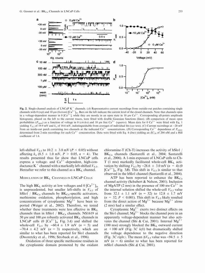
values were 153 ± 3 pS (n = 9) and 178 ± 7 pS(n = 7) in 0 and 10 lM [Ca2+]in, respectively. Thelower c in 0 [Ca2+]in (P < 0.05) may result fromblock by internal Mg2+. A similar difference (P <0.01) was obtained with 5 mM K+ outside and 140mM inside in the inside-out (IO) configuration with100 nM [Ca2+]in; calculated c values were 149 ± 3 pS(n = 10) and 167 ± 3 pS (n = 15) with 2 mM and0 [Mg2+]in, respectively. Under a symmetrical (140mM) K+ condition (IO, 0 [Ca2+]in), the single-chan-nel conductance was appreciably greater, being220 ± 10 pS (n = 10), and the conductance valueincreased further to 264 ± 7 pS (n = 11) whenMg2+ was removed from the internal solution.
The voltage dependence of open probability(Popen) was consistent with that determined from thewhole-cell measurements. In 0 [Ca2+]in, V0.5determined from the single-channel openings was19.7 ± 1.4 mV (OO, n = 9) and 24.9 ± 3.2 mV (IO,n = 15) and the corresponding ka values were10.3 ± 0.9 mV and 10.0 ± 0.8 mV. In 10 lM[Ca2+]in, the channels were fully activated at )40 mVand a small decrease in Popen was observed at positivevoltages. The open probability strongly depended on[Ca2+]in (Fig. 2C). The Ca
2+ dependence of the openprobability at )20 mV (Fig. 2D) showed that thehalf-maximal activation concentration (EC50) was437 ± 266 nM with a Hill coefficent of 2.2 ± 0.7(n = 5). The close similarity of the single-channeland whole-cell results strongly supports the notionthat a BKCa-like channel type predominates theLNCaP whole-cell conductance.
The high activity of this channel at physiologicalvoltages (< 0 mV) in 0 [Ca2+]in is surprising. Suchchannel activity could potentially arise either fromclose colocalization with Ca2+ channels (Marrion &Tavalin, 1998), spontaneous Ca2+ release from cal-cium stores still present in excised patches, or enhancedMg2+-activation of the channels. The results from thefollowing experiments exclude these possibilities. Evenin the virtual absence of both Ca2+ andMg2+ on bothsides of the membrane chelated with a high concen-tration of EGTA (10 mM) with no added divalent ions,the channels remained active. The V0.5 and ka valuesdetermined from (IO) single-channel measurementswere 22.8 ± 8.1 mV and 10.2 ± 0.5 mV (n = 6) andthose measured from whole-cell currents were52.9 ± 1.4 mV and 16.0 ± 0.7 mV, respectively(n = 10). The measured V0.5 value is �100 mV left-shifted compared with that of heterologously ex-pressed Slo1 channels under a similar condition(Horrigan et al., 1999;Meera et al., 1996). Depletion ofintracellular calcium stores (Gutierrez et al., 1999) viapreincubation of LNCaP cells with thapsigargin (2 h,0.5 lM) had no effect on the V0.5 and ka values(28.8 ± 8.2 mV and 12.5 ± 1.7 mV, respectively,n = 5). Addition of 2 mM Mg2+ to the cytoplasmicside in the inside-out configuration only slightly
232 G. Gessner et al.: BKCa Channels in LNCaP Cells
left-shifted V0.5 to 10.2 ± 3.8 mV (P< 0.05) withoutaffecting ka (8.5 ± 1.0 mV, P > 0.05, n = 4). Theresults presented thus far show that LNCaP cellsexpress a voltage- and Ca2+-dependent, high-con-ductanceK+ channel with amarkedly left-shiftedV0.5.Hereafter we refer to this channel as a BKL channel.
MODULATION OF BKL CHANNELS IN LNCaP CELLS
The high BKL activity at low voltages and 0 [Ca2+]in
is unprecedented, but smaller left-shifts in V0.5 ofhSlo1 / BKCa channels by BKCa-specific activators,methionine oxidation, phosphorylation, and highconcentrations of cytoplasmic Mg2+ have been re-ported (Weiger et al., 2002). Therefore, we testedwhether these treatments were less effective in BKLchannels than in hSlo1 / BKCa channels. NS1619 at50 lM and 100 lM robustly activated BKL channels inLNCaP cells (0 [Ca2+]in Fig. 3A) and shifted thewhole-cell V0.5 by )60.4 ± 1.9 mV (n = 5) and)70.4 ± 4.2 mV (n = 3) respectively, which aresimilar to what has been reported for Slo1 channels(Dworetzky et al., 1996; Str�baek et al., 1996).
Oxidation of three specific methionine residues inthe cytoplasmic domain promoted by the oxidant
chloramine-T (Ch-T) increases the activity of hSlo1 /BKCa channels (Santarelli et al., 2004; Santarelliet al., 2006). A 1-min exposure of LNCaP cells to Ch-T (1 mM) markedly facilitated whole-cell BKL acti-vation by shifting V0.5 by )28.6 ± 3.0 mV (n = 4) (0[Ca2+]in Fig. 3B). This shift in V0.5 is similar to thatobserved in the hSlo1 channel (Santarelli et al., 2004).
ATP has been reported to enhance the BKCachannel activity (Schubert & Nelson, 2001). Inclusionof MgATP (2 mM) in the presence of 100 nM Ca2+ inthe internal solution shifted the whole-cell V0.5 valuefrom 32.1 ± 1.1 mV (n = 32) to 12.8 ± 1.7 mV(n = 32; P< 0.001). This shift in V0.5 likely resultedfrom the direct action of Mg2+ because Mg2+ alone(2 mM) had a similar effect.
Cytoplasmic Mg2+ exerts two distinct effects onthe Slo1 channel; Mg2+ blocks the channel pore in anapparently voltage-dependent manner but also acti-vates the channel (Shi & Cui, 2001). Internal Mg2+
(100 mM) strongly blocked the BKL outward currentat +100 mV (Fig. 3C left) but dramatically shiftedthe voltage dependence to the negative direction(Fig. 3C right). The mean DV0.5 value was )94.5 ± 5mV (n = 6) similar to what has been reported formSlo1 channels (Shi & Cui, 2001).
Fig. 2. Single-channel analysis of LNCaP K+ channels. (A) Representative current recordings from outside-out patches containing single
channels with 0 (top) and 10 lM (bottom) [Ca2+]in. Bars on the left indicate the current level of the closed channels. Note that channels openin a voltage-dependent manner in 0 [Ca2+], while they are mostly in an open state in 10 lM Ca2+. Corresponding all-points amplitudehistograms, placed on the left to the current traces, were fitted with double Gaussian functions (lines). (B) comparison of mean open
probabilities (Popen) as a function of voltage in 0 (circles) and 10 lM free Ca2+ (squares). Mean data for 0 Ca2+ were fitted with Eq. 1yielding V0.5 of 19.9 mV and ka of 10.6 mV, indistinguishable from averages of individual fits (see text). (C) Current recordings at )20 mVfrom an inside-out patch containing two channels at the indicated Ca2+ concentrations. (D) Corresponding Ca2+ dependence of Popen,
determined from 2-min recordings for each Ca2+ concentration. Data were fitted with Eq. 4 (line) yielding an EC50 of 204 nM and a Hill
coefficient of 1.6.
G. Gessner et al.: BKCa Channels in LNCaP Cells 233

DIRECT INVOLVEMENT OF hSlo1 IN LNCaPCONDUCTANCE
The close similarity between the Slo1 and BKLchannels in their responses to the multiple modula-tors suggests that the left-shift in voltage dependenceof BKL channels is caused by different means, maybenovel hSlo1 isoforms that contribute to the BKLchannel. Alternatively, we reasoned that some fac-tor(s) unique to LNCaP cells may modify the prop-erties of standard hSlo1 channels to give rise to theBKL channels. Thus we heterologously expressedhSlo1 in LNCaP cells to see if the resulting currentsresemble the endogenous BKL currents. Consistentwith previous reports (Meera et al., 1996; Horriganet al., 1999), marked activation of the hSlo1 channelsexpressed in HEK293 cells with 0 lM [Ca2+]in re-quired large depolarization; the mean V0.5 value was155 ± 4 mV and the slope factor (ka) was 19.9 ± 0.9mV (n = 10). Transfection of LNCaP cells with thesame hSlo1 construct increased the whole-cell currentdensity at +100 mV by a four-fold compared withthe control LNCaP cells transfected with EGFP.
Furthermore, the voltage dependence of the whole-cell current densities in the LNCaP cells transfectedwith hSlo1 was not adequately described by linearsummations of the voltage dependence of theendogenous BKL currents in the control LNCaP cellsand multiples of the hSlo1 currents in HEK293 cells(Fig. 4A). The Gnorm / V relation of the currents inthe LNCaP cells transfected with hSlo1 more closelyresembled that of BKL channels and was very dif-ferent from that of hSlo1 expressed in HEK293 cells(Fig. 4B). Simple linear summations of the hSlo1Gnorm / V curve obtained in HEK293 cells and theGnorm / V curve of the endogenous BKL channel in aratio of 1:3 roughly describe the voltage dependenceof the currents recorded in the LNCaP cells trans-fected with hSlo1 (Fig. 4B). The results thus supportthe idea that expression of hSlo1 in LNCaP cells leadsto expression of a novel K+ channel whose voltagedependence is markedly different from the hSlo1channel expressed in HEK293 cells.
If cytosolic proteins expressed in LNCaP cellsbut not in HEK293 cells confer the unusual voltagedependence to the hSlo1 channel, acute application ofcell extracts from LNCaP may modify the propertiesof hSlo1 channels in HEK293 cells so that the
Fig. 3. BKCa-specific modulation of BKL channels in LNCaP cells.
(A, B) LNCaP whole-cell currents with 0 [Ca2+]in and 5 mM
external K+ before and after addition of 50 lM NS1619 and 1-minperfusion with 1 mM chloramine-T (Ch-T), respectively. Corre-
sponding normalized tail currrent (Itail,norm)/voltage curves are
shown on the right. (C) The effect of intracellular [Mg2+] was
analyzed in inside-out patches from LNCaP cells in 140 mM
external K+. In 100 mM Mg2+, BKL channels are partially acti-
vated at )60 mV, deactivate at )150 mV and permit outwardcurrent at +100 mV, clearly reduced, compared to control (0
Mg2+). Corresponding Itail,norm/voltage curves are shown on the
right.
Fig. 4. Transient expression of hSlo1 in LNCaP cells. (A) Mean
whole-cell current densities of EGFP (empty circles) and hSlo1
(filled circles) transfected LNCaP (Slo-LNCaP) cells upon 100-ms
depolarizations. hSlo1 current densities (triangles), obtained from
inside-out recordings of hSlo1-expressing HEK293 cells, were
scaled to fit the slope conductance of Slo-LNCaP. The dashed lines
represent attempts to fit the Slo-LNCaP conductance with the sums
of control LNCaP conductance plus multiples of hSlo1 conduc-
tance as obtained in HEK293 cells to fit the mean current-density
at +50, +100, and +150 mV, respectively. The solid line resem-
bles the sum of the 4.2-fold endogenous LNCaP currents plus
multiples of hSlo1 currents. Corresponding Gnorm/voltage plots (B)
clearly show that hSlo1 channels seem to be transformed to the
BKL-phenotype upon expression in LNCaP cells. Linear summa-
tions of BKL and hSlo1 Gnorm/V data are shown as dashed lines.
Numbers indicate the percentage contribution of hSlo1. Error bars
are left out for clarity.
234 G. Gessner et al.: BKCa Channels in LNCaP Cells

channels resemble endogenous BKL channels inLNCaP cells. Crude cell extracts were prepared by10-times passaging through a 26-G needle and sepa-rated from the insoluble fraction by centrifugation.Application of the LNCaP extract together withMgATP (1 mM) to the cytoplasmic side of inside-outpatches taken from hSlo1)expressing HEK293 cellsincreased the peak current size at +30 mV by 40-fold(Fig. 5A), much greater than the effect of MgATPalone (P< 0.05). This current-enhancing effect of thecell extract and MgATP involved a shift in the volt-age dependence of the hSlo1 channel to the negativedirection (Fig. 5B). When applied in 0 [Ca2+]in, theextracts from HEK293 and LNCaP cells shifted thehSlo1 V0.5 value by )20 mV, similar to that observedwith MgATP (1 mM). In the presence of MgATP (1mM) and 0 [Ca2+]in, the LNCaP extract produced agreater (P < 0.05) shift in V0.5 than the HEK293extract did. With 0.3 lM [Ca2+]in, the V0.5 shifts byboth extracts were greater (up to )80 mV withLNCaP extract, see Fig. 5B). Similar left-shifts wereobserved for BKL channels themselves exposed to theLNCaP extract with MgATP (not shown). The V0.5shifts were always reversible with wash. The DV0.5values were too small to fully account for the BKLphenotype. In addition, BKL channels in inside-out
patches showed no rundown, as expected whenbound proteins dissociate. Even application of 0.01–0.1% Triton X-100 to LNCaP inside-out patches didnot reduce BKL activity. In fact, Triton X-100 led toa further left-shift of V0.5 (not shown). Therefore,endogenous cytosolic proteins do not underlie themodification of hSlo1 channels in LNCaP cells.
EFFECT OF hSlo-b1 EXPRESSION ON LNCaPCONDUCTANCE
To test whether association of hSlol a subunits with anovel b subunit (bL) underlies the BKL phenotype, weoverexpressed hSlo-b1 to displace these putativeendogenous bL subunits. As shown in Fig. 6, hSlo1-b1slowed down hSlo1 activation (�3-fold) and deacti-
Fig. 5. Modulation of hSlo1 channels by cell lysates. (A, B) Effect
of cytosolic LNCaP (L) or HEK2g3 (H) cell extracts on hSlo1
channels expressed in HEK293 cells. (A) Representative current
responses to +30 mV depolarizations in 0.3 lM Ca2+ followingsuperfusion of the patches with LNCaP cytosolic extracts and
ATP. Corresponding I/V data, with and without Ca2+ (0.3 lM) andATP (1 mM), for LNCaP (L) and HEK293 (H) cytosolic extracts,
were fitted with Eq. (2) resulting in V0.5 data shown in (B), aver-
aged for 4–6 patches, each. Open bars show control V0.5 data be-
fore superfusion of the patches with solutions containing ATP and/
or cytosolic extracts, as indicated, resulting in V0.5 data represented
by the shaded bars. The dashed lines indicate the V0.5 for BKLchannels under the respective control conditions (left group 0
Ca2+, right group 0.3 lM Ca2+). *: P < 0.05.
Fig. 6. Expression of hSlo-b1 affects BKL conductance in LNCaPcells. (A) Activation (+100 mV) and deactivation ()150 mV)kinetics of whole-cell LNCaP currents (left) and inside-out hSlo1
currents (HEK293) (right) with and without hSlo-b1 expression.Currents, measured with 3 lM free Ca2+ in 140 mM K+, were
normalized to their steady-state outward current. (B) The current
time course was fitted with monoexponential functions, yielding
time constants, averaged among 9–15 cells/patches and plotted
against voltage. (C) Normalized tail-current amplitudes (Itail,norm)
at )100 mV following 250-ms depolarizations to different voltages,averaged and plotted against voltage. (D) Gnorm data calculated
from steady-state outward currents (Eq. 2) measured in 0 lM[Ca2+]in with 5 mM K
+ outside. Lines in (C) and (D) resemble best
fits with Boltzmann functions, with V0.5 and ka data given in the
text. Note the difference in axis range of panels C and D.
G. Gessner et al.: BKCa Channels in LNCaP Cells 235

vation (�7-fold) and left-shifted hSlo1 V0.5 inHEK293 cells. When expressed in LNCaP cells, hSlo-b1 slowed down the whole-cell activation kineticsprominently (�6-fold) and slightly (< 2-fold) affectedthe deactivation kinetics in a voltage-dependentmanner at 3 lM [Ca2+]in (Fig. 6A, B). The voltagedependence was also noticeably affected, such that theItail / V relation of the whole-cell currents lay betweenthose of the BKL channels in LNCaP cells and hSlo1channels expressed in HEK293 cells (Fig. 6C). TheV0.5 value of the whole-cell currents in the LNCaPcells transfected with hSlo-b1 was 14.1 ± 5.9 mV(n = 14) and the ka value was 33.2 ± 2.3 mV. Incontrast, for BKL, hSlo1 in HEK293 cells, and hSlo1+ hSlo-b1 in HEK293 cells, the estimated V0.5 valueswere )38.6 ± 2.7 (n = 15), 44.3 ± 2.9 (n = 11),and 26.9 ± 3.6 mV (n = 9), respectively, and thecorresponding ka values were 12.1 ± 0.5, 12.2 ± 1.0,and 15.6 ± 0.5 mV.
While inducing a left-shift of V0.5 in the presenceof micromolar calcium, in the absence of Ca2+, theb1 subunit does not markedly alter the voltagedependence of (standard) hSlo1 channel (Fig. 6C,Meera et al., 1996). In contrast, b1 expression affectsBKL voltage dependence in 0 and 3 lM [Ca2+]insimilarly (compare Fig. 6C, and D).
In 0 [Ca2+]in hSlo-b1 transfected LNCaP cellsexhibited a V0.5 of 94.5 ± 3.1 mV (n = 20) com-pared to BKL (48.1 ± 1.4 mV, n = 18), hSlo1(152 ± 3 mV, n = 9), and hSlo1 + hSlo-b1 channels(162 ± 7 mV, n = 12) with corresponding ka valuesof 25.2 ± 1.1, 13.3 ± 0.5, 21.8 ± 0.8, and34.2 ± 2.0 mV.
These data indicate Ca2+-independent interac-tions between overexpressed hSlo-b1 and endogenousBKL channel subunits. Functional interaction wasfurther proven by oxidation experiments: Treatmentwith Ch-T of the LNCaP cells transfected with hSlo-b1 shifted the V0.5 of the whole-cell currents by)52.4 ± 5.7 mV (n = 6) in the absence of Ca2+.This shift is �2-fold greater than that observed withBKL without hSlo-b1 overexpression and is alsoconsistent with the greater effect of oxidation re-ported in hSlo1 led to b1 expressed in HEK293 cells(Santarelli et al., 2004).
The changes in the kinetics and the voltagedependence of the whole-cell currents in LNCaP cellsby overexpression of hSlo-b1 described above aresimilar to the reported effects of b1 in other expressionsystems (Santarelli et al., 2004). Thus, the observedchanges in LNCaP cells are likely to result from thefunctional interactions between hSlo-b1 and BKLproteins. However, the (right) shift of V0.5 of LNCaPwhole-cell currents upon hSlo-b1 expression is con-trary to the (left) shift of V0.5 upon hSlo-b1 expressionin (hSlo1 expressing) HEK293 cells. This might rep-resent either the result of functional interactionsbetween hSlo-b1 and novel hSlo variants (aL) or the
result of (partial) displacement of endogenous bL byheterologously expressed hSlo-b1 subunits with(standard) hSlo1 a subunits, endogenous to LNCaPcells.
BKL-LIKE CHANNELS IN T47D CELLS
In search for other proliferating cells expressing BKLchannels, we screened various cell lines, includingmelanoma (IGR1, IGR39, IPC298) and neuroblas-toma (SH-SY5Y, SK-N-AS, SK-N-MC, SK-N-SH,SK-N-FI), and T47D breast cancer cell lines. OnlyT47D breast cancer cells showed a positive signal forhSlo1 transcription using RT-PCR and were furtheranalyzed with the patch-clamp technique. The ioniccurrents in T47D cells exhibited at least two com-ponents (Fig. 7A). The first was classified as a de-layed-rectifier (DR) type K+ channel, since it wasvoltage dependent, outwardly rectifying, non-inacti-vating, and K+-selective (Vrev < )40mV). A secondoutward K+ current, present in < 5% of the cells,clearly differed in voltage dependence (Fig. 7B).Outside-out patch-clamp recordings (Fig. 7C) re-vealed large-conductance K+ channel openings(163 ± 11 pS, n = 6), reminiscent of BKL channels.With 2 mM [Mg2+]in and 0 [Ca
2+]in the V0.5 estimatedfrom the whole-cell tail-currents was 45.6 ± 6.5 mV
Fig. 7. BKL-like currents in T47D breast cancer cells. (A) Repre-
sentative whole-cell currents of two different T47D cells containing
delayed rectifier (DR) or BKL type voltage-dependent K+ chan-
nels, measured under the same conditions as for Fig. 1 in 0
[Ca2+]in. Corresponding Itail,norm/V plots are shown in (B). (C)
Outside-out single-channel recordings at +40 mV (left) and +80
mV (right) of two different cells containing BKL-like channels. Bars
left to the current traces indicate the closed-state current level.
236 G. Gessner et al.: BKCa Channels in LNCaP Cells

(ka = 8.5 ± 0.8 mV, n = 6) 60 mV left-shiftedcompared to hSlo1 (V0.5 = 110 ± 4 mV,ka = 17.4 ± 2.9 mV, n =17) or hSlo1 + hSlo-b1channels (V0.5 = 115 ± 9 mV, ka = 21.4 ± 1.1mV, n =17). We note that T47D cells contain morethan one type of BKL-like channels, clearly differingin voltage dependence (Fig. 7C).
Growth of T47D and LNCaP cells depends onhormones, progesterone and androgens, respectively.We therefore tested whether expression or biophysicalproperties of BKL channels in LNCaP cells maychange under different growth conditions. We foundthat withdrawal of serum from the growth mediummarkedly slowed the LNCaP cell proliferation andchanged the cell morphology from being predomi-nantly bipolar, weakly adherent cells to multipolar,strongly adherent cells with longer, thinner, andbranched processes. In addition, serumdeprivation fortwo weeks significantly decreased the whole-cell cur-rent density at +100 mV with 0 [Ca2+]in from1.19 ± 0.10 nA / pF to 0.25 ± 0.02 nA / pF (n = 103,each) and reintroduction of serum to the mediumrecovered the density to the control level in one week(1.36 ± 0.14 nA / pF, n = 53). The reduction ofcurrent density was likely due to the absence of sexhormones and/or other lipophilic substances likegrowth factors. Removal of these components fromthe serum used for cell culture via charcoal treatmentsimilarly decreased the current density to 30.9 ± 4.1%(n = 4 experiments, each with 35 cells per group)within 5 days. This decrease, however, did not resultfrom the lack of androgens since the current decreasewas unaffected by an antagonist (100 nMcasodex) or anagonist (100 pM R1881); the current densities of caso-dex- or R1881-treated cells were significantly reduced(to 30.1 ± 8.3% and 30.9 ± 4.3%; n = 4 experi-ments, P< 0.01) compared with control cells, but notdifferent fromcells treatedwith charcoal-treated serum(CTS). The voltage dependencies of the LNCaPwhole-cell currents in the control and CTS-treated cells wereindistinguishable; in 0 [Ca2+]in and 2mM [Mg
2+]in, thewhole-cell V0.5 values were 34.6 ± 1.4 mV (control)and 35.2 ± 2.4 mV (CTS). However, theseV0.5 valueswere significantly different (P< 0.05) from those ob-tained for the CTS+casodex (42.0 ± 1.8 mV) orCTS+R1881 (41.6 ± 1.6 mV) -treated cells (n =35,each), We have to conclude that the BKL phenotype,i.e., the strong left-shift in V0.5 does not result from anandrogen signal and speculate that BKL expression isnot restricted to sex hormone-dependent cells.
Discussion
K+ CHANNELS IN LNCaP CELLS
Conflicting results have been reported regarding K+
channels in LNCaP cells. Here we show that BKL, aBKCa channel with unusual properties, predominates
large robust whole-cell currents in LNCaP cells. Incontrast, Skryma et al. (1999) measured �20-foldsmaller whole-cell currents (580 pA vs. 12.4 nA at+40 mV) and attributed the currents to a Ca2+-inhibited, 78-pS, voltage-dependent K+ channel. Weobserved BKL channels in LNCaP and T47D cellsobtained from DSMZ (Germany) as well as in inde-pendently grown LNCaP cells obtained from ATCC(USA) and LNCaP-derived androgen-independentC4-2 cells (Thalmann et al., 1994; not shown). In thecells used by Skryma and coworkers (1999), BKLexpression may be down-regulated due to clonalselection or different culture conditions. However, theculture condition described by Skryma et al. (1999)was essentially identical to that in our study exceptfor the reagent suppliers and the addition of antibi-otics. In our hands, BKL density was reduced neitherby addition of antibiotics (1.4 ± 0.3 nA / pF,n = 25) nor by cultivation in completely different(HEK293) medium (1.0 ± 0.1 nA / pF, n = 35)within 4 weeks, suggesting that not the presence, butrather the absence of BKL currents is the exception.We speculate that differences in the sera used for cellculture may be important. Our results exclude anyregulatory role for the androgen receptor but insteadfavor the involvement of receptors for other steroids(e.g., estrogens or glucocorticoids) or growth factorsthat also are removed in the charcoal-treated serum.Expression and physiological functions of BKLchannels in prostate LNCaP precursor cells as well asthe mechanism of BKL-regulation in LNCaP cellsneed to be investigated in the future. These issues areimportant because serum starvation increases BKCaactivity in glioma cells (Weaver et al., 2004).
CHARACTERISTICS OF BKL CHANNELS
All BKCa channels share common features: (1) Highsingle-channel conductance in combination withstrong K+ selectivity and (2) voltage dependence thatis allosterically modified (left-shift in V0.5) by an in-crease in [Ca2+]in, [Mg
2+]in or by BKCa-specificactivators (Kaczorowski et al., 1996; Gribkoff et al.,1997; Orio et al., 2002). The BKL channel in LNCaPcells meets all these criteria and, hence, (per defini-tion) is a novel type of BKCa channel. Althoughinhibition of BKL channels by ChTx, IbTx, paxilline,and penitrem A is slightly less potent compared withhSlo1 / BKCa channels (Knaus et al., 1994;Dworetzky et al., 1996; Str�baek et al., 1996; Meeraet al., 2000; Lippiat et al., 2003), the rank order ofpotency of the blockers is the same.
BKL clearly differs from conventional hSlo1channels, especially in its voltage dependence; in 0[Ca2+]in V0.5 of BKL channels is around +50 mV,about 100 mV left-shifted compared with hSlo1channels (+155 mV). Furthermore, BKL chan-nels exhibit an about 10-fold lower EC50 for
G. Gessner et al.: BKCa Channels in LNCaP Cells 237

Ca2+-dependent activation (440 nM at )20 mV vs. 4.1lM at +50 mV for hSlo1 channels) although the Hillcoefficient of the overall Ca2+ dependence is similarto that of hSlo1 (n = 2.2 vs. 2.8). Therefore, incontrast to BKCa channels, BKL channels are ex-pected to be noticeably activated under physiologicalresting conditions; �2 mM [Mg2+]in, �100 nM[Ca2+]in, and a resting potential of around )25 mV.
Low EC50 values for calcium-dependent activa-tion of BKCa channels are not unique for LNCaPcells. They have also been reported for various cancercell lines (Kraft et al., 2000; Basrai et al., 2002; Liuet al., 2002) and non-cancer cells (Anwer et al., 1992;Sansom & Stockand, 1994). This may result fromspecific Slo1 a subunits, like a glioma-BK isoform(Liu et al., 2002), or from differences in signalingpathways that also affect BKCa channels. In contrastthe strong shift in V0.5 appears specific for BKL,which raises the question regarding expression pat-tern and possible physiological roles of this channel.Since we found no indication for androgen depen-dence of channel expression or biophysical channelfeatures in LNCaP cells, BKL channels may also existin sex hormone-independent, non-cancerogenic cells/tissues. In fact, BKL-like currents might have beenoverlooked, i.e., taken for Kv channels, due to theirhigh activity in 0 [Ca2+]in. A BKCa channel with asimilar shifted voltage dependence was functionallydescribed in inner hair cells from mouse and rat(Thurm et al., 2005). Future investigations have toclarify whether BKL channels in LNCaP cells repre-sent the corresponding channels in humans.
SPECULATIONS ABOUT THE MOLECULAR BASIS OF BKL
CHANNELS
Those reagents and treatments that are known to left-shift the activation of BKCa channels, such as Ca
2+,Mg2+, BKCa activators, and Ch-T mediated oxida-tion, also affected BKL channels in the same manner.Although this is no direct proof, we hypothesize thatthese means are not the crucial factors for determi-nation of the difference between the voltage depen-dencies of BKL and hSlo1 channels. With our resultswe cannot firmly exclude that a novel phosphoryla-tion pattern in LNCaP and T47D cells cause the BKLphenotype. The observed reversibility of ATP effectsand the stability of channel activity upon patchexcision in ATP-free solution indicate that, if at all,lack of dephosphorylation is important in this re-spect. Up to date, however, a 100-mV left-shift hasbeen described neither due to dissociation of phos-phatases nor due to drug-mediated inhibition ofphosphatases (Schubert & Nelson, 2001). Cytoplas-mic proteins are unlikely to be responsible for theBKL phenotype either. For BKL channels, we neitherobserved washout of putative cytoplasmic proteins,nor could intracellular application of cytosolic
extracts from LNCaP cells transform hSlo1 channelsto the BKL phenotype (Fig. 5).
BKCa channels are multimeric complexesformed by four Slo1 a subunits that can coassemblewith up to four accessory b subunits (Orio et al.,2002). Several Slo1 a subunit splice variants anddifferent b subunits provide a huge number ofpossible combinations. Even point mutations orpolymorphisms in both, Slo1 a (Dıaz et al., 1998;Lippiat et al., 2000) and Slo-b1 (Fernandez-Fern-andez et al., 2004), can result in tremendous func-tional changes. The diversity is further increased bythe variable stoichiometry of Slo1 a and b subunits(Wang et al., 2002) and posttranslational modifi-cations like phosphorylation (Zhou et al., 2001;Erxleben et al., 2002), which in turn can determinethe effect of b subunit association (Jin et al., 2002).Because of the tremendous diversity, we cannotcompletely exclude that a combination of alreadyknown a and b subunits forms BKL channels.Nevertheless, we propose that the features of BKLchannels result from a unique subunit compositionincluding novel hSlo subunits of the a (aL) and/or b(bL) type, expressed in LNCaP and T47D cells.
From our transfection experiments (Fig. 4,Fig. 6), we infer that hSlo1 a and b1 subunits canparticipate in BKL formation. hSlo-b1 expression inLNCaP cells clearly affects BKL whole-cell currentkinetics and voltage dependence without effect oncurrent density. In addition, the increased suscepti-bility to Ch-T (Santarelli et al., 2004) clearly indicatesfunctional interaction. Assuming that aL subunits areresponsible for the BKL phenotype, LNCaP cellsrepresent an interesting system to study the molecularmechanism of a / b interactions in BKCa channelssince the functional effects of hSlo-b1 on aL (calcium-independent right-shift of V0.5) would be in partcontrary to those on standard a subunits (calcium-dependent left-shift of V0.5). On the other hand, theeffects of hSlo-b1 expression may be also explainedby functional replacement of bL subunits and theirmain functional effect, i.e., the Ca2+-independentlyleft-shifted V0.5. Both scenarios would be compatiblewith the 4-fold increase in BKL density observedupon hSlo1 expression in LNCaP cells assuming thatone aL and/or bL subunit is sufficient to transformBKCa to BKL channels.
Future studies will have to identify the molecularcounterparts of the BKL phenotype which may givefurther insight into Slo a / b subunit interaction aswell as the synergistic channel activation by voltageand calcium.
We are grateful for technical assistance by S. Arend and A. Rossner
and for helpful discussions with R. Schonherr. This work was
supported by the DFG (HE 2993/2), TMWFK (B378-01027) and
National Institute of Health.
238 G. Gessner et al.: BKCa Channels in LNCaP Cells

References
Anwer, K., Toro, L., Oberti, C., Stefani, E., Sanborn, B. M. 1992.
Ca2+-activated K+ channels in pregnant rat myometrium:
modulation by a b-adrenergic agent. Am. J. Physiol. 263:1049–1056
Basrai, D., Kraft, R., Bollensdorff, C., Liebmann, L., Benndorf,
K., Patt, S. 2002. BK channel blockers inhibit potassium-in-
duced proliferation of human astrocytoma cells. Neuro Report
13:403–407
Brenner, R., Perez, G.J., Bonev, A.D., Eckman, D.M., Kosek, J.C,
Wiler, S.W., Patterson, A.J., Nelson, M.T., Aldrich, R.W.
2000. Vasoregulation by the b1 subunit of the calcium-activatedpotassium channel. Nature 407:870–876
Dıaz, L., Meera, P., Amigo, J., Stefani, E., Alvarez, O., Toro, L.,Latorre, R. 1998. Role of the S4 segment in a voltage-depen-
dent calcium-sensitive potassium (hSlo) channel. J. Biol. Chem.
273:32430–32436
Dworetzky, S.I., Boissard, C.G., Lum-Ragan, J.T., McKay, M.C.,
Post-Munson, D.J., Trojnacki., J.T., Chang, C.-P., Gribkoff,
V.K. 1996. Phenotypic alteration of a human BK (hSlo)
channel by hSlob subunit coexpression; changes in blockersensitivity, activation/relaxation and inactivation kinetics, and
protein kinase A modulation. J. Neurosci. 16:4543–4550
Erxleben, C., Everhart, A.L., Romeo, C., Florence, H., Bauer,
M.B., Alcorta, D. A., Rossie, S., Shipston, M.J., Armstrong,
D.L. 2002. Interacting effects of N-terminal variation and strex
exon splicing on slo potassium channel regulation by calcium,
phosphorylation, and oxidation. J. Biol. Chem. 277:27045–
27052
Fernandez-Fernandez, J.M., Tomas, M., Vazquez, E., Orio, P.,
Latorre, R., Senti, M., Marrugat, J., Valverde, M.A. 2004.
Gain-of-function mutation in the KCNMB1 potassium channel
subunit is associated with low prevalence of diastolic hyper-
tension. J. Clin. Invest. 113:1032–1039
Gribkoff, V.K., Starret, J.E. Jr., Dworetzky, S.I. 1997. The phar-
macology and molecular biology of large-conductance calcium-
activated (BK) potassium channels. Adv. Pharmacol. 37:319–
348
Gutierrez, A.A., Arias, J.M., Garcia, L., Mas-Oliva, J., Guerrero-
Hernandez, A. 1999. Activation of a Ca2+-permeable cation
channel by two different inducers of apoptosis in a human
prostatic cancer cell line. J. Physiol. 517:95–107
Horoszewicz, J.S., Leong, S.S, Kawinski, E., Karr, J.P., Rosenthal,
H., Chu, T.M., Mirand, E.A., Murphy, G.P. 1983. LNCaP
model of human prostatic carcinoma. Cancer Res 43:1908–1918
Horrigan, F.T., Cui, J., Aldrich, R.W. 1999. Allosteric voltage
gating of potassium channels I: mSlo ionic currents in the ab-
sence of Ca2+. J. Gen. Physiol. 114:277–304
Jin, P., Weiger, T.M., Wu, Y., Levitan, I.B. 2002. Phosphorylation-
dependent functional coupling of hSlo calcium-dependent
potassium channel and its hb4 subunit. J. Biol. Chem.
277:10014–10020
Kaczorowski, G.J., Knaus, H.-G., Leonard, R.J., McManus, O.B.,
Garcia, M.L. 1996. High-conductance calcium-activated
potassium channels: structure, pharmacology, and function. J
Bioenerg. Biomembr. 28:255–267
Knaus, H.-G., McManus, O.B., Lee, S.H., Schmalhofer, W.A.,
Garcia-Calvo, M., Helms, L.M.H., Sanchez, M., Giangiacomo,
K., Reuben, J.P., Smith, A.B. 3rd, Kaczorowski, G.J., Garcia,
M.L. 1994. Tremorgenic indole alkaloids potently inhibit
smooth muscle high-conductance calcium-activated potassium
channels. Biochemistry 33:5819–5828
Kraft, R., Benndorf, K., Patt, S. 2000. Large conductance Ca2+
activated K+ channels in human meningioma cells. J. Mem-
brane Biol. 175:25–33
Lai, G.-J., McCobb, D.P. 2002. Opposing actions of adrenal
androgens and glucocorticoids on alternative splicing of Slo
potassium channels in bovine chromaffin cells. Proc. Natl.
Acad. Sci. USA 99:7722–7727
Lippiat, J.D., Standen, N.B., Davies., N.W. 2000. A residue in the
intracellular vestibule of the pore is critical for gating and
permeation in Ca2+-activated K+ (BKCa) channels. J. Physiol.
529:131–138
Lippiat, J.D., Standen, N.B., Harrow, I.D., Phillips, S.C., Davies,
N.W. 2003. Properties of BKCa channels formed by bicistronic
expression of hSlo a and b1-4 subunits in HEK293 cells. J.Membrane Biol. 192:141–148
Liu, X., Chang, Y., Reinhart, P. H., Sontheimer, H. 2002. Cloning
and characterization of glioma BK, a novel BK channel iso-
form highly expressed in human glioma cells. J. Neurosci.
22:1840–1849
Marrion, N. V., Tavalin, S.J. 1998. Selective activation of Ca2+-
activated K+ channels by co-localized Ca2+ channels in hip-
pocampal neurons. Nature 395:900–905
Meera, P., Wallner, M., Toro, L. 2000. A neuronal b subunit(hKCNMB4) makes the large conductance, voltage- and Ca2+-
activated K+ channel resistant to charybdotoxin and iberio-
toxin. Proc. Natl. Acad. Sci. USA. 97:5562–5567
Meera, P., Wallner, M., Jiang, Z., Toro, L. 1996. A calcium
switch for the functional coupling between a (hslo) and
b subunits (KV,Cab) of maxi K channels. FEBS Lett. 382:84–38
Meera, P., Wallner, M., Song, M., Toro, L. 1997. Large con-
ductance voltage- and calcium-dependent K+ channel, a
distinct member of voltage-dependent ion channels with seven
N-terminal transmembrane segments (S0-S6), an extracellular
N terminus, and an intracellular (S9-S10) C terminus. Proc.
Natl. Acad. Sci. USA 94:14066–14071
Orio, P., Rojas, P., Ferreira, G., Latorre, R. 2002. New disguises
for an old channel. MaxiK channel b-subunits. News Physiol.Sci. 17:156–161
Ramanathan, K., Michael, T.H., Jiang, G.-J., Hiel, H., Fuchs, P.A.
1999. A molecular mechanism for electrical tuning of cochlear
hair cells. Science 283:215–217
Sansom, S.C., Stockand, J.D. 1994. Differential Ca2+ sensitivities
of BK(Ca) isochannels in bovine mesenteric vascular smooth
muscle. Am. J. Physiol. 266:1182–1189
Santarelli, L.C., Chen, J., Heinemann, S.H., Hoshi, T. 2004. The
b1 subunit enhances oxidative regulation of large-conduc-tance calcium-activated K+ channels. J. Gen. Physiol.
124:357–370
Santarelli, L.C., Wassef, R., Heinemann, S.H., Hoshi, I. 2006.
Three methionine residues located within the regulator
of conductance for K+ (RCK) domains confer oxidative
sensitivity to large-conductance Ca2+- activated K+ channels.
J. Physiol. 571:329–359
Schubert, R., Nelson, M.T. 2001. Protein kinases: tuners of the
BKCa channel in smooth muscle. Trends Pharmacol. Sci.
22:505–512
Shao, L.-R., Halvorsrud, R., Borg-Graham, L., Storm, J.F. 1999.
The role of BK-type Ca2+-dependent K+ channels in spike
broadening during repetitive firing in rat hippocampal pyra-
midal cells. J. Physiol. 521:135–146
Shen, K.-Z., Lagrutta, A., Davies, N., Standen, N., Adliman, J.,
North, R. 1994. Tetraethylammonium block of Slowpoke cal-
cium-activated potassium channels expressed in Xenopus
oocytes: evidence for tetrameric channel formation. Pfluegers
Arch. 426:440–445
Shi, J., Cui, J. 2001. Intracellular Mg2+ enhances the function of
BK-type Ca2+-activated K+ channels. J. Gen. Physiol.
118:589–605
G. Gessner et al.: BKCa Channels in LNCaP Cells 239

Skryma, R., van Coppenolle, P., Dufy-Barbe, L., Dufy, B.,
Prevarskaya, N. 1999. Characterization of Ca2+-inhibited
potassium channels in the LNCaP human prostate cancer cell
line. Receptors Channels 6:241–253
Str�baek, D., Christopherson, P., Holm, N.R., Moldt, P., Ahring,
P.K., Johansen, T.E., Olesen, S.-P. 1996. Modulation of the
Ca2+-dependent K+ channel hslo, by the substituted
diphenylurea NS 1608, paxilline and internal Ca2+. Neuro-
pharmacology 35:903–914
Thalmann., G.N., Anezinis, P.E., Chang, S.M., Zhau, H.E., Kim,
E.E., Hopwood, V.L., Pathak, S., von Eschenbach, A.C.,
Chung, L.W 1994. Androgen-independent cancer progression
and bone metastasis in the LNCaP model of human prostate
cancer. Cancer Res. 54:2577–2581
Thurm, H., Fakler, B., Oliver, D. 2005. Ca2+-independent acti-
vation of BKCa channels at negative potentials in mammalian
inner hair cells J. Physiol. 569:137–151
Tseng-Crank, J., Foster, C.D., Krause, J.D., Mertz, R., Godinot,
N., DiChiara, T.J., Reinhart, P.H. 1994. Cloning, expression,
and distribution of functionally distinct Ca2+-activated K+
channel isoforms from: human brain. Neuron 13:1315–1330
Wang, Y.-W., Ding, J.P., Xia, X.-M., Lingie, C.J. 2002. Conse-
quences of the stoichiometry of Slo1 a and auxiliary b subunits
on functional properties of large-conductance Ca2+-activated
K+ channels. J. Neurosci. 22:1550–1561
Weiger, T.M., Hermann, A., Levitan, I.B. 2002. Modulation of
calcium-activated potassium channels. J. Comp. Physiol. A.
188:79–87
Weaver, A.K., Liu, X., Sontheimer, H. 2004. Role for cal-
cium-activated potassium channels (BK) in growth control
of human malignant glioma cells. J. Neurosci. Res. 78:224–
234
Xia, X.-M., Zeng, X., Lingle., C.J. 2002. Multiple regulatory sites
in large-conductance calcium-activated potassium channels.
Nature 418:880–884
Xie, J., McCobb, D.P. 1998. Control of alternative splicing of
potassium channels by stress hormones. Science 280:443–
446
Zhau, H.Y., Chang, S.M., Chen, B.Q., Wang, Y., Zhang, H., Kao,
C., Sang, Q.A., Pathak, S.J., Chung, L.W. 1996. Androgen-
repressed phenotype in human prostate cancer. Proc. Natl.
Acad. Sci. USA 93:15152–15157
Zhou, X.-B., Arntz, C., Kamm, S., Motejlek, K., Sausbier, U.,
Wang, G.-X., Ruth, P., Korth, M. 2001. A molecular switch for
specific stimulation of the BKCa channel by cGMP and cAMP
kinase. J. Biol. Chem. 276:43239–43245
240 G. Gessner et al.: BKCa Channels in LNCaP Cells