Isosilybin B and isosilybin A inhibit growth, induce G1 arrest and cause apoptosis in human prostate...
10
Carcinogenesis vol.28 no.7 pp.1533–1542, 2007 doi:10.1093/carcin/bgm069 Advance Access publication March 26, 2007 Isosilybin B and isosilybin A inhibit growth, induce G 1 arrest and cause apoptosis in human prostate cancer LNCaP and 22Rv1 cells Gagan Deep 1 , Nicholas H.Oberlies 2 , David J.Kroll 2 and Rajesh Agarwal 1,3, 1 Department of Pharmaceutical Sciences, School of Pharmacy, University of Colorado Health Sciences Center, Denver, CO, USA, 2 Natural Products Laboratory, Center for Organic and Medicinal Chemistry, Research Triangle Institute, Research Triangle Park, NC, USA and 3 University of Colorado Cancer Center, University of Colorado Health Sciences Center, Denver, CO, USA To whom correspondence should be addressed. Tel: þ303 315 1381; Fax: þ303 315 6281; Email: [email protected] Silymarin and, one of its constituents, silibinin exert strong effi- cacy against prostate cancer (PCA); however, anticancer efficacy and associated mechanisms of other components of silymarin, which is a mixture of flavonolignans, are largely unknown. Here we have assessed the anticancer efficacy of two pure compounds isosilybin B and isosilybin A, isolated from silymarin, in human prostate carcinoma LNCaP and 22Rv1 cells. Isosilybin B and isosilybin A treatment resulted in growth inhibition and cell death together with a strong G 1 arrest and apoptosis in both the cell lines. In the studies examining changes in cell cycle and apoptosis regulators, isosilybin B and isosilybin A resulted in a decrease in the levels of both cyclins (D1, D3, E and A) and cyclin-dependent kinases (Cdk2, Cdk4 and cell division cycle 25A), but caused an increase in p21, p27 and p53 levels, except in 22Rv1 cells where isosilybin B caused a decrease in p21 protein level. Isosilybin B- and isosilybin A-induced apoptosis was accompanied with an in- crease in the cleavage of poly (ADP-ribose) polymerase, caspase-9 and caspase-3 and a decrease in survivin levels. Compared with LNCaP and 22Rv1 cells, the antiproliferative and cytotoxic po- tentials of isosilybin B and isosilybin A were of much lesser mag- nitude in non-neoplastic human prostate epithelial PWR-1E cells suggesting the transformation-selective effect of these compounds. Together, this study for the first time identified that isosilybin B and isosilybin A, two diastereoisomers isolated from silymarin, have anti-PCA activity that is mediated via cell cycle arrest and apoptosis induction. Introduction Prostate cancer (PCA) is the leading cause of cancer incidences ac- counting for 33% (234 460) of estimated new cases in 2006 exclud- ing non-melanoma skin cancers and is the third leading cause of cancer-related deaths that accounts for 9% (27 350 cases) of all cancer-related deaths in US males (1). Surgical ablation of androgens and anti-androgen therapy are two of the therapeutic approaches against locally advanced or metastatic PCA (2). Although most pa- tients respond to these therapies initially, the disease ultimately pro- gresses to hormone-refractory state (2). Thus, the surgery and current chemotherapeutic options seem to be inadequate in controlling and curing PCA, and there is an urgent need for the identification and development of alternative strategies. In this regard, prevention and therapeutic intervention by dietary and plant-based drugs have been suggested as a newer dimension in the arena of cancer management (3,4). Among various groups of cancer chemopreventive agents, ex- tensive experimental data have been generated, which suggest the role of polyphenolic flavonoids in chemoprevention of various cancers including PCA (5–8). Silymarin is a crude mixture of flavonolignans obtained from the seeds of milk thistle [Silybum marianum (L.) Gaertn.]. This group of compounds results from the peroxidase-mediated condensation in the plant between the flavonoid, dihydroquercetin and coniferyl alcohol. Silymarin and silibinin (a 50:50 mixture of silybin A and silybin B) are widely known for their hepatoprotective activity and has been in clinical use for over three decades in Europe and recently in USA and Asia (9). Several studies by others and us in the last decade have demonstrated the pre-clinical efficacy of both silymarin and silibinin against various epithelial cancers including PCA (10–13), and their efficacy is currently being evaluated in cancer patients (14). This has led us to identify and purify specific, biologically active chemical entities present in silymarin with a goal to maximize the therapeutic utility of this botanical agent. In this direction, we have isolated, purified and characterized eight pure compounds (seven flavonoli- gnans and a flavonoid) from silymarin, namely silybin A, silybin B, isosilybin A, isosilybin B, silydianin, silychristin, isosilychristin and taxifolin(10,15). Distinct biological efficacy of these compounds was observed in human PCA cells on various cancer end point markers such as cell proliferation, prostate-specific antigen secretion and top- oisomerase IIa promoter activity (10). Isosilybin B and isosilybin A, which constitute about 2–4.5% and 5–6.5% of silymarin (w/w), re- spectively, depending upon the source, were effective in most of the parameters studied in both androgen-dependent LNCaP and andro- gen-independent PC3 and DU145 cells (10). For example, the IC50 in DU145 cells for isosilybin B and isosilybin A was 20.5 and 32 lM, respectively, which was lower compared with silymarin (IC50 67.9 lM) and silibinin (IC50 55.6 lM) (10). Though it would be premature to conclude that isosilybin B and isosilybin A were the most effective among all the constituents from this one study alone, the data pro- vided us a strong impetus to further examine the efficacy and associ- ated mechanisms for isosilybin isomers in PCA cells. De-regulated cell cycle and evasion of apoptosis have been recog- nized as hallmark of cancer progression including PCA (16). Cell cycle is known to be regulated by various kinases, namely Cdk4, Cdk2 and Cdk1 (or cdc2), whose activity is controlled by binding of different cyclins, cyclin-dependent kinase inhibitors (CDKIs) and Cdc25 phosphatases (17,18). Apoptosis refers to programmed cell death in response to various intrinsic or extrinsic death signals, and is executed by series of cysteine proteases known as caspases (19,20). Cancer chemopreventive agents are known to exert their anticancer effect via modulating the expression of cyclins–cyclin-dependent kinases (CDKs)–CDKIs and caspases resulting in induction of cell cycle arrest and apoptosis (7,8,12,21). Accordingly, in the present study, we investigated the efficacy and mechanism of isosilybin B and isosilybin A in human PCA LNCaP and 22Rv1 cells. For the first time, our findings suggest that both these agents impart anti-PCA activity via cell cycle arrest and apoptosis induction involving mod- ulation in cyclins–CDKs–CDKIs expression and caspase activation together with a decrease in survivin levels. Materials and methods Cell lines and reagents Isosilybin B and isosilybin A (Figure 1A) were isolated (purity between 98–100%) from silymarin (Madus AG, Cologne, Germany) as described pre- viously with modifications to enhance the separation of large-scale amounts of each isomer (10,15,22). Human prostate carcinoma LNCaP and 22Rv1 cells and non-neoplastic PWR-1E cells were obtained from the American Type Culture Collection (Manassas, VA). RPMI1640, keratinocyte–SFM media and other cell culture materials were from Invitrogen Corporation (Gaithersberg, MD). Antibody for Cip1/p21 was from Upstate (Charlottesville, VA) and for Abbreviations: PCA, prostate cancer; PI, propidium iodide; CDK, cyclin-de- pendent kinase; CDKI, cyclin-dependent kinase inhibitor; Cdc25A, cell divi- sion cycle 25A; PARP, poly (ADP-ribose) polymerase. Ó The Author 2007. Published by Oxford University Press. All rights reserved. For Permissions, please email: [email protected] 1533 by guest on November 12, 2014 http://carcin.oxfordjournals.org/ Downloaded from
-
Upload
independent -
Category
Documents
-
view
1 -
download
0
Transcript of Isosilybin B and isosilybin A inhibit growth, induce G1 arrest and cause apoptosis in human prostate...
Carcinogenesis vol.28 no.7 pp.1533–1542, 2007doi:10.1093/carcin/bgm069Advance Access publication March 26, 2007
Isosilybin B and isosilybin A inhibit growth, induce G1 arrest and cause apoptosis inhuman prostate cancer LNCaP and 22Rv1 cells
Gagan Deep1, Nicholas H.Oberlies2, David J.Kroll2 andRajesh Agarwal1,3,�
1Department of Pharmaceutical Sciences, School of Pharmacy, University ofColorado Health Sciences Center, Denver, CO, USA, 2Natural ProductsLaboratory, Center for Organic and Medicinal Chemistry, Research TriangleInstitute, Research Triangle Park, NC, USA and 3University of ColoradoCancer Center, University of Colorado Health Sciences Center, Denver, CO,USA
�To whom correspondence should be addressed. Tel: þ303 315 1381;Fax: þ303 315 6281;Email: [email protected]
Silymarin and, one of its constituents, silibinin exert strong effi-cacy against prostate cancer (PCA); however, anticancer efficacyand associated mechanisms of other components of silymarin,which is a mixture of flavonolignans, are largely unknown. Herewe have assessed the anticancer efficacy of two pure compoundsisosilybin B and isosilybin A, isolated from silymarin, in humanprostate carcinoma LNCaP and 22Rv1 cells. Isosilybin B andisosilybin A treatment resulted in growth inhibition and cell deathtogether with a strong G1 arrest and apoptosis in both the celllines. In the studies examining changes in cell cycle and apoptosisregulators, isosilybin B and isosilybin A resulted in a decrease inthe levels of both cyclins (D1, D3, E and A) and cyclin-dependentkinases (Cdk2, Cdk4 and cell division cycle 25A), but caused anincrease in p21, p27 and p53 levels, except in 22Rv1 cells whereisosilybin B caused a decrease in p21 protein level. Isosilybin B-and isosilybin A-induced apoptosis was accompanied with an in-crease in the cleavage of poly (ADP-ribose) polymerase, caspase-9and caspase-3 and a decrease in survivin levels. Compared withLNCaP and 22Rv1 cells, the antiproliferative and cytotoxic po-tentials of isosilybin B and isosilybin A were of much lesser mag-nitude in non-neoplastic human prostate epithelial PWR-1E cellssuggesting the transformation-selective effect of these compounds.Together, this study for the first time identified that isosilybin Band isosilybin A, two diastereoisomers isolated from silymarin,have anti-PCA activity that is mediated via cell cycle arrest andapoptosis induction.
Introduction
Prostate cancer (PCA) is the leading cause of cancer incidences ac-counting for �33% (234 460) of estimated new cases in 2006 exclud-ing non-melanoma skin cancers and is the third leading cause ofcancer-related deaths that accounts for �9% (27 350 cases) of allcancer-related deaths in US males (1). Surgical ablation of androgensand anti-androgen therapy are two of the therapeutic approachesagainst locally advanced or metastatic PCA (2). Although most pa-tients respond to these therapies initially, the disease ultimately pro-gresses to hormone-refractory state (2). Thus, the surgery and currentchemotherapeutic options seem to be inadequate in controlling andcuring PCA, and there is an urgent need for the identification anddevelopment of alternative strategies. In this regard, prevention andtherapeutic intervention by dietary and plant-based drugs have beensuggested as a newer dimension in the arena of cancer management(3,4). Among various groups of cancer chemopreventive agents, ex-tensive experimental data have been generated, which suggest the role
of polyphenolic flavonoids in chemoprevention of various cancersincluding PCA (5–8).
Silymarin is a crude mixture of flavonolignans obtained from theseeds of milk thistle [Silybum marianum (L.) Gaertn.]. This group ofcompounds results from the peroxidase-mediated condensation in theplant between the flavonoid, dihydroquercetin and coniferyl alcohol.Silymarin and silibinin (a 50:50 mixture of silybin A and silybin B)are widely known for their hepatoprotective activity and has been inclinical use for over three decades in Europe and recently in USA andAsia (9). Several studies by others and us in the last decade havedemonstrated the pre-clinical efficacy of both silymarin and silibininagainst various epithelial cancers including PCA (10–13), and theirefficacy is currently being evaluated in cancer patients (14). This hasled us to identify and purify specific, biologically active chemicalentities present in silymarin with a goal to maximize the therapeuticutility of this botanical agent. In this direction, we have isolated,purified and characterized eight pure compounds (seven flavonoli-gnans and a flavonoid) from silymarin, namely silybin A, silybin B,isosilybin A, isosilybin B, silydianin, silychristin, isosilychristin andtaxifolin(10,15). Distinct biological efficacy of these compounds wasobserved in human PCA cells on various cancer end point markerssuch as cell proliferation, prostate-specific antigen secretion and top-oisomerase IIa promoter activity (10). Isosilybin B and isosilybin A,which constitute about 2–4.5% and 5–6.5% of silymarin (w/w), re-spectively, depending upon the source, were effective in most of theparameters studied in both androgen-dependent LNCaP and andro-gen-independent PC3 and DU145 cells (10). For example, the IC50 inDU145 cells for isosilybin B and isosilybin A was 20.5 and 32 lM,respectively, which was lower compared with silymarin (IC50 67.9 lM)and silibinin (IC50 55.6 lM) (10). Though it would be premature toconclude that isosilybin B and isosilybin A were the most effectiveamong all the constituents from this one study alone, the data pro-vided us a strong impetus to further examine the efficacy and associ-ated mechanisms for isosilybin isomers in PCA cells.
De-regulated cell cycle and evasion of apoptosis have been recog-nized as hallmark of cancer progression including PCA (16). Cellcycle is known to be regulated by various kinases, namely Cdk4,Cdk2 and Cdk1 (or cdc2), whose activity is controlled by bindingof different cyclins, cyclin-dependent kinase inhibitors (CDKIs) andCdc25 phosphatases (17,18). Apoptosis refers to programmed celldeath in response to various intrinsic or extrinsic death signals, andis executed by series of cysteine proteases known as caspases (19,20).Cancer chemopreventive agents are known to exert their anticancereffect via modulating the expression of cyclins–cyclin-dependentkinases (CDKs)–CDKIs and caspases resulting in induction of cellcycle arrest and apoptosis (7,8,12,21). Accordingly, in the presentstudy, we investigated the efficacy and mechanism of isosilybin Band isosilybin A in human PCA LNCaP and 22Rv1 cells. For the firsttime, our findings suggest that both these agents impart anti-PCAactivity via cell cycle arrest and apoptosis induction involving mod-ulation in cyclins–CDKs–CDKIs expression and caspase activationtogether with a decrease in survivin levels.
Materials and methods
Cell lines and reagents
Isosilybin B and isosilybin A (Figure 1A) were isolated (purity between98–100%) from silymarin (Madus AG, Cologne, Germany) as described pre-viously with modifications to enhance the separation of large-scale amounts ofeach isomer (10,15,22). Human prostate carcinoma LNCaP and 22Rv1 cellsand non-neoplastic PWR-1E cells were obtained from the American TypeCulture Collection (Manassas, VA). RPMI1640, keratinocyte–SFM mediaand other cell culture materials were from Invitrogen Corporation (Gaithersberg,MD). Antibody for Cip1/p21 was from Upstate (Charlottesville, VA) and for
Abbreviations: PCA, prostate cancer; PI, propidium iodide; CDK, cyclin-de-pendent kinase; CDKI, cyclin-dependent kinase inhibitor; Cdc25A, cell divi-sion cycle 25A; PARP, poly (ADP-ribose) polymerase.
� The Author 2007. Published by Oxford University Press. All rights reserved. For Permissions, please email: [email protected] 1533
by guest on Novem
ber 12, 2014http://carcin.oxfordjournals.org/
Dow
nloaded from
Kip1/p27 and tubulin was from Neomarkers (Fremont, CA). Antibodies forCdk2, Cdk4, cell division cycle 25A (Cdc25A), cyclin D1, cyclin D3, cyclin Eand cyclin A were from Santa Cruz Biotechnology (Santa Cruz, CA). Hoechst33342, propidium iodide (PI) and antibody for b-actin were from Sigma–Aldrich Chemical Co. (St Louis, MO). ECL detection system and anti-mouseperoxidase-conjugated secondary antibody were from GE Healthcare (Buck-inghamshire, UK). Antibodies for cleaved poly (ADP-ribose) polymerase(PARP), cleaved caspase-3, cleaved caspase-9 and anti-rabbit peroxidase-con-jugated secondary antibody were obtained from Cell Signaling (Beverly, MA).Anti-survivin antibody was from Novus Biologicals (Littleton, CO). All
other reagents were obtained in their commercially available highest puritygrade.
Cell culture and treatments
LNCaP cells (passage 5–45) and 22Rv1 cells (passage 5–30) were cultured inRPMI1640 medium supplemented with 10% fetal bovine serum and 100 U/mlpenicillin G and 100 lg/ml streptomycin sulfate at 37�C in a humidified 5%CO2 incubator. PWR-1E cells were cultured in keratinocyte–Serum FreeMedia supplemented with 10% fetal bovine serum, 5 ng/ml human recombi-nant epidermal growth factor and 0.05 mg/ml bovine pituitary extract. Cells
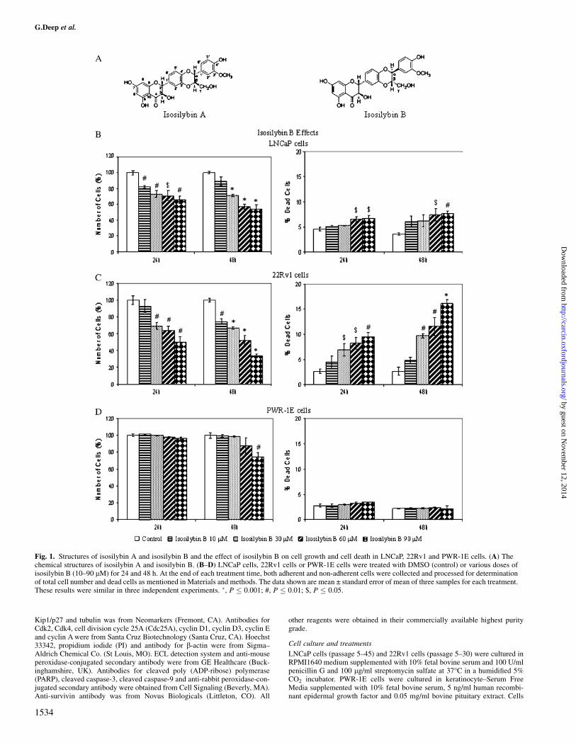
Fig. 1. Structures of isosilybin A and isosilybin B and the effect of isosilybin B on cell growth and cell death in LNCaP, 22Rv1 and PWR-1E cells. (A) Thechemical structures of isosilybin A and isosilybin B. (B–D) LNCaP cells, 22Rv1 cells or PWR-1E cells were treated with DMSO (control) or various doses ofisosilybin B (10–90 lM) for 24 and 48 h. At the end of each treatment time, both adherent and non-adherent cells were collected and processed for determinationof total cell number and dead cells as mentioned in Materials and methods. The data shown are mean ± standard error of mean of three samples for each treatment.These results were similar in three independent experiments. �, P � 0.001; #, P � 0.01; $, P � 0.05.
G.Deep et al.
1534
by guest on Novem
ber 12, 2014http://carcin.oxfordjournals.org/
Dow
nloaded from
were plated and treated at 40–50% confluency with different doses of isosily-bin B and isosilybin A (10–90 lM in medium) dissolved originally in Dimethylsulfoxide (DMSO) for the desired time periods (24–48 h) in serum condition.An equal amount of DMSO (vehicle) was present in each treatment, includingcontrol; DMSO concentration did not exceed 0.1% (v/v) in any treatment. Atthe end of desired treatments, various analyses were done as described below.
Cell growth and death assays
In each case, cells were plated to about 40–50% confluency and treated withisosilybin B and isosilybin A under serum condition as detailed above. After 24and 48 h of treatments, cells were collected by brief trypsinization and washedwith phosphate-buffered saline. Total cell number was determined by countingeach sample in duplicate using a hemocytometer under an inverted microscope.Cell viability was determined using trypan blue exclusion method. Each treat-ment and time point had three independent plates. The experiment was re-peated atleast twice under identical conditions.
Fluorescence-activated cell sorting analysis for cell cycle distribution
After identical treatments as detailed above, cells were collected and processedfor cell cycle analysis. Briefly, 0.5 � 105 cells were suspended in 0.5 ml ofsaponin/PI solution (0.3% saponin (w/v), 25 lg/ml PI (w/v), 0.1 mM ethyl-enediaminetetraacetic acid and 10 lg/ml RNase A (w/v) in phosphate-bufferedsaline) and incubated overnight at 4�C in dark. Cell cycle distribution was thenanalyzed by flow cytometry using fluorescence-activated cell sorting analysiscore facility of the University of Colorado Cancer Center. The experiment wasrepeated atleast twice under identical conditions.
Western immunoblotting
At the end of the desired treatments detailed above, cell lysates were preparedin non-denaturing lysis buffer as reported by us (23), and 50–70 lg of proteinlysate per sample was denatured in 2� sample buffer and subjected to sodiumdodecyl sulfate–polyacrylamide gel electrophoresis on 8, 12 or 16% Tris–glycine gel. The separated proteins were transferred on to nitrocellulosemembrane followed by blocking with 5% non-fat milk powder (w/v) in Tris-buffered saline (10 mM Tris–HCl, pH 7.5, 100 mM NaCl, 0.1% Tween 20) for1 h at room temperature. Membranes were probed for the protein levels ofdesired molecules using specific primary antibodies followed by peroxidase-conjugated appropriate secondary antibody and visualized by ECL detectionsystem. To ensure equal protein loading, each membrane was stripped andreprobed with anti-b-actin or anti-tubulin antibody to normalize for differencesin protein loading.
Apoptosis assay
Quantitative apoptotic cell death by isosilybin B and isosilybin A in LNCaPand 22Rv1 cells was measured by Hoechst assay as described previously (21).Briefly, after desired treatments, cells were collected and then stained withDNA-binding dye Hoechst 33342 and PI. Apoptotic cells were quantified usingfluorescent microscope (Axioskope 2 plus-HBO 100, Zeiss, Jena, Germany) bycounting 100 cells per microscopic field (at 100� magnification) in five fieldsfor each sample in triplicate.
Statistical analysis
Statistical analysis was performed using SigmaStat 2.03 software (Jandel Sci-entific, San Rafael, CA). Data were analyzed using t-test and a statisticallysignificant difference was considered to be at P , 0.05. For all the resultswhere applicable, the autoradiograms/bands were scanned with Adobe Photo-shop 6.0 (Adobe Systems, San Jose, CA), and the mean density of each bandwas analyzed by the Scion Image program (National Institutes of Health,Bethesda, MD). In each case, blots were subjected to multiple exposures onthe film to make sure that the band density is in the linear range. Densitometrydata presented below the bands are �fold change� as compared with control forboth isosilybin B and isosilybin A treatments in each case.
Results
Pure isosilybin diastereoisomers inhibit growth and induce death ofLNCaP and 22Rv1 cells
We first examined the effect of isosilybin B and isosilybin A on cellgrowth and cell death in human prostate carcinoma LNCaP and22Rv1 cells. Cells were treated with various doses of these isosilybinisomers and cell number and cell death was analyzed by trypan blueexclusion assay. Treatment with isosilybin B (10–90 lM) for 24 and48 h resulted in a decrease in cell number in both LNCaP and 22Rv1cells in both dose- and time-dependent manner. In case of LNCaPcells, 24 and 48 h treatments with isosilybin B resulted in 18–35% and
11–46% decrease in cell number, respectively (Figure 1B). Relatively,a somewhat greater effect was observed in 22Rv1 cells where identi-cal isosilybin B treatments for 24 and 48 h exhibited 7–50% and 26–66% decreases in cell number, respectively (Figure 1C). In LNCaPcells, there was a slight but significant increase in cell death after 24and 48 h of isosilybin B treatment at 60 and 90 lM concentrations(Figure 1B); however, the effect was more remarkable in 22Rv1 cellswhere compared with both 24 and 48 h controls showing 2.7% deadcells, isosilybin B treatment for 24 and 48 h at 30–90 lM dosesresulted in 7–10% and 10–16% dead cells, respectively (Figure 1C).
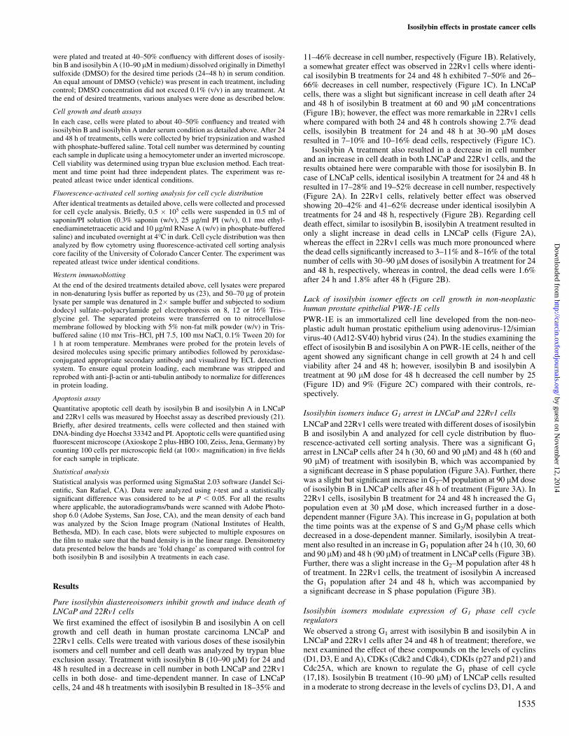
Isosilybin A treatment also resulted in a decrease in cell numberand an increase in cell death in both LNCaP and 22Rv1 cells, and theresults obtained here were comparable with those for isosilybin B. Incase of LNCaP cells, identical isosilybin A treatment for 24 and 48 hresulted in 17–28% and 19–52% decrease in cell number, respectively(Figure 2A). In 22Rv1 cells, relatively better effect was observedshowing 20–42% and 41–62% decrease under identical isosilybin Atreatments for 24 and 48 h, respectively (Figure 2B). Regarding celldeath effect, similar to isosilybin B, isosilybin A treatment resulted inonly a slight increase in dead cells in LNCaP cells (Figure 2A),whereas the effect in 22Rv1 cells was much more pronounced wherethe dead cells significantly increased to 3–11% and 8–16% of the totalnumber of cells with 30–90 lM doses of isosilybin A treatment for 24and 48 h, respectively, whereas in control, the dead cells were 1.6%after 24 h and 1.8% after 48 h (Figure 2B).
Lack of isosilybin isomer effects on cell growth in non-neoplastichuman prostate epithelial PWR-1E cells
PWR-1E is an immortalized cell line developed from the non-neo-plastic adult human prostatic epithelium using adenovirus-12/simianvirus-40 (Ad12-SV40) hybrid virus (24). In the studies examining theeffect of isosilybin B and isosilybin A on PWR-1E cells, neither of theagent showed any significant change in cell growth at 24 h and cellviability after 24 and 48 h; however, isosilybin B and isosilybin Atreatment at 90 lM dose for 48 h decreased the cell number by 25(Figure 1D) and 9% (Figure 2C) compared with their controls, re-spectively.
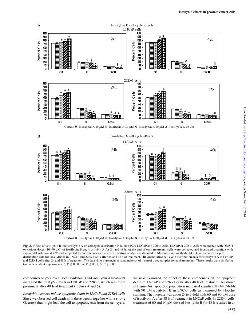
Isosilybin isomers induce G1 arrest in LNCaP and 22Rv1 cells
LNCaP and 22Rv1 cells were treated with different doses of isosilybinB and isosilybin A and analyzed for cell cycle distribution by fluo-rescence-activated cell sorting analysis. There was a significant G1
arrest in LNCaP cells after 24 h (30, 60 and 90 lM) and 48 h (60 and90 lM) of treatment with isosilybin B, which was accompanied bya significant decrease in S phase population (Figure 3A). Further, therewas a slight but significant increase in G2–M population at 90 lM doseof isosilybin B in LNCaP cells after 48 h of treatment (Figure 3A). In22Rv1 cells, isosilybin B treatment for 24 and 48 h increased the G1
population even at 30 lM dose, which increased further in a dose-dependent manner (Figure 3A). This increase in G1 population at boththe time points was at the expense of S and G2/M phase cells whichdecreased in a dose-dependent manner. Similarly, isosilybin A treat-ment also resulted in an increase in G1 population after 24 h (10, 30, 60and 90 lM) and 48 h (90 lM) of treatment in LNCaP cells (Figure 3B).Further, there was a slight increase in the G2–M population after 48 hof treatment. In 22Rv1 cells, the treatment of isosilybin A increasedthe G1 population after 24 and 48 h, which was accompanied bya significant decrease in S phase population (Figure 3B).
Isosilybin isomers modulate expression of G1 phase cell cycleregulators
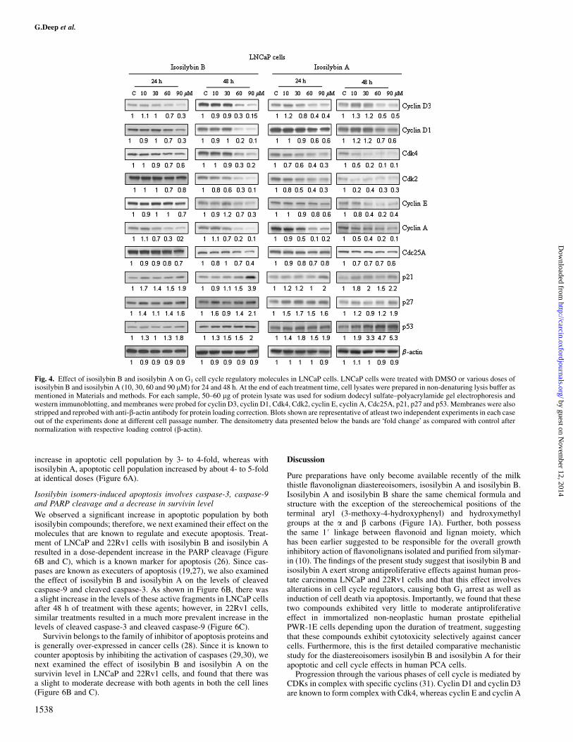
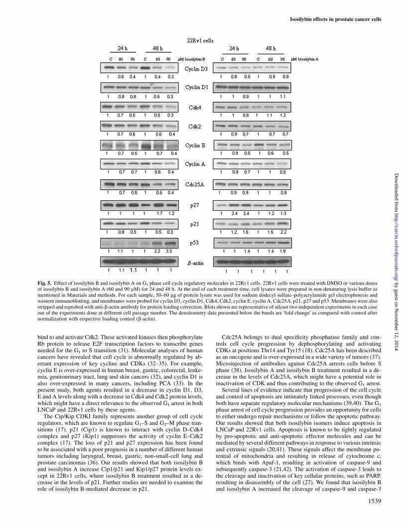
We observed a strong G1 arrest with isosilybin B and isosilybin A inLNCaP and 22Rv1 cells after 24 and 48 h of treatment; therefore, wenext examined the effect of these compounds on the levels of cyclins(D1, D3, E and A), CDKs (Cdk2 and Cdk4), CDKIs (p27 and p21) andCdc25A, which are known to regulate the G1 phase of cell cycle(17,18). Isosilybin B treatment (10–90 lM) of LNCaP cells resultedin a moderate to strong decrease in the levels of cyclins D3, D1, A and
Isosilybin effects in prostate cancer cells
1535
by guest on Novem
ber 12, 2014http://carcin.oxfordjournals.org/
Dow
nloaded from
E along with a decrease in the level of Cdk4, Cdk2 and Cdc25A(Figure 4). Isosilybin B treatment, however, increased the levels ofp27 and p21 in LNCaP cells (Figure 4). In 22Rv1 cells also, treatmentof isosilybin B (60 and 90 lM doses) for 24 and 48 h resulted ina decrease in the levels of cyclins D3, D1, E and A along with a de-crease in the levels of Cdk2, Cdk4 and Cdc25A in a dose- and time-dependent manner (Figure 5). Isosilybin B treatment also resulted ina slight to moderate increase in the levels of p27, whereas the levels ofp21 decreased under similar treatment conditions (Figure 5). Similarto isosilybin B, isosilybin A treatment resulted in a decrease in thelevels of cyclins D3, D1, E and A along with decrease in the levelsCdk2 and Cdk4 in LNCaP cells (Figure 4). In 22Rv1 cells, isosilybinA treatment caused only a slight to moderate decrease in the levels ofcyclins D3, E, A and Cdk2 (Figure 4). Isosilybin A treatment also
increased the levels of p21 and p27 in both LNCaP and 22Rv1 cells(Figures 4 and 5). Further, there was a decrease in the level of Cdc25Ain both the cell lines with isosilybin A treatment (Figures 4 and 5).Membranes were stripped and reprobed with anti-b-actin antibody forprotein loading correction.
Isosilybin B and isosilybin A treatment increases p53 level
P53 is known as a tumor suppressor gene important for the mainte-nance of genome integrity. In response to stress signal, the p53 proteinis accumulated in a specific manner by post-translational modifica-tions leading to either cell cycle arrest or apoptosis (25). Since weobserved an induction of cell cycle arrest and apoptosis with isosily-bin B and isosilybin A treatment in LNCaP and 22Rv1 cells both ofwhich harbor wild-type p53, we also examined the effect of these
Fig. 2. Effect of isosilybin A on cell growth and cell death in LNCaP, 22Rv1 and PWR-1E cells. (A–C) LNCaP cells, 22Rv1 cells or PWR-1E cells were treatedwith DMSO (control) or various doses of isosilybin A (10–90 lM) for 24 and 48 h. At the end of each treatment time, both adherent and non-adherent cells werecollected and processed for determination of total cell number and dead cells as mentioned in Materials and methods. The data shown are mean ± standard error ofmean of three samples for each treatment. These results were similar in three independent experiments. �, P � 0.001; #, P � 0.01; $, P � 0.05.
G.Deep et al.
1536
by guest on Novem
ber 12, 2014http://carcin.oxfordjournals.org/
Dow
nloaded from
compounds on p53 level. Both isosilybin B and isosilybin A treatmentincreased the total p53 levels in LNCaP and 22Rv1, which was moreprominent after 48 h of treatment (Figures 4 and 5).
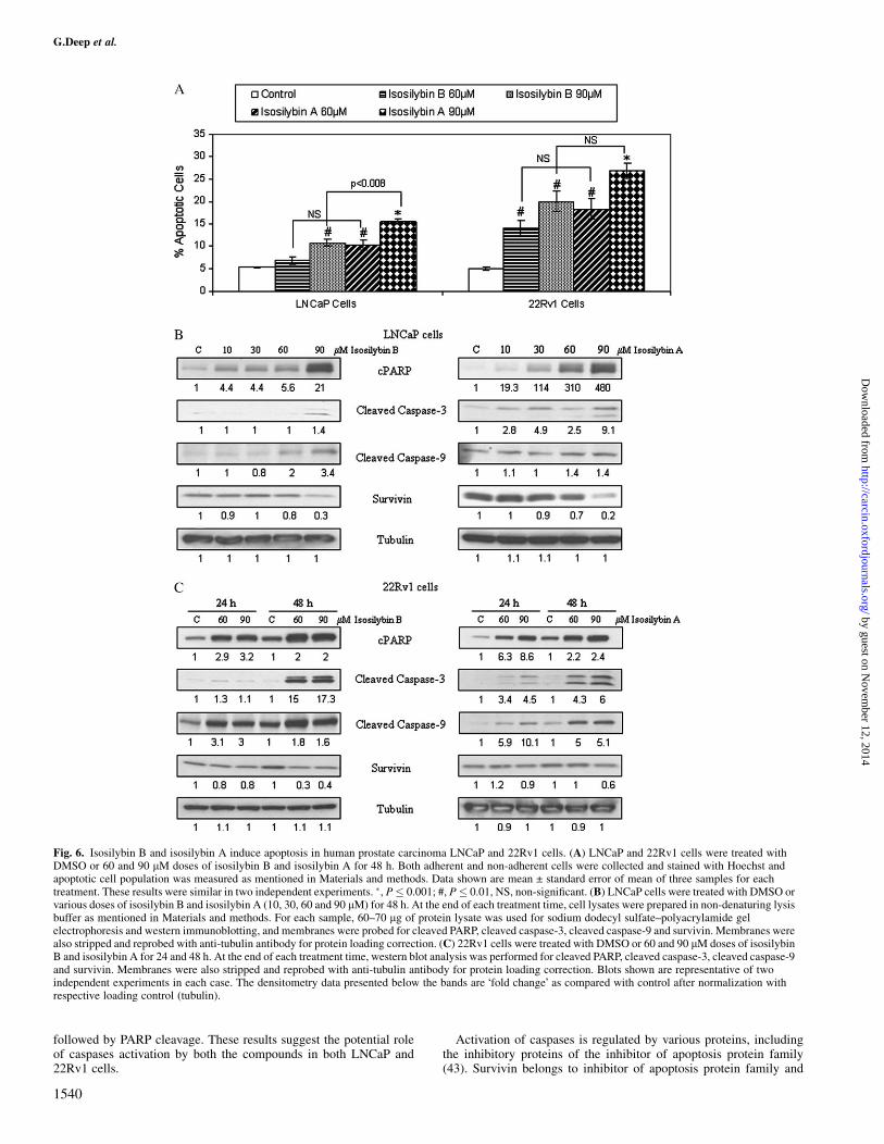
Isosilybin isomers induce apoptotic death in LNCaP and 22Rv1 cells
Since we observed cell death with these agents together with a strongG1 arrest that might lead the cell to apoptotic exit from the cell cycle,
we next examined the effect of these compounds on the apoptoticdeath of LNCaP and 22Rv1 cells after 48 h of treatment. As shownin Figure 6A, apoptotic population increased significantly by 2-foldswith 90 lM isosilybin B in LNCaP cells as measured by Hoechststaining. The increase was about 2- to 3-fold with 60 and 90 lM doseof isosilybin A after 48 h of treatment in LNCaP cells. In 22Rv1 cells,treatment of 60 and 90 lM dose of isosilybin B for 48 h resulted in an
Fig. 3. Effect of isosilybin B and isosilybin A on cell cycle distribution in human PCA LNCaP and 22Rv1 cells. LNCaP or 22Rv1 cells were treated with DMSOor various doses (10–90 lM) of isosilybin B and isosilybin A for 24 and 48 h. At the end of each treatment, cells were collected and incubated overnight withsaponin/PI solution at 4�C and subjected to fluorescence-activated cell sorting analysis as detailed in Materials and methods. (A) Quantitative cell cycledistribution data for isosilybin B in LNCaP and 22Rv1 cells after 24 and 48 h of treatment. (B) Quantitative cell cycle distribution data for isosilybin A in LNCaPand 22Rv1 cells after 24 and 48 h of treatment. The data shown are mean ± standard error of mean of three samples for each treatment. These results were similar intwo independent experiments. �, P � 0.001; #, P � 0.01; $, P � 0.05.
Isosilybin effects in prostate cancer cells
1537
by guest on Novem
ber 12, 2014http://carcin.oxfordjournals.org/
Dow
nloaded from
increase in apoptotic cell population by 3- to 4-fold, whereas withisosilybin A, apoptotic cell population increased by about 4- to 5-foldat identical doses (Figure 6A).
Isosilybin isomers-induced apoptosis involves caspase-3, caspase-9and PARP cleavage and a decrease in survivin level
We observed a significant increase in apoptotic population by bothisosilybin compounds; therefore, we next examined their effect on themolecules that are known to regulate and execute apoptosis. Treat-ment of LNCaP and 22Rv1 cells with isosilybin B and isosilybin Aresulted in a dose-dependent increase in the PARP cleavage (Figure6B and C), which is a known marker for apoptosis (26). Since cas-pases are known as executers of apoptosis (19,27), we also examinedthe effect of isosilybin B and isosilybin A on the levels of cleavedcaspase-9 and cleaved caspase-3. As shown in Figure 6B, there wasa slight increase in the levels of these active fragments in LNCaP cellsafter 48 h of treatment with these agents; however, in 22Rv1 cells,similar treatments resulted in a much more prevalent increase in thelevels of cleaved caspase-3 and cleaved caspase-9 (Figure 6C).
Survivin belongs to the family of inhibitor of apoptosis proteins andis generally over-expressed in cancer cells (28). Since it is known tocounter apoptosis by inhibiting the activation of caspases (29,30), wenext examined the effect of isosilybin B and isosilybin A on thesurvivin level in LNCaP and 22Rv1 cells, and found that there wasa slight to moderate decrease with both agents in both the cell lines(Figure 6B and C).
Discussion
Pure preparations have only become available recently of the milkthistle flavonolignan diastereoisomers, isosilybin A and isosilybin B.Isosilybin A and isosilybin B share the same chemical formula andstructure with the exception of the stereochemical positions of theterminal aryl (3-methoxy-4-hydroxyphenyl) and hydroxymethylgroups at the a and b carbons (Figure 1A). Further, both possessthe same 1# linkage between flavonoid and lignan moiety, whichhas been earlier suggested to be responsible for the overall growthinhibitory action of flavonolignans isolated and purified from silymar-in (10). The findings of the present study suggest that isosilybin B andisosilybin A exert strong antiproliferative effects against human pros-tate carcinoma LNCaP and 22Rv1 cells and that this effect involvesalterations in cell cycle regulators, causing both G1 arrest as well asinduction of cell death via apoptosis. Importantly, we found that thesetwo compounds exhibited very little to moderate antiproliferativeeffect in immortalized non-neoplastic human prostate epithelialPWR-1E cells depending upon the duration of treatment, suggestingthat these compounds exhibit cytotoxicity selectively against cancercells. Furthermore, this is the first detailed comparative mechanisticstudy for the diastereoisomers isosilybin B and isosilybin A for theirapoptotic and cell cycle effects in human PCA cells.
Progression through the various phases of cell cycle is mediated byCDKs in complex with specific cyclins (31). Cyclin D1 and cyclin D3are known to form complex with Cdk4, whereas cyclin E and cyclin A
Fig. 4. Effect of isosilybin B and isosilybin A on G1 cell cycle regulatory molecules in LNCaP cells. LNCaP cells were treated with DMSO or various doses ofisosilybin B and isosilybin A (10, 30, 60 and 90 lM) for 24 and 48 h. At the end of each treatment time, cell lysates were prepared in non-denaturing lysis buffer asmentioned in Materials and methods. For each sample, 50–60 lg of protein lysate was used for sodium dodecyl sulfate–polyacrylamide gel electrophoresis andwestern immunoblotting, and membranes were probed for cyclin D3, cyclin D1, Cdk4, Cdk2, cyclin E, cyclin A, Cdc25A, p21, p27 and p53. Membranes were alsostripped and reprobed with anti-b-actin antibody for protein loading correction. Blots shown are representative of atleast two independent experiments in each caseout of the experiments done at different cell passage number. The densitometry data presented below the bands are �fold change� as compared with control afternormalization with respective loading control (b-actin).
G.Deep et al.
1538
by guest on Novem
ber 12, 2014http://carcin.oxfordjournals.org/
Dow
nloaded from
bind to and activate Cdk2. These activated kinases then phosphorylateRb protein to release E2F transcription factors to transcribe genesneeded for the G1 to S transition (31). Molecular analyses of humancancers have revealed that cell cycle is abnormally regulated by ab-errant expression of key cyclins and CDKs (32–35). For example,cyclin E is over-expressed in human breast, gastric, colorectal, leuke-mia, genitourinary tract, lung and skin cancers (32), and cyclin D1 isalso over-expressed in many cancers, including PCA (33). In thepresent study, both agents resulted in a decrease in cyclin D1, D3,E and A levels along with a decrease in Cdk4 and Cdk2 protein levels,which might have a direct relevance to the observed G1 arrest in bothLNCaP and 22Rv1 cells by these agents.
The Cip/Kip CDKI family represents another group of cell cycleregulators, which are known to regulate G1–S and G2–M phase tran-sitions (17). p21 (Cip1) is known to interact with cyclin D–Cdk4complex and p27 (Kip1) suppresses the activity of cyclin E–Cdk2complex (17). The loss of p21 and p27 expression has been foundto be associated with a poor prognosis in a number of different humantumors including laryngeal, breast, gastric, non-small-cell lung andprostate carcinomas (36). Our results showed that both isosilybin Band isosilybin A increase Cip1/p21 and Kip1/p27 protein levels ex-cept in 22Rv1 cells, where isosilybin B treatment resulted in a de-crease in the levels of p21. Further studies are needed to examine therole of isosilybin B-mediated decrease in p21.
Cdc25A belongs to dual specificity phosphatase family and con-trols cell cycle progression by dephosphorylating and activatingCDKs at positions Thr14 and Tyr15 (18). Cdc25A has been describedas an oncogene and is over-expressed in a wide variety of tumors (37).Microinjection of antibodies against Cdc25A arrests cells before Sphase (38). Isosilybin A and isosilybin B treatment resulted in a de-crease in the levels of Cdc25A, which might have a potential role ininactivation of CDK and thus contributing to the observed G1 arrest.
Several lines of evidence indicate that progression of the cell cycleand control of apoptosis are intimately linked processes, even thoughboth have separate regulatory molecular mechanisms (39,40). The G1
phase arrest of cell cycle progression provides an opportunity for cellsto either undergo repair mechanisms or follow the apoptotic pathway.Our results showed that both isosilybin isomers induce apoptosis inLNCaP and 22Rv1 cells. Apoptosis is known to be tightly regulatedby pro-apoptotic and anti-apoptotic effector molecules and can bemediated by several different pathways in response to various intrinsicand extrinsic signals (20,41). These signals affect the membrane po-tential of mitochondria and resulting in release of cytochrome c,which binds with Apaf-1, resulting in activation of caspase-9 andsubsequently caspase-3 (21,42). The activation of caspase-3 leads tothe cleavage and inactivation of key cellular proteins, such as PARP,resulting in disassembly of the cell (27). We found that isosilybin Band isosilybin A increased the cleavage of caspase-9 and caspase-3
Fig. 5. Effect of isosilybin B and isosilybin A on G1 phase cell cycle regulatory molecules in 22Rv1 cells. 22Rv1 cells were treated with DMSO or various dosesof isosilybin B and isosilybin A (60 and 90 lM) for 24 and 48 h. At the end of each treatment time, cell lysates were prepared in non-denaturing lysis buffer asmentioned in Materials and methods. For each sample, 50–60 lg of protein lysate was used for sodium dodecyl sulfate–polyacrylamide gel electrophoresis andwestern immunoblotting, and membranes were probed for cyclin D3, cyclin D1, Cdk4, Cdk2, cyclin E, cyclin A, Cdc25A, p21, p27 and p53. Membranes were alsostripped and reprobed with anti-b-actin antibody for protein loading correction. Blots shown are representative of atleast two independent experiments in each caseout of the experiments done at different cell passage number. The densitometry data presented below the bands are �fold change� as compared with control afternormalization with respective loading control (b-actin).
Isosilybin effects in prostate cancer cells
1539
by guest on Novem
ber 12, 2014http://carcin.oxfordjournals.org/
Dow
nloaded from
followed by PARP cleavage. These results suggest the potential roleof caspases activation by both the compounds in both LNCaP and22Rv1 cells.
Activation of caspases is regulated by various proteins, includingthe inhibitory proteins of the inhibitor of apoptosis protein family(43). Survivin belongs to inhibitor of apoptosis protein family and
Fig. 6. Isosilybin B and isosilybin A induce apoptosis in human prostate carcinoma LNCaP and 22Rv1 cells. (A) LNCaP and 22Rv1 cells were treated withDMSO or 60 and 90 lM doses of isosilybin B and isosilybin A for 48 h. Both adherent and non-adherent cells were collected and stained with Hoechst andapoptotic cell population was measured as mentioned in Materials and methods. Data shown are mean ± standard error of mean of three samples for eachtreatment. These results were similar in two independent experiments. �, P� 0.001; #, P� 0.01, NS, non-significant. (B) LNCaP cells were treated with DMSO orvarious doses of isosilybin B and isosilybin A (10, 30, 60 and 90 lM) for 48 h. At the end of each treatment time, cell lysates were prepared in non-denaturing lysisbuffer as mentioned in Materials and methods. For each sample, 60–70 lg of protein lysate was used for sodium dodecyl sulfate–polyacrylamide gelelectrophoresis and western immunoblotting, and membranes were probed for cleaved PARP, cleaved caspase-3, cleaved caspase-9 and survivin. Membranes werealso stripped and reprobed with anti-tubulin antibody for protein loading correction. (C) 22Rv1 cells were treated with DMSO or 60 and 90 lM doses of isosilybinB and isosilybin A for 24 and 48 h. At the end of each treatment time, western blot analysis was performed for cleaved PARP, cleaved caspase-3, cleaved caspase-9and survivin. Membranes were also stripped and reprobed with anti-tubulin antibody for protein loading correction. Blots shown are representative of twoindependent experiments in each case. The densitometry data presented below the bands are �fold change� as compared with control after normalization withrespective loading control (tubulin).
G.Deep et al.
1540
by guest on Novem
ber 12, 2014http://carcin.oxfordjournals.org/
Dow
nloaded from

is over-expressed in vast majority of cancers including esophageal,lung, ovarian, breast, colorectal, bladder, gastric, prostate, pancreatic,laryngeal, uterine etc. (28). Survivin is known to act as death suppres-sor as a result of caspase inhibition (29,30). Additionally, survivin hasbeen shown to interact directly with Cdk4 and plays a role in Cdk2/cyclin E activation resulting in G1 transition (44). Isosilybin B andisosilybin A treatment resulted in a decrease in the levels of survivin,which might contribute to both cell cycle arrest and induction ofapoptosis as observed in LNCaP and 22Rv1 cells.
As mentioned earlier, p53 is a tumor suppressor gene and is acti-vated in response to various intrinsic and extrinsic stress signals (25).Activated p53 is a sequence specific DNA-binding transcriptionalfactor and some of the target genes of its transcriptional activity areimportant for cell cycle arrest or for inducing apoptosis (45). Forexample, p53 regulated genes (Apaf-1, bax, noxa, puma etc.) areknown to initiate a protein cascade leading to the activation of cas-pase-9 and then caspase-3 followed by apoptosis (45,46). Similarly,role of p53 in cell cycle arrest has been shown via regulating theexpression of p21, cdc25A and 14-3-3 sigma (37,45,46). In the pres-ent work, we observed an increase in the level of p53 with isosilybin Band isosilybin A treatment, which might contribute to the observedcell cycle arrest and apoptosis; however, more studies are needed infuture to establish such a cause and effect relationship.
In conclusion, the results of the present study indicate that isosily-bin B and isosilybin A inhibit proliferation and induce G1 phase arrestand apoptosis in human PCA cells. Isosilybin B was more effective inits effect on reducing cyclin D1 and cyclin D3 levels compared withisosilybin A in LNCaP cells. Similarly, in 22Rv1 cells, isosilybin Bwas more effective in down-regulating the levels of cyclins (D1, D3, Eand A) and CDKs (Cdk4 and Cdk2) and Cdc25A, whereas isosilybinA was more effective in the induction of CDKIs (p21 and p27). Theseresults point toward a difference in the mechanism for induction of G1
phase arrest by isosilybin B and isosilybin A. Similarly, we foundhigher apoptotic cell population with isosilybin A compared withisosilybin B in both LNCaP and 22Rv1 cells suggesting that isosilybinA might be more potent in its apoptotic action. In non-tumorigenicPWR-1E cells where both compounds had minimal effects, isosilybinA showed lesser potency compared with isosilybin B in inhibiting cellproliferation. These results suggest that these two diastereoisomers dohave distinct cellular targets, which might be due to the difference inthe positioning of functional groups in their lignan moieties (Figure 1A).However, further studies are needed in future to address these issuesas both compounds also have many overlapping effects as summa-
rized in Table 1. Overall, this study provided the first detailed accountof anticancer efficacy of isosilybin B and isosilybin A against humanprostate carcinoma cells.
Acknowledgements
This work was supported by U.S. Public Health Service grants RO1 CA102514and CA104286 from the National Cancer Institute, National Institutes ofHealth. Special thanks to Tyler N.Graf of Research Triangle Institute for hishard work in isolating these diastereoisomers.
Conflict of Interest Statement: None declared.
References
1.Jemal,A. et al. (2006) Cancer statistics, 2006.CACancer J. Clin., 56, 106–130.2.Chen,C.D. et al. (2004) Molecular determinants of resistance to antiandro-
gen therapy. Nat. Med., 10, 33–39.3.Surh,Y.J. (2003) Cancer chemoprevention with dietary phytochemicals.Nat. Rev. Cancer, 3, 768–780.
4.Shukla,S. et al. (2005) Dietary agents in the chemoprevention of prostatecancer. Nutr. Cancer, 53, 18–32.
5.Neuhouser,M.L. (2004) Dietary flavonoids and cancer risk: evidence fromhuman population studies. Nutr. Cancer, 50, 1–7.
6.Lee,K.W. et al. (2006) The roles of polyphenols in cancer chemopreven-tion. Biofactors, 26, 105–121.
7.Nichenametla,S.N. et al. (2006) A review of the effects and mechanisms ofpolyphenolics in cancer. Crit. Rev. Food Sci. Nutr., 46, 161–183.
8.Singh,R.P. et al. (2006) Natural flavonoids targeting deregulated cell cycleprogression in cancer cells. Curr. Drug Targets, 7, 345–354.
9.Wellington,K. et al. (2001) Silymarin: a review of its clinical properties inthe management of hepatic disorders. BioDrugs, 15, 465–489.
10.Davis-Searles,P.R. et al. (2005) Milk thistle and prostate cancer: differentialeffects of pure flavonolignans from Silybum marianum on antiproliferativeend points in human prostate carcinoma cells. Cancer Res., 65, 4448–4457.
11.Zhu,W. et al. (2001) Silymarin inhibits function of the androgen receptor byreducing nuclear localization of the receptor in the human prostate cancercell line LNCaP. Carcinogenesis, 22, 1399–1403.
12.Deep,G. et al. (2006) Silymarin and silibinin cause G1 and G2-M cell cyclearrest via distinct circuitries in human prostate cancer PC3 cells: a compar-ison of flavanone silibinin with flavanolignan mixture silymarin. Oncogene,25, 1053–1069.
13.Kohno,H. et al. (2005) Dietary supplementation with silymarin inhibits3,2#-dimethyl-4-aminobiphenyl-induced prostate carcinogenesis in maleF344 rats. Clin. Cancer Res., 11, 4962–4967.
14.Ladas,E.J. et al. (2003) Milk thistle: is there a role for its use as an adjuncttherapy in patients with cancer? J. Altern. Complement. Med., 9, 411–416.
15.Kim,N.C. et al. (2003) Complete isolation and characterization of silybinsand isosilybins from milk thistle (Silybum marianum). Org. Biomol. Chem.,1, 1684–1689.
16.Hanahan,D. et al. (2000) The hallmarks of cancer. Cell, 100, 57–70.17.Sherr,C.J. et al. (1999) CDK inhibitors: positive and negative regulators of
G1-phase progression. Genes Dev., 13, 1501–1512.18.Nilsson,I. et al. (2000) Cell cycle regulation by the Cdc25 phosphatase
family. Prog. Cell Cycle Res., 4, 107–114.19.Nicholson,D.W. et al. (1997) Caspases: killer proteases. Trends Biochem.
Sci., 22, 299–306.20.Fadeel,B. et al. (2005) Apoptosis: a basic biological phenomenon with
wide-ranging implications in human disease. J. Intern. Med., 258, 479–517.21.Kaur,M. et al. (2006) Grape seed extract induces anoikis and caspase-
mediated apoptosis in human prostate carcinoma LNCaP cells: possiblerole of ataxia telangiectasia mutated-p53 activation. Mol. Cancer Ther.,5, 1265–1274.
22.Graf,T.N. et al. (2006) Preparation of large-scale samples of the major bio-active constituents in milk thistle (Silybum marianum (L.) Gaertn.) for pre-clinical investigations of prostate cancer chemoprevention and treatment. In:2006 AACR International Conference on Frontiers in Cancer Prevention Re-search, Boston, MA. Cadmus Journal Services, Linthicum, MD Abstract 791.
23.Tyagi,A.K. et al. (2003) Silibinin down-regulates survivin protein andmRNA expression and causes caspases activation and apoptosis in humanbladder transitional-cell papilloma RT4 cells. Biochem. Biophys. Res.Commun., 312, 1178–1184.
24.Webber,M.M. et al. (1996) Prostate specific antigen and androgen receptorinduction and characterization of an immortalized adult human prostaticepithelial cell line. Carcinogenesis, 17, 1641–1646.
Table I. Summary of the results of isosilybin B and isosilybin A in LNCaPand 22Rv1 cells
Molecules studied Isosilybin B Isosilybin A Significance
LNCaP 22Rv1 LNCaP 22Rv1
Cyclin D3 Y Y Y Y Cell cycle arrestCyclin D1 Y Y Y — Cell cycle arrestCdk4 Y Y Y — Cell cycle arrestCdk2 Y Y Y Y Cell cycle arrestCyclin E Y Y Y Y Cell cycle arrestCyclin A Y Y Y Y Cell cycle arrestCdc25A Y Y Y Y Cell cycle arrestp21 [ Y [ [ Cell cycle arrestp27 [ [ [ [ Cell cycle arrestCleaved PARP [ [ [ [ ApoptosisCleaved caspase-3 [ [ [ [ ApoptosisCleaved caspase-9 [ [ [ [ ApoptosisSurvivin Y Y Y Y Cell cycle arrest,
apoptosisp53 [ [ [ [ Cell cycle arrest,
apoptosis
[ Increases with isosilybin B or isosilybin A treatment.Y Decreases with isosilybin B or isosilybin A treatment.— No change with isosilybin B or isosilybin A treatment.
Isosilybin effects in prostate cancer cells
1541
by guest on Novem
ber 12, 2014http://carcin.oxfordjournals.org/
Dow
nloaded from

25. Jin,S. et al. (2001) The p53 functional circuit. J. Cell Sci., 114, 4139–4140.26.Boulares,A.H. et al. (1999) Role of poly(ADP-ribose) polymerase (PARP)
cleavage in apoptosis. Caspase 3-resistant PARP mutant increases rates ofapoptosis in transfected cells. J. Biol. Chem., 274, 22932–22940.
27.Thornberry,N.A. et al. (1998) Caspases: enemies within. Science, 281,1312–1316.
28.Fukuda,S. et al. (2006) Survivin, a cancer target with an emerging role innormal adult tissues. Mol. Cancer Ther., 5, 1087–1098.
29.Ambrosini,G. et al. (1997) A novel anti-apoptosis gene, survivin, expressedin cancer and lymphoma. Nat. Med., 3, 917–921.
30.Li,F. et al. (1998) Control of apoptosis and mitotic spindle checkpoint bysurvivin. Nature, 396, 580–584.
31.Sherr,C.J. (1995) D-type cyclins. Trends Biochem. Sci., 20, 187–190.32.Donnellan,R. et al. (1999) Cyclin E in human cancers. FASEB J., 13,
773–780.33.Donnellan,R. et al. (1998) Cyclin D1 and human neoplasia. Mol. Pathol.,
51, 1–7.34.Husdal,A. et al. (2006) The prognostic value and overexpression of cyclin
A is correlated with gene amplification of both cyclin A and cyclin E inbreast cancer patient. Cell Oncol., 28, 107–116.
35.Keyomarsi,K. et al. (1993) Redundant cyclin overexpression and geneamplification in breast cancer cells. Proc. Natl Acad. Sci. USA, 90,1112–1116.
36.Pruneri,G. et al. (1999) Clinical relevance of expression of the CIP/KIPcell-cycle inhibitors p21 and p27 in laryngeal cancer. J. Clin. Oncol., 17,3150–3159.
37.Rother,K. et al. (2007) p53 Downregulates expression of the G(1)/S cellcycle phosphatase Cdc25A. Oncogene, 26, 1949–1953.
38.Hoffmann,I. et al. (1994) Activation of the phosphatase activity of humancdc25A by a cdk2-cyclin E dependent phosphorylation at the G1/S transi-tion. EMBO J., 13, 4302–4310.
39.Evan,G.I. et al. (1995) Apoptosis and the cell cycle. Curr. Opin. Cell Biol.,7, 825–834.
40.Kastan,M.B. et al. (1995) P53, cell cycle control and apoptosis: implica-tions for cancer. Cancer Metastasis Rev., 14, 3–15.
41.Hickman,J.A. (1992) Apoptosis induced by anticancer drugs. CancerMetastasis Rev., 11, 121–139.
42.Li,P. et al. (1997) Cytochrome c and dATP-dependent formation of Apaf-1/caspase-9 complex initiates an apoptotic protease cascade. Cell, 91,479–489.
43.LaCasse,E.C. et al. (1998) The inhibitors of apoptosis (IAPs) and theiremerging role in cancer. Oncogene, 17, 3247–3259.
44.Suzuki,A. et al. (2000) Survivin initiates cell cycle entry by the competitiveinteraction with Cdk4/p16(INK4a) and Cdk2/cyclin E complex activation.Oncogene, 19, 3225–3234.
45.Harris,S.L. et al. (2005) The p53 pathway: positive and negative feedbackloops. Oncogene, 24, 2899–2908.
46.Yu,J. et al. (2005) The transcriptional targets of p53 in apoptosis control.Biochem. Biophys. Res. Commun., 331, 851–858.
Received December 11, 2006; revised March 12, 2007;accepted March 19, 2007
G.Deep et al.
1542
by guest on Novem
ber 12, 2014http://carcin.oxfordjournals.org/
Dow
nloaded from