106. Nutrients 2012,4,91-111
21
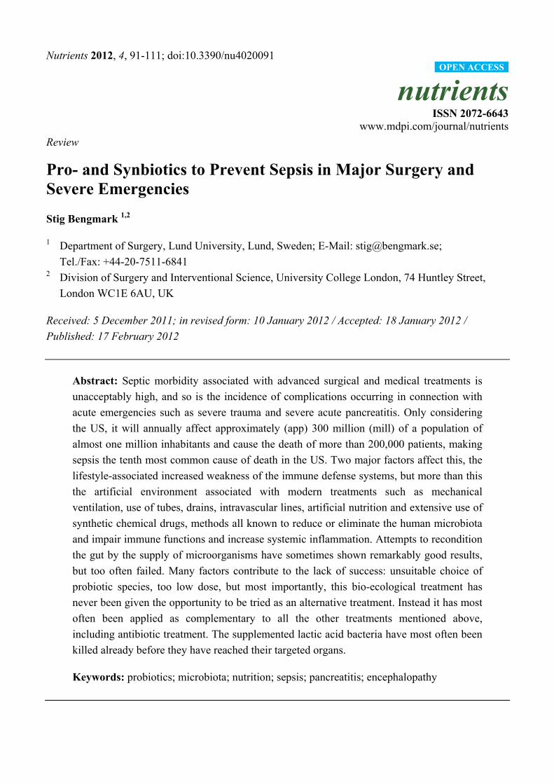
Nutrients 2012, 4, 91-111; doi:10.3390/nu4020091 nutrients ISSN 2072-6643 www.mdpi.com/journal/nutrients Review Pro- and Synbiotics to Prevent Sepsis in Major Surgery and Severe Emergencies Stig Bengmark 1,2 1 Department of Surgery, Lund University, Lund, Sweden; E-Mail: [email protected]; Tel./Fax: +44-20-7511-6841 2 Division of Surgery and Interventional Science, University College London, 74 Huntley Street, London WC1E 6AU, UK Received: 5 December 2011; in revised form: 10 January 2012 / Accepted: 18 January 2012 / Published: 17 February 2012 Abstract: Septic morbidity associated with advanced surgical and medical treatments is unacceptably high, and so is the incidence of complications occurring in connection with acute emergencies such as severe trauma and severe acute pancreatitis. Only considering the US, it will annually affect approximately (app) 300 million (mill) of a population of almost one million inhabitants and cause the death of more than 200,000 patients, making sepsis the tenth most common cause of death in the US. Two major factors affect this, the lifestyle-associated increased weakness of the immune defense systems, but more than this the artificial environment associated with modern treatments such as mechanical ventilation, use of tubes, drains, intravascular lines, artificial nutrition and extensive use of synthetic chemical drugs, methods all known to reduce or eliminate the human microbiota and impair immune functions and increase systemic inflammation. Attempts to recondition the gut by the supply of microorganisms have sometimes shown remarkably good results, but too often failed. Many factors contribute to the lack of success: unsuitable choice of probiotic species, too low dose, but most importantly, this bio-ecological treatment has never been given the opportunity to be tried as an alternative treatment. Instead it has most often been applied as complementary to all the other treatments mentioned above, including antibiotic treatment. The supplemented lactic acid bacteria have most often been killed already before they have reached their targeted organs. Keywords: probiotics; microbiota; nutrition; sepsis; pancreatitis; encephalopathy OPEN ACCESS
Transcript of 106. Nutrients 2012,4,91-111
Nutrients 2012, 4, 91-111; doi:10.3390/nu4020091
nutrients ISSN 2072-6643
www.mdpi.com/journal/nutrients
Review
Pro- and Synbiotics to Prevent Sepsis in Major Surgery and Severe Emergencies
Stig Bengmark 1,2
1 Department of Surgery, Lund University, Lund, Sweden; E-Mail: [email protected];
Tel./Fax: +44-20-7511-6841 2 Division of Surgery and Interventional Science, University College London, 74 Huntley Street,
London WC1E 6AU, UK
Received: 5 December 2011; in revised form: 10 January 2012 / Accepted: 18 January 2012 /
Published: 17 February 2012
Abstract: Septic morbidity associated with advanced surgical and medical treatments is
unacceptably high, and so is the incidence of complications occurring in connection with
acute emergencies such as severe trauma and severe acute pancreatitis. Only considering
the US, it will annually affect approximately (app) 300 million (mill) of a population of
almost one million inhabitants and cause the death of more than 200,000 patients, making
sepsis the tenth most common cause of death in the US. Two major factors affect this, the
lifestyle-associated increased weakness of the immune defense systems, but more than this
the artificial environment associated with modern treatments such as mechanical
ventilation, use of tubes, drains, intravascular lines, artificial nutrition and extensive use of
synthetic chemical drugs, methods all known to reduce or eliminate the human microbiota
and impair immune functions and increase systemic inflammation. Attempts to recondition
the gut by the supply of microorganisms have sometimes shown remarkably good results,
but too often failed. Many factors contribute to the lack of success: unsuitable choice of
probiotic species, too low dose, but most importantly, this bio-ecological treatment has
never been given the opportunity to be tried as an alternative treatment. Instead it has most
often been applied as complementary to all the other treatments mentioned above,
including antibiotic treatment. The supplemented lactic acid bacteria have most often been
killed already before they have reached their targeted organs.
Keywords: probiotics; microbiota; nutrition; sepsis; pancreatitis; encephalopathy
OPEN ACCESS

Nutrients 2012, 4 92
1. Introduction: A Pandemic of Critical Illness
Advanced surgical and medical treatments, as well as medical and surgical emergencies, are,
despite some breath-taking advances in medico-pharmaceutical and surgical treatment, still
accompanied by an unacceptably high morbidity and also mortality. Worse than this, morbidity and
mortality in these conditions is fast increasing and has done so all over the world for several decades.
Apart from the suffering associated with this, it is also extremely costly to the individual and the health
care system.
Dominating among the treatment-induced morbidities is sepsis due to bacterial, fungal or viral
infections. Recent observations in the US suggest that not only the incidence, but also the severity of
sepsis, has significantly increased during the last decades [1–3]. Especially advanced surgery carries a
high rate of septic morbidity and especially esophageal, pancreatic, and gastric procedures are
particularly known to represent great risk for the development of sepsis, but thoracic, adrenal, and
hepatic procedures are those that have the highest sepsis-induced mortality [1,2]. It is also documented
in the literature that elderly patients, men, and nonwhites are the most likely to develop sepsis as a
complication to surgical treatment [1,2]. Sepsis is by far the most common medical and surgical
complication, and estimated only in the US to annually affect as many as 751,000 [4,5], and cause
death of app 215,000 patients (29% of treated patients) [5], making sepsis the tenth most common
cause of death in this country.
2. Sepsis: Often a Product of So Called Auxiliary/Supportive Measures
Post-surgical morbidity and due to septic manifestations is only partly attributed to the surgeon and
the surgical technique. Increasing evidence suggests instead that it is the patient’s ability to resist
disease/immune defense and especially supportive measures during and around the treatment, such as
mechanical ventilation, use of implants, drains and intravascular lines, but also choice of content and
routes to provide nutrition, blood transfusions, choice of anesthesia and prescription of drugs, also
antibiotics and immune-suppressives, that are the largest contributors to the development of
septic manifestations.
Mechanical ventilation in association with management of emergencies and surgical procedures has
in recent years received increasing attention as a major contributor to chest-infections, but also to
general and localized septic manifestations in the body; according to a recent study applied in the US
in about 800,000 individuals every year. It is estimated to represent no less than 12% of total hospital
costs, no less than $27 billion in the US alone [6]. This treatment is responsible for, not only a
disproportional amount of resources used, but also for the unacceptably high morbidity and mortality
associated with the treatment, especially in elderly people [6].
A main contributor to intensive care unit (ICU)-associated sepsis is also artificial nutrition, both
enteral and parenteral; catheter-related sepsis is reported to occur in about 25% of patients fed via
intravenous feeding-tubes [7]. Numerous drugs used in the ICUs including antibiotics are known to
derange the immune functions, impair macrophage functions, bactericidal efficacy as well as
production and secretion of cytokines. For example, supply of an antibiotic, Mezlocillin (Bayer,
150 mg/kg body weight) is demonstrated to significantly suppress essential macrophage functions and

Nutrients 2012, 4 93
derange chemiluminescence response, chemotactic motility, bactericidal and cytostatic ability and
impair lymphocyte proliferation [8]. Other common perioperative practices like use of artificial
feeding regimens, preoperative antibiotics [9], and mechanical bowel preparation [10,11] will also,
according to recent studies, instead of the expected prevention as expected, contribute to increased
rates of treatment-associated infections.
3. Gut and Chest Infections Dominate
Most of the surgical infections originate in areas with the most exposure to the exterior world,
i.e., the gut (63%) and chest (20%) and are pure gram-negative (22%), pure gram-positive (15%),
mixed (23) or fungal (8%) [12]. It is also here that the dominating parts of the immune functions are
localized: between 70–80% of the Ig-producing immunocytes of the body are found in the gut [13]. It
was Marshall and his group who, in 1993, directed attention to the gut as a special source of surgical
sepsis and especially to its most severe form—multiple organ failure (MOF) [14]. They observed that
the most common organisms to cause ICU-acquired infections—Candida, Streptococcus faecalis,
Pseudomonas, and coagulase-negative Staphylococci—were also the most common species that
seemed to colonize especially the proximal GI tract. They reported that this colonization correlated
well with development of invasive infection within one week: Pseudomonas (90% vs. 13% in
non-colonized patients, p < 0.0001) and Staphylococcus epidermidis (80% vs. 6%, p < 0.0001). As a
consequence of these observations, they coined the phrase: “the gut—the undrained abscess”. Studies
during subsequent years focused mainly on microbial translocation as the major course of sepsis.
Important observations had, however, been made by Baue, Faist and their groups already ten years
earlier, and their finding that a significant portion of patients, who develop multiple organ dysfunction
syndrome (MODS), and do not have an identifiable infection [15], should have a great impact on
future understanding of treatment and emergency-associated sepsis. It was Goris and his collaborators,
who, in 1986, based on observations like those of Baue and Faist, but also after extensive studies in
animals, suggested inflammation precedes septic manifestations and an “auto-destructive inflammatory
response” with or without bacterial infection, is a major cause of this severe condition [16].
4. Preceding Uncontrolled Exuberant Systemic Inflammation
Patients who develop severe septic complications are known to respond to physical and mental
stress with an early exuberant acute, or chronic, super-inflammation, with signs of exaggerated and
prolonged release of pro-inflammatory cytokines such as interleukin-6 (IL-6), acute phase proteins
such as C-reactive protein, and plasminogen activator inhibitor 1 (PAI-1)—see [17]—a reaction
strongly associated with subsequent severe exacerbation of disease, including acute respiratory distress
syndrome (ARDS), and MOF. Among the observed changes associated with an exuberant inflammation
in the early nervous phase are: augmented endothelial adhesion of polymorphonuclear (PMN) cells,
increased production of intracellular adhesion molecule-1 (ICAM-1), priming of the PMNs for an
oxidative burst, release of pro-inflammatory platelet activating factor (PAF), and a delay in PMN
apoptosis [18]. Visceral adipocytes are, compared to subcutaneous fat cells, known to secrete per gram
tissue much more of free fatty acids but also about three times as much IL-6, and PAI-1; observations
that well explain the high risk of both chronic and acute diseases in individuals with visceral

Nutrients 2012, 4 94
obesity [19]. The stress-induced load of these and other proinflammatory and procoagulant molecules
on organs, such as the lung and the liver, can vary a thousand times or more, as the amount of fat in the
abdomen can vary from a few milliliters in a lean subject to about six liters in gross obesity [20].
5. Mental and Physical Stress Potentiates the Response
An increase in growth of Gram-negative bacteria of up to 100,000 times (5 logs of order) has been
demonstrated in animals exposed to noradrenaline; see Lyte’s review [21]. Old observations suggest a
strong and significant association between higher blood levels of noradrenaline and adrenaline and
development of severe septic conditions [22]. Luminal release of noradrenaline is a documented strong
inducer of virulence of luminal bacteria [23], and much suggests that potentially pathogenic
microorganisms (PPMs), normally indolent colonizers, under stress change their phenotype and
become life-threatening pathogens [24]. These observations are particularly interesting as classical
studies published already in the 1940s demonstrated that a 10,000 times lower dose of Clostridium
welchii is needed to induce death in gas-gangrene in animals, when also adrenaline is administered to
the animals [25]. Most likely similar potentiating effects exist when stress and stress-hormones are
applied also to other pathogenic bacteria.
6. Deranged and Dysfunctioning Microbiota
Microbiota is significantly reduced in Westerners. A study published in the US in 1983 reported
that Lactobacillus plantarum, a dominating lactic acid bacterium (LAB) among plant eaters, is found
in only about 25% of omnivorous Americans and in about two thirds of vegetarian Americans [26]. A
more recent Scandinavian study suggest that also in healthy individuals, the most common colonic
LAB are present in only half or less of the individuals: Lactobacillus plantarum in 52%, Lactobacillus
rhamnosus in 26% and Lactobacillus paracasei ssp. paracasei 17% [27]. Western lifestyle with frequent
mental and physical stress, and eating of processed foods with high content of saturated fats and
trans-fatty acids, lack of dietary fibers, containing chemicals and pharmaceuticals, but also lack of
microorganisms content in food, will significantly reduce the microbiota both with regard to diversity
and extent of existing flora. Recent studies report that in Westerners, a significant shift in balance from
gram-positives to gram-negatives, and subsequent increase in production of endotoxins/lipopolysaccharide
(LPS) [28]. Magnesium (Mg) requirements are known to be much higher for Gram+ than for Gram−
bacteria and Mg deficiency, common in Westerners, was recently shown to induce decreased content
in the gut especially of Bifidobacteria, to increase colonic and systemic inflammation, and increase
intestinal permeability. These observations are of particular interest as Mg deficiency is associated
with systemic inflammation and common metabolic disorders such as type 2 diabetes, metabolic
syndrome, dyslipidemia, and hypertension [29], but also observed in ICU patients. Mg-deficient
patients, in comparison to patients with normal magnesium levels, are reported to exhibit a higher
prevalence of severe sepsis and septic shock (57 vs. 11%, p < 0.01), a longer ICU stay (15.4 ± 15.5 vs.
2.8 ± 4.7 days, p < 0.01), and a higher mortality rate [30].
A several log-fold decrease in the commensal bacteria like Bifidobacteria and Lactobacilli are
reported in trauma patients in parallel to increased populations of pathogenic bacteria such as
Pseudomonas aeruginosa and Staphylococcus aureus [31]. The Western lifestyle-induced derangement
Nutrients 2012, 4 95
of the microbiota is associated with increased permeability of intestinal mucosa, increased LPS
absorption, increased endotoxemia, exaggerated inflammation, and strongly associated with metabolic
disorders such as obesity and diabetes [32]—conditions, which also are commonly associated with
increased surgical morbidity, surgical infections and MOF.
7. Overreacting Neutrophils
Severe trauma, major surgery and severe sepsis will, parallel to a significant decrease in
lymphocytes, induce a significant, sometimes disproportionate, increase in circulating and tissue
neutrophils, and be accompanied by persistent decline in T-4 helper lymphocytes and elevation of T-8
suppressor lymphocytes [33]. It is suggested that a T-4/T-8 lymphocyte cell ratio of <1 is a sign of
severe immunosuppression and predictor of complication, such as multiple organ dysfunction
syndrome, myocardial infarction, acute pancreatitis, multiple severe trauma and chemotherapeutic
treatment, especially in oncology patients [34]. An early and large increase in circulating neutrophils is
accompanied by tissue infiltration of neutrophils, and responsible for common posttrauma/postoperative
dysfunctions such as paralytic ileus [35,36], bone marrow suppression, endothelial cell dysfunction,
and to lead to tissue destruction and organ failure, particularly in lungs [37–39], intestines [40],
liver [41] and kidney [42]. Neutrophil infiltration of distant organs [16], especially the lungs [37], is a
characteristic finding in patients dying of sepsis. The extent of neutrophil infiltration is significantly
aggravated by mechanical therapeutic efforts such as handling of the bowels during operation [35], and
ventilation of the lungs [43]. Poor nutritional status, preexisting immune deficiency, obesity, diabetes
and high levels of blood sugar [44] contribute to immune deterioration and to increased expressions in
the body of molecules such as NF-κB, COX-2, LOX and iNOS [45,46]. The disproportionate increase
in circulating neutrophils is to a great extent inhibited by supplementation of antioxidants [47,48] and
specific probiotics [49].
Supplementation of probiotics will effectively prevent neutrophil infiltration of the lung and also
reduce the subsequent tissue destruction, as demonstrated in studies with inflammation induced by
cecal ligation and puncture (CLP). A synbiotic formulation, Synbiotic 2000 Forte (see further below),
was administered orally before the induced trauma and effectively prevented both neutrophil
accumulation and tissue destruction in the lungs [50]. Most interestingly, these effects were obtained
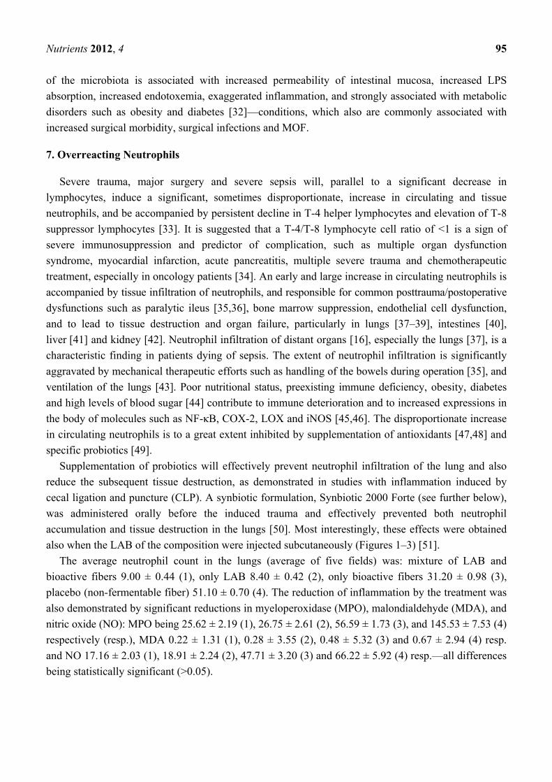
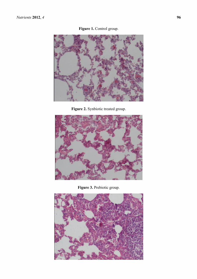
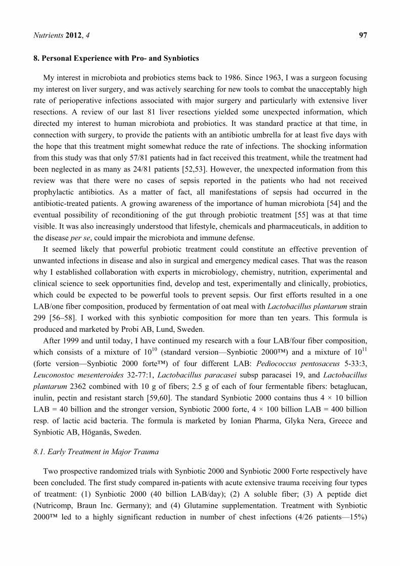
also when the LAB of the composition were injected subcutaneously (Figures 1–3) [51].
The average neutrophil count in the lungs (average of five fields) was: mixture of LAB and
bioactive fibers 9.00 ± 0.44 (1), only LAB 8.40 ± 0.42 (2), only bioactive fibers 31.20 ± 0.98 (3),
placebo (non-fermentable fiber) 51.10 ± 0.70 (4). The reduction of inflammation by the treatment was
also demonstrated by significant reductions in myeloperoxidase (MPO), malondialdehyde (MDA), and
nitric oxide (NO): MPO being 25.62 ± 2.19 (1), 26.75 ± 2.61 (2), 56.59 ± 1.73 (3), and 145.53 ± 7.53 (4)
respectively (resp.), MDA 0.22 ± 1.31 (1), 0.28 ± 3.55 (2), 0.48 ± 5.32 (3) and 0.67 ± 2.94 (4) resp.
and NO 17.16 ± 2.03 (1), 18.91 ± 2.24 (2), 47.71 ± 3.20 (3) and 66.22 ± 5.92 (4) resp.—all differences
being statistically significant (>0.05).
Nutrients 2012, 4 96
Figure 1. Control group.
Figure 2. Synbiotic treated group.
Figure 3. Prebiotic group.
Nutrients 2012, 4 97
8. Personal Experience with Pro- and Synbiotics
My interest in microbiota and probiotics stems back to 1986. Since 1963, I was a surgeon focusing
my interest on liver surgery, and was actively searching for new tools to combat the unacceptably high
rate of perioperative infections associated with major surgery and particularly with extensive liver
resections. A review of our last 81 liver resections yielded some unexpected information, which
directed my interest to human microbiota and probiotics. It was standard practice at that time, in
connection with surgery, to provide the patients with an antibiotic umbrella for at least five days with
the hope that this treatment might somewhat reduce the rate of infections. The shocking information
from this study was that only 57/81 patients had in fact received this treatment, while the treatment had
been neglected in as many as 24/81 patients [52,53]. However, the unexpected information from this
review was that there were no cases of sepsis reported in the patients who had not received
prophylactic antibiotics. As a matter of fact, all manifestations of sepsis had occurred in the
antibiotic-treated patients. A growing awareness of the importance of human microbiota [54] and the
eventual possibility of reconditioning of the gut through probiotic treatment [55] was at that time
visible. It was also increasingly understood that lifestyle, chemicals and pharmaceuticals, in addition to
the disease per se, could impair the microbiota and immune defense.
It seemed likely that powerful probiotic treatment could constitute an effective prevention of
unwanted infections in disease and also in surgical and emergency medical cases. That was the reason
why I established collaboration with experts in microbiology, chemistry, nutrition, experimental and
clinical science to seek opportunities find, develop and test, experimentally and clinically, probiotics,
which could be expected to be powerful tools to prevent sepsis. Our first efforts resulted in a one
LAB/one fiber composition, produced by fermentation of oat meal with Lactobacillus plantarum strain
299 [56–58]. I worked with this synbiotic composition for more than ten years. This formula is
produced and marketed by Probi AB, Lund, Sweden.
After 1999 and until today, I have continued my research with a four LAB/four fiber composition,
which consists of a mixture of 1010 (standard version—Synbiotic 2000™) and a mixture of 1011
(forte version—Synbiotic 2000 forte™) of four different LAB: Pediococcus pentosaceus 5-33:3,
Leuconostoc mesenteroides 32-77:1, Lactobacillus paracasei subsp paracasei 19, and Lactobacillus
plantarum 2362 combined with 10 g of fibers; 2.5 g of each of four fermentable fibers: betaglucan,
inulin, pectin and resistant starch [59,60]. The standard Synbiotic 2000 contains thus 4 × 10 billion
LAB = 40 billion and the stronger version, Synbiotic 2000 forte, 4 × 100 billion LAB = 400 billion
resp. of lactic acid bacteria. The formula is marketed by Ionian Pharma, Glyka Nera, Greece and
Synbiotic AB, Höganäs, Sweden.
8.1. Early Treatment in Major Trauma
Two prospective randomized trials with Synbiotic 2000 and Synbiotic 2000 Forte respectively have
been concluded. The first study compared in-patients with acute extensive trauma receiving four types
of treatment: (1) Synbiotic 2000 (40 billion LAB/day); (2) A soluble fiber; (3) A peptide diet
(Nutricomp, Braun Inc. Germany); and (4) Glutamine supplementation. Treatment with Synbiotic
2000™ led to a highly significant reduction in number of chest infections (4/26 patients—15%)
Nutrients 2012, 4 98
compared to peptide diet (11/26 patients—42%, p < 0.04), glutamine treatment (11/32 patients—34%,
p < 0.03) and only fiber treatment (12/29 patients—41%, p < 0.002) [61]. Also the total number of
infections was significantly decreased: Synbiotic 2000™ 5/26 patients (19%), peptide 13/26 patients
(50%) glutamine16/32 patients (50%) and only fibers 17/29 patients (59%).
In another study, 65 polytrauma patients were randomized to receive either Synbiotic 2000 Forte
(400 billion LAB + 10 gram of fibers, see above) or maltodextrine as placebo, once daily, for 15 days.
Significant reductions were observed in number of deaths (5/35 vs. 9/30, p < 0.02), severe sepsis (6/35
vs. 13/30, p < 0.02), chest infections (19/35 vs. 24/30, p < 0.03), central line infections (13/32 vs.
20/30, p < 0.02), and ventilation days (average 15 vs. 26 days [62]). A total of pathogenic
microorganisms of 54 were cultivated in the Synbiotic treated group compared to 103 in the fiber-only
group [63,64]. Repeat analyses also revealed that serum levels of endotoxin (lipopolysaccharide
(LPS)) were decreased and time to bloodstream infection significantly prolonged in patients treated
with Synbiotic 2000 forte.
8.2. Early Treatment in Severe Acute Pancreatitis
Patients with severe acute pancreatitis were randomized to receive, during the first 7 days
daily administered through a nasojejunal tube, either a freeze-dried preparation containing live
Lb plantarum 299 in a dose of 109 together with a substrate of oat fiber or a similar preparation, but
heat-inactivated [65]. The study was concluded when, on repeat statistical analysis, significant
differences in favor of one of the two groups were obtained. This occurred when a total of 45 patients
had entered the study. 22 patients had at that time received treatment with live and 23 with the
heat-killed Lb plantarum 299. Infected pancreatic necrosis and abscesses were seen in 1/22 (4.5%) in
the live LAB group vs. 7/23 (30%) in the heat-inactivated group (p = 0.023). The only patient in the
lactobacillus group, who developed infection, a urinary infection, did that on the 15th day, e.g., at a
time when he had not received treatment for eight days. The length of stay was also considerably
shorter in the live LAB group (13.7 days vs. 21.4 days), but the limited size of the material did not
allow statistical significance to be reached.
Sixty-two patients with severe acute pancreatitis (SAP) (Apache II scores: Synbiotic 2000-treated
11.7 ± 1.9, controls 10.4 ± 1.5) were given either two sachets/day of Synbiotic 2000™ (2 × 40 billion
LAB/day and totally 20 g fibers) or the same amounts of fibers (20 g) as in Synbiotic 2000™ during
the first 14 days after arrival to the hospital [66]. 9/33 patients (27%) in the Synbiotic 2000-treated
group and 15/29 patients (52%) in the only fiber-treated group developed subsequent infections.
Eight out of 33 (24%) Synbiotic 2000-treated patients, and 14/29 (48%) of the only fiber-treated
patients, developed systemic inflammatory response syndrome (SIRS), MOF or both (p < 0.005). A
total of pathogenic microorganisms of seven were cultivated in the Synbiotic-treated group compared
to 17 in the only-fiber group.
8.3. Effects on “Mind Clarity”: Encephalopathy
Patients with critical illness, as well as patients with chronic disorders such as liver cirrhosis and
diabetes, often suffer a mild, but sometimes, severe confusion, which often has its origin in the
gut [67]. Increasing evidence suggest that probiotics, alone and/or in combination with plant antioxidants
Nutrients 2012, 4 99
and fibers, possess strong neuro-endocrine modulatory effects and alleviates effects of physical and
mental stressors both early [68] as well as later in life [69]. We undertook some studies to explore the
effects of synbiotic treatment in patients with liver cirrhosis and minimal hepatic encephalopathy (MHE).
Fifty-five patients with MHE were randomized to receive, for 30 days: (1) Synbiotic 2000 (n = 20),
(2) The fibers in the composition alone (n = 20), or (3) Placebo (n = 15). All cirrhotic patients with
MHE were found to have severe derangements of the gut micro-ecology, and significant overgrowth of
potentially pathogenic Escherichia coli and Staphylococcal species. Synbiotic treatment increased the
fecal content of non-urease-producing Lactobacillus species significantly and reduced the numbers of
potentially pathogenic micro-organisms. The treatment was also associated with a significant reduction
in endotoxemia and in blood ammonia levels. A documented reversal of MHE was obtained in half of
the treated patients; the Child-Turcotte-Pugh functional class improved in about 50% of cases [70].
Treatment with fermentable fibers alone also demonstrated substantial benefit in a proportion of patients.
Thirty cirrhotic patients were randomized in a second study to receive Synbiotic 2000 or placebo
for only 7 days. Viable fecal counts of Lactobacillus species, Child-Pugh class, plasma retention rate
of indocyanine green (ICGR15), whole blood tumor necrosis factor alpha (TNF-a) mRNA and IL-6
mRNA, serum TNF-a, soluble TNF receptor (sTNFR)I, sTNFRII, IL-6 and plasma endotoxin levels
were measured pre- and post-treatment. The treatment with Synbiotic 2000 was associated with
significantly increased fecal lactobacilli counts and significant improvements in ICGR15 and
Child-Pugh class [71]. Significant increases in whole blood TNF-a mRNA and IL-6 mRNA, along
with serum levels of sTNFRI and sTNFRII, were also observed and TNF-a and IL-6 levels correlated
significantly, both at baseline and post-synbiotic treatment [71]. Synbiotic-related improvement in
ICGR15 was significantly accompanied by changes in IL-6, both at mRNA and protein levels, but was
unrelated to levels of plasma endotoxin. No significant changes in any parameter were observed
following placebo treatment. Even this study concluded that short-term synbiotic treatment modulates
gut flora significantly and improves liver function in patients with cirrhosis.
9. Studies by Others
In a recent study twenty-nine SIRS patients with a serum C-reactive protein (CRP) level above
10 mg/dL, received a synbiotic composition consisting of Bifidobacterium breve and Lactobacillus
casei, in combination with galactooligosaccharides. The incidence of infectious complications such as
enteritis, pneumonia, and bacteremia were (compared to historical controls) significantly lower in the
treated group [72]. Analysis of fecal flora demonstrated significantly higher levels of Bifidobacteria
and Lactobacillus, and also of total organic acids, particularly short-chain fatty acids.
10. Studies with No, or Adverse, Effects
10.1. Ecologic 641™
Two hundred and ninety eight patients with predicted severe acute pancreatitis and with APACHE
II score ≥8, Imrie score > 3, or C-reactive protein >150 mg/L) within 72 h of onset of symptoms were
included in a multicenter, double-blind, placebo-controlled trial, randomly assigned to receive either a
multispecies probiotic preparation (n = 153) or placebo (n = 145), administered enterally twice daily
Nutrients 2012, 4 100
for 28 days. The probiotic composition supplemented, Ecologic 641 (Winclove Bio Industries,
Amsterdam, The Netherlands), consisted of 1010 of each of six different strains of freeze-dried, viable
bacteria: Lactobacillus acidophilus, Lactobacillus casei, Lactobacillus salivarius, Lactococcus lactis,
Bifi dobacterium bifidum, and Bifidobacterium lactis. It contained also cornstarch and maltodextrins.
The subsequent analyses were based on 152 individuals in the treated group and 144 in the placebo
groups. Infectious complications occurred in 46 (30%) of patients in the probiotics group and in
41 (28%) of those in the placebo group (relative risk 1.06, 95% CI 0.75–1.51). Twenty-five (16%)
patients in the probiotics group died, compared to nine (6%) in the placebo group (relative risk 2.53,
95% CI 1.22–5.25) [73]. Furthermore, nine patients in the probiotics group developed bowel ischemia,
eight of which with fatal outcome, compared to none in the placebo group (p = 0.004).
10.2. Lactobacillus plantarum 299™: ProViva™
One hundred and three critically ill patients were randomized to receive 1 ProViva, a fruit drink
containing 5% of LAB-fermented oat and live Lactobacillusb plantarum 299v with a density of
5 × 10⁷ (n = 52) or conventional nutrition therapy alone (n = 51). The treatment demonstrated no
identifiable effect in terms of bacterial translocation (12% vs. 12%; p = 0.82), gastric colonization with
enteric organisms (11% vs. 17%, p = 0.42), or septic morbidity (13% vs. 15%; p = 074), serum CRP
levels or mortality [74]. In another study, 11 patients undergoing elective abdominal surgery during a
median time of 9 days (range 5–18 days) received lactobacillus 299v (ProViva) to total average
amounts of 3250 mL (range 2100–9000 mL), and were compared to 11 control patients. The authors
found no significant differences between the ProViva group and the control group in terms of
concentrations of plasma cells, IgA positive cells or IgM positive cells in the lamina propria [75].
There was a significantly higher concentration of IgM at the mucosal surface in the control group
(P = 0.02, Fishers Exact test mid P), but no difference in terms of IgA.
10.3. Lactobacillus rhamnosus GG™
Sixty-one patients were in a study in pediatric ICU, randomized: 31 patients to receive treatment
with one capsule of Lactobacillus rhamnosus strain GG in a dose of 1010, and 30 patients to receive
one capsule with insulin daily (control group). No difference in rate of infections was observed
between the groups; the mean number of infections in the treatment and control groups was 1.83 and
1.33, respectively. 9/31 patients in the probiotic-treated group developed in total 15 nosocomial
infections: six bloodstream infections (40%), five tracheo-bronchitis (33%), two pneumonia (13%),
and two UTI (13%) [76]. There were totally six deaths during the study period; four in the placebo
group and two in the treatment group. No cases of Lactobacillus bacteremia or other serious adverse
effects were observed.
10.4. Synbiotic 2000™/Synbiotic 2000 Forte™
Synbiotic 2000 (40 billion LAB) was given daily to 162 patients and 168 control patients received
similarly only the fibers of the synbiotic composition. No significant differences in demographics,
mortality (ICU or hospital), morbidity, maximal multiple organ dysfunction (MOD) score, ICU length
Nutrients 2012, 4 101
of stay, diarrhea days or septic complications were observed between intervention and control
groups [77]. Similarly post hoc analysis of those patients who received at least 75% of prescribed
doses revealed no difference in ICU mortality (21% vs. 20%) or hospital mortality (32% vs. 26%)
between the intervention and control groups. It was especially noted that no episodes of intestinal
ischemia were observed in either group.
Two hundred and fifty nine enterally fed critically ill patients, expected to require mechanical
ventilation for 48 h or more were enrolled in a study; 130 patients received Synbiotic 2000 FORTE®
(twice a day) and 129 patients a cellulose-based placebo for a maximum of 28 days. The oropharyngeal
microbial flora and colonization rates were unaffected by the synbiotic treatment. The overall
incidence of ventilator associated pneumonia (VAP) was lower than anticipated (11.2%) and no
statistical difference was demonstrated between the groups receiving synbiotic or placebo; incidence
of VAP (9 and 13%, P = 0.42), VAP rate per 1000 ventilator days (13 and 14.6, P = 0.91) or hospital
mortality (27 and 33%, P = 0.39), respectively [78]. No negative effects of the treatment
were observed.
10.5. Trevis™
A total of 90 patients admitted to an ICU were randomized to receive either synbiotic or placebo
(45 into each group). The synbiotic treatment consisted of supplying, three times a day, a capsule of
TrevisTM (Chr Hansen Biosystem, Denmark), containing 4 × 10⁹ colony forming units of each of
L. acidophilus La5 (La5), B. lactis Bb-12 (Bb-12), S. thermophilus and L. bulgaricus. In addition the
prebiotic oligofructose (7.5 g of RaftiloseTM powder, Orafti Active Food Ingredients, Belgium) was
administered twice a day. The patients in the synbiotic group demonstrated after 1 week of therapy
significantly lower incidence of potentially pathogenic bacteria (43% vs. 75%, P = 0.05) and multiple
organisms (39% vs. 75%, P = 0.01) in their nasogastric aspirates than the controls [79]. However,
there were no significant differences between the groups in terms of intestinal permeability, septic
complications or mortality.
10.6. VSL#3™
Twenty-eight patients were enrolled and randomly assigned to one of three treatment groups:
(1) placebo (n = 9); (2) viable probiotics—2 sachets daily of VSL#3™ (n = 10); or (3) bacterial
sonicates—not viable VSL#3 bacteria (n = 9). Each sachet of the supplemented probiotic, VSL#3
(VSL Pharmaceuticals, Ft Lauderdale, FL) contains 900 billion viable lyophilized bacteria of 4 strains
of Lactobacillus (L. casei, L. plantarum, L. acidophilus, and L. delbrueckii subsp. bulgaricus) plus
3 strains of Bifidobacterium (B. longum, B. breve, and B. infantis), plus Streptococcus salivarius subsp.
Thermophiles, totally eight strains. Intestinal permeability decreased in all treatment groups. The rate
of severe sepsis and MODS were not significantly affected by the treatment, although a significantly
larger increase in systemic IgA and IgG concentrations were observed in the group supplied with live
bacteria than in the patients who received placebo or sonicated bacteria (P < 0.05) [80].
Nutrients 2012, 4 102
11. Why do Studies Fail?
Critical care units are in general a highly artificial environment and the burden of environment-induced
physical and mental stress and subsequent status of systemic hyper-inflammation on the patient is
unbearable. Patients treated under these conditions are in many aspects dysfunctional, the whole
microbiota is gone and probiotic bacteria supplied will most often be killed already before they have
reached its target organ. This artificiality seems to vary from country to country, and sometimes also
from hospital to hospital, observations that might explain the great variation in outcome from studies
undertaken in different regions and countries.
Probiotic treatment has never been given the chance as an alternative treatment; it has only been
tried as a treatment complementary to all the other standard treatments. As discussed above, a series of
auxiliary measures and particularly mechanical ventilation [7] and treatment with various drugs,
including antibiotics [8,9], but also clinical nutrition solutions belong to those factors which promote
super-inflammation and, indirectly, infection. Enteral nutrition formulas are also known to induce loss
of intestinal barrier function, promotes bacterial translocation, and impairs host immune defense [81],
a phenomenon, observed in humans and further elucidated in animal studies. The incidence of bacterial
translocation to the mesenteric lymph node was in such studies significantly increased when the
animals were fed nutrition formulas such as Vivonex (53%), Criticare (67%), or Ensure (60%)
(p < 0.05) [82–84]. Similar observations have also been made in patients. Significant elevations in
pro-inflammatory cytokines were observed in patients, who after pancreatoduodenectomy are fed a
standard enteral nutrition solution (Nutrison): IL-1beta—day 7 (P < 0.001), day 14 (P = 0.022);
TNF-alpha—day 3 (P = 0.006), day 7 (P < 0.001) [85]. Of special interest are the observations that
such changes were not observed when the standard nutrition was replaced with a formula that claimed
to have immune-modulatory effects (Stresson). Instead anti-inflammatory cytokines were seen
significantly elevated: IL-1ra/s: day 7 (P < 0.001), IL-6: day 10 (P = 0.017), IL-8: day 1 (P = 0.011)
days 3, 7, 10, and 14 (P < 0.001) and IL-10: days 3 and 10 (P < 0.001).
The type of bacteria to be chosen for probiotic purpose is also critical. Only a few LAB strains have
demonstrated ability to influence the immune system, reduce inflammation and/or eliminate or reduce
unwanted pro-inflammatory molecules from foods. Even strains, which carry sometimes the same
name have often different and sometimes opposite effects. A recent study selected 46 strains of
Lactococcus lactis from about 2600 LAB and compared their ability to induce cytokines. It was
demonstrated that the inter-strain differences in ability to produce pro- and anti-inflammatory
cytokines were great [86], an observation which underlines the importance of extensive animal and
preclinical studies before a LAB or combination of LAB be chosen as probiotic.
12. Choice of Lactic Acid Bacteria as Probiotics
Especially desirable as probiotics are strains that improve immune function by increasing the
number of IgA-producing plasma cells, improve phagocytosis, and influence the proportion of Th1
cells and NK cells [87]. Among the strains with a strong anti-inflammatory record are Lactobacillus
paracasei subsp. paracasei, Lactobacillus plantarum, and Pediococcus pentosaceus. Especially
Lactobacillus paracasei seems to have a solid record. It has been shown to induce cellular immunity,
Nutrients 2012, 4 103
stimulating the production of suppressive cytokines such as TGFβ and IL-10; to suppress Th2 activity,
CD4 T-cells [88,89], and splenocyte proliferation [90]; and to decrease antigen-specific IgE and
IgG1 [91]. Lactobacillus paracasei was shown to be the strongest inducer of Th1 and repressor of Th2
cytokines when more than a hundred LAB strains were compared [92]. A recent study in rats
compared the ability of four different strains: Lactobacillus paracasei, Lactobacillus johnsonii,
Bifidobacterium longum, or Bifidobacterium lactis to control Trichinella spiralis—induced infection.
Lactobacillus paracasei, but none of the others, were able to reduce infection-associated Th2
response, muscle levels of TGF-β, COX-2 and PGE2 and to attenuate infection-induced muscle
hyper-contractility [93]. Another recent study compared the ability to reduce stress-induced changes in
gut permeability and sensitivity to colorectal distension of three probiotic strains: Bifidobacterium
lactis NCC362, Lactobacillus johnsonii NCC533, and Lactobacillus paracasei NCC2461.
Lactobacillus paracasei but none of the other LAB, significantly restored normal gut permeability, and
reduce visceral hyperalgesia and visceral pain [94].
Lactobacillus plantarum has also an excellent record. When the ability of fifty different LAB to
control twenty-three different Clostridium difficile (C diff) strains were studied, Lactobacillus
paracasei and Lactobacillus plantarum were the only strains with ability to effectively eliminate all
C. diff strains—more than half of the tried LAB strains were totally ineffective, and some only against
a few [95]. Some LAB can be potentiated in their efficacy by simultaneous supply of prebiotic fibers
(probiotics + prebiotics ≥ synbiotics). However, there are great differences in the ability of different
strains to ferment and utilize plant fibers, especially when it comes to semi-fermentable fibers such as
oligofructans. Only a handful of LAB from the 712 tested strains demonstrated in a study ability to
ferment inulin and phlein, namely: Lactobacillus plantarum (several strains), Lactobacillus paracasei
subsp. paracasei, Lactobacillus brevis and Pediococcus pentosaceus [96].
13. Discussion: The Future “Designer Probiotics”?
Two recent studies provide a fascinating insight into the future. Gene expression of human
duodenal mucosa cells were studied after exposure to one of the following four lactic acid bacteria:
Lactobacillus plantarum WCFS1 [97], Lactobacillus acidophilus L10, Lactobacillus casei CRL-431
and L. rhamnosus GG [98], administered in a cross-over study to healthy volunteers in a dose of 1010.
Mucosal biopsies were taken from duodenum after 6 h and compared to control biopsies. The
interventions did not impair immune and metabolic homeostasis. Nevertheless, a fascinating and most
distinct influence on expression of several hundred genes (transcriptome) was reported after
administration of each of the LAB. For seemingly the first time, different probiotic lactobacilli are
reported to induce more or less strain-specific and markedly different expression profiles, much similar
to what is known to occur with ingestion of various foods, especially plant ingredients [99,100], but
also similar to the effects observed after supplying certain pharmaceuticals. Thus L. plantarum was
reported to modulate overt adaptive immune responses [97], L. acidophilus to suppress inflammation,
L. casei to stimulate Th1 response and improve the Th1–Th2 balance, L. rhamnosis to influence
cellular growth and proliferation [98]. The effects were suggested to resemble, although distinctly
milder, those obtained by specific pharmaceuticals: L. acidophilus—antagonists of α-receptor activity,
guanine antagonists, synthetic corticosteroids and flavonoids, L. casei—modulators of GABA receptors,
Nutrients 2012, 4 104
cholinergic blocking agents, antagonists of β-adrenergic receptors, L. rhamnosus—glycoside steroids,
alkaloids, protein synthesis inhibitors and protein kinase C inhibitors. A large person-to-person
variation in response was also reported.
The responsiveness to ingestion of various LAB seems to be strongly influenced, not only by eventual
genetic background and existing resident microbiota, but also by lifestyle, and particularly by diet,
which might explain the person-to-person differences in response observed in the above studies [97,98],
but also differences in outcome, when applied in critically ill patients, as reported in this review.
In the immediate future, probiotics will most likely continue to find its dominating niche as
treatment given to rather healthy and health-concerned individuals with the main aim to promote
health and prevent disease. However, it is likely that probiotics, as our knowledge in nutragenomics
and therapeutic microbiology increases, will be generally regarded also as valuable tools for treatment
of various acute and chronic diseases, also in critically ill patients. The review of studies reported to
date and presented in this review supports such an assumption. Probiotics will, however, not be
generally accepted and utilized as clinical tools until radical and profound changes in treatment
practices in care of the critically ill and other patient groups are implemented.
References
1. Dombrovskiy, V.Y.; Martin, A.A.; Sunderram, J.; Paz, H.L. Facing the challenge: decreasing
case fatality rates in severe sepsis despite increasing hospitalizations. Crit. Care Med. 2005, 33,
2555–2562.
2. Dombrovskiy, V.Y.; Martin, A.A.; Sunderram, J.; Paz, H.L. Rapid increase in hospitalization and
mortality rates for severe sepsis in the United States: a trend analysis from 1993 to 2003. Crit.
Care Med. 2007, 35, 1244–1250.
3. Vogel, T.R.; Dombrovskiy, V.Y.; Carson, J.L.; Graham, A.M.; Lowry, S.F. Postoperative sepsis
in the United States. Ann. Surg. 2010, 252, 1065–1071.
4. Arias, E.; Smith, B.L. Deaths: preliminary data for 2001. Natl. Vital Stat. Rep. 2003, 51, 1–44.
5. Angus, D.C.; Linde-Zwirble, W.T.; Lidicker, J.; Clermont, G.; Carcillo, J.; Pinsky, M.R.
Epidemiology of severe sepsis in the United States: Analysis of incidence, outcome and
associated costs of care. Crit. Care Med. 2001, 29, 1303–1310.
6. Wunsch, H.; Linde-Zwirble, W.T.; Angus, D.C.; Hartman, M.E.; Milbrandt, E.B.; Kahn, J.M.
The epidemiology of mechanical ventilation use in the United States. Crit. Care Med. 2010, 38,
1947–1953.
7. Beghetto, M.G.; Victorino, J.; Teixeira, L.; de Azevedo, M.J. Parenteral nutrition as a risk factor
or central venous catheter-related infection. JPEN J. Parenter. Enteral Nutr. 2005, 29, 367–373.
8. Roszkowski, K.; Ko, K.L.; Beuth, J.; Ohshima, Y.; Roszkowski, W.; Jeljaszewicz, J.; Pulverer, G.
Intestinal microflora of BALB/c-mice and function of local immune cells. Zeitschr. Bakteriol.
Hygiene 1988, 270, 270–279.
9. Wren, S.M.; Ahmed, N.; Jamal, A.; Safadi, B.Y. Preoperative oral antibiotics in colorectal
surgery increase the rate of Clostridium difficile colitis. Arch. Surg. 2005, 140, 752–756.
Nutrients 2012, 4 105
10. Bucher, P.; Gervaz, P.; Soravia, C.; Mermillod, B.; Erne, M.; Morel, P. Randomized clinical trial
of mechanical bowel preparation versus no preparation before elective left-sided colorectal
surgery. Br. J. Surg. 2005, 92, 409–414.
11. Bucher, P.; Gervaz, P.; Egger, J.F.; Soravia, C.; Morel, P. Morphologic alterations associated
with mechanical bowel preparation before elective colorectal surgery: A randomized trial.
Dis. Colon Rectum 2006, 49, 109–112.
12. Barie, P.S.; Williams, M.D.; McCollam, J.S.; Bates, B.M.; Qualy, R.L.; Lowry, S.F.; Fry, D.E.
PROWESS Surgical Evaluation Committee. Benefit/risk profile of drotrecogin alfa (activated) in
surgical patients with severe sepsis. Am. J. Surg. 2004, 188, 212–220.
13. Brandtzaeg, P.; Halstensen, T.S.; Kett, K.; Krajci, P.; Kvale, D.; Rognum, T.O.; Scott, H.;
Sollid, L.M. Immunobiology and immunopathology of human gut mucosa: humoral immunity
and intraepithelial lymphocytes. Gastroenterology 1989, 97, 1562–1584.
14. Marshall, J.C.; Christou, N.V.; Meakins, J.L. The gastrointestinal tract. The “undrained abscess”
of multiple organ failure. Ann. Surg. 1993, 218, 111–119.
15. Faist, E.; Baue, A.E.; Dittmer, H.; Heberer, G. Multiple organ failure in polytrauma patients.
J. Trauma 1983, 23, 775–787.
16. Goris, R.J.; Boekholtz, W.K.; van Bebber, I.P.; Nuytinck, J.K.; Schillings, P.H. Multiple-organ
failure and sepsis without bacteria. An experimental model. Arch. Surg. 1986, 121, 897–901.
17. Bengmark, S. Nutritional modulation of acute and “chronic” phase response. Nutrition 2001, 17,
489–495.
18. Biffl, W.L.; Moore, E.E.; Moore, F.A.; Barnett, C.C. Interleukin-6 delays neutrophil apoptosis
via a mechanism involving platelet-activating factor. J. Trauma 1996, 40, 575–579.
19. Alessi, M.C.; Peiretti, F.; Morange, P.; Henry, M.; Nalbone, G.; Juhan-Vague, I. Production of
plasminogen activator inhibitor 1 by human adipose tissue. Possible link between visceral fat
accumulation and vascular disease. Diabetes 1997, 46, 860–867.
20. Thomas, E.L.; Saed, N.; Hajnal, J.V.; Brynes, A.; Goldstone, A.P.; Frost, G.; Bell, J.D. Magnetic
resonance imaging of total body fat. J. Appl. Physiol. 1998, 85, 1778–1785.
21. Lyte, M. Microbial endocrinology and infectious disease in the 21st century. Trends
Microbiol. 2004, 12, 14–20.
22. Groves, A.C.; Griffiths, J.; Leung, F.; Meek, R.N. Plasma catecholamines in patients with serious
postoperative infection. Ann. Surg. 1973, 178, 102–107.
23. Kinney, K.S.; Austin, C.E.; Morton, D.S.; Sonnenfeld, G. Norepinephrine as a growth
stimulating factor in bacteria: Mechanistic studies. Life Sci. 2000, 67, 3075–3085.
24. Alverdy, J.C.; Laughlin, R.S.; Wu, L. Influence of the critically ill state on host-pathogen
interactions within the intestine: gut-derived sepsis redefined. Crit. Care Med. 2003, 31, 598–607.
25. Cooper, E.V. Gas gangrene following injection of adrenaline. Lancet 1946, 247, 459–461.
26. Finegold, S.M.; Sutter, V.L.; Mathisen, G.E. Normal Indigenous Intestinal Flora. In Human
Intestinal Microflora in Health and Disease; Hentges, D.J., Ed.; Academic Press: London, UK,
1983; pp. 3–31.
27. Ahrné, S.; Nobaek, S.; Jeppsson, B.; Adlerberth, I.; Wold, A.E.; Molin, G. The normal
Lactobacillus flora in healthy human rectal and oral mucosa. J. Appl. Microbiol. 1998, 85, 88–94.

Nutrients 2012, 4 106
28. Serino, M.; Luche, E.; Chabo, C.; Amar, J.; Burcelin, R. Intestinal microflora and metabolic
diseases. Diabetes Metab. 2009, 35, 262–272.
29. Pachikian, B.D.; Neyrinck, A.M.; Deldicque, L.; De Backer, F.C.; Catry, E.; Dewulf, E.M.;
Sohet, F.M.; Bindels, L.B.; Everard, A.; Francaux, M.; et al. Changes in intestinal bifidobacteria
levels are associated with the inflammatory response in magnesium-deficient mice. J. Nutr. 2010,
140, 509–514.
30. Soliman, H.M.; Mercan, D.; Lobo, S.S.; Mélot, C.; Vincent, J.L. Development of ionized
hypomagnesemia is associated with higher mortality rates. Crit. Care Med. 2003, 31, 1082–1087.
31. Shimizu, K.; Ogura, H.; Goto, M.; Asahara, T.; Nomoto, K.; Morotomi, M.; Yoshiya, K.;
Matsushima, A.; Sumi, Y.; Kuwagata, Y.; et al. Altered gut flora and environment in patients
with severe SIRS. J. Trauma 2006, 60, 126–133.
32. Cani, P.D.; Bibiloni, R.; Knauf, C.; Waget, A.; Neyrinck, A.M.; Delzenne, N.M.; Burcelin, R.
Changes in gut microbiota control metabolic endotoxemia-induced inflammation in high-fat
diet-induced obesity and diabetes in mice. Diabetes 2008, 57, 1470–1481.
33. Menges, T.; Engel, J.; Welters, I.; Wagner, R.M.; Little, S.; Ruwoldt, R.; Wollbrueck, M.;
Hempelmann, G. Changes in blood lymphocyte populations after multiple trauma. Crit. Care
Med. 1999, 27, 733–740.
34. Zahorec, R. Ratio of neutrophil to lymphocyte counts—rapid and simple parameter of systemic
inflammation and stress in critically ill. Bratisl. Lek. Listy 2001, 102, 5–14.
35. Kalff, C.; Carlos, T.M.; Schraut, W.H.; Billiar, T.R.; Simmons, R.L.; Bauer, A.J. Surgically
induced leukocytic infiltrates within the rat intestinal muscularis mediate postoperative ileus.
Gastroenterology 1999, 117, 378–387.
36. De Jonge, W.J.; Van den Wungaard, R.M.; The, F.O.; Ter Bek, M.L.; Bennink, R.J.;
Tytgat, G.N.J.; Buijs, R.M.; Reitsma, P.H.; van Deventer, S.J.; Boeckxstaens, G.E. Postoperative
ileus is maintained by intestinal immune infiltrates that activate inhibitory neural pathways in
mice. Gastroenterology 2003, 125, 1137–1147.
37. Steinberg, K.P.; Milberg, J.A.; Martin, T.A.; Maunder, R.J.; Cockrill, B.A.; Hudson, L.D.
Evolution of bronchoalveolar cell populations in the adult respiratory distress syndrome. Am. J.
Respir. Crit. Care Med. 1994, 150, 113–122.
38. Sookhai, S.; Wang, J.H.; McCourt, M.; Di Wu, Q.; Kirwan Hayes, D.; Redmond, H.P. A novel
mechanism for attenuating neutrophil-mediated lung injury in vivo. Surg. Forum 1999, 50, 205–208.
39. Wei, L.; Wei, H.; Frenkel, K. Sensitivity to tumor promotion of SENCAR and C57BL/6J mice
correlates with oxidative events and DNA damage. Carcinogenesis 1993, 14, 841–847.
40. Kubes, P.; Hunter, J.; Granger, D.N. Ischemia/reperfusion induced feline intestinal dysfunction:
importance of granulocyte recruitment. Gastroenterology 1992, 103, 807–812.
41. Ho, J.S.; Buchweitz, J.P.; Roth, R.A.; Ganey, P.E. Identification of factors from rat neutrophil
responsible for cytotoxicity to isolated hepatocytes. Leuk. Biol. 1996, 59, 716–724.
42. Lowell, C.A.; Bertin, G. Resistance to endotoxic shock and reduced neutrophil migration in mice
deficient for the Src-family kinases Hck and Fgr. Proc. Natl. Acad. Sci. USA 1998, 95, 7580–7584.
43. Wilson, M.R.; Choudhury, S.; Takata, M. Pulmonary inflammation induced by high-stretch
ventilation is mediated by tumor necrosis factor signaling in mice. Am. J. Physiol. Lung Cell
Mol. Physiol. 2005, 288, L599–L607.
Nutrients 2012, 4 107
44. Rassias, A.J.; Marrin, C.A.S.; Arruda, J.; Whalen, P.K.; Beach, M.; Yeager, M.P. Insulin
infusion improves neutrophil function in diabetic cardiac surgery patients. Anaesth. Analg. 1999,
88, 1011–1016.
45. O’Brien, G.; Shields, C.J.; Winter, D.C.; Dillin, J.P.; Kirwan, W.O.; Redmont, H.P.
Cyclooxygenase-2 plays a central role in the genesis of pancreatitis and associated lung injury.
Hepatobiliary Pancreat. Dis. Int. 2005, 4, 126–129.
46. Bengmark, S. Curcumin: An atoxic antioxidant and natural NF-κB, COX-2, LOX and iNOS
inhibitor—a shield against acute and chronic diseases. JPEN J. Parenter. Enteral Nutr. 2006, 30,
45–51.
47. Bengmark, S. Control of Systemic Inflammation and Chronic Disease—the Use of Turmeric and
Curcumenoids. In Nutrigenomics and Proteonomics in Health and Disease. Food Factors and
Gene Interaction; Mine, Y., Miyashita, K., Shahidi, F., Eds.; Wiley-Blackwell: Hoboken, NJ,
USA, 2009; pp. 161–180.
48. Lee, J.C.; Kinniry, P.A.; Arguiri, E.; Serota, M.; Kanterakis, S.; Chatterjee, S.; Solomides, C.C.;
Javvadi, P.; Koumenis, C.; Cengel, K.A.; et al. Dietary curcumin increases antioxidant defenses
in lung, ameliorates radiation-induced pulmonary fibrosis, and improves survival in mice. Radiat.
Res. 2010, 173, 590–601.
49. Bengmark, S. Synbiotics in human Medicine. In Therapeutic Microbiology: Probiotics and
Related Strategies; Versalovic, J., Wilson, M., Eds.; ASM Press: Washington, DC, USA, 2008;
pp. 307–321.
50. Tok, D.; Ilkgul, O.; Bengmark, S.; Aydede, H.; Erhan, Y.; Taneli, F.; Ulman, C.; Vatansever, S.;
Kose, C.; Ok, G. Pretreatment with pro- and synbiotics reduces peritonitis-induced acute lung
injury in rats. J. Trauma 2007, 62, 880–885.
51. Ilkgul, O.; Aydede, H.; Erhan, Y.; Surucuoglu, S.; Gazi, H.; Vatansever, S.; Taneli, F.;
Ulman, C.; Kose, C.; Bengmark, S. Subcutaneous administration of live lactobacillus prevents
sepsis-induced lung organ failure in rats. Br. J. Int. Care 2005, 15, 52–57.
52. Ekberg, H. Colorectal liver cancer, resection and regional chemotherapy. Bull. Dep. Surg. Lund
Univ. 1986, 61, 1–76.
53. Ekberg, H.; Tranberg, K.G.; Andersson, R.; Jeppsson, B.; Bengmark, S. Major liver resection:
perioperative course and management. Surgery 1986, 100, 1–8.
54. Gustafsson, B.E. The physiological importance of the colonic microflora. Scand. J.
Gastroenterol. Suppl. 1982, 77, 117–131.
55. Gilliland, S.E.; Speck, M.L. Antagonistic action of Lactobacillus acidophilus towards intestinal
and food-borne pathogens in associative cultures. J. Food Prot. 1977, 40, 820–823.
56. Molin, G.; Andersson, R.; Ahrné, S.; Lönner, C.; Marklinder, I.; Johansson, M.L.; Jeppsson, B.;
Bengmark, S. Effect of fermented oatmeal soup on the cholesterol level and the Lactobacillus
colonization of rat intestinal mucosa. Antonie Van Leeuwenhoek 1992, 61, 167–173.
57. Molin, G.; Jeppsson, B.; Johansson, M.L.; Ahrné, S.; Nobaek, S.; Ståhl, M.; Bengmark, S.
Numerical taxonomy of Lactobacillus spp. associated with healthy and diseased mucosa of the
human intestines. J. Appl. Bacteriol. 1993, 74, 314–323.
Nutrients 2012, 4 108
58. Johansson, M.L.; Molin, G.; Jeppsson, B.; Nobaek, S.; Ahrné, S.; Bengmark, S. Administration
of different Lactobacillus strains in fermented oatmeal soup: in vivo colonization of human
intestinal mucosa and effect on the indigenous flora. Appl. Environ. Microbiol. 1993, 59, 15–20.
59. Ljungh, Å.; Lan, J.G.; Yamagisawa, N. Isolation, selection and characteristics of Lactobacillus
paracasei subsp. paracasei F19. Microb. Ecol. Health Dis. 2002, 14, 4–6.
60. Kruszewska, K.; Lan, J.; Lorca, G.; Yanagisawa, N.; Marklinder, I.; Ljungh, Å. Selection of
lactic acid bacteria as probiotic strains by in vitro tests. Microecol. Ther. 2002, 29, 37–51.
61. Spindler-Vesel, A.; Bengmark, S.; Vovk, I.; Cerovic, O.; Kompan, L. Synbiotics, prebiotics,
glutamine, or peptide in early enteral nutrition: A randomized study in trauma patients.
JPEN J. Parenter. Enteral Nutr. 2007, 31, 119–126.
62. Kotzampassi, K.; Giamerellos-Bourboulis, E.J.; Voudouris, A.; Kazamias, P.; Eleftheriadis, E.
Benefits of Synbiotic 2000 Forte in critically ill trauma patients—early results of a
randomized controlled trial. World J. Surg. 2006, 30, 1848–1855.
63. Giamarellos-Bourboulis, E.J.; Bengmark, S.; Kanellakopoulou, K.; Kotzampassi, K.
Pro-andsynbiotics to control inflammation and infection in patients with multipleinjuries.
J. Trauma 2009, 67, 815–821.
64. Koutelidakis, I.M.; Bezirtzoglou, E.; Giamarellos-Bourboulis, E.J.; Grosomanidis, V.;
Kotzampassi, K. Impact of synbiotics on the intestinal flora of critically ill patients with multiple
injuries. Int. J. Antimicrob. Agents 2010, 36, 90–91.
65. Oláh, A.; Belágyi, T.; Issekutz, Á.; Gamal, M.E.; Bengmark, S. Early enteral nutrition with
specific lactobacillus and fibre reduces sepsis in severe acute pancreatitis. Br. J. Surg. 2002, 89,
1103–1107.
66. Oláh, A.; Belágyi, T.; Pótó, L.; Romics, L., Jr.; Bengmark, S. Synbiotic control of
inflammation and infection in severe acute pancreatitis, a randomized double blind study.
Hepatogastroenterology 2007, 54, 36–41.
67. Bengmark, S. Bio-ecological control of chronic liver disease and encephalopathy. Metab. Brain
Dis. 2009, 24, 223–236.
68. Gareau, M.G.; Jury, J.; MacQueen, G.; Sherman, P.M.; Perdue, M.H. Probiotic treatment of rat
pups normalises corticosterone release and ameliorates colonic dysfunction induced by maternal
separation. Gut 2007, 56, 1522–1528.
69. Eutamene, H.; Bueno, L. Role of probiotics in correcting abnormalities of colonic flora induced
by stress. Gut 2007, 56, 1495–1497.
70. Liu, Q.; Duan, Z.P.; Ha, D.K.; Bengmark, S.; Kurtovic, J.; Riordan, S.M. Synbiotic modulation
of gut flora: effect on minimal hepatic encephalopathy in patients with cirrhosis. Hepatology
2004, 39, 1441–1449.
71. Riordan, S.M.; Skinner, N.A.; McIver, C.J.; Liu, Q.; Bengmark, S.; Bihari, D.; Visvanathan, K.
Synbiotic-associated improvement in liver function in cirrhotic patients: Relation to changes in
circulating cytokine messenger RNA and protein levels. Microb. Ecol. Health Dis. 2007, 19, 7–16.
72. Shimizu, K.; Ogura, H.; Goto, M.; Asahara, T.; Nomoto, K.; Morotomi, M.; Matsushima, A.;
Tasaki, O.; Fujita, K.; Hosotsubo, H.; et al. Synbiotics decrease the incidence of septic
complications in patients with severe SIRS: A preliminary report. Dig. Dis. Sci. 2009, 54,
1071–1078.

Nutrients 2012, 4 109
73. Besselink, M.G.; van Santvoort, H.C.; Buskens, E.; Boermeester, M.A.; van Goor, H.;
Timmerman, H.M.; Nieuwenhuijs, V.B.; Bollen, T.L.; van Ramshorst, B.; Witteman, B.J.; et al.
Dutch Acute Pancreatitis Study Group. Probiotic prophylaxis in predicted severe acute
pancreatitis: a randomised, double-blind, placebo-controlled trial. Lancet 2008, 371, 651–659.
74. McNaught, C.E.; Woodcock, N.P.; Anderson, A.D.; MacFie, J. A prospective randomised trial of
probiotics in critically ill patients. Clin. Nutr. 2005, 24, 211–219.
75. Woodcock, N.P.; McNaught, C.E.; Morgan, D.R.; Gregg, K.L.; MacFie, J. An investigation into
the effect of a probiotic on gut immune function in surgical patients. Clin. Nutr. 2004, 23,
1069–1073.
76. Honeycutt, T.C.; El Khashab, M.; Wardrop, R.M., III; McNeal-Trice, K.; Honeycutt, A.L.;
Christy, C.G.; Mistry, K.; Harris, B.D.; Meliones, J.N.; Kocis, K.C. Probiotic administration and
the incidence of nosocomial infection in pediatric intensive care: A randomized placebo-controlled
trial. Pediatr. Crit. Care Med. 2007, 8, 452–458.
77. Gomersall, C.D.; Joynt, G.M.; Leung, P.; Tan, P.; Bengmark, S. Does routine administration of
probiotics improve outcome of critically ill patients? Anaesth. Intensive Care 2006, 34, 544.
78. Knight, D.; Girling, K.; Banks, A.; Snape, S.; Weston, W.; Bengmark, S. The effect of enteral
synbiotics on the incidence of ventilator associated pneumonia in mechanically ventilated
critically ill patients. Br. J. Anaesth. 2004, 92, 307P–308P.
79. Jain, P.K.; McNaught, C.E.; Anderson, A.D.; MacFie, J.; Mitchell, C.J. Influence of synbiotic
containing Lactobacillus acidophilus La5, Bifidobacterium lactis Bb 12, Streptococcus
thermophilus, Lactobacillus bulgaricus and oligofructose on gut barrier function and sepsis in
critically ill patients: a randomised controlled trial. Clin. Nutr. 2004, 23, 467–475.
80. Alberda, C.; Gramlich, L.; Meddings, J.; Field, C.; McCargar, L.; Kutsogiannis, D.; Fedorak, R.;
Madsen, K. Effects of probiotic therapy in critically ill patients: a randomized, double-blind,
placebo-controlled trial. Am. J. Clin. Nutr. 2007, 85, 816–823.
81. Deitch, E.A.; Xu, D.; Naruhn, M.B.; Deitch, D.C.; Lu, Q.; Marino, A.A. Elemental diet and
IV-TPN-induced bacterial translocation is associated with loss of intestinal mucosal barrier
function against bacteria. Ann. Surg. 1995, 221, 299–307.
82. Haskel, Y.; Xu, D.; Lu, Q.; Deitch, E. Elemental diet-induced bacterial translocation can be
hormonally modulated. Ann. Surg. 1993, 217, 634–642.
83. Haskel, Y.; Xu, D.; Lu, Q.; Deitch, E. Bombesin protects against bacterial translocation induced
by three commercially available liquid enteral diets: a prospective, randomized, multigroup trial.
Crit. Care Med. 1994, 22, 108–113.
84. Haskel, Y.; Xu, D.; Lu, Q.; Deitch, E. The modulatory role of gut hormones in elemental diet and
intravenous total parenteral nutrition-induced bacterial translocation in rats. JPEN J. Parenter.
Enteral Nutr. 1994, 18, 159–166.
85. Slotwinski, R.; Olszewski, W.L.; Slotkowski, M.; Lech, G.; Zaleska, M.; Slotwinska, S.M.;
Krasnodebski, W.I. Can the interleukin-1 receptor antagonist (IL-1ra) be a marker of
anti-inflammatory response to enteral immunonutrition in malnourished patients after
pancreaticoduodenectomy? JOP J. Pancreas 2007, 8, 759–769.

Nutrients 2012, 4 110
86. Suzuki, C.; Kimoto-Nira, H.; Kobayashi, M.; Nomura, M.; Sasaki, K.; Mizumachi, K.
Immunomodulatory and cytotoxic effects of various Lactococcus strains on the murine
macrophage cell line J774.1. Int. J. Food Microbiol. 2008, 123, 159–165.
87. Ouwehand, A.C.; Salminen, S.; Isolauri, E. Probiotics: an overview of beneficial effects. Antonie
Van Leeuwenhoek 2002, 82, 279–289.
88. Von der Weid, T.; Bulliard, C.; Schiffrin, E.J. Induction by a lactic acid bacterium of a
population of CD4(+) T cells with low proliferative capacity that produce transforming growth
factor beta and interleukin-10. Clin. Diagn. Lab. Immunol. 2001, 8, 695–701.
89. Ibnou-Zekri, N.; Blum, S.; Schiffrin, E.J.; von der Weid, T. Divergent patterns of colonization
and immune response elicited from two intestinal Lactobacillus strains that display similar
properties in vitro. Infect. Immun. 2003, 71, 428–436.
90. Nagler-Andersson, C. Tolerance and immunity in the intestinal immune system. Crit. Rev.
Immunol. 2000, 20, 103–120.
91. Prioult, G.; Fliss, I.; Pecquet, S. Effect of probiotic bacteria on induction and maintenance of oral
tolerance to beta-lactoglobulin in gnotobiotic mice. Clin. Diagn. Lab. Immunol. 2003, 10, 787–792.
92. Fujiwara, D.; Inoue, S.; Wakabayashi, H.; Fujii, T. The anti-allergic effects of lactic acid bacteria
are strain dependent and mediated by effects on both Th1/Th2 cytokine expression and balance.
Int. Arch. Allergy Immunol. 2004, 135, 205–215.
93. Verdú, E.F.; Bercík, P.; Bergonzelli, G.E.; Huang, X.X.; Blennerhasset, P.; Rochat, F.;
Fiaux, M.; Mansourian, R.; Corthésy-Theulaz, I.; Collins, S.M. Lactobacillus paracasei
normalizes muscle hypercontractility in a murine model of postinfective gut dysfunction.
Gastroenterology 2004, 127, 826–837.
94. Eutamene, H.; Lamine, F.; Chabo, C.; Theodorou, V.; Rochat, F.; Bergonzelli, G.E. Synergy
between Lactobacillus paracasei and its bacterial products to counteract stress-induced gut
permeability and sensitivity increase in rats. J. Nutr. 2007, 137, 1901–1907.
95. Naaber, P.; Smidt, I.; Stsepetova, J.; Brilene, T.; Annuk, H.; Mikelsaar, M. Inhibition of
Clostridium difficile strains by intestinal Lactobacillus species. J. Med. Microbiol. 2004, 53,
551–554.
96. Müller, M.; Lier, D. Fermentation of fructans by epiphytic lactic acid bacteria. J. Appl. Bacteriol.
1994, 76, 406–411.
97. van Baarlen, P.; Troost, F.J.; van Hemert, S.; van der Meer, C.; de Vos, W.M.; de Groot, P.J.;
Hooiveld, G.J.; Brummer, R.J.; Kleerebezem, M. Differential NF-kappaB pathways induction by
Lactobacillus plantarum in the duodenum of healthy humans correlating with immune tolerance.
Proc. Natl. Acad. Sci. USA 2009, 106, 2371–2376.
98. van Baarlen, P.; Troost, F.; van der Meer, C.; Hooiveld, G.; Boekschoten, M.; Brummer, R.J.;
Kleerebezem, M. Microbes and Health Sackler Colloquium: Human mucosal in vivo
transcriptome responses to three lactobacilli indicate how probiotics may modulate human
cellular pathways. Proc. Natl. Acad. Sci. USA 2011, 108, 4562–4569.
99. Aggarwal, B.B.; Shishodia, S. Molecular targets of dietary agents for prevention and therapy of
cancer. Biochem. Pharmacol. 2006, 71, 1397–1421.
Nutrients 2012, 4 111
100. Afman, L.; Müller, M. Nutrigenomics: From molecular nutrition to prevention of disease. J. Am.
Diet. Assoc. 2006, 106, 569–576.
© 2012 by the authors; licensee MDPI, Basel, Switzerland. This article is an open access article
distributed under the terms and conditions of the Creative Commons Attribution license
(http://creativecommons.org/licenses/by/3.0/).