p53 immunostaining pattern in Brazilian patients with hepatocellular carcinoma
7
Rev. Inst. Med. trop. S. Paulo 46(1):25-31, January-February, 2004 (1) Department of Pathology, São Paulo University School of Medicine & Immunohistochemistry Laboratory, Adolfo Lutz Institute, São Paulo, SP, Brazil. (2) Discipline of Digestive Tract Surgery, Department of Gastroenterology, São Paulo, SP, Brazil. (3) Hepatology Branch, Department of Gastroenterology, São Paulo University School of Medicine, São Paulo, SP, Brazil. Correspondence to: Marcelo Eidi Nita, Rua Caraíbas 326/181, Perdizes, 05020-000 São Paulo, SP, Brasil. Email: [email protected] p53 IMMUNOSTAINING PATTERN IN BRAZILIAN PATIENTS WITH HEPATOCELLULAR CARCINOMA Venâncio Avancini Ferreira ALVES(1), Marcelo Eidi NITA(2), Flair José CARRILHO(3), Suzane Kioko ONO-NITA(3), Alda WAKAMATSU(1), Dárcio Matenhauer LEHRBACH(2), Maria Fernanda Pimentel de CARVALHO(2), Evandro Sobroza DE MELLO(1), Luiz Carlos da Costa GAYOTTO(1) & Luiz Caetano DA SILVA(3) SUMMARY Hepatocellular carcinoma (HCC) is an important type of cancer etiologically related to some viruses, chemical carcinogens and other host or environmental factors associated to chronic liver injury in humans. The tumor suppressor gene p53 is mutated in highly variable levels (0-52%) of HCC in different countries. OBJECTIVE. The objective of the present study was to compare the frequency of aberrant immunohistochemical expression of p53 in HCC occurring in cirrhotic or in non-cirrhotic patients as well as in liver cell dysplasia and in adenomatous hyperplasia. We studied 84 patients with HCC or cirrhosis. RESULTS. We detected p53 altered immuno-expression in 58.3% of patients in Grade III-IV contrasting to 22.2% of patients in Grade I-II (p = 0.02). Nontumorous areas either in the vicinity of HCC or in the 30 purely cirrhotic cases showed no nuclear p53 altered expression, even in foci of dysplasia or adenomatous hyperplasia. No significant difference was found among cases related to HBV, HCV or alcohol. CONCLUSION. The high frequency of p53 immunoexpression in this population is closer to those reported in China and Africa, demanding further studies to explain the differences with European and North American reports. KEYWORDS: Liver cancer; p53; Cell cycle; Apoptosis; Hepatitis B; Hepatitis C; Immunohistochemistry. INTRODUCTION Hepatocellular Carcinoma (HCC) is the most frequent type of primary liver cancer and one of the commonest cancers worldwide, and, in some countries in Asia and Africa, a leading cause of deaths 50 . Due to its heterogeneous geographical distribution, HCC has been instrumental in the identification of major risk factors. HCC is etiologically linked to viruses (Hepatitis B and Hepatitis C), chemical carcinogens (aflatoxins) and other host or environmental factors that cause chronic liver injury 8,9,45 . These factors trigger a sequence of events leading to malignant transformation of liver parenchymal cells. Indeed, the previously normal liver (quiescent cells) after chronic exposure to different risk factors, in advanced stages of cirrhosis with repetitive bouts of cell death, regeneration and proliferation acquire preneoplastic features. The oxidative stress and the inflammatory cytokines liberation triggered by this process may cause serious genetic lesions that govern cell proliferation (clonal distribution) and the genesis of the tumour. Since 1991, genetic studies have shown high prevalence of point mutations in p53 in southern Africa and in the region of Qidong, China, areas with high prevalence of viral infections as well as high concentration of aflatoxins in dietary intake 23,30 . The prevalence is much lower in other parts of the world. Cancer cells are characterized by unrestrained growth which may be due to an imbalance between cell proliferation and death. Recently, several studies have shown that various types of alterations in cell cycle regulators are found in many kinds of cancer. p21 Waf1/Cip1 (p21) and p27 Kip1 (p27) are part of this cell cycle regulators 31 . They belong to a family of genes, which negatively regulate the cell cycle, therefore, inhibiting the cell proliferation. The cell cycle is positively controlled by complex of cyclins and cyclin-dependent kinases (CDK) 11,50 . The main substrate of cyclin and CDK is the retinoblastoma gene product (Rb). Phosphorylation of Rb by cyclin/CDK complex allows progression of the cell cycle. p21 and p27 by binding to cyclin/CDK abrogate their activity 40,41,43 . Consequently, Rb is not phosphorylated and cell cycle progression is arrested. Hence, p21 and p27 are small proteins that have the ability to halt the cell cycle proliferation by inhibiting the cyclin/CDK complex, acting as tumour growth suppressors. Loss or decreased expressions of p21 and/or p27 may facilitate the proliferation of cancer cells. It is accepted that cancer proceeds through the accumulation of mutations in genes that govern cell proliferation and death. In recent years, the most commonly observed genetic alterations in HCC, as well as in many other tumours affecting humans, have been reported to be the mutations of p53 gene 50 .
Transcript of p53 immunostaining pattern in Brazilian patients with hepatocellular carcinoma
Rev. Inst. Med. trop. S. Paulo46(1):25-31, January-February, 2004
(1) Department of Pathology, São Paulo University School of Medicine & Immunohistochemistry Laboratory, Adolfo Lutz Institute, São Paulo, SP, Brazil.(2) Discipline of Digestive Tract Surgery, Department of Gastroenterology, São Paulo, SP, Brazil.(3) Hepatology Branch, Department of Gastroenterology, São Paulo University School of Medicine, São Paulo, SP, Brazil.Correspondence to: Marcelo Eidi Nita, Rua Caraíbas 326/181, Perdizes, 05020-000 São Paulo, SP, Brasil. Email: [email protected]
p53 IMMUNOSTAINING PATTERN IN BRAZILIAN PATIENTS WITH HEPATOCELLULAR CARCINOMA
Venâncio Avancini Ferreira ALVES(1), Marcelo Eidi NITA(2), Flair José CARRILHO(3), Suzane Kioko ONO-NITA(3), Alda WAKAMATSU(1),Dárcio Matenhauer LEHRBACH(2), Maria Fernanda Pimentel de CARVALHO(2), Evandro Sobroza DE MELLO(1),
Luiz Carlos da Costa GAYOTTO(1) & Luiz Caetano DA SILVA(3)
SUMMARY
Hepatocellular carcinoma (HCC) is an important type of cancer etiologically related to some viruses, chemical carcinogens andother host or environmental factors associated to chronic liver injury in humans. The tumor suppressor gene p53 is mutated in highlyvariable levels (0-52%) of HCC in different countries. OBJECTIVE. The objective of the present study was to compare the frequencyof aberrant immunohistochemical expression of p53 in HCC occurring in cirrhotic or in non-cirrhotic patients as well as in liver celldysplasia and in adenomatous hyperplasia. We studied 84 patients with HCC or cirrhosis. RESULTS. We detected p53 alteredimmuno-expression in 58.3% of patients in Grade III-IV contrasting to 22.2% of patients in Grade I-II (p = 0.02). Nontumorous areaseither in the vicinity of HCC or in the 30 purely cirrhotic cases showed no nuclear p53 altered expression, even in foci of dysplasia oradenomatous hyperplasia. No significant difference was found among cases related to HBV, HCV or alcohol. CONCLUSION. Thehigh frequency of p53 immunoexpression in this population is closer to those reported in China and Africa, demanding further studiesto explain the differences with European and North American reports.
KEYWORDS: Liver cancer; p53; Cell cycle; Apoptosis; Hepatitis B; Hepatitis C; Immunohistochemistry.
INTRODUCTION
Hepatocellular Carcinoma (HCC) is the most frequent type of primaryliver cancer and one of the commonest cancers worldwide, and, in somecountries in Asia and Africa, a leading cause of deaths50. Due to itsheterogeneous geographical distribution, HCC has been instrumental inthe identification of major risk factors. HCC is etiologically linked toviruses (Hepatitis B and Hepatitis C), chemical carcinogens (aflatoxins)and other host or environmental factors that cause chronic liver injury8,9,45.These factors trigger a sequence of events leading to malignanttransformation of liver parenchymal cells. Indeed, the previously normalliver (quiescent cells) after chronic exposure to different risk factors, inadvanced stages of cirrhosis with repetitive bouts of cell death,regeneration and proliferation acquire preneoplastic features. Theoxidative stress and the inflammatory cytokines liberation triggered bythis process may cause serious genetic lesions that govern cellproliferation (clonal distribution) and the genesis of the tumour.
Since 1991, genetic studies have shown high prevalence of pointmutations in p53 in southern Africa and in the region of Qidong, China,areas with high prevalence of viral infections as well as high concentrationof aflatoxins in dietary intake23,30. The prevalence is much lower in otherparts of the world.
Cancer cells are characterized by unrestrained growth which may bedue to an imbalance between cell proliferation and death. Recently,several studies have shown that various types of alterations in cell cycleregulators are found in many kinds of cancer. p21Waf1/Cip1 (p21) and p27Kip1
(p27) are part of this cell cycle regulators31. They belong to a family ofgenes, which negatively regulate the cell cycle, therefore, inhibiting thecell proliferation. The cell cycle is positively controlled by complex ofcyclins and cyclin-dependent kinases (CDK)11,50. The main substrate ofcyclin and CDK is the retinoblastoma gene product (Rb). Phosphorylationof Rb by cyclin/CDK complex allows progression of the cell cycle. p21and p27 by binding to cyclin/CDK abrogate their activity40,41,43.Consequently, Rb is not phosphorylated and cell cycle progression isarrested. Hence, p21 and p27 are small proteins that have the ability tohalt the cell cycle proliferation by inhibiting the cyclin/CDK complex,acting as tumour growth suppressors. Loss or decreased expressions ofp21 and/or p27 may facilitate the proliferation of cancer cells.
It is accepted that cancer proceeds through the accumulation ofmutations in genes that govern cell proliferation and death. In recentyears, the most commonly observed genetic alterations in HCC, as wellas in many other tumours affecting humans, have been reported to bethe mutations of p53 gene50.
26
ALVES, V.A.F.; NITA, M.E.; CARRILHO, F.J.; ONO-NITA, S.K.; WAKAMATSU, A.; LEHRBACH, D.M.; CARVALHO, M.F.P.; DE MELLO, E.S.; GAYOTTO, L.C.C. & DA SILVA, L.C.- p53 immunostaining pattern in Brazilian patients with hepatocellular carcinoma. Rev. Inst. Med. trop. S. Paulo, 46(1):25-31, 2004.
The anti-oncogene p53 is located on chromosome 17p and contains11 exon interrupted by 10 introns, down-regulates cell proliferation bycoding for a nuclear phosphoprotein which binds to specific DNAsequencies, arresting cell growth at late G1 phase of the cell cycle. Thisprotein has a half-life of about 20 minutes and so is not regularlydetectable by immunohistochemistry (IHC).
Most p53 mutations are missense and produce a faulty protein calledmp53, functionally inactive but chemically stable. So, this gene has thefeatures of a tumour suppressor gene in its wild type form and can be adominant oncogene in its mutated form. Since its discovery two decadesago, the diverse role of p53 provided an explanation why mutations inthis gene drive a normal cell towards cancer31.
The most common techniques used for p53 mutation detectioninclude PCR based assays like Single Strand ConformationalPolymorphism (SSCP), Denaturing Gradient Gel Electrophoresis(DGGE)26 and DNA Sequencing. Recently, another method based onyeast functional assay was developed to detect p53 mutations. However,most genetic alterations are point mutations between codons 120 and290 of the 393 amino-acid residues leading to mp53 and these can bedetectable in the liver cell nucleus by IHC.
The p53 gene is mutated in about 27% of HCCs worldwide11. Allreported p53 mutations in HCC are somatic; therefore, germ linemutations of p53 appear not to predispose to HCC. Recently, the p53
overexpression was associated with poor prognosis in patients withHCC28.
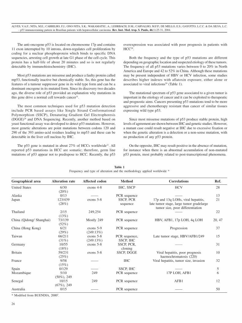
Both the frequency and the type of p53 mutations are differentdepending on geographic location and suspected etiology of these tumors.The frequency of all p53 mutations varies between 0 to 20% in NorthAmerica and Europe and 42 to 52% in China. Although these mutationsmay be present independent of HBV or HCV infection, some studiesdescribes higher indexes with aflatoxin exposure, either alone orassociated to viral infections50 (Table 1).
The mutational spectrum of p53 gene associated to a given tumor isimportant in the etiology of cancer and it can be exploited to therapeuticand prognostic aims. Cancers presenting p53 mutations tend to be moreaggressive and chemotherapy resistant than cancer of similar tissuesexpressing wild type p53.
Since most missense mutations of p53 produce stable protein, highlevels of agreement are shown between IHC and genetic studies. However,a mutant case could result negative at IHC due to excessive fixation orwhen the genetic alteration is a delection or a non-sense mutation, withno production of any p53 protein.
On the opposite, IHC may result positive in the absence of mutation,for instance when there is an abnormal accumulation of non-mutatedp53 protein, most probably related to post-transcriptional phenomena.
Table 1Frequency and type of alteration and the methodology applied worldwide *
Geographical area Alteration rate Affected codon Method Correlations Ref.
United States 6/30 exons 4-8 IHC, SSCP HCV 28(20%)
Alaska 0/13 —— PCR sequence —— 13Japan 123/439 exons 5-8 SSCP, PCR 17p and 13q LOHs, viral hepatitis, 21
(28%) sequence late tumor stage, large tumor gradelargetumor size, poor differentiation
Thailand 2/15 249,254 PCR sequence —— 22(13%)
China (Qidong/ Shanghai) 73/139 Mostly 249 PCR sequence HBV, AFB1, 17p LOH, 4q LOH 20, 47(52%)
China (Hong Kong) 6/21 exons 5-9 PCR sequence Progression 37(29%) (249:13%)
Taiwan 66/211 exons 5-8 PCR sequence, Late tumor stage, HBV/AFB1/249 15(31%) (249:13%) SSCP, IHC
Germany 10/55 exons 5-8 SSCP, PCR, —— 31(18%) cloning
Britain 59/231 exons 5-8 SSCP, DGGE Viral hepatitis, poor prognosis 10(25%) haemochromatosis (220)
France 9/58 —— IHC Viral hepatitis, tumor size, invasion 32(15%)
Spain 0/129 —— SSCP, IHC —— 5Mozambique 5/10 249 PCR sequence 17P LOH, AFB1 6
(50%), 249Senegal 10/15 249 PCR sequence AFB1 12
(67%), 249Australia 0/15 —— PCR sequence —— 50
* Modified from BUENDIA, 20007.
ALVES, V.A.F.; NITA, M.E.; CARRILHO, F.J.; ONO-NITA, S.K.; WAKAMATSU, A.; LEHRBACH, D.M.; CARVALHO, M.F.P.; DE MELLO, E.S.; GAYOTTO, L.C.C. & DA SILVA, L.C.- p53 immunostaining pattern in Brazilian patients with hepatocellular carcinoma. Rev. Inst. Med. trop. S. Paulo, 46(1):25-31, 2004.
27
Since major functions of p53 in the cell cycle and cell transformationare performed directly by its protein, IHC may serve as a very usefultool for the study of p53 alterations in carcinogenesis57.
The development of HCC appears to be preceded by liver celldysplasia, a condition subject to controversy and confusion. First,ANTHONY et al.3 suggested the preneoplastic capacity of liver celldysplasia and then several studies have stressed the relation withcarcinoma. Recently, LIBBRECHT et al.38 investigated the predictivevalue of small and large cell dysplasia, revealing large cell dysplasia asan identifier of patients at risk for developing HCC.
In Japan, the presence of a large regenerative nodule within a cirrhoticliver, referred to as a macroregenerative nodule or adenomatoushyperplasia is thought to play a role in the pathogenesis of hepatocellularcarcinoma4. In hepatocarcinogenesis of the cirrhotic liver, severalJapanese authors suggest that the adenomatous hyperplasia might be thefirst step in the development of HCC, going through phases of dysplasicnodule and small HCC in a multistep fashion4,34,48.
The objective of the present study was to compare the frequency ofabnormal immunohistochemical expression of p53 phosphoprotein in HCCoccurring in cirrhotic versus non-cirrhotic patients as well as in large livercell dysplasia (large cell dysplasia) and in adenomatous hyperplasia.
CASUISTIC AND METHODS
We studied 84 patients with hepatocellular carcinoma or cirrhosis.54 patients were cases of hepatocellular carcinoma; 39 of them werecirrhotic and 15 non-cirrhotic patients. The other 30 patients werecirrhotic patients without hepatocellular carcinoma. Records wereretrieved from the Hepatology Branch, Department of Gastroenterologyor Division of Pathology of Hospital das Clínicas, São Paulo UniversitySchool of Medicine.
Immunohistochemical data analysis: All reactions were performedat the Immunohistochemistry Laboratory, Adolfo Lutz Institute, thePublic Health Laboratory, São Paulo, Brazil.
The following primary monoclonal antibodies used were DO-7 (Dako,USA), 1801 (Bio-gennex, USA) and DO-1 (Santa Cruz, USA). The epitopesrecognized by the antibodies are located between the N-terminal aminoacids 1-45 (DO-7), 46-55 (1801) and 37-45 (DO-1) of the human p53 protein.Four-micrometer-thick histopathological sections were prepared fromformalin-fixed paraffin-embedded tru-cut needle liver biopsies. Antigenretrieval was achieved with pressure cooker for 3 minutes. The specimenswere incubated overnight with the primary antibody at 4 °C. Amplificationwas achieved by streptavidin-biotin-peroxidase (Dako Duet, USA) and, in4 cases with cytoplasmic p53 staining where samples were still available, anew reaction amplified with polymer-enzyme Envision-Alkaline PhosphataseSystem (Dako, USA) was performed, thus assuring that reactivity was notdue to endogenous peroxidase or biotin (Fig. 1). The reaction products ofperoxidase were visualized by amplification with avidin-biotin-peroxidasecomplex (Vector Laboratories, USA) and developed with H
20
2 and diamino-
benzidine (Sigma Corp., USA).
One observer (VAFA) blinded to the clinical outcome of patientsperformed assessment of staining21.
Chi square test was used for statistical analysis. All p values reportedare for a two-sided test, and the level of significance was set at 0.05.
RESULTS
In the present study, antibodies DO-7 and DO-1 yielded quite similarresults, with sharp nuclear staining in many neoplastic hepatocytes andoccasionally also in the cytoplasm of some tumor cells. Antibody 1801was shown less sensitive with fewer cases positive, showing rather faiblenuclear staining.
In our sample, considering all the 54 patients with HCC, p53immuno-expression was found in 19 patients (35.2%) (Fig. 1). Out of39 cirrhotic patients with HCC, nuclear p53 expression was detected in13 (33.3%). The comparison of p53 immuno-expression in 6 of 27 welland moderately differentiated HCC (grades I or II)19,35,39 versus the 7 of12 poorly differentiated (grades III or IV)18,30,34 a statistically significantdifference in the results obtained (Fig. 2). Nontumorous areas either inthe vicinity of HCC or in the 30 purely cirrhotic cases showed no p53expression, even in foci of large cell dysplasia or adenomatoushyperplasia. The reproducibility of this staining pattern in the 4 caseswhere samples were still available for reaction with the recentlyintroduced Envision System-Alkaline Phosphatase demonstrate this is agenuine pattern. These data shows a very high immuno-expression ofabnormal p53 in Brazilian patients mostly in those with high-gradeadvanced tumors suggesting that p53 alteration occurs late in thecarcinogenesis. When we considered HBV, HCV or alcohol in the casesstudied, no significant difference was detected. In the noncirrhotic HCCgroup the p53 expression was detected in 6 in 15 cases (40%), slightlyhigher than in those occurring in cirrhotic patients.
DISCUSSION
Abnormal immunohistochemical expression of p53 was detected in35.2% in the present Brazilian group of HCC patients, in a remarkablesimilarity to the findings from PAIVA et al., reporting p53 immunoexpressionin 5/15 HCC cases also from São Paulo, Brazil51. Several studies haveindicated a relation between the p53 gene alteration and the development ofHCC41. This alteration occurs in the HCC of different causes, includingHBV and HCV infection, aflatoxin intake, hemochromatosis, and alfa1-antitrypsin1,17,30,56. This alteration in p53 is linked to cellular events whichare accompanied by increased expression of several growth factors thatenhance the survival of carcinogen-activated cells by suppressing apoptosisand increasing elements entering the cell cycle17,45. Trying to take thiscorrelation in order to improve the diagnosis and the prognosis knowledgeof this tumor, we studied the p53 immuno-expression using aimmunohistochemical method with monoclonal antibodies to differentepitopes of N-terminal region of p53 DO-7, 1801 and DO-1.
We could not find significant differences in the p53 expression incirrhotic versus noncirrhotic patients with HCC contrasting to results ofhigh-throughput gene analysis using cDNA microarrays study whichsupported the idea of different genetic alterations in HCC with or withoutcirrhosis53.
Higher detection of p53 alteration in advanced grade tumor (GRADEIII-IV) and absence of that alteration in dysplastic and adenomatoushyperplastic areas in the vicinity of the tumor are in agreement with a

28
ALVES, V.A.F.; NITA, M.E.; CARRILHO, F.J.; ONO-NITA, S.K.; WAKAMATSU, A.; LEHRBACH, D.M.; CARVALHO, M.F.P.; DE MELLO, E.S.; GAYOTTO, L.C.C. & DA SILVA, L.C.- p53 immunostaining pattern in Brazilian patients with hepatocellular carcinoma. Rev. Inst. Med. trop. S. Paulo, 46(1):25-31, 2004.
research which shows p53 gene alteration of 15% and 52% in moderatelyand poorly differentiated HCCs respectively and not at all in welldifferentiated HCC. Since in the preferential pathway for liver cellcarcinogenesis, early/small hepatocellular carcinomas are usually well-
differentiated (grades I and II) whereas advanced/large carcinomas arepreferentially grades III-IV, this finding further demonstrates that p53alterations are late event in hepatocarcinogenesis. Another study thatexamined p53 gene by PCR-RFLP also corroborates to this conclusion,showing a p53 mutation rate of 10%, 55.2% and 80.8% in the well-,moderately- and poorly-differentiated HCC respectively32. In contrast,it was demonstrated that in animals the p53 gene mutations is present ininitial hepatocarcinogenesis22, and HUANG et al. showed that in humansa serine substitution at codon 249 of the p53 gene might serve as a newearly diagnostic marker for HCC29.
Moreover, it was showed in a Japanese study that the spectrum ofp53 mutation did not differ among HCCs in relation to the type of hepatitisvirus infection, sex, age, and background liver disease of patients, tumorsize or presence of metastasis, however incidence and site weresignificantly associated with the degree of differentiation of cancer cells.Using Single Strand Conformation Polymorphism analysis, it was found54% of p53 mutation in poorly differentiated HCC while the mutationwas less frequent (21%) in well or moderately differentiated HCC46.
p53 gene mutation is suggested to occur independently of the typeof infection or status of preexisting liver disease, as well showed in ourresearch where HBV, HCV or alcohol was not a factor of difference inthe mp53 expression46. However, in a recent study, ALTAF found p53
Fig. 2 - p53 immuno-expression versus histological grade.
Fig. 1 - Immuno-expression of p53 is abundant in nuclei, whereas cytoplasmic immunostaining is found in occasional neoplastic cells (DO-7, Envision Alk. Phosphatase, x 200).

ALVES, V.A.F.; NITA, M.E.; CARRILHO, F.J.; ONO-NITA, S.K.; WAKAMATSU, A.; LEHRBACH, D.M.; CARVALHO, M.F.P.; DE MELLO, E.S.; GAYOTTO, L.C.C. & DA SILVA, L.C.- p53 immunostaining pattern in Brazilian patients with hepatocellular carcinoma. Rev. Inst. Med. trop. S. Paulo, 46(1):25-31, 2004.
29
immunoexpression in 52% of cases and 53% of them were having antihepatitis B surface antigen positivity, detected in their serum and 23%were having hepatitis C antibodies positive2. On the other hand, DINGet al. found only 20 p53 point mutation at exon 7 in 239 HCC samplesexamined16.
In a study using immunohistochemical detection for mutant p53protein with monoclonal antibody PAb1801, TERAMOTO et al. foundp53 overexpression in 22 tumours (31%) but in none of the non-tumorousliver specimens54. Overexpression of p53 was more frequent in poorlydifferentiated tumours (p = 0.01), in tumours > 5 cm (p = 0.05), and inthose presenting giant cells (p = 0.03). No significant correlation withhepatitis B or C status, background liver disease was detected, suggestingthat p53 mutation in HCC was a late event in HCC54. The present study,along with other papers further corroborate this hypothesis2,60.
In a study that has assessed p53 expression by immunoluminometricassay on liver samples from 40 patients with biopsy-proven HCV-relatedchronic hepatitis, was found an overexpression of p53 in 17.5% of themand this p53 positive group had a daily ethanol intake significantly higherin respect to that of the p53 negative group (p < 0.05)18. Another studyfound that tumors associated with alcoholism had more frequentalterations in the p53 pathway than those caused by HCV infection20. Inour study, a different factor related to carcinogenesis of HCC is alsoevident, since no difference was found among patients with HBV, HCVor alcoholic influence.
Regarding the less commonly reported finding of cytoplasmic p53immunoexpression, the reproducibility of results in cases with enoughsample in the block for the repetition of the reaction with the newpolymer-enzyme Envision method reinforces the hypothesis that thismay be an odd, but genuine localization of p53, as previously suggestedby ZHAO et al.59 and by ZERBINI et al.58. In a recent experiment OH etal. showed that bile duct ligation shifts p53 immunohistochemicalexpression from nuclei to cytoplasm and advocate that such shiftingcould play an important role in induction of apoptosis47.
CONCLUSION
In this study we tried to correlate the immunohistochemical expressionof altered p53 (mp53) to the degree of alterations of hepatic cells.
We found a frequency of mp53 expression in HCC of 35.2%, whichis intermediate between data from USA (6-18%) and Africa (40-50%)where this alteration would be related to aflatoxin. Moreover, it was notfound a significant difference between cirrhotics versus non-cirrhotics,as well as among HBV and HCV and alcoholic suggesting anothertriggering factor for such alteration in Brazil. The higher frequency ofaberrant immuno-expression of p53 in less differentiated HCCs furtherdemonstrates that p53 alterations are late phenomena in livercarcinogenesis.
Future studies should be aimed at mapping hot spot areas of p53mutation as well as at assessing the possible role of other etiologicalagents, including toxics, such as aflatoxins and organic contaminants inHCC in Brazil.
RESUMO
Expressão imunohistoquímica do p53 carcinoma hepatocelular depacientes brasileiros
O carcinoma hepatocelular (CHC) é um importante tipo de câncerrelacionado etiologicamente a alguns vírus, carcinógenos químicos e outrosfatores ambientais que causam danos crônicos ao fígado em humanos. Afreqüência de mutação do gene p53 em CHC é altamente heterogênea (0-52%) nos diversos países. OBJETIVO. O objetivo deste estudo foideterminar, imuno-histologicamente, a freqüência da expressão anômalade p53 em CHCs em pacientes cirróticos versus não-cirróticos, bem comoem displasia hepática e hiperplasia adenomatosa. Para isso, foram estudados84 pacientes com carcinoma hepatocelular ou cirrose. RESULTADOS.Foram detectadas expressões do p53 alterado em 58,3% dos pacientescom CHC graus III-IV, contrastando com os 22,2% dos pacientes comCHC graus I-II (p = 0,02). Áreas não tumorais, tanto nas proximidades doCHC como nos 30 casos de cirrose não mostraram expressão nuclearalterada do p53, mesmo nas displasias ou hiperplasias adenomatosas.Quando se considerou HBV, HCV ou alcoolismo nos casos estudados,não se encontrou diferença significativa. CONCLUSÃO. A elevadafreqüência de imuno-expressão de p53 nesta população é próxima à relatadana China e África, tornando necessárias outras pesquisas para explicar asdiferenças com os CHC estudados na Europa e na América do Norte.
ACKNOWLEDGMENTS
This paper was supported in part by the Projeto Hepatologia –Hepatites / Câncer with a grant from Alves de Queiróz Family Fund forResearch.
REFERENCES
1. AHSAN, H.; WANG, L.Y.; CHEN, C.J.; TSAI, W.Y. & SANTELLA, R.M. - Variabilityin aflatoxin-albumin adduct levels and effects of hepatitis B and C virus infectionand glutathione S-transferase M1 and T1 genotype. Environ. Hlth. perspect., 109:833-837, 2001.
2. ALTAF, F.J. - Hepatocellular carcinoma. Saudi Med. J., 22: 416-418, 2001.
3. ANTHONY, P.P.; VOGEL, C.L. & BARKER, L.F. - Liver cell dysplasia: a premalignantcondition. J. clin. Path., 26: 217-223, 1973.
4. ARAKAWA, M.; KAGE, M.; SUGIHARA, S. et al. - Emergence of malignant lesionswithin an adenomatous hyperplastic nodule in a cirrhotic liver. Observations in fivecases. Gastroenterology, 91: 198-208, 1986.
5. BOIX-FERRERO, J.; PELLIN, A.; BLESA, R.; ADRADOS, M. & LLOMBART-BOSCH, A. - Absence of p53 gene mutations in hepatocarcinomas from aMediterranean area of Spain. A study of 129 archival tumour samples. VirchowsArch., 434: 497-501, 1999.
6. BRESSAC, B.; KEW, M.; WANDS, J. & OZTURK, M. - Selective G to T mutations ofp53 gene in hepatocellular carcinoma from southern Africa. Nature (Lond.), 350:429-431, 1991.
7. BUENDIA, M.A. - Genetics of hepatocellular carcinoma. Semin. Cancer Biol., 10:185-200, 2000.
8. CARRILHO, F.J. - Carcinoma hepatocelular e cirrose hepática. Estudo caso-controlede variáveis clínicas, bioquímicas, sorológicas e morfológicas. São Paulo, 1993.(Tese de Doutorado – Faculdade de Medicina da Universidade de São Paulo).

30
ALVES, V.A.F.; NITA, M.E.; CARRILHO, F.J.; ONO-NITA, S.K.; WAKAMATSU, A.; LEHRBACH, D.M.; CARVALHO, M.F.P.; DE MELLO, E.S.; GAYOTTO, L.C.C. & DA SILVA, L.C.- p53 immunostaining pattern in Brazilian patients with hepatocellular carcinoma. Rev. Inst. Med. trop. S. Paulo, 46(1):25-31, 2004.
9. CARRILHO, F.J.; ALVES, V.A.F.; MELLO, E.S. & VEZOZZO, D.C.P. - Carcinomahepatocelular. In: GAYOTTO, L.C.C. & ALVES, V.A. Doenças do fígado e das viasbiliares. São Paulo, Atheneu, 2001. p. 997-1016.
10. CHALLEN, C.; LUNEC, J.; WARREN, W.; COLLIER, J. & BASSENDINE, M.F. -Analysis of the p53 tumor-suppressor gene in hepatocellular carcinomas from Britain.Hepatology, 16: 1362-1366, 1992.
11. CHEN, P.-J. & CHEN, D.-S. - Hepatitis B virus infection and hepatocellular carcinoma:molecular genetics and clinical perspectives. Semin. Liver Dis., 19: 253-262, 1999.
12. COURSAGET, P.; DEPRIL, N.; CHABAUD, M. et al. - High prevalence of mutations atcodon 249 of the p53 gene in hepatocellular carcinomas from Senegal. Brit. J. Cancer,67: 1395-1397, 1993.
13. DE BENEDETTI, V.M.; WELSH, J.A.; TRIVERS, G.E. et al. - p53 is not mutated inhepatocellular carcinomas from Alaska Natives. Cancer Epidem. Biomarkers Prev.,4:79-82, 1995.
14. DENG, Z.; PAN, L. & MA, Y. - Sequence alterations in p53 gene of hepatocellularcarcinoma from high aflatoxin risk area in Guangxi. Zhonghua Zhong Liu Za.Zhi., 19: 18-21, 1997.
15. DIAMANTS, I.D.; MCGANDY, C.; CHEN, T.J. et al. – A new mutational hot-spot inthe p53 gene in human hepatocellular carcinoma. J. Hepatol., 20: 553-556, 1994.
16. DING, X.; PARK, Y.N.; TALTAVULL, T.C. et al. - Geographic characterization of hepatitisvirus infections, genotyping of hepatitis B virus, and p53 mutation in hepatocellularcarcinoma analyzed by in situ detection of viral genomes from carcinoma tissues:comparison among six different countries. Jap. J. infect. Dis., 56: 12-18, 2003.
17. DOMINGUES-MALAGON, H. & GAYTAN-GRAHAM, S. - Hepatocellular carcinoma:an update. Ultrastruct. Path., 25: 497-516, 2001.
18. DONATO, F.; BOFFETTA, P. & PUOTI, M. - A meta-analysis of epidemiological studieson the combined effect of hepatitis B and C virus infections in causing hepatocellularcarcinoma. Int. J. Cancer, 75: 347-354, 1998.
19. DOWELL, S.P. & HALL, P.A. - The p53 tumour suppressor gene and tumour prognosis:is there a relationship? J. Path., 177: 221-224, 1995.
20. EDAMOTO, Y.; HARA, A.; BIERNAT, W. et al. - Alterations of RB1, p53 and Wntpathways in hepatocellular carcinomas associated with hepatitis C, hepatitis B andalcoholic liver cirrhosis. Int. J. Cancer, 106: 334-341, 2003.
21. FISHER, C.J.; GILLETT, C.E.; VOJTESEK, B.; BARNES, D.M. & MILLIS, R.R. -Problems with p53 immunohistochemical staining: the effect of fixation and variationin the methods of evaluation. Brit. J. Cancer, 69: 26-31, 1994.
22. FU, Y.; DENG, W.; KAWARADA, Y. et al. - Mutation and expression of the p53 geneduring chemical hepatocarcinogenesis in F344 rats. Biochim. Biophys. Acta, 1628:40-49, 2003.
23. FUJIMOTO, Y.; HAMPTON, L.L.; WIRTH, P.J. et al. - Alterations of tumor suppressorgenes and allelic losses in human hepatocellular carcinomas in China. Cancer Res.,54: 281-285, 1994.
24. HAYASHI, H.; SUGIO, K.; MATSUMATA, T. et al. - The clinical significance of p53 genemutation in hepatocellular carcinomas from Japan. Hepatology, 22: 1702-1707, 1995.
25. HOLLSTEIN, M.C.; WILD, C.P.; BLEICHER, F. et al. - p53 mutations and aflatoxin B1exposure in hepatocellular carcinoma patients from Thailand. Int. J. Cancer, 53: 51-55, 1993.
26. HONDA, K.; SBISA, E.; TULLO, A. et al. - p53 mutation is a poor prognostic indicatorfor survival in patients with hepatocellular carcinoma undergoing surgical tumourablation. Brit. J. Cancer, 77: 776-782, 1998.
27. HSU, I.C.; METCALF, R.A.; SUN, T. et al. - Mutational hotspot in the p53 gene inhuman hepatocellular carcinomas. Nature (Lond.), 350: 427-428, 1991.
28. HU, T.H.; HUANG, C.C.; LIN, P.R. et al. - Expression and prognostic role of tumorsuppressor gene PTEN/MMAC1/TEP1 in hepatocellular carcinoma. Cancer, 97:1929-1940, 2003.
29. HUANG, X.H.; SUN, L.H.; LU, D.D. et al. - Codon 249 mutation in exon 7 of p53 genein plasma DNA: maybe a new early diagnostic marker of hepatocellular carcinomain Qidong risk area, China. Wld. J. Gastroent., 9: 692-695, 2003.
30. HUO, T.I.; WANG, X.W.; FORGUES, M. et al. - Hepatitis B virus X mutants derivedfrom human hepatocellular carcinoma retain the ability to abrogate p53-inducedapoptosis. Oncogene, 20: 3620-3628, 2001.
31. HUSSAIN, S.P. & HARRIS, C.C. - Molecular epidemiology and carcinogenesis:endogenous and exogenous carcinogens. Mutation Res., 462: 311-322, 2000.
32. JIANG, W.; LU, Q. & PAN, G. - p53 gene mutation in hepatocellular carcinoma.Zhonghua Wai Ke. Za. Zhi., 36: 531-532, 1998.
33. KAZACHKOV, Y.; KHAOUSTOV, V.; YOFFE, B. et al. - p53 abnormalities inhepatocellular carcinoma from United States patients: analysis of all 11 exons.Carcinogenesis, 17: 2207-2212, 1996.
34. KOJIRO, M. & NAKASHIMA, T. - Pathology of hepatocellular carcinoma. In: OKUDA,K. & ISHAK, K.G. Neoplasms of the liver. Tokyo, Springer-Verlag, 1987. p. 81-104.
35. KONDO, F.; WADA, K. & KONDO, Y. - Morphometric analysis of hepatocellularcarcinoma. Virchows Arch. A Path. Anat. Histopath., 413: 425-430, 1988.
36. KRESS, S.; JAHN, U.R.; BUCHMANN, A.; BANNASCH, P. & SCHWARZ, M. - p53mutations in human hepatocellular carcinomas from Germany. Cancer Res., 52:3220-3223, 1992.
37. LAURENT-PUIG, P.; FLEJOU, J.F.; FABRE, M. et al. - Overexpression of p53: a rareevent in a large series of white patients with hepatocellular carcinoma. Hepatology,16: 1171-1175, 1992.
38. LIBBRECHT, L.; CRANINX, M.; NEVENS, F.; DESMET, V. & ROSKAMS, T. -Predictive value of liver cell dysplasia for development of hepatocellular carcinomain patients with non-cirrhotic and cirrhotic chronic viral hepatitis. Histopathology,39: 66-73, 2001.
39. MELLO, E.S. - Macronódulos em fígados cirróticos: estudo morfológico com ênfasenos aspectos macroscópicos, proliferação e apoptose. São Paulo, 2001. (Tese deDoutorado – Faculdade de Medicina da Universidade de São Paulo).
40. MURAKAMI, Y.; HAYASHI, K.; HIROHASHI, S. & SEKIYA, T. - Aberrations of thetumor suppressor p53 and retinoblastoma genes in human hepatocellular carcinomas.Cancer Res., 51: 5520-5525, 1991.
41. NAKAMURA, T.; IWAMURA, Y.; KANEKO, M. et al. - Deletions and rearrangements ofthe retinoblastoma gene in hepatocellular carcinoma, insulinoma and some neurogenictumors as found in a study of 121 tumors. Jap. J. clin. Oncol., 21: 325-329, 1991.
42. NG, I.O.; SRIVASTAVA, G.; CHUNG, L.P.; TSANG, S.W. & NG, M.M. - Overexpressionand point mutations of p53 tumor suppressor gene in hepatocellular carcinomas inHong Kong Chinese people. Cancer, 74: 30-37, 1994.
43. NISHIDA, N.; FUKUDA, Y.; KOKURYU, H. et al. - Accumulation of allelic loss onarms of chromosomes 13q, 16q and 17p in the advanced stages of humanhepatocellular carcinoma. Int. J. Cancer, 51: 862-868, 1992.
44. NITA, M.E.; ALVES, V.A F.; CARRILHO, F.J. et al. - Molecular aspects of hepaticcarcinogenesis. Rev. Inst. Med. trop. S. Paulo, 44: 39-48, 2002.
45. NITA, M.E.; NAGAWA, H.; TOMINAGA, O. et al. - p21Waf1/Cip1 expression is aprognostic marker in curatively resected esophageal squamous cell carcinoma, butnot p27Kip1, p53, or Rb. Ann. surg. Oncol., 6: 481-488, 1999.

ALVES, V.A.F.; NITA, M.E.; CARRILHO, F.J.; ONO-NITA, S.K.; WAKAMATSU, A.; LEHRBACH, D.M.; CARVALHO, M.F.P.; DE MELLO, E.S.; GAYOTTO, L.C.C. & DA SILVA, L.C.- p53 immunostaining pattern in Brazilian patients with hepatocellular carcinoma. Rev. Inst. Med. trop. S. Paulo, 46(1):25-31, 2004.
31
46. ODA, T.; TSUDA, H.; SCARPA, A.; SAKAMOTO, M. & HIROHASHI, S. - p53 genemutation spectrum in hepatocellular carcinoma. Cancer Res., 52: 6358-6364, 1992.
47. OH, S.H.; YAN, K.J.; NAN, J.X.; SOHN, D.H. & LEE, B.H. - Changes in expressionand immunolocalization of protein associated with toxic bile salts-induced apoptosisin rat hepatocytes. Arch. Toxicol., 77: 110-115, 2003.
48. OHTA, G. & NAKANUMA, Y. - Comparative study of three nodular lesions in cirrhosis:adenomatoid hyperplasia, adenomatoid hyperplasia with intermediate lesion and smallhepatocellular carcinoma. In: OKUDA, K. & ISHAK, K.G. Neoplasms of the liver.Tokyo, Springer-Verlag, 1987. p. 177-187.
49. OKUDA, K. - Hepatocellular carcinoma. J. Hepat., 32: 225-237, 2000.
50. OZTURK, M. - Genetic aspects of hepatocellular carcinogenesis. Semin. Liver Dis.,19: 235-242, 1999.
51. PAIVA, C.; OSHIMA, C.T.; LANZONI, V.P. & FORONES, N.M. - Apoptosis, PCNAand p53 in hepatocellular carcinoma. Hepatogastroenterology, 49: 1058-1061, 2002.
52. SCORSONE, K.A.; ZHOU, Y.Z.; BUTEL J.S. & SLAGLE, B.L. - p53 mutations clusterat codon 249 in hepatitis B virus-positive hepatocellular carcinomas from China.Cancer Res., 52: 1635-1638, 1992.
53. TANNAPFEL, A. & WITTEKIND, C. - Genes involved in hepatocellular carcinoma:deregulation in cell cycling and apoptosis. Virchows Arch., 440: 345-352, 2002.
54. TERAMOTO, T.; SATONAKA, K.; KITAZAWA, S. et al. - p53 gene abnormalities areclosely related to hepatoviral infections and occur at a late stage ofhepatocarcinogenesis. Cancer Res., 54: 231-235, 1994.
55. VESEY, D.A.; HAYWARD, N.K. & COOKSLEY, W.G. - p53 gene in hepatocellularcarcinomas from Australia. Cancer Detect. Prev., 18: 123-130, 1994.
56. WONG, N.; LAI, P.; PANG, E. et al. - Genomic aberrations in human hepatocellularcarcinomas of differing etiologies. Clin. Cancer Res., 6: 4000-4009, 2000.
57. WYNFORD-THOMAS, D. - p53 in tumour pathology: can we trustimmunocytochemistry? J. Path., 166: 329-330, 1992.
58. ZERBINI, M.C.; SREDNI, S.T.; GRIER, H. et al. - Primary malignant epithelial tumorsof the liver in children: a study of DNA content and oncogene expression. Pediat.Dev. Path., 1: 270-280, 1998.
59. ZHAO, M.; ZHANG, N.X.; LAISSUE, J.A. & ZIMMERMANN, A. -Immunohistochemical analysis of p53 protein overexpression in liver cell dysplasiaand in hepatocellular carcinoma. Virchows Arch., 424: 613-621, 1994.
60. ZHOU, F.; WANG, J.; LEI, B. et al. - Detection of p53 gene mutations in hepatocellularcarcinomas. Hua Xi Yi Ke Da Xue Xue, 28: 50-54, 1997.
Received: 9 September 2003Accepted: 8 December 2003