Cochlin immunostaining of inner ear pathologic deposits and proteomic analysis in DFNA9 deafness and...
44
1 Cochlin immunostaining of inner ear pathologic deposits and proteomic analysis in DFNA9 deafness and vestibular dysfunction Nahid G. Robertson 1 , Cor W. R. J. Cremers 2 , Patrick L. M. Huygen 2 , Tetsuo Ikezono 3 , Bryan Krastins 4 , Hannie Kremer 2 , Sharon F. Kuo 1 , M. Charles Liberman 5 , Saumil N. Merchant 5 , Constance E. Miller 5 , Joseph B. Nadol, Jr. 5 , David A. Sarracino 4 , Wim I. M. Verhagen 6 , and Cynthia C. Morton 1* 1 Departments of Pathology, Obstetrics, Gynecology and Reproductive Biology, Brigham and Women's Hospital, Harvard Medical School, Boston, MA, USA 2 Department of Otorhinolaryngology, Radboud University Medical Center, Nijmegen, The Netherlands 3 Department of Otorhinolaryngology, Nippon Medical School, Tokyo, Japan 4 Harvard Medical School-Partners Healthcare Center for Genetics and Genomics, Cambridge, Massachusetts, USA 5 Department of Otology and Laryngology, Massachusetts Eye and Ear Infirmary, Eaton- Peabody Laboratory, Harvard Medical School Boston, MA, USA 6 Department of Neurology, Canisius Wilhelmina Hospital, Nijmegen, The Netherlands * Address correspondence to: Cynthia C. Morton, Ph.D. Departments of Ob/Gyn and Pathology Brigham and Women's Hospital Harvard Medical School 77 Avenue Louis Pasteur, NRB 160 Boston, MA 02115, USA Tel: (617) 525-4535 Fax: (617) 525-4533 Email: [email protected] © The Author 2006. Published by Oxford University Press. All rights reserved HMG Advance Access published February 15, 2006
-
Upload
independent -
Category
Documents
-
view
1 -
download
0
Transcript of Cochlin immunostaining of inner ear pathologic deposits and proteomic analysis in DFNA9 deafness and...

1
Cochlin immunostaining of inner ear pathologic deposits and proteomic analysis in DFNA9
deafness and vestibular dysfunction
Nahid G. Robertson1, Cor W. R. J. Cremers2, Patrick L. M. Huygen2, Tetsuo Ikezono3, Bryan Krastins4, Hannie Kremer2, Sharon F. Kuo1, M. Charles Liberman5, Saumil N. Merchant5, Constance E. Miller5, Joseph B. Nadol, Jr. 5, David A. Sarracino4, Wim I. M. Verhagen6, and Cynthia C. Morton1* 1 Departments of Pathology, Obstetrics, Gynecology and Reproductive Biology, Brigham and Women's Hospital, Harvard Medical School, Boston, MA, USA 2 Department of Otorhinolaryngology, Radboud University Medical Center, Nijmegen, The Netherlands 3 Department of Otorhinolaryngology, Nippon Medical School, Tokyo, Japan 4 Harvard Medical School-Partners Healthcare Center for Genetics and Genomics, Cambridge, Massachusetts, USA 5 Department of Otology and Laryngology, Massachusetts Eye and Ear Infirmary, Eaton-Peabody Laboratory, Harvard Medical School Boston, MA, USA 6 Department of Neurology, Canisius Wilhelmina Hospital, Nijmegen, The Netherlands *Address correspondence to: Cynthia C. Morton, Ph.D. Departments of Ob/Gyn and Pathology Brigham and Women's Hospital Harvard Medical School 77 Avenue Louis Pasteur, NRB 160 Boston, MA 02115, USA Tel: (617) 525-4535 Fax: (617) 525-4533 Email: [email protected]
© The Author 2006. Published by Oxford University Press. All rights reserved
HMG Advance Access published February 15, 2006

2
ABSTRACT
Seven missense mutations and one in-frame deletion mutation have been reported in the
COCH (coagulation factor C homology) gene, causing the adult-onset, progressive sensorineural
hearing loss and vestibular disorder at the DFNA9 locus. Prevalence of COCH mutations
worldwide is unknown, as there is no systematic screening effort for late-onset hearing disorders;
however, to date, COCH mutations have been found on four continents, and the possibility of
COCH playing an important role in presbycusis and disorders of imbalance has been considered.
Cochlin (encoded by COCH) has also been shown as a major target antigen for autoimmune
sensorineural hearing loss. In this report, we present histopathology, immunohistochemistry, and
proteomic analyses of inner ear tissues from post-mortem DFNA9 temporal bone samples of an
individual from a large Dutch kindred segregating the P51S mutation and adult human
unaffected controls, and wild-type (+/+) and Coch null (-/-) knock-out mice. DFNA9 is an inner
ear disorder with a unique histopathology showing loss of cellularity and aggregation of
abundant homogeneous acellular eosinophilic deposits in the cochlear and vestibular labyrinths,
similar to protein aggregation in well-known neurodegenerative disorders. By
immunohistochemistry on the DFNA9 temporal bone sections, we have shown cochlin staining
of the characteristic cochlear and vestibular deposits, indicating aggregation of cochlin in the
same structures in which it is normally expressed. Proteomic analysis identified cochlin as the
most abundant protein in mouse and human cochleae. The high-level expression and stability of
cochlin in the inner ear, even in the absence and severe atrophy of the fibrocytes that normally
express COCH, are shown through these studies and further elucidate the pathobiologic events
occurring in DFNA9 leading to hearing loss and vestibular dysfunction.

3
INTRODUCTION
A large number of loci have been mapped for syndromic and nonsyndromic hereditary
hearing loss and the gene mutations responsible for these disorders are being continually
discovered and characterized (1). Elucidation of the functions of these genes and their roles in
the inner ear and in pathogenesis of hearing and balance disorders are important ongoing
endeavors.
The autosomal dominant deafness disorder at the DFNA9 locus has been described and
the clinical aspects extensively characterized, showing adult-onset, progressive sensorineural
hearing loss and vestibular dysfunction (2-9). Different missense mutations in the COCH
(coagulation factor C homology) gene were found initially in three families in the United States,
and subsequently in families in the Netherlands, Belgium, and Australia (Table 1) (Fig. 1) (10-
14). Two simplex cases of another missense mutation and an in-frame deletion in the same
domain of COCH (FCH/LCCL domain) have been reported in Japan and Hungary, respectively
(15, 16). A recent report describes the first finding of a COCH mutation outside of the
FCH/LCCL domain in the vWFA domain in a large DFNA9 kindred in the United States (9).
The prevalence of COCH mutations worldwide is not known, as systematic genetic testing of
adult-onset hearing loss is not currently performed.
COCH was isolated initially by organ-specific subtractive approaches from a human fetal
cochlear cDNA library and found to be expressed at high levels in the inner ear by Northern blot,
tissue in situ hybridization, and immunohistochemistry (17-20). The secreted protein, cochlin,
was detected by proteomic analysis as the most abundant protein in the bovine inner ear (21).
Histopathological analyses of DFNA9-affected temporal bones in the three original U. S.
families have revealed very valuable information about the end-point changes in these inner ears

4
(3, 22). A striking and unique finding in these temporal bones, which actually allowed initial
identification of several of these families as DFNA9 kindreds, is the presence of homogeneous
extracellular eosinophilic deposits in the same areas as fibrocyte atrophy. Other well-
characterized neurodegenerative disorders with aberrant protein accumulation include Alzheimer
disease (β-amyloid precursor protein) (23), Huntington disease (huntingtin) (24-26), and
Parkinson disease (α-synuclein) (27). However, DFNA9 is the only known inner ear disorder
showing this type of aggregate as the signature pathological finding, whereas findings in other
disorders of the inner ear, such as endolymphatic hydrops, and degeneration of structures such as
the sensory epithelium, ganglion cells, spiral ligament and the stria vascularis are observed in a
variety of different conditions showing hearing loss and vestibular dysfunction.
The cochlear and vestibular fibrocytes, which are severely atrophied in DFNA9 are the
very cells expressing COCH, and the homogenous acellular deposits are found in the same areas
as cochlin immunostaining in the normal inner ear (19). However, previous to this report, it has
not been shown whether this eosinophilic substance in DFNA9 is the abnormal cochlin which
has precipitated and aggregated, or whether it is another component of the inner ear, a cochlin
interacting protein, or some other downstream effect of the COCH mutations.
A recent postmortem donation of a temporal bone from an individual from the
Netherlands with DFNA9 (COCH P51S mutation) provided the opportunity to obtain additional
histopathological data for DFNA9. By using an antibody to the vWFA domain of cochlin, which
detects all known size isoforms of cochlin (Fig. 1), we have undertaken a thorough
characterization of cochlin immunostaining in the cochlear and vestibular labyrinths. We present
proteomic analysis of DFNA9-affected and unaffected adult human temporal bone sections, as
well as of wild-type (+/+) and Coch null (-/-) knock-out mouse inner ears. These studies have

5
enabled examination of the content of the abnormal deposits seen in DFNA9 and provided
insight into the role of cochlin in the inner ear and the mechanism of pathobiology underlying
DFNA9 by COCH mutations.
RESULTS AND DISCUSSION
Histopathology of DFNA9 temporal bones with the P51S cochlin mutation
Histopathologic findings in the P51S DFNA9 temporal bone (Figs. 2 and 5) are
consistent with those previously reported for other DFNA9 families designated 1W (V66G
mutation), 1Su (G88E mutation), and 1St (W117R) (3, 22, 28), suggesting that the P51S
mutation causes the same pathologic changes and acts via the same mechanism as the other
mutations in the FCH/LCCL domain of COCH. A marked reduction in the number of fibrocytes
is observed throughout the spiral ligament and limbus of the cochlear duct and in the vestibular
organs. In the same areas of fibrocyte loss and atrophy is the DFNA9-characteristic
eosinophilic-staining extracellular ground substance. The deposits are particularly prominent in
the more medial parts of the ligament underlying the stria vascularis and in the area of the
insertion of the ligament into the basilar membrane. Eosinophilic material is also present in the
osseous spiral lamina, along with loss of dendrites in these channels and in the modiolus.
Cochlin immunostaining in normal inner ear
Prior to performing immunohistochemistry on DFNA9-affected temporal bones, we
optimized anti-cochlin antibody staining on normal mouse and human adult tissues, as well as on
a Coch (-/-) mouse (29, 30). In our previous studies, we used a polyclonal antibody to the entire
FCH/LCCL and ivd1 domain of cochlin (19). However, given different-sized isoforms of

6
cochlin detected by proteomic analysis and N-terminal sequencing (21) (Fig. 1), four antibodies
to small peptides in different regions of cochlin were developed and shown by Western blot
analysis to be specific for the isoforms that each was expected to recognize (31). The anti-
cochlin antibody to the vWFA1 domain reacts with all three known cochlin isoforms: p60 (full-
length), and p44 and p40 (both lacking the FCH/LCCL domain) (Fig. 1). We chose this antibody
(anti-cochlin/vWFA1 domain) for our studies because it would provide a more complete
representation of cochlin localization in unaffected and affected tissues.
In the normal adult mouse cochlea (Fig. 3), cochlin immunostaining is strong in the spiral
ligament and spiral limbus. In the ligament, the staining is darkest in the basilar crest, near the
basilar membrane and weakest in the spiral prominence. Cells lining Rosenthal’s canal and the
channels of the osseous spiral lamina are cochlin-positive, whereas the cochlear ganglion cell
bodies and the neural processes are cochlin-negative. Distinct cochlin staining of pericytes
surrounding blood vessels in the modiolus and throughout the cochlear duct is observed. In
contrast, adjacent areas of surrounding bony tissues clearly lack cochlin staining. Cochlin-
negative structures in the cochlear duct are the organ of Corti, including the sensory epithelium
and tectorial membrane, stria vascularis, Reissner’s membrane, cochlear ganglion cells and their
neuronal processes. In the vestibular labyrinth, the cristae (Fig. 3G) show intense cochlin
staining in the fibrocytes and stroma underlying the sensory epithelium, as well as in the
ampullary wall. The sensory epithelium, the neuronal processes within the ampullary stroma, as
well as the surrounding bone and connective tissues are all cochlin-negative.
To confirm antibody specificity, we immunostained sections from a Coch (-/-) mouse
(29, 30), and no staining was detected (Fig. 3E). Negative controls with secondary antibody
alone also show no background staining (data not shown). The intense staining for cochlin in the

7
(+/+) mouse inner ear corroborates the finding of cochlin by proteomic analysis as a very
abundant and stable protein in cochlea and vestibular organs.
In the unaffected control human adult inner ear (Fig. 4), a similar pattern of cochlin
staining is detected. Cochlin immunoreactivity was prominent throughout the spiral ligament,
spiral limbus, and within the osseous spiral lamina. Immunostaining in the modiolus was
observed also around the blood vessels (data not shown). As in the mouse, the organ of Corti,
the neuronal cell bodies, and central and peripheral axons lack cochlin expression. The
surrounding outer bony and mesenchymal tissues are also unstained. In the human adult
vestibular labyrinth, the cristae also show immunostaining in the area of the stromal fibrocytes,
and a lack of staining in the adjacent overlying sensory epithelium. The ampullary wall also
contains cochlin, as observed in mouse sections.
In both mouse and human cochlea and vestibular organs, cochlin immunostaining is
restricted to tissues that are mesodermal in origin; neuroectodermally-derived structures clearly
lack cochlin expression. Within the mesodermal structures, there is widespread and high-level
expression of cochlin in areas such as the spiral ligament, which comprises a large percentage of
the total mass of the membranous cochlea, in agreement with findings of high levels of cochlin
mRNA by EST, Northern blot and tissue in situ hybridization analyses (17, 19, 32), and with
abundance and stability of cochlin protein as observed by Western blot and proteomic analyses
(19, 21).
Cochlin immunostaining in DFNA9-affected inner ear
The cochlin staining pattern in the DFNA9 temporal bone sections (Fig. 5) is similar to
that in the unaffected control sections. Immunostaining is strong throughout the spiral ligament,

8
spiral limbus, stroma of the crista ampullaris and the ampullary wall. As a negative control, no
staining was detected using the secondary antibody alone (data not shown). There is no
detectable background staining in the tissues immediately adjacent to the spiral ligament lateral
wall and tissues surrounding the ampulla. The regions of the osseous spiral lamina normally
occupied by cochlear peripheral axons are immunopositive for cochlin, as are perivascular areas
in the modiolus. Other structures such as the organ of Corti, vestibular sensory epithelium, and
stria vascularis, which are cochlin-negative in normal tissues, also lack staining.
The large amounts of eosinophilic acellular deposits contained throughout the spiral
ligament, limbus, and osseous spiral lamina are darkly and evenly immunostained with anti-
cochlin, but completely lack nonspecific staining with secondary antibody alone. The ampullary
stroma and wall, which show distortion, collapse and thickening, and contain the acellular
material, also show prominent cochlin staining. These results are consistent with the view that
cochlin is intimately associated with the eosinophilic deposits characteristic of the temporal bone
histopathology in DFNA9.
Proteomic analysis in mouse inner ear
Proteomic analysis of the cochlear and vestibular labyrinths of (+/+) and Coch (-/-) mice
were performed, and an abridged list of the representative peptide matches is presented (Table
2). The number of tryptic peptides identified from mass spectrometry analysis reflects the
relative abundance of proteins detected within each tissue by this method. A striking finding is
the presence of cochlin peptides as the most abundantly detected protein in the cochlea of (+/+)
mice. In the vestibular organs, cochlin is the second most frequently detected protein, with
albumin being primary. In both tissues, cochlin is more abundant than β-hemoglobin. Findings

9
for two other proteins, α-tectorin and keratin 9, are representative of structural proteins
expressed in these tissues. Our proteomic analysis combines results of four gel fractions where
digestion and mass spectrometry were performed separately. Identification of cochlin as the
most abundant protein in mouse inner ear lysates corroborates previous proteomic analysis by
the alternative method of 2D-gel electrophoresis, also revealing cochlin as the most prevalent
protein in bovine inner ear (21). As a negative control, we studied the Coch (-/-) mouse. No
cochlin peptides were detected in either cochlear or vestibular tissues, confirming the lack of
cochlin protein as shown also by our immunohistochemistry and by previous Western blot
analysis (30).
In (+/+) mouse cochlea, a total of 123 cochlin tryptic peptides representing 38 unique
peptides are detected, ranging from 7 to 35 amino acid residues each. Extensive peptide
coverage for cochlin was observed (Fig. 6) throughout all domains of the full-length protein
(excluding the signal peptide). In conjunction with detection of cochlin as a highly abundant and
stable protein in the cochlear and vestibular organs, and its fairly restricted expression at a high
level in the inner ear, it is interesting to note that several studies have implicated cochlin as a
target antigen for autoimmune sensorineural hearing loss via both immunoglobulin and T-cell
mediated mechanisms. Elevated serum levels of anti-cochlin immunoglobulins have been
detected in a number of patients with autoimmune hearing loss (33, 34). In addition, cochlin has
been shown to co-immunoprecipitate with choline transporter-like protein 2 (CTL2) as targets of
antibody-induced hearing loss (35, 36). Studies have also demonstrated experimentally induced
CD4+ T cell-mediated autoimmune hearing loss with cochlin as the target antigen (37, 38).
Furthermore, recent investigations have revealed significantly higher frequencies of cochlin-
specific circulating T cells as well as elevated cochlin-specific serum antibody titers in

10
individuals with autoimmune sensorineural hearing loss, as compared to unaffected age-matched
controls (39). These reports implicate cochlin as a prominent inner ear target antigen in both
antibody and T cell-mediated autoimmune hearing loss.
Proteomic analysis in human adult unaffected and DFNA9-affected and temporal bones
Because the only material available from the DFNA9-affected and unaffected human
samples are formalin-fixed, paraffin-embedded temporal bones, we had to employ a different
approach than that used in the mouse. Thus, proteomic analysis was performed on proteins
extracted from 8-micron paraffin-embedded sections, whereas fresh whole tissue lysates or
frozen tissues are typically used.
In the human adult unaffected temporal bone sample, cochlin is also the most abundantly
detected protein by mass spectrometry, as was the case in the mouse using fresh cochlear lysates.
A total of 66 cochlin peptides, representing 17 unique peptides, are detected (Table 3), ranging
from 10 to 35 amino acid residues each. Cochlin peptides identified in the human sample are
also representative throughout the protein in all domains of cochlin, although peptide coverage is
not as complete as that found in the mouse sample (Fig. 6). This is not surprising given that
peptide extraction from archival formalin-fixed, paraffin-embedded sections (40) is much more
difficult than from fresh total tissue lysates. Nonetheless, these results indicate that cochlin is
also a prevalent and stable component of the human adult inner ear.
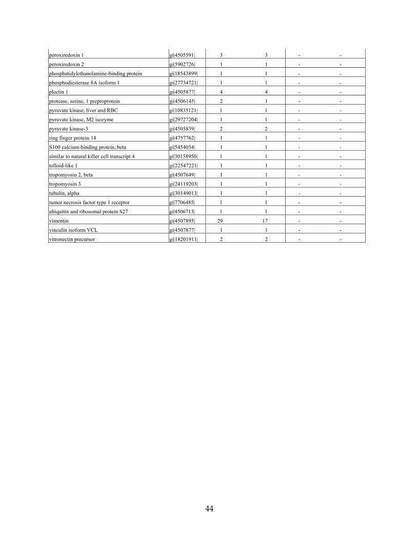
A complete alphabetical listing of all proteins detected from the proteomic analysis of the
human adult temporal bone sections is presented in Table 3. The major classes of proteins found
are extracellular and structural components of the cochlea, cochlin being the primary. Many of
the proteins are known to be expressed in the spiral ligament, which comprises a major amount

11
of the total cochlear mass. Collagen types I, II, IX, and XI, which are also abundantly detected
in our analysis, are very representative of the known and stable components of cochlear tissue
and involved in both nonsyndromic and syndromic types of hearing loss (1). Other proteins
frequently represented in our analysis are those comprising cytoskeletal elements such as
keratins, β-actin, and tubulin, which are also known to be expressed in the cochlea. A total of 13
unique peptides are identified, representing five distinct keratins. The intermediate filament,
vimentin, is the second most prevalent protein detected in our analysis (29 peptide matches,
reflecting 17 unique peptides), followed by collagen type II, alpha 1 (11 peptide matches,
representing 2 unique peptides). Immunohistochemical localization of vimentin in the gerbil
inner ear shows high-level expression of this protein in most connective tissue cells and in
several different types of epithelial cells, including some types of organ of Corti supporting cells
(41).
Our proteomic analysis of formalin-fixed, paraffin-embedded adult human tissue sections
has some limitations in the number and complexity of cochlear proteins identified, likely as a
result of the inaccessibility and possible degradation of proteins during the extraction process
from very small amounts of tissue in 8-micron paraffin sections. However, even with these
limitations, many representative proteins were detected. Cochlin is the most prevalent, showing
over twice as many detected peptides as vimentin, and reflects its high expression and stability
even after the processing and extraction protocol from fixed and embedded tissues.
In the proteomic analysis of the DFNA9-affected temporal bone sections (Table 3) there
are substantially fewer proteins identified, as well as fewer peptide matches for each protein.
However, there is significant overlap in the proteins that were identified in the DFNA9-affected
and unaffected samples. Aside from blood components, albumin and alpha and beta globins, the

12
major proteins found in the DFNA9-affected temporal bone (and also present in the unaffected
sample) are cochlin, collagen types I alpha 1 and alpha 2, and keratins 1 and 2a. A quantitative
comparison of proteins across the DFNA9-affected and unaffected samples cannot be made
because the two samples varied greatly in complexity and number of total peptide matches for
each protein, likely as a result of the greater difficulty of extraction and solubility of proteins in
the DFNA9 sections which was apparent during the processing of samples (also discussed
below). For every protein identified in the DFNA9 sample, the total number of peptide matches
ranges from 1 to 3, whereas in the unaffected sample 1 to 66 matches were found. For instance,
in the unaffected sample, the total number of peptide matches are 66 for cochlin, 5 for keratin 1,
and 2 for collagen type I alpha 2; corresponding peptide matches in the DFNA9-affected sample
are 3, 1, and 2, respectively. The number of cochlin peptides found in the DFNA9 sample is
greater than or equivalent to that for any other protein. Cochlin peptides identified in the
DFNA9 sample are from both the N-terminal and C-terminal portions of cochlin spanning amino
acid residues 81 to 91, in the FCH/LCCL domain, and amino acid residues 526 to 539 in the
vWFA2 domain.
One major factor in identification of proteins was that the tissue composition of DFNA9-
affected temporal bone samples presented a challenge in total protein extraction due to relative
insolubility of these samples. Much of this sample remained as insoluble aggregates during the
extraction protocol. Therefore, a large number of proteins were likely not solubilized or
extracted, making them inaccessible for mass spectrometry analysis, and resulting in lower
complexity and number of total proteins as well as a smaller overall number of peptides. The
difficulty in extraction and solubility of proteins from DFNA9-affected sections is perhaps not
surprising given the observation of the large amounts of DFNA9 eosinophilic aggregates in the
13
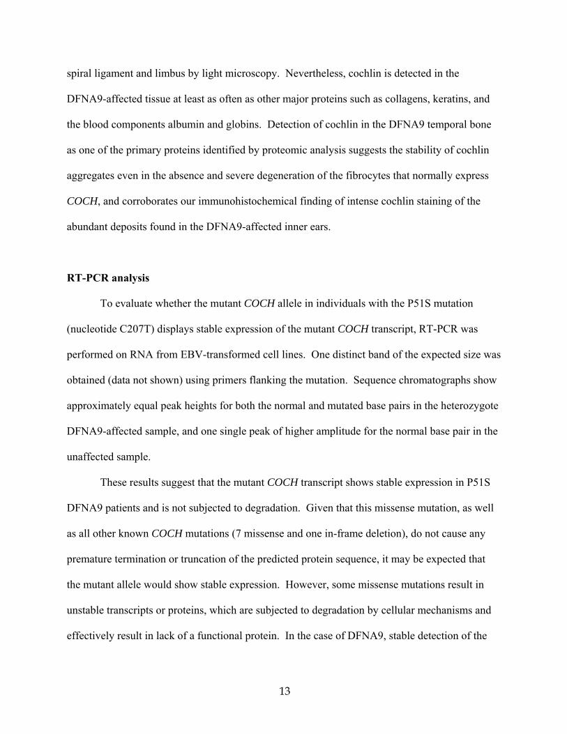
spiral ligament and limbus by light microscopy. Nevertheless, cochlin is detected in the
DFNA9-affected tissue at least as often as other major proteins such as collagens, keratins, and
the blood components albumin and globins. Detection of cochlin in the DFNA9 temporal bone
as one of the primary proteins identified by proteomic analysis suggests the stability of cochlin
aggregates even in the absence and severe degeneration of the fibrocytes that normally express
COCH, and corroborates our immunohistochemical finding of intense cochlin staining of the
abundant deposits found in the DFNA9-affected inner ears.
RT-PCR analysis
To evaluate whether the mutant COCH allele in individuals with the P51S mutation
(nucleotide C207T) displays stable expression of the mutant COCH transcript, RT-PCR was
performed on RNA from EBV-transformed cell lines. One distinct band of the expected size was
obtained (data not shown) using primers flanking the mutation. Sequence chromatographs show
approximately equal peak heights for both the normal and mutated base pairs in the heterozygote
DFNA9-affected sample, and one single peak of higher amplitude for the normal base pair in the
unaffected sample.
These results suggest that the mutant COCH transcript shows stable expression in P51S
DFNA9 patients and is not subjected to degradation. Given that this missense mutation, as well
as all other known COCH mutations (7 missense and one in-frame deletion), do not cause any
premature termination or truncation of the predicted protein sequence, it may be expected that
the mutant allele would show stable expression. However, some missense mutations result in
unstable transcripts or proteins, which are subjected to degradation by cellular mechanisms and
effectively result in lack of a functional protein. In the case of DFNA9, stable detection of the

14
mutant COCH transcript is consistent with the hypothesis of a dominant -negative effect of the
mutation in DFNA9 pathology rather than haploinsufficiency of cochlin.
Cochlin deposition in DFNA9
Our previous studies have shown cochlin to be secreted and glycosylated in cultured
mammalian cells transfected with full-length COCH cDNA (42). As an extracellular
glycoprotein, with the FCH/LCCL and vWFA domains, cochlin is likely to bind to, or interact
with, other cellular components such as extracellular proteins, glycoproteins and proteoglycans.
Such interactions have been shown for other proteins containing these types of domains (43-47).
Our light microscopy studies of DFNA9 sections have identified the prominent eosinophilic
material as an acellular extracellular homogeneous material. Movats pentachrome staining
suggests that this material may contain a mucopolysaccharide-like substance (3). Electron
microscopic examination of DNFA9 inner ear sections shows these extracellular deposits to be a
highly branched, disarrayed, microfibrillar substance, along with scattered glycosaminoglycan-
like granules (48). These observations are consistent with our finding of cochlin immunostaining
of the eosinophilic ground substance, suggesting that these aggregates contain extracellularly
deposited cochlin. It is also possible that other proteins associating with cochlin may be present
and that the nature of these interactions are altered as a result of the presence of mutated cochlin.
In terms of the actual mechanism of the missense mutations leading to misfolding and
aggregation of cochlin, several in vitro studies have been performed to investigate this
possibility. Initial studies of the FCH/LCCL domain of cochlin in bacterial cells showed
misfolding of this domain due to several of the known disease-causing mutations and
precipitation of the mutant FCH/LCCL peptide during the folding process (49). Our transient

15
transfections of full-length COCH with several of the inherited mutations did not reveal any
differences in secretion or apparent steady-state levels of cochlin (9, 42). Another study
corroborated these findings, and reported differences in cochlin deposition in the extracellular
matrix (ECM) of cultured cells, suggesting altered integration of mutated cochlin into the matrix
(50). However, a caveat of in vitro studies is the lack of the appropriate extracellular
environment of the inner ear. Furthermore, the late-onset and progressive nature of DFNA9
suggests that the effects of mutant cochlin such as aggregation or altered interaction with other
ECM components may be a cumulative process. A DFNA9 mouse model would more clearly
address long-term and progressive changes in the inner ear as a result of Coch mutation, and
such a model is currently being evaluated in our laboratory.
Possible etiology and pathogenesis of DFNA9
Cochlin immunostaining of eosinophilic aggregates in DFNA9, and the severe atrophy of
fibrocytes in the same areas point toward the primary sites where pathological changes are likely
to have initiated as a result of COCH mutations. Other observations in DFNA9 post-mortem
sections are neuroepithelial and neural degeneration in the inner ear. Because cochlin is not
expressed in these neuroectodermal structures, it is likely that these changes are secondary to
those in mesodermal tissues. The striking reduction and degeneration of fibrocytes and
replacement by aggregates throughout the spiral ligament and spiral limbus are in the very same
sites as the pathways of K+ recycling from epithelial cells of the organ of Corti back into the
endolymphatic scala media compartment (51), indicating a disruption of the integrity of the
network of gap junctions that normally exists between these cells and plays a critical role in the
ion homeostasis necessary for proper hair cell function. Therefore, a disruption of inner ear ionic

16
balance likely occurs in DFNA9 as a result of the lack of fibrocytes and presence of deposits
throughout the areas critical for K+ recycling.
Another observation in both unaffected and DFNA9 inner ears is cochlin immunostaining
throughout the osseous spiral lamina and in modiolar areas surrounding neurons and their
processes, but not in the neural cell bodies or processes. The detection of cochlin deposits within
these neural channels suggests that obstruction of these channels and neuronal damage may also
be occurring as a result of mutant cochlin aggregates.
An intriguing finding is immunostaining of perivascular areas in the modiolus and
throughout the cochlear duct. Such a finding suggests the possibility that cochlin deposition in
perivascular aggregates in organs outside the inner ear is implicated in the high prevalence of
vascular disorders that has been found in some P51S DFNA9 kindreds (4), including the one to
which the present 67-year-old female individual belonged (52, 53). These observations warrant
further studies of cochlin expression in extralabyrinthine blood vessels.
Another interesting observation is the lack of any similar histopathological findings
between DFNA9-affected temporal bones and those of Coch (-/-) mice (at ~5 months of age)
(30) (Figs. 3 and 5). In fact, these mice, which do not express cochlin, do not show any apparent
inner ear abnormalities and any significant hearing loss although later time-points have yet to be
evaluated. Our studies showing cochlin-staining eosinophilic deposits in DFNA9, in addition to
absence of overt pathology in Coch (-/-) mice at 5 months of age, are further support that this
disorder is not likely due to COCH haploinsufficiency, but rather a result of deleterious effects
by a “gain-of-function” molecular mechanism of COCH missense mutations.
Cochlin deposits found in glaucoma
17
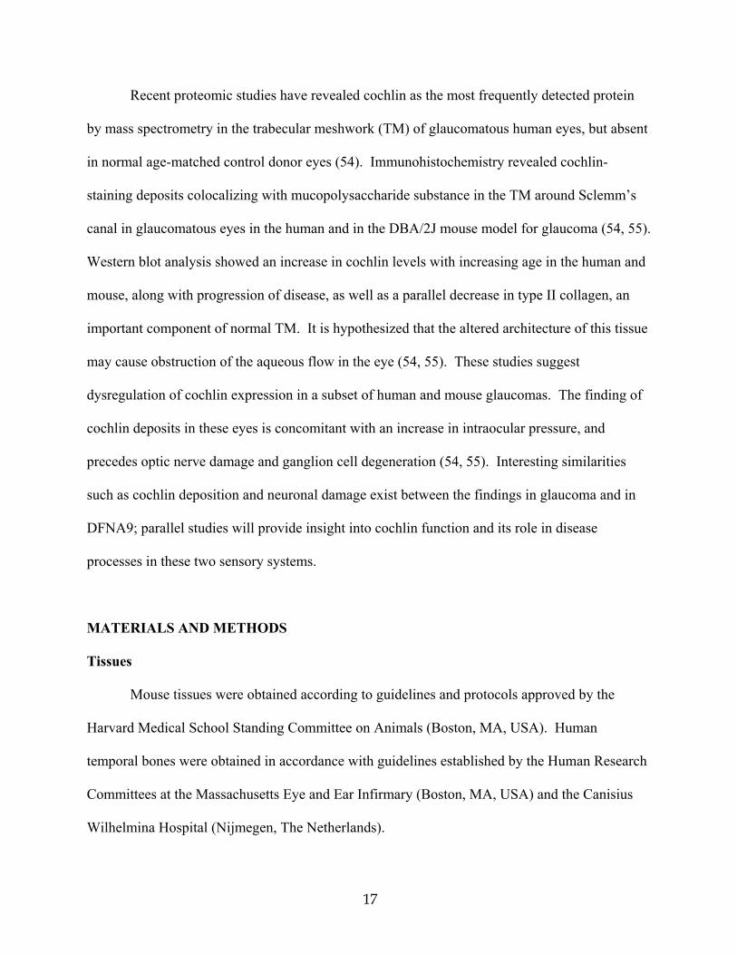
Recent proteomic studies have revealed cochlin as the most frequently detected protein
by mass spectrometry in the trabecular meshwork (TM) of glaucomatous human eyes, but absent
in normal age-matched control donor eyes (54). Immunohistochemistry revealed cochlin-
staining deposits colocalizing with mucopolysaccharide substance in the TM around Sclemm’s
canal in glaucomatous eyes in the human and in the DBA/2J mouse model for glaucoma (54, 55).
Western blot analysis showed an increase in cochlin levels with increasing age in the human and
mouse, along with progression of disease, as well as a parallel decrease in type II collagen, an
important component of normal TM. It is hypothesized that the altered architecture of this tissue
may cause obstruction of the aqueous flow in the eye (54, 55). These studies suggest
dysregulation of cochlin expression in a subset of human and mouse glaucomas. The finding of
cochlin deposits in these eyes is concomitant with an increase in intraocular pressure, and
precedes optic nerve damage and ganglion cell degeneration (54, 55). Interesting similarities
such as cochlin deposition and neuronal damage exist between the findings in glaucoma and in
DFNA9; parallel studies will provide insight into cochlin function and its role in disease
processes in these two sensory systems.
MATERIALS AND METHODS
Tissues
Mouse tissues were obtained according to guidelines and protocols approved by the
Harvard Medical School Standing Committee on Animals (Boston, MA, USA). Human
temporal bones were obtained in accordance with guidelines established by the Human Research
Committees at the Massachusetts Eye and Ear Infirmary (Boston, MA, USA) and the Canisius
Wilhelmina Hospital (Nijmegen, The Netherlands).

18
Postnatal mice (3-6 months of age) were used in this study. Wild-type (+/+) mice were
used for assessment of normal histology, cochlin localization, and proteomic analysis. Coch (-/-)
mice, used as negative controls, were a gift of Drs. Colin Stewart and Clara Rodriguez (29).
Mice were perfused intracardially and tissues fixed in 4% paraformaldehyde. After removal of
the stapes from the oval window, and piercing of the round window, 4% paraformaldehyde
fixative was perfused gently through the cochlea. Inner ears were immersed in fixative for 2-4
hours, followed by decalcification in 120mM EDTA for one week at room temperature, and
embedded in paraffin by standard histologic procedures. Serial sections were obtained at 5-8
micron thickness and used for staining with hematoxylin and eosin (H&E) and for
immunohistochemistry. For proteomic analysis, cochlear and vestibular tissues were dissected
separately and processed as described below (in the “Proteomic analysis” section).
For human tissues, temporal bones from a 67-year-old female who was a member of a
large DFNA9 kindred in the Netherlands segregating the P51S cochlin mutation (5, 11), were
donated and processed at the Otopathology Laboratory of the Massachusetts Eye and Ear
Infirmary, Boston, MA. Postmortem time for obtaining tissues was six hours. In this DFNA9
individual, the onset of bilateral sensorineural hearing loss and disequilibrium occurred around
age 40 years with progression to profound deafness by age 63, which was documented by
audiological evaluation. Vestibular symptoms consisted of gait imbalance, instability in the dark
and oscillopsia. Vestibular testing revealed bilateral peripheral vestibular hypofunction. For
unaffected controls (with no history of hearing loss), temporal bones obtained from two donors, a
63-year-old female and a 75-year-old male were used. Postmortem times for obtaining control
tissues were 8 and 15 hours, respectively. Temporal bones were fixed in 10% formalin,
decalcified in EDTA, and reduced in size using razor blades to contain only the otic capsule and

19
inner ear. To obtain optimal morphology of sections, one of the DFNA9 temporal bones (left
side) was embedded in celloidin, as is standard with temporal bones processed for light
microscopic study. Immunostaining is challenging in celloidin-embedded sections, even though
there is much better tissue integrity in this medium (Fig. 2); therefore, to facilitate
immunohistochemistry, the other temporal bone (right side) was embedded in paraffin. This is
the first and only temporal bone from a DFNA9 family member that has been embedded in
paraffin to date, thereby facilitating our analyses. For the unaffected controls, the temporal
bones from the 63-year-old female were embedded in celloidin, and those from the 75-year-old
male in paraffin. Specimens were serially sectioned at a thickness of 20 microns for celloidin
and 8 microns for paraffin, and selected sections stained with H&E. Paraffin sections were
processed for immunohistochemistry and proteomic analysis as described below.
Immunohistochemistry
Immunostaining was performed using an anti-cochlin antibody generated against a
peptide in the vWFA1 domain of cochlin (Fig. 1), corresponding to amino acid residues 163-181
of human cochlin, identical to the residues in murine and bovine cochlin (31). Anti-serum was
purified through a protein A sepharose column, followed by peptide-affinity chromatography.
This antibody (anti-cochlin/vWFA1 domain) recognizes all three different-sized isoforms of
cochlin (Fig. 1).
Immunohistochemistry was performed as previously described (19), except for
modifications as described below, including lack of the antigen-retrieval step. Paraffin-
embedded sections from postnatal (+/+) and Coch (-/-) mice, unaffected control and DFNA9-
affected human adult temporal bones were incubated with anti-cochlin/vWFA1 domain antibody
20
overnight at room temperature, washed, and incubated with a secondary biotinylated anti-rabbit
IgG (Vector Labs, Burlingame, CA). Immunostaining was visualized by incubation with the
Vectastain ABC reagent (Vector Labs) followed by 3,3’-diaminobenzidine (DAB). Sections
were not counterstained.
Proteomic analysis
For proteomic analysis in the mouse, membranous cochlear and vestibular labyrinths
were dissected separately from approximately 3-month-old (+/+) and Coch (-/-) mice. Protein
lysates were prepared by incubation of tissues in lysis buffer (2% SDS, 100mM ammonium
bicarbonate, 10mM DTT, pH 8.5) at 90°C for 10 min, 37°C for 60 min, with subsequent
sonication in 0.2% SDS, and incubation at 90°C for 10 min. Three rounds of sonication and
boiling were performed, followed by alkylation with 30mM iodoacetamide. The reaction was
quenched with 10mM DTT and samples were separated by SDS-gel electrophoresis in 8-16%
polyacrylamide gels. Gels were size-fractionated into four sections, destained with two washes
of 50% methanol and 5% acetic acid, followed by three alternating washes of ammonium
bicarbonate and acetonitrile. Gel slices were dried and subsequently suspended individually in
trypsin (5.5µg/mL in 50mM ammonium bicarbonate) prior to incubation at 37°C for 18 hr for
digestion of proteins. Peptides were extracted with two rinses of 50mM ammonium bicarbonate
and two rinses of 50% acetonitrile and 0.1% formic acid. Samples were prepared for mass
spectrometry by lyophilization and rehydration in 5% acetonitrile and 0.1% formic acid.
For proteomic analysis of the adult human inner ear, the only materials available were
formalin-fixed, paraffin-embedded tissues. Sections, 8 microns in thickness, were used from the
same DFNA9-affected temporal bone as used for immunohistochemistry. For a human adult

21
temporal bone control, formalin-fixed, paraffin-embedded, 8 micron-thick sections were used
from an 85-year-old female with otosclerosis, but showing no inner ear histopathology and with
normal cochlear duct structures including the spiral ligament and spiral limbus.
Extraction of proteins from human paraffin-embedded sections was performed by adding
heptane and incubating at room temperature for 60 min, followed by the addition of methanol to
pellet extracted proteins. Protein extracts were resolubilized and sonicated in 2% SDS, 100mM
ammonium bicarbonate, 10mM DTT, pH 8.5. Reduction of proteins was achieved by boiling
samples at 90°C for 20 min, followed by incubation at 37°C for 60 min and alkylation in 30mM
iodoacetamide for 60 min at room temperature. Trypsin digestion of proteins and preparation for
mass spectrometry were performed as described for the mouse samples.
For mass spectrometry, samples were run on an LCQ DECA XP plus Proteome X
workstation (Thermo Electron Corporation, San Jose, CA). Peptide identifications were made
using Sequest through the Bioworks Browser 3.1 (Thermo Electron Corporation). Database
searches were made using the NCBI RefSeqHuman and RefSeqMurine databases using static
carbamidomethyl modified cysteines and differential oxidized methionines, followed by further
searches using differential modifications.
Reverse transcription-PCR
Reverse transcription-PCR (RT-PCR) was performed with total RNA isolated from EBV-
transformed cell lines from two P51S DFNA9 individuals, using the RNAeasy midi-kit (Qiagen,
Leusden, The Netherlands). cDNA synthesis was performed as described (56) and PCR was
done for 35 cycles under standard conditions using the following primers flanking the mutation:
forward 5'-ACC AGA GGC TTG GAC ATC AG-3' in exon 4 and reverse 5'-TTT GAG ACT

22
GGA TGC CAT TG-3' in exon 5. Amplified products were gel isolated and sequenced using an
ABI PRISM Big Dye Terminator Cycle Sequencing V2 Ready reaction kit and the ABI 3730
DNA sequencing apparatus (Applied Biosystems, Foster City, CA).
ACKNOWLEDGEMENTS
We are especially grateful to the individuals and their families for donation of temporal
bones, and a more detailed description of the P51S DFNA9 histopathology is in preparation for
publication. We would like to thank Dr. Roderick Bronson and Li Zhang at the Dana
Farber/Harvard Cancer Center Rodent Histopathology Core. We also thank Drs. Colin Stewart
and Clara Rodriguez for their gift of the Coch (-/-) mice. This work was supported by
NIH/NIDCD grants R01-DC03402 (to C.C.M.), R01-DC0188 and P30-05209 (to M.C.L.), the
NIDCD National Temporal Bone, Hearing and Balance Pathology Resource Registry and by Mr.
Axel Eliasen, and the Health and Labor Sciences Research Grants in Japan (Research on
Measures for Intractable Diseases, and Sensory and Communicative Disorders) (to T.I.).
REFERENCES
1. Van Camp, G. and Smith, R.J.H. (2005) Hereditary Hearing Loss Homepage. URL:
http://webhost.ua.ac.be/hhh/.
2. Halpin, C., Khetarpal, U. and McKenna, M. (1996) Autosomal dominant progressive
sensorineural hearing loss in a large North American Family. Am. J. Audiol., 5, 105-111.
3. Khetarpal, U., Schuknecht, H.F., Gacek, R.R. and Holmes, L.B. (1991) Autosomal
dominant sensorineural hearing loss: pedigrees, audiologic and temporal bone findings in
two kindreds. Arch. Otolaryngol. Head Neck Surg., 117, 1032-1042.

23
4. Bom, S.J.H., Kemperman, M.H., De Kok, Y.J.M., Huygen, P.L.M., Verhagen, W.I.M.,
Cremers, F.P.M. and Cremers, C.W.R.J. (1999) Progressive cochleovestibular
impairment caused by a point mutation in the COCH gene at DFNA9. Laryngoscope,
109, 1525-1530.
5. Verhagen, W.I.M., Bom, S.J.H., Huygen, P.L.M., Fransen, E., Van Camp, G. and
Cremers, C.W.R.J. (2000) Familial progressive vestibulocochlear dysfunction caused by
a COCH mutation (DFNA9). Arch. Neurol., 57, 1045-1047.
6. Verstreken, M., Declau, F., Wuyts, F.L., D'Haese, P., Van Camp, G., Fransen, E., Van
den Hauwe, L., Buyle, S., Smets, R.E., Feenstra, L. et al. (2001) Hereditary otovestibular
dysfunction and Meniere's disease in a large Belgian family is caused by a missense
mutation in the COCH gene. Otol. Neurotol., 22, 874-881.
7. Kemperman, M.H., Bom, S.J.H., Lemaire, F.X., Verhagen, W.I.M., Huygen, P.L.M. and
Cremers, C.W.R.J. (2002) DFNA9/COCH and its phenotype. Adv. Otorhinolaryngol., 61,
66-72.
8. Lemaire, F.X., Feenstra, L., Huygen, P.L.M., Fransen, E., Devriendt, K., Van Camp, G.,
Vantrappen, G. and Cremers, C.W.R.J. (2003) Progressive late-onset sensorineural
hearing loss and vestibular impairment with vertigo (DFNA9/COCH): longitudinal
analyses in a Belgian family. Otol. Neurotol., 24, 743-748.
9. Street, V.A., Kallman, J.C., Robertson, N.G., Kuo, S.F., Morton, C.C. and Phillips, J.O.
(2005) A novel DFNA9 mutation in the vWFA2 domain of COCH alters a conserved
cysteine residue and intrachain disulfide bond formation, resulting in progressive hearing
loss and site-specific vestibular and central oculomotor dysfunction. Am. J. Med. Genet.
A., 139, 86-95.

24
10. Robertson, N.G., Lu, L., Heller, S., Merchant, S.N., Eavey, R.D., McKenna, M., Nadol,
J.B., Jr., Miyamoto, R.T., Linthicum, F.H., Jr., Lubianca Neto, J.F. et al. (1998)
Mutations in a novel cochlear gene cause DFNA9, a human nonsyndromic deafness with
vestibular dysfunction. Nature Genet., 20, 299-303.
11. de Kok, Y.J.M., Bom, S.J.H., Brunt, T.M., Kemperman, M.H., van Beusekom, E., van
der Velde-Visser, S.D., Robertson, N.G., Morton, C.C., Huygen, P.L.M., Verhagen,
W.I.M. et al. (1999) A Pro51Ser mutation in the COCH gene is associated with late onset
autosomal dominant progressive sensorineural hearing loss with vestibular defects. Hum.
Mol. Genet., 8, 361-366.
12. Fransen, E., Verstreken, M., Verhagen, W.I.M., Wuyts, F.L., Huygen, P.L.M., D'Haese,
P., Robertson, N.G., Morton, C.C., McGuirt, W.T., Smith, R.J.H. et al. (1999) High
prevalence of symptoms of Meniere's disease in three families with a mutation in the
COCH gene. Hum. Mol. Genet., 8, 1425-1429.
13. Kamarinos, M., McGill, J., Lynch, M. and Dahl, H. (2001) Identification of a novel
COCH mutation, I109N, highlights the similar clinical features observed in DFNA9
families. Hum. Mutat., 17, 351.
14. Kemperman, M.H., De Leenheer, E.M.R., Huygen, P.L.M., van Duijnhoven, G., Morton,
C.C., Robertson, N.G., Cremers, F.P.M., Kremer, H. and Cremers, C.W.R.J. (2005)
Audiometric, vestibular, and genetic aspects of a DFNA9 family with a G88E COCH
mutation. Otol. Neurotol., 26, 926-933.
15. Usami, S., Takahashi, K., Yuge, I., Ohtsuka, A., Namba, A., Abe, S., Fransen, E., Patthy,
L., Otting, G. and Van Camp, G. (2003) Mutations in the COCH gene are a frequent

25
cause of autosomal dominant progressive cochleo-vestibular dysfunction, but not of
Meniere's disease. Eur. J. Hum. Genet., 11, 744-748.
16. Nagy, I., Horvath, M., Trexler, M., Repassy, G. and Patthy, L. (2004) A novel COCH
mutation, V104del, impairs folding of the LCCL domain of cochlin and causes
progressive hearing loss. J. Med. Genet., 41, e9.
17. Robertson, N.G., Khetarpal, U., Gutiérrez-Espeleta, G.A., Bieber, F.R. and Morton, C.C.
(1994) Isolation of novel and known genes from a human fetal cochlear cDNA library
using subtractive hybridization and differential screening. Genomics, 23, 42-50.
18. Robertson, N.G., Skvorak, A.B., Yin, Y., Weremowicz, S., Johnson, K.R., Kovatch,
K.A., Battey, J.F., Bieber, F.R. and Morton, C.C. (1997) Mapping and characterization of
a novel cochlear gene in human and in mouse: a positional candidate gene for a deafness
disorder, DFNA9. Genomics, 46, 345-354.
19. Robertson, N.G., Resendes, B.L., Lin, J.S., Lee, C., Aster, J.C., Adams, J.C. and Morton,
C.C. (2001) Inner ear localization of mRNA and protein products of COCH, mutated in
the sensorineural deafness and vestibular disorder, DFNA9. Hum. Mol. Genet., 10, 2493-
2500.
20. Ikezono, T., Shindo, S., Ishizaki, M., Li, L., Tomiyama, S., Takumida, M., Pawankar, R.,
Watanabe, A., Saito, A. and Yagi, T. (2005) Expression of cochlin in the vestibular organ
of rats. O.R.L. J. Otorhinolaryngol. Relat. Spec., 67, 252-258.
21. Ikezono, T., Omori, A., Ichinose, S., Pawankar, R., Watanabe, A. and Yagi, T. (2001)
Identification of the protein product of the Coch gene (hereditary deafness gene) as the
major component of bovine inner ear protein. Biochim. Biophys. Acta, 1535, 258-265.

26
22. Merchant, S.N., Linthicum, F.H. and Nadol, J.B., Jr. (2000) Histopathology of the inner
ear in DFNA9. Adv. Otorhinolaryngol., 56, 212-217.
23. Selkoe, D.J. (2004) Cell biology of protein misfolding: the examples of Alzheimer's and
Parkinson's diseases. Nat. Cell Biol., 6, 1054-1061.
24. Davies, S.W., Turmaine, M., Cozens, B.A., DiFiglia, M., Sharp, A.H., Ross, C.A.,
Scherzinger, E., Wanker, E.E., Mangiarini, L. and Bates, G.P. (1997) Formation of
neuronal intranuclear inclusions underlies the neurological dysfunction in mice
transgenic for the HD mutation. Cell, 90, 537-548.
25. Zoghbi, H.Y. and Orr, H.T. (2000) Glutamine repeats and neurodegeneration. Annu. Rev.
Neurosci., 23, 217-247.
26. Waelter, S., Boeddrich, A., Lurz, R., Scherzinger, E., Lueder, G., Lehrach, H. and
Wanker, E.E. (2001) Accumulation of mutant huntingtin fragments in aggresome-like
inclusion bodies as a result of insufficient protein degradation. Mol. Biol. Cell, 12, 1393-
1407.
27. Masliah, E., Rockenstein, E., Veinbergs, I., Mallory, M., Hashimoto, M., Takeda, A.,
Sagara, Y., Sisk, A. and Mucke, L. (2000) Dopaminergic loss and inclusion body
formation in alpha-synuclein mice: implications for neurodegenerative disorders. Science,
287, 1265-1269.
28. Khetarpal, U. (1993) Autosomal dominant sensorineural hearing loss: further temporal
bone findings. Arch. Otolaryngol. Head Neck Surg., 119, 106-108.
29. Rodriguez, C.I., Cheng, J.G., Liu, L. and Stewart, C.L. (2004) Cochlin, a secreted von
Willebrand factor type a domain-containing factor, is regulated by leukemia inhibitory
factor in the uterus at the time of embryo implantation. Endocrinology, 145, 1410-1418.

27
30. Makishima, T., Rodriguez, C.I., Robertson, N.G., Morton, C.C., Stewart, C.L. and
Griffith, A.J. (2005) Targeted disruption of mouse Coch provides functional evidence
that DFNA9 hearing loss is not a COCH haploinsufficiency disorder. Hum. Genet., 118,
29-34.
31. Ikezono, T., Shindo, S., Li, L., Omori, A., Ichinose, S., Watanabe, A., Kobayashi, T.,
Pawankar, R. and Yagi, T. (2004) Identification of a novel Cochlin isoform in the
perilymph: insights to Cochlin function and the pathogenesis of DFNA9. Biochem.
Biophys. Res. Commun., 314, 440-446.
32. Skvorak, A.B., Weng, Z., Yee, A.J., Robertson, N.G. and Morton, C.C. (1999) Human
cochlear expressed sequence tags provide insight into cochlear gene expression and
identify candidate genes for deafness. Hum. Mol. Genet., 8, 439-452.
33. Boulassel, M.R., Deggouj, N., Tomasi, J.P. and Gersdorff, M. (2001) Inner ear
autoantibodies and their targets in patients with autoimmune inner ear diseases. Acta
Otolaryngol., 121, 28-34.
34. Boulassel, M.R., Tomasi, J.P., Deggouj, N. and Gersdorff, M. (2001) COCH5B2 is a
target antigen of anti-inner ear antibodies in autoimmune inner ear diseases. Otol.
Neurotol., 22, 614-618.
35. Kommareddi, P.K., Nair, T.S. and Carey, T.E. (2003) Cochlin is expressed in the
supporting cells of the organ of Corti and co-precipitates with CTL2. Association for
research in Otolaryngology, Daytona Beach, FL.
36. Nair, T.S., Kozma, K.E., Hoefling, N.L., Kommareddi, P.K., Ueda, Y., Gong, T.W.,
Lomax, M.I., Lansford, C.D., Telian, S.A., Satar, B. et al. (2004) Identification and

28
characterization of choline transporter-like protein 2, an inner ear glycoprotein of 68 and
72 kDa that is the target of antibody-induced hearing loss. J. Neurosci., 24, 1772-1779.
37. Solares, C.A., Edling, A.E., Johnson, J.M., Baek, M.J., Hirose, K., Hughes, G.B. and
Tuohy, V.K. (2004) Murine autoimmune hearing loss mediated by CD4+ T cells specific
for inner ear peptides. J. Clin. Invest., 113, 1210-1217.
38. Billings, P. (2004) Experimental autoimmune hearing loss. J. Clin. Invest., 113, 1114-
1117.
39. Baek, M.J., Park, H.M., Johnson, J.M., Altuntas, C.Z., Jaini, R., Thomas, D.M., Ball,
E.J., Robertson, N.G., Morton, C.C., Hughes, G.B. et al. (2005) Increased frequencies of
cochlin-specific T cells in patients with autoimmune sensorinerual hearing loss. J.
Immunol., submitted.
40. Palmer-Toy, D.E., Krastins, B., Sarracino, D.A., Nadol, J.B., Jr. and Merchant, S.N.
(2005) Efficient method for the proteomic analysis of fixed and embedded tissues. J.
Proteome Res., 4, 2404-2411.
41. Schulte, B.A. and Adams, J.C. (1989) Immunohistochemical localization of vimentin in
the gerbil inner ear. J. Histochem. Cytochem., 37, 1787-1797.
42. Robertson, N.G., Hamaker, S.A., Patriub, V., Aster, J.C. and Morton, C.C. (2003)
Subcellular localisation, secretion, and post-translational processing of normal cochlin,
and of mutants causing the sensorineural deafness and vestibular disorder, DFNA9. J.
Med. Genet., 40, 479-486.
43. Colombatti, A. and Bonaldo, P. (1991) The superfamily of proteins with von Willebrand
factor type A-like domains: one theme common to components of extracellular matrix,
hemostasis, cellular adhesion, and defense mechanisms. Blood, 77, 2305-2315.

29
44. Colombatti, A., Bonaldo, P. and Doliana, R. (1993) Type A modules: interacting domains
found in several non-fibrillar collagens and in other extracellular matrix proteins. Matrix,
13, 297-306.
45. Lee, J.O., Rieu, P., Arnaout, M.A. and Liddington, R. (1995) Crystal structure of the A
domain from the alpha subunit of integrin CR3 (CD11b/CD18). Cell, 80, 631-638.
46. Delrieu, I., Waller, C.C., Mota, M.M., Grainger, M., Langhorne, J. and Holder, A.A.
(2002) PSLAP, a protein with multiple adhesive motifs, is expressed in Plasmodium
falciparum gametocytes. Mol. Biochem. Parasitol., 121, 11-20.
47. Whittaker, C.A. and Hynes, R.O. (2002) Distribution and evolution of von
Willebrand/integrin A domains: widely dispersed domains with roles in cell adhesion and
elsewhere. Mol. Biol. Cell, 13, 3369-3387.
48. Khetarpal, U. (2000) DFNA9 is a progressive audiovestibular dysfunction with a
microfibrillar deposit in the inner ear. Laryngoscope, 110, 1379-1384.
49. Liepinsh, E., Trexler, M., Kaikkonen, A., Weigelt, J., Banyai, L., Patthy, L. and Otting,
G. (2001) NMR structure of the LCCL domain and implications for DFNA9 deafness
disorder. Embo J., 20, 5347-5353.
50. Grabski, R., Szul, T., Sasaki, T., Timpl, R., Mayne, R., Hicks, B. and Sztul, E. (2003)
Mutations in COCH that result in non-syndromic autosomal dominant deafness (DFNA9)
affect matrix deposition of cochlin. Hum. Genet., 113, 406-416.
51. Steel, K.P. (1999) Perspectives: biomedicine. The benefits of recycling. Science, 285,
1363-1364.

30
52. Verhagen, W.I.M., Huygen, P.L.M., Theunissen, E.J.J.M. and Joosten, E.M.G. (1989)
Hereditary vestibulo-cochlear dysfunction and vascular disorders. J. Neurol. Sci., 92, 55-
63.
53. Verhagen, W.I.M., Bom, S.J.H., Fransen, E., Van Camp, G., Huygen, P.L.M.,
Theunissen, E.J.J.M. and Cremers, C.W.R.J. (2001) Hereditary cochleovestibular
dysfunction due to a COCH gene mutation (DFNA9): a follow-up study of a family. Clin.
Otolaryngol. Allied Sci., 26, 477-483.
54. Bhattacharya, S.K., Rockwood, E.J., Smith, S.D., Bonilha, V.L., Crabb, J.S., Kuchtey,
R.W., Robertson, N.G., Peachey, N.S., Morton, C.C. and Crabb, J.W. (2005) Proteomics
reveal Cochlin deposits associated with glaucomatous trabecular meshwork. J. Biol.
Chem., 280, 6080-6084.
55. Bhattacharya, S.K., Annangudi, S.P., Salomon, R.G., Kuchtey, R.W., Peachey, N.S. and
Crabb, J.W. (2005) Cochlin deposits in the trabecular meshwork of the glaucomatous
DBA/2J mouse. Exp. Eye Res., 80, 741-744.
56. Luijendijk, M.W., van de Pol, T.J., van Duijnhoven, G., den Hollander, A.I., ten Caat, J.,
van Limpt, V., Brunner, H.G., Kremer, H. and Cremers, F.P. (2003) Cloning,
characterization, and mRNA expression analysis of novel human fetal cochlear cDNAs.
Genomics, 82, 480-490.

31
FIGURE LEGENDS
Figure 1. Schematic representation of the deduced amino acid structure of human COCH,
encoding the protein cochlin, shows a predicted signal peptide (SP), followed by a domain
initially designated as FCH (factor C-homology), also known as the LCCL domain, (Limulus
factor C, cochlin, lung gestational protein), followed by an intervening domain (ivd1), and two
von Willebrand factor A-like domains (vWFA1 and vWFA2) separated by an intervening
domain (ivd2). Six familial missense mutations, (five in the FCH/LCCL domain and one in the
vWFA2 domain), causing DFNA9 deafness and vestibular disorder, are indicated by arrows.
The positions of all cysteine residues are shown as “C”. The three known isoforms of cochlin
are represented by horizontal lines corresponding to their sequence and designated as p60 (full-
length, excluding the SP) and two shorter isoforms (p44 and p40, both lacking the FCH/LCCL
domain). The cochlin antibody used in this study was made against a small peptide in the N-
terminus of the vWFA1 domain (amino acid residues 163 to 181), shown in the figure. This
antibody recognizes all three isoforms of cochlin.

32
Figure 2. Hematoxylin and eosin (H&E)-stained celloidin-embedded temporal bone sections
from an individual with DFNA9 from a Dutch kindred segregating the P51S mutation (67-year-
old female) (B, D, F) and from an age-matched unaffected control (63-year-old female) (A, C,
E). In the cochlear duct of the DFNA9-affected individual (B, D, X100), as compared to the
unaffected control (A, C, X100) the most striking findings are the presence of abundant
extracellular eosinophilic aggregates throughout the spiral ligament, spiral limbus, and the
osseous spiral lamina, and significant loss and degeneration of fibrocytes in the ligament and
limbus. In particular, the more medial parts of the spiral ligament, underlying the stria vascularis
and the area of the insertion of the ligament into the basilar membrane are more severely affected
with presence of deposits and fibrocyte atrophy. Degeneration of the organ of Corti and of
neural processes in the osseous spiral lamina is also observed.
In the ampulla of the posterior semi-circular canal in the vestibular labyrinth (E, F, X40)
pathologic changes, similar to those in the cochlear duct, are present in the DFNA9-affected
ampulla (F) as compared to the unaffected control (E). Abundant eosinophilic deposition is
present within the DFNA9 ampullary stroma, with reduction and atrophy of the stromal
fibrocytes, as well as degeneration of the sensory epithelium of the crista, and atrophy of the
ampullary nerve. In addition, there is appreciable thickening and partial collapse of the
ampullary wall, also showing presence of eosinophilic aggregates.

33

34
Figure 3. Immunohistochemistry on postnatal (5-month-old) (+/+) (A, C, G) and Coch (-/-) (E) mouse
inner ear sections with anti-cochlin. Immunostaining (on the left panels) appears as a reddish brown
DAB reaction product; no counterstain was applied on these sections. Serial sections stained with H&E
are shown in parallel on the right panels (B, D, F, H).
In the (+/+) cochlear duct (A, X100; C, X150) prominent cochlin immunostaining is present in
fibrocytes and in the extracellular matrix throughout the spiral ligament and spiral limbus. Cells lining
Rosenthal’s canal (surrounding the spiral ganglion) and the channels of the osseous spiral lamina also
contain cochlin, whereas the cochlear ganglion cell bodies and the neural processes are negative for
cochlin immunostaining. Distinct perivascular rings around blood vessels in the modiolus and
throughout the cochlear duct are stained. In contrast, adjacent areas of surrounding bony tissues clearly
lack cochlin staining. The structures of the cochlea that show absence of cochlin expression are the
organ of Corti, including the sensory epithelium and tectorial membrane (TM), stria vascularis,
Reissner’s membrane, and spiral ganglion cells. The cochlear duct in the Coch (-/-) mouse (E, X100)
was used as a negative control and lacks any cochlin immunostaining, confirming the specificity of this
antibody.
In the (+/+) crista of the posterior ampulla in the vestibular labyrinth (G, X200), intense cochlin
staining is observed in the fibrocytes and stroma underlying the sensory epithelium, as well as in the
ampullary wall. Neuronal processes within the ampullary stroma as well as the surrounding bone and
connective tissues lack any immunostaining. The sensory epithelium is also completely devoid of any
cochlin staining, as observed in the cochlear duct.

35

36
Figure 4. Immunohistochemistry on unaffected human adult (75-year-old male) temporal bone
sections with anti-cochlin (A, C, E). No counterstain was used on these sections; serial H&E
sections are shown (B, D, F). In the cochlear duct (A, C, X100), cochlin immunostaining is
prominent throughout the spiral ligament, spiral limbus and the channels of the osseous spiral
lamina. Adjacent areas of surrounding bony tissues are not stained with the anti-cochlin
antibody. Structures of the cochlea shown in this figure, which lack cochlin expression are the
organ of Corti, including the sensory epithelium and tectorial membrane (TM), stria vascularis,
and Reissner’s membrane (RM). Some of these structures show artifactual disruption as a result
of paraffin embedding of adult temporal bones. In the posterior crista ampullaris of the
vestibular labyrinth (E, X200), intense cochlin staining is observed in the fibrocytes and stroma
underlying the sensory epithelium. The sensory epithelium is completely devoid of any cochlin
expression, as was observed in the cochlear duct.

37

38
Figure 5. Immunohistochemistry on DFNA9-affected human adult (67-year-old female)
temporal bone sections with the anti-cochlin antibody (A, C, E, G). No counterstain was used
on these sections; serial H&E sections are shown (B, D, F, H). In the cochlear duct (A, C,
X100), cochlin immunostaining is observed throughout the spiral ligament, spiral limbus and the
channels of the osseous spiral lamina. The homogeneous eosinophilic deposits seen on the H&E
sections are stained darkly and evenly. The cochlin immunostaining of this acellular material is
prominent in the spiral ligament, particularly in the area of insertion into the basilar membrane,
the spiral limbus, and within the channels of the osseous spiral lamina. The organ of Corti and
the stria vascularis are negative for cochlin staining. Some stained tissue underlying the stria
vascularis appears to be a part of the spiral ligament that was detached along with the stria.
Adjacent areas of surrounding bony tissues do not show any immunostaining. In the posterior
ampulla of the vestibular labyrinth (E, X4; G, X100), cochlin staining of the ampullary stroma
containing the eosinophilic deposits is observed. The collapsed ampullary wall showing
prominent thickening and acellular deposition (F) also contains cochlin (E). The sensory
epithelium does not show cochlin expression, as seen in the cochlear duct in both DFNA9 and
unaffected control inner ears.

39

40
Figure 6. Amino acid sequence of human and mouse cochlin, showing distribution of tryptic
digest fragments identified by mass spectrometry from cochlear tissue. The domains of cochlin
are underlined and labeled (FCH/LCCL in red, vWFA1 and vWFA2 in blue), as well as the
signal peptide (SP), cleaved in mature cochlin and therefore not detected among tryptic
fragments. Amino acid residues identical between human and mouse are indicated by a dot.
Cochlin peptide coverage, as found in our proteomic analysis from human unaffected formalin-
fixed, paraffin-embedded temporal bone sections is highlighted in yellow, and from mouse fresh
whole cochlear tissue is highlighted in green.

41
Table 1. COCH mutations in DFNA9
Origin
Exon with mutation
Nucleotide
change*
Amino acid
change**
Protein domain
References The Netherlands1 Belgium1
4
C207T
P51S
FCH/LCCL
(11, 12)
United States1
4
T253G
V66G
FCH/LCCL
(10)
United States1
The Netherlands1
5
G319A
G88E
FCH/LCCL
(10, 14)
Hungary2
5
366_368delGTA
V104del
FCH/LCCL
(16)
Australia1
5
T382A
I109N
FCH/LCCL
(13)
United States1
5
T405C
W117R
FCH/LCCL
(10)
Japan2
5
G411A
A119T
FCH/LCCL
(15)
United States1
12
G1681T
C542F
vWFA2
(9)
* Numbering of nucleotides is according to the human COCH cDNA sequence (GenBank Accession No. AF006740), which starts in the 5’ untranslated region, 56 bp upstream of the start ATG. ** Numbering of amino acids begins at the start methionine. 1 Familial (autosomal dominant) cases 2 Simplex cases

42
Table 2. Relative abundance of inner ear peptides in wild-type +/+ and Coch -/- mice Cochlea Vestibular organs
Peptide match
wild-type +/+
Coch -/-
wild-type +/+
Coch -/-
cochlin
123
0
50
0
albumin
68
109
55
38
β-hemoglobin
76
81
47
28
α-tectorin
12
19
1
1
keratin 9
18
20
31
36

43
Table 3. Proteins identified from DFNA9-affected and unaffected adult human temporal bones unaffected control DFNA9-affected
# total peptide #unique peptide # total peptide #unique peptide
Protein Name Accession # matches matches matches matches
actin, beta gi|4501885| 3 2 - -
albumin precursor gi|4502027| 4 4 3 3
annexin A2; annexin II gi|4757756| 3 3 - -
annexin V; endonexin II gi|4502107| 5 5 - -
ATP synthase, mitochondrial F1 complex gi|4757810| 1 1 - -
ATPase, alpha 1 polypeptide gi|21361181| 1 1 - -
chondromodulin I precursor gi|5901932| 1 1 - -
clusterin gi|4502905| 7 4 - -
cochlin (coagulation factor C homology) gi|4758022| 66 17 3 2
collagen type I, alpha 1 gi|4502945| 2 1 3 3
collagen type I, alpha 2 gi|4502947| 2 2 2 2
collagen type II, alpha 1, isoform 1 gi|13435125| 11 2 - -
collagen type IX, alpha 2 gi|11386161| 3 2 - -
collagen type IX, alpha 3 gi|17921995| 1 1 - -
collagen type XI, alpha isoform A gi|18375518| 2 1 - -
dermatan sulfate proteoglycan 3 gi|4826704| 1 1 - -
desmin gi|18105050| 3 2 - -
eukaryotic translation initiation factor 4A1 gi|30153769| 1 1 - -
globin, alpha 2 gi|4504345| 1 1 2 2
globin, beta gi|4504349| 3 2 3 3
growth factor receptor-bound protein 10 gi|19923303| 1 1 - -
histone H2a gi|15617199| 1 1 - -
histone, H1, member 2 gi|4885375| 2 2 - -
histone, H2A, member Q gi|24638446| 5 4 - -
histone, H2A, member Z gi|4504255| 1 1 - -
histone, H2B, member Q gi|4504277| 2 2 - -
histone, H3, family 3A gi|4504279| 1 1 - -
histone, H4, family 2, member N gi|4504323| 9 6 - -
hypothetical protein FLJ10618 gi|8922551| 1 1 - -
hypothetical protein LOC221302 gi|21450836| 2 1 - -
hypothetical protein XP_298917 gi|29735940| 1 1 - -
IMP (inosine monophosphate) dehydrogenase 2 gi|30150307| - - 1 1
keratin 1 gi|17318569| 5 5 1 1
keratin 10 gi|4557697| 3 3 - -
keratin 2a gi|4557703| 2 2 1 1
keratin 8 gi|30154839| 1 1 - -
keratin 9 gi|4557705| 2 2 - -
lamin A/C isoform 2 gi|5031875| 2 2 - -
lanthionine synthetase C-like protein 1 gi|5174445| 1 1 - -
norrin (Norrie disease protein) gi|4557789| 1 1 - -
nuclear ribonucleoprotein A2/B1 isoform A2 gi|4504447| 1 1 - -
44
peroxiredoxin 1 gi|4505591| 3 3 - -
peroxiredoxin 2 gi|5902726| 1 1 - -
phosphatidylethanolamine-binding protein gi|18543899| 1 1 - -
phosphodiesterase 8A isoform 1 gi|27734721| 1 1 - -
plectin 1 gi|4505877| 4 4 - -
protease, serine, 1 preproprotein gi|4506145| 2 1 - -
pyruvate kinase, liver and RBC gi|10835121| 1 1 - -
pyruvate kinase, M2 isozyme gi|29727204| 1 1 - -
pyruvate kinase-3 gi|4505839| 2 2 - -
ring finger protein 14 gi|4757762| 1 1 - -
S100 calcium-binding protein, beta gi|5454034| 1 1 - -
similar to natural killer cell transcript 4 gi|30158950| 1 1 - -
tolloid-like 1 gi|22547221| 1 1 - -
tropomyosin 2, beta gi|4507649| 1 1 - -
tropomyosin 3 gi|24119203| 1 1 - -
tubulin, alpha gi|30149013| 1 1 - -
tumor necrosis factor type 1 receptor gi|7706485| 1 1 - -
ubiquitin and ribosomal protein S27 gi|4506713| 1 1 - -
vimentin gi|4507895| 29 17 - -
vinculin isoform VCL gi|4507877| 1 1 - -
vitronectin precursor gi|18201911| 2 2 - -