Imaging appearance of treated hepatocellular carcinoma
9
Imaging appearance of treated hepatocellular carcinoma Francesco Agnello, Giuseppe Salvaggio, Giuseppe Cabibbo, Marcello Maida, Roberto Lagalla, Massimo Midiri, Giuseppe Brancatelli Francesco Agnello, Giuseppe Salvaggio, Roberto Lagalla, Massimo Midiri, Giuseppe Brancatelli, Section of Radiologi- cal Sciences, DIBIMEF, University of Palermo, 90127 Palermo, Italy Giuseppe Cabibbo, Marcello Maida, Section of Gastroenter- ology, DIBIMIS, University of Palermo, 90100 Palermo, Italy Author contributions: Agnello F, Salvaggio G, Cabibbo G and Brancatelli G were guarantors of integrity of entire study; Agnello F, Salvaggio G and Brancatelli G contributed to the manuscript drafting and manuscript revision for important intellectual content; Brancatelli G, Cabibbo G and Maida M contributed to the manuscript editing; all authors contributed to clinical studies, literature research and approval of final version of submitted manuscript. Correspondence to: Giuseppe Cabibbo, MD, PhD, Section of Gastroenterology, DIBIMIS, University of Palermo, Pal- ermo/IT, Piazza delle Cliniche 2, 90127 Palermo, Italy. [email protected] Telephone: +39-91-6552280 Fax: +39-91-6552156 Received: May 17, 2013 Revised: July 4, 2013 Accepted: August 12, 2013 Published online: August 27, 2013 Abstract Surgical resection and imaging guided treatments play a crucial role in the management of hepatocel- lular carcinoma (HCC). Although the primary end point of treatment of HCC is survival, radiological response could be a surrogate end point of survival, and has a key role in HCC decision-making process. However, radiological assessment of HCC treatment efficacy is often controversial. There are few doubts on the evalu- ation of surgical resection; in fact, all known tumor sites should be removed. However, an unenhancing partial linear peripheral halo, in most cases, surround- ing a fluid collection reducing in size during follow-up is demonstrated in successfully resected tumor with bipolar radiofrequency electrosurgical device. Efficacy assessment of locoregional therapies is more contro- versial and differs between percutaneous ablation ( e.g. , radiofrequency ablation and percutaneous ethanol MINIREVIEWS 417 August 27, 2013|Volume 5|Issue 8| WJH|www.wjgnet.com Online Submissions: http://www.wjgnet.com/esps/ [email protected] doi:10.4254/wjh.v5.i8.417 World J Hepatol 2013 August 27; 5(8): 417-424 ISSN 1948-5182 (online) © 2013 Baishideng. All rights reserved. injection) and transarterial treatments ( e.g. , conven- tional transarterial chemoembolization, transarterial chemoembolization with drug eluting beads and radio- embolization). Finally, a different approach should be used for new systemic agent that, though not reducing tumor mass, could have a benefit on survival by delay- ing tumor progression and death. The purpose of this brief article is to review HCC imaging appearance after treatment. © 2013 Baishideng. All rights reserved. Key words: Hepatocellular carcinoma; Imaging; Treat- ment Core tip: Surgical resection and imaging guided treat- ments play a crucial role in the management of hepa- tocellular carcinoma (HCC). Moreover, recent studies have underlined the potential of antiangiogenetic treat- ment in patients with untreatable, unresectable HCCs. The purpose of this article is to review HCC imaging appearance after treatment. Agnello F, Salvaggio G, Cabibbo G, Maida M, Lagalla R, Midiri M, Brancatelli G. Imaging appearance of treated hepatocellular carcinoma. World J Hepatol 2013; 5(8): 417-424 Available from: URL: http://www.wjgnet.com/1948-5182/full/v5/i8/417.htm DOI: http://dx.doi.org/10.4254/wjh.v5.i8.417 INTRODUCTION Hepatocellular carcinoma (HCC) is the most common primary malignant liver tumor. Surgical resection and liver transplantation are the only potentially curative options, but they are contraindicated in most patients [1,2] . Other available imaging guided tools for the treatment of HCC are radiofrequency ablation, ethanol injection, transarteri- al chemoembolization and radioembolization. These op- tions are not curative, they do increase survival and they
Transcript of Imaging appearance of treated hepatocellular carcinoma
Imaging appearance of treated hepatocellular carcinoma
Francesco Agnello, Giuseppe Salvaggio, Giuseppe Cabibbo, Marcello Maida, Roberto Lagalla, Massimo Midiri, Giuseppe Brancatelli
Francesco Agnello, Giuseppe Salvaggio, Roberto Lagalla, Massimo Midiri, Giuseppe Brancatelli, Section of Radiologi-cal Sciences, DIBIMEF, University of Palermo, 90127 Palermo, ItalyGiuseppe Cabibbo, Marcello Maida, Section of Gastroenter-ology, DIBIMIS, University of Palermo, 90100 Palermo, ItalyAuthor contributions: Agnello F, Salvaggio G, Cabibbo G and Brancatelli G were guarantors of integrity of entire study; Agnello F, Salvaggio G and Brancatelli G contributed to the manuscript drafting and manuscript revision for important intellectual content; Brancatelli G, Cabibbo G and Maida M contributed to the manuscript editing; all authors contributed to clinical studies, literature research and approval of final version of submitted manuscript.Correspondence to: Giuseppe Cabibbo, MD, PhD, Section of Gastroenterology, DIBIMIS, University of Palermo, Pal-ermo/IT, Piazza delle Cliniche 2, 90127 Palermo, Italy. [email protected]: +39-91-6552280 Fax: +39-91-6552156Received: May 17, 2013 Revised: July 4, 2013 Accepted: August 12, 2013Published online: August 27, 2013
AbstractSurgical resection and imaging guided treatments play a crucial role in the management of hepatocel-lular carcinoma (HCC). Although the primary end point of treatment of HCC is survival, radiological response could be a surrogate end point of survival, and has a key role in HCC decision-making process. However, radiological assessment of HCC treatment efficacy is often controversial. There are few doubts on the evalu-ation of surgical resection; in fact, all known tumor sites should be removed. However, an unenhancing partial linear peripheral halo, in most cases, surround-ing a fluid collection reducing in size during follow-up is demonstrated in successfully resected tumor with bipolar radiofrequency electrosurgical device. Efficacy assessment of locoregional therapies is more contro-versial and differs between percutaneous ablation (e.g. , radiofrequency ablation and percutaneous ethanol
MINIREVIEWS
417 August 27, 2013|Volume 5|Issue 8|WJH|www.wjgnet.com
Online Submissions: http://www.wjgnet.com/esps/[email protected]:10.4254/wjh.v5.i8.417
World J Hepatol 2013 August 27; 5(8): 417-424ISSN 1948-5182 (online)
© 2013 Baishideng. All rights reserved.
injection) and transarterial treatments (e.g. , conven-tional transarterial chemoembolization, transarterial chemoembolization with drug eluting beads and radio-embolization). Finally, a different approach should be used for new systemic agent that, though not reducing tumor mass, could have a benefit on survival by delay-ing tumor progression and death. The purpose of this brief article is to review HCC imaging appearance after treatment.
© 2013 Baishideng. All rights reserved.
Key words: Hepatocellular carcinoma; Imaging; Treat-ment
Core tip: Surgical resection and imaging guided treat-ments play a crucial role in the management of hepa-tocellular carcinoma (HCC). Moreover, recent studies have underlined the potential of antiangiogenetic treat-ment in patients with untreatable, unresectable HCCs. The purpose of this article is to review HCC imaging appearance after treatment.
Agnello F, Salvaggio G, Cabibbo G, Maida M, Lagalla R, Midiri M, Brancatelli G. Imaging appearance of treated hepatocellular carcinoma. World J Hepatol 2013; 5(8): 417-424 Available from: URL: http://www.wjgnet.com/1948-5182/full/v5/i8/417.htm DOI: http://dx.doi.org/10.4254/wjh.v5.i8.417
INTRODUCTIONHepatocellular carcinoma (HCC) is the most common primary malignant liver tumor. Surgical resection and liver transplantation are the only potentially curative options, but they are contraindicated in most patients[1,2]. Other available imaging guided tools for the treatment of HCC are radiofrequency ablation, ethanol injection, transarteri-al chemoembolization and radioembolization. These op-tions are not curative, they do increase survival and they
are used to downstage patients in order to be suitable to liver transplantation. Moreover, recent studies have underlined the potential of antiangiogenetic treatment in patients with untreatable, unresectable HCCs[3,4]. Imaging follow-up plays a crucial role in evaluating treatment ef-fectiveness and therefore in taking important decisions in the management of these patients. In this article, we will review the imaging appearance of HCC after treatment.
EVALUATION OF TUMOR RESPONSE WITH IMAGING Currently there are no recommendations regarding the timing for radiological follow-up to assess treatment re-sponse and schedules shaped by randomized controlled trials (RCTs) are very heterogeneous[5]. At our institution, treatment efficacy is evaluated using dynamic computed tomography (CT) or magnetic resonance (MR) exami-nations at one, 3 and 6 mo after treatment, and every 6 mo afterwards. The first evaluation performed at one month is crucial to assess response to treatment and the presence of postprocedural complications. Subsequent follow-up is essential to detect tumor recurrence or the occurrence of new foci of HCC.
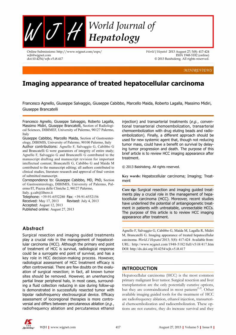
Liver resection with radiofrequency tissue coagulation deviceRadiofrequency (RF)-assisted liver resection technique uses RF energy to obtain parenchymal dissection by cre-ating a zone of coagulative necrosis along the transection plane[6]. This reduces the risk of intraoperative blood loss when compared with conventional liver resection[6]. Liver resection is indicated in patients with preserved liver function and single HCC, ideally in a subcapsular location[1]. Resected area must be larger than the original tumor and with a margin greater than 5 mm[7]. On CT, resection margin typically appears as an hypoattenuat-ing, non enhancing halo[8,9] (Figure 1). On MR, resection margin is commonly hyperintense on T2-weighted im-ages and hypointense on T1-weighted images and does not enhance after gadolinium injection[8,9]. Residual viable tissue, when present, is usually located along the resec-tion site and shows arterial enhancement and venous wash-out[8,9]. A fluid collection within the resected area is
commonly found at initial follow-up and disappears with time[9]. Uncommon complications include biloma, hepatic abscess, pleural effusion and adjacent organ injury (i.e., small bowel perforation).
Percutaneous ablationPercutaneous ablation induces coagulative necrosis by modifying tumor temperature using RF, microwave, laser, cryotherapy or by direct intratumoral injection of chemi-cal substances (ethanol or acetic acid). This procedure is recommended for patients with preserved liver function and a maximum of 3 small (≤ 3 cm) HCCs[1].
RF ablation: RF ablation (RFA) is the most widely used ablation therapy. RFA ablation consists of placing a nee-dle electrode directly into the tumor, by US or CT guid-ance, and heating tissue to temperatures exceeding 60 ℃[10] to obtain the coagulative necrosis of the tumor. Similarly to resected area, ablation zone should be 5-10 mm larger in comparison to the preexisting tumor[11]. Due to a coag-ulative necrosis and hemorrhagic products, treated HCC is typically hypoattenuating or heterogeneously hyperat-tenuating on unenhanced CT. Contrast enhanced images help differentiate viable from necrotic tumor. In general, successfully treated HCC shows absence of arterial en-hancement (Figure 2), whereas any nodular arterially enhancing area within or along the margin of the ablated zone is suspicious of viable tumor[10]. Moreover, patients treated with RFA are considered at higher risk in com-parison to the general cirrhotic population for the occur-rence of new foci of HCC (Figure 3). However, absence of arterial enhancement at CT does not imperatively cor-respond to absence of viable tissue[11]. Lu et al[11] reported 100% specificity and 36% sensitivity of CT for the depic-tion of residual or recurrent tumor. In their study, only 4/11 (37%) HCCs with positive histological findings were detected at CT[11]. These observations are in agreement with a study by Dromain et al[12] who also found that MRI allowed earlier detection of residual tumor than does CT. At MR, successfully treated HCC shows T2-hypointensity and strong T1-hyperintensity[12]. The inclusion of sub-traction images in the MR protocol increases detection of residual arterial enhancement in those cases of HCC with spontaneuous T1 hyperintensity[13]. At initial follow-up studies, treated HCC may show a thin and peripheral
Agnello F et al . Imaging in treated hepatocellular carcinoma
418 August 27, 2013|Volume 5|Issue 8|WJH|www.wjgnet.com
Figure 1 Successful hepatocellular carcinoma resection with radiofrequency tissue coagulation device. A: Pretreatment arterial phase T1-weighted gradient-echo magnetic resonance shows hypervas-cular hepatocellular carcinoma (HCC) (arrow); B: On arterial phase computed tomography obtained 4 mo after treatment no hypervascular focus is evident (ar-row). Note that resected area is larger to preexisting HCC.
A B

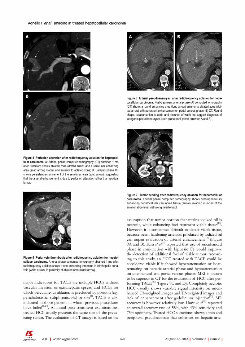
arterially enhancing rim, due to inflammatory reaction to thermal necrosis[14,15]. This rim sometimes shows hypoat-tenuation on unenhanced CT and mild hypertintensity on T2-weighted images. More rarely, needle electrode placing can cause formation of arterio-venous shunts[14,15]. These shunts can be easily diagnosed on the basis of wedge-shaped morphology, peripheral location and lack of wash out on portal venous phase (Figure 4). However, if the arterially enhancing zone is small and nodular, a correct characterization may be difficult and a 3-6 mo follow-up is recommended[16]. Size increase of arterially enhanc-ing area, or the appearance of wash-out may suggest presence of viable tumor. Other complications include biloma, hepatic abscess, portal vein thrombosis (Figure 5), arterial pseudoaneurysm (Figure 6), tumor seeding (Figure 7) and adjacent organ injury[17].
Percutaneous ethanol injection: Percutaneous ethanol injection (PEI) consists of percutaneous ethanol instilla-tion into the HCC by sonographic or CT guidance. It is a well-tolerated, inexpensive procedure with few adverse effects. This alternative procedure may be performed in those patients with small HCCs, in whom RFA is not suitable to be performed due to tumor location[18]. In fact, some tumors are located at risky sites (defined as less than 5 mm from a large vessel or an extrahepatic organ, near gallbladder, or in subphrenic locations) and RFA treatment can cause severe complications. In addi-tion, in tumors larger than 2 cm in size, initial RF may leave a tiny nest of viable tissue that will easily be ablated by ethanol with a relevant saving of resources. However, several studies demonstrated that PEI provides a lower rate of complete necrosis in comparison to RFA[19-21]. CT and MR post-treatment imaging features are similar to those obtained after RFA[22] (Figure 8).
Transarterial chemoembolizationTransarterial chemoembolization (TACE) consists of transarterial administration of a mixture of chemotherapy
(Doxorubicin or Cisplatin in most cases) and iodized oil (Lipiodol, Guerbert, France), followed by emboliz-ing particles[23]. Since HCC receives blood supply almost completely from the hepatic artery (as opposed to nor-mal liver), these agents accumulate preferentially into HCC lesions. Iodized oil acts as chemotherapy carrier, while particles occlude tumor feeding arteries. Therefore, TACE combines delivery of high dose chemotherapy to the tumor with embolization of its feeding arteries. The
419 August 27, 2013|Volume 5|Issue 8|WJH|www.wjgnet.com
A B
Figure 2 Complete necrosis after radiofrequency ablation for hepatocel-lular carcinoma. A: Pretreatment arterial phase computed tomography (CT) shows hypervascular hepatocellular carcinoma (arrow); B: Arterial phase CT obtained 1 mo after treatment shows ablated area (arrow). Absence of arterial enhancement suggests complete tumor necrosis.
A
B
C
Figure 3 Hepatocellular carcinoma after multiple radiofrequency abla-tions. A: Arterial phase computed tomography (CT) obtained 8 mo after radio-frequency ablation shows two new enhancing hepatocellular carcinoma (HCC) nodules located anteriorly (dotted arrow) and posteriorly (short arrow) to ablated HCC (long arrow). These findings suggest occurrence of new HCC nodules; B: Arterial phase CT obtained 2 mo after additional radiofrequency (RF) ablation shows that HCC nodule located anteriorly (dotted arrow) has been replaced by a hypoattenuating, non enhancing area (arrow) that is larger than preexisting tumor. These findings suggest complete necrosis. Posteriorly located HCC (short arrow) increased in size; C: Arterial phase CT obtained 2 mo after posterior. HCC had been replaced by a hypoattenuating, nonenhancing ablation area as a result of additional RF ablation. This example shows that RF ablation is a repeatable procedure.
Agnello F et al . Imaging in treated hepatocellular carcinoma
assumption that tumor portion that retains iodized oil is necrotic, while enhancing foci represent viable tissue[10]. However, it is sometimes difficult to detect viable tissue, because beam hardening artefacts produced by iodized oil can impair evaluation of arterial enhancement[10] (Figure 9A and B). Kim et al[25] reported that use of unenhanced phase in conjunction with biphasic CT could improve the detection of additional foci of viable tumor. Accord-ing to this study, an HCC treated with TACE could be considered viable if it showed hyperattenuation or isoat-tenuating on hepatic arterial phase and hypoattenuation on unenhanced and portal venous phases. MRI is known to be superior to CT for the evaluation of HCC after per-forming TACE[26] (Figure 9C and D). Completely necrotic HCC usually shows variable signal intensity on unen-hanced T1-weighted images and T2-weighted images and lack of enhancement after gadolinium injection[27]. MR accuracy is however relatively low. Hunt et al[28] reported an overall accuracy rate of 55%, with 43% sensitivity and 75% specificity. Treated HCC sometimes shows a thin and peripheral pseudocapsule that enhances on hepatic arte-
major indications for TACE are multiple HCCs without vascular invasion or extrahepatic spread and HCCs for which percutaneous ablation is precluded by position (e.g., pericholecistic, subphrenic, etc.) or size[1]. TACE is also indicated in those patients in whom previous procedures have failed[1,24]. At initial post-treatment examinations, treated HCC usually presents the same size of the preex-isting tumor. The evaluation of CT images is based on the
420 August 27, 2013|Volume 5|Issue 8|WJH|www.wjgnet.com
Figure 5 Portal vein thrombosis after radiofrequency ablation for hepato-cellular carcinoma. Arterial phase computed tomography obtained 1 mo after radiofrequency ablation shows a non enhancing thrombus in intrahepatic portal vein (white arrow), in proximity of ablated area (black arrow).
A B
Figure 6 Arterial pseudoaneurysm after radiofrequency ablation for hepa-tocellular carcinoma. Post-treatment arterial phase (A) computed tomography (CT) shows a round enhancing area (long arrow) anterior to ablated zone (dot-ted arrow) with persistent enhancement on portal venous phase (B) CT. Round shape, isoattenuation to aorta and absence of wash-out suggest diagnosis of iatrogenic pseudoaneurysm. Note probe track (short arrow on A and B).
Figure 7 Tumor seeding after radiofrequency ablation for hepatocellular carcinoma. Arterial phase computed tomography shows heterogeneously enhancing hepatocellular carcinoma tissue (arrow) invading muscles of the anterior abdominal wall along needle tract.
A
B
Figure 4 Perfusion alteration after radiofrequency ablation for hepatocel-lular carcinoma. A: Arterial phase computed tomography (CT) obtained 1 mo after treatment shows ablated zone (dotted arrow) and a semilunar enhancing area (solid arrow) medial and anterior to ablated zone; B: Delayed phase CT shows persistent enhancement of the semilunar area (solid arrow), suggesting that the arterial enhancement is due to perfusion alteration rather than residual tumor.
Agnello F et al . Imaging in treated hepatocellular carcinoma

rial and delayed phases. Moreover, iodized oil can some-times injury small hepatic arteries and cause formation of arterio-portal shunts. Absence of wash-out is crucial to differentiate these pseudolesions from residual viable tumor. Other complications include hepatic artery injuries (dissection or thrombosis), biloma, hepatic abscess and embolization of non target vessels[29]. Non target vessels include arteries that arise from the hepatic circulation (gastroduodenal, right gastric, accessory left gastric, cystic arteries)[30]. Embolization of these vessels results in gas-trointestinal ulcers, skin ulcerations, and, rarely, ischemic
cholecystitis[29,30]. Furthermore, although the purpose of TACE is to obtain a selective intratumoral delivery of chemioterapy, several studies demonstrated that many pa-tients may have higher plasma levels of chemotherapeutic agents with systemic toxic effects[31]. Therefore, alternative techniques that increase the precision of drug delivery are required. TACE with drug eluting beads (DEB) is recently emerging as an alternative option to conventional TACE. TACE with DEB consists of transarterial injection of microspheres that sequester chemotherapy immediately before administration and release it into the tumor in a sustained and controlled manner[18]. In addition, absence of iodized oil does not mask arterial enhancement[32]
421 August 27, 2013|Volume 5|Issue 8|WJH|www.wjgnet.com
Figure 8 Incomplete hepatocellular carcinoma necrosis after percutaneous ethanol injec-tion. Arterial (A) and portal venous (B) computed tomography obtained 1 mo after treatment shows that approximately 10%-20% of the tumor, located in the dorsal and lateral portion of the treated, hy-poattenuating area, is still viable as demonstrated by the presence of enhancement in arterial phase and hypoattenuation (“washout”) on portal venous phase (dotted arrow on A and B). The majority of tumor (solid arrow) does not show enhancement as a result of the treatment.
A B
A B
C D
Figure 9 Incomplete hepatocellular carcinoma necrosis after transarte-rial chemoembolization. Arterial (A) and portal venous (B) phase computed tomography (CT) obtained 1 mo after transarterial chemoembolization (TACE) shows that hepatocellular carcinoma (HCC) is entirely replaced by Lipiodol ac-cumulation (arrow). No evidence of residual tumor was found. Arterial (C) and portal venous (D) phase T1-weighted gradient-echo magnetic resonance (MR) obtained 1 wk after CT shows residual viable tumor (arrow) in the posterolateral portion of the tumor. This case shows higher accuracy of MR in comparison to CT in assessing HCC response after TACE.
A
B
Figure 10 Complete hepatocellular carcinoma necrosis after transarterial chemoembolization with drug-eluting beads. A: Pretreatment arterial phase computed tomography (CT) shows a hypervascular hepatocellular carcinoma (HCC) (arrow); B: Arterial phase CT obtained 3 mo after transarterial chemoem-bolization shows a hypoattenuating, non enhancing nodule (arrow). Absence of arterial enhancement suggests complete HCC necrosis.
Agnello F et al . Imaging in treated hepatocellular carcinoma

(Figure 10), allowing easier assessment of residual, viable tumor in comparison to traditional TACE performed with iodized oil.
RadioembolizationAn alternative technique to treat advanced HCC is radio-embolization. It consists of transarterial administration of yttrium-90 microspheres that are preferentially depos-ited within hypervascular tumors and that emit lethal beta radiation[18]. This induces tumor coagulative necrosis and avascularity. Post-treatment HCC appearance is similar to that obtained following RFA and PEI[33].
Targeted therapyTargeted therapies are a new generation of anticancer drugs designed to interfere with tumor growth and pro-gression. Sorafenib, a multikinase inhibitor, is the only drug that has been demonstrated to significantly improve survival in patients with untreatable, unresectable HCCs in a large randomized controlled trial[3]. Sorafenib reduces tumor vascularization and subsequently induces tumor necrosis and hemorrhage[3] (Figure 11). For these rea-sons, the traditional WHO and RECIST criteria based on evaluation of tumor size may not be ideal to determine tumor response. Therefore, different criteria based on assessment of viable and enhancing tumor are needed. Modified RECIST (mRECIST) differ from RECIST criteria, since they consider only the diameter of viable
tumor, defined as the portion of the tumor that shows arterial enhancement and venous wash-out[5,34]. Recent studies have reported the potential of perfusion CT and MR[35,36]. However, high CT radiation dose, MR breath-ing, cardiac motion and high costs limit the use of these techniques[35,37,38].
CONCLUSIONAdvances in HCC treatment have led to an increased use of therapeutic tools such as RF and percutaneous abla-tion, TACE and antiangiogenetic therapy. In this scenario, CT and MR play an important role in the assessment of response to treatment. Therefore, every radiologist and hepatologist should be familiar with the HCC appearance after treatment and should be able to distinguish normal post-treatment changes from residual or recurrent tumor.
REFERENCES1 Bruix J, Sherman M. Management of hepatocellular car-
cinoma: an update. Hepatology 2011; 53: 1020-1022 [PMID: 21374666 DOI: 10.1002/hep.24199]
2 Genco C, Cabibbo G, Maida M, Brancatelli G, Galia M, Alessi N, Butera G, Genova C, Romano P, Raineri M, Giar-ratano A, Midiri M, Cammà C. Treatment of hepatocellular carcinoma: present and future. Expert Rev Anticancer Ther 2013; 13: 469-479 [PMID: 23560841 DOI: 10.1586/era.13.21]
3 Llovet JM, Ricci S, Mazzaferro V, Hilgard P, Gane E, Blanc JF, de Oliveira AC, Santoro A, Raoul JL, Forner A, Schwartz
422 August 27, 2013|Volume 5|Issue 8|WJH|www.wjgnet.com
A B
C D
Figure 11 Hepatocellular carcinomas after sorafenib. A: Pretreatment arterial phase computed tomography (CT) shows a large hypervascular hepatocellular carci-noma (HCC) (arrow) in right hepatic lobe; B: Arterial phase CT obtained at the same level of A, 9 mo after the start of sorafenib, shows increase in size of HCC (arrow); C: Pretreatment arterial phase CT image shows a second, small hypervascular HCC (arrow) arising within a larger hypoattenuating dysplastic nodule in left hepatic lobe; D: Arterial phase CT obtained at the same level of C, 9 mo after the start of sorafenib, shows disappearance of HCC enhancement (arrow). This case shows that different HCC response to sorafenib may occur in the same patient.
Agnello F et al . Imaging in treated hepatocellular carcinoma

M, Porta C, Zeuzem S, Bolondi L, Greten TF, Galle PR, Seitz JF, Borbath I, Häussinger D, Giannaris T, Shan M, Moscovici M, Voliotis D, Bruix J. Sorafenib in advanced hepatocellular carcinoma. N Engl J Med 2008; 359: 378-390 [PMID: 18650514 DOI: 10.1056/NEJMoa0708857]
4 Iavarone M, Cabibbo G, Piscaglia F, Zavaglia C, Grieco A, Villa E, Cammà C, Colombo M. Field-practice study of sorafenib therapy for hepatocellular carcinoma: a pro-spective multicenter study in Italy. Hepatology 2011; 54: 2055-2063 [PMID: 21898496 DOI: 10.1002/hep.24644]
5 Maida M, Cabibbo G, Brancatelli G, Genco C, Alessi N, Genova C, Romano P, Raineri M, Giarratano A, Midiri M, Cammà C. Assessment of treatment response in hepatocel-lular carcinoma: a review of the literature. Future Oncol 2013; 9: 845-854 [PMID: 23718305 DOI: 10.2217/fon.13.33]
6 Curro G, Jiao L, Scisca C, Baccarani U, Mucciardi M, Habib N, Navarra G. Radiofrequency-assisted liver resection in cir-rhotic patients with hepatocellular carcinoma. J Surg Oncol 2008; 98: 407-410 [PMID: 18683211 DOI: 10.1002/jso.21129]
7 Kleinert R, Wahba R, Bangard C, Prenzel K, Hölscher AH, Stippel D. Radiomorphology of the Habib sealer-induced resection plane during long-time followup: a longitudinal single center experience after 64 radiofrequency-assisted liv-er resections. HPB Surg 2010; 2010: 403097 [PMID: 20862384 DOI: 10.1155/2010/403097]
8 McGahan JP, Khatri VP. Imaging findings after liver resec-tion by using radiofrequency parenchymal coagulation de-vices: initial experiences. Radiology 2008; 247: 896-902 [PMID: 18487541 DOI: 10.1148/radiol.2473070949]
9 Taibbi A, Furlan A, Sandonato L, Bova V, Galia M, Marin D, Cabibbo G, Soresi M, Bartolotta TV, Midiri M, Lagalla R, Brancatelli G. Imaging findings of liver resection using a bipolar radiofrequency electrosurgical device--initial obser-vations. Eur J Radiol 2012; 81: 663-670 [PMID: 21306849 DOI: 10.1016/j.ejrad.2011.01.015]
10 Yaghmai V, Besa C, Kim E, Gatlin JL, Siddiqui NA, Taouli B. Imaging assessment of hepatocellular carcinoma response to locoregional and systemic therapy. AJR Am J Roentgenol 2013; 201: 80-96 [PMID: 23789661 DOI: 10.2214/AJR.13.10706]
11 Lu DS, Yu NC, Raman SS, Limanond P, Lassman C, Mur-ray K, Tong MJ, Amado RG, Busuttil RW. Radiofrequency ablation of hepatocellular carcinoma: treatment success as defined by histologic examination of the explanted liver. Radiology 2005; 234: 954-960 [PMID: 15681691 DOI: 10.1148/radiol.2343040153]
12 Dromain C, de Baere T, Elias D, Kuoch V, Ducreux M, Boige V, Petrow P, Roche A, Sigal R. Hepatic tumors treated with percutaneous radio-frequency ablation: CT and MR imag-ing follow-up. Radiology 2002; 223: 255-262 [PMID: 11930075 DOI: 10.1148/radiol.2231010780]
13 Winters SD, Jackson S, Armstrong GA, Birchall IW, Lee KH, Low G. Value of subtraction MRI in assessing treatment response following image-guided loco-regional therapies for hepatocellular carcinoma. Clin Radiol 2012; 67: 649-655 [PMID: 22300821 DOI: 10.1016/j.crad.2011.11.013]
14 Jiang T, Zhu AX, Sahani DV. Established and novel imaging biomarkers for assessing response to therapy in hepatocel-lular carcinoma. J Hepatol 2013; 58: 169-177 [PMID: 22944253 DOI: 10.1016/j.jhep.2012.08.022]
15 Sainani NI, Gervais DA, Mueller PR, Arellano RS. Imag-ing after percutaneous radiofrequency ablation of hepatic tumors: Part 1, Normal findings. AJR Am J Roentgenol 2013; 200: 184-193 [PMID: 23255761 DOI: 10.2214/AJR.12.8478]
16 Park Y, Choi D, Lim HK, Rhim H, Kim YS, Kim SH, Lee WJ. Growth rate of new hepatocellular carcinoma after percuta-neous radiofrequency ablation: evaluation with multiphase CT. AJR Am J Roentgenol 2008; 191: 215-220 [PMID: 18562748 DOI: 10.2214/AJR.07.3297]
17 Sainani NI, Gervais DA, Mueller PR, Arellano RS. Imaging after percutaneous radiofrequency ablation of hepatic tu-
mors: Part 2, Abnormal findings. AJR Am J Roentgenol 2013; 200: 194-204 [PMID: 23255762 DOI: 10.2214/AJR.12.8479]
18 Lencioni R, Crocetti L. Local-regional treatment of hepa-tocellular carcinoma. Radiology 2012; 262: 43-58 [PMID: 22190656 DOI: 10.1148/radiol.11110144]
19 Orlando A, Leandro G, Olivo M, Andriulli A, Cottone M. Radiofrequency thermal ablation vs. percutaneous ethanol injection for small hepatocellular carcinoma in cirrhosis: meta-analysis of randomized controlled trials. Am J Gastro-enterol 2009; 104: 514-524 [PMID: 19174803 DOI: 10.1038/ajg.2008.80]
20 Oeda S, Mizuta T, Isoda H, Kuwashiro T, Iwane S, Taka-hashi H, Kawaguchi Y, Eguchi Y, Ozaki I, Tanaka K, Fuji-moto K. Survival advantage of radiofrequency ablation for hepatocellular carcinoma: Comparison with ethanol injec-tion. Hepatogastroenterology 2013 May 1; Epub ahead of print [PMID: 23635508 DOI: 10.5754/hge121270]
21 Cho YK, Kim JK, Kim MY, Rhim H, Han JK. Systematic review of randomized trials for hepatocellular carcinoma treated with percutaneous ablation therapies. Hepatology 2009; 49: 453-459 [PMID: 19065676 DOI: 10.1002/hep.22648]
22 Bartolozzi C, Lencioni R, Caramella D, Mazzeo S, Ciancia EM. Treatment of hepatocellular carcinoma with percutane-ous ethanol injection: evaluation with contrast-enhanced MR imaging. AJR Am J Roentgenol 1994; 162: 827-831 [PMID: 8141000]
23 Brown DB, Gould JE, Gervais DA, Goldberg SN, Murthy R, Millward SF, Rilling WS, Geschwind JF, Salem R, Vedan-tham S, Cardella JF, Soulen MC. Transcatheter therapy for hepatic malignancy: standardization of terminology and reporting criteria. J Vasc Interv Radiol 2009; 20: S425-S434 [PMID: 19560030 DOI: 10.1016/j.jvir.2009.04.021]
24 Cabibbo G, Genco C, Di Marco V, Barbara M, Enea M, Parisi P, Brancatelli G, Romano P, Craxì A, Cammà C. Pre-dicting survival in patients with hepatocellular carcinoma treated by transarterial chemoembolisation. Aliment Phar-macol Ther 2011; 34: 196-204 [PMID: 21564144 DOI: 10.1111/j.1365-2036.2011.04694.x]
25 Kim HC, Kim AY, Han JK, Chung JW, Lee JY, Park JH, Choi BI. Hepatic arterial and portal venous phase helical CT in patients treated with transcatheter arterial chemo-embolization for hepatocellular carcinoma: added value of unenhanced images. Radiology 2002; 225: 773-780 [PMID: 12461260]
26 Kloeckner R, Otto G, Biesterfeld S, Oberholzer K, Dueber C, Pitton MB. MDCT versus MRI assessment of tumor response after transarterial chemoembolization for the treatment of hepatocellular carcinoma. Cardiovasc Intervent Radiol 2010; 33: 532-540 [PMID: 19847482 DOI: 10.1007/s00270-009-9728-y]
27 Mannelli L, Kim S, Hajdu CH, Babb JS, Clark TW, Taouli B. Assessment of tumor necrosis of hepatocellular carcinoma after chemoembolization: diffusion-weighted and contrast-enhanced MRI with histopathologic correlation of the explanted liver. AJR Am J Roentgenol 2009; 193: 1044-1052 [PMID: 19770328 DOI: 10.2214/AJR.08.1461]
28 Hunt SJ, Yu W, Weintraub J, Prince MR, Kothary N. Radio-logic monitoring of hepatocellular carcinoma tumor viabil-ity after transhepatic arterial chemoembolization: estimating the accuracy of contrast-enhanced cross-sectional imaging with histopathologic correlation. J Vasc Interv Radiol 2009; 20: 30-38 [PMID: 19028117 DOI: 10.1016/j.jvir.2008.09.034]
29 Clark TW. Complications of hepatic chemoembolization. Semin Intervent Radiol 2006; 23: 119-125 [PMID: 21326755 DOI: 10.1055/s-2006-941442]
30 Lee AJ, Gomes AS, Liu DM, Kee ST, Loh CT, McWilliams JP. The road less traveled: importance of the lesser branches of the celiac axis in liver embolotherapy. Radiographics 2012; 32: 1121-1132 [PMID: 22786998 DOI: 10.1148/rg.324115114]
31 Lammer J, Malagari K, Vogl T, Pilleul F, Denys A, Wat-kinson A, Pitton M, Sergent G, Pfammatter T, Terraz S,
423 August 27, 2013|Volume 5|Issue 8|WJH|www.wjgnet.com
Agnello F et al . Imaging in treated hepatocellular carcinoma

Benhamou Y, Avajon Y, Gruenberger T, Pomoni M, Lan-genberger H, Schuchmann M, Dumortier J, Mueller C, Chevallier P, Lencioni R. Prospective randomized study of doxorubicin-eluting-bead embolization in the treatment of hepatocellular carcinoma: results of the PRECISION V study. Cardiovasc Intervent Radiol 2010; 33: 41-52 [PMID: 19908093 DOI: 10.1007/s00270-009-9711-7]
32 Chung WS, Lee KH, Park MS, Lee YJ, Kwon J, Baek SE, Kim MJ. Enhancement patterns of hepatocellular carcinoma after transarterial chemoembolization using drug-eluting beads on arterial phase CT images: a pilot retrospective study. AJR Am J Roentgenol 2012; 199: 349-359 [PMID: 22826396 DOI: 10.2214/AJR.11.7563]
33 Atassi B, Bangash AK, Bahrani A, Pizzi G, Lewandowski RJ, Ryu RK, Sato KT, Gates VL, Mulcahy MF, Kulik L, Miller F, Yaghmai V, Murthy R, Larson A, Omary RA, Salem R. Multimodality imaging following 90Y radioembolization: a comprehensive review and pictorial essay. Radiographics 2008; 28: 81-99 [PMID: 18203932 DOI: 10.1148/rg.281065721]
34 Lencioni R, Llovet JM. Modified RECIST (mRECIST) assess-ment for hepatocellular carcinoma. Semin Liver Dis 2010; 30:
52-60 [PMID: 20175033 DOI: 10.1055/s-0030-1247132]35 Zhu AX, Chabner BA, Tanabe KK. New trends and novel
treatment for hepatocellular carcinoma: a global perspec-tive. Oncologist 2010; 15 Suppl 4: 1-4 [PMID: 21115575 DOI: 10.1634/theoncologist.2010-S4-01]
36 Sacco R, Faggioni L, Bargellini I, Ginanni B, Battaglia V, Romano A, Bertini M, Bresci G, Bartolozzi C. Assessment of response to sorafenib in advanced hepatocellular carcinoma using perfusion computed tomography: Results of a pilot study. Dig Liver Dis 2013 Apr 8; Epub ahead of print [PMID: 23578581 DOI: 10.1016/j.dld.2013.03.004]
37 Sabir A, Schor-Bardach R, Wilcox CJ, Rahmanuddin S, Atkins MB, Kruskal JB, Signoretti S, Raptopoulos VD, Goldberg SN. Perfusion MDCT enables early detection of therapeutic response to antiangiogenic therapy. AJR Am J Roentgenol 2008; 191: 133-139 [PMID: 18562736 DOI: 10.2214/AJR.07.2848]
38 Goh V, Halligan S, Hugill JA, Bassett P, Bartram CI. Quan-titative assessment of colorectal cancer perfusion using MDCT: inter- and intraobserver agreement. AJR Am J Roent-genol 2005; 185: 225-231 [PMID: 15972428]
P- Reviewers Chiu KW, Sazci A S- Editor Gou SX L- Editor A E- Editor Ma S
424 August 27, 2013|Volume 5|Issue 8|WJH|www.wjgnet.com
Agnello F et al . Imaging in treated hepatocellular carcinoma

Published by Baishideng Publishing Group Co., LimitedFlat C, 23/F., Lucky Plaza,
315-321 Lockhart Road, Wan Chai, Hong Kong, ChinaFax: +852-65557188
Telephone: +852-31779906E-mail: [email protected]
http://www.wjgnet.com
Baishideng Publishing Group Co., Limited © 2013 Baishideng. All rights reserved.