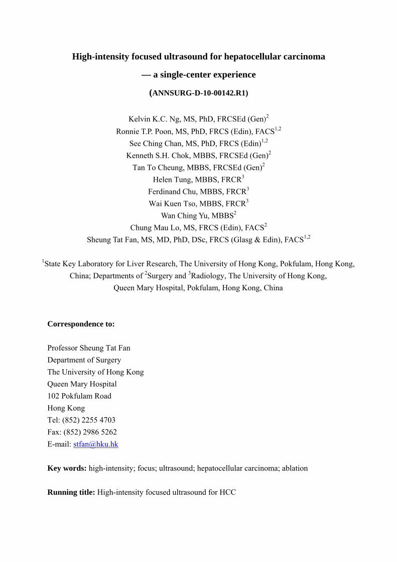
High-intensity focused ultrasound for hepatocellular carcinoma
33
High-intensity focused ultrasound for hepatocellular carcinoma — a single-center experience (ANNSURG-D-10-00142.R1) Kelvin K.C. Ng, MS, PhD, FRCSEd (Gen) 2 Ronnie T.P. Poon, MS, PhD, FRCS (Edin), FACS 1,2 See Ching Chan, MS, PhD, FRCS (Edin) 1,2 Kenneth S.H. Chok, MBBS, FRCSEd (Gen) 2 Tan To Cheung, MBBS, FRCSEd (Gen) 2 Helen Tung, MBBS, FRCR 3 Ferdinand Chu, MBBS, FRCR 3 Wai Kuen Tso, MBBS, FRCR 3 Wan Ching Yu, MBBS 2 Chung Mau Lo, MS, FRCS (Edin), FACS 2 Sheung Tat Fan, MS, MD, PhD, DSc, FRCS (Glasg & Edin), FACS 1,2 1 State Key Laboratory for Liver Research, The University of Hong Kong, Pokfulam, Hong Kong, China; Departments of 2 Surgery and 3 Radiology, The University of Hong Kong, Queen Mary Hospital, Pokfulam, Hong Kong, China Correspondence to: Professor Sheung Tat Fan Department of Surgery The University of Hong Kong Queen Mary Hospital 102 Pokfulam Road Hong Kong Tel: (852) 2255 4703 Fax: (852) 2986 5262 E-mail: [email protected] Key words: high-intensity; focus; ultrasound; hepatocellular carcinoma; ablation Running title: High-intensity focused ultrasound for HCC
-
Upload
khangminh22 -
Category
Documents
-
view
2 -
download
0
Transcript of High-intensity focused ultrasound for hepatocellular carcinoma
High-intensity focused ultrasound for hepatocellular carcinoma
— a single-center experience
(ANNSURG-D-10-00142.R1)
Kelvin K.C. Ng, MS, PhD, FRCSEd (Gen)2
Ronnie T.P. Poon, MS, PhD, FRCS (Edin), FACS1,2
See Ching Chan, MS, PhD, FRCS (Edin)1,2
Kenneth S.H. Chok, MBBS, FRCSEd (Gen)2
Tan To Cheung, MBBS, FRCSEd (Gen)2
Helen Tung, MBBS, FRCR3
Ferdinand Chu, MBBS, FRCR3
Wai Kuen Tso, MBBS, FRCR3
Wan Ching Yu, MBBS2
Chung Mau Lo, MS, FRCS (Edin), FACS2
Sheung Tat Fan, MS, MD, PhD, DSc, FRCS (Glasg & Edin), FACS1,2
1State Key Laboratory for Liver Research, The University of Hong Kong, Pokfulam, Hong Kong,
China; Departments of 2Surgery and 3Radiology, The University of Hong Kong,
Queen Mary Hospital, Pokfulam, Hong Kong, China
Correspondence to:
Professor Sheung Tat Fan
Department of Surgery
The University of Hong Kong
Queen Mary Hospital
102 Pokfulam Road
Hong Kong
Tel: (852) 2255 4703
Fax: (852) 2986 5262
E-mail: [email protected]
Key words: high-intensity; focus; ultrasound; hepatocellular carcinoma; ablation
Running title: High-intensity focused ultrasound for HCC

Mini-Abstract
High-intensity focused ultrasound (HIFU) is a totally non-invasive ablation treatment
for hepatocellular carcinoma. The complete tumor ablation rate was 79.5%. The
1-year survival rate was 87.7%.

Abstract
Objective: This study aims to evaluate the outcome of patients with
hepatocellular carcinoma (HCC) treated by high-intensity focused ultrasound (HIFU)
in a single tertiary referral center.
Summary Background data: HIFU is the latest developed local ablation
technique for unresectable HCC. The initial experience on its efficacy is promising,
but the survival benefit of patients undergoing HIFU for HCC is poorly defined.
Methods: From October 2006 to December 2008, 49 patients received HIFU for
unresectable HCC. Each patient underwent a single session of HIFU with a curative
intent. Treatment efficacy and survival outcome were evaluated. Clinicopathologic
factors affecting the primary technique effectiveness and overall survival rates were
investigated by univariate analysis.
Results: The median size of the treated tumors was 2.2 cm, ranging from 0.9 cm
to 8 cm. The majority of patients had single tumors (n = 41, 83.6%). Thirty one
patients (63.2%) had artificial right pleural effusion during HIFU treatment to reduce
damage to the lung and diaphragm. The hospital mortality rate was 2% (n = 1) and
the complication rate was 8.1% (n = 4). The primary technique effectiveness rate was
79.5% (39 out of 49 patients). It increased from 66.6% in the initial series to 89.2% in
the last 28 patients. Tumor size ( 3.0 cm) was the significant risk factor affecting the

complete ablation rate. The 1- and 3-year overall survival rates were 87.7% and
62.4%, respectively. Child-Pugh liver function grading was the significant prognostic
factor influencing the overall survival rate.
Conclusions: HIFU is an effective treatment modality for unresectable HCC
with a high technique effectiveness rate and favorable survival outcome.

2
Introduction
Hepatocellular carcinoma (HCC) is the most common primary liver cancer, with a high
prevalence in Asia and an increasing incidence in Western countries. With advancement in
technologies, local ablation therapies have emerged as effective treatment options for
unresectable HCC. These include cryoablation therapy, interstitial laser therapy, microwave
coagulation, radiofrequency ablation (RFA), and high-intensity focused ultrasound (HIFU).
Among these treatment options, HIFU is the only treatment modality that is completely
extracorporeal.
HIFU is based on the unique characteristic of ultrasound beams (0.8 – 3.5 MHz), which
can be focused at a distance from the radiating transducer. The accumulated energy at the
focal region induces tissue necrosis of the targeted lesion without causing damage to the
surrounding vital structures. The ability of inducing immediate cell death at a distance from
the ultrasound source without the need of surgery or insertion of ablation instruments makes
HIFU an attractive treatment option for HCC. While data on the efficacy of HIFU in treating
HCC remain scarce, initial clinical results obtained from pioneer researchers in China have
been encouraging.1-7 The reported complete ablation rates ranged from 28.5% to 68%.1,6,7
The short-term survival (one-year) rates ranged from 42.9% to 61.5%.1,4 However, there is no
report on identification of the possible risk factor affecting the complete ablation rate and the
possible prognostic factor influencing the overall survival. Such information is crucial in

3
establishing the role of HIFU in the management of patients with HCC. The present study
aims to evaluate the clinical outcome of patients with HCC treated by HIFU, and investigate
the clinicopathologic factors affecting the complete ablation rate of HIFU and patient
survival.
Methods
Selection of patients
From October 2006 to December 2008, 49 patients with unresectable HCC received
HIFU treatment. Patients with advanced diseases due to tumor invasion to major intrahepatic
blood vessels or extrahepatic metastasis were not selected for the treatment. Patients with
HCC that could not be visualized by diagnostic ultrasound of the HIFU system were also
excluded. Diagnosis of HCC was based on radiological features shown by computed
tomography scan or magnetic resonance imaging (MRI) scan and/or raised serum
-fetoprotein concentration (over 400 g/ml). A tumor was considered unresectable if the
patient had unsatisfactory liver function or if there was a high medical risk for the patient to
undergo hepatic resection. The selection criteria for HIFU were as follows: (1) The maximal
tumor diameter was less than 8 cm. (2) The number of tumor nodules was less than 3. (3) The
tumor could be detected by ultrasound imaging and there were no bowel adjacent to the tumor.
Each selected patient underwent a single session of HIFU aiming at complete ablation of all

4
detected tumors. A total of 57 tumors were ablated. During the initial phase of the study
period (from October 2006 to May 2007), 21 patients had transarterial injection of iodized
poppyseed oil (Lipiodol) into their tumors about two weeks before HIFU treatment because
previous researchers suggested that Lipiodol could reduce tumor blood supply and increase
the deposition of ultrasonic energy in the tumor. In the later phase of the study (from June
2007 to December 2008), 28 patients received HIFU only.
Treatment procedures
The JC HIFU system (Chongqing Haifu Technology, Chongqing, China) was used in
this study. The ablation process was guided by real-time ultrasound imaging. This system is
composed of a real-time diagnostic ultrasound device, an integrated ultrasound therapy
transducer (12 cm in diameter), a six-directional therapeutic planning system, an ultrasound
generator, a degassed water circulation unit, and a computer unit for automated master control.
The focused ultrasound was produced by the transducer operating at 0.8 MHz (aperture 120
mm, focal length 150 mm). The target lesion was identified using a central 3.5-MHz
diagnostic ultrasound probe, which was integrated in the center of the therapeutic transducer.
Both diagnostic and therapeutic ultrasound beams were emitted simultaneously in the same
direction.

5
In patients receiving pre-HIFU Lipiodol deposition into their tumors, hepatic
angiography was performed two weeks before HIFU treatment. Lipiodol was delivered via
selective cannulation of the feeding artery of the target tumor.
HIFU treatment was performed under general anesthesia to alleviate deep visceral pain
caused by HIFU and to ensure immobilization of patients. Temporary inspiratory or
expiratory control by the anesthesiologist helped to minimize liver movement caused by
ventilation during the treatment.8 In selected patients with a tumor at the dome of the liver,
artificial right pleural effusion was induced before treatment. Detailed planning was carried
out according to the tumor size and location as detected by the diagnostic ultrasound
transducer. Parallel slides of the target tumor with 5-mm separation were obtained. Using
provisional therapeutic parameters based on the depth and vascular supply of the target tumor,
tissue of each tumor slide was completely ablated from deep to superficial region by
successful sweeps of the HIFU head. The ablation process was repeated slide by slide to
achieve entire tumor ablation. During the ablation process, grey-scale changes were noted in
the ablation zone, signifying the effectiveness of ablation.
6
Data collection and outcome measures
Clinical details of all 49 patients were prospectively collected in a database. Clinical
parameters included patient demographics, tumor characteristics, and treatment parameters
(total treatment duration and acoustic power). Short-term outcome measures were post-HIFU
complication rate, hospital mortality, and tumor responses. A complication was defined as
any adverse event after HIFU, and hospital mortality was defined as any death in the same
admission for the procedure. Tumor responses were classified as the primary technique
effectiveness rate and secondary technique effectiveness rate, according to the
recommendation by the international Working Group on Image-guided Tumor Ablation9. The
primary technique effectiveness rate was defined as the percentage of tumors that were
successfully eradicated following the initial course of HIFU, whereas the secondary technique
effectiveness rate was defined as the percentage of tumors that have undergone successful
repeat ablation following identification of local tumor progression. Tumor response to HIFU
was assessed by MRI, which was performed one month after the procedure. Successful tumor
ablation was defined as complete absence of hyperintensity signal in T2W images and
absence of contrast enhancement within the original tumor region (Figure 1). Any
contrast-enhancing area within the original tumor region on post-ablation MRI scan indicated
a residual tumor. RFA or chemoembolization was performed in selected patients to treat
7
residual tumors. All patients had monitoring of serum -fetoprotein concentration, chest
radiograph and MRI scan every three months to detect tumor recurrence.
Statistical analysis
Continuous data were expressed as medians with ranges and were compared using the
Mann-Whitney U test. Categorical data were compared using the χ2 test with Yates correction
or Fisher exact test where appropriate. The overall and disease-free survival rates were
calculated by the Kaplan-Meier method and compared between the groups using the log-rank
test. The end-point of disease-free survival was recurrence of HCC at or outside of the
ablation site or death of the patient, but excluding hospital mortality. Clinicopathologic
variables were analyzed for their effects on the primary technique effectiveness and overall
survival rates. Host factors included age, gender, hepatitis B surface antigen status,
anti-hepatitis C antibody status, Child-Pugh liver function grading10, serum bilirubin level,
serum albumin level, platelet count, and previous treatment (hepatectomy, RFA or
transarterial chemoembolization). Tumor factors included maximum tumor size, number of
tumors (solitary vs. multiple) and serum -fetoprotein level. Finally, the practice of pre-HIFU
Lipiodol deposition, the use of artificial right pleural effusion, and the primary and secondary
technique effectiveness were included in the analysis of overall survival. For the significant
continuous variables identified by the univariate analysis, the cut-off value was determined

8
using the discriminant analysis. All statistical analyses were performed using the statistical
software SPSS 11.0 for Windows (SPSS Inc., Chicago, IL). A P-value of less than 0.05 was
considered statistically significant.
Results
Patient characteristics
The demographic and clinicopathologic data of all 49 patients treated by HIFU are
shown in Table 1. Among the 49 patients, 37 patients (76%) were hepatitis B carriers and 7
patients (14%) were hepatitis C carriers. The majority of patients (n = 41, 83.6%) had
preserved liver function (Child-Pugh class A). These patients had high medical risks
(presence of co-morbidities) to undergo hepatic resection for HCC. Another 8 patients had
marginal liver function (Child-Pugh class B) that prohibited major hepatic resection of tumors.
Liver transplantation was not offered to them because of the local policy of offering scarce
deceased organs to patients with Child-Pugh class C liver function only. HIFU was performed
for intrahepatic recurrent tumors in 17 patients and 14 patients following hepatectomy and
RFA, respectively. Eighteen patients (36%) received HIFU for tumors that failed previous
transarterial chemoembolization. Twenty-eight patients (57.1%) received HIFU as the
primary treatment for their newly diagnosed HCC. The median tumor size was 2.2 cm (range,
0.9 – 8 cm). The majority of patients (n = 41, 83.6%) had solitary tumors. Artificial right

9
pleural effusion was induced before HIFU treatment in 31 patients (63%) with tumors near to
the diaphragm to reduce damage to the lung and diaphragm by heat generated during the
treatment. The median HIFU treatment duration was 26 minutes (range, 3 – 124 minutes).
Short-term outcome
One patient in the early part of the series died of myocardial infarction one day after
HIFU treatment. This patient had underlying ischemic heart disease, which was not diagnosed
before the HIFU treatment. The hospital mortality rate was 2%. The treatment-related
complication rate was 8.1% (n = 4). The complications included first-degree and
second-degree skin burn around the treatment zone in 2 patients and 1 patient, respectively.
These complications were due to an error of using high acoustic power. One patient
developed bruising over the right chest wall, extending to the right loin region. This was
caused by bleeding from intercostal vessels that were injured during induction of artificial
pleural effusion. The median hospital stay was 4 days (range, 2 – 16 days) (Table 2).
Ten patients (20.4%) had residual tumors detected by MRI one month after the treatment.
The primary technique effectiveness rate was 79.5% (39 of 49 patients). Taking into
consideration the total number of ablated tumor nodules, the primary technique effectiveness
rate was 82.4% (47 out of 57 nodules). Four patients received percutaneous RFA treatment
for residual tumors and three of them were rendered tumor-free after RFA. Another 4 patients
10
having residual tumors underwent transarterial chemoembolization but tumor control was
incomplete. The overall secondary technique effectiveness rate after HIFU and RFA was
85.7% (42 out of 49 patients).
Table 3 showed the results of univariate analysis on the possible risk factors for
incomplete ablation after HIFU treatment. Tumor size was the only significant risk factor
affecting the primary technique effectiveness rate of HIFU. Patients with primary technique
effectiveness had significantly smaller tumors than those with residual tumors after HIFU
treatment (median tumor size: 2.29 cm vs. 3.75 cm, P = 0.013). The cut-off value of tumor
size as the significant risk factor for incomplete ablation by HIFU was 3.0 cm. The primary
technique effectiveness for tumors < 3.0 cm was 90.6% (29 out of 32 patients), whereas that
for tumors 3.0 cm was 58.8% (10 out of 17 patients). The primary technique effectiveness
rate of patients with HIFU alone (89.2%) was higher than that of those with HIFU and
pre-treatment Lipiodol deposition (66.6%), although the difference was not significant
statistically.
Tumor recurrence and survival outcome
The median follow-up period was 24 months (range, 3 – 38 months). Among the 42
patients with tumors completely ablated by primary and secondary techniques, 9 patients
(21.4%) developed local recurrence at the HIFU treatment zone. The long-term local tumor

11
control rate is 67%. All the 9 patients with local tumor recurrence belonged to the group in
which HIFU treatment was preceded by Lipiodol deposition. On the other hand, all patients
treated by HFIU alone did not develop local tumor recurrence at the site of HIFU treatment.
Seventeen patients (40.4%) had intrahepatic tumor recurrence (away from the ablation site).
Four patients (9.5%) developed extrahepatic metastasis. The overall recurrence rate was
61.9% (26 out of 42 patients). The 1- and 3-year overall survival rates were 87.7% and 62.4%,
respectively (Figure 2). The 1- and 3-year disease-free survival rates were 40.7% and 0%,
respectively (Figure 3). Among the clinicopathologic factors, Child-Pugh grade was the only
significant prognostic factors influencing overall survival. The overall 1- and 3-year survival
rates of the patients with Child-Pugh class A were 90.2% and 68.5%, respectively, whereas
those of the patients with Child-Pugh class B were 75% and 33.3%, respectively (P = 0.028).
The overall survival rates of patients with secondary technique effectiveness (1-year survival
rate: 92.9%; 3-year survival rate: 66.8%) were better than those of the patients with residual
tumors after sequential local ablation (1-year survival rate: 53.6%; 3-year survival rate:
35.7%) (P=0.06) (Table 4).
Discussion
HIFU is a newly developed non-invasive treatment modality for liver tumors. Compared
with other local ablation therapies, HIFU treatment has the major advantage of being totally

12
extracorporeal without the need of insertion of any ablation needle in the target lesion. With
high acoustic intensities (up to 10,000 Watt/cm2), HIFU induces instantaneous cell death by
two major mechanisms, namely, thermal effect and mechanical effect.11 The thermal effect of
HIFU features heat generation due to absorption of acoustic energy by the target tissue. A
lethal temperature of up to 60C causes coagulative necrosis within a few seconds. Since
high-intensity energy is focused at a small volume, damage to tissues between the transducer
and the target lesion is minimized. The mechanical effect involves cavitation12,
microstreaming13 and radiation forces14. With these destructive mechanisms, irreversible cell
death occurs through coagulative necrosis and apoptosis.
The application of HIFU technology in the management of patients with HCC is still in
its infancy period. The feasibility and safety of HIFU for liver tumors were initially
demonstrated in the early 1990s.11 However, this technology has not gained much enthusiasm,
primarily because of the difficulties in tumor targeting and monitoring of the ablation process.
However, with recent advances in the ultrasound technology, the accuracy of targeting of
HIFU has improved considerably.
The initial experience of HIFU for HCC was obtained from researchers in China using
the JC HIFU system, which was also used in the present study. In a study by Wu et al1 in
which 55 patients with large HCC (with a mean diameter of 8.14 cm) and cirrhosis received
HIFU treatment, no major complications were recorded. Completeness of ablation was

13
assessed in 26 patients and the complete ablation rate was 69.2%. The overall survival rates
were 61.5% at 12 months and 35.3% at 18 months. In another study by the same group4, the
efficacy of HIFU combined with chemoembolization was compared with that of
chemoembolization alone in 50 patients with advanced HCC. Patients who underwent
combined treatment had significantly better survival than those who received
chemoembolization alone. In the Western population, the efficacy of this HIFU system in
treating liver tumors has also been validated.15 In this study, the effectiveness of HIFU was
confirmed and a higher primary technique effectiveness rate (79.5%) was achieved.
Although the treatment efficacy and survival benefits of HIFU for patients with liver
cancer were well documented in the previous studies1-7, clinicopathologic factors influencing
the completeness of tumor ablation and patient survival were not studied in detail. We found
that tumor size ( 3.0 cm) was a significant risk factor accounting for incomplete tumor
ablation after HIFU. Although HIFU has the merit of being extracorporeal in nature, the
portion of skin and subcutaneous tissue along the pathway of focused ultrasound was
frequently affected, causing tissue edema. As large tumors require longer ablation time by the
HIFU machine, the resulting cutaneous and subcutaneous tissue edema will reduce the
targeting ability of the diagnostic ultrasound of the HIFU machine. Hence, the precision of the
deposition of focused ultrasound onto the target lesion will be negatively affected. As shown
in our study, patients with residual tumors after a single session of HIFU treatment had

14
significantly larger tumors (median tumor size: 3.75 cm vs. 2.29 cm). Large tumors may,
therefore, need “planned” repeated HIFU treatment or a second treatment once a residual
lesion is detected by imaging.
Apart from tumor size, the use of pre-treatment Lipiodol deposition was another possible
factor affecting the completeness of tumor ablation. In the treatment protocol of HIFU
designed by researchers in China, Lipiodol deposition via hepatic angiography is usually
combined with this ablation technique.1,4 Theoretically, Lipiodol deposition in the tumor can
increase the ablation volume of HIFU by two possible mechanisms. First, tumor blood flow
decreases after Lipiodol occlusion of tumor microvasculatures, resulting in reduced heat loss
during thermal treatment by HIFU. Second, Lipiodol deposition in the tumor causes increased
deposition of ultrasonic energy.16 Nevertheless, the administration of Lipiodol shortly before
HIFU is not without disadvantages. In fact, non-specific deposition of Lipiodol within the
same liver segment as the target tumor invariably affected ultrasound localization of the
tumor and hence the targeting accuracy of HIFU because the affected liver segment was also
filled with Lipiodol (Figure 4). In such instance, tumor margins could only be poorly defined
by the diagnostic ultrasound. Moreover, the non-tumorous liver parenchyma within the
acoustic window might be involved in the HIFU ablation process because non-specific
deposits of Lipiodol could absorb high-intensity ultrasonic energy. It could be disastrous if
any vital vasculatures or the biliary system were within this acoustic window.
15
We postulated that HIFU without Lipiodol deposition might be more effective than the
combined-treatment approach in terms of completeness of ablation, provided that tumor
margins could be clearly defined by the HIFU system. With this assumption, we modified the
treatment protocol of HIFU in the later phase of our study by adopting the HIFU-alone
treatment. The effectiveness of HIFU-alone approach was supported by the finding of our
study. The primary technique effectiveness rate of patients with HIFU alone (89.2%) was
higher than that of those with HIFU and pre-treatment Lipiodol deposition (66.6%, P = 0.076).
Such high complete ablation rate is comparable with that achieved by RFA17-21, the most
commonly used local ablation technique for HCC at present. It should be emphasized that
meticulous techniques are necessary to ensure complete ablation of the tumor in the
HIFU-alone approach. It is important to carry out pre-HIFU planning using diagnostic
ultrasound to ensure that the liver tumor is clearly visible before administering HIFU. We
believe that complete tumor ablation can be achieved by ultrasonic energy using the HIFU
system alone as long as tumor margins can be clearly defined during the procedure.
We identified that the Child-Pugh grade was the prognostic factors influencing the
overall patient survival. This is compatible with the natural course of disease and the patients’
suboptimal liver function may not allow them to receive further treatments. Meanwhile,
patients with secondary technique effectiveness tended to have better overall survival than
those with residual tumors after sequential local ablation. The assessment of completeness of

16
ablation after HIFU was an important step in our series. Every patient had MRI scan after
treatment to document the completeness of tumor ablation.22 The advantage of MRI over
computed tomography scan is that assessment of tumor viability will not be influenced by the
deposition of Lipiodol in the case that pre-treatment Lipiodol deposition has been performed.
With accurate assessment, aggressive treatment for any residual tumors after HIFU can be
carried out without any delay as long as their liver function is optimal. In our study,
percutaneous RFA was performed in 4 of 10 patients with residual tumors after HIFU,
making the overall secondary technique effectiveness rate 85.7%. Repeated HIFU treatment
to residual tumors was not performed in this series. In the future, with accumulation of
experience, repeated HIFU for residual tumors may be the treatment of choice. Although
HIFU for HCC is still not widely accepted in many centers, the high treatment efficacy of the
HIFU-alone approach should not be underestimated. Hence, HIFU treatment can be
considered as one of the effective treatment options in the setting of current tumor ablation
technology. In particular, a multidisciplinary approach using different tumor ablation
techniques might be the future direction of management of HCC patients.
Our study has confirmed the efficacy of HIFU for patients with HCC. However, HIFU is
not without complications. Compared with a recent reported series23, the complication rate of
the present series is lower (8.1%). First and second degree skin burn is especially disturbing.
Further refinement of the technique, such as artificial ascites24 and intermittent delivery of

17
acoustic energy to allow skin cooling, has been introduced in our recent practice. Their
efficacy will be evaluated in future reports.
This study, a retrospective data analysis, has two limitations, namely, a relatively short
follow-up period and a small patient number. Nevertheless, it has provided an insight into a
new direction for ablation treatment for HCC. The protocol of HIFU without prior Lipiodol
deposition can benefit patients with a higher rate of tumor control, making HIFU a favorable
non-invasive treatment option. In this study, we used the JC HIFU system, which relies on
ultrasound for tumor targeting and ablation monitoring. The problem of reduction in targeting
ability of diagnostic ultrasound of this system by cutaneous and subcutaneous tissue edema
can be overcome by using MRI guidance, which relies on the temperature change of the
ablated area as the targeting index. As the MRI-guided HIFU system is coming into clinical
practice,25 comparison of these two systems on efficacy of ablation will be another future goal
in the evaluation of HIFU for HCC patients.
In conclusion, HIFU is an effective treatment modality for unresectable HCC with a high
technique effectiveness rate and favorable survival outcome. Further studies to compare its
effectiveness with other ablation modalities are warranted.

18
Acknowledgments
We would like to thank Dr. Zhu Hui and Dr. Jin Chengbin from the Clinical Center for
Tumor Therapy, Second Hospital of Chongqing University of Medical Science, Chongqing,
China for their guidance in performing HIFU procedures.
We declare that we have no conflict of interest in the present study.

19
References
1. Wu F, Wang ZB, Chen WZ, et al. Extracorporeal high intensity focused ultrasound
ablation in the treatment of patients with large hepatocellular carcinoma. Ann Surg
Oncol 2004; 11:1061-1069.
2. Wu F, Wang ZB, Chen WZ, et al. Extracorporeal high intensity focused ultrasound
ablation in the treatment of 1038 patients with solid carcinomas in China: an overview.
Ultrason Sonochem 2004; 11:149-154.
3. Wu F, Wang ZB, Chen WZ, et al. Extracorporeal focused ultrasound surgery for
treatment of human solid carcinomas: early Chinese clinical experience. Ultrasound
Med Biol 2004; 30:245-260.
4. Wu F, Wang ZB, Chen WZ, et al. Advanced hepatocellular carcinoma: treatment with
high-intensity focused ultrasound ablation combined with transcatheter arterial
embolization. Radiology 2005; 235:659-667.
5. Li CX, Xu GL, Jiang ZY, et al. Analysis of clinical effect of high-intensity focused
ultrasound on liver cancer. World J Gastroenterol 2004; 10:2201-2204.
6. Li YY, Sha WH, Zhou YJ, et al. Short and long term efficacy of high intensity focused
ultrasound therapy for advanced hepatocellular carcinoma. J Gastroenterol Hepatol 2007;
22:2148-2154.
7. Zhang L, Zhu H, Jin C, et al. High-intensity focused ultrasound (HIFU): effective and
safe therapy for hepatocellular carcinoma adjacent to major hepatic veins. Eur Radiol
2009; 19:437-445.
8. Yao CL, Trinh T, Wong GT, et al. Anaesthesia for high intensity focused ultrasound
(HIFU) therapy. Anaesthesia 2008; 63:865-872.
9. Goldberg SN, Charboneau JW, Dodd GD, III, et al. Image-guided tumor ablation:
proposal for standardization of terms and reporting criteria. Radiology 2003;
228:335-345.
10. Pugh RN, Murray-Lyon IM, Dawson JL, et al. Transection of the oesophagus for
bleeding oesophageal varices. Br J Surg 1973; 60:646-649.
20
11. Dubinsky TJ, Cuevas C, Dighe MK, et al. High-intensity focused ultrasound: current
potential and oncologic applications. AJR Am J Roentgenol 2008; 190:191-199.
12. Yang R, Reilly CR, Rescorla FJ, et al. High-intensity focused ultrasound in the
treatment of experimental liver cancer. Arch Surg 1991; 126:1002-1009.
13. Holland CK, Apfel RE. Thresholds for transient cavitation produced by pulsed
ultrasound in a controlled nuclei environment. J Acoust Soc Am 1990; 88:2059-2069.
14. Vaezy S, Shi X, Martin RW, et al. Real-time visualization of high-intensity focused
ultrasound treatment using ultrasound imaging. Ultrasound Med Biol 2001; 27:33-42.
15. Illing RO, Kennedy JE, Wu F, et al. The safety and feasibility of extracorporeal
high-intensity focused ultrasound (HIFU) for the treatment of liver and kidney tumours
in a Western population. Br J Cancer 2005; 93:890-895.
16. Cheng SQ, Zhou XD, Tang ZY, et al. Iodized oil enhances the thermal effect of
high-intensity focused ultrasound on ablating experimental liver cancer. J Cancer Res
Clin Oncol 1997; 123:639-644.
17. Hori T, Nagata K, Hasuike S, et al. Risk factors for the local recurrence of
hepatocellular carcinoma after a single session of percutaneous radiofrequency ablation.
J Gastroenterol 2003; 38:977-981.
18. Lam VW, Ng KK, Chok KS, et al. Risk factors and prognostic factors of local
recurrence after radiofrequency ablation of hepatocellular carcinoma. J Am Coll Surg
2008; 207:20-29.
19. Ng KK, Poon RT, Lo CM, et al. Analysis of recurrence pattern and its influence on
survival outcome after radiofrequency ablation of hepatocellular carcinoma. J
Gastrointest Surg 2008; 12:183-191.
20. Siperstein A, Garland A, Engle K, et al. Local recurrence after laparoscopic
radiofrequency thermal ablation of hepatic tumors. Ann Surg Oncol 2000; 7:106-113.
21. Vivarelli M, Guglielmi A, Ruzzenente A, et al. Surgical resection versus percutaneous
radiofrequency ablation in the treatment of hepatocellular carcinoma on cirrhotic liver.
Ann Surg 2004; 240:102-107.
22. Leslie TA, Kennedy JE, Illing RO, et al. High-intensity focused ultrasound ablation of
liver tumours: can radiological assessment predict the histological response? Br J Radiol
2008; 81:564-571.

21
23. Li JJ, Gu MF, Luo GY, et al. Complications of high intensity focused ultrasound for
patients with hepatocellular carcinoma. Technol Cancer Res Treat 2009; 8:217-224.
24. Wu CC, Chen WS, Ho MC, et al. Minimizing abdominal wall damage during
high-intensity focused ultrasound ablation by inducing artificial ascites. J Acoust Soc
Am 2008; 124:674-679.
25. Bradley WG, Jr. MR-guided focused ultrasound: a potentially disruptive technology. J
Am Coll Radiol 2009; 6:510-513.

22
Figure legends
Figure 1 MRI scan shows a 2.7-cm segment VIII HCC before (a) and after (b) HIFU
treatment. Arrow indicates the tumor impinging on the middle hepatic vein
before HIFU treatment. Complete ablation was achieved after a single session
of HIFU treatment without pre-treatment Lipiodol deposition.
Figure 2 Overall survival rate of 49 patients after HIFU treatment.
Figure 3 Disease-free survival rate of 48 patients after HIFU treatment.
Figure 4 CT scan shows non-specific deposition of Lipiodol in the left liver after
transarterial administration. Arrow indicates the target tumor.

Table 1. Demographic and clinicopathologic data of 49 patients treated by HIFU.
Characteristics Values
Age, years, median (range) 65 (44 – 84)
Sex ratio, M : F 40 : 9
Hepatitis B surface antigen positive 37 (76)
Hepatitis C virus antibody positive 7 (14)
Child-Pugh liver function classification
Class A 41 (84)
Class B 8 (16)
Serum bilirubin, mol/L, median (range) 16 (4 – 46)
Serum albumin, g/L, median (range) 38 (24 – 45)
Platelet count (×109/L), median (range) 103 (26 – 268)
Previous hepatic resection 17 (35)
Previous transarterial chemoembolization 18 (36)
Previous radiofrequency ablation 14 (29)
Serum -fetoprotein, g/ml, median (range) 11 (2 – 8840)
Size of largest tumor, cm, median (range) 2.2 (0.9 – 8)
Number of tumors treated (solitary / 2 lesions) 41 / 8
Pre-HIFU Lipiodol deposition in tumor 21 (43)
Artificial right pleural effusion during HIFU 31 (63)
Total treatment duration, min, median (range) 26 (3 – 124)
Average acoustic power, watt, median (range) 376 (155 – 473)
Values are numbers of patients (percentage) unless stated otherwise.
Table
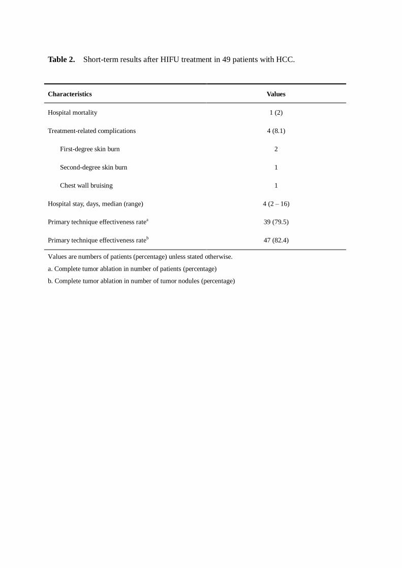
Table 2. Short-term results after HIFU treatment in 49 patients with HCC.
Characteristics Values
Hospital mortality 1 (2)
Treatment-related complications 4 (8.1)
First-degree skin burn 2
Second-degree skin burn 1
Chest wall bruising 1
Hospital stay, days, median (range) 4 (2 – 16)
Primary technique effectiveness ratea 39 (79.5)
Primary technique effectiveness rateb 47 (82.4)
Values are numbers of patients (percentage) unless stated otherwise.
a. Complete tumor ablation in number of patients (percentage)
b. Complete tumor ablation in number of tumor nodules (percentage)

Table 3. Univariate analysis on possible risk factors for incomplete ablation after HIFU.
Factors Complete ablation P value
Yes
(n=39)
No
(n=10)
Age, years, median (range) 65 (44 – 81) 68.5 (48 – 84) 0.275
Sex ratio, M : F 31 : 8 9 : 1 0.663
Hepatitis B infection 28 (71.7) 9 (90) 0.414
Hepatitis C infection 6 (15.3) 1 (10) 1.000
Child-Pugh liver function
class A : class B
34 : 5
7 : 3
0.333
Serum bilirubin, mol/L, median (range) 16 (4 – 44) 14 (8 – 46) 0.673
Serum albumin, g/L, median (range) 39 (24 – 45) 37 (27 – 45) 0.456
Platelet count, (109/L), median (range) 103 (46 – 268) 123 (26 – 192) 0.775
Serum -fetoprotein, g/ml, median
(range)
15 (2 – 3951) 9 (5 – 8840) 0.813
Previous hepatectomy 13 (33.3) 4 (40) 0.721
Previous radiofrequency ablation 9 (23) 5 (50) 0.093
Previous chemoembolization 22 (56.4) 8 (80) 0.278
Tumor size, cm, median (range) 2.29 (0.9 – 8) 3.75 (1.1 – 5.7) 0.013*
Number of tumors, median (range) 1 (1 – 2) 1 (1 – 2) 0.727
Pre-HIFU Lipiodol deposition 14 (35.8) 7 (70) 0.076
Use of artificial pleural effusion 25 (64.1) 6 (60) 1.000
Values are numbers of patients (percentage) unless stated otherwise.
*Statistically significant.

Table 4. Univariate analysis on possible prognostic factors affecting the overall survival
after HIFU.
Factors 1-year survival
rate, %
3-year survival
rate, %
P value
Age < 60 (n = 15)
> 60 (n = 34)
100
82.1
67.9
60.8
0.222
Sex Male (n = 40)
Female (n = 9)
89.9
77.8
64.3
50
0.244
Hepatitis B infection Yes (n = 37)
No (n = 12)
91.7
75
66.9
56.3
0.698
Hepatitis C infection Yes (n = 7)
No (n = 42)
71.4
90.3
47.6
68.3
0.512
Child-Pugh liver function class A (n = 41)
class B (n = 8)
90.2
75
68.5
33.3
0.028*
Serum bilirubin < 23 mol/L (n = 35)
> 23 mol/L (n = 14)
88.6
85.7
71.8
49.6
0.741
Serum albumin < 30 g/L (n = 5)
> 30 g/L (n = 44)
80
88.5
30
66.7
0.102
Platelet count < 150 ×109/L (n = 35)
> 150 ×109/L (n = 14)
85.5
92.9
61.8
66.8
0.888
Serum -fetoprotein < 200 g/ml (n = 37)
> 200 g/ml (n = 12)
91.7
75
57.5
75
0.831
Previous hepatectomy Yes (n = 17)
No (n = 32)
100
81.1
72.5
57.9
0.103
Previous RFA Yes (n = 14)
No (n = 35)
85.7
79
77.9
56.2
0.543
Previous TACE Yes (n = 30)
No (n = 19)
86.5
89.5
61.6
77.4
0.891
Tumor size < 3.0 cm ( n = 32)
3.0 cm (n = 17)
96.9
70.1
57.9
61.3
0.474
No. of tumors Solitary (n = 41)
Multiple (n = 8)
90.1
75
63.4
62.5
0.352
Pre-HIFU Lipiodol deposition Yes (n = 21)
No (n = 28)
85.7
89.3
60
76.9
0.744
Use of artificial pleural effusion Yes (n = 31)
No (n = 18)
87
88.9
60.5
70
0.971
Primary technique effectiveness Yes (n = 39)
No (n = 10)
92.3
68.6
68.1
45.7
0.249
Secondary technique effectiveness Yes (n = 42)
No (n = 7)
92.9
53.6
66.8
35.7
0.060
*Statistically significant. RFA, radiofrequency ablation. TACE, transarterial chemoembolization

Figure 1
(a) (b)
Figure 1

Figure 2Click here to download high resolution image
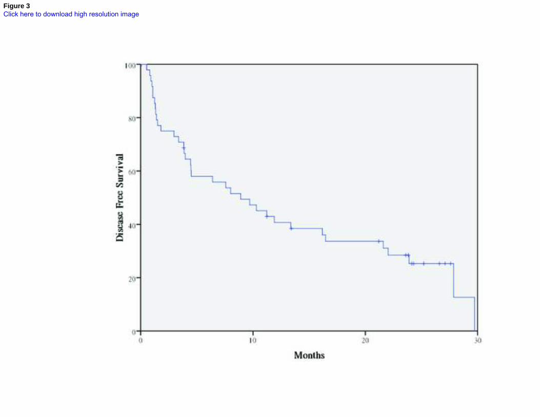
Figure 3Click here to download high resolution image

Figure 4
Figure 4