diTFPP, a Phenoxyphenol, Sensitizes Hepatocellular ... - MDPI

Mini-Reviews in Medicinal Chemistry, 2011, 11, 000-000 1
1389-5575/11 $58.00+.00 © 2011 Bentham Science Publishers Ltd.
Has Selenium a Chemopreventive Effect on Hepatocellular Carcinoma?
S. Costantini*,1, M.G. Lepore
1, G. Castello
1 and G. Colonna
2
1Istituto Nazionale Tumori “Fondazione Pascale” - Centro Ricerche Oncologiche di Mercogliano, Mercogliano (AV),
Italy
2Dipartimento di Biochimica e Biofisica & Centro di Ricerca Interdipartimentale di Scienze Computazionali e
Biotecnologiche, Seconda Università di Napoli, Napoli, Italy
Abstract: Hepatocellular carcinoma (HCC) is one of the most lethal cancers in the world. Its etiology includes chronic
liver disease, viral hepatitis, alcoholism, and hepatic cirrhosis. Both oxidative stress and inflammatory mechanisms have
been implicated in HCC pathophysiology. Surgical resection and liver transplants are currently used to treat HCC.
Consequently, there exists a decisive requirement to explore possible alternative chemopreventive and therapeutic
strategies for HCC. The use of dietary antioxidants and micronutrients has been proposed as a useful means for the HCC
management.
Trace elements such as selenium are involved in several major metabolic pathways as well as antioxidant defense
systems. In particular, selenium is an important oligo-element that plays a central role in cellular redox processes even if
the amount necessary for the cell functions is in a very narrow range. However, selenium is involved in the prevention of
numerous chronic diseases and cancers.
This review will examine the potential role of selenium in HCC prevention and treatment and, in detail, focus on: i)
description of selenium in biological systems and in mammalian proteins, ii) involvement of selenium in HCC, iii) in vivo
and in vitro effects of selenium in preclinical models of HCC and iv) potential challenges involved in the selenium use in
the prevention and treatment of HCC.
Keywords: ?????????????????????????.
INTRODUCTION
Yearly 550000 people worldwide die from HCC, with a 2:1 ratio for men versus women. It is a major health problem worldwide being the fifth most common malignancy in men and the eighth in women, and also the third most common cause of cancer-related death in the world [1]. Its incidence is increasing dramatically, with marked variations among geographic areas [2], racial and ethnic groups, environmental risk factors [3,4]. The estimated annual number of HCC cases exceeds 700000, with a mean annual incidence of 3-4% [2]. Most HCC cases (>80%) occur in either sub-Saharan Africa or in Eastern Asia (China alone accounts for more than 50% of the world’s cases) [2]. In the United States (US) HCC incidence is lower than other countries even if there has been a significant and alarming increase in the incidence of HCC in the US, from 1.3 in the late 70s’ to 3 in the late 90s’, due to Hepatitis C virus (HCV) infection. In 2008, 21370 new cases of HCC and intrahepatic bile duct cancer were estimated with 18410 deaths [2]. In Europe, Oceania and America, chronic HCV and cirrhosis are the main risk factors for HCC. Among patients with HCV and cirrhosis, the annual incidence rate of HCC ranges between 1-8%, and is higher in Japan (4-8%) intermediate in Italy (2-4%) and lower in US (1.4%) [5]. Mortality analysis for HCC in
*Address correspondence to this author at the INT Pascale - CROM, Via
Ammiraglio Bianco, 83013 Mercogliano (AV), Italy; Tel: ??????????; Fax:
?????????????; E-mail: [email protected]
Europe confirmed large variability, with high rates in France and Italy due to HCV [6]. However, Southern Italy has the highest rates of HCC in Europe [7].
HCC is unique among cancers occurring mostly in patients with chronic liver inflammation and cirrhosis [2]. In particular, hepatitis B (HBV) and C (HCV) viruses are the major cause of liver disease worldwide. About 80% of newly infected patients develop chronic infection. Moreover, 10% - 20% of these patients will develop cirrhosis and, then, 1-5% will proceed to end-stage liver cancer over a period of 20 - 30 years. In HCV patients, HCC is observed always as a complication of cirrhosis, whereas it is found in non-cirrhotic patients in the case of HBV [8].
HCC treatment is challenging since it is largely refractory to chemotherapy. Thus, HCC prevention might represent the best opportunity to reduce the worldwide burden of this cancer. Although HBV vaccination will reduce the number of individuals at risk for HCC development, a tremendous number of people are currently at elevated risk for HCC due to HCV-correlated chronic hepatitis and cirrhosis. This population with known risk factors has to be monitored on a regular basis to detect early cancerous lesions. HCC detection and diagnosis at an early stage may significantly improve the patients survival. Hence, there is also an obvious critical need to develop alternative strategies to prevent HCC development. In fact, HCC chemoprevention may be useful to develop new preventive strategies and to reduce the inflammation process [8].

2 Mini-Reviews in Medicinal Chemistry, 2011, Vol. 11, No. 7 Costantini et al.
Both oxidative stress and inflammatory mechanisms have been implicated in HCC pathophysiology. The use of dietary antioxidants and micronutrients has been proposed as an effective means for HCC management. Trace elements such as vanadium and selenium are involved in several major metabolic pathways as well as antioxidant defense systems. Selenium has been shown to be involved in the prevention of numerous chronic illnesses such as several specific cancers and neurodegenerative diseases [9]. Therefore, in this review we examine its potential role in HCC prevention and treatment.
SELENIUM BIOAVAILABILITY
Selenium is an important oligo-element and has been shown to play a central role in cellular redox processes. The selenium amount necessary for the cellular functions is in a very narrow range. It is reported that the selenium lack tends to suppress the expression of various enzymes and leads to cell abnormality and diseases. However, high concentrations of free selenium are toxic to the cell because it adversely affects numerous cell metabolic pathways [10].
On the basis of the Dietary Reference Allowances (RDA) developed by the Institute of Medicine, the recommended daily amount of selenium is 55 μg for adults. The RDA is based on the amount of dietary selenium required to maximize the activity of the antioxidant enzyme glutathione peroxidase in plasma. However, the dietary intake can vary in basis to the diet and geographic location [11]. A tolerable upper intake level (UL) for selenium is 400 μg per day for adults to prevent the risk of developing selenosis. UL includes selenium obtained from food and supplements. Selenium toxicity can occur at doses of 600 - 750 μg. Early signs of selenium toxicity include fatigue, irritability and dry hair. Other symptoms of excess intake include dental caries in children, hair loss, skin depigmentation, abnormal nails, vomiting, nervous system problems, and bad breath [12].
Selenium is stored in the tissues in varying density: 30 percent of tissue selenium is in the liver, 15 percent in the
kidney, 30 percent in muscle, 10 percent in the plasma, and the remaining 15 percent throughout other organs. Trace quantities of selenium have been found to be present in several dietary agents such as milk, meat products, poultry, fish, fruits, vegetables and cereals. Brazil nuts happen to be a very rich dietary source of selenium [13]. Selenium enters the food chain through the plants as it is present in the soil. However, there exists a considerable variation in the selenium amount consumed in the diet due to differences in selenium soil content in diverse regions of the world.
Selenium occurs naturally in plants as selenomethionine, Se-methylselenomethionine, selenocysteine (Sec), and selenocystine. Selenite (commercially available as sodium selenite) is greater than 80% bioavailable whereas selenomethionine or selenate can be greater than 90% bioavailable. In particular, selenomethionine is the major organic seleno-compound in cereal grains, grassland legumes, and soybeans [14]. Selenomethionine competes with methionine for absorption on the gut surface, and is incorporated and stored in body proteins containing methionine (Fig. 1) [15]. It may also be converted through transulfuration to selenocysteine and degraded to hydrogen selenide via the beta-lyase enzyme. Moreover, selenite is also metabolized to hydrogen selenide via a pathway in which it is complexed with glutathione. Hydrogen selenide acts as a precursor for selenoprotein synthesis and represents also the excreted selenium form; in fact it is methylated and excreted through urine and breath. More than 90% of animal chemoprevention studies have used either sodium selenite or selenomethionine as the test agent [16]. Other methylated selenium compounds, found in plants and in the metabolic breakdown of sodium selenide in humans, are indicated as responsible for the chemopreventive effect of selenium [15].
HUMAN SELENOPROTEINS
25 different human selenoproteins are known and have different tissue distribution and subcellular localization. The most important families are the glutathione peroxidises
Fig. (1). Selenium metabolism.

Has Selenium a Chemopreventive Effect Mini-Reviews in Medicinal Chemistry, 2011, Vol. 11, No. 7 3
(GPxs), the iodothyronine deiodinases (DIOs) and the thioredoxin reductases (TrxRs). In details the selenoproteins can be classified into two groups according to the location of Sec [17]. In fact, the first group, consisting of TrxRs and selenoproteins S, R, O, I and K, has Sec in a site to the C terminus of protein whereas the second one (GPxs, DIOs and selenoproteins H, M, N, T, V and W, SPS2 and Sep15) has Sec in the N-terminal part [18].
The selenoproteins belonging to the GPx family use glutathione (GSH) to catalyze the reduction of hydrogen peroxide to water and the reduction of phospholipid peroxides to their corresponding alcohols [19]. In particular, the catalytic redox cycle of GPxs involves the Sec oxidation to seleninic acid by the hydrogen peroxide and organic hydroperoxides and its reduction to the selenolate anion form by the GSH system (Fig. 2). There are five different GPxs. GPx1 is expressed in the cytoplasm of all mammalian tissues, and plays an important role in protecting cells against oxidative stress [20]. Some studies showed that its expression decreased in breast cancer models [21], and its activity increased in malignant human lung tissue compared to nonmalignant tissue [22]. GPx2 is mainly expressed in the whole gastrointestinal tract [23], protects intestinal epithelium from oxidative stress induced by ingested prooxidants and gut microbial [24] and has a protective role against several types of cancer [25]. GPx3 shows a role in modulating nitric oxide (NO) concentration [26, 27]. GPx4 is involved in protection of lipid peroxides in membranes [28] and plays also a role in lipid metabolism and spermatogenesis [29]. Finally, GPx6 is probably involved in the cell protection by oxidative stress but further studies are available. The structures for all human GPxs are reported by crystallographic studies and show the typical structure motif of the thioredoxin fold, which consists mainly of a central beta-sheet and several flanking alpha-helices [30]. Sec
residue and its neighboring residues are located in a surface pocket. In particular, this residue is located at the bottom of the pocket composed by residues in loops connecting alpha-helices and beta-strands.
TrxRs are oxidoreductases that catalyze the NADPH-dependent reduction of the redox protein Trx [31], as well as of other endogenous and exogenous compounds including lipoic acid [32], lipid hydroperoxides [33], the cytotoxic peptide NK-lysin [34], vitamin K3 [35], dehydroascorbic acid [36], the ascorbyl free radical [37] and the tumor-suppressor protein p53 [38]. The proposed mechanism of TrxR-dependent reduction involves electron transfer from NADPH to FAD via the N-terminal active site of one subunit of the Cys-Sec selenylsulfide bond within the C-terminal active site of the opposite subunit and finally to the substrate Trx (see Fig. 2). However, there are three forms of mammalian TrxRs: cytoplasmic/nuclear TrxR1 [39] and mitochondrial TrxR2, distributed in a variety of tissues [40], and testes-specific thioredoxin-glutathione reductase (also called Txnrd3, TR2, TxnR3, or TGR) [41].
It is reported that Trx1 is over-expressed in many human cancers such as melanomas, thyroid, breast, and prostate and colorectal carcinomas [42]. In 2007 the structure of human TrxR1 was obtained in complex with FAD and NADP+ and is composed by three domains of alpha-beta fold. Sec residue is located in C-terminal active site being essential for catalysis. In particular, the redox center is on a flexible arm, that is solvent-exposed and reactive, and, thus, represents a target for antitumor drug development [43]. During the catalysis this C terminal arm acts as an electron shuttle between the buried N-terminal dithiol and the substrate and, for this reason, it can adopt different conformations, one near the N-terminal redox site to pick up electrons, and another one at the surface of the enzyme to donate the electrons to
Fig. (2). Proposed mechanisms of some mammalian selenoproteins: A) glutathione peroxidises (GPxs), B) thioredoxin reductases (TrxRs)
and C) iodothyronine deiodinases (DIOs).

4 Mini-Reviews in Medicinal Chemistry, 2011, Vol. 11, No. 7 Costantini et al.
the substrate. In fact, Fritz-Wolf et al. (2007) captured different conformations for the last six amino acid residues (QAGCCG) [56].
DIOs represent a family of selenium-containing enzymes involved in the activation or deactivation of thyroid hormones [44]. There are three types of enzymes: DI is found in the liver and kidney and can deiodinate both rings, DII can deiodinate only the outer ring of the prohormone thyroxine in the heart, skeletal muscle, fat and thyroid, and DIII is found in the fetal tissue and the placenta, can deiodinate only the inner ring of thyroxine (T4) or triiodothyronine (T3) and is the major inactivating enzyme [45]. The proposed deiodination mechanism of DIOs is involved in the generation of theoxidized DIO-Se intermediate that is then reduced by thiol-containing reductants and releases iodide (Fig. 2)
HUMAN SELENIUM BINDING PROTEINS
Among selenoproteins we find the selenium as Sec incorporated into the polypeptide backbone even if recent evidence supports the existence of other selenium-containing proteins with different selenium chemistry. This last group is generally referred as ‘‘selenium binding proteins” [46]. Two selenium-binding proteins in which the selenium moiety is externally bound to the polypeptide have been identified in mammals: a 14-kDa protein (SLP-14) [47] and a 56-kDa protein (SLP-56, SBP56, hSP56, and selenium-binding protein 1 (SELENBP1 or SBP1) [48]. Moreover, a second 56-kDa protein almost identical at cDNA and protein level to SP56, but encoded by a different gene, was subsequently identified and named SELENBP2 [49]. SELENBP1 is implicated to play a role in toxification/detoxification processes, cell-growth regulation, intra-Golgi protein transport, aging, and lipid metabolism. Since no atomic level structural data is available for SELENBP1, its three-dimensional structure has been recently modeled by computational methods [50]. Its model shows an alpha-beta structure characterized by 4 short alpha-helices, one 310 helix (residues 207-210) and 30 antiparallel beta-strands, and is stabilized by two disulfide bonds (Cys80–Cys141 and Cys83–Cys466) (Fig. 3). These structural studies suggested also a functional involvement of Cys57 as the only cysteine in the protein able to bind the selenium [50].
Interestingly, several papers have reported decreased expression levels of both SELENBP1 and SELENBP2 in tumors compared with normal tissue. In particular, reduced expression of SELENBP1 has been reported for prostate, lung, ovarian, thyroid, stomach, colorectal cancers, uterine leiomyoma, and esophageal adenocarcinoma [51-54]. Recently we have evaluated the expression of this protein also in tissue samples of HCC patients by common immunohistochemistry techniques [50]. Our evaluations highlighted that the gradual SELENBP1 loss was associated with an increased malignant grade and suggested that the evaluation of the SELENBP1 down-expression in liver tissues of HCC patients could to be a good prognostic tool for the disease progression. Certainly the storage of very large amount of these data in specialized archives, equipped with image analysis software, could allow rapid and precise prognosis of this disease [50]. Also, liver selenium
concentrations were measured in tissue samples of patients with HCC by atomic absorption spectrometry. The obtained results showed that the selenium concentrations decreased when the malignant grade increased, and there was a significant correlation between selenium levels and human selenium binding protein-1 (SELENBP1) down-expression in the liver (manuscript in preparation).
All these observations not only provide an additional link between selenium and its anticancer properties, but suggest also that SELENBP1 and selenium levels could be used as a potential marker for cancer development. Therefore it is possible to hypothesize that selenium uptake may be used to prevent some cancers in which SELENBP1 is resulted down-expressed [50-54]. In fact, several studies which include geographic, pre-clinical animal, prospective as well as intervention studies have implicated selenium in several gastrointestinal cancers, such as esophageal, colorectal, prostate as well as liver and have suggested its putative role in cancer as well as progression and subsequently metastasis prevention [50-54].
Fig. (3). Ribbon diagram of human SELENBP1 model. Helices are
shown in black and beta-strands in white.
INVOLVEMENT OF SELENIUM IN HCC
The role of trace element micronutrients in HCC has been the subject of many investigations [55, 56]. Some studies have reviewed the potential inverse relationship between selenium levels and HCC occurrence [57] and have shown that there is a significantly reduced activity of antioxidant enzymes such as glutathione peroxidases along with selenium deficiency in patients with HCC [58-59]. In a pioneering study, Yu et al. [60-61] highlighted that a significant inverse correlation between plasma selenium

Has Selenium a Chemopreventive Effect Mini-Reviews in Medicinal Chemistry, 2011, Vol. 11, No. 7 5
levels and the occurrence of human HCC exists. In particular, they showed that selenium supplementation protected hepatitis B antigen carriers against development of HCC [60]. A subsequent trial conducted in China showed a 50% selenium-induced reduction in HCC occurrence [62]. Therefore, the following sections review the selenium effects on the liver trascriptome and the preclinical in vitro and in vivo studies investigating the selenium effects on HCC.
EFFECTS OF SELENIUM ON LIVER TRANSCRIP-
TOME
In 2007 Katzenellenbogen et al. published a study on the selenomethionine effects on HCC development in Mdr2 knockout mice using cDNA microarrays containing probes for 240 genes that regulate responses to oxidative stress and inflammation or lipid metabolism [63]. Selenomethionine resulted to inhibit gene expression and to reverse up-regulation of many genes that control inflammation or response to oxidative stress in the related livers at age 3 months. This inhibitory effect on gene expression correlated with the ability of this agent to reduce the incidence of large tumors [63]. A recent study have examined also the influence of selenite and selenate on the differential expression of genes encoding non-selenoproteins in the rat liver using microarray technology [64]. Five groups of nine growing male rats were fed with a Se-deficient diet or diets supplemented with 0.20 or 1.0 mg of Se/kg as sodium selenite or sodium selenate for 8 weeks. Genes that were more than 2.5-fold up- or down-regulated by selenite or selenate compared with Se deficiency were selected. GPx1 was up-regulated 5.5-fold by both Se compounds, whereas GPx4 was up-regulated by only 1.4-fold. Selenite and selenate down-regulated three phase II enzymes. The comparison of selenite- and selenate-regulated genes revealed that selenate may have additional functions in the liver protection, and that it may be more active in metabolic regulation [64].
EFFECTS OF SELENIUM IN PRECLINICAL
MODELS OF HCC
In the past years many studies on human hepatoma cells and on animal models of liver cancers showed the cytotoxic and antioxidant effects of selenium (Tables 1 and 2) [9].
Table 1. In Vitro Studies of Selenium in HCC Cells
References Mechanisms of action
[97] GPx
[98] apoptosis, NO
[99] apoptosis, LDH, GSH
[100] apoptosis, LDH, GSH
[104] apoptosis
[109] MDA, ROS, LDH
In Vitro Studies
Baker et al. investigated the biological effects of selenium and evidenced that selenium supplementation
restored the GPx activity in human hepatoma cells maintained in selenium-deficient media [65]. In particular, the selenium treatment induced also a decrease of cell proliferation, production of cell death and apoptosis in various mammalian hepatoma cells. Liu et al. [66] showed that induced selenium decreased in mice H22 hepatoma cell proliferation was accompanied by an increase in nitric oxide.
Fig. (4). Schematic illustration showing the metabolic reduction of
MSeA and selenite with GSH. Different from selenite, MSeA is a
precursor of methylselenol without the formation of hydrogen
selenide and superoxide anion radicals.
Studies by Shen et al. [67-68] evidenced that GSH was a selenite cofactor, facilitating selenite induced oxidative stress and apoptosis, and, also, an antioxidant that protects against selenite-mediated oxidative stress and apoptosis. However, both sodium selenite and methylseleninic acid-induced cytotoxicity released lactate dehydrogenase (LDH) and produced a significant decrease in glutathione content in HepG2 hepatoma cells. In particular, methylseleninic acid (CH3SeOOH, MSeA) is a synthesized monomethylated selenium compound and is an excellent tool for the molecular mechanism studies of selenium in cell culture because it mimics the chemoprevention of monomethylated selenoamino acids [69]. As shown in Fig. 4, MSeA is a rather strong oxidizing agent able to readily react with reduced glutathione (GSH) to form methylselenol, a highly reactive compound responsible for the anticancer effect of methylated selenium compounds [70]. The metabolic pathway of MSeA in cells is found to be different respect to that of sodium selenite, because it reacts with GSH and leads to the production of selenodiglutathione, hydrogen selenide (H2Se), and superoxide anion radicals [71] (Fig. 4).
The antineoplastic properties of selenium compounds can be accomplished by either a decrease in cell proliferation or

6 Mini-Reviews in Medicinal Chemistry, 2011, Vol. 11, No. 7 Costantini et al.
an increase in apoptosis. Sodium selenite also produced a dose-dependent decrease in cell viability, cell proliferation and extensive vacuole formation in HepG2 cells [72]. Some authors have showed that hydrogen selenite was a regulatory point of selenium metabolism and proposed also mechanisms of selenite-induced apoptosis in HepG2 cells (Fig. 5) [73-75].
In particular, Celik et al. demonstrated that selenite induced cellular proliferation at low (<10 μM) concentrations and apoptosis at higher concentrations ( 10 μM) and that there was a relation between selenite concentration and apoptosis [72]. They found that a low selenite concentration (10 μM) had a higher apoptotic pattern than the high selenite concentration (25 μM). Accumulation of excessive hydrogen selenite can explain the mechanism and, hence, the formation of hydrogen selenite can be a bottleneck of selenium metabolism in the cells. In fact, selenite was metabolized to hydrogen selenite by several glutathione/NADPH reductions. Thus, selenite accumulation can block the selenite metabolism as a result of the methylation pathway, and the hydrogen selenite concentration can increase with augmented oxidative stress by reductive metabolism. Therefore, cell cycle checkpoint controls at the G1 to S transition and the G2 to M transition prevented the cell cycle progressing when DNA was damaged. The G2/M DNA damage checkpoint prevented the
cell from entering mitosis (the M-phase). The cdk2-cyclin B kinase was pivotal in regulating this transition. On the other hand, the genes, that turned on by p53, constituted effectors cascade [76] and selenite inhibited caspase-3-like protease activity through a redox mechanism [74]. The increased rate of apoptosis can inhibit the cancer induction during the promotional phase and the related decrease in cdk2 kinase activity was accompanied by prolonged arrest in the S-phase [75]. The inhibitory effect of selenite on the cell cycle resulted in cell cycle arrest at the G2/M boundary, that was followed by apoptosis at selenite concentrations above 10 μM. Some experiments showed that the percentage of G0/G1-phase cells of selenite groups were notably lower than that of the control group. These results pointed out that selenite can increase the cell transformation from the G0/G2 phase to the S phase and the enrichment in the G2/M-phase in a concentration-dependent manner. These findings suggested that selenite can modulate cellular response through some common pathways involving both apoptotic and cell cycle regulatory pathways.
Finally Cuello and coworkers [77] showed the protective and antioxidant effects of selenium. In fact, the selenium pretreatment in HepG2 cells decreased the production of reactive oxygen species (ROS) as well as reduced the LDH release and also reduced levels of malondialdehyde (MDA), a marker for lipid peroxidation.
Fig. (5). Proposed mechanisms of selenite-induced apoptosis in HepG2 cells.

Has Selenium a Chemopreventive Effect Mini-Reviews in Medicinal Chemistry, 2011, Vol. 11, No. 7 7
In Vivo Studies
Many studies have evidenced the selenium effects in preclinical animal models of HCC and reported three possible HCC models, i.e. genetic, xenograft and chemical models.
Genetic Models
Popova showed that selenium decreased significantly the spontaneous liver tumorigenesis in CBA mice [78] whereas Novoselov et al. evidenced that selenium supplementation in the diet inhibited hepatocarcinogenesis and decreased cell proliferation in Myc transgenic mice, that developed liver cancer by 6 months of age [79]. This selenium-induced suppression of HCC was also accompanied by increase in apoptosis.
In a study conducted in 2007 the development of HCC in Mdr2 knockout mice was suppressed by selenomethionine and the related observed anticancer effects were accompanied by changes in expression of several genes involved in the regulation of inflammatory processes and oxidative stress mechanisms [80].
Xenograft Model
In 2007 Xu et al. investigated the anti-cancer properties of selenium and showed that green tea extracts enriched with selenium suppressed HCC in mice that were xenografted with human hepatoma HepG2 cells [81].
Chemical Models
Chemically-induced HCC remains one of the most used methods to study the biochemical mechanisms involved in the development of HCC and to elucidate the potential chemopreventive and therapeutic properties of several compounds. Many potent hepatocarcinogen were used as models for chemically-induced HCC, i.e. azo dye 3’-methyl 4-dimethylaminoazobenzene, aflatoxin B1, azoxymethane, diethylnitrosamine, 2-Acetylaminoflurene, ciprofibrate, and N-Nitrosobis (2-Oxopropyl)Amine.
In 1977 Griffen and Jacobs demonstrated that supplementation by sodium selenite attenuated the azo dye 3’-methyl 4-dimethylaminoazobenzene (3’-MeDAB)-induced hepatocarcinogenesis and reduced tumor formation in rodents [82] whereas Daoud and Griffin evidenced that the selenium afforded protection when administered during the early stages 3’-MeDAB-induced hepatocarcinogenesis in rats [83]. Selenium-induced chemoprevention has also been studied in aflatoxin B1 (AFB1)-induced HCC in rodents. Milks et al. [84] showed that the selenium reduced the occurrence of gamma-glutamyl transpeptidase (GGT)-positive foci during AFB1- induced hepatocellular carcinogenesis in rats. Lei et al. [85] have shown that sodium selenite supplementation significantly suppressed the AFB1-induced initiation and promotion and produced a decrease in both the nodule and foci formation in rats.
Moreover, selenium pretreatment induced a significant protection against the formation of 8- hydroxydeoxyguanosine (8-OHdG), indicating its ability to ameliorate oxidative DNA damage in vivo [86]. In particular, an organic selenium compound, i.e. ebselen, attenuated both
the area and mean density of GSTP positive foci induced by AFB1 in rats, the expression of liver alpha-fetoprotein and the formation of hepatic AFB1-DNA adduct as well as (8- OHdG) [87].
In 1985 the effect of dietary pmethoxybenzylselenol was investigated on azoxymethane (AOM) induced hepatocarcinogenesis in rats. This compound resulted to inhibit the foci incidence, the tumor incidence and the multiplicity during AOM-induced hepatic neoplasia [88].
Several studies demonstrated the chemopreventive effects of selenium in the 2- acetylaminoflurene (2-AAF) model. Le-Boeuf et al. showed that selenium supplementation caused a significant decrease of mean focal volume in the 2-AAF induced HCC in female rats [89]. Then, Mukherjee et al. evidenced that selenium-induced chemoprevention in the 2-AAF model was accompanied by increase in levels of antioxidant enzymes SOD and CAT and a decrease in GPx and GST levels [90].
In 1990 the chemopreventive effect of selenium has also been studied in the ciprofibrate stress model in which this compound induced both tumor and foci formation in rodent liver. Chronic pretreatment with dietary selenium significantly attenuated this effect whereas selenium-induced chemoprevention was accompanied by a dramatic increase in glutathione content and GPx activity [91].
In 2008 a study evidenced that sodium selenite suppressed the N-nitrosobis (2-oxopropyl)amine induced HCC in Syrian hamsters and the related chemopreventive effects were accompanied both by a parallel decrease in levels of liver enzymes (alanine transaminase (AST) and aspartate transaminase (ALT)) and by an increase in apoptosis and caspase-3 levels [92].
One of the more used models of chemically-induced HCC is the two-stage model that is initiated with diethylnitrosamine (DENA) and promoted by phenobarbital (PB). LeBoeuf et al. showed that selenium enhanced foci formation and increased nodule volume in the DENA-PB model in female rats [89]. In the last decade Thirunavukkarasu et al. highlighted the chemopreventive properties of selenium in the DENA-initiated and PB-promoted HCC rodent model and the related biochemical mechanisms implicated in the selenium-mediated chemoprevention in the DENA-PB model [93-103]. Particularly in the DENA-initiated and PB-promoted model, sodium selenite supplementation significantly suppressed HCC, elevating several key enzymes of the citric acid cycle, such as malate dehydrogenase [93]. Moreover, sodium selenite supplementation produced also antioxidant effects as evidenced: i) by decreasing the markers of oxidative stress, i.e. thiobarbituric acid reactive substance, ii) by increasing levels of antioxidant enzymes such as superoxide dismutase (SOD) and catalase (CAT) and iii) by decreasing enzymes such as glutathione transferase (GST) [94, 95, 97]. The same Authors showed that sodium selenite decreased elevated levels of liver enzymes such as AST, ALT and LDH as well as of glycoproteins such as globulin and hexosamine [98, 99]. Moreover they investigated also the biochemical effects of selenium in the DENA-PB model. In particular, selenium

8 Mini-Reviews in Medicinal Chemistry, 2011, Vol. 11, No. 7 Costantini et al.
elevated several enzymes of the ATPase and caused significant alterations to serum mineral levels [96, 100], produced a significant increase in levels of vitamins C and E in the DENA-PB HCC model [101], decreased polyamine
levels in the rodent DENA-PB model [102] and enhanced significantly the DNA strand breaks in the DENA-PB rat liver carcinogenesis while increasing the GPx activity [103].
Table 2. In Vivo Studies of Selenium in HCC Models
HCC Models References Mechanisms of action
Genetic Models [110] tumor formation
[111] apoptosis, cell proliferation
[112] Regulation expression of genes involved in inflammation and oxidative stress
Xenograft Models [113] Se concentration
Chemical Models
Azo dye (3’-MeDAB) [114] tumor formation
[115] tumor formation
2-AAF [121] focal volume
[122] GST, UDP-GT
[122] GST, GPx, CAT,SOD
AFB1 [116] GGT positive foci
[117] Inhibition of AFB1-induced initiation and promotion
[118] 8-OHdG
[119] serum GGT, AFP, AFB1-DNA adduct, 8-OHdG
AOM [120] foci, nodule
ciprofibrate [123] GPx, glutathione
BOP [124] ALT, AST, GST,apoptosis, caspase-3
DENA [121] nodule volume, foci
[125] ICDH, SDH, MDH
[126] TBARS, CAT, SOD
[127] GST, UDP-GT,GGT, G6PD, AHH
[129] TBARS,CAT,SOD
[130] LDH, ALT, AST
[131] globulin, hexose, hexosamine, sialic acid
[128] Na+/K+-ATPase,Mg2+-ATPase, Ca2+-ATPase
[132] K+, Ca2+, Fe2+, Na+
[133] vitamin C, vitamin E,protein thiol
[134] AFP, histamine, putrescine, spermicidine, spermine
[135] GPx
[136] cell proliferation, TRxR
[137] GPx
[138] glucose, insulin, IGF-II glucagon
[139] VEGF, IGF-II, NO,TNF-
Has Selenium a Chemopreventive Effect Mini-Reviews in Medicinal Chemistry, 2011, Vol. 11, No. 7 9
In 2005 Björkhem-Bergman et al. investigated the chemopreventive effects of selenium supplementation in the initiation, promotion and progression phases of a synchronized rat model of HCC initiated by DENA and promoted by 2-AAF. In this study, although selenium during the initiation phase did not exhibit preventive effects, it reduced preneoplastic nodule volume during the promotional phase [104], inhibited cell proliferation and reduced TRxR levels in liver nodules. In the DENA-initiated and polychlorobiphenyl-promoted HCC in rodents selenium supplementation inducted a decrease in GPx as well as chemoprevention effect [105].
Recently Liu et al. [106] have investigated the effects of pretreatment by selenium enriched malt (SEM) in DENA-induced nodule formation in rodents. Selenium decreased several liver enzymes such as AST, ALT and gamma-glutamyltranspeptidase and reduced hypoglycemia as evidenced by an increase in glucose levels and by a complementary decrease in insulin. In an extension of this study, Liu et al. [107] showed that SEM inhibited the angiogenesis during DENA-initiated carcinoma by down-regulation of nitric oxide, VEGF expression and of several angiogenic cytokines, namely tumor necrosis factor- .
POTENTIAL CHALLENGES INVOLVED IN THE
ADVANCE OF SELENIUM USE IN THE
PREVENTION AND TREATMENT OF HCC
It is evident from the studies presented that selenium holds great promise as a potential agent for HCC chemoprevention and treatment. The potential effectiveness of this agent is evident from both in vitro as well as in vivo studies reviewed here. However, both considerable amounts of work and significant efforts are required before this element finds clinical usefulness in HCC treatment. Although all the studies reviewed in this paper provide substantial evidence for its chemopreventive and therapeutic potential against HCC, in scientific literature exists considerable caveats. In fact, the selenium effect both in various signaling pathways implicated in inflammation and in the hepatocarcinogenesis stages need to be elucidated in greater detail. In regard to preclinical studies, a critical need exists to systematically examine the chemopreventive as well as therapeutic effects of selenium in human HCC. This aim can be achieved through well designed clinical trials in geographical regions which show high-risk population cohorts and a greater incidence of HCC. Also long-term epidemiological studies would be useful to assess the correlation between selenium intake and the occurrence of human HCC.
ACKNOWLEDGEMENT
We thank Dr. Raffaele Raucci for its help in the preparation of figures.
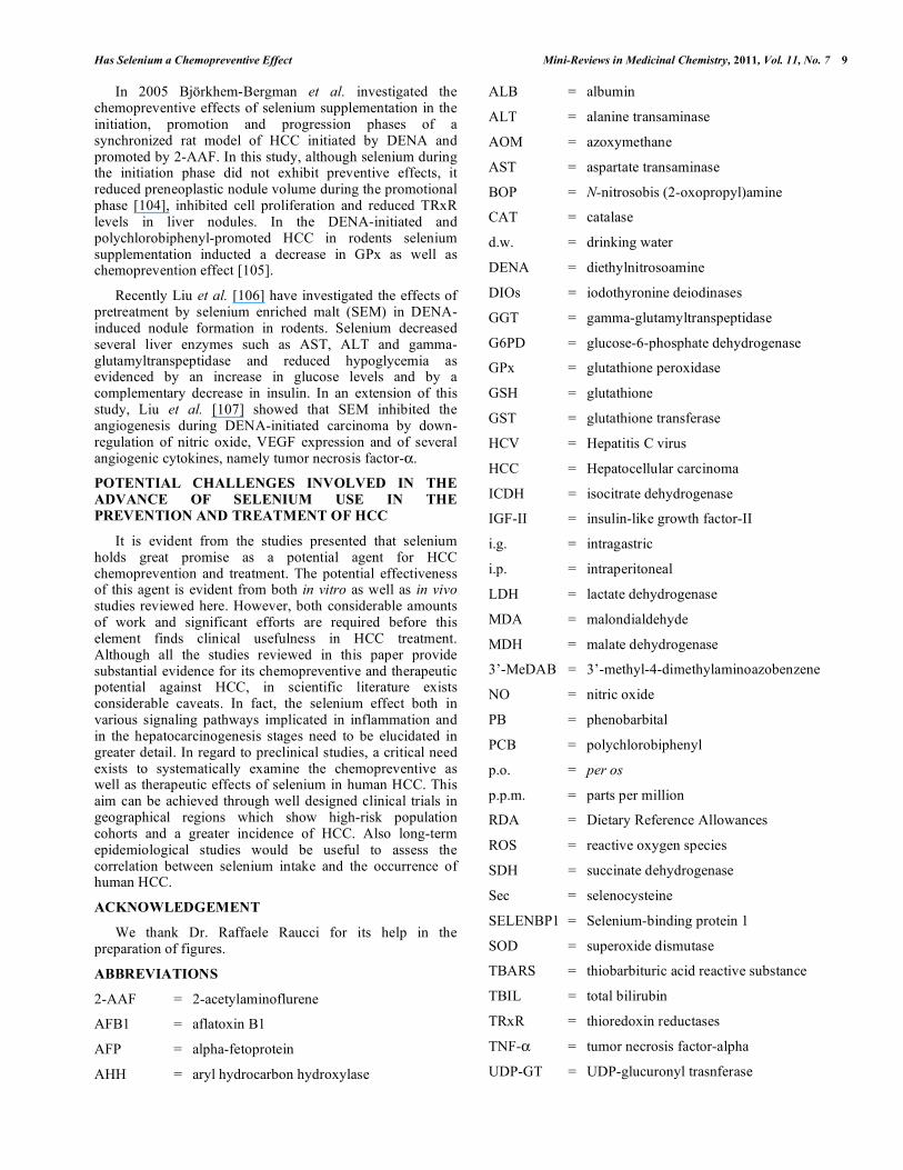
ABBREVIATIONS
2-AAF = 2-acetylaminoflurene
AFB1 = aflatoxin B1
AFP = alpha-fetoprotein
AHH = aryl hydrocarbon hydroxylase
ALB = albumin
ALT = alanine transaminase
AOM = azoxymethane
AST = aspartate transaminase
BOP = N-nitrosobis (2-oxopropyl)amine
CAT = catalase
d.w. = drinking water
DENA = diethylnitrosoamine
DIOs = iodothyronine deiodinases
GGT = gamma-glutamyltranspeptidase
G6PD = glucose-6-phosphate dehydrogenase
GPx = glutathione peroxidase
GSH = glutathione
GST = glutathione transferase
HCV = Hepatitis C virus
HCC = Hepatocellular carcinoma
ICDH = isocitrate dehydrogenase
IGF-II = insulin-like growth factor-II
i.g. = intragastric
i.p. = intraperitoneal
LDH = lactate dehydrogenase
MDA = malondialdehyde
MDH = malate dehydrogenase
3’-MeDAB = 3’-methyl-4-dimethylaminoazobenzene
NO = nitric oxide
PB = phenobarbital
PCB = polychlorobiphenyl
p.o. = per os
p.p.m. = parts per million
RDA = Dietary Reference Allowances
ROS = reactive oxygen species
SDH = succinate dehydrogenase
Sec = selenocysteine
SELENBP1 = Selenium-binding protein 1
SOD = superoxide dismutase
TBARS = thiobarbituric acid reactive substance
TBIL = total bilirubin
TRxR = thioredoxin reductases
TNF- = tumor necrosis factor-alpha
UDP-GT = UDP-glucuronyl trasnferase

10 Mini-Reviews in Medicinal Chemistry, 2011, Vol. 11, No. 7 Costantini et al.
UL = upper intake level
US = United States
VEGF = vascular endothelial growth factor
REFERENCES
[1] Altekruse, S.F.; McGlynn, K.A.; Reichman, M.E. Hepatocellular Carcinoma Incidence, Mortality, and Survival Trends in the United
States From 1975 to 2005. J. Clin. Oncol., 2009, 27,1485-1491. [2] Jemal, A.; Siegel, R.; Ward, E.; Murray, T.; Xu, J.; Thun, M.J.
Cancer Statistics 2007. C.A. Cancer J. Clin., 2007, 57, 43-66. [3] El-Serag, H.B.; Rudolph, K.L. Hepatocellular Carcinoma:
Epidemiology and Molecular Carcinogenesis. Gastroenterology, 2007, 132, 2557-2576.
[4] Castello, G.; Costantini, S.; Scala, S. Targeting the inflammation in HCV-associated hepatocellular carcinoma: a role in the prevention
and treatment. J. Trasl. Med., 2010, 8,109. [5] Fassio, E. Hepatitis C and hepatocellular carcinoma. Ann. Hepatol.,
2010, 9, 119-122. [6] Bosetti, C.; Levi, F.; Boffetta, P.; Lucchini, F.; Negri, E. La
Vecchia C: Trends in mortality from hepatocellular carcinoma in Europe, 1980-2004. Hepatology, 2008, 48, 137-145.
[7] Fusco, M.; Girardi, E.; Piselli, P.; Palombino, R.; Polesel, J.; Maione, C.; Scognamiglio, P.; Pisanti, F.A.; Solmone, M.; Di
Cicco, P.; Ippolito, G.; Franceschi, S.; Serraino, D. Epidemiology of viral hepatitis infections in an area of southern Italy with high
incidence rates of liver cancer. Eur. J. Cancer 2008, 44, 847-853. [8] Castello, G.; Scala, S.; Palmieri, G.; Curley, S.A.; Izzo, F. HCV-
related hepatocellular carcinoma: From chronic inflammation to cancer. Clin. Immunol., 2010, 134, 237-250.
[9] Darvesh, A.S.; Bishayee, A. Selenium in the Prevention and Treatment of Hepatocellular Carcinoma. Anti Canc. Agents Med.
Chem., 2010, 10, 338-345. [10] Suzuki, M.; Lee, D.-Y.; Inyamah, N.; Stadtman, T.C.; Tjandra, N.
Solution NMR Structure of Selenium-binding Protein from Methanococcus vannielii. J. Biol. Chem., 2008, 283, 25936–25943.
[11] Institute of Medicine, Food and Nutrition Board. Dietary Reference Intakes: Vitamin C, Vitamin E, Selenium, and Carotenoids.
National Academy Press, Washington, DC, 2000. [12] Agency for Toxic Substances and Disease Registry (ATSDR).
Toxicologic Profile for Selenium. Atlanta, GA: US Department of Health and Human Services, Public Health Service. 2003.
[13] Brown, K.M.; Arthur, J.R. Selenium, selenoproteins and human health: a review. Public Health Nutr., 2001, 4, 593.
[14] Whanger, P.D. Selenocompounds in plants and animals and their biological significance. J. Am. Coll. Nutr., 2002, 21, 223-232.
[15] Ip, C. Lessons from basic research in selenium and cancer prevention. J. Nutr., 1998, 128, 1845-1854.
[16] Behne, D.; Kyriakopoulos, A. Mammalian selenium-containing proteins. Annu. Rev. Nutr., 2001, 21, 453-473.
[17] Kryukov, G.V.; Castellano, S.; Novoselov, S.V.; Lobanov, A.V.; Zehtab, O.; Guigo, R.; Gladyshev, V.N. Characterization of
mammalian selenoproteomes. Science, 2003, 300,1439–1443. [18] Novoselov, S. V.; Kryukov, G. V.; Xu, X. M.; Carlson, B. A.;
Hatfield, D. L.; Gladyshev, V. N. J. Biol. Chem., 2007, 282, 11960–11968.
[19] Flohe, L.; Gunzler, W.A.; Schock, H.H. Glutathione peroxidase: a selenoenzyme. FEBS Lett., 1973, 32, 132–134.
[20] Lei, X.G.; Cheng, W.H.; McClung, J.P. Metabolic regulation and function of glutathione peroxidase-1. Annu. Rev. Nutr., 2007, 27,
41–61. [21] Gladyshev, V.N.; Factor, V.M.; Housseau, F.; Hatfield, D.L.
Contrasting patterns of regulation of the antioxidant selenoproteins, thioredoxin reductase, and glutathione peroxidase, in cancer cells.
Biochem. Biophys. Res. Commun., 1998, 251,488–493. [22] Gresner, P.; Gromadzinska, J.; Jablonska, E.; Kaczmarski, J.;
Wasowicz, W. Expression of selenoprotein-coding genes SEPP1, SEP15 and hGPX1 in non-small cell lung cancer. Lung Cancer.,
2009, 65, 34-40. [23] Wingler, K.; Brigelius-Flohe, R. Gastrointestinal glutathione
peroxidase. Biofactors, 1999, 10, 245–249. [24] Esworthy, R.S.; Swiderek, K.M.; Ho, Y.S.; Chu, F.F. Selenium-
dependent glutathione peroxidase-GI is a major glutathione
peroxidase activity in the mucosal epithelium of rodent intestine.
Biochim. Biophys. Acta., 1998, 1381, 213–226. [25] Murawaki, Y.; Tsuchiya, H.; Kanbe, T.; Harada, K.; Yashima, K.;
Nozaka, K.; Tanida, O.; Kohno, M.; Mukoyama, T.; Nishimuki, E.; Kojo, H.; Matsura, T.; Takahashi, K.; Osaki, M.; Ito, H.; Yodoi, J.;
Murawaki, Y.; Shiota, G. Aberrant expression of selenoproteins in the progression of colorectal cancer. Cancer Lett., 2008, 259, 218–
230. [26] Freedman, J.E.; Frei, B.; Welch, G.N.; Loscalzo, J. Glutathione
peroxidase potentiates the inhibition of platelet function by S-nitrosothiols. J. Clin. Invest., 1995, 96, 394–400.
[27] Bjornstedt, M.; Xue, J.; Huang, W.; Akesson, B.; Holmgren, A. The thioredoxin and glutaredoxin systems are efficient electron
donors to human plasma glutathione peroxidase. J. Biol. Chem., 1994, 269, 29382–29384.
[28] Conrad, M.; Schneider, M.; Seiler, A.; Bornkamm, G.W. Physiological role of phospholipid hydroperoxide glutathione
peroxidase in mammals. Biol. Chem., 2007, 388, 1019–1025. [29] Chen, C.J.; Huang, H.S.; Chang, W.C. Depletion of phospholipid
hydroperoxide glutathione peroxidise up-regulates arachidonate metabolism by 12S-lipoxygenase and cyclooxygenase 1 in human
epidermoid carcinoma A431 cells. FASEB J., 2003, 17, 1694–1696. .
[30] Ren, B.; Huang, W.; Akesson, B.; Ladenstein, R. The crystal structure of seleno-glutathione peroxidase from human plasma at
2.9 A resolution. J. Mol. Biol., 1997, 268, 869-885. [31] Arner, E.S.; Holmgren, A. Physiological functions of thioredoxin
and thioredoxin reductase. Eur. J. Biochem., 2000, 267, 6102–6109.
[32] Arner, E. S.; Nordberg, J.; Holmgren, A. Efficient reduction of lipoamide and lipoic acid by mammalian thioredoxin reductase.
Biochem. Biophys. Res. Commun., 1996, 225, 268–274. [33] Björnstedt, M.; Hamberg, M.; Kumar, S.; Xue, J.; Holmgren, A.
Human thioredoxin reductase directly reduces lipid hydroperoxides by NADPH and selenocystine strongly stimulates the reaction via
catalytically generated selenols. J. Biol. Chem., 1995, 270, 11761–11764.
[34] Andersson, M.; Holmgren, A.; Spyrou, G. NK-lysin, a disulfide-containing effector peptide of T-lymphocytes, is reduced and
inactivated by human thioredoxin reductase. Implication for a protective mechanism against NK-lysin cytotoxicity. J. Biol.
Chem., 1996, 271, 10116–10120. [35] Holmgren, A. Reduction of disulfides by thioredoxin. Exceptional
reactivity of insulin and suggested functions of thioredoxin in mechanism of hormone action. J. Biol. Chem., 1979, 254, 9113–
9119. [36] May, J. M.; Mendiratta, S.; Hill, K.E.; Burk, R.F. Reduction of
dehydroascorbate to ascorbate by the selenoenzyme thioredoxin reductase. J. Biol. Chem., 1997, 272, 22607–22610.
[37] May, J.M.; Cobb, C.E.; Mendiratta, S.; Hill, K.E.; Burk, R.F. Reduction of the ascorbyl free radical to ascorbate by thioredoxin
reductase. J. Biol. Chem., 1998, 273, 23039–23045. [38] Casso, D.; Beach, D. A mutation in a thioredoxin reductase
homolog suppresses p53-induced growth inhibition in the fission yeast Schizosaccharomyces pombe. Mol. Gen. Genet., 1996, 252,
518–529. [39] Zhong, L.; Arner, E.S.; Ljung, J.; Aslund, F.; Holmgren, A. Rat and
Calf Thioredoxin Reductase Are Homologous to Glutathione Reductase with a Carboxyl-terminal Elongation Containing a
Conserved Catalytically Active Penultimate Selenocysteine Residue. J. Biol. Chem., 1998, 273, 8581–8591.
[40] Biterova, E.I.; Turanov, A.A.; Gladyshev, V.N.; Barycki, J.J. Crystal structures of oxidized and reduced mitochondrial
thioredoxin reductase provide molecular details of the reaction mechanism. Proc. Natl. Acad. Sci. U. S. A., 2005, 102, 15018–
15023. [41] Sun, Q.A.; Kirnarsky, L.; Sherman, S.; Gladyshev, V.N.
Selenoprotein oxidoreductase with specificity for thioredoxin and glutathione systems. Proc. Natl. Acad. Sci. U. S. A., 2001, 98,
3673–3678. [42] Lincoln, D.T.; Ali Emadi, E.M.; Tonissen, K.F.; Clarke, F.M. The
thioredoxin-thioredoxin reductase system: over-expression in human cancer. Anticancer Res., 2003, 23, 2425–2433.
[43] Fritz-Wolf, K.; Urig, S.; Becker, K. The Structure of Human Thioredoxin Reductase 1 Provides Insights into C-terminal

Has Selenium a Chemopreventive Effect Mini-Reviews in Medicinal Chemistry, 2011, Vol. 11, No. 7 11
Rearrangements During Catalysis J. Mol. Biol., 2007, 370, 116–
127. [44] Köhrle, J. The selenoenzyme family of deiodinase isozymes
controls local thyroid hormone availability. Rev. Endocr. Metab. Disord., 2000, 1, 49-58.
[45] Bianco, A.C.; Salvatore, D.; Gereben, B.; Berry, M.J.; Larsen, P.R. Biochemistry, cellular and molecular biology, and physiological
roles of the iodothyronine selenodeiodinases. Endocr. Rev., 2002, 23, 38–89.
[46] Papp, L.V.; Lu, J.; Holmgren, A.; Khanna, K.K. From Selenium to Selenoproteins: Synthesis, Identity, and Their Role in Human
Health. Antioxidants & Redox Signaling, 2007, 9, 775-806. [47] Bansal, M.P.; Cook, R.G.; Danielson, K.G.; Medina, D. A 14-
kilodalton selenium-binding protein in mouse liver is fatty acid binding protein. J. Biol. Chem., 1989, 264, 13780–13784.
[48] Bansal, M.P.; Mukhopadhyay, T.; Scott, J.; Cook, R.G.; Mukhopadhyay, R.; Medina, D. DNA sequencing of a mouse liver
protein that binds selenium: implications for selenium’s mechanism of action in cancer prevention. Carcinogenesis, 1990, 11, 2071–
2073. [49] Lanfear, J.; Fleming, J.; Walker, M.; Harrison, P. Different patterns
of regulation of the genes encoding the closely related 56 kDa selenium- and acetaminophen-binding proteins in normal tissues
and during carcinogenesis. Carcinogenesis, 1993, 14, 335–340. [50] Raucci, R.; Colonna, G.; Guerriero, E.; Capone, F.; Accardo, M.;
Castello, G.; Costantini, S. Human selenium binding protein-1: immunohistochemistry, structural analysis and functional
hypothesis. Biochim Biophys Acta: Proteins and Proteomics, 2011, 1814, 513-522.
[51] Brown, L.M.; Helmke, S.M.; Hunsucker, S.W.; Netea-Maier, R.T.; Chiang, S.A.; Heinz, D.E.; Shroyer, K.R.; Duncan, M.W.; Haugen,
B.R. Quantitative and qualitative differences in protein expression between papillary thyroid carcinoma and normal thyroid tissue.
Mol. Carcinog., 2006, 45, 613–626. [52] Kim, H.; Kang, H.J.; You, K.T.; Kim, S.H.; Lee, K.Y.; Kim, T.I.;
Kim, C.; Song, S.Y.; Kim, H.J.; Lee, C. Suppression of human selenium-binding protein 1 is a late event in colorectal
carcinogenesis and is associated with poor survival. Proteomics, 2006, 6, 3466–3476.
[53] Silvers, A.L.; Lin, L.; Bass, A.J.; Chen, G.; Wang, Z.; Thomas, D.G.; Lin, J.; Giordano, T.J.; Orringer, M.B.; Beer, D.G.; Chang,
A.C.; Decreased selenium-binding protein 1 in esophageal adenocarcinoma results from posttranscriptional and epigenetic
regulation and affects chemosensitivity. Clin. Cancer Res., 2010, 16, 2009-21.
[54] Zhang, P.; Zhang, C.; Wang, X.; Liu, F.; Sung, C.J.; Quddus, M.R.; Lawrence, W.D.; The expression of selenium-binding protein 1 is
decreased in uterine leiomyoma. Diagn Pathol., 2010, 5, 80. [55] Bishayee, A.; Chatterjee, M. Inhibitory effect of vanadium on rat
liver carcinogenesis initiated with diethylnitrosoamine and promoted with phenobaribital. Br. J. Cancer, 1995, 71, 1214-1220.
[56] Bishayee, A.; Roy, S.; Chatterjee, M. Characterization of selective induction and alteration of xenobiotic biotransforming enzymes by
vanadium during diethylnitrosoamine-induced chemical rat liver carcinogenesis. Oncol. Res., 1999, 11, 41-53.
[57] Gurusamy, K. Trace elements concentration in primary liver cancers – a systematic review. Biol. Trace Elem. Res., 2007, 118,
191-206. [58] Casaril, M.; Corso, F.; Bassi, A., Capra, F.; Gabrielli,
G.B.;Stanzial, A.M.; Nicoli, N.; Corrocher, R. Decreased activity of scavenger enzymes in human hepatocellular carcinoma, but not
liver metastases. Int. J. Clin. Lab. Res., 1994, 24, 94-97. [59] Czeczot, H.; Scibior, D.; Skrzycki, M., Podsiad, M. Glutathione
and GSH-dependent enzymes in patients with liver cirrhosis and hepatocellular carcinoma. Acta Biochim. Polonica, 2006, 53, 237-
242. [60] Yu, S.Y.; Zhu, Y.J.; Li, W.G. Protective role of selenium against
hepatitis B virus and primary liver cancer in Qidong. Biol. Trace Elem. Res., 1997, 56, 117-124.
[61] Yu, M.W.; Horng, I.S.; Hsu, K.H.; Chiang, Y.C.; Liaw, Y.F.; Chen, C.J. Plasma selenium levels and the risk of hepatocellular
carcinoma among men with chronic hepatitis virus infection. Am. J. Epidemiol., 1999, 150, 367-374.
[62] Li, W.; Zhu, Y.; Yan, X.; Zhang, Q.; Li, X.; Ni, Z.; Shen, Z.; Yao, H.; Zhu, J. The prevention of primary liver cancer by selenium in
high risk populations. Zhonghua Yu Fang Yi Xue Za Zhi, 2000, 34,
336-338. [63] Katzenellenbogen, M.; Mizrahi, L.; Pappo, O.; Klopstock, N.;
Olam, D.; Barash, H.; Domany, E.; Galun, E.; Goldenberg, D. Molecular mechanisms of the chemopreventive effect on
hepatocellular carcinoma development in Mdr2 knockout mice. Mol. Cancer Ther., 2007, 6, 1283-91.
[64] Bosse, A.C,; Pallauf, J.; Hommel, B.; Sturm, M.; Fischer, S.; Wolf, N.M.; Mueller, A.S. Impact of selenite and selenate on
differentially expressed genes in rat liver examined by microarray analysis. Biosci Rep., 2010, 30, 293-306.
[65] Baker, R.D.; Baker, S.S.; LaRosa, K.; Whitney, C.; Newburger, P.E. Selenium regulation of glutathione peroxidase in human
hepatoma cell line Hep3B. Arch. Biochem. Biophys., 1993, 304, 53-57.
[66] Liu, S.; Shia, D.; Liu, G.; Chen, H.; Liu, S.; Hu, Y. Roles of Se and NO in apoptosis of hepatoma cells. Life Sci., 2000, 68, 603-610.
[67] Shen, H.M.; Yang, C.F.; Liu, J.; Ong, C.N. Dual role of glutathione in selenite-induced oxidative stress and apoptosis in human
hepatoma cells. Free Radic. Biol. Med., 2000, 28, 1115-1124. [68] Shen, H.M.; Ding, W.X.; Ong, C.N. Intracellular glutathione is a
cofactor in methylseleninic acid-induced apoptotic cell death of human hepatoma HepG2 cells. Free Radic. Biol. Med., 2002, 33,
552-561. [69] Ip, C.; Thompson, H. J.; Zhu, Z.; Ganther, H. E. In vitro and in vivo
studies of methylseleninic acid: evidence that a monomethylated selenium metabolite is critical for cancer chemoprevention. Cancer
Res. 2000, 60, 2882–2886. [70] Sinha, R.; Unni, E.; Ganther, H. E.; Medina, D. Methylseleninic
acid, a potent growth inhibitor of synchronized mouse mammary epithelial tumor cells in vitro. Biochem. Pharmacol. 2001, 61, 311–
317. [71] Davis, R. L.; Spallholz, J. E. Inhibition of selenite-catalyzed
superoxide generation and formation of elemental selenium (Se(o)) by copper, zinc, and aurintricarboxylic acid (ATA). Biochem.
Pharmacol. 1996, 51, 1015–1020. [72] Celik, H.A.; Aydin, H.H.; Deveci, R.; Terzioglu, E.; Karacali, S.;
Saydam, R.; Akarca, U.; Batur, Y. Biochemical and morphological characteristics of selenite-induced apoptosis in human hepatoma
hep G2 cells. Biol. Trace Elem. Res., 2004, 99, 27-40. [73] Ganther, H. E. Pathways of selenium metabolism including
respiratory excretory products. J. Am. Coll. Toxicol., 1986, 5, 1–5. [74] Park, H. S., Huh, S.H.; Kim, Y.; et al. Selenite negatively regulates
caspase-3 through a redox mechanism. J. Biol. Chem., 2000, 275, 8487–8491.
[75] Sinha, R.; Medina, D. Inhibition of cdk2 kinase activity by methylselenocysteine in synchronized mouse mammary epithelial
tumor cells. Carcinogenesis, 1997, 18, 1541–1547. [76] Sherr, C.J. Cancer cell cycles. Science, 1996, 274, 1672–1677.
[77] Cuello, S.; Ramos, S.; Mateos, R.; Martín, M.A.; Madrid, Y.; Cámara, C.; Bravo, L.; Goya, L. Selenium methylselenocysteine
protects human hepatoma hepG2 cells against oxidative stress induced by tert-butyl hydroperoxide. Anal. Bioanal. Chem., 2007,
389, 2167-2178. [78] Popova, N.V. Perinatal selenium exposure decreases spontaneous
liver tumorigenesis in CBA mice. Cancer Lett., 2002, 179, 39-42. [79] Novoselov, S.V.; Calvisi, D.F.; Labunskyy, V.M.; Factor, V.M.;
Carlson, B.A.; Fomenko, D.E.; Moustafa, M.E.; Hatfield, D.L.; Gladyshev, V.N. Selenoprotein deficiency and high levels of
selenium compounds can effectively inhibit hepatocarcinogenesis in transgenic mice. Oncogene, 2005, 24, 8003-8011.
[80] Katzenellenbogen, M.; Mizrahi, L.; Pappo, O.; Klopstock, N.; Olam, D.; Barash, H.; Domany, E.; Galum, E.; Goldenberg, D.
Molecular mechanisms of the chemopreventive effect on hepatocellular carcinoma development in Mdr2 knockout mice.
Mol. Cancer Ther., 2007, 6, 1283-1291. [81] Xu, J.; Yang, F.; An, X.; Hu, Q. Anticarcinogenic activity of
selenium- enriched green tea extracts in vivo. J. Agric. Food Chem., 2007, 55, 5349-5353.
[82] Griffin A.C.; Jacobs, M.M. Effects of selenium on azo dye hepatocarcinogenesis. Cancer Lett., 1977, 3, 177-181.
[83] Daoud, A.H.H; Griffin, A.C. Effect of retinoic acid, butylated hydroxytoluene, selenium and sorbic acid on azo-dye
hepatocarcinogenesis. Cancer Lett., 1980, 9, 299-304.

12 Mini-Reviews in Medicinal Chemistry, 2011, Vol. 11, No. 7 Costantini et al.
[84] Milks, M.M.; Wilt, D.R., Ali, II; Couri D. The effects of selenium
on the emergence of afltatoxinB1- induced enzyme-altered foci in rat liver. Fundam. Appl. Toxiol., 1985, 5, 320-326.
[85] Lei, D.N.; Wang, L.Q.; Ruebner, B.H.; Hsieh, D.P.H.; Wu, B.F.; Zhu, C.R.; Du, M.J. Effect of selenium on aflatoxin
hepatocarcinogenesis in the rat. Biomed. Environ. Sci., 1990, 3, 65-80.
[86] Shen, H.M.; Ong, C.N.; Lee. B.L.; Shi, C.Y. Aflatoxin B1-induced 8-hydoxydeoxy guanosine formation in rat hepatic DNA. J.
Carcinogenesis, 1995, 16, 419-422. [87] Yang, C.F.; Liu, J.; Wasser, S.; Shen, H.M.; Tan, C.E.L.; Ong,
C.N. Inhibition of ebselen on aflatoxins B1-induced hepatocarcinogenesis in Fischer 344 rats. Carcinogenesis, 2000,
12, 2237-2243. [88] Tanaka, T.; Reddy, B.S.; el-Bayoumy, K. Inhibition by dietary
organoselenium, p-methoxybenxene-selenol, of hepatocarcinogenesis induced by azoxymethaone in rats. Jpn. J.
Cancer Res., 1985, 76, 462-467. [89] LeBoeuf, R.A.; Laishes, B.A.; Hoekstra, W.G. Effects of dietary
selenium concentration on the development of enzyme-altered liver foci and hepatocellular carcinoma induced by diethylnitrosoamine
or N-acetylaminoflurene in rats. Cancer Res., 1985, 45, 5489-5495. [90] Mukherjee, B.; Sarkar, A.; Chatterjee, M. Biochemical basis of
selenomethionine-mediated inhibition during 2-acetylaminoflurene- induced hepatocarcinogenesis in the rat. Eur.
J. Cancer Prev., 1996, 5, 455-463. [91] Glauert, H.P.; Beaty, M.M.; Clark, T.D.; Greenwell, W.S.; Chow,
C.K. Effect of dietary selenium on the induction of latered hepatic foci and hepatic tumors by the peroxisome proliferator ciprofibrate.
Nutr. Cancer, 1990, 14, 261-271. [92] Lee, C.Y.; Hsu, Y.C.; Wang, J.Y.; Chen, C.C.; Chiu, J.H.
Chemopreventive effect of selenium and Chinese medicinal herbs on Nnitrosobis( 2-oxopropyl)amine-induced hepatocellular
carcinoma in Syrian hamsters. Liver Int., 2008, 28, 841-855. [93] Thirunavukkarasu, C.; Singh, V.J.P.; Selvendiran, K.;
Sakthisekaran, D. Chemopreventive efficacy of selenium against Nnitrosodiethylamine- induced hepatoma in albino rats. Cell
Biochem. Funct., 2001, 19, 265-271. [94] Thirunavukkarasu, C.; Sakthisekaran, D. Effect of selenium on
Nnitrosodiethylamine-induced multistage hepatocarcinogenesis with reference to lipid peroxidation and enzymic antioxidants. Cell
Biochem. Funct., 2001, 19, 27-35. [95] Thirunavukkarasu, C.; Singh, V. J. P.; Thangavel, M.; Selvendiran,
K.; Sakthisekaran, D. Dietary influence of selenium on the incidence of N-nitrosodiethylamine-induced hepatoma with
reference to drug and glutathione metabolizing enzymes. Cell Biochem. Funct., 2002, 20, 347-356.
[96] Thirunavukkarasu, C.; Sakthisekaran, D. Stabilization of membrane bound enzyme profiles by sodium selenite in Nnitrosodiethylamine
induced and phenobarbital promoted hepatocarcinogenesis in rats. Biomed. Pharmacother., 2003, 57, 117-123.
[97] Thirunavukkarasu, C.; Sakthisekaran, D. Sodium selenite, dietary
micronutrient, prevents the lymphocyte DNA damage induced by N-nitrodiethylamine and phenobarbital promoted experimental
hepatocarcinogenesis. J. Cell Biochem., 2003, 88, 578-588. [98] Thirunavukkarasu, C.; Sakthisekaran, D. Sodium selenite
modulates tumour marker indices in N-nitrosodiethylamine-initiated and phenobarbital-promoted rat liver carcinogenesis. Cell
Biochem. Funct., 2003, 21, 147-153. [99] Thirunavukkarasu, C.; Sakthisekaran, D. Influence of sodium
selenite on glycoprotein content in normal and N-nitrosodiethylamine initiated and phenobarbital promoted rat liver
tumors. Pharmacol. Res., 2003, 48, 167-173. [100] Thirunavukkarasu, C.; Sakthisekaran, D. Effect of dietary selenite
on N-nitrosodiethylamine-induced and phenobarbital promoted multistage hepatocarcinogenesis in rat: reflection in some minerals.
Biomed. Pharmacother., 2003, 57, 416-421. [101] Thirunavukkarasu, C.; Babu, E.; Ebrahim, A.S.; Chandramohan,
N.; Sakthisekaran, D. Antioxidant-associated chemoprevention by sodium selenite in N-nitrosodiethylamine-induced and
phenobarbital- promoted hepatocarcinogenesis in rats. Cell Biochem. Funct., 2004, 22, 265-271.
[102] Thirunavukkarasu, C.; Premkumar, K.; Jagadeeswaran, R.; Sakthisekaran, D. The inhibitor effect of sodium selenite on
Nnitrosodiethylamine- induced and phenobarbital promoted liver tumorigenesis in rats based on the modulation of polyamine levels.
Mol. Cell. Biochem., 2005, 280, 165-172. [103] Thirunavukkarasu, C.; Premkumar, K.; Sheriff, A.K.;
Sakthisekaran, D. Sodium selenite enhances glutathione peroxidase activity and DNA strand breaks in hepatoma induced by
Nnitrosodiethylamine and promoted by phenobarbital. Mol. Cell. Biochem., 2008, 310, 129-139.
[104] Björkhem-Bergman, L.; Torndal, U.B.; Eken, S.; Nyström, C.; Capitanio, A.; Larsen, E. H.; Björnstedt, M.; Larson, L.C. Selenium
prevents tumor development in a rat model for chemical carcinogenesis. Carcinogenesis, 2005, 26, 125-131.
[105] Stemm, D.N.; Tharappel, J.C.; Lehmler, H.J.; Srinivasan, C.; Morris, J.S.; Spate, V.L.; Robertson, L.W.; Spear, B.T.; Glauert,
H.P. Effect of dietary selenium on the promotion of hepatocarcinogenesis by 3,3’, 4,4’-tetrachlorobiphenyl and 2,2’,
4,4’, 5,5’-hexachlorobiphenyl. Exp. Biol. Med., 2008, 233, 366-376.
[106] Liu, J.G.; Zhao, J.H.; Liu, Y.J.; Wang, X.L. Effect of seleniumenriched malt on hypoglycemia and regulatory hormones
in diethylnitrosoamine- induced hepatocarcinoma SD rats. Res. Vet. Sci., 2009, 87, 438-444.
[107] Liu, J.G.; Zhao, H.J.; Liu, Y.J.; Wang, X.L. Effect of seleniumenriched malt on VEGF and several relevant angiogenic
cytokines in diethylnitrosoamine-induced hepatocarcinoma rats. J. Trace Elem. Med. Biol., 2010, 24, 52-57.
Copyright © 2022 FDOKUMEN